Overview
Background
Conjunctival foreign bodies of the eye are common and can be removed with proper technique. A conjunctival foreign body should be suspected if a patient presents with a sensation of something in the eye. Patients with a conjunctival foreign body often state that their eye feels as if an irritating object—like grit, “junk,” sand, or glass—is in it but that they cannot localize exactly where the sensation is. The foreign body sensation is often worse upon blinking when the foreign body is located on the conjunctival (inner) surface of the upper lid.
Technical Considerations
Before embarking on removal of the foreign body, ensure that a conjunctival foreign body is the cause of the presenting complaint, and perform an appropriate preprocedural assessment.
Procedural planning
Before any manipulation of the eyelid or eye, measure the visual acuity in both the affected eye and the fellow eye. A conjunctival foreign body should not cause a significant decrease in visual acuity.
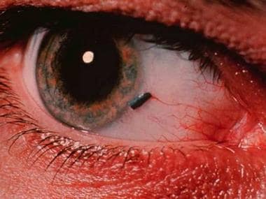
Before pressing on the eyelid or eye, make certain that no penetrating injury of the eye has occurred. Such an injury should always be considered when there is a history of power tool use or of hammering metal with metal (see the image below). If something on the bulbar conjunctiva is not easily removable with a cotton swab, it could be a penetrating foreign body or a conjunctival pigmented lesion.
Complication prevention
To minimize the chance of complications, take a careful history, and always perform a visual acuity determination and a corneal and anterior chamber examination. In addition, use an ophthalmoscope to look for the presence of a red reflex. An isolated conjunctival foreign body has few complications but may be associated with more significant ocular trauma.
Corneal abrasions related to conjunctival foreign bodies must be recognized and treated appropriately to prevent corneal infection or scarring.
Periprocedural Care
Equipment
The following equipment is required for removal of a conjunctival foreign body:
-
Topical anesthetic drops
-
A cotton swab moistened with saline
-
A cotton applicator stick or paper clip for eversion of the upper lid
-
A saline irrigation bottle
-
Fluorescein stain
-
A source of illumination, such as a penlight or a minor operating room light
-
A head-mounted magnification device
-
If a slit lamp is available, no additional illumination or magnification is necessary
Patient Preparation
Conjunctival foreign bodies can often be removed without anesthesia. If anesthesia is necessary, instill 2 drops of proparacaine 0.5% (Alcaine, Ophthetic) into the affected eye. Keep in mind that, once these drops are administered, the patient usually states that the symptoms have resolved.
The patient should be comfortably seated, with the back of the head against a firm surface.
Alternatively, the patient can be recumbent, with the head on a pillow. If a slit lamp is available, the procedure may be performed at the instrument lamp, with the patient's head in the chin rest and the forehead pressed against the plastic strap.
Technique
If a patient complains that something has gotten into the eye, it is usually in the upper or lower conjunctival fornix, more commonly the upper one. Do not be content with finding only 1 foreign body: search for more than 1, and expect to find several more.
First, inspect the eye for any foreign bodies on the bulbar conjunctiva, which overlies the sclera (the white of the eye). Have the patient look up, down, left, and right. The eye may be somewhat reddened if a conjunctival foreign body is present.
Next, inspect the inferior conjunctival cul-de-sac by having the patient look up while the examiner pulls the lower lid down.
Elevation of the upper lid while the patient looks down usually does not permit effective visualization of foreign bodies in the upper conjunctival cul-de-sac. Upper lid eversion is required. Have the patient look down, then place a cotton stick applicator or paper clip in the upper lid recess. While the patient continues to look down, grasp the upper lid margin lashes with the fingertips of one hand, and pull the lid downward and toward you (see the image below). The conjunctival surface of the inside of the upper lid is now visible.

Remove the cotton applicator stick or paper clip, and use a thumb to press the lid margin of the everted lid against the patient's brow (see the first image below). The stiff tarsal plate usually keeps the upper lid everted after the swab is removed, as long as the patient continues looking down. Looking up returns the lid to its usual position. A foreign body on the conjunctival surface of the upper lid will usually be easily seen when the lid is everted (see the second image below).


Conjunctival foreign bodies located in any of these areas can be removed by gently wiping the foreign body with a saline-moistened cotton swab. If a visible foreign body does not adhere to the moistened swab, irrigation with saline may be attempted.
If the patient's discomfort before attempted foreign body removal precludes the use of these techniques, 2 drops of local anesthetic may be placed into the eye to allow examination and treatment.
Once a drop of anesthetic is placed in the eye of a patient with foreign body sensation, the physician is committed to either finding a conjunctival foreign body or finding another definite reason for the foreign body sensation. At this point, relief of symptoms is no longer a measure of successful diagnosis and treatment. Be sure that the patient's symptoms are resolved by the physician's actions, not by the anesthetic agent.
If, after a topical anesthetic agent has been used, doubt remains as to whether the cause of the foreign body sensation has been found, wait at least 30 minutes for the anesthetic to wear off, then reassess the patient to ensure that the problem has been properly addressed. In a patient with successful removal of a conjunctival foreign body, no symptoms of foreign body sensation should return after the topical anesthetic has worn off.
After conjunctival foreign bodies are removed, always examine the patient’s cornea carefully and stain it with fluorescein; this should be done even if a foreign body was not found on the conjunctiva. Foreign bodies of the upper lid often cause vertical scratches in the cornea as a result of the foreign body having rubbed against the cornea with each blink. Transparent foreign bodies can be invisible in the tear film and become more visible using fluorescein stain.
If no topical anesthetic was used, removal of the foreign body causes immediate and almost total resolution of the symptoms—and evokes marked gratitude on the part of the patient—in most cases. If significant symptoms persist, consider the possibility of a second foreign body or a significant corneal abrasion (either causally related to the foreign body or occurring as an independent injury).
Following removal of the conjunctival foreign body, two drops of a topical broad-spectrum antibiotic drop should be placed in the affected eye. If there is no corneal abrasion or significant inflammation of the eye, no further treatment or follow-up is necessary. The patient should be instructed to return if the foreign body sensation returns or if any symptoms of pain, redness, or visual changes occur.
-
Eversion of upper lid: step 1.
-
Eversion of upper lid: step 2.
-
Conjunctival foreign body on upper lid.
-
Penetrating metallic foreign body—not simple conjunctival foreign body.