WO2014134587A1 - Anti-p40 antibodies systems and methods - Google Patents
Anti-p40 antibodies systems and methods Download PDFInfo
- Publication number
- WO2014134587A1 WO2014134587A1 PCT/US2014/019705 US2014019705W WO2014134587A1 WO 2014134587 A1 WO2014134587 A1 WO 2014134587A1 US 2014019705 W US2014019705 W US 2014019705W WO 2014134587 A1 WO2014134587 A1 WO 2014134587A1
- Authority
- WO
- WIPO (PCT)
- Prior art keywords
- antibody
- seq
- antibodies
- staining
- fragment
- Prior art date
Links
Classifications
-
- C—CHEMISTRY; METALLURGY
- C07—ORGANIC CHEMISTRY
- C07K—PEPTIDES
- C07K16/00—Immunoglobulins [IGs], e.g. monoclonal or polyclonal antibodies
- C07K16/18—Immunoglobulins [IGs], e.g. monoclonal or polyclonal antibodies against material from animals or humans
-
- C—CHEMISTRY; METALLURGY
- C07—ORGANIC CHEMISTRY
- C07K—PEPTIDES
- C07K16/00—Immunoglobulins [IGs], e.g. monoclonal or polyclonal antibodies
- C07K16/18—Immunoglobulins [IGs], e.g. monoclonal or polyclonal antibodies against material from animals or humans
- C07K16/28—Immunoglobulins [IGs], e.g. monoclonal or polyclonal antibodies against material from animals or humans against receptors, cell surface antigens or cell surface determinants
-
- C—CHEMISTRY; METALLURGY
- C07—ORGANIC CHEMISTRY
- C07K—PEPTIDES
- C07K16/00—Immunoglobulins [IGs], e.g. monoclonal or polyclonal antibodies
- C07K16/18—Immunoglobulins [IGs], e.g. monoclonal or polyclonal antibodies against material from animals or humans
- C07K16/32—Immunoglobulins [IGs], e.g. monoclonal or polyclonal antibodies against material from animals or humans against translation products of oncogenes
-
- G—PHYSICS
- G01—MEASURING; TESTING
- G01N—INVESTIGATING OR ANALYSING MATERIALS BY DETERMINING THEIR CHEMICAL OR PHYSICAL PROPERTIES
- G01N33/00—Investigating or analysing materials by specific methods not covered by groups G01N1/00 - G01N31/00
- G01N33/48—Biological material, e.g. blood, urine; Haemocytometers
- G01N33/50—Chemical analysis of biological material, e.g. blood, urine; Testing involving biospecific ligand binding methods; Immunological testing
- G01N33/5005—Chemical analysis of biological material, e.g. blood, urine; Testing involving biospecific ligand binding methods; Immunological testing involving human or animal cells
- G01N33/5008—Chemical analysis of biological material, e.g. blood, urine; Testing involving biospecific ligand binding methods; Immunological testing involving human or animal cells for testing or evaluating the effect of chemical or biological compounds, e.g. drugs, cosmetics
- G01N33/5014—Chemical analysis of biological material, e.g. blood, urine; Testing involving biospecific ligand binding methods; Immunological testing involving human or animal cells for testing or evaluating the effect of chemical or biological compounds, e.g. drugs, cosmetics for testing toxicity
- G01N33/5017—Chemical analysis of biological material, e.g. blood, urine; Testing involving biospecific ligand binding methods; Immunological testing involving human or animal cells for testing or evaluating the effect of chemical or biological compounds, e.g. drugs, cosmetics for testing toxicity for testing neoplastic activity
-
- G—PHYSICS
- G01—MEASURING; TESTING
- G01N—INVESTIGATING OR ANALYSING MATERIALS BY DETERMINING THEIR CHEMICAL OR PHYSICAL PROPERTIES
- G01N33/00—Investigating or analysing materials by specific methods not covered by groups G01N1/00 - G01N31/00
- G01N33/48—Biological material, e.g. blood, urine; Haemocytometers
- G01N33/50—Chemical analysis of biological material, e.g. blood, urine; Testing involving biospecific ligand binding methods; Immunological testing
- G01N33/53—Immunoassay; Biospecific binding assay; Materials therefor
- G01N33/574—Immunoassay; Biospecific binding assay; Materials therefor for cancer
- G01N33/57407—Specifically defined cancers
- G01N33/57423—Specifically defined cancers of lung
-
- G—PHYSICS
- G01—MEASURING; TESTING
- G01N—INVESTIGATING OR ANALYSING MATERIALS BY DETERMINING THEIR CHEMICAL OR PHYSICAL PROPERTIES
- G01N33/00—Investigating or analysing materials by specific methods not covered by groups G01N1/00 - G01N31/00
- G01N33/48—Biological material, e.g. blood, urine; Haemocytometers
- G01N33/50—Chemical analysis of biological material, e.g. blood, urine; Testing involving biospecific ligand binding methods; Immunological testing
- G01N33/53—Immunoassay; Biospecific binding assay; Materials therefor
- G01N33/574—Immunoassay; Biospecific binding assay; Materials therefor for cancer
- G01N33/57484—Immunoassay; Biospecific binding assay; Materials therefor for cancer involving compounds serving as markers for tumor, cancer, neoplasia, e.g. cellular determinants, receptors, heat shock/stress proteins, A-protein, oligosaccharides, metabolites
- G01N33/57488—Immunoassay; Biospecific binding assay; Materials therefor for cancer involving compounds serving as markers for tumor, cancer, neoplasia, e.g. cellular determinants, receptors, heat shock/stress proteins, A-protein, oligosaccharides, metabolites involving compounds identifable in body fluids
-
- G—PHYSICS
- G01—MEASURING; TESTING
- G01N—INVESTIGATING OR ANALYSING MATERIALS BY DETERMINING THEIR CHEMICAL OR PHYSICAL PROPERTIES
- G01N33/00—Investigating or analysing materials by specific methods not covered by groups G01N1/00 - G01N31/00
- G01N33/48—Biological material, e.g. blood, urine; Haemocytometers
- G01N33/50—Chemical analysis of biological material, e.g. blood, urine; Testing involving biospecific ligand binding methods; Immunological testing
- G01N33/53—Immunoassay; Biospecific binding assay; Materials therefor
- G01N33/577—Immunoassay; Biospecific binding assay; Materials therefor involving monoclonal antibodies binding reaction mechanisms characterised by the use of monoclonal antibodies; monoclonal antibodies per se are classified with their corresponding antigens
-
- C—CHEMISTRY; METALLURGY
- C07—ORGANIC CHEMISTRY
- C07K—PEPTIDES
- C07K2317/00—Immunoglobulins specific features
- C07K2317/20—Immunoglobulins specific features characterized by taxonomic origin
- C07K2317/21—Immunoglobulins specific features characterized by taxonomic origin from primates, e.g. man
-
- C—CHEMISTRY; METALLURGY
- C07—ORGANIC CHEMISTRY
- C07K—PEPTIDES
- C07K2317/00—Immunoglobulins specific features
- C07K2317/20—Immunoglobulins specific features characterized by taxonomic origin
- C07K2317/24—Immunoglobulins specific features characterized by taxonomic origin containing regions, domains or residues from different species, e.g. chimeric, humanized or veneered
-
- C—CHEMISTRY; METALLURGY
- C07—ORGANIC CHEMISTRY
- C07K—PEPTIDES
- C07K2317/00—Immunoglobulins specific features
- C07K2317/30—Immunoglobulins specific features characterized by aspects of specificity or valency
- C07K2317/34—Identification of a linear epitope shorter than 20 amino acid residues or of a conformational epitope defined by amino acid residues
-
- C—CHEMISTRY; METALLURGY
- C07—ORGANIC CHEMISTRY
- C07K—PEPTIDES
- C07K2317/00—Immunoglobulins specific features
- C07K2317/50—Immunoglobulins specific features characterized by immunoglobulin fragments
- C07K2317/56—Immunoglobulins specific features characterized by immunoglobulin fragments variable (Fv) region, i.e. VH and/or VL
- C07K2317/565—Complementarity determining region [CDR]
Definitions
- This invention relates to the novel anti-p40 antibodies, compositions, cocktails, and kits comprising the antibodies and methods for using the antibodies.
- IHC immunohistochemistry
- p53 is a tumor suppressor gene that may be mutated in numerous human cancers, (e.g., see article, "p53 Isoforms: An Intracellular Microprocessor?” Khoury MP, Bourdon JC. Genes Cancer. 2011 Apr;2(4):453-65, and article "p53/p63/p73 isoforms: an orchestra of isoforms to harmonise cell differentiation and response to stress.” Murray-Zmijewski F, Lane DP, Bourdon JC. Cell Death Differ. 2006 Jun;13(6):962-72, each hereby incorporated by reference herein.) Inactivation of p53 may be one of the most common genetic alterations in human cancers and may even be present in about 50% of all human cancers.
- p53 may respond to a wide variety of cellular stresses, including DNA damage, hypoxia, and metabolic changes, perhaps by activating cellular pathways that may result in cell cycle arrest or apoptosis.
- p73 may have been cloned and identified as a member of the p53 family, based on sequence homology with key regions of p53. p73 may have demonstrated similar transcriptional activities to p53; however, p73 may not exhibit different activities than p53.
- a gene may have been cloned and the cloned genetic sequence may even have corresponded to a protein of approximately 40-kDa. Therefore, this newly identified gene, an additional member of the p53 family may have been identified as p40. (e.g., see article, "A new human p53 homologue.”
- the p40/p63/p51/p73L gene may have been found to be overexpressed in cell lines of head and neck squamous cell carcinomas and primary lung squamous cell carcinomas, (e.g., see article, "AIS is an oncogene amplified in squamous cell carcinoma.”
- the p40/p63/p51/p73L gene may be referred to as AIS (amplified in squamous cell carcinoma).
- AIS amplified in squamous cell carcinoma.
- the longest isoforms of the p40/p63/p51/p73L gene, perhaps including transcriptional activation domains, may be those cloned and named as p63.
- p40/p63/p51/p73L gene may frequently be referred to as p63; however, it should be noted that p40, p63, p51, p73L, and AIS are all terms for the same gene, which may produce transcripts of various lengths, perhaps resulting in protein isoforms of correspondingly various lengths.
- the full length of the p63 gene may encode an N-terminal transcriptional activation domain, a DNA binding domain, and a carboxy-oligomerization domain.
- p63 may have two promoters, which perhaps results in two distinctly different classes of proteins, where one class may contain the transactivation domain (TA) (perhaps known as p63 or TAp63) and the other class may lack the N-terminal transactivation domain (perhaps known as p40 or ⁇ 63). (e.g., see article, "p40: A p63 Isoform Useful for Lung Cancer Diagnosis - A Review of the Physiological and Pathological Role of p63.” Nobre AR, Albergaria A, Schmitt F. Acta Cytol.
- the p40 protein may act as a dominant negative factor for transcription by either competing for DNA binding sites or directly binding to p53, p63, or p73 isoforms, perhaps inhibiting their ability to induce transcription. However, in some instances, p40 may activate transcription of genes that are perhaps otherwise not activated by the TA forms of p53, p63, or p73. In one view, the p63 gene may be thought of as producing a family of opposing molecules: perhaps proteins containing a N-terminal transactivation domain, with p53-like tumor suppressor properties, and perhaps proteins lacking an N-terminal domain, with oncogenic properties.
- p63 isoforms may be restricted to the nuclei of basal cells of normal epithelia, such as skin, esophagus, and urothelium or the like, and basal cells of glandular structures in prostate, breast and bronchi or the like. In these cells, expression of p40 may be found to be perhaps about 100-fold higher than that of TAp63. Although p40 and TAp63 may exhibit overlapping tissue distributions, TAp63 may be more expressed in differentiated cells and perhaps less expressed in basal cells, relative to p40 expression.
- Determining expression levels of the TAp63 and p40 isoforms may have been useful in diagnosis of cancer.
- IHC using antibodies that bind the TAp63 and/or p40 proteins may have been useful for detecting protein expression and perhaps diagnosing cancer, particularly lung, prostate, breast, and bladder cancer.
- a rabbit polyclonal (RP) anti- p40 antibody may have been produced and used in an IHC method to identify expression of p40 in head and neck squamous cell carcinoma cell lines and primary lung tumors of the squamous cell carcinoma type, (e.g., see article, "A new human p53 homologue.”
- RP polyclonal
- a mouse monoclonal anti-p63 antibody (4A4) may also have been produced and used in an IHC method to detect isoforms of p63 protein, (e.g., see article, "p63, a p53 homolog at 3q27-29, encodes multiple products with transactivating, death-inducing, and dominant- negative activities.”
- p63 a p53 homolog at 3q27-29, encodes multiple products with transactivating, death-inducing, and dominant- negative activities.
- the RP anti-p40 antibody may bind an epitope sequence that is unique to the p40 isoform, a sequence that is perhaps not present in TAp63.
- the anti-p63 antibody 4A4 may bind an epitope sequence that may be common to both the TA and p40 isoforms, perhaps resulting in 4A4 exhibiting properties of a pan-p63 marker by recognizing both classes of proteins derived from the p63 gene, whereas the RP anti-p40 antibody may recognize only the p40 isoform and may perhaps be a more selective marker.
- Non-small cell lung carcinoma comprises approximately 80% of lung cancers and may be classified into several histological types, most commonly may include adenocarcinoma (ADC) or even squamous cell carcinoma (SCC). Classification of lung carcinomas into histological types may be performed by morphological examination using hematoxylin and eosin (H&E) or IHC, and in some cases even, mucin stains; however, accurate classification can be difficult with poorly differentiated or even undifferentiated lung carcinoma.
- ADC adenocarcinoma
- SCC squamous cell carcinoma
- Diagnosis can be further complicated by the use of needle core biopsies, which may provide limited amounts of tissue for both immunohistochemistry and molecular testing, and may include crush artifacts. Additionally, cytology specimens may lack morphological features necessary for diagnosis with H&E alone.
- bevacizumab a therapeutic humanized monoclonal antibody targeting vascular endothelial growth factor
- NSCLC patients may be a common treatment for NSCLC patients; however, patients with the SCC subtype should not receive bevacizumab, perhaps due to about 30% mortality rate by fatal pulmonary hemorrhage.
- enhanced efficacy may have been demonstrated with the addition of premextred to conventional chemotherapy in non-squamous cell carcinomas, but may not in SCC. Therefore, accurate methods for subtyping NSCLC specimens may be useful for the best patient care, with optimal therapeutic efficacy and minimal adverse effects.
- IHC may be commonly used to assist pathologists in determining histologic subtype of NSCLC specimens, perhaps particularly discriminating ADC from SCC, as well as from Small Cell Carcinomas of the lung.
- IHC antibodies to p40 may be useful in the diagnosis of lung cancer, and perhaps in discriminating histologic subtypes, such as ADC and SCC.
- p40 protein may not have been detected in cases of lung adenocarcinoma, (e.g., see article, "AIS is an oncogene amplified in squamous cell carcinoma.” Hibi K, Trink B, Patturajan M, Westra WH, Caballero OL, Hill DE, Ratovitski EA, Jen J, Sidransky D. Proc Natl Acad Sci U S A. 2000 May 9;97(10):5462-7, hereby incorporated by reference herein.)
- the RP anti-p40 antibody may be superior to the pan-p63 antibody 4A4 for the diagnosis of squamous cell carcinoma, and that perhaps the RP anti-p40 antibody may be more specific.
- both the RP anti-p40 antibody and pan-p63 antibody stained all 81 cases of SCC that were evaluated, perhaps indicating that both antibodies exhibit equal sensitivity in SCC.
- the pan-p63 antibody stained 74/237 (about 31%) of ADC cases, while the RP anti-p40 antibody stained only 7/205 (about 3%) of ADC cases, perhaps indicating that the RP anti-p40 antibody may be more specific than the pan-p63 4A4.
- pan-p63 4A4 antibody stained 82/152 (about 54%) of the large cell lymphoma cases tested, whereas, the RP anti-p40 antibody lacked staining in any of these cases, perhaps resulting in improved specificity for the RP anti-p40 antibody.
- This study may have shown that IHC detection of p40 instead of detection of multiple p63 isoforms with 4A4 prevents misinterpretation of poorly differentiated ADC or lymphoma as SCC. (e.g., see article, "p40 ( ⁇ 63) is superior to p63 for the diagnosis of pulmonary squamous cell carcinoma.”
- RP anti-p40 antibody in identifying SCC, perhaps in combination with an anti-TTF-1 antibody, which may be a marker for ADC.
- IHC of small tissue samples from biopsy specimens 45/46 (about 98%) cases of ADC and SCC were diagnosed, using a combination of RP anti-p40 and anti-TTF-1 antibodies (e.g., see article, " ⁇ 63 (p40) and thyroid transcription factor- 1 immunoreactivity on small biopsies or cellblocks for typing non-small cell lung cancer: a novel two-hit, sparing-material approach.”
- an antibody cocktail containing the RP anti-p40 antibody and an anti-TTF-1 antibody resulted in about 92% sensitivity and about 93% specificity of the RP anti-p40 antibody for SCC, and about 77% sensitivity and about 100% specificity of anti- TTF-1 for ADC.
- TAp63 may be expressed in basal and intermediate urothelial cells, whereas p40 may not be detected.
- p40 expression may be detected by IHC in urothelial carcinoma, perhaps in 29/147 (about 19.7%) of non-invasive cases, and possibly 23/55 (about 41.8%) cases of invasive carcinoma, (e.g., see article, "Distinct expression profiles of p63 variants during urothelial development and bladder cancer progression.”
- Karni-Schmidt O Castillo-Martin M, Shen TH, Gladoun N, Domingo- Domenech J, Sanchez-Carbayo M, Li Y, Lowe S, Prives C, Cordon-Cardo C. Am J Pathol.
- Expression of p40 may also be a prognostic factor in urothelial carcinoma.
- cases of muscle invasive urothelial carcinoma that demonstrate expression of p40 may exhibit a median + SD overall survival of perhaps about 11.6 ⁇ about 1.3 months, perhaps indicating that p40 expression is a predictor of poor prognosis.
- cases of invasive urothelial carcinoma that lack p40 expression may have demonstrated a longer median + SD overall survival of perhaps about 25 + about 6.4 months, perhaps indicating that a lack of p40 expression is a predictor of better prognosis.
- total p63 expression may not be a prognostic indicator in invasive carcinoma, perhaps because TAp63 and p40 isoforms exhibit opposing biological activities.
- a marker that is specific for the p40 isoform may be used for urothelial carcinoma, compared to a pan-p63 marker.
- Loss of expression of p40 which may normally be expressed in basal cells of prostate glands, may commonly be used as an indicator adenocarcinoma of the prostate. Specifically, the quantity of p40 mRNA transcript may be lower, perhaps approximately about 2000-times lower or perhaps even undetectable, in prostate cancer cell lines compared to the amount of TAp63 mRNA. Therefore, perhaps p40 is virtually absent in prostate cancer cell lines, whereas TAp63 mRNA is detectable at various levels.
- a method that may detect the loss of p40 may be preferred over a method that may detect TAp63 (e.g., see article, "p63 is a prostate basal cell marker and is required for prostate development.” Signoretti S, Waltregny D, Dilks J, Isaac B, Lin D, Garraway L, Yang A, Montironi R, McKeon F, Loda M. Am J Pathol.
- myoepithelial cells may form a continuous basal rim along the epithelial structure.
- the gradual loss of continuity of this basal boundary may be indicative of carcinoma in situ, and the total loss of myoepithelial cells may often be observed in invasive breast carcinoma.
- Benign lesions may typically retain their continuous myoepithelial layer, while morphologically resembling a malignant lesion.
- Nuclear staining of myoepithelial cells by IHC may be observed using an anti-p40 antibody.
- an anti-p40 antibody may be useful for evaluating the continuity of the myoepithelial cells in the basal layer and thus aid in the diagnosis of breast lesions, (e.g., see article, "p63, a p53 homologue, is a selective nuclear marker of myoepithelial cells of the human breast.”
- Barbareschi M Pecciarini L, Cangi MG, Macri E, Rizzo A, Viale G, Doglioni C. Am J Surg Pathol. 2001 Aug;25(8): 1054-60, hereby incorporated by reference herein.
- Embodiments of the present invention provide an anti-p40 mouse monoclonal antibody [clone BC28] which may be highly sensitive and may even be highly specific.
- An example of the present invention provides a mouse monoclonal anti-p40 antibody that may detect the presence or absence of p40 protein in certain cancers, including but not limited to SCC, bladder, breast and prostate cancer or the like.
- SCC single cell lung cancer
- bladder a single cell lung cancer
- breast and prostate cancer including but not limited to human cells.
- an example of the present invention may have demonstrated excellent sensitivity for lung SCC (about 65/67, about 97%) with perhaps even excellent specificity versus lung ADC (about 0/71, about 0%).
- the mouse monoclonal anti-p40 BC28 may have typically demonstrated cleaner staining patterns, perhaps with fewer artifacts, as well as a lack of staining of macrophages, while even offering the advantages of a monoclonal antibody. BC28 also may not stain some specimens of lung ADC, which may have been stained by the RP anti-p40 antibody, perhaps indicating the superior specificity of BC28 over alternatives. Therefore, a monoclonal anti-p40 antibody, such as BC28, may be preferred for diagnosis, compared to other pan-p63 markers or alternative anti-p40 antibodies.
- an anti-p40 antibody may aid in the diagnosis of primary and even metastatic cancers, particularly lung SCC, urothelial carcinoma, prostate adenocarcinoma, and breast carcinoma or the like, and may even aid in distinguishing protein expression of p40 versus TAp63.
- New anti-p40 antibodies such as mouse monoclonal anti-p40 antibody [BC28], with perhaps equal or superior staining sensitivity, and perhaps even superior staining specificity such as compared to the pan-p63 marker 4A4 and the RP anti-p40 antibody, have been provided in the present invention.
- anti-p40 antibody clones such as the anti-p40 antibody clone BC28 can be obtained by immunizing Balb/C mice with one or more peptides corresponding to a subset of amino acids 1-17 of the human p40 protein.
- the p40 peptide may be injected into the BALB/c mice, with an adjuvant, via intraperitoneal injections, perhaps about 5 times at about three week intervals.
- the immune reactivity to p40 may be assessed by direct ELISA on recombinant p40 protein.
- Mice with the highest titer may be chosen for developing hybridomas by cell fusion.
- a hybridoma clone demonstrating the best reactivity to p40 on human tissues may be chosen and may be designated as BC28.
- the BC28 clone may be tested for isotype and may be identified as a mouse IgGl/kappa.
- the BC28 antibody may be produced by large scale tissue culture of the hybridoma cells and by ascites in BALB/c mice. The supernatant and antibody ascites may be collected and the antibody may be purified by Protein A affinity column.
- BC28 may demonstrate specific reactivity to human p40 protein by ELISA, Western blotting, and even human tissues.
- Anti-p40 antibodies such as the mouse monoclonal anti-p40 antibody BC28 may be useful for the detection of p40 in tissue samples, perhaps with several significant, but unexpected advantages over currently known antibodies to TAp63 and p40 isoforms.
- anti-p40 antibodies such as the mouse anti-p40 antibody BC28 may result in nuclear staining of p40 with sensitivity perhaps similar to that of known anti-p63 and anti-p40 antibodies.
- anti-p40 antibodies such as BC28 may exhibit increased specificity, perhaps as compared to anti-p63 antibodies and other known anti-p40 antibodies, which may offer significant improvements.
- anti-p40 antibodies such as BC28 may also offer cleaner staining, with fewer artifacts, and greater cell-type specificity, for example perhaps not staining macrophages, when compared to other known anti-p40 and anti-p63 antibodies.
- anti-p40 antibodies such as BC28, analysis of the sample may be simplified and p40 expression in tumor cells may be readily identifiable, allowing diagnosis in cases that may otherwise be difficult, ambiguous, or not even possible, to diagnose.
- Figure 1 shows a black and white version of an example of anti-p40 antibody BC28 staining a case of lung squamous cell carcinoma.
- Figure 2 shows a black and white version of an example of anti-p40 antibody BC28 staining a case of lung squamous cell carcinoma.
- Figure 3 shows a black and white version of an example of RP anti-p40 antibody staining the same case of lung squamous cell carcinoma as shown in Figure 2.
- Figure 4 shows a black and white version of an example of anti-p40 antibody BC28 on a case of lung adenocarcinoma, with no staining by BC28.
- Figure 5 shows a black and white version of an example of RP anti-p40 antibody staining the same case of lung adenocarcinoma as shown in Figure 4.
- Figure 6 shows a black and white version of an example of anti-p40 antibody BC28 staining a case of bladder cancer.
- Figure 7 shows a black and white version of an example of anti-p63 antibody 4A4 staining the same specimen of bladder cancer as shown in Figure 6.
- Figure 8 shows a black and white version of an example of anti-p40 antibody BC28 staining a case of bladder cancer.
- Figure 9 shows a black and white version of an example of anti-p40 antibody BC28 staining normal prostate glands.
- Figure 10 shows a black and white version of an example of anti-p40 antibody BC28 on a case of prostate adenocarcinoma, with no staining by BC28.
- Figure 11 shows a black and white version of an example of anti-p40 antibody BC28 staining normal breast ducts.
- Figure 12 shows a black and white version of an example of anti-p40 antibody BC28 staining a case of breast ductal carcinoma in situ (DCIS).
- DCIS breast ductal carcinoma in situ
- Figure 13 shows a black and white version of an example of anti-p40 antibody BC28 staining a case of basal cell carcinoma of the skin.
- Figure 14 shows a black and white version of an example of anti-p40 antibody BC28 staining a case of squamous cell carcinoma of the larynx.
- Figure 15 shows a black and white version of an example of anti-p40 antibody BC28 staining a case of squamous cell carcinoma of the epiglottis.
- Figure 16 shows a black and white version of an example of moderate staining of intraalveolar macrophages in the same specimen of lung in Figure 17, by the RP anti-p40 antibody.
- Figure 17 shows a black and white version of an example of very weak, or perhaps the absence of staining of intraalveolar macrophages in the lung by anti-p40 antibody BC28.
- Figure 18 shows a black and white version of an example of anti-p40 antibody BC28 staining small intestine, with very weak, or perhaps the absence of background staining.
- Figure 19 shows a black and white version of an example of anti-p63 antibody 4A4 staining the same case of small intestine as shown in Figure 18, where background staining is present.
- Figure 20 shows a black and white version of an example of anti-p40 antibody BC28 staining nuclei of myoepithelial cells of a breast duct, without cytoplasmic staining.
- Figure 21 shows a black and white version of an example of anti-p63 antibody 4A4 staining the same breast specimen of Figure 20; however, strong, cytoplasmic staining is present.
- Figure 22 shows a black and white version of an example of anti-p40 antibody BC28 staining prostate tissue, perhaps a specimen of prostatic intraepithelial neoplasia (PIN), with strong nuclear staining of the basal cells of the gland. Staining is slightly more intense and sharper than that of Figure 23.
- PIN prostatic intraepithelial neoplasia
- Figure 23 shows a black and white version of an example of anti-p63 antibody 4A4 staining the same prostate specimen of Figure 22, with strong nuclear staining of the basal cells of the gland. Staining is slightly less intense and less sharp than that of BC28 in Figure 22. Also, fewer basal cells are stained by 4A4 than BC28.
- Figure 24 shows a black and white version of an example of anti-p40 antibody BC28 staining prostate tissue, perhaps a specimen of prostatic intraepithelial neoplasia (PIN), with strong nuclear staining of the basal cells of the gland.
- PIN prostatic intraepithelial neoplasia
- Figure 25 shows a black and white version of an example of anti-p63 antibody 4A4 staining the same prostate specimen of Figure 22, with strong nuclear staining of the basal cells of the gland.
- Figure 26 shows a black and white version of an example of DSG-3 + CK5 + p40 [BC28] + Napsin A staining a specimen of lung squamous cell carcinoma. Staining of DSG- 3 (brown) is membranous; staining of CK5 (brown) is cytoplasmic; staining of p40 (brown) is nuclear. Staining of Napsin A may be reduced, or perhaps restricted to residual normal lung in this specimen.
- Figure 27 shows a black and white version of an example of DSG-3 + CK5 + p40 [BC28] + Napsin A staining a specimen of lung adenocarcinoma. Staining of Napsin A (red) is cytoplasmic. Staining of DSG-3, CK5 and p40 (brown) may be reduced, or perhaps absent, in this sample.
- Figure 28 shows a black and white version of an example of DSG-3 + CK5 + p40 [BC28] staining a specimen of lung squamous cell carcinoma. Staining of DSG-3 (brown) is membranous; staining of CK5 (brown) is cytoplasmic; staining of p40 (brown) is nuclear.
- Figure 29 shows a black and white version of an example of DSG-3 + CK5 + p40 [BC28] staining a specimen of lung adenocarcinoma. Staining of DSG-3, CK5 and p40 (brown) may be reduced, or perhaps absent, in this sample.
- Figure 30 shows a black and white version of an example of the antibody cocktail DSG-3 + p40 [BC28] + Napsin A staining a specimen of lung adenocarcinoma. Staining of Napsin A (red) is cytoplasmic. Staining of DSG-3 (brown, membranous) and p40 (brown, nuclear) may be reduced, or perhaps absent, in this sample.
- Figure 31 shows a black and white version of an example of the antibody cocktail DSG-3 + p40 [BC28] + Napsin A staining a specimen of lung adenocarcinoma. Staining of Napsin A (red) is cytoplasmic. Staining of DSG-3 (brown, membranous) and p40 (brown, nuclear) may be reduced, or perhaps restricted to normal lung tissue, in this sample.
- Figure 32 shows a black and white version of an example of the antibody cocktail
- Figure 33 shows a black and white version of an example of the antibody cocktail DSG-3 + p40 [BC28] + Napsin A staining a specimen of lung squamous cell carcinoma. Staining of DSG-3 (brown) is membranous and staining of p40 (brown) in nuclear. Napsin A (red, cytoplasmic) may be reduced, or perhaps restricted to normal lung tissue, in this sample.
- Figure 34 shows a black and white version of an example of the antibody cocktail DSG-3 + p40 [BC28] + Napsin A staining a specimen of lung squamous cell carcinoma. Staining of p40 (brown) in nuclear. Staining of DSG-3 (brown, membranous) and/or Napsin A (red, cytoplasmic) may be reduced, or perhaps absent, in this sample.
- Figure 35 shows a black and white version of an example of the antibody cocktail DSG-3 + p40 [BC28] staining a specimen of lung squamous cell carcinoma. Staining of DSG-3 (brown) is membranous and staining of p40 (brown) in nuclear.
- Figure 36 shows a black and white version of an example of the antibody cocktail p40 [BC28] + CK5 (RM) staining a specimen of lung squamous cell carcinoma. Staining of p40 (brown) is nuclear and staining of CK5 (red) is cytoplasmic.
- Figure 37 shows a black and white version of an example of the antibody cocktail p40 [BC28] + CK5 (RM) staining a specimen of lung adenocarcinoma. Staining of p40 (brown, nuclear) and CK5 (red, cytoplasmic) may be reduced, or perhaps absent, in this sample.
- Figure 38 shows a black and white version of an example of the antibody cocktail of p40 [BC28] + Napsin A staining a specimen of lung squamous cell carcinoma. Staining of p40 (brown) in nuclear. Napsin A (red, cytoplasmic) may be reduced, or perhaps restricted to normal lung tissue, in this sample.
- Figure 39 shows a black and white version of an example of the antibody cocktail of p40 [BC28] + Napsin A staining a specimen of lung adenocarcinoma. Staining of Napsin A (red) is cytoplasmic. Staining of p40 (brown, nuclear) may be reduced, or perhaps absent, in this sample.
- Figure 40 shows a black and white version of an example of an antibody cocktail of p40 [BC28] + HMWCK + P504S staining a specimen of prostate, which may contain areas of perhaps normal prostate, perhaps prostatic adenocarcinoma and perhaps even prostatic intraepithelial neoplasia (PIN). Staining of p40 (brown) is nuclear. Staining of HMWCK (brown) is cytoplasmic. Staining of P504S (red) is cytoplasmic, and perhaps granular.
- Figure 41 shows a black and white version of an example of an antibody cocktail of p40 [BC28] + CK5/14 + P504S staining a specimen of prostate, which may contain areas of perhaps normal prostate, perhaps prostatic adenocarcinoma and perhaps even prostatic intraepithelial neoplasia (PIN). Staining of p40 (brown) is nuclear. Staining of CK5/14 (brown) is cytoplasmic. Staining of P504S (red) is cytoplasmic, and perhaps granular.
- Figure 42 shows a black and white version of an example of an antibody cocktail of p40 [BC28] + CK5/14 + CK7/18 staining a specimen of breast tissue, perhaps abnormal tissue, or perhaps even breast cancer. Staining of p40 (brown) is nuclear. Staining of CK5/14 (brown) is cytoplasmic. Staining of CK7/18 (red) is cytoplasmic.
- Figure 43 shows a black and white version of an example of an antibody cocktail of p40 [BC28] + CK5/14 + CK7/18 staining a specimen of breast tissue, perhaps abnormal tissue, or perhaps even breast cancer. Staining of p40 (brown) is nuclear and perhaps reduced, or absent, in this sample. Staining of CK5/14 (brown) and CK7/18 (red) is cytoplasmic and membranous. The staining pattern observed is perhaps bimodal staining.
- Figure 44 shows a black and white version of an example of an antibody cocktail of p40 [BC28] + CK5/14 + CK8/18 staining a specimen of breast tissue, perhaps abnormal tissue, or perhaps even breast cancer. Staining of p40 (brown) is nuclear and perhaps reduced, or absent, in this sample. Staining of CK5/14 (brown) and CK8/18 (red) is cytoplasmic.
- Figure 45 shows a black and white version of an example of an antibody cocktail of p40 [BC28] + Uroplakin II + Uroplakin III staining a specimen of urothelial carcinoma. Staining of p40 (brown) is nuclear. Staining of Uroplakin II (brown) and Uroplakin III (brown) is cytoplasmic and membranous.
- Figure 46 shows a black and white version of an example of an antibody cocktail of p40 [BC28] + Uroplakin II + Uroplakin III + GATA-3 staining a specimen of urothelial carcinoma. Staining of p40 (brown) and GATA-3 (red) is nuclear. Staining of Uroplakin II (brown) and Uroplakin III (brown) is cytoplasmic and membranous.
- Figure 47 shows a black and white version of an example of an antibody cocktail of p40 [BC28] + CK5 staining a specimen of lung squamous cell carcinoma. Staining of p40 (brown) is nuclear and staining of CK5 (brown) is cytoplasmic.
- Figure 48 shows a black and white version of the reactivity of anti-p40 antibody BC28 with p40 protein (left panel, lane 2) and TAp63 protein (left panel, lane 3) by Western blot.
- the right panel shows the reactivity of anti-p63 antibody 4A4 with p40 protein (right panel, lane 2) and TAp63 protein (right panel, lane 3) by Western Blot.
- Lane 1 is control lysate in both panels.
- Figure 49 shows an example of a schematic summary of a kit in accordance with various embodiments of the present invention.
- Figure 50 shows an example of a schematic summary of an immunoassay method in accordance with various embodiments of the present invention.
- Figure 51 shows the color version of Figure 1 of an example of anti-p40 antibody BC28 staining a case of lung squamous cell carcinoma.
- Figure 52 shows the color version of Figure 2 of an example of anti-p40 antibody
- Figure 53 shows the color version of Figure 3 of an example of RP anti-p40 antibody staining the same case of lung squamous cell carcinoma as shown in Figure 52.
- Figure 54 shows the color version of Figure 4of an example of anti-p40 antibody BC28 on a case of lung adenocarcinoma, with no staining by BC28.
- Figure 55 the color version of Figure 5of an example of RP anti-p40 antibody staining the same case of lung adenocarcinoma as shown in Figure 54.
- Figure 56 shows the color version of Figure 6 of an example of anti-p40 antibody BC28 staining a case of bladder cancer.
- Figure 57 shows the color version of Figure 7of an example of anti-p63 antibody
- Figure 58 shows the color version of Figure 8 of an example of anti-p40 antibody BC28 staining a case of bladder cancer.
- Figure 59 shows the color version of Figure 9 of an example of anti-p40 antibody BC28 staining normal prostate glands.
- Figure 60 shows the color version of Figure 10 of an example of anti-p40 antibody BC28 on a case of prostate adenocarcinoma, with no staining by BC28.
- Figure 61 shows the color version of Figure 11 of an example of anti-p40 antibody BC28 staining normal breast ducts.
- Figure 62 shows the color version of Figure 12 of an example of anti-p40 antibody
- Figure 63 shows the color version of Figure 13 of an example of anti-p40 antibody BC28 staining a case of basal cell carcinoma of the skin.
- Figure 64 shows the color version of Figure 14 of an example of anti-p40 antibody BC28 staining a case of squamous cell carcinoma of the larynx.
- Figure 65 shows the color version of Figure 15of an example of anti-p40 antibody BC28 staining a case of squamous cell carcinoma of the epiglottis.
- Figure 66 shows the color version of Figure 16of an example of moderate staining of intraalveolar macrophages in the same specimen of lung in Figure 67, by the RP anti-p40 antibody.
- Figure 67 shows the color version of Figure 17of an example of very weak, or perhaps the absence of staining of intraalveolar macrophages in the lung by anti-p40 antibody BC28.
- Figure 68 shows the color version of Figure 18of an example of anti-p40 antibody BC28 staining small intestine, with very weak, or perhaps the absence of background staining.
- Figure 69 shows the color version of Figure 19of an example of anti-p63 antibody 4A4 staining the same case of small intestine as shown in Figure 18, where background staining is present.
- Figure 70 shows the color version of Figure 20of an example of anti-p40 antibody
- Figure 71 shows the color version of Figure 21 of an example of anti-p63 antibody 4A4 staining the same breast specimen of Figure 70; however, strong, cytoplasmic staining is present.
- Figure 72 shows the color version of Figure 22of an example of anti-p40 antibody
- Figure 73 shows the color version of Figure 23of an example of anti-p63 antibody 4A4 staining the same prostate specimen of Figure 72, with strong nuclear staining of the basal cells of the gland. Staining is slightly less intense and less sharp than that of BC28 in Figure 72. Also, fewer basal cells are stained by 4A4 than BC28.
- Figure 74 shows the color version of Figure 24of an example of anti-p40 antibody BC28 staining prostate tissue, perhaps a specimen of prostatic intraepithelial neoplasia (PIN), with strong nuclear staining of the basal cells of the gland.
- PIN prostatic intraepithelial neoplasia
- Figure 75 shows the color version of Figure 25of an example of anti-p63 antibody
- Figure 76 shows the color version of Figure 26of an example of DSG-3 + CK5 + p40 [BC28] + Napsin A staining a specimen of lung squamous cell carcinoma. Staining of DSG- 3 (brown) is membranous; staining of CK5 (brown) is cytoplasmic; staining of p40 (brown) is nuclear. Staining of Napsin A may be reduced, or perhaps restricted to residual normal lung in this specimen.
- Figure 77 shows the color version of Figure 27of an example of DSG-3 + CK5 + p40 [BC28] + Napsin A staining a specimen of lung adenocarcinoma. Staining of Napsin A (red) is cytoplasmic. Staining of DSG-3, CK5 and p40 (brown) may be reduced, or perhaps absent, in this sample.
- Figure 78 shows the color version of Figure 28of an example of DSG-3 + CK5 + p40 [BC28] staining a specimen of lung squamous cell carcinoma. Staining of DSG-3 (brown) is membranous; staining of CK5 (brown) is cytoplasmic; staining of p40 (brown) is nuclear.
- Figure 79 shows the color version of Figure 29of an example of DSG-3 + CK5 + p40
- Figure 80 shows the color version of Figure 30of an example of the antibody cocktail DSG-3 + p40 [BC28] + Napsin A staining a specimen of lung adenocarcinoma. Staining of Napsin A (red) is cytoplasmic. Staining of DSG-3 (brown, membranous) and p40 (brown, nuclear) may be reduced, or perhaps absent, in this sample.
- Figure 81 shows the color version of Figure 31 of an example of the antibody cocktail DSG-3 + p40 [BC28] + Napsin A staining a specimen of lung adenocarcinoma. Staining of Napsin A (red) is cytoplasmic. Staining of DSG-3 (brown, membranous) and p40 (brown, nuclear) may be reduced, or perhaps restricted to normal lung tissue, in this sample.
- Figure 82 shows the color version of Figure 32of an example of the antibody cocktail
- Figure 83 shows the color version of Figure 33of an example of the antibody cocktail DSG-3 + p40 [BC28] + Napsin A staining a specimen of lung squamous cell carcinoma. Staining of DSG-3 (brown) is membranous and staining of p40 (brown) in nuclear. Napsin A (red, cytoplasmic) may be reduced, or perhaps restricted to normal lung tissue, in this sample.
- Figure 84 shows the color version of Figure 34of an example of the antibody cocktail DSG-3 + p40 [BC28] + Napsin A staining a specimen of lung squamous cell carcinoma. Staining of p40 (brown) in nuclear. Staining of DSG-3 (brown, membranous) and/or Napsin A (red, cytoplasmic) may be reduced, or perhaps absent, in this sample.
- Figure 85 shows the color version of Figure 35of an example of the antibody cocktail DSG-3 + p40 [BC28] staining a specimen of lung squamous cell carcinoma. Staining of DSG-3 (brown) is membranous and staining of p40 (brown) in nuclear.
- Figure 86 shows the color version of Figure 36of an example of the antibody cocktail p40 [BC28] + CK5 (RM) staining a specimen of lung squamous cell carcinoma. Staining of p40 (brown) is nuclear and staining of CK5 (red) is cytoplasmic.
- Figure 87 shows the color version of Figure 37of an example of the antibody cocktail p40 [BC28] + CK5 (RM) staining a specimen of lung adenocarcinoma. Staining of p40 (brown, nuclear) and CK5 (red, cytoplasmic) may be reduced, or perhaps absent, in this sample.
- p40 brown, nuclear
- CK5 red, cytoplasmic
- Figure 88 shows the color version of Figure 38of an example of the antibody cocktail of p40 [BC28] + Napsin A staining a specimen of lung squamous cell carcinoma. Staining of p40 (brown) in nuclear. Napsin A (red, cytoplasmic) may be reduced, or perhaps restricted to normal lung tissue, in this sample.
- Figure 89 shows the color version of Figure 39of an example of the antibody cocktail of p40 [BC28] + Napsin A staining a specimen of lung adenocarcinoma. Staining of Napsin A (red) is cytoplasmic. Staining of p40 (brown, nuclear) may be reduced, or perhaps absent, in this sample.
- Figure 90 shows the color version of Figure 40of an example of an antibody cocktail of p40 [BC28] + HMWCK + P504S staining a specimen of prostate, which may contain areas of perhaps normal prostate, perhaps prostatic adenocarcinoma and perhaps even prostatic intraepithelial neoplasia (PIN). Staining of p40 (brown) is nuclear. Staining of HMWCK (brown) is cytoplasmic. Staining of P504S (red) is cytoplasmic, and perhaps granular.
- Figure 91 shows the color version of Figure 41 of an example of an antibody cocktail of p40 [BC28] + CK5/14 + P504S staining a specimen of prostate, which may contain areas of perhaps normal prostate, perhaps prostatic adenocarcinoma and perhaps even prostatic intraepithelial neoplasia (PIN). Staining of p40 (brown) is nuclear. Staining of CK5/14 (brown) is cytoplasmic. Staining of P504S (red) is cytoplasmic, and perhaps granular.
- Figure 92 shows the color version of Figure 42of an example of an antibody cocktail of p40 [BC28] + CK5/14 + CK7/18 staining a specimen of breast tissue, perhaps abnormal tissue, or perhaps even breast cancer. Staining of p40 (brown) is nuclear. Staining of CK5/14 (brown) is cytoplasmic. Staining of CK7/18 (red) is cytoplasmic.
- Figure 93 shows the color version of Figure 43of an example of an antibody cocktail of p40 [BC28] + CK5/14 + CK7/18 staining a specimen of breast tissue, perhaps abnormal tissue, or perhaps even breast cancer. Staining of p40 (brown) is nuclear and perhaps reduced, or absent, in this sample. Staining of CK5/14 (brown) and CK7/18 (red) is cytoplasmic and membranous. The staining pattern observed is perhaps bimodal staining.
- Figure 94 shows the color version of Figure 44of an example of an antibody cocktail of p40 [BC28] + CK5/14 + CK8/18 staining a specimen of breast tissue, perhaps abnormal tissue, or perhaps even breast cancer. Staining of p40 (brown) is nuclear and perhaps reduced, or absent, in this sample. Staining of CK5/14 (brown) and CK8/18 (red) is cytoplasmic.
- Figure 95 shows the color version of Figure 45of an example of an antibody cocktail of p40 [BC28] + Uroplakin II + Uroplakin III staining a specimen of urothelial carcinoma. Staining of p40 (brown) is nuclear. Staining of Uroplakin II (brown) and Uroplakin III (brown) is cytoplasmic and membranous.
- Figure 96 shows the color version of Figure 46of an example of an antibody cocktail of p40 [BC28] + Uroplakin II + Uroplakin III + GATA-3 staining a specimen of urothelial carcinoma. Staining of p40 (brown) and GATA-3 (red) is nuclear. Staining of Uroplakin II (brown) and Uroplakin III (brown) is cytoplasmic and membranous.
- Figure 97 shows the color version of Figure 47of an example of an antibody cocktail of p40 [BC28] + CK5 staining a specimen of lung squamous cell carcinoma. Staining of p40 (brown) is nuclear and staining of CK5 (brown) is cytoplasmic.
- Figure 98 shows the color version of Figure 48of the reactivity of anti-p40 antibody
- the right panel shows the reactivity of anti-p63 antibody 4A4 with p40 protein (right panel, lane 2) and TAp63 protein (right panel, lane 3) by Western Blot.
- Lane 1 is control lysate in both panels.
- the present invention includes a variety of aspects, which may be combined in different ways.
- the following descriptions are provided to list elements and describe some of the embodiments of the present invention. These elements are listed with initial embodiments, however it should be understood that they may be combined in any manner and in any number to create additional embodiments.
- the variously described examples and preferred embodiments should not be construed to limit the present invention to only the explicitly described systems, techniques, and applications. Further, this description should be understood to support and encompass descriptions and claims of all the various embodiments, systems, techniques, methods, devices, and applications with any number of the disclosed elements, with each element alone, and also with any and all various permutations and combinations of all elements in this or any subsequent application.
- Embodiments of the present invention may provide monoclonal antibodies and methods thereof that specifically bind to p40 and may be used for the detection of p40 in the diagnosis for several types of cancers.
- the monoclonal antibody may be an antibody fragment, a mouse monoclonal antibody, a rabbit monoclonal antibody, a chimeric antibody, a humanized monoclonal antibody, a human monoclonal antibody, an antibody labeled with a detectable signal or stain, an antibody labeled with a toxin, or the like.
- Systems and methods of the present invention may relate to the monoclonal antibody or its antigen binding portion capable of binding to p40.
- Mouse monoclonal antibodies may be commonly used in immunoassay methods to identify specific analytes, including as primary antibodies in immunohistochemistry procedures.
- Mouse monoclonal antibodies specific for the protein target of interest can be produced using generally known procedures.
- exposing a mouse to the antigen of interest e.g. a peptide fragment of the desired target or the full-length protein target
- the antigen of interest e.g. a peptide fragment of the desired target or the full-length protein target
- B-cells may be isolated from the mouse spleen and the antibodies produced may be evaluated for their suitability as primary antibodies in IHC.
- the associated B-cell may be fused with a tumor cell using known procedures, perhaps resulting in a hybridoma, a new cell line that can endlessly replicate and may continuously produce the desired antibody.
- Monoclonal antibodies may be preferred in certain embodiments over polyclonal antibodies for several reasons.
- monoclonal antibodies may be derived from a single B-cell and as such may recognize a single epitope, perhaps resulting in greater specificity.
- Monoclonal antibodies may also be conveniently and reproducibly generated in cell culture, perhaps resulting in a constant supply of the desired antibody.
- polyclonal antibodies may be utilized in other embodiments.
- Anti-p40 antibodies such as a mouse monoclonal anti-p40 antibody BC28 may be produced using these general procedures and may be evaluated by immunohistochemistry for sensitivity and specificity on a variety of normal and neoplastic tissues, perhaps particularly in comparison to the previously known RP anti-p40 antibody and anti-p63 antibody [4A4].
- a p40 peptide from amino acid sequence 1 to 17aa, corresponding to the sequence MLYLENNAQTQFSEPQY, may be synthesized on a semi-automatic peptide synthesizer.
- a p40 peptide from amino acid sequence 5 to 17aa, corresponding to the sequence ENNAQTQFSEPQY may be synthesized on a semiautomatic peptide synthesizer.
- the peptide may be covalently conjugated to a carrier molecule.
- a keyhole limpet haemocyanin (KLH) carrier molecule may be used.
- the peptide may be also conjugated to goat gamma globulin (GGG) for immunoassay.
- mice Female BALB/c (about 6 to about 8 weeks old) mice may be immunized intraperitoneally (i.p.) with about 10( g of human p40 peptide per mouse in complete Freund' s adjuvant.
- p40 peptides used for immunization may correspond to the amino acid sequence of 1-17 from p40, the amino acid sequence of 1-9 from p40, a mixture of the amino acid sequences 1-17 with 5-17 from p40, or any portion of this sequence, and all permutations and combinations thereof.
- a single peptide may be used as the immunogen, or perhaps a mixture of peptides derived from the amino acid sequence of 1-17, 1-9, a mixture of 1-17 with 5-17, or the like.
- an immunogen that includes the amino acid at position 4, or perhaps includes the amino acids in position 1, 2 or 3, may be used and may produce antibodies with advantageous properties, such as those observed with BC28.
- the mice may be boosted with another about 100 ⁇ g human p40 per mouse in incomplete Freund' s adjuvant about 4 more times in about 3 week intervals.
- Mice may be bled from the tails, and sera may be collected and stored at about -20°C for later analysis of antibody titers by enzyme-linked immunosorbent assay (ELISA).
- ELISA enzyme-linked immunosorbent assay
- Hybridomas producing antibodies to p40 may be generated by standard techniques from splenocytes of p40-immunized BALB/c mice. For example, splenocytes from p40-immunized mice may be fused to P3-X63-Ag 8.653 myeloma cells (non-secreting myeloma cells derived from Balb/c mouse) by incubation with about 50% polyethylene glycol at a ratio of about 4: 1.
- cells may be pelleted by centrifugation perhaps at about 300 x g for about 10 minutes, washed in about 25 ml of PBS, recentrifuged, and the cell pellet may be resuspended in about 100 ml of fresh Dulbecco's Medium containing about 20% fetal bovine serum (Hyclone, Logan, UT). Aliquots of about 100 ⁇ can be added to each well of about ten 96- well microtiter plates (Corning, Lowell, MA). About twenty four hours later, about 100 ⁇ DMEM culture medium supplemented with hypoxanthine-aminopterin-thymidine (HAT) can be added to each microtiter well.
- HAT hypoxanthine-aminopterin-thymidine
- Media may be replaced perhaps after about 4 days with complete media (perhaps containing hypoxanthine-thymidine (HT)). Over the following about 10 days, media may be removed and replaced with fresh media with reduced or perhaps even no HT added.
- Hybridoma supernatants may be screened by ELISA for antibody reactivity to p40, and hybridoma clones may then be selected and stabilized perhaps by cloning twice by limiting dilution.
- Hybridoma cells referred to as Anti-human p40 hybridoma clone BC28 Lot:011713 have been deposited at American Type Culture Collection (ATCC) in Manassas, Virginia on January 29, 2013 and has received ATCC Patent Deposit Designation No. PTA-120163 as shown in the attached exhibit entitled, "Budapest Restricted Certificate of Deposit” hereby incorporated by reference herein.
- Embodiments of the present invention may provide an antibody or fragment thereof produced by the hybridoma deposited at the ATCC and may even include a method for producing a monoclonal antibody by culturing the hybridoma cell which produces the monoclonal antibody capable of specifically recognizing p40 and even allowing the hybridoma to produce monoclonal antibodies.
- ELISA Host anti-sera immune responses to p40 may be measured by ELISA.
- a solution of p40 (about ⁇ g/ml) in phosphate-buffered saline (PBS) may be used to coat about 96-well flat bottom polystyrene plates. The plates may then be blocked with about 1% bovine serum albumin (BSA)-PBS. Either diluted immune sera or hybridoma supernatants may be added and incubated at about 37 °C for about 1 hour. After washing the plates with PBS, the plates may be incubated with goat anti-mouse-HRP reagents (Jackson Labs). Incubations may be done at about 37°C for about 30 minutes.
- ABTS substrate may be added to develop color and the absorbance at about 405 nm (A405) may be measured in a microtiter plate reader.
- Anti-p40 antibodies such as the BC28 monoclonal antibody may be isotyped using a mouse monoclonal antibody isotyping kit (Invitrogen, Carlsbad CA). For example, about 100 ⁇ of supernatant from mouse monoclonal antibody [BC28] cells may be added to the plate coated goat anti mouse IgGl, IgG2A, IgG2B, IgG3, IgM, and IgA heavy chains, kappa and lambda light chains. After about 30 minutes incubation, the plate may be washed about 3 times with PBS and may be incubated with goat anti mouse Ig-HRP reagent.
- ABTS substrate may be added to develop color and the absorbance at about 405 nm (A405) may be measured in a microtiter plate reader.
- the BC28 clone may be tested for isotype and may be identified as a mouse IgGl/kappa.
- the selected hybridoma cells from clone BC28 may be cultured with DMEM culture medium supplemented with about 10% FBS or any serum-free medium.
- the culture supernatants may be further purified by protein A affinity column.
- the hybridoma cells may also be injected into pristane-primed BALB/c mice to produce antibody ascites.
- the antibody ascites may be further purified by protein A affinity column.
- IgG concentration may be measured spectrophotometrically using the extinction coefficient for human IgG of about 1.4 at about 280nm.
- the purity of IgG may be determined by SDS-PAGE.
- the purified monoclonal antibody [BC28] may be characterized by Western Blotting ( Figure 48 and 98).
- Full-length p40 or TAp63 transfected cell lysates (Origene, Rockville, MD) may be subjected to protein gel electrophoresis using about 4 to about 12% SDS-PAGE with Tris-glycine buffer and may be transferred onto nitrocellulose filters in Tris-glycine buffer.
- Proteins on the blots may be visualized by incubating p63 monoclonal antibody 4A4 or BC28 antibody for about 60 minutes in room temperature after blocking with blocking buffer, perhaps followed by incubating with peroxidase-conjugated goat anti-mouse immunoglobulins.
- the blots may be detected using TMB chromogen.
- Total RNA may be extracted from hybridomas using Qiagen kit (USA, Gaithersburg, MD) as per the manufacturer's instructions.
- First-round RT-PCR may be carried out with QIAGEN® OneStep RT-PCR Kit.
- RT-PCR may be performed with primer sets specific for the heavy and light chains. For each RNA sample, about 12 individual heavy chain and about 11 light chain RT-PCR reactions can be set up using degenerate forward primer mixtures covering the leader sequences of variable regions. Reverse primers may be located in the constant regions of heavy and light chains. Restriction sites may not be engineered into the primers.
- the RT-PCR products from the first-round reactions may be amplified in the second-round PCR. About 12 individual heavy chain and about 11 light chain RT-PCR reactions can be set up using semi-nested primer sets specific for antibody variable regions.
- the amplified cDNAs can be gel purified and may then be sequenced.
- BC28 variable domains were sequenced to provide isolated polynucleotides that comprise nucleic acid sequences encoding the amino acid sequences of one or more of the CDRs of the light and/or heavy chain variable regions of a monoclonal antibody described herein that binds to the p40 epitope MLYLENN AQTQFS EPQ Y identified as SEQ ID NO: 3 or MLYLENNAQ identified as SEQ ID NO: 4.
- the sequence of the variable region of the heavy chain is identified as SEQ ID NO: 1 and the sequence of the variable region of the light chain is identified as SEQ ID NO: 2.
- An antibody or fragment thereof may include a polypeptide of the amino acid sequence encoded by the nucleic acid sequence of SEQ ID NO: 1 and/or SEQ ID NO: 2.
- An antibody or fragment thereof may include a light chain variable region having an amino acid sequence encoded by the nucleic acid sequence of SEQ ID NO: 2 and may even include a heavy chain variable region having an amino acid sequence encoded by a nucleic acid sequence of SEQ ID NO: 1.
- An antibody or fragment thereof may specifically bind to at least one polypeptide of an amino acid sequence of SEQ ID NO: 3 and/or SEQ ID NO:4. As mentioned herein, a fragment thereof may include an antigen binding fragment thereof.
- an antibody or fragment thereof, or even an isolated and purified nucleic acid sequence may have an amino acid sequence of at least about 70% identical to an amino acid sequence encoded by a nucleic acid sequence of SEQ ID NO: 1 and/or SEQ ID NO: 2 and/or the amino acid sequence of SEQ ID NO: 3 and/or SEQ ID NO: 4.
- An antibody or fragment thereof may specifically bind to at least one polypeptide with an amino acid sequence that is at least about 70% identical to residues of SEQ ID NO: 3 and/or SEQ ID NO: 4.
- percentages may include, but are not limited to, at least about 71%, at least about 72%, at least about 73%, at least about 74%, at least about 75%, at least about 76%, at least about 77%, at least about 78%, at least about 79%, at least about 80%, at least about 81%, at least about 82%, at least about 83%, at least about 84%, at least about 85%, at least about 86%, at least about 87%, at least about 88%, at least about 89%, at least about 90%, at least about 91%, at least about 92%, at least about 93%, at least about 94%, at least about 95%, at least about 96%, at least about 97%, at least about 98%, and perhaps even at least about 99%, or the like.
- Epitope Mapping of the mouse anti-p40 [BC28] Binding Sequence In order to determine the peptide sequence of p40 that is recognized by anti-p40 antibodies such as BC28, epitope mapping may be conducted perhaps using two assays: direct ELISA and even dot blot. In an ELISA assay, the sensitivity and specificity of the anti-p40 [BC28] antibody may be determined by measuring the antibody titer at about 1:500 and about 1: 1000. Overlapping peptides at a length of about 15 amino acids each, covering the human p40 protein sequence from perhaps 1 to 17 amino acids, may be used to determine a sequence of BC28 binding.
- One of the epitopes for BC28 may include the residues 1-17 amino acids of p40, which is MLYLENN AQTQFS EPQ Y identified as SEQ ID NO: 3.
- Another epitope may include the residues 1-9 amino acids of p40, which is MLYLENN AQ identified as SEQ ID NO:4.
- Yet another epitope may include a mixture of SEQ ID NO:4 and the residues 5-17 amino acids of p40 which is ENNAQTQFSEPQY identified as SEQ ID NO:5.
- Antibodies may be generated using as an immunogen a peptide sequence of SEQ ID NO: 3, SEQ ID NO:4, or perhaps even a mixture of SEQ ID NO.3 with SEQ. ID NO: 5.
- the epitope of the mouse monoclonal p40 [BC28] antibody, or a portion thereof, may be a useful antigen for the production of new monoclonal antibodies, including production in species other than mouse (e.g. rabbit, goat, horse, chicken, etc.).
- a polyclonal antibody may specifically bind to an epitope in SEQ ID NO: 3, SEQ NO ID:4, or perhaps even a mixture of SEQ ID NO: 4 and SEQ ID NO. 5, or any combination or permutation thereof.
- the plates may be first coated with about ⁇ of p40 peptides at about 5 ⁇ g/mL in coating buffer (pH about 9.5) overnight at about 4°C, followed by blocking (about 3% BSA) at about 200 ⁇ /well for about 1 hour at room temperature.
- the plates may be incubated with purified p40 antibody at about 100 ng/mL and about 200 ng/mL separately for about 1 hour at about room temperature on an ELISA-plate shaker. Then the plates may be washed perhaps five times with PBST (about 300 ⁇ 1 ⁇ 11) followed by the addition of goat anti-mouse IgG-HRP to the plates and incubation for about 1 hour on a plate-shaker.
- the plates may then be washed with PBST (about 300 ⁇ 1 ⁇ 11) and blotted to dry, and TMB may be added at about ⁇ /well, developed for about 5 min on a shaker, and may even be followed by a stop solution (about 50 ⁇ 1 /well).
- Absorbance may be measured at about 450nm on an ELISA plate reader perhaps according to the manufacturer's recommendation.
- a nitrocellulose membrane may be blotted with about 1 ⁇ at a concentration of about lmg/ml the peptide, quadruplicates per peptide. This membrane may be incubated for about 1 hour at room temperature until it may be completely dry. The membrane may be blocked with about 3% BSA in TBST (e.g., about 50 mM Tris, about 0.5 M NaCl, about 0.05% Tween-20, pH about 7.4) for about 1 hour at room temperature, then mouse anti p40 antibody [BC28] may be added at about 200ng/ml for about lhr at RT in TBST.
- BSA e.g., about 50 mM Tris, about 0.5 M NaCl, about 0.05% Tween-20, pH about 7.4
- the membrane may be washed for about 3 times (about 10 minutes each) in TBST on an orbital shaker, followed by incubating with secondary antibody goat anti mouse IgGl-AP for about 1 hour at room temperature in TBST.
- the membrane may be washed perhaps about 3 times (about 10 minutes each) in TBST on a rocker.
- the binding may be detected by adding Western Glo Chemiluminescent detection reagents and exposing to film.
- Immunohistochemistry using anti-p40 antibodies such as the mouse monoclonal anti-p40 antibody BC28 may be performed on formalin-fixed paraffin embedded (FFPE) tissue samples using procedures generally known to those in the art, as generally exemplified by the following non-limiting examples (e.g., washes with Tris-buffered saline, pH about 7.6, between steps):
- FFPE formalin-fixed paraffin embedded
- Sections ( ⁇ 5 ⁇ ) of formalin fixed paraffin-embedded tissues may be mounted on commercially available microscope slides perhaps coated with polylysine.
- Sections may be deparaffinized (using xylenes or a xylene- substitute) and may be rehydrated perhaps through a series of alcohol/water solutions, perhaps followed by blocking of endogenous peroxidases perhaps with about 3% hydrogen peroxide solution.
- Samples may be subjected to heat-induced antigen retrieval using a citrate buffer in a pressure cooker (Diva, Decloaking Chamber; Biocare Medical) and may be heated to about 125°C for about 30 seconds. [Other antigen retrieval methods known to those skilled in the art (e.g., steamer, microwave oven, enzyme, or the like) may also be acceptable.] Tissues may be allowed to cool for about 10 minutes and then may be rinsed with deionized water. 4) The p40 antibody BC28 may be applied in a phosphate-buffered solution (pH about 6.0) with bovine serum albumin as carrier protein for about 30 minutes. The p40 antibody BC28 may be diluted perhaps 1:500.
- HRP horseradish peroxidase conjugated secondary antibody
- DAB 3,3'-diaminobenzidine
- buffer perhaps containing about 0.02% hydrogen peroxide
- the oxidation of DAB through an HRP-mediated mechanism may result in precipitation of a brown, chromogenic product, perhaps allowing identification of sites of p40 expression.
- Slides may be briefly counterstained perhaps in a modified Mayer's hematoxylin.
- results of IHC Staining with mouse monoclonal anti-p40 antibody BC28 Using the above protocol, a variety of normal and neoplastic tissues were evaluated for p40 expression using BC28 and in some cases compared to staining patterns using a RP anti-p40 antibody and a mouse monoclonal anti-p63 antibody [4A4]. All antibodies were optimized for titer (e.g., concentration) using methods well known to those in the art. For example, various antibody titers were evaluated to maximize staining intensity, perhaps while minimizing or even eliminating background staining. For each antibody, the titer that provided the maximum staining intensity, perhaps with the minimal background staining, was used.
- concentration e.g., concentration
- FIG 1 and 51 shows staining of a FFPE specimen of lung squamous carcinoma by anti-p40 antibody [BC28]. Nuclear staining of p40 is observed, as expected, with no apparent background staining.
- a different case of lung squamous cell carcinoma stained with BC28 is shown in Figure 2 and 52. Staining of p40 with BC28 may be notably strong, perhaps even with no apparent background or non-specific staining. In contrast, staining the same case with RP anti-40 antibody may result in less intense staining and noticeably light background staining in the stromal tissue ( Figure 3 and 53).
- BC28 was applied to a case of lung adenocarcinoma, with perhaps no visible staining.
- Figures 6, 7, 56 and 57 show examples of staining by BC28 and RP anti-p40 on a case of bladder cancer (transitional cell carcinoma of the bladder, or urothelial carcinoma).
- strong nuclear staining of p40 may be observed, perhaps even with minimal or no background staining.
- lighter nuclear staining of p63 is observed with cytoplasmic background staining.
- Figure 8 and 58 shows an example of strong staining with BC28 in urothelial carcinoma.
- Expression of p40 may be expected in the basal cells of prostate glands, perhaps consistent with the staining observed in normal prostate with BC28 ( Figure 9 and 59). Progression from pre-malignant lesions such as prostatic intraepithelial neoplasia (PIN) to invasive prostatic adenocarcinoma may result in the loss of expression of p40 in basal cells. As a result, a lack of staining with BC28 may be a useful marker for the absence of p40 in prostate cancer, as shown in Figure 10 and 60. In some cases of prostate, BC28 may provide more intense, sharper staining, and perhaps stain a greater number of basal cells than alternative antibodies, such as 4A4 ( Figures 22 and 72, and 23 and 73, respectively). Figure 24, 74, 25 and 75 show examples of prostate tissue stained with BC28 and 4A4, respectively, at low-power magnification. In this case, BC28 and 4A4 demonstrate similar overall staining patterns, perhaps with more intense and sharper staining by BC28
- p40 may be observed in the myoepithelial cells of normal breast ducts, with the gradual loss in early lesions, and may have ultimate absence in invasive breast carcinoma. Staining of p40 in myoepithelial cells of the basal layer of normal breast ducts may beis observed with BC28 ( Figure 11 and 61). In cases of ductal carcinoma in situ, the integrity of the basal layer may be compromised and staining of p40 with BC28 may become discontinuous ( Figure 12 and 62). In some cases, alternative antibodies, such as 4A4 may produce anomalous cytoplasmic staining in breast tissue, which is absent with BC28 ( Figures 20, 70, 21, and 71). This cytoplasmic staining with 4A4 may be widespread. The absence of cytoplasmic staining with BC28 may be an advantage.
- staining with BC28 may be a useful marker to identify tumor cells by p40 expression ( Figure 13 and 63).
- staining with BC28 can be useful for identifying p40 expression in squamous cell carcinomas, perhaps even such as those of the larynx or epiglottis ( Figures 14 and 64 and 15 and 65, respectively).
- anti-p40 antibodies other than BC28 may be the staining of intraalveolar macrophages in lung tissue. Positive staining of macrophages may lead to a false positive or an incorrect diagnosis due to erroneous staining. For example, moderate staining of intraalveolar macrophages may be observed with the RP anti-p40 antibody ( Figure 16 and 66). In contrast, BC28 may not stain intraalveloar macrophages in the same manner, perhaps a distinct advantage ( Figure 17 and 67). The lack of staining of intraalveloar macrophages by BC28 may simplify a diagnostic interpretation and may even eliminate the potential error of a false positive or incorrect diagnosis perhaps due to inaccurate staining of macrophages.
- BC28 may provide similar or even superior staining to alternative antibodies. For example, staining of prostate glands with BC28 may be perhaps stronger and even possibly may stain more cells in the basal layer than the anti-p63 antibody 4A4 ( Figures 22, 72 and 23 and 73, and Figures 24 and 74 and 25 and 75).
- BC28 may have the advantage of providing cleaner nuclear staining, perhaps even without cytoplasmic staining.
- BC28 may stain a case of bladder cancer with clear, nuclear staining, and no cytoplasmic staining ( Figure 6 and 56), compared to 4A4 which may stain the same specimen with weak to moderate cytoplasmic staining ( Figure 7 and 57).
- BC28 may be negative in an example of small intestine ( Figure 18 and 68); whereas, 4A4 may show distinct cytoplasmic staining in the same case ( Figure 19 and 69).
- BC28 also may not produce cytoplasmic staining in breast tissue ( Figure 21 and 71), whereas cytoplasmic staining is observed with 4A4 ( Figure 20 and 70).
- Table 1 shows the sensitivity of anti-p40 antibody [BC28] staining 67 cases of lung squamous cell carcinoma, using a tissue microarray (TMA) of FFPE tissue.
- Table 2 shows an example of the specificity of anti-p40 antibody [BC28] staining 71 specimens of lung adenocarcinoma carcinoma, using a tissue microarray (TMA). Employing a cut-off of > about 5% of tumor cells staining as the criteria to determine a case as "positive” for p40, and conversely ⁇ about 5% of tumor cells staining as the criteria to determine a case "negative,” 0 of 71 (about 0%) were found to be positive for p40 [BC28].
- TMA tissue microarray
- Table 3 shows an example of the sensitivity of the anti-p40 antibody [BC28] in several other neoplastic tissues.
- BC28 was also highly sensitive in urothelial carcinoma of the bladder, staining 41/48 (about 85.4%) cases positive.
- Loss of p40 expression may be expected in breast cancer and prostate cancer.
- 47 cases were negative for p40.
- prostate cancer none of the 12 cases tested were positive for p40.
- anti-p40 antibody [BC28] compared to RP anti-p40 antibody and anti-p63 antibody [4A4]
- RP anti-p40 antibody [BC28] identified 65/67 specimens as positive (about 97%).
- RP anti-p40 stained 34/41 about 83%) of cases of lung squamous cell carcinoma as positive
- anti- p63 [4A4] stained 35/41 about 85% of cases as positive. Greater specificity may be observed for BC28, compared to RP anti-p40 and 4A4 on cases of lung adenocarcinoma.
- BC28 did not stain any of the 71 cases of lung adenocarcinoma that were evaluated, therefore, perhaps resulting in no false positives.
- staining with RP anti-p40 resulted in 1/50 (about 2%) cases as positive and 5/50 (about 10%) of cases were found positive when using 4A4.
- Anti-p40 antibody [BC28] may be highly specific perhaps when evaluated on a variety of normal (Table 5) tissues. Staining of normal tissues with p40 may be observed in the expected tissues based on the presence of squamous epithelium (for example, skin, esophagus, and bladder or the like) and glandular tissues (for example, breast and prostate or the like). Such staining may be expected and anti-p40 antibody [BC28] may not stain any other normal or neoplastic tissues, which may demonstrate its high specificity.
- Anti-p40 antibodies such as the monoclonal mouse anti-p40 antibody [BC28] may offer distinct advantages with its improved sensitivity, specificity, and staining properties, perhaps even as compared to RP anti-p40 antibody and monoclonal mouse anti-p63 antibody [4A4] .
- Figures 2, 52 and Figures 3, 53 show a comparison of BC28 with RP anti- p40 antibody staining serial sections of the same specimen of lung squamous cell carcinoma, perhaps demonstrating the greater staining intensity and specificity of BC28.
- the specimen of Figure 2 and 52 may exhibit strong nuclear staining with BC28 and no apparent staining in stromal tissue, while the staining of the same specimen with RP anti- p40 antibody Figure 3 and 53 may be less intense and result in weak staining in the stromal tissue.
- One important advantage of BC28 may be improved specificity against lung adenocarcinoma, perhaps even as compared to RP anti-p40 antibody or 4A4.
- Figure 5 and 55 shows a case of lung adenocarcinoma stained with RP anti-p40 antibody, resulting in positive staining; however, staining the same specimen with BC28 ( Figure 4 and 54) is entirely negative, perhaps demonstrating the superior specificity of BC28.
- the anti- p40 antibody BC28 may also offer an advantage by not staining intraalveolar macrophages in lung tissue.
- RP anti-p40 antibody may stain intraalveolar macrophages moderately to strongly ( Figure 16 and 66).
- BC28 may stain the macrophages of the same specimen either weakly, or perhaps not at all ( Figure 17 and 67), possibly demonstrating superior specificity of BC28 and even cleaner staining pattern.
- Binding of BC28 to p40 protein may be demonstrated by Western blot ( Figure 31, left panel, lane 2). The absence of similar binding of BC28 to TAp63 protein may also be shown by Western blot ( Figure 48 and 98, left panel, lane 3). Conversely, the anti-p63 antibody 4A4 may bind both p40 protein and TAp63 protein ( Figure 48 and 98, right panel, lanes 2 and 3). The greater specificity of BC28 (binding of p40, but not TAp63) may be an advantage that contributes to its superior properties.
- anti-p40 antibodies such as the mouse monoclonal anti-p40 antibody BC28 may be suitable for use in many variations of the above protocols and other methods known to those in the art. Specimens stained with BC28 may be archived using a permanent mounting media and a coverslip. The antibody BC28 may also be used in an automated staining instrument, using standard protocols.
- detection methods e.g., fluorescence
- detection enzymes e.g., alkaline phosphatase (AP), beta-galactosidase, or the like
- chromogens e.g., Fast Red, Fast Blue, 3-amino-9-ethylcarbazole, 5-bromo-4- chloro-3-indolyl phosphate, 3,3 ⁇ 5,5'-tetramethylbenzidine, 5-bromo-4-chloro-3-indolyl- - D-glucuronide, or the like
- fluorescence e.g., fluorescence
- detection enzymes e.g., alkaline phosphatase (AP), beta-galactosidase, or the like
- chromogens e.g., Fast Red, Fast Blue, 3-amino-9-ethylcarbazole, 5-bromo-4- chloro-3-indolyl phosphate, 3,3 ⁇ 5,5'-tetra
- an epitope of an anti-p40 antibody such as mouse monoclonal anti-p40 antibody, or a portion thereof, may be a useful antigen for the production of new monoclonal antibodies, including production in species other than mouse (e.g. rabbit, goat, horse, chicken, etc.) as one skilled in the art would understand.
- anti-p40 antibodies such as BC28 in immunohistochemistry of formalin-fixed paraffin embedded tissues
- its utility in other immunoassays may be readily envisioned and are meant to be included in this application.
- many of the same reagents used in IHC of FFPE may also be used in IHC of frozen-tissue sections.
- Anti-p40 antibodies such as BC28 may also be useful in other immunoassays, including ELISA, perhaps using generally known methods.
- an anti-p40 antibody may be used in conjunction with one or more additional primary antibodies as part of a cocktail, to perform a "double-stain” procedure (also described as multi-stain or even multiplex).
- double-stain procedures may be generally well known in the art; however, the best combinations of primary antibodies for a particular diagnostic application may not be known.
- anti-p40 antibodies such as a mouse monoclonal anti-p40 antibody BC28 could be combined with one or more antibodies in a single primary antibody cocktail, perhaps suitable for simultaneous application to a specimen.
- the antibodies may be derived from a mouse host or a rabbit host or the like.
- the antibodies may be monoclonal or polyclonal.
- an antibody cocktail may be used in a double-stain IHC procedure to produce two or more colored stains that may identify the presence or absence of target protein antigens in the tissue specimen.
- a detection system may include an anti-mouse antibody conjugated to horseradish peroxidase (HRP) and perhaps even an anti-rabbit antibody conjugated to alkaline phosphatase (AP) may be used to produce the two-color stain.
- HRP horseradish peroxidase
- AP alkaline phosphatase
- DAB 3,3'-diaminobenzidine
- HRP horseradish peroxidase
- AP alkaline phosphatase
- DAB 3,3'-diaminobenzidine
- Fast Red may be used to produce a fuchsia/red stain, perhaps facilitated by AP, and it may identify the presence or absence, and/or location, of rabbit antibodies in the specimen.
- a detection system may include an anti-mouse antibody conjugated to AP and an anti-rabbit antibody conjugated to HRP which may be used to produce a two-color stain that may identify the presence or absence, and/or location of the mouse antibodies with a red stain and the rabbit antibodies with a brown stain, perhaps when Fast Red and DAB may be used as chromogens.
- an anti-mouse antibody conjugated to HRP and perhaps an anti-rabbit antibody conjugated to AP may be applied to the specimen as a cocktail, in a single solution, or they may be applied in separate, sequential steps.
- the anti-mouse or anti-rabbit antibodies comprising the antibody-enzyme conjugates may be derived from a different host species, including, but not limited to mouse, rabbit, chicken, horse, rat, goat, sheep, or the like.
- a primary antibody may be from a variety of host species, including, but not limited to mouse, rabbit, chicken, horse, rat, goat, sheep, or the like.
- an antibody may include an antibody-enzyme conjugate and a primary antibody could be obtained from two different host species. Chromogens other than DAB and/or Fast Red may be used as well.
- Double-staining method including but not limited to the use of more than two antibodies, the use of species other than mouse and rabbit, other chromogens and detection systems, a different order of detection steps, and perhaps even modifications resulting in three or more colors (which may require a denaturing step).
- Embodiments of the present invention may provide a composition having at least two antibodies or fragments thereof, perhaps as a cocktail, where at least one of the two antibodies or fragments thereof specifically binds to at least p40.
- This may provide a method for detecting at least two different proteins in a biological sample perhaps by contacting a biological sample with a composition comprising at least two antibodies or fragments thereof, where at least one of the at least two antibodies or fragments thereof may bind specifically to at least p40, to form an antigen-antibody complex and an antigen- antibody complex may be detected.
- a composition may have at least one first primary antibody and at least one second primary antibody.
- At least one of the antibodies or fragments thereof may specifically bind to at least p40 and may even have a positive indication cut-off value of greater than 1% of stained cells.
- a positive indication cut-off value may provide a percentage of stained cells needed to indicate a positive staining result.
- Other cut-off value may include but are not limited to greater than about 1% of stained cells, greater than about 2% of stained cells, greater than about 3% of stained cells, greater than about 4% of stained cells, greater than about 5% of stained cells, greater than about 6% of stained cells, greater than about 7% of stained cells, greater than about 8% of stained cells, greater than about 9% of stained cells, and perhaps even greater than about 10% of stained cells, or more, or the like.
- the present invention may provide a composition with at least two antibodies or fragments thereof which may be capable of providing different visualization results such as different color results.
- a composition may provide that at least one other of an at least two antibodies or fragments thereof may bind specifically to DSG-3, CK4, Napsin A, HMWCK, p504s, CK5/14, CK7/18, CK8/18, Uroplakin II, Uroplakin III, GATA-3, and any combination thereof, or the like.
- Antibodies, compositions thereof, perhaps with anti-p40 antibodies may provide a detection system including but not limited to SCC carcinoma detection composition, bladder detection composition, breast carcinoma detection composition, prostate carcinoma detection composition, and any combination thereof, or the like.
- a single color stain may be used for a primary antibody cocktail.
- the primary antibody cocktail is comprised of antibodies all derived from the same host species, then a single detection system may be used to stain for the presence of all of the antibodies with a single color. The presence or absence of each antibody may be determined based on cellular localization, or perhaps such determination is not necessary and the staining may be interpreted effectively without identifying the presence or absence of each antibody.
- mouse monoclonal anti-p40 antibody BC28 may be combined with mouse monoclonal Desmoglein-3 (DSG-3) [BC11] in a primary antibody cocktail and used in an IHC procedure with anti-mouse conjugated HRP detection and DAB for visualization, to produce a brown stain.
- a primary antibody cocktail comprised of two or more antibodies from different host species may be used in a similar manner to produce a single color stain.
- mouse monoclonal anti-p40 BC28 may be combined with rabbit monoclonal anti-CK20 antibody EPR1622Y and used in an IHC procedure with anti-mouse conjugated HRP and anti-rabbit conjugated HRP, and DAB for visualization, to produce a brown stain.
- Certain steps of an IHC procedure may be performed sequentially or simultaneously, perhaps by using a cocktail of reagents, as known to those skilled in the art.
- a cocktail of reagents as known to those skilled in the art.
- antibodies described in a primary antibody cocktail may alternatively be applied in sequential steps of one or more antibodies.
- detection reagents may be applied simultaneously in reagent cocktail or separate reagents in sequential steps.
- a first primary antibody may be applied, followed by a first antibody-enzyme conjugate and first chromogen, and then a denaturing step, before proceeding to application of a second primary antibody, followed by a second antibody- enzyme conjugate and a second chromogen.
- a double-stain of two different colors may be achieved using primary antibodies derived from the same species.
- certain steps of an IHC procedure may be performed sequentially or simultaneously, perhaps by using a cocktail of reagents, as known to those skilled in the art.
- a cocktail of reagents as known to those skilled in the art.
- antibodies described in a primary antibody cocktail may alternatively be applied in sequential steps of one or more antibodies.
- detection reagents may be applied simultaneously in reagent cocktail or separate reagents in sequential steps.
- Antibodies that may be useful for diagnosis when combined with an anti-p40 antibody such as a mouse monoclonal anti-p40 antibody BC28 in a primary antibody cocktail for use in multi-stain procedures include:
- CK5 (Rabbit) CK5 (Cytoplasmic, Red)
- p40 [BC28] (Mouse) p40 (Nuclear, Brown) p40 staining may DS#2
- P504S (Rabbit) HMWCK (Cytoplasmic, may be decreased,
- Figure 41 91 CK5/14 (Mouse) Brown) staining may be
- P504S (Cytoplasmic, Red) prostate cancer
- Figures 42, 43, 92, CK5/14 may be useful to
- Brown lesions such as
- Figure 44 94 CK5/14 (Mouse) Brown) may be useful to
- Brown lesions such as
- Uroplakin II (Membrane Uroplakin III Figure 45, 95
- Uroplakin III Membrane observed in bladder
- Uroplakin III and cytoplasmic, Brown cancer.
- Uroplakin II (Membrane Uroplakin III
- Uroplakin III and cytoplasmic, Brown observed in bladder
- CK5 (Mouse)
- CK5 (Cytoplasmic, Brown)
- each stain may be a result of a detection system that may include an anti-mouse antibody perhaps conjugated to HRP and even an anti-rabbit antibody perhaps conjugated to AP, perhaps even with DAB and Fast Red as chromogens, which may result in brown staining for mouse antibodies and red staining for rabbit antibodies (referred to as
- the detection system may include an anti-mouse antibody perhaps conjugated to AP and even an anti-rabbit antibody perhaps conjugated to HRP, perhaps even with DAB and Fast Red as chromogens, which may result in red staining for mouse antibodies and brown staining for rabbit antibodies (referred to as DS#1).
- two colors may not be necessary because the antigens may be distinguished by cellular localization of staining, or perhaps it is not diagnostically significant to determine which antigen is staining. Other color combinations may be obtained using other detection systems or chromogens and all are meant to be included in this disclosure.
- IHC double-stain method with an antibody cocktail of DSG-3, CK5, p40 [BC28] and Napsin A Immunohistochemistry, perhaps to produce a "double-stain” or a "multiplex procedure," using antibody cocktails such as DSG-3 + CK5 + p40 [BC28] + Napsin A may be performed on formalin-fixed paraffin embedded (FFPE) tissue samples using procedures generally known to those in the art, as generally exemplified by the following non-limiting examples (e.g., washes with Tris-buffered saline, pH about 7.6, between steps):
- FFPE formalin-fixed paraffin embedded
- Sections ( ⁇ 5 ⁇ ) of formalin fixed paraffin-embedded tissues may be mounted on commercially available microscope slides perhaps coated with polylysine.
- Sections may be deparaffinized (using xylenes or a xylene- substitute) and may be rehydrated perhaps through a series of alcohol/water solutions, perhaps followed by blocking of endogenous peroxidases perhaps with about 3% hydrogen peroxide solution.
- Samples may be subjected to heat-induced antigen retrieval using a citrate buffer in a pressure cooker (Diva, Decloaking Chamber; Biocare Medical) and may be heated to about 125°C for about 30 seconds.
- a pressure cooker Diva, Decloaking Chamber; Biocare Medical
- Tissues may be allowed to cool for about 10 minutes and then may be rinsed with deionized water.
- the antibody cocktail of DSG-3 + CK5 + p40 [BC28] + Napsin A may be applied in a phosphate-buffered solution (pH about 6.0) with bovine serum albumin as carrier protein for about 30 minutes.
- HRP horseradish peroxidase
- AP goat anti-rabbit alkaline phosphatase conjugated secondary antibody
- Fast Red chromogen with naphthol phosphate substrate perhaps in a buffer of about pH 8.3 (Vulcan Fast Red, Biocare Medical) may be applied for about 20 minutes, which may produce a fuschia/red chromogenic product as a stain.
- DAB 3,3'-diaminobenzidine
- Betazoid DAB, Biocare Medical may be applied.
- the oxidation of DAB through an HRP-mediated mechanism may result in precipitation of a brown, chromogenic product, perhaps allowing identification of the sites of antigen expression for each antibody.
- the presence of p40, DSG-3 and CK5 antibodies may be determined by brown staining.
- the presence of Napsin A antibodies may be determined by red staining.
- Slides may be briefly counterstained perhaps in a modified Mayer's hematoxylin.
- Immunohistochemistry using an antibody cocktail such as DSG-3 + CK5 + p40 [BC28] may be performed on formalin-fixed paraffin embedded (FFPE) tissue samples using procedures generally known to those in the art, as generally exemplified by the following non-limiting examples (e.g., washes with Tris-buffered saline, pH about 7.6, between steps):
- FFPE formalin-fixed paraffin embedded
- Sections ( ⁇ 5 ⁇ ) of formalin fixed paraffin-embedded tissues may be mounted on commercially available microscope slides perhaps coated with polylysine.
- Sections may be deparaffinized (using xylenes or a xylene- substitute) and may be rehydrated perhaps through a series of alcohol/water solutions, perhaps followed by blocking of endogenous peroxidases perhaps with about 3% hydrogen peroxide solution.
- Samples may be subjected to heat-induced antigen retrieval using a citrate buffer in a pressure cooker (Diva, Decloaking Chamber; Biocare Medical) and may be heated to about 125°C for about 30 seconds.
- a pressure cooker Diva, Decloaking Chamber; Biocare Medical
- Tissues may be allowed to cool for about 10 minutes and then may be rinsed with deionized water.
- the antibody cocktail of DSG-3 + CK5 + p40 [BC28] may be applied in a phosphate- buffered solution (pH about 6.0) with bovine serum albumin as carrier protein for about 30 minutes.
- HRP horseradish peroxidase
- MACH 4 Universal HRP-Polymer Detection Biocare Medical
- An initial application of a rabbit anti-mouse IgG antibody for about 10 minutes may be followed by incubation with a goat anti-rabbit-HRP conjugate for about 10 minutes.
- DAB 3,3'-diaminobenzidine
- Betazoid DAB Biocare Medical
- the oxidation of DAB through an HRP-mediated mechanism may result in precipitation of a brown, chromogenic product, perhaps allowing identification of the sites of antigen expression for each antibody.
- the presence of p40, DSG-3 and CK5 antibodies may be determined by brown staining.
- Slides may be briefly counterstained perhaps in a modified Mayer's hematoxylin.
- results of IHC Staining with DSG-3, CK5, p40 [BC28] and Napsin A FFPE tissues may be stained with a cocktail of DSG-3, p40 [BC28], CK5 and Napsin A.
- An example of staining of lung SCC is shown in Figure 26 and 76.
- the presence of DSG-3, p40, and/or CK5 antibodies may result in brown staining.
- Red staining of Napsin A may be reduced or absent, or perhaps restricted to residual normal lung tissue.
- An example of staining of lung ADC is shown in Figure 27 and 77.
- the presence of Napsin A may result in the red staining.
- Brown staining from p40, DSG-3, and/or CK5 may be reduced, or absent (e.g., see Figure 27 and 77).
- results of IHC Staining with DSG-3, CK5, and p40 [BC28] Using the above single-stain protocol, FFPE tissues may be stained with a cocktail of DSG-3, p40 [BC28], and CK5.
- An example of staining of lung SCC is shown in Figure 28 and 78.
- the presence of DSG-3, p40, and/or CK5 antibodies may result in brown staining.
- using a single-color stain for detection such as goat anti-mouse HRP, is acceptable because the presence of DSG-3, p40, or CK5 all are indicative of squamous cell carcinoma.
- An example of staining of lung ADC is shown in Figure 29 and 79. Brown staining from DSG-3, p40, and/or CK5 may be reduced, or absent (e.g., see Figure 29 and 79), or even restricted to residual normal lung tissue.
- results of IHC Staining with DSG-3, p40 (M) and Napsin A Using the above double-stain protocol, FFPE tissues may be stained with a cocktail of DSG-3, p40 (M) (e.g., mouse monoclonal [BC28]) and Napsin A. Examples of staining of ADC are shown in Figures 20A and 20B. The presence of Napsin A may result in the red staining. Brown staining from p40 or DSG-3 may be reduced, or absent (e.g., see Figures 30, 80, 31 and 81), or even restricted to residual normal lung tissue (e.g., see Figure 31 and 81).
- FIGS 32, 82, 33, 83, 34, and 84 Examples of staining of SCC are shown in Figures 32, 82, 33, 83, 34, and 84.
- the presence of DSG-3 or p40 antibodies may result in brown staining.
- Red staining of Napsin A may be reduced or absent, or perhaps restricted to residual normal lung tissue (e.g., see Figures 33 and 83).
- results of IHC Staining with DSG-3 and p40 (M): Using the above single- stain protocol, with a detection system of goat anti-mouse HRP, FFPE tissues may be stained with a cocktail of DSG-3 and p40 (M). An example of staining of SCC is shown in Figure 35 and 85. The presence of DSG-3 and/or CK5 antibodies may result in the brown staining.
- results of IHC Staining with p40 (M) and CK5 (RM) Using the above double- stain protocol, FFPE tissues may be stained with a cocktail of p40 [BC28] and CK5 (RM) (e.g., rabbit monoclonal [EP1601Y]).
- An example of staining of lung SCC is shown in Figure 36 and 86.
- the presence of p40 (M) antibodies may result in the brown staining and the presence of CK5 (RM) antibodies results in the red staining.
- An example of staining of lung ADC is shown in Figure 37 and 87. No brown or red staining may be observed, suggesting perhaps the absence of p40 and CK5.
- results of IHC Staining with p40 (M) and Napsin A FFPE tissues may be stained with a cocktail of p40 (M) (e.g., mouse monoclonal [BC28]) and Napsin A.
- M e.g., mouse monoclonal [BC28]
- An example of staining of SCC is shown in Figure 38 and 88.
- the presence of p40 antibodies may result in brown staining.
- Red staining of Napsin A may be reduced or absent, or perhaps restricted to residual normal lung tissue.
- An example of staining of ADC is shown in Figure 39 and 89.
- the presence of Napsin A may result in the red staining. Brown staining from p40 may be reduced, or absent.
- results of IHC Staining with p40 [BC28], HMWCK, and P504S FFPE tissues may be stained with a cocktail of p40 [BC28], HMWCK (e.g. mouse monoclonal clone 34 ⁇ 12), and P504S (perhaps also known as AMACR, e.g. rabbit monoclonal anti-AMACR [13H4], or a rabbit polyclonal antibody).
- An example of staining of prostate tissue is shown in Figure 40 and 90.
- the presence of p40 and or HMWCK antibodies may result in brown staining.
- the presence of P504S antibodies may result in red staining.
- the tissue may contain normal prostate glands, areas of prostate cancer, or perhaps prostatic intraepithelial neoplasia (PIN).
- results of IHC Staining with p40 [BC28], CK5/14, and P504S FFPE tissues may be stained with a cocktail of p40 [BC28], CK5/14 (e.g. a combination of anti-CK5 clone [XM26] and anti-CK14 clone [LL002]), and P504S (perhaps also known as AMACR, e.g. rabbit monoclonal anti-AMACR [13H4] or a rabbit polyclonal antibody).
- An example of staining of prostate tissue is shown in Figure 41 and 91. The presence of p40 and or CK5/14 antibodies may result in brown staining.
- the presence of P504S antibodies may result in red staining.
- the tissue may contain normal prostate glands, areas of prostate cancer, or perhaps prostatic intraepithelial neoplasia (PIN).
- PIN prostatic intraepithelial neoplasia
- the examples in Figures 40, 90, 41, and 91 are representative of instances where cytokeratin markers may be interchangeable. In these examples, HMWCK and CK5/14 are both effective cytokeratin markers and may be considered effectively equivalent.
- results of IHC Staining with p40 [BC28], CK5/14, and CK7/18 FFPE tissues may be stained with a cocktail of p40 [BC28], CK5/14 (e.g. a combination of anti-CK5 clone [XM26] and anti-CK14 clone [LL002]), and CK7/18 (e.g. a combination of rabbit monoclonal anti-CK7 antibody [BC1] and rabbit monoclonal anti-CK18 antibody clone [E431-1]).
- An example of staining of breast tissue, perhaps breast cancer, is shown in Figures 42, 92, 43, and 93.
- the presence of p40 and or CK5/14 antibodies may result in brown staining.
- the presence of CK7/18 antibodies may result in red staining.
- Figure 43 and 93 may be an example of bimodal staining.
- the antibody cocktail of p63 + CK5/14 + CK7/18 may have been shown to be useful in clinical diagnosis, (e.g., see article, "Atypical ductal hyperplasia: interobserver and intraobserver variability.” Jain RK, Mehta R, Dimitrov R, Larsson LG, Musto PM, Hodges KB, Ulbright TM, Hattab EM, Agaram N, Idrees MT, Badve S. Mod Pathol.
- An antibody cocktail that uses p40 in place of p63 may be similarly useful for diagnosis, perhaps even offering the advantages of p40 [BC28].
- results of IHC Staining with p40 [BC28], CK5/14, and CK8/18 FFPE tissues may be stained with a cocktail of p40 [BC28], CK5/14 (e.g. a combination of anti-CK5 clone [XM26] and anti-CK14 clone [LL002]), and CK8/18 (e.g. a combination of rabbit monoclonal anti-CK8 antibody [EP17] and rabbit monoclonal anti-CK18 antibody clone [E431-1]).
- An example of staining of breast tissue, perhaps breast cancer, is shown in Figure 44 and 94.
- FFPE tissues may be stained with a cocktail of p40 [BC28], Uroplakin II [BC21] and Uroplakin III [BC17].
- An example of staining of bladder tissue, perhaps urothelial carcinoma, is shown in Figure 45 and 95.
- the presence of p40, Uroplakin II, and/or Uroplakin III antibodies may result in brown staining.
- the example of Figure 45 and 95 is an instance where a single-color stain, perhaps using goat anti-mouse HRP, is useful because p40, Uroplakin II or Uroplakin III all are indicative of urothelial carcinoma.
- results of IHC Staining with p40 [BC28], Uroplakin II, Uroplakin III, and GATA-3 FFPE tissues may be stained with a cocktail of p40 [BC28], Uroplakin II [BC21], Uroplakin III [BC17], and GATA-3 (e.g. rabbit anti-GATA-3 antibody).
- An example of staining of bladder tissue, perhaps urothelial carcinoma, is shown in Figure 46 and 96.
- the presence of p40, Uroplakin II, and/or Uroplakin III antibodies may result in brown staining.
- the presence of GATA-3 may result in red staining.
- results of IHC Staining with p40 (M) and CK5 (M) Using the above single- stain protocol, FFPE tissues may be stained with a cocktail of p40 [BC28] and CK5 (M) (e.g., mouse monoclonal [XM26]). An example of staining of lung SCC is shown in Figure 47 and 97. The presence of p40 and/or CK5 antibodies may result in the brown staining.
- antibodies that bind cytokeratin markers may be used in different combinations, and in some cases, interchangeably, as known to those skilled in the art.
- CK5 may perhaps be interchangeable with CK5/6 or CK5/14.
- HMWCK high molecular weight cytokeratin
- CK7 may be used interchangeably with CK8.
- inventions may include primary antibody cocktails comprised of p40
- anti-p40 antibodies other than BC28 may also be useful.
- Rabbit polyclonal or rabbit monoclonal anti-p40 antibodies may be used in some embodiments.
- a mouse monoclonal anti-p40 antibody clone other than BC28 may be similarly useful, and perhaps interchangeable with BC28.
- diagnosis may often be performed on limited tissue samples from cytology or a biopsy, and it may be important to conserve tissue for further molecular testing; therefore, an efficient approach to diagnosis that consumes minimal tissue, but provides optimal specificity and/or sensitivity may be preferred.
- a method that provides useful diagnostic information, while consuming minimal tissue from the specimen, perhaps by the application of multiple antibodies to the same ample, perhaps through use of an antibody cocktail, or by the sequential application of antibodies, or conceivably by the feature of improved sensitivity or specificity, may be used.
- an anti-p40 antibody such as a mouse monoclonal anti-p40 antibody BC28 may be specific for detection of p40 and may be useful in immunohistochemical procedures for diagnosis of several types of cancers in human tissue samples.
- anti-p40 antibody such as BC28 has advantages over RP anti-p40 antibody and anti-p63 antibody [4A4], including but not limited to greater specificity and cleaner staining, including the absence of staining of some cases of lung adenocarcinoma, as well as perhaps a lack of staining of macrophages.
- Determination of p40 levels may be a useful prognostic factor and indicator of patient outcomes.
- Loss of p40 expression, as determined by IHC, has been associated with aggressive bladder cancer and shown to be predictive of patient survival (see Matsumoto K et. al. Urology, 2008; 72:444-449, and Ohtsuka Y et. al. BJU Int, 2006; 97: 1322-1326, hereby incorporated by reference herein).
- the various systems and methods as discussed herein may provide a detection system such as but not limited to detection of cancer, diagnose or even prognose cancer, predict an outcome of cancer, assess efficacy of treatment of cancer, predict recurrence of cancer, or the like.
- Detection may be made automatically, manually, by image analysis, or the like. Detection may utilize a method including but not limited to immunohistochemistry (IHC), IHC of FFPE, IHC of frozen-tissue sections, immunocytochemistry, ELISA, or the like.
- IHC immunohistochemistry
- IHC of FFPE IHC of frozen-tissue sections
- immunocytochemistry ELISA, or the like.
- embodiments of the present invention may provide obtaining tissue from an animal or human to be tested (6), fixing or freezing said tissue (7), treating said fixed or frozen tissue to unmask epitopes to p40 (8), contacting said treated tissue with an antibody or fragment thereof as discussed herein in an amount and under conditions such that an antibody or fragment thereof binds to a p40 protein if the protein is present in said tissue (9); and perhaps even detecting the presences of said bound antibodies (10), as schematically represented in Figure 50.
- Figure 49 shows a schematic summary of various embodiments of the present invention including a kit (5) which may provide an antibody, fragment thereof, portion thereof, in a composition or even in a cocktail, perhaps even provided from a hybridoma, the antibody (1) or the like may be contacted with a biological sample (2) to form at least one antibody-antigen complex (3) which may then be detected with a detector (4).
- a kit (5) which may provide an antibody, fragment thereof, portion thereof, in a composition or even in a cocktail, perhaps even provided from a hybridoma, the antibody (1) or the like may be contacted with a biological sample (2) to form at least one antibody-antigen complex (3) which may then be detected with a detector (4).
- the present invention may provide, in embodiments, a diagnostic or even prognostic test kit which may include an antibody or fragment thereof (as discussed herein) with an antibody detection element of the antibody or fragment thereof perhaps when bound to an antigen.
- a diagnostic or even prognostic test kit which may include an antibody or fragment thereof (as discussed herein) with an antibody detection element of the antibody or fragment thereof perhaps when bound to an antigen.
- This may provide a method of contacting a biological sample with an antibody or fragment thereof and even detecting binding of, or even the presence of the antibody or fragment thereof bound to a protein or with an antigen in the biological sample perhaps using an antibody detection element.
- Embodiments may provide an immunoassay method for detecting p40 protein in a mammal or human perhaps by obtaining a tissue from an animal or a human to be tested, contacting the tissue with an antibody or fragment thereof in accordance with the various embodiments presented herein perhaps in an amount and under conditions such that the antibody or fragment thereof may bind to a p40 protein if the protein is present in the tissue; and even detecting the presence of bound antibodies.
- a biological sample may include but is not limited to blood, urine, urothelial tissue, transitional cell tissue, bladder tissue, normal tissue, neoplastic tissue, kidney tissue, ovarian tissue, thyroid tissue, endometrial tissue, renal tissue, tonsil tissue, pancreas tissue, colon tissue, lymph node tissue, neoplastic pancreatic tissue, stomach tissue, prostate tissue, lung tissue, breast tissue, or the like perhaps depending on the antibody or even cocktail being used.
- anti-p63 antibody may be replaced with an anti-p40 antibody (e.g. BC28), perhaps with equivalent, or even perhaps superior, results.
- an anti-p40 antibody e.g. BC28
- use of terms such as p40, p40 antibody, BC28, or the like may relate to anti-p40 antibodies or the like as appropriate as one skilled in the art would understand.
- the basic concepts of the present invention may be embodied in a variety of ways. It involves both antibody techniques as well as devices to accomplish the appropriate antibody.
- the antibody techniques are disclosed as part of the results shown to be achieved by the various devices described and as steps which are inherent to utilization. They are simply the natural result of utilizing the devices as intended and described.
- the devices are disclosed, it should be understood that these not only accomplish certain methods but also can be varied in a number of ways. Importantly, as to all of the foregoing, all of these facets should be understood to be encompassed by this disclosure.
- each of the various elements of the invention and claims may also be achieved in a variety of manners.
- an element is to be understood as encompassing individual as well as plural structures that may or may not be physically connected.
- This disclosure should be understood to encompass each such variation, be it a variation of an embodiment of any apparatus embodiment, a method or process embodiment, or even merely a variation of any element of these.
- the words for each element may be expressed by equivalent apparatus terms or method terms— even if only the function or result is the same. Such equivalent, broader, or even more generic terms should be considered to be encompassed in the description of each element or action.
- Senoo M. et al. A second p53-related protein, p73L, with high homology to p73, Biochem Biophys Res Commun. 1998 Jul 30;248(3):603-7
- Hibi K. et al. AIS is an oncogene amplified in squamous cell carcinoma, Proc Natl Acad
- Signoretti S. et al. p63 is a prostate basal cell marker and is required for prostate
- Barbareschi M et al. p63, a p53 homologue, is a selective nuclear marker of myoepithelial cells of the human breast,Am J Surg Pathol. 2001 Aug;25(8): 1054-60
- each of the antibody devices as herein disclosed and described ii) the related methods disclosed and described, iii) similar, equivalent, and even implicit variations of each of these devices and methods, iv) those alternative designs which accomplish each of the functions shown as are disclosed and described, v) those alternative designs and methods which accomplish each of the functions shown as are implicit to accomplish that which is disclosed and described, vi) each feature, component, and step shown as separate and independent inventions, vii) the applications enhanced by the various systems or components disclosed, viii) the resulting products produced by such systems or components, ix) each system, method, and element shown or described as now applied to any specific field or devices mentioned, x) methods and apparatuses substantially as described hereinbefore and with reference to any of the accompanying examples, xi) an apparatus for performing the methods described herein comprising means for performing the steps, xii) the various combinations and permutations of each of the elements disclosed,
- any claims set forth at any time are hereby incorporated by reference as part of this description of the invention and the incorporated claims can "depend on any other claim", and the applicant expressly reserves the right to use all of or a portion of such incorporated content of such claims as additional description to support any of or all of the claims or any element or component thereof, and the applicant further expressly reserves the right to move any portion of or all of the incorporated content of such claims or any element or component thereof from the description into the claims or vice-versa as necessary to define the matter for which protection is sought by this application or by any subsequent continuation, division, or continuation-in-part application thereof, or to obtain any benefit of, reduction in fees pursuant to, or to comply with the patent laws, rules, or regulations of any country or treaty, and such content incorporated by reference shall survive during the entire pendency of this application including any subsequent continuation, division, or continuation-in-part application thereof or any reissue or extension thereon.
Landscapes
- Health & Medical Sciences (AREA)
- Life Sciences & Earth Sciences (AREA)
- Immunology (AREA)
- Chemical & Material Sciences (AREA)
- Engineering & Computer Science (AREA)
- Molecular Biology (AREA)
- Biomedical Technology (AREA)
- Urology & Nephrology (AREA)
- Hematology (AREA)
- Medicinal Chemistry (AREA)
- General Health & Medical Sciences (AREA)
- Biochemistry (AREA)
- Cell Biology (AREA)
- Organic Chemistry (AREA)
- General Physics & Mathematics (AREA)
- Analytical Chemistry (AREA)
- Biotechnology (AREA)
- Pathology (AREA)
- Microbiology (AREA)
- Food Science & Technology (AREA)
- Physics & Mathematics (AREA)
- Oncology (AREA)
- Hospice & Palliative Care (AREA)
- Biophysics (AREA)
- Genetics & Genomics (AREA)
- Proteomics, Peptides & Aminoacids (AREA)
- Toxicology (AREA)
- Bioinformatics & Cheminformatics (AREA)
- Tropical Medicine & Parasitology (AREA)
- Chemical Kinetics & Catalysis (AREA)
- Peptides Or Proteins (AREA)
- Preparation Of Compounds By Using Micro-Organisms (AREA)
- Measuring Or Testing Involving Enzymes Or Micro-Organisms (AREA)
- Micro-Organisms Or Cultivation Processes Thereof (AREA)
- Medicines Containing Antibodies Or Antigens For Use As Internal Diagnostic Agents (AREA)
Abstract
The present invention is related to the anti-p40 antibodies, kits, cocktails, and anti-p40 antibodies for detection of cancer.
Description
ANTI-p40 ANTIBODIES SYSTEMS AND METHODS
This application is an International PCT Patent Application claiming priority to and the benefit of U.S. Provisional Application No. 61/770,956 filed February 28, 2013, hereby incorporated by reference herein in its entirety.
TECHNICAL FIELD
This invention relates to the novel anti-p40 antibodies, compositions, cocktails, and kits comprising the antibodies and methods for using the antibodies.
BACKGROUND OF THE INVENTION
Microscopic examination of tissue samples, particularly those obtained by biopsy, is a common method for diagnosis of disease. In particular, immunohistochemistry (IHC), a technique in which specific antibodies are used to detect expression of specific proteins in the tissue sample, may be a valuable tool for diagnosis, particularly for the detection and diagnosis of cancer.
p53 is a tumor suppressor gene that may be mutated in numerous human cancers, (e.g., see article, "p53 Isoforms: An Intracellular Microprocessor?" Khoury MP, Bourdon JC. Genes Cancer. 2011 Apr;2(4):453-65, and article "p53/p63/p73 isoforms: an orchestra of isoforms to harmonise cell differentiation and response to stress." Murray-Zmijewski F, Lane DP, Bourdon JC. Cell Death Differ. 2006 Jun;13(6):962-72, each hereby incorporated by reference herein.) Inactivation of p53 may be one of the most common genetic alterations in human cancers and may even be present in about 50% of all human cancers. p53 may respond to a wide variety of cellular stresses, including DNA damage, hypoxia, and metabolic changes, perhaps by activating cellular pathways that may result in cell cycle arrest or apoptosis. p73 may have been cloned and identified as a member of the p53 family, based on sequence homology with key regions of p53. p73 may have demonstrated similar transcriptional activities to p53; however, p73 may not exhibit different activities than p53. (e.g., see article, "Monoallelically expressed gene related to p53 at lp36, a region frequently deleted in neuroblastoma and other human cancers." Kaghad M, Bonnet H, Yang A, Creancier L, Biscan JC, Valent A, Minty A, Chalon P, Lelias JM, Dumont X, Ferrara P,
McKeon F, Caput D. Cell. 1997 Aug 22;90(4):809-19, each hereby incorporated by reference herein.)
Using PCR primers based on the DNA binding domains of p53 and p73, a gene may have been cloned and the cloned genetic sequence may even have corresponded to a protein of approximately 40-kDa. Therefore, this newly identified gene, an additional member of the p53 family may have been identified as p40. (e.g., see article, "A new human p53 homologue." Trink B, Okami K, Wu L, Sriuranpong V, Jen J, Sidransky D. Nat Med. 1998 Jul;4(7):747-8, hereby incorporated by reference herein.) Other isoforms of the same gene may have been cloned and their products may have been referred to as p51, p63, or p73L. (e.g., see articles, "Cloning and functional analysis of human p51, which structurally and functionally resembles p53." Osada M, Ohba M, Kawahara C, Ishioka C, Kanamaru R, Katoh I, Ikawa Y, Nimura Y, Nakagawara A, Obinata M, Ikawa S. Nat Med. 1998 Jul;4(7):839-43, and article, "p63, a p53 homolog at 3q27-29, encodes multiple products with transactivating, death-inducing, and dominant-negative activities." Yang A, Kaghad M, Wang Y, Gillett E, Fleming MD, Dotsch V, Andrews NC, Caput D, McKeon F. Mol Cell. 1998 Sep;2(3):305-16, and article, "A second p53-related protein, p73L, with high homology to p73." Senoo M, Seki N, Ohira M, Sugano S, Watanabe M, Inuzuka S, Okamoto T, Tachibana M, Tanaka T, Shinkai Y, Kato H. Biochem Biophys Res Commun. 1998 Jul 30;248(3):603-7, and article, "Cloning and functional analysis of human p51, which structurally and functionally resembles p53." Osada M, Ohba M, Kawahara C, Ishioka C, Kanamaru R, Katoh I, Ikawa Y, Nimura Y, Nakagawara A, Obinata M, Ikawa S. Nat Med. 1998 Jul;4(7):839-43, each hereby incorporated by reference herein.) Each of these isoforms may differ by the presence or even absence of an N-terminal transcriptional activating domain. p40, ΔΝρ63 and p73L may lack this domain.
The p40/p63/p51/p73L gene may have been found to be overexpressed in cell lines of head and neck squamous cell carcinomas and primary lung squamous cell carcinomas, (e.g., see article, "AIS is an oncogene amplified in squamous cell carcinoma." Hibi K, Trink B, Patturajan M, Westra WH, Caballero OL, Hill DE, Ratovitski EA, Jen J, Sidransky D. Proc Natl Acad Sci U S A. 2000 May 9;97(10):5462-7, hereby incorporated by reference herein.) Due to this amplification in squamous cell carcinomas, the p40/p63/p51/p73L gene may be referred to as AIS (amplified in squamous cell carcinoma). The longest isoforms of
the p40/p63/p51/p73L gene, perhaps including transcriptional activation domains, may be those cloned and named as p63. (e.g., see article, "p63, a p53 homolog at 3q27-29, encodes multiple products with transactivating, death-inducing, and dominant-negative activities." Yang A, Kaghad M, Wang Y, Gillett E, Fleming MD, Dotsch V, Andrews NC, Caput D, McKeon F. Mol Cell. 1998 Sep;2(3):305-16, hereby incorporated by reference herein.)
Therefore, for simplicity, the p40/p63/p51/p73L gene may frequently be referred to as p63; however, it should be noted that p40, p63, p51, p73L, and AIS are all terms for the same gene, which may produce transcripts of various lengths, perhaps resulting in protein isoforms of correspondingly various lengths.
The full length of the p63 gene may encode an N-terminal transcriptional activation domain, a DNA binding domain, and a carboxy-oligomerization domain. p63 may have two promoters, which perhaps results in two distinctly different classes of proteins, where one class may contain the transactivation domain (TA) (perhaps known as p63 or TAp63) and the other class may lack the N-terminal transactivation domain (perhaps known as p40 or ΔΝρ63). (e.g., see article, "p40: A p63 Isoform Useful for Lung Cancer Diagnosis - A Review of the Physiological and Pathological Role of p63." Nobre AR, Albergaria A, Schmitt F. Acta Cytol. 2013;57(l): l-8, hereby incorporated by reference herein.) The p40 protein may act as a dominant negative factor for transcription by either competing for DNA binding sites or directly binding to p53, p63, or p73 isoforms, perhaps inhibiting their ability to induce transcription. However, in some instances, p40 may activate transcription of genes that are perhaps otherwise not activated by the TA forms of p53, p63, or p73. In one view, the p63 gene may be thought of as producing a family of opposing molecules: perhaps proteins containing a N-terminal transactivation domain, with p53-like tumor suppressor properties, and perhaps proteins lacking an N-terminal domain, with oncogenic properties.
In normal tissue of adults, expression of p63 isoforms may be restricted to the nuclei of basal cells of normal epithelia, such as skin, esophagus, and urothelium or the like, and basal cells of glandular structures in prostate, breast and bronchi or the like. In these cells, expression of p40 may be found to be perhaps about 100-fold higher than that of TAp63. Although p40 and TAp63 may exhibit overlapping tissue distributions, TAp63 may be more expressed in differentiated cells and perhaps less expressed in basal cells, relative to p40 expression. Differences in expression patterns for the isoforms may perhaps indicate
different roles for each isoform in development and disease, (e.g., see article, "p40: A p63 Isoform Useful for Lung Cancer Diagnosis - A Review of the Physiological and Pathological Role of p63." Nobre AR, Albergaria A, Schmitt F. Acta Cytol. 2013;57(l): l-8, hereby incorporated by reference herein.)
Determining expression levels of the TAp63 and p40 isoforms may have been useful in diagnosis of cancer. In particular, IHC, using antibodies that bind the TAp63 and/or p40 proteins may have been useful for detecting protein expression and perhaps diagnosing cancer, particularly lung, prostate, breast, and bladder cancer. A rabbit polyclonal (RP) anti- p40 antibody may have been produced and used in an IHC method to identify expression of p40 in head and neck squamous cell carcinoma cell lines and primary lung tumors of the squamous cell carcinoma type, (e.g., see article, "A new human p53 homologue." Trink B, Okami K, Wu L, Sriuranpong V, Jen J, Sidransky D. Nat Med. 1998 Jul;4(7):747-8, hereby incorporated by reference herein.) A mouse monoclonal anti-p63 antibody (4A4) may also have been produced and used in an IHC method to detect isoforms of p63 protein, (e.g., see article, "p63, a p53 homolog at 3q27-29, encodes multiple products with transactivating, death-inducing, and dominant- negative activities." Yang A, Kaghad M, Wang Y, Gillett E, Fleming MD, Dotsch V, Andrews NC, Caput D, McKeon F. Mol Cell. 1998 Sep;2(3):305- 16, hereby incorporated by reference herein.) Importantly, the RP anti-p40 antibody may bind an epitope sequence that is unique to the p40 isoform, a sequence that is perhaps not present in TAp63. In contrast, the anti-p63 antibody 4A4 may bind an epitope sequence that may be common to both the TA and p40 isoforms, perhaps resulting in 4A4 exhibiting properties of a pan-p63 marker by recognizing both classes of proteins derived from the p63 gene, whereas the RP anti-p40 antibody may recognize only the p40 isoform and may perhaps be a more selective marker.
Lung cancer is the leading cause of cancer death for both men and women. More people die of lung cancer than of colon, breast, and prostate cancers combined. Non-small cell lung carcinoma (NSCLC) comprises approximately 80% of lung cancers and may be classified into several histological types, most commonly may include adenocarcinoma (ADC) or even squamous cell carcinoma (SCC). Classification of lung carcinomas into histological types may be performed by morphological examination using hematoxylin and eosin (H&E) or IHC, and in some cases even, mucin stains; however, accurate classification
can be difficult with poorly differentiated or even undifferentiated lung carcinoma. Diagnosis can be further complicated by the use of needle core biopsies, which may provide limited amounts of tissue for both immunohistochemistry and molecular testing, and may include crush artifacts. Additionally, cytology specimens may lack morphological features necessary for diagnosis with H&E alone.
Although the majority of lung cancers (particularly grades I and II) can be diagnosed with only H&E staining, with the advent of targeted therapies, diagnostic needs have changed, and an improved method for classification of a greater number of NSCLC cases is needed. In the past, histologic subtyping of NSCLCs had limited diagnostic value, due to the fact that the same treatment may have been provided to the patient, perhaps regardless of NSCLC subtype. However, the availability of targeted therapies has created a need for accurate subtyping of NSCLC. For example, bevacizumab, a therapeutic humanized monoclonal antibody targeting vascular endothelial growth factor, may be a common treatment for NSCLC patients; however, patients with the SCC subtype should not receive bevacizumab, perhaps due to about 30% mortality rate by fatal pulmonary hemorrhage. Furthermore, enhanced efficacy may have been demonstrated with the addition of premextred to conventional chemotherapy in non-squamous cell carcinomas, but may not in SCC. Therefore, accurate methods for subtyping NSCLC specimens may be useful for the best patient care, with optimal therapeutic efficacy and minimal adverse effects.
IHC may be commonly used to assist pathologists in determining histologic subtype of NSCLC specimens, perhaps particularly discriminating ADC from SCC, as well as from Small Cell Carcinomas of the lung. In particular, IHC antibodies to p40 may be useful in the diagnosis of lung cancer, and perhaps in discriminating histologic subtypes, such as ADC and SCC. Using the RP anti-p40 antibody, expression of p40 protein in primary lung SqCC may have been identified by IHC; whereas, p40 protein may not have been detected in cases of lung adenocarcinoma, (e.g., see article, "AIS is an oncogene amplified in squamous cell carcinoma." Hibi K, Trink B, Patturajan M, Westra WH, Caballero OL, Hill DE, Ratovitski EA, Jen J, Sidransky D. Proc Natl Acad Sci U S A. 2000 May 9;97(10):5462-7, hereby incorporated by reference herein.)
Further studies may have shown that the RP anti-p40 antibody may be superior to the pan-p63 antibody 4A4 for the diagnosis of squamous cell carcinoma, and that perhaps the
RP anti-p40 antibody may be more specific. In one study, both the RP anti-p40 antibody and pan-p63 antibody stained all 81 cases of SCC that were evaluated, perhaps indicating that both antibodies exhibit equal sensitivity in SCC. In contrast, the pan-p63 antibody stained 74/237 (about 31%) of ADC cases, while the RP anti-p40 antibody stained only 7/205 (about 3%) of ADC cases, perhaps indicating that the RP anti-p40 antibody may be more specific than the pan-p63 4A4. Furthermore, the pan-p63 4A4 antibody stained 82/152 (about 54%) of the large cell lymphoma cases tested, whereas, the RP anti-p40 antibody lacked staining in any of these cases, perhaps resulting in improved specificity for the RP anti-p40 antibody. This study may have shown that IHC detection of p40 instead of detection of multiple p63 isoforms with 4A4 prevents misinterpretation of poorly differentiated ADC or lymphoma as SCC. (e.g., see article, "p40 (ΔΝρ63) is superior to p63 for the diagnosis of pulmonary squamous cell carcinoma." Bishop JA, Teruya-Feldstein J, Westra WH, Pelosi G, Travis WD, Rekhtman N. Mod Pathol. 2012 Mar;25(3):405-15 and article, "A study of ΔΝρ63 expression in lung non-small cell carcinomas." Nonaka D. Am J Surg Pathol. 2012 Jun;36(6):895-9, each hereby incorporated by reference herein.)
Other studies may also have reported use of the RP anti-p40 antibody in identifying SCC, perhaps in combination with an anti-TTF-1 antibody, which may be a marker for ADC. In one such study, using IHC of small tissue samples from biopsy specimens, 45/46 (about 98%) cases of ADC and SCC were diagnosed, using a combination of RP anti-p40 and anti-TTF-1 antibodies (e.g., see article, "ΔΝρ63 (p40) and thyroid transcription factor- 1 immunoreactivity on small biopsies or cellblocks for typing non-small cell lung cancer: a novel two-hit, sparing-material approach." Pelosi G, Fabbri A, Bianchi F, Maisonneuve P, Rossi G, Barbareschi M, Graziano P, Cavazza A, Rekhtman N, Pastorino U, Scanagatta P, Papotti M. / Thorac Oncol. 2012 Feb;7(2):281-90, hereby incorporated by reference herein.) In a similar study, an antibody cocktail containing the RP anti-p40 antibody and an anti-TTF-1 antibody resulted in about 92% sensitivity and about 93% specificity of the RP anti-p40 antibody for SCC, and about 77% sensitivity and about 100% specificity of anti- TTF-1 for ADC. (e.g., see article, "Tissue-Preserving Antibody Cocktails to Differentiate Primary Squamous Cell Carcinoma, Adenocarcinoma, and Small Cell Carcinoma of Lung." Brown AF, Sirohi D, Fukuoka J, Cagle P, Policarpio-Nicolas M, Tacha D, Jagirdar J. Arch Pathol Lab Med. 2013 Jan 4, hereby incorporated by reference herein.)
In normal bladder tissue, TAp63 may be expressed in basal and intermediate urothelial cells, whereas p40 may not be detected. In contrast, p40 expression may be detected by IHC in urothelial carcinoma, perhaps in 29/147 (about 19.7%) of non-invasive cases, and possibly 23/55 (about 41.8%) cases of invasive carcinoma, (e.g., see article, "Distinct expression profiles of p63 variants during urothelial development and bladder cancer progression." Karni-Schmidt O, Castillo-Martin M, Shen TH, Gladoun N, Domingo- Domenech J, Sanchez-Carbayo M, Li Y, Lowe S, Prives C, Cordon-Cardo C. Am J Pathol. 2011 Mar; 178(3): 1350-60, hereby incorporated by reference herein.) Expression of p40 may also be a prognostic factor in urothelial carcinoma. For example, cases of muscle invasive urothelial carcinoma that demonstrate expression of p40 may exhibit a median + SD overall survival of perhaps about 11.6 ± about 1.3 months, perhaps indicating that p40 expression is a predictor of poor prognosis. In contrast, cases of invasive urothelial carcinoma that lack p40 expression may have demonstrated a longer median + SD overall survival of perhaps about 25 + about 6.4 months, perhaps indicating that a lack of p40 expression is a predictor of better prognosis. Notably, total p63 expression may not be a prognostic indicator in invasive carcinoma, perhaps because TAp63 and p40 isoforms exhibit opposing biological activities. As a result, a marker that is specific for the p40 isoform may be used for urothelial carcinoma, compared to a pan-p63 marker.
Loss of expression of p40, which may normally be expressed in basal cells of prostate glands, may commonly be used as an indicator adenocarcinoma of the prostate. Specifically, the quantity of p40 mRNA transcript may be lower, perhaps approximately about 2000-times lower or perhaps even undetectable, in prostate cancer cell lines compared to the amount of TAp63 mRNA. Therefore, perhaps p40 is virtually absent in prostate cancer cell lines, whereas TAp63 mRNA is detectable at various levels. In the differential diagnosis of benign and malignant prostate tissue, a method that may detect the loss of p40 may be preferred over a method that may detect TAp63 (e.g., see article, "p63 is a prostate basal cell marker and is required for prostate development." Signoretti S, Waltregny D, Dilks J, Isaac B, Lin D, Garraway L, Yang A, Montironi R, McKeon F, Loda M. Am J Pathol. 2000 Dec; 157(6): 1769-75, and article, "Differential expression of p63 isoforms in normal tissues and neoplastic cells." Nylander K, Vojtesek B, Nenutil R, Lindgren B, Roos
G, Zhanxiang W, Sjostrom B, Dahlqvist A, Coates PJ. J Pathol. 2002 Dec;198(4):417-27, each hereby incorporated by reference herein.)
In normal breast ducts, myoepithelial cells may form a continuous basal rim along the epithelial structure. The gradual loss of continuity of this basal boundary may be indicative of carcinoma in situ, and the total loss of myoepithelial cells may often be observed in invasive breast carcinoma. Benign lesions may typically retain their continuous myoepithelial layer, while morphologically resembling a malignant lesion. Nuclear staining of myoepithelial cells by IHC may be observed using an anti-p40 antibody. In this manner, an anti-p40 antibody may be useful for evaluating the continuity of the myoepithelial cells in the basal layer and thus aid in the diagnosis of breast lesions, (e.g., see article, "p63, a p53 homologue, is a selective nuclear marker of myoepithelial cells of the human breast." Barbareschi M, Pecciarini L, Cangi MG, Macri E, Rizzo A, Viale G, Doglioni C. Am J Surg Pathol. 2001 Aug;25(8): 1054-60, hereby incorporated by reference herein.)
Therefore, a clear need exists for a sensitive and even specific anti-p40 antibody for use in cancer diagnosis. Embodiments of the present invention provide an anti-p40 mouse monoclonal antibody [clone BC28] which may be highly sensitive and may even be highly specific. An example of the present invention provides a mouse monoclonal anti-p40 antibody that may detect the presence or absence of p40 protein in certain cancers, including but not limited to SCC, bladder, breast and prostate cancer or the like. In cases of NSCLC, an example of the present invention may have demonstrated excellent sensitivity for lung SCC (about 65/67, about 97%) with perhaps even excellent specificity versus lung ADC (about 0/71, about 0%). When compared to the RP anti-p40 antibody, the mouse monoclonal anti-p40 BC28 may have typically demonstrated cleaner staining patterns, perhaps with fewer artifacts, as well as a lack of staining of macrophages, while even offering the advantages of a monoclonal antibody. BC28 also may not stain some specimens of lung ADC, which may have been stained by the RP anti-p40 antibody, perhaps indicating the superior specificity of BC28 over alternatives. Therefore, a monoclonal anti-p40 antibody, such as BC28, may be preferred for diagnosis, compared to other pan-p63 markers or alternative anti-p40 antibodies.
The development of an anti-p40 antibody may aid in the diagnosis of primary and even metastatic cancers, particularly lung SCC, urothelial carcinoma, prostate
adenocarcinoma, and breast carcinoma or the like, and may even aid in distinguishing protein expression of p40 versus TAp63. New anti-p40 antibodies such as mouse monoclonal anti-p40 antibody [BC28], with perhaps equal or superior staining sensitivity, and perhaps even superior staining specificity such as compared to the pan-p63 marker 4A4 and the RP anti-p40 antibody, have been provided in the present invention.
DISCLOUSRE OF THE INVENTION
General embodiments of the present invention may include monoclonal antibodies for recognizing p40, methods for their preparation, use in immunohistochemistry, or the like. In embodiments, anti-p40 antibody clones such as the anti-p40 antibody clone BC28 can be obtained by immunizing Balb/C mice with one or more peptides corresponding to a subset of amino acids 1-17 of the human p40 protein. The p40 peptide may be injected into the BALB/c mice, with an adjuvant, via intraperitoneal injections, perhaps about 5 times at about three week intervals. The immune reactivity to p40 may be assessed by direct ELISA on recombinant p40 protein. Mice with the highest titer may be chosen for developing hybridomas by cell fusion. A hybridoma clone demonstrating the best reactivity to p40 on human tissues may be chosen and may be designated as BC28. The BC28 clone may be tested for isotype and may be identified as a mouse IgGl/kappa. The BC28 antibody may be produced by large scale tissue culture of the hybridoma cells and by ascites in BALB/c mice. The supernatant and antibody ascites may be collected and the antibody may be purified by Protein A affinity column. BC28 may demonstrate specific reactivity to human p40 protein by ELISA, Western blotting, and even human tissues.
Anti-p40 antibodies such as the mouse monoclonal anti-p40 antibody BC28 may be useful for the detection of p40 in tissue samples, perhaps with several significant, but unexpected advantages over currently known antibodies to TAp63 and p40 isoforms. When used in traditional immunohistochemistry procedures, anti-p40 antibodies such as the mouse anti-p40 antibody BC28 may result in nuclear staining of p40 with sensitivity perhaps similar to that of known anti-p63 and anti-p40 antibodies. However, anti-p40 antibodies such as BC28 may exhibit increased specificity, perhaps as compared to anti-p63 antibodies and other known anti-p40 antibodies, which may offer significant improvements. In addition to the possible advantages of being derived from a monoclonal source, anti-p40 antibodies
such as BC28 may also offer cleaner staining, with fewer artifacts, and greater cell-type specificity, for example perhaps not staining macrophages, when compared to other known anti-p40 and anti-p63 antibodies. With anti-p40 antibodies such as BC28, analysis of the sample may be simplified and p40 expression in tumor cells may be readily identifiable, allowing diagnosis in cases that may otherwise be difficult, ambiguous, or not even possible, to diagnose.
BRIEF DESCRIPTION OF THE DRAWINGS
Figure 1 shows a black and white version of an example of anti-p40 antibody BC28 staining a case of lung squamous cell carcinoma.
Figure 2 shows a black and white version of an example of anti-p40 antibody BC28 staining a case of lung squamous cell carcinoma.
Figure 3 shows a black and white version of an example of RP anti-p40 antibody staining the same case of lung squamous cell carcinoma as shown in Figure 2.
Figure 4 shows a black and white version of an example of anti-p40 antibody BC28 on a case of lung adenocarcinoma, with no staining by BC28.
Figure 5 shows a black and white version of an example of RP anti-p40 antibody staining the same case of lung adenocarcinoma as shown in Figure 4.
Figure 6 shows a black and white version of an example of anti-p40 antibody BC28 staining a case of bladder cancer.
Figure 7 shows a black and white version of an example of anti-p63 antibody 4A4 staining the same specimen of bladder cancer as shown in Figure 6.
Figure 8 shows a black and white version of an example of anti-p40 antibody BC28 staining a case of bladder cancer.
Figure 9 shows a black and white version of an example of anti-p40 antibody BC28 staining normal prostate glands.
Figure 10 shows a black and white version of an example of anti-p40 antibody BC28 on a case of prostate adenocarcinoma, with no staining by BC28.
Figure 11 shows a black and white version of an example of anti-p40 antibody BC28 staining normal breast ducts.
Figure 12 shows a black and white version of an example of anti-p40 antibody BC28 staining a case of breast ductal carcinoma in situ (DCIS).
Figure 13 shows a black and white version of an example of anti-p40 antibody BC28 staining a case of basal cell carcinoma of the skin.
Figure 14 shows a black and white version of an example of anti-p40 antibody BC28 staining a case of squamous cell carcinoma of the larynx.
Figure 15 shows a black and white version of an example of anti-p40 antibody BC28 staining a case of squamous cell carcinoma of the epiglottis.
Figure 16 shows a black and white version of an example of moderate staining of intraalveolar macrophages in the same specimen of lung in Figure 17, by the RP anti-p40 antibody.
Figure 17 shows a black and white version of an example of very weak, or perhaps the absence of staining of intraalveolar macrophages in the lung by anti-p40 antibody BC28.
Figure 18 shows a black and white version of an example of anti-p40 antibody BC28 staining small intestine, with very weak, or perhaps the absence of background staining.
Figure 19 shows a black and white version of an example of anti-p63 antibody 4A4 staining the same case of small intestine as shown in Figure 18, where background staining is present.
Figure 20 shows a black and white version of an example of anti-p40 antibody BC28 staining nuclei of myoepithelial cells of a breast duct, without cytoplasmic staining.
Figure 21 shows a black and white version of an example of anti-p63 antibody 4A4 staining the same breast specimen of Figure 20; however, strong, cytoplasmic staining is present.
Figure 22 shows a black and white version of an example of anti-p40 antibody BC28 staining prostate tissue, perhaps a specimen of prostatic intraepithelial neoplasia (PIN), with strong nuclear staining of the basal cells of the gland. Staining is slightly more intense and sharper than that of Figure 23.
Figure 23 shows a black and white version of an example of anti-p63 antibody 4A4 staining the same prostate specimen of Figure 22, with strong nuclear staining of the basal cells of the gland. Staining is slightly less intense and less sharp than that of BC28 in Figure 22. Also, fewer basal cells are stained by 4A4 than BC28.
Figure 24 shows a black and white version of an example of anti-p40 antibody BC28 staining prostate tissue, perhaps a specimen of prostatic intraepithelial neoplasia (PIN), with strong nuclear staining of the basal cells of the gland.
Figure 25 shows a black and white version of an example of anti-p63 antibody 4A4 staining the same prostate specimen of Figure 22, with strong nuclear staining of the basal cells of the gland.
Figure 26 shows a black and white version of an example of DSG-3 + CK5 + p40 [BC28] + Napsin A staining a specimen of lung squamous cell carcinoma. Staining of DSG- 3 (brown) is membranous; staining of CK5 (brown) is cytoplasmic; staining of p40 (brown) is nuclear. Staining of Napsin A may be reduced, or perhaps restricted to residual normal lung in this specimen.
Figure 27 shows a black and white version of an example of DSG-3 + CK5 + p40 [BC28] + Napsin A staining a specimen of lung adenocarcinoma. Staining of Napsin A (red) is cytoplasmic. Staining of DSG-3, CK5 and p40 (brown) may be reduced, or perhaps absent, in this sample.
Figure 28 shows a black and white version of an example of DSG-3 + CK5 + p40 [BC28] staining a specimen of lung squamous cell carcinoma. Staining of DSG-3 (brown) is membranous; staining of CK5 (brown) is cytoplasmic; staining of p40 (brown) is nuclear.
Figure 29 shows a black and white version of an example of DSG-3 + CK5 + p40 [BC28] staining a specimen of lung adenocarcinoma. Staining of DSG-3, CK5 and p40 (brown) may be reduced, or perhaps absent, in this sample.
Figure 30 shows a black and white version of an example of the antibody cocktail DSG-3 + p40 [BC28] + Napsin A staining a specimen of lung adenocarcinoma. Staining of Napsin A (red) is cytoplasmic. Staining of DSG-3 (brown, membranous) and p40 (brown, nuclear) may be reduced, or perhaps absent, in this sample.
Figure 31 shows a black and white version of an example of the antibody cocktail DSG-3 + p40 [BC28] + Napsin A staining a specimen of lung adenocarcinoma. Staining of Napsin A (red) is cytoplasmic. Staining of DSG-3 (brown, membranous) and p40 (brown, nuclear) may be reduced, or perhaps restricted to normal lung tissue, in this sample.
Figure 32 shows a black and white version of an example of the antibody cocktail
DSG-3 + p40 [BC28] + Napsin A staining a specimen of lung squamous cell carcinoma.
Staining of DSG-3 (brown) is membranous. Staining of p40 (brown, nuclear) and Napsin A (red, cytoplasmic) may be reduced, or perhaps absent, in this sample.
Figure 33 shows a black and white version of an example of the antibody cocktail DSG-3 + p40 [BC28] + Napsin A staining a specimen of lung squamous cell carcinoma. Staining of DSG-3 (brown) is membranous and staining of p40 (brown) in nuclear. Napsin A (red, cytoplasmic) may be reduced, or perhaps restricted to normal lung tissue, in this sample.
Figure 34 shows a black and white version of an example of the antibody cocktail DSG-3 + p40 [BC28] + Napsin A staining a specimen of lung squamous cell carcinoma. Staining of p40 (brown) in nuclear. Staining of DSG-3 (brown, membranous) and/or Napsin A (red, cytoplasmic) may be reduced, or perhaps absent, in this sample.
Figure 35 shows a black and white version of an example of the antibody cocktail DSG-3 + p40 [BC28] staining a specimen of lung squamous cell carcinoma. Staining of DSG-3 (brown) is membranous and staining of p40 (brown) in nuclear.
Figure 36 shows a black and white version of an example of the antibody cocktail p40 [BC28] + CK5 (RM) staining a specimen of lung squamous cell carcinoma. Staining of p40 (brown) is nuclear and staining of CK5 (red) is cytoplasmic.
Figure 37 shows a black and white version of an example of the antibody cocktail p40 [BC28] + CK5 (RM) staining a specimen of lung adenocarcinoma. Staining of p40 (brown, nuclear) and CK5 (red, cytoplasmic) may be reduced, or perhaps absent, in this sample.
Figure 38 shows a black and white version of an example of the antibody cocktail of p40 [BC28] + Napsin A staining a specimen of lung squamous cell carcinoma. Staining of p40 (brown) in nuclear. Napsin A (red, cytoplasmic) may be reduced, or perhaps restricted to normal lung tissue, in this sample.
Figure 39 shows a black and white version of an example of the antibody cocktail of p40 [BC28] + Napsin A staining a specimen of lung adenocarcinoma. Staining of Napsin A (red) is cytoplasmic. Staining of p40 (brown, nuclear) may be reduced, or perhaps absent, in this sample.
Figure 40 shows a black and white version of an example of an antibody cocktail of p40 [BC28] + HMWCK + P504S staining a specimen of prostate, which may contain areas
of perhaps normal prostate, perhaps prostatic adenocarcinoma and perhaps even prostatic intraepithelial neoplasia (PIN). Staining of p40 (brown) is nuclear. Staining of HMWCK (brown) is cytoplasmic. Staining of P504S (red) is cytoplasmic, and perhaps granular.
Figure 41 shows a black and white version of an example of an antibody cocktail of p40 [BC28] + CK5/14 + P504S staining a specimen of prostate, which may contain areas of perhaps normal prostate, perhaps prostatic adenocarcinoma and perhaps even prostatic intraepithelial neoplasia (PIN). Staining of p40 (brown) is nuclear. Staining of CK5/14 (brown) is cytoplasmic. Staining of P504S (red) is cytoplasmic, and perhaps granular.
Figure 42 shows a black and white version of an example of an antibody cocktail of p40 [BC28] + CK5/14 + CK7/18 staining a specimen of breast tissue, perhaps abnormal tissue, or perhaps even breast cancer. Staining of p40 (brown) is nuclear. Staining of CK5/14 (brown) is cytoplasmic. Staining of CK7/18 (red) is cytoplasmic.
Figure 43 shows a black and white version of an example of an antibody cocktail of p40 [BC28] + CK5/14 + CK7/18 staining a specimen of breast tissue, perhaps abnormal tissue, or perhaps even breast cancer. Staining of p40 (brown) is nuclear and perhaps reduced, or absent, in this sample. Staining of CK5/14 (brown) and CK7/18 (red) is cytoplasmic and membranous. The staining pattern observed is perhaps bimodal staining.
Figure 44 shows a black and white version of an example of an antibody cocktail of p40 [BC28] + CK5/14 + CK8/18 staining a specimen of breast tissue, perhaps abnormal tissue, or perhaps even breast cancer. Staining of p40 (brown) is nuclear and perhaps reduced, or absent, in this sample. Staining of CK5/14 (brown) and CK8/18 (red) is cytoplasmic.
Figure 45 shows a black and white version of an example of an antibody cocktail of p40 [BC28] + Uroplakin II + Uroplakin III staining a specimen of urothelial carcinoma. Staining of p40 (brown) is nuclear. Staining of Uroplakin II (brown) and Uroplakin III (brown) is cytoplasmic and membranous.
Figure 46 shows a black and white version of an example of an antibody cocktail of p40 [BC28] + Uroplakin II + Uroplakin III + GATA-3 staining a specimen of urothelial carcinoma. Staining of p40 (brown) and GATA-3 (red) is nuclear. Staining of Uroplakin II (brown) and Uroplakin III (brown) is cytoplasmic and membranous.
Figure 47 shows a black and white version of an example of an antibody cocktail of p40 [BC28] + CK5 staining a specimen of lung squamous cell carcinoma. Staining of p40 (brown) is nuclear and staining of CK5 (brown) is cytoplasmic.
Figure 48 shows a black and white version of the reactivity of anti-p40 antibody BC28 with p40 protein (left panel, lane 2) and TAp63 protein (left panel, lane 3) by Western blot. The right panel shows the reactivity of anti-p63 antibody 4A4 with p40 protein (right panel, lane 2) and TAp63 protein (right panel, lane 3) by Western Blot. Lane 1 is control lysate in both panels.
Figure 49 shows an example of a schematic summary of a kit in accordance with various embodiments of the present invention.
Figure 50 shows an example of a schematic summary of an immunoassay method in accordance with various embodiments of the present invention.
Figure 51 shows the color version of Figure 1 of an example of anti-p40 antibody BC28 staining a case of lung squamous cell carcinoma.
Figure 52 shows the color version of Figure 2 of an example of anti-p40 antibody
BC28 staining a case of lung squamous cell carcinoma.
Figure 53 shows the color version of Figure 3 of an example of RP anti-p40 antibody staining the same case of lung squamous cell carcinoma as shown in Figure 52.
Figure 54 shows the color version of Figure 4of an example of anti-p40 antibody BC28 on a case of lung adenocarcinoma, with no staining by BC28.
Figure 55 the color version of Figure 5of an example of RP anti-p40 antibody staining the same case of lung adenocarcinoma as shown in Figure 54.
Figure 56 shows the color version of Figure 6 of an example of anti-p40 antibody BC28 staining a case of bladder cancer.
Figure 57 shows the color version of Figure 7of an example of anti-p63 antibody
4A4 staining the same specimen of bladder cancer as shown in Figure 56.
Figure 58 shows the color version of Figure 8 of an example of anti-p40 antibody BC28 staining a case of bladder cancer.
Figure 59 shows the color version of Figure 9 of an example of anti-p40 antibody BC28 staining normal prostate glands.
Figure 60 shows the color version of Figure 10 of an example of anti-p40 antibody BC28 on a case of prostate adenocarcinoma, with no staining by BC28.
Figure 61 shows the color version of Figure 11 of an example of anti-p40 antibody BC28 staining normal breast ducts.
Figure 62 shows the color version of Figure 12 of an example of anti-p40 antibody
BC28 staining a case of breast ductal carcinoma in situ (DCIS).
Figure 63 shows the color version of Figure 13 of an example of anti-p40 antibody BC28 staining a case of basal cell carcinoma of the skin.
Figure 64 shows the color version of Figure 14 of an example of anti-p40 antibody BC28 staining a case of squamous cell carcinoma of the larynx.
Figure 65 shows the color version of Figure 15of an example of anti-p40 antibody BC28 staining a case of squamous cell carcinoma of the epiglottis.
Figure 66 shows the color version of Figure 16of an example of moderate staining of intraalveolar macrophages in the same specimen of lung in Figure 67, by the RP anti-p40 antibody.
Figure 67 shows the color version of Figure 17of an example of very weak, or perhaps the absence of staining of intraalveolar macrophages in the lung by anti-p40 antibody BC28.
Figure 68 shows the color version of Figure 18of an example of anti-p40 antibody BC28 staining small intestine, with very weak, or perhaps the absence of background staining.
Figure 69 shows the color version of Figure 19of an example of anti-p63 antibody 4A4 staining the same case of small intestine as shown in Figure 18, where background staining is present.
Figure 70 shows the color version of Figure 20of an example of anti-p40 antibody
BC28 staining nuclei of myoepithelial cells of a breast duct, without cytoplasmic staining.
Figure 71 shows the color version of Figure 21 of an example of anti-p63 antibody 4A4 staining the same breast specimen of Figure 70; however, strong, cytoplasmic staining is present.
Figure 72 shows the color version of Figure 22of an example of anti-p40 antibody
BC28 staining prostate tissue, perhaps a specimen of prostatic intraepithelial neoplasia
(PIN), with strong nuclear staining of the basal cells of the gland. Staining is slightly more intense and sharper than that of Figure 73.
Figure 73 shows the color version of Figure 23of an example of anti-p63 antibody 4A4 staining the same prostate specimen of Figure 72, with strong nuclear staining of the basal cells of the gland. Staining is slightly less intense and less sharp than that of BC28 in Figure 72. Also, fewer basal cells are stained by 4A4 than BC28.
Figure 74 shows the color version of Figure 24of an example of anti-p40 antibody BC28 staining prostate tissue, perhaps a specimen of prostatic intraepithelial neoplasia (PIN), with strong nuclear staining of the basal cells of the gland.
Figure 75 shows the color version of Figure 25of an example of anti-p63 antibody
4A4 staining the same prostate specimen of Figure 72, with strong nuclear staining of the basal cells of the gland.
Figure 76 shows the color version of Figure 26of an example of DSG-3 + CK5 + p40 [BC28] + Napsin A staining a specimen of lung squamous cell carcinoma. Staining of DSG- 3 (brown) is membranous; staining of CK5 (brown) is cytoplasmic; staining of p40 (brown) is nuclear. Staining of Napsin A may be reduced, or perhaps restricted to residual normal lung in this specimen.
Figure 77 shows the color version of Figure 27of an example of DSG-3 + CK5 + p40 [BC28] + Napsin A staining a specimen of lung adenocarcinoma. Staining of Napsin A (red) is cytoplasmic. Staining of DSG-3, CK5 and p40 (brown) may be reduced, or perhaps absent, in this sample.
Figure 78 shows the color version of Figure 28of an example of DSG-3 + CK5 + p40 [BC28] staining a specimen of lung squamous cell carcinoma. Staining of DSG-3 (brown) is membranous; staining of CK5 (brown) is cytoplasmic; staining of p40 (brown) is nuclear.
Figure 79 shows the color version of Figure 29of an example of DSG-3 + CK5 + p40
[BC28] staining a specimen of lung adenocarcinoma. Staining of DSG-3, CK5 and p40 (brown) may be reduced, or perhaps absent, in this sample.
Figure 80 shows the color version of Figure 30of an example of the antibody cocktail DSG-3 + p40 [BC28] + Napsin A staining a specimen of lung adenocarcinoma. Staining of Napsin A (red) is cytoplasmic. Staining of DSG-3 (brown, membranous) and p40 (brown, nuclear) may be reduced, or perhaps absent, in this sample.
Figure 81 shows the color version of Figure 31 of an example of the antibody cocktail DSG-3 + p40 [BC28] + Napsin A staining a specimen of lung adenocarcinoma. Staining of Napsin A (red) is cytoplasmic. Staining of DSG-3 (brown, membranous) and p40 (brown, nuclear) may be reduced, or perhaps restricted to normal lung tissue, in this sample.
Figure 82 shows the color version of Figure 32of an example of the antibody cocktail
DSG-3 + p40 [BC28] + Napsin A staining a specimen of lung squamous cell carcinoma. Staining of DSG-3 (brown) is membranous. Staining of p40 (brown, nuclear) and Napsin A (red, cytoplasmic) may be reduced, or perhaps absent, in this sample.
Figure 83 shows the color version of Figure 33of an example of the antibody cocktail DSG-3 + p40 [BC28] + Napsin A staining a specimen of lung squamous cell carcinoma. Staining of DSG-3 (brown) is membranous and staining of p40 (brown) in nuclear. Napsin A (red, cytoplasmic) may be reduced, or perhaps restricted to normal lung tissue, in this sample.
Figure 84 shows the color version of Figure 34of an example of the antibody cocktail DSG-3 + p40 [BC28] + Napsin A staining a specimen of lung squamous cell carcinoma. Staining of p40 (brown) in nuclear. Staining of DSG-3 (brown, membranous) and/or Napsin A (red, cytoplasmic) may be reduced, or perhaps absent, in this sample.
Figure 85 shows the color version of Figure 35of an example of the antibody cocktail DSG-3 + p40 [BC28] staining a specimen of lung squamous cell carcinoma. Staining of DSG-3 (brown) is membranous and staining of p40 (brown) in nuclear.
Figure 86 shows the color version of Figure 36of an example of the antibody cocktail p40 [BC28] + CK5 (RM) staining a specimen of lung squamous cell carcinoma. Staining of p40 (brown) is nuclear and staining of CK5 (red) is cytoplasmic.
Figure 87 shows the color version of Figure 37of an example of the antibody cocktail p40 [BC28] + CK5 (RM) staining a specimen of lung adenocarcinoma. Staining of p40 (brown, nuclear) and CK5 (red, cytoplasmic) may be reduced, or perhaps absent, in this sample.
Figure 88 shows the color version of Figure 38of an example of the antibody cocktail of p40 [BC28] + Napsin A staining a specimen of lung squamous cell carcinoma. Staining of p40 (brown) in nuclear. Napsin A (red, cytoplasmic) may be reduced, or perhaps restricted to normal lung tissue, in this sample.
Figure 89 shows the color version of Figure 39of an example of the antibody cocktail of p40 [BC28] + Napsin A staining a specimen of lung adenocarcinoma. Staining of Napsin A (red) is cytoplasmic. Staining of p40 (brown, nuclear) may be reduced, or perhaps absent, in this sample.
Figure 90 shows the color version of Figure 40of an example of an antibody cocktail of p40 [BC28] + HMWCK + P504S staining a specimen of prostate, which may contain areas of perhaps normal prostate, perhaps prostatic adenocarcinoma and perhaps even prostatic intraepithelial neoplasia (PIN). Staining of p40 (brown) is nuclear. Staining of HMWCK (brown) is cytoplasmic. Staining of P504S (red) is cytoplasmic, and perhaps granular.
Figure 91 shows the color version of Figure 41 of an example of an antibody cocktail of p40 [BC28] + CK5/14 + P504S staining a specimen of prostate, which may contain areas of perhaps normal prostate, perhaps prostatic adenocarcinoma and perhaps even prostatic intraepithelial neoplasia (PIN). Staining of p40 (brown) is nuclear. Staining of CK5/14 (brown) is cytoplasmic. Staining of P504S (red) is cytoplasmic, and perhaps granular.
Figure 92 shows the color version of Figure 42of an example of an antibody cocktail of p40 [BC28] + CK5/14 + CK7/18 staining a specimen of breast tissue, perhaps abnormal tissue, or perhaps even breast cancer. Staining of p40 (brown) is nuclear. Staining of CK5/14 (brown) is cytoplasmic. Staining of CK7/18 (red) is cytoplasmic.
Figure 93 shows the color version of Figure 43of an example of an antibody cocktail of p40 [BC28] + CK5/14 + CK7/18 staining a specimen of breast tissue, perhaps abnormal tissue, or perhaps even breast cancer. Staining of p40 (brown) is nuclear and perhaps reduced, or absent, in this sample. Staining of CK5/14 (brown) and CK7/18 (red) is cytoplasmic and membranous. The staining pattern observed is perhaps bimodal staining.
Figure 94 shows the color version of Figure 44of an example of an antibody cocktail of p40 [BC28] + CK5/14 + CK8/18 staining a specimen of breast tissue, perhaps abnormal tissue, or perhaps even breast cancer. Staining of p40 (brown) is nuclear and perhaps reduced, or absent, in this sample. Staining of CK5/14 (brown) and CK8/18 (red) is cytoplasmic.
Figure 95 shows the color version of Figure 45of an example of an antibody cocktail of p40 [BC28] + Uroplakin II + Uroplakin III staining a specimen of urothelial carcinoma.
Staining of p40 (brown) is nuclear. Staining of Uroplakin II (brown) and Uroplakin III (brown) is cytoplasmic and membranous.
Figure 96 shows the color version of Figure 46of an example of an antibody cocktail of p40 [BC28] + Uroplakin II + Uroplakin III + GATA-3 staining a specimen of urothelial carcinoma. Staining of p40 (brown) and GATA-3 (red) is nuclear. Staining of Uroplakin II (brown) and Uroplakin III (brown) is cytoplasmic and membranous.
Figure 97 shows the color version of Figure 47of an example of an antibody cocktail of p40 [BC28] + CK5 staining a specimen of lung squamous cell carcinoma. Staining of p40 (brown) is nuclear and staining of CK5 (brown) is cytoplasmic.
Figure 98 shows the color version of Figure 48of the reactivity of anti-p40 antibody
BC28 with p40 protein (left panel, lane 2) and TAp63 protein (left panel, lane 3) by Western blot. The right panel shows the reactivity of anti-p63 antibody 4A4 with p40 protein (right panel, lane 2) and TAp63 protein (right panel, lane 3) by Western Blot. Lane 1 is control lysate in both panels.
MODE(S) FOR CARRYING OUT THE INVENTION
As may be understood from the earlier discussion, the present invention includes a variety of aspects, which may be combined in different ways. The following descriptions are provided to list elements and describe some of the embodiments of the present invention. These elements are listed with initial embodiments, however it should be understood that they may be combined in any manner and in any number to create additional embodiments. The variously described examples and preferred embodiments should not be construed to limit the present invention to only the explicitly described systems, techniques, and applications. Further, this description should be understood to support and encompass descriptions and claims of all the various embodiments, systems, techniques, methods, devices, and applications with any number of the disclosed elements, with each element alone, and also with any and all various permutations and combinations of all elements in this or any subsequent application.
Embodiments of the present invention may provide monoclonal antibodies and methods thereof that specifically bind to p40 and may be used for the detection of p40 in the diagnosis for several types of cancers. The monoclonal antibody may be an antibody
fragment, a mouse monoclonal antibody, a rabbit monoclonal antibody, a chimeric antibody, a humanized monoclonal antibody, a human monoclonal antibody, an antibody labeled with a detectable signal or stain, an antibody labeled with a toxin, or the like. Systems and methods of the present invention may relate to the monoclonal antibody or its antigen binding portion capable of binding to p40.
Mouse monoclonal antibodies may be commonly used in immunoassay methods to identify specific analytes, including as primary antibodies in immunohistochemistry procedures. Mouse monoclonal antibodies specific for the protein target of interest can be produced using generally known procedures. Generally, exposing a mouse to the antigen of interest (e.g. a peptide fragment of the desired target or the full-length protein target) may induce an immune response in which the mouse generates multiple antibodies that bind the antigen, each of which may be produced by a particular B-cell. These B-cells may be isolated from the mouse spleen and the antibodies produced may be evaluated for their suitability as primary antibodies in IHC. After selecting the optimal antibody, the associated B-cell may be fused with a tumor cell using known procedures, perhaps resulting in a hybridoma, a new cell line that can endlessly replicate and may continuously produce the desired antibody.
Monoclonal antibodies may be preferred in certain embodiments over polyclonal antibodies for several reasons. In particular, monoclonal antibodies may be derived from a single B-cell and as such may recognize a single epitope, perhaps resulting in greater specificity. Monoclonal antibodies may also be conveniently and reproducibly generated in cell culture, perhaps resulting in a constant supply of the desired antibody. Of course, polyclonal antibodies may be utilized in other embodiments.
Anti-p40 antibodies such as a mouse monoclonal anti-p40 antibody BC28 may be produced using these general procedures and may be evaluated by immunohistochemistry for sensitivity and specificity on a variety of normal and neoplastic tissues, perhaps particularly in comparison to the previously known RP anti-p40 antibody and anti-p63 antibody [4A4].
Example of Peptide Preparation: A p40 peptide from amino acid sequence 1 to 17aa, corresponding to the sequence MLYLENNAQTQFSEPQY, may be synthesized on a semi-automatic peptide synthesizer. Similarly, a p40 peptide from amino acid sequence 5 to
17aa, corresponding to the sequence ENNAQTQFSEPQY, may be synthesized on a semiautomatic peptide synthesizer. The peptide may be covalently conjugated to a carrier molecule. In one embodiment, a keyhole limpet haemocyanin (KLH) carrier molecule may be used. The peptide may be also conjugated to goat gamma globulin (GGG) for immunoassay.
Example of Host Immunization: Female BALB/c (about 6 to about 8 weeks old) mice may be immunized intraperitoneally (i.p.) with about 10( g of human p40 peptide per mouse in complete Freund' s adjuvant. p40 peptides used for immunization may correspond to the amino acid sequence of 1-17 from p40, the amino acid sequence of 1-9 from p40, a mixture of the amino acid sequences 1-17 with 5-17 from p40, or any portion of this sequence, and all permutations and combinations thereof. A single peptide may be used as the immunogen, or perhaps a mixture of peptides derived from the amino acid sequence of 1-17, 1-9, a mixture of 1-17 with 5-17, or the like. In certain embodiments, an immunogen that includes the amino acid at position 4, or perhaps includes the amino acids in position 1, 2 or 3, may be used and may produce antibodies with advantageous properties, such as those observed with BC28. About three weeks later, the mice may be boosted with another about 100μg human p40 per mouse in incomplete Freund' s adjuvant about 4 more times in about 3 week intervals. Mice may be bled from the tails, and sera may be collected and stored at about -20°C for later analysis of antibody titers by enzyme-linked immunosorbent assay (ELISA). Antibodies based from amino acid sequence 5-17 of the p40 protein were not successful and may not have allowed good fusion. Therefore, it may not be desirable to utilize the amino acid sequence 5-17 of the p40 protein to develop an antibody therefrom.
Example of Hybridomas: Hybridomas producing antibodies to p40 may be generated by standard techniques from splenocytes of p40-immunized BALB/c mice. For example, splenocytes from p40-immunized mice may be fused to P3-X63-Ag 8.653 myeloma cells (non-secreting myeloma cells derived from Balb/c mouse) by incubation with about 50% polyethylene glycol at a ratio of about 4: 1. Following incubation, cells may be pelleted by centrifugation perhaps at about 300 x g for about 10 minutes, washed in about 25 ml of PBS, recentrifuged, and the cell pellet may be resuspended in about 100 ml of fresh Dulbecco's Medium containing about 20% fetal bovine serum (Hyclone, Logan, UT). Aliquots of about 100 μΐ can be added to each well of about ten 96- well microtiter plates
(Corning, Lowell, MA). About twenty four hours later, about 100 μΐ DMEM culture medium supplemented with hypoxanthine-aminopterin-thymidine (HAT) can be added to each microtiter well. Media may be replaced perhaps after about 4 days with complete media (perhaps containing hypoxanthine-thymidine (HT)). Over the following about 10 days, media may be removed and replaced with fresh media with reduced or perhaps even no HT added. Hybridoma supernatants may be screened by ELISA for antibody reactivity to p40, and hybridoma clones may then be selected and stabilized perhaps by cloning twice by limiting dilution.
Hybridoma cells referred to as Anti-human p40 hybridoma clone BC28 Lot:011713 have been deposited at American Type Culture Collection (ATCC) in Manassas, Virginia on January 29, 2013 and has received ATCC Patent Deposit Designation No. PTA-120163 as shown in the attached exhibit entitled, "Budapest Restricted Certificate of Deposit" hereby incorporated by reference herein. Embodiments of the present invention may provide an antibody or fragment thereof produced by the hybridoma deposited at the ATCC and may even include a method for producing a monoclonal antibody by culturing the hybridoma cell which produces the monoclonal antibody capable of specifically recognizing p40 and even allowing the hybridoma to produce monoclonal antibodies.
ELISA: Host anti-sera immune responses to p40 may be measured by ELISA. For example, a solution of p40 (about ^g/ml) in phosphate-buffered saline (PBS) may be used to coat about 96-well flat bottom polystyrene plates. The plates may then be blocked with about 1% bovine serum albumin (BSA)-PBS. Either diluted immune sera or hybridoma supernatants may be added and incubated at about 37 °C for about 1 hour. After washing the plates with PBS, the plates may be incubated with goat anti-mouse-HRP reagents (Jackson Labs). Incubations may be done at about 37°C for about 30 minutes. ABTS substrate may be added to develop color and the absorbance at about 405 nm (A405) may be measured in a microtiter plate reader.
Isotype of monoclonal antibodies: Anti-p40 antibodies such as the BC28 monoclonal antibody may be isotyped using a mouse monoclonal antibody isotyping kit (Invitrogen, Carlsbad CA). For example, about 100 μΐ of supernatant from mouse monoclonal antibody [BC28] cells may be added to the plate coated goat anti mouse IgGl, IgG2A, IgG2B, IgG3, IgM, and IgA heavy chains, kappa and lambda light chains. After
about 30 minutes incubation, the plate may be washed about 3 times with PBS and may be incubated with goat anti mouse Ig-HRP reagent. ABTS substrate may be added to develop color and the absorbance at about 405 nm (A405) may be measured in a microtiter plate reader. The BC28 clone may be tested for isotype and may be identified as a mouse IgGl/kappa.
Antibody Production and purification: The selected hybridoma cells from clone BC28 may be cultured with DMEM culture medium supplemented with about 10% FBS or any serum-free medium. The culture supernatants may be further purified by protein A affinity column. The hybridoma cells may also be injected into pristane-primed BALB/c mice to produce antibody ascites. The antibody ascites may be further purified by protein A affinity column. IgG concentration may be measured spectrophotometrically using the extinction coefficient for human IgG of about 1.4 at about 280nm. The purity of IgG may be determined by SDS-PAGE.
Cross-reactivity tested by Western Blotting: The purified monoclonal antibody [BC28] may be characterized by Western Blotting (Figure 48 and 98). Full-length p40 or TAp63 transfected cell lysates (Origene, Rockville, MD) may be subjected to protein gel electrophoresis using about 4 to about 12% SDS-PAGE with Tris-glycine buffer and may be transferred onto nitrocellulose filters in Tris-glycine buffer. Proteins on the blots may be visualized by incubating p63 monoclonal antibody 4A4 or BC28 antibody for about 60 minutes in room temperature after blocking with blocking buffer, perhaps followed by incubating with peroxidase-conjugated goat anti-mouse immunoglobulins. The blots may be detected using TMB chromogen.
Determination of VH and VL sequences: Total RNA may be extracted from hybridomas using Qiagen kit (USA, Gaithersburg, MD) as per the manufacturer's instructions. First-round RT-PCR may be carried out with QIAGEN® OneStep RT-PCR Kit. RT-PCR may be performed with primer sets specific for the heavy and light chains. For each RNA sample, about 12 individual heavy chain and about 11 light chain RT-PCR reactions can be set up using degenerate forward primer mixtures covering the leader sequences of variable regions. Reverse primers may be located in the constant regions of heavy and light chains. Restriction sites may not be engineered into the primers. The RT-PCR products from the first-round reactions may be amplified in the second-round PCR.
About 12 individual heavy chain and about 11 light chain RT-PCR reactions can be set up using semi-nested primer sets specific for antibody variable regions. The amplified cDNAs can be gel purified and may then be sequenced.
BC28 variable domains were sequenced to provide isolated polynucleotides that comprise nucleic acid sequences encoding the amino acid sequences of one or more of the CDRs of the light and/or heavy chain variable regions of a monoclonal antibody described herein that binds to the p40 epitope MLYLENN AQTQFS EPQ Y identified as SEQ ID NO: 3 or MLYLENNAQ identified as SEQ ID NO: 4. The sequence of the variable region of the heavy chain is identified as SEQ ID NO: 1 and the sequence of the variable region of the light chain is identified as SEQ ID NO: 2. An antibody or fragment thereof may include a polypeptide of the amino acid sequence encoded by the nucleic acid sequence of SEQ ID NO: 1 and/or SEQ ID NO: 2. An antibody or fragment thereof may include a light chain variable region having an amino acid sequence encoded by the nucleic acid sequence of SEQ ID NO: 2 and may even include a heavy chain variable region having an amino acid sequence encoded by a nucleic acid sequence of SEQ ID NO: 1. An antibody or fragment thereof may specifically bind to at least one polypeptide of an amino acid sequence of SEQ ID NO: 3 and/or SEQ ID NO:4. As mentioned herein, a fragment thereof may include an antigen binding fragment thereof.
In embodiments, an antibody or fragment thereof, or even an isolated and purified nucleic acid sequence may have an amino acid sequence of at least about 70% identical to an amino acid sequence encoded by a nucleic acid sequence of SEQ ID NO: 1 and/or SEQ ID NO: 2 and/or the amino acid sequence of SEQ ID NO: 3 and/or SEQ ID NO: 4. An antibody or fragment thereof may specifically bind to at least one polypeptide with an amino acid sequence that is at least about 70% identical to residues of SEQ ID NO: 3 and/or SEQ ID NO: 4. Other percentages may include, but are not limited to, at least about 71%, at least about 72%, at least about 73%, at least about 74%, at least about 75%, at least about 76%, at least about 77%, at least about 78%, at least about 79%, at least about 80%, at least about 81%, at least about 82%, at least about 83%, at least about 84%, at least about 85%, at least about 86%, at least about 87%, at least about 88%, at least about 89%, at least about 90%, at least about 91%, at least about 92%, at least about 93%, at least about 94%, at least about
95%, at least about 96%, at least about 97%, at least about 98%, and perhaps even at least about 99%, or the like.
Epitope Mapping of the mouse anti-p40 [BC28] Binding Sequence: In order to determine the peptide sequence of p40 that is recognized by anti-p40 antibodies such as BC28, epitope mapping may be conducted perhaps using two assays: direct ELISA and even dot blot. In an ELISA assay, the sensitivity and specificity of the anti-p40 [BC28] antibody may be determined by measuring the antibody titer at about 1:500 and about 1: 1000. Overlapping peptides at a length of about 15 amino acids each, covering the human p40 protein sequence from perhaps 1 to 17 amino acids, may be used to determine a sequence of BC28 binding.
One of the epitopes for BC28 may include the residues 1-17 amino acids of p40, which is MLYLENN AQTQFS EPQ Y identified as SEQ ID NO: 3. Another epitope may include the residues 1-9 amino acids of p40, which is MLYLENN AQ identified as SEQ ID NO:4. Yet another epitope may include a mixture of SEQ ID NO:4 and the residues 5-17 amino acids of p40 which is ENNAQTQFSEPQY identified as SEQ ID NO:5. Antibodies may be generated using as an immunogen a peptide sequence of SEQ ID NO: 3, SEQ ID NO:4, or perhaps even a mixture of SEQ ID NO.3 with SEQ. ID NO: 5. The epitope of the mouse monoclonal p40 [BC28] antibody, or a portion thereof, may be a useful antigen for the production of new monoclonal antibodies, including production in species other than mouse (e.g. rabbit, goat, horse, chicken, etc.). Of course, a polyclonal antibody may specifically bind to an epitope in SEQ ID NO: 3, SEQ NO ID:4, or perhaps even a mixture of SEQ ID NO: 4 and SEQ ID NO. 5, or any combination or permutation thereof.
For direct ELISA protocol, the plates may be first coated with about ΙΟΟμΙ of p40 peptides at about 5μg/mL in coating buffer (pH about 9.5) overnight at about 4°C, followed by blocking (about 3% BSA) at about 200 μΐ/well for about 1 hour at room temperature. The plates may be incubated with purified p40 antibody at about 100 ng/mL and about 200 ng/mL separately for about 1 hour at about room temperature on an ELISA-plate shaker. Then the plates may be washed perhaps five times with PBST (about 300μ1Λνε11) followed by the addition of goat anti-mouse IgG-HRP to the plates and incubation for about 1 hour on a plate-shaker. The plates may then be washed with PBST (about 300μ1Λνε11) and blotted to dry, and TMB may be added at about ΙΟΟμΙ /well, developed for about 5 min on a shaker,
and may even be followed by a stop solution (about 50μ1 /well). Absorbance may be measured at about 450nm on an ELISA plate reader perhaps according to the manufacturer's recommendation.
For the dot blot assay, a nitrocellulose membrane may be blotted with about 1 μΐ at a concentration of about lmg/ml the peptide, quadruplicates per peptide. This membrane may be incubated for about 1 hour at room temperature until it may be completely dry. The membrane may be blocked with about 3% BSA in TBST (e.g., about 50 mM Tris, about 0.5 M NaCl, about 0.05% Tween-20, pH about 7.4) for about 1 hour at room temperature, then mouse anti p40 antibody [BC28] may be added at about 200ng/ml for about lhr at RT in TBST. Then the membrane may be washed for about 3 times (about 10 minutes each) in TBST on an orbital shaker, followed by incubating with secondary antibody goat anti mouse IgGl-AP for about 1 hour at room temperature in TBST. The membrane may be washed perhaps about 3 times (about 10 minutes each) in TBST on a rocker. The binding may be detected by adding Western Glo Chemiluminescent detection reagents and exposing to film.
IHC method with anti-p40 BC28: Immunohistochemistry using anti-p40 antibodies such as the mouse monoclonal anti-p40 antibody BC28 may be performed on formalin-fixed paraffin embedded (FFPE) tissue samples using procedures generally known to those in the art, as generally exemplified by the following non-limiting examples (e.g., washes with Tris-buffered saline, pH about 7.6, between steps):
1) Sections (~5μιη) of formalin fixed paraffin-embedded tissues may be mounted on commercially available microscope slides perhaps coated with polylysine.
2) Sections may be deparaffinized (using xylenes or a xylene- substitute) and may be rehydrated perhaps through a series of alcohol/water solutions, perhaps followed by blocking of endogenous peroxidases perhaps with about 3% hydrogen peroxide solution. 3) Samples may be subjected to heat-induced antigen retrieval using a citrate buffer in a pressure cooker (Diva, Decloaking Chamber; Biocare Medical) and may be heated to about 125°C for about 30 seconds. [Other antigen retrieval methods known to those skilled in the art (e.g., steamer, microwave oven, enzyme, or the like) may also be acceptable.] Tissues may be allowed to cool for about 10 minutes and then may be rinsed with deionized water.
4) The p40 antibody BC28 may be applied in a phosphate-buffered solution (pH about 6.0) with bovine serum albumin as carrier protein for about 30 minutes. The p40 antibody BC28 may be diluted perhaps 1:500.
5) Detection of the p40 antibody perhaps with a horseradish peroxidase (HRP) conjugated secondary antibody (MACH 4 Universal HRP-Polymer Detection, Biocare Medical) may be accomplished in two steps. An initial application of a rabbit anti-mouse IgG antibody for about 10 minutes may be followed by incubation with a goat anti-rabbit-HRP conjugate for about 10 minutes.
6) In perhaps a final detection step, 3,3'-diaminobenzidine (DAB) in buffer perhaps containing about 0.02% hydrogen peroxide (Betazoid DAB, Biocare Medical) may be applied. The oxidation of DAB through an HRP-mediated mechanism may result in precipitation of a brown, chromogenic product, perhaps allowing identification of sites of p40 expression.
7) Slides may be briefly counterstained perhaps in a modified Mayer's hematoxylin.
Results of IHC Staining with mouse monoclonal anti-p40 antibody BC28: Using the above protocol, a variety of normal and neoplastic tissues were evaluated for p40 expression using BC28 and in some cases compared to staining patterns using a RP anti-p40 antibody and a mouse monoclonal anti-p63 antibody [4A4]. All antibodies were optimized for titer (e.g., concentration) using methods well known to those in the art. For example, various antibody titers were evaluated to maximize staining intensity, perhaps while minimizing or even eliminating background staining. For each antibody, the titer that provided the maximum staining intensity, perhaps with the minimal background staining, was used.
Figure 1 and 51 shows staining of a FFPE specimen of lung squamous carcinoma by anti-p40 antibody [BC28]. Nuclear staining of p40 is observed, as expected, with no apparent background staining. A different case of lung squamous cell carcinoma stained with BC28 is shown in Figure 2 and 52. Staining of p40 with BC28 may be notably strong, perhaps even with no apparent background or non-specific staining. In contrast, staining the same case with RP anti-40 antibody may result in less intense staining and noticeably light background staining in the stromal tissue (Figure 3 and 53).
In Figure 4 and 54, BC28 was applied to a case of lung adenocarcinoma, with perhaps no visible staining. This may be a useful result, as p40 may not be expected to be significantly expressed in lung adenocarcinoma, therefore negative staining may be preferred. Staining the same case of lung adenocarcinoma with RP anti-p40 antibody may result in positive cytoplasmic staining of moderate intensity, a potentially ambiguous result that makes it more difficult for interpretation (Figure 5 and 55).
Figures 6, 7, 56 and 57 show examples of staining by BC28 and RP anti-p40 on a case of bladder cancer (transitional cell carcinoma of the bladder, or urothelial carcinoma). In Figure 6 and 56, strong nuclear staining of p40 may be observed, perhaps even with minimal or no background staining. In Figure 7 and 57, lighter nuclear staining of p63 is observed with cytoplasmic background staining. Figure 8 and 58 shows an example of strong staining with BC28 in urothelial carcinoma.
Expression of p40 may be expected in the basal cells of prostate glands, perhaps consistent with the staining observed in normal prostate with BC28 (Figure 9 and 59). Progression from pre-malignant lesions such as prostatic intraepithelial neoplasia (PIN) to invasive prostatic adenocarcinoma may result in the loss of expression of p40 in basal cells. As a result, a lack of staining with BC28 may be a useful marker for the absence of p40 in prostate cancer, as shown in Figure 10 and 60. In some cases of prostate, BC28 may provide more intense, sharper staining, and perhaps stain a greater number of basal cells than alternative antibodies, such as 4A4 (Figures 22 and 72, and 23 and 73, respectively). Figure 24, 74, 25 and 75 show examples of prostate tissue stained with BC28 and 4A4, respectively, at low-power magnification. In this case, BC28 and 4A4 demonstrate similar overall staining patterns, perhaps with more intense and sharper staining by BC28.
Similar to prostate, expression of p40 may be observed in the myoepithelial cells of normal breast ducts, with the gradual loss in early lesions, and may have ultimate absence in invasive breast carcinoma. Staining of p40 in myoepithelial cells of the basal layer of normal breast ducts may beis observed with BC28 (Figure 11 and 61). In cases of ductal carcinoma in situ, the integrity of the basal layer may be compromised and staining of p40 with BC28 may become discontinuous (Figure 12 and 62). In some cases, alternative antibodies, such as 4A4 may produce anomalous cytoplasmic staining in breast tissue, which is absent with
BC28 (Figures 20, 70, 21, and 71). This cytoplasmic staining with 4A4 may be widespread. The absence of cytoplasmic staining with BC28 may be an advantage.
In basal cell carcinoma of the skin, staining with BC28 may be a useful marker to identify tumor cells by p40 expression (Figure 13 and 63). Similarly, staining with BC28 can be useful for identifying p40 expression in squamous cell carcinomas, perhaps even such as those of the larynx or epiglottis (Figures 14 and 64 and 15 and 65, respectively).
One disadvantage of anti-p40 antibodies other than BC28 may be the staining of intraalveolar macrophages in lung tissue. Positive staining of macrophages may lead to a false positive or an incorrect diagnosis due to erroneous staining. For example, moderate staining of intraalveolar macrophages may be observed with the RP anti-p40 antibody (Figure 16 and 66). In contrast, BC28 may not stain intraalveloar macrophages in the same manner, perhaps a distinct advantage (Figure 17 and 67). The lack of staining of intraalveloar macrophages by BC28 may simplify a diagnostic interpretation and may even eliminate the potential error of a false positive or incorrect diagnosis perhaps due to inaccurate staining of macrophages.
BC28 may provide similar or even superior staining to alternative antibodies. For example, staining of prostate glands with BC28 may be perhaps stronger and even possibly may stain more cells in the basal layer than the anti-p63 antibody 4A4 (Figures 22, 72 and 23 and 73, and Figures 24 and 74 and 25 and 75).
When staining nuclear markers, such as p40 or p63, cytoplasmic staining may be a complicating factor for accurate diagnosis. In some cases, BC28 may have the advantage of providing cleaner nuclear staining, perhaps even without cytoplasmic staining. For example, BC28 may stain a case of bladder cancer with clear, nuclear staining, and no cytoplasmic staining (Figure 6 and 56), compared to 4A4 which may stain the same specimen with weak to moderate cytoplasmic staining (Figure 7 and 57). Similarly, BC28 may be negative in an example of small intestine (Figure 18 and 68); whereas, 4A4 may show distinct cytoplasmic staining in the same case (Figure 19 and 69). In one example, BC28 also may not produce cytoplasmic staining in breast tissue (Figure 21 and 71), whereas cytoplasmic staining is observed with 4A4 (Figure 20 and 70). The absence of cytoplasmic staining in cases such as these may be a distinct advantage of BC28.
Table 1 shows the sensitivity of anti-p40 antibody [BC28] staining 67 cases of lung squamous cell carcinoma, using a tissue microarray (TMA) of FFPE tissue. Employing a cut-off of > about 5% of tumor cells staining as the criteria to determine a case as "positive" for p40, and conversely < about 5% of tumor cells staining as the criteria to determine a case "negative," 65 of 67 (about 97%) were found to be positive for p40 [BC28]. Diagnosis of tumors of higher grade can sometimes be a challenge. In these specimens, anti-p40 antibody [BC28] identified 35 of 35 (about 100%) of Grade II tumors, and 26 of 28 (about 92.9%) of Grade III tumors.
Table 2 shows an example of the specificity of anti-p40 antibody [BC28] staining 71 specimens of lung adenocarcinoma carcinoma, using a tissue microarray (TMA). Employing a cut-off of > about 5% of tumor cells staining as the criteria to determine a case as "positive" for p40, and conversely < about 5% of tumor cells staining as the criteria to determine a case "negative," 0 of 71 (about 0%) were found to be positive for p40 [BC28].
Table 1: Anti-p40 Antibody [BC28] staining lung SCC

Table 2: Anti-p40 Antibody [BC28] staining lung ADC


Table 3 shows an example of the sensitivity of the anti-p40 antibody [BC28] in several other neoplastic tissues. Of the 59 cases of head and neck squamous cell carcinoma stained with BC28, 46 (about 78%) were found to be positive. BC28 was also highly sensitive in urothelial carcinoma of the bladder, staining 41/48 (about 85.4%) cases positive. Loss of p40 expression may be expected in breast cancer and prostate cancer. Of the 65 cases of breast cancer stained with BC28, 47 cases (about 72.3%) were negative for p40. In prostate cancer, none of the 12 cases tested were positive for p40. Additionally, 15 cases of prostate tissue, containing benign glands and prostatic intraepithelial neoplasia, were stained with BC28 and compared to p63 [4A4]. Representative areas were compared for the same specimen stained with BC28 and p63 by two separate investigators who found BC28 to be equivalent, or perhaps superior to p63 in staining intensity and sharpness, with perhaps less background; however p63 stained striated muscle and BC28 was negative. Perhaps the absence of staining of striated muscle with BC28 is an advantage.
The sensitivity of anti-p40 antibody [BC28], compared to RP anti-p40 antibody and anti-p63 antibody [4A4], may be demonstrated by staining various cases of lung squamous cell carcinoma with each antibody (Table 4). Using the same criteria, anti-p40 antibody [BC28] identified 65/67 specimens as positive (about 97%). By comparison, RP anti-p40 stained 34/41 (about 83%) of cases of lung squamous cell carcinoma as positive and anti- p63 [4A4] stained 35/41 (about 85%) of cases as positive. Greater specificity may be observed for BC28, compared to RP anti-p40 and 4A4 on cases of lung adenocarcinoma. BC28 did not stain any of the 71 cases of lung adenocarcinoma that were evaluated, therefore, perhaps resulting in no false positives. By comparison, staining with RP anti-p40
resulted in 1/50 (about 2%) cases as positive and 5/50 (about 10%) of cases were found positive when using 4A4.
Table 4: Comparison of anti-p40 antibody [BC28], RP anti-p40, and anti-p63 [4A4]

Anti-p40 antibody [BC28] may be highly specific perhaps when evaluated on a variety of normal (Table 5) tissues. Staining of normal tissues with p40 may be observed in the expected tissues based on the presence of squamous epithelium (for example, skin, esophagus, and bladder or the like) and glandular tissues (for example, breast and prostate or the like). Such staining may be expected and anti-p40 antibody [BC28] may not stain any other normal or neoplastic tissues, which may demonstrate its high specificity.
Anti-p40 antibodies such as the monoclonal mouse anti-p40 antibody [BC28] may offer distinct advantages with its improved sensitivity, specificity, and staining properties, perhaps even as compared to RP anti-p40 antibody and monoclonal mouse anti-p63 antibody [4A4] . Figures 2, 52 and Figures 3, 53 show a comparison of BC28 with RP anti- p40 antibody staining serial sections of the same specimen of lung squamous cell carcinoma, perhaps demonstrating the greater staining intensity and specificity of BC28. For example, the specimen of Figure 2 and 52 may exhibit strong nuclear staining with BC28 and no apparent staining in stromal tissue, while the staining of the same specimen with RP anti- p40 antibody Figure 3 and 53 may be less intense and result in weak staining in the stromal tissue. One important advantage of BC28 may be improved specificity against lung adenocarcinoma, perhaps even as compared to RP anti-p40 antibody or 4A4. For example, Figure 5 and 55 shows a case of lung adenocarcinoma stained with RP anti-p40 antibody, resulting in positive staining; however, staining the same specimen with BC28 (Figure 4 and 54) is entirely negative, perhaps demonstrating the superior specificity of BC28. The anti-
p40 antibody BC28 may also offer an advantage by not staining intraalveolar macrophages in lung tissue. For example, RP anti-p40 antibody may stain intraalveolar macrophages moderately to strongly (Figure 16 and 66). In contrast, BC28 may stain the macrophages of the same specimen either weakly, or perhaps not at all (Figure 17 and 67), possibly demonstrating superior specificity of BC28 and even cleaner staining pattern. These examples demonstrate the advantages of BC28 and perhaps show that BC28 has several advantages over known antibodies, including superior sensitivity or specificity, possibly resulting in cleaner staining patterns, with less background or undesirable cytoplasmic staining.
Table 5: Anti-p40 antibody [BC28] staining of normal tissues

B-cells and T-cells are negative. Only normal squamous epithelium is positive.
Results of Western blots with mouse monoclonal anti-p40 antibody BC28
Binding of BC28 to p40 protein may be demonstrated by Western blot (Figure 31, left panel, lane 2). The absence of similar binding of BC28 to TAp63 protein may also be shown by Western blot (Figure 48 and 98, left panel, lane 3). Conversely, the anti-p63 antibody 4A4 may bind both p40 protein and TAp63 protein (Figure 48 and 98, right panel, lanes 2 and 3). The greater specificity of BC28 (binding of p40, but not TAp63) may be an advantage that contributes to its superior properties.
In some embodiments of the present invention, anti-p40 antibodies such as the mouse monoclonal anti-p40 antibody BC28 may be suitable for use in many variations of the above protocols and other methods known to those in the art. Specimens stained with BC28 may be archived using a permanent mounting media and a coverslip. The antibody BC28 may also be used in an automated staining instrument, using standard protocols. One can also envision the use of many alternative detection methods (e.g., fluorescence), detection enzymes (e.g., alkaline phosphatase (AP), beta-galactosidase, or the like), and perhaps even chromogens (e.g., Fast Red, Fast Blue, 3-amino-9-ethylcarbazole, 5-bromo-4- chloro-3-indolyl phosphate, 3,3\5,5'-tetramethylbenzidine, 5-bromo-4-chloro-3-indolyl- - D-glucuronide, or the like), generally known to those in the art.
An epitope of an anti-p40 antibody such as mouse monoclonal anti-p40 antibody, or a portion thereof, may be a useful antigen for the production of new monoclonal antibodies, including production in species other than mouse (e.g. rabbit, goat, horse, chicken, etc.) as one skilled in the art would understand.
While the use of anti-p40 antibodies such as BC28 in immunohistochemistry of formalin-fixed paraffin embedded tissues may be described here, its utility in other immunoassays may be readily envisioned and are meant to be included in this application. In particular, it may be well known that many of the same reagents used in IHC of FFPE may also be used in IHC of frozen-tissue sections. Anti-p40 antibodies such as BC28 may also be useful in other immunoassays, including ELISA, perhaps using generally known methods.
In another aspect of the invention, perhaps related to IHC, an anti-p40 antibody may be used in conjunction with one or more additional primary antibodies as part of a cocktail, to perform a "double-stain" procedure (also described as multi-stain or even multiplex).
Such "double- stain" procedures may be generally well known in the art; however, the best combinations of primary antibodies for a particular diagnostic application may not be known.
In this method, anti-p40 antibodies such as a mouse monoclonal anti-p40 antibody BC28 could be combined with one or more antibodies in a single primary antibody cocktail, perhaps suitable for simultaneous application to a specimen. The antibodies may be derived from a mouse host or a rabbit host or the like. The antibodies may be monoclonal or polyclonal. In embodiments, an antibody cocktail may be used in a double-stain IHC procedure to produce two or more colored stains that may identify the presence or absence of target protein antigens in the tissue specimen. For example, in embodiments where an antibody cocktail may be comprised of mouse and rabbit antibodies, a detection system may include an anti-mouse antibody conjugated to horseradish peroxidase (HRP) and perhaps even an anti-rabbit antibody conjugated to alkaline phosphatase (AP) may be used to produce the two-color stain. 3,3'-diaminobenzidine (DAB) may be used to produce a brown stain, perhaps facilitated by HRP, and it may identify the presence or absence, and/or location, of mouse antibodies bound in the specimen; Fast Red may be used to produce a fuchsia/red stain, perhaps facilitated by AP, and it may identify the presence or absence, and/or location, of rabbit antibodies in the specimen. In other embodiments, a detection system may include an anti-mouse antibody conjugated to AP and an anti-rabbit antibody conjugated to HRP which may be used to produce a two-color stain that may identify the presence or absence, and/or location of the mouse antibodies with a red stain and the rabbit antibodies with a brown stain, perhaps when Fast Red and DAB may be used as chromogens. In some embodiments, an anti-mouse antibody conjugated to HRP and perhaps an anti-rabbit antibody conjugated to AP may be applied to the specimen as a cocktail, in a single solution, or they may be applied in separate, sequential steps.
The anti-mouse or anti-rabbit antibodies comprising the antibody-enzyme conjugates may be derived from a different host species, including, but not limited to mouse, rabbit, chicken, horse, rat, goat, sheep, or the like. A primary antibody may be from a variety of host species, including, but not limited to mouse, rabbit, chicken, horse, rat, goat, sheep, or the like. In embodiments, an antibody may include an antibody-enzyme conjugate and a
primary antibody could be obtained from two different host species. Chromogens other than DAB and/or Fast Red may be used as well.
Multiple alternatives to a double-staining method are possible, including but not limited to the use of more than two antibodies, the use of species other than mouse and rabbit, other chromogens and detection systems, a different order of detection steps, and perhaps even modifications resulting in three or more colors (which may require a denaturing step).
Embodiments of the present invention may provide a composition having at least two antibodies or fragments thereof, perhaps as a cocktail, where at least one of the two antibodies or fragments thereof specifically binds to at least p40. This may provide a method for detecting at least two different proteins in a biological sample perhaps by contacting a biological sample with a composition comprising at least two antibodies or fragments thereof, where at least one of the at least two antibodies or fragments thereof may bind specifically to at least p40, to form an antigen-antibody complex and an antigen- antibody complex may be detected. A composition may have at least one first primary antibody and at least one second primary antibody.
At least one of the antibodies or fragments thereof may specifically bind to at least p40 and may even have a positive indication cut-off value of greater than 1% of stained cells. As mentioned herein, a positive indication cut-off value may provide a percentage of stained cells needed to indicate a positive staining result. Other cut-off value may include but are not limited to greater than about 1% of stained cells, greater than about 2% of stained cells, greater than about 3% of stained cells, greater than about 4% of stained cells, greater than about 5% of stained cells, greater than about 6% of stained cells, greater than about 7% of stained cells, greater than about 8% of stained cells, greater than about 9% of stained cells, and perhaps even greater than about 10% of stained cells, or more, or the like.
In embodiments, the present invention may provide a composition with at least two antibodies or fragments thereof which may be capable of providing different visualization results such as different color results. As discussed in other embodiments, below, a composition may provide that at least one other of an at least two antibodies or fragments thereof may bind specifically to DSG-3, CK4, Napsin A, HMWCK, p504s, CK5/14, CK7/18, CK8/18, Uroplakin II, Uroplakin III, GATA-3, and any combination thereof, or the
like. Antibodies, compositions thereof, perhaps with anti-p40 antibodies may provide a detection system including but not limited to SCC carcinoma detection composition, bladder detection composition, breast carcinoma detection composition, prostate carcinoma detection composition, and any combination thereof, or the like.
In some embodiments, a single color stain may be used for a primary antibody cocktail. In one example, if the primary antibody cocktail is comprised of antibodies all derived from the same host species, then a single detection system may be used to stain for the presence of all of the antibodies with a single color. The presence or absence of each antibody may be determined based on cellular localization, or perhaps such determination is not necessary and the staining may be interpreted effectively without identifying the presence or absence of each antibody. For example, mouse monoclonal anti-p40 antibody BC28 may be combined with mouse monoclonal Desmoglein-3 (DSG-3) [BC11] in a primary antibody cocktail and used in an IHC procedure with anti-mouse conjugated HRP detection and DAB for visualization, to produce a brown stain. In another aspect, a primary antibody cocktail comprised of two or more antibodies from different host species may be used in a similar manner to produce a single color stain. For example, mouse monoclonal anti-p40 BC28 may be combined with rabbit monoclonal anti-CK20 antibody EPR1622Y and used in an IHC procedure with anti-mouse conjugated HRP and anti-rabbit conjugated HRP, and DAB for visualization, to produce a brown stain.
Certain steps of an IHC procedure may be performed sequentially or simultaneously, perhaps by using a cocktail of reagents, as known to those skilled in the art. For example, antibodies described in a primary antibody cocktail may alternatively be applied in sequential steps of one or more antibodies. Similarly, detection reagents may be applied simultaneously in reagent cocktail or separate reagents in sequential steps.
In some embodiments, a first primary antibody may be applied, followed by a first antibody-enzyme conjugate and first chromogen, and then a denaturing step, before proceeding to application of a second primary antibody, followed by a second antibody- enzyme conjugate and a second chromogen. In this manner, a double-stain of two different colors may be achieved using primary antibodies derived from the same species.
As well known to those in the art, certain steps of an IHC procedure may be performed sequentially or simultaneously, perhaps by using a cocktail of reagents, as known
to those skilled in the art. For example, antibodies described in a primary antibody cocktail may alternatively be applied in sequential steps of one or more antibodies. Similarly, detection reagents may be applied simultaneously in reagent cocktail or separate reagents in sequential steps.
Antibodies that may be useful for diagnosis when combined with an anti-p40 antibody such as a mouse monoclonal anti-p40 antibody BC28 in a primary antibody cocktail for use in multi-stain procedures include:
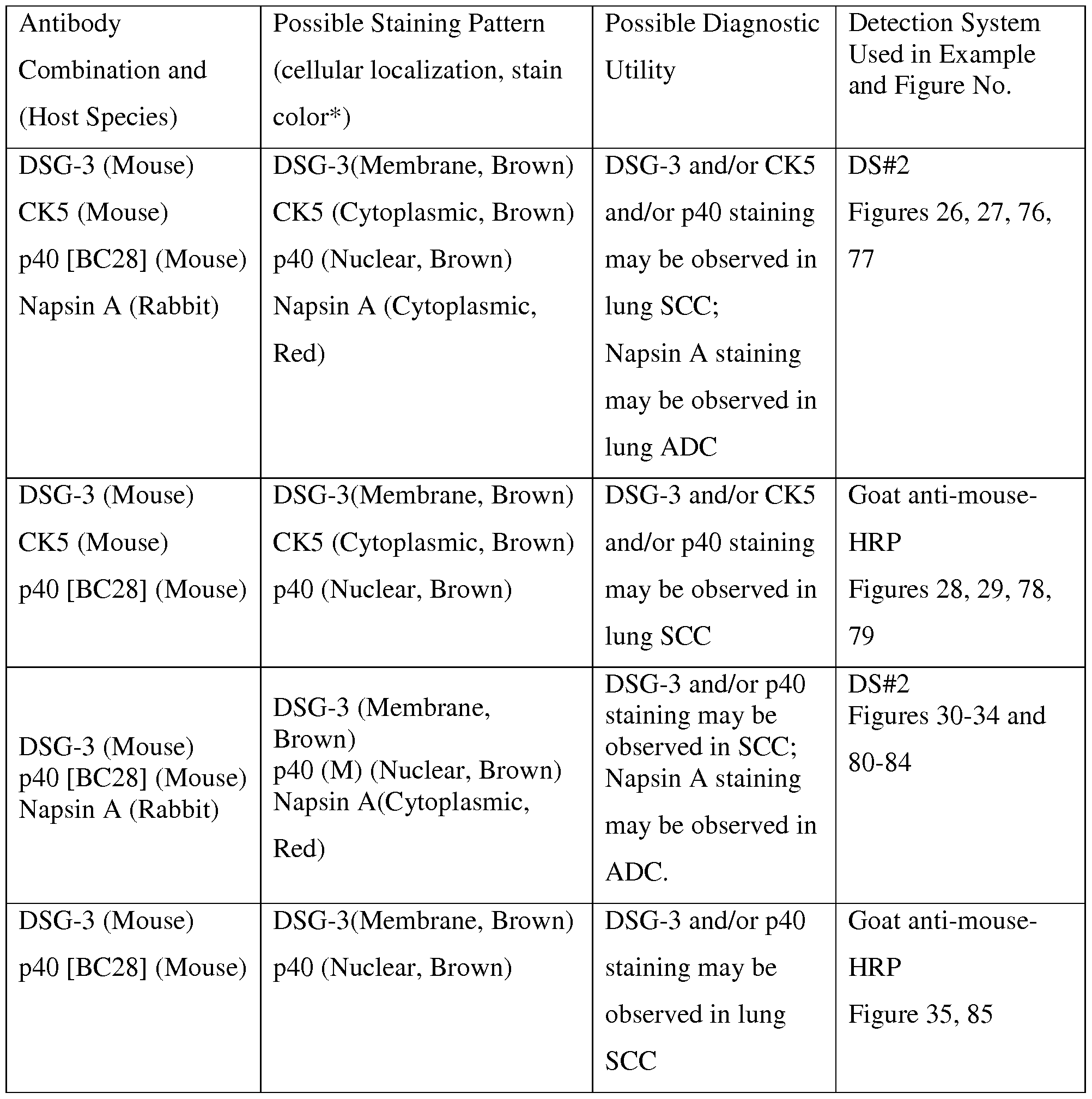
Used in Example Combination and (cellular localization, stain Utility
and Figure No. (Host Species) color*)
CK5 and/or p40 DS#2
p40 [BC28] (Mouse) p40 (Nuclear, Brown) Figures 36, 37, 86, staining may be
CK5 (Rabbit) CK5 (Cytoplasmic, Red)
87
observed in SCC.
p40 [BC28] (Mouse) p40 (Nuclear, Brown) p40 staining may DS#2
Figures 38, 39, 88, Napsin A (Rabbit) Napsin A (Cytoplasmic, be observed in lung
89
Red) SCC;
Napsin A staining
may be observed in
lung ADC
p40 [BC28] (Mouse) p40 [BC28] (Nuclear, p40 and/or DS#2
Figure 40, 90 HMWCK (Mouse) Brown) HMWCK staining
P504S (Rabbit) HMWCK (Cytoplasmic, may be decreased,
Brown) or perhaps absent,
P504S (Cytoplasmic, Red) in prostate cancer
P504S staining may
be observed in
prostate cancer
p40 [BC28] (Mouse) p40 [BC28] (Nuclear, p40 and/or CK5/14 DS#2
Figure 41, 91 CK5/14 (Mouse) Brown) staining may be
P504S (Rabbit) CK5/14 (Cytoplasmic, decreased, or
Brown) perhaps absent, in
P504S (Cytoplasmic, Red) prostate cancer
P504S staining may
be observed in
prostate cancer
Antibody Possible Staining Pattern Possible Diagnostic Detection System
Used in Example Combination and (cellular localization, stain Utility
and Figure No. (Host Species) color*)
p40 [BC28] (Mouse) p40 [BC28] (Nuclear, Staining pattern DS#2
Figures 42, 43, 92, CK5/14 (Mouse) Brown) may be useful to
93
CK7/18 (Rabbit) CK5/14 (Cytoplasmic, identify breast
Brown) lesions, such as
CK7/18 (Cytoplasmic, Red) carcinoma in situ
and perhaps
distinguish benign
from neoplastic
lesions
p40 [BC28] (Mouse) p40 [BC28] (Nuclear, Staining pattern DS#2
Figure 44, 94 CK5/14 (Mouse) Brown) may be useful to
CK8/18 (Rabbit) CK5/14 (Cytoplasmic, identify breast
Brown) lesions, such as
CK8/18 (Cytoplasmic, Red) carcinoma in situ
and perhaps
distinguish benign
from neoplastic
lesions
p40 [BC28] (Mouse) p40 [BC28] (Nuclear, p40 and/or Goat anti-mouse- and/or Brown) Uroplakin II and/or HRP
Uroplakin II [BC21] Uroplakin II (Membrane Uroplakin III Figure 45, 95
(Mouse) and cytoplasmic, Brown) staining may be
and/or Uroplakin III (Membrane observed in bladder
Uroplakin III and cytoplasmic, Brown) cancer.
[BC17] (Mouse)
Antibody Possible Staining Pattern Possible Diagnostic Detection System
Used in Example Combination and (cellular localization, stain Utility
and Figure No. (Host Species) color*)
p40 [BC28] (Mouse) p40 [BC28] (Nuclear, p40 and/or DS#2
Figure 46, 96 and/or Brown) Uroplakin II and/or
Uroplakin II [BC21] Uroplakin II (Membrane Uroplakin III
(Mouse) and cytoplasmic, Brown) and/or GATA-3
and/or Uroplakin III (Membrane staining may be
Uroplakin III and cytoplasmic, Brown) observed in bladder
[BC17] (Mouse) GATA-3 (Nuclear, Red) cancer.
and/or
GATA-3 (Rabbit)
CK5 and/or p40 Goat anti-mouse- p40 [BC28] (Mouse) p40 (Nuclear, Brown)
staining may be HRP
CK5 (Mouse) CK5 (Cytoplasmic, Brown)
observed in SCC. Figure 47, 97
*The listed color of each stain may be a result of a detection system that may include an anti-mouse antibody perhaps conjugated to HRP and even an anti-rabbit antibody perhaps conjugated to AP, perhaps even with DAB and Fast Red as chromogens, which may result in brown staining for mouse antibodies and red staining for rabbit antibodies (referred to as
DS#2). Alternatively, the detection system may include an anti-mouse antibody perhaps conjugated to AP and even an anti-rabbit antibody perhaps conjugated to HRP, perhaps even with DAB and Fast Red as chromogens, which may result in red staining for mouse antibodies and brown staining for rabbit antibodies (referred to as DS#1). In some instances, two colors may not be necessary because the antigens may be distinguished by cellular localization of staining, or perhaps it is not diagnostically significant to determine which antigen is staining. Other color combinations may be obtained using other detection systems or chromogens and all are meant to be included in this disclosure.
Other examples cocktails may be provided in PCT patent application no. PCT/US2013/076203, filed December 18, 2013, entitled "Antibody Cocktail Systems and Methods for Classification of Histologic Subtypes in Lung Cancer," hereby incorporated by reference herein.
IHC double-stain method with an antibody cocktail of DSG-3, CK5, p40 [BC28] and Napsin A: Immunohistochemistry, perhaps to produce a "double-stain" or a "multiplex procedure," using antibody cocktails such as DSG-3 + CK5 + p40 [BC28] + Napsin A may be performed on formalin-fixed paraffin embedded (FFPE) tissue samples using procedures generally known to those in the art, as generally exemplified by the following non-limiting examples (e.g., washes with Tris-buffered saline, pH about 7.6, between steps):
1) Sections (~5μιη) of formalin fixed paraffin-embedded tissues may be mounted on commercially available microscope slides perhaps coated with polylysine.
2) Sections may be deparaffinized (using xylenes or a xylene- substitute) and may be rehydrated perhaps through a series of alcohol/water solutions, perhaps followed by blocking of endogenous peroxidases perhaps with about 3% hydrogen peroxide solution.
3) Samples may be subjected to heat-induced antigen retrieval using a citrate buffer in a pressure cooker (Diva, Decloaking Chamber; Biocare Medical) and may be heated to about 125°C for about 30 seconds. [Other antigen retrieval methods known to those skilled in the art (e.g., steamer, microwave oven, enzyme, or the like) may also be acceptable.] Tissues may be allowed to cool for about 10 minutes and then may be rinsed with deionized water.
4) The antibody cocktail of DSG-3 + CK5 + p40 [BC28] + Napsin A may be applied in a phosphate-buffered solution (pH about 6.0) with bovine serum albumin as carrier protein for about 30 minutes.
5) Detection of the antibodies perhaps with a cocktail of goat anti-mouse horseradish peroxidase (HRP) conjugated secondary antibody and goat anti-rabbit alkaline phosphatase (AP) conjugated secondary antibody (MACH 2 Double Stain #2, Biocare Medical) with an incubation of about 30 minutes.
6) In perhaps a final detection step, Fast Red chromogen with naphthol phosphate substrate, perhaps in a buffer of about pH 8.3 (Vulcan Fast Red, Biocare Medical) may be applied for about 20 minutes, which may produce a fuschia/red chromogenic product as a stain. Subsequently, 3,3'-diaminobenzidine (DAB) in buffer perhaps containing about 0.02% hydrogen peroxide (Betazoid DAB, Biocare Medical) may be applied. The oxidation of DAB through an HRP-mediated mechanism may result in precipitation of a brown, chromogenic product, perhaps allowing identification of the sites of antigen expression
for each antibody. The presence of p40, DSG-3 and CK5 antibodies may be determined by brown staining. The presence of Napsin A antibodies may be determined by red staining.
7) Slides may be briefly counterstained perhaps in a modified Mayer's hematoxylin.
IHC single-stain method with an antibody cocktail of DSG-3, CK5 and p40 [BC28]:
Immunohistochemistry using an antibody cocktail such as DSG-3 + CK5 + p40 [BC28] may be performed on formalin-fixed paraffin embedded (FFPE) tissue samples using procedures generally known to those in the art, as generally exemplified by the following non-limiting examples (e.g., washes with Tris-buffered saline, pH about 7.6, between steps):
1) Sections (~5μιη) of formalin fixed paraffin-embedded tissues may be mounted on commercially available microscope slides perhaps coated with polylysine.
2) Sections may be deparaffinized (using xylenes or a xylene- substitute) and may be rehydrated perhaps through a series of alcohol/water solutions, perhaps followed by blocking of endogenous peroxidases perhaps with about 3% hydrogen peroxide solution. 3) Samples may be subjected to heat-induced antigen retrieval using a citrate buffer in a pressure cooker (Diva, Decloaking Chamber; Biocare Medical) and may be heated to about 125°C for about 30 seconds. [Other antigen retrieval methods known to those skilled in the art (e.g., steamer, microwave oven, enzyme, or the like) may also be acceptable.] Tissues may be allowed to cool for about 10 minutes and then may be rinsed with deionized water.
4) The antibody cocktail of DSG-3 + CK5 + p40 [BC28] may be applied in a phosphate- buffered solution (pH about 6.0) with bovine serum albumin as carrier protein for about 30 minutes.
5) Detection of the antibodies perhaps with a cocktail of goat anti-mouse horseradish peroxidase (HRP) conjugated secondary antibody (MACH 2 Mouse-HRP Polymer
Detection, Biocare Medical) with an incubation of about 30 minutes. Alternatively, detection of the anbtibodies perhaps with a horseradish peroxidase (HRP) conjugated secondary antibody (MACH 4 Universal HRP-Polymer Detection, Biocare Medical) may be accomplished in two steps. An initial application of a rabbit anti-mouse IgG antibody for about 10 minutes may be followed by incubation with a goat anti-rabbit-HRP conjugate for about 10 minutes.
6) In perhaps a final detection step, 3,3'-diaminobenzidine (DAB) in buffer perhaps containing about 0.02% hydrogen peroxide (Betazoid DAB, Biocare Medical) may be applied. The oxidation of DAB through an HRP-mediated mechanism may result in precipitation of a brown, chromogenic product, perhaps allowing identification of the sites of antigen expression for each antibody. The presence of p40, DSG-3 and CK5 antibodies may be determined by brown staining.
7) Slides may be briefly counterstained perhaps in a modified Mayer's hematoxylin.
Results of IHC Staining with DSG-3, CK5, p40 [BC28] and Napsin A: Using the above double-stain protocol, FFPE tissues may be stained with a cocktail of DSG-3, p40 [BC28], CK5 and Napsin A. An example of staining of lung SCC is shown in Figure 26 and 76. The presence of DSG-3, p40, and/or CK5 antibodies may result in brown staining. Red staining of Napsin A may be reduced or absent, or perhaps restricted to residual normal lung tissue. An example of staining of lung ADC is shown in Figure 27 and 77. The presence of Napsin A may result in the red staining. Brown staining from p40, DSG-3, and/or CK5 may be reduced, or absent (e.g., see Figure 27 and 77).
Results of IHC Staining with DSG-3, CK5, and p40 [BC28]: Using the above single-stain protocol, FFPE tissues may be stained with a cocktail of DSG-3, p40 [BC28], and CK5. An example of staining of lung SCC is shown in Figure 28 and 78. The presence of DSG-3, p40, and/or CK5 antibodies may result in brown staining. In this example, using a single-color stain for detection, such as goat anti-mouse HRP, is acceptable because the presence of DSG-3, p40, or CK5 all are indicative of squamous cell carcinoma. An example of staining of lung ADC is shown in Figure 29 and 79. Brown staining from DSG-3, p40, and/or CK5 may be reduced, or absent (e.g., see Figure 29 and 79), or even restricted to residual normal lung tissue.
Results of IHC Staining with DSG-3, p40 (M) and Napsin A: Using the above double-stain protocol, FFPE tissues may be stained with a cocktail of DSG-3, p40 (M) (e.g., mouse monoclonal [BC28]) and Napsin A. Examples of staining of ADC are shown in Figures 20A and 20B. The presence of Napsin A may result in the red staining. Brown staining from p40 or DSG-3 may be reduced, or absent (e.g., see Figures 30, 80, 31 and 81), or even restricted to residual normal lung tissue (e.g., see Figure 31 and 81). Examples of staining of SCC are shown in Figures 32, 82, 33, 83, 34, and 84. The presence of DSG-3 or
p40 antibodies may result in brown staining. Red staining of Napsin A may be reduced or absent, or perhaps restricted to residual normal lung tissue (e.g., see Figures 33 and 83).
Results of IHC Staining with DSG-3 and p40 (M): Using the above single- stain protocol, with a detection system of goat anti-mouse HRP, FFPE tissues may be stained with a cocktail of DSG-3 and p40 (M). An example of staining of SCC is shown in Figure 35 and 85. The presence of DSG-3 and/or CK5 antibodies may result in the brown staining.
Results of IHC Staining with p40 (M) and CK5 (RM): Using the above double- stain protocol, FFPE tissues may be stained with a cocktail of p40 [BC28] and CK5 (RM) (e.g., rabbit monoclonal [EP1601Y]). An example of staining of lung SCC is shown in Figure 36 and 86. The presence of p40 (M) antibodies may result in the brown staining and the presence of CK5 (RM) antibodies results in the red staining. An example of staining of lung ADC is shown in Figure 37 and 87. No brown or red staining may be observed, suggesting perhaps the absence of p40 and CK5.
Results of IHC Staining with p40 (M) and Napsin A: Using the above double- stain protocol, FFPE tissues may be stained with a cocktail of p40 (M) (e.g., mouse monoclonal [BC28]) and Napsin A. An example of staining of SCC is shown in Figure 38 and 88. The presence of p40 antibodies may result in brown staining. Red staining of Napsin A may be reduced or absent, or perhaps restricted to residual normal lung tissue. An example of staining of ADC is shown in Figure 39 and 89. The presence of Napsin A may result in the red staining. Brown staining from p40 may be reduced, or absent.
Results of IHC Staining with p40 [BC28], HMWCK, and P504S: Using the above double-stain protocol, FFPE tissues may be stained with a cocktail of p40 [BC28], HMWCK (e.g. mouse monoclonal clone 34βΕ12), and P504S (perhaps also known as AMACR, e.g. rabbit monoclonal anti-AMACR [13H4], or a rabbit polyclonal antibody). An example of staining of prostate tissue is shown in Figure 40 and 90. The presence of p40 and or HMWCK antibodies may result in brown staining. The presence of P504S antibodies may result in red staining. The tissue may contain normal prostate glands, areas of prostate cancer, or perhaps prostatic intraepithelial neoplasia (PIN).
Results of IHC Staining with p40 [BC28], CK5/14, and P504S: Using the above double-stain protocol, FFPE tissues may be stained with a cocktail of p40 [BC28], CK5/14 (e.g. a combination of anti-CK5 clone [XM26] and anti-CK14 clone [LL002]), and P504S
(perhaps also known as AMACR, e.g. rabbit monoclonal anti-AMACR [13H4] or a rabbit polyclonal antibody). An example of staining of prostate tissue is shown in Figure 41 and 91. The presence of p40 and or CK5/14 antibodies may result in brown staining. The presence of P504S antibodies may result in red staining. The tissue may contain normal prostate glands, areas of prostate cancer, or perhaps prostatic intraepithelial neoplasia (PIN). The examples in Figures 40, 90, 41, and 91 are representative of instances where cytokeratin markers may be interchangeable. In these examples, HMWCK and CK5/14 are both effective cytokeratin markers and may be considered effectively equivalent.
Results of IHC Staining with p40 [BC28], CK5/14, and CK7/18: Using the above double-stain protocol, FFPE tissues may be stained with a cocktail of p40 [BC28], CK5/14 (e.g. a combination of anti-CK5 clone [XM26] and anti-CK14 clone [LL002]), and CK7/18 (e.g. a combination of rabbit monoclonal anti-CK7 antibody [BC1] and rabbit monoclonal anti-CK18 antibody clone [E431-1]). An example of staining of breast tissue, perhaps breast cancer, is shown in Figures 42, 92, 43, and 93. The presence of p40 and or CK5/14 antibodies may result in brown staining. The presence of CK7/18 antibodies may result in red staining. Figure 43 and 93 may be an example of bimodal staining. The antibody cocktail of p63 + CK5/14 + CK7/18 may have been shown to be useful in clinical diagnosis, (e.g., see article, "Atypical ductal hyperplasia: interobserver and intraobserver variability." Jain RK, Mehta R, Dimitrov R, Larsson LG, Musto PM, Hodges KB, Ulbright TM, Hattab EM, Agaram N, Idrees MT, Badve S. Mod Pathol. 2011 Jul;24(7):917-23, hereby incorporated by reference herein.) An antibody cocktail that uses p40 in place of p63 (e.g. p40 [BC28] + CK5/14 + CK7/18) may be similarly useful for diagnosis, perhaps even offering the advantages of p40 [BC28].
Results of IHC Staining with p40 [BC28], CK5/14, and CK8/18: Using the above double-stain protocol, FFPE tissues may be stained with a cocktail of p40 [BC28], CK5/14 (e.g. a combination of anti-CK5 clone [XM26] and anti-CK14 clone [LL002]), and CK8/18 (e.g. a combination of rabbit monoclonal anti-CK8 antibody [EP17] and rabbit monoclonal anti-CK18 antibody clone [E431-1]). An example of staining of breast tissue, perhaps breast cancer, is shown in Figure 44 and 94. The presence of p40 and or CK5/14 antibodies may result in brown staining. The presence of CK8/18 antibodies may result in red staining.
Results of IHC Staining with p40 [BC28], Uroplakin II and Uroplakin III:
Using the above single-stain protocol, FFPE tissues may be stained with a cocktail of p40 [BC28], Uroplakin II [BC21] and Uroplakin III [BC17]. An example of staining of bladder tissue, perhaps urothelial carcinoma, is shown in Figure 45 and 95. The presence of p40, Uroplakin II, and/or Uroplakin III antibodies may result in brown staining. The example of Figure 45 and 95 is an instance where a single-color stain, perhaps using goat anti-mouse HRP, is useful because p40, Uroplakin II or Uroplakin III all are indicative of urothelial carcinoma.
Results of IHC Staining with p40 [BC28], Uroplakin II, Uroplakin III, and GATA-3: Using the above double-stain protocol, FFPE tissues may be stained with a cocktail of p40 [BC28], Uroplakin II [BC21], Uroplakin III [BC17], and GATA-3 (e.g. rabbit anti-GATA-3 antibody). An example of staining of bladder tissue, perhaps urothelial carcinoma, is shown in Figure 46 and 96. The presence of p40, Uroplakin II, and/or Uroplakin III antibodies may result in brown staining. The presence of GATA-3 may result in red staining.
Results of IHC Staining with p40 (M) and CK5 (M): Using the above single- stain protocol, FFPE tissues may be stained with a cocktail of p40 [BC28] and CK5 (M) (e.g., mouse monoclonal [XM26]). An example of staining of lung SCC is shown in Figure 47 and 97. The presence of p40 and/or CK5 antibodies may result in the brown staining.
In many embodiments, antibodies that bind cytokeratin markers may be used in different combinations, and in some cases, interchangeably, as known to those skilled in the art. For example, CK5 may perhaps be interchangeable with CK5/6 or CK5/14. Similarly, HMWCK (high molecular weight cytokeratin) may be used interchangeably with CK5/6 or CK5/14. In another example, CK7 may be used interchangeably with CK8.
Other embodiments may include primary antibody cocktails comprised of p40
[BC28] with other combinations of antibodies, including variations on the example cocktails. In some embodiments, other host species may be used. Embodiments that lack one or more of the antibodies included in the listed examples may also be useful.
In some embodiments, anti-p40 antibodies other than BC28 may also be useful. Rabbit polyclonal or rabbit monoclonal anti-p40 antibodies may be used in some
embodiments. In other embodiments, a mouse monoclonal anti-p40 antibody clone other than BC28 may be similarly useful, and perhaps interchangeable with BC28.
In many cases, but perhaps particularly in cases of lung cancer, diagnosis may often be performed on limited tissue samples from cytology or a biopsy, and it may be important to conserve tissue for further molecular testing; therefore, an efficient approach to diagnosis that consumes minimal tissue, but provides optimal specificity and/or sensitivity may be preferred. A method that provides useful diagnostic information, while consuming minimal tissue from the specimen, perhaps by the application of multiple antibodies to the same ample, perhaps through use of an antibody cocktail, or by the sequential application of antibodies, or conceivably by the feature of improved sensitivity or specificity, may be used.
An anti-p40 antibody such as a mouse monoclonal anti-p40 antibody BC28 may be specific for detection of p40 and may be useful in immunohistochemical procedures for diagnosis of several types of cancers in human tissue samples. In particular, anti-p40 antibody such as BC28 has advantages over RP anti-p40 antibody and anti-p63 antibody [4A4], including but not limited to greater specificity and cleaner staining, including the absence of staining of some cases of lung adenocarcinoma, as well as perhaps a lack of staining of macrophages.
Determination of p40 levels may be a useful prognostic factor and indicator of patient outcomes. Loss of p40 expression, as determined by IHC, has been associated with aggressive bladder cancer and shown to be predictive of patient survival (see Matsumoto K et. al. Urology, 2008; 72:444-449, and Ohtsuka Y et. al. BJU Int, 2006; 97: 1322-1326, hereby incorporated by reference herein). As such, the various systems and methods as discussed herein may provide a detection system such as but not limited to detection of cancer, diagnose or even prognose cancer, predict an outcome of cancer, assess efficacy of treatment of cancer, predict recurrence of cancer, or the like. As mentioned herein, use of an antibody or fragment thereof may be performed on an automated staining device. Detection may be made automatically, manually, by image analysis, or the like. Detection may utilize a method including but not limited to immunohistochemistry (IHC), IHC of FFPE, IHC of frozen-tissue sections, immunocytochemistry, ELISA, or the like.
As but one example of an immunoassay method, embodiments of the present invention may provide obtaining tissue from an animal or human to be tested (6), fixing or
freezing said tissue (7), treating said fixed or frozen tissue to unmask epitopes to p40 (8), contacting said treated tissue with an antibody or fragment thereof as discussed herein in an amount and under conditions such that an antibody or fragment thereof binds to a p40 protein if the protein is present in said tissue (9); and perhaps even detecting the presences of said bound antibodies (10), as schematically represented in Figure 50.
Figure 49 shows a schematic summary of various embodiments of the present invention including a kit (5) which may provide an antibody, fragment thereof, portion thereof, in a composition or even in a cocktail, perhaps even provided from a hybridoma, the antibody (1) or the like may be contacted with a biological sample (2) to form at least one antibody-antigen complex (3) which may then be detected with a detector (4).
The present invention may provide, in embodiments, a diagnostic or even prognostic test kit which may include an antibody or fragment thereof (as discussed herein) with an antibody detection element of the antibody or fragment thereof perhaps when bound to an antigen. This may provide a method of contacting a biological sample with an antibody or fragment thereof and even detecting binding of, or even the presence of the antibody or fragment thereof bound to a protein or with an antigen in the biological sample perhaps using an antibody detection element. Embodiments may provide an immunoassay method for detecting p40 protein in a mammal or human perhaps by obtaining a tissue from an animal or a human to be tested, contacting the tissue with an antibody or fragment thereof in accordance with the various embodiments presented herein perhaps in an amount and under conditions such that the antibody or fragment thereof may bind to a p40 protein if the protein is present in the tissue; and even detecting the presence of bound antibodies. A biological sample may include but is not limited to blood, urine, urothelial tissue, transitional cell tissue, bladder tissue, normal tissue, neoplastic tissue, kidney tissue, ovarian tissue, thyroid tissue, endometrial tissue, renal tissue, tonsil tissue, pancreas tissue, colon tissue, lymph node tissue, neoplastic pancreatic tissue, stomach tissue, prostate tissue, lung tissue, breast tissue, or the like perhaps depending on the antibody or even cocktail being used.
It is noted that of the numerous applications and methods that use anti-p63 antibodies currently known to those in the art, the anti-p63 antibody may be replaced with an anti-p40 antibody (e.g. BC28), perhaps with equivalent, or even perhaps superior, results.
It is noted that use of terms such as p40, p40 antibody, BC28, or the like may relate to anti-p40 antibodies or the like as appropriate as one skilled in the art would understand.
As can be easily understood from the foregoing, the basic concepts of the present invention may be embodied in a variety of ways. It involves both antibody techniques as well as devices to accomplish the appropriate antibody. In this application, the antibody techniques are disclosed as part of the results shown to be achieved by the various devices described and as steps which are inherent to utilization. They are simply the natural result of utilizing the devices as intended and described. In addition, while some devices are disclosed, it should be understood that these not only accomplish certain methods but also can be varied in a number of ways. Importantly, as to all of the foregoing, all of these facets should be understood to be encompassed by this disclosure.
The discussion included in this application is intended to serve as a basic description. The reader should be aware that the specific discussion may not explicitly describe all embodiments possible; many alternatives are implicit. It also may not fully explain the generic nature of the invention and may not explicitly show how each feature or element can actually be representative of a broader function or of a great variety of alternative or equivalent elements. Again, these are implicitly included in this disclosure. Where the invention is described in device- oriented terminology, each element of the device implicitly performs a function. Apparatus claims may not only be included for the device described, but also method or process claims may be included to address the functions the invention and each element performs. Neither the description nor the terminology is intended to limit the scope of the claims that will be included in any subsequent patent application.
It should also be understood that a variety of changes may be made without departing from the essence of the invention. Such changes are also implicitly included in the description. They still fall within the scope of this invention. A broad disclosure encompassing the explicit embodiment(s) shown, the great variety of implicit alternative embodiments, and the broad methods or processes and the like are encompassed by this disclosure and may be relied upon when drafting the claims for any subsequent patent application. It should be understood that such language changes and broader or more detailed claiming may be accomplished at a later date (such as by any required deadline) or in the event the applicant subsequently seeks a patent filing based on this filing. With this
understanding, the reader should be aware that this disclosure is to be understood to support any subsequently filed patent application that may seek examination of as broad a base of claims as deemed within the applicant's right and may be designed to yield a patent covering numerous aspects of the invention both independently and as an overall system.
Further, each of the various elements of the invention and claims may also be achieved in a variety of manners. Additionally, when used or implied, an element is to be understood as encompassing individual as well as plural structures that may or may not be physically connected. This disclosure should be understood to encompass each such variation, be it a variation of an embodiment of any apparatus embodiment, a method or process embodiment, or even merely a variation of any element of these. Particularly, it should be understood that as the disclosure relates to elements of the invention, the words for each element may be expressed by equivalent apparatus terms or method terms— even if only the function or result is the same. Such equivalent, broader, or even more generic terms should be considered to be encompassed in the description of each element or action. Such terms can be substituted where desired to make explicit the implicitly broad coverage to which this invention is entitled. As but one example, it should be understood that all actions may be expressed as a means for taking that action or as an element which causes that action. Similarly, each physical element disclosed should be understood to encompass a disclosure of the action which that physical element facilitates. Regarding this last aspect, as but one example, the disclosure of a "detection" or "detector" should be understood to encompass disclosure of the act of "detecting"— whether explicitly discussed or not— and, conversely, were there effectively disclosure of the act of "detecting", such a disclosure should be understood to encompass disclosure of a "detector" and even a "means for detecting." Such changes and alternative terms are to be understood to be explicitly included in the description. Further, each such means (whether explicitly so described or not) should be understood as encompassing all elements that can perform the given function, and all descriptions of elements that perform a described function should be understood as a non-limiting example of means for performing that function.
Any law, statutes, regulations, or rules mentioned in this application for patent; or patents, publications, or other references mentioned in this application for patent are hereby incorporated by reference. Any priority case(s) claimed by this application is hereby
appended and hereby incorporated by reference. In addition, as to each term used it should be understood that unless its utilization in this application is inconsistent with a broadly supporting interpretation, common dictionary definitions should be understood as incorporated for each term and all definitions, alternative terms, and synonyms such as contained in the Random House Webster's Unabridged Dictionary, second edition are hereby incorporated by reference. Finally, all references listed in the list below or other information statement filed with the application are hereby appended and hereby incorporated by reference, however, as to each of the above, to the extent that such information or statements incorporated by reference might be considered inconsistent with the patenting of this/these invention(s) such statements are expressly not to be considered as made by the applicant(s).
I. U.S. PATENT DOCUMENTS

II. NON-PATENT LITERATURE DOCUMENTS
Jain, r., et al.; Atypical ductal hyperplasia: interobserver and intraobserver variability,
Modern Pathology (2011) 24, 917-923
Tacha, d., et al.; An Innunohistochemical Analysis of a Newly Developed, Mouse
Monoclonal p40 (BC28) in Lung, Bladder, Skin, Breast, Prostate, and Head and Neck Cancers; 2014 College of American Pathologists, pg 1-7, posted 02/2014
Khoury M. P. et al. p53 Isoforms: An Intracellular Microprocessor? Genes Cancer. 2011 Apr;2(4):453-65
Murray-Zmijewski F. et al. p53/p63/p73 isoforms: an orchestra of isoforms to harmonise cell differentiation and response to stress, Cell Death Differ. 2006 Jun;13(6):962-72
Kaghad M. et al. Monoallelically expressed gene related to p53 at lp36, a region
frequently deleted in neuroblastoma and other human cancers, Cell. 1997 Aug
22;90(4):809-19
Trink B. et al. A new human p53 homologue, Nat Med. 1998 Jul;4(7):747-8
Osada M. et al. Cloning and functional analysis of human p51, which structurally and functionally resembles p53, Nat Med. 1998 Jul;4(7):839-43
Yang A. et al. p63, a p53 homolog at 3q27-29, encodes multiple products with
transactivating, death-inducing, and dominant-negative activities, Mol Cell. 1998
Sep;2(3):305-16
Senoo M. et al. A second p53-related protein, p73L, with high homology to p73, Biochem
Biophys Res Commun. 1998 Jul 30;248(3):603-7
Hibi K. et al. AIS is an oncogene amplified in squamous cell carcinoma, Proc Natl Acad
Sci U S A. 2000 May 9;97(10):5462-7
Nobre A. R. et al. p40: A p63 Isoform Useful for Lung Cancer Diagnosis - A Review of the Physiological and Pathological Role of p63, Acta Cytol. 2013;57(l): l-8
Bishop J. A. et al. p40 (ΔΝρ63) is superior to p63 for the diagnosis of pulmonary
squamous cell carcinoma, Mod Pathol. 2012 Mar;25(3):405-15
Nonaka D. A study of ΔΝρ63 expression in lung non- small cell carcinomas, Am J Surg
Pathol. 2012 Jun;36(6):895-9
Pelosi G, et al. ΔΝρ63 (p40) and thyroid transcription factor- 1 immunoreactivity on small biopsies or cellblocks for typing non-small cell lung cancer: a novel two-hit, sparing- material approach, J Thorac Oncol. 2012 Feb;7(2):281-90
Brown A. F. et al. Tissue-Preserving Antibody Cocktails to Differentiate Primary
Squamous Cell Carcinoma, Adenocarcinoma, and Small Cell Carcinoma of Lung, Arch
Pathol Lab Med. 2013 Jan 4
Karni-Schmidt O. et al. Distinct expression profiles of p63 variants during urothelial development and bladder cancer progression, Am J Pathol. 2011 Mar;178(3): 1350-60
Signoretti S. et al. p63 is a prostate basal cell marker and is required for prostate
development, Am J Pathol. 2000 Dec; 157(6): 1769-75
Nylander K. et al. Differential expression of p63 isoforms in normal tissues and neoplastic cells, J Pathol. 2002 Dec;198(4):417-27
Barbareschi M, et al. p63, a p53 homologue, is a selective nuclear marker of myoepithelial cells of the human breast,Am J Surg Pathol. 2001 Aug;25(8): 1054-60
US Provisional Patent Application No. 61/770,956, Filed February 28, 2013, entitled
"Systems and Methods for Anti-p40 Antibodies",
PCT patent application no. PCT/US2013/076203, filed December 18, 2013, entitled
"Antibody Cocktail Systems and Methods for Classification of Histologic Subtypes in
Lung Cancer"
PCT patent application no. PCT/US2013/062043, filed September 26, 2013, entitled
"Anti-Uroplakin II Antibodies Systems and Methods"
Thus, the applicant(s) should be understood to have support to claim and make a statement of invention to at least: i) each of the antibody devices as herein disclosed and described, ii) the related methods disclosed and described, iii) similar, equivalent, and even implicit variations of each of these devices and methods, iv) those alternative designs which accomplish each of the functions shown as are disclosed and described, v) those alternative designs and methods which accomplish each of the functions shown as are implicit to accomplish that which is disclosed and described, vi) each feature, component, and step shown as separate and independent inventions, vii) the applications enhanced by the various systems or components disclosed, viii) the resulting products produced by such systems or components, ix) each system, method, and element shown or described as now applied to any specific field or devices mentioned, x) methods and apparatuses substantially as described hereinbefore and with reference to any of the accompanying examples, xi) an
apparatus for performing the methods described herein comprising means for performing the steps, xii) the various combinations and permutations of each of the elements disclosed, xiii) each potentially dependent claim or concept as a dependency on each and every one of the independent claims or concepts presented, and xiv) all inventions described herein.
With regard to claims whether now or later presented for examination, it should be understood that for practical reasons and so as to avoid great expansion of the examination burden, the applicant may at any time present only initial claims or perhaps only initial claims with only initial dependencies. The office and any third persons interested in potential scope of this or subsequent applications should understand that broader claims may be presented at a later date in this case, in a case claiming the benefit of this case, or in any continuation in spite of any preliminary amendments, other amendments, claim language, or arguments presented, thus throughout the pendency of any case there is no intention to disclaim or surrender any potential subject matter. It should be understood that if or when broader claims are presented, such may require that any relevant prior art that may have been considered at any prior time may need to be re-visited since it is possible that to the extent any amendments, claim language, or arguments presented in this or any subsequent application are considered as made to avoid such prior art, such reasons may be eliminated by later presented claims or the like. Both the examiner and any person otherwise interested in existing or later potential coverage, or considering if there has at any time been any possibility of an indication of disclaimer or surrender of potential coverage, should be aware that no such surrender or disclaimer is ever intended or ever exists in this or any subsequent application. Limitations such as arose in Hakim v. Cannon Avent Group, PLC, 479 F.3d 1313 (Fed. Cir 2007), or the like are expressly not intended in this or any subsequent related matter. In addition, support should be understood to exist to the degree required under new matter laws— including but not limited to European Patent Convention Article 123(2) and United States Patent Law 35 USC 132 or other such laws— to permit the addition of any of the various dependencies or other elements presented under one independent claim or concept as dependencies or elements under any other independent claim or concept. In drafting any claims at any time whether in this application or in any subsequent application, it should also be understood that the applicant has intended to capture as full and broad a scope of coverage as legally available. To the extent that insubstantial substitutes are made,
to the extent that the applicant did not in fact draft any claim so as to literally encompass any particular embodiment, and to the extent otherwise applicable, the applicant should not be understood to have in any way intended to or actually relinquished such coverage as the applicant simply may not have been able to anticipate all eventualities; one skilled in the art, should not be reasonably expected to have drafted a claim that would have literally encompassed such alternative embodiments.
Further, if or when used, the use of the transitional phrase "comprising" is used to maintain the "open-end" claims herein, according to traditional claim interpretation. Thus, unless the context requires otherwise, it should be understood that the term "comprise" or variations such as "comprises" or "comprising", are intended to imply the inclusion of a stated element or step or group of elements or steps but not the exclusion of any other element or step or group of elements or steps. Such terms should be interpreted in their most expansive form so as to afford the applicant the broadest coverage legally permissible. Each claim may be amended to include "or any other claim" and such amendment will be fully supported by the original application. The use of the phrase, "or any other claim" is used to provide support for any claim to be dependent on any other claim, such as another dependent claim, another independent claim, a previously listed claim, a subsequently listed claim, and the like. As one clarifying example, if a claim were dependent "on claim 20 or any other claim" or the like, it could be re-drafted as dependent on claim 1, claim 15, or even claim 25 (if such were to exist) if desired and still fall with the disclosure. It should be understood that this phrase also provides support for any combination of elements in the claims and even incorporates any desired proper antecedent basis for certain claim combinations such as with combinations of method, apparatus, process, and the like claims.
Finally, any claims set forth at any time are hereby incorporated by reference as part of this description of the invention and the incorporated claims can "depend on any other claim", and the applicant expressly reserves the right to use all of or a portion of such incorporated content of such claims as additional description to support any of or all of the claims or any element or component thereof, and the applicant further expressly reserves the right to move any portion of or all of the incorporated content of such claims or any element or component thereof from the description into the claims or vice-versa as necessary to define the matter for which protection is sought by this application or by any subsequent
continuation, division, or continuation-in-part application thereof, or to obtain any benefit of, reduction in fees pursuant to, or to comply with the patent laws, rules, or regulations of any country or treaty, and such content incorporated by reference shall survive during the entire pendency of this application including any subsequent continuation, division, or continuation-in-part application thereof or any reissue or extension thereon.
Claims
What is claimed is:
1. An isolated preparation of antibodies which specifically bind to a p40 protein wherein said antibodies bind to an epitope in peptide SEQ ID NO: 3.
2. An isolated preparation of antibodies which specifically bind to a p40 protein wherein said antibodies bind to an epitope in peptide SEQ ID NO: 4.
3. An isolated preparation of antibodies which specifically bind to a p40 protein wherein said antibodies bind to an epitope comprising a mixture of SEQ ID NO: 3 and SEQ ID NO: 5.
4. An isolated preparation of antibodies which specifically bind to a p40 protein wherein said antibodies bind to an epitope consisting of a mixture of SEQ ID NO: 3 and SEQ ID NO: 5.
5. An isolated preparation of antibodies which specifically bind to a p40 protein wherein said antibodies were generated using as an immunogen a peptide consisting of SEQ ID NO: 3.
6. An isolated preparation of antibodies which specifically bind to a p40 protein wherein said antibodies were generated using as an immunogen a peptide consisting of SEQ ID NO: 4.
7. An isolated preparation of antibodies which specifically bind to a p40 protein wherein said antibodies were generated using as an immunogen a peptide comprising a mixture of SEQ ID NO: 3 and SEQ ID NO: 5.
8. An isolated preparation of antibodies which specifically bind to a p40 protein wherein said antibodies were generated using as an immunogen a peptide consisting of a mixture of SEQ ID NO: 3 and SEQ ID NO: 5.
9. An isolated preparation of antibodies which specifically bind to an epitope in peptide SEQ ID NO: 3.
10. An isolated preparation of antibodies which specifically bind to an epitope in peptide SEQ ID NO: 4.
11. An isolated preparation of antibodies which specifically bind to an epitope in a peptide comprising a mixture of SEQ ID NO: 3 and SEQ ID NO: 5.
12. An isolated preparation of antibodies which specifically bind to an epitope in a peptide consisting of a mixture of SEQ ID NO: 3 and SEQ ID NO: 5.
13. An isolated preparation of antibodies according to claims 1, 2, 3, 4, 5, 6, 7, 8, 9, 10, 11, or 12 wherein said antibodies are monoclonal.
14. An isolated preparation of antibodies according to claims 1, 2, 3, 4, 5, 6, 7, 8, 9, 10, 11, or 12 wherein said antibodies are polyclonal.
15. An antibody or fragment thereof produced by the hybridoma deposited with the American Type Culture Collection (ATCC) under ATCC Patent Deposit Designation No. PTA-120163.
16. A hybridoma cell that is deposited at the American Type Culture Collection (ATCC) under ATCC Patent Deposit Designation No. PTA-120163.
17. A hybridoma cell according to claim 16 and further comprising an antibody or fragment thereof produced by said hybridoma cell.
18. A method for producing a monoclonal antibody according to claim 15 comprising the steps of:
culturing said hybridoma which produces a monoclonal antibody capable of specifically recognizing p40; and
allowing said hybridoma to produce the monoclonal antibody.
19. An antibody or fragment thereof comprising a polypeptide of the amino acid sequence encoded by the nucleic acid sequence of SEQ ID NO: 1 or SEQ ID NO: 2.
20. An anti-p40 antibody or fragment thereof, wherein said antibody or said fragment thereof comprises a light chain variable region comprising the amino acid sequence encoded by the nucleic acid sequence of SEQ ID NO: 2 and a heavy chain variable region comprising the amino acid sequence encoded by the nucleic acid sequence of SEQ ID NO: 1.
21. An antibody comprising an amino acid sequence at least about 70% identical to an amino acid sequence encoded by a nucleic acid sequence of SEQ ID NO: 1 or SEQ ID NO: 2.
22. An isolated and purified nucleic acid sequence comprising a nucleic acid sequence that is at least about 70% identical to SEQ ID NO: 1 or SEQ ID NO: 2.
A sequence according to claim 21 or 22 wherein said at least about 70% identical to SEQ ID NO: 1 or SEQ ID NO: 2 comprises at least about 70% identical to SEQ ID NO: 1 and SEQ ID NO: 2
A sequence according to claim 23 wherein said at least about 70% identical comprises a percentage selected from a group consisting of at least about 71%, at least about 72%, at least about 73%, at least about 74%, at least about 75%, at least about 76%, at least about 77%, at least about 78%, at least about 79%, at least about 80%, at least about 81%, at least about 82%, at least about 83%, at least about 84%, at least about 85%, at least about 86%, at least about 87%, at least about 88%, at least about 89%, at least about 90%, at least about 91%, at least about 92%, at least about 93%, at least about 94%, at least about 95%, at least about 96%, at least about 97%, at least about 98%, and at least about 99%.
An antibody or fragment thereof that specifically binds to at least one polypeptide with an amino acid sequence comprising residues of SEQ ID NO: 3.
A composition comprising at least two antibodies or fragments thereof, wherein at least one of said at least two antibodies or fragments thereof binds specifically to at least p40.
A composition according to claim 26 wherein said at least one of said at least two antibodies or fragments thereof which binds specifically to at least p40 comprises the p40 [BC28] antibody or fragments thereof.
A composition according to claim 26 wherein said at least one of said at least two antibodies or fragments thereof which binds specifically to at least p40 comprises at least one of said at least two antibodies or fragments thereof which binds specifically to p40 and has a positive indication cut-off value of greater than about 5% of stained cells.
A composition according to claim 26 wherein said at least one of said at least two antibodies or fragments thereof which binds specifically to p40 comprises at least one of said at least two antibodies or fragments thereof which binds specifically to p40 and has a positive indication cut-off value selected from a group consisting of:
- greater than about 2% of stained cells;
- greater than about 3% of stained cells;
- greater than about 4% of stained cells;
- greater than about 5% of stained cells;
- greater than about 6% of stained cells;
- greater than about 7% of stained cells;
- greater than about 8% of stained cells;
- greater than about 9% of stained cells; and
- greater than about 10% of stained cells.
A composition according to claim 26 wherein said at least one of said at least two antibodies or fragments thereof comprises a light chain variable region comprising the amino acid sequence encoded by a nucleic acid sequence of SEQ ID NO: 2 and a heavy chain variable region comprising the amino acid sequence encoded by a nucleic acid sequence of SEQ ID NO: 1.
A composition according to claim 26 wherein said at least one of said at least two antibodies or fragments thereof comprises a light chain variable region comprising an amino acid sequence that is identical to at least about 70% of an amino acid sequence encoded by a nucleic acid sequence of SEQ ID NO: 2 and a heavy chain variable region comprising the amino acid sequence that is at least about 70% of an amino acid sequence encoded by a nucleic acid sequence of SEQ ID NO: 1.
A composition according to claim 31 wherein said at least about 70% identical comprises a percentage selected from a group consisting of at least about 71%, at least about 72%, at least about 73%, at least about 74%, at least about 75%, at least about 76%, at least about 77%, at least about 78%, at least about 79%, at least about 80%, at least about 81%, at least about 82%, at least about 83%, at least about 84%, at least about 85%, at least about 86%, at least about 87%, at least about 88%, at least about 89%, at least about 90%, at least about 91%, at least about 92%, at least about 93%, at least about 94%, at least about 95%, at least about 96%, at least about 97%, at least about 98%, and at least about 99%.
A composition according to claim 26 wherein said at least one of said at least two antibodies or fragments thereof specifically binds to at least one polypeptide with an amino acid sequence comprising residues selected from a group consisting of SEQ ID NO: 3, SEQ ID NO: 4, and a mixture of SEQ ID NO: 3 and SEQ ID NO: 4.
A composition according to claim 26 wherein said at least one of said at least two antibodies or fragments thereof specifically binds to at least one polypeptide with an amino acid sequence that is at least about 70% identical to residues selected from a group consisting of SEQ ID NO: 3, SEQ ID NO: 4, and a mixture of SEQ ID NO: 3 and SEQ ID NO: 4.
A composition according to claim 34 wherein said at least about 70% identical comprises a percentage selected from a group consisting of at least about 71%, at least about 72%, at least about 73%, at least about 74%, at least about 75%, at least about 76%, at least about 77%, at least about 78%, at least about 79%, at least about 80%, at least about 81%, at least about 82%, at least about 83%, at least about 84%, at least about 85%, at least about 86%, at least about 87%, at least about 88%, at least about 89%, at least about 90%, at least about 91%, at least about 92%, at least about 93%, at least about 94%, at least about 95%, at least about 96%, at least about 97%, at least about 98%, and at least about 99%.
A composition according to claim 26 wherein said at least one of said at least two antibodies or fragments thereof comprises an antibody or fragment thereof produced by the hybridoma deposited with the American Type Culture Collection (ATCC) under ATCC Patent Deposit Designation No. PTA-120163.
A composition according to claim 26 wherein said composition comprises a primary antibody cocktail.
A composition according to claim 27 wherein said at least two antibodies or fragments thereof are derived from at least two different species.
A composition according to claim 38 wherein said at least two different species is selected from a group consisting of mouse, rabbit, goat, horse, chicken, human, and any combination thereof.
A composition according to claim 26 wherein said at least two antibodies or fragments thereof comprises a double-stain procedure.
A composition according to claim 26 wherein said at least two antibodies or fragments thereof are capable of providing different visualization results.
A composition according to claim 41 wherein said visualization results comprises color results.
A composition according to claim 26 wherein said at least one other of said at least two antibodies or fragments thereof binds specifically to a protein selected from a group consisting of DSG-3, CK4, Napsin A, HMWCK, p504s, CK5/14, CK7/18, CK8/18, Uroplakin II, Uroplakin III, GATA-3, and any combination thereof.
A composition according to claim 26 wherein at least two antibodies or fragments thereof each bind specifically to proteins selected from a group consisting of:
p40 and DSG-3 and CK5 and Napsin A;
p40 and DSG-3 and CK5;
p40 and Napsin A;
p40 and HMWCK and p504s;
p40 and p504s and CK5/14;
p40 and CK5/14 and CK7/18;
p40 and CK5/14 and CK8/18.
p40 and Uroplakin II and Uroplakin III; and
p40 and Uroplakin II and Uroplakin III and GATA-3.
A composition according to claim 26 wherein at least two antibodies or fragments thereof each bind specifically to proteins selected from a group consisting of:
p40 and DSG-3 and Napsin A;
p40 and DSG-3; and
p40 and CK5.
A composition according to claim 26 wherein said composition comprises a detection composition selected from a group consisting of SCC carcinoma detection composition, bladder detection composition, breast carcinoma detection composition, prostate carcinoma detection composition, and any combination thereof.
A monoclonal antibody or fragment thereof which binds specifically to p40 and has a positive indication cut-off value of greater than 1% of stained cells.
A monoclonal antibody or fragment thereof according to claim 47 wherein said positive indication cut-off value is selected from a group consisting of:
- greater than about 2% of stained cells;
- greater than about 3% of stained cells;
- greater than about 4% of stained cells;
- greater than about 5% of stained cells;
- greater than about 6% of stained cells;
- greater than about 7% of stained cells;
- greater than about 8% of stained cells;
- greater than about 9% of stained cells; and
- greater than about 10% of stained cells.
An antibody according to claim 19, 20, 25, 26, or 47 wherein said antibody or fragment thereof is produced by the hybridoma cells that are deposited at the American Type Culture Collection under ATCC Patent Deposit Designation No. PTA-120163.
An antibody according to claim 15, 17, 25, or 47 wherein said antibody or fragment thereof comprises a polypeptide of the amino acid sequence encoded by the nucleic acid sequence of SEQ ID NO: 1 or SEQ ID NO: 2.
An antibody according to claim 15, 17, 25, or 47 wherein said antibody or fragment thereof comprises a polypeptide of the amino acid sequence encoded by the nucleic acid sequence of SEQ ID NO: 1 and SEQ ID NO: 2.
An antibody according to claim 15, 17, 25, or 47 wherein said antibody or fragment thereof comprises a polypeptide that is at least about 70% identical to an amino acid sequence encoded by the nucleic acid sequence of SEQ ID NO: 1 or SEQ ID NO: 2. An antibody according to claim 15, 17, 25, or 47 wherein said antibody or fragment thereof comprises a polypeptide that is at least about 70% identical to an amino acid sequence encoded by the nucleic acid sequence of SEQ ID NO: 1 and SEQ ID NO: 2.
An antibody according to claim 15, 17, 25, or 47wherein said antibody or fragment thereof specifically binds to at least one polypeptide with an amino acid sequence comprising residues selected from a group consisting of SEQ ID NO: 3, SEQ ID NO: 4, and a mixture of SEQ ID NO: 3 and SEQ ID NO:5.
An antibody according to claim 15, 17, 25, or 47 wherein said antibody or fragment thereof specifically binds to at least one polypeptide with an amino acid sequence
that is at least about 70% identical to residues selected from a group consisting of SEQ ID NO: 3, SEQ ID NO: 4, and a mixture of SEQ ID NO: 3 and SEQ ID NO:5. An antibody according to claim 55 wherein said at least about 70% identical comprises a percentage selected from a group consisting of at least about 71%, at least about 72%, at least about 73%, at least about 74%, at least about 75%, at least about 76%, at least about 77%, at least about 78%, at least about 79%, at least about 80%, at least about 81%, at least about 82%, at least about 83%, at least about 84%, at least about 85%, at least about 86%, at least about 87%, at least about 88%, at least about 89%, at least about 90%, at least about 91%, at least about 92%, at least about 93%, at least about 94%, at least about 95%, at least about 96%, at least about 97%, at least about 98%, and at least about 99%.
An antibody according to claim 15, 17, 19, 20, 25, 26, or 47 wherein said antibody comprises a monoclonal antibody.
An antibody according to claim 57 wherein said monoclonal antibody has a positive indication cut-off value of greater than 1% of stained cells.
An antibody according to claim 58 wherein said positive indication cut-off value is selected from a group consisting of:
- greater than about 2% of stained cells;
- greater than about 3% of stained cells;
- greater than about 4% of stained cells;
- greater than about 5% of stained cells;
- greater than about 6% of stained cells;
- greater than about 7% of stained cells;
- greater than about 8% of stained cells;
- greater than about 9% of stained cells; and
- greater than about 10% of stained cells.
An antibody according to claim 57 wherein said monoclonal antibody is selected from a group consisting of a mouse monoclonal antibody, a rabbit monoclonal antibody, a goat monoclonal antibody, a horse monoclonal antibody, a chicken monoclonal antibody, a humanized monoclonal antibody, a chimeric antibody, and any combination thereof.
An antibody according to claim 15, 17, 19, 20, 25, 26, or 47 wherein said antibody comprises a polyclonal antibody.
An antibody according to claim 61 wherein said polyclonal antibody is selected from a group consisting of rabbit polyclonal antibody, mouse polyclonal antibody, a goat polyclonal antibody, a horse polyclonal antibody, a chicken polyclonal antibody, a humanized polyclonal antibody, and any combination thereof
An antibody according to claim 15, 17, 19, 20, 25, 26, or 47 wherein said antibody comprises an isolated antibody.
An antibody according to claim 15, 17, 19, 20, 25, 26, or 47 wherein said fragment thereof comprises an antigen binding fragment thereof.
An antibody according to claim 15, 17, 19, 20, 25, 26, or 47 and further comprising a label attached to said antibody or fragment thereof.
A cancer diagnostic agent which comprises said antibody or fragment thereof according to claim 15, 17, 19, 20, 25, 26, or 47 conjugated with a label.
An antibody according to claim 65 wherein said label is selected from a group consisting of a radioactive element, magnetic particles, radioisotope, fluorescent dye, enzyme, toxin, signal, stain, detection enzymes, horseradish peroxidase (HRP), alkaline phosphatase (AP), beta-galactosidase, chromogens, Fast Red, 3,3'- diaminobenzidine, 3-amino-9-ethylcarbazole, 5-bromo-4-chloro-3-indolyl phosphate, 3,3',5,5'-tetramethylbenzidine, 5-bromo-4-chloro-3-indolyl- -D- glucuronide, and any combination thereof.
An antibody according to claim 66 wherein said label is selected from a group consisting of a radioactive element, magnetic particles, radioisotope, fluorescent dye, enzyme, toxin, signal, stain, detection enzymes, horseradish peroxidase (HRP), alkaline phosphatase (AP), beta-galactosidase, chromogens, Fast Red, 3,3'- diaminobenzidine, 3-amino-9-ethylcarbazole, 5-bromo-4-chloro-3-indolyl phosphate, 3,3',5,5'-tetramethylbenzidine, 5-bromo-4-chloro-3-indolyl- -D- glucuronide, and any combination thereof.
A diagnostic or prognostic test kit comprising:
an antibody or fragment thereof according to claim 1, 2, 3, 4, 5, 6, 7, 8, 9, 10,
11, 12, 15, 17, 19, 20, 25, 26, or 47; and
an antibody detection element of said antibody or said fragment thereof when bound to an antigen.
A method for detecting p40 in a biological sample using the kit of claim 69 comprising the steps of:
contacting a biological sample with said antibody or fragment thereof; and detecting binding of said antibody or said fragment thereof with an antigen in said biological sample using said antibody detection element.
An antibody according to claim 15, 17, 19, 20, or 25 wherein said antibody or said fragment thereof specifically binds to p40.
Use of the antibody or fragment thereof or composition according to claim 15, 17, 19, 20, 25 or 47 to detect cancer.
Use of the antibody or fragment thereof or composition according to claim 15, 17, 19, 20, 25 or 47 to diagnose or prognose cancer.
Use of the antibody or fragment thereof or composition according to claim 15, 17, 19, 20, 25 or 47 to predict outcome of treatment of cancer.
Use of the antibody or fragment thereof or composition according to claim 15, 17, 19, 20, 25 or 47 to assess efficacy of treatment of cancer.
Use of the antibody or fragment thereof or composition according to claim 15, 17, 19, 20, 25 or 47 to predict recurrence of cancer.
The antibody or fragment thereof or composition according to claim 72 wherein said use of said antibody or fragment thereof or composition is performed on an automated staining device.
The antibody or fragment thereof or composition according to claim 72 wherein said detecting is made manually.
The antibody or fragment thereof or composition according to claim 72 wherein said detecting is made automatically.
The antibody or fragment thereof or composition according to claim 72 wherein said detecting is made by image analysis.
A method for detecting a protein to which an antibody or fragment thereof of claim 15, 17, 19, 20, 25, 26, or 47 binds to a biological sample, comprising the steps of contacting a biological sample with the antibody or fragment thereof; and detecting
the presence of the antibody or fragment thereof bound to the protein in the biological sample.
82. A method according to claim 81 wherein said biological sample comprises lung tissue, bladder tissue, breast tissue, and prostate tissue.
83. A method according to claim 81 wherein said biological sample is selected from a group consisting of a normal tissue, neoplastic tissue, bladder tissue, kidney tissue, ovarian tissue, thyroid tissue, endometrial tissue, renal tissue, tonsil tissue, pancreas tissue, colon tissue, lymph node tissue, neoplastic pancreatic tissue, stomach tissue, prostate tissue, lung tissue and breast tissue.
84. A method according to claim 81 wherein said detecting said presence of the antibody or fragment thereof bound to the protein is performed on an automated staining device.
85. A method according to claim 81 wherein said detecting said presence of the antibody or fragment thereof bound to the protein is made manually.
86. A method according to claim 81 wherein said detecting said presence of the antibody or fragment thereof bound to the protein is made automatically.
87. A method according to claim 81 wherein said detecting said presence of the antibody or fragment thereof bound to the protein is made by image analysis.
88. A method according to claim 81 wherein said detecting comprises a method selected from a group consisting of immunohistochemistry (IHC), IHC of FFPE, IHC of frozen-tissue sections, immunocytochemistry, and ELISA.
89. A method for detecting at least two different proteins in a biological sample, comprising the steps of:
- contacting said biological sample with a composition comprising at least two antibodies or fragments thereof, wherein at least one of said at least two antibodies or fragments thereof binds specifically to at least p40, to form an antigen-antibody complex; and
- detecting said antigen-antibody complex.
90. A method according to claim 89 wherein said at least one of said at least two antibodies or fragments thereof binds specifically to at least p40 comprises at least
one of said at least two antibodies or fragments thereof binds specifically to at least p40 and has a positive indication cut-off value of greater than 1% of stained cells.
91. A method according to claim 90 wherein said positive indication cut-off value is selected from a group consisting of:
- greater than about 2% of stained cells;
- greater than about 3% of stained cells;
- greater than about 4% of stained cells;
- greater than about 5% of stained cells;
- greater than about 6% of stained cells;
- greater than about 7% of stained cells;
- greater than about 8% of stained cells;
- greater than about 9% of stained cells; and
- greater than about 10% of stained cells.
92. A method according to claim 89 wherein said at least two antibodies or fragments thereof comprise at least one first primary antibody and at least one second primary antibody.
93. A method according to claim 89 wherein said at least one of said at least two antibodies or fragments thereof comprises a light chain variable region comprising the amino acid sequence encoded by a nucleic acid sequence of SEQ ID NO: 2 and a heavy chain variable region comprising the amino acid sequence encoded by a nucleic acid sequence of SEQ ID NO: 1.
94. A method according to claim 89 wherein said at least one of said at least two antibodies or fragments thereof comprises a light chain variable region comprising an amino acid sequence that is identical to at least about 70% of an amino acid sequence encoded by a nucleic acid sequence of SEQ ID NO: 2 and a heavy chain variable region comprising the amino acid sequence that is at least about 70% of an amino acid sequence encoded by a nucleic acid sequence of SEQ ID NO: 1.
95. A method according to claim 94 wherein said at least about 70% identical comprises a percentage selected from a group consisting of at least about 71%, at least about 72%, at least about 73%, at least about 74%, at least about 75%, at least about 76%, at least about 77%, at least about 78%, at least about 79%, at least about 80%, at
least about 81%, at least about 82%, at least about 83%, at least about 84%, at least about 85%, at least about 86%, at least about 87%, at least about 88%, at least about 89%, at least about 90%, at least about 91%, at least about 92%, at least about 93%, at least about 94%, at least about 95%, at least about 96%, at least about 97%, at least about 98%, and at least about 99%.
96. A method according to claim 89 wherein said at least one of said at least two antibodies or fragments thereof specifically binds to at least one polypeptide with an amino acid sequence comprising residues selected from a group consisting of SEQ ID NO: 3, SEQ ID NO: 4, and a mixture of SEQ ID NO: 3 and SEQ ID NO:5 97. A method according to claim 89 wherein said at least one of said at least two antibodies or fragments thereof specifically binds to at least one polypeptide with an amino acid sequence that is at least about 70% identical to residues selected from a group consisting of SEQ ID NO: 3, SEQ ID NO: 4, and a mixture of SEQ ID NO: 3 and SEQ ID NO:5.
98. A method according to claim 97 wherein said at least about 70% identical comprises a percentage selected from a group consisting of at least about 71%, at least about 72%, at least about 73%, at least about 74%, at least about 75%, at least about 76%, at least about 77%, at least about 78%, at least about 79%, at least about 80%, at least about 81%, at least about 82%, at least about 83%, at least about 84%, at least about 85%, at least about 86%, at least about 87%, at least about 88%, at least about
89%, at least about 90%, at least about 91%, at least about 92%, at least about 93%, at least about 94%, at least about 95%, at least about 96%, at least about 97%, at least about 98%, and at least about 99%.
99. A method according to claim 89 wherein said at least one of said at least two antibodies or fragments thereof comprises an antibody or fragment thereof produced by the hybridoma deposited with the American Type Culture Collection (ATCC) under ATCC Patent Deposit Designation No. PTA-120163.
100. A method according to claim 89 wherein said composition comprises a primary antibody cocktail.
101. A method according to claim 89 wherein said at least two antibodies or fragments thereof are derived from at least two different species.
102. A method according to claim 101 wherein said at least two different species is selected from a group consisting of mouse, rabbit, goat, horse, chicken, human, and any combination thereof.
103. A method according to claim 89 and further comprising the step of double-staining said biological sample.
104. A method according to claim 89 and further comprising the step of providing different visualization results.
105. A method according to claim 104 wherein said visualization results comprises color results.
106. A method according to claim 89 wherein at least two antibodies or fragments thereof each bind specifically to proteins selected from a group consisting of:
p40 and DSG-3 and CK5 and Napsin A;
p40 and DSG-3 and CK5;
p40 and Napsin A;
p40 and HMWCK and p504s;
p40 and p504s and CK5/14;
p40 and CK5/14 and CK7/18;
p40 and CK5/14 and CK8/18.
p40 and Uroplakin II and Uroplakin III; and
p40 and Uroplakin II and Uroplakin III and GATA-3.
107. . A method according to claim 89 wherein at least two antibodies or fragments thereof each bind specifically to proteins selected from a group consisting of:
p40 and DSG-3 and Napsin A;
p40 and DSG-3; and
p40 and CK5.
108. A method according to claim 89 wherein said step of detecting said antigen-antibody complex comprises the step of detecting a formation of at least two antigen-antibody complexes on said sample, wherein said first antibody specifically binds to p40, wherein said second antibody specifically binds to an antigen selected from a group consisting of: DSG-3, CK4, Napsin A, HMWCK, p504s, CK5/14, CK7/18, CK8/18, Uroplakin II, Uroplakin III, GATA-3, and any combination thereof.
109. An immunoassay method for detecting p40 protein in an animal or human comprising the steps of:
obtaining tissue from an animal or human to be tested;
contacting said tissue with an antibody or fragment thereof according to claim 15, 17, 19, 20, 25, 26, or 47, in an amount and under conditions such that said antibody or fragment thereof binds to a p40 protein if said protein is present in said tissue; and
detecting the presence of said bound antibodies.
110. An immunoassay method for detecting p40 protein in an animal or human according to claim 109 and further comprising the step of fixing or freezing said tissue.
111. An immunoassay method for detecting p40 protein in an animal or human according to claim 110 and further comprising the step of treating said fixed or frozen tissue to unmask epitopes to p40.
112. An immunoassay method for detecting p40 protein according to claim 109 and further comprising detecting said p40 protein in said animal or human with a method selected from a group consisting of immunohistochemistry (IHC), IHC of FFPE, IHC of frozen-tissue sections, immunocytochemistry, and ELISA.
113. An isolated preparations of antibodies which specifically bind to a p40 protein wherein said antibodies bind to an epitope selected from a group consisting of SEQ ID NO: 3, SEQ ID NO: 4, and a mixture of SEQ ID NO: 3 and SEQ ID NO:5
Priority Applications (6)
| Application Number | Priority Date | Filing Date | Title |
|---|---|---|---|
| EP14756627.7A EP2962113B1 (en) | 2013-02-28 | 2014-02-28 | Anti-p40 antibodies systems and methods |
| ES14756627T ES2720483T3 (en) | 2013-02-28 | 2014-02-28 | Anti p40 antibody systems and procedures |
| JP2015560386A JP6445467B2 (en) | 2013-02-28 | 2014-02-28 | Anti-P40 antibody system and method |
| DK14756627.7T DK2962113T3 (en) | 2013-02-28 | 2014-02-28 | Systems and Methods with Anti-p40 Antibodies |
| US14/839,744 US9428576B2 (en) | 2013-02-28 | 2015-08-28 | Anti-p40 antibodies systems and methods |
| US15/228,341 US9708395B2 (en) | 2013-02-28 | 2016-08-04 | Anti-p40 antibodies systems and methods |
Applications Claiming Priority (2)
| Application Number | Priority Date | Filing Date | Title |
|---|---|---|---|
| US201361770956P | 2013-02-28 | 2013-02-28 | |
| US61/770,956 | 2013-02-28 |
Related Child Applications (1)
| Application Number | Title | Priority Date | Filing Date |
|---|---|---|---|
| US14/839,744 Continuation US9428576B2 (en) | 2013-02-28 | 2015-08-28 | Anti-p40 antibodies systems and methods |
Publications (1)
| Publication Number | Publication Date |
|---|---|
| WO2014134587A1 true WO2014134587A1 (en) | 2014-09-04 |
Family
ID=51428881
Family Applications (1)
| Application Number | Title | Priority Date | Filing Date |
|---|---|---|---|
| PCT/US2014/019705 WO2014134587A1 (en) | 2013-02-28 | 2014-02-28 | Anti-p40 antibodies systems and methods |
Country Status (6)
| Country | Link |
|---|---|
| US (2) | US9428576B2 (en) |
| EP (1) | EP2962113B1 (en) |
| JP (1) | JP6445467B2 (en) |
| DK (1) | DK2962113T3 (en) |
| ES (1) | ES2720483T3 (en) |
| WO (1) | WO2014134587A1 (en) |
Cited By (8)
| Publication number | Priority date | Publication date | Assignee | Title |
|---|---|---|---|---|
| US9417243B2 (en) | 2011-05-10 | 2016-08-16 | Biocare Medical, Llc | Systems and methods for anti-PAX8 antibodies |
| US9429577B2 (en) | 2012-09-27 | 2016-08-30 | Biocare Medical, Llc | Anti-uroplakin II antibodies systems and methods |
| US9428576B2 (en) | 2013-02-28 | 2016-08-30 | Biocare Medical, Llc | Anti-p40 antibodies systems and methods |
| US9816997B2 (en) | 2013-10-03 | 2017-11-14 | Biocare Medical, Llc | Anti-SOX10 antibody systems and methods |
| CN108467433A (en) * | 2018-05-30 | 2018-08-31 | 福州迈新生物技术开发有限公司 | The monoclonal antibody and its cell strain, preparation method and application of anti-Napsin A albumen |
| US10316103B1 (en) | 2012-03-30 | 2019-06-11 | Biocare Medical, Llc | Systems and methods for anti-Uroplakin III antibodies |
| US10429390B2 (en) | 2012-12-18 | 2019-10-01 | Biocare Medical, Llc | Antibody cocktail systems and methods for classification of histologic subtypes in lung cancer |
| WO2022046885A1 (en) * | 2020-08-26 | 2022-03-03 | Agilent Technologies, Inc. | Antibodies for use in immunohistochemistry (ihc) protocols to diagnose cancer |
Families Citing this family (3)
| Publication number | Priority date | Publication date | Assignee | Title |
|---|---|---|---|---|
| EP3166974A1 (en) | 2014-07-11 | 2017-05-17 | Genentech, Inc. | Anti-pd-l1 antibodies and diagnostic uses thereof |
| TWI794761B (en) | 2015-08-28 | 2023-03-01 | 德商英麥提克生物技術股份有限公司 | Novel peptides, combination of peptides and scaffolds for use in immunotherapeutic treatment of various cancers |
| WO2020243733A1 (en) * | 2019-05-30 | 2020-12-03 | Sakura Finetek Usa, Inc. | Detection of antigens in a biological specimen using antibody reagents |
Citations (6)
| Publication number | Priority date | Publication date | Assignee | Title |
|---|---|---|---|---|
| WO1999050287A2 (en) | 1998-03-27 | 1999-10-07 | The Johns Hopkins University | P40 protein acts as an oncogene |
| US20060148063A1 (en) * | 2003-05-14 | 2006-07-06 | Fauzzi John A | Method and apparatus for automated pre-treatment and processing of biological samples |
| US20090191190A1 (en) * | 2005-11-30 | 2009-07-30 | Stefan Barghorn | Anti-ABeta Globulomer Antibodies, Antigen-Binding Moieties Thereof, Corresponding Hybridomas, Nucleic Acids, Vectors, Host Cells, Methods of Producing Said Antibodies, Compositions Comprising Said Antibodies, Uses Of Said Antibodies And Methods Of Using Said Antibodies |
| US7674605B2 (en) * | 2006-06-07 | 2010-03-09 | Bioalliance C.V. | Antibodies recognizing a carbohydrate containing epitope on CD-43 and CEA expressed on cancer cells and methods using same |
| EP2402370A1 (en) * | 2010-06-29 | 2012-01-04 | Pierre Fabre Médicament | Novel antibody for the diagnosis and/or prognosis of cancer |
| US8338576B2 (en) * | 2009-05-12 | 2012-12-25 | Rinat Neuroscience Corp. | Antibodies specific for DKK-1 and their uses |
Family Cites Families (65)
| Publication number | Priority date | Publication date | Assignee | Title |
|---|---|---|---|---|
| US4145406A (en) | 1977-04-11 | 1979-03-20 | Miles Laboratories, Inc. | Specific binding - adsorbent assay method and test means |
| US4254082A (en) | 1978-06-12 | 1981-03-03 | Miles Laboratories, Inc. | Specific binding-adsorbent assay test means |
| CA1247538A (en) | 1982-05-21 | 1988-12-28 | Mark C. Glassy | Human-human hybridomas for solid tumors |
| US4687732A (en) | 1983-06-10 | 1987-08-18 | Yale University | Visualization polymers and their application to diagnostic medicine |
| US4690890A (en) | 1984-04-04 | 1987-09-01 | Cetus Corporation | Process for simultaneously detecting multiple antigens using dual sandwich immunometric assay |
| JPS60248618A (en) | 1984-05-24 | 1985-12-09 | Nippon Zoki Pharmaceut Co Ltd | Dipeptide-containing remedy for ulcer |
| US4792521A (en) | 1985-08-15 | 1988-12-20 | Immunomedics, Inc. | Non-enzymatic immunohistochemical staining system and reagents |
| US4863875A (en) | 1985-08-26 | 1989-09-05 | Gia Research Company, L.P. | Dye labelled antibodies as reagents for use in immunoassay systems |
| GB8607101D0 (en) | 1986-03-21 | 1986-04-30 | Serono Diagnostics Ltd | Immunoassay |
| JPS6447952A (en) | 1987-05-06 | 1989-02-22 | Saibaafuruua Inc | Immunoassay, reagent used therefor and making thereof |
| US5620845A (en) | 1988-06-06 | 1997-04-15 | Ampcor, Inc. | Immunoassay diagnostic kit |
| US5252487A (en) | 1989-05-19 | 1993-10-12 | Cell Analysis Systems, Inc. | Method and apparatus for determining the amount of oncogene protein product in a cell sample |
| US6008057A (en) | 1989-08-25 | 1999-12-28 | Roche Diagnostics Corporation | Immunoassay system |
| US5719063A (en) | 1989-08-25 | 1998-02-17 | Boehringer Mannheim Corporation | Multiplex immunoassay system |
| US5492821A (en) | 1990-11-14 | 1996-02-20 | Cargill, Inc. | Stabilized polyacrylic saccharide protein conjugates |
| ES2136092T3 (en) | 1991-09-23 | 1999-11-16 | Medical Res Council | PROCEDURES FOR THE PRODUCTION OF HUMANIZED ANTIBODIES. |
| US5280108A (en) * | 1991-09-27 | 1994-01-18 | United States Of America | Antibodies to p40 |
| CA2149329C (en) | 1992-11-13 | 2008-07-15 | Darrell R. Anderson | Therapeutic application of chimeric and radiolabeled antibodies to human b lymphocyte restricted differentiation antigen for treatment of b cell lymphoma |
| US5482698A (en) | 1993-04-22 | 1996-01-09 | Immunomedics, Inc. | Detection and therapy of lesions with biotin/avidin polymer conjugates |
| US5487975A (en) | 1993-11-15 | 1996-01-30 | Ventana Medical Systems, Inc. | Biotin/avidin formulation |
| EP0742830B1 (en) | 1994-02-01 | 2001-07-18 | THE UNITED STATES OF AMERICA, as represented by the Secretary of the Department of Health and Human Services | Fusion proteins that include antibody and nonantibody portions |
| US5869274A (en) | 1995-08-09 | 1999-02-09 | Zymed Laboratories, Inc. | Immuno-histochemical method that reduces background staining |
| US5891658A (en) | 1996-06-27 | 1999-04-06 | FCI--FiberChem, Inc. | Single-step, solid-state competitive immunoassay |
| DK0956339T3 (en) | 1996-08-23 | 2006-01-30 | Licentia Oy | Recombinant vascular endothelial cell growth factor D (VEGF-D) |
| US7196164B2 (en) | 1997-07-08 | 2007-03-27 | Human Genome Sciences, Inc. | Secreted protein HHTLF25 |
| DE69840334D1 (en) | 1997-09-22 | 2009-01-22 | Novartis Vaccines & Diagnostic | BUFFERS FOR STABILIZING HCV ANTIGENES |
| US7294459B1 (en) | 1997-10-15 | 2007-11-13 | President And Fellows Of Harvard College | Cell regulatory genes, encoded products, and uses related thereto |
| JP3524401B2 (en) | 1998-09-16 | 2004-05-10 | 株式会社ニチレイ | Enzyme-antibody complex and method for producing the same |
| JP4056701B2 (en) | 1999-05-14 | 2008-03-05 | ザ レジェンツ オブ ザ ユニヴァースティ オブ カリフォルニア | Polymeric carriers for the delivery of drugs and diagnostics |
| US6723506B2 (en) | 2000-01-20 | 2004-04-20 | Brigham And Women's Hospital | Method of identifying PAX8-PPAR gamma-nucleic acid molecules |
| US20030017491A1 (en) | 2000-09-14 | 2003-01-23 | Zuo-Rong Shi | Chromogenic in situ hybridization methods, kits, and compositions |
| DE10063179A1 (en) | 2000-12-18 | 2002-06-20 | Bayer Ag | Detecting tumor cells and their precursors in cervical smears, by simultaneous detection of two markers, suitable for automation and providing an objective diagnosis |
| US20030133939A1 (en) | 2001-01-17 | 2003-07-17 | Genecraft, Inc. | Binding domain-immunoglobulin fusion proteins |
| US20020173053A1 (en) | 2001-04-27 | 2002-11-21 | Bassam Damaj | Multiple simultaneous antigen detection by immunohistochemistry |
| US20040076955A1 (en) | 2001-07-03 | 2004-04-22 | Eos Biotechnology, Inc. | Methods of diagnosis of bladder cancer, compositions and methods of screening for modulators of bladder cancer |
| US6580056B1 (en) | 2001-09-21 | 2003-06-17 | Biocare Medical | Biological specimen heating device and quality control method for immunohistochemistry heat retrieval procedures |
| MXPA04007739A (en) | 2002-02-12 | 2005-07-01 | Raven Biotechnologies Inc | Human fetal bladder-derived epithelial cells. |
| CN102659946A (en) | 2002-03-13 | 2012-09-12 | 拜奥根Idec马萨诸塞公司 | Anti-alpha v beta 6 antibodies |
| KR101224235B1 (en) | 2003-04-11 | 2013-01-25 | 메디뮨 엘엘씨 | Recombinant IL-9 Antibodies and Uses Thereof |
| CA2527878A1 (en) | 2003-05-30 | 2005-01-27 | Alexion Pharmaceuticals, Inc. | Antibodies and fusion proteins that include engineered constant regions |
| JP4949038B2 (en) | 2003-12-01 | 2012-06-06 | ダコ デンマーク アクティーゼルスカブ | Methods and compositions for immunohistochemical detection |
| WO2005076005A2 (en) | 2004-01-30 | 2005-08-18 | Medizinische Universität Wien | A method for classifying a tumor cell sample based upon differential expression of at least two genes |
| US20050186642A1 (en) | 2004-02-24 | 2005-08-25 | Biocare Medical, Inc. | Immunoassay reagents and methods of use thereof |
| MY146381A (en) | 2004-12-22 | 2012-08-15 | Amgen Inc | Compositions and methods relating relating to anti-igf-1 receptor antibodies |
| ATE483732T1 (en) | 2005-07-08 | 2010-10-15 | Pfizer Ltd | MADCAM ANTIBODIES |
| JP4736037B2 (en) | 2005-10-26 | 2011-07-27 | 株式会社イーベック | Human monoclonal antibody binding to human GM-CSF and antigen-binding portion thereof |
| WO2008020586A1 (en) | 2006-08-14 | 2008-02-21 | Forerunner Pharma Research Co., Ltd. | Diagnosis and treatment of cancer using anti-desmoglein-3 antibody |
| PT2121751T (en) | 2006-12-08 | 2017-04-18 | Lexicon Pharmaceuticals Inc | Monoclonal antibodies against angptl3 |
| US20080267988A1 (en) | 2007-04-11 | 2008-10-30 | Enteron Limited Partnership | Myelin specific IgE unencumbered by corresponding blocking antibodies as a causative factor in multiple sclerosis |
| JP5117765B2 (en) | 2007-05-28 | 2013-01-16 | 国立大学法人 東京大学 | Tumor diagnostic agent for PET containing anti-ROBO1 antibody |
| SI2200700T1 (en) | 2007-09-26 | 2016-04-29 | Genentech, Inc. | Novel antibodies |
| CN102137874B (en) | 2008-08-29 | 2015-02-18 | 西福根有限公司 | Recombinant anti-epidermal growth factor receptor antibody compositions |
| AU2009290544B2 (en) | 2008-09-12 | 2015-07-16 | Oxford University Innovation Limited | PD-1 specific antibodies and uses thereof |
| US7846762B2 (en) | 2008-09-22 | 2010-12-07 | Applied Materials, Inc. | Integrated emitter formation and passivation |
| CA2760569C (en) | 2009-05-01 | 2018-01-02 | Dako Denmark A/S | New antibody cocktail |
| US20120245051A1 (en) | 2009-10-13 | 2012-09-27 | Rimm David L | Objective, quantitative method to predict histological subtype in non-small cell lung cancer |
| WO2011105573A1 (en) | 2010-02-26 | 2011-09-01 | 株式会社未来創薬研究所 | Anti-icam3 antibody and use thereof |
| CN106620693A (en) | 2010-09-03 | 2017-05-10 | 艾伯维施特姆森特克斯有限责任公司 | Novel modulators and methods of use |
| DK2625502T3 (en) | 2010-10-06 | 2018-12-17 | Biocare Medical Llc | Method and system for efficient processing of biological samples |
| WO2012154983A2 (en) | 2011-05-10 | 2012-11-15 | Biocare Medical, Llc | Systems and methods for anti-pax8 antibodies |
| US9156915B2 (en) | 2012-04-26 | 2015-10-13 | Thomas Jefferson University | Anti-GCC antibody molecules |
| EP2900265B1 (en) | 2012-09-27 | 2018-05-30 | Biocare Medical, LLC | Anti-uroplakin ii antibodies systems and methods |
| US10429390B2 (en) * | 2012-12-18 | 2019-10-01 | Biocare Medical, Llc | Antibody cocktail systems and methods for classification of histologic subtypes in lung cancer |
| JP6445467B2 (en) | 2013-02-28 | 2019-01-09 | バイオケア メディカル, エルエルシー | Anti-P40 antibody system and method |
| WO2015051320A2 (en) | 2013-10-03 | 2015-04-09 | Biocare Medical, Llc | Anti-sox10 antibody systems and methods |
-
2014
- 2014-02-28 JP JP2015560386A patent/JP6445467B2/en active Active
- 2014-02-28 WO PCT/US2014/019705 patent/WO2014134587A1/en active Application Filing
- 2014-02-28 ES ES14756627T patent/ES2720483T3/en active Active
- 2014-02-28 EP EP14756627.7A patent/EP2962113B1/en active Active
- 2014-02-28 DK DK14756627.7T patent/DK2962113T3/en active
-
2015
- 2015-08-28 US US14/839,744 patent/US9428576B2/en active Active
-
2016
- 2016-08-04 US US15/228,341 patent/US9708395B2/en active Active
Patent Citations (7)
| Publication number | Priority date | Publication date | Assignee | Title |
|---|---|---|---|---|
| WO1999050287A2 (en) | 1998-03-27 | 1999-10-07 | The Johns Hopkins University | P40 protein acts as an oncogene |
| US6476206B1 (en) * | 1998-03-27 | 2002-11-05 | The Johns Hopkins University | p40 protein acts as an oncogene |
| US20060148063A1 (en) * | 2003-05-14 | 2006-07-06 | Fauzzi John A | Method and apparatus for automated pre-treatment and processing of biological samples |
| US20090191190A1 (en) * | 2005-11-30 | 2009-07-30 | Stefan Barghorn | Anti-ABeta Globulomer Antibodies, Antigen-Binding Moieties Thereof, Corresponding Hybridomas, Nucleic Acids, Vectors, Host Cells, Methods of Producing Said Antibodies, Compositions Comprising Said Antibodies, Uses Of Said Antibodies And Methods Of Using Said Antibodies |
| US7674605B2 (en) * | 2006-06-07 | 2010-03-09 | Bioalliance C.V. | Antibodies recognizing a carbohydrate containing epitope on CD-43 and CEA expressed on cancer cells and methods using same |
| US8338576B2 (en) * | 2009-05-12 | 2012-12-25 | Rinat Neuroscience Corp. | Antibodies specific for DKK-1 and their uses |
| EP2402370A1 (en) * | 2010-06-29 | 2012-01-04 | Pierre Fabre Médicament | Novel antibody for the diagnosis and/or prognosis of cancer |
Non-Patent Citations (29)
| Title |
|---|
| ABCAM., UNDERSTANDING SECONDARY ANTIBODIES: FRAGMENT ANTIGEN BINDING ANTIBODIES AND ISOTOPES., 2012, U.S.A., pages 2, XP055280520 * |
| BARBARESCHI M; PECCIARINI L; CANGI MG; MACRI E; RIZZO A; VIALE G; DOGLIONI C: "p63, a p53 homologue, is a selective nuclear marker of myoepithelial cells of the human breast", AM J SURG PATHOL., vol. 25, no. 8, August 2001 (2001-08-01), pages 1054 - 60 |
| BIOCARE MEDICAL., DOUBLE-STAIN 2 [DATASHEET]., 2 March 2011 (2011-03-02), U.S.A., pages 1, XP055280518 * |
| BISHOP JA; TERUYA-FELDSTEIN J; WESTRA WH; PELOSI G; TRAVIS WD; REKHTMAN N: "p40 (ANp63) is superior to p63 for the diagnosis of pulmonary squamous cell carcinoma", MOD PATHOL., vol. 25, no. 3, March 2012 (2012-03-01), pages 405 - 15, XP055280512, DOI: doi:10.1038/modpathol.2011.173 |
| BISHOP, JA ET AL.: "P40 (delta-Np63) is Superior To P63 For The Diagnosis Of Pulmonary Squamous Cell Carcinoma.", MOD. PATHOL., vol. 25, no. 3, March 2012 (2012-03-01), pages 405 - 415, XP055280512 * |
| BROWN AF; SIROHI D; FUKUOKA J; CAGLE P; POLICARPIO-NICOLAS M; TACHA D; JAGIRDAR J.: "Tissue-Preserving Antibody Cocktails to Differentiate Primary Squamous Cell Carcinoma, Adenocarcinoma, and Small Cell Carcinoma of Lung", ARCH PATHOL LAB MED., 4 January 2013 (2013-01-04) |
| BROWN, AF.: "Tissue-Preserving Antibody Cocktails To Differentiate Primary Squamous Cell Carcinoma, Adenocarcinoma, And Small Cell Carcinoma Of Lung.", ARCH. PATHOL. LAB. MED., vol. 137, no. 9, 4 January 2013 (2013-01-04), pages 1274 - 1281, XP055224484 * |
| CALBIOCHEM-NOVABIOCHEM, INTERNATIONAL., 2000, U.S.A., pages 1 * |
| CHOPRA, N ET AL.: "Inducing Protective Antibodies Against Ring-Infected Erythrocyte Surface Peptide Antigen Of Plasmodium Falciparum Using Immunostimulating Complex (Iscoms) Delivery.", MED MICROBIOL. IMMUNOL., vol. 189, no. 2, November 2000 (2000-11-01), pages 75 - 83, XP055280514 * |
| HIBI K; TRINK B; PATTURAJAN M; WESTRA WH; CABALLERO OL; HILL DE; RATOVITSKI EA; JEN J; SIDRANSKY D: "AIS is an oncogene amplified in squamous cell carcinoma", PROC NATL ACAD SCI U S A., vol. 97, no. 10, 9 May 2000 (2000-05-09), pages 5462 - 7 |
| HIBI K; TRINK B; PATTURAJAN M; WESTRA WH; CABALLERO OL; HILL DE; RATOVITSKI EA; JEN J; SIDRANSKY D: "AIS is an oncogene amplified in squamous cell carcinoma", PROC NATL ACAD SCI USA, vol. 97, no. 10, 9 May 2000 (2000-05-09), pages 5462 - 7 |
| JAIN RK; MEHTA R; DIMITROV R; LARSSON LG; MUSTO PM; HODGES KB; ULBRIGHT TM; HATTAB EM; AGARAM N; IDREES MT: "Atypical ductal hyperplasia: interobserver and intraobserver variability", MOD PATHOL., vol. 24, no. 7, July 2011 (2011-07-01), pages 917 - 23 |
| KAGHAD M; BONNET H; YANG A; CREANCIER L; BISCAN JC; VALENT A; MINTY A; CHALON P; LELIAS JM; DUMONT X: "Monoallelically expressed gene related to p53 at lp36, a region frequently deleted in neuroblastoma and other human cancers", CELL, vol. 90, no. 4, 22 August 1997 (1997-08-22), pages 809 - 19, XP002934937, DOI: doi:10.1016/S0092-8674(00)80540-1 |
| KARNI-SCHMIDT O; CASTILLO-MARTIN M; SHEN TH; GLADOUN N; DOMINGO-DOMENECH J; SANCHEZ-CARBAYO M; LI Y; LOWE S; PRIVES C; CORDON-CARD: "Distinct expression profiles of p63 variants during urothelial development and bladder cancer progression", AM J PATHOL., vol. 178, no. 3, March 2011 (2011-03-01), pages 1350 - 60 |
| KHOURY MP; BOURDON JC: "p53 Isoforms: An Intracellular Microprocessor?", GENES CANCER, vol. 2, no. 4, April 2011 (2011-04-01), pages 453 - 65, XP055185971, DOI: doi:10.1177/1947601911408893 |
| MATSUMOTO K, UROLOGY, vol. 72, 2008, pages 444 - 449 |
| MURRAY-ZMIJEWSKI F; LANE DP; BOURDON JC: "p53/p63/p73 isoforms: an orchestra of isoforms to harmonise cell differentiation and response to stress", CELL DEATH DIFFER, vol. 13, no. 6, June 2006 (2006-06-01), pages 962 - 72 |
| NOBRE AR; ALBERGARIA A; SCHMITT F: "p40: A p63 Isoform Useful for Lung Cancer Diagnosis - A Review of the Physiological and Pathological Role of p63", ACTA CYTOL., vol. 57, no. 1, 2013, pages 1 - 8, XP055305847, DOI: doi:10.1159/000345245 |
| NONAKA D: "A study of ANp63 expression in lung non-small cell carcinomas", AM J SUM PATHOL., vol. 36, no. 6, June 2012 (2012-06-01), pages 895 - 9, XP008181802 |
| NYLANDER K; VOJTESEK B; NENUTIL R; LINDGREN B; ROOS G; ZHANXIANG W; SJOSTROM B; DAHLQVIST A; COATES PJ: "Differential expression of p63 isoforms in normal tissues and neoplastic cells", J PATHOL., vol. 198, no. 4, December 2002 (2002-12-01), pages 417 - 27 |
| OHTSUKA Y, BJU INT, vol. 97, 2006, pages 1322 - 1326 |
| OSADA M; OHBA M; KAWAHARA C; ISHIOKA C; KANAMARU R; KATOH I; IKAWA Y; NIMURA Y; NAKAGAWARA A; OBINATA M: "Cloning and functional analysis of human p51, which structurally and functionally resembles p53", NAT MED., vol. 4, no. 7, July 1998 (1998-07-01), pages 839 - 43, XP002931717, DOI: doi:10.1038/nm0798-839 |
| PELOSI G; FABBRI A; BIANCHI F; MAISONNEUVE P; ROSSI G; BARBARESCHI M; GRAZIANO P; CAVAZZA A; REKHTMAN N; PASTORINO U: "ANp63 (p40) and thyroid transcription factor-1 immunoreactivity on small biopsies or cellblocks for typing non-small cell lung cancer: a novel two-hit, sparing-material approach", JTHORAC ONCOL., vol. 7, no. 2, February 2012 (2012-02-01), pages 281 - 90 |
| SENOO M; SEKI N; OHIRA M; SUGANO S; WATANABE M; INUZUKA S; OKAMOTO T; TACHIBANA M; TANAKA T; SHINKAI Y: "A second p53-related protein, p73L, with high homology to p73", BIOCHEM BIOPHYS RES COMMUN., vol. 248, no. 3, 30 July 1998 (1998-07-30), pages 603 - 7, XP002127506, DOI: doi:10.1006/bbrc.1998.9013 |
| SIGNORETTI S; WALTREGNY D; DILKS J; ISAAC B; LIN D; GARRAWAY L; YANG A; MONTIRONI R; MCKEON F; LODA M: "p63 is a prostate basal cell marker and is required for prostate development", AM J PATHOL., vol. 157, no. 6, December 2000 (2000-12-01), pages 1769 - 75 |
| TRINK B; OKAMI K; WU L; SRIURANPONG V; JEN J; SIDRANSKY D: "A new human p53 homologue", NAT MED., vol. 4, no. 7, July 1998 (1998-07-01), pages 747 - 8, XP002114355, DOI: doi:10.1038/nm0798-747 |
| VAIDYANATHAN, P.: "Aperio-Definins Digital Pathology Solutions [Presentation].", 7 July 2011 (2011-07-07), pages 4, XP055280523, Retrieved from the Internet <URL:http://www.aperio.com/sites/default/files/eventsl070611_Spectrum_Plus_ppt_for_webinar_on_Integration.pd> * |
| YAMAGUCHI, K. ET AL.: "Circulating Antibodies To P40AlS In The Sera Of Respiratory Tract Cancer Patients.", INT. J. CANCER., vol. 89, no. 6, 20 November 2000 (2000-11-20), pages 524 - 528, XP055280515 * |
| YANG A; KAGHAD M; WANG Y; GILLETT E; FLEMING MD; DOTSCH V; ANDREWS NC; CAPUT D; MCKEON F: "p63, a p53 homolog at 3q27-29, encodes multiple products with transactivating, death-inducing, and dominant-negative activities", MOL CELL, vol. 2, no. 3, September 1998 (1998-09-01), pages 305 - 16, XP002101716, DOI: doi:10.1016/S1097-2765(00)80275-0 |
Cited By (11)
| Publication number | Priority date | Publication date | Assignee | Title |
|---|---|---|---|---|
| US9417243B2 (en) | 2011-05-10 | 2016-08-16 | Biocare Medical, Llc | Systems and methods for anti-PAX8 antibodies |
| US10316103B1 (en) | 2012-03-30 | 2019-06-11 | Biocare Medical, Llc | Systems and methods for anti-Uroplakin III antibodies |
| US9429577B2 (en) | 2012-09-27 | 2016-08-30 | Biocare Medical, Llc | Anti-uroplakin II antibodies systems and methods |
| US9823251B2 (en) | 2012-09-27 | 2017-11-21 | Biocare Medical, Llc | Anti-Uroplakin II antibodies systems and methods |
| US10429390B2 (en) | 2012-12-18 | 2019-10-01 | Biocare Medical, Llc | Antibody cocktail systems and methods for classification of histologic subtypes in lung cancer |
| US9428576B2 (en) | 2013-02-28 | 2016-08-30 | Biocare Medical, Llc | Anti-p40 antibodies systems and methods |
| US9708395B2 (en) | 2013-02-28 | 2017-07-18 | Biocare Medical, Llc | Anti-p40 antibodies systems and methods |
| US9816997B2 (en) | 2013-10-03 | 2017-11-14 | Biocare Medical, Llc | Anti-SOX10 antibody systems and methods |
| US10295542B2 (en) | 2013-10-03 | 2019-05-21 | Biocare Medical, Llc | Systems and methods for anti-SOX10 antibodies |
| CN108467433A (en) * | 2018-05-30 | 2018-08-31 | 福州迈新生物技术开发有限公司 | The monoclonal antibody and its cell strain, preparation method and application of anti-Napsin A albumen |
| WO2022046885A1 (en) * | 2020-08-26 | 2022-03-03 | Agilent Technologies, Inc. | Antibodies for use in immunohistochemistry (ihc) protocols to diagnose cancer |
Also Published As
| Publication number | Publication date |
|---|---|
| DK2962113T3 (en) | 2019-07-01 |
| EP2962113A1 (en) | 2016-01-06 |
| US9428576B2 (en) | 2016-08-30 |
| JP2016516008A (en) | 2016-06-02 |
| JP6445467B2 (en) | 2019-01-09 |
| EP2962113B1 (en) | 2019-04-03 |
| EP2962113A4 (en) | 2016-11-09 |
| US20160009795A1 (en) | 2016-01-14 |
| US20160333085A1 (en) | 2016-11-17 |
| ES2720483T3 (en) | 2019-07-22 |
| US9708395B2 (en) | 2017-07-18 |
Similar Documents
| Publication | Publication Date | Title |
|---|---|---|
| US9708395B2 (en) | Anti-p40 antibodies systems and methods | |
| US9823251B2 (en) | Anti-Uroplakin II antibodies systems and methods | |
| US9417243B2 (en) | Systems and methods for anti-PAX8 antibodies | |
| KR101043921B1 (en) | Method of diagnosing cancer by detecting gpc3 | |
| JP6506267B2 (en) | Anti-SOX 10 Antibody Systems and Methods | |
| CA2605745A1 (en) | Cancer specific pcna isoform binding antibodies and uses thereof | |
| AU2010221188A1 (en) | Glycodelin monoclonal antibodies and methods for their use in the detection of ovarian cancer | |
| JP2020156486A (en) | Monoclonal antibodies binding mcm5 | |
| US20050214301A1 (en) | Antibodies specific for BCR-ABL fusion protein and uses thereof | |
| KR102429562B1 (en) | Biomarker for diagnosing hepatocellular carcinoma, Cereblon and novel monoclonal antibody specific to the same | |
| KR20120095301A (en) | A marker comprising anti-ck8/18 complex autoantibodies and a composition comprising antigen thereof for diagnosing cancer | |
| EP2738250B1 (en) | Anti-beta1,6-N-acetylglucosaminyltransferase 5B antibody for detecting epithelial ovarian cancer and method for diagnosing epithelial ovarian cancer | |
| CN111303289B (en) | Anti-human Tn-type glycosylated MUC1 antibody and application thereof | |
| US10316103B1 (en) | Systems and methods for anti-Uroplakin III antibodies | |
| US20230340147A1 (en) | Antibodies specific for alpha-1,6-core-fucosylated psa and fucosylated fragments thereof | |
| JPWO2006118338A1 (en) | Tumor diagnostic agent | |
| JP6793514B2 (en) | Antibodies that bind to novel thyroid cancer-related antigens and thyroid cancer diagnostic agents |
Legal Events
| Date | Code | Title | Description |
|---|---|---|---|
| 121 | Ep: the epo has been informed by wipo that ep was designated in this application |
Ref document number: 14756627 Country of ref document: EP Kind code of ref document: A1 |
|
| ENP | Entry into the national phase |
Ref document number: 2015560386 Country of ref document: JP Kind code of ref document: A |
|
| NENP | Non-entry into the national phase |
Ref country code: DE |
|
| WWE | Wipo information: entry into national phase |
Ref document number: 2014756627 Country of ref document: EP |