WO2013149219A2 - Fully human antibodies that bind to vegfr2 - Google Patents
Fully human antibodies that bind to vegfr2 Download PDFInfo
- Publication number
- WO2013149219A2 WO2013149219A2 PCT/US2013/034732 US2013034732W WO2013149219A2 WO 2013149219 A2 WO2013149219 A2 WO 2013149219A2 US 2013034732 W US2013034732 W US 2013034732W WO 2013149219 A2 WO2013149219 A2 WO 2013149219A2
- Authority
- WO
- WIPO (PCT)
- Prior art keywords
- seq
- variable domain
- vegfr2
- antibody
- chain variable
- Prior art date
Links
Classifications
-
- C—CHEMISTRY; METALLURGY
- C07—ORGANIC CHEMISTRY
- C07K—PEPTIDES
- C07K16/00—Immunoglobulins [IGs], e.g. monoclonal or polyclonal antibodies
- C07K16/18—Immunoglobulins [IGs], e.g. monoclonal or polyclonal antibodies against material from animals or humans
- C07K16/28—Immunoglobulins [IGs], e.g. monoclonal or polyclonal antibodies against material from animals or humans against receptors, cell surface antigens or cell surface determinants
- C07K16/2863—Immunoglobulins [IGs], e.g. monoclonal or polyclonal antibodies against material from animals or humans against receptors, cell surface antigens or cell surface determinants against receptors for growth factors, growth regulators
-
- C—CHEMISTRY; METALLURGY
- C07—ORGANIC CHEMISTRY
- C07K—PEPTIDES
- C07K16/00—Immunoglobulins [IGs], e.g. monoclonal or polyclonal antibodies
- C07K16/18—Immunoglobulins [IGs], e.g. monoclonal or polyclonal antibodies against material from animals or humans
- C07K16/22—Immunoglobulins [IGs], e.g. monoclonal or polyclonal antibodies against material from animals or humans against growth factors ; against growth regulators
-
- A—HUMAN NECESSITIES
- A61—MEDICAL OR VETERINARY SCIENCE; HYGIENE
- A61K—PREPARATIONS FOR MEDICAL, DENTAL OR TOILETRY PURPOSES
- A61K39/00—Medicinal preparations containing antigens or antibodies
- A61K2039/505—Medicinal preparations containing antigens or antibodies comprising antibodies
-
- A—HUMAN NECESSITIES
- A61—MEDICAL OR VETERINARY SCIENCE; HYGIENE
- A61K—PREPARATIONS FOR MEDICAL, DENTAL OR TOILETRY PURPOSES
- A61K39/00—Medicinal preparations containing antigens or antibodies
- A61K2039/505—Medicinal preparations containing antigens or antibodies comprising antibodies
- A61K2039/507—Comprising a combination of two or more separate antibodies
-
- C—CHEMISTRY; METALLURGY
- C07—ORGANIC CHEMISTRY
- C07K—PEPTIDES
- C07K2317/00—Immunoglobulins specific features
- C07K2317/10—Immunoglobulins specific features characterized by their source of isolation or production
- C07K2317/14—Specific host cells or culture conditions, e.g. components, pH or temperature
-
- C—CHEMISTRY; METALLURGY
- C07—ORGANIC CHEMISTRY
- C07K—PEPTIDES
- C07K2317/00—Immunoglobulins specific features
- C07K2317/20—Immunoglobulins specific features characterized by taxonomic origin
- C07K2317/21—Immunoglobulins specific features characterized by taxonomic origin from primates, e.g. man
-
- C—CHEMISTRY; METALLURGY
- C07—ORGANIC CHEMISTRY
- C07K—PEPTIDES
- C07K2317/00—Immunoglobulins specific features
- C07K2317/20—Immunoglobulins specific features characterized by taxonomic origin
- C07K2317/24—Immunoglobulins specific features characterized by taxonomic origin containing regions, domains or residues from different species, e.g. chimeric, humanized or veneered
-
- C—CHEMISTRY; METALLURGY
- C07—ORGANIC CHEMISTRY
- C07K—PEPTIDES
- C07K2317/00—Immunoglobulins specific features
- C07K2317/70—Immunoglobulins specific features characterized by effect upon binding to a cell or to an antigen
- C07K2317/73—Inducing cell death, e.g. apoptosis, necrosis or inhibition of cell proliferation
-
- C—CHEMISTRY; METALLURGY
- C07—ORGANIC CHEMISTRY
- C07K—PEPTIDES
- C07K2317/00—Immunoglobulins specific features
- C07K2317/90—Immunoglobulins specific features characterized by (pharmaco)kinetic aspects or by stability of the immunoglobulin
- C07K2317/92—Affinity (KD), association rate (Ka), dissociation rate (Kd) or EC50 value
Definitions
- the present disclosure provides compositions and methods for fully human anti- VEGFR2 antibodies. More specifically, the present disclosure provides human antibodies that bind VEGFR2, VEGFR2-binding fragments and derivatives of such antibodies, and
- VEGFR2 -binding polypeptides comprising such fragments.
- present disclosure provides antibody fragments and derivatives and polypeptides, and methods of using such antibodies, antibody fragments and derivatives and polypeptides, including methods of treating or diagnosing subjects having VEGFR2-related disorders or conditions.
- Angiogenesis is an important cellular event in which vascular endothelial cells proliferate, prune and reorganize to form new vessels from preexisting vascular networks.
- Angiogenesis is also implicated in the pathogenesis of a variety of disorders, including but not limited to, cancer, proliferative retinopathies, age-related macular degeneration, rheumatoid arthritis (RA), and psoriasis.
- Angiogenesis is essential for the growth of most primary solid tumors and their subsequent metastasis. Tumors can absorb sufficient nutrients and oxygen by simple diffusion up to a size of 1 -2 mm, at which point their further growth requires the elaboration of vascular supply. This process is thought to involve recruitment of the neighboring host mature vasculature to begin sprouting new blood vessel capillaries, which grow towards, and subsequently infiltrate, the tumor mass.
- tumor angiogenesis involves the recruitment of circulating endothelial precursor cells from the bone marrow to promote neovascularization Kerbel, Carcinogenesis 21 :505- 515, 2000; and Lynden et al., Nat. Med. 7: 1194-1201 , 2001.
- the process begins with the activation of existing vascular endothelial cells in response to a variety of cytokines and growth factors.
- cytokines or angiogenic factors stimulate vascular endothelial cells by interacting with specific cell surface receptors.
- the activated endothelial cells secrete enzymes that degrade the basement membrane of the vessels, allowing invasion of the endothelial cells into the tumor tissue.
- the endothelial cells differentiate to form new vessel offshoots of pre-existing vessels.
- the new blood vessels provide nutrients to the tumor, facilitating further growth, and also provide a route for metastasis.
- VEGFRs are tyrosine kinase receptors, many of which are important regulators of angiogenesis.
- the VEGFR family includes 3 major subtypes:
- VEGFR1 VEGFR2 (also known as Kinase Insert Domain Receptor, "KDR", in humans), and VEGFR3.
- KDR Kinase Insert Domain Receptor
- VEGF-A, VEGF-C and VEGF-D are known to bind and activate VEGFR2.
- VEGF acting through its cognate receptors, can function as an endothelial specific mitogen during angiogenesis.
- VEGF vascular endothelial specific mitogen
- VEGFRs are up-regulated in conditions characterized by inappropriate angiogenesis, such as cancer.
- angiogenesis such as cancer.
- a great deal of research has focused on the identification of therapeutics that target and inhibit VEGFs or VEGFRs.
- Therapeutic approaches that target or inhibit VEGFs or VEGFRs include antibodies, peptides, and small molecule kinase inhibitors. Of these, antibodies are widely used for in vivo recognition and inhibition of ligands and cellular receptors. Highly specific antibodies have been used to block receptor- ligand interaction, thereby neutralizing the biological activity of the components, and also to specifically deliver toxic agents to cells expressing the cognate receptor on its surface. As a result, there remains a need for effective therapeutics that can specifically inhibit VEGF/VEGFR pathways as a treatment for disorders characterized by inappropriate angiogenesis, such as cancer.
- Bevacizumab (BV)
- BV also known as “rhuMAb VEGF” or “Avastin®”
- rhuMAb VEGF vascular endothelial growth factor
- Avastin® is a recombinant humanized anti-VEGF monoclonal antibody generated according to Presta et al., Cancer Res. 57:4593-4599, 1997. It comprises mutated human IgGl framework regions and antigen-binding complementarity-determining regions from the murine anti-hVEGF monoclonal antibody A.4.6.1 that blocks binding of human VEGF to its receptors. Approximately 93% of the amino acid sequence of Bevacizumab, including most of the framework regions, is derived from human IgGl, and about 7% of the sequence is derived from the murine antibody A4.6.1. Bevacizumab has a molecular mass of about 149,000 daltons and is glycosylated.
- the anti-VEGFR2 binding proteins described herein may be used, for example, to detect VEGFR2 in vivo or in vitro. Additionally, certain VEGFR2 binding proteins described herein may be used to treat diseases associated with VEGFR2-mediated biological activity. For example, VEGFR2 mediates the pro- angiogenic effects of VEGF, and accordingly, certain VEGFR2 binding proteins of the disclosure may be used to inhibit angiogenesis in a human patient. Certain VEGFR2 binding proteins of the disclosure may be used to treat disorders such as cancers, inflammatory diseases, autoimmune diseases and retinopathies. Many disorders related to the hyperproliferation of cells of a tissue will include an angiogenic component, and thus it is expected that certain VEGFR2 binding proteins described herein can be used to treat such disorders.
- the present disclosure provides a fully human antibody of an IgG class that binds to a VEGFR2 epitope with a binding affinity of at least 10 "6 M, that has a heavy chain variable domain sequence that is at least 95% identical to the amino acid sequences selected from the group consisting of SEQ ID NO. 1, SEQ ID NO. 3, SEQ ID NO. 5, SEQ ID NO. 7, SEQ ID NO. 9, SEQ ID NO. 11, SEQ ID NO. 13, SEQ ID NO. 15, SEQ ID NO. 17, SEQ ID NO. 19, SEQ ID NO. 21, SEQ ID NO. 23, SEQ ID NO. 25, SEQ ID NO. 27, SEQ ID NO. 29, SEQ ID NO. 31 , SEQ ID NO. 33, SEQ ID NO. 35, SEQ ID NO.
- SEQ ID NO. 39 SEQ ID NO. 41, SEQ ID NO. 43, SEQ ID NO. 45, SEQ ID NO. 47, SEQ ID NO. 49, SEQ ID NO. 51, SEQ ID NO. 53, SEQ ID NO. 55, SEQ ID NO. 57, SEQ ID NO. 59, SEQ ID NO. 61, SEQ ID NO. 63, SEQ ID NO. 65, SEQ ID NO. 67, SEQ ID NO. 69, SEQ ID NO. 71, SEQ ID NO. 73, SEQ ID NO. 75, SEQ ID NO. 77, SEQ ID NO. 79, SEQ ID NO.
- SEQ ID NO. 2 SEQ ID NO. 4, SEQ ID NO. 6, SEQ ID NO. 8, SEQ ID NO. 10, SEQ ID NO. 12, SEQ ID NO. 14, SEQ ID NO. 16, SEQ ID NO. 18, SEQ ID NO. 20, SEQ ID NO. 22, SEQ ID NO. 24, SEQ ID NO. 26, SEQ ID NO. 28, SEQ ID NO. 30, SEQ ID NO. 32, SEQ ID NO. 34, SEQ ID NO. 36, SEQ ID NO. 38, SEQ ID NO. 40, SEQ ID NO. 42, SEQ ID NO. 44, SEQ ID NO. 46, SEQ ID NO.
- the fully human antibody has both a heavy chain and a light chain wherein the antibody has a heavy chain/light chain variable domain sequence selected from the group consisting of SEQ ID NO. 1/SEQ ID NO.
- VB-A2 herein
- SEQ ID NO. 3/SEQ ID NO. 4 called VB-A3 herein
- SEQ ID NO. 5/SEQ ID NO. 6 called VB-A7 herein
- SEQ ID NO. 7/SEQ ID NO. 8 called VBV-A7 herein
- SEQ ID NO. 9/SEQ ID NO. 10 called VB-A9 herein
- SEQ ID NO. 11/SEQ ID NO. 12 called VB-A10 herein
- SEQ ID NO. 13/SEQ ID NO. 14 called VB-B6 herein
- SEQ ID NO. 15/SEQ ID NO. 16 called VB-B10 herein
- RV-H2 herein
- SEQ ID NO. 49/SEQ ID NO. 50 (called RV-H4 herein)
- SEQ ID NO. 51/SEQ ID NO. 52 (called RV-H5 herein)
- SEQ ID NO. 53/SEQ ID NO. 54 (called CI herein)
- SEQ ID NO. 55/SEQ ID NO. 56 (called VR-A2 herein)
- SEQ ID NO. 57/SEQ ID NO. 58 (called VR-A3 herein)
- SEQ ID NO. 59/SEQ ID NO. 60 (called VR-A10 herein)
- SEQ ID NO. 61/SEQ ID NO. 62 (called VR-B2 herein)
- the present disclosure provides a fully human antibody Fab fragment, having a variable domain region from a heavy chain and a variable domain region from a light chain, wherein the heavy chain variable domain sequence that is at least 95% identical to the amino acid sequences selected from the group consisting of SEQ ID NO. 1, SEQ ID NO. 3, SEQ ID NO. 5, SEQ ID NO. 7, SEQ ID NO. 9, SEQ ID NO. 11, SEQ ID NO. 13, SEQ ID NO. 15, SEQ ID NO. 17, SEQ ID NO. 19, SEQ ID NO. 21, SEQ ID NO. 23, SEQ ID NO. 25, SEQ ID NO. 27, SEQ ID NO. 29, SEQ ID NO. 31, SEQ ID NO. 33, SEQ ID NO. 35, SEQ ID NO. 37, SEQ ID NO.
- SEQ ID NO. 41 SEQ ID NO. 43, SEQ ID NO. 45, SEQ ID NO. 47, SEQ ID NO. 49, SEQ ID NO. 51, SEQ ID NO. 53, SEQ ID NO. 55, SEQ ID NO. 57, SEQ ID NO. 59, SEQ ID NO. 61, SEQ ID NO. 63, SEQ ID NO. 65, SEQ ID NO. 67, SEQ ID NO. 69, SEQ ID NO. 71, SEQ ID NO. 73, SEQ ID NO. 75, SEQ ID NO. 77, SEQ ID NO. 79, SEQ ID NO.
- SEQ ID NO. 2 SEQ ID NO. 4, SEQ ID NO. 6, SEQ ID NO. 8, SEQ ID NO. 10, SEQ ID NO. 12, SEQ ID NO. 14, SEQ ID NO. 16, SEQ ID NO. 18, SEQ ID NO. 20, SEQ ID NO. 22, SEQ ID NO. 24, SEQ ID NO. 26, SEQ ID NO. 28, SEQ ID NO. 30, SEQ ID NO. 32, SEQ ID NO. 34, SEQ ID NO. 36, SEQ ID NO. 38, SEQ ID NO. 40, SEQ ID NO. 42, SEQ ID NO. 44, SEQ ID NO. 46, SEQ ID NO.
- the fully human antibody Fab fragment has both a heavy chain variable domain region and a light chain variable domain region wherein the antibody has a heavy chain/light chain variable domain sequence selected from the group consisting of SEQ ID NO. 1/SEQ ID NO.
- SEQ ID NO. 3/SEQ ID NO. 4 SEQ ID NO. 5/SEQ ID NO. 6, SEQ ID NO. 7/SEQ ID NO. 8, SEQ ID NO. 9/SEQ ID NO. 10, SEQ ID NO. 11/SEQ ID NO. 12, SEQ ID NO. 13/SEQ ID NO. 14, SEQ ID NO. 15/SEQ ID NO. 16, SEQ ID NO. 17/SEQ ID NO. 18, SEQ ID NO. 19/SEQ ID NO. 20, SEQ ID NO. 21/SEQ ID NO. 22, SEQ ID NO. 23/SEQ ID NO. 24, SEQ ID NO. 25/SEQ ID NO. 26, SEQ ID NO. 27/SEQ ID NO. 28, SEQ ID NO. 29/SEQ ID NO. 30, SEQ ID NO.
- SEQ ID NO. 59/SEQ ID NO. 60 SEQ ID NO. 61/SEQ ID NO. 62, SEQ ID NO. 63/SEQ ID NO. 64, SEQ ID NO. 65/SEQ ID NO. 66, SEQ ID NO. 67/SEQ ID NO. 68, SEQ ID NO. 69/SEQ ID NO. 70, SEQ ID NO. 71/SEQ ID NO. 72, SEQ ID NO. 73/SEQ ID NO. 74, SEQ ID NO. 75/SEQ ID NO. 76, SEQ ID NO. 77/ SEQ ID NO. 78, SEQ ID NO. 79/SEQ ID NO. 80, and combinations thereof.
- the present disclosure provides a single chain human antibody, having a variable domain region from a heavy chain and a variable domain region from a light chain and a peptide linker connecting the heavy chain and light chain variable domain regions, wherein the heavy chain variable domain sequence that is at least 95% identical to the amino acid sequences selected from the group consisting of SEQ ID NO. 1, SEQ ID NO. 3, SEQ ID NO. 5, SEQ ID NO. 7, SEQ ID NO. 9, SEQ ID NO. 11, SEQ ID NO. 13, SEQ ID NO. 15, SEQ ID NO. 17, SEQ ID NO. 19, SEQ ID NO. 21, SEQ ID NO. 23, SEQ ID NO. 25, SEQ ID NO. 27, SEQ ID NO. 29, SEQ ID NO. 31, SEQ ID NO. 33, SEQ ID NO.
- SEQ ID NO. 35 SEQ ID NO. 37, SEQ ID NO. 39, SEQ ID NO. 41, SEQ ID NO. 43, SEQ ID NO. 45, SEQ ID NO. 47, SEQ ID NO. 49, SEQ ID NO. 51, SEQ ID NO. 53, SEQ ID NO. 55, SEQ ID NO. 57, SEQ ID NO. 59, SEQ ID NO. 61, SEQ ID NO. 63, SEQ ID NO. 65, SEQ ID NO. 67, SEQ ID NO. 69, SEQ ID NO. 71, SEQ ID NO. 73, SEQ ID NO. 75, SEQ ID NO. 77, SEQ ID NO. 79, SEQ ID NO.
- SEQ ID NO. 2 SEQ ID NO. 4, SEQ ID NO. 6, SEQ ID NO. 8, SEQ ID NO. 10, SEQ ID NO. 12, SEQ ID NO. 14, SEQ ID NO. 16, SEQ ID NO. 18, SEQ ID NO. 20, SEQ ID NO. 22, SEQ ID NO. 24, SEQ ID NO. 26, SEQ ID NO. 28, SEQ ID NO. 30, SEQ ID NO. 32, SEQ ID NO. 34, SEQ ID NO. 36, SEQ ID NO. 38, SEQ ID NO. 40, SEQ ID NO. 42, SEQ ID NO. 44, SEQ ID NO. 46, SEQ ID NO.
- the fully human single chain antibody has both a heavy chain variable domain region and a light chain variable domain region, wherein the single chain fully human antibody has a heavy chain/light chain variable domain sequence selected from the group consisting of SEQ ID NO. 1/SEQ ID NO.
- SEQ ID NO. 3/SEQ ID NO. 4 SEQ ID NO. 5/SEQ ID NO. 6, SEQ ID NO. 7/SEQ ID NO. 8, SEQ ID NO. 9/SEQ ID NO. 10, SEQ ID NO. 11/SEQ ID NO. 12, SEQ ID NO. 13/SEQ ID NO. 14, SEQ ID NO. 15/SEQ ID NO. 16, SEQ ID NO. 17/SEQ ID NO. 18, SEQ ID NO. 19/SEQ ID NO. 20, SEQ ID NO. 21/SEQ ID NO. 22, SEQ ID NO. 23/SEQ ID NO. 24, SEQ ID NO. 25/SEQ ID NO. 26, SEQ ID NO. 27/SEQ ID NO. 28, SEQ ID NO. 29/SEQ ID NO. 30, SEQ ID NO.
- SEQ ID NO. 59/SEQ ID NO. 60 SEQ ID NO. 61/SEQ ID NO. 62, SEQ ID NO. 63/SEQ ID NO. 64, SEQ ID NO. 65/SEQ ID NO. 66, SEQ ID NO. 67/SEQ ID NO. 68, SEQ ID NO. 69/SEQ ID NO. 70, SEQ ID NO. 71/SEQ ID NO. 72, SEQ ID NO. 73/SEQ ID NO. 74, SEQ ID NO. 75/SEQ ID NO. 76, SEQ ID NO. 77/SEQ ID NO. 78, SEQ ID NO. 79/SEQ ID NO. 80, and combinations thereof.
- the present disclosure further provides a method for treating a broad spectrum of mammalian cancers, comprising administering an effective amount of an anti-VEGFR2 polypeptide, wherein the anti-VEGFR2 polypeptide is selected from the group consisting of a fully human antibody of an IgG class that binds to a VEGFR2 epitope with a binding affinity of at least 10 "6 M, a fully human antibody Fab fragment, having a variable domain region from a heavy chain and a variable domain region from a light chain, a single chain human antibody, having a variable domain region from a heavy chain and a variable domain region from a light chain and a peptide linker connecting the heavy chain and light chain variable domain regions, and combinations thereof;
- the fully human antibody has a heavy chain variable domain sequence that is at least 95% identical to the amino acid sequences selected from the group consisting of SEQ ID NO. 1, SEQ ID NO. 3, SEQ ID NO. 5, SEQ ID NO. 7, SEQ ID NO. 9, SEQ ID NO. 11, SEQ ID NO. 13, SEQ ID NO. 15, SEQ ID NO. 17, SEQ ID NO. 19, SEQ ID NO. 21, SEQ ID NO. 23, SEQ ID NO. 25, SEQ ID NO. 27, SEQ ID NO. 29, SEQ ID NO. 31, SEQ ID NO. 33, SEQ ID NO. 35, SEQ ID NO. 37, SEQ ID NO. 39, SEQ ID NO. 41, SEQ ID NO. 43, SEQ ID NO. 45, SEQ ID NO. 47, SEQ ID NO.
- SEQ ID NO. 51 SEQ ID NO. 53, SEQ ID NO. 55, SEQ ID NO. 57, SEQ ID NO. 59, SEQ ID NO. 61, SEQ ID NO. 63, SEQ ID NO. 65, SEQ ID NO. 67, SEQ ID NO. 69, SEQ ID NO. 71, SEQ ID NO. 73, SEQ ID NO. 75, SEQ ID NO. 77, SEQ ID NO. 79, SEQ ID NO. 81, and combinations thereof, and that has a light chain variable domain sequence that is at least 95% identical to the amino acid sequences selected from the group consisting of SEQ ID NO. 2, SEQ ID NO. 4, SEQ ID NO. 6, SEQ ID NO. 8, SEQ ID NO.
- SEQ ID NO. 10 10
- SEQ ID NO. 12 SEQ ID NO. 14, SEQ ID NO. 16, SEQ ID NO. 18, SEQ ID NO. 20, SEQ ID NO. 22, SEQ ID NO. 24, SEQ ID NO. 26, SEQ ID NO. 28, SEQ ID NO. 30, SEQ ID NO. 32, SEQ ID NO. 34, SEQ ID NO. 36, SEQ ID NO. 38, SEQ ID NO. 40, SEQ ID NO. 42, SEQ ID NO. 44, SEQ ID NO. 46, SEQ ID NO. 48, SEQ ID NO. 50, SEQ ID NO. 52, SEQ ID NO. 54, SEQ ID NO. 56, SEQ ID NO. 58, SEQ ID NO. 60, SEQ ID NO. 62, SEQ ID NO. 64, SEQ ID NO. 66, SEQ ID NO. 68, SEQ ID NO. 70, SEQ ID NO. 72, SEQ ID NO. 74, SEQ ID NO. 76, SEQ ID NO. 78, SEQ ID NO. 80, and combinations thereof
- the fully human antibody Fab fragment has the heavy chain variable domain sequence that is at least 95% identical to the amino acid sequences selected from the group consisting of SEQ ID NO. 1, SEQ ID NO. 3, SEQ ID NO. 5, SEQ ID NO. 7, SEQ ID NO. 9, SEQ ID NO. 11, SEQ ID NO. 13, SEQ ID NO. 15, SEQ ID NO. 17, SEQ ID NO. 19, SEQ ID NO. 21, SEQ ID NO. 23, SEQ ID NO. 25, SEQ ID NO. 27, SEQ ID NO. 29, SEQ ID NO. 31, SEQ ID NO. 33, SEQ ID NO. 35, SEQ ID NO. 37, SEQ ID NO. 39, SEQ ID NO. 41, SEQ ID NO. 43, SEQ ID NO. 45, SEQ ID NO.
- SEQ ID NO. 49 SEQ ID NO. 51, SEQ ID NO. 53, SEQ ID NO. 55, SEQ ID NO. 57, SEQ ID NO. 59, SEQ ID NO. 61, SEQ ID NO. 63, SEQ ID NO. 65, SEQ ID NO. 67, SEQ ID NO. 69, SEQ ID NO. 71, SEQ ID NO. 73, SEQ ID NO. 75, SEQ ID NO. 77, SEQ ID NO. 79, SEQ ID NO. 81, and combinations thereof, and that has the light chain variable domain sequence that is at least 95% identical to the amino acid sequences selected from the group consisting of SEQ ID NO. 2, SEQ ID NO. 4, SEQ ID NO. 6, SEQ ID NO.
- SEQ ID NO. 8 SEQ ID NO. 10
- the single chain human antibody has the heavy chain variable domain sequence that is at least 95% identical to the amino acid sequences selected from the group consisting of SEQ ID NO. 1, SEQ ID NO. 3, SEQ ID NO. 5, SEQ ID NO. 7, SEQ ID NO. 9, SEQ ID NO. 11, SEQ ID NO. 13, SEQ ID NO. 15, SEQ ID NO. 17, SEQ ID NO. 19, SEQ ID NO. 21, SEQ ID NO. 23, SEQ ID NO. 25, SEQ ID NO. 27, SEQ ID NO. 29, SEQ ID NO. 31, SEQ ID NO. 33, SEQ ID NO. 35, SEQ ID NO. 37, SEQ ID NO. 39, SEQ ID NO. 41, SEQ ID NO. 43, SEQ ID NO. 45, SEQ ID NO. 47, SEQ ID NO.
- SEQ ID NO. 51 SEQ ID NO. 53, SEQ ID NO. 55, SEQ ID NO. 57, SEQ ID NO. 59, SEQ ID NO. 61, SEQ ID NO. 63, SEQ ID NO. 65, SEQ ID NO. 67, SEQ ID NO. 69, SEQ ID NO. 71, SEQ ID NO. 73, SEQ ID NO. 75, SEQ ID NO. 77, SEQ ID NO. 79, SEQ ID NO. 81, and combinations thereof, and that has the light chain variable domain sequence that is at least 95% identical to the amino acid sequences selected from the group consisting of SEQ ID NO. 2, SEQ ID NO. 4, SEQ ID NO. 6, SEQ ID NO. 8, SEQ ID NO.
- SEQ ID NO. 10 10
- SEQ ID NO. 12 SEQ ID NO. 14, SEQ ID NO. 16, SEQ ID NO. 18, SEQ ID NO. 20, SEQ ID NO. 22, SEQ ID NO. 24, SEQ ID NO. 26, SEQ ID NO. 28, SEQ ID NO. 30, SEQ ID NO. 32, SEQ ID NO. 34, SEQ ID NO. 36, SEQ ID NO. 38, SEQ ID NO. 40, SEQ ID NO. 42, SEQ ID NO. 44, SEQ ID NO. 46, SEQ ID NO. 48, SEQ ID NO. 50, SEQ ID NO. 52, SEQ ID NO. 54, SEQ ID NO. 56, SEQ ID NO. 58, SEQ ID NO. 60, SEQ ID NO. 62, SEQ ID NO. 64, SEQ ID NO. 66, SEQ ID NO. 68, SEQ ID NO. 70, SEQ ID NO. 72, SEQ ID NO. 74, SEQ ID NO. 76, SEQ ID NO. 78, SEQ ID NO. 80, and combinations thereof
- the fully human antibody has both a heavy chain and a light chain wherein the antibody has a heavy chain/light chain variable domain sequence selected from the group consisting of SEQ ID NO. 1/SEQ ID NO. 2, SEQ ID NO. 3/SEQ ID NO. 4, SEQ ID NO. 5/SEQ ID NO. 6, SEQ ID NO. 7/SEQ ID NO. 8, SEQ ID NO. 9/SEQ ID NO. 10, SEQ ID NO. 11/SEQ ID NO. 12, SEQ ID NO. 13/SEQ ID NO. 14, SEQ ID NO. 15/SEQ ID NO. 16, SEQ ID NO. 17/SEQ ID NO. 18, SEQ ID NO. 19/SEQ ID NO. 20, SEQ ID NO. 21/SEQ ID NO. 22, SEQ ID NO.
- SEQ ID NO. 24 SEQ ID NO. 25/SEQ ID NO. 26, SEQ ID NO. 27/SEQ ID NO. 28, SEQ ID NO. 29/SEQ ID NO. 30, SEQ ID NO. 31/SEQ ID NO. 32, SEQ ID NO. 33/SEQ ID NO. 34, SEQ ID NO. 35/SEQ ID NO. 36, SEQ ID NO. 37/SEQ ID NO. 38, SEQ ID NO. 39/SEQ ID NO. 40, SEQ ID NO. 41/SEQ ID NO. 42, SEQ ID NO. 43/SEQ ID NO. 44, SEQ ID NO. 45/SEQ ID NO. 46, SEQ ID NO. 47/SEQ ID NO. 48, SEQ ID NO. 49/SEQ ID NO. 50, SEQ ID NO.
- the fully human antibody Fab fragment has both a heavy chain variable domain region and a light chain variable domain region wherein the antibody has a heavy chain/light chain variable domain sequence selected from the group consisting of SEQ ID NO. 1/SEQ ID NO. 2 (called VB-A2 herein), SEQ ID NO. 3/SEQ ID NO. 4 (called VB-A3 herein), SEQ ID NO. 5/SEQ ID NO. 6 (called VB-A7 herein), SEQ ID NO. 7/SEQ ID NO.
- VBV-A7 herein
- SEQ ID NO. 9/SEQ ID NO. 10 (called VB- A9 herein)
- SEQ ID NO. 11/SEQ ID NO. 12 (called VB-A10 herein)
- SEQ ID NO. 13/SEQ ID NO. 14 (called VB-B6 herein)
- SEQ ID NO. 15/SEQ ID NO. 16 (called VB-B10 herein)
- SEQ ID NO. 17/SEQ ID NO. 18 called VB-D5 herein
- SEQ ID NO. 19/SEQ ID NO. 20 (called VB-D6 herein), SEQ ID NO. 21/SEQ ID NO. 22 (called VB-D11 herein), SEQ ID NO. 23/SEQ ID NO.
- the fully human single chain antibody has both a heavy chain variable domain region and a light chain variable domain region, wherein the single chain fully human antibody has a heavy chain/light chain variable domain sequence selected from the group consisting of SEQ ID NO. 81
- Figure 1 shows SDS-B GE analysis of VK-B8, an exemplary anti-VEGFR2 antibody disclosed herein and a commercially available therapeutic monoclonal antibody.
- Figure 2 shows how VK-B8 blocks VEGF binding to soluble VEGFR2-Fc with an
- Figure 3 shows VK-B8 cell binding to HUVE cells and an EC 50 of about 1.299 "9 M.
- FIG. 4 shows VEGF-mediated HUVEC proliferation at 100 mg/ml VEGF.
- VK-B8 was compared to Bevacizumab (Avastin®) a marketed anti-VEGF-A (ligand) antibody.
- VK- B8 shows comparable efficacy to Bevacizumab (Avastin®) in this in vitro model.
- Figure 5 shows VEGF- stimulated autophosphorylation at 100 ng/ml VEGF of the VEFGR2 receptor in HUVECs.
- Various anti-VEGFR2 antibodies were compared at an antibody concentration of 10 ⁇ g/ml.
- VK-B8 is the third column from the left.
- Figure 6 shows overlaid ANSEC (analytical size-exclusion chromatography) chromatograms of VK-B8 and Avastin® in PBS buffer at pH 6.8: STD/standard run (grey dotted line),VK-B8 spectrum (red), Avastin® spectrum (green).
- ANSEC analytical size-exclusion chromatography
- Figures 7A and 7B show overlaid ANSEC chromatograms (Ultra Violet trace at 280nm) of VK-B8 in PBS buffer at pH 6.8 immediately after purification and after 4 months at 4 °C: VK-B8 immediately after purification spectrum (blue), VK-B8 after 4 months at 4 °C spectrum (green).
- Figure 8B is a zoomed in region of 8A to examine baseline fluctuations.
- FIG 8 shows an effect of two antibodies on MC38 colon tumor growth.
- VK-B8 was compared to Bevacizumab (Avastin®) a marketed anti-VEGF-A (ligand) antibody.
- VK-B8 shows superior efficacy to Bevacizumab (Avastin) in this in vivo model.
- FIG 9 shows an effect of two antibodies on A431 epidermoid carcinoma cell growth.
- VK-B8 was compared to Bevacizumab (Avastin) a marketed anti-VEGF-A (ligand) antibody.
- VK-B8 shows superior efficacy to Bevacizumab (Avastin®) in this in vivo model.
- Figure 10 shows the synergistic combination of an anti-VEGFR2 antibody when administered with an anti-EGFR antibody (Erbitux®, a humanized anti-EGFR antibody or A6, a fully human anti-EGFR antibody) inhibiting tumor cell growth in vivo.
- an anti-EGFR antibody Erbitux®, a humanized anti-EGFR antibody or A6, a fully human anti-EGFR antibody
- Figure 11 shows the IC 50 value for inhibition of VEGF-induced VEGFR2 activation by VK-B8.
- the IC 50 is 0.12 nM, that is, inhibition of VEGF-mediated, VEGFR2-activating autophosphorylation.
- Figure 12 shows the IC 50 value for the inhibition of VEGF-induced p44/p42 MAPK (Erkl/2) phosphorylation by VK-B8, or VEGF-mediated p44/p42 MAPK (Erkl/2) phosphorylation.
- the IC 50 is 0.08 nM.
- Figure 13 shows the IC 50 value for the inhibition of VEGF-induced HUVEC cell proliferation by VK-B8.
- the IC 50 is 14.9 nM.
- Figure 14 shows the IC 50 value for the inhibition of VEGF-induced HUVEC cell migration by VK-B8.
- the IC 50 is 0.53 nM.
- Figure 15 shows the IC 50 value for the inhibition of VEGF-C-induced, VEGFR2 activation by VK-B8 to show VEGF-C-mediated, VEGFR2-activating autophosphorylation.
- the lCso is 1.9 nM.
- the present disclosure provides a fully human antibody of an IgG class that binds to a VEGFR2 epitope with a binding affinity of 100 nM or less, that has a heavy chain variable domain sequence that is at least 95% identical to the amino acid sequences selected from the group consisting of SEQ ID NO. 1, SEQ ID NO. 3, SEQ ID NO. 5, SEQ ID NO. 7, SEQ ID NO. 9, SEQ ID NO. 11, SEQ ID NO. 13, SEQ ID NO. 15, SEQ ID NO. 17, SEQ ID NO. 19, SEQ ID NO. 21, SEQ ID NO. 23, SEQ ID NO. 25, SEQ ID NO. 27, SEQ ID NO. 29, SEQ ID NO. 31 , SEQ ID NO. 33, SEQ ID NO. 35, SEQ ID NO.
- SEQ ID NO. 39 SEQ ID NO. 41, SEQ ID NO. 43, SEQ ID NO. 45, SEQ ID NO. 47, SEQ ID NO. 49, SEQ ID NO. 51, SEQ ID NO. 53, SEQ ID NO. 55, SEQ ID NO. 57, SEQ ID NO. 59, SEQ ID NO. 61, SEQ ID NO. 63, SEQ ID NO. 65, SEQ ID NO. 67, SEQ ID NO. 69, SEQ ID NO. 71, SEQ ID NO. 73, SEQ ID NO. 75, SEQ ID NO. 77, SEQ ID NO. 79, SEQ ID NO.
- SEQ ID NO. 2 SEQ ID NO. 4, SEQ ID NO. 6, SEQ ID NO. 8, SEQ ID NO. 10, SEQ ID NO. 12, SEQ ID NO. 14, SEQ ID NO. 16, SEQ ID NO. 18, SEQ ID NO. 20, SEQ ID NO. 22, SEQ ID NO. 24, SEQ ID NO. 26, SEQ ID NO. 28, SEQ ID NO. 30, SEQ ID NO. 32, SEQ ID NO. 34, SEQ ID NO. 36, SEQ ID NO. 38, SEQ ID NO. 40, SEQ ID NO. 42, SEQ ID NO. 44, SEQ ID NO. 46, SEQ ID NO.
- the fully human antibody has both a heavy chain and a light chain wherein the antibody has a heavy chain/light chain variable domain sequence selected from the group consisting of SEQ ID NO. 1/SEQ ID NO.
- VB-A2 herein
- SEQ ID NO. 3/SEQ ID NO. 4 called VB-A3 herein
- SEQ ID NO. 5/SEQ ID NO. 6 called VB-A7 herein
- SEQ ID NO. 7/SEQ ID NO. 8 called VBV-A7 herein
- SEQ ID NO. 9/SEQ ID NO. 10 called VB-A9 herein
- SEQ ID NO. 11/SEQ ID NO. 12 called VB-A10 herein
- SEQ ID NO. 13/SEQ ID NO. 14 called VB-B6 herein
- SEQ ID NO. 15/SEQ ID NO. 16 called VB-B10 herein
- SEQ ID NO. 17/SEQ ID NO. 18 called VB-D5 herein
- SEQ ID NO. 19/SEQ ID NO. 20 called VB-D6 herein
- the present disclosure provides a fully human antibody Fab fragment, having a variable domain region from a heavy chain and a variable domain region from a light chain, wherein the heavy chain variable domain sequence that is at least 95% identical to the amino acid sequences selected from the group consisting of SEQ ID NO. 1, SEQ ID NO. 3, SEQ ID NO. 5, SEQ ID NO. 7, SEQ ID NO. 9, SEQ ID NO. 11, SEQ ID NO. 13, SEQ ID NO. 15, SEQ ID NO. 17, SEQ ID NO. 19, SEQ ID NO. 21, SEQ ID NO. 23, SEQ ID NO. 25, SEQ ID NO. 27, SEQ ID NO. 29, SEQ ID NO. 31, SEQ ID NO. 33, SEQ ID NO. 35, SEQ ID NO. 37, SEQ ID NO.
- SEQ ID NO. 41 SEQ ID NO. 43, SEQ ID NO. 45, SEQ ID NO. 47, SEQ ID NO. 49, SEQ ID NO. 51, SEQ ID NO. 53, SEQ ID NO. 55, SEQ ID NO. 57, SEQ ID NO. 59, SEQ ID NO. 61, SEQ ID NO. 63, SEQ ID NO. 65, SEQ ID NO. 67, SEQ ID NO. 69, SEQ ID NO. 71, SEQ ID NO. 73, SEQ ID NO. 75, SEQ ID NO. 77, SEQ ID NO. 79, SEQ ID NO.
- SEQ ID NO. 2 SEQ ID NO. 4, SEQ ID NO. 6, SEQ ID NO. 8, SEQ ID NO. 10, SEQ ID NO. 12, SEQ ID NO. 14, SEQ ID NO. 16, SEQ ID NO. 18, SEQ ID NO. 20, SEQ ID NO. 22, SEQ ID NO. 24, SEQ ID NO. 26, SEQ ID NO. 28, SEQ ID NO. 30, SEQ ID NO. 32, SEQ ID NO. 34, SEQ ID NO. 36, SEQ ID NO. 38, SEQ ID NO. 40, SEQ ID NO. 42, SEQ ID NO. 44, SEQ ID NO. 46, SEQ ID NO.
- the fully human antibody Fab fragment has both a heavy chain variable domain region and a light chain variable domain region wherein the antibody has a heavy chain/light chain variable domain sequence selected from the group consisting of SEQ ID NO. 1/SEQ ID NO.
- SEQ ID NO. 3/SEQ ID NO. 4 SEQ ID NO. 5/SEQ ID NO. 6, SEQ ID NO. 7/SEQ ID NO. 8, SEQ ID NO. 9/SEQ ID NO. 10, SEQ ID NO. 11/SEQ ID NO. 12, SEQ ID NO. 13/SEQ ID NO. 14, SEQ ID NO. 15/SEQ ID NO. 16, SEQ ID NO. 17/SEQ ID NO. 18, SEQ ID NO. 19/SEQ ID NO. 20, SEQ ID NO. 21/SEQ ID NO. 22, SEQ ID NO. 23/SEQ ID NO. 24, SEQ ID NO. 25/SEQ ID NO. 26, SEQ ID NO. 27/SEQ ID NO. 28, SEQ ID NO. 29/SEQ ID NO. 30, SEQ ID NO.
- SEQ ID NO. 59/SEQ ID NO. 60 SEQ ID NO. 61/SEQ ID NO. 62, SEQ ID NO. 63/SEQ ID NO. 64, SEQ ID NO. 65/SEQ ID NO. 66, SEQ ID NO. 67/SEQ ID NO. 68, SEQ ID NO. 69/SEQ ID NO. 70, SEQ ID NO. 71/SEQ ID NO. 72, SEQ ID NO. 73/SEQ ID NO. 74, SEQ ID NO. 75/SEQ ID NO. 76, SEQ ID NO. 77/SEQ ID NO. 78, SEQ ID NO. 79/SEQ ID NO. 80, SEQ ID NO. 77/ SEQ ID NO. 81, and combinations thereof.
- the present disclosure provides a single chain human antibody, having a variable domain region from a heavy chain and a variable domain region from a light chain and a peptide linker connection the heavy chain and light chain variable domain regions, wherein the heavy chain variable domain sequence that is at least 95% identical to the amino acid sequences selected from the group consisting of SEQ ID NO. 1, SEQ ID NO. 3, SEQ ID NO. 5, SEQ ID NO. 7, SEQ ID NO. 9, SEQ ID NO. 11, SEQ ID NO. 13, SEQ ID NO. 15, SEQ ID NO. 17, SEQ ID NO. 19, SEQ ID NO. 21, SEQ ID NO. 23, SEQ ID NO. 25, SEQ ID NO. 27, SEQ ID NO. 29, SEQ ID NO. 31, SEQ ID NO. 33, SEQ ID NO.
- SEQ ID NO. 35 SEQ ID NO. 37, SEQ ID NO. 39, SEQ ID NO. 41, SEQ ID NO. 43, SEQ ID NO. 45, SEQ ID NO. 47, SEQ ID NO. 49, SEQ ID NO. 51, SEQ ID NO. 53, SEQ ID NO. 55, SEQ ID NO. 57, SEQ ID NO. 59, SEQ ID NO. 61, SEQ ID NO. 63, SEQ ID NO. 65, SEQ ID NO. 67, SEQ ID NO. 69, SEQ ID NO. 71, SEQ ID NO. 73, SEQ ID NO. 75, SEQ ID NO. 77, SEQ ID NO. 79, SEQ ID NO.
- SEQ ID NO. 2 SEQ ID NO. 4, SEQ ID NO. 6, SEQ ID NO. 8, SEQ ID NO. 10, SEQ ID NO. 12, SEQ ID NO. 14, SEQ ID NO. 16, SEQ ID NO. 18, SEQ ID NO. 20, SEQ ID NO. 22, SEQ ID NO. 24, SEQ ID NO. 26, SEQ ID NO. 28, SEQ ID NO. 30, SEQ ID NO. 32, SEQ ID NO. 34, SEQ ID NO. 36, SEQ ID NO. 38, SEQ ID NO. 40, SEQ ID NO. 42, SEQ ID NO. 44, SEQ ID NO. 46, SEQ ID NO.
- the fully human single chain antibody has both a heavy chain variable domain region and a light chain variable domain region, wherein the single chain fully human antibody has a heavy chain/light chain variable domain sequence selected from the group consisting of SEQ ID NO. 1/SEQ ID NO.
- SEQ ID NO. 3/SEQ ID NO. 4 SEQ ID NO. 5/SEQ ID NO. 6, SEQ ID NO. 7/SEQ ID NO. 8, SEQ ID NO. 9/SEQ ID NO. 10, SEQ ID NO. 11/SEQ ID NO. 12, SEQ ID NO. 13/SEQ ID NO. 14, SEQ ID NO. 15/SEQ ID NO. 16, SEQ ID NO. 17/SEQ ID NO. 18, SEQ ID NO. 19/SEQ ID NO. 20, SEQ ID NO. 21/SEQ ID NO. 22, SEQ ID NO. 23/SEQ ID NO. 24, SEQ ID NO. 25/SEQ ID NO. 26, SEQ ID NO. 27/SEQ ID NO. 28, SEQ ID NO. 29/SEQ ID NO. 30, SEQ ID NO.
- SEQ ID NO. 59/SEQ ID NO. 60 SEQ ID NO. 61/SEQ ID NO. 62, SEQ ID NO. 63/SEQ ID NO. 64, SEQ ID NO. 65/SEQ ID NO. 66, SEQ ID NO. 67/SEQ ID NO. 68, SEQ ID NO. 69/SEQ ID NO. 70, SEQ ID NO. 71/SEQ ID NO. 72, SEQ ID NO. 73/SEQ ID NO. 74, SEQ ID NO. 75/SEQ ID NO. 76, SEQ ID NO. 77/SEQ ID NO. 78, SEQ ID NO. 79/SEQ ID NO. 80, SEQ ID NO. 77/ SEQ ID NO. 81, and combinations thereof.
- the present disclosure further provides a method for treating a broad spectrum of mammalian cancers, comprising administering an effective amount of an anti-VEGFR2 polypeptide, wherein the anti-VEGFR2 polypeptide is selected from the group consisting of a fully human antibody of an IgG class that binds to a VEGFR2 epitope with a binding affinity of at least 10 "6 M, a fully human antibody Fab fragment, having a variable domain region from a heavy chain and a variable domain region from a light chain, a single chain human antibody, having a variable domain region from a heavy chain and a variable domain region from a light chain and a peptide linker connecting the heavy chain and light chain variable domain regions, and combinations thereof;
- the fully human antibody has a heavy chain variable domain sequence that is at least 95% identical to the amino acid sequences selected from the group consisting SEQ ID NO. 1, SEQ ID NO. 3, SEQ ID NO. 5, SEQ ID NO. 7, SEQ ID NO. 9, SEQ ID NO. 11, SEQ ID NO. 13, SEQ ID NO. 15, SEQ ID NO. 17, SEQ ID NO. 19, SEQ ID NO. 21, SEQ ID NO. 23, SEQ ID NO. 25, SEQ ID NO. 27, SEQ ID NO. 29, SEQ ID NO. 31, SEQ ID NO. 33, SEQ ID NO. 35, SEQ ID NO. 37, SEQ ID NO. 39, SEQ ID NO. 41, SEQ ID NO. 43, SEQ ID NO. 45, SEQ ID NO. 47, SEQ ID NO.
- SEQ ID NO. 51 SEQ ID NO. 53, SEQ ID NO. 55, SEQ ID NO. 57, SEQ ID NO. 59, SEQ ID NO. 61, SEQ ID NO. 63, SEQ ID NO. 65, SEQ ID NO. 67, SEQ ID NO. 69, SEQ ID NO. 71, SEQ ID NO. 73, SEQ ID NO. 75, SEQ ID NO. 77, SEQ ID NO. 79, SEQ ID NO. 81, and combinations thereof, and that has a light chain variable domain sequence that is at least 95% identical to the amino acid sequences selected from the group consisting of SEQ ID NO. 2, SEQ ID NO. 4, SEQ ID NO. 6, SEQ ID NO. 8, SEQ ID NO.
- SEQ ID NO. 10 10
- SEQ ID NO. 12 SEQ ID NO. 14, SEQ ID NO. 16, SEQ ID NO. 18, SEQ ID NO. 20, SEQ ID NO. 22, SEQ ID NO. 24, SEQ ID NO. 26, SEQ ID NO. 28, SEQ ID NO. 30, SEQ ID NO. 32, SEQ ID NO. 34, SEQ ID NO. 36, SEQ ID NO. 38, SEQ ID NO. 40, SEQ ID NO. 42, SEQ ID NO. 44, SEQ ID NO. 46, SEQ ID NO. 48, SEQ ID NO. 50, SEQ ID NO. 52, SEQ ID NO. 54, SEQ ID NO. 56, SEQ ID NO. 58, SEQ ID NO. 60, SEQ ID NO. 62, SEQ ID NO. 64, SEQ ID NO. 66, SEQ ID NO. 68, SEQ ID NO. 70, SEQ ID NO. 72, SEQ ID NO. 74, SEQ ID NO. 76, SEQ ID NO. 78, SEQ ID NO. 80, and combinations thereof
- the fully human antibody Fab fragment has the heavy chain variable domain sequence that is at least 95% identical to the amino acid sequences selected from the group consisting of SEQ ID NO. 1, SEQ ID NO. 3, SEQ ID NO. 5, SEQ ID NO. 7, SEQ ID NO. 9, SEQ ID NO. 11, SEQ ID NO. 13, SEQ ID NO. 15, SEQ ID NO. 17, SEQ ID NO. 19, SEQ ID NO. 21, SEQ ID NO. 23, SEQ ID NO. 25, SEQ ID NO. 27, SEQ ID NO. 29, SEQ ID NO. 31, SEQ ID NO. 33, SEQ ID NO. 35, SEQ ID NO. 37, SEQ ID NO. 39, SEQ ID NO. 41, SEQ ID NO. 43, SEQ ID NO. 45, SEQ ID NO.
- SEQ ID NO. 49 SEQ ID NO. 51, SEQ ID NO. 53, SEQ ID NO. 55, SEQ ID NO. 57, SEQ ID NO. 59, SEQ ID NO. 61, SEQ ID NO. 63, SEQ ID NO. 65, SEQ ID NO. 67, SEQ ID NO. 69, SEQ ID NO. 71, SEQ ID NO. 73, SEQ ID NO. 75, SEQ ID NO. 77, SEQ ID NO. 79, SEQ ID NO. 81, and combinations thereof, and that has the light chain variable domain sequence that is at least 95% identical to the amino acid sequences selected from the group consisting of SEQ ID NO. 2, SEQ ID NO. 4, SEQ ID NO. 6, SEQ ID NO.
- SEQ ID NO. 8 SEQ ID NO. 10
- the single chain human antibody has the heavy chain variable domain sequence that is at least 95% identical to the amino acid sequences selected from the group consisting of SEQ ID NO. 1, SEQ ID NO. 3, SEQ ID NO. 5, SEQ ID NO. 7, SEQ ID NO. 9, SEQ ID NO. 11, SEQ ID NO. 13, SEQ ID NO. 15, SEQ ID NO. 17, SEQ ID NO. 19, SEQ ID NO. 21, SEQ ID NO. 23, SEQ ID NO. 25, SEQ ID NO. 27, SEQ ID NO. 29, SEQ ID NO. 31, SEQ ID NO. 33, SEQ ID NO. 35, SEQ ID NO. 37, SEQ ID NO. 39, SEQ ID NO. 41, SEQ ID NO. 43, SEQ ID NO. 45, SEQ ID NO. 47, SEQ ID NO.
- SEQ ID NO. 51 SEQ ID NO. 53, SEQ ID NO. 55, SEQ ID NO. 57, SEQ ID NO. 59, SEQ ID NO. 61, SEQ ID NO. 63, SEQ ID NO. 65, SEQ ID NO. 67, SEQ ID NO. 69, SEQ ID NO. 71, SEQ ID NO. 73, SEQ ID NO. 75, SEQ ID NO. 77, SEQ ID NO. 79, SEQ ID NO. 81, and combinations thereof, and that has the light chain variable domain sequence that is at least 95% identical to the amino acid sequences selected from the group consisting of SEQ ID NO. 2, SEQ ID NO. 4, SEQ ID NO. 6, SEQ ID NO. 8, SEQ ID NO.
- SEQ ID NO. 10 10
- SEQ ID NO. 12 SEQ ID NO. 14, SEQ ID NO. 16, SEQ ID NO. 18, SEQ ID NO. 20, SEQ ID NO. 22, SEQ ID NO. 24, SEQ ID NO. 26, SEQ ID NO. 28, SEQ ID NO. 30, SEQ ID NO. 32, SEQ ID NO. 34, SEQ ID NO. 36, SEQ ID NO. 38, SEQ ID NO. 40, SEQ ID NO. 42, SEQ ID NO. 44, SEQ ID NO. 46, SEQ ID NO. 48, SEQ ID NO. 50, SEQ ID NO. 52, SEQ ID NO. 54, SEQ ID NO. 56, SEQ ID NO. 58, SEQ ID NO. 60, SEQ ID NO. 62, SEQ ID NO. 64, SEQ ID NO. 66, SEQ ID NO. 68, SEQ ID NO. 70, SEQ ID NO. 72, SEQ ID NO. 74, SEQ ID NO. 76, SEQ ID NO. 78, SEQ ID NO. 80, and combinations thereof
- the fully human antibody has both a heavy chain and a light chain wherein the antibody has a heavy chain/light chain variable domain sequence selected from the group consisting of SEQ ID NO. 1/SEQ ID NO. 2, SEQ ID NO. 3/SEQ ID NO. 4, SEQ ID NO. 5/SEQ ID NO. 6, SEQ ID NO. 7/SEQ ID NO. 8, SEQ ID NO. 9/SEQ ID NO. 10, SEQ ID NO. 11/SEQ ID NO. 12, SEQ ID NO. 13/SEQ ID NO. 14, SEQ ID NO. 15/SEQ ID NO. 16, SEQ ID NO. 17/SEQ ID NO. 18, SEQ ID NO. 19/SEQ ID NO. 20, SEQ ID NO. 21/SEQ ID NO. 22, SEQ ID NO.
- SEQ ID NO. 24 SEQ ID NO. 25/SEQ ID NO. 26, SEQ ID NO. 27/SEQ ID NO. 28, SEQ ID NO. 29/SEQ ID NO. 30, SEQ ID NO. 31/SEQ ID NO. 32, SEQ ID NO. 33/SEQ ID NO. 34, SEQ ID NO. 35/SEQ ID NO. 36, SEQ ID NO. 37/SEQ ID NO. 38, SEQ ID NO. 39/SEQ ID NO. 40, SEQ ID NO. 41/SEQ ID NO. 42, SEQ ID NO. 43/SEQ ID NO. 44, SEQ ID NO. 45/SEQ ID NO. 46, SEQ ID NO. 47/SEQ ID NO. 48, SEQ ID NO. 49/SEQ ID NO. 50, SEQ ID NO.
- the fully human antibody Fab fragment has both a heavy chain variable domain region and a light chain variable domain region wherein the antibody has a heavy chain/light chain variable domain sequence selected from the group consisting of SEQ ID NO. 1/SEQ ID NO. 2, SEQ ID NO. 3/SEQ ID NO. 4, SEQ ID NO. 5/SEQ ID NO. 6, SEQ ID NO. 7/SEQ ID NO. 8, SEQ ID NO. 9/SEQ ID NO. 10, SEQ ID NO. 11/SEQ ID NO. 12, SEQ ID NO.
- the fully human single chain antibody has both a heavy chain variable domain region and a light chain variable domain region, wherein the single chain fully human antibody has a heavy chain/light chain variable domain sequence selected from the group consisting SEQ ID NO. 1/SEQ ID NO. 2, SEQ ID NO. 3/SEQ ID NO.
- SEQ ID NO. 4 SEQ ID NO. 5/SEQ ID NO. 6, SEQ ID NO. 7/SEQ ID NO. 8, SEQ ID NO. 9/SEQ ID NO. 10, SEQ ID NO. 11/SEQ ID NO. 12, SEQ ID NO. 13/SEQ ID NO. 14, SEQ ID NO. 15/SEQ ID NO. 16, SEQ ID NO. 17/SEQ ID NO. 18, SEQ ID NO. 19/SEQ ID NO. 20, SEQ ID NO. 21/SEQ ID NO. 22, SEQ ID NO. 23/SEQ ID NO. 24, SEQ ID NO. 25/SEQ ID NO. 26, SEQ ID NO. 27/SEQ ID NO. 28, SEQ ID NO. 29/SEQ ID NO. 30, SEQ ID NO. 31/SEQ ID NO. 32, SEQ ID NO.
- the mammalian cancer to be treated is selected from the group consisting of ovarian, colon, breast or hepatic carcinoma cell lines, myelomas, neuroblastic-derived CNS tumors, monocytic leukemias, B-cell derived leukemias, T-cell derived leukemias, B- cell derived lymphomas, T-cell derived lymphomas, mast cell derived tumors, and combinations thereof.
- inhibitor is meant a measurable reduction in a phenomenon, often used herein in reference to any of the following: the interaction of VEGF with a VEGFR, VEGF- or VEGFR-mediated angiogenesis, angiogenesis, symptoms of angiogenesis, the viability of VEGFR-containing cells, the viability of VEGF-dependent Ba/F3 cells, or VEGF- or VEGFR-mediated cellular proliferation as compared to a control sample not treated with the polypeptide.
- a polypeptide will inhibit a VEGF- or VEGFR2 mediated activity if the reduction in activity or interaction is at least 10%, preferably 20%, 30%, 40%, or 50%, and more preferably 60%, 70%, 80%, 90% or more.
- VEGF biological activity is meant any function of any VEGF family member acting through any VEGF receptor, but particularly signaling through a VEGFR2 receptor.
- the VEGF ligand family includes VEGF-A, VEGF-B, VEGF-C, VEGF-D, and placental growth factor (PIGF), as well as various alternatively spliced forms of VEGF including VEGF121, VEGF 145, VEGF165, VEGF189, and VEGF206 (Tischer et al., /. Biol. Chem, 266: 11947-11954, 1991).
- VEGFR-1 also known as Fit- 1
- VEGFR2 also known as KDR (human form) or Flk-1 (mouse form)
- VEGFR-3 also known as Flt-4
- VEGF ligands bind to the VEGF receptors to induce, for example, angiogenesis, vasculogenesis, endothelial cell proliferation, vasodilation, and cell migration.
- VEGFR2 is believed to be the VEGFR most involved in angiogenesis.
- VEGFR2 or KDR-mediated biological activity is any biological function in which VEGFR2 or KDR participates in significantly, such that antagonism of VEGFR2 or KDR causes a measurable decrease in the biological activity.
- Methods for measuring angiogenesis are standard, and are described, for example, in Jain et al. (Nat. Rev. Cancer 2:266-276, 2002).
- Angiogenesis can be assayed by measuring the number of non-branching blood vessel segments (number of segments per unit area), the functional vascular density (total length of perfused blood vessel per unit area), the vessel diameter, the formation of vascular channels, or the vessel volume density (total of calculated blood vessel volume based on length and diameter of each segment per unit area).
- VEGF-mediated proliferation and angiogenesis can be found in U.S. Patent 6,559, 126, the disclosure of which is incorporated by reference herein, Lyden et al, Nature Medicine 7:1194 (2001), Jacob et al, Exp. Pathol. 15: 1234 (1978) and Bae et al, /. Biol Chem. 275: 13588 (2000).
- These assays can be performed using either purified receptor or ligand or both, and can be performed in vitro or in vivo.
- These assays can also be performed in cells using a genetically introduced or the naturally-occurring ligand or receptor or both.
- a polypeptide that inhibits the biological activity of VEGF will cause a decrease of at least 10%, preferably 20%, 30%, 40%, or 50%, and more preferably 60%, 70%, 80%, 90% or greater decrease in the biological activity of VEGF.
- the inhibition of biological activity can also be measured by the IC 50 .
- a polypeptide that inhibits the biological activity of VEGF or VEGFR2 will have an IC 50 of less than 100 nM, more preferably less than 10 nM and most preferably less than 1 nM.
- Polypeptides of the present invention can be produced using any standard methods.
- the polypeptides are produced by recombinant DNA methods by inserting a nucleic acid sequence (e.g., a cDNA) encoding the polypeptide into a recombinant expression vector and expressing the DNA sequence under conditions promoting expression.
- a nucleic acid sequence e.g., a cDNA
- Nucleic acids encoding any of the various polypeptides disclosed herein may be synthesized chemically. Codon usage may be selected so as to improve expression in a cell. Such codon usage will depend on the cell type selected. Specialized codon usage patterns have been developed for E. coli and other bacteria, as well as mammalian cells, plant cells, yeast cells and insect cells. See for example: Mayfield et al., Proc. Natl. Acad. Sci. USA.
- the DNA encoding the polypeptide is operably linked to suitable transcriptional or translational regulatory elements derived from mammalian, viral, or insect genes.
- suitable transcriptional or translational regulatory elements include a transcriptional promoter, an optional operator sequence to control transcription, a sequence encoding suitable mRNA ribosomal binding sites, and sequences that control the termination of transcription and translation.
- the recombinant DNA can also include any type of protein tag sequence that may be useful for purifying the protein.
- protein tags include but are not limited to a poly-histidine tag, a FLAG tag, a myc tag, an HA tag, or a GST tag.
- Appropriate cloning and expression vectors for use with bacterial, fungal, yeast, and mammalian cellular hosts can be found in Cloning Vectors: A Laboratory Manual, (Elsevier, N.Y., 1985).
- the expression construct is introduced into the host cell using a method appropriate to the host cell.
- a variety of methods for introducing nucleic acids into host cells are known, including, but not limited to, electroporation; transfection employing calcium chloride, rubidium chloride, calcium phosphate, DEAE-dextran, or other substances; microprojectile bombardment; lipofection; and infection (where the vector is an infectious agent).
- Suitable host cells include prokaryotes, yeast, mammalian cells, or bacterial cells.
- Suitable bacteria include gram negative or gram positive organisms, for example, E. coli or Bacillus spp. Yeast, preferably from the Saccharomyces species, such as S. cerevisiae, may also be used for production of polypeptides.
- Various mammalian or insect cell culture systems can also be employed to express recombinant proteins. Baculovirus systems for production of heterologous proteins in insect cells are reviewed by Luckow and Summers, (Bio/Technology, 6:47, 1988). Examples of suitable mammalian host cell lines include endothelial cells, COS-7 monkey kidney cells, CV-1, L cells, C127, 3T3, Chinese hamster ovary (CHO), human embryonic kidney cells, HeLa, 293, and BHK cell lines.
- Purified polypeptides are prepared by culturing suitable host/vector systems to express the recombinant proteins. For many applications, the small size of many of the polypeptides disclosed herein would make expression in E. coli as the preferred method for expression. The protein is then purified from culture media or cell extracts.
- Proteins disclosed herein can also be produced using cell-translation systems.
- the nucleic acids encoding the polypeptide must be modified to allow in vitro transcription to produce mRNA and to allow cell-free translation of the mRNA in the particular cell-free system being utilized (eukaryotic such as a mammalian or yeast cell-free translation system or prokaryotic such as a bacterial cell-free translation system.
- the polypeptide can be purified by isolation/purification methods for proteins generally known in the field of protein chemistry.
- Non-limiting examples include extraction, recrystallization, salting out ⁇ e.g., with ammonium sulfate or sodium sulfate), centrifugation, dialysis, ultrafiltration, adsorption chromatography, ion exchange chromatography, hydrophobic chromatography, normal phase chromatography, reversed-phase
- polypeptides may be exchanged into different buffers and/or concentrated by any of a variety of methods known to the art, including, but not limited to, filtration and dialysis.
- the purified polypeptide is preferably at least 85% pure, more preferably at least 95% pure, and most preferably at least 98% pure. Regardless of the exact numerical value of the purity, the polypeptide is sufficiently pure for use as a pharmaceutical product.
- the binding polypeptides of the invention may further comprise post-translational modifications.
- exemplary post-translational protein modifications include phosphorylation, acetylation, methylation, ADP-ribosylation, ubiquitination, glycosylation, carbonylation, sumoylation, biotinylation or addition of a polypeptide side chain or of a hydrophobic group.
- the modified soluble polypeptides may contain non-amino acid elements, such as lipids, poly- or mono-saccharide, and phosphates.
- glycosylation is sialylation, which conjugates one or more sialic acid moieties to the polypeptide.
- Sialic acid moieties improve solubility and serum half-life while also reducing the possible immunogeneticity of the protein. See, e.g., Raju et al.
- modified forms of the subject soluble polypeptides comprise linking the subject soluble polypeptides to nonproteinaceous polymers.
- the polymer is polyethylene glycol ("PEG"), polypropylene glycol, or poly oxy alky lenes, in the manner as set forth in U.S. Patents 4,640,835; 4,496,689; 4,301, 144; 4,670,417; 4,791, 192 or 4,179,337.
- PEG polyethylene glycol
- polypropylene glycol polypropylene glycol
- poly oxy alky lenes in the manner as set forth in U.S. Patents 4,640,835; 4,496,689; 4,301, 144; 4,670,417; 4,791, 192 or 4,179,337.
- the modified polypeptide include PEGylated VK-B8.
- PEG is a water soluble polymer that is commercially available or can be prepared by ring-opening polymerization of ethylene glycol according to methods well known in the art (Sandler and Karo, Polymer Synthesis, Academic Press, New York, Vol. 3, pages 138-161).
- the term "PEG” is used broadly to encompass any polyethylene glycol molecule, without regard to size or to modification at an end of the PEG, and can be represented by the formula: X-C CHzCHzC n -lCHzCHzOH (1), where n is 20 to 2300 and X is H or a terminal modification, e.g., a C 1-4 alkyl.
- the PEG of the invention terminates on one end with hydroxy or methoxy, i.e., X is H or CH 3 ("methoxy PEG").
- a PEG can contain further chemical groups which are necessary for binding reactions; which results from the chemical synthesis of the molecule; or which is a spacer for optimal distance of parts of the molecule.
- such a PEG can consist of one or more PEG side-chains which are linked together. PEGs with more than one PEG chain are called multiarmed or branched
- Branched PEGs can be prepared, for example, by the addition of polyethylene oxide to various polyols, including glycerol, pentaerythriol, and sorbitol.
- a four-armed branched PEG can be prepared from pentaerythriol and ethylene oxide.
- Branched PEG are described in, for example, EP-A 0 473 084 and U.S. Patent. 5,932,462.
- One form of PEGs includes two PEG side-chains (PEG2) linked via the primary amino groups of a lysine (Monfardini et al., Bioconjugate Chem. 6 (1995) 62-69).
- a variety of molecular mass forms of PEG can be selected, e.g., from about 1,000 Daltons (Da) to 100,000 Da (n is 20 to 2300), for conjugating to VEGFR2 binding polypeptides.
- the number of repeating units "n" in the PEG is approximated for the molecular mass described in Daltons. It is preferred that the combined molecular mass of PEG on an activated linker is suitable for pharmaceutical use. Thus, in one embodiment, the molecular mass of the PEG molecules does not exceed 100,000 Da.
- each PEG molecule has the same molecular mass of 12,000 Da (each n is about 270)
- the total molecular mass of PEG on the linker is about 36,000 Da (total n is about 820).
- the molecular masses of the PEG attached to the linker can also be different, e.g., of three molecules on a linker two PEG molecules can be 5,000 Da each (each n is about 110) and one PEG molecule can be 12,000 Da (n is about 270).
- a VEGFR2 binding polypeptide is covalently linked to one poly(ethylene glycol) group of the formula:— CO— (CH 2 ) X ⁇
- a binding polypeptide's 6- amino group of a lysine is the available (free) amino group.
- the above conjugates may be more specifically presented by formula (II): P-NHCO- -(CH 2 )x-(OCH 2 CH 2 )m-OR (II) , wherein P is the group of a binding polypeptide as described herein, (i.e. without the amino group or amino groups which form an amide linkage with the carbonyl shown in formula (II); and wherein R is lower alkyl; x is 2 or 3; m is from about 450 to about 950 and is chosen so that the molecular weight of the conjugate minus the binding polypeptide is from about 10 to about 40 kDa.
- the given ranges of "m” have an orientational meaning. The ranges of "m” are determined in any case, and exactly, by the molecular weight of the PEG group.
- carbonate esters of PEG are used to form the PEG- binding polypeptide conjugates.
- ⁇ , ⁇ '-disuccinimidylcarbonate (DSC) may be used in the reaction with PEG to form active mixed PEG-succinimidyl carbonate that may be subsequently reacted with a nucleophilic group of a linker or an amino group of a binding polypeptide (see U.S. Patents 5,281 ,698 and 5,932,462).
- 1,1'- (dibenzotriazolyl)carbonate and di-(2-pyridyl)carbonate may be reacted with PEG to form PEG-benzotriazolyl and PEG-pyridyl mixed carbonate (U.S. Patent 5,382,657), respectively.
- the pegylated binding polypeptide comprises a PEG molecule covalently attached to the alpha amino group of the N-terminal amino acid.
- Site specific N- terminal reductive amination is described in Pepinsky et al., (2001) JPET, 297, 1059, and U.S. Patent 5,824,784.
- the use of a PEG-aldehyde for the reductive amination of a protein utilizing other available nucleophilic amino groups is described in U.S. Patent 4,002,531, in Wieder et al., (1979) /. Biol. Chem. 254,12579, and in Chamow et al., (1994) Bioconjugate Chem. 5, 133.
- pegylated binding polypeptide comprises one or more PEG molecules covalently attached to a linker, which in turn is attached to the alpha amino group of the amino acid residue at the N-terminus of the binding polypeptide.
- a binding polypeptide is pegylated at the C-terminus.
- a protein is pegylated at the C-terminus by the introduction of C- terminal azido-methionine and the subsequent conjugation of a methyl-PEG-triarylphosphine compound via the Staudinger reaction. This C-terminal conjugation method is described in Cazalis et al., Bioconjug. Chem. 2004; 15(5): 1005-1009.
- the ratio of a binding polypeptide to activated PEG in the conjugation reaction can be from about 1 :0.5 to 1 :50, between from about 1 : 1 to 1 :30, or from about 1 :5 to 1 : 15.
- Various aqueous buffers can be used in the present method to catalyze the covalent addition of PEG to the binding polypeptide.
- the pH of a buffer used is from about 7.0 to 9.0.
- the pH is in a slightly basic range, e.g., from about 7.5 to 8.5. Buffers having a pKa close to neutral pH range may be used, e.g., phosphate buffer.
- compositions in which, for example, at least ninety-two percent or at least ninety-six percent of the conjugates are mono-PEG species may be desired.
- the percentage of mono-PEG conjugates is from ninety percent to ninety-six percent.
- PEGylated binding polypeptide of the invention contain one, two or more PEG moieties.
- the PEG moiety(ies) are bound to an amino acid residue which is on the surface of the protein and/or away from the surface that contacts the target ligand.
- the combined or total molecular mass of PEG in PEG- binding polypeptide is from about 3,000 Da to 60,000 Da, optionally from about 10,000 Da to 36,000 Da.
- the PEG in pegylated binding polypeptide is a substantially linear, straight-chain PEG.
- the PEG in pegylated binding polypeptide is not hydrolyzed from the pegylated amino acid residue using a hydroxylamine assay, e.g., 450 mM hydroxylamine (pH 6.5) over 8 to 16 hours at room temperature, and is thus stable.
- a hydroxylamine assay e.g., 450 mM hydroxylamine (pH 6.5) over 8 to 16 hours at room temperature
- greater than 80% of the composition is stable mono-PEG-binding polypeptide, more preferably at least 90%, and most preferably at least 95%.
- the pegylated binding polypeptides of the invention will preferably retain at least 25%, 50%, 60%, 70%least 80%, 85%, 90%, 95% or 100% of the biological activity associated with the unmodified protein.
- biological activity refers to its ability to bind to VEGFR2, as assessed by KD, k on or k cff .
- the pegylated binding polypeptide protein shows an increase in binding to VEGFR relative to unpegylated binding polypeptide.
- the serum clearance rate of PEG-modified polypeptide may be decreased by about 10%, 20%, 30%, 40%, 50%, 60%, 70%, 80%, or even 90%, relative to the clearance rate of the unmodified binding polypeptide.
- the PEG-modified polypeptide may have a half-life (t. sub.1/2) which is enhanced relative to the half-life of the unmodified protein.
- the half-life of PEG-binding polypeptide may be enhanced by at least 10%, 20%, 30%, 40%, 50%, 60%, 70%, 80%, 90%, 100%, 125%, 150%, 175%, 200%, 250%, 300%, 400% or 500%, or even by 1000% relative to the half-life of the unmodified binding polypeptide.
- the protein half-life is determined in vitro, such as in a buffered saline solution or in serum. In other embodiments, the protein half-life is an in vivo half-life, such as the half-life of the protein in the serum or other bodily fluid of an animal.
- the present disclosure features methods for treating conditions or preventing preconditions which respond to an inhibition of VEGF biological activity.
- Preferred examples are conditions that are characterized by inappropriate angiogenesis.
- Techniques and dosages for administration vary depending on the type of specific polypeptide and the specific condition being treated but can be readily determined by the skilled artisan.
- regulatory agencies require that a protein reagent to be used as a therapeutic is formulated so as to have acceptably low levels of pyrogens.
- therapeutic formulations will generally be distinguished from other formulations in that they are substantially pyrogen free, or at least contain no more than acceptable levels of pyrogen as determined by the appropriate regulatory agency (e.g., FDA).
- compositions of the present disclosure may be administered with a pharmaceutically acceptable diluent, carrier, or excipient, in unit dosage form.
- Administration may be parenteral (e.g., intravenous, subcutaneous), oral, or topical, as non-limiting examples.
- any gene therapy technique using nucleic acids encoding the polypeptides of the invention, may be employed, such as naked DNA delivery, recombinant genes and vectors, cell-based delivery, including ex vivo manipulation of patients' cells, and the like.
- the composition can be in the form of a pill, tablet, capsule, liquid, or sustained release tablet for oral administration; or a liquid for intravenous, subcutaneous or parenteral administration; gel, lotion, ointment, cream, or a polymer or other sustained release vehicle for local administration.
- Formulations for parenteral administration may, for example, contain excipients, sterile water, saline, polyalkylene glycols such as polyethylene glycol, oils of vegetable origin, or hydrogenated napthalenes.
- excipients sterile water, saline, polyalkylene glycols such as polyethylene glycol, oils of vegetable origin, or hydrogenated napthalenes.
- Nanoparticulate formulations e.g., biodegradable nanoparticles, solid lipid nanoparticles, liposomes
- Other potentially useful parenteral delivery systems include ethylene-vinyl acetate copolymer particles, osmotic pumps, implantable infusion systems, and liposomes.
- concentration of the compound in the formulation varies depending upon a number of factors, including the dosage of the drug to be administered, and the route of administration.
- a therapeutically effective dose refers to a dose that produces the therapeutic effects for which it is administered.
- the exact dose will depend on the disorder to be treated, and may be ascertained by one skilled in the art using known techniques.
- the polypeptide is administered at about 0.01 g/kg to about 50 mg/kg per day, preferably 0.01 mg/kg to about 30 mg/kg per day, and most preferably 0.1 mg/kg to about 20 mg/kg per day.
- the polypeptide may be given daily (e.g., once, twice, three times, or four times daily) or preferably less frequently (e.g., weekly, every two weeks, every three weeks, monthly, or quarterly).
- adjustments for age as well as the body weight, general health, sex, diet, time of administration, drug interaction, and the severity of the disease may be necessary.
- VEGFR2 binding proteins described herein and their related variants are useful in a number of therapeutic and diagnostic applications. These include the inhibition of the biological activity of VEGF by competing for or blocking the binding to a VEGFR2.
- the polypeptides of the invention are effective against a number of conditions associated with inappropriate angiogenesis, including but not limited to autoimmune disorders (e.g., rheumatoid arthritis, inflammatory bowel disease or psoriasis); cardiac disorders (e.g., atherosclerosis or blood vessel restenosis); retinopathies (e.g., proliferative retinopathies generally, diabetic retinopathy, age-related macular degeneration or neovascular glaucoma), renal disease (e.g., diabetic nephropathy, malignant nephrosclerosis, thrombotic
- autoimmune disorders e.g., rheumatoid arthritis, inflammatory bowel disease or psoriasis
- cardiac disorders e.g., atherosclerosis or blood vessel restenosis
- retinopathies e.g., proliferative retinopathies generally, diabetic retinopathy, age-related macular degeneration or neo
- microangiopathy syndromes syndromes; transplant rejection; inflammatory renal disease;
- VEGFR2-binding polypeptides of the invention can be used for the treatment or prevention of
- Non-limiting examples of cancers include bladder, blood, bone, brain, breast, cartilage, colon kidney, liver, lung, lymph node, nervous tissue, ovary, pancreatic, prostate, skeletal muscle, skin, spinal cord, spleen, stomach, testes, thymus, thyroid, trachea, urogenital tract, ureter, urethra, uterus, or vaginal cancer. Additional treatable conditions can be found in U.S. Patent. 6,524,583, incorporated by reference herein. Other references describing uses for VEGFR2 binding polypeptides include: McLeod et al., Invest. Ophthalmol. Vis. Sci.
- angiogenesis-associated diseases include, but are not limited to, angiogenesis -dependent cancer, including, for example, solid tumors, blood born tumors such as leukemias, and tumor metastases; benign tumors, for example hemangiomas, acoustic neuromas, neurofibromas, trachomas, and pyogenic granulomas; inflammatory disorders such as immune and non-immune inflammation; chronic articular rheumatism and psoriasis; ocular angiogenic diseases, for example, diabetic retinopathy, retinopathy of prematurity, macular degeneration, corneal graft rejection, neovascular glaucoma, retrolental fibroplasia, rubeosis; Osier- Webber Syndrome; myocardial angiogenesis; plaque neovascularization;
- telangiectasia telangiectasia
- hemophiliac joints angiofibroma
- wound granulation and wound healing telangiectasia
- telangiectasia psoriasis scleroderma, pyogenic granuloma, cororany collaterals, ischemic limb angiogenesis, corneal diseases, rubeosis, arthritis, diabetic neovascularization, fractures, vasculogenesis, hematopoiesis.
- a VEGFR2 binding polypeptide can be administered alone or in combination with one or more additional therapies such as chemotherapy, radiotherapy, immunotherapy, surgical intervention, or any combination of these. Long-term therapy is equally possible as is adjuvant therapy in the context of other treatment strategies, as described above.
- polypeptide therapeutic agents can be administered, together (simultaneously) or at different times (sequentially).
- polypeptide therapeutic agents can be administered with another type of compounds for treating cancer or for inhibiting angiogenesis.
- the subject anti-VEGFR2 antibodies agents of the invention can be used alone.
- the subject agents may be used in combination with other conventional anti-cancer therapeutic approaches directed to treatment or prevention of proliferative disorders (e.g., tumor).
- proliferative disorders e.g., tumor
- methods can be used in prophylactic cancer prevention, prevention of cancer recurrence and metastases after surgery, and as an adjuvant of other conventional cancer therapy.
- conventional cancer therapies e.g., chemotherapy, radiation therapy, phototherapy, immunotherapy, and surgery
- a wide array of conventional compounds has been shown to have anti-neoplastic activities. These compounds have been used as pharmaceutical agents in chemotherapy to shrink solid tumors, prevent metastases and further growth, or decrease the number of malignant cells in leukemic or bone marrow malignancies.
- chemotherapy has been effective in treating various types of malignancies, many anti-neoplastic compounds induce undesirable side effects. It has been shown that when two or more different treatments are combined, the treatments may work synergistically and allow reduction of dosage of each of the treatments, thereby reducing the detrimental side effects exerted by each compound at higher dosages. In other instances, malignancies that are refractory to a treatment may respond to a combination therapy of two or more different treatments.
- a polypeptide therapeutic agent of the present invention When a polypeptide therapeutic agent of the present invention is administered in combination with another conventional anti-neoplastic agent, either concomitantly or sequentially, such therapeutic agent may be found to enhance the therapeutic effect of the anti-neoplastic agent or overcome cellular resistance to such anti-neoplastic agent. This allows decrease of dosage of an anti-neoplastic agent, thereby reducing the undesirable side effects, or restores the effectiveness of an anti-neoplastic agent in resistant cells.
- Pharmaceutical compounds that may be used for combinatory anti-tumor therapy include, merely to illustrate: aminoglutethimide, amsacrine, anastrozole, asparaginase, beg, bicalutamide, bleomycin, buserelin, busulfan, campothecin, capecitabine, carboplatin, carmustine, chlorambucil, cisplatin, cladribine, clodronate, colchicine, cyclophosphamide, cyproterone, cytarabine, dacarbazine, dactinomycin, daunorubicin, dienestrol,
- diethylstilbestrol docetaxel, doxorubicin, epirubicin, estradiol, estramustine, etoposide, exemestane, filgrastim, fludarabine, fludrocortisone, fluorouracil, fluoxymesterone, flutamide, gemcitabine, genistein, goserelin, hydroxyurea, idarubicin, ifosfamide, imatinib, interferon, irinotecan, ironotecan, letrozole, leucovorin, leuprolide, levamisole, lomustine, mechlorethamine, medroxyprogesterone, megestrol, melphalan, mercaptopurine, mesna, methotrexate, mitomycin, mitotane, mitoxantrone, nilutamide, nocodazole, octreotide, oxaliplatin, paclitaxe
- chemotherapeutic anti-tumor compounds may be categorized by their mechanism of action into, for example, following groups: anti-metabolites/anti-cancer agents, such as pyrimidine analogs (5 -fluorouracil, floxuridine, capecitabine, gemcitabine and cytarabine) and purine analogs, folate antagonists and related inhibitors (mercaptopurine, thioguanine, pentostatin and 2-chlorodeoxyadenosine (cladribine)); antiproliferative/antimitotic agents including natural products such as vinca alkaloids (vinblastine, vincristine, and vinorelbine), microtubule disruptors such as taxane (paclitaxel, docetaxel), vincristin, vinblastin, nocodazole, epothilones and navelbine,
- anti-metabolites/anti-cancer agents such as pyrimidine analogs (5 -fluorouracil, floxuridine, capecitabine,
- epidipodophyllotoxins etoposide, teniposide
- DNA damaging agents actinomycin, amsacrine, anthracyclines, bleomycin, busulfan, camptothecin, carboplatin, chlorambucil, cisplatin, cyclophosphamide, Cytoxan, dactinomycin, daunorubicin, doxorubicin, epirubicin, hexamethylmelamineoxaliplatin, iphosphamide, melphalan, merchlorehtamine, mitomycin, mitoxantrone, nitrosourea, plicamycin, procarbazine, taxol, taxotere, teniposide,
- antibiotics such as dactinomycin (actinomycin D), daunorubicin, doxorubicin (adriamycin), idarubicin, anthracyclines, mitoxantrone, bleomycins, plicamycin (mithramycin) and mitomycin; enzymes (L- asparaginase which systemically metabolizes L-asparagine and deprives cells which do not have the capacity to synthesize their own asparagine); antiplatelet agents;
- antiproliferative/antimitotic alkylating agents such as nitrogen mustards (mechlorethamine, cyclophosphamide and analogs, melphalan, chlorambucil), ethylenimines and
- methylmelamines hexamethylmelamine and thiotepa
- alkyl sulfonates-busulfan nitrosoureas (carmustine (BCNU) and analogs, strep tozocin), trazenes-dacarbazinine (DTIC); antiproliferative/antimitotic antimetabolites such as folic acid analogs
- platinum coordination complexes cisplatin, carboplatin), procarbazine, hydroxyurea, mitotane, aminoglutethimide; hormones, hormone analogs (estrogen, tamoxifen, goserelin, bicalutamide, nilutamide) and aromatase inhibitors (letrozole, anastrozole); anticoagulants (heparin, synthetic heparin salts and other inhibitors of thrombin); fibrinolytic agents (such as tissue plasminogen activator, streptokinase and urokinase), aspirin, dipyridamole, ticlopidine, clopidogrel, abciximab; antimigratory agents; antisecretory agents (breveldin); immunosuppressives (cyclosporine, tacrolimus (FK-506), sirolimus (rapamycin), azathioprine, mycophenolate mofetil); anti-
- hydrocortisone hydrocortisone, methylpednisolone, prednisone, and prenisolone
- growth factor signal transduction kinase inhibitors growth factor signal transduction kinase inhibitors
- mitochondrial dysfunction inducers and caspase activators mitochondrial dysfunction inducers and caspase activators
- chromatin disrupters hydrocortisone, methylpednisolone, prednisone, and prenisolone
- combinatory anti-angiogenesis therapy include: (1) inhibitors of release of "angiogenic molecules,” such as bFGF (basic fibroblast growth factor); (2) neutralizers of angiogenic molecules, such as an anti- FGF antibodies; and (3) inhibitors of endothelial cell response to angiogenic stimuli, including collagenase inhibitor, basement membrane turnover inhibitors, angiostatic steroids, fungal-derived angiogenesis inhibitors, platelet factor 4,
- angiogenic molecules such as bFGF (basic fibroblast growth factor)
- neutralizers of angiogenic molecules such as an anti- FGF antibodies
- inhibitors of endothelial cell response to angiogenic stimuli including collagenase inhibitor, basement membrane turnover inhibitors, angiostatic steroids, fungal-derived angiogenesis inhibitors, platelet factor 4,
- angiogenesis there are a wide variety of compounds that can be used to inhibit angiogenesis, for example, endostatin protein or derivatives, lysine binding fragments of angiostatin, melanin or melanin-promoting compounds, plasminogen fragments (e.g., Kringles 1-3 of plasminogen), tropoin subunits, antagonists of vitronectin ⁇ ⁇ ⁇ , peptides derived from Saposin B, antibiotics or analogs (e.g., tetracycline, or neomycin), dienogest-containing compositions, compounds comprising a MetAP-2 inhibitory core coupled to a peptide, the compound EM-138, chalcone and its analogs, and naaladase inhibitors. See, for example, U.S. Patents 6,395,718; 6,462,075;6,465,431 ; 6,475,784;
- administration of the polypeptide therapeutic agents may be continued while the other therapy is being
- Administration of the polypeptide therapeutic agents may be made in a single dose, or in multiple doses. In some instances, administration of the polypeptide therapeutic agents is commenced at least several days prior to the conventional therapy, while in other instances, administration is begun either immediately before or at the time of the administration of the conventional therapy.
- VEGFR2 binding proteins described herein can also be detectably labeled and used to contact cells expressing VEGFR2 for imaging applications or diagnostic applications.
- the polypeptide of the invention is preferably immobilized on a solid support.
- Preferred solid supports include columns (for example, affinity columns, such as agarose-based affinity columns), microchips, or beads.
- a biological sample such as serum or a tissue biopsy, from a patient suspected of having a condition characterized by inappropriate angiogenesis is contacted with a detectably labeled polypeptide of the invention to detect levels of VEGFR2.
- the levels of VEGFR2 detected are then compared to levels of VEGFR2 detected in a normal sample also contacted with the labeled polypeptide.
- An increase of at least 10%, 20%, 30%, 40%, 50%, 60%, 70%, 80%, or 90% in the levels of the VEGFR2 may be considered a diagnostic indicator of a condition characterized by inappropriate angiogenesis.
- the VEGFR2 binding polypeptides of the invention are further attached to a label that is able to be detected (e.g., the label can be a radioisotope, fluorescent compound, enzyme or enzyme co-factor).
- the active moiety may be a radioactive agent, such as: radioactive heavy metals such as iron chelates, radioactive chelates of gadolinium or manganese, positron emitters of oxygen, nitrogen, iron, carbon, or gallium,
- a binding agent affixed to such a moiety may be used as an imaging agent and is administered in an amount effective for diagnostic use in a mammal such as a human and the localization and accumulation of the imaging agent is then detected.
- the localization and accumulation of the imaging agent may be detected by radioscintigraphy, nuclear magnetic resonance imaging, computed tomography or positron emission tomography. Immunoscintigraphy using VEGFR2 binding polypeptides directed at VEGFR2 may be used to detect and/or diagnose cancers and vasculature.
- any of the binding polypeptide against the VEGFR2 marker labeled with "Technetium, i n Indium, or 125 Iodine may be effectively used for such imaging.
- the amount of radioisotope to be administered is dependent upon the radioisotope.
- Those having ordinary skill in the art can readily formulate the amount of the imaging agent to be administered based upon the specific activity and energy of a given radionuclide used as the active moiety.
- a person skilled in the art administers 0.1- 100 millicuries per dose of imaging agent, preferably 1 -10 millicuries, most often 2-5 millicuries.
- compositions according to the present invention useful as imaging agents comprising a targeting moiety conjugated to a radioactive moiety comprise 0.1 -100 millicuries, in some embodiments preferably 1-10 millicuries, in some embodiments preferably 2-5 millicuries, in some embodiments more preferably 1 -5 millicuries.
- the VEGFR2 binding polypeptides can also be used to deliver additional therapeutic agents (including but not limited to drug compounds, chemotherapeutic compounds, and radiotherapeutic compounds) to a cell or tissue expressing VEGFR2.
- the VEGFR2 binding polypeptide is fused to a chemotherapeutic agent for targeted delivery of the chemotherapeutic agent to a tumor cell or tissue expressing VEGFR2.
- VEGFR2 binding polypeptides are useful in a variety of applications, including research, diagnostic and therapeutic applications. For instance, they can be used to isolate and/or purify receptor or portions thereof, and to study receptor structure (e.g., conformation) and function.
- the various binding polypeptides can be used to detect or measure the expression of VEGFR2, for example, on endothelial cells (e.g., venous endothelial cells), or on cells transfected with a VEGFR2 gene.
- endothelial cells e.g., venous endothelial cells
- cells transfected with a VEGFR2 gene e.g., VEnous endothelial cells
- they also have utility in applications such as cell sorting and imaging (e.g., flow cytometry, and fluorescence activated cell sorting), for diagnostic or research purposes.
- the binding polypeptides of fragments thereof can be labeled or unlabeled for diagnostic purposes.
- diagnostic assays entail detecting the formation of a complex resulting from the binding of a binding polypeptide to VEGFR2.
- the binding polypeptides or fragments can be directly labeled, similar to antibodies.
- labels can be employed, including, but not limited to, radionuclides, fluorescers, enzymes, enzyme substrates, enzyme cofactors, enzyme inhibitors and ligands (e.g., biotin, haptens). Numerous appropriate immunoassays are known to the skilled artisan (see, for example, U.S. Patents.
- the binding polypeptides can be used in assays, such as agglutination assays. Unlabeled binding polypeptides can also be used in combination with another (one or more) suitable reagent which can be used to detect the binding polypeptide, such as a labeled antibody reactive with the binding polypeptide or other suitable reagent (e.g., labeled protein A).
- a suitable reagent which can be used to detect the binding polypeptide, such as a labeled antibody reactive with the binding polypeptide or other suitable reagent (e.g., labeled protein A).
- the binding polypeptides of the present invention can be utilized in enzyme immunoassays, wherein the subject polypeptides are conjugated to an enzyme.
- a biological sample comprising a VEGFR2 protein is combined with the subject binding polypeptides, binding occurs between the binding polypeptides and the VEGFR2 protein.
- a sample containing cells expressing a VEGFR2 protein e.g., endothelial cells
- binding occurs between the binding polypeptides and cells bearing a VEGFR2 protein recognized by the binding polypeptide.
- binding polypeptide-enzyme conjugate specifically bound to the cells can be determined, for example, by contacting the sample with a substrate of the enzyme which produces a color or other detectable change when acted on by the enzyme.
- the subject binding polypeptides can be unlabeled, and a second, labeled polypeptide (e.g., an antibody) can be added which recognizes the subject binding polypeptide.
- kits for use in detecting the presence of a VEGFR2 protein in a biological sample can also be prepared.
- Such kits will include a VEGFR2 binding polypeptide which binds to a VEGFR2 protein or portion of said receptor, as well as one or more ancillary reagents suitable for detecting the presence of a complex between the binding polypeptide and the receptor protein or portions thereof.
- the polypeptide compositions of the present invention can be provided in lyophilized form, either alone or in combination with additional antibodies specific for other epitopes.
- the binding polypeptides and/or antibodies which can be labeled or unlabeled, can be included in the kits with adjunct ingredients (e.g., buffers, such as Tris, phosphate and carbonate, stabilizers, excipients, biocides and/or inert proteins, e.g., bovine serum albumin).
- adjunct ingredients e.g., buffers, such as Tris, phosphate and carbonate, stabilizers, excipients, biocides and/or inert proteins, e.g., bovine serum albumin.
- the binding polypeptides and/or antibodies can be provided as a lyophilized mixture with the adjunct ingredients, or the adjunct ingredients can be separately provided for combination by the user.
- these adjunct materials will be present in less than about 5% weight based on the amount of active binding polypeptide or antibody, and usually will be present in a total amount of at least about 0.001 weight based on polypeptide or antibody concentration.
- a second antibody capable of binding to the binding polypeptide can be provided in the kit, for instance in a separate vial or container.
- the second antibody if present, is typically labeled, and can be formulated in an analogous manner with the antibody formulations described above.
- the present disclosure also provides a method of detecting and/or quantitating expression of VEGFR2 , wherein a composition comprising a cell or fraction thereof (e.g., membrane fraction) is contacted with a binding polypeptide which binds to a VEGFR2 or portion of the receptor under conditions appropriate for binding thereto, and the binding is monitored. Detection of the binding polypeptide, indicative of the formation of a complex between binding polypeptide and VEGFR2 or a portion thereof, indicates the presence of the receptor. Binding of a polypeptide to the cell can be determined by standard methods, such as those described in the working examples. The method can be used to detect expression of VEGFR2 on cells from an individual. Optionally, a quantitative expression of VEGFR2 on the surface of endothelial cells can be evaluated, for instance, by flow cytometry, and the staining intensity can be correlated with disease susceptibility, progression or risk.
- a composition comprising a cell or fraction thereof (e.g., membrane fraction) is
- the present disclosure also provides a method of detecting the susceptibility of a mammal to certain diseases.
- the method can be used to detect the susceptibility of a mammal to diseases which progress based on the amount of VEGFR2 present on cells and/or the number of VEGFR2-positive cells in a mammal.
- the invention relates to a method of detecting susceptibility of a mammal to a tumor.
- a sample to be tested is contacted with a binding polypeptide which binds to a VEGFR2 or portion thereof under conditions appropriate for binding thereto, wherein the sample comprises cells which express VEGFR2 in normal individuals.
- the binding and/or amount of binding is detected, which indicates the susceptibility of the individual to a tumor, wherein higher levels of receptor correlate with increased susceptibility of the individual to a tumor.
- VEGFR2 inhibitor and “VEGFR2 antagonist” are used interchangeably.
- VEGFR2 agonist is a molecule that detectably increases at least one function of VEGFR2.
- the inhibition caused by a VEGFR2 inhibitor need not be complete so long as it is detectable using an assay. Any assay of a function of VEGFR2 can be used, examples of which are provided herein. Examples of functions of VEGFR2 that can be inhibited by a VEGFR2 inhibitor, or increased by a VEGFR2 agonist, include cancer cell growth or apoptosis (programmed cell death), and so on.
- VEGFR2 inhibitors and VEGFR2 agonists include, but are not limited to, VEGFR2 binding polypeptides such as antigen binding proteins (e.g., VEGFR2 inhibiting antigen binding proteins), antibodies, antibody fragments, and antibody derivatives.
- VEGFR2 binding polypeptides such as antigen binding proteins (e.g., VEGFR2 inhibiting antigen binding proteins), antibodies, antibody fragments, and antibody derivatives.
- peptide each refers to a molecule comprising two or more amino acid residues joined to each other by peptide bonds. These terms encompass, e.g., native and artificial proteins, protein fragments and polypeptide analogs (such as muteins, variants, and fusion proteins) of a protein sequence as well as post- translationally, or otherwise covalently or non-covalently, modified proteins.
- a peptide, polypeptide, or protein may be monomeric or polymeric.
- a "variant" of a polypeptide comprises an amino acid sequence wherein one or more amino acid residues are inserted into, deleted from and/or substituted into the amino acid sequence relative to another polypeptide sequence.
- Disclosed variants include, for example, fusion proteins.
- a “derivative" of a polypeptide is a polypeptide (e.g., an antibody) that has been chemically modified, e.g., via conjugation to another chemical moiety (such as, for example, polyethylene glycol or albumin, e.g., human serum albumin), phosphorylation, and glycosylation.
- another chemical moiety such as, for example, polyethylene glycol or albumin, e.g., human serum albumin
- phosphorylation phosphorylation
- glycosylation e.g., glycosylation
- the term “antibody” includes, in addition to antibodies comprising two full-length heavy chains and two full-length light chains, derivatives, variants, fragments, and muteins thereof, examples of which are described below.
- an "antigen binding protein” is a protein comprising a portion that binds to an antigen and, optionally, a scaffold or framework portion that allows the antigen binding portion to adopt a conformation that promotes binding of the antigen binding protein to the antigen.
- antigen binding proteins include antibodies, antibody fragments (e.g., an antigen binding portion of an antibody), antibody derivatives, and antibody analogs.
- the antigen binding protein can comprise, for example, an alternative protein scaffold or artificial scaffold with grafted CDRs or CDR derivatives.
- Such scaffolds include, but are not limited to, antibody-derived scaffolds comprising mutations introduced to, for example, stabilize the three-dimensional structure of the antigen binding protein as well as wholly synthetic scaffolds comprising, for example, a biocompatible polymer.
- PAMs can be used, as well as scaffolds based on antibody mimetics utilizing fibronection components as a scaffold.
- An antigen binding protein can have, for example, the structure of a naturally occurring immunoglobulin.
- An "immunoglobulin” is a tetrameric molecule. In a naturally occurring immunoglobulin, each tetramer is composed of two identical pairs of polypeptide chains, each pair having one "light” (about 25 kDa) and one "heavy" chain (about 50-70 kDa).
- the amino-terminal portion of each chain includes a variable region of about 100 to 110 or more amino acids primarily responsible for antigen recognition.
- the carboxy-terminal portion of each chain defines a constant region primarily responsible for effector function. Human light chains are classified as kappa or lambda light chains.
- Heavy chains are classified as mu, delta, gamma, alpha, or epsilon, and define the antibody's isotype as IgM, IgD, IgG, IgA, and IgE, respectively.
- the anti-VEGFR2 antibodies disclosed herein are characterized by their variable domain region sequences in the heavy VH and light VL amino acid sequences.
- the preferred antibody is VK-B8, which is a kappa IgG antibody.
- the variable and constant regions are joined by a "J" region of about 12 or more amino acids, with the heavy chain also including a "D" region of about 10 more amino acids. See generally, Fundamental Immunology Ch. 7 (Paul, W., ed., 2nd ed. Raven Press, N.Y. (1989)).
- the variable regions of each light/heavy chain pair form the antibody binding site such that an intact immunoglobulin has two binding sites.
- variable regions of naturally occurring immunoglobulin chains exhibit the same general structure of relatively conserved framework regions (FR) joined by three
- hypervariable regions also called complementarity determining regions or CDRs.
- CDRs complementarity determining regions
- the assignment of amino acids to each domain is in accordance with the definitions of Kabat et al. in Sequences of Proteins of Immunological Interest, 5.sup.th Ed., US Dept. of Health and Human Services, PHS, NIH, NIH Publication no. 91- 3242, 1991.
- Other numbering systems for the amino acids in immunoglobulin chains include IMGT.RTM. (International ImMunoGeneTics information system; Lefranc et al., Dev. Comp. Immunol. 29: 185-203; 2005) and AHo (Honegger and Pluckthun, /. Mol. Biol. 309(3):657- 670; 2001).
- an “antibody” refers to an intact immunoglobulin or to an antigen binding portion (Fab) thereof that competes with the intact antibody for specific binding, unless otherwise specified.
- Antigen binding portions may be produced by recombinant DNA techniques or by enzymatic or chemical cleavage of intact antibodies.
- Antigen binding portions include, inter alia, Fab, Fab', F(ab') 2 , Fv, domain antibodies (dAbs), and complementarity determining region (CDR) fragments, single-chain antibodies (scFv), chimeric antibodies, diabodies, triabodies, tetrabodies, and polypeptides that contain at least a portion of an immunoglobulin that is sufficient to confer specific antigen binding to the polypeptide.
- a Fab fragment is a monovalent fragment having the VL, VH, CL and CHI domains;
- F(ab') 2 fragment is a bivalent fragment having two Fab fragments linked by a disulfide bridge at the hinge region; a Fd fragment has the VH and CHI domains; an Fv fragment has the VL and VH domains of a single arm of an antibody; and a dAb fragment has a VH domain, a VL domain, or an antigen-binding fragment of a VH or VL domain (U.S. Patents. 6,846,634 and 6,696,245, the disclosures of which are incorporated by reference herein).
- a single-chain antibody is an antibody in which a VL and a VH region are joined via a linker (e.g., a synthetic sequence of amino acid residues) to form a continuous protein chain wherein the linker is long enough to allow the protein chain to fold back on itself and form a monovalent antigen binding site (Bird et al., 1988, Science 242:423-26 and Huston et al., 1988, Proc. Natl. Acad. Sci. USA 85:5879-83).
- a linker e.g., a synthetic sequence of amino acid residues
- Diabodies are bivalent antibodies comprising two polypeptide chains, wherein each polypeptide chain comprises VH and VL domains joined by a linker that is too short to allow for pairing between two domains on the same chain, thus allowing each domain to pair with a complementary domain on another polypeptide chain (Holliger et al., 1993, Proc. Natl. Acad. Sci. USA 90:6444-48, and Poljak et al., 1994, Structure 2:1121-23). If the two polypeptide chains of a diabody are identical, then a diabody resulting from their pairing will have two identical antigen binding sites. Polypeptide chains having different sequences can be used to make a diabody with two different antigen binding sites. Similarly, tribodies and tetrabodies are antibodies comprising three and four polypeptide chains, respectively, and forming three and four antigen binding sites, respectively, which can be the same or different.
- CDRs Complementarity determining regions
- FR framework regions
- Other numbering systems for the amino acids in immunoglobulin chains include
- IMGT.RTM International ImMunoGeneTics information system; Lefranc et al., Dev. Comp. Immunol. 29: 185-203; 2005
- AHo Hegger and Pluckthun, /. Mol. Biol.
- One or more CDRs may be incorporated into a molecule either covalently or noncovalently to make it an antigen binding protein.
- An antigen binding protein may incorporate the CDR(s) as part of a larger polypeptide chain, may covalently link the CDR(s) to another polypeptide chain, or may incorporate the CDR(s) noncovalently.
- the CDRs permit the antigen binding protein to specifically bind to a particular antigen of interest.
- An antigen binding protein may have one or more binding sites. If there is more than one binding site, the binding sites may be identical to one another or may be different. For example, a naturally occurring human immunoglobulin typically has two identical binding sites, while a "bispecific" or "bifunctional” antibody has two different binding sites.
- human antibody includes all antibodies that have each and every variable and constant regions derived from human immunoglobulin sequences.
- fully human antibody all of the variable and constant domains are derived from human immunoglobulin sequences (a fully human antibody).
- a humanized antibody has a sequence that differs from the sequence of an antibody derived from a non-human species by one or more amino acid substitutions, deletions, and/or additions, such that the humanized antibody is less likely to induce an immune response, and/or induces a less severe immune response, as compared to the non-human species antibody, when it is administered to a human subject.
- certain amino acids in the framework and constant domains of the heavy and/or light chains of the non- human species antibody are mutated to produce the humanized antibody.
- the constant domain(s) from a human antibody are fused to the variable domain(s) of a non-human species.
- one or more amino acid residues in one or more CDR sequences of a non-human antibody are changed to reduce the likely immunogenicity of the non-human antibody when it is administered to a human subject, wherein the changed amino acid residues either are not critical for immunospecific binding of the antibody to its antigen, or the changes to the amino acid sequence that are made are conservative changes, such that the binding of the humanized antibody to the antigen is not significantly worse than the binding of the non-human antibody to the antigen. Examples of how to make humanized antibodies may be found in U.S. Patents. 6,054,297; 5,886,152; and 5,877,293.
- chimeric antibody refers to an antibody that contains one or more regions from one antibody and one or more regions from one or more other antibodies.
- the framework regions may be derived from one of the same anti-VEGFR2 antibodies, from one or more different antibodies, such as a human antibody, or from a humanized antibody.
- a portion of the heavy and/or light chain is identical with, homologous to, or derived from an antibody from a particular species or belonging to a particular antibody class or subclass, while the remainder of the chain(s) is/are identical with, homologous to, or derived from an antibody (-ies) from another species or belonging to another antibody class or subclass.
- fragments of such antibodies that exhibit the desired biological activity U.S. Patent 4,816,567)
- a “neutralizing antibody” or an “inhibitory antibody” is an antibody that inhibits the activation of VEGFR2 when an excess of the anti-VEGFR2 antibody reduces the amount of activation or inhibition by at least about 20% using an assay such as those described herein in the Examples.
- the antigen binding protein reduces the amount of amount of activation of VEGFR2 by at least 30%, 40%, 50%, 60%, 70%, 75%, 80%, 85%, 90%, 95%, 97%, 99%, and 99.9%.
- Fragments or analogs of antibodies can be readily prepared by those of ordinary skill in the art following the teachings of this specification and using techniques well-known in the art. Preferred amino- and carboxy-termini of fragments or analogs occur near boundaries of functional domains. Structural and functional domains can be identified by comparison of the nucleotide and/or amino acid sequence data to public or proprietary sequence databases. Computerized comparison methods can be used to identify sequence motifs or predicted protein conformation domains that occur in other proteins of known structure and/or function.
- a “CDR grafted antibody” is an antibody comprising one or more CDRs derived from an antibody of a particular species or isotype and the framework of another antibody of the same or different species or isotype.
- a “multi- specific antibody” is an antibody that recognizes more than one epitope on one or more antigens.
- a subclass of this type of antibody is a "bi-specific antibody” which recognizes two distinct epitopes on the same or different antigens.
- An antigen binding protein "specifically binds" to an antigen (e.g., human VEGFR2) if it binds to the antigen with a dissociation constant of 1 nM or less.
- an “antigen binding domain, "antigen binding region,” or “antigen binding site” is a portion of an antigen binding protein that contains amino acid residues (or other moieties) that interact with an antigen and contribute to the antigen binding protein's specificity and affinity for the antigen. For an antibody that specifically binds to its antigen, this will include at least part of at least one of its CDR domains.
- An “epitope” is the portion of a molecule that is bound by an antigen binding protein (e.g., by an antibody).
- An epitope can comprise non-contiguous portions of the molecule (e.g., in a polypeptide, amino acid residues that are not contiguous in the polypeptide's primary sequence but that, in the context of the polypeptide's tertiary and quaternary structure, are near enough to each other to be bound by an antigen binding protein).
- the "percent homology" of two polynucleotide or two polypeptide sequences is determined by comparing the sequences using the GAP computer program (a part of the GCG Wisconsin Package, version 10.3 (Accelrys, San Diego, Calif.)) using its default parameters.
- a "host cell” is a cell that can be used to express a nucleic acid.
- a host cell can be a prokaryote, for example, E. coli, or it can be a eukaryote, for example, a single-celled eukaryote (e.g., a yeast or other fungus), a plant cell (e.g., a tobacco or tomato plant cell), an animal cell (e.g., a human cell, a monkey cell, a hamster cell, a rat cell, a mouse cell, or an insect cell) or a hybridoma.
- a prokaryote for example, E. coli
- a eukaryote for example, a single-celled eukaryote (e.g., a yeast or other fungus)
- a plant cell e.g., a tobacco or tomato plant cell
- an animal cell e.g., a human cell,
- host cells examples include the COS-7 line of monkey kidney cells (ATCC CRL 1651) (Gluzman et al., 1981, Cell 23: 175), L cells, C127 cells, 3T3 cells (ATCC CCL 163), Chinese hamster ovary (CHO) cells or their derivatives such as Veggie CHO and related cell lines which grow in serum-free media (Rasmussen et al., 1998, Cytotechnology 28:31) or CHO strain DX-B11, which is deficient in DHFR (Urlaub et al., 1980, Proc. Natl. Acad. Sci.
- COS-7 line of monkey kidney cells ATCC CRL 1651
- L cells C127 cells
- 3T3 cells ATCC CCL 163
- CHO Chinese hamster ovary
- HeLa cells derived from the African green monkey kidney cell line CV1 (ATCC CCL 70) (McMahan et al., 1991, EMBO J. 10:2821), human embryonic kidney cells such as 293,293 EBNA or MSR 293, human epidermal A431 cells, human Colo205 cells, other transformed primate cell lines, normal diploid cells, cell strains derived from in vitro culture of primary tissue, primary explants, HL-60, U937, HaK or Jurkat cells.
- CV1/EBNA cell line derived from the African green monkey kidney cell line CV1 (ATCC CCL 70) (McMahan et al., 1991, EMBO J. 10:2821)
- human embryonic kidney cells such as 293,293 EBNA or MSR 293, human epidermal A431 cells
- human Colo205 cells other transformed primate cell lines, normal diploid cells, cell strains derived from in vitro culture of primary tissue, primary explants, HL-60, U937, HaK
- a host cell is a cultured cell that can be transformed or transfected with a polypeptide-encoding nucleic acid, which can then be expressed in the host cell.
- the phrase "recombinant host cell” can be used to denote a host cell that has been transformed or transfected with a nucleic acid to be expressed.
- a host cell also can be a cell that comprises the nucleic acid but does not express it at a desired level unless a regulatory sequence is introduced into the host cell such that it becomes operably linked with the nucleic acid. It is understood that the term host cell refers not only to the particular subject cell but also to the progeny or potential progeny of such a cell. Because certain modifications may occur in succeeding generations due to, e.g., mutation or environmental influence, such progeny may not, in fact, be identical to the parent cell, but are still included within the scope of the term as used herein.
- Antigen binding proteins ⁇ e.g., antibodies, antibody fragments, antibody derivatives, antibody muteins, and antibody variants) are polypeptides that bind to VEGFR2, (preferably, human VEGFR2).
- Antigen binding proteins include antigen binding proteins that inhibit a biological activity of VEGFR2.
- Oligomers that contain one or more antigen binding proteins may be employed as VEGFR2 antagonists. Oligomers may be in the form of covalently-linked or non-covalently- linked dimers, trimers, or higher oligomers. Oligomers comprising two or more antigen binding proteins are contemplated for use, with one example being a homodimer. Other oligomers include
- One embodiment is directed to a dimer comprising two fusion proteins created by fusing a VEGFR2 binding fragment of an anti-VEGFR2 antibody to the Fc region of an antibody.
- the dimer can be made by, for example, inserting a gene fusion encoding the fusion protein into an appropriate expression vector, expressing the gene fusion in host cells transformed with the recombinant expression vector, and allowing the expressed fusion protein to assemble much like antibody molecules, whereupon interchain disulfide bonds form between the Fc moieties to yield the dimer.
- the term "Fc polypeptide" includes native and mutein forms of polypeptides derived from the Fc region of an antibody.
- Truncated forms of such polypeptides containing the hinge region that promotes dimerization also are included. Fusion proteins comprising Fc moieties (and oligomers formed therefrom) offer the advantage of facile purification by affinity chromatography over Protein A or Protein G columns.
- Leucine zipper domains are peptides that promote oligomerization of the proteins in which they are found.
- Leucine zippers were originally identified in several DNA- binding proteins (Landschulz et al., 1988, Science 240: 1759), and have since been found in a variety of different proteins.
- leucine zippers are naturally occurring peptides and derivatives thereof that dimerize or trimerize.
- leucine zipper domains suitable for producing soluble oligomeric proteins are described in WO 94/10308, and the leucine zipper derived from lung surfactant protein D (SPD) described in Hoppe et al., 1994, FEBS Letters 344: 191.
- SPD lung surfactant protein D
- the use of a modified leucine zipper that allows for stable trimerization of a heterologous protein fused thereto is described in Fanslow et al., 1994,
- recombinant fusion proteins comprising an anti- VEGFR2 antibody fragment or derivative fused to a leucine zipper peptide are expressed in suitable host cells, and the soluble oligomeric anti-VEGFR2 antibody fragments or derivatives that form are recovered from the culture supernatant.
- VEGFR2 antigen binding protein for example, an anti-VEGFR2 antibody
- a VEGFR2 antigen binding protein that has one or more of the following characteristics: binds to both human and murine VEGFR2, inhibits the activation of human VEGFR2, inhibits the activation of murine VEGFR2, and binds to or near the ligand binding domain of VEGFR2.
- Antigen-binding fragments of antigen binding proteins of the invention may be produced by conventional techniques. Examples of such fragments include, but are not limited to, Fab and F(ab') 2 fragments.
- the present disclosure provides monoclonal antibodies that bind to VEGFR2.
- Monoclonal antibodies may be produced using any technique known in the art, e.g., by immortalizing spleen cells harvested from the transgenic animal after completion of the immunization schedule.
- the spleen cells can be immortalized using any technique known in the art, e.g., by fusing them with myeloma cells to produce hybridomas.
- Myeloma cells for use in hybridoma-producing fusion procedures preferably are non-antibody-producing, have high fusion efficiency, and enzyme deficiencies that render them incapable of growing in certain selective media which support the growth of only the desired fused cells (hybridomas).
- Examples of suitable cell lines for use in mouse fusions include Sp-20, P3- X63/Ag8, P3-X63-Ag8.653, NSl/l.Ag 4 1, Sp210-Agl4, FO, NSO/U, MPC-11, MPC11- X45-GTG 1.7 and S194/5XX0 Bui; examples of cell lines used in rat fusions include R210.RCY3, Y3-Ag 1.2.3, IR983F and 48210.
- Other cell lines useful for cell fusions are U- 266, GM 1500-GRG2, LICR-LON-HMy2 and UC729-6.
- Antigen binding proteins directed against VEGFR2 can be used, for example, in assays to detect the presence of VEGFR2 polypeptides, either in vitro or in vivo.
- the antigen binding proteins also may be employed in purifying VEGFR2 proteins by immunoaffinity chromatography. Those antigen binding proteins that additionally can block ligand binding- mediated activation of VEGFR2 may be used to inhibit a biological activity that results from such binding. Blocking antigen binding proteins can be used in the methods disclosed herein.
- Such antigen binding proteins that function as VEGFR2 antagonists may be employed in treating any VEGFR2-induced condition, including but not limited to various cancers.
- Antigen binding proteins may be employed in an in vitro procedure, or administered in vivo to inhibit a VEGFR2-induced biological activity. Disorders caused or exacerbated
- the present invention provides a therapeutic method comprising in vivo administration of a VEGFR2 blocking antigen binding protein to a mammal in need thereof in an amount effective for reducing a VEGFR2-induced biological activity.
- Antigen binding proteins include fully human monoclonal antibodies that inhibit a biological activity of VEGFR2, such as angiogenesis.
- Antigen binding proteins may be prepared by any of a number of conventional techniques. For example, they may be purified from cells that naturally express them (e.g., an antibody can be purified from a hybridoma that produces it), or produced in recombinant expression systems, using any technique known in the art. See, for example, Monoclonal Antibodies, Hybridomas: A New Dimension in Biological Analyses, Kennet et al. (eds.), Plenum Press, New York (1980); and Antibodies: A Laboratory Manual, Harlow and Land (eds.), Cold Spring Harbor Laboratory Press, Cold Spring Harbor, N.Y., (1988).
- any expression system known in the art can be used to make the recombinant polypeptides of the invention.
- host cells are transformed with a recombinant expression vector that comprises DNA encoding a desired polypeptide.
- the host cells that may be employed are prokaryotes, yeast or higher eukaryotic cells.
- Prokaryotes include gram negative or gram positive organisms, for example E. coli or bacilli.
- Higher eukaryotic cells include insect cells and established cell lines of mammalian origin.
- suitable mammalian host cell lines include the COS-7 line of monkey kidney cells (ATCC CRL 1651) (Gluzman et al., 1981, Cell 23: 175), L cells, 293 cells, C127 cells, 3T3 cells (ATCC CCL 163), Chinese hamster ovary (CHO) cells, HeLa cells, BHK (ATCC CRL 10) cell lines, and the CV1/EBNA cell line derived from the African green monkey kidney cell line CV1 (ATCC CCL 70) as described by McMahan et al., 1991 , EMBO J. 10: 2821.
- Appropriate cloning and expression vectors for use with bacterial, fungal, yeast, and mammalian cellular hosts are described by Pouwels et al. (Cloning Vectors: A Laboratory Manual, Elsevier, N.Y., 1985).
- the transformed cells can be cultured under conditions that promote expression of the polypeptide, and the polypeptide recovered by conventional protein purification procedures.
- One such purification procedure includes the use of affinity chromatography, e.g., over a matrix having all or a portion (e.g., the extracellular domain) of VEGFR2 bound thereto.
- Polypeptides contemplated for use herein include substantially homogeneous recombinant mammalian anti-VEGFR2 antibody polypeptides substantially free of contaminating endogenous materials.
- Antigen binding proteins may be prepared, and screened for desired properties, by any of a number of known techniques. Certain techniques involve isolating a nucleic acid encoding a polypeptide chain (or portion thereof) of an antigen binding protein of interest (e.g., an anti-VEGFR2 antibody), and manipulating the nucleic acid through recombinant
- the nucleic acid may be fused to another nucleic acid of interest, or altered (e.g., by mutagenesis or other conventional techniques) to add, delete, or substitute one or more amino acid residues, for example.
- Single chain antibodies may be formed by linking heavy and light chain variable domain (Fv region) fragments via an amino acid bridge (short peptide linker), resulting in a single polypeptide chain.
- Fv region heavy and light chain variable domain
- short peptide linker short peptide linker
- Such single-chain Fvs have been prepared by fusing DNA encoding a peptide linker between DNAs encoding the two variable domain polypeptides (VL and VH).
- the resulting polypeptides can fold back on themselves to form antigen-binding monomers, or they can form multimers (e.g., dimers, trimers, or tetramers), depending on the length of a flexible linker between the two variable domains (Kortt et al., 1997, Prot. Eng.
- IgG antibodies may be derived from an IgM antibody, for example, and vice versa.
- Such techniques allow the preparation of new antibodies that possess the antigen-binding properties of a given antibody (the parent antibody), but also exhibit biological properties associated with an antibody isotype or subclass different from that of the parent antibody.
- Recombinant DNA techniques may be employed. Cloned DNA encoding particular antibody polypeptides may be employed in such procedures, e.g., DNA encoding the constant domain of an antibody of the desired isotype (Lantto et al., 2002, Methods Mol. Biol. 178:303-16).
- an IgG4 it may also be desired to introduce a point mutation (CPSCP->CPPCP) in the hinge region (Bloom et al., 1997, Protein Science 6:407) to alleviate a tendency to form intra-H chain disulfide bonds that can lead to heterogeneity in the IgG4 antibodies.
- CPSCP->CPPCP point mutation
- antigen binding proteins of the present invention have a binding affinity (K a ) for VEGFR2 of at least 10 6 nM. In other embodiments, the antigen
- the 7 8 9 10 binding proteins exhibit a K a of at least 10 , at least 10 , at least 10 , or at least 10 M.
- the antigen binding protein exhibits a K a substantially the same as that of an antibody described herein in the Examples.
- the present disclosure provides an antigen binding protein that has a low dissociation rate from VEGFR2.
- the antigen binding protein has a K c ff of 1 X 10 "4 tol X 10 _1 M or lower.
- the K o ff is 5 X 10 "5 to 5 X 10 _1 M or lower.
- the K c ff is substantially the same as an antibody described herein in the Examples.
- the antigen binding protein binds to VEGFR2 with substantially the same K c ff as an antibody described herein in the Examples.
- the present disclosure provides aVEGFR2 membrane binding protein.
- the antigen binding protein has an IC5 0 of 1000 nM or lower. In another embodiment, the IC5 0 is 100 nM or lower; in another embodiment, the IC5 0 is 10 nM or lower. In another embodiment, the IC5 0 is substantially the same as that of an antibody described herein in the Examples. In another embodiment, the antigen binding protein inhibits an activity of VEGFR2 with substantially the same IC5 0 as an antibody described herein in the Examples. In another aspect, the present disclosure provides an antigen binding protein that binds to human VEGFR2 expressed on the surface of a cell and, when so bound, inhibits VEGFR2 signaling activity in the cell. Any method for determining or estimating the amount of VEGFR2 on the surface and/or in the interior of the cell can be used. In other
- binding of the antigen binding protein to the VEGFR2-expressing cell causes less than about 75%, 50%, 40%, 30%, 20%, 15%, 10%, 5%, 1%, or 0.1% of the cell-surface VEGFR2 to be internalized.
- the present disclosure provides an antigen binding protein having a half-life of at least one day in vitro or in vivo (e.g., when administered to a human subject).
- the antigen binding protein has a half-life of at least three days.
- the antigen binding protein has a half-life of four days or longer.
- the antigen binding protein has a half-life of eight days or longer.
- the antigen binding protein is derivatized or modified such that it has a longer half-life as compared to the underivatized or unmodified antigen binding protein.
- the antigen binding protein contains one or more point mutations to increase serum half life, such as described in WO00/09560, incorporated by reference herein.
- bispecific antigen binding protein e.g., antigen binding protein that binds to two different epitopes of VEGFR2, or to an epitope of VEGFR2 and an epitope of another molecule, via two different antigen binding sites or regions.
- bispecific antigen binding protein as disclosed herein can comprise a VEGFR2 binding site from one of the herein-described antibodies and a second VEGFR2 binding region from another of the herein-described antibodies, including those described herein by reference to other publications.
- a bispecific antigen binding protein may comprise an antigen binding site from one of the herein described antibodies and a second antigen binding site from another VEGFR2 antibody that is known in the art, or from an antibody that is prepared by known methods or the methods described herein.
- bispecific antibodies Numerous methods of preparing bispecific antibodies are known in the art. Such methods include the use of hybrid-hybridomas as described by Milstein et al., 1983, Nature 305:537, and chemical coupling of antibody fragments (Brennan et al., 1985, Science 229:81 ; Glennie et al., 1987, /. Immunol. 139:2367; U.S. Patent 6,010,902). Moreover, bispecific antibodies can be produced via recombinant means, for example by using leucine zipper moieties (i.e., from the Fos and Jun proteins, which preferentially form heterodimers;
- the antigen binding protein comprises a derivative of an antibody.
- the derivatized antibody can comprise any molecule or substance that imparts a desired property to the antibody, such as increased half-life in a particular use.
- the derivatized antibody can comprise, for example, a detectable (or labeling) moiety (e.g., a radioactive, colorimetric, antigenic or enzymatic molecule), a detectable bead (such as a magnetic or electrodense (e.g., gold bead), a molecule that binds to another molecule (e.g., biotin or streptavidin), a therapeutic or diagnostic moiety (e.g., a radioactive, cytotoxic, or pharmaceutically active moiety), or a molecule that increases the suitability of the antibody for a particular use (e.g., administration to a subject, such as a human subject, or other in vivo or in vitro uses).
- a detectable (or labeling) moiety e.g.,
- an antibody examples include albumin (e.g., human serum albumin) and polyethylene glycol (PEG).
- Albumin-linked and PEGylated derivatives of antibodies can be prepared using techniques well known in the art.
- the antibody is conjugated or otherwise linked to transthyretin (TTR) or a TTR variant.
- TTR or TTR variant can be chemically modified with, for example, a chemical selected from the group consisting of dextran, poly(n- vinyl pyurrolidone), polyethylene glycols, propropylene glycol homopolymers, polypropylene oxide/ethylene oxide co-polymers, polyoxyethylated polyols and polyvinyl alcohols.
- This example illustrates production runs and how to make anti-VEGFR2 antibody VK-B8.
- the expression cassette coding for the heavy chain and dihydrofolate reductase (DHFR) was driven by the CMV major immediate early promoter.
- the expression cassette coding for the light chain and neomycin phosphotransferase was driven by the CMV major immediate early promoter.
- Transfected cells were cultured in CD OptiCHO medium (Life Technologies) without nucleosides prior to Fluorescence- Activated Cell Sorting (FACS). Cells were "sorted" once using FACS to enrich for high producers. After FACS enrichment, the cell pool was cultured and passaged until the cell viability was greater than 90%.
- VK-B8 cells were scaled to a desirable density and volume at 37 °C and then placed at 28 °C for the duration of the production run.
- VK-B8 was produced in Chinese hamster ovary (CHO) cells stably transfected with expression vectors containing the human VK-B8 mAb IgGl heavy and kappa light chain structural genes.
- the host cell line used for the construction of the cell line was a Chinese hamster ovary dihydrofolate reductase (ift r)-deficient host cell line, CHO-DG44 (Life Technologies).
- the cell line was maintained in shake flasks and routinely passaged every 3 to 4 days using DG44 (Life Technologies) medium and supplemented with PLURONIC F-68 (Life Technologies) and GlutaMAX I-CTS (Life Technologies). Two plasmids were used to generate the stable pool; the heavy chain and light chain expression vectors
- the plasmid coding for the heavy chain also coded for dhf. r as a second cistron which was used as a selectable and amplifiable marker.
- the plasmid coding for the light chain also contains neomycin phosphotransferase as a second cistron which was used as a selectable marker.
- the vectors were co-transfected into the host cell line using the cationic lipid Freestyle Max and selected for DHFR and neomycin phosphotransferase expression.
- This example illustrates a differential scanning calorimetry study measuring thermodynamic stability of the anti-VEGFR2 monoclonal antibody VK-B8, compared with Bevacizumab (Avastin®), using MicroCalTM DSC (GE Healthcare).
- a thermo gram for each antibody (0.48-0.55 mg/mL) was obtained in PBS buffer, pH 7.4 from 25 °C to 85 °C, using a scan rate of 1 °C per minute, unless otherwise mentioned.
- the VK-B8 melting temperature profile was monitored in PBS buffer at both pH 7.4 and 6.
- ANSEC analysis of the VK-B 8 IgG sample at 4 °C after 0 days and 4 months is shown in Figures 7A and 7B which show overlaid ANSEC analysis (Ultra Violet trace at 280nm0 days (blue) and 4 months (green))
- VEGFR-2 antibodies include VK-B8
- This example illustrates in vivo data comparing a marketed anti-VEGF (ligand) humanized monoclonal antibody (Bevacizumab (Avastin®)) to VK-B8, a disclosed herein fully human anti-VEGFR2 (receptor) antibody, for effects on MC38 colon cancer growth measured by solid tumor volume.
- C57BL6/6 mice were injected subcutaneously with 2 X 10 6 MC38 colon tumor cells. After 4 days, a treatment protocol was initiated.
- Test antibody was administered at 0.2 mg/mouse ip 3X per week in a volume of 0.1 ml. The tumor volume was measured as W x L x L/2 wherein W is tumor width and L is tumor length.
- Figure 8 shows that the disclosed antibody VK-B8 displays greater efficacy than the marketed antibody Bevacizumab (Avastin®; anti-VEGF ligand humanized antibody).
- This example illustrates in vivo data comparing a marketed anti-VEGF (ligand) humanized monoclonal antibody (Bevacizumab (Avastin®)) to VK-B8, a disclosed herein fully human anti-VEGFR2 (receptor) antibody, for effects on A431 epidermoid carcinoma cell growth measured by solid tumor volume.
- Nude mice were injected subcutaneously with 2 X 10 6 A431 epidermoid carcinoma cells. After 4 days, a treatment protocol was initiated.
- Test antibody was administered at 0.2 mg/mouse ip 3X per week in a volume of 0.1 ml. The tumor volume was measured as W x L x L/2 wherein W is tumor width and L is tumor length.
- Figure 9 shows that the disclosed antibody, VK-B8, shows similar efficacy compared to the marketed antibody Bevacizumab (Avastin®; anti-VEGF ligand humanized antibody).
- This example illustrates in vivo data comparing a marketed anti-VEGF (ligand) humanized monoclonal antibody (Bevacizumab (Avastin®)) to (1) Erbitux (cetuximab), a humanized anti-EGFR antibody, (2) A6, a fully human anti-EGFR antibody, (3) combination of A6 plus Bevacizumab (Avastin®), (4) combination of A6 plus Erbitux, and (5) A6 plus VK-B8 wherein VK-B8 is a disclosed herein fully human anti-VEGFR2 (receptor) antibody for effects on A431 epidermoid carcinoma cell growth by measuring solid tumor volume. Nude mice were injected subcutaneously with 2 X 10 6 A431 epidermoid carcinoma cells.
- Test antibody was administered at 0.2 mg/mouse ip 3X per week in a volume of 0.1 ml.
- the tumor volume was measured as W x L x L/2 wherein W is tumor width and L is tumor length.
- Figure 10 shows similar efficacy for each of the treatment groups with the combination of A6 + Avastin® and A6 + VK-B8 showing the highest efficacy for tumor growth inhibition.
- This example illustrates in vitro data for VK-B8 cellular EC 50 measurements.
- This example illustrates in vitro data showing inhibition of VEGF-mediated HUVEC proliferation by VK-B8 (anti-VEGFR2) versus FDA approved anti-VEGF Bevacizumab (Avastin®) Protocol: 5000 HUVECs were plated into the wells of a 96-well white opaque cell culture cluster in 100 ⁇ EBM-2 media supplemented with the growth factors etc.
- VEGF was added at a concentration of 200 ng/ml in 50 ⁇ (final concentration of VEGF is 100 ng/ml). Cells were then incubated for 48 hr. after which the Promega Cell Titer Glo kit was used to evaluate proliferation. Luminescence output is directly proportional to cell number.
- This example illustrates in vitro data showing VEGF stimulated auto-phosphorylation of VEGFR2 receptor in HUVECs.
- Protocol 100,000 HUVECs were plated in the wells of a 12- well cell culture cluster in 2.5 ml EBM-2 media supplemented with the growth factors etc. (Lonza). 24 hr. later, media were removed and the cells washed lx with PBS, and then starved for 18 hr. in 1 ml non-supplemented (basal) EBM-2 media. Antibodies were diluted to 2x the indicated concentration in 1 ml non- supplemented (basal) EBM-2 media then added to the cells after removal of starvation media. After 0.5 hr.
- VEGF was added at a concentration of 200 ng/ml in 1 ml basal media (final VEGF concentration is 100 ng/ml). Cells were then incubated for 30 min. Cells were washed with PBS and lysed in lx Cell Lysis Buffer (Cell Signaling). Phosphorylation of VEGFR2 was detected using the Cell Signaling Path Scan ELISA Antibody Pair (#7824) according to manufacturer's protocol with modification of the volumes to support half-area plates (all volumes cut in half). Results: HUVECs were treated with 100 ng/ml VEGF to stimulate activating auto- phosphorylation of VEGFR2.
- This example illustrates in vitro data showing the inhibition of VEGF stimulated auto- phosphorylation of the VEGFR2 receptor in HUVECs by VK-B8.
- VEGFR2 Upon binding of VEGF, VEGFR2 undergoes activating auto-phosphorylation at Tyrosinel l75 leading to downstream signaling events and the activation of cellular events such as proliferation and cell migration.
- VK-B8 To assess the ability of VK-B8 to inhibit activation of VEGFR2 by VEGF, 50,000 HUVECs were plated in the wells of a 96- well cell culture cluster in 100 ⁇ EGM-2 media
- This example illustrates in vitro data showing the inhibition of VEGF stimulated phosphorylation of p44/p42 MAPK (Erkl/2) by VK-B8 in HUVECs.
- VEGFR2 Upon binding of VEGF, VEGFR2 undergoes activating auto-phosphorylation, establishing a signaling cascade which results in the activating phosphorylation of p44/p42 MAPK (Erkl/2).
- 50,000 HUVECs were plated in the wells of a 96- well cell culture cluster in lOOul EGM-2 media supplemented with growth factors (Lonza). 24 hr.
- the medium was removed and the cells washed lx with PBS followed by starvation for 18 hr. in 100 ⁇ non- supplemented (basal) EBM-2 media. Cells were then preincubated with a serial dilution curve of VK-B8 for 15 minutes, followed by incubation with VEGF at a final concentration of 50 ng/ml for 5 minutes. Cells were washed with PBS and lysed in lx Cell Lysis Buffer (Cell Signaling).
- This example illustrates in vitro data showing inhibition of VEGF-mediated HUVEC proliferation by VK-B8.
- the effect of VEGF activation of VEGFR2 on endothelial cells is to trigger proliferation.
- a tumor will secrete VEGF to manipulate the vascular micro-environment, leading to proliferation and expansion of the vasculature and finally invasion of the vasculature into the tumor. Inhibition of the process has been shown to functionally starve the tumor.
- 5000 HUVECs were plated into the wells of a 96-well cell culture cluster in 100 ⁇ EGM-2 media supplemented with growth factors (Lonza).
- This example illustrates in vitro data showing inhibition of VEGF-mediated HUVEC migration by VK-B8.
- An effect of VEGF activation of VEGFR2 on endothelial cells is to trigger cell migration.
- a tumor will secrete VEGF to manipulate the vascular micro-environment, leading to migration and invasion of the vasculature into the tumor. Inhibition of this process has been shown to functionally starve the tumor.
- VEGF-induced endothelial cell migration by VK-B8 80,000 HUVECs per sample were preincubated with a serial dilution curve of VK-B8 for 20 minutes and then plated onto the sites of the upper plate of a 96-well modified Boyden Chamber (NeuroProbe, 8 ⁇ pores).
- the lower chamber of the plate contained VEGF at a final concentration of 5 ng/ml. Twenty hours after the plating of cells, the number of cells which migrated through the membrane to the lower chamber were counted and plotted vs. the antibody concentration ( Figure 14).
- the IC 50 value for the inhibition of VEGF-induced HUVEC migration by VK- B8 was calculated using non- linear regression and determined to be 0.5 nM.
- This example illustrates in vitro data showing the inhibition of VEGF-C stimulated auto-phosphorylation of the VEGFR2 receptor in HUVECs by VK-B8.
- VEGF VEGF- A
- other members of this growth factor family can bind and activate VEGFR2 leading to functional outcomes.
- the growth factor VEGF-C can bind and activate VEGFR2 and VEGFR3.
- VK-B8 we demonstrate the ability of VK-B8 to block the activation of VEGFR2 by VEGF-C.
- VK-B8 To assess the ability of VK-B8 to inhibit activation of VEGFR2 by VEGF-C, 50,000 HUVECs were plated in the wells of a 96-well cell culture cluster in 100 ⁇ EGM-2 media supplemented with growth factors (Lonza). 24 hr. later, the medium was removed and the cells washed lx with PBS followed by starvation for 18 hr. in 100 ⁇ non-supplemented (basal) EGM-2 media. Cells were then preincubated with a serial dilution curve of STI- A0168 (VK-B8) for 15 minutes, followed by incubation with VEGF-C at a final
- This example provides a summary table of affinity measurements, EC 50 measurement, IC5 0 measurements of the identified antibodies.
- VB-H4 SEQ ID NO. 37 SEQ ID NO. 38 MAQVQLVQSGAEVKKPGASVKVSCKASGYTFTGYY QAVLTQPASVSGSPGQSITISCTGTSSDVGG M H WVRQATGQGLEWLGWM N PKTGVTGYAQKF YNYVSWYQQHPGKAPKLMIYEVNNRPSGV QGRVTMTRNTSINTAYM ELNSLTSEDTADYYCARG SNRFSGSKSGNTASLTISGLQAEDEADYYCT DYGGPQDMDVWGQGTTVTVSS SYTSSNTRMFGGGTKLTVL
- RV-A9 SEQ ID NO. 43 SEQ ID NO. 44
- RV-F8 SEQ ID NO. 45 SEQ ID NO. 46
- RV-H2 SEQ ID NO. 47 SEQ ID NO. 48
- RV-H4 SEQ ID NO. 49 SEQ ID NO. 50
- TMTRDTSTSTVN MELSSLTSDDTAVYYCVREYRGGY SGVPDRFSGSGSGTDFTLTISSLQAEDVAVY
- VR-A3 SEQ ID NO. 57 SEQ ID NO. 58 MAQVQLVESGAEVKKPGASVKVSCKASGYTFTSYY QSVVTQPPSVSGAPGQRVTISCTGSSSNIGA
- VK-B8 SEQ ID NO. 77 SEQ ID NO. 78 MAQMQLVQSGAEVKKPGSSVKVSCKASGGTFSSY QPVLTQPPSVSKDLRQTATLTCTGNGNNVG AISWVRQAPGQGLEWMGRIIPILGIANYAQKFQGR YQGAAWLQQHQGHPPKLLSYRNNNRPSGI VTITADKSTSTAYM ELSSLRS EDTAVYYCARG H DYYG SERFSASRSGNTASLTISGLQPEDEADYFCSA SGNNQEDYFDPWGQGTLVTVSS WDNSLSAWVFGGGTKLTVL
- VK-B8A SEQ ID NO. 77 SEQ ID NO. 81
Abstract
There is disclosed compositions and methods relating to anti-VEGFR2 antibodies. More specifically, there is disclosed fully human antibodies that bind VEGFR2, VEGFR2- binding fragments and derivatives of such antibodies, and VEGFR2-binding polypeptides comprising such fragments. Further still, there is disclosed antibody fragments and derivatives and polypeptides, and methods of using such antibodies, antibody fragments and derivatives and polypeptides, including methods of treating or diagnosing subjects having various cancers.
Description
Fully Human Antibodies that Bind to VEGFR2
Technical Field
The present disclosure provides compositions and methods for fully human anti- VEGFR2 antibodies. More specifically, the present disclosure provides human antibodies that bind VEGFR2, VEGFR2-binding fragments and derivatives of such antibodies, and
VEGFR2 -binding polypeptides comprising such fragments. Further still, the present disclosure provides antibody fragments and derivatives and polypeptides, and methods of using such antibodies, antibody fragments and derivatives and polypeptides, including methods of treating or diagnosing subjects having VEGFR2-related disorders or conditions. Background
Angiogenesis is an important cellular event in which vascular endothelial cells proliferate, prune and reorganize to form new vessels from preexisting vascular networks. There are compelling evidences that the development of a vascular supply is essential for normal and pathological proliferative processes (Folkman and Klagsbrun (1987) Science
235:442-447). Delivery of oxygen and nutrients, as well as the removal of catabolic products, represent rate-limiting steps in the majority of growth processes occurring in multicellular organisms. Thus, it has been generally assumed that the vascular compartment is necessary, not only for organ development and differentiation during embryogenesis, but also for wound healing and reproductive functions in the adult.
Angiogenesis is also implicated in the pathogenesis of a variety of disorders, including but not limited to, cancer, proliferative retinopathies, age-related macular degeneration, rheumatoid arthritis (RA), and psoriasis. Angiogenesis is essential for the growth of most primary solid tumors and their subsequent metastasis. Tumors can absorb sufficient nutrients and oxygen by simple diffusion up to a size of 1 -2 mm, at which point their further growth requires the elaboration of vascular supply. This process is thought to involve recruitment of the neighboring host mature vasculature to begin sprouting new blood vessel capillaries, which grow towards, and subsequently infiltrate, the tumor mass. In addition, tumor angiogenesis involves the recruitment of circulating endothelial precursor cells from the bone marrow to promote neovascularization Kerbel, Carcinogenesis 21 :505- 515, 2000; and Lynden et al., Nat. Med. 7: 1194-1201 , 2001.
While induction of new blood vessels is considered to be the predominant mode of tumor angiogenesis, some tumors may grow by co-opting existing host blood vessels. The co- opted vasculature then regresses, leading to tumor regression that is eventually reversed by
hypoxia- induced angiogenesis at the tumor margin. Holash et al., Science 284: 1994-1998, 1999.
In many instances, the process begins with the activation of existing vascular endothelial cells in response to a variety of cytokines and growth factors. In cancer, tumor released cytokines or angiogenic factors stimulate vascular endothelial cells by interacting with specific cell surface receptors. The activated endothelial cells secrete enzymes that degrade the basement membrane of the vessels, allowing invasion of the endothelial cells into the tumor tissue. Once situated, the endothelial cells differentiate to form new vessel offshoots of pre-existing vessels. The new blood vessels provide nutrients to the tumor, facilitating further growth, and also provide a route for metastasis.
Numerous angiogenic factors have been identified, including the particularly potent factor VEGF. VEGF was initially purified from the conditioned media of folliculostellate cells and from a variety of cell lines. Various forms of VEGF bind as high affinity ligands to a suite of VEGF receptors (VEGFRs). VEGFRs are tyrosine kinase receptors, many of which are important regulators of angiogenesis. The VEGFR family includes 3 major subtypes:
VEGFR1, VEGFR2 (also known as Kinase Insert Domain Receptor, "KDR", in humans), and VEGFR3. Among VEGF forms, VEGF-A, VEGF-C and VEGF-D are known to bind and activate VEGFR2.
VEGF, acting through its cognate receptors, can function as an endothelial specific mitogen during angiogenesis. In addition, there is substantial evidence that VEGF and
VEGFRs are up-regulated in conditions characterized by inappropriate angiogenesis, such as cancer. As a result, a great deal of research has focused on the identification of therapeutics that target and inhibit VEGFs or VEGFRs.
Therapeutic approaches that target or inhibit VEGFs or VEGFRs include antibodies, peptides, and small molecule kinase inhibitors. Of these, antibodies are widely used for in vivo recognition and inhibition of ligands and cellular receptors. Highly specific antibodies have been used to block receptor- ligand interaction, thereby neutralizing the biological activity of the components, and also to specifically deliver toxic agents to cells expressing the cognate receptor on its surface. As a result, there remains a need for effective therapeutics that can specifically inhibit VEGF/VEGFR pathways as a treatment for disorders characterized by inappropriate angiogenesis, such as cancer.
The anti-VEGF antibody "Bevacizumab (BV)", also known as "rhuMAb VEGF" or "Avastin®" is a recombinant humanized anti-VEGF monoclonal antibody generated according to Presta et al., Cancer Res. 57:4593-4599, 1997. It comprises mutated human
IgGl framework regions and antigen-binding complementarity-determining regions from the murine anti-hVEGF monoclonal antibody A.4.6.1 that blocks binding of human VEGF to its receptors. Approximately 93% of the amino acid sequence of Bevacizumab, including most of the framework regions, is derived from human IgGl, and about 7% of the sequence is derived from the murine antibody A4.6.1. Bevacizumab has a molecular mass of about 149,000 daltons and is glycosylated.
Summary
The anti-VEGFR2 binding proteins described herein may be used, for example, to detect VEGFR2 in vivo or in vitro. Additionally, certain VEGFR2 binding proteins described herein may be used to treat diseases associated with VEGFR2-mediated biological activity. For example, VEGFR2 mediates the pro- angiogenic effects of VEGF, and accordingly, certain VEGFR2 binding proteins of the disclosure may be used to inhibit angiogenesis in a human patient. Certain VEGFR2 binding proteins of the disclosure may be used to treat disorders such as cancers, inflammatory diseases, autoimmune diseases and retinopathies. Many disorders related to the hyperproliferation of cells of a tissue will include an angiogenic component, and thus it is expected that certain VEGFR2 binding proteins described herein can be used to treat such disorders.
The present disclosure provides a fully human antibody of an IgG class that binds to a VEGFR2 epitope with a binding affinity of at least 10"6M, that has a heavy chain variable domain sequence that is at least 95% identical to the amino acid sequences selected from the group consisting of SEQ ID NO. 1, SEQ ID NO. 3, SEQ ID NO. 5, SEQ ID NO. 7, SEQ ID NO. 9, SEQ ID NO. 11, SEQ ID NO. 13, SEQ ID NO. 15, SEQ ID NO. 17, SEQ ID NO. 19, SEQ ID NO. 21, SEQ ID NO. 23, SEQ ID NO. 25, SEQ ID NO. 27, SEQ ID NO. 29, SEQ ID NO. 31 , SEQ ID NO. 33, SEQ ID NO. 35, SEQ ID NO. 37, SEQ ID NO. 39, SEQ ID NO. 41, SEQ ID NO. 43, SEQ ID NO. 45, SEQ ID NO. 47, SEQ ID NO. 49, SEQ ID NO. 51, SEQ ID NO. 53, SEQ ID NO. 55, SEQ ID NO. 57, SEQ ID NO. 59, SEQ ID NO. 61, SEQ ID NO. 63, SEQ ID NO. 65, SEQ ID NO. 67, SEQ ID NO. 69, SEQ ID NO. 71, SEQ ID NO. 73, SEQ ID NO. 75, SEQ ID NO. 77, SEQ ID NO. 79, SEQ ID NO. 81, and combinations thereof, and that has a light chain variable domain sequence that is at least 95% identical to the amino acid sequences selected from the group consisting of SEQ ID NO. 2, SEQ ID NO. 4, SEQ ID NO. 6, SEQ ID NO. 8, SEQ ID NO. 10, SEQ ID NO. 12, SEQ ID NO. 14, SEQ ID NO. 16, SEQ ID NO. 18, SEQ ID NO. 20, SEQ ID NO. 22, SEQ ID NO. 24, SEQ ID NO. 26, SEQ ID NO. 28, SEQ ID NO. 30, SEQ ID NO. 32, SEQ ID NO. 34, SEQ ID NO. 36, SEQ ID NO. 38, SEQ ID NO. 40, SEQ ID NO. 42, SEQ ID NO. 44, SEQ ID NO. 46, SEQ
ID NO. 48, SEQ ID NO. 50, SEQ ID NO. 52, SEQ ID NO. 54, SEQ ID NO. 56, SEQ ID NO. 58, SEQ ID NO. 60, SEQ ID NO. 62, SEQ ID NO. 64, SEQ ID NO. 66, SEQ ID NO. 68, SEQ ID NO. 70, SEQ ID NO. 72, SEQ ID NO. 74, SEQ ID NO. 76, SEQ ID NO. 78, SEQ ID NO. 80, and combinations thereof. Preferably, the fully human antibody has both a heavy chain and a light chain wherein the antibody has a heavy chain/light chain variable domain sequence selected from the group consisting of SEQ ID NO. 1/SEQ ID NO. 2 (called VB-A2 herein), SEQ ID NO. 3/SEQ ID NO. 4 (called VB-A3 herein), SEQ ID NO. 5/SEQ ID NO. 6 (called VB-A7 herein), SEQ ID NO. 7/SEQ ID NO. 8 (called VBV-A7 herein), SEQ ID NO. 9/SEQ ID NO. 10 (called VB-A9 herein), SEQ ID NO. 11/SEQ ID NO. 12 (called VB-A10 herein), SEQ ID NO. 13/SEQ ID NO. 14 (called VB-B6 herein), SEQ ID NO. 15/SEQ ID NO. 16 (called VB-B10 herein), SEQ ID NO. 17/SEQ ID NO. 18 (called VB-D5 herein), SEQ ID NO. 19/SEQ ID NO. 20 (called VB-D6 herein), SEQ ID NO. 21/SEQ ID NO. 22 (called VB-D11 herein), SEQ ID NO. 23/SEQ ID NO. 24 (called VB-E1 herein), SEQ ID NO. 25/SEQ ID NO. 26 (called VB-E2 herein), SEQ ID NO. 27/SEQ ID NO. 28 (called VB- E7 herein), SEQ ID NO. 29/SEQ ID NO. 30 (called VB-F2 herein), SEQ ID NO. 31/SEQ ID NO. 32 (called VB-F8 herein), SEQ ID NO. 33/SEQ ID NO. 34 (called VB-G4 herein), SEQ ID NO. 35/SEQ ID NO. 36 (called VB-G6 herein), SEQ ID NO. 37/SEQ ID NO. 38 (called VB-H4 herein), SEQ ID NO. 39/SEQ ID NO. 40 (called VB-H7 herein), SEQ ID NO.
41/SEQ ID NO. 42 (called VB-H9 herein), SEQ ID NO. 43/SEQ ID NO. 44 (called RV-A9 herein), SEQ ID NO. 45/SEQ ID NO. 46 (called RV-F8 herein), SEQ ID NO. 47/SEQ ID
NO. 48 (called RV-H2 herein), SEQ ID NO. 49/SEQ ID NO. 50 (called RV-H4 herein), SEQ ID NO. 51/SEQ ID NO. 52 (called RV-H5 herein), SEQ ID NO. 53/SEQ ID NO. 54 (called CI herein), SEQ ID NO. 55/SEQ ID NO. 56 (called VR-A2 herein), SEQ ID NO. 57/SEQ ID NO. 58 (called VR-A3 herein), SEQ ID NO. 59/SEQ ID NO. 60 (called VR-A10 herein), SEQ ID NO. 61/SEQ ID NO. 62 (called VR-B2 herein), SEQ ID NO. 63/SEQ ID NO. 64 (called VR-B4 herein), SEQ ID NO. 65/SEQ ID NO. 66 (called VR-B11 herein), SEQ ID NO. 67/SEQ ID NO. 68 (called VR-C5 herein), SEQ ID NO. 69/SEQ ID NO. 70 (called VR- C7 herein), SEQ ID NO. 71/SEQ ID NO. 72 (called VR-C11 herein), SEQ ID NO. 73/SEQ ID NO. 74 (called VR-E3 herein), SEQ ID NO. 75/SEQ ID NO. 76 (called VR-G11 herein), SEQ ID NO. 77/SEQ ID NO. 78 (called VK-B8 herein), SEQ ID NO. 79/SEQ ID NO. 80 called VR-H9 herein), SEQ ID NO. 77/ SEQ ID NO. 81 (called VK-B8A herein), and combinations thereof.
The present disclosure provides a fully human antibody Fab fragment, having a variable domain region from a heavy chain and a variable domain region from a light chain,
wherein the heavy chain variable domain sequence that is at least 95% identical to the amino acid sequences selected from the group consisting of SEQ ID NO. 1, SEQ ID NO. 3, SEQ ID NO. 5, SEQ ID NO. 7, SEQ ID NO. 9, SEQ ID NO. 11, SEQ ID NO. 13, SEQ ID NO. 15, SEQ ID NO. 17, SEQ ID NO. 19, SEQ ID NO. 21, SEQ ID NO. 23, SEQ ID NO. 25, SEQ ID NO. 27, SEQ ID NO. 29, SEQ ID NO. 31, SEQ ID NO. 33, SEQ ID NO. 35, SEQ ID NO. 37, SEQ ID NO. 39, SEQ ID NO. 41, SEQ ID NO. 43, SEQ ID NO. 45, SEQ ID NO. 47, SEQ ID NO. 49, SEQ ID NO. 51, SEQ ID NO. 53, SEQ ID NO. 55, SEQ ID NO. 57, SEQ ID NO. 59, SEQ ID NO. 61, SEQ ID NO. 63, SEQ ID NO. 65, SEQ ID NO. 67, SEQ ID NO. 69, SEQ ID NO. 71, SEQ ID NO. 73, SEQ ID NO. 75, SEQ ID NO. 77, SEQ ID NO. 79, SEQ ID NO. 81, and combinations thereof, and that has a light chain variable domain sequence that is at least 95% identical to the amino acid sequences selected from the group consisting of SEQ ID NO. 2, SEQ ID NO. 4, SEQ ID NO. 6, SEQ ID NO. 8, SEQ ID NO. 10, SEQ ID NO. 12, SEQ ID NO. 14, SEQ ID NO. 16, SEQ ID NO. 18, SEQ ID NO. 20, SEQ ID NO. 22, SEQ ID NO. 24, SEQ ID NO. 26, SEQ ID NO. 28, SEQ ID NO. 30, SEQ ID NO. 32, SEQ ID NO. 34, SEQ ID NO. 36, SEQ ID NO. 38, SEQ ID NO. 40, SEQ ID NO. 42, SEQ ID NO. 44, SEQ ID NO. 46, SEQ ID NO. 48, SEQ ID NO. 50, SEQ ID NO. 52, SEQ ID NO. 54, SEQ ID NO. 56, SEQ ID NO. 58, SEQ ID NO. 60, SEQ ID NO. 62, SEQ ID NO. 64, SEQ ID NO. 66, SEQ ID NO. 68, SEQ ID NO. 70, SEQ ID NO. 72, SEQ ID NO. 74, SEQ ID NO. 76, SEQ ID NO. 78, SEQ ID NO. 80, and combinations thereof. Preferably, the fully human antibody Fab fragment has both a heavy chain variable domain region and a light chain variable domain region wherein the antibody has a heavy chain/light chain variable domain sequence selected from the group consisting of SEQ ID NO. 1/SEQ ID NO. 2, SEQ ID NO. 3/SEQ ID NO. 4, SEQ ID NO. 5/SEQ ID NO. 6, SEQ ID NO. 7/SEQ ID NO. 8, SEQ ID NO. 9/SEQ ID NO. 10, SEQ ID NO. 11/SEQ ID NO. 12, SEQ ID NO. 13/SEQ ID NO. 14, SEQ ID NO. 15/SEQ ID NO. 16, SEQ ID NO. 17/SEQ ID NO. 18, SEQ ID NO. 19/SEQ ID NO. 20, SEQ ID NO. 21/SEQ ID NO. 22, SEQ ID NO. 23/SEQ ID NO. 24, SEQ ID NO. 25/SEQ ID NO. 26, SEQ ID NO. 27/SEQ ID NO. 28, SEQ ID NO. 29/SEQ ID NO. 30, SEQ ID NO. 31/SEQ ID NO. 32, SEQ ID NO. 33/SEQ ID NO. 34, SEQ ID NO. 35/SEQ ID NO. 36, SEQ ID NO. 37/SEQ ID NO. 38, SEQ ID NO. 39/SEQ ID NO. 40, SEQ ID NO. 41/SEQ ID NO. 42, SEQ ID NO. 43/SEQ ID NO. 44, SEQ ID NO. 45/SEQ ID NO. 46, SEQ ID NO. 47/SEQ ID NO. 48, SEQ ID NO. 49/SEQ ID NO. 50, SEQ ID NO. 51/SEQ ID NO. 52, SEQ ID NO. 53/SEQ ID NO. 54, SEQ ID NO. 55/SEQ ID NO. 56, SEQ ID NO. 57/SEQ ID NO. 58, SEQ ID NO. 59/SEQ ID NO. 60, SEQ ID NO. 61/SEQ ID NO. 62, SEQ ID NO. 63/SEQ ID NO. 64, SEQ ID NO. 65/SEQ ID NO. 66, SEQ ID NO. 67/SEQ ID NO. 68, SEQ ID NO.
69/SEQ ID NO. 70, SEQ ID NO. 71/SEQ ID NO. 72, SEQ ID NO. 73/SEQ ID NO. 74, SEQ ID NO. 75/SEQ ID NO. 76, SEQ ID NO. 77/ SEQ ID NO. 78, SEQ ID NO. 79/SEQ ID NO. 80, and combinations thereof.
The present disclosure provides a single chain human antibody, having a variable domain region from a heavy chain and a variable domain region from a light chain and a peptide linker connecting the heavy chain and light chain variable domain regions, wherein the heavy chain variable domain sequence that is at least 95% identical to the amino acid sequences selected from the group consisting of SEQ ID NO. 1, SEQ ID NO. 3, SEQ ID NO. 5, SEQ ID NO. 7, SEQ ID NO. 9, SEQ ID NO. 11, SEQ ID NO. 13, SEQ ID NO. 15, SEQ ID NO. 17, SEQ ID NO. 19, SEQ ID NO. 21, SEQ ID NO. 23, SEQ ID NO. 25, SEQ ID NO. 27, SEQ ID NO. 29, SEQ ID NO. 31, SEQ ID NO. 33, SEQ ID NO. 35, SEQ ID NO. 37, SEQ ID NO. 39, SEQ ID NO. 41, SEQ ID NO. 43, SEQ ID NO. 45, SEQ ID NO. 47, SEQ ID NO. 49, SEQ ID NO. 51, SEQ ID NO. 53, SEQ ID NO. 55, SEQ ID NO. 57, SEQ ID NO. 59, SEQ ID NO. 61, SEQ ID NO. 63, SEQ ID NO. 65, SEQ ID NO. 67, SEQ ID NO. 69, SEQ ID NO. 71, SEQ ID NO. 73, SEQ ID NO. 75, SEQ ID NO. 77, SEQ ID NO. 79, SEQ ID NO. 81, and combinations thereof, and that has a light chain variable domain sequence that is at least 95% identical to the amino acid sequences selected from the group consisting of SEQ ID NO. 2, SEQ ID NO. 4, SEQ ID NO. 6, SEQ ID NO. 8, SEQ ID NO. 10, SEQ ID NO. 12, SEQ ID NO. 14, SEQ ID NO. 16, SEQ ID NO. 18, SEQ ID NO. 20, SEQ ID NO. 22, SEQ ID NO. 24, SEQ ID NO. 26, SEQ ID NO. 28, SEQ ID NO. 30, SEQ ID NO. 32, SEQ ID NO. 34, SEQ ID NO. 36, SEQ ID NO. 38, SEQ ID NO. 40, SEQ ID NO. 42, SEQ ID NO. 44, SEQ ID NO. 46, SEQ ID NO. 48, SEQ ID NO. 50, SEQ ID NO. 52, SEQ ID NO. 54, SEQ ID NO. 56, SEQ ID NO. 58, SEQ ID NO. 60, SEQ ID NO. 62, SEQ ID NO. 64, SEQ ID NO. 66, SEQ ID NO. 68, SEQ ID NO. 70, SEQ ID NO. 72, SEQ ID NO. 74, SEQ ID NO. 76, SEQ ID NO. 78, SEQ ID NO. 80, and combinations thereof. Preferably, the fully human single chain antibody has both a heavy chain variable domain region and a light chain variable domain region, wherein the single chain fully human antibody has a heavy chain/light chain variable domain sequence selected from the group consisting of SEQ ID NO. 1/SEQ ID NO. 2, SEQ ID NO. 3/SEQ ID NO. 4, SEQ ID NO. 5/SEQ ID NO. 6, SEQ ID NO. 7/SEQ ID NO. 8, SEQ ID NO. 9/SEQ ID NO. 10, SEQ ID NO. 11/SEQ ID NO. 12, SEQ ID NO. 13/SEQ ID NO. 14, SEQ ID NO. 15/SEQ ID NO. 16, SEQ ID NO. 17/SEQ ID NO. 18, SEQ ID NO. 19/SEQ ID NO. 20, SEQ ID NO. 21/SEQ ID NO. 22, SEQ ID NO. 23/SEQ ID NO. 24, SEQ ID NO. 25/SEQ ID NO. 26, SEQ ID NO. 27/SEQ ID NO. 28, SEQ ID NO. 29/SEQ ID NO. 30, SEQ ID NO. 31/SEQ ID NO. 32, SEQ ID NO. 33/SEQ ID NO. 34, SEQ
ID NO. 35/SEQ ID NO. 36, SEQ ID NO. 37/SEQ ID NO. 38, SEQ ID NO. 39/SEQ ID NO. 40, SEQ ID NO. 41/SEQ ID NO. 42, SEQ ID NO. 43/SEQ ID NO. 44, SEQ ID NO. 45/SEQ ID NO. 46, SEQ ID NO. 47/SEQ ID NO. 48, SEQ ID NO. 49/SEQ ID NO. 50, SEQ ID NO. 51/SEQ ID NO. 52, SEQ ID NO. 53/SEQ ID NO. 54, SEQ ID NO. 55/SEQ ID NO. 56, SEQ ID NO. 57/SEQ ID NO. 58, SEQ ID NO. 59/SEQ ID NO. 60, SEQ ID NO. 61/SEQ ID NO. 62, SEQ ID NO. 63/SEQ ID NO. 64, SEQ ID NO. 65/SEQ ID NO. 66, SEQ ID NO. 67/SEQ ID NO. 68, SEQ ID NO. 69/SEQ ID NO. 70, SEQ ID NO. 71/SEQ ID NO. 72, SEQ ID NO. 73/SEQ ID NO. 74, SEQ ID NO. 75/SEQ ID NO. 76, SEQ ID NO. 77/SEQ ID NO. 78, SEQ ID NO. 79/SEQ ID NO. 80, and combinations thereof.
The present disclosure further provides a method for treating a broad spectrum of mammalian cancers, comprising administering an effective amount of an anti-VEGFR2 polypeptide, wherein the anti-VEGFR2 polypeptide is selected from the group consisting of a fully human antibody of an IgG class that binds to a VEGFR2 epitope with a binding affinity of at least 10"6M, a fully human antibody Fab fragment, having a variable domain region from a heavy chain and a variable domain region from a light chain, a single chain human antibody, having a variable domain region from a heavy chain and a variable domain region from a light chain and a peptide linker connecting the heavy chain and light chain variable domain regions, and combinations thereof;
wherein the fully human antibody has a heavy chain variable domain sequence that is at least 95% identical to the amino acid sequences selected from the group consisting of SEQ ID NO. 1, SEQ ID NO. 3, SEQ ID NO. 5, SEQ ID NO. 7, SEQ ID NO. 9, SEQ ID NO. 11, SEQ ID NO. 13, SEQ ID NO. 15, SEQ ID NO. 17, SEQ ID NO. 19, SEQ ID NO. 21, SEQ ID NO. 23, SEQ ID NO. 25, SEQ ID NO. 27, SEQ ID NO. 29, SEQ ID NO. 31, SEQ ID NO. 33, SEQ ID NO. 35, SEQ ID NO. 37, SEQ ID NO. 39, SEQ ID NO. 41, SEQ ID NO. 43, SEQ ID NO. 45, SEQ ID NO. 47, SEQ ID NO. 49, SEQ ID NO. 51, SEQ ID NO. 53, SEQ ID NO. 55, SEQ ID NO. 57, SEQ ID NO. 59, SEQ ID NO. 61, SEQ ID NO. 63, SEQ ID NO. 65, SEQ ID NO. 67, SEQ ID NO. 69, SEQ ID NO. 71, SEQ ID NO. 73, SEQ ID NO. 75, SEQ ID NO. 77, SEQ ID NO. 79, SEQ ID NO. 81, and combinations thereof, and that has a light chain variable domain sequence that is at least 95% identical to the amino acid sequences selected from the group consisting of SEQ ID NO. 2, SEQ ID NO. 4, SEQ ID NO. 6, SEQ ID NO. 8, SEQ ID NO. 10, SEQ ID NO. 12, SEQ ID NO. 14, SEQ ID NO. 16, SEQ ID NO. 18, SEQ ID NO. 20, SEQ ID NO. 22, SEQ ID NO. 24, SEQ ID NO. 26, SEQ ID NO. 28, SEQ ID NO. 30, SEQ ID NO. 32, SEQ ID NO. 34, SEQ ID NO. 36, SEQ ID NO. 38, SEQ ID NO. 40, SEQ ID NO. 42, SEQ ID NO. 44, SEQ ID NO. 46, SEQ ID NO. 48, SEQ
ID NO. 50, SEQ ID NO. 52, SEQ ID NO. 54, SEQ ID NO. 56, SEQ ID NO. 58, SEQ ID NO. 60, SEQ ID NO. 62, SEQ ID NO. 64, SEQ ID NO. 66, SEQ ID NO. 68, SEQ ID NO. 70, SEQ ID NO. 72, SEQ ID NO. 74, SEQ ID NO. 76, SEQ ID NO. 78, SEQ ID NO. 80, and combinations thereof;
wherein the fully human antibody Fab fragment has the heavy chain variable domain sequence that is at least 95% identical to the amino acid sequences selected from the group consisting of SEQ ID NO. 1, SEQ ID NO. 3, SEQ ID NO. 5, SEQ ID NO. 7, SEQ ID NO. 9, SEQ ID NO. 11, SEQ ID NO. 13, SEQ ID NO. 15, SEQ ID NO. 17, SEQ ID NO. 19, SEQ ID NO. 21, SEQ ID NO. 23, SEQ ID NO. 25, SEQ ID NO. 27, SEQ ID NO. 29, SEQ ID NO. 31, SEQ ID NO. 33, SEQ ID NO. 35, SEQ ID NO. 37, SEQ ID NO. 39, SEQ ID NO. 41, SEQ ID NO. 43, SEQ ID NO. 45, SEQ ID NO. 47, SEQ ID NO. 49, SEQ ID NO. 51, SEQ ID NO. 53, SEQ ID NO. 55, SEQ ID NO. 57, SEQ ID NO. 59, SEQ ID NO. 61, SEQ ID NO. 63, SEQ ID NO. 65, SEQ ID NO. 67, SEQ ID NO. 69, SEQ ID NO. 71, SEQ ID NO. 73, SEQ ID NO. 75, SEQ ID NO. 77, SEQ ID NO. 79, SEQ ID NO. 81, and combinations thereof, and that has the light chain variable domain sequence that is at least 95% identical to the amino acid sequences selected from the group consisting of SEQ ID NO. 2, SEQ ID NO. 4, SEQ ID NO. 6, SEQ ID NO. 8, SEQ ID NO. 10, SEQ ID NO. 12, SEQ ID NO. 14, SEQ ID NO. 16, SEQ ID NO. 18, SEQ ID NO. 20, SEQ ID NO. 22, SEQ ID NO. 24, SEQ ID NO. 26, SEQ ID NO. 28, SEQ ID NO. 30, SEQ ID NO. 32, SEQ ID NO. 34, SEQ ID NO. 36, SEQ ID NO. 38, SEQ ID NO. 40, SEQ ID NO. 42, SEQ ID NO. 44, SEQ ID NO. 46, SEQ ID NO. 48, SEQ ID NO. 50, SEQ ID NO. 52, SEQ ID NO. 54, SEQ ID NO. 56, SEQ ID NO. 58, SEQ ID NO. 60, SEQ ID NO. 62, SEQ ID NO. 64, SEQ ID NO. 66, SEQ ID NO. 68, SEQ ID NO. 70, SEQ ID NO. 72, SEQ ID NO. 74, SEQ ID NO. 76, SEQ ID NO. 78, SEQ ID NO. 80, and combinations thereof; and
wherein the single chain human antibody has the heavy chain variable domain sequence that is at least 95% identical to the amino acid sequences selected from the group consisting of SEQ ID NO. 1, SEQ ID NO. 3, SEQ ID NO. 5, SEQ ID NO. 7, SEQ ID NO. 9, SEQ ID NO. 11, SEQ ID NO. 13, SEQ ID NO. 15, SEQ ID NO. 17, SEQ ID NO. 19, SEQ ID NO. 21, SEQ ID NO. 23, SEQ ID NO. 25, SEQ ID NO. 27, SEQ ID NO. 29, SEQ ID NO. 31, SEQ ID NO. 33, SEQ ID NO. 35, SEQ ID NO. 37, SEQ ID NO. 39, SEQ ID NO. 41, SEQ ID NO. 43, SEQ ID NO. 45, SEQ ID NO. 47, SEQ ID NO. 49, SEQ ID NO. 51, SEQ ID NO. 53, SEQ ID NO. 55, SEQ ID NO. 57, SEQ ID NO. 59, SEQ ID NO. 61, SEQ ID NO. 63, SEQ ID NO. 65, SEQ ID NO. 67, SEQ ID NO. 69, SEQ ID NO. 71, SEQ ID NO. 73, SEQ ID NO. 75, SEQ ID NO. 77, SEQ ID NO. 79, SEQ ID NO. 81, and combinations
thereof, and that has the light chain variable domain sequence that is at least 95% identical to the amino acid sequences selected from the group consisting of SEQ ID NO. 2, SEQ ID NO. 4, SEQ ID NO. 6, SEQ ID NO. 8, SEQ ID NO. 10, SEQ ID NO. 12, SEQ ID NO. 14, SEQ ID NO. 16, SEQ ID NO. 18, SEQ ID NO. 20, SEQ ID NO. 22, SEQ ID NO. 24, SEQ ID NO. 26, SEQ ID NO. 28, SEQ ID NO. 30, SEQ ID NO. 32, SEQ ID NO. 34, SEQ ID NO. 36, SEQ ID NO. 38, SEQ ID NO. 40, SEQ ID NO. 42, SEQ ID NO. 44, SEQ ID NO. 46, SEQ ID NO. 48, SEQ ID NO. 50, SEQ ID NO. 52, SEQ ID NO. 54, SEQ ID NO. 56, SEQ ID NO. 58, SEQ ID NO. 60, SEQ ID NO. 62, SEQ ID NO. 64, SEQ ID NO. 66, SEQ ID NO. 68, SEQ ID NO. 70, SEQ ID NO. 72, SEQ ID NO. 74, SEQ ID NO. 76, SEQ ID NO. 78, SEQ ID NO. 80, and combinations thereof.
Preferably, the fully human antibody has both a heavy chain and a light chain wherein the antibody has a heavy chain/light chain variable domain sequence selected from the group consisting of SEQ ID NO. 1/SEQ ID NO. 2, SEQ ID NO. 3/SEQ ID NO. 4, SEQ ID NO. 5/SEQ ID NO. 6, SEQ ID NO. 7/SEQ ID NO. 8, SEQ ID NO. 9/SEQ ID NO. 10, SEQ ID NO. 11/SEQ ID NO. 12, SEQ ID NO. 13/SEQ ID NO. 14, SEQ ID NO. 15/SEQ ID NO. 16, SEQ ID NO. 17/SEQ ID NO. 18, SEQ ID NO. 19/SEQ ID NO. 20, SEQ ID NO. 21/SEQ ID NO. 22, SEQ ID NO. 23/SEQ ID NO. 24, SEQ ID NO. 25/SEQ ID NO. 26, SEQ ID NO. 27/SEQ ID NO. 28, SEQ ID NO. 29/SEQ ID NO. 30, SEQ ID NO. 31/SEQ ID NO. 32, SEQ ID NO. 33/SEQ ID NO. 34, SEQ ID NO. 35/SEQ ID NO. 36, SEQ ID NO. 37/SEQ ID NO. 38, SEQ ID NO. 39/SEQ ID NO. 40, SEQ ID NO. 41/SEQ ID NO. 42, SEQ ID NO. 43/SEQ ID NO. 44, SEQ ID NO. 45/SEQ ID NO. 46, SEQ ID NO. 47/SEQ ID NO. 48, SEQ ID NO. 49/SEQ ID NO. 50, SEQ ID NO. 51/SEQ ID NO. 52, SEQ ID NO. 53/SEQ ID NO. 54, SEQ ID NO. 55/SEQ ID NO. 56, SEQ ID NO. 57/SEQ ID NO. 58, SEQ ID NO. 59/SEQ ID NO. 60, SEQ ID NO. 61/SEQ ID NO. 62, SEQ ID NO. 63/SEQ ID NO. 64, SEQ ID NO. 65/SEQ ID NO. 66, SEQ ID NO. 67/SEQ ID NO. 68, SEQ ID NO. 69/SEQ ID NO. 70, SEQ ID NO. 71/SEQ ID NO. 72, SEQ ID NO. 73/SEQ ID NO. 74, SEQ ID NO. 75/SEQ ID NO. 76, SEQ ID NO. 77/SEQ ID NO. 78, SEQ ID NO. 79/SEQ ID NO. 80, SEQ ID NO. 77/ SEQ ID NO. 81 , and combinations thereof. Preferably, the fully human antibody Fab fragment has both a heavy chain variable domain region and a light chain variable domain region wherein the antibody has a heavy chain/light chain variable domain sequence selected from the group consisting of SEQ ID NO. 1/SEQ ID NO. 2 (called VB-A2 herein), SEQ ID NO. 3/SEQ ID NO. 4 (called VB-A3 herein), SEQ ID NO. 5/SEQ ID NO. 6 (called VB-A7 herein), SEQ ID NO. 7/SEQ ID NO. 8 (called VBV-A7 herein), SEQ ID NO. 9/SEQ ID NO. 10 (called VB- A9 herein), SEQ ID NO. 11/SEQ ID NO. 12 (called VB-A10 herein), SEQ ID NO. 13/SEQ
ID NO. 14 (called VB-B6 herein), SEQ ID NO. 15/SEQ ID NO. 16 (called VB-B10 herein), SEQ ID NO. 17/SEQ ID NO. 18 (called VB-D5 herein), SEQ ID NO. 19/SEQ ID NO. 20 (called VB-D6 herein), SEQ ID NO. 21/SEQ ID NO. 22 (called VB-D11 herein), SEQ ID NO. 23/SEQ ID NO. 24 (called VB-E1 herein), SEQ ID NO. 25/SEQ ID NO. 26 (called VB- E2 herein), SEQ ID NO. 27/SEQ ID NO. 28 (called VB-E7 herein), SEQ ID NO. 29/SEQ ID NO. 30 (called VB-F2 herein), SEQ ID NO. 31/SEQ ID NO. 32 (called VB-F8 herein), SEQ ID NO. 33/SEQ ID NO. 34 (called VB-G4 herein), SEQ ID NO. 35/SEQ ID NO. 36 (called VB-G6 herein), SEQ ID NO. 37/SEQ ID NO. 38 (called VB-H4 herein), SEQ ID NO.
39/SEQ ID NO. 40 (called VB-H7 herein), SEQ ID NO. 41/SEQ ID NO. 42 (called VB-H9 herein), SEQ ID NO. 43/SEQ ID NO. 44 (called RV-A9 herein), SEQ ID NO. 45/SEQ ID NO. 46 (called RV-F8 herein), SEQ ID NO. 47/SEQ ID NO. 48 (called RV-H2 herein), SEQ ID NO. 49/SEQ ID NO. 50 (called RV-H4 herein), SEQ ID NO. 51/SEQ ID NO. 52 (called RV-H5 herein), SEQ ID NO. 53/SEQ ID NO. 54 (called CI herein), SEQ ID NO. 55/SEQ ID NO. 56 (called VR-A2 herein), SEQ ID NO. 57/SEQ ID NO. 58 (called VR-A3 herein), SEQ ID NO. 59/SEQ ID NO. 60 (called VR-A10 herein), SEQ ID NO. 61/SEQ ID NO. 62 (called VR-B2 herein), SEQ ID NO. 63/SEQ ID NO. 64 (called VR-B4 herein), SEQ ID NO.
65/SEQ ID NO. 66 (called VR-B11 herein), SEQ ID NO. 67/SEQ ID NO. 68 (called VR-C5 herein), SEQ ID NO. 69/SEQ ID NO. 70 (called VR-C7 herein), SEQ ID NO. 71/SEQ ID NO. 72 (called VR-C11 herein), SEQ ID NO. 73/SEQ ID NO. 74 (called VR-E3 herein), SEQ ID NO. 75/SEQ ID NO. 76 (called VR-G11 herein), SEQ ID NO. 77/SEQ ID NO. 78 (called VK-B8 herein), SEQ ID NO. 79/SEQ ID NO. 80 (called VR-H9 herein), SEQ ID NO. 77/ SEQ ID NO. 81 (called VK-B8A herein), and combinations thereof. Preferably, the fully human single chain antibody has both a heavy chain variable domain region and a light chain variable domain region, wherein the single chain fully human antibody has a heavy chain/light chain variable domain sequence selected from the group consisting of SEQ ID
NO. 1/SEQ ID NO. 2, SEQ ID NO. 3/SEQ ID NO. 4, SEQ ID NO. 5/SEQ ID NO. 6, SEQ ID NO. 7/SEQ ID NO. 8, SEQ ID NO. 9/SEQ ID NO. 10, SEQ ID NO. 11/SEQ ID NO. 12, SEQ ID NO. 13/SEQ ID NO. 14, SEQ ID NO. 15/SEQ ID NO. 16, SEQ ID NO. 17/SEQ ID NO. 18, SEQ ID NO. 19/SEQ ID NO. 20, SEQ ID NO. 21/SEQ ID NO. 22, SEQ ID NO. 23/SEQ ID NO. 24, SEQ ID NO. 25/SEQ ID NO. 26, SEQ ID NO. 27/SEQ ID NO. 28, SEQ ID NO. 29/SEQ ID NO. 30, SEQ ID NO. 31/SEQ ID NO. 32, SEQ ID NO. 33/SEQ ID NO. 34, SEQ ID NO. 35/SEQ ID NO. 36, SEQ ID NO. 37/SEQ ID NO. 38, SEQ ID NO. 39/SEQ ID NO. 40, SEQ ID NO. 41/SEQ ID NO. 42, SEQ ID NO. 43/SEQ ID NO. 44, SEQ ID NO. 45/SEQ ID NO. 46, SEQ ID NO. 47/SEQ ID NO. 48, SEQ ID NO. 49/SEQ ID NO. 50, SEQ ID NO.
51/SEQ ID NO. 52, SEQ ID NO. 53/SEQ ID NO. 54, SEQ ID NO. 55/SEQ ID NO. 56, SEQ ID NO. 57/SEQ ID NO. 58, SEQ ID NO. 59/SEQ ID NO. 60, SEQ ID NO. 61/SEQ ID NO. 62, SEQ ID NO. 63/SEQ ID NO. 64, SEQ ID NO. 65/SEQ ID NO. 66, SEQ ID NO. 67/SEQ ID NO. 68, SEQ ID NO. 69/SEQ ID NO. 70, SEQ ID NO. 71/SEQ ID NO. 72, SEQ ID NO. 73/SEQ ID NO. 74, SEQ ID NO. 75/SEQ ID NO. 76, SEQ ID NO. 77/SEQ ID NO. 78, SEQ ID NO. 79/SEQ ID NO. 80, SEQ ID NO. 77/ SEQ ID NO. 81, and combinations thereof. Brief Description of the Figures
Figure 1 shows SDS-B GE analysis of VK-B8, an exemplary anti-VEGFR2 antibody disclosed herein and a commercially available therapeutic monoclonal antibody.
Figure 2 shows how VK-B8 blocks VEGF binding to soluble VEGFR2-Fc with an
ICso of about 7.7 x 10"10M.
Figure 3 shows VK-B8 cell binding to HUVE cells and an EC50 of about 1.299"9 M.
Figure 4 shows VEGF-mediated HUVEC proliferation at 100 mg/ml VEGF. VK-B8 was compared to Bevacizumab (Avastin®) a marketed anti-VEGF-A (ligand) antibody. VK- B8 shows comparable efficacy to Bevacizumab (Avastin®) in this in vitro model.
Figure 5 shows VEGF- stimulated autophosphorylation at 100 ng/ml VEGF of the VEFGR2 receptor in HUVECs. Various anti-VEGFR2 antibodies were compared at an antibody concentration of 10 μg/ml. VK-B8 is the third column from the left.
Figure 6 shows overlaid ANSEC (analytical size-exclusion chromatography) chromatograms of VK-B8 and Avastin® in PBS buffer at pH 6.8: STD/standard run (grey dotted line),VK-B8 spectrum (red), Avastin® spectrum (green).
Figures 7A and 7B show overlaid ANSEC chromatograms (Ultra Violet trace at 280nm) of VK-B8 in PBS buffer at pH 6.8 immediately after purification and after 4 months at 4 °C: VK-B8 immediately after purification spectrum (blue), VK-B8 after 4 months at 4 °C spectrum (green). Figure 8B is a zoomed in region of 8A to examine baseline fluctuations.
Figure 8 shows an effect of two antibodies on MC38 colon tumor growth. VK-B8 was compared to Bevacizumab (Avastin®) a marketed anti-VEGF-A (ligand) antibody. VK-B8 shows superior efficacy to Bevacizumab (Avastin) in this in vivo model.
Figure 9 shows an effect of two antibodies on A431 epidermoid carcinoma cell growth. VK-B8 was compared to Bevacizumab (Avastin) a marketed anti-VEGF-A (ligand) antibody. VK-B8 shows superior efficacy to Bevacizumab (Avastin®) in this in vivo model.
Figure 10 shows the synergistic combination of an anti-VEGFR2 antibody when administered with an anti-EGFR antibody (Erbitux®, a humanized anti-EGFR antibody or A6, a fully human anti-EGFR antibody) inhibiting tumor cell growth in vivo.
Figure 11 shows the IC50 value for inhibition of VEGF-induced VEGFR2 activation by VK-B8. The IC50 is 0.12 nM, that is, inhibition of VEGF-mediated, VEGFR2-activating autophosphorylation.
Figure 12 shows the IC50 value for the inhibition of VEGF-induced p44/p42 MAPK (Erkl/2) phosphorylation by VK-B8, or VEGF-mediated p44/p42 MAPK (Erkl/2) phosphorylation. The IC50 is 0.08 nM.
Figure 13 shows the IC50 value for the inhibition of VEGF-induced HUVEC cell proliferation by VK-B8. The IC50 is 14.9 nM.
Figure 14 shows the IC50 value for the inhibition of VEGF-induced HUVEC cell migration by VK-B8. The IC50 is 0.53 nM.
Figure 15 shows the IC50 value for the inhibition of VEGF-C-induced, VEGFR2 activation by VK-B8 to show VEGF-C-mediated, VEGFR2-activating autophosphorylation. The lCso is 1.9 nM.
Detailed Description
The present disclosure provides a fully human antibody of an IgG class that binds to a VEGFR2 epitope with a binding affinity of 100 nM or less, that has a heavy chain variable domain sequence that is at least 95% identical to the amino acid sequences selected from the group consisting of SEQ ID NO. 1, SEQ ID NO. 3, SEQ ID NO. 5, SEQ ID NO. 7, SEQ ID NO. 9, SEQ ID NO. 11, SEQ ID NO. 13, SEQ ID NO. 15, SEQ ID NO. 17, SEQ ID NO. 19, SEQ ID NO. 21, SEQ ID NO. 23, SEQ ID NO. 25, SEQ ID NO. 27, SEQ ID NO. 29, SEQ ID NO. 31 , SEQ ID NO. 33, SEQ ID NO. 35, SEQ ID NO. 37, SEQ ID NO. 39, SEQ ID NO. 41, SEQ ID NO. 43, SEQ ID NO. 45, SEQ ID NO. 47, SEQ ID NO. 49, SEQ ID NO. 51, SEQ ID NO. 53, SEQ ID NO. 55, SEQ ID NO. 57, SEQ ID NO. 59, SEQ ID NO. 61, SEQ ID NO. 63, SEQ ID NO. 65, SEQ ID NO. 67, SEQ ID NO. 69, SEQ ID NO. 71, SEQ ID NO. 73, SEQ ID NO. 75, SEQ ID NO. 77, SEQ ID NO. 79, SEQ ID NO. 81, and combinations thereof, and that has a light chain variable domain sequence that is at least 95% identical to the amino acid sequences selected from the group consisting of SEQ ID NO. 2, SEQ ID NO. 4, SEQ ID NO. 6, SEQ ID NO. 8, SEQ ID NO. 10, SEQ ID NO. 12, SEQ ID NO. 14, SEQ ID NO. 16, SEQ ID NO. 18, SEQ ID NO. 20, SEQ ID NO. 22, SEQ ID NO. 24, SEQ ID NO. 26, SEQ ID NO. 28, SEQ ID NO. 30, SEQ ID NO. 32, SEQ ID NO. 34, SEQ ID NO. 36, SEQ ID NO. 38, SEQ ID NO. 40, SEQ ID NO. 42, SEQ ID NO. 44, SEQ ID NO. 46, SEQ
ID NO. 48, SEQ ID NO. 50, SEQ ID NO. 52, SEQ ID NO. 54, SEQ ID NO. 56, SEQ ID NO. 58, SEQ ID NO. 60, SEQ ID NO. 62, SEQ ID NO. 64, SEQ ID NO. 66, SEQ ID NO. 68, SEQ ID NO. 70, SEQ ID NO. 72, SEQ ID NO. 74, SEQ ID NO. 76, SEQ ID NO. 78, SEQ ID NO. 79/SEQ ID NO. 80, and combinations thereof. Preferably, the fully human antibody has both a heavy chain and a light chain wherein the antibody has a heavy chain/light chain variable domain sequence selected from the group consisting of SEQ ID NO. 1/SEQ ID NO. 2 (called VB-A2 herein), SEQ ID NO. 3/SEQ ID NO. 4 (called VB-A3 herein), SEQ ID NO. 5/SEQ ID NO. 6 (called VB-A7 herein), SEQ ID NO. 7/SEQ ID NO. 8 (called VBV-A7 herein), SEQ ID NO. 9/SEQ ID NO. 10 (called VB-A9 herein), SEQ ID NO. 11/SEQ ID NO. 12 (called VB-A10 herein), SEQ ID NO. 13/SEQ ID NO. 14 (called VB-B6 herein), SEQ ID NO. 15/SEQ ID NO. 16 (called VB-B10 herein), SEQ ID NO. 17/SEQ ID NO. 18 (called VB-D5 herein), SEQ ID NO. 19/SEQ ID NO. 20 (called VB-D6 herein), SEQ ID NO.
21/SEQ ID NO. 22 (called VB-D11 herein), SEQ ID NO. 23/SEQ ID NO. 24 (called VB-E1 herein), SEQ ID NO. 25/SEQ ID NO. 26 (called VB-E2 herein), SEQ ID NO. 27/SEQ ID NO. 28 (called VB-E7 herein), SEQ ID NO. 29/SEQ ID NO. 30 (called VB-F2 herein), SEQ ID NO. 31/SEQ ID NO. 32 (called VB-F8 herein), SEQ ID NO. 33/SEQ ID NO. 34 (called VB-G4 herein), SEQ ID NO. 35/SEQ ID NO. 36 (called VB-G6 herein), SEQ ID NO.
37/SEQ ID NO. 38 (called VB-H4 herein), SEQ ID NO. 39/SEQ ID NO. 40 (called VB-H7 herein), SEQ ID NO. 41/SEQ ID NO. 42 (called VB-H9 herein), SEQ ID NO. 43/SEQ ID NO. 44 (called RV-A9 herein), SEQ ID NO. 45/SEQ ID NO. 46 (called RV-F8 herein), SEQ ID NO. 47/SEQ ID NO. 48 (called RV-H2 herein), SEQ ID NO. 49/SEQ ID NO. 50 (called RV-H4 herein), SEQ ID NO. 51/SEQ ID NO. 52 (called RV-H5 herein), SEQ ID NO.
53/SEQ ID NO. 54 (called CI herein), SEQ ID NO. 55/SEQ ID NO. 56 (called VR-A2 herein), SEQ ID NO. 57/SEQ ID NO. 58 (called VR-A3 herein), SEQ ID NO. 59/SEQ ID NO. 60 (called VR-A10 herein), SEQ ID NO. 61/SEQ ID NO. 62 (called VR-B2 herein), SEQ ID NO. 63/SEQ ID NO. 64 (called VR-B4 herein), SEQ ID NO. 65/SEQ ID NO. 66 (called VR-B11 herein), SEQ ID NO. 67/SEQ ID NO. 68 (called VR-C5 herein), SEQ ID NO. 69/SEQ ID NO. 70 (called VR-C7 herein), SEQ ID NO. 71/SEQ ID NO. 72 (called VR- Cl 1 herein), SEQ ID NO. 73/SEQ ID NO. 74 (called VR-E3 herein), SEQ ID NO. 75/SEQ ID NO. 76 (called VR-G11 herein), SEQ ID NO. 77/SEQ ID NO. 78 (called VK-B8 herein), SEQ ID NO. 79/SEQ ID NO. 80 (called VR-H9 herein), SEQ ID NO. 77/ SEQ ID NO. 81 (called VK-B8 herein), and combinations thereof.
The present disclosure provides a fully human antibody Fab fragment, having a variable domain region from a heavy chain and a variable domain region from a light chain,
wherein the heavy chain variable domain sequence that is at least 95% identical to the amino acid sequences selected from the group consisting of SEQ ID NO. 1, SEQ ID NO. 3, SEQ ID NO. 5, SEQ ID NO. 7, SEQ ID NO. 9, SEQ ID NO. 11, SEQ ID NO. 13, SEQ ID NO. 15, SEQ ID NO. 17, SEQ ID NO. 19, SEQ ID NO. 21, SEQ ID NO. 23, SEQ ID NO. 25, SEQ ID NO. 27, SEQ ID NO. 29, SEQ ID NO. 31, SEQ ID NO. 33, SEQ ID NO. 35, SEQ ID NO. 37, SEQ ID NO. 39, SEQ ID NO. 41, SEQ ID NO. 43, SEQ ID NO. 45, SEQ ID NO. 47, SEQ ID NO. 49, SEQ ID NO. 51, SEQ ID NO. 53, SEQ ID NO. 55, SEQ ID NO. 57, SEQ ID NO. 59, SEQ ID NO. 61, SEQ ID NO. 63, SEQ ID NO. 65, SEQ ID NO. 67, SEQ ID NO. 69, SEQ ID NO. 71, SEQ ID NO. 73, SEQ ID NO. 75, SEQ ID NO. 77, SEQ ID NO. 79, SEQ ID NO. 81, and combinations thereof, and that has a light chain variable domain sequence that is at least 95% identical to the amino acid sequences selected from the group consisting of SEQ ID NO. 2, SEQ ID NO. 4, SEQ ID NO. 6, SEQ ID NO. 8, SEQ ID NO. 10, SEQ ID NO. 12, SEQ ID NO. 14, SEQ ID NO. 16, SEQ ID NO. 18, SEQ ID NO. 20, SEQ ID NO. 22, SEQ ID NO. 24, SEQ ID NO. 26, SEQ ID NO. 28, SEQ ID NO. 30, SEQ ID NO. 32, SEQ ID NO. 34, SEQ ID NO. 36, SEQ ID NO. 38, SEQ ID NO. 40, SEQ ID NO. 42, SEQ ID NO. 44, SEQ ID NO. 46, SEQ ID NO. 48, SEQ ID NO. 50, SEQ ID NO. 52, SEQ ID NO. 54, SEQ ID NO. 56, SEQ ID NO. 58, SEQ ID NO. 60, SEQ ID NO. 62, SEQ ID NO. 64, SEQ ID NO. 66, SEQ ID NO. 68, SEQ ID NO. 70, SEQ ID NO. 72, SEQ ID NO. 74, SEQ ID NO. 76, SEQ ID NO. 78, SEQ ID NO. 80, and combinations thereof. Preferably, the fully human antibody Fab fragment has both a heavy chain variable domain region and a light chain variable domain region wherein the antibody has a heavy chain/light chain variable domain sequence selected from the group consisting of SEQ ID NO. 1/SEQ ID NO. 2, SEQ ID NO. 3/SEQ ID NO. 4, SEQ ID NO. 5/SEQ ID NO. 6, SEQ ID NO. 7/SEQ ID NO. 8, SEQ ID NO. 9/SEQ ID NO. 10, SEQ ID NO. 11/SEQ ID NO. 12, SEQ ID NO. 13/SEQ ID NO. 14, SEQ ID NO. 15/SEQ ID NO. 16, SEQ ID NO. 17/SEQ ID NO. 18, SEQ ID NO. 19/SEQ ID NO. 20, SEQ ID NO. 21/SEQ ID NO. 22, SEQ ID NO. 23/SEQ ID NO. 24, SEQ ID NO. 25/SEQ ID NO. 26, SEQ ID NO. 27/SEQ ID NO. 28, SEQ ID NO. 29/SEQ ID NO. 30, SEQ ID NO. 31/SEQ ID NO. 32, SEQ ID NO. 33/SEQ ID NO. 34, SEQ ID NO. 35/SEQ ID NO. 36, SEQ ID NO. 37/SEQ ID NO. 38, SEQ ID NO. 39/SEQ ID NO. 40, SEQ ID NO. 41/SEQ ID NO. 42, SEQ ID NO. 43/SEQ ID NO. 44, SEQ ID NO. 45/SEQ ID NO. 46, SEQ ID NO. 47/SEQ ID NO. 48, SEQ ID NO. 49/SEQ ID NO. 50, SEQ ID NO. 51/SEQ ID NO. 52, SEQ ID NO. 53/SEQ ID NO. 54, SEQ ID NO. 55/SEQ ID NO. 56, SEQ ID NO. 57/SEQ ID NO. 58, SEQ ID NO. 59/SEQ ID NO. 60, SEQ ID NO. 61/SEQ ID NO. 62, SEQ ID NO. 63/SEQ ID NO. 64, SEQ ID NO. 65/SEQ ID NO. 66, SEQ ID NO. 67/SEQ ID NO. 68, SEQ ID NO.
69/SEQ ID NO. 70, SEQ ID NO. 71/SEQ ID NO. 72, SEQ ID NO. 73/SEQ ID NO. 74, SEQ ID NO. 75/SEQ ID NO. 76, SEQ ID NO. 77/SEQ ID NO. 78, SEQ ID NO. 79/SEQ ID NO. 80, SEQ ID NO. 77/ SEQ ID NO. 81, and combinations thereof.
The present disclosure provides a single chain human antibody, having a variable domain region from a heavy chain and a variable domain region from a light chain and a peptide linker connection the heavy chain and light chain variable domain regions, wherein the heavy chain variable domain sequence that is at least 95% identical to the amino acid sequences selected from the group consisting of SEQ ID NO. 1, SEQ ID NO. 3, SEQ ID NO. 5, SEQ ID NO. 7, SEQ ID NO. 9, SEQ ID NO. 11, SEQ ID NO. 13, SEQ ID NO. 15, SEQ ID NO. 17, SEQ ID NO. 19, SEQ ID NO. 21, SEQ ID NO. 23, SEQ ID NO. 25, SEQ ID NO. 27, SEQ ID NO. 29, SEQ ID NO. 31, SEQ ID NO. 33, SEQ ID NO. 35, SEQ ID NO. 37, SEQ ID NO. 39, SEQ ID NO. 41, SEQ ID NO. 43, SEQ ID NO. 45, SEQ ID NO. 47, SEQ ID NO. 49, SEQ ID NO. 51, SEQ ID NO. 53, SEQ ID NO. 55, SEQ ID NO. 57, SEQ ID NO. 59, SEQ ID NO. 61, SEQ ID NO. 63, SEQ ID NO. 65, SEQ ID NO. 67, SEQ ID NO. 69, SEQ ID NO. 71, SEQ ID NO. 73, SEQ ID NO. 75, SEQ ID NO. 77, SEQ ID NO. 79, SEQ ID NO. 81, and combinations thereof, and that has a light chain variable domain sequence that is at least 95% identical to the amino acid sequences selected from the group consisting of SEQ ID NO. 2, SEQ ID NO. 4, SEQ ID NO. 6, SEQ ID NO. 8, SEQ ID NO. 10, SEQ ID NO. 12, SEQ ID NO. 14, SEQ ID NO. 16, SEQ ID NO. 18, SEQ ID NO. 20, SEQ ID NO. 22, SEQ ID NO. 24, SEQ ID NO. 26, SEQ ID NO. 28, SEQ ID NO. 30, SEQ ID NO. 32, SEQ ID NO. 34, SEQ ID NO. 36, SEQ ID NO. 38, SEQ ID NO. 40, SEQ ID NO. 42, SEQ ID NO. 44, SEQ ID NO. 46, SEQ ID NO. 48, SEQ ID NO. 50, SEQ ID NO. 52, SEQ ID NO. 54, SEQ ID NO. 56, SEQ ID NO. 58, SEQ ID NO. 60, SEQ ID NO. 62, SEQ ID NO. 64, SEQ ID NO. 66, SEQ ID NO. 68, SEQ ID NO. 70, SEQ ID NO. 72, SEQ ID NO. 74, SEQ ID NO. 76, SEQ ID NO. 78, SEQ ID NO. 80, and combinations thereof. Preferably, the fully human single chain antibody has both a heavy chain variable domain region and a light chain variable domain region, wherein the single chain fully human antibody has a heavy chain/light chain variable domain sequence selected from the group consisting of SEQ ID NO. 1/SEQ ID NO. 2, SEQ ID NO. 3/SEQ ID NO. 4, SEQ ID NO. 5/SEQ ID NO. 6, SEQ ID NO. 7/SEQ ID NO. 8, SEQ ID NO. 9/SEQ ID NO. 10, SEQ ID NO. 11/SEQ ID NO. 12, SEQ ID NO. 13/SEQ ID NO. 14, SEQ ID NO. 15/SEQ ID NO. 16, SEQ ID NO. 17/SEQ ID NO. 18, SEQ ID NO. 19/SEQ ID NO. 20, SEQ ID NO. 21/SEQ ID NO. 22, SEQ ID NO. 23/SEQ ID NO. 24, SEQ ID NO. 25/SEQ ID NO. 26, SEQ ID NO. 27/SEQ ID NO. 28, SEQ ID NO. 29/SEQ ID NO. 30, SEQ ID NO. 31/SEQ ID NO. 32, SEQ ID NO. 33/SEQ ID NO. 34, SEQ
ID NO. 35/SEQ ID NO. 36, SEQ ID NO. 37/SEQ ID NO. 38, SEQ ID NO. 39/SEQ ID NO. 40, SEQ ID NO. 41/SEQ ID NO. 42, SEQ ID NO. 43/SEQ ID NO. 44, SEQ ID NO. 45/SEQ ID NO. 46, SEQ ID NO. 47/SEQ ID NO. 48, SEQ ID NO. 49/SEQ ID NO. 50, SEQ ID NO. 51/SEQ ID NO. 52, SEQ ID NO. 53/SEQ ID NO. 54, SEQ ID NO. 55/SEQ ID NO. 56, SEQ ID NO. 57/SEQ ID NO. 58, SEQ ID NO. 59/SEQ ID NO. 60, SEQ ID NO. 61/SEQ ID NO. 62, SEQ ID NO. 63/SEQ ID NO. 64, SEQ ID NO. 65/SEQ ID NO. 66, SEQ ID NO. 67/SEQ ID NO. 68, SEQ ID NO. 69/SEQ ID NO. 70, SEQ ID NO. 71/SEQ ID NO. 72, SEQ ID NO. 73/SEQ ID NO. 74, SEQ ID NO. 75/SEQ ID NO. 76, SEQ ID NO. 77/SEQ ID NO. 78, SEQ ID NO. 79/SEQ ID NO. 80, SEQ ID NO. 77/ SEQ ID NO. 81, and combinations thereof.
The present disclosure further provides a method for treating a broad spectrum of mammalian cancers, comprising administering an effective amount of an anti-VEGFR2 polypeptide, wherein the anti-VEGFR2 polypeptide is selected from the group consisting of a fully human antibody of an IgG class that binds to a VEGFR2 epitope with a binding affinity of at least 10"6M, a fully human antibody Fab fragment, having a variable domain region from a heavy chain and a variable domain region from a light chain, a single chain human antibody, having a variable domain region from a heavy chain and a variable domain region from a light chain and a peptide linker connecting the heavy chain and light chain variable domain regions, and combinations thereof;
wherein the fully human antibody has a heavy chain variable domain sequence that is at least 95% identical to the amino acid sequences selected from the group consisting SEQ ID NO. 1, SEQ ID NO. 3, SEQ ID NO. 5, SEQ ID NO. 7, SEQ ID NO. 9, SEQ ID NO. 11, SEQ ID NO. 13, SEQ ID NO. 15, SEQ ID NO. 17, SEQ ID NO. 19, SEQ ID NO. 21, SEQ ID NO. 23, SEQ ID NO. 25, SEQ ID NO. 27, SEQ ID NO. 29, SEQ ID NO. 31, SEQ ID NO. 33, SEQ ID NO. 35, SEQ ID NO. 37, SEQ ID NO. 39, SEQ ID NO. 41, SEQ ID NO. 43, SEQ ID NO. 45, SEQ ID NO. 47, SEQ ID NO. 49, SEQ ID NO. 51, SEQ ID NO. 53, SEQ ID NO. 55, SEQ ID NO. 57, SEQ ID NO. 59, SEQ ID NO. 61, SEQ ID NO. 63, SEQ ID NO. 65, SEQ ID NO. 67, SEQ ID NO. 69, SEQ ID NO. 71, SEQ ID NO. 73, SEQ ID NO. 75, SEQ ID NO. 77, SEQ ID NO. 79, SEQ ID NO. 81, and combinations thereof, and that has a light chain variable domain sequence that is at least 95% identical to the amino acid sequences selected from the group consisting of SEQ ID NO. 2, SEQ ID NO. 4, SEQ ID NO. 6, SEQ ID NO. 8, SEQ ID NO. 10, SEQ ID NO. 12, SEQ ID NO. 14, SEQ ID NO. 16, SEQ ID NO. 18, SEQ ID NO. 20, SEQ ID NO. 22, SEQ ID NO. 24, SEQ ID NO. 26, SEQ ID NO. 28, SEQ ID NO. 30, SEQ ID NO. 32, SEQ ID NO. 34, SEQ ID NO. 36, SEQ ID NO. 38, SEQ ID NO. 40, SEQ ID NO. 42, SEQ ID NO. 44, SEQ ID NO. 46, SEQ ID NO. 48, SEQ ID NO. 50,
SEQ ID NO. 52, SEQ ID NO. 54, SEQ ID NO. 56, SEQ ID NO. 58, SEQ ID NO. 60, SEQ ID NO. 62, SEQ ID NO. 64, SEQ ID NO. 66, SEQ ID NO. 68, SEQ ID NO. 70, SEQ ID NO. 72, SEQ ID NO. 74, SEQ ID NO. 76, SEQ ID NO. 78, SEQ ID NO. 80, and combinations thereof;
wherein the fully human antibody Fab fragment has the heavy chain variable domain sequence that is at least 95% identical to the amino acid sequences selected from the group consisting of SEQ ID NO. 1, SEQ ID NO. 3, SEQ ID NO. 5, SEQ ID NO. 7, SEQ ID NO. 9, SEQ ID NO. 11, SEQ ID NO. 13, SEQ ID NO. 15, SEQ ID NO. 17, SEQ ID NO. 19, SEQ ID NO. 21, SEQ ID NO. 23, SEQ ID NO. 25, SEQ ID NO. 27, SEQ ID NO. 29, SEQ ID NO. 31, SEQ ID NO. 33, SEQ ID NO. 35, SEQ ID NO. 37, SEQ ID NO. 39, SEQ ID NO. 41, SEQ ID NO. 43, SEQ ID NO. 45, SEQ ID NO. 47, SEQ ID NO. 49, SEQ ID NO. 51, SEQ ID NO. 53, SEQ ID NO. 55, SEQ ID NO. 57, SEQ ID NO. 59, SEQ ID NO. 61, SEQ ID NO. 63, SEQ ID NO. 65, SEQ ID NO. 67, SEQ ID NO. 69, SEQ ID NO. 71, SEQ ID NO. 73, SEQ ID NO. 75, SEQ ID NO. 77, SEQ ID NO. 79, SEQ ID NO. 81, and combinations thereof, and that has the light chain variable domain sequence that is at least 95% identical to the amino acid sequences selected from the group consisting of SEQ ID NO. 2, SEQ ID NO. 4, SEQ ID NO. 6, SEQ ID NO. 8, SEQ ID NO. 10, SEQ ID NO. 12, SEQ ID NO. 14, SEQ ID NO. 16, SEQ ID NO. 18, SEQ ID NO. 20, SEQ ID NO. 22, SEQ ID NO. 24, SEQ ID NO. 26, SEQ ID NO. 28, SEQ ID NO. 30, SEQ ID NO. 32, SEQ ID NO. 34, SEQ ID NO. 36, SEQ ID NO. 38, SEQ ID NO. 40, SEQ ID NO. 42, SEQ ID NO. 44, SEQ ID NO. 46, SEQ ID NO. 48, SEQ ID NO. 50, SEQ ID NO. 52, SEQ ID NO. 54, SEQ ID NO. 56, SEQ ID NO. 58, SEQ ID NO. 60, SEQ ID NO. 62, SEQ ID NO. 64, SEQ ID NO. 66, SEQ ID NO. 68, SEQ ID NO. 70, SEQ ID NO. 72, SEQ ID NO. 74, SEQ ID NO. 76, SEQ ID NO. 78, SEQ ID NO. 80, and combinations thereof; and
wherein the single chain human antibody has the heavy chain variable domain sequence that is at least 95% identical to the amino acid sequences selected from the group consisting of SEQ ID NO. 1, SEQ ID NO. 3, SEQ ID NO. 5, SEQ ID NO. 7, SEQ ID NO. 9, SEQ ID NO. 11, SEQ ID NO. 13, SEQ ID NO. 15, SEQ ID NO. 17, SEQ ID NO. 19, SEQ ID NO. 21, SEQ ID NO. 23, SEQ ID NO. 25, SEQ ID NO. 27, SEQ ID NO. 29, SEQ ID NO. 31, SEQ ID NO. 33, SEQ ID NO. 35, SEQ ID NO. 37, SEQ ID NO. 39, SEQ ID NO. 41, SEQ ID NO. 43, SEQ ID NO. 45, SEQ ID NO. 47, SEQ ID NO. 49, SEQ ID NO. 51, SEQ ID NO. 53, SEQ ID NO. 55, SEQ ID NO. 57, SEQ ID NO. 59, SEQ ID NO. 61, SEQ ID NO. 63, SEQ ID NO. 65, SEQ ID NO. 67, SEQ ID NO. 69, SEQ ID NO. 71, SEQ ID NO. 73, SEQ ID NO. 75, SEQ ID NO. 77, SEQ ID NO. 79, SEQ ID NO. 81, and combinations
thereof, and that has the light chain variable domain sequence that is at least 95% identical to the amino acid sequences selected from the group consisting of SEQ ID NO. 2, SEQ ID NO. 4, SEQ ID NO. 6, SEQ ID NO. 8, SEQ ID NO. 10, SEQ ID NO. 12, SEQ ID NO. 14, SEQ ID NO. 16, SEQ ID NO. 18, SEQ ID NO. 20, SEQ ID NO. 22, SEQ ID NO. 24, SEQ ID NO. 26, SEQ ID NO. 28, SEQ ID NO. 30, SEQ ID NO. 32, SEQ ID NO. 34, SEQ ID NO. 36, SEQ ID NO. 38, SEQ ID NO. 40, SEQ ID NO. 42, SEQ ID NO. 44, SEQ ID NO. 46, SEQ ID NO. 48, SEQ ID NO. 50, SEQ ID NO. 52, SEQ ID NO. 54, SEQ ID NO. 56, SEQ ID NO. 58, SEQ ID NO. 60, SEQ ID NO. 62, SEQ ID NO. 64, SEQ ID NO. 66, SEQ ID NO. 68, SEQ ID NO. 70, SEQ ID NO. 72, SEQ ID NO. 74, SEQ ID NO. 76, SEQ ID NO. 78, SEQ ID NO. 80, and combinations thereof.
Preferably, the fully human antibody has both a heavy chain and a light chain wherein the antibody has a heavy chain/light chain variable domain sequence selected from the group consisting of SEQ ID NO. 1/SEQ ID NO. 2, SEQ ID NO. 3/SEQ ID NO. 4, SEQ ID NO. 5/SEQ ID NO. 6, SEQ ID NO. 7/SEQ ID NO. 8, SEQ ID NO. 9/SEQ ID NO. 10, SEQ ID NO. 11/SEQ ID NO. 12, SEQ ID NO. 13/SEQ ID NO. 14, SEQ ID NO. 15/SEQ ID NO. 16, SEQ ID NO. 17/SEQ ID NO. 18, SEQ ID NO. 19/SEQ ID NO. 20, SEQ ID NO. 21/SEQ ID NO. 22, SEQ ID NO. 23/SEQ ID NO. 24, SEQ ID NO. 25/SEQ ID NO. 26, SEQ ID NO. 27/SEQ ID NO. 28, SEQ ID NO. 29/SEQ ID NO. 30, SEQ ID NO. 31/SEQ ID NO. 32, SEQ ID NO. 33/SEQ ID NO. 34, SEQ ID NO. 35/SEQ ID NO. 36, SEQ ID NO. 37/SEQ ID NO. 38, SEQ ID NO. 39/SEQ ID NO. 40, SEQ ID NO. 41/SEQ ID NO. 42, SEQ ID NO. 43/SEQ ID NO. 44, SEQ ID NO. 45/SEQ ID NO. 46, SEQ ID NO. 47/SEQ ID NO. 48, SEQ ID NO. 49/SEQ ID NO. 50, SEQ ID NO. 51/SEQ ID NO. 52, SEQ ID NO. 53/SEQ ID NO. 54, SEQ ID NO. 55/SEQ ID NO. 56, SEQ ID NO. 57/SEQ ID NO. 58, SEQ ID NO. 59/SEQ ID NO. 60, SEQ ID NO. 61/SEQ ID NO. 62, SEQ ID NO. 63/SEQ ID NO. 64, SEQ ID NO. 65/SEQ ID NO. 66, SEQ ID NO. 67/SEQ ID NO. 68, SEQ ID NO. 69/SEQ ID NO. 70, SEQ ID NO. 71/SEQ ID NO. 72, SEQ ID NO. 73/SEQ ID NO. 74, SEQ ID NO. 75/SEQ ID NO. 76, SEQ ID NO. 77/SEQ ID NO. 78, SEQ ID NO. 79/SEQ ID NO. 80, SEQ ID NO. 77/ SEQ ID NO. 81 , and combinations thereof. Preferably, the fully human antibody Fab fragment has both a heavy chain variable domain region and a light chain variable domain region wherein the antibody has a heavy chain/light chain variable domain sequence selected from the group consisting of SEQ ID NO. 1/SEQ ID NO. 2, SEQ ID NO. 3/SEQ ID NO. 4, SEQ ID NO. 5/SEQ ID NO. 6, SEQ ID NO. 7/SEQ ID NO. 8, SEQ ID NO. 9/SEQ ID NO. 10, SEQ ID NO. 11/SEQ ID NO. 12, SEQ ID NO. 13/SEQ ID NO. 14, SEQ ID NO. 15/SEQ ID NO. 16, SEQ ID NO. 17/SEQ ID NO. 18, SEQ ID NO. 19/SEQ ID NO. 20, SEQ ID NO. 21/SEQ ID
NO. 22, SEQ ID NO. 23/SEQ ID NO. 24, SEQ ID NO. 25/SEQ ID NO. 26, SEQ ID NO. 27/SEQ ID NO. 28, SEQ ID NO. 29/SEQ ID NO. 30, SEQ ID NO. 31/SEQ ID NO. 32, SEQ ID NO. 33/SEQ ID NO. 34, SEQ ID NO. 35/SEQ ID NO. 36, SEQ ID NO. 37/SEQ ID NO. 38, SEQ ID NO. 39/SEQ ID NO. 40, SEQ ID NO. 41/SEQ ID NO. 42, SEQ ID NO. 43/SEQ ID NO. 44, SEQ ID NO. 45/SEQ ID NO. 46, SEQ ID NO. 47/SEQ ID NO. 48, SEQ ID NO. 49/SEQ ID NO. 50, SEQ ID NO. 51/SEQ ID NO. 52, SEQ ID NO. 53/SEQ ID NO. 54, SEQ ID NO. 55/SEQ ID NO. 56, SEQ ID NO. 57/SEQ ID NO. 58, SEQ ID NO. 59/SEQ ID NO. 60, SEQ ID NO. 61/SEQ ID NO. 62, SEQ ID NO. 63/SEQ ID NO. 64, SEQ ID NO. 65/SEQ ID NO. 66, SEQ ID NO. 67/SEQ ID NO. 68, SEQ ID NO. 69/SEQ ID NO. 70, SEQ ID NO. 71/SEQ ID NO. 72, SEQ ID NO. 73/SEQ ID NO. 74, SEQ ID NO. 75/SEQ ID NO. 76, SEQ ID NO. 77/SEQ ID NO. 78, SEQ ID NO. 79/SEQ ID NO. 80, SEQ ID NO. 77/ SEQ ID NO. 81 , and combinations thereof. Preferably, the fully human single chain antibody has both a heavy chain variable domain region and a light chain variable domain region, wherein the single chain fully human antibody has a heavy chain/light chain variable domain sequence selected from the group consisting SEQ ID NO. 1/SEQ ID NO. 2, SEQ ID NO. 3/SEQ ID
NO. 4, SEQ ID NO. 5/SEQ ID NO. 6, SEQ ID NO. 7/SEQ ID NO. 8, SEQ ID NO. 9/SEQ ID NO. 10, SEQ ID NO. 11/SEQ ID NO. 12, SEQ ID NO. 13/SEQ ID NO. 14, SEQ ID NO. 15/SEQ ID NO. 16, SEQ ID NO. 17/SEQ ID NO. 18, SEQ ID NO. 19/SEQ ID NO. 20, SEQ ID NO. 21/SEQ ID NO. 22, SEQ ID NO. 23/SEQ ID NO. 24, SEQ ID NO. 25/SEQ ID NO. 26, SEQ ID NO. 27/SEQ ID NO. 28, SEQ ID NO. 29/SEQ ID NO. 30, SEQ ID NO. 31/SEQ ID NO. 32, SEQ ID NO. 33/SEQ ID NO. 34, SEQ ID NO. 35/SEQ ID NO. 36, SEQ ID NO. 37/SEQ ID NO. 38, SEQ ID NO. 39/SEQ ID NO. 40, SEQ ID NO. 41/SEQ ID NO. 42, SEQ ID NO. 43/SEQ ID NO. 44, SEQ ID NO. 45/SEQ ID NO. 46, SEQ ID NO. 47/SEQ ID NO. 48, SEQ ID NO. 49/SEQ ID NO. 50, SEQ ID NO. 51/SEQ ID NO. 52, SEQ ID NO. 53/SEQ ID NO. 54, SEQ ID NO. 55/SEQ ID NO. 56, SEQ ID NO. 57/SEQ ID NO. 58, SEQ ID NO. 59/SEQ ID NO. 60, SEQ ID NO. 61/SEQ ID NO. 62, SEQ ID NO. 63/SEQ ID NO. 64, SEQ ID NO. 65/SEQ ID NO. 66, SEQ ID NO. 67/SEQ ID NO. 68, SEQ ID NO. 69/SEQ ID NO. 70, SEQ ID NO. 71/SEQ ID NO. 72, SEQ ID NO. 73/SEQ ID NO. 74, SEQ ID NO. 75/SEQ ID NO. 76, SEQ ID NO. 77/SEQ ID NO. 78, SEQ ID NO. 79/SEQ ID NO. 80, SEQ ID NO. 77/ SEQ ID NO. 81 , and combinations thereof.
Preferably, the mammalian cancer to be treated is selected from the group consisting of ovarian, colon, breast or hepatic carcinoma cell lines, myelomas, neuroblastic-derived CNS tumors, monocytic leukemias, B-cell derived leukemias, T-cell derived leukemias, B-
cell derived lymphomas, T-cell derived lymphomas, mast cell derived tumors, and combinations thereof.
By "inhibit" is meant a measurable reduction in a phenomenon, often used herein in reference to any of the following: the interaction of VEGF with a VEGFR, VEGF- or VEGFR-mediated angiogenesis, angiogenesis, symptoms of angiogenesis, the viability of VEGFR-containing cells, the viability of VEGF-dependent Ba/F3 cells, or VEGF- or VEGFR-mediated cellular proliferation as compared to a control sample not treated with the polypeptide. A polypeptide will inhibit a VEGF- or VEGFR2 mediated activity if the reduction in activity or interaction is at least 10%, preferably 20%, 30%, 40%, or 50%, and more preferably 60%, 70%, 80%, 90% or more.
By "VEGF biological activity" is meant any function of any VEGF family member acting through any VEGF receptor, but particularly signaling through a VEGFR2 receptor. The VEGF ligand family includes VEGF-A, VEGF-B, VEGF-C, VEGF-D, and placental growth factor (PIGF), as well as various alternatively spliced forms of VEGF including VEGF121, VEGF 145, VEGF165, VEGF189, and VEGF206 (Tischer et al., /. Biol. Chem, 266: 11947-11954, 1991). The VEGFR family of tyrosine kinase receptors includes VEGFR-1 (also known as Fit- 1 ) , VEGFR2 (also known as KDR (human form) or Flk-1 (mouse form)), and VEGFR-3 (also known as Flt-4). VEGF ligands bind to the VEGF receptors to induce, for example, angiogenesis, vasculogenesis, endothelial cell proliferation, vasodilation, and cell migration. VEGFR2 is believed to be the VEGFR most involved in angiogenesis. A
VEGFR2 or KDR-mediated biological activity is any biological function in which VEGFR2 or KDR participates in significantly, such that antagonism of VEGFR2 or KDR causes a measurable decrease in the biological activity. Methods for measuring angiogenesis are standard, and are described, for example, in Jain et al. (Nat. Rev. Cancer 2:266-276, 2002). Angiogenesis can be assayed by measuring the number of non-branching blood vessel segments (number of segments per unit area), the functional vascular density (total length of perfused blood vessel per unit area), the vessel diameter, the formation of vascular channels, or the vessel volume density (total of calculated blood vessel volume based on length and diameter of each segment per unit area). Exemplary assays for VEGF-mediated proliferation and angiogenesis can be found in U.S. Patent 6,559, 126, the disclosure of which is incorporated by reference herein, Lyden et al, Nature Medicine 7:1194 (2001), Jacob et al, Exp. Pathol. 15: 1234 (1978) and Bae et al, /. Biol Chem. 275: 13588 (2000). These assays can be performed using either purified receptor or ligand or both, and can be performed in vitro or in vivo. These assays can also be performed in cells using a genetically introduced or
the naturally-occurring ligand or receptor or both. A polypeptide that inhibits the biological activity of VEGF will cause a decrease of at least 10%, preferably 20%, 30%, 40%, or 50%, and more preferably 60%, 70%, 80%, 90% or greater decrease in the biological activity of VEGF. The inhibition of biological activity can also be measured by the IC50. Preferably, a polypeptide that inhibits the biological activity of VEGF or VEGFR2 will have an IC50 of less than 100 nM, more preferably less than 10 nM and most preferably less than 1 nM.
Polypeptides of the present invention can be produced using any standard methods. In one example, the polypeptides are produced by recombinant DNA methods by inserting a nucleic acid sequence (e.g., a cDNA) encoding the polypeptide into a recombinant expression vector and expressing the DNA sequence under conditions promoting expression.
Nucleic acids encoding any of the various polypeptides disclosed herein may be synthesized chemically. Codon usage may be selected so as to improve expression in a cell. Such codon usage will depend on the cell type selected. Specialized codon usage patterns have been developed for E. coli and other bacteria, as well as mammalian cells, plant cells, yeast cells and insect cells. See for example: Mayfield et al., Proc. Natl. Acad. Sci. USA.
2003 100(2):438-42; Sinclair et al. Protein Expr. Purif. 2002 (1):96-105; Connell N D. Curr. Opin. Biotechnol. 2001 12(5):446-9; Makrides et al. Microbiol. Rev. 1996 60(3):512-38; and Sharp et al. Yeast. 1991 7(7):657-78.
General techniques for nucleic acid manipulation are described for example in Sambrook et al., Molecular Cloning: A Laboratory Manual, Vols. 1-3, Cold Spring Harbor Laboratory Press, 2 ed., 1989, or F. Ausubel et al., Current Protocols in Molecular Biology (Green Publishing and Wiley-Interscience: New York, 1987) and periodic updates, herein incorporated by reference. The DNA encoding the polypeptide is operably linked to suitable transcriptional or translational regulatory elements derived from mammalian, viral, or insect genes. Such regulatory elements include a transcriptional promoter, an optional operator sequence to control transcription, a sequence encoding suitable mRNA ribosomal binding sites, and sequences that control the termination of transcription and translation.
The recombinant DNA can also include any type of protein tag sequence that may be useful for purifying the protein. Examples of protein tags include but are not limited to a poly-histidine tag, a FLAG tag, a myc tag, an HA tag, or a GST tag. Appropriate cloning and expression vectors for use with bacterial, fungal, yeast, and mammalian cellular hosts can be found in Cloning Vectors: A Laboratory Manual, (Elsevier, N.Y., 1985).
The expression construct is introduced into the host cell using a method appropriate to the host cell. A variety of methods for introducing nucleic acids into host cells are known,
including, but not limited to, electroporation; transfection employing calcium chloride, rubidium chloride, calcium phosphate, DEAE-dextran, or other substances; microprojectile bombardment; lipofection; and infection (where the vector is an infectious agent). Suitable host cells include prokaryotes, yeast, mammalian cells, or bacterial cells.
Suitable bacteria include gram negative or gram positive organisms, for example, E. coli or Bacillus spp. Yeast, preferably from the Saccharomyces species, such as S. cerevisiae, may also be used for production of polypeptides. Various mammalian or insect cell culture systems can also be employed to express recombinant proteins. Baculovirus systems for production of heterologous proteins in insect cells are reviewed by Luckow and Summers, (Bio/Technology, 6:47, 1988). Examples of suitable mammalian host cell lines include endothelial cells, COS-7 monkey kidney cells, CV-1, L cells, C127, 3T3, Chinese hamster ovary (CHO), human embryonic kidney cells, HeLa, 293, and BHK cell lines. Purified polypeptides are prepared by culturing suitable host/vector systems to express the recombinant proteins. For many applications, the small size of many of the polypeptides disclosed herein would make expression in E. coli as the preferred method for expression. The protein is then purified from culture media or cell extracts.
Proteins disclosed herein can also be produced using cell-translation systems. For such purposes the nucleic acids encoding the polypeptide must be modified to allow in vitro transcription to produce mRNA and to allow cell-free translation of the mRNA in the particular cell-free system being utilized (eukaryotic such as a mammalian or yeast cell-free translation system or prokaryotic such as a bacterial cell-free translation system.
The polypeptide can be purified by isolation/purification methods for proteins generally known in the field of protein chemistry. Non-limiting examples include extraction, recrystallization, salting out {e.g., with ammonium sulfate or sodium sulfate), centrifugation, dialysis, ultrafiltration, adsorption chromatography, ion exchange chromatography, hydrophobic chromatography, normal phase chromatography, reversed-phase
chromatography, gel filtration, gel permeation chromatography, affinity chromatography, electrophoresis, countercurrent distribution or any combinations of these. After purification, polypeptides may be exchanged into different buffers and/or concentrated by any of a variety of methods known to the art, including, but not limited to, filtration and dialysis.
The purified polypeptide is preferably at least 85% pure, more preferably at least 95% pure, and most preferably at least 98% pure. Regardless of the exact numerical value of the purity, the polypeptide is sufficiently pure for use as a pharmaceutical product.
Post-Translational Modifications of Polypeptides
In certain embodiments, the binding polypeptides of the invention may further comprise post-translational modifications. Exemplary post-translational protein modifications include phosphorylation, acetylation, methylation, ADP-ribosylation, ubiquitination, glycosylation, carbonylation, sumoylation, biotinylation or addition of a polypeptide side chain or of a hydrophobic group. As a result, the modified soluble polypeptides may contain non-amino acid elements, such as lipids, poly- or mono-saccharide, and phosphates. A preferred form of glycosylation is sialylation, which conjugates one or more sialic acid moieties to the polypeptide. Sialic acid moieties improve solubility and serum half-life while also reducing the possible immunogeneticity of the protein. See, e.g., Raju et al.
Biochemistry. 2001 31 ; 40(30):8868-76. Effects of such non-amino acid elements on the functionality of a polypeptide may be tested for its antagonizing role of VEGFR2 or VEGF function, e.g., its inhibitory effect on angiogenesis or on tumor growth.
In one specific embodiment, modified forms of the subject soluble polypeptides comprise linking the subject soluble polypeptides to nonproteinaceous polymers. In one specific embodiment, the polymer is polyethylene glycol ("PEG"), polypropylene glycol, or poly oxy alky lenes, in the manner as set forth in U.S. Patents 4,640,835; 4,496,689; 4,301, 144; 4,670,417; 4,791, 192 or 4,179,337. Examples of the modified polypeptide include PEGylated VK-B8.
PEG is a water soluble polymer that is commercially available or can be prepared by ring-opening polymerization of ethylene glycol according to methods well known in the art (Sandler and Karo, Polymer Synthesis, Academic Press, New York, Vol. 3, pages 138-161). The term "PEG" is used broadly to encompass any polyethylene glycol molecule, without regard to size or to modification at an end of the PEG, and can be represented by the formula: X-C CHzCHzC n-lCHzCHzOH (1), where n is 20 to 2300 and X is H or a terminal modification, e.g., a C 1-4 alkyl. In one embodiment, the PEG of the invention terminates on one end with hydroxy or methoxy, i.e., X is H or CH3 ("methoxy PEG"). A PEG can contain further chemical groups which are necessary for binding reactions; which results from the chemical synthesis of the molecule; or which is a spacer for optimal distance of parts of the molecule. In addition, such a PEG can consist of one or more PEG side-chains which are linked together. PEGs with more than one PEG chain are called multiarmed or branched
PEGs. Branched PEGs can be prepared, for example, by the addition of polyethylene oxide to various polyols, including glycerol, pentaerythriol, and sorbitol. For example, a four-armed branched PEG can be prepared from pentaerythriol and ethylene oxide. Branched PEG are described in, for example, EP-A 0 473 084 and U.S. Patent. 5,932,462. One form of PEGs
includes two PEG side-chains (PEG2) linked via the primary amino groups of a lysine (Monfardini et al., Bioconjugate Chem. 6 (1995) 62-69).
A variety of molecular mass forms of PEG can be selected, e.g., from about 1,000 Daltons (Da) to 100,000 Da (n is 20 to 2300), for conjugating to VEGFR2 binding polypeptides. The number of repeating units "n" in the PEG is approximated for the molecular mass described in Daltons. It is preferred that the combined molecular mass of PEG on an activated linker is suitable for pharmaceutical use. Thus, in one embodiment, the molecular mass of the PEG molecules does not exceed 100,000 Da. For example, if three PEG molecules are attached to a linker, where each PEG molecule has the same molecular mass of 12,000 Da (each n is about 270), then the total molecular mass of PEG on the linker is about 36,000 Da (total n is about 820). The molecular masses of the PEG attached to the linker can also be different, e.g., of three molecules on a linker two PEG molecules can be 5,000 Da each (each n is about 110) and one PEG molecule can be 12,000 Da (n is about 270).
In a specific embodiment of the invention, a VEGFR2 binding polypeptide is covalently linked to one poly(ethylene glycol) group of the formula:— CO— (CH2)X~
(OCH2CH2)m-OR , with the—CO (i.e. carbonyl) of the poly(ethylene glycol) group forming an amide bond with one of the amino groups of the binding polypeptide; R being lower alkyl; x being 2 or 3; m being from about 450 to about 950; and n and m being chosen so that the molecular weight of the conjugate minus the binding polypeptide is from about 10 to 40 kDa. In one embodiment, a binding polypeptide's 6- amino group of a lysine is the available (free) amino group.
The above conjugates may be more specifically presented by formula (II): P-NHCO- -(CH2)x-(OCH2CH2)m-OR (II) , wherein P is the group of a binding polypeptide as described herein, (i.e. without the amino group or amino groups which form an amide linkage with the carbonyl shown in formula (II); and wherein R is lower alkyl; x is 2 or 3; m is from about 450 to about 950 and is chosen so that the molecular weight of the conjugate minus the binding polypeptide is from about 10 to about 40 kDa. As used herein, the given ranges of "m" have an orientational meaning. The ranges of "m" are determined in any case, and exactly, by the molecular weight of the PEG group.
In one specific embodiment, carbonate esters of PEG are used to form the PEG- binding polypeptide conjugates. Ν,Ν'-disuccinimidylcarbonate (DSC) may be used in the reaction with PEG to form active mixed PEG-succinimidyl carbonate that may be subsequently reacted with a nucleophilic group of a linker or an amino group of a binding
polypeptide (see U.S. Patents 5,281 ,698 and 5,932,462). In a similar type of reaction, 1,1'- (dibenzotriazolyl)carbonate and di-(2-pyridyl)carbonate may be reacted with PEG to form PEG-benzotriazolyl and PEG-pyridyl mixed carbonate (U.S. Patent 5,382,657), respectively.
In some embodiments, the pegylated binding polypeptide comprises a PEG molecule covalently attached to the alpha amino group of the N-terminal amino acid. Site specific N- terminal reductive amination is described in Pepinsky et al., (2001) JPET, 297, 1059, and U.S. Patent 5,824,784. The use of a PEG-aldehyde for the reductive amination of a protein utilizing other available nucleophilic amino groups is described in U.S. Patent 4,002,531, in Wieder et al., (1979) /. Biol. Chem. 254,12579, and in Chamow et al., (1994) Bioconjugate Chem. 5, 133.
In another embodiment, pegylated binding polypeptide comprises one or more PEG molecules covalently attached to a linker, which in turn is attached to the alpha amino group of the amino acid residue at the N-terminus of the binding polypeptide. Such an approach is disclosed in U.S. Patent Publication No. 2002/0044921 and in WO094/01451.
In one embodiment, a binding polypeptide is pegylated at the C-terminus. In a specific embodiment, a protein is pegylated at the C-terminus by the introduction of C- terminal azido-methionine and the subsequent conjugation of a methyl-PEG-triarylphosphine compound via the Staudinger reaction. This C-terminal conjugation method is described in Cazalis et al., Bioconjug. Chem. 2004; 15(5): 1005-1009.
The ratio of a binding polypeptide to activated PEG in the conjugation reaction can be from about 1 :0.5 to 1 :50, between from about 1 : 1 to 1 :30, or from about 1 :5 to 1 : 15. Various aqueous buffers can be used in the present method to catalyze the covalent addition of PEG to the binding polypeptide. In one embodiment, the pH of a buffer used is from about 7.0 to 9.0. In another embodiment, the pH is in a slightly basic range, e.g., from about 7.5 to 8.5. Buffers having a pKa close to neutral pH range may be used, e.g., phosphate buffer.
Conventional separation and purification techniques known in the art can be used to purify PEGylated binding polypeptide, such as size exclusion {e.g. gel filtration) and ion exchange chromatography. Products may also be separated using SDS-PAGE. Products that may be separated include mono-, di-, tri- poly- and un-pegylated binding polypeptide, as well as free PEG. The percentage of mono-PEG conjugates can be controlled by pooling broader fractions around the elution peak to increase the percentage of mono-PEG in the composition. About ninety percent of mono-PEG conjugates represents a good balance of yield and activity. Compositions in which, for example, at least ninety-two percent or at least ninety-six percent of the conjugates are mono-PEG species may be desired. In an embodiment of this
invention the percentage of mono-PEG conjugates is from ninety percent to ninety-six percent.
In one embodiment, PEGylated binding polypeptide of the invention contain one, two or more PEG moieties. In one embodiment, the PEG moiety(ies) are bound to an amino acid residue which is on the surface of the protein and/or away from the surface that contacts the target ligand. In one embodiment, the combined or total molecular mass of PEG in PEG- binding polypeptide is from about 3,000 Da to 60,000 Da, optionally from about 10,000 Da to 36,000 Da. In a one embodiment, the PEG in pegylated binding polypeptide is a substantially linear, straight-chain PEG.
In one embodiment of the invention, the PEG in pegylated binding polypeptide is not hydrolyzed from the pegylated amino acid residue using a hydroxylamine assay, e.g., 450 mM hydroxylamine (pH 6.5) over 8 to 16 hours at room temperature, and is thus stable. In one embodiment, greater than 80% of the composition is stable mono-PEG-binding polypeptide, more preferably at least 90%, and most preferably at least 95%.
In another embodiment, the pegylated binding polypeptides of the invention will preferably retain at least 25%, 50%, 60%, 70%least 80%, 85%, 90%, 95% or 100% of the biological activity associated with the unmodified protein. In one embodiment, biological activity refers to its ability to bind to VEGFR2, as assessed by KD, kon or kcff. In one specific embodiment, the pegylated binding polypeptide protein shows an increase in binding to VEGFR relative to unpegylated binding polypeptide.
The serum clearance rate of PEG-modified polypeptide may be decreased by about 10%, 20%, 30%, 40%, 50%, 60%, 70%, 80%, or even 90%, relative to the clearance rate of the unmodified binding polypeptide. The PEG-modified polypeptide may have a half-life (t. sub.1/2) which is enhanced relative to the half-life of the unmodified protein. The half-life of PEG-binding polypeptide may be enhanced by at least 10%, 20%, 30%, 40%, 50%, 60%, 70%, 80%, 90%, 100%, 125%, 150%, 175%, 200%, 250%, 300%, 400% or 500%, or even by 1000% relative to the half-life of the unmodified binding polypeptide. In some embodiments, the protein half-life is determined in vitro, such as in a buffered saline solution or in serum. In other embodiments, the protein half-life is an in vivo half-life, such as the half-life of the protein in the serum or other bodily fluid of an animal.
Therapeutic Formulations and Modes of Administration
The present disclosure features methods for treating conditions or preventing preconditions which respond to an inhibition of VEGF biological activity. Preferred examples are conditions that are characterized by inappropriate angiogenesis. Techniques and dosages
for administration vary depending on the type of specific polypeptide and the specific condition being treated but can be readily determined by the skilled artisan. In general, regulatory agencies require that a protein reagent to be used as a therapeutic is formulated so as to have acceptably low levels of pyrogens. Accordingly, therapeutic formulations will generally be distinguished from other formulations in that they are substantially pyrogen free, or at least contain no more than acceptable levels of pyrogen as determined by the appropriate regulatory agency (e.g., FDA).
Therapeutic compositions of the present disclosure may be administered with a pharmaceutically acceptable diluent, carrier, or excipient, in unit dosage form. Administration may be parenteral (e.g., intravenous, subcutaneous), oral, or topical, as non-limiting examples. In addition, any gene therapy technique, using nucleic acids encoding the polypeptides of the invention, may be employed, such as naked DNA delivery, recombinant genes and vectors, cell-based delivery, including ex vivo manipulation of patients' cells, and the like.
The composition can be in the form of a pill, tablet, capsule, liquid, or sustained release tablet for oral administration; or a liquid for intravenous, subcutaneous or parenteral administration; gel, lotion, ointment, cream, or a polymer or other sustained release vehicle for local administration.
Methods well known in the art for making formulations are found, for example, in "Remington: The Science and Practice of Pharmacy" (20th ed., ed. A. R. Gennaro A R., 2000, Lippincott Williams & Wilkins, Philadelphia, Pa.). Formulations for parenteral administration may, for example, contain excipients, sterile water, saline, polyalkylene glycols such as polyethylene glycol, oils of vegetable origin, or hydrogenated napthalenes. Biocompatible, biodegradable lactide polymer, lactide/glycolide copolymer, or
polyoxyethylene-polyoxypropylene copolymers may be used to control the release of the compounds. Nanoparticulate formulations (e.g., biodegradable nanoparticles, solid lipid nanoparticles, liposomes) may be used to control the biodistribution of the compounds. Other potentially useful parenteral delivery systems include ethylene-vinyl acetate copolymer particles, osmotic pumps, implantable infusion systems, and liposomes. The concentration of the compound in the formulation varies depending upon a number of factors, including the dosage of the drug to be administered, and the route of administration.
A therapeutically effective dose refers to a dose that produces the therapeutic effects for which it is administered. The exact dose will depend on the disorder to be treated, and may be ascertained by one skilled in the art using known techniques. In general, the
polypeptide is administered at about 0.01 g/kg to about 50 mg/kg per day, preferably 0.01 mg/kg to about 30 mg/kg per day, and most preferably 0.1 mg/kg to about 20 mg/kg per day. The polypeptide may be given daily (e.g., once, twice, three times, or four times daily) or preferably less frequently (e.g., weekly, every two weeks, every three weeks, monthly, or quarterly). In addition, as is known in the art, adjustments for age as well as the body weight, general health, sex, diet, time of administration, drug interaction, and the severity of the disease may be necessary.
Exemplary Uses
The VEGFR2 binding proteins described herein and their related variants are useful in a number of therapeutic and diagnostic applications. These include the inhibition of the biological activity of VEGF by competing for or blocking the binding to a VEGFR2.
On the basis of their efficacy as inhibitors of VEGF biological activity, the polypeptides of the invention are effective against a number of conditions associated with inappropriate angiogenesis, including but not limited to autoimmune disorders (e.g., rheumatoid arthritis, inflammatory bowel disease or psoriasis); cardiac disorders (e.g., atherosclerosis or blood vessel restenosis); retinopathies (e.g., proliferative retinopathies generally, diabetic retinopathy, age-related macular degeneration or neovascular glaucoma), renal disease (e.g., diabetic nephropathy, malignant nephrosclerosis, thrombotic
microangiopathy syndromes; transplant rejection; inflammatory renal disease;
glomerulonephritis; mesangioproliferative glomerulonephritis; haemolytic-uraemic syndrome; and hypertensive nephrosclerosis); hemangioblastoma; hemangiomas; thyroid hyperplasias; tissue transplantations; chronic inflammation; Meigs's syndrome; pericardial effusion; pleural effusion; autoimmune diseases; diabetes; endometriosis; chronic asthma; undesirable fibrosis (particularly hepatic fibrosis) and cancer, as well as complications arising from cancer, such as pleural effusion and ascites. Preferably, the VEGFR2-binding polypeptides of the invention can be used for the treatment or prevention of
hyperproliferative diseases or cancer and the metastatic spread of cancers. Non-limiting examples of cancers include bladder, blood, bone, brain, breast, cartilage, colon kidney, liver, lung, lymph node, nervous tissue, ovary, pancreatic, prostate, skeletal muscle, skin, spinal cord, spleen, stomach, testes, thymus, thyroid, trachea, urogenital tract, ureter, urethra, uterus, or vaginal cancer. Additional treatable conditions can be found in U.S. Patent. 6,524,583, incorporated by reference herein. Other references describing uses for VEGFR2 binding polypeptides include: McLeod et al., Invest. Ophthalmol. Vis. Sci. 2002; 43(2):474-82;
Watanabe et al., Exp. Dermatol. 2004; 13(11):671-81 ; Yoshiji et al., Gut. 2003 52(9): 1347- 54; Verheul et al., Oncologist. 2000;5 Suppl 1 :45-50; and Boldicke et al., Stem Cells. 2001 19(l):24-36.
As described herein, angiogenesis-associated diseases include, but are not limited to, angiogenesis -dependent cancer, including, for example, solid tumors, blood born tumors such as leukemias, and tumor metastases; benign tumors, for example hemangiomas, acoustic neuromas, neurofibromas, trachomas, and pyogenic granulomas; inflammatory disorders such as immune and non-immune inflammation; chronic articular rheumatism and psoriasis; ocular angiogenic diseases, for example, diabetic retinopathy, retinopathy of prematurity, macular degeneration, corneal graft rejection, neovascular glaucoma, retrolental fibroplasia, rubeosis; Osier- Webber Syndrome; myocardial angiogenesis; plaque neovascularization;
telangiectasia; hemophiliac joints; angiofibroma; and wound granulation and wound healing; telangiectasia psoriasis scleroderma, pyogenic granuloma, cororany collaterals, ischemic limb angiogenesis, corneal diseases, rubeosis, arthritis, diabetic neovascularization, fractures, vasculogenesis, hematopoiesis.
A VEGFR2 binding polypeptide can be administered alone or in combination with one or more additional therapies such as chemotherapy, radiotherapy, immunotherapy, surgical intervention, or any combination of these. Long-term therapy is equally possible as is adjuvant therapy in the context of other treatment strategies, as described above.
In certain embodiments of such methods, one or more polypeptide therapeutic agents can be administered, together (simultaneously) or at different times (sequentially). In addition, polypeptide therapeutic agents can be administered with another type of compounds for treating cancer or for inhibiting angiogenesis.
In certain embodiments, the subject anti-VEGFR2 antibodies agents of the invention can be used alone. Alternatively, the subject agents may be used in combination with other conventional anti-cancer therapeutic approaches directed to treatment or prevention of proliferative disorders (e.g., tumor). For example, such methods can be used in prophylactic cancer prevention, prevention of cancer recurrence and metastases after surgery, and as an adjuvant of other conventional cancer therapy. The present disclosure recognizes that the effectiveness of conventional cancer therapies (e.g., chemotherapy, radiation therapy, phototherapy, immunotherapy, and surgery) can be enhanced through the use of a subject polypeptide therapeutic agent.
A wide array of conventional compounds has been shown to have anti-neoplastic activities. These compounds have been used as pharmaceutical agents in chemotherapy to
shrink solid tumors, prevent metastases and further growth, or decrease the number of malignant cells in leukemic or bone marrow malignancies. Although chemotherapy has been effective in treating various types of malignancies, many anti-neoplastic compounds induce undesirable side effects. It has been shown that when two or more different treatments are combined, the treatments may work synergistically and allow reduction of dosage of each of the treatments, thereby reducing the detrimental side effects exerted by each compound at higher dosages. In other instances, malignancies that are refractory to a treatment may respond to a combination therapy of two or more different treatments.
When a polypeptide therapeutic agent of the present invention is administered in combination with another conventional anti-neoplastic agent, either concomitantly or sequentially, such therapeutic agent may be found to enhance the therapeutic effect of the anti-neoplastic agent or overcome cellular resistance to such anti-neoplastic agent. This allows decrease of dosage of an anti-neoplastic agent, thereby reducing the undesirable side effects, or restores the effectiveness of an anti-neoplastic agent in resistant cells.
Pharmaceutical compounds that may be used for combinatory anti-tumor therapy include, merely to illustrate: aminoglutethimide, amsacrine, anastrozole, asparaginase, beg, bicalutamide, bleomycin, buserelin, busulfan, campothecin, capecitabine, carboplatin, carmustine, chlorambucil, cisplatin, cladribine, clodronate, colchicine, cyclophosphamide, cyproterone, cytarabine, dacarbazine, dactinomycin, daunorubicin, dienestrol,
diethylstilbestrol, docetaxel, doxorubicin, epirubicin, estradiol, estramustine, etoposide, exemestane, filgrastim, fludarabine, fludrocortisone, fluorouracil, fluoxymesterone, flutamide, gemcitabine, genistein, goserelin, hydroxyurea, idarubicin, ifosfamide, imatinib, interferon, irinotecan, ironotecan, letrozole, leucovorin, leuprolide, levamisole, lomustine, mechlorethamine, medroxyprogesterone, megestrol, melphalan, mercaptopurine, mesna, methotrexate, mitomycin, mitotane, mitoxantrone, nilutamide, nocodazole, octreotide, oxaliplatin, paclitaxel, pamidronate, pentostatin, plicamycin, porfimer, procarbazine, raltitrexed, rituximab, streptozocin, suramin, tamoxifen, temozolomide, teniposide, testosterone, thioguanine, thiotepa, titanocene dichloride, topotecan, trastuzumab, tretinoin, vinblastine, vincristine, vindesine, and vinorelbine.
Certain chemotherapeutic anti-tumor compounds may be categorized by their mechanism of action into, for example, following groups: anti-metabolites/anti-cancer agents, such as pyrimidine analogs (5 -fluorouracil, floxuridine, capecitabine, gemcitabine and cytarabine) and purine analogs, folate antagonists and related inhibitors (mercaptopurine, thioguanine, pentostatin and 2-chlorodeoxyadenosine (cladribine));
antiproliferative/antimitotic agents including natural products such as vinca alkaloids (vinblastine, vincristine, and vinorelbine), microtubule disruptors such as taxane (paclitaxel, docetaxel), vincristin, vinblastin, nocodazole, epothilones and navelbine,
epidipodophyllotoxins (etoposide, teniposide), DNA damaging agents (actinomycin, amsacrine, anthracyclines, bleomycin, busulfan, camptothecin, carboplatin, chlorambucil, cisplatin, cyclophosphamide, Cytoxan, dactinomycin, daunorubicin, doxorubicin, epirubicin, hexamethylmelamineoxaliplatin, iphosphamide, melphalan, merchlorehtamine, mitomycin, mitoxantrone, nitrosourea, plicamycin, procarbazine, taxol, taxotere, teniposide,
triethylenethiophosphoramide and etoposide (VP 16)); antibiotics such as dactinomycin (actinomycin D), daunorubicin, doxorubicin (adriamycin), idarubicin, anthracyclines, mitoxantrone, bleomycins, plicamycin (mithramycin) and mitomycin; enzymes (L- asparaginase which systemically metabolizes L-asparagine and deprives cells which do not have the capacity to synthesize their own asparagine); antiplatelet agents;
antiproliferative/antimitotic alkylating agents such as nitrogen mustards (mechlorethamine, cyclophosphamide and analogs, melphalan, chlorambucil), ethylenimines and
methylmelamines (hexamethylmelamine and thiotepa), alkyl sulfonates-busulfan, nitrosoureas (carmustine (BCNU) and analogs, strep tozocin), trazenes-dacarbazinine (DTIC); antiproliferative/antimitotic antimetabolites such as folic acid analogs
(methotrexate); platinum coordination complexes (cisplatin, carboplatin), procarbazine, hydroxyurea, mitotane, aminoglutethimide; hormones, hormone analogs (estrogen, tamoxifen, goserelin, bicalutamide, nilutamide) and aromatase inhibitors (letrozole, anastrozole); anticoagulants (heparin, synthetic heparin salts and other inhibitors of thrombin); fibrinolytic agents (such as tissue plasminogen activator, streptokinase and urokinase), aspirin, dipyridamole, ticlopidine, clopidogrel, abciximab; antimigratory agents; antisecretory agents (breveldin); immunosuppressives (cyclosporine, tacrolimus (FK-506), sirolimus (rapamycin), azathioprine, mycophenolate mofetil); anti- angiogenic compounds (TNP-470, genistein) and growth factor inhibitors (e.g., VEGF inhibitors, fibroblast growth factor (FGF) inhibitors); angiotensin receptor blocker; nitric oxide donors; anti-sense oligonucleotides; antibodies (trastuzumab); cell cycle inhibitors and differentiation inducers (tretinoin); mTOR inhibitors, topoisomerase inhibitors (doxorubicin (adriamycin), amsacrine, camptothecin, daunorubicin, dactinomycin, eniposide, epirubicin, etoposide, idarubicin and mitoxantrone, topotecan, irinotecan), corticosteroids (cortisone, dexamethasone,
hydrocortisone, methylpednisolone, prednisone, and prenisolone); growth factor signal
transduction kinase inhibitors; mitochondrial dysfunction inducers and caspase activators; and chromatin disrupters.
In certain embodiments, pharmaceutical compounds that may be used for
combinatory anti-angiogenesis therapy include: (1) inhibitors of release of "angiogenic molecules," such as bFGF (basic fibroblast growth factor); (2) neutralizers of angiogenic molecules, such as an anti- FGF antibodies; and (3) inhibitors of endothelial cell response to angiogenic stimuli, including collagenase inhibitor, basement membrane turnover inhibitors, angiostatic steroids, fungal-derived angiogenesis inhibitors, platelet factor 4,
thrombospondin, arthritis drugs such as D-penicillamine and gold thiomalate, vitamin D3 analogs, alpha-interferon, and the like. For additional proposed inhibitors of angiogenesis, see Blood et al., Bioch. Biophys. Acta. 1032:89-118 (1990), Moses et al., Science, 248: 1408-1410 (1990), Ingber et al., Lab. Invest., 59:44-51 (1988), and U.S. Patents 5,092,885; 5, 112,946; 5,192,744; 5,202,352; and 6573256. In addition, there are a wide variety of compounds that can be used to inhibit angiogenesis, for example, endostatin protein or derivatives, lysine binding fragments of angiostatin, melanin or melanin-promoting compounds, plasminogen fragments (e.g., Kringles 1-3 of plasminogen), tropoin subunits, antagonists of vitronectin ανβ, peptides derived from Saposin B, antibiotics or analogs (e.g., tetracycline, or neomycin), dienogest-containing compositions, compounds comprising a MetAP-2 inhibitory core coupled to a peptide, the compound EM-138, chalcone and its analogs, and naaladase inhibitors. See, for example, U.S. Patents 6,395,718; 6,462,075;6,465,431 ; 6,475,784;
6,482,802; 6,482,810; 6,500,431 ; 6,500,924; 6,518,298; 6,521,439; 6,525,019; 6,538,103; 6,544,758; 6,544,947; 6,548,477; 6,559,126; and 6,569,845).
Depending on the nature of the combinatory therapy, administration of the polypeptide therapeutic agents may be continued while the other therapy is being
administered and/or thereafter. Administration of the polypeptide therapeutic agents may be made in a single dose, or in multiple doses. In some instances, administration of the polypeptide therapeutic agents is commenced at least several days prior to the conventional therapy, while in other instances, administration is begun either immediately before or at the time of the administration of the conventional therapy.
The VEGFR2 binding proteins described herein can also be detectably labeled and used to contact cells expressing VEGFR2 for imaging applications or diagnostic applications. For diagnostic purposes, the polypeptide of the invention is preferably immobilized on a solid
support. Preferred solid supports include columns (for example, affinity columns, such as agarose-based affinity columns), microchips, or beads.
In one example of a diagnostic application, a biological sample, such as serum or a tissue biopsy, from a patient suspected of having a condition characterized by inappropriate angiogenesis is contacted with a detectably labeled polypeptide of the invention to detect levels of VEGFR2. The levels of VEGFR2 detected are then compared to levels of VEGFR2 detected in a normal sample also contacted with the labeled polypeptide. An increase of at least 10%, 20%, 30%, 40%, 50%, 60%, 70%, 80%, or 90% in the levels of the VEGFR2 may be considered a diagnostic indicator of a condition characterized by inappropriate angiogenesis.
In certain embodiments, the VEGFR2 binding polypeptides of the invention are further attached to a label that is able to be detected (e.g., the label can be a radioisotope, fluorescent compound, enzyme or enzyme co-factor). The active moiety may be a radioactive agent, such as: radioactive heavy metals such as iron chelates, radioactive chelates of gadolinium or manganese, positron emitters of oxygen, nitrogen, iron, carbon, or gallium,
43K, 52Fe, 57Co, 67Cu, 67Ga, 68 Ga, 123I, 125I, 13 T, 132I, or 99Tc. A binding agent affixed to such a moiety may be used as an imaging agent and is administered in an amount effective for diagnostic use in a mammal such as a human and the localization and accumulation of the imaging agent is then detected. The localization and accumulation of the imaging agent may be detected by radioscintigraphy, nuclear magnetic resonance imaging, computed tomography or positron emission tomography. Immunoscintigraphy using VEGFR2 binding polypeptides directed at VEGFR2 may be used to detect and/or diagnose cancers and vasculature. For example, any of the binding polypeptide against the VEGFR2 marker labeled with "Technetium, i nIndium, or 125Iodine may be effectively used for such imaging. As will be evident to the skilled artisan, the amount of radioisotope to be administered is dependent upon the radioisotope. Those having ordinary skill in the art can readily formulate the amount of the imaging agent to be administered based upon the specific activity and energy of a given radionuclide used as the active moiety. Typically a person skilled in the art administers 0.1- 100 millicuries per dose of imaging agent, preferably 1 -10 millicuries, most often 2-5 millicuries. Thus, compositions according to the present invention useful as imaging agents comprising a targeting moiety conjugated to a radioactive moiety comprise 0.1 -100 millicuries, in some embodiments preferably 1-10 millicuries, in some embodiments preferably 2-5 millicuries, in some embodiments more preferably 1 -5 millicuries.
The VEGFR2 binding polypeptides can also be used to deliver additional therapeutic agents (including but not limited to drug compounds, chemotherapeutic compounds, and radiotherapeutic compounds) to a cell or tissue expressing VEGFR2. In one example, the VEGFR2 binding polypeptide is fused to a chemotherapeutic agent for targeted delivery of the chemotherapeutic agent to a tumor cell or tissue expressing VEGFR2.
The VEGFR2 binding polypeptides are useful in a variety of applications, including research, diagnostic and therapeutic applications. For instance, they can be used to isolate and/or purify receptor or portions thereof, and to study receptor structure (e.g., conformation) and function.
In certain aspects, the various binding polypeptides can be used to detect or measure the expression of VEGFR2, for example, on endothelial cells (e.g., venous endothelial cells), or on cells transfected with a VEGFR2 gene. Thus, they also have utility in applications such as cell sorting and imaging (e.g., flow cytometry, and fluorescence activated cell sorting), for diagnostic or research purposes.
In certain embodiments, the binding polypeptides of fragments thereof can be labeled or unlabeled for diagnostic purposes. Typically, diagnostic assays entail detecting the formation of a complex resulting from the binding of a binding polypeptide to VEGFR2. The binding polypeptides or fragments can be directly labeled, similar to antibodies. A variety of labels can be employed, including, but not limited to, radionuclides, fluorescers, enzymes, enzyme substrates, enzyme cofactors, enzyme inhibitors and ligands (e.g., biotin, haptens). Numerous appropriate immunoassays are known to the skilled artisan (see, for example, U.S. Patents. 3,817,827; 3,850,752; 3,901 ,654; and 4,098,876). When unlabeled, the binding polypeptides can be used in assays, such as agglutination assays. Unlabeled binding polypeptides can also be used in combination with another (one or more) suitable reagent which can be used to detect the binding polypeptide, such as a labeled antibody reactive with the binding polypeptide or other suitable reagent (e.g., labeled protein A).
In one embodiment, the binding polypeptides of the present invention can be utilized in enzyme immunoassays, wherein the subject polypeptides are conjugated to an enzyme. When a biological sample comprising a VEGFR2 protein is combined with the subject binding polypeptides, binding occurs between the binding polypeptides and the VEGFR2 protein. In one embodiment, a sample containing cells expressing a VEGFR2 protein (e.g., endothelial cells) is combined with the subject antibodies, and binding occurs between the binding polypeptides and cells bearing a VEGFR2 protein recognized by the binding polypeptide. These bound cells can be separated from unbound reagents and the presence of
the binding polypeptide-enzyme conjugate specifically bound to the cells can be determined, for example, by contacting the sample with a substrate of the enzyme which produces a color or other detectable change when acted on by the enzyme. In another embodiment, the subject binding polypeptides can be unlabeled, and a second, labeled polypeptide (e.g., an antibody) can be added which recognizes the subject binding polypeptide.
In certain aspects, kits for use in detecting the presence of a VEGFR2 protein in a biological sample can also be prepared. Such kits will include a VEGFR2 binding polypeptide which binds to a VEGFR2 protein or portion of said receptor, as well as one or more ancillary reagents suitable for detecting the presence of a complex between the binding polypeptide and the receptor protein or portions thereof. The polypeptide compositions of the present invention can be provided in lyophilized form, either alone or in combination with additional antibodies specific for other epitopes. The binding polypeptides and/or antibodies, which can be labeled or unlabeled, can be included in the kits with adjunct ingredients (e.g., buffers, such as Tris, phosphate and carbonate, stabilizers, excipients, biocides and/or inert proteins, e.g., bovine serum albumin). For example, the binding polypeptides and/or antibodies can be provided as a lyophilized mixture with the adjunct ingredients, or the adjunct ingredients can be separately provided for combination by the user. Generally these adjunct materials will be present in less than about 5% weight based on the amount of active binding polypeptide or antibody, and usually will be present in a total amount of at least about 0.001 weight based on polypeptide or antibody concentration. Where a second antibody capable of binding to the binding polypeptide is employed, such antibody can be provided in the kit, for instance in a separate vial or container. The second antibody, if present, is typically labeled, and can be formulated in an analogous manner with the antibody formulations described above.
Similarly, the present disclosure also provides a method of detecting and/or quantitating expression of VEGFR2 , wherein a composition comprising a cell or fraction thereof (e.g., membrane fraction) is contacted with a binding polypeptide which binds to a VEGFR2 or portion of the receptor under conditions appropriate for binding thereto, and the binding is monitored. Detection of the binding polypeptide, indicative of the formation of a complex between binding polypeptide and VEGFR2 or a portion thereof, indicates the presence of the receptor. Binding of a polypeptide to the cell can be determined by standard methods, such as those described in the working examples. The method can be used to detect expression of VEGFR2 on cells from an individual. Optionally, a quantitative expression of VEGFR2 on the surface of endothelial cells can be evaluated, for instance, by flow
cytometry, and the staining intensity can be correlated with disease susceptibility, progression or risk.
The present disclosure also provides a method of detecting the susceptibility of a mammal to certain diseases. To illustrate, the method can be used to detect the susceptibility of a mammal to diseases which progress based on the amount of VEGFR2 present on cells and/or the number of VEGFR2-positive cells in a mammal. In one embodiment, the invention relates to a method of detecting susceptibility of a mammal to a tumor. In this embodiment, a sample to be tested is contacted with a binding polypeptide which binds to a VEGFR2 or portion thereof under conditions appropriate for binding thereto, wherein the sample comprises cells which express VEGFR2 in normal individuals. The binding and/or amount of binding is detected, which indicates the susceptibility of the individual to a tumor, wherein higher levels of receptor correlate with increased susceptibility of the individual to a tumor.
The following terms, unless otherwise indicated, shall be understood to have the following meanings:
The terms "VEGFR2 inhibitor" and "VEGFR2 antagonist" are used interchangeably.
Each is a molecule that detectably inhibits at least one function of VEGFR2. Conversely, a " VEGFR2 agonist" is a molecule that detectably increases at least one function of VEGFR2. The inhibition caused by a VEGFR2 inhibitor need not be complete so long as it is detectable using an assay. Any assay of a function of VEGFR2 can be used, examples of which are provided herein. Examples of functions of VEGFR2 that can be inhibited by a VEGFR2 inhibitor, or increased by a VEGFR2 agonist, include cancer cell growth or apoptosis (programmed cell death), and so on. Examples of types of VEGFR2 inhibitors and VEGFR2 agonists include, but are not limited to, VEGFR2 binding polypeptides such as antigen binding proteins (e.g., VEGFR2 inhibiting antigen binding proteins), antibodies, antibody fragments, and antibody derivatives.
The terms "peptide," "polypeptide" and "protein" each refers to a molecule comprising two or more amino acid residues joined to each other by peptide bonds. These terms encompass, e.g., native and artificial proteins, protein fragments and polypeptide analogs (such as muteins, variants, and fusion proteins) of a protein sequence as well as post- translationally, or otherwise covalently or non-covalently, modified proteins. A peptide, polypeptide, or protein may be monomeric or polymeric.
A "variant" of a polypeptide (for example, an antibody) comprises an amino acid sequence wherein one or more amino acid residues are inserted into, deleted from and/or
substituted into the amino acid sequence relative to another polypeptide sequence. Disclosed variants include, for example, fusion proteins.
A "derivative" of a polypeptide is a polypeptide (e.g., an antibody) that has been chemically modified, e.g., via conjugation to another chemical moiety (such as, for example, polyethylene glycol or albumin, e.g., human serum albumin), phosphorylation, and glycosylation. Unless otherwise indicated, the term "antibody" includes, in addition to antibodies comprising two full-length heavy chains and two full-length light chains, derivatives, variants, fragments, and muteins thereof, examples of which are described below.
An "antigen binding protein" is a protein comprising a portion that binds to an antigen and, optionally, a scaffold or framework portion that allows the antigen binding portion to adopt a conformation that promotes binding of the antigen binding protein to the antigen. Examples of antigen binding proteins include antibodies, antibody fragments (e.g., an antigen binding portion of an antibody), antibody derivatives, and antibody analogs. The antigen binding protein can comprise, for example, an alternative protein scaffold or artificial scaffold with grafted CDRs or CDR derivatives. Such scaffolds include, but are not limited to, antibody-derived scaffolds comprising mutations introduced to, for example, stabilize the three-dimensional structure of the antigen binding protein as well as wholly synthetic scaffolds comprising, for example, a biocompatible polymer. See, for example, Korndorfer et al., 2003, Proteins: Structure, Function, and Bioinformatics, Volume 53, Issue 1 : 121-129; Roque et al., 2004, Biotechnol. Prog. 20:639-654. In addition, peptide antibody mimetics
("PAMs") can be used, as well as scaffolds based on antibody mimetics utilizing fibronection components as a scaffold.
An antigen binding protein can have, for example, the structure of a naturally occurring immunoglobulin. An "immunoglobulin" is a tetrameric molecule. In a naturally occurring immunoglobulin, each tetramer is composed of two identical pairs of polypeptide chains, each pair having one "light" (about 25 kDa) and one "heavy" chain (about 50-70 kDa). The amino-terminal portion of each chain includes a variable region of about 100 to 110 or more amino acids primarily responsible for antigen recognition. The carboxy-terminal portion of each chain defines a constant region primarily responsible for effector function. Human light chains are classified as kappa or lambda light chains. Heavy chains are classified as mu, delta, gamma, alpha, or epsilon, and define the antibody's isotype as IgM, IgD, IgG, IgA, and IgE, respectively. Preferably, the anti-VEGFR2 antibodies disclosed herein are characterized by their variable domain region sequences in the heavy VH and light VL amino acid sequences. The preferred antibody is VK-B8, which is a kappa IgG antibody.
Within light and heavy chains, the variable and constant regions are joined by a "J" region of about 12 or more amino acids, with the heavy chain also including a "D" region of about 10 more amino acids. See generally, Fundamental Immunology Ch. 7 (Paul, W., ed., 2nd ed. Raven Press, N.Y. (1989)). The variable regions of each light/heavy chain pair form the antibody binding site such that an intact immunoglobulin has two binding sites.
The variable regions of naturally occurring immunoglobulin chains exhibit the same general structure of relatively conserved framework regions (FR) joined by three
hypervariable regions, also called complementarity determining regions or CDRs. From N- terminus to C-terminus, both light and heavy chains comprise the domains FR1, CDR1, FR2, CDR2, FR3, CDR3 and FR4. The assignment of amino acids to each domain is in accordance with the definitions of Kabat et al. in Sequences of Proteins of Immunological Interest, 5.sup.th Ed., US Dept. of Health and Human Services, PHS, NIH, NIH Publication no. 91- 3242, 1991. Other numbering systems for the amino acids in immunoglobulin chains include IMGT.RTM. (International ImMunoGeneTics information system; Lefranc et al., Dev. Comp. Immunol. 29: 185-203; 2005) and AHo (Honegger and Pluckthun, /. Mol. Biol. 309(3):657- 670; 2001).
An "antibody" refers to an intact immunoglobulin or to an antigen binding portion (Fab) thereof that competes with the intact antibody for specific binding, unless otherwise specified. Antigen binding portions may be produced by recombinant DNA techniques or by enzymatic or chemical cleavage of intact antibodies. Antigen binding portions include, inter alia, Fab, Fab', F(ab')2, Fv, domain antibodies (dAbs), and complementarity determining region (CDR) fragments, single-chain antibodies (scFv), chimeric antibodies, diabodies, triabodies, tetrabodies, and polypeptides that contain at least a portion of an immunoglobulin that is sufficient to confer specific antigen binding to the polypeptide.
A Fab fragment is a monovalent fragment having the VL, VH, CL and CHI domains; a
F(ab')2 fragment is a bivalent fragment having two Fab fragments linked by a disulfide bridge at the hinge region; a Fd fragment has the VH and CHI domains; an Fv fragment has the VL and VH domains of a single arm of an antibody; and a dAb fragment has a VH domain, a VL domain, or an antigen-binding fragment of a VH or VL domain (U.S. Patents. 6,846,634 and 6,696,245, the disclosures of which are incorporated by reference herein).
A single-chain antibody (scFv) is an antibody in which a VL and a VH region are joined via a linker (e.g., a synthetic sequence of amino acid residues) to form a continuous protein chain wherein the linker is long enough to allow the protein chain to fold back on
itself and form a monovalent antigen binding site (Bird et al., 1988, Science 242:423-26 and Huston et al., 1988, Proc. Natl. Acad. Sci. USA 85:5879-83).
Diabodies are bivalent antibodies comprising two polypeptide chains, wherein each polypeptide chain comprises VH and VL domains joined by a linker that is too short to allow for pairing between two domains on the same chain, thus allowing each domain to pair with a complementary domain on another polypeptide chain (Holliger et al., 1993, Proc. Natl. Acad. Sci. USA 90:6444-48, and Poljak et al., 1994, Structure 2:1121-23). If the two polypeptide chains of a diabody are identical, then a diabody resulting from their pairing will have two identical antigen binding sites. Polypeptide chains having different sequences can be used to make a diabody with two different antigen binding sites. Similarly, tribodies and tetrabodies are antibodies comprising three and four polypeptide chains, respectively, and forming three and four antigen binding sites, respectively, which can be the same or different.
Complementarity determining regions (CDRs) and framework regions (FR) of a given antibody may be identified (Sequences of Proteins of Immunological Interest, 5.sup.th Ed., US Dept. of Health and Human Services, PHS, NIH, NIH Publication no. 91-3242, 1991). Other numbering systems for the amino acids in immunoglobulin chains include
IMGT.RTM. (Iinternational ImMunoGeneTics information system; Lefranc et al., Dev. Comp. Immunol. 29: 185-203; 2005) and AHo (Honegger and Pluckthun, /. Mol. Biol.
309(3):657-670; 2001). One or more CDRs may be incorporated into a molecule either covalently or noncovalently to make it an antigen binding protein. An antigen binding protein may incorporate the CDR(s) as part of a larger polypeptide chain, may covalently link the CDR(s) to another polypeptide chain, or may incorporate the CDR(s) noncovalently. The CDRs permit the antigen binding protein to specifically bind to a particular antigen of interest.
An antigen binding protein may have one or more binding sites. If there is more than one binding site, the binding sites may be identical to one another or may be different. For example, a naturally occurring human immunoglobulin typically has two identical binding sites, while a "bispecific" or "bifunctional" antibody has two different binding sites.
The term "human antibody" includes all antibodies that have each and every variable and constant regions derived from human immunoglobulin sequences. In one embodiment, fully human antibody, all of the variable and constant domains are derived from human immunoglobulin sequences (a fully human antibody).
A humanized antibody has a sequence that differs from the sequence of an antibody derived from a non-human species by one or more amino acid substitutions, deletions, and/or
additions, such that the humanized antibody is less likely to induce an immune response, and/or induces a less severe immune response, as compared to the non-human species antibody, when it is administered to a human subject. In one embodiment, certain amino acids in the framework and constant domains of the heavy and/or light chains of the non- human species antibody are mutated to produce the humanized antibody. In another embodiment, the constant domain(s) from a human antibody are fused to the variable domain(s) of a non-human species. In another embodiment, one or more amino acid residues in one or more CDR sequences of a non-human antibody are changed to reduce the likely immunogenicity of the non-human antibody when it is administered to a human subject, wherein the changed amino acid residues either are not critical for immunospecific binding of the antibody to its antigen, or the changes to the amino acid sequence that are made are conservative changes, such that the binding of the humanized antibody to the antigen is not significantly worse than the binding of the non-human antibody to the antigen. Examples of how to make humanized antibodies may be found in U.S. Patents. 6,054,297; 5,886,152; and 5,877,293.
The term "chimeric antibody" refers to an antibody that contains one or more regions from one antibody and one or more regions from one or more other antibodies.
Further, the framework regions may be derived from one of the same anti-VEGFR2 antibodies, from one or more different antibodies, such as a human antibody, or from a humanized antibody. In one example of a chimeric antibody, a portion of the heavy and/or light chain is identical with, homologous to, or derived from an antibody from a particular species or belonging to a particular antibody class or subclass, while the remainder of the chain(s) is/are identical with, homologous to, or derived from an antibody (-ies) from another species or belonging to another antibody class or subclass. Also included are fragments of such antibodies that exhibit the desired biological activity (U.S. Patent 4,816,567)
A "neutralizing antibody" or an "inhibitory antibody" is an antibody that inhibits the activation of VEGFR2 when an excess of the anti-VEGFR2 antibody reduces the amount of activation or inhibition by at least about 20% using an assay such as those described herein in the Examples. In various embodiments, the antigen binding protein reduces the amount of amount of activation of VEGFR2 by at least 30%, 40%, 50%, 60%, 70%, 75%, 80%, 85%, 90%, 95%, 97%, 99%, and 99.9%.
Fragments or analogs of antibodies can be readily prepared by those of ordinary skill in the art following the teachings of this specification and using techniques well-known in the art. Preferred amino- and carboxy-termini of fragments or analogs occur near boundaries of
functional domains. Structural and functional domains can be identified by comparison of the nucleotide and/or amino acid sequence data to public or proprietary sequence databases. Computerized comparison methods can be used to identify sequence motifs or predicted protein conformation domains that occur in other proteins of known structure and/or function.
A "CDR grafted antibody" is an antibody comprising one or more CDRs derived from an antibody of a particular species or isotype and the framework of another antibody of the same or different species or isotype.
A "multi- specific antibody" is an antibody that recognizes more than one epitope on one or more antigens. A subclass of this type of antibody is a "bi-specific antibody" which recognizes two distinct epitopes on the same or different antigens.
An antigen binding protein "specifically binds" to an antigen (e.g., human VEGFR2) if it binds to the antigen with a dissociation constant of 1 nM or less.
An "antigen binding domain, "antigen binding region," or "antigen binding site" is a portion of an antigen binding protein that contains amino acid residues (or other moieties) that interact with an antigen and contribute to the antigen binding protein's specificity and affinity for the antigen. For an antibody that specifically binds to its antigen, this will include at least part of at least one of its CDR domains.
An "epitope" is the portion of a molecule that is bound by an antigen binding protein (e.g., by an antibody). An epitope can comprise non-contiguous portions of the molecule (e.g., in a polypeptide, amino acid residues that are not contiguous in the polypeptide's primary sequence but that, in the context of the polypeptide's tertiary and quaternary structure, are near enough to each other to be bound by an antigen binding protein).
The "percent homology" of two polynucleotide or two polypeptide sequences is determined by comparing the sequences using the GAP computer program (a part of the GCG Wisconsin Package, version 10.3 (Accelrys, San Diego, Calif.)) using its default parameters.
A "host cell" is a cell that can be used to express a nucleic acid. A host cell can be a prokaryote, for example, E. coli, or it can be a eukaryote, for example, a single-celled eukaryote (e.g., a yeast or other fungus), a plant cell (e.g., a tobacco or tomato plant cell), an animal cell (e.g., a human cell, a monkey cell, a hamster cell, a rat cell, a mouse cell, or an insect cell) or a hybridoma. Examples of host cells include the COS-7 line of monkey kidney cells (ATCC CRL 1651) (Gluzman et al., 1981, Cell 23: 175), L cells, C127 cells, 3T3 cells (ATCC CCL 163), Chinese hamster ovary (CHO) cells or their derivatives such as Veggie CHO and related cell lines which grow in serum-free media (Rasmussen et al., 1998, Cytotechnology 28:31) or CHO strain DX-B11, which is deficient in DHFR (Urlaub et al.,
1980, Proc. Natl. Acad. Sci. USA 77:4216-20), HeLa cells, BHK (ATCC CRL 10) cell lines, the CV1/EBNA cell line derived from the African green monkey kidney cell line CV1 (ATCC CCL 70) (McMahan et al., 1991, EMBO J. 10:2821), human embryonic kidney cells such as 293,293 EBNA or MSR 293, human epidermal A431 cells, human Colo205 cells, other transformed primate cell lines, normal diploid cells, cell strains derived from in vitro culture of primary tissue, primary explants, HL-60, U937, HaK or Jurkat cells. Typically, a host cell is a cultured cell that can be transformed or transfected with a polypeptide-encoding nucleic acid, which can then be expressed in the host cell. The phrase "recombinant host cell" can be used to denote a host cell that has been transformed or transfected with a nucleic acid to be expressed. A host cell also can be a cell that comprises the nucleic acid but does not express it at a desired level unless a regulatory sequence is introduced into the host cell such that it becomes operably linked with the nucleic acid. It is understood that the term host cell refers not only to the particular subject cell but also to the progeny or potential progeny of such a cell. Because certain modifications may occur in succeeding generations due to, e.g., mutation or environmental influence, such progeny may not, in fact, be identical to the parent cell, but are still included within the scope of the term as used herein.
Antigen Binding Proteins
Antigen binding proteins {e.g., antibodies, antibody fragments, antibody derivatives, antibody muteins, and antibody variants) are polypeptides that bind to VEGFR2, (preferably, human VEGFR2). Antigen binding proteins include antigen binding proteins that inhibit a biological activity of VEGFR2.
Oligomers that contain one or more antigen binding proteins may be employed as VEGFR2 antagonists. Oligomers may be in the form of covalently-linked or non-covalently- linked dimers, trimers, or higher oligomers. Oligomers comprising two or more antigen binding proteins are contemplated for use, with one example being a homodimer. Other oligomers include
One embodiment is directed to a dimer comprising two fusion proteins created by fusing a VEGFR2 binding fragment of an anti-VEGFR2 antibody to the Fc region of an antibody. The dimer can be made by, for example, inserting a gene fusion encoding the fusion protein into an appropriate expression vector, expressing the gene fusion in host cells transformed with the recombinant expression vector, and allowing the expressed fusion protein to assemble much like antibody molecules, whereupon interchain disulfide bonds form between the Fc moieties to yield the dimer.
The term "Fc polypeptide" includes native and mutein forms of polypeptides derived from the Fc region of an antibody. Truncated forms of such polypeptides containing the hinge region that promotes dimerization also are included. Fusion proteins comprising Fc moieties (and oligomers formed therefrom) offer the advantage of facile purification by affinity chromatography over Protein A or Protein G columns.
Another method for preparing oligomeric antigen binding proteins involves use of a leucine zipper. Leucine zipper domains are peptides that promote oligomerization of the proteins in which they are found. Leucine zippers were originally identified in several DNA- binding proteins (Landschulz et al., 1988, Science 240: 1759), and have since been found in a variety of different proteins. Among the known leucine zippers are naturally occurring peptides and derivatives thereof that dimerize or trimerize. Examples of leucine zipper domains suitable for producing soluble oligomeric proteins are described in WO 94/10308, and the leucine zipper derived from lung surfactant protein D (SPD) described in Hoppe et al., 1994, FEBS Letters 344: 191. The use of a modified leucine zipper that allows for stable trimerization of a heterologous protein fused thereto is described in Fanslow et al., 1994,
Semin. Immunol. 6:267-78. In one approach, recombinant fusion proteins comprising an anti- VEGFR2 antibody fragment or derivative fused to a leucine zipper peptide are expressed in suitable host cells, and the soluble oligomeric anti-VEGFR2 antibody fragments or derivatives that form are recovered from the culture supernatant.
The present disclosure provides a VEGFR2 antigen binding protein (for example, an anti-VEGFR2 antibody), that has one or more of the following characteristics: binds to both human and murine VEGFR2, inhibits the activation of human VEGFR2, inhibits the activation of murine VEGFR2, and binds to or near the ligand binding domain of VEGFR2.
Antigen-binding fragments of antigen binding proteins of the invention may be produced by conventional techniques. Examples of such fragments include, but are not limited to, Fab and F(ab')2 fragments.
The present disclosure provides monoclonal antibodies that bind to VEGFR2.
Monoclonal antibodies may be produced using any technique known in the art, e.g., by immortalizing spleen cells harvested from the transgenic animal after completion of the immunization schedule. The spleen cells can be immortalized using any technique known in the art, e.g., by fusing them with myeloma cells to produce hybridomas. Myeloma cells for use in hybridoma-producing fusion procedures preferably are non-antibody-producing, have high fusion efficiency, and enzyme deficiencies that render them incapable of growing in certain selective media which support the growth of only the desired fused cells
(hybridomas). Examples of suitable cell lines for use in mouse fusions include Sp-20, P3- X63/Ag8, P3-X63-Ag8.653, NSl/l.Ag 4 1, Sp210-Agl4, FO, NSO/U, MPC-11, MPC11- X45-GTG 1.7 and S194/5XX0 Bui; examples of cell lines used in rat fusions include R210.RCY3, Y3-Ag 1.2.3, IR983F and 48210. Other cell lines useful for cell fusions are U- 266, GM 1500-GRG2, LICR-LON-HMy2 and UC729-6.
Antigen binding proteins directed against VEGFR2 can be used, for example, in assays to detect the presence of VEGFR2 polypeptides, either in vitro or in vivo. The antigen binding proteins also may be employed in purifying VEGFR2 proteins by immunoaffinity chromatography. Those antigen binding proteins that additionally can block ligand binding- mediated activation of VEGFR2 may be used to inhibit a biological activity that results from such binding. Blocking antigen binding proteins can be used in the methods disclosed herein. Such antigen binding proteins that function as VEGFR2 antagonists may be employed in treating any VEGFR2-induced condition, including but not limited to various cancers.
Antigen binding proteins may be employed in an in vitro procedure, or administered in vivo to inhibit a VEGFR2-induced biological activity. Disorders caused or exacerbated
(directly or indirectly) by the activation of VEGFR2, examples of which are provided herein, thus may be treated. In one embodiment, the present invention provides a therapeutic method comprising in vivo administration of a VEGFR2 blocking antigen binding protein to a mammal in need thereof in an amount effective for reducing a VEGFR2-induced biological activity.
Antigen binding proteins include fully human monoclonal antibodies that inhibit a biological activity of VEGFR2, such as angiogenesis.
Antigen binding proteins may be prepared by any of a number of conventional techniques. For example, they may be purified from cells that naturally express them (e.g., an antibody can be purified from a hybridoma that produces it), or produced in recombinant expression systems, using any technique known in the art. See, for example, Monoclonal Antibodies, Hybridomas: A New Dimension in Biological Analyses, Kennet et al. (eds.), Plenum Press, New York (1980); and Antibodies: A Laboratory Manual, Harlow and Land (eds.), Cold Spring Harbor Laboratory Press, Cold Spring Harbor, N.Y., (1988).
Any expression system known in the art can be used to make the recombinant polypeptides of the invention. In general, host cells are transformed with a recombinant expression vector that comprises DNA encoding a desired polypeptide. Among the host cells that may be employed are prokaryotes, yeast or higher eukaryotic cells. Prokaryotes include gram negative or gram positive organisms, for example E. coli or bacilli. Higher eukaryotic
cells include insect cells and established cell lines of mammalian origin. Examples of suitable mammalian host cell lines include the COS-7 line of monkey kidney cells (ATCC CRL 1651) (Gluzman et al., 1981, Cell 23: 175), L cells, 293 cells, C127 cells, 3T3 cells (ATCC CCL 163), Chinese hamster ovary (CHO) cells, HeLa cells, BHK (ATCC CRL 10) cell lines, and the CV1/EBNA cell line derived from the African green monkey kidney cell line CV1 (ATCC CCL 70) as described by McMahan et al., 1991 , EMBO J. 10: 2821. Appropriate cloning and expression vectors for use with bacterial, fungal, yeast, and mammalian cellular hosts are described by Pouwels et al. (Cloning Vectors: A Laboratory Manual, Elsevier, N.Y., 1985).
The transformed cells can be cultured under conditions that promote expression of the polypeptide, and the polypeptide recovered by conventional protein purification procedures. One such purification procedure includes the use of affinity chromatography, e.g., over a matrix having all or a portion (e.g., the extracellular domain) of VEGFR2 bound thereto. Polypeptides contemplated for use herein include substantially homogeneous recombinant mammalian anti-VEGFR2 antibody polypeptides substantially free of contaminating endogenous materials.
Antigen binding proteins may be prepared, and screened for desired properties, by any of a number of known techniques. Certain techniques involve isolating a nucleic acid encoding a polypeptide chain (or portion thereof) of an antigen binding protein of interest (e.g., an anti-VEGFR2 antibody), and manipulating the nucleic acid through recombinant
DNA technology. The nucleic acid may be fused to another nucleic acid of interest, or altered (e.g., by mutagenesis or other conventional techniques) to add, delete, or substitute one or more amino acid residues, for example.
Single chain antibodies may be formed by linking heavy and light chain variable domain (Fv region) fragments via an amino acid bridge (short peptide linker), resulting in a single polypeptide chain. Such single-chain Fvs (scFvs) have been prepared by fusing DNA encoding a peptide linker between DNAs encoding the two variable domain polypeptides (VL and VH). The resulting polypeptides can fold back on themselves to form antigen-binding monomers, or they can form multimers (e.g., dimers, trimers, or tetramers), depending on the length of a flexible linker between the two variable domains (Kortt et al., 1997, Prot. Eng. 10:423; Kortt et al., 2001, Biomol. Eng. 18:95-108). By combining different VL and VH- comprising polypeptides, one can form multimeric scFvs that bind to different epitopes (Kriangkum et al., 2001, Biomol. Eng. 18:31-40). Techniques developed for the production of single chain antibodies include those described in U.S. Patent 4,946,778; Bird, 1988, Science
242:423; Huston et al., 1988, Proc. Natl. Acad. Sci. USA 85:5879; Ward et al., 1989, Nature 334:544, de Graaf et al., 2002, Methods Mol. Biol. 178:379-87.
Techniques are known for deriving an antibody of a different subclass or isotype from an antibody of interest, i.e., subclass switching. Thus, IgG antibodies may be derived from an IgM antibody, for example, and vice versa. Such techniques allow the preparation of new antibodies that possess the antigen-binding properties of a given antibody (the parent antibody), but also exhibit biological properties associated with an antibody isotype or subclass different from that of the parent antibody. Recombinant DNA techniques may be employed. Cloned DNA encoding particular antibody polypeptides may be employed in such procedures, e.g., DNA encoding the constant domain of an antibody of the desired isotype (Lantto et al., 2002, Methods Mol. Biol. 178:303-16). Moreover, if an IgG4 is desired, it may also be desired to introduce a point mutation (CPSCP->CPPCP) in the hinge region (Bloom et al., 1997, Protein Science 6:407) to alleviate a tendency to form intra-H chain disulfide bonds that can lead to heterogeneity in the IgG4 antibodies.
In particular embodiments, antigen binding proteins of the present invention have a binding affinity (Ka) for VEGFR2 of at least 106 nM. In other embodiments, the antigen
7 8 9 10 binding proteins exhibit a Ka of at least 10 , at least 10 , at least 10 , or at least 10 M. In another embodiment, the antigen binding protein exhibits a Ka substantially the same as that of an antibody described herein in the Examples.
In another embodiment, the present disclosure provides an antigen binding protein that has a low dissociation rate from VEGFR2. In one embodiment, the antigen binding protein has a Kcff of 1 X 10"4 tol X 10_1M or lower. In another embodiment, the Koff is 5 X 10"5 to 5 X 10_1M or lower. In another embodiment, the Kcff is substantially the same as an antibody described herein in the Examples. In another embodiment, the antigen binding protein binds to VEGFR2 with substantially the same Kcff as an antibody described herein in the Examples.
In another aspect, the present disclosure provides aVEGFR2 membrane binding protein. In one embodiment, the antigen binding protein has an IC50 of 1000 nM or lower. In another embodiment, the IC50 is 100 nM or lower; in another embodiment, the IC50 is 10 nM or lower. In another embodiment, the IC50 is substantially the same as that of an antibody described herein in the Examples. In another embodiment, the antigen binding protein inhibits an activity of VEGFR2 with substantially the same IC50 as an antibody described herein in the Examples.
In another aspect, the present disclosure provides an antigen binding protein that binds to human VEGFR2 expressed on the surface of a cell and, when so bound, inhibits VEGFR2 signaling activity in the cell. Any method for determining or estimating the amount of VEGFR2 on the surface and/or in the interior of the cell can be used. In other
embodiments, binding of the antigen binding protein to the VEGFR2-expressing cell causes less than about 75%, 50%, 40%, 30%, 20%, 15%, 10%, 5%, 1%, or 0.1% of the cell-surface VEGFR2 to be internalized.
In another aspect, the present disclosure provides an antigen binding protein having a half-life of at least one day in vitro or in vivo (e.g., when administered to a human subject). In one embodiment, the antigen binding protein has a half-life of at least three days. In another embodiment, the antigen binding protein has a half-life of four days or longer. In another embodiment, the antigen binding protein has a half-life of eight days or longer. In another embodiment, the antigen binding protein is derivatized or modified such that it has a longer half-life as compared to the underivatized or unmodified antigen binding protein. In another embodiment, the antigen binding protein contains one or more point mutations to increase serum half life, such as described in WO00/09560, incorporated by reference herein.
The present disclosure further provides multi- specific antigen binding proteins, for example, bispecific antigen binding protein, e.g., antigen binding protein that binds to two different epitopes of VEGFR2, or to an epitope of VEGFR2 and an epitope of another molecule, via two different antigen binding sites or regions. Moreover, bispecific antigen binding protein as disclosed herein can comprise a VEGFR2 binding site from one of the herein-described antibodies and a second VEGFR2 binding region from another of the herein-described antibodies, including those described herein by reference to other publications. Alternatively, a bispecific antigen binding protein may comprise an antigen binding site from one of the herein described antibodies and a second antigen binding site from another VEGFR2 antibody that is known in the art, or from an antibody that is prepared by known methods or the methods described herein.
Numerous methods of preparing bispecific antibodies are known in the art. Such methods include the use of hybrid-hybridomas as described by Milstein et al., 1983, Nature 305:537, and chemical coupling of antibody fragments (Brennan et al., 1985, Science 229:81 ; Glennie et al., 1987, /. Immunol. 139:2367; U.S. Patent 6,010,902). Moreover, bispecific antibodies can be produced via recombinant means, for example by using leucine zipper moieties (i.e., from the Fos and Jun proteins, which preferentially form heterodimers;
Kostelny et al., 1992, /. Immunol. 148: 1547) or other lock and key interactive domain
structures as described in U.S. Patent 5,582,996. Additional useful techniques include those described in U.S. Patents 5,959,083; and 5,807,706.
In another aspect, the antigen binding protein comprises a derivative of an antibody. The derivatized antibody can comprise any molecule or substance that imparts a desired property to the antibody, such as increased half-life in a particular use. The derivatized antibody can comprise, for example, a detectable (or labeling) moiety (e.g., a radioactive, colorimetric, antigenic or enzymatic molecule), a detectable bead (such as a magnetic or electrodense (e.g., gold bead), a molecule that binds to another molecule (e.g., biotin or streptavidin), a therapeutic or diagnostic moiety (e.g., a radioactive, cytotoxic, or pharmaceutically active moiety), or a molecule that increases the suitability of the antibody for a particular use (e.g., administration to a subject, such as a human subject, or other in vivo or in vitro uses). Examples of molecules that can be used to derivatize an antibody include albumin (e.g., human serum albumin) and polyethylene glycol (PEG). Albumin-linked and PEGylated derivatives of antibodies can be prepared using techniques well known in the art. In one embodiment, the antibody is conjugated or otherwise linked to transthyretin (TTR) or a TTR variant. The TTR or TTR variant can be chemically modified with, for example, a chemical selected from the group consisting of dextran, poly(n- vinyl pyurrolidone), polyethylene glycols, propropylene glycol homopolymers, polypropylene oxide/ethylene oxide co-polymers, polyoxyethylated polyols and polyvinyl alcohols.
Example 1
This example illustrates production runs and how to make anti-VEGFR2 antibody VK-B8. The expression cassette coding for the heavy chain and dihydrofolate reductase (DHFR) was driven by the CMV major immediate early promoter. The expression cassette coding for the light chain and neomycin phosphotransferase was driven by the CMV major immediate early promoter. Transfected cells were cultured in CD OptiCHO medium (Life Technologies) without nucleosides prior to Fluorescence- Activated Cell Sorting (FACS). Cells were "sorted" once using FACS to enrich for high producers. After FACS enrichment, the cell pool was cultured and passaged until the cell viability was greater than 90%. For production of VK-B8, cells were scaled to a desirable density and volume at 37 °C and then placed at 28 °C for the duration of the production run.
VK-B8 was produced in Chinese hamster ovary (CHO) cells stably transfected with expression vectors containing the human VK-B8 mAb IgGl heavy and kappa light chain structural genes. The host cell line used for the construction of the cell line was a Chinese hamster ovary dihydrofolate reductase (ift r)-deficient host cell line, CHO-DG44 (Life
Technologies). The cell line was maintained in shake flasks and routinely passaged every 3 to 4 days using DG44 (Life Technologies) medium and supplemented with PLURONIC F-68 (Life Technologies) and GlutaMAX I-CTS (Life Technologies). Two plasmids were used to generate the stable pool; the heavy chain and light chain expression vectors
pIRES.VEGFR2.VKB8.HC and pIRES.VEGFR2.VKB 8. LC, respectively. The plasmid coding for the heavy chain also coded for dhf. r as a second cistron which was used as a selectable and amplifiable marker. The plasmid coding for the light chain also contains neomycin phosphotransferase as a second cistron which was used as a selectable marker. The vectors were co-transfected into the host cell line using the cationic lipid Freestyle Max and selected for DHFR and neomycin phosphotransferase expression.
Example 2
This example illustrates a differential scanning calorimetry study measuring thermodynamic stability of the anti-VEGFR2 monoclonal antibody VK-B8, compared with Bevacizumab (Avastin®), using MicroCal™ DSC (GE Healthcare). A thermo gram for each antibody (0.48-0.55 mg/mL) was obtained in PBS buffer, pH 7.4 from 25 °C to 85 °C, using a scan rate of 1 °C per minute, unless otherwise mentioned. The VK-B8 melting temperature profile was monitored in PBS buffer at both pH 7.4 and 6.

An Analytical Size-Exclusion Chromatography (ANSEC) (Water' s Breeze-HPLC) analysis of the monoclonal antibodies VK-B 8 and Bevacizumab (Avastin®) was performed in PBS buffer, pH 6.8 at 0.5mL/min flow rate using TSKgel Supper SW3000 column (TOSOH Biosciences). Aggregate formation of VK-B8 in PBS buffer at 4 °C was monitored for 4 months using the same system. IgG samples were compared with the BIO-RAD gel filtration protein standard (Cat #, 151-1901 : Thyroglobi=670KDa; Gamma-globulin=158KDa;
Ovalbumin=44KDa; Myoglobin=17KDa; Vitamin B 12=1.35KDa). The results are shown in Figure 7, which provide overlaid ANSEC chromatograms (Ultra Violet trace at 280nm) of VK-B8 and Bevacizumab (Avastin) in PBS buffer at pH 6.8; STD/standard run (grey dotted line), VK-B8 spectrum (red), Bevacizumab (Avastin) spectrum (green).
ANSEC analysis of the VK-B 8 IgG sample at 4 °C after 0 days and 4 months is shown in Figures 7A and 7B which show overlaid ANSEC analysis (Ultra Violet trace at
280nm0 days (blue) and 4 months (green)) A: representative full spectra, B: Enlarged of A indicating that VK-B8 remained intact after 4 months.
Further, a Biacore analysis of four antibodies was done for binding.
Biacore analysis of several VEGFR-2 antibodies, including VK-B8

Example 3
This example illustrates in vivo data comparing a marketed anti-VEGF (ligand) humanized monoclonal antibody (Bevacizumab (Avastin®)) to VK-B8, a disclosed herein fully human anti-VEGFR2 (receptor) antibody, for effects on MC38 colon cancer growth measured by solid tumor volume. C57BL6/6 mice were injected subcutaneously with 2 X 10⁶ MC38 colon tumor cells. After 4 days, a treatment protocol was initiated. Test antibody was administered at 0.2 mg/mouse ip 3X per week in a volume of 0.1 ml. The tumor volume was measured as W x L x L/2 wherein W is tumor width and L is tumor length. Figure 8 shows that the disclosed antibody VK-B8 displays greater efficacy than the marketed antibody Bevacizumab (Avastin®; anti-VEGF ligand humanized antibody).
Example 4
This example illustrates in vivo data comparing a marketed anti-VEGF (ligand) humanized monoclonal antibody (Bevacizumab (Avastin®)) to VK-B8, a disclosed herein fully human anti-VEGFR2 (receptor) antibody, for effects on A431 epidermoid carcinoma cell growth measured by solid tumor volume. Nude mice were injected subcutaneously with 2 X 10⁶ A431 epidermoid carcinoma cells. After 4 days, a treatment protocol was initiated. Test antibody was administered at 0.2 mg/mouse ip 3X per week in a volume of 0.1 ml. The tumor volume was measured as W x L x L/2 wherein W is tumor width and L is tumor length. Figure 9 shows that the disclosed antibody, VK-B8, shows similar efficacy compared to the marketed antibody Bevacizumab (Avastin®; anti-VEGF ligand humanized antibody).
Example 5
This example illustrates in vivo data comparing a marketed anti-VEGF (ligand) humanized monoclonal antibody (Bevacizumab (Avastin®)) to (1) Erbitux (cetuximab), a humanized anti-EGFR antibody, (2) A6, a fully human anti-EGFR antibody, (3) combination of A6 plus Bevacizumab (Avastin®), (4) combination of A6 plus Erbitux, and (5) A6 plus VK-B8 wherein VK-B8 is a disclosed herein fully human anti-VEGFR2 (receptor) antibody
for effects on A431 epidermoid carcinoma cell growth by measuring solid tumor volume. Nude mice were injected subcutaneously with 2 X 10⁶ A431 epidermoid carcinoma cells. After 4 days, a treatment protocol was initiated. Test antibody was administered at 0.2 mg/mouse ip 3X per week in a volume of 0.1 ml. The tumor volume was measured as W x L x L/2 wherein W is tumor width and L is tumor length. Figure 10 shows similar efficacy for each of the treatment groups with the combination of A6 + Avastin® and A6 + VK-B8 showing the highest efficacy for tumor growth inhibition.
Example 6
This example illustrates in vitro data for VK-B8 cellular EC50 measurements.
Protocol: 50,000 HUVECs were aliquoted into the wells of a 96-well, v-bottom plate in 100 ul FACS Buffer (PBS + 2% FBS). A twelve point, 3x dilution curve of VK-B8 was made in FACS Buffer starting at 50 μg/ml (3.33 X 10"7 M). Cells were spun down, washed lx with FACS Buffer, and then resuspended in 25 μΐ of antibody solution in triplicate. After 0.5 hr. incubation, cells were washed lx with FACS Buffer and resuspended in 50 μΐ PE-conjugated, goat anti-human IgG (γ-chain specific) secondary antibody (Southern Biotech Cat #2040-09). Cells were further incubated for 0.5 hr. and then washed lx with FACS Buffer. Cells were resuspended in 25 μΐ FACS Buffer and the median fluorescence intensity in the FL2-H channel was determined using the Intellicyt HTFC flow cytometer.
Results: The cell binding EC50 for VK-B8 on HUVECs was determined to be 1.3 nM. Data was analyzed and plotted in Graph Pad Prizm using non-linear regression fit. Data points are shown (Figure 4) as the median fluorescence intensity (MFI) of positively labeled cells +/- Std Error.
Example 7
This example illustrates in vitro data for cell binding as part of an initial screen for several disclosed antibodies, including VK-B8. Protocol: 100,000 HUVECs were aliquoted into tubes in 100 μΐ FACS Buffer (PBS + 2% FBS). Cells were spun down and then resuspended in 100 μΐ of FACS Buffer plus 10 μg/ml of the indicated antibody in triplicate. After 0.5 hr incubation, cells were washed lx with FACS Buffer and resuspended in 100 μΐ PE-conjugated, goat anti-human IgG (γ-chain specific) secondary antibody (Southern Biotech Cat #2040-09). Cells were further incubated for 0.5 hr and then washed lx with FACS
Buffer. Cells were resuspended in 300 μΐ FACS Buffer and the median fluorescence intensity in the FL2-H channel was determined using the FACS Aria flow cytometer (BD).
Results: The cell binding for the anti-VEGFR2 antibodies disclosed (Figure 3) on HUVECs was strong compared to background staining (control). The histograms shown depict the number events with specific fluorescence intensities. Blue histograms are the VEGFR2-specific antibody while the red histograms are the background control. Data shown is representative of multiple experiments.
Example 8
This example illustrates in vitro data showing inhibition of VEGF-mediated HUVEC proliferation by VK-B8 (anti-VEGFR2) versus FDA approved anti-VEGF Bevacizumab (Avastin®) Protocol: 5000 HUVECs were plated into the wells of a 96-well white opaque cell culture cluster in 100 μΐ EBM-2 media supplemented with the growth factors etc.
(Lonza). 24hr later, media was removed, cells washed lx with PBS, and then starved for 18 hr in 50 μΐ non- supplemented (basal) EBM-2 media. Antibodies were diluted to 2x the indicated concentration in 50 μΐ non- supplemented (basal) EBM-2 media then added to the cells after removal of the starvation media. After lhr incubation, VEGF was added at a concentration of 200 ng/ml in 50 μΐ (final concentration of VEGF is 100 ng/ml). Cells were then incubated for 48 hr. after which the Promega Cell Titer Glo kit was used to evaluate proliferation. Luminescence output is directly proportional to cell number.
Results: VK-B8 inhibited VEGF-stimulated HUVEC proliferation (Figure 7).
Proliferation inhibition was equal to that conferred by Bevacizumab (Avastin®) at the same dose. Data shown is the mean relative light units of triplicate samples +/- Std Error
Example 9
This example illustrates in vitro data showing VEGF stimulated auto-phosphorylation of VEGFR2 receptor in HUVECs. Protocol: 100,000 HUVECs were plated in the wells of a 12- well cell culture cluster in 2.5 ml EBM-2 media supplemented with the growth factors etc. (Lonza). 24 hr. later, media were removed and the cells washed lx with PBS, and then starved for 18 hr. in 1 ml non-supplemented (basal) EBM-2 media. Antibodies were diluted to 2x the indicated concentration in 1 ml non- supplemented (basal) EBM-2 media then added to the cells after removal of starvation media. After 0.5 hr. incubation, VEGF was added at a concentration of 200 ng/ml in 1 ml basal media (final VEGF concentration is 100 ng/ml). Cells were then incubated for 30 min. Cells were washed with PBS and lysed in lx Cell Lysis Buffer (Cell Signaling). Phosphorylation of VEGFR2 was detected using the Cell Signaling Path Scan ELISA Antibody Pair (#7824) according to manufacturer's protocol with modification of the volumes to support half-area plates (all volumes cut in half).
Results: HUVECs were treated with 100 ng/ml VEGF to stimulate activating auto- phosphorylation of VEGFR2. Pre-treatment of cells with VK-B8 and other clones blocked this activation of VEGFR2. Bevacizumab (Avastin®) treatment shows expected inhibition of VEGFR2 auto-phosphorylation. Data shown (Figure 6) is the mean absorbance at 450 nm of triplicate samples +/- Std Error.
Example 10
This example illustrates in vitro data showing the inhibition of VEGF stimulated auto- phosphorylation of the VEGFR2 receptor in HUVECs by VK-B8. Upon binding of VEGF, VEGFR2 undergoes activating auto-phosphorylation at Tyrosinel l75 leading to downstream signaling events and the activation of cellular events such as proliferation and cell migration. To assess the ability of VK-B8 to inhibit activation of VEGFR2 by VEGF, 50,000 HUVECs were plated in the wells of a 96- well cell culture cluster in 100 μΐ EGM-2 media
supplemented with growth factors (Lonza). 24 hr. later, the medium was removed and the cells washed lx with PBS followed by starvation for 18 hr. in 100 μΐ non- supplemented (basal) EGM-2 media. Cells were then preincubated with a serial dilution curve of VK-B8 for 15 minutes, followed by incubation with VEGF at a final concentration of 50 ng/ml for 5 minutes. Cells were washed with PBS and lysed in lx Cell Lysis Buffer (Cell Signaling). Phosphorylation of VEGFR2 at Tyrl 175 was detected using the Cell Signaling Path Scan ELISA Antibody Pair (#7824) according to manufacturer's protocol with modification of the volumes to support half-area plates (all volumes cut in half). Absorbance at 450 nm was plotted vs. antibody concentration and non-linear regression was used to determine the IC50 value (Figure 11). The IC50 for VK-B8 inhibition of VEGF-induced VEGFR2 auto- phosphorylation was 0.12 nM.
Example 11
This example illustrates in vitro data showing the inhibition of VEGF stimulated phosphorylation of p44/p42 MAPK (Erkl/2) by VK-B8 in HUVECs. Upon binding of VEGF, VEGFR2 undergoes activating auto-phosphorylation, establishing a signaling cascade which results in the activating phosphorylation of p44/p42 MAPK (Erkl/2). To assess the ability of VK-B8 to inhibit this activation, 50,000 HUVECs were plated in the wells of a 96- well cell culture cluster in lOOul EGM-2 media supplemented with growth factors (Lonza). 24 hr. later, the medium was removed and the cells washed lx with PBS followed by starvation for 18 hr. in 100 μΐ non- supplemented (basal) EBM-2 media. Cells were then preincubated with a serial dilution curve of VK-B8 for 15 minutes, followed by incubation
with VEGF at a final concentration of 50 ng/ml for 5 minutes. Cells were washed with PBS and lysed in lx Cell Lysis Buffer (Cell Signaling). Phosphorylation of p44/p42 MAPK (Erkl/2) was detected using a Cell Signaling Path Scan ELISA Antibody Pair (#7246) according to manufacturer's protocol with modification of the volumes to support half-area plates (all volumes cut in half). Absorbance at 450 nm was plotted vs. antibody concentration and non- linear regression was used to determine the IC50 value (Figure 12). The IC50 for VK- B8 inhibition of VEGF-induced p44/p42 MAPK (Erkl/2) phosphorylation was 0.0 8 nM.
Example 12
This example illustrates in vitro data showing inhibition of VEGF-mediated HUVEC proliferation by VK-B8.The effect of VEGF activation of VEGFR2 on endothelial cells is to trigger proliferation. In oncology, a tumor will secrete VEGF to manipulate the vascular micro-environment, leading to proliferation and expansion of the vasculature and finally invasion of the vasculature into the tumor. Inhibition of the process has been shown to functionally starve the tumor. To assess the inhibition of VEGF-induced endothelial cell proliferation by VK-B8, 5000 HUVECs were plated into the wells of a 96-well cell culture cluster in 100 μΐ EGM-2 media supplemented with growth factors (Lonza). 24 hr later, media was removed, cells washed lx with PBS, and then starved for 18 hr. in 100 μΐ non- supplemented (basal) EGM-2 media. Cells were then preincubated with a serial dilution curve of VK-B8 for 15 minutes, followed by incubation with VEGF at a final concentration of 50 ng/ml for 48 hr. To measure proliferation, the Promega Cell Titer 96 Non-Radioactive Cell Proliferation kit was used. Absorbance at 570 nm was directly proportional to cell number. Abs570 nm was plotted vs. antibody concentration and non-linear regression was used to determine the IC50 for this effect (Figure 13). The IC50 value for the inhibition of proliferation by VK-B8 was 14 nM.
Example 13
This example illustrates in vitro data showing inhibition of VEGF-mediated HUVEC migration by VK-B8. An effect of VEGF activation of VEGFR2 on endothelial cells is to trigger cell migration. In oncology, a tumor will secrete VEGF to manipulate the vascular micro-environment, leading to migration and invasion of the vasculature into the tumor. Inhibition of this process has been shown to functionally starve the tumor. To assess the inhibition of VEGF-induced endothelial cell migration by VK-B8, 80,000 HUVECs per sample were preincubated with a serial dilution curve of VK-B8 for 20 minutes and then plated onto the sites of the upper plate of a 96-well modified Boyden Chamber (NeuroProbe,
8 μηι pores). The lower chamber of the plate contained VEGF at a final concentration of 5 ng/ml. Twenty hours after the plating of cells, the number of cells which migrated through the membrane to the lower chamber were counted and plotted vs. the antibody concentration (Figure 14). The IC50 value for the inhibition of VEGF-induced HUVEC migration by VK- B8 was calculated using non- linear regression and determined to be 0.5 nM.
Example 14
This example illustrates in vitro data showing the inhibition of VEGF-C stimulated auto-phosphorylation of the VEGFR2 receptor in HUVECs by VK-B8. In addition to VEGF (VEGF- A), other members of this growth factor family can bind and activate VEGFR2 leading to functional outcomes. The growth factor VEGF-C can bind and activate VEGFR2 and VEGFR3. Here we demonstrate the ability of VK-B8 to block the activation of VEGFR2 by VEGF-C. To assess the ability of VK-B8 to inhibit activation of VEGFR2 by VEGF-C, 50,000 HUVECs were plated in the wells of a 96-well cell culture cluster in 100 μΐ EGM-2 media supplemented with growth factors (Lonza). 24 hr. later, the medium was removed and the cells washed lx with PBS followed by starvation for 18 hr. in 100 μΐ non-supplemented (basal) EGM-2 media. Cells were then preincubated with a serial dilution curve of STI- A0168 (VK-B8) for 15 minutes, followed by incubation with VEGF-C at a final
concentration of 1 μg/ml for 5 minutes. Cells were washed with PBS and lysed in lx Cell Lysis Buffer (Cell Signaling). Phosphorylation of VEGFR2 at Tyrl 175 was detected using the Cell Signaling Path Scan ELISA Antibody Pair (#7824) according to manufacturer's protocol with modification of the volumes to support half-area plates (all volumes cut in half). Absorbance at 450 nm was plotted vs. antibody concentration and non-linear regression was used to determine the IC50 value (Figure 15). The IC50 for VK-B8 inhibition of VEGF-C- induced VEGFR2 auto-phosphorylation was 1.9 nM.
Example 15
This example provides a summary table of affinity measurements, EC 50 measurement, IC50 measurements of the identified antibodies.

0.50 0.25 0.5 weak 0.747 0.323 24.3 1.29 nM
IC50,
25.3 2.05 weak weak - 5.79 - 9.32 nM
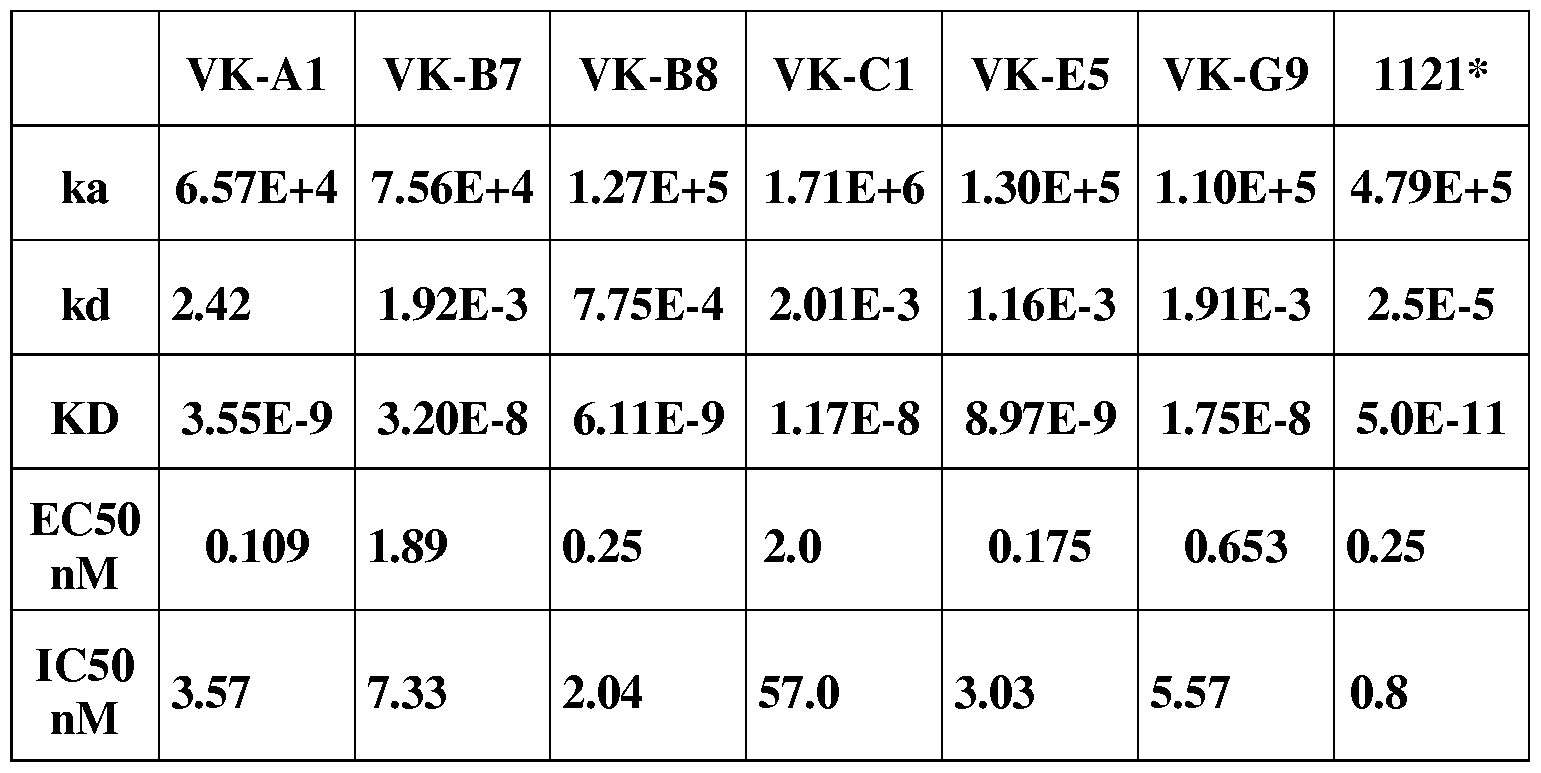
*published data in the literature: Lu, D., et al., 278: 43496-43507, JBC (2003)
Sequence Listing:

VB-D6 SEQ ID NO. 19 SEQ ID NO. 20
MAQVQLVQSGAEVKKPGSSVKVSCKASGGTFSSYA QSVVTQQPSVSAAPGQKVTISCSGGSSN IG ISWVRQAPGQGLEWMGGIIPIFGTANYAQKFQGR NNYVSWYQQLPGTAPKLLIYDNNKRPSGIP VTITADESTSTAYMELSSLRSEDTAVYYCARGSERVY DRFSGSKSGSSASLAITGLQAEDEADYYCQS SS HTYYG M D VWGQGTTVTVSS YDSSLSGYVFGTGTKLTVL
VB-D11 SEQ ID NO. 21 SEQ ID NO. 22
MAQVQLVESGAEVKKPGASVKVSCKASGYTFTNYYI Dl VM TQS P DS LAVS LG E R ATI N CKTSQS VLY HWVRQAPGQGLEWMGIIN PSSGSTNYAQKFQGR NANNKNYLNWYQQKPGQPPKLLIYWASAR VTMTRDTSTSTVYMELSSLRSEDTAVYYCARQRWD ESGVPDRFSGSGSGTDFTLTIRSLQPDDFAT LLDDAFDIWG QGTM VTVSS YYCQQAISFPLTFGGGTKVEIK
VB-E1 SEQ ID NO. 23 SEQ ID NO. 24
MAEVQLVESGGGVVQPGRSLRLSCAASGFTFSSYG SYELMQPPSVSEAPGMTAQITCGGNNIGSK
MHWVRQAPGKGLEWVAVISYDGSNKYYADSVKG SVHWYQQKPGQAPVLVIYYDSERPSGIPDR
RFTISRDNSKNTLYLQMNSLRAEDTAVYYCAKDWS FSGSNSGNTASLTINRVEAGDEADYYCQVW
GPITLWGQGTLVTVSS DSSSDHHVVFGGGTKLTVL
VB-E2 SEQ ID NO. 25 SEQ ID NO. 26
MAEVQLVESGGGLVQPGGSLRLSCAASGFTFSSYE QSALTQPPSMSAAPGQKVTISCSGTSSNIG MNWVRQAPGKGLEWVSYISSSGSTIYYADSVKGRF N HYVSWYQRLPDTAPKLLIYDNNRRPSGIP TISRDNAKNSLYLQMNGLRAEDTAVYYCAREGDSS DRFSGSKSGTSATLGITGLQTGDEADYYCGT GFDYWGQGT LVTVS S WDSSLSGYVFGTGTKVTVL
VB-E7 SEQ ID NO. 27 SEQ ID NO. 28
MAQVQLVESGGGLVQPGGSLRLSCAASGFTFSDHY DIVMTQTPLFLPVTLGQPASISCRSSQSLVHS MDWVRQAPGKGLEWVGRIRN KPNSYTSEYAASVK DGNTYLNWFQQRPGQSPRRLIYKVSIRDSG GRFTISRDDSKNSLYLQMNSLRAEDTAVYYCASRSG VPGRFSGSGSGTDFTLKISSVEAEDIGVYYC SYYDHM D VWG QGTTVTVSS MQGTDRPYTFGQGTKLEIK
VB-F2 SEQ ID NO. 29 SEQ ID NO. 30
MAQMQLVQSGGGVVQPGRSLRLSCVVSGFTFNDY QAGLTQSPSVSAAPGQRVTISCTGSSSNIGA PMHWVRQAPGKGLEWVALLSYDGTSAYYADSVEG GYDVHWYQQLPGTAPKLLIYGNSNRPSGVP RFTISRDNSKNTLYLQMNTLRTEDTAVYYCASEGSP DRFSGSKSGTSASLAITGLQAEDEADYYCQS DAFDIWGQGTM VTVSS YDSSLSGSVFGGGTKVTVL
VB-F8 SEQ ID NO. 31 SEQ ID NO. 32
MAQVQLVQSGAEVKKPGESLKISCKGSGYSFTSYWI QSVVTQPASVSGSPGQSITISCTGTTTDVGA GWVRQMPGKGLEWMGIIYPGDSDTRYSPSFQGQ YNYVSWYQHHPGKAPKLIIYDLN NRPSGISN VTISADKSISTAYLQWSSLKASDTAMYYCARLDYDSS RFSGFKSGNTASLTISGLQAEDEADYYCSSYT GYYGAFDIWGQGTM VTVSS SSSTWVFGGGTKLTVL
VB-G4 SEQ ID NO. 33 SEQ ID NO. 34
MAQVQLVESGAEVKKPGASVKVSCKASGYTFTSYY QAG LTQP PSASGSPGQSVTI SCTGTSS D VG M H WVRQAPGQGLQWMGI 1 N PSGGSTSYAQN FQ GYNYVSWYQQHPGKAPKLMIYEVSKRPSG GRVTMTRDTSTSTVYMELSSLISEDTAVYYCARSGYS VPDRFSGSKSGNTASLTVSGLQAEDEADYY SSWLSYGMDVWGQGTTVTVSS CSSFTTSSTWVFGGGTQLTVL
VB-G6 SEQ ID NO. 35 SEQ ID NO. 36
MAEVQLVQSGAEVKKPGASVKLSCKASGYTFNNYA QPVLTQPPSASGTPGQRVTIYCSGSNSNIGG TIWVRQAPEQGLEYVGWISAYSGHTNYAQKLQGR NSVNWYQQLPGTAPKLLIYHNNQRPSGVP VSMTTDTSTTTAYMELRSLRSDDTAVYYCARWSG DRFSGSRSGTSASLAISGLQSGDEADYSCAA WGSYHLLGM DVWG QGTTVTVSS WDDSLRGYVFGTGTKVTVL
VB-H4 SEQ ID NO. 37 SEQ ID NO. 38
MAQVQLVQSGAEVKKPGASVKVSCKASGYTFTGYY QAVLTQPASVSGSPGQSITISCTGTSSDVGG M H WVRQATGQGLEWLGWM N PKTGVTGYAQKF YNYVSWYQQHPGKAPKLMIYEVNNRPSGV QGRVTMTRNTSINTAYM ELNSLTSEDTADYYCARG SNRFSGSKSGNTASLTISGLQAEDEADYYCT DYGGPQDMDVWGQGTTVTVSS SYTSSNTRMFGGGTKLTVL
VB-H7 SEQ ID NO. 39 SEQ ID NO. 40
MAEVQLVQSGAEVKKPGASVKVSCKASGYTFTGYY QAVLTQPRSVSGSPGQSVTISCTGTRSDVGT MHWVRQAPGQGLEWMGWINPNSGGTNYAQKF YNYVSWYQQLPGKAPKLMIHDVSKRPSGV QGRVTMTRDTSISTAYMELSRLRSDDTAVYYCARDL PDRLSGSKSGNTASLTISGLQSEDEADYYCIS QGITIFGVANSPPYYYGMDVWGQGTTVTVSS YTLH RTWM FGGGTKLTVL
VB-H9 SEQ ID NO. 41 SEQ ID NO. 42
MAEVQLVQSGAEVKKPGASVKISCKASGYIFNTYSIH QPVLTQPPSVSGTPGQRVSISCSGSSSNIGS
WVRQAPGQSFEWMGWSSAGDDNTKYSDDFHHR NSVNWYQQLPGTAPRLLIYNN DQRPSGVP
LTIARDTSASTVYMELRGLTSDDTAIYYCARGYELDF DRFSASKSGTSASLAIGGLQSEDEGDYYCSA
WGQGTLVTVSS WDDSLNGPWVFGGGTKLTVL
RV-A9 SEQ ID NO. 43 SEQ ID NO. 44
MAEVQLVESGGGLVKPGGSLRLSCAASGFTFSSYS QSVLTQPPSVSAAPGQKVTISCSGSTSNIAN
MNWVRQAPGKGLEWVSSISSSSSYIYYADSVKGRFT NFVSWYQQLPGTAPKLLIYDNNKRPSGIPD
ISRDNAKNSLYLQMNSLRAEDTAVYYCARVGATMG RFSGSKSGTSATLGITGLQAGDEADYFCGT
DYWGQGTLVTVSS W DSS LS AS Y VFGTGTKVTVL
RV-F8 SEQ ID NO. 45 SEQ ID NO. 46
MAQVQLVQSGAEVKKPGESLKISCKGSGYSFTSYWI DIVMTQSPSSVSAFVGDRVTITCRASQDVG
GWVRQMPGKGLEWMGIIYPGDSDTRYSPSFQGQ S W LVWYQQK PG K VP K LLI YG ASTLQS G VPS
VTISADKSTSTAYLQWSSLKASDTAMYYCARLGSSG RFSGGGSGTDFTLTISSLQPEDFATYYCQQA
WYDAFDIWGQGTMVTVSS KSLPYTFGQGTKLEIK
RV-H2 SEQ ID NO. 47 SEQ ID NO. 48
MAQVQLVQSGGGLVKPGGSLRLSCAASGFTFSSYS LPVLTQPASVSGSPGQSITISCTGTDSDVGG
MNWVRQAPGKGLEWVSSISSSSSYIYYADSVKGRFT YNYVSWYQQHPGKAPKLIISDVSNRPSGVS
ISRDNAKNSLYLQMNSLRAEDTAVYYCARVGATMG N RFSGSKSGNTASLTISGLQADDEADYYCNS
DYWGQGTLVTVSS YTVHATVLFGGGTKVTVL
RV-H4 SEQ ID NO. 49 SEQ ID NO. 50
MAEVQLLESGGGLVKPRGSLRLSCAASGFTFSNAW QAGLTQPPSVSVSPGQTASITCSGDKLGDKY MSWVRQAPGKGLEWVGRIKSKTDGGTTDYAAPVK ISWYQQKPGQSPVMVIFQDTKRPSGIPERF G RFTI S RD DS KN TLYLQM N S LKTE DTA VYYCTTVW R SGSNSGNTATLTISGTQAMDEADYYCQAW FGELFRWGQGTLVTVSS DTSTVFGGGTKLTVL
RV-H5 SEQ ID NO. 51 SEQ ID NO. 52
MAEVQLVQSGSELKKPGASVKISCKASGYTLTNHAL QPVLTQPPSVSVAPGQTARITCGGNNIGSK
NWVRQAPGQGLEWMAWM NTNTGNPTYAQDFT LVHWYQQKPGQAPVLVVYDDSDRPSGIPE
GRFVFSLDTSVSTAYLEISSLKAEDTAIYYCAREPRDA RFSGSNSGNTATLTISRVEAGDEADYYCQV
DAFDIWGQGTMVTVSS WDSSSDLVVFGGGTKLTVL
CI SEQ ID NO. 53 SEQ ID NO. 54
MAQVQLVQSGTEVKKPGESLRISCRSSGYKFTNYWI QSVVTQPPSVSAAPRQKVTISCSGSSSNIGN GWVRQLPGQGLEWMGVILPGDSDTRYGPSFQGH NYVSWYQQLPGTAPKLLIYDNNRRPSGIPD VS 1 S VD KS 1 STVYLE W ES LKASDTAM YYCASW DN F D RFSGSKSGTSATLGITGLQTGDEADYYCGT HWGQGTLVTVSS WDSSLSAGVFGTGTKVTVL
VR-A2 SEQ ID NO. 55 SEQ ID NO. 56
MAEVQLVQSGAEVKKPGTSVTISCKTSGYTFTTYYIH AIRMTQSPDSLAVSLGERATINCKSSQSVLY
WVRQAPGQGLEWMGIILPSGGNTNYAPNFQGRV SSN NKNYLAWYQQKPGQPPKLLIYWASTRE
TMTRDTSTSTVN MELSSLTSDDTAVYYCVREYRGGY SGVPDRFSGSGSGTDFTLTISSLQAEDVAVY
FDYWGQGTLVTVSS YCQQYYSTP YTFG QGTK LE 1 K
VR-A3 SEQ ID NO. 57 SEQ ID NO. 58
MAQVQLVESGAEVKKPGASVKVSCKASGYTFTSYY QSVVTQPPSVSGAPGQRVTISCTGSSSNIGA
MHWVRQAPGQGLEWMGIINPSGGSTSYAQKFQG GYDVHWYKQLPGTAPKLLIYGNNNRPSGV
RVTMTRDTSTSTVYM ELSSLRSEDTAVYYCAREGAN PDRFSGSKSGTSASLAITGLQAEDEADYYCQ
APNSYYYMDVWGKGTTVTVSS SYDSSLSEGVFGTGTKVTVL
VR-A10 SEQ ID NO. 59 SEQ ID NO. 60
MAEVQLLESGGGLVQPGGSLRLSCAASGFTFSSYA DIQLTQSPPSVSASVGDRVTITCRASQDIST
MHWVRQAPGKGLEWVAVISYDGSNKYYADSVKG WLAWYQQKPGSAPKVLIYAASTFQSGVPSR
RFTISRDNSKNTLYLQMNSLRAEDTAVYYCARDLGA FRGSGSGTYFTLTISGLQPEDFATYYCQQGN
ASYWYFDLWGRGTLVTVSS SFPPTFGQGTKLEIK
VR-B2 SEQ ID NO. 61 SEQ ID NO. 62
MAQVQLVQSGAEVKKPGSSVKVSCKAYGGTFGSY SYELTQPASVSGSPGQSITISCTGSSSDVGGY
GVSWVRRAPGQGLEWMGRLIPIFGTRDYAQKFQG NFVSWYRQHAGKAPKLMIYDVTNRPSGVS
RVTLTADESTNTAYMELSSLRSEDTAVYYCARDGDY TRFSGSKSGTTASLTISGLQPDDEAHYYCSSY
YGSGSYYGMDVWGQGTTVTVSS TTTSTW V F G G GT K LTV L
VR-B4 SEQ ID NO. 63 SEQ ID NO. 64
M AEVQLVESGG G VVQPG RS LRLSCAASG FTFSSYG QSVVTQPPSVSAAPGQRVTISCTGSSSNIGA MHWVRQAPGKGLEWVAVISYDGSNKYYADSVKG GYDVHWYQQLPGTAPKLLI YAN N N RPSGV RFTISRDNSKNTLYLQMNSLRAEDTAVYYCAKDGYS PDRFSGSKTGTSASLAITGLQADDEADYFCQ YG G G F DYWG QGTLVTVSS SYDSSLSGWVFGGGTKLTVL
VR-B11 SEQ ID NO. 65 SEQ ID NO. 66
MAEVQLVESGGGLVKPGGSLRLSCAASGFTFSSYS QSVLTQPASVSGSPGQSITISCTGTSSDVGG
MNWVRQAPGKGLEWVSSISSGSSYIYYADSLKGRF YNYVSWYQQHPGKAPKLMIYDVSN RPSGV
TISRDNAKNSLYLQMNSLRAEDTAVYYCARDFGNW SNRFSGSKSGNTASLTISGLQAEDEADYYCS
GQGTLVTVSS SYTSSSTPYVFGTGTKVTVL
VR-C5 SEQ ID NO. 67 SEQ ID NO. 68
MAQVQLVESGGGLVQPGGSLRLSCAASGFNFSSYE SYELMQPHSVSESPGKTVTISCTGSSGSIASN
MNWVRQAPGKGLEWVSYISSSGSTKHYADSVKGR YVQWYQQRPGSAPTTVIYEDDQRPSGVPD
FTISRDNAKNSLYLQMNSLRAEDTAVYYCAREHYNS RFSGSIDSSSNSAALTISGLKTEDEADYYCQS
WYFDLWGRGTLVTVSS YDSANVVFGGGTKLTVL
VR-C7 SEQ ID NO. 69 SEQ ID NO. 70
MAEVQLLESGGGWVKPGGSLRLSCAASGFPFSDYY LPVLTQPPSVSAAPGQKVTIPCSGTYSN IVN MTWVRQAPGKGLEWVSYITTGGRIIYSADSVRGRF NYVSWYQQLPGTAPKLLIYDNNKRPSGIPD TISRDNAKNSVYLQMNSLRAEDTAVYYCARPLRELS RFSGSKSGTSATLGITGLQTGDEADYYCGT PG GF DLWG RGTM VTVSS WDNSLRRWVFGEGTKLTVL
VR-C11 SEQ ID N0.71 SEQ ID NO. 72
MAEVQLVESGGGLVKPGGSLRLSCAASGFTFSSYS QPVLTQPPSASGSPGQSVTISCTGTSSDVGG
MNWVRQAPGKGLEWVSSISSASSYIYYADSVKGRF YNYVSWYQQHPGKAPKLMIYDVNN RPSGV
TISRDNAKKSLYLQLNSLTVEDTAVYYCAREYWGSP PDRFSGSKSGNTASLTISGLQAEDEADYYCS
DYWGRGTLVTVSS SYTSSSTRVFGGGTKLTVL
VR-E3 SEQ ID NO. 73 SEQ ID NO. 74
MAEVQLVESGGGLVKPGGSLRLSCAASGFTFSSYS QSVVTQPPSVSAAPGQKVTISCSGSSSN IGN
MNWVRQAPGKGLEWVSSISSSSSYIYYADSVKGRFT NYVSWYQQLPGTAPKLLIYDYNKRPSGIPDR
ISRDNAKNSLYLQMNSLRAEDTAVYYCARGGVSPG FSGSQSGTSATLGITGLQTGDEADYYCGTW
DYWGQGTLVTVSS DSSLTLYVFGTGTKLTVL
VR-G11 SEQ ID NO. 75 SEQ ID NO. 76
MAQVQLVQSGAEVKKPGSSVKVSCKAYG
GTFGSYGVSWVRRAPGQGLEWMGRLIPI ETTLTQSPATLSVSPGERATVSCRASQSLGS
FGTRDYAQKFQGRVTLTADESTNTAYMEL N LGWFQQKPGQAPRLLIYGASTRATGIPAR
SS LRSEDTAVYYCARDG DYYGSGSYYG M D FSGSGSGTEFTLTISSLQSEDFAVYFCQQYN
VWGQGTLVTVSS DWPITFGQGTRLEIK
VK-B8 SEQ ID NO. 77 SEQ ID NO. 78
MAQMQLVQSGAEVKKPGSSVKVSCKASGGTFSSY QPVLTQPPSVSKDLRQTATLTCTGNGNNVG AISWVRQAPGQGLEWMGRIIPILGIANYAQKFQGR YQGAAWLQQHQGHPPKLLSYRNNNRPSGI VTITADKSTSTAYM ELSSLRS EDTAVYYCARG H DYYG SERFSASRSGNTASLTISGLQPEDEADYFCSA SGNNQEDYFDPWGQGTLVTVSS WDNSLSAWVFGGGTKLTVL
VR-H9 SEQ ID NO. 79 SEQ ID NO. 80
MAQVQLVQSGAEVKKPGSSVKVSCKAYG
GTFGSYGVSWVRRAPGQGLEWMGRLIPI ETTLTQSPATLSVSPGERATVSCRASQSLGS
FGTRDYAQKFQGRVTLTADESTNTAYMEL N LGWFQQKPGQAPRLLIYGASTRATGIPAR
SS LRSEDTAVYYCARDG DYYGSGSYYG M D FSGSGSGTEFTLTISSLQSEDFAVYFCQQYN
VWGQGTLVTVSS DWPITFGQGTKLEIK
VK-B8A SEQ ID NO. 77 SEQ ID NO. 81
Claims
1. A fully human antibody of an IgG class that binds to a VEGFR2-epitope with a binding affinity of at least 10~6M, that has a heavy chain variable domain sequence that is at least 95% identical to the amino acid sequences selected from the group consisting of SEQ ID NO. 1, SEQ ID NO. 3, SEQ ID NO. 5, SEQ ID NO. 7, SEQ ID NO. 9, SEQ ID NO. 11, SEQ ID NO. 13, SEQ ID NO. 15, SEQ ID NO. 17, SEQ ID NO. 19, SEQ ID NO. 21, SEQ ID NO. 23, SEQ ID NO. 25, SEQ ID NO. 27, SEQ ID NO. 29, SEQ ID NO. 31, SEQ ID NO. 33, SEQ ID NO. 35, SEQ ID NO. 37, SEQ ID NO. 39, SEQ ID NO. 41, SEQ ID NO. 43, SEQ ID NO. 45, SEQ ID NO. 47, SEQ ID NO. 49, SEQ ID NO. 51, SEQ ID NO. 53, SEQ ID NO. 55, SEQ ID NO. 57, SEQ ID NO. 59, SEQ ID NO. 61, SEQ ID NO. 63, SEQ ID NO. 65, SEQ ID NO. 67, SEQ ID NO. 69, SEQ ID NO. 71, SEQ ID NO. 73, SEQ ID NO. 75, SEQ ID NO. 77, SEQ ID NO. 79, SEQ ID NO. 81, and combinations thereof, and that has a light chain variable domain sequence that is at least 95% identical to the amino acid sequences selected from the group consisting of SEQ ID NO. 2, SEQ ID NO. 4, SEQ ID NO. 6, SEQ ID NO. 8, SEQ ID NO. 10, SEQ ID NO. 12, SEQ ID NO. 14, SEQ ID NO. 16, SEQ ID NO. 18, SEQ ID NO. 20, SEQ ID NO. 22, SEQ ID NO. 24, SEQ ID NO. 26, SEQ ID NO. 28, SEQ ID NO. 30, SEQ ID NO. 32, SEQ ID NO. 34, SEQ ID NO. 36, SEQ ID NO. 38, SEQ ID NO. 40, SEQ ID NO. 42, SEQ ID NO. 44, SEQ ID NO. 46, SEQ ID NO. 48, SEQ ID NO. 50, SEQ ID NO. 52, SEQ ID NO. 54, SEQ ID NO. 56, SEQ ID NO. 58, SEQ ID NO. 60, SEQ ID NO. 62, SEQ ID NO. 64, SEQ ID NO. 66, SEQ ID NO. 68, SEQ ID NO. 70, SEQ ID NO. 72, SEQ ID NO. 74, SEQ ID NO. 76, SEQ ID NO. 78, SEQ ID NO. 80 and combinations thereof.
2. The fully human antibody of claim 1, wherein the antibody has a heavy chain/light chain variable domain sequence selected from the group consisting of SEQ ID NO. 1/SEQ ID NO. 2, SEQ ID NO. 3/SEQ ID NO. 4, SEQ ID NO. 5/SEQ ID NO. 6, SEQ ID NO. 7/SEQ ID NO. 8, SEQ ID NO. 9/SEQ ID NO. 10, SEQ ID NO. 11/SEQ ID NO. 12, SEQ ID NO. 13/SEQ ID NO. 14, SEQ ID NO. 15/SEQ ID NO. 16, SEQ ID NO. 17/SEQ ID NO. 18, SEQ ID NO. 19/SEQ ID NO. 20, SEQ ID NO. 21/SEQ ID NO. 22, SEQ ID NO. 23/SEQ ID NO. 24, SEQ ID NO. 25/SEQ ID NO. 26, SEQ ID NO. 27/SEQ ID NO. 28, SEQ ID NO. 29/SEQ ID NO. 30, SEQ ID NO. 31/SEQ ID NO. 32, SEQ ID NO. 33/SEQ ID NO. 34, SEQ ID NO. 35/SEQ ID NO. 36, SEQ ID NO. 37/SEQ ID NO. 38, SEQ ID NO. 39/SEQ ID NO. 40, SEQ ID NO. 41/SEQ ID NO. 42, SEQ ID NO. 43/SEQ ID NO. 44, SEQ ID NO. 45/SEQ ID NO. 46, SEQ ID NO. 47/SEQ ID NO. 48, SEQ ID NO. 49/SEQ ID NO. 50, SEQ ID NO. 51/SEQ ID NO. 52, SEQ ID NO. 53/SEQ ID NO. 54, SEQ ID NO. 55/SEQ ID NO. 56, SEQ ID NO. 57/SEQ ID NO. 58, SEQ ID NO. 59/SEQ ID NO. 60, SEQ ID NO. 61/SEQ ID NO. 62, SEQ ID NO. 63/SEQ ID NO. 64, SEQ ID NO. 65/SEQ ID NO. 66, SEQ ID NO. 67/SEQ ID NO. 68, SEQ ID NO. 69/SEQ ID NO. 70, SEQ ID NO. 71/SEQ ID NO. 72, SEQ ID NO. 73/SEQ ID NO. 74, SEQ ID NO. 75/SEQ ID NO. 76, SEQ ID NO. 77/SEQ ID NO. 78, SEQ ID NO. 80, and combinations thereof.
3. A fully human antibody Fab fragment, having a variable domain region from a heavy chain and a variable domain region from a light chain, wherein the heavy chain variable domain sequence that is at least 95% identical to the amino acid sequences selected from the group consisting of SEQ ID NO. 1, SEQ ID NO. 3, SEQ ID NO. 5, SEQ ID NO. 7, SEQ ID NO. 9, SEQ ID NO. 11, SEQ ID NO. 13, SEQ ID NO. 15, SEQ ID NO. 17, SEQ ID NO. 19, SEQ ID NO. 21, SEQ ID NO. 23, SEQ ID NO. 25, SEQ ID NO. 27, SEQ ID NO. 29, SEQ ID NO. 31, SEQ ID NO. 33, SEQ ID NO. 35, SEQ ID NO. 37, SEQ ID NO. 39, SEQ ID NO. 41, SEQ ID NO. 43, SEQ ID NO. 45, SEQ ID NO. 47, SEQ ID NO. 49, SEQ ID NO. 51, SEQ ID NO. 53, SEQ ID NO. 55, SEQ ID NO. 57, SEQ ID NO. 59, SEQ ID NO. 61, SEQ ID NO. 63, SEQ ID NO. 65, SEQ ID NO. 67, SEQ ID NO. 69, SEQ ID NO. 71, SEQ ID NO. 73, SEQ ID NO. 75, SEQ ID NO. 77, SEQ ID NO. 79, SEQ ID NO. 81, and combinations thereof, and that has a light chain variable domain sequence that is at least 95% identical to the amino acid sequences selected from the group consisting of SEQ ID NO. 2, SEQ ID NO. 4, SEQ ID NO. 6, SEQ ID NO. 8, SEQ ID NO. 10, SEQ ID NO. 12, SEQ ID NO. 14, SEQ ID NO. 16, SEQ ID NO. 18, SEQ ID NO. 20, SEQ ID NO. 22, SEQ ID NO. 24, SEQ ID NO. 26, SEQ ID NO. 28, SEQ ID NO. 30, SEQ ID NO. 32, SEQ ID NO. 34, SEQ ID NO. 36, SEQ ID NO. 38, SEQ ID NO. 40, SEQ ID NO. 42, SEQ ID NO. 44, SEQ ID NO. 46, SEQ ID NO. 48, SEQ ID NO. 50, SEQ ID NO. 52, SEQ ID NO. 54, SEQ ID NO. 56, SEQ ID NO. 58, SEQ ID NO. 60, SEQ ID NO. 62, SEQ ID NO. 64, SEQ ID NO. 66, SEQ ID NO. 68, SEQ ID NO. 70, SEQ ID NO. 72, SEQ ID NO. 74, SEQ ID NO. 76, SEQ ID NO. 78, SEQ ID NO. 80, and combinations thereof.
4. The fully human antibody Fab fragment of claim 3, wherein the antibody has a heavy chain/light chain variable domain sequence selected from the group consisting of SEQ ID NO. 1/SEQ ID NO. 2, SEQ ID NO. 3/SEQ ID NO. 4, SEQ ID NO. 5/SEQ ID NO. 6, SEQ ID NO. 7/SEQ ID NO. 8, SEQ ID NO. 9/SEQ ID NO. 10, SEQ ID NO. 11/SEQ ID NO. 12, SEQ ID NO. 13/SEQ ID NO. 14, SEQ ID NO. 15/SEQ ID NO. 16, SEQ ID NO. 17/SEQ ID NO. 18, SEQ ID NO. 19/SEQ ID NO. 20, SEQ ID NO. 21/SEQ ID NO. 22, SEQ ID NO. 23/SEQ ID NO. 24, SEQ ID NO. 25/SEQ ID NO. 26, SEQ ID NO. 27/SEQ ID NO. 28, SEQ ID NO. 29/SEQ ID NO. 30, SEQ ID NO. 31/SEQ ID NO. 32, SEQ ID NO. 33/SEQ ID NO. 34, SEQ ID NO. 35/SEQ ID NO. 36, SEQ ID NO. 37/SEQ ID NO. 38, SEQ ID NO. 39/SEQ ID NO. 40, SEQ ID NO. 41/SEQ ID NO. 42, SEQ ID NO. 43/SEQ ID NO. 44, SEQ ID NO. 45/SEQ ID NO. 46, SEQ ID NO. 47/SEQ ID NO. 48, SEQ ID NO. 49/SEQ ID NO. 50, SEQ ID NO. 51/SEQ ID NO. 52, SEQ ID NO. 53/SEQ ID NO. 54, SEQ ID NO. 55/SEQ ID NO. 56, SEQ ID NO. 57/SEQ ID NO. 58, SEQ ID NO. 59/SEQ ID NO. 60, SEQ ID NO. 61/SEQ ID NO. 62, SEQ ID NO. 63/SEQ ID NO. 64, SEQ ID NO. 65/SEQ ID NO. 66, SEQ ID NO. 67/SEQ ID NO. 68, SEQ ID NO. 69/SEQ ID NO. 70, SEQ ID NO. 71/SEQ ID NO. 72, SEQ ID NO. 73/SEQ ID NO. 74, SEQ ID NO. 75/SEQ ID NO. 76, SEQ ID NO. 77/SEQ ID NO. 78, SEQ ID NO. 79/ SEQ ID NO. 80, SEQ ID NO. 77/ SEQ ID NO. 81, and combinations thereof.
5. A single chain human antibody, having a variable domain region from a heavy chain and a variable domain region from a light chain and a peptide linker connecting the heavy chain and light chain variable domain regions, wherein the heavy chain variable domain sequence that is at least 95% identical to the amino acid sequences selected from the group consisting of SEQ ID NO. 1, SEQ ID NO. 3, SEQ ID NO. 5, SEQ ID NO. 7, SEQ ID NO. 9, SEQ ID NO. 11, SEQ ID NO. 13, SEQ ID NO. 15, SEQ ID NO. 17, SEQ ID NO. 19, SEQ ID NO. 21, SEQ ID NO. 23, SEQ ID NO. 25, SEQ ID NO. 27, SEQ ID NO. 29, SEQ ID NO. 31, SEQ ID NO. 33, SEQ ID NO. 35, SEQ ID NO. 37, SEQ ID NO. 39, SEQ ID NO. 41, SEQ ID NO. 43, SEQ ID NO. 45, SEQ ID NO. 47, SEQ ID NO. 49, SEQ ID NO. 51, SEQ ID NO. 53, SEQ ID NO. 55, SEQ ID NO. 57, SEQ ID NO. 59, SEQ ID NO. 61, SEQ ID NO. 63, SEQ ID NO. 65, SEQ ID NO. 67, SEQ ID NO. 69, SEQ ID NO. 71, SEQ ID NO. 73, SEQ ID NO. 75, SEQ ID NO. 77, SEQ ID NO. 79, SEQ ID NO. 81, and combinations thereof, and that has a light chain variable domain sequence that is at least 95% identical to the amino acid sequences selected from the group consisting of SEQ ID NO. 2, SEQ ID NO. 4, SEQ ID NO. 6, SEQ ID NO. 8, SEQ ID NO. 10, SEQ ID NO. 12, SEQ ID NO. 14, SEQ ID NO. 16, SEQ ID NO. 18, SEQ ID NO. 20, SEQ ID NO. 22, SEQ ID NO. 24, SEQ ID NO. 26, SEQ ID NO. 28, SEQ ID NO. 30, SEQ ID NO. 32, SEQ ID NO. 34, SEQ ID NO. 36, SEQ ID NO. 38, SEQ ID NO. 40, SEQ ID NO. 42, SEQ ID NO. 44, SEQ ID NO. 46, SEQ ID NO. 48, SEQ ID NO. 50, SEQ ID NO. 52, SEQ ID NO. 54, SEQ ID NO. 56, SEQ ID NO. 58, SEQ ID NO. 60, SEQ ID NO. 62, SEQ ID NO. 64, SEQ ID NO. 66, SEQ ID NO. 68, SEQ ID NO. 70, SEQ ID NO. 72, SEQ ID NO. 74, SEQ ID NO. 76, SEQ ID NO. 78, SEQ ID NO. 80, and combinations thereof.
6. The fully human single chain antibody of claim 5, wherein the single chain fully human antibody has a heavy chain/light chain variable domain sequence selected from the group consisting of SEQ ID NO. 1/SEQ ID NO. 2, SEQ ID NO. 3/SEQ ID NO. 4, SEQ ID NO. 5/SEQ ID NO. 6, SEQ ID NO. 7/SEQ ID NO. 8, SEQ ID NO. 9/SEQ ID NO. 10, SEQ ID NO. 11/SEQ ID NO. 12, SEQ ID NO. 13/SEQ ID NO. 14, SEQ ID NO. 15/SEQ ID NO. 16, SEQ ID NO. 17/SEQ ID NO. 18, SEQ ID NO. 19/SEQ ID NO. 20, SEQ ID NO. 21/SEQ ID NO. 22, SEQ ID NO. 23/SEQ ID NO. 24, SEQ ID NO. 25/SEQ ID NO. 26, SEQ ID NO. 27/SEQ ID NO. 28, SEQ ID NO. 29/SEQ ID NO. 30, SEQ ID NO. 31/SEQ ID NO.
32, SEQ ID NO. 33/SEQ ID NO. 34, SEQ ID NO. 35/SEQ ID NO. 36, SEQ ID NO. 37/SEQ ID NO. 38, SEQ ID NO. 39/SEQ ID NO. 40, SEQ ID NO. 41/SEQ ID NO. 42, SEQ ID NO. 43/SEQ ID NO. 44, SEQ ID NO. 45/SEQ ID NO. 46, SEQ ID NO. 47/SEQ ID NO. 48, SEQ ID NO. 49/SEQ ID NO. 50, SEQ ID NO. 51/SEQ ID NO. 52, SEQ ID NO. 53/SEQ ID NO. 54, SEQ ID NO. 55/SEQ ID NO. 56, SEQ ID NO. 57/SEQ ID NO. 58, SEQ ID NO. 59/SEQ ID NO. 60, SEQ ID NO. 61/SEQ ID NO. 62, SEQ ID NO. 63/SEQ ID NO. 64, SEQ ID NO. 65/SEQ ID NO. 66, SEQ ID NO. 67/SEQ ID NO. 68, SEQ ID NO. 69/SEQ ID NO. 70, SEQ ID NO. 71/SEQ ID NO. 72, SEQ ID NO. 73/SEQ ID NO. 74, SEQ ID NO. 75/SEQ ID NO. 76, SEQ ID NO. 77/SEQ ID NO. 78, SEQ ID NO. 80, SEQ ID NO. 77/ SEQ ID NO. 81, and combinations thereof.
7. A method for treating a broad spectrum of mammalian cancers, comprising administering an effective amount of an anti-VEGFR2 polypeptide, wherein the anti- VEGFR2 polypeptide is selected from the group consisting of a fully human antibody of an IgG class that binds to a VEGFR2 epitope with a binding affinity of at least 10"6M, a fully human antibody Fab fragment, having a variable domain region from a heavy chain and a variable domain region from a light chain, a single chain human antibody, having a variable domain region from a heavy chain and a variable domain region from a light chain and a peptide linker connecting the heavy chain and light chain variable domain regions, and combinations thereof;
wherein the fully human antibody has a heavy chain variable domain sequence that is at least 95% identical to the amino acid sequences selected from the group consisting of SEQ ID NO. 1, SEQ ID NO. 3, SEQ ID NO. 5, SEQ ID NO. 7, SEQ ID NO. 9, SEQ ID NO. 11, SEQ ID NO. 13, SEQ ID NO. 15, SEQ ID NO. 17, SEQ ID NO. 19, SEQ ID NO. 21, SEQ ID NO. 23, SEQ ID NO. 25, SEQ ID NO. 27, SEQ ID NO. 29, SEQ ID NO. 31, SEQ ID NO.
33, SEQ ID NO. 35, SEQ ID NO. 37, SEQ ID NO. 39, SEQ ID NO. 41, SEQ ID NO. 43, SEQ ID NO. 45, SEQ ID NO. 47, SEQ ID NO. 49, SEQ ID NO. 51, SEQ ID NO. 53, SEQ ID NO. 55, SEQ ID NO. 57, SEQ ID NO. 59, SEQ ID NO. 61, SEQ ID NO. 63, SEQ ID NO. 65, SEQ ID NO. 67, SEQ ID NO. 69, SEQ ID NO. 71, SEQ ID NO. 73, SEQ ID NO. 75, SEQ ID NO. 77, SEQ ID NO. 79, SEQ ID NO. 81, and combinations thereof, and that has a light chain variable domain sequence that is at least 95% identical to the amino acid sequences selected from the group consisting of SEQ ID NO. 2, SEQ ID NO. 4, SEQ ID NO. 6, SEQ ID NO. 8, SEQ ID NO. 10, SEQ ID NO. 12, SEQ ID NO. 14, SEQ ID NO. 16, SEQ ID NO. 18, SEQ ID NO. 20, SEQ ID NO. 22, SEQ ID NO. 24, SEQ ID NO. 26, SEQ ID NO. 28, SEQ ID NO. 30, SEQ ID NO. 32, SEQ ID NO. 34, SEQ ID NO. 36, SEQ ID NO. 38, SEQ ID NO. 40, SEQ ID NO. 42, SEQ ID NO. 44, SEQ ID NO. 46, SEQ ID NO. 48, SEQ ID NO. 50, SEQ ID NO. 52, SEQ ID NO. 54, SEQ ID NO. 56, SEQ ID NO. 58, SEQ ID NO. 60, SEQ ID NO. 62, SEQ ID NO. 64, SEQ ID NO. 66, SEQ ID NO. 68, SEQ ID NO. 70, SEQ ID NO. 72, SEQ ID NO. 74, SEQ ID NO. 76, SEQ ID NO. 78, SEQ ID NO. 80, and combinations thereof;
wherein the Fab fully human antibody fragment has the heavy chain variable domain sequence that is at least 95% identical to the amino acid sequences selected from the group consisting of SEQ ID NO. 1, SEQ ID NO. 3, SEQ ID NO. 5, SEQ ID NO. 7, SEQ ID NO. 9, SEQ ID NO. 11, SEQ ID NO. 13, SEQ ID NO. 15, SEQ ID NO. 17, SEQ ID NO. 19, SEQ ID NO. 21, SEQ ID NO. 23, SEQ ID NO. 25, SEQ ID NO. 27, SEQ ID NO. 29, SEQ ID NO. 31, SEQ ID NO. 33, SEQ ID NO. 35, SEQ ID NO. 37, SEQ ID NO. 39, SEQ ID NO. 41, SEQ ID NO. 43, SEQ ID NO. 45, SEQ ID NO. 47, SEQ ID NO. 49, SEQ ID NO. 51, SEQ ID NO. 53, SEQ ID NO. 55, SEQ ID NO. 57, SEQ ID NO. 59, SEQ ID NO. 61, SEQ ID NO. 63, SEQ ID NO. 65, SEQ ID NO. 67, SEQ ID NO. 69, SEQ ID NO. 71, SEQ ID NO. 73, SEQ ID NO. 75, SEQ ID NO. 77, SEQ ID NO. 79, SEQ ID NO. 81, and combinations thereof, and that has the light chain variable domain sequence that is at least 95% identical to the amino acid sequences selected from the group consisting of SEQ ID NO. 2, SEQ ID NO. 4, SEQ ID NO. 6, SEQ ID NO. 8, SEQ ID NO. 10, SEQ ID NO. 12, SEQ ID NO. 14, SEQ ID NO. 16, SEQ ID NO. 18, SEQ ID NO. 20, SEQ ID NO. 22, SEQ ID NO. 24, SEQ ID NO. 26, SEQ ID NO. 28, SEQ ID NO. 30, SEQ ID NO. 32, SEQ ID NO. 34, SEQ ID NO. 36, SEQ ID NO. 38, SEQ ID NO. 40, SEQ ID NO. 42, SEQ ID NO. 44, SEQ ID NO. 46, SEQ ID NO. 48, SEQ ID NO. 50, SEQ ID NO. 52, SEQ ID NO. 54, SEQ ID NO. 56, SEQ ID NO. 58, SEQ ID NO. 60, SEQ ID NO. 62, SEQ ID NO. 64, SEQ ID NO. 66, SEQ ID NO. 68, SEQ ID NO. 70, SEQ ID NO. 72, SEQ ID NO. 74, SEQ ID NO. 76, SEQ ID NO. 78, SEQ ID NO. 80, and combinations thereof; and
wherein the single chain human antibody has the heavy chain variable domain sequence that is at least 95% identical to the amino acid sequences selected from the group consisting of SEQ ID NO. 1, SEQ ID NO. 3, SEQ ID NO. 5, SEQ ID NO. 7, SEQ ID NO. 9, SEQ ID NO. 11, SEQ ID NO. 13, SEQ ID NO. 15, SEQ ID NO. 17, SEQ ID NO. 19, SEQ ID NO. 21, SEQ ID NO. 23, SEQ ID NO. 25, SEQ ID NO. 27, SEQ ID NO. 29, SEQ ID NO. 31, SEQ ID NO. 33, SEQ ID NO. 35, SEQ ID NO. 37, SEQ ID NO. 39, SEQ ID NO. 41, SEQ ID NO. 43, SEQ ID NO. 45, SEQ ID NO. 47, SEQ ID NO. 49, SEQ ID NO. 51, SEQ ID NO. 53, SEQ ID NO. 55, SEQ ID NO. 57, SEQ ID NO. 59, SEQ ID NO. 61, SEQ ID NO. 63, SEQ ID NO. 65, SEQ ID NO. 67, SEQ ID NO. 69, SEQ ID NO. 71, SEQ ID NO. 73, SEQ ID NO. 75, SEQ ID NO. 77, SEQ ID NO. 79, SEQ ID NO. 81, and combinations thereof, and that has the light chain variable domain sequence that is at least 95% identical to the amino acid sequences selected from the group consisting of SEQ ID NO. 2, SEQ ID NO. 4, SEQ ID NO. 6, SEQ ID NO. 8, SEQ ID NO. 10, SEQ ID NO. 12, SEQ ID NO. 14, SEQ ID NO. 16, SEQ ID NO. 18, SEQ ID NO. 20, SEQ ID NO. 22, SEQ ID NO. 24, SEQ ID NO. 26, SEQ ID NO. 28, SEQ ID NO. 30, SEQ ID NO. 32, SEQ ID NO. 34, SEQ ID NO. 36, SEQ ID NO. 38, SEQ ID NO. 40, SEQ ID NO. 42, SEQ ID NO. 44, SEQ ID NO. 46, SEQ ID NO. 48, SEQ ID NO. 50, SEQ ID NO. 52, SEQ ID NO. 54, SEQ ID NO. 56, SEQ ID NO. 58, SEQ ID NO. 60, SEQ ID NO. 62, SEQ ID NO. 64, SEQ ID NO. 66, SEQ ID NO. 68, SEQ ID NO. 70, SEQ ID NO. 72, SEQ ID NO. 74, SEQ ID NO. 76, SEQ ID NO. 78, SEQ ID NO. 80, and combinations thereof.
8. The method for treating a broad spectrum of mammalian cancers of claim 7, wherein the fully human antibody has a heavy chain/light chain variable domain sequence selected from the group consisting of SEQ ID NO. 1/SEQ ID NO. 2, SEQ ID NO. 3/SEQ ID NO. 4, SEQ ID NO. 5/SEQ ID NO. 6, SEQ ID NO. 7/SEQ ID NO. 8, SEQ ID NO. 9/SEQ ID NO. 10, SEQ ID NO. 11/SEQ ID NO. 12, SEQ ID NO. 13/SEQ ID NO. 14, SEQ ID NO. 15/SEQ ID NO. 16, SEQ ID NO. 17/SEQ ID NO. 18, SEQ ID NO. 19/SEQ ID NO. 20, SEQ ID NO. 21/SEQ ID NO. 22, SEQ ID NO. 23/SEQ ID NO. 24, SEQ ID NO. 25/SEQ ID NO. 26, SEQ ID NO. 27/SEQ ID NO. 28, SEQ ID NO. 29/SEQ ID NO. 30, SEQ ID NO. 31/SEQ ID NO. 32, SEQ ID NO. 33/SEQ ID NO. 34, SEQ ID NO. 35/SEQ ID NO. 36, SEQ ID NO. 37/SEQ ID NO. 38, SEQ ID NO. 39/SEQ ID NO. 40, SEQ ID NO. 41/SEQ ID NO. 42, SEQ ID NO. 43/SEQ ID NO. 44, SEQ ID NO. 45/SEQ ID NO. 46, SEQ ID NO. 47/SEQ ID NO. 48, SEQ ID NO. 49/SEQ ID NO. 50, SEQ ID NO. 51/SEQ ID NO. 52, SEQ ID NO. 53/SEQ ID NO. 54, SEQ ID NO. 55/SEQ ID NO. 56, SEQ ID NO. 57/SEQ ID NO. 58, SEQ ID NO. 59/SEQ ID NO. 60, SEQ ID NO. 61/SEQ ID NO. 62, SEQ ID NO. 63/SEQ ID NO. 64, SEQ ID NO. 65/SEQ ID NO. 66, SEQ ID NO. 67/SEQ ID NO. 68, SEQ ID NO. 69/SEQ ID NO. 70, SEQ ID NO. 71/SEQ ID NO. 72, SEQ ID NO. 73/SEQ ID NO. 74, SEQ ID NO. 75/SEQ ID NO. 76, SEQ ID NO. 77/SEQ ID NO. 78, SEQ ID NO. 79/SEQ ID NO. 80, SEQ ID NO. 77/ SEQ ID NO. 81, and combinations thereof.
9. The method for treating a broad spectrum of mammalian cancers of claim 7, wherein the fully human antibody Fab fragment has both a heavy chain variable domain region and a light chain variable domain region wherein the antibody has a heavy chain/light chain variable domain sequence selected from the group consisting of SEQ ID NO. 1/SEQ ID NO. 2, SEQ ID NO. 3/SEQ ID NO. 4, SEQ ID NO. 5/SEQ ID NO. 6, SEQ ID NO. 7/SEQ ID NO. 8, SEQ ID NO. 9/SEQ ID NO. 10, SEQ ID NO. 11/SEQ ID NO. 12, SEQ ID NO.
13/SEQ ID NO. 14, SEQ ID NO. 15/SEQ ID NO. 16, SEQ ID NO. 17/SEQ ID NO. 18, SEQ ID NO. 19/SEQ ID NO. 20, SEQ ID NO. 21/SEQ ID NO. 22, SEQ ID NO. 23/SEQ ID NO. 24, SEQ ID NO. 25/SEQ ID NO. 26, SEQ ID NO. 27/SEQ ID NO. 28, SEQ ID NO. 29/SEQ ID NO. 30, SEQ ID NO. 31/SEQ ID NO. 32, SEQ ID NO. 33/SEQ ID NO. 34, SEQ ID NO. 35/SEQ ID NO. 36, SEQ ID NO. 37/SEQ ID NO. 38, SEQ ID NO. 39/SEQ ID NO. 40, SEQ ID NO. 41/SEQ ID NO. 42, SEQ ID NO. 43/SEQ ID NO. 44, SEQ ID NO. 45/SEQ ID NO. 46, SEQ ID NO. 47/SEQ ID NO. 48, SEQ ID NO. 49/SEQ ID NO. 50, SEQ ID NO. 51/SEQ ID NO. 52, SEQ ID NO. 53/SEQ ID NO. 54, SEQ ID NO. 55/SEQ ID NO. 56, SEQ ID NO. 57/SEQ ID NO. 58, SEQ ID NO. 59/SEQ ID NO. 60, SEQ ID NO. 61/SEQ ID NO. 62, SEQ ID NO. 63/SEQ ID NO. 64, SEQ ID NO. 65/SEQ ID NO. 66, SEQ ID NO. 67/SEQ ID NO. 68, SEQ ID NO. 69/SEQ ID NO. 70, SEQ ID NO. 71/SEQ ID NO. 72, SEQ ID NO. 73/SEQ ID NO. 74, SEQ ID NO. 75/SEQ ID NO. 76, SEQ ID NO. 77/SEQ ID NO. 78, SEQ ID NO. 79/SEQ ID NO. 80, SEQ ID NO. 77/ SEQ ID NO. 81, and combinations thereof.
10. The method for treating a broad spectrum of mammalian cancers of claim 7, wherein the fully human single chain antibody has both a heavy chain variable domain region and a light chain variable domain region, wherein the single chain fully human antibody has a heavy chain/light chain variable domain sequence selected from the group consisting of SEQ ID NO. 1/SEQ ID NO. 2, SEQ ID NO. 3/SEQ ID NO. 4, SEQ ID NO. 5/SEQ ID NO. 6, SEQ ID NO. 7/SEQ ID NO. 8, SEQ ID NO. 9/SEQ ID NO. 10, SEQ ID NO. 11/SEQ ID NO. 12, SEQ ID NO. 13/SEQ ID NO. 14, SEQ ID NO. 15/SEQ ID NO. 16, SEQ ID NO. 17/SEQ ID NO. 18, SEQ ID NO. 19/SEQ ID NO. 20, SEQ ID NO. 21/SEQ ID NO. 22, SEQ ID NO. 23/SEQ ID NO. 24, SEQ ID NO. 25/SEQ ID NO. 26, SEQ ID NO. 27/SEQ ID NO. 28, SEQ ID NO. 29/SEQ ID NO. 30, SEQ ID NO. 31/SEQ ID NO. 32, SEQ ID NO. 33/SEQ ID NO. 34, SEQ ID NO. 35/SEQ ID NO. 36, SEQ ID NO. 37/SEQ ID NO. 38, SEQ ID NO. 39/SEQ ID NO. 40, SEQ ID NO. 41/SEQ ID NO. 42, SEQ ID NO. 43/SEQ ID NO. 44, SEQ ID NO. 45/SEQ ID NO. 46, SEQ ID NO. 47/SEQ ID NO. 48, SEQ ID NO. 49/SEQ ID NO. 50, SEQ ID NO. 51/SEQ ID NO. 52, SEQ ID NO. 53/SEQ ID NO. 54, SEQ ID NO. 55/SEQ ID NO. 56, SEQ ID NO. 57/SEQ ID NO. 58, SEQ ID NO. 59/SEQ ID NO. 60, SEQ ID NO. 61/SEQ ID NO. 62, SEQ ID NO. 63/SEQ ID NO. 64, SEQ ID NO. 65/SEQ ID NO. 66, SEQ ID NO. 67/SEQ ID NO. 68, SEQ ID NO. 69/SEQ ID NO. 70, SEQ ID NO. 71/SEQ ID NO. 72, SEQ ID NO. 73/SEQ ID NO. 74, SEQ ID NO. 75/SEQ ID NO. 76, SEQ ID NO. 77/SEQ ID NO. 78, SEQ ID NO. 79/SEQ ID NO. 80, SEQ ID NO. 77/ SEQ ID NO. 81, and combinations thereof.
11. The method for treating a broad spectrum of mammalian cancers of claim 7, wherein the mammalian cancer to be treated is selected from the group consisting of solid tumors, blood born tumors such as leukemias, and tumor metastases; benign tumors, for example hemangiomas, acoustic neuromas, neurofibromas, trachomas, and pyogenic granulomas; inflammatory disorders such as immune and non- immune inflammation; chronic articular rheumatism and psoriasis; ocular angiogenic diseases, for example, diabetic retinopathy, retinopathy of prematurity, macular degeneration, corneal graft rejection, neovascular glaucoma, retrolental fibroplasia, rubeosis; Osier- Webber Syndrome; myocardial angiogenesis; plaque neovascularization; telangiectasia; hemophiliac joints; angiofibroma; and wound granulation and wound healing; telangiectasia psoriasis scleroderma, pyogenic granuloma, cororany collaterals, ischemic limb angiogenesis, corneal diseases, rubeosis, arthritis, diabetic neovascularization, fractures, vasculogenesis, and hematopoiesis..
12. A method for treating a broad spectrum of mammalian cancers, comprising administering an effective amount of an anti-VEGFR2 polypeptide and an antineoplastic agent:
wherein the anti-VEGFR2 polypeptide is selected from the group consisting of a fully human antibody of an IgG class that binds to a VEGFR2 epitope with a binding affinity of at least 10"6M, a fully human antibody Fab fragment, having a variable domain region from a heavy chain and a variable domain region from a light chain, a single chain human antibody, having a variable domain region from a heavy chain and a variable domain region from a light chain and a peptide linker connecting the heavy chain and light chain variable domain regions, and combinations thereof;
wherein the fully human antibody has a heavy chain variable domain sequence that is at least 95% identical to the amino acid sequences selected from the group consisting of SEQ ID NO. 1, SEQ ID NO. 3, SEQ ID NO. 5, SEQ ID NO. 7, SEQ ID NO. 9, SEQ ID NO. 11, SEQ ID NO. 13, SEQ ID NO. 15, SEQ ID NO. 17, SEQ ID NO. 19, SEQ ID NO. 21, SEQ ID NO. 23, SEQ ID NO. 25, SEQ ID NO. 27, SEQ ID NO. 29, SEQ ID NO. 31, SEQ ID NO. 33, SEQ ID NO. 35, SEQ ID NO. 37, SEQ ID NO. 39, SEQ ID NO. 41, SEQ ID NO. 43, SEQ ID NO. 45, SEQ ID NO. 47, SEQ ID NO. 49, SEQ ID NO. 51, SEQ ID NO. 53, SEQ ID NO. 55, SEQ ID NO. 57, SEQ ID NO. 59, SEQ ID NO. 61, SEQ ID NO. 63, SEQ ID NO. 65, SEQ ID NO. 67, SEQ ID NO. 69, SEQ ID NO. 71, SEQ ID NO. 73, SEQ ID NO. 75, SEQ ID NO. 77, SEQ ID NO. 79, SEQ ID NO. 81, and combinations thereof, and that has a light chain variable domain sequence that is at least 95% identical to the amino acid sequences selected from the group consisting of SEQ ID NO. 2, SEQ ID NO. 4, SEQ ID NO. 6, SEQ ID NO. 8, SEQ ID NO. 10, SEQ ID NO. 12, SEQ ID NO. 14, SEQ ID NO. 16, SEQ ID NO. 18, SEQ ID NO. 20, SEQ ID NO. 22, SEQ ID NO. 24, SEQ ID NO. 26, SEQ ID NO. 28, SEQ ID NO. 30, SEQ ID NO. 32, SEQ ID NO. 34, SEQ ID NO. 36, SEQ ID NO. 38, SEQ ID NO. 40, SEQ ID NO. 42, SEQ ID NO. 44, SEQ ID NO. 46, SEQ ID NO. 48, SEQ ID NO. 50, SEQ ID NO. 52, SEQ ID NO. 54, SEQ ID NO. 56, SEQ ID NO. 58, SEQ ID NO. 60, SEQ ID NO. 62, SEQ ID NO. 64, SEQ ID NO. 66, SEQ ID NO. 68, SEQ ID NO. 70, SEQ ID NO. 72, SEQ ID NO. 74, SEQ ID NO. 76, SEQ ID NO. 78, SEQ ID NO. 80, and combinations thereof;
wherein the antineoplastic agent is selected from the group consisting of anti- metabolites/anti-cancer agents, such as pyrimidine analogs (5-fluorouracil, floxuridine, capecitabine, gemcitabine and cytarabine) and purine analogs, folate antagonists and related inhibitors (mercaptopurine, thioguanine, pentostatin and 2-chlorodeoxyadenosine
(cladribine)); antiproliferative/antimitotic agents including natural products such as vinca alkaloids (vinblastine, vincristine, and vinorelbine), microtubule disrupters such as taxane (paclitaxel, docetaxel), vincristin, vinblastin, nocodazole, epothilones and navelbine, epidipodophyllotoxins (etoposide, teniposide), DNA damaging agents (actinomycin, amsacrine, anthracyclines, bleomycin, busulfan, camptothecin, carboplatin, chlorambucil, cisplatin, cyclophosphamide, Cytoxan, dactinomycin, daunorubicin, doxorubicin, epirubicin, hexamethylmelamineoxaliplatin, iphosphamide, melphalan, merchlorehtamine, mitomycin, mitoxantrone, nitrosourea, plicamycin, procarbazine, taxol, taxotere, teniposide,
triethylenethiophosphoramide and etoposide (VP 16)); antibiotics such as dactinomycin (actinomycin D), daunorubicin, doxorubicin (adriamycin), idarubicin, anthracyclines, mitoxantrone, bleomycins, plicamycin (mithramycin) and mitomycin; enzymes (L- asparaginase which systemically metabolizes L-asparagine and deprives cells which do not have the capacity to synthesize their own asparagine); antiplatelet agents;
antiproliferative/antimitotic alkylating agents such as nitrogen mustards (mechlorethamine, cyclophosphamide and analogs, melphalan, chlorambucil), ethylenimines and methylmelamines (hexamethylmelamine and thiotepa), alkyl sulfonates-busulfan, nitrosoureas (carmustine (BCNU) and analogs, stre tozocin), trazenes-dacarbazinine (DTIC); antiproliferative/antimitotic antimetabolites such as folic acid analogs
(methotrexate); platinum coordination complexes (cisplatin, carboplatin), procarbazine, hydroxyurea, mitotane, aminoglutethimide; hormones, hormone analogs (estrogen, tamoxifen, goserelin, bicalutamide, nilutamide) and aromatase inhibitors (letrozole, anastrozole); anticoagulants (heparin, synthetic heparin salts and other inhibitors of thrombin); fibrinolytic agents (such as tissue plasminogen activator, streptokinase and urokinase), aspirin, dipyridamole, ticlopidine, clopidogrel, abciximab; antimigratory agents; antisecretory agents (breveldin); immunosuppressives (cyclosporine, tacrolimus (FK-506), sirolimus (rapamycin), azathioprine, mycophenolate mofetil); anti- angiogenic compounds (TNP-470, genistein) and growth factor inhibitors (e.g., VEGF inhibitors, fibroblast growth factor (FGF) inhibitors); angiotensin receptor blocker; nitric oxide donors; anti-sense oligonucleotides; antibodies (trastuzumab); cell cycle inhibitors and differentiation inducers (tretinoin); mTOR inhibitors, topoisomerase inhibitors (doxorubicin (adriamycin), amsacrine, camptothecin, daunorubicin, dactinomycin, eniposide, epirubicin, etoposide, idarubicin and mitoxantrone, topotecan, irinotecan), corticosteroids (cortisone, dexamethasone,
hydrocortisone, methylpednisolone, prednisone, and prenisolone); growth factor signal transduction kinase inhibitors; mitochondrial dysfunction inducers and caspase activators; and chromatin disrupters.
Priority Applications (4)
| Application Number | Priority Date | Filing Date | Title |
|---|---|---|---|
| CN201380029000.8A CN104411332B (en) | 2012-03-30 | 2013-03-30 | Human antibody in conjunction with VEGFR2 |
| CA2868883A CA2868883C (en) | 2012-03-30 | 2013-03-30 | Fully human antibodies that bind to vegfr2 |
| EP13770063.9A EP2830663B1 (en) | 2012-03-30 | 2013-03-30 | Fully human antibodies that bind to vegfr2 |
| HK15107172.8A HK1206610A1 (en) | 2012-03-30 | 2015-07-27 | Fully human antibodies that bind to vegfr2 vegfr2 |
Applications Claiming Priority (2)
| Application Number | Priority Date | Filing Date | Title |
|---|---|---|---|
| US201261618658P | 2012-03-30 | 2012-03-30 | |
| US61/618,658 | 2012-03-30 |
Publications (2)
| Publication Number | Publication Date |
|---|---|
| WO2013149219A2 true WO2013149219A2 (en) | 2013-10-03 |
| WO2013149219A3 WO2013149219A3 (en) | 2013-11-21 |
Family
ID=49261400
Family Applications (1)
| Application Number | Title | Priority Date | Filing Date |
|---|---|---|---|
| PCT/US2013/034732 WO2013149219A2 (en) | 2012-03-30 | 2013-03-30 | Fully human antibodies that bind to vegfr2 |
Country Status (5)
| Country | Link |
|---|---|
| EP (1) | EP2830663B1 (en) |
| CN (1) | CN104411332B (en) |
| CA (1) | CA2868883C (en) |
| HK (1) | HK1206610A1 (en) |
| WO (1) | WO2013149219A2 (en) |
Cited By (5)
| Publication number | Priority date | Publication date | Assignee | Title |
|---|---|---|---|---|
| WO2016164658A1 (en) * | 2015-04-08 | 2016-10-13 | Sorrento Therapeutics, Inc. | Antibody therapeutics that bind cd123 |
| JP2017531434A (en) * | 2014-10-07 | 2017-10-26 | カドモン コーポレイション,リミティド ライアビリティ カンパニー | Human anti-VEGFR-2 / KDR antibody S |
| US10100118B2 (en) | 2015-04-08 | 2018-10-16 | Sorrento Therapeutics, Inc. | Antibody therapeutics that bind CD123 |
| WO2022218324A1 (en) * | 2021-04-14 | 2022-10-20 | Suzhou Transcenta Therapeutics Co., Ltd. | Novel anti-hvegfr2 antibodies |
| US11905329B2 (en) | 2016-11-02 | 2024-02-20 | Jounce Therapeutics, Inc. | Anti-PD-1 antibodies and uses thereof |
Citations (53)
| Publication number | Priority date | Publication date | Assignee | Title |
|---|---|---|---|---|
| US3817827A (en) | 1972-03-30 | 1974-06-18 | Scott Paper Co | Soft absorbent fibrous webs containing elastomeric bonding material and formed by creping and embossing |
| US3850752A (en) | 1970-11-10 | 1974-11-26 | Akzona Inc | Process for the demonstration and determination of low molecular compounds and of proteins capable of binding these compounds specifically |
| US3901654A (en) | 1971-06-21 | 1975-08-26 | Biological Developments | Receptor assays of biologically active compounds employing biologically specific receptors |
| US4002531A (en) | 1976-01-22 | 1977-01-11 | Pierce Chemical Company | Modifying enzymes with polyethylene glycol and product produced thereby |
| US4098876A (en) | 1976-10-26 | 1978-07-04 | Corning Glass Works | Reverse sandwich immunoassay |
| US4179337A (en) | 1973-07-20 | 1979-12-18 | Davis Frank F | Non-immunogenic polypeptides |
| US4301144A (en) | 1979-07-11 | 1981-11-17 | Ajinomoto Company, Incorporated | Blood substitute containing modified hemoglobin |
| US4496689A (en) | 1983-12-27 | 1985-01-29 | Miles Laboratories, Inc. | Covalently attached complex of alpha-1-proteinase inhibitor with a water soluble polymer |
| US4640835A (en) | 1981-10-30 | 1987-02-03 | Nippon Chemiphar Company, Ltd. | Plasminogen activator derivatives |
| US4670417A (en) | 1985-06-19 | 1987-06-02 | Ajinomoto Co., Inc. | Hemoglobin combined with a poly(alkylene oxide) |
| US4791192A (en) | 1986-06-26 | 1988-12-13 | Takeda Chemical Industries, Ltd. | Chemically modified protein with polyethyleneglycol |
| US4816567A (en) | 1983-04-08 | 1989-03-28 | Genentech, Inc. | Recombinant immunoglobin preparations |
| US4946778A (en) | 1987-09-21 | 1990-08-07 | Genex Corporation | Single polypeptide chain binding molecules |
| US5092885A (en) | 1987-02-12 | 1992-03-03 | The Government Of The United States Of America As Represented By The Secretary Of The Department Of Health And Human Services | Peptides with laminin activity |
| EP0473084A2 (en) | 1990-08-28 | 1992-03-04 | Sumitomo Pharmaceuticals Company, Limited | Polyethylene glycol derivatives, their modified peptides, methods for producing them and use of the modified peptides |
| US5112946A (en) | 1989-07-06 | 1992-05-12 | Repligen Corporation | Modified pf4 compositions and methods of use |
| US5192744A (en) | 1990-01-12 | 1993-03-09 | Northwestern University | Method of inhibiting angiogenesis of tumors |
| US5202352A (en) | 1990-08-08 | 1993-04-13 | Takeda Chemical Industries, Ltd. | Intravascular embolizing agent containing angiogenesis-inhibiting substance |
| WO1994001451A2 (en) | 1992-07-13 | 1994-01-20 | Bionebraska, Inc. | Method for modification of recombinant polypeptides |
| US5281698A (en) | 1991-07-23 | 1994-01-25 | Cetus Oncology Corporation | Preparation of an activated polymer ester for protein conjugation |
| WO1994010308A1 (en) | 1992-10-23 | 1994-05-11 | Immunex Corporation | Methods of preparing soluble, oligomeric proteins |
| US5382657A (en) | 1992-08-26 | 1995-01-17 | Hoffmann-La Roche Inc. | Peg-interferon conjugates |
| US5582996A (en) | 1990-12-04 | 1996-12-10 | The Wistar Institute Of Anatomy & Biology | Bifunctional antibodies and method of preparing same |
| US5807706A (en) | 1995-03-01 | 1998-09-15 | Genentech, Inc. | Method for making heteromultimeric polypeptides |
| US5824784A (en) | 1994-10-12 | 1998-10-20 | Amgen Inc. | N-terminally chemically modified protein compositions and methods |
| US5877293A (en) | 1990-07-05 | 1999-03-02 | Celltech Therapeutics Limited | CDR grafted anti-CEA antibodies and their production |
| US5886152A (en) | 1991-12-06 | 1999-03-23 | Sumitomo Pharmaceuticals Company, Limited | Humanized B-B10 |
| US5932462A (en) | 1995-01-10 | 1999-08-03 | Shearwater Polymers, Inc. | Multiarmed, monofunctional, polymer for coupling to molecules and surfaces |
| US5959083A (en) | 1991-06-03 | 1999-09-28 | Behringwerke Aktiengellschaft | Tetravalent bispecific receptors, the preparation and use thereof |
| US6010902A (en) | 1988-04-04 | 2000-01-04 | Bristol-Meyers Squibb Company | Antibody heteroconjugates and bispecific antibodies for use in regulation of lymphocyte activity |
| WO2000009560A2 (en) | 1998-08-17 | 2000-02-24 | Abgenix, Inc. | Generation of modified molecules with increased serum half-lives |
| US6054297A (en) | 1991-06-14 | 2000-04-25 | Genentech, Inc. | Humanized antibodies and methods for making them |
| US20020044921A1 (en) | 2000-09-29 | 2002-04-18 | Seoju Lee | Pegylated interleukin-10 |
| US6395718B1 (en) | 1998-07-06 | 2002-05-28 | Guilford Pharmaceuticals Inc. | Pharmaceutical compositions and methods of inhibiting angiogenesis using naaladase inhibitors |
| US6462075B1 (en) | 1999-12-23 | 2002-10-08 | The University Of Georgia Research Foundation, Inc. | Chalcone and its analogs as agents for the inhibition of angiogensis and related disease states |
| US6465431B1 (en) | 1999-11-17 | 2002-10-15 | Boston Life Sciences, Inc. | Pharmaceutical compositions comprising troponin subunits, fragments and homologs thereof and methods of their use to inhibit angiogenesis |
| US6475784B1 (en) | 1997-11-14 | 2002-11-05 | Valentis, Inc. | Inhibition of angiogenesis by delivery of nucleic acids encoding anti-angiogenic polypeptides |
| US6482802B1 (en) | 1998-05-11 | 2002-11-19 | Endowment For Research In Human Biology, Inc. | Use of neomycin for treating angiogenesis-related diseases |
| US6482810B1 (en) | 1991-01-15 | 2002-11-19 | Henry Brem | Antibiotic composition for inhibition of angiogenesis |
| US6500431B1 (en) | 1998-07-13 | 2002-12-31 | University Of Southern California | Inhibitors of angiogenesis and tumor growth |
| US6500924B1 (en) | 1996-05-31 | 2002-12-31 | The Scripps Research Institute | Methods and compositions useful for inhibition of angiogenesis |
| US6518298B2 (en) | 1997-10-16 | 2003-02-11 | Entremed, Inc. | Methods and compositions for inhibition of angiogenesis with EM-138 |
| US6521439B2 (en) | 1996-03-08 | 2003-02-18 | The Children's Medical Center Corporation | Nucleic acids encoding plasminogen fragments |
| US6524583B1 (en) | 1999-04-28 | 2003-02-25 | Board Of Regents, The University Of Texas System | Antibody methods for selectively inhibiting VEGF |
| US6525019B2 (en) | 1998-08-21 | 2003-02-25 | The Children's Medical Center Corporation | Use of melanin for inhibition of angiogenesis and macular degeneration |
| US6538103B1 (en) | 1998-07-14 | 2003-03-25 | Bristol--Myers Squibb Company | Lysine binding fragments of angiostatin |
| US6544947B2 (en) | 1998-05-22 | 2003-04-08 | Entremed, Inc. | Compositions and methods for inhibiting endothelial cell proliferation and regulating angiogenesis using cancer markers |
| US6544758B2 (en) | 1995-10-23 | 2003-04-08 | The Children's Medical Center Corporation | Methods for expressing endostatin protein |
| US6548477B1 (en) | 2000-11-01 | 2003-04-15 | Praecis Pharmaceuticals Inc. | Therapeutic agents and methods of use thereof for the modulation of angiogenesis |
| US6559126B2 (en) | 2000-03-31 | 2003-05-06 | Institut Pasteur | Peptides blocking vascular endothelial growth factor (VEGF)-mediated angiogenesis, polynucleotides encoding said peptides and methods of use thereof |
| US6569845B1 (en) | 1997-12-26 | 2003-05-27 | Mochida Pharmaceutical Co., Ltd. | Neovascularization inhibitor containing dienogest as the active ingredient |
| US6573256B2 (en) | 1996-12-30 | 2003-06-03 | Bone Care International, Inc. | Method of inhibiting angiogenesis using active vitamin D analogues |
| US6696245B2 (en) | 1997-10-20 | 2004-02-24 | Domantis Limited | Methods for selecting functional polypeptides |
Family Cites Families (5)
| Publication number | Priority date | Publication date | Assignee | Title |
|---|---|---|---|---|
| AU2002211658A1 (en) * | 2000-10-13 | 2002-04-22 | Uab Research Foundation | Human anti-epidermal growth factor receptor single-chain antibodies |
| EP1807451A2 (en) * | 2004-08-03 | 2007-07-18 | Dyax Corporation | Hk1-binding proteins |
| CA2642270A1 (en) * | 2006-02-15 | 2007-08-23 | Imclone Systems Incorporated | Antibody formulation |
| CA2705152C (en) * | 2007-11-09 | 2016-10-11 | Peregrine Pharmaceuticals, Inc. | Anti-vegf antibody compositions and methods |
| US8128932B2 (en) * | 2009-08-17 | 2012-03-06 | Shanghai Aosaiersi Biotech Co., Ltd. | Anti-VEGFR monoclonal antibody, method of making and uses thereof |
-
2013
- 2013-03-30 EP EP13770063.9A patent/EP2830663B1/en active Active
- 2013-03-30 WO PCT/US2013/034732 patent/WO2013149219A2/en active Application Filing
- 2013-03-30 CA CA2868883A patent/CA2868883C/en active Active
- 2013-03-30 CN CN201380029000.8A patent/CN104411332B/en active Active
-
2015
- 2015-07-27 HK HK15107172.8A patent/HK1206610A1/en unknown
Patent Citations (54)
| Publication number | Priority date | Publication date | Assignee | Title |
|---|---|---|---|---|
| US3850752A (en) | 1970-11-10 | 1974-11-26 | Akzona Inc | Process for the demonstration and determination of low molecular compounds and of proteins capable of binding these compounds specifically |
| US3901654A (en) | 1971-06-21 | 1975-08-26 | Biological Developments | Receptor assays of biologically active compounds employing biologically specific receptors |
| US3817827A (en) | 1972-03-30 | 1974-06-18 | Scott Paper Co | Soft absorbent fibrous webs containing elastomeric bonding material and formed by creping and embossing |
| US4179337A (en) | 1973-07-20 | 1979-12-18 | Davis Frank F | Non-immunogenic polypeptides |
| US4002531A (en) | 1976-01-22 | 1977-01-11 | Pierce Chemical Company | Modifying enzymes with polyethylene glycol and product produced thereby |
| US4098876A (en) | 1976-10-26 | 1978-07-04 | Corning Glass Works | Reverse sandwich immunoassay |
| US4301144A (en) | 1979-07-11 | 1981-11-17 | Ajinomoto Company, Incorporated | Blood substitute containing modified hemoglobin |
| US4640835A (en) | 1981-10-30 | 1987-02-03 | Nippon Chemiphar Company, Ltd. | Plasminogen activator derivatives |
| US4816567A (en) | 1983-04-08 | 1989-03-28 | Genentech, Inc. | Recombinant immunoglobin preparations |
| US4496689A (en) | 1983-12-27 | 1985-01-29 | Miles Laboratories, Inc. | Covalently attached complex of alpha-1-proteinase inhibitor with a water soluble polymer |
| US4670417A (en) | 1985-06-19 | 1987-06-02 | Ajinomoto Co., Inc. | Hemoglobin combined with a poly(alkylene oxide) |
| US4791192A (en) | 1986-06-26 | 1988-12-13 | Takeda Chemical Industries, Ltd. | Chemically modified protein with polyethyleneglycol |
| US5092885A (en) | 1987-02-12 | 1992-03-03 | The Government Of The United States Of America As Represented By The Secretary Of The Department Of Health And Human Services | Peptides with laminin activity |
| US4946778A (en) | 1987-09-21 | 1990-08-07 | Genex Corporation | Single polypeptide chain binding molecules |
| US6010902A (en) | 1988-04-04 | 2000-01-04 | Bristol-Meyers Squibb Company | Antibody heteroconjugates and bispecific antibodies for use in regulation of lymphocyte activity |
| US5112946A (en) | 1989-07-06 | 1992-05-12 | Repligen Corporation | Modified pf4 compositions and methods of use |
| US5192744A (en) | 1990-01-12 | 1993-03-09 | Northwestern University | Method of inhibiting angiogenesis of tumors |
| US5877293A (en) | 1990-07-05 | 1999-03-02 | Celltech Therapeutics Limited | CDR grafted anti-CEA antibodies and their production |
| US5202352A (en) | 1990-08-08 | 1993-04-13 | Takeda Chemical Industries, Ltd. | Intravascular embolizing agent containing angiogenesis-inhibiting substance |
| EP0473084A2 (en) | 1990-08-28 | 1992-03-04 | Sumitomo Pharmaceuticals Company, Limited | Polyethylene glycol derivatives, their modified peptides, methods for producing them and use of the modified peptides |
| US5582996A (en) | 1990-12-04 | 1996-12-10 | The Wistar Institute Of Anatomy & Biology | Bifunctional antibodies and method of preparing same |
| US6482810B1 (en) | 1991-01-15 | 2002-11-19 | Henry Brem | Antibiotic composition for inhibition of angiogenesis |
| US5959083A (en) | 1991-06-03 | 1999-09-28 | Behringwerke Aktiengellschaft | Tetravalent bispecific receptors, the preparation and use thereof |
| US6054297A (en) | 1991-06-14 | 2000-04-25 | Genentech, Inc. | Humanized antibodies and methods for making them |
| US5281698A (en) | 1991-07-23 | 1994-01-25 | Cetus Oncology Corporation | Preparation of an activated polymer ester for protein conjugation |
| US5886152A (en) | 1991-12-06 | 1999-03-23 | Sumitomo Pharmaceuticals Company, Limited | Humanized B-B10 |
| WO1994001451A2 (en) | 1992-07-13 | 1994-01-20 | Bionebraska, Inc. | Method for modification of recombinant polypeptides |
| US5382657A (en) | 1992-08-26 | 1995-01-17 | Hoffmann-La Roche Inc. | Peg-interferon conjugates |
| WO1994010308A1 (en) | 1992-10-23 | 1994-05-11 | Immunex Corporation | Methods of preparing soluble, oligomeric proteins |
| US5824784A (en) | 1994-10-12 | 1998-10-20 | Amgen Inc. | N-terminally chemically modified protein compositions and methods |
| US5932462A (en) | 1995-01-10 | 1999-08-03 | Shearwater Polymers, Inc. | Multiarmed, monofunctional, polymer for coupling to molecules and surfaces |
| US5807706A (en) | 1995-03-01 | 1998-09-15 | Genentech, Inc. | Method for making heteromultimeric polypeptides |
| US6544758B2 (en) | 1995-10-23 | 2003-04-08 | The Children's Medical Center Corporation | Methods for expressing endostatin protein |
| US6521439B2 (en) | 1996-03-08 | 2003-02-18 | The Children's Medical Center Corporation | Nucleic acids encoding plasminogen fragments |
| US6500924B1 (en) | 1996-05-31 | 2002-12-31 | The Scripps Research Institute | Methods and compositions useful for inhibition of angiogenesis |
| US6573256B2 (en) | 1996-12-30 | 2003-06-03 | Bone Care International, Inc. | Method of inhibiting angiogenesis using active vitamin D analogues |
| US6518298B2 (en) | 1997-10-16 | 2003-02-11 | Entremed, Inc. | Methods and compositions for inhibition of angiogenesis with EM-138 |
| US6846634B1 (en) | 1997-10-20 | 2005-01-25 | Domantis Limited | Method to screen phage display libraries with different ligands |
| US6696245B2 (en) | 1997-10-20 | 2004-02-24 | Domantis Limited | Methods for selecting functional polypeptides |
| US6475784B1 (en) | 1997-11-14 | 2002-11-05 | Valentis, Inc. | Inhibition of angiogenesis by delivery of nucleic acids encoding anti-angiogenic polypeptides |
| US6569845B1 (en) | 1997-12-26 | 2003-05-27 | Mochida Pharmaceutical Co., Ltd. | Neovascularization inhibitor containing dienogest as the active ingredient |
| US6482802B1 (en) | 1998-05-11 | 2002-11-19 | Endowment For Research In Human Biology, Inc. | Use of neomycin for treating angiogenesis-related diseases |
| US6544947B2 (en) | 1998-05-22 | 2003-04-08 | Entremed, Inc. | Compositions and methods for inhibiting endothelial cell proliferation and regulating angiogenesis using cancer markers |
| US6395718B1 (en) | 1998-07-06 | 2002-05-28 | Guilford Pharmaceuticals Inc. | Pharmaceutical compositions and methods of inhibiting angiogenesis using naaladase inhibitors |
| US6500431B1 (en) | 1998-07-13 | 2002-12-31 | University Of Southern California | Inhibitors of angiogenesis and tumor growth |
| US6538103B1 (en) | 1998-07-14 | 2003-03-25 | Bristol--Myers Squibb Company | Lysine binding fragments of angiostatin |
| WO2000009560A2 (en) | 1998-08-17 | 2000-02-24 | Abgenix, Inc. | Generation of modified molecules with increased serum half-lives |
| US6525019B2 (en) | 1998-08-21 | 2003-02-25 | The Children's Medical Center Corporation | Use of melanin for inhibition of angiogenesis and macular degeneration |
| US6524583B1 (en) | 1999-04-28 | 2003-02-25 | Board Of Regents, The University Of Texas System | Antibody methods for selectively inhibiting VEGF |
| US6465431B1 (en) | 1999-11-17 | 2002-10-15 | Boston Life Sciences, Inc. | Pharmaceutical compositions comprising troponin subunits, fragments and homologs thereof and methods of their use to inhibit angiogenesis |
| US6462075B1 (en) | 1999-12-23 | 2002-10-08 | The University Of Georgia Research Foundation, Inc. | Chalcone and its analogs as agents for the inhibition of angiogensis and related disease states |
| US6559126B2 (en) | 2000-03-31 | 2003-05-06 | Institut Pasteur | Peptides blocking vascular endothelial growth factor (VEGF)-mediated angiogenesis, polynucleotides encoding said peptides and methods of use thereof |
| US20020044921A1 (en) | 2000-09-29 | 2002-04-18 | Seoju Lee | Pegylated interleukin-10 |
| US6548477B1 (en) | 2000-11-01 | 2003-04-15 | Praecis Pharmaceuticals Inc. | Therapeutic agents and methods of use thereof for the modulation of angiogenesis |
Non-Patent Citations (70)
| Title |
|---|
| "Cloning Vectors: A Laboratory Manual", 1985, ELSEVIER |
| A. R. GENNARO: "Remington: The Science and Practice of Pharmacy, 20th ed.,", 2000, LIPPINCOTT WILLIAMS & WILKINS |
| BAE ET AL., J. BIOL. CHEM., vol. 275, 2000, pages 13588 |
| BIRD ET AL., SCIENCE, vol. 242, 1988, pages 423 - 26 |
| BIRD, SCIENCE, vol. 242, 1988, pages 423 |
| BLOOD ET AL., BIOCH. BIOPHYS. ACTA, vol. 1032, 1990, pages 89 - 118 |
| BLOOM ET AL., PROTEIN SCIENCE, vol. 6, 1997, pages 407 |
| BOLDICKE ET AL., STEM CELLS, vol. 19, no. 1, 2001, pages 24 - 36 |
| BRENNAN ET AL., SCIENCE, vol. 229, 1985, pages 81 |
| CAZALIS ET AL., BIOCONJUG. CHEM, vol. 15, no. 5, 2004, pages 1005 - 1009 |
| CHAMOW ET AL., BIOCONJUGATE CHEM., vol. 5, 1994, pages 133 |
| CONNELL N D, CURR. OPIN. BIOTECHNOL., vol. 12, no. 5, 2001, pages 446 - 9 |
| DE GRAAF ET AL., METHODS MOL. BIOL., vol. 178, 2002, pages 379 - 87 |
| F. AUSUBEL ET AL.: "Current Protocols in Molecular Biology", 1987, GREEN PUBLISHING AND WILEY-INTERSCIENCE |
| FANSLOW ET AL., SEMIN. IMMUNOL, vol. 6, 1994, pages 267 - 78 |
| FOLKMAN; KLAGSBRUN, SCIENCE, vol. 235, 1987, pages 442 - 447 |
| GLENNIE ET AL., J. IMMUNOL., vol. 139, 1987, pages 2367 |
| GLUZMAN ET AL., CELL, vol. 23, 1981, pages 175 |
| HARLOW AND LAND: "Antibodies: A Laboratory Manual", 1988, COLD SPRING HARBOR LABORATORY PRESS |
| HOLASH ET AL., SCIENCE, vol. 284, 1999, pages 1994 - 1998 |
| HOLLIGER ET AL., PROC. NATL. ACAD. SCI. USA, vol. 90, 1993, pages 6444 - 48 |
| HONEGGER; PLUCKTHUN, J. MOL. BIOL., vol. 309, no. 3, 2001, pages 657 - 670 |
| HOPPE ET AL., FEBS LETTERS, vol. 344, 1994, pages 191 |
| HUSTON ET AL., PROC. NATL. ACAD. SCI. USA, vol. 85, 1988, pages 5879 |
| HUSTON ET AL., PROC. NATL. ACAD. SCI. USA, vol. 85, 1988, pages 5879 - 83 |
| INGBER ET AL., LAB. INVEST., vol. 59, 1988, pages 44 - 51 |
| JACOB ET AL., EXP. PATHOL., vol. 15, 1978, pages 1234 |
| JAIN ET AL., NAT. REV. CANCER, vol. 2, 2002, pages 266 - 276 |
| KABAT ET AL.: "Sequences of Proteins of Immunological Interest, 5.sup.th Ed.,", 1991, NIH PUBLICATION NO. 91-3242 |
| KENNET ET AL.: "Monoclonal Antibodies, Hybridomas: A New Dimension in Biological Analyses", 1980, PLENUM PRESS |
| KERBEL, CARCINOGENESIS, vol. 21, 2000, pages 505 - 515 |
| KORNDORFER ET AL., PROTEINS: STRUCTURE, FUNCTION, AND BIOINFORMATICS, vol. 53, no. 1, 2003, pages 121 - 129 |
| KORTT ET AL., BIOMOL. ENG., vol. 18, 2001, pages 95 - 108 |
| KORTT ET AL., PROT. ENG, vol. 10, 1997, pages 423 |
| KOSTELNY ET AL., J. IMMUNOL., vol. 148, 1992, pages 1547 |
| KRIANGKUM ET AL., BIOMOL. ENG, vol. 18, 2001, pages 31 - 40 |
| LANDSCHULZ ET AL., SCIENCE, vol. 240, 1988, pages 1759 |
| LANTTO ET AL., METHODS MOL. BIOL, vol. 178, 2002, pages 303 - 16 |
| LEFRANC ET AL., DEV. COMP. IMMUNOL, vol. 29, 2005, pages 185 - 203 |
| LEFRANC ET AL., DEV. COMP. IMMUNOL., vol. 29, 2005, pages 185 - 203 |
| LUCKOW; SUMMERS, BIOLTECHNOLOGY, vol. 6, 1988, pages 47 |
| LYDEN ET AL., NATURE MEDICINE, vol. 7, 2001, pages 1194 |
| LYNDEN ET AL., NAT. MED., vol. 7, 2001, pages 1194 - 1201 |
| MAKRIDES ET AL., MICROBIOL. REV, vol. 60, no. 3, 1996, pages 512 - 38 |
| MAYFIELD ET AL., PROC. NATL. ACAD. SCI. USA., vol. 100, no. 2, 2003, pages 438 - 42 |
| MCLEOD ET AL., INVEST. OPHTHALMOL. VIS. SCI., vol. 43, no. 2, 2002, pages 474 - 82 |
| MCMAHAN ET AL., EMBO J., vol. 10, 1991, pages 2821 |
| MILSTEIN ET AL., NATURE, vol. 305, 1983, pages 537 |
| MONFARDINI ET AL., BIOCONJUGATE CHEM., vol. 6, 1995, pages 62 - 69 |
| MOSES ET AL., SCIENCE, vol. 248, 1990, pages 1408 - 1410 |
| PAUL, W.,: "Fundamental Immunology, 2nd ed.", 1989, RAVEN PRESS, article "cH. 7" |
| PEPINSKY ET AL., JPET, vol. 297, 2001, pages 1059 |
| POLJAK ET AL., STRUCTURE, vol. 2, 1994, pages 1121 - 23 |
| POUWELS ET AL.: "Cloning Vectors: A Laboratory Manual", 1985, ELSEVIER |
| PRESTA ET AL., CANCER RES., vol. 57, 1997, pages 4593 - 4599 |
| RAJU ET AL., BIOCHEMISTRY, vol. 40, no. 30, 31 December 2000 (2000-12-31), pages 8868 - 76 |
| RASMUSSEN ET AL., CYTOTECHNOLOGY, vol. 28, 1998, pages 31 |
| ROQUE ET AL., BIOTECHNOL. PROG., vol. 20, 2004, pages 639 - 654 |
| SAMBROOK ET AL.: "Molecular Cloning: A Laboratory Manual, 2 ed.,", vol. 1-3, 1989, COLD SPRING HARBOR LABORATORY PRESS |
| SANDLER; KARO: "Polymer Synthesis", vol. 3, ACADEMIC PRESS, pages: 138 - 161 |
| See also references of EP2830663A4 |
| SHARP ET AL., YEAST, vol. 7, no. 7, 1991, pages 657 - 78 |
| SINCLAIR ET AL., PROTEIN EXPR. PURIF., 2002, pages 96 - 105 |
| TISCHER ET AL., J. BIOL. CHEM, vol. 266, 1991, pages 11947 - 11954 |
| URLAUB ET AL., PROC. NATL. ACAD. SCI. USA, vol. 77, 1980, pages 4216 - 20 |
| VERHEUL ET AL., ONCOLOGIST, vol. 5, no. 1, 2000, pages 45 - 50 |
| WARD ET AL., NATURE, vol. 334, 1989, pages 544 |
| WATANABE ET AL., EXP. DERMATOL., vol. 13, no. 11, 2004, pages 671 - 81 |
| WIEDER ET AL., J. BIOL. CHEM., vol. 254, 1979, pages 12579 |
| YOSHIJI ET AL., GUT., vol. 52, no. 9, 2003, pages 1347 - 54 |
Cited By (11)
| Publication number | Priority date | Publication date | Assignee | Title |
|---|---|---|---|---|
| JP2017531434A (en) * | 2014-10-07 | 2017-10-26 | カドモン コーポレイション,リミティド ライアビリティ カンパニー | Human anti-VEGFR-2 / KDR antibody S |
| EP3204423A4 (en) * | 2014-10-07 | 2018-03-28 | Kadmon Corporation, LLC | Human anti-vegfr-2/kdr antibodies |
| US10588968B2 (en) | 2014-10-07 | 2020-03-17 | Kadmon Corporation, Llc | Human anti-VEGFR-2/KDR antibodies |
| US11633475B2 (en) | 2014-10-07 | 2023-04-25 | Kadmon Corporation, Llc | Human anti-VEGFR-2/KDR antibodies |
| WO2016164658A1 (en) * | 2015-04-08 | 2016-10-13 | Sorrento Therapeutics, Inc. | Antibody therapeutics that bind cd123 |
| US9969807B2 (en) | 2015-04-08 | 2018-05-15 | Sorrento Therapeutics, Inc. | Antibody therapeutics that bind CD123 |
| US10100118B2 (en) | 2015-04-08 | 2018-10-16 | Sorrento Therapeutics, Inc. | Antibody therapeutics that bind CD123 |
| US10934361B2 (en) | 2015-04-08 | 2021-03-02 | Sorrento Therapeutics, Inc. | Antibody therapeutics that bind CD123 |
| US11746156B2 (en) | 2015-04-08 | 2023-09-05 | Sorrento Therapeutics, Inc. | Antibody therapeutics that bind CD123 |
| US11905329B2 (en) | 2016-11-02 | 2024-02-20 | Jounce Therapeutics, Inc. | Anti-PD-1 antibodies and uses thereof |
| WO2022218324A1 (en) * | 2021-04-14 | 2022-10-20 | Suzhou Transcenta Therapeutics Co., Ltd. | Novel anti-hvegfr2 antibodies |
Also Published As
| Publication number | Publication date |
|---|---|
| WO2013149219A3 (en) | 2013-11-21 |
| CN104411332A (en) | 2015-03-11 |
| EP2830663A2 (en) | 2015-02-04 |
| EP2830663A4 (en) | 2016-07-27 |
| CA2868883C (en) | 2022-10-04 |
| CN104411332B (en) | 2018-11-23 |
| EP2830663B1 (en) | 2019-02-06 |
| CA2868883A1 (en) | 2013-10-03 |
| HK1206610A1 (en) | 2016-01-15 |
Similar Documents
| Publication | Publication Date | Title |
|---|---|---|
| US20220025054A1 (en) | Fully Human Antibodies That Bind to Vascular Endothelial Growth Factor Receptor (VEGFR2) | |
| AU2020204113B2 (en) | Antigen binding proteins that bind PD-L1 | |
| US9944707B2 (en) | Antibodies that bind epidermal growth factor receptor (EGFR) | |
| US10717779B2 (en) | Methods for blocking an interaction between notch-1 and delta-like 4 (DLL-4) by administering a DDL-4 antibody | |
| WO2014194302A2 (en) | Antigen binding proteins that bind pd-1 | |
| AU2017212717A1 (en) | Antigen binding proteins that bind PD-L1 | |
| US9938352B2 (en) | Antigen binding proteins that bind ErbB3 | |
| JP2015518827A5 (en) | ||
| EP2830663B1 (en) | Fully human antibodies that bind to vegfr2 | |
| EP2863947B1 (en) | Antigen binding proteins that bind igf1r |
Legal Events
| Date | Code | Title | Description |
|---|---|---|---|
| 121 | Ep: the epo has been informed by wipo that ep was designated in this application |
Ref document number: 13770063 Country of ref document: EP Kind code of ref document: A2 |
|
| WWE | Wipo information: entry into national phase |
Ref document number: 2013770063 Country of ref document: EP |
|
| ENP | Entry into the national phase |
Ref document number: 2868883 Country of ref document: CA |