WO2008062479A2 - A high sensitivity assay for molecular typing of biological sample, probes and a kit thereof - Google Patents
A high sensitivity assay for molecular typing of biological sample, probes and a kit thereof Download PDFInfo
- Publication number
- WO2008062479A2 WO2008062479A2 PCT/IN2007/000543 IN2007000543W WO2008062479A2 WO 2008062479 A2 WO2008062479 A2 WO 2008062479A2 IN 2007000543 W IN2007000543 W IN 2007000543W WO 2008062479 A2 WO2008062479 A2 WO 2008062479A2
- Authority
- WO
- WIPO (PCT)
- Prior art keywords
- assay
- nucleic acid
- kit
- capture
- biological sample
- Prior art date
Links
Classifications
-
- C—CHEMISTRY; METALLURGY
- C12—BIOCHEMISTRY; BEER; SPIRITS; WINE; VINEGAR; MICROBIOLOGY; ENZYMOLOGY; MUTATION OR GENETIC ENGINEERING
- C12Q—MEASURING OR TESTING PROCESSES INVOLVING ENZYMES, NUCLEIC ACIDS OR MICROORGANISMS; COMPOSITIONS OR TEST PAPERS THEREFOR; PROCESSES OF PREPARING SUCH COMPOSITIONS; CONDITION-RESPONSIVE CONTROL IN MICROBIOLOGICAL OR ENZYMOLOGICAL PROCESSES
- C12Q1/00—Measuring or testing processes involving enzymes, nucleic acids or microorganisms; Compositions therefor; Processes of preparing such compositions
- C12Q1/68—Measuring or testing processes involving enzymes, nucleic acids or microorganisms; Compositions therefor; Processes of preparing such compositions involving nucleic acids
- C12Q1/6813—Hybridisation assays
- C12Q1/6827—Hybridisation assays for detection of mutation or polymorphism
-
- C—CHEMISTRY; METALLURGY
- C12—BIOCHEMISTRY; BEER; SPIRITS; WINE; VINEGAR; MICROBIOLOGY; ENZYMOLOGY; MUTATION OR GENETIC ENGINEERING
- C12Q—MEASURING OR TESTING PROCESSES INVOLVING ENZYMES, NUCLEIC ACIDS OR MICROORGANISMS; COMPOSITIONS OR TEST PAPERS THEREFOR; PROCESSES OF PREPARING SUCH COMPOSITIONS; CONDITION-RESPONSIVE CONTROL IN MICROBIOLOGICAL OR ENZYMOLOGICAL PROCESSES
- C12Q1/00—Measuring or testing processes involving enzymes, nucleic acids or microorganisms; Compositions therefor; Processes of preparing such compositions
- C12Q1/70—Measuring or testing processes involving enzymes, nucleic acids or microorganisms; Compositions therefor; Processes of preparing such compositions involving virus or bacteriophage
- C12Q1/701—Specific hybridization probes
- C12Q1/702—Specific hybridization probes for retroviruses
- C12Q1/703—Viruses associated with AIDS
Definitions
- the present invention relates to a field of high sensitivity assay for molecular typing of biological sample using surface-enhanced Raman scattering, including resonance scattering.
- the invention also relates to capture probes, detector probes, a kit for molecular typing of biological sample and a method of manufacturing the kit.
- HIV/AIDS has become a serious global health hazard at a dimension unprecedented by any other infection.
- Human Immunodeficiency Virus type-1 (HIV-I) exhibits extremely high degree of variation at genetic level (Leitner et al., 2005). Based on the genetic homology, HIV-I is classified into 9 distinct and primary genetic subtypes designated A through J (with the exception of E and I which are recombinants). Distribution of the viral subtypes across the globe is non-uniform. Additionally, epidemic outbreaks due to recombinant forms of the viruses are also becoming a serious problem in several geographical regions.
- recombinant viruses arise when a genetic exchange takes place between two or more different viral strains generating a novel recombinant form (Najera et al., 2002).
- recombinant viruses are of two types.
- First, circulating recombinant forms (CRF) are strains that have established successful infection in a geographical region or population and been characterized at the molecular level.
- the A/E virus of Thailand is one such recombinant virus.
- the unique recombinant forms (URF) viruses that have been isolated from limited number of subjects and not adequately characterized at the molecular level.
- the objective of the present invention is to develop a novel and innovative strategy to detect primary subtypes and recombinant strains of HIV, preferably HIV-I .
- the same strategy is useful in assaying other types of viruses, bacteria, fungi, protozoa and parasites.
- the assay is applicable to any kind of nucleic acids isolated from any kind of biological source including RNA and DNA without amplification.
- MHA Multi-region hybridization assay
- the TaqMan probes are designed to target distinct sequences (usually 5-7 windows) across the length of the virus and of defined viral subtypes (usually 2 or 3) (Hoelscher et al., 2002; Herbinger et al., 2006; Gerhardt et al., 2005). Each probe is linked to two different fluorescent dyes one a reporter and the other a quencher. Depending on the pattern of report signal generated for the individual windows, the subtype nature of a viral strain can be determined.
- the technique is relatively less expensive than full- length genome sequencing. Additionally, it can be applied to relatively larger number of samples.
- the technique suffers from several technical problems. There are multiple problems with the technique. First, the technique practically can not be used to analyze more than 2 or 3 different viral subtypes. A prior knowledge of the viral subtypes in circulation in a given context is necessary for assay design. Second, the assay can not target a large number of windows with the virus. Usually 5-7 windows are targeted. A total of 15 individual TaqMan probes will be required to analyze 3 different subtype and 5 individual windows in each subtype.
- the assay will require a total of 60 primers and 15 TaqMan probes to molecularly characterize three different viral subtypes. This will be enormously expensive and labor-intensive and as a consequence, practically of limited application. Third, the assay will fail to detect a recombinant or a subtype that is not targeted by the experimental scheme. Fourth, since the technique invariably requires target amplification, false positive results are a serious concern (see above).
- Branched DNA (bDNA) technology This technology is identical in one important aspect to our novel strategy described below. It is for this similarity this technique is being quoted here although the main objective of this technique is not to molecularly characterize diverse subtypes and recombinants of HIV-I, but to determine the viral load in biological samples.
- bDNA does not amplify the target viral RNA or DNA but captures the nucleic acid using a capture probe and cross-links it to a plastic surface in a micro titer plate.
- the technique involves the application of a complex network of detector probes that cross link a large number of alkaline phosphatase enzyme to the captured nucleic acid.
- the technique finally uses the enzyme activity and the read out is the development of color and is akin to the Enzyme-linked immunosorbent assay (ELISA).
- ELISA Enzyme-linked immunosorbent assay
- the technique compensates for lack of target amplification by recruiting a large number of enzyme molecules.
- a commercial kit is marketed by Chiron Diagnostics using this technique (Kern et al., 1996; Collins et al., 1997).
- bDNA uses an enzyme to develop a signal
- SERS SERS
- our technique is technically less complicated, more sensitive and economical. Further, the primary objectives of these two techniques are quite different. While bDNA is mainly designed for viral load determination, our technique is developed for the molecular characterization of the viral subtypes and recombinants.
- HMA heteroduplex mobility assay
- RFLP restriction length fragment polymorphism
- sequencing of short genome segments Barbosa et al., 1998) subtype-specific polymerase chain reaction (Peeters et al., 1998) and others (Abravaya et al., 2000; de Baar et al., 2001 ; Luo et al., 1998; Robbins et al., 1999).
- HMA heteroduplex mobility assay
- RFLP restriction length fragment polymorphism
- Tuan Vo Dinh et.al (Anal. Chem.1998, 70,1352-1356) and Tuan Vo-Dinh et.al (J. Raman spectrosc.2005;36: 640-647) reports the use of SERS-active labels for primers used in polymerase chain reaction amplification of specific target DNA sequences for HIV detection.
- multiple primers are required to be tagged in addition to amplification which will be expensive, laborious and time-consuming.
- Our strategy in the present invention doesn't require amplification of target nucleic acids, avoids false-positive/false-negative results and is inexpensive. It requires the use of a single probe tagged to a Raman Reporter. Further, we adsorb the detector probe on to the silver nanoparticles which functions as an enhancement factor as well as not to hinder Raman signal emission.
- the present invention relates to a high sensitivity assay for molecular typing of a biological sample using surface-enhanced Raman scattering (SERS) including resonance scattering (SERRS), wherein said assay comprising steps of: a) extracting nucleic acid from the biological sample; b) capturing the extracted nucleic acid in a microarray format using subtype-specific and window-specific capture probe(s); and c) detecting the captured nucleic acid with a detector probe tagged to a reporter to determine molecular typing, subtypes and recombinants on the basis of pattern of signals generated.
- SERS surface-enhanced Raman scattering
- SERRS resonance scattering
- the main object of the present invention is to develop a high sensitivity assay for molecular typing of a biological sample using surface-enhanced Raman scattering (SERS) including resonance scattering (SERRS).
- SERS surface-enhanced Raman scattering
- SERRS resonance scattering
- Another main object of the present invention is to develop a high sensitivity assay for molecular typing of HIV.
- Yet another object of the present invention is to develop a high sensitivity assay which avoids false-positive/false-negative results and is inexpensive.
- Still another object of the present invention is to design capture probes for capturing the nucleic acid, wherein the nucleic acid is either DNA or RNA.
- Still another object of the present invention is to design a detector probe to detect captured nucleic acid, wherein the nucleic acid is either DNA or RNA.
- Still another object of the present invention is to develop a kit for molecular typing of biological sample.
- Still another object of the present invention is to develop a method of manufacturing a kit for molecular typing of biological sample.
- the present invention relates to a high sensitivity assay for molecular typing of a biological sample using surface-enhanced Raman scattering (SERS) including resonance scattering (SERRS), wherein said assay comprising steps of: (a) extracting nucleic acid from the biological sample; (b) capturing the extracted nucleic acid in a microarray format using subtype-specific and window-specific capture probe(s); and (c) detecting the captured nucleic acid with a detector probe tagged .to a reporter to determine molecular typing, subtypes and recombinants on the basis of pattern of signals generated; a high sensitivity assay for molecular typing of HIV using surface-enhanced Raman scattering (SERS) including resonance scattering (SERRS), wherein said assay comprising steps of: (a) extracting nucleic acid from the HIV; (b) capturing the extracted nucleic acid in a microarray format using subtype-specific and window-specific capture probe(s); and (c) detecting the captured
- Fig 1 Schematic representation of HIV-I molecular characterization.
- Fig 2 showing subtype-C capture probe captures homologous but not heterologous viral DNA.
- Fig 3 confirming subtype-specificity of the capture probes.
- Fig 4 showing specific detection of the complete viral RNA.
- Fig 5 showing detection of subtype-C viral DNA using Cy-5 tagged detector probe.
- the present invention relates to a high sensitivity assay for molecular typing of a biological sample using surface-enhanced Raman scattering (SERS) including resonance scattering (SERRS), wherein said assay comprising steps of: a) extracting nucleic acid from the biological sample; b) capturing the extracted nucleic acid in a microarray format using subtype-specific and window-specific capture probe(s); and c) detecting the captured nucleic acid with a detector probe tagged to a reporter to determine molecular typing, subtypes and recombinants on the basis of pattern of signals generated.
- SERS surface-enhanced Raman scattering
- SERRS resonance scattering
- the biological sample is selected from a group comprising infectious agents, disease causing agents, microorganisms, higher forms of life including human beings.
- the infectious agents are selected from a group comprising bacteria, viruses, fungi, protozoa and parasites.
- the viruses are selected from a group comprising helical viruses, icosahedral viruses, enveloped viruses and complex viruses, preferably HIV.
- the nucleic acid is either DNA or RNA.
- the capture probes are an array of oligonucleotides spotted in an array fashion on surface of an appropriate glass slide.
- the detector probes are adsorbed onto silver nanoparticles for further enhancement of the assay.
- the reporter is selected from a group comprising Raman reporter, fluorescent dye, radio-isotope and an enzyme.
- the reporter is Raman reporter selected from a group comprising Rhodamine, Cy-5, TAMRA and DSNB.
- the Raman reporter is preferably Rhodamine and Cy-5.
- the assay is applicable without amplification of the nucleic acid.
- the assay avoids false- positive/false-negative results and is inexpensive.
- the assay detects single nucleotide polymorphism in all types of organisms including human beings.
- the present invention also relates to a high sensitivity assay for molecular typing of HIV using surface-enhanced Raman scattering (SERS) including resonance scattering (SERRS), wherein said assay comprising steps of: a) extracting nucleic acid from the HIV; b) capturing the extracted nucleic acid in a microarray format using subtype- specific and window-specific capture probe(s); and c) detecting the captured nucleic acid with a detector probe tagged to a reporter to determine molecular typing, subtypes and recombinants on the basis of pattern of signals generated.
- SERS surface-enhanced Raman scattering
- SERRS resonance scattering
- the present invention also relates to capture probes of SEQ ID NO: I 5 SEQ ID NO: 2 and SEQ ID NO: 3 for capturing nucleic acid isolated from biological sample, wherein the capture probes are sub-type specific and window specific.
- the capture probes are an array of oligonucleotides spotted in an array fashion on surface of an appropriate glass slide.
- the oligonucleotides are synthesized in situ on glass surface using photolithography or pre-synthesized oligonucleotides selected from a group comprising succinylated oligonucleotides, 5' end amino-modified oligonucleotides, disulfide-modified oligonucleotides and other oligonucleotides linked through hetero-bi-functional cross-linking molecules to the surface are used.
- the nucleic acid is either DNA or RNA.
- the biological sample is selected from a group comprising infectious agents, disease causing agents, microorganisms, higher forms of life including human beings.
- the infectious agents are selected from a group comprising bacteria, viruses, fungi, protozoa and parasites.
- the viruses are selected from a group comprising helical viruses, icosahedral viruses, enveloped viruses and complex viruses, preferably HIV.
- the detector probe is non-specific and identifies all subtypes.
- the nucleic acid is either DNA or RNA.
- the present invention also relates to a kit for molecular typing of biological sample using surface-enhanced Raman scattering (SERS) including resonance scattering (SERRS), wherein the kit comprising:
- reagents and buffers to extract nucleic acid from the biological sample b) capture probes to capture the nucleic acid; c) microarray plate to spot capture probes; r x d) detector probe to detect capture probe; e) reporter, preferably Raman reporter; and f) device to detect pattern of signals generated from microarray.
- the kit comprise capture probes of SEQ ID NO: 1, SEQ ID NO: 2 and SEQ ID NO: 3 which are sub-type specific and window specific.
- the kit comprise the detector probe of SEQ ID NO: 4 which is non-specific and identifies all subtypes.
- the present invention also relates to a method of manufacturing a kit for molecular typing of biological sample using surface-enhanced Raman scattering (SERS) including resonance scattering (SERRS), wherein the method comprising: a) providing reagents and buffers in the kit to extract nucleic acid from the biological sample; b) providing appropriate capture probes in the kit to capture the extracted nucleic acid; c) providing microarray plate in the kit to spot the capture probes;* d) providing detector probe in the kit to detect the capture probe; e) providing reporter, preferably Raman Reporter; and f) providing device to detect pattern of signals generated from microarray to manufacture the kit.
- SERS surface-enhanced Raman scattering
- SERRS resonance scattering
- An ideal strategy for the molecular subtyping of the HIV-I strains must have all or several of the following properties.
- the technique should not require target template amplification. This will be less expensive and circumvent the contamination problem.
- the technique should directly detect the viral RNA isolated from a biological sample.
- the technique should have broader application in that it should be capable of detecting all the diverse viral subtypes in multiple windows. 3. The technique must avoid application of expensive fluorescent detection technology, unlike the real-time PCR that employs TaqMan probes chemically conjugated to fluorescent dyes.
- the technique must exploit a powerful strategy for target detection especially compensating for the lack of target amplification.
- Our strategy is based on capturing the viral RNA using a capture probe and detecting the latter with a reporter probe.
- An array of oligo-nucleotides that are subtype-specific and window-specific are amino-modified at the 5' end and are spotted in an array fashion on the surface of a silane-modified glass slide (Guo et al., 1994; Joos et al., 1997; Kumar et al., 2000; Lindroos et al., 2001).
- the capture probes are designed to target 5-10 individual windows spanning the entire length of the virus each window consisting of 1000 bp.
- the viral RNA extracted from a biological sample is directly captured in a series of the windows of the microarray depending on the molecular nature of the viral subtype.
- the captured viral RNA is detected using one or a pool of two or three detector probes that are tagged with a reporter molecule, a radio-isotope, fluorescent dye or a Raman reporter (RR).
- a reporter molecule a radio-isotope, fluorescent dye or a Raman reporter (RR).
- RR Raman reporter
- Raman Reporters a few fluorescent dyes as Raman Reporters as their vibrational properties can be detected by Raman spectroscopy using SERS/SERRS.
- Chemicals without a fluorescent property such as 5, 5'-dithiobis succinimidyl-2-nitrobenzoate) (DSNB), can also be employed as Raman reporters in the present invention.
- Raman reporter exclusively for fluorescent dyes like Rhodamine, TAMRA and others.
- the detector probes are highly conserved and detect all the diverse viral subtypes regardless of the subtype identity.
- the assay is characterized by moderate sensitivity when isotope- or fluorophore-labeled detector probe is used.
- the assay attains high sensitivity when the detector probes are tagged to a Raman reporter as the latter can exploit the phenomenon of surface-enhanced Raman scattering (SERS) or surface- enhanced Raman resonance scattering (SERRS) (Kneipp et al., 2002; Vo-Dinh et al., 2005; Xie et al., 2006).
- SERS surface-enhanced Raman scattering
- SERRS surface- enhanced Raman resonance scattering
- the high sensitivity of RR-tagged reporter probe functionally compensate for the lack of template amplification in our strategy.
- the reporter probe/detector probe is linked to a Raman reporter, fluorescent dye, radioisotope, an enzyme or several other such reporter molecules. Further, the detector probe is adsorbed onto the silver nanoparticles which functions as an enhancement factor and doesn't hinder Raman signal emission.
- the capture probes are synthesized in situ on the glass surface in a spatially addressable manner using standard photolithography.
- presynthesized oligonucleotide are captured by several of the available chemistries.
- Oligonucleotides modified with an amino group are immobilized onto epoxy-silane-derivatised or isothiocyanate coated glass slides.
- Succinylated oligonucleotides can be coupled to aminophenyl- or aminopropyl-derivatised glass slides by peptide bonds.
- Disulfide-modified oligonucleotides can be immobilized onto a mercaptosilanised glass support by a thiol/disulfide exchange reaction.
- Many more attachment strategies using heterobifunctional crosslinking molecules giving many alternatives to the linking molecule and to the surface can be exploited.
- Our strategy is innovative in immobilizing the intact viral RNA molecule, extracted from a biological sample, to an array of subtype- and window-specific capture probes and detecting the viral RNA using a RR-tagged reporter probe(s) and exploiting the phenomenon of SERS or SERRS for a sensitive detection and determination of the viral molecular nature based on the pattern of signals generated from the microarray (Fig 1).
- Viral RNA was released from the viral particles by adding the detergent NP-40 to the stocks to a final concentration of 1%. Viral RNA was extracted from the culture medium using paramagnetic beads and miniMag EXTRACTOR (bioMerieux, http://www.biomerieux-usa.com). Alternatively, viral RNA was extracted using commercial kits from the plasma or stock preparations according to manufacturer's instructions (http://wwwl .qiagen.com/HB/DNeasy96PlantKit EN). To make single-stranded viral DNA representing a defined window of a given viral subtype, a previously published protocol was used
- Viral molecular clones representing viral subtypes were obtained from The National Institutes of Health AIDS Research and Reference Reagent Program (https://www.aidsreagent.org/about_program.cfrn). The following viral molecular clones were used in the experiments to represent defined viral subtypes, p92RW009.6 (#4006) for subtype-A, pYU-2 (#1350) and pNL4-3 (#1 14) for subtype-B and p93IN904 (#3958) for subtype-C.
- the full-length HIV-I sequences were down loaded from the Los Alamos database (http://www.hiv.lanl.aov/content/hiv-db/mainpaae.html) and aligned using CLUSTAL X with minor manual adjustments (Thompson JD, Gibson TJ, Plewniak F, Jeanmougin F, Higgins DG.
- CLUSTAL X windows interface flexible strategies for multiple sequence alignment aided by quality analysis tools. Nucleic Acids Res 1997; 25 (24): 4876-4882). Regions that are highly conserved within a given viral subtype and deviated by at least one base pair from all other subtypes were identified by visual inspection.
- Oligonucleotides of 20-25 bp were designed using Oligo software version-6 to serve as capture or detector probes (http://www.oligo.net/). Oligonucleotide synthesis along with chemical modification and fluorescent dye tagging has been ordered from a few commercial companies that offer custom services including AIphaDNA (alphadna(g>,alphadna.com ' ). MWG (oligoindia(g)mwgdna.com) and Sigma- Aldrich (http://www.sigmaaldrich.com/Local/SA_Splash.html).
- Subtype-C capture probe captures subtype-C, but not -A or -B, viral DNA:
- Amino-modified subtype-C capture probe (N730c) was captured to silane-modified glass surface. Single-stranded DNA within the LTR region of the virus derived from 3 different viral subtypes A, B or C, was added to the capture probe at numbers shown above the wells. After washing to remove unbound viral DNA, the wells were incubated with the detector probe N707 that was tagged with Rhodamine-6-G (R6G). Following incubation and washing, the wells were incubated with silver nanoparticles and the Raman signal was acquired for one second. The data have been presented in (Fig 2). The slide on which capture probe was deposited was shown in the left panel with the number of viral DNA molecules indicated above the columns in Fig 2. The right-hand panel presents data obtained.
- the capture probe immobilized only the subtype-C DNA (top row) but not subtypes-A or B (middle and bottom rows, respectively) confirming the specificity of the capture probe. While the total area of the spot where the capture probe is immobilized is approximately 3.1 x 10 "6 M 2 (2 mm x 2 mm), the area scanned in SERS is only one millionth of the total area 1.2 x 10 "13 M 2 (4 ⁇ M x 4 ⁇ M). This area is equivalent to approximately 10 4 viral DNA molecules in the top most and left most panel where 10 10 copies were actually used. This experiment confirmed that the present strategy can specifically detect target nucleic acid molecules without PCR amplification with high sensitivity.
- Capture probes are specific to viral subtypes:
- subtype-specific probes specifically identify intended viral target nucleic acids efficiently.
- the following experiment illustrates capture and detection of full-length viral RNA from the cell culture supernatant.
- Subtype-B molecular clone HXB-2 was cultured in the laboratory in T-cells using standard culture conditions. Virus was concentrated from 1 ml of the culture medium by high-speed centrifugation and resuspended in 0.1 ml of lysis buffer containing 1% NP-40 and 0.1% BSA and 5 ⁇ l of this solution was used in the experiment.
- Subtype-B specific capture probe N729b and -C specific probe N730c were used for the capture of the viral RNA.
- Detector probe N707 tagged to R6G was used for SERS detection.
- the results (Fig 4) prove that, using the novel strategy it is possible to detect full-length viral RNA. Only the subtype-B specific capture probe (top panels) but not -C specific probe (bottom panels), detects the subtype-B virus.
- Cy-5 fluorescent dye "to be as good Raman Reporter as R6G for the novel detection strategy.
- the top panel contains B-specific capture probe N729b that should not capture the C-viral DNA used in the experiment.
- the middle and bottom panels contain C-specific capture probe N730c.
- the detector probe 19649 bottom panel which must detect all the viral strains regardless of the subtype differences was tagged to Cy-5.
- the same probe was also chemically modified by adding an additional thiol group (and Cy-5) for efficient binding to the silver nano- particles.
- the variant probe is called 19648.
- the copy number of the subtype-C DNA used in the experiment is indicated.
- a large number of capture probes can target several windows within the virus and essentially every viral subtype can be included in the detection strategy.
- the reporter probe is conjugated to a Raman reporter hence exploiting the phenomenon of SERS or SERRS there by a single molecule detection should be possible theoretically.
- the technique here uses SERRS, where both surface enhancement as well as resonant enhancement are employed, making this technique highly sensitive to detect a small number of detector probe molecules.
- Our strategy of generating the reporter signal by adsorbing the probe onto the silver nanoparticle is far superior to the alternative technique commonly employed where the RR is coated with thin layer of silver. Our strategy is superior in terms of both signal enhancement as well as effects of absorption coefficient of silver.
- Raman reporters also offers the advantage of employing low output LASERs and/or diode lasers that are less expensive. Technically less complicated and less expensive equipment could be designed.
- the strategy we developed have a universal application where genetic diversity is a serious problem with other infectious organisms including HIV-2, Influenza, Polio, Dengue, HBV, HCV, Dengue, several bacteria including Tuberculosis, parasites including malaria and other microorganisms. Additionally, the technique also finds application to SNP (single nucleotide polymorphism) in all types of organisms including the human beings. The strategy is technically less complicated and find broader application in resource poor conditions.
- Joos B, Kuster H and Cone R Covalent attachment of hybridizable oligonucleotides to glass supports. Anal Biochem, 247, 96-101 (1997).
- Lindroos K, Liljedahl U, Raitio M and Syvanen AC Minisequencing on oligonucleotide microarrays: comparison of immobilisation chemistries. Nucleic Acids Res, 29, E69(2001).
- Luo CC Downing RG et al, The development and evaluation of a probe hybridization method for subtyping HIV type 1 infection in Kenya. AIDS Res Hum Retroviruses, 14, 691-694 (1998).
- Peeters M. Recombinant HIV sequences: Their role in the global epidemic. In: HIV sequence compendium 2000, eds. C.Kuiken, B.Foley, B.Hahn, P.Marx, F.E.McCutchan, J.Mellors, J.Mullins, J.Sodroski, S.Wolinsky and B.Korber (Theoritical Biology and Biophysics Group, Los Alamos National Laboratory, Los Alamos, New Mexico, USA, Los Alamos) pp. 54-72 (2000). 26) Peeters M, Montois F et al, Subtype-specific polymerase chain reaction for the identification of HIV-I genetic subtypes circulating in Africa. AIDS, 12, 671- 673 (1998).
Landscapes
- Chemical & Material Sciences (AREA)
- Life Sciences & Earth Sciences (AREA)
- Health & Medical Sciences (AREA)
- Organic Chemistry (AREA)
- Engineering & Computer Science (AREA)
- Zoology (AREA)
- Wood Science & Technology (AREA)
- Proteomics, Peptides & Aminoacids (AREA)
- Virology (AREA)
- Immunology (AREA)
- Microbiology (AREA)
- Biochemistry (AREA)
- Biophysics (AREA)
- Molecular Biology (AREA)
- Analytical Chemistry (AREA)
- Physics & Mathematics (AREA)
- Genetics & Genomics (AREA)
- Biotechnology (AREA)
- Bioinformatics & Cheminformatics (AREA)
- General Engineering & Computer Science (AREA)
- General Health & Medical Sciences (AREA)
- AIDS & HIV (AREA)
- Measuring Or Testing Involving Enzymes Or Micro-Organisms (AREA)
- Investigating, Analyzing Materials By Fluorescence Or Luminescence (AREA)
Abstract
The present invention relates to a high sensitivity assay for molecular typing of a biological sample using surface-enhanced Raman scattering (SERS) including resonance scattering (SERRS); capture probes for capturing nucleic acid; a detector probe to detect captured nucleic acid; a kit for molecular typing of biological sample using surface-enhanced Raman scattering (SERS) including resonance scattering (SERRS); and lastly a method of manufacturing said kit.
Description
A HIGH SENSITIVITY ASSAY FOR MOLECULAR TYPING OF BIOLOGICAL SAMPLE, PROBES AND A KIT THEREOF
FIELD OF THE INVENTION
The present invention relates to a field of high sensitivity assay for molecular typing of biological sample using surface-enhanced Raman scattering, including resonance scattering. The invention also relates to capture probes, detector probes, a kit for molecular typing of biological sample and a method of manufacturing the kit.
BACKGROUND OF THE INVENTION
HIV/AIDS has become a serious global health hazard at a dimension unprecedented by any other infection. Human Immunodeficiency Virus type-1 (HIV-I) exhibits extremely high degree of variation at genetic level (Leitner et al., 2005). Based on the genetic homology, HIV-I is classified into 9 distinct and primary genetic subtypes designated A through J (with the exception of E and I which are recombinants). Distribution of the viral subtypes across the globe is non-uniform. Additionally, epidemic outbreaks due to recombinant forms of the viruses are also becoming a serious problem in several geographical regions.
In addition to the above mentioned primary viral subtypes, a large number of recombinant viruses have been identified globally. Recombinant viruses arise when a genetic exchange takes place between two or more different viral strains generating a novel recombinant form (Najera et al., 2002). Depending on the epidemiologic incidence, recombinant viruses are of two types. First, circulating recombinant forms (CRF) are strains that have established successful infection in a geographical region or population and been characterized at the molecular level. The A/E virus of Thailand is one such recombinant virus. Second, the unique recombinant forms (URF), viruses that have been isolated from limited number of subjects and not adequately characterized at the molecular level. The incidence of global viral infections due to both the recombinant forms is on the rise at an alarmingly exponential rate. The total numbers of molecularly characterized CRFs has increased to 34 presently from 8 during the past 2- 3 years. On the other hand, a large number of URFs are being identified on a regular basis. Generation and expansion of the recombinant viruses may throw serious challenges at disease intervention strategies like vaccine development and drug therapy
(Najera et al., 2002; LaI et al., 2005; Peeters, 2000). It is therefore important to develop simple and inexpensive diagnostic strategies to detect recombinant viruses on a priority basis.
The objective of the present invention is to develop a novel and innovative strategy to detect primary subtypes and recombinant strains of HIV, preferably HIV-I . The same strategy is useful in assaying other types of viruses, bacteria, fungi, protozoa and parasites.
The assay is applicable to any kind of nucleic acids isolated from any kind of biological source including RNA and DNA without amplification.
PRIOR ART OF THE INVENTION
Detection of HIV has also been approached by some of the known techniques which are discussed below with their limitations.
A) Full-genome sequencings Determination of the complete nucleic acid sequence is the golden standard for subtype determination of a viral strain (Peeters, 2000; Carr et al., 1999). This strategy can molecularly characterize not only the standard viral subtypes and the existing recombinant forms but also any novel recombinant viruses that may emerge in future.
Shortcoming of the technique: Despite its universal and broad range appeal, this strategy is too expensive, labor intensive and can not be applied to a large number of samples. This strategy is applied only to a small number of selected samples and unsuitable for a large-scale epidemiological study. Importantly, since sequencing is often performed following PCR amplification of the target sequence, false positive results can not be avoided especially in situation where sophisticated diagnostic laboratories are not available (Kwok and Higuchi, 1989; Rys and Persing, 1993; Scherczinger et al., 1999; Wilke et al., 1995). The bulk of the viral infections occur in poor countries with limited technical resources. False positive result could be a serious technical limitation in these countries.
B) Multi-region hybridization assay (MHA)i This assay is presently the most widely employed for subtype characterization of HIV-I (Hoelscher et al., 2002; Herbinger et al., 2006). The assay is based on the real-PCR strategy (Niesters, 2001 ; Espy et al., 2006) using a set of well characterized TaqMan probes (Yeung et al., 2004; Desire et al., 2001). The TaqMan probes are designed to target distinct sequences (usually 5-7 windows) across the length of the virus and of defined viral subtypes (usually 2 or 3) (Hoelscher et al., 2002; Herbinger et al., 2006; Gerhardt et al., 2005). Each probe is linked to two different fluorescent dyes one a reporter and the other a quencher. Depending on the pattern of report signal generated for the individual windows, the subtype nature of a viral strain can be determined.
Shortcoming of the technique: The technique is relatively less expensive than full- length genome sequencing. Additionally, it can be applied to relatively larger number of samples. The technique, however, suffers from several technical problems. There are multiple problems with the technique. First, the technique practically can not be used to analyze more than 2 or 3 different viral subtypes. A prior knowledge of the viral subtypes in circulation in a given context is necessary for assay design. Second, the assay can not target a large number of windows with the virus. Usually 5-7 windows are targeted. A total of 15 individual TaqMan probes will be required to analyze 3 different subtype and 5 individual windows in each subtype. Given that four individual primers are required (due to the nested-PCR format employed) for each amplification, the assay will require a total of 60 primers and 15 TaqMan probes to molecularly characterize three different viral subtypes. This will be enormously expensive and labor-intensive and as a consequence, practically of limited application. Third, the assay will fail to detect a recombinant or a subtype that is not targeted by the experimental scheme. Fourth, since the technique invariably requires target amplification, false positive results are a serious concern (see above).
C) Branched DNA (bDNA) technology: This technology is identical in one important aspect to our novel strategy described below. It is for this similarity this technique is being quoted here although the main objective of this technique is not to molecularly characterize diverse subtypes and recombinants of HIV-I, but to determine the viral load in biological samples. Like our technique described below, bDNA does not
amplify the target viral RNA or DNA but captures the nucleic acid using a capture probe and cross-links it to a plastic surface in a micro titer plate. The technique involves the application of a complex network of detector probes that cross link a large number of alkaline phosphatase enzyme to the captured nucleic acid. The technique finally uses the enzyme activity and the read out is the development of color and is akin to the Enzyme-linked immunosorbent assay (ELISA). The technique compensates for lack of target amplification by recruiting a large number of enzyme molecules. A commercial kit is marketed by Chiron Diagnostics using this technique (Kern et al., 1996; Collins et al., 1997).
The strategy of capturing viral RNA in our invention and the bDNA technology differ from each other in the design of the capture probe and importantly in subsequent detection technology following target capture. While bDNA uses an enzyme to develop a signal, our strategy employs SERS. Unlike bDNA technique, our technique is technically less complicated, more sensitive and economical. Further, the primary objectives of these two techniques are quite different. While bDNA is mainly designed for viral load determination, our technique is developed for the molecular characterization of the viral subtypes and recombinants.
D) DNA array detection of Hepatitis virus RNA: In this publication, the authors attempted to detect Hepatitis A virus in sewage samples (Wan et al., 2005). The virus was sedimented, viral RNA reverse transcribed and PCR amplified. The amplified DNA was captured to glass surface using a chemically cross-linked capture probe and detected using a gold nanoparticle conjugated oligonucleotide. Additionally, they used silver enhancement technique to improve the assay sensitivity. This assay is similar to the strategy developed by us in capturing and detecting a target nucleic acid using a capture probe and a detector probe. However, our strategy employs diction of a sensitive Raman reporter rather than a gold nonoparticle. Also our strategy doesn't require PCR amplification but direct capture of the viral RNA. Additionally, our strategy is characterized by targeting multiple windows of the same target thus offering the advantage of detection of recombinant viruses and multiplexing that could eliminate the possibility of false negative results.
E) Other techniques: A range of several other techniques can be used for viral subtype determination. These techniques include the heteroduplex mobility assay (HMA) (Delwart et al., 1993; Heyndrickx et al., 2000), restriction length fragment polymorphism (RFLP) (van Harmelen et al., 1999), sequencing of short genome segments (Barbosa et al., 1998) subtype-specific polymerase chain reaction (Peeters et al., 1998) and others (Abravaya et al., 2000; de Baar et al., 2001 ; Luo et al., 1998; Robbins et al., 1999). Most of these technique have limited application in molecular typing of the viral strains as they are too cumbersome, fail to target diverse viral subtypes, lead to contamination problem and/or expensive.
1
Tuan Vo Dinh et.al (Anal. Chem.1998, 70,1352-1356) and Tuan Vo-Dinh et.al (J. Raman spectrosc.2005;36: 640-647) reports the use of SERS-active labels for primers used in polymerase chain reaction amplification of specific target DNA sequences for HIV detection. Here, multiple primers are required to be tagged in addition to amplification which will be expensive, laborious and time-consuming.
Our strategy in the present invention doesn't require amplification of target nucleic acids, avoids false-positive/false-negative results and is inexpensive. It requires the use of a single probe tagged to a Raman Reporter. Further, we adsorb the detector probe on to the silver nanoparticles which functions as an enhancement factor as well as not to hinder Raman signal emission.
The present invention relates to a high sensitivity assay for molecular typing of a biological sample using surface-enhanced Raman scattering (SERS) including resonance scattering (SERRS), wherein said assay comprising steps of: a) extracting nucleic acid from the biological sample; b) capturing the extracted nucleic acid in a microarray format using subtype-specific and window-specific capture probe(s); and c) detecting the captured nucleic acid with a detector probe tagged to a reporter to determine molecular typing, subtypes and recombinants on the basis of pattern of signals generated.
OBJECTS OF THE INVENTION
The main object of the present invention is to develop a high sensitivity assay for molecular typing of a biological sample using surface-enhanced Raman scattering (SERS) including resonance scattering (SERRS).
Another main object of the present invention is to develop a high sensitivity assay for molecular typing of HIV.
Yet another object of the present invention is to develop a high sensitivity assay which avoids false-positive/false-negative results and is inexpensive.
Still another object of the present invention is to design capture probes for capturing the nucleic acid, wherein the nucleic acid is either DNA or RNA.
Still another object of the present invention is to design a detector probe to detect captured nucleic acid, wherein the nucleic acid is either DNA or RNA.
Still another object of the present invention is to develop a kit for molecular typing of biological sample.
Still another object of the present invention is to develop a method of manufacturing a kit for molecular typing of biological sample.
STATEMENT OF THE INVENTION
Accordingly, the present invention relates to a high sensitivity assay for molecular typing of a biological sample using surface-enhanced Raman scattering (SERS) including resonance scattering (SERRS), wherein said assay comprising steps of: (a) extracting nucleic acid from the biological sample; (b) capturing the extracted nucleic acid in a microarray format using subtype-specific and window-specific capture probe(s); and (c) detecting the captured nucleic acid with a detector probe tagged .to a reporter to determine molecular typing, subtypes and recombinants on the basis of pattern of signals generated; a high sensitivity assay for molecular typing of HIV using surface-enhanced Raman scattering (SERS) including resonance scattering (SERRS), wherein said assay comprising steps of: (a) extracting nucleic acid from the HIV; (b)
capturing the extracted nucleic acid in a microarray format using subtype-specific and window-specific capture probe(s); and (c) detecting the captured nucleic acid with a detector probe tagged to a reporter to determine molecular typing, subtypes and recombinants on the basis of pattern of signals generated; Capture probes of SEQ ID NO: 1 , SEQ ID NO: 2 and SEQ ID NO: 3 for capturing nucleic acid isolated from biological sample, wherein the capture probes are sub-type specific and window specific; a detector probe of SEQ ID NO: 4 to detect captured nucleic acid, wherein the detector probe is non-specific and identifies all subtypes, a kit for molecular typing of biological sample using surface-enhanced Raman scattering (SERS) including resonance scattering (SERRS), wherein the kit comprising: (a) reagents and buffers to extract nucleic acid from the biological sample; (b) capture probes to capture the nucleic acid; (c) microarray plate to spot capture probes; (d) detector probe to detect capture probe; (e) reporter, preferably Raman reporter; and (f) device to detect pattern of signals generated from microarray; and a method of manufacturing a kit for molecular typing of biological sample using surface-enhanced Raman scattering (SERS) including resonance scattering (SERRS), wherein the method comprising: (a) providing reagents and buffers in the kit to extract nucleic acid from the biological sample; (b) providing appropriate capture probes in the kit to capture the extracted nucleic acid; (c) providing microarray plate in the kit to spot the capture probes; (d) providing detector probe in the kit to detect the capture probe; (e) providing reporter, preferably Raman Reporter; and (f) providing device to detect pattern of signals generated from microarray to manufacture the kit.
BRIEF DESCRIPTION OF ACCOMPANYING DRAWINGS
Fig 1: Schematic representation of HIV-I molecular characterization.
Fig 2: showing subtype-C capture probe captures homologous but not heterologous viral DNA.
Fig 3: confirming subtype-specificity of the capture probes.
Fig 4: showing specific detection of the complete viral RNA.
Fig 5: showing detection of subtype-C viral DNA using Cy-5 tagged detector probe.
DETAILED DESCRIPTION OF THE INVENTION
Accordingly, the present invention relates to a high sensitivity assay for molecular typing of a biological sample using surface-enhanced Raman scattering (SERS) including resonance scattering (SERRS), wherein said assay comprising steps of: a) extracting nucleic acid from the biological sample; b) capturing the extracted nucleic acid in a microarray format using subtype-specific and window-specific capture probe(s); and c) detecting the captured nucleic acid with a detector probe tagged to a reporter to determine molecular typing, subtypes and recombinants on the basis of pattern of signals generated.
In yet another embodiment of the present invention, the biological sample is selected from a group comprising infectious agents, disease causing agents, microorganisms, higher forms of life including human beings.
In still another embodiment of the present invention, the infectious agents are selected from a group comprising bacteria, viruses, fungi, protozoa and parasites.
In still another embodiment of the present invention, the viruses are selected from a group comprising helical viruses, icosahedral viruses, enveloped viruses and complex viruses, preferably HIV.
In still another embodiment of the present invention, the nucleic acid is either DNA or RNA.
In still another embodiment of the present invention, the capture probes are an array of oligonucleotides spotted in an array fashion on surface of an appropriate glass slide.
In still another embodiment of the present invention, the detector probes are adsorbed onto silver nanoparticles for further enhancement of the assay.
In still another embodiment of the present invention, the reporter is selected from a group comprising Raman reporter, fluorescent dye, radio-isotope and an enzyme.
In still another embodiment of the present invention, the reporter is Raman reporter selected from a group comprising Rhodamine, Cy-5, TAMRA and DSNB.
In still another embodiment of the present invention, the Raman reporter is preferably Rhodamine and Cy-5.
In still another embodiment of the present invention, the assay is applicable without amplification of the nucleic acid.
In still another embodiment of the present invention, the assay avoids false- positive/false-negative results and is inexpensive.
In still another embodiment of the present invention, the assay detects single nucleotide polymorphism in all types of organisms including human beings.
The present invention also relates to a high sensitivity assay for molecular typing of HIV using surface-enhanced Raman scattering (SERS) including resonance scattering (SERRS), wherein said assay comprising steps of: a) extracting nucleic acid from the HIV; b) capturing the extracted nucleic acid in a microarray format using subtype- specific and window-specific capture probe(s); and c) detecting the captured nucleic acid with a detector probe tagged to a reporter to determine molecular typing, subtypes and recombinants on the basis of pattern of signals generated.
The present invention also relates to capture probes of SEQ ID NO: I5 SEQ ID NO: 2 and SEQ ID NO: 3 for capturing nucleic acid isolated from biological sample, wherein the capture probes are sub-type specific and window specific.
In still another embodiment of the present invention, the capture probes are an array of oligonucleotides spotted in an array fashion on surface of an appropriate glass slide. -
In still another embodiment of the present invention, the oligonucleotides are synthesized in situ on glass surface using photolithography or pre-synthesized oligonucleotides selected from a group comprising succinylated oligonucleotides, 5' end amino-modified oligonucleotides, disulfide-modified oligonucleotides and other oligonucleotides linked through hetero-bi-functional cross-linking molecules to the surface are used.
In still another embodiment of the present invention, the nucleic acid is either DNA or RNA.
In still another embodiment of the present invention, the biological sample is selected from a group comprising infectious agents, disease causing agents, microorganisms, higher forms of life including human beings.
In still another embodiment of the present invention, the infectious agents are selected from a group comprising bacteria, viruses, fungi, protozoa and parasites.
In still another embodiment of the present invention, the viruses are selected from a group comprising helical viruses, icosahedral viruses, enveloped viruses and complex viruses, preferably HIV.
In still another embodiment of the present invention, the detector probe is non-specific and identifies all subtypes.
In still another embodiment of the present invention, the nucleic acid is either DNA or RNA.
The present invention also relates to a kit for molecular typing of biological sample using surface-enhanced Raman scattering (SERS) including resonance scattering (SERRS), wherein the kit comprising:
a) reagents and buffers to extract nucleic acid from the biological sample; b) capture probes to capture the nucleic acid; c) microarray plate to spot capture probes; r x d) detector probe to detect capture probe;
e) reporter, preferably Raman reporter; and f) device to detect pattern of signals generated from microarray.
In still another embodiment of the present invention, the kit comprise capture probes of SEQ ID NO: 1, SEQ ID NO: 2 and SEQ ID NO: 3 which are sub-type specific and window specific.
In still another embodiment of the present invention, the kit comprise the detector probe of SEQ ID NO: 4 which is non-specific and identifies all subtypes.
The present invention also relates to a method of manufacturing a kit for molecular typing of biological sample using surface-enhanced Raman scattering (SERS) including resonance scattering (SERRS), wherein the method comprising: a) providing reagents and buffers in the kit to extract nucleic acid from the biological sample; b) providing appropriate capture probes in the kit to capture the extracted nucleic acid; c) providing microarray plate in the kit to spot the capture probes;* d) providing detector probe in the kit to detect the capture probe; e) providing reporter, preferably Raman Reporter; and f) providing device to detect pattern of signals generated from microarray to manufacture the kit.
An ideal strategy for the molecular subtyping of the HIV-I strains must have all or several of the following properties.
1. The technique should not require target template amplification. This will be less expensive and circumvent the contamination problem. The technique should directly detect the viral RNA isolated from a biological sample.
2. The technique should have broader application in that it should be capable of detecting all the diverse viral subtypes in multiple windows.
3. The technique must avoid application of expensive fluorescent detection technology, unlike the real-time PCR that employs TaqMan probes chemically conjugated to fluorescent dyes.
4. The technique must exploit a powerful strategy for target detection especially compensating for the lack of target amplification.
We identified a novel and innovative strategy that satisfies the above requirements and molecularly characterize a viral subtype. Our strategy is based on capturing the viral RNA using a capture probe and detecting the latter with a reporter probe. An array of oligo-nucleotides that are subtype-specific and window-specific are amino-modified at the 5' end and are spotted in an array fashion on the surface of a silane-modified glass slide (Guo et al., 1994; Joos et al., 1997; Kumar et al., 2000; Lindroos et al., 2001). The capture probes are designed to target 5-10 individual windows spanning the entire length of the virus each window consisting of 1000 bp. The viral RNA extracted from a biological sample is directly captured in a series of the windows of the microarray depending on the molecular nature of the viral subtype. The captured viral RNA is detected using one or a pool of two or three detector probes that are tagged with a reporter molecule, a radio-isotope, fluorescent dye or a Raman reporter (RR). In the present example/invention, we employed a few fluorescent dyes as Raman Reporters as their vibrational properties can be detected by Raman spectroscopy using SERS/SERRS. Chemicals without a fluorescent property, such as 5, 5'-dithiobis succinimidyl-2-nitrobenzoate) (DSNB), can also be employed as Raman reporters in the present invention. In the present invention, we, however, use the name Raman reporter exclusively for fluorescent dyes like Rhodamine, TAMRA and others. The detector probes are highly conserved and detect all the diverse viral subtypes regardless of the subtype identity. The assay is characterized by moderate sensitivity when isotope- or fluorophore-labeled detector probe is used. However, the assay attains high sensitivity when the detector probes are tagged to a Raman reporter as the latter can exploit the phenomenon of surface-enhanced Raman scattering (SERS) or surface- enhanced Raman resonance scattering (SERRS) (Kneipp et al., 2002; Vo-Dinh et al., 2005; Xie et al., 2006). The high sensitivity of RR-tagged reporter probe functionally compensate for the lack of template amplification in our strategy.
The reporter probe/detector probe is linked to a Raman reporter, fluorescent dye, radioisotope, an enzyme or several other such reporter molecules. Further, the detector probe is adsorbed onto the silver nanoparticles which functions as an enhancement factor and doesn't hinder Raman signal emission.
The capture probes are synthesized in situ on the glass surface in a spatially addressable manner using standard photolithography. Alternatively, presynthesized oligonucleotide are captured by several of the available chemistries. Oligonucleotides modified with an amino group are immobilized onto epoxy-silane-derivatised or isothiocyanate coated glass slides. Succinylated oligonucleotides can be coupled to aminophenyl- or aminopropyl-derivatised glass slides by peptide bonds. Disulfide-modified oligonucleotides can be immobilized onto a mercaptosilanised glass support by a thiol/disulfide exchange reaction. Many more attachment strategies using heterobifunctional crosslinking molecules giving many alternatives to the linking molecule and to the surface can be exploited.
Our strategy is innovative in immobilizing the intact viral RNA molecule, extracted from a biological sample, to an array of subtype- and window-specific capture probes and detecting the viral RNA using a RR-tagged reporter probe(s) and exploiting the phenomenon of SERS or SERRS for a sensitive detection and determination of the viral molecular nature based on the pattern of signals generated from the microarray (Fig 1).
The invention is further elaborated with the help of following examples. However, these examples should not be construed to limit the scope of the invention.
Example 1
Preparation of the target viral nucleic acids for capture:
Experiments were conducted using full-length or partial segments of viral RNA or DNA, single-stranded or double-stranded. To make full-length viral RNA, plasmid DNA containing full-length viral sequences were transfected into HEK293 cells using standard calcium phosphate precipitation technique (Jordan M et al, http://www.pubmedcentral.nih.gov/picrender.fcgi?artid=145683&action=stream&blobt ype=pdf). Culture medium was collected at 72 h and saved in aliquots in a deep freezer.
Concentration of the viral antigen p24 in the culture medium (viral stock) was determined using a commercial kit (Perkin Elmer Life Sciences, Boston, MA, USA). Alternatively, tissue culture infection dose 50 (TCID50) titers of the viral stocks were determined in TZM-bl cells essentially as described
(http://www.hiv.lanl.gov/content/nab-reference-strains/htm l/Clade-C/TZM-bl_Assay- SOP_Jan2007.pdf). Viral RNA was released from the viral particles by adding the detergent NP-40 to the stocks to a final concentration of 1%. Viral RNA was extracted from the culture medium using paramagnetic beads and miniMag EXTRACTOR (bioMerieux, http://www.biomerieux-usa.com). Alternatively, viral RNA was extracted using commercial kits from the plasma or stock preparations according to manufacturer's instructions (http://wwwl .qiagen.com/HB/DNeasy96PlantKit EN). To make single-stranded viral DNA representing a defined window of a given viral subtype, a previously published protocol was used
(http://www.jhc.Org/cgi/reprint/48/2/285.pdf). Viral molecular clones representing viral subtypes were obtained from The National Institutes of Health AIDS Research and Reference Reagent Program (https://www.aidsreagent.org/about_program.cfrn). The following viral molecular clones were used in the experiments to represent defined viral subtypes, p92RW009.6 (#4006) for subtype-A, pYU-2 (#1350) and pNL4-3 (#1 14) for subtype-B and p93IN904 (#3958) for subtype-C.
Example 2
Design and synthesis of the oligonucleotide probes:
The full-length HIV-I sequences were down loaded from the Los Alamos database (http://www.hiv.lanl.aov/content/hiv-db/mainpaae.html) and aligned using CLUSTAL X with minor manual adjustments (Thompson JD, Gibson TJ, Plewniak F, Jeanmougin F, Higgins DG. The CLUSTAL X windows interface: flexible strategies for multiple sequence alignment aided by quality analysis tools. Nucleic Acids Res 1997; 25 (24): 4876-4882). Regions that are highly conserved within a given viral subtype and deviated by at least one base pair from all other subtypes were identified by visual inspection. Oligonucleotides of 20-25 bp were designed using Oligo software version-6 to serve as capture or detector probes (http://www.oligo.net/). Oligonucleotide synthesis along with chemical modification and fluorescent dye tagging has been
ordered from a few commercial companies that offer custom services including AIphaDNA (alphadna(g>,alphadna.com'). MWG (oligoindia(g)mwgdna.com) and Sigma- Aldrich (http://www.sigmaaldrich.com/Local/SA_Splash.html).
Example 3
Sequences of probes used in the experiments:
(All the sequences from the anti-sense strand)
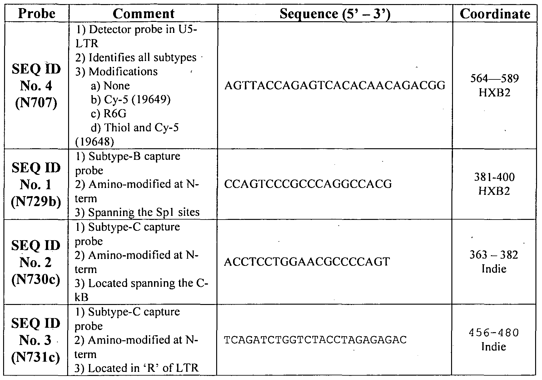
Example 4
Subtype-C capture probe captures subtype-C, but not -A or -B, viral DNA:
Amino-modified subtype-C capture probe (N730c) was captured to silane-modified glass surface. Single-stranded DNA within the LTR region of the virus derived from 3 different viral subtypes A, B or C, was added to the capture probe at numbers shown above the wells. After washing to remove unbound viral DNA, the wells were incubated with the detector probe N707 that was tagged with Rhodamine-6-G (R6G). Following incubation and washing, the wells were incubated with silver nanoparticles and the Raman signal was acquired for one second. The data have been presented in
(Fig 2). The slide on which capture probe was deposited was shown in the left panel with the number of viral DNA molecules indicated above the columns in Fig 2. The right-hand panel presents data obtained. The capture probe immobilized only the subtype-C DNA (top row) but not subtypes-A or B (middle and bottom rows, respectively) confirming the specificity of the capture probe. While the total area of the spot where the capture probe is immobilized is approximately 3.1 x 10"6 M2 (2 mm x 2 mm), the area scanned in SERS is only one millionth of the total area 1.2 x 10"13 M2 (4 μM x 4 μM). This area is equivalent to approximately 104 viral DNA molecules in the top most and left most panel where 1010 copies were actually used. This experiment confirmed that the present strategy can specifically detect target nucleic acid molecules without PCR amplification with high sensitivity.
Example 5
Capture probes are specific to viral subtypes:
The following experiment, using a non-SERS detection strategy, confirmed that subtype-specific probes specifically identify intended viral target nucleic acids efficiently. In this experiment, one capture probe for HIV-I subtype-B (N729b) and two different probes for subtype-C (N730c and N731c) were used independently. A detector probe (N707) was end-labeled with radio-isotope 32P using standard molecular techniques. The detector probe must bind to all viral strains regardless of subtype differences. Single-stranded DNA derived from the LTR region of subtype-B or -C viruses was used, each at 2xlO13 copies, in the experiment. Following a series of hybridizations and washing, the slides were exposed to X-ray films and an autoradiogram was developed. The results (Fig 3) demonstrate that subtype-B specific capture probe binds only subtype-B DNA but not subtype-C DNA. In a similar fashion, both the subtype-C specific capture probes capture only subtype-C, but not -B, DNA. The non-SERS detection strategy thus confirmed specific binding of the designed probes only to homologous, but not heterologous, target nucleic acids even though they are closely related.
Example 6
Capture and detection of the viral RNA from culture supernatant:
The following experiment illustrates capture and detection of full-length viral RNA from the cell culture supernatant. Subtype-B molecular clone HXB-2 was cultured in the laboratory in T-cells using standard culture conditions. Virus was concentrated from 1 ml of the culture medium by high-speed centrifugation and resuspended in 0.1 ml of lysis buffer containing 1% NP-40 and 0.1% BSA and 5 μl of this solution was used in the experiment. Subtype-B specific capture probe N729b and -C specific probe N730c were used for the capture of the viral RNA. Detector probe N707 tagged to R6G was used for SERS detection. The results (Fig 4) prove that, using the novel strategy it is possible to detect full-length viral RNA. Only the subtype-B specific capture probe (top panels) but not -C specific probe (bottom panels), detects the subtype-B virus.
Example 7
Detection of subtype-C viral DNA with Cy-5 tagged detector probe:
We identified that Cy-5 fluorescent dye "to be as good Raman Reporter as R6G for the novel detection strategy. In the following experiment, the top panel contains B-specific capture probe N729b that should not capture the C-viral DNA used in the experiment. The middle and bottom panels contain C-specific capture probe N730c. The detector probe 19649 (bottom panel) which must detect all the viral strains regardless of the subtype differences was tagged to Cy-5. The same probe was also chemically modified by adding an additional thiol group (and Cy-5) for efficient binding to the silver nano- particles. The variant probe is called 19648. The copy number of the subtype-C DNA used in the experiment is indicated. The data (Fig 5) demonstrated that Cy-5 linked detector probe can identify subtype-C DNA with high efficiency (middle and bottom panels). B-specific capture probe doesn't detect the C-DNA confirming specificity (top panels). Presence of a thiol group in the detector probe doesn't seem to give an additional advantage as both the detector probes with and without thiol group show identical detection sensitivity (compare middle and bottom panels).
ADVANTAGES OF THE INVENTION:
1. Does not require template amplification. False positive results are avoided. Economically inexpensive.
2. Broader range. A large number of capture probes can target several windows within the virus and essentially every viral subtype can be included in the detection strategy.
3. Highly sensitive detection. The reporter probe is conjugated to a Raman reporter hence exploiting the phenomenon of SERS or SERRS there by a single molecule detection should be possible theoretically. The technique here uses SERRS, where both surface enhancement as well as resonant enhancement are employed, making this technique highly sensitive to detect a small number of detector probe molecules. Our strategy of generating the reporter signal by adsorbing the probe onto the silver nanoparticle is far superior to the alternative technique commonly employed where the RR is coated with thin layer of silver. Our strategy is superior in terms of both signal enhancement as well as effects of absorption coefficient of silver.
Application of Raman reporters also offers the advantage of employing low output LASERs and/or diode lasers that are less expensive. Technically less complicated and less expensive equipment could be designed.
Our strategy of microarray capture of the nucleic acid template blended with SERRS- mediated detection have wider global application where HIV-I infections are common. The diverse viral subtypes of HIV-I, presently known recombinant viruses and recombinant strains that may arise in future can be detected efficiently. Determination of the HIV-I molecular nature will be important in the following areas and can have significant impact on treatment decisions, and intervention strategies.
1) Basic research where determination of the subtype and/or recombinant nature of viruses is required
2) Drug therapy, where drug resistant mutants should be quickly identified
3) Vaccine studies, where a knowledge of the viral strains in a given population or geographical region is essential
Importantly, the strategy we developed have a universal application where genetic diversity is a serious problem with other infectious organisms including HIV-2, Influenza, Polio, Dengue, HBV, HCV, Dengue, several bacteria including Tuberculosis, parasites including malaria and other microorganisms. Additionally, the technique also finds application to SNP (single nucleotide polymorphism) in all types of organisms including the human beings. The strategy is technically less complicated and find broader application in resource poor conditions.
Reference List;
1) Abravaya K, Esping C et al, Performance of a multiplex qualitative PCR LCx assay for detection of human immunodeficiency virus type 1 (HIV-I) group M subtypes, group O, and HIV-2. J Clin Microbiol, 38, 716-723 (2000).
2) Barbosa EF, Carneiro-Proietti AB et al, HIV-I detection and subtyping by PCR and heteroduplex mobility assay in blood donors: can these tests help to elucidate conflicting serological results? Trans/us Sci, 19, 39-43 (1998).
3) Carr JK, Laukkanen T et al, Characterization of subtype A HIV-I from Africa by full genome sequencing. AIDS, 13, 1819-1826 (1999).
4) Collins ML, Irvine B et al, A branched DNA signal amplification assay for quantification of nucleic acid targets below 100 molecules/ml. Nucleic Acids Res, 25, 2979-2984 (1997).
5) de Baar MP, Timmermans EC et al, One-tube real-time isothermal amplification assay to identify and distinguish human immunodeficiency virus type 1 subtypes A, B, and C and circulating recombinant forms AE and AG. J Clin Microbiol, 39, 1895-1902 (2001).
6) Delwart EL, Shpaer EG et al, Genetic relationships determined by a DNA heteroduplex mobility assay: analysis of HIV-I env genes. Science, 262, 1257- 1261 (1993).
7) Desire N, Dehee A et al, Quantification of human immunodeficiency virus type 1 proviral load by a TaqMan real-time PCR assay. J Clin Microbiol, 39, 1303- 1310 (2001).
8) Espy MJ, UhI JR et al, Real-Time PCR in Clinical Microbiology: Applications for Routine Laboratory Testing. CHn Microbiol Rev, 19, 165-256 (2006).
9) Gerhardt M, Mloka D et al, In-depth, longitudinal analysis of viral quasispecies from an individual triply infected with late-stage human immunodeficiency virus type 1, using a multiple PCR primer approach. J Virol, 79, 8249-8261 (2005).
10) Guo Z, Guilfoyle RA et al, Direct fluorescence analysis of genetic polymorphisms by hybridization with oligonucleotide arrays on glass supports. Nucleic Acids Res, 22, 5456-5465 ( 1994).
l l) Herbinger KH, Gerhardt M et al, Frequency of HIV type 1 dual infection and HIV diversity: analysis of low- and high-risk populations in Mbeya Region, Tanzania. AIDS Res Hum Retroviruses, 22, 599-606 (2006).
12) Heyndrickx L, Janssens W et al, Simplified Strategy for Detection of Recombinant Human Immunodeficiency Virus Type 1 Group M Isolates by gag/env Heteroduplex Mobility Assay. J Virol, 74, 363-3,70 (2000). -
13) Hoelscher M, Dowling WE et al, Detection of HIV-I subtypes, recombinants, and dual infections in east Africa by a multi-region hybridization assay. AIDS, 16, 2055-2064 (2002).
14) Joos B, Kuster H and Cone R: Covalent attachment of hybridizable oligonucleotides to glass supports. Anal Biochem, 247, 96-101 (1997).
15) Kern D, Collins M et al, An enhanced-sensitivity branched-DNA assay for quantification of human immunodeficiency virus type 1 RNA in plasma. J Clin Microbiol, 34, 3196-3202 (1996).
16) Kneipp K, Kneipp H et al, Surface-enhanced Raman scattering and biophysics. JPhys : Condens Matter, 14, R597-R624(2002).
17) Kumar A, Larsson O, Parodi D and Liang Z: Silanized nucleic acids: a general platform for DNA immobilization. Nucleic Acids Res, 28, E71 (2000).
18) Kwok S and Higuchi R: Avoiding false positives with PCR. Nature, 339, 237- 238 (1989).
19) LaI RB, Chakrabarti S and Yang C: Impact of genetic diversity of HIV-lon diagnosis, antiretroviral therapy & vaccine development. Indian J Med Res, 121, 287-314 (2005).
20) Leitner T., B.Foley, B.Hahn, P.Marx, F.McCutchan, J.Mellors, S.Wolinsky and B.Korber: HIV Sequence Compendium 2005. In: AnonymousTheoretical Biology and Biophysics Group, Los Alamos National Laboratory, LA, (2005).
21) Lindroos K, Liljedahl U, Raitio M and Syvanen AC: Minisequencing on oligonucleotide microarrays: comparison of immobilisation chemistries. Nucleic Acids Res, 29, E69(2001).
22) Luo CC, Downing RG et al, The development and evaluation of a probe hybridization method for subtyping HIV type 1 infection in Uganda. AIDS Res Hum Retroviruses, 14, 691-694 (1998).
23) Najera R, Delgado E, Perez-Alvarez L and Thomson MM: Genetic recombination and its role in the development of the HIV-I pandemic. AIDS, 16 Suppl 4, S3-16 (2002).
24)Niesters HG: Quantitation of viral load using real-time amplification techniques. Methods, 25, 419-429 (2001).
25) Peeters M.: Recombinant HIV sequences: Their role in the global epidemic. In: HIV sequence compendium 2000, eds. C.Kuiken, B.Foley, B.Hahn, P.Marx, F.E.McCutchan, J.Mellors, J.Mullins, J.Sodroski, S.Wolinsky and B.Korber (Theoritical Biology and Biophysics Group, Los Alamos National Laboratory, Los Alamos, New Mexico, USA, Los Alamos) pp. 54-72 (2000).
26) Peeters M, Liegeois F et al, Subtype-specific polymerase chain reaction for the identification of HIV-I genetic subtypes circulating in Africa. AIDS, 12, 671- 673 (1998).
27) Robbins KE, Kostrikis LG et al, Genetic analysis of human immunodeficiency virus type 1 strains in Kenya: a comparison using phylogenetic analysis and a combinatorial melting assay. AIDS Res Hum Retroviruses, 15, 329-335 (1999).
28) Rys PN and Persing DH: Preventing false positives: quantitative evaluation of three protocols for inactivation of polymerase chain reaction amplification products. J Clin Microbiol, 31, 2356-2360 (1993).
29) Scherczinger CA, Ladd C et al, A systematic analysis of PCR contamination. J Forensic Sci, 44, 1042-1045 (1999).
30) Van Harmelen J, van der Ryst E et al, Restriction fragment length polymorphism analysis for rapid gag subtype determination of human immunodeficiency virus type 1 in South Africa. J Virol Methods, 78, 51-59 (1999).
31) Vo-Dinh T, Yan F and Wabuyele MB: Surface-enhanced Raman scattering for medical diagnostics and biological imaging. J Raman Spectroscopy, 36, 640- 647 (2005).
32) Wan Z, Wang Y et al, Development of array-based technology for detection of HAV using gold-DNA probes. J Biochem MoI Biol, 38, 399-406 (2005).
33) Wilke WW, Sutton LD and Jones RN: Automation of polymerase chain reaction tests to achieve acceptable contamination rates. Clin Chem, 41, 622-623 (1995)."
34) Xie XS, Yu J and Yang WY: Living cells as test tubes. Science, 312, 228-230 (2006).
35) Yeung AT, Holloway BP, Adams PS and Shipley GL: Evaluation of dual- labeled fluorescent DNA probe purity versus performance in real-time PCR. BioTechniques, 36, 266-5 (2004).
Claims
25
We Claim:
1) A high sensitivity assay for molecular typing of a biological sample using surface-enhanced Raman scattering (SERS) including resonance scattering (SERRS), wherein said assay comprising steps of: a) extracting nucleic acid from the biological sample; b) capturing the extracted nucleic acid in a microarray format using subtype-specific and window-specific capture probe(s); and c) detecting the captured nucleic acid with a detector probe tagged to a reporter to determine molecular typing, subtypes and recombinants on the basis of pattern of signals generated.
2) The assay as claimed in claim 1, wherein the biological sample is selected from a group comprising infectious agents, disease causing agents, microorganisms, higher forms of life including human beings.
3) The assay as claimed in claim 2, wherein the infectious agents are selected from a group comprising bacteria, viruses, fungi, protozoa and parasites.
4) The assay as claimed in claim 3, wherein the viruses are selected from a group comprising helical viruses, icosahedral viruses, enveloped viruses and complex viruses, preferably HIV.
5) The assay as claimed in claim 1, wherein the nucleic acid is either DNA or RNA.
6) The assay as claimed in claim 1, wherein the capture probes are an array of oligonucleotides spotted in an array fashion on surface of an appropriate glass slide.
7) The assay as claimed in claim 1, wherein the detector probes are adsorbed onto silver nanoparticles for further enhancement of the assay.
26
8) The assay as claimed in claim 1, wherein the reporter is selected from a group comprising Raman reporter, fluorescent dye, radio-isotope and an enzyme.
9) The assay as claimed in claim 8, wherein the reporter is Raman reporter selected from a group comprising Rhodamine, Cy-5, TAMRA and DSNB.
10) The assay as claimed in claim 9, wherein the Raman reporter is preferably Rhodamine and Cy-5.
1 1) The assay as claimed in claim 1, wherein the assay is applicable without amplification of the nucleic acid.
12) The assay as claimed in claim 1, wherein the assay avoids false-positive/false- negative results and is inexpensive.
13) The assay as claimed in claim 1, wherein the assay detects single nucleotide polymorphism in all types of organisms including human beings.
14) A high sensitivity assay for molecular typing of HIV using surface-enhanced Raman scattering (SERS) including resonance scattering (SERRS), wherein said assay comprising steps of: a) extracting nucleic acid from the HIV; b) capturing the extracted nucleic acid in a microarray format using subtype-specific and window-specific capture probe(s); and c) detecting the captured nucleic acid with a detector probe tagged to a reporter to determine molecular typing, subtypes and recombinants on the basis of pattern of signals generated.
15) Capture probes of SEQ ID NO: 1, SEQ ID NO: 2 and SEQ ID NO: 3 for capturing nucleic acid isolated from biological sample, wherein the capture probes are sub-type specific and window specific.
27
16) The capture probes as claimed in claim 15, wherein the capture probes are an array of oligonucleotides spotted in an array fashion on surface of an appropriate glass slide.
17) The capture probes as claimed in claim 16, wherein the oligonucleotides are synthesized in situ on glass surface using photolithography or pre-synthesized oligonucleotides selected from a group comprising succinylated oligonucleotides, 5' end amino-modified oligonucleotides, disulfide-modified oligonucleotides and other oligonucleotides linked through hetero-bi-functional cross-linking molecules to the surface are used.
18) The capture probes as claimed in claim 15, wherein the nucleic acid is either DNA or RNA.
19) The capture probes as claimed in claim 15, wherein the biological sample is selected from a group comprising infectious agents, disease causing agents, microorganisms, higher forms of life including human beings.
20) The capture probes as claimed in claim 19, wherein the infectious agents are selected from a group comprising bacteria, viruses, fungi, protozoa and parasites.
21) The capture probes as claimed in claim 20, wherein the viruses are selected from a group comprising helical viruses, icosahedral viruses, enveloped viruses and complex viruses, preferably HIV.
22) A detector probe of SEQ ID NO: 4 to detect captured nucleic acid, wherein the detector probe is non-specific and identifies all subtypes.
23) The detector probe as claimed in claim 22, wherein the nucleic acid is either DNA or RNA.
28
24) A kit for molecular typing of biological sample using surface-enhanced Raman scattering (SERS) including resonance scattering (SERRS), wherein the kit comprising: a) reagents and buffers to extract nucleic acid from the biological sample; b) capture probes to capture the nucleic acid; c) microarray plate to spot capture probes; d) detector probe to detect capture probe; e) reporter, preferably Raman reporter; and f) device to detect pattern of signals generated from microarray.
25) The kit as claimed in claim 24, wherein the kit comprise capture probes of SEQ ID NO: 1, SEQ ID NO: 2 and SEQ ID NO: 3 which are sub-type specific and window specific.
26) The kit as claimed in claim 24, wherein the kit comprise the detector probe of SEQ ID NO: 4 which is non-specific and identifies all subtypes.
27) A method of manufacturing a kit for molecular typing of biological sample using surface-enhanced Raman scattering (SERS) including resonance scattering (SERRS), wherein the method comprising: a) providing reagents and buffers in the kit to extract nucleic acid from the biological sample; b) providing appropriate capture probes in the kit to capture the extracted nucleic acid; c) providing microarray plate in the kit to spot the capture probes; d) providing detector probe in the kit to detect the capture probe; e) providing reporter, preferably Raman Reporter; and f) providing device to detect pattern of signals generated from microarray to manufacture the kit.
Priority Applications (2)
| Application Number | Priority Date | Filing Date | Title |
|---|---|---|---|
| US12/515,766 US8227590B2 (en) | 2006-11-21 | 2007-11-19 | High sensitivity assay for molecular typing of biological sample, probes and a kit thereof |
| EP07866744.1A EP2104742B1 (en) | 2006-11-21 | 2007-11-19 | A high sensitivity assay for molecular typing of biological sample, probes and a kit thereof |
Applications Claiming Priority (2)
| Application Number | Priority Date | Filing Date | Title |
|---|---|---|---|
| IN02161/CHE/2006 | 2006-11-21 | ||
| IN2161CH2006 | 2006-11-21 |
Publications (2)
| Publication Number | Publication Date |
|---|---|
| WO2008062479A2 true WO2008062479A2 (en) | 2008-05-29 |
| WO2008062479A3 WO2008062479A3 (en) | 2008-07-10 |
Family
ID=39430184
Family Applications (1)
| Application Number | Title | Priority Date | Filing Date |
|---|---|---|---|
| PCT/IN2007/000543 WO2008062479A2 (en) | 2006-11-21 | 2007-11-19 | A high sensitivity assay for molecular typing of biological sample, probes and a kit thereof |
Country Status (4)
| Country | Link |
|---|---|
| US (1) | US8227590B2 (en) |
| EP (1) | EP2104742B1 (en) |
| WO (1) | WO2008062479A2 (en) |
| ZA (1) | ZA200903128B (en) |
Cited By (1)
| Publication number | Priority date | Publication date | Assignee | Title |
|---|---|---|---|---|
| CN112414991A (en) * | 2019-08-22 | 2021-02-26 | 合肥国研汉因检测科技有限公司 | Raman spectrum-based African swine fever virus qualitative rapid detection method and kit |
Families Citing this family (1)
| Publication number | Priority date | Publication date | Assignee | Title |
|---|---|---|---|---|
| WO2019028430A1 (en) * | 2017-08-03 | 2019-02-07 | The Trustees Of Columbia University In The City Of New York | Optical super-multiplexing by polyynes |
Citations (1)
| Publication number | Priority date | Publication date | Assignee | Title |
|---|---|---|---|---|
| US20040086897A1 (en) | 2002-05-07 | 2004-05-06 | Mirkin Chad A. | Nanoparticle probes with Raman Spectroscopic fingerprints for analyte detection |
Family Cites Families (9)
| Publication number | Priority date | Publication date | Assignee | Title |
|---|---|---|---|---|
| US6015661A (en) * | 1994-02-14 | 2000-01-18 | The Macfarlane Burnet Centre For Medical Research Limited | Methods for the detection of non-pathogenic HIV-1 strains containing deletions in the Nef coding region and U3 region of the LTR |
| US5871902A (en) * | 1994-12-09 | 1999-02-16 | The Gene Pool, Inc. | Sequence-specific detection of nucleic acid hybrids using a DNA-binding molecule or assembly capable of discriminating perfect hybrids from non-perfect hybrids |
| US7927595B1 (en) * | 1997-02-21 | 2011-04-19 | The United States Of America As Represented By The Secretary Of The Navy | Methods for downregulating CCR5 in T cells with anti-CD3 antibodies and anti-CD28 antibodies |
| US6881537B1 (en) * | 1997-08-08 | 2005-04-19 | Biomerieux, B.V. | Nucleic acid sequences that can be used as primers and probes in the amplification and detection of all subtypes of HIV-1 |
| US6197503B1 (en) | 1997-11-26 | 2001-03-06 | Ut-Battelle, Llc | Integrated circuit biochip microsystem containing lens |
| WO2002087341A1 (en) * | 2001-05-01 | 2002-11-07 | Genetix Pharmaceuticals, Inc. | Novel self-inactivating (sin) lentiviral vectors |
| KR100542542B1 (en) * | 2003-07-02 | 2006-01-20 | 대한민국 | A nucleotide sequence of HIV-1 subtype B genomic DNA from Korean, a molecular clone comprising the nucleotide sequence and a method for preparation thereof |
| KR20050074815A (en) * | 2004-01-14 | 2005-07-19 | 주식회사 툴젠 | Zinc finger polypeptides capable of repressing a transcription starting from the hiv-1 ltr promoter and a use thereof |
| CA2567937A1 (en) * | 2004-05-26 | 2005-12-08 | Rosetta Genomics Ltd. | Viral and viral associated mirnas and uses thereof |
-
2007
- 2007-11-19 EP EP07866744.1A patent/EP2104742B1/en not_active Not-in-force
- 2007-11-19 US US12/515,766 patent/US8227590B2/en not_active Expired - Fee Related
- 2007-11-19 WO PCT/IN2007/000543 patent/WO2008062479A2/en active Application Filing
-
2009
- 2009-05-06 ZA ZA200903128A patent/ZA200903128B/en unknown
Patent Citations (1)
| Publication number | Priority date | Publication date | Assignee | Title |
|---|---|---|---|---|
| US20040086897A1 (en) | 2002-05-07 | 2004-05-06 | Mirkin Chad A. | Nanoparticle probes with Raman Spectroscopic fingerprints for analyte detection |
Non-Patent Citations (3)
| Title |
|---|
| See also references of EP2104742A4 |
| TUAN VO DINH, ANAL. CHEM., vol. 70, 1998, pages 1352 - 1356 |
| TUAN VO-DINH, J. RAMAN SPECTROSC., vol. 36, 2005, pages 640 - 647 |
Cited By (2)
| Publication number | Priority date | Publication date | Assignee | Title |
|---|---|---|---|---|
| CN112414991A (en) * | 2019-08-22 | 2021-02-26 | 合肥国研汉因检测科技有限公司 | Raman spectrum-based African swine fever virus qualitative rapid detection method and kit |
| CN112414991B (en) * | 2019-08-22 | 2023-08-15 | 中检国研(北京)科技有限公司 | Raman spectrum-based African swine fever virus qualitative rapid detection method and kit |
Also Published As
| Publication number | Publication date |
|---|---|
| US20100028858A1 (en) | 2010-02-04 |
| WO2008062479A3 (en) | 2008-07-10 |
| EP2104742A2 (en) | 2009-09-30 |
| ZA200903128B (en) | 2010-04-28 |
| US8227590B2 (en) | 2012-07-24 |
| EP2104742B1 (en) | 2014-05-07 |
| EP2104742A4 (en) | 2010-11-03 |
Similar Documents
| Publication | Publication Date | Title |
|---|---|---|
| JP5205346B2 (en) | HIV-1 amplification and detection reagents | |
| US5800984A (en) | Nucleic acid sequence detection by triple helix formation at primer site in amplification reactions | |
| Gheit et al. | Development of a sensitive and specific multiplex PCR method combined with DNA microarray primer extension to detect Betapapillomavirus types | |
| US8617816B2 (en) | System and method for detection of HIV drug resistant variants | |
| Parida | Rapid and real-time detection technologies for emerging viruses of biomedical importance | |
| US8232058B2 (en) | Multiplex detection of respiratory pathogens | |
| CA2692633C (en) | Method for the simultaneous detection of multiple nucleic acid sequences in a sample | |
| EP2563939B1 (en) | Rapid genotyping analysis for human papillomavirus and the device thereof | |
| Edelstein et al. | Oligonucleotide ligation assay for detecting mutations in the human immunodeficiency virus type 1 pol gene that are associated with resistance to zidovudine, didanosine, and lamivudine | |
| CN106661637B (en) | Method for detecting nucleic acid and use thereof | |
| Wang et al. | Simultaneous detection of four human pathogenic microsporidian species from clinical samples by oligonucleotide microarray | |
| Vernet | Molecular diagnostics in virology | |
| EP2104742B1 (en) | A high sensitivity assay for molecular typing of biological sample, probes and a kit thereof | |
| Wen et al. | A visual DNA chip for simultaneous detection of hepatitis B virus, hepatitis C virus and human immunodeficiency virus type-1 | |
| Nguyen et al. | HPAI 9G DNAChip: Discrimination of highly pathogenic influenza virus genes | |
| Sunzeri et al. | Rapid simultaneous detection of multiple retroviral DNA sequences using the polymerase chain reaction and capillary DNA chromatography | |
| Xiao et al. | Molecular diagnosis of HIV and relevant novel technologies in mutation analysis | |
| US8808993B2 (en) | Methods and kits to detect new H1N1 “swine flu” variants | |
| US20090111108A1 (en) | Method of Detecting Genetic Mutations | |
| Käller et al. | Tag-array based HPV genotyping by competitive hybridization and extension | |
| Parida et al. | Rapid and real-time detection of human viral infections: current trends and future perspectives | |
| US9238844B2 (en) | Compositions and methods to detect influenza variants | |
| González-Villaseñor et al. | A solid phase plate assay for HIV-1 genotyping | |
| US20110039715A1 (en) | Influenza b virus detection method and kit therefor |
Legal Events
| Date | Code | Title | Description |
|---|---|---|---|
| DPE1 | Request for preliminary examination filed after expiration of 19th month from priority date (pct application filed from 20040101) | ||
| WWE | Wipo information: entry into national phase |
Ref document number: 2007866744 Country of ref document: EP |
|
| WWE | Wipo information: entry into national phase |
Ref document number: 12515766 Country of ref document: US |
|
| NENP | Non-entry into the national phase |
Ref country code: DE |