WO2005031002A2 - Synthetic lethal screen using rna interference - Google Patents
Synthetic lethal screen using rna interference Download PDFInfo
- Publication number
- WO2005031002A2 WO2005031002A2 PCT/US2004/031629 US2004031629W WO2005031002A2 WO 2005031002 A2 WO2005031002 A2 WO 2005031002A2 US 2004031629 W US2004031629 W US 2004031629W WO 2005031002 A2 WO2005031002 A2 WO 2005031002A2
- Authority
- WO
- WIPO (PCT)
- Prior art keywords
- agent
- gene
- cell
- sirnas
- different
- Prior art date
Links
Classifications
-
- C—CHEMISTRY; METALLURGY
- C12—BIOCHEMISTRY; BEER; SPIRITS; WINE; VINEGAR; MICROBIOLOGY; ENZYMOLOGY; MUTATION OR GENETIC ENGINEERING
- C12N—MICROORGANISMS OR ENZYMES; COMPOSITIONS THEREOF; PROPAGATING, PRESERVING, OR MAINTAINING MICROORGANISMS; MUTATION OR GENETIC ENGINEERING; CULTURE MEDIA
- C12N15/00—Mutation or genetic engineering; DNA or RNA concerning genetic engineering, vectors, e.g. plasmids, or their isolation, preparation or purification; Use of hosts therefor
- C12N15/09—Recombinant DNA-technology
- C12N15/11—DNA or RNA fragments; Modified forms thereof; Non-coding nucleic acids having a biological activity
- C12N15/111—General methods applicable to biologically active non-coding nucleic acids
-
- A—HUMAN NECESSITIES
- A61—MEDICAL OR VETERINARY SCIENCE; HYGIENE
- A61P—SPECIFIC THERAPEUTIC ACTIVITY OF CHEMICAL COMPOUNDS OR MEDICINAL PREPARATIONS
- A61P35/00—Antineoplastic agents
-
- A—HUMAN NECESSITIES
- A61—MEDICAL OR VETERINARY SCIENCE; HYGIENE
- A61P—SPECIFIC THERAPEUTIC ACTIVITY OF CHEMICAL COMPOUNDS OR MEDICINAL PREPARATIONS
- A61P43/00—Drugs for specific purposes, not provided for in groups A61P1/00-A61P41/00
-
- C—CHEMISTRY; METALLURGY
- C12—BIOCHEMISTRY; BEER; SPIRITS; WINE; VINEGAR; MICROBIOLOGY; ENZYMOLOGY; MUTATION OR GENETIC ENGINEERING
- C12N—MICROORGANISMS OR ENZYMES; COMPOSITIONS THEREOF; PROPAGATING, PRESERVING, OR MAINTAINING MICROORGANISMS; MUTATION OR GENETIC ENGINEERING; CULTURE MEDIA
- C12N2310/00—Structure or type of the nucleic acid
- C12N2310/10—Type of nucleic acid
- C12N2310/11—Antisense
- C12N2310/111—Antisense spanning the whole gene, or a large part of it
-
- C—CHEMISTRY; METALLURGY
- C12—BIOCHEMISTRY; BEER; SPIRITS; WINE; VINEGAR; MICROBIOLOGY; ENZYMOLOGY; MUTATION OR GENETIC ENGINEERING
- C12N—MICROORGANISMS OR ENZYMES; COMPOSITIONS THEREOF; PROPAGATING, PRESERVING, OR MAINTAINING MICROORGANISMS; MUTATION OR GENETIC ENGINEERING; CULTURE MEDIA
- C12N2310/00—Structure or type of the nucleic acid
- C12N2310/10—Type of nucleic acid
- C12N2310/14—Type of nucleic acid interfering N.A.
-
- C—CHEMISTRY; METALLURGY
- C12—BIOCHEMISTRY; BEER; SPIRITS; WINE; VINEGAR; MICROBIOLOGY; ENZYMOLOGY; MUTATION OR GENETIC ENGINEERING
- C12N—MICROORGANISMS OR ENZYMES; COMPOSITIONS THEREOF; PROPAGATING, PRESERVING, OR MAINTAINING MICROORGANISMS; MUTATION OR GENETIC ENGINEERING; CULTURE MEDIA
- C12N2310/00—Structure or type of the nucleic acid
- C12N2310/50—Physical structure
- C12N2310/53—Physical structure partially self-complementary or closed
-
- C—CHEMISTRY; METALLURGY
- C12—BIOCHEMISTRY; BEER; SPIRITS; WINE; VINEGAR; MICROBIOLOGY; ENZYMOLOGY; MUTATION OR GENETIC ENGINEERING
- C12N—MICROORGANISMS OR ENZYMES; COMPOSITIONS THEREOF; PROPAGATING, PRESERVING, OR MAINTAINING MICROORGANISMS; MUTATION OR GENETIC ENGINEERING; CULTURE MEDIA
- C12N2320/00—Applications; Uses
- C12N2320/10—Applications; Uses in screening processes
- C12N2320/12—Applications; Uses in screening processes in functional genomics, i.e. for the determination of gene function
Definitions
- the present invention relates to methods and compositions for carrying out interaction screening, e.g., lethal/synthetic lethal screening, using RNA interference.
- the invention also relates to genes exhibiting synthetic lethal interactions with KSP, a kinesin-like motor protein, and their therapeutic uses.
- the invention also relates to genes involved in .cellular response to DNA damage, and their therapeutic uses. 2. BACKGROUND OF THE INVENTION
- RNA interference is a potent method to suppress gene expression in mammalian cells, and has generated much excitement in the scientific community (Couzin, 2002, Science 298:2296-2297; McManus et al, 2002, Nat. Rev. Genet. 3, 111-1 Al; Harmon, G. J., 2002, Nature 418, 244-251; Paddison et al., 2002, Cancer Cell 2, 17-23).
- RNA interference is conserved throughout evolution, from C. elegans to humans, and is believed to function in protecting cells from invasion by RNA viruses. When a cell is infected by a dsRNA virus, the dsRNA is recognized and targeted for cleavage by an RNaselll-type enzyme termed Dicer.
- the Dicer enzyme "dices" the RNA into short duplexes of 21nt, termed siRNAs or short-interfering RNAs, composed of 19nt of perfectly paired ribonucleotides with two unpaired nucleotides on the 3' end of each strand.
- siRNAs short-interfering RNAs
- RISC multiprotein complex
- nucleases present in the RISC complex cleave the mRNA transcript, thereby abolishing expression of the gene product. In the case of viral infection, this mechanism would result in destruction of viral transcripts, thus preventing viral synthesis. Since the siRNAs are double-stranded, either strand has the potential to associate with RISC and direct silencing of transcripts with sequence similarity.
- miRNAs are regulatory RNAs expressed from the genome, and are processed from precursor stem-loop structures to produce single-stranded nucleic acids that bind to sequences in the 3' UTR of the target mRNA (Lee et al., 1993, Cell 75:843-854; Reinhart et al., 2000, Nature 403:901-906; Lee et al, 2001, Science 294:862-864; Lau et al., 2001, Science 294:858-862; Hutvagner et al., 2001, Science 293:834-838).
- miRNAs bind to transcript sequences with only partial complementarity (Zeng et al., 2002, Molec. Cell 9: 1327-1333) and repress translation without affecting steady-state RNA levels (Lee et al., 1993, Cell 75:843-854; Wightman et al, 1993, Cell 75:855-862). Both miRNAs and siRNAs are processed by Dicer and associate with components of the RNA-induced silencing complex (Hutvagner et al., 2001, Science 293:834-838; Grisho et. al., 2001, Cell 106: 23-34; Ketting et al., 2001, Genes Dev.
- silencing Fas expression holds therapeutic promise to prevent liver injury by protecting hepatocytes from cytotoxicity.
- injected mice intraperitoneally with siRNA targeting TNF-a Lipopolysaccharide-induced TNF-a gene expression was inhibited, and these mice were protected from sepsis.
- RNA interference can be used to selectively target oncogenic mutations (Martinez et al., 2002, Proc. Natl. Acad. Sci. USA 99:14849-14854).
- an siRNA that targets the region of the R248W mutant of p53 containing the point mutation was shown to silence the expression of the mutant p53 but not the wild-type p53.
- Wilda et al. reported that an siRNA targeting the M-BCR/ABL fusion mRNA can be used to deplete the M-BCR/ABL mRNA and the M-BRC/ABL oncoprotein in leukemic cells (Wilda et al., 2002, Oncogene 21:5716-5724). However, the report also showed that applying the siRNA in combination with Imatinib, a small-molecule ABL kinase tyrosine inhibitor, to leukemic cells did not further increase in the induction of apoptosis.
- U.S. Patent No. 6,506,559 discloses a RNA interference process for inhibiting expression of a target gene in a cell.
- the process comprises introducing partially or fully doubled-stranded RNA having a sequence in the duplex region that is identical to a sequence in the target gene into the cell or into the extracellular environment.
- RNA sequences with insertions, deletions, and single point mutations relative to the target sequence are also found as effective for expression inhibition.
- U.S. Patent Application Publication No. US 2002/0086356 discloses RNA interference in a Drosophila in vitro system using RNA segments 21-23 nucleotides (nt) in length.
- the patent application publication teaches that when these 21-23 nt fragments are purified and added back to Drosophila extracts, they mediate sequence-specific RNA interference in the absence of long dsRNA.
- the patent application publication also teaches that chemically synthesized oligonucleotides of the same or similar nature can also be used to target specific mRNAs for degradation in mammalian cells.
- PCT publication WO 02/44321 discloses that double-stranded RNA (dsRNA) 19-23 nt in length induces sequence-specific post-transcriptional gene silencing in a Drosophila in vitro system.
- dsRNA double-stranded RNA
- the PCT publication teaches that short interfering RNAs (siRNAs) generated by an RNase Ill-like processing reaction from long dsRNA or chemically synthesized siRNA duplexes with overhanging 3' ends mediate efficient target RNA cleavage in the lysate, and the cleavage site is located near the center of the region spanned by the guiding siRNA.
- siRNAs short interfering RNAs
- the PCT publication also provides evidence that the direction of dsRNA processing determines whether sense or antisense target RNA can be cleaved by the produced siRNP complex.
- U.S. Patent Application Publication No. US 2002/016216 discloses a method for attenuating expression of a target gene in cultured cells by introducing double stranded RNA (dsRNA) that comprises a nucleotide sequence that hybridizes under stringent conditions to a nucleotide sequence of the target gene into the cells in an amount sufficient to attenuate expression of the target gene.
- dsRNA double stranded RNA
- PCT publication WO 03/006477 discloses engineered RNA precursors that when expressed in a cell are processed by the cell to produce targeted small interfering RNAs (siRNAs) that selectively silence targeted genes (by cleaving specific mRNAs) using the cell's own RNA interference (RNAi) pathway.
- siRNAs small interfering RNAs
- RNAi RNA interference pathway
- the PCT publication teaches that by introducing nucleic acid molecules that encode these engineered RNA precursors into cells in vivo with appropriate regulatory sequences, expression of the engineered RNA precursors can be selectively controlled both temporally and spatially, i.e., at particular times and/or in particular tissues, organs, or cells. Discussion or citation of a reference herein shall not be construed as an admission that such reference is prior art to the present invention.
- the invention provides methods and compositions for identifying interactions, e.g., lethal synthetic lethal interactions, between a gene or its product and an agent, e.g., a drug, and/or another gene or its product, using RNA interference.
- the invention also provides methods and compositions for treating cancer utilizing the synthetic lethal interaction between STK6 kinase or TPX2 and kinesin-like motor protein KSP inhibitors.
- the invention also provides genes involved in cellular response to DNA damage, and their therapeutic uses.
- the invention provides a method for identifying a gene whose product modulates the effect of an agent on a cell of a cell type.
- the method comprises (a) contacting a plurality of groups of one or more cells of said cell type with said agent, wherein each said group of one or more cells comprises one or more different small interfering RNAs (siRNAs) from among a plurality of different siRNAs, said one or more different siRNAs targeting a same gene, and said plurality of different siRNAs comprising siRNAs targeting respectively different genes in cells of said cell type; (b) comparing the effect of said agent on each said group of one or more cells to the effect of said agent on a cell of said cell type which does not comprise an siRNA targeting any one of said different genes; and (c) identifying a gene as said gene whose product modulates the effect of said agent on a cell of said cell type if the effect of said agent on said group of one or more cells comprising said one or more different siRNAs targeting said gene is different as compared to the effect of said agent on a cell of said cell type which does not comprise an siRNA targeting any one of said different genes.
- siRNAs small interfer
- each said group of cells comprising one or more of said plurality of siRNAs is obtained by transfection with said one or more siRNAs prior to said step of contacting.
- the contacting step (a) is carried out separately for each said groups of one or more cells.
- the invention provides a method for identifying a gene whose product modulates the effect of an agent on a cell of a cell type, said method comprising (a) transfacting each of a plurality of groups of one or more cells of said cell type with a composition comprising one or more different small interfering RNAs (siRNAs) from among a plurality of different siRNAs, said one or more different siRNAs targeting a same gene, and said plurality of different siRNAs comprising siRNAs targeting respectively different genes in cells of said cell type; (b) contacting each of said plurality of groups of one or more cells with said agent; (c) comparing the effect of said agent on each said group of one or more cells to the effect of said agent on a cell of said cell type which is not transfected with an siRNA targeting any one of said different genes; and (d) identifying a gene as said gene whose product modulates the effect of said agent on a cell of said cell type if the effect of said agent on said group of one or more
- siRNAs
- the effect of said agent on each said group of one or more cells comprising said one or more different siRNAs can be enhanced as compared to the effect of said agent on a cell of said cell type which does not comprise an siRNA targeting any one of said different genes.
- the effect of said agent on said group of one or more cells comprising said one or more different siRNAs can be reduced as compared to the effect of said agent on a cell of said cell type which does not comprise an siRNA targeting any one of said different genes.
- the agent acts on a gene other than any one of said different genes targeted by said plurality of siRNAs, or a protein encoded thereof.
- the plurality of siRNAs comprises at least k different siRNAs targeting at least one of said different genes, wherein said k is selected from the group consisting of 2, 3, 4, 5, 6 and 10. More preferably, the plurality of siRNAs comprises at least k different siRNAs targeting each of at least 2 different genes of said different genes, wherein said k is selected from the group consisting of 2, 3, 4, 5, 6 and 10. Still more preferably, the plurality of siRNAs comprises at least k different siRNAs targeting each of said different genes, wherein said k is selected from the group consisting of 2, 3, 4, 5, 6 and 10.
- the one or more different siRNAs for at least one, at least two, or each of of the plurality of different genes comprises 2, 3, 4, 5, 6, or 10 different siRNAs targeting a same target gene.
- the total siRNA concentration of the one or more siRNAs is about the same as the concentration of a single siRNA when used individually, e.g., lOOnM.
- the total concentration of the one or more siRNAs is an optimal concentration for silencing the intended target gene.
- An optimal concentration is a concentration further increase of which does not increase the level of silencing substantially.
- the optimal concentration is a concentration further increase of which does not increase the level of silencing by more than 5%, 10% or 20%.
- the one or more siRNAs comprise each siRNA in equal proportion. In another preferred embodiment, the one or more siRNAs comprise each siRNA in proportions different from each other by less than 5%,10%, 20% or 50%. In a preferred embodiment, at least one of the one or more siRNAs constitutes more than 90%, 80%, 70%, 50%, or 20% of the total siRNA concentration of the one or more siRNAs! In another preferred embodiment, none of the siRNAs in the one or more siRNAs constitutes more than 90%, 80%, 70%, 50%, or 20% of the total siRNA concentration of the one or more siRNAs.
- the composition of the one or more siRNAs is chosen such that the one or more siRNAs causes less than 30%, 20%, 10% or 5%, 1%, 0.1% or 0.01% of silencing of any off-target genes.
- each siRNA in the one or more siRNAs has a concentration that is lower than the optimal concentration when used individually.
- at least one siRNA in the one or more siRNAs has an concentration that is lower than the concentration of the siRNA that is effective to achieve at least 30%, 50%, 75%, 80%, 85%, 90% or 95 % silencing when used in the absence of other siRNAs or in the absence of other siRNAs designed to silence the gene.
- each different siRNA in the one or more siRNAs has a concentration that causes less than 30%, 20%, 10% or 5% of silencing of the gene when used in the absence of other siRNAs or in the absence of other siRNAs designed to silence the gene.
- each siRNA has a concentration that causes less than 30%, 20%, 10% or 5% of silencing of the target gene when used alone, while the plurality of siRNAs causes at least 80% or 90% of silencing of the target gene.
- said cell type is a cancer cell type.
- said effect is growth inhibitory effect.
- said agent is a KSP inhibitor.
- said different genes comprises at least 5, at least 10, at least 100, or at least 1,000 different genes. In one embodiment, said different genes are different endogenous genes.
- the invention provides a method for identifying a gene which interacts with a primary target gene in a cell of a cell type.
- the method comprises (a) contacting a plurality of groups of one or more cells of said cell type with an agent, wherein said agent modulates the expression of said primary target gene and/or the activity of a protein encoded by said primary target gene, and wherein each said group of cells comprises one or more different siRNAs among a plurality of different siRNAs, said one or more different siRNAs targeting a same gene, and said plurality of different siRNAs comprising siRNAs targeting respectively different secondary genes in said cell; (b) comparing the effect of said agent on each said group of one or more cells to the effect of said agent on a cell of said cell type which does not comprise an siRNA targeting any one of said different secondary genes; and (c) identifying a gene as said gene that interacts with said primary target gene in a cell of said cell type if the effect of said agent on said group of one or more cells comprising one or more siRNA
- the invention provides method for identifying a gene which interacts with a primary target gene in a cell of a cell type, said method comprising (a) transfacting each of a plurality of groups of one or more cells of said cell type with a composition comprismg one or more different small interfering RNAs (siRNAs) from among a plurality of different siRNAs, said one or more different siRNAs targeting a same gene, and said plurality of different siRNAs comprising siRNAs targeting respectively different genes in cells of said cell type; (b) contacting said plurality of groups of one or more cells of said cell type with an agent, wherein said agent modulates the expression of said primary target gene and/or the activity of a protein encoded by said primary target gene; (c) comparing the effect of said agent on each said group of one or more cells to the effect of said agent on a cell of said cell type which does not comprise an siRNA targeting any one of said different secondary genes; and (d) identifying a gene as said gene that interacts with a composition comprism
- said agent comprises an siRNA targeting and silencing said primary target gene.
- said agent comprises 2, 3, 4, 5, 6, or 10 different siRNAs targeting said primary target gene.
- the total siRNA concentration of said different siRNAs is about the same as the concentration of a single siRNA when used individually, e.g., lOOnM.
- the total concentration of said different siRNAs is an optimal concentration for silencing the primary target gene.
- An optimal concentration is a concentration further increase of which does not increase the level of silencing substantially.
- the optimal concentration is a concentration further increase of which does not increase the level of silencing by more than 5%, 10% or 20%.
- the different siRNAs comprise each siRNA in equal proportion. In another preferred embodiment, the different siRNAs comprise each siRNA in proportions different from each other by less than 5%, 10%, 20% or 50%. In a preferred embodiment, at least one of the different siRNAs constitutes more than 90%, 80%, 70%, 50%, or 20% of the total siRNA concentration of the different siRNAs. In another preferred embodiment, none of the siRNAs constitutes more than 90%, 80%, 70%, 50%, or 20% of the total siRNA concentration of the different siRNAs.
- the composition of the different siRNAs is chosen such that the different siRNAs causes less than 30%, 20%, 10% or 5%, 1%, 0.1% or 0.01% of silencing of any off-target genes.
- each siRNA has a concentration that is lower than the optimal concentration when used individually.
- at least one siRNA has an concentration that is lower than the concentration of the siRNA that is effective to achieve at least 30%,
- each different siRNA has a concentration that causes less than 30%, 20%, 10% or 5% of silencing of the gene when used in the absence of other siRNAs or in the absence of other siRNAs designed to silence the gene.
- each siRNA has a concentration that causes less than 30%, 20%, 10% or 5% of silencing of the target gene when used alone, while all of the siRNAs together causes at least 80% or 90% of silencing of the target gene.
- said agent comprises an inhibitor of a protein encoded by said primary target gene.
- the effect of said agent on said group of one or more cells can be enhanced as compared to the effect of said agent on a cell of said cell type which does not comprise an siRNA targeting any one of said different secondary genes.
- the effect of said agent on said group of one or more cells can be reduced as compared to the effect of said agent on a cell of said cell type which does not comprise an siRNA targeting any one of said different secondary genes.
- the plurality of siRNAs comprises at least k different siRNAs targeting at least one of said different secondary genes, wherein said k is selected from the group consisting of 2, 3, 4, 5, 6 and 10. More preferably, the plurality of siRNAs comprises at least k different siRNAs targeting each of at least 2 different genes of said different secondary genes, wherein said k is selected from the group consisting of 2, 3, 4, 5, 6 and 10. Still more preferably, the plurality of siRNAs comprises at least k different siRNAs targeting each of said different secondary genes, wherein said k is selected from the group consisting of 2, 3, 4, 5, 6 and 10.
- the one or more different siRNAs for at least one, at least two, or each of of the plurality of different genes comprises 2, 3, 4, 5, 6, or 10 different siRNAs targeting a same target gene.
- the total siRNA concentration of the one or more siRNAs targeting a same gene is about the same as the concentration of a single siRNA when used individually, e.g., lOOnM.
- the total concentration of the one or more siRNAs is an optimal concentration for silencing the intended target gene.
- An optimal concentration is a concentration further increase of which does not increase the level of silencing substantially.
- the optimal concentration is a concentration further increase of which does not increase the level of silencing by more than 5%, 10% or 20%.
- the one or more siRNAs comprise each siRNA in equal proportion. In another preferred embodiment, the one or more siRNAs comprise each siRNA in proportions different from each other by less than 5%, 10%, 20% or 50%. In a preferred embodiment, at least one of the one or more siRNAs constitutes more than 90%, 80%, 70%, 50%, or 20% of the total siRNA concentration of the one or more siRNAs. In another preferred embodiment, none of the siRNAs in the one or more siRNAs constitutes more than 90%, 80%, 70%, 50%, or 20% of the total siRNA concentration of the one or more siRNAs.
- the composition of the one or more siRNAs is chosen such that the one or more siRNAs causes less than 30%, 20%, 10% or 5%, 1%, 0.1% or 0.01% of silencing of any off-target genes.
- each siRNA in the one or more siRNAs has a concentration that is lower than the optimal concentration when used individually, h a preferred embodiment, at least one siRNA in the one or more siRNAs has an concentration that is lower than the concentration of the siRNA that is effective to achieve at least 30%, 50%, 75%, 80%, 85%, 90% or 95% silencing when used in the absence of other siRNAs or in the absence of other siRNAs designed to silence the gene.
- each different siRNA in the one or more siRNAs has a concentration that causes less than 30%, 20%, 10% or 5% of silencing of the gene when used in the absence of other siRNAs or in the absence of other siRNAs designed to silence the gene.
- each siRNA has a concentration that causes less than 30%, 20%, 10% or 5% of silencing of the target gene when used alone, while the plurality of siRNAs causes at least 80% or 90% of silencing of the target gene.
- each said group of one or more cells is obtained by transfection with said one or more different siRNAs prior to said step of contacting, h another embodiment, the primary target is KSP.
- said different secondary genes comprises at least 5, at least 10, at least 100, at least 1,000, at least 5,000 different genes.
- said different secondary genes are different endogenous genes.
- said cell type is a cancer cell type.
- the invention provides a method for treating a mammal having a cancer, comprising administering to said mammal a therapeutically sufficient amount of an agent, said agent regulating the expression of a STK6 or TPX2 gene and/or activity of a protein encoded by said STK6 or TPX2 gene, wherein said mammal is subject to a therapy comprising administering to said mammal a therapeutically sufficient amount of a KSP inhibitor.
- the invention also provides a method for treating a mammal having a cancer, comprising administering to said mammal i) a therapeutically sufficient amount of an agent, said agent regulating the expression of a STK6 or TPX2 gene and/or activity of a protein encoded by said STK6 or TPX2 gene, and ii) a therapeutically sufficient amount of a KSP inhibitor.
- said agent reduces the expression of said STK6 or TPX2 gene in cells of said cancer.
- said agent comprises an siRNA targeting said STK6 or TPX2 gene.
- the mammal is a human, and the siRNA can be selected from the group consisting of siRNAs described by SEQ ID NO:l, SEQ ID NO:2, SEQ ID NO:3, SEQ ID NO:4, SEQ ID NO:5, and SEQ ID NO:6.
- said agent comprises an siRNA targeting said TPX2 gene.
- mammal is a human, and the siRNA can be selected from the group consisting of siRNAs described by SEQ ID NO:1237, SEQ ID NO: 1238, and SEQ ID NO: 1239.
- the invention provides a method for treating a mammal having a cancer, comprising administering to said mammal i) a therapeutically sufficient amount of a first agent, said first agent regulating the expression of a STK6 or TPX2 gene and/or activity of a protein encoded by said STK6 or TPX2 gene, and ii) a therapeutically sufficient amount of a second agent, said second agent regulating the expression of a KSP gene and or activity of a protein encoded by said KSP gene, h a preferred embodiment, the first agent is an siRNA targeting said STK6 or TPX2 gene, and said second agent comprises an siRNA targeting said KSP gene.
- said mammal is a human, and wherein said siRNA is selected from the group consisting of siRNAs described by SEQ ID NO.l, SEQ ID NO:2, SEQ ID NO:3, SEQ ID NO:4, SEQ ID NO:5, and SEQ ID NO:6.
- said agent comprises an siRNA targeting said TPX2 gene.
- mammal is a human, and the siRNA can be selected from the group consisting of siRNAs described by SEQ ID NO: 1237, SEQ ID NO: 1238, and SEQ ID NO: 1239.
- the invention provides a method for evaluating resistance of a cell to the growth inhibitory effect of a KSP inhibitor, said method comprising determining an expression level of a STK6 or TPX2 gene in said cell, wherein said expression level above a predetermined threshold level indicates that said cell is resistant to the growth inhibitory effect of said KSP inhibitor.
- the expression level of said STK6 or TPX2 gene is determined by a method comprising measuring the expression level of said STK6 or TPX2 gene using one or more polynucleotide probes, each of said one or more polynucleotide probes comprising a nucleotide sequence in said STK6 or TPX2 gene.
- Said one or more polynucleotide probes can be polynucleotide probes on a microarray.
- the invention provides a method for evaluating resistance of a cell to the growth inhibitory effect of a KSP inhibitor, said method comprising determining a level of abundance of a protein encoded by a STK6 or TPX2 gene in said cell, wherein said level of abundance of said protein above a predetermined threshold level indicates that said cell is resistant to the growth inhibitory effect of said KSP inhibitor.
- the invention also provides a method for evaluating resistance of a cell to the growth inhibitory effect of a KSP inhibitor, said method comprising determining a level of activity of a protein encoded by a STK6 or TPX2 gene in said cell, wherein said activity level above a predetermined threshold level indicates that said cell is resistant to the growth inhibitory effect of said KSP inhibitor.
- said cell is a human cell.
- the invention provides a method for regulating resistance of a cell to the growth inhibitory effect of a KSP inhibitor, comprising contacting said cell with a sufficient amount of an agent, said agent regulating the expression of a STK6 or TPX2 gene and/or the activity of a protein encoded by said STK6 or TPX2 gene.
- the invention also provides a method for regulating resistance of a cell to the growth inhibitory effect of a KSP inhibitor in a mammal, comprising administering to said mammal a therapeutically sufficient amount of an agent, said agent regulating the expression of a STK6 or TPX2 gene and/or the activity of a protein encoded by said STK6 or TPX2 gene.
- the invention further provides a method for regulating growth of a cell, comprising contacting said cell with i) a sufficient amount of an agent that regulates the expression of a STK6 or TPX2 gene and/or the activity of a protein encoded by said STK6 or TPX2 gene; and ii) a sufficient amount of a KSP inhibitor.
- the agent reduces the expression of said STK6 or TPX2 gene in said cell.
- said agent comprises an siRNA targeting said STK 6 gene.
- said cell is a human cell, and wherein said siRNA is selected from the group consisting of siRNAs described by SEQ ID NO: 1, SEQ ID NO:2, SEQ ID NO:3, SEQ ID NO:4, SEQ ID NO:5, and SEQ ID NO:6.
- said agent comprises an siRNA targeting said TPX2 gene.
- cell is a human cell, and the siRNA can be selected from the group consisting of siRNAs described by SEQ ID NO: 1237, SEQ ID NO: 1238, and SEQ ID NO: 1239.
- the invention provides a method of identifying an agent that is capable of regulating resistance of a cell to the growth inhibitory effect of a KSP inhibitor, wherein said agent is capable of modulating the expression of a STK6 or TPX2 gene and/or the activity of a protein encoded by said STK6 or TPX2 gene, said method comprising comparing inhibitory effect of said KSP inhibitor on cells expressing said STK6 or TPX2 gene in the presence of said agent with inhibitory effect of said KSP inhibitor on cells expressing said STK6 or TPX2 gene in the absence of said agent, wherein a difference in said inhibitory effect of said KSP inhibitor identifies said agent as capable of regulating resistance of said cell to the growth inhibitory effect of said KSP inhibitor.
- the invention also provides a method of identifying an agent that is capable of regulating resistance of a cell to the growth inhibitory effect of a KSP inhibitor, wherein said agent is capable of modulating the expression of a STK6 or TPX2 gene and/or activity of a protein encoded by said STK6 or TPX2 gene, said method comprising: (a) contacting a first cell expressing said STK6 or TPX2 gene with said KSP inhibitor in the presence of said agent and measuring a first growth inhibitory effect; (b) contacting a second cell expressing said STK6 or TPX2 gene with said KSP inhibitor in the absence of said agent and measuring a second growth inhibitory effect; and (c) comparing said first and second inhibitory effects measured in said step (a) and (b), wherein a difference between said first and second inhibitory effects identifies said agent as capable of regulating resistance of a cell to the growth inhibitory effect of said KSP inhibitor.
- said agent is a molecule which reduces expression of said STK6 or TPX2 gene.
- said agent comprises an siRNA targeting said STK 6 gene.
- said cell is a human cell, and wherein said siRNA is selected from the group consisting of siRNAs described by SEQ ID NO:l, SEQ ID NO:2, SEQ ID NO:3, SEQ ID NO:4, SEQ ID NO:5, and SEQ ID NO:6.
- said agent comprises an siRNA targeting said TPX2 gene.
- the cell is a human cell, and the siRNA can be selected from the group consisting of siRNAs described by SEQ ID NO: SEQ ID NO: 1237, SEQ ID NO: 1238, and SEQ ID NO: 1239.
- the invention provides a cell comprising one or more different small interfering RNAs (siRNAs) targeting a STK6 or TPX2 gene in said cell.
- the cell can be a human cell.
- the cell can also be a murine cell, h one embodiment, said cell is a human cell, and each of said one or more different siRNA is selected from the group consisting of siRNAs described by SEQ ID NO:l, SEQ ID NO:2, SEQ ID NO:3, SEQ ID NO:4, SEQ ID NO:5, and SEQ ID NO:6.
- the cell is a human cell
- the siRNA can be selected from the group consisting of siRNAs described by SEQ ID NO: 1237, SEQ ID NO: 1238, and SEQ ID NO: 1239.
- said cell is produced by transfection using a composition of said one or more different siRNAs, wherein the total siRNA concentration of said composition is an optimal concentration for silencing said STK6 or TPX2 gene, wherein said optimal concentration is a concentration further increase of which does not increase the level of silencing substantially.
- said optimal concentration is a concentration further increase of which does not increase the level of silencing by more than 20%, more than 10%, or more than 5%.
- the concentration of each said different siRNA is about the same.
- the respective concentrations of said different siRNAs are different from each other by less than 50%, less than 20%, or less than 10%.
- none of the siRNAs in said composition has a concentration that is more than 80%, more than 50%, or more than 20% of said total siRNA concentration of said different siRNAs.
- at least one siRNA in said composition has a concentration that is more than 20% or more than 50% of said total siRNA concentration of said different siRNAs.
- the number of different siRNAs and the concentration of each siRNA in said composition is chosen such that said composition causes less than 10%, less than 1%, less than 0.1%, or less than 0.01% of silencing of any off-target genes.
- the invention provides a microarray for diagnosing resistance of a cell to the growth inhibitory effect of a KSP inhibitor.
- the microarray comprising one or more polynucleotide probes, wherein each said polynucleotide probe comprises a nucleotide sequence in a STK6 or TPX2 gene.
- the invention provides kit for diagnosis of resistance of a cell to the growth inhibitory effect of a KSP inhibitor.
- the kit comprises in one or more containers one or more polynucleotide probes, wherein each said polynucleotide probe comprises a nucleotide sequence in a STK6 or TPX2 gene.
- the invention also provides a kit for screening for agents which regulate resistance of a cell to the growth inhibitory effect of a KSP inhibitor.
- the kit comprises in one or more containers (i) a cell comprising one or more different small interfering RNAs (siRNAs) targeting a STK6 or TPX2 gene in said cell; and (ii) a KSP inhibitor.
- siRNAs small interfering RNAs
- the invention provides a kit for treating a mammal having a cancer, which comprises in one or more containers (i) a sufficient amount of an agent that regulates the expression of a STK6 or TPX2 gene and/or the activity of a protein encoded by said STK6 or TPX2 gene; and (ii) a KSP inhibitor.
- the KSP inhibitor can be (lS)-l- ⁇ [(2S)-4-(2,5-difluorophenyl)-2- phenyl-2,5-dihydro-lH-pyrrol-l-yl]carbonyl ⁇ -2-methylpropylamine as described in PCT application PCT/US03/18482, filed June 12, 2003.
- the invention also provides a method for identifying a gene which interacts with a primary target gene in a cell of a cell type.
- the method comprises (a) contacting one or more cells of said cell type with an agent, wherein said agent modulates the expression of a secondary target gene and/or the activity of a protein encoded by said secondary target gene, and wherein said one or more cells express a first small interference RNA (siRNA) targeting said primary target gene; (b) comparing the effect of said agent on said one or more cells of said clone to the effect of said agent on a cell of said cell type not expressing said first siRNA; and (c) identifying said secondary target gene as a gene that interacts with said primary target gene in a cell of said cell type if the effect of said agent on said one or more cells expressing said first siRNA is different as compared to the effect of said agent on a cell of said cell type not expressing said first siRNA.
- siRNA small interference RNA
- the method comprises (a) generating a clone of cells of said cell type which express a first small interference RNA (siRNA) targeting said primary target gene; (b) contacting one or more cells of said clone with an agent, wherein said agent modulates the expression of a secondary target gene and/or the activity of a protein encoded by said secondary target gene; (c) comparing the effect of said agent on said one or more cells of said clone to the effect of said agent on a cell of said cell type not expressing said first siRNA; and (d) identifying said secondary target gene as a gene that interacts with said primary target gene in a cell of said cell type if the effect of said agent on said one or more cells expressing said first siRNA is different as compared to the effect of said agent on a cell of said cell type not expressing said first siRNA.
- siRNA small interference RNA
- the effect of said agent on said one or more cells expressing said first siRNA is enhanced as compared to the effect of said agent on a cell of said cell type not expressing said first siRNA. In some other embodiments, the effect of said agent on said one or more cells expressing said first siRNA is reduced as compared to the effect of said agent on a cell of said cell type not expressing said first siRNA. In one embodiment, said agent is an inhibitor of said secondary target gene.
- the effect of said agent can be a change - in the sensitivity of cells of said cell type to a drug, e.g., to a DNA damaging agent, e.g., a topoisomerase I inhibitor, e.g., camptothecin, a topoisomerase II inhibitor, e.g., doxorubicin, a DNA binding agent, e.g., cisplatin, an anti-metabolite, or ionizing radiation.
- said agent comprises one or more second siRNAs targeting and silencing said secondary target gene.
- said one or more second siRNAs comprises at least k different siRNAs, e.g., at least 2, 3, 4, 5, 6 and 10 different siRNAs.
- the total siRNA concentration of the one or more second siRNAs is about the same as the concentration of a single siRNA when used individually, e.g., lOOnM.
- the total concentration of the one or more second siRNAs is an optimal concentration for silencing the intended secondary target gene.
- An optimal concentration is a concentration further increase of which does not increase the level of silencing substantially.
- the optimal concentration is a concentration further increase of which does not increase the level of silencing by more than 5%, 10% or 20%.
- the one or more second siRNAs comprise each siRNA in equal proportion.
- the one or more second siRNAs comprise each siRNA in proportions different from each other by less than 5%, 10%, 20% or 50%.
- at least one of the one or more second siRNAs constitutes more than 90%, 80%, 70%, 50%, or 20% of the total siRNA concentration of the one or more second siRNAs.
- none of the siRNAs in the one or more second siRNAs constitutes more than 90%, 80%, 70%, 50%, or 20% of the total siRNA concentration of the one or more second siRNAs.
- the composition of the one or more second siRNAs is chosen such that the one or more second siRNAs causes less than 30%, 20%, 10% or 5%, 1%, 0.1% or 0.01% of silencing of any off-target genes.
- each siRNA in the one or more second siRNAs has a concentration that is lower than the optimal concentration when used individually.
- At least one siRNA in the one or more second siRNAs has an concentration that is lower than the concentration of the siRNA that is effective to achieve at least 30%, 50%, 75%, 80%, 85%, 90% or 95% silencing when used in the absence of other siRNAs or in the absence of other siRNAs designed to silence the gene.
- each different siRNA in the one or more second siRNAs has a concentration that causes less than 30%, 20%, 10% or 5% of silencing of the gene when used in the absence of other siRNAs or in the absence of other siRNAs designed to silence the gene.
- each siRNA has a concentration that causes less than 30%, 20%, 10% or 5% of silencing of the secondary target gene when used alone, while the plurality of siRNAs causes at least 80% or 90% of silencing of the secondary target gene.
- said cell type is a cancer cell type.
- said primary target gene is p53.
- steps (b)-(d) of the method are repeated for each of a plurality of different secondary target genes.
- the plurality of secondary target genes can comprise at least 5, 10, 100, 1,000, and 5,000 different genes.
- the invention also provides a method for treating a mammal having a cancer.
- the method comprises administering to said mammal a therapeutically sufficient amount of an agent, said agent regulating the expression of a gene and/or activity of a protein encoded by said gene, wherein said mammal is subject to a therapy comprising administering to said mammal a therapeutically sufficient amount of a composition comprising one or more DNA damaging agents.
- the invention provides a method for treating a mammal having a cancer, comprising administering to said mammal i) a therapeutically sufficient amount of an agent, said agent regulating the expression of a gene and/or activity of a protein encoded by said gene, and ii) a therapeutically sufficient amount of a composition comprising one or more DNA damaging agents.
- said agent reduces the expression of said gene in cells of said cancer.
- said agent comprises an siRNA targeting said gene.
- said gene is EPHB3, WEE1, ELK1, STK6, CHEKl or BRCA2.
- the agent can also be an agent that enhances the expression of said gene in cells of said cancer.
- the one or more DNA damaging agents can comprise a topoisomerase I inhibitor, e.g., camptothecin, a topoisomerase II inhibitor, e.g., doxorubicin, a DNA binding agent, e.g., cisplatin, an anti- metabolite, or ionizing radiation.
- the invention also provides a method for evaluating sensitivity of a cell to the growth inhibitory effect of a DNA damaging agent.
- the method comprises determining a transcript level of a gene in said cell, wherein said transcript level below a predetermined threshold level indicates that said cell is sensitive to the growth inhibitory effect of said DNA damaging agent.
- the DNA damaging agent can be a topoisomerase I inhibitor, e.g., camptothecin, a topoisomerase II inhibitor, e.g., doxorubicin, a DNA binding agent, e.g., cisplatin, an anti- metabolite, or ionizing radiation.
- said gene is EPHB3, WEE1, ELK1, STK6, CHEKl or BRCA2.
- said transcript level of said gene is determined by a method comprising measuring the transcript level of said gene using one or more polynucleotide probes, each of said one or more polynucleotide probes comprising a nucleotide sequence in said gene.
- said one or more polynucleotide probes are polynucleotide probes on a microarray.
- the invention provides a method for evaluating sensitivity of a cell, e.g., a human cell, to the growth inhibitory effect of a DNA damaging agent.
- the method comprises determining a level of abundance of a protein encoded by a gene in said cell, wherein said level of abundance of said protein below a predetermined threshold level indicates that said cell is sensitive to the growth inhibitory effect of said DNA damaging agent.
- the invention also provides a method for evaluating sensitivity of a cell, e.g., a human cell, to the growth inhibitory effect of a DNA damaging agent, said method comprising determining a level of activity of a protein encoded by a gene in said cell, wherein said activity level above a predetermined threshold level indicates that said cell is sensitive to the growth inhibitory effect of said DNA damaging agent.
- the DNA damaging agent can be a topoisomerase I inhibitor, e.g., camptothecin, a topoisomerase II inhibitor, e.g., doxorubicin, a DNA binding agent, e.g., cisplatin, an anti-metabolite, or ionizing radiation.
- said gene is EPHB3, WEE1, ELK1, STK6, CHEKl or BRCA2.
- the invention also provides a method for regulating sensitivity of a cell to DNA damage. The method comprises contacting said cell with a sufficient amount of an agent, said agent regulating the expression of a gene selected from the group consisting of EPHB3, WEE1, ELK1, STK6, BRCAl, BRCA2, BARDl, and RAD51 and/or the activity of a protein encoded by said gene.
- the invention also provides a method for regulating growth of a cell, comprising contacting said cell with i) a sufficient amount of an agent that regulates the expression of a gene selected from the group consisting of EPHB3, WEE1, ELK1, STK6, BRCAl, BRCA2, BARDl, and RAD51 and/or the activity of a protein encoded by said gene; and ii) a sufficient amount of a DNA damaging agent.
- the DNA damaging agent can be a topoisomerase I inhibitor, e.g., camptothecin, a topoisomerase II inhibitor, e.g., doxorubicin, a DNA binding agent, e.g., cisplatin, an anti-metabolite, or ionizing radiation.
- said agent reduces the expression of said gene in said cell, hi a preferred embodiment, said agent comprises an siRNA targeting said gene.
- said agent comprises 2, 3, 4, 5, 6, or 10 different siRNAs targeting said gene, m a preferred embodiment, the total siRNA concentration of the different siRNAs targeting said is about the same as the concentration of a single siRNA when used individually, e.g., lOOnM.
- the total concentration of the different siRNAs targeting said gene is an optimal concentration for silencing the gene.
- An optimal concentration is a concentration further increase of which does not increase the level of silencing substantially.
- the optimal concentration is a concentration further increase of which does not increase the level of silencing by more than 5%, 10% or 20%.
- the different siRNAs comprise each siRNA in equal proportion. In another preferred embodiment, the different siRNAs comprise each siRNA in proportions different from each other by less than 5%, 10%, 20% or 50%. In a preferred embodiment, at least one of the different siRNAs constitutes more than 90%, 80%, 70%, 50%, or 20% of the total siRNA concentration of the different siRNAs. In another preferred embodiment, none of the siRNAs in the different siRNAs constitutes more than 90%, 80%, 70%, 50%, or 20% of the total siRNA concentration of the different siRNAs.
- the composition of the different siRNAs is chosen such that the different siRNAs causes less than 30%, 20%, 10% or 5%, 1%, 0.1% or 0.01% of silencing of any off-target genes.
- each siRNA in the different siRNAs has a concentration that is lower than the optimal concentration when used individually.
- at least one siRNA in the different siRNAs has an concentration that is lower than the concentration of the siRNA that is effective to achieve at least 30%, 50%, 75%, 80%, 85%, 90% or 95 % silencing when used in the absence of other siRNAs or in the absence of other siRNAs designed to silence the gene.
- each different siRNA has a concentration that causes less than 30%, 20%, 10% or 5% of silencing of the gene when used in the absence of other siRNAs or in the absence of other siRNAs designed to silence the gene.
- each siRNA has a concentration that causes less than 30%, 20%, 10% or 5% of silencing of the target gene when used alone, while the plurality of siRNAs causes at least 80% or 90% of silencing of the target gene.
- the invention also provides a method of identifying an agent that is capable of regulating sensitivity of a cell to the growth inhibitory effect of a DNA damaging agent, wherein said agent is capable of modulating the expression of a gene selected from the group consisting of EPHB3, WEE1, ELK1, STK6, BRCAl, BRCA2, BARDl, and RAD51 and/or the activity of a protein encoded by said gene, said method comprising comparing inhibitory effect of said DNA damaging agent on cells expressing said gene in the presence of said agent with inhibitory effect of said DNA damaging agent on cells expressing said gene in the absence of said agent, wherein a difference in said inhibitory effect of said DNA damaging agent identifies said agent as capable of regulating sensitivity of said cell to the growth inhibitory effect of said DNA damaging agent.
- the invention provides a method of identifying an agent that is capable of regulating sensitivity of a cell to the growth inhibitory effect of a DNA damaging agent, wherein said agent is capable of modulating the expression of a gene selected from the group consisting of EPHB3, WEE1, ELK1, STK6, BRCAl, BRCA2, BARDl, and RAD51 and/or activity of a protein encoded by said gene, said method comprising: (a) contacting a first cell expressing said gene with said DNA damaging agent in the presence of said agent and measuring a first growth inhibitory effect; (b) contacting a second cell expressing said gene with said DNA damaging agent in the absence of said agent and measuring a second growth inhibitory effect; and (c) comparing said first and second inhibitory effects measured in said step (a) and (b), wherein a difference between said first and second inhibitory effects identifies said agent as capable of regulating sensitivity of a cell to the growth inhibitory effect of said DNA damaging agent.
- said cell expresses an siRNA targeting
- said agent is a molecule that reduces expression of said gene.
- said agent comprises an siRNA targeting said gene.
- said agent comprises 2, 3, 4, 5, 6, or 10 different siRNAs targethig said gene.
- the total siRNA concentration of the different siRNAs targeting said is about the same as the concentration of a single siRNA when used individually, e.g., lOOnM.
- the total concentration of the different siRNAs targeting said gene is an optimal concentration for silencing the gene.
- An optimal concentration is a concentration further increase of which does not increase the level of silencing substantially.
- the optimal concentration is a concentration further increase of which does not increase the level of silencing by more than 5%, 10% or 20%.
- the different siRNAs comprise each siRNA in equal proportion. In another preferred embodiment, the different siRNAs comprise each siRNA in proportions different from each other by less than 5%, 10%, 20% or 50%. In a preferred embodiment, at least one of the different siRNAs constitutes more than 90%, 80%, 70%, 50%, or 20% of the total siRNA concentration of the different siRNAs. In another preferred embodiment, none of the siRNAs in the different siRNAs constitutes more than 90%, 80%, 70%, 50%, or 20% of the total siRNA concentration of the different siRNAs.
- the composition of the different siRNAs is chosen such that the different siRNAs causes less than 30%, 20%, 10% or 5%, 1%, 0.1% or 0.01% of silencing of any off-target genes.
- each siRNA in the different siRNAs has a concentration that is lower than the optimal concentration when used individually.
- at least one siRNA in the different siRNAs has an concentration that is lower than the concentration of the siRNA that is effective to achieve at least 30%, 50%, 75%, 80%, 85%, 90% or 95 % silencing when used in the absence of other siRNAs or in the absence of other siRNAs designed to silence the gene.
- each different siRNA has a concentration that causes less than 30%, 20%, 10% or 5% of silencing of the gene when used in the absence of other siRNAs or in the absence of other siRNAs designed to silence the gene.
- each siRNA has a concentration that causes less than 30%, 20%, 10% or 5% of silencing of the target gene when used alone, while the plurality of siRNAs causes at least 80% or 90% of silencing of the target gene.
- said DNA damaging agent can be a topoisomerase I inhibitor, e.g., camptothecin, a topoisomerase II inhibitor, e.g., doxorubicin, a DNA binding agent, e.g., cisplatin, an anti-metabolite, or an ionizing radiation.
- the invention also provides a cell comprising one or more different small interfering RNAs (siRNAs) targeting a gene selected from the group consisting of EPHB3, WEE1,
- said one or more different siRNAs comprises 2, 3, 4, 5, 6, or 10 different siRNAs.
- the total siRNA concentration of the one or more different siRNAs is about the same as the concentration of a single siRNA when used individually, e.g., lOOnM.
- the total concentration of the one or more siRNAs is an optimal concentration for silencing the intended target gene.
- An optimal concentration is a concentration further increase of which does not increase the level of silencing substantially.
- the optimal concentration is a concentration further increase of which does not increase the level of silencing by more than 5%, 10% or 20%.
- the one or more siRNAs comprise each siRNA in equal proportion. In another preferred embodiment, the one or more siRNAs comprise each siRNA in proportions different from each other by less than 5%, 10%, 20% or 50%. In a preferred embodiment, at least one of the one or more siRNAs constitutes more than 90%, 80%, 70%, 50%, or 20% of the total siRNA concentration of the one or more siRNAs. In another preferred embodiment, none of the siRNAs in the one or more siRNAs constitutes more than 90%, 80%, 70%, 50%, or 20% of the total siRNA concentration of the one or more siRNAs.
- the composition of the one or more siRNAs is chosen such that the one or more siRNAs causes less than 30%, 20%, 10% or 5%, 1%, 0.1% or 0.01% of silencing of any off-target genes.
- each siRNA in the one or more siRNAs has a concentration that is lower than the optimal concentration when used individually.
- at least one siRNA in the one or more siRNAs has an concentration that is lower than the concentration of the siRNA that is effective to achieve at least 30%, 50%, 75%, 80%, 85%, 90% or 95 % silencing when used in the absence of other siRNAs or in the absence of other siRNAs designed to silence the gene.
- each different siRNA in the one or more siRNAs has a concentration that causes less than 30%, 20%, 10% or 5% of silencing of the gene when used in the absence of other siRNAs or in the absence of other siRNAs designed to silence the gene.
- each siRNA has a concentration that causes less than 30%, 20%, 10% or 5% of silencing of the target gene when used alone, while the plurality of siRNAs causes at least 80% or 90% of silencing of the target gene.
- the invention also provides a microarray for diagnosing sensitivity of a cell to the growth inhibitory effect of a DNA damaging agent.
- the microarray comprises one or more polynucleotide probes, wherein each said polynucleotide probe comprises a nucleotide sequence in one or more genes selected from the group consisting of EPHB3, WEEI, ELKl, STK6, BRCAl, BRCA2, BARDl, and RAD51.
- the invention also provides a kit for diagnosis of sensitivity of a cell to the growth inhibitory effect of a DNA damaging agent.
- the kit comprises in one or more containers one or more polynucleotide probes, wherein each said polynucleotide probe comprises a nucleotide sequence in a gene selected from the group consisting of EPHB3, WEEI, ELKl, STK6, BRCAl, BRCA2, BARDl, and RAD51.
- the invention also provides a kit for screening for agents which regulate sensitivity of a cell to the growth inhibitory effect of a DNA damaging agent.
- the kit comprises in one or more containers (i) a cell comprising one or more different small interfering RNAs (siRNAs) targeting a gene selected from the group consisting of EPHB3, WEEI , ELKl, BRCAl, BRCA2, BARDl, and RAD51 in said cell; and (ii) said DNA damaging agent.
- siRNAs small interfering RNAs
- the invention also provides a kit for treating a mammal having a cancer, comprising in one or more containers (i) a sufficient amount of an agent that regulates the expression of a gene selected from the group consisting of EPHB3 , WEEI , ELKl , STK6, BRCAl , BRCA2, BARDl, and RAD51 and/or the activity of a protein encoded by said gene; and (ii) a DNA damaging agent.
- the DNA damaging agent can be a topoisomerase I inhibitor, e.g., camptothecin, a topoisomerase II inhibitor, e.g., doxorubicin, a DNA binding agent, e.g., cisplatin, or an anti-metabolite.
- a topoisomerase I inhibitor e.g., camptothecin
- a topoisomerase II inhibitor e.g., doxorubicin
- a DNA binding agent e.g., cisplatin
- an anti-metabolite e.g., doxorubicin
- the invention also provides a method of evaluating the responsiveness of cells of a cell type to treatment of a drug, comprising (a) contacting one or more cells of said cell type with said drug, wherein said one or more cells express a first small interference RNA (siRNA) targeting a primary target gene, and wherein said one or more cells are subject to treatment of a composition that modulates the expression of one or more secondary target genes and/or the activity of one or more proteins encoded respectively by said one or more secondary target genes; (b) contacting one or more cells of said cell type with said drug, wherein said one or more cells do not express a small interference RNA (siRNA) targeting said primary target gene, and wherein said one or more cells are subject to treatment of said agent that modulates the expression of a secondary target gene and/or the activity of a protein encoded by said secondary target gene; and (c) comparing the effect of said drug on said one or more cells measured in step (a) to the effect of said drug on said one or more cells measured in step (b), thereby
- the method further comprises a step (d) repeating steps (a)-(b) for each of a plurality of different secondary target genes.
- the invention provides a method for evaluating the responsiveness of cells of a cell type to treatment of a drug, said method comprising (a) generating a clone of cells of said cell type which express a first small interference RNA (siRNA) targeting a primary target gene; (b) contacting one or more cells of said clone which express said first siRNA with said drug, wherein said one or more cells are subject to treatment of an agent that modulates the expression of a secondary target gene and/or the activity of a protein encoded by said secondary target gene; (c) contacting one or more cells of said cell type which do not express a small interference RNA (siRNA) targeting said primary target gene with said drug, wherein said one or more cells are subject to treatment of said agent that modulates the expression of a secondary target gene and/or the activity of a protein encoded by said secondary target
- the method further comprises a step (e) repeating steps (b)-(d) for each of a plurality of different secondary target genes.
- the effect of said drug on said one or more cells expressing said first siRNA is enhanced as compared to the effect of said drug on a cell of said cell type not expressing said first siRNA.
- the effect of said drug on said one or more cells expressing said first siRNA is reduced as compared to the effect of said drug on a cell of said cell type not expressing said first siRNA.
- said composition comprises one or more inhibitors of said one or more secondary target gene.
- said composition comprises one or more second siRNAs targeting and silencing said one or more secondary target gene.
- said one or more second siRNAs comprises at least k different siRNAs, wherein said k is selected from the group consisting of 2, 3, 4, 5, 6 and 10.
- the total siRNA concentration of said at least k different siRNAs in said agent is an optimal concentration for silencing said secondary target gene, wherein said optimal concentration is a concentration further increase of which does not increase the level of silencing substantially.
- said optimal concentration is a concentration further increase of which does not increase the level of silencing by more than 20%, more than 10%, or more than 5%.
- the concentration of each said at least k different siRNA is about the same.
- the respective concentrations of said at least k different siRNAs are different from each other by less than 50%, less than
- none of the siRNAs in said agent has a concentration that is more than 80%, more than 50%, or more than 20% of said total siRNA concentration of said different siRNAs.
- at least one siRNA in said agent has a concentration that is more than 20% or more than 50% of said total siRNA concentration of said at least k different siRNAs.
- the number of different siRNAs and the concentration of each siRNA in said agent is chosen such that said agent causes less than 10%, less than 1%, less than 0.1%, or less than 0.01% of silencing of any off-target genes.
- said cell type is a cancer cell type
- said primary target gene is p53.
- said plurality of secondary target genes comprises at least the number of different genes selected from the group consisting of 5, 10, 100, 1,000, and 5,000 different genes.
- said drug is a DNA damaging agent, e.g., a DNA damaging agent selected from the group consisting of topoisomerase I inhibitor, topoisomerase II inhibitor, DNA binding agent, and ionizing radiation.
- said DNA damaging agent is selected from the group consisting of doxorubicin, camptothecin, and cisplatin.
- FIG. 1 shows correlation between mRNA silencing and growth inhibition phenotype for STK6.
- HeLa cells were transfected with six individual siRNAs to STK6.
- one set of cells was harvested for RNA isolation and determination of STK6 mRNA levels by TaqMan analysis using an Assay on Demand (Applied Biosciences).
- Another set of cells was incubated further (72 hrs total) and cellular growth was assessed in triplicate wells using an Alamar Blue assay. Values for mRNA levels (X axis) and cell growth (Y axis) for each were normalized to a mock transfected control.
- each data point represents a single RNA sample assayed in triplicate (and normalized to GUS); variation between replicates was generally ⁇ 10%.
- each data point represents the average of triplicate determinations that generally varied from the mean by ⁇ 20%.
- the solid line represents an ideal 1:1 relationship between silencing and phenotype.
- FIG. 2 shows synthetic lethal interactions between STK6 and KSP.
- HeLa cells were transfected with increasing concentrations of siRNA to luciferase (negative control) and STK6 (top panel) or PTEN (bottom panel) and tested for growth relative to control (luciferase-treated) in the three-day Alamar Blue assay. Where indicated, cells were also treated with 25nM KSPi, (lS)-l- ⁇ [(2S)-4-(2,5-difluorophenyl)-2-phenyl-2,5-dihydro-lH- pyrrol-l-yl]carbonyl ⁇ -2-methylpropylamine; the EC50 for HeLa cells assayed under these conditions was ⁇ 80nM. Shown are the mean + SD (error bars) of triplicate determinations.
- FIG. 3 demonstrates that stable expression of a TP53 shRNA effectively silences the target gene.
- HCTl 16 cells were transfected with a TP53-targeting shRNA plasmid (pRS- p53). Shown are the TP53 mRNA levels in wild type (WT) cells and in two independent clones (A5 and All) of cells stably transfected with pRS-p53. TP53 mRNA levels were silenced > 95% in clones A5 and Al 1 (Middle bars). Transient introduction of the pRS-p53 into HCTl 16 cells achieves -80% silencing 24 hr post transfection (Right bar).
- FIG. 4 shows maintenance of mRNA silencing by stable shRNA expression following siRNA supertransfection.
- pRS-p53 does not affect CHEKl silencing by siRNAs and vice versa.
- a pool of three siRNAs targeting CHEKl was transiently transfected into WT and pRS-p53 stably transfected HCTl 16 cells (clone All).
- CHEKl and TP53 mRNA levels were measured by Taqman analysis (left and right panels, respectively).
- B Supertransfected KNSL1 siRNAs do not competitively inhibit silencing by pRS-STK6.
- STK6 and KNSL1 siRNAs were transiently co-transfected into WT SW480 cells and KNSL1 siRNAs were supertransfected into pRS-STK6 stably transfected SW480 cells.
- STK6 mRNA levels were measured by Taqman analysis.
- STK6 siRNA (lOnM) was used alone or together with one of three different individual KNSL1 siRNAs (10 nM each).
- the KNSL1 siRNAs variably inhibit silencing by STK6 siRNAs.
- KNSL1 siRNAs were used as competitors at 10 or 100 nM against the stably expressed STK6 shRNA.
- FIG. 5 demonstrates that siRNA library screens in the absence of DNA damage show good correlation between cells with and without a shRNA targeting p53.
- (x axis) pRS (vector alone) cells were supertransfected with pools of three siRNAs each targeting one of 800 genes and tested for growth related phenotypes;
- (y axis) pRS-p53 cells assayed in the same manner. The tight correlation between the two sets of data indicates that the performance of the siRNA pools is likely not affected by the presence of the shRNA suggesting that the shRNA does not compete with the siRNAs.
- FIG. 6 shows that CHEKl silencing decreases G2 checkpoint arrest in pRS-p53 cells.
- A549 cells stably transfected with vector only (pRS) or pRS-p53 cells were supertransfected with control (luc, luciferase) siRNA or with a pool of three siRNAs to CHEKl.
- Doxorubicin 200 ng/ml was added 24 hr post-transfection and cell cycle profiles were analyzed 48 hr after doxorubicin addition.
- FIG. 7 illustrates the identification of genes that sensitize to Cisplatin.
- HeLa cells grown in 384 well plates were transfected with siRNA pools representing -800 human genes (3 siRNAs/gene, total siRNA concentration 100 nM).
- siRNAs/gene total siRNA concentration 100 nM.
- cells were treated with either medium alone (or plus vehicle) (- drug) or medium plus an EC 10 concentration of Cisplatin (Cis, + drug). Cell growth was then measured 72 hrs post- transfection using an Alamar Blue assay and is expressed as % growth measured in wells transfected with luciferase siRNA. Each point represents the average of 2-4 replicate determinations.
- FIG. 8 shows a comparison of genes that sensitize to different drug treatments.
- HeLa cells were transfected with siRNAs as shown in Figure 1 and treated with either medium alone (or plus vehicle), or medium plus an EC 10 concentration of Cis, Doxorubicin (Dox) or Camoptothecin (Campto). Cell growth was measured and is expressed the ratio of growth - drug/growth + drug. Dotted red lines indicate two-fold sensitization. Selected genes are indicated.
- FIGS. 9A-9C show that silencing of WEEI sensitizes HeLa cells to DNA damage induced by Dox, Campto, and Cis.
- FIGS. 9D-9I show that silencing of WEEI sensitizes p53- A549 cells to DNA damage induced by Dox, Campto, and Cis, but does not sensitize p53+ A549 cells to such DNA damage.
- FIGS. 10A-10C show that silencing of EPHB3 sensitizes HeLa cells and p53- A549 C7, and to a lesser extent p53+ A549 pRS cells, to DNA damage induced by Dox, Campto, and Cis.
- FIGS. 11A-11C show that silencing of STK6 sensitizes HeLa cells and p53- A549 C7, and to a lesser extent p53+ A549 pRS cells to DNA damage induced by Dox, Campto, and Cis.
- FIGS. 12A-12C show that silencing of BRCAl sensitizes HeLa cells and p53- A549 C7 cells to DNA damage induced by Dox, Campto, and Cis. Silencing of BRCA also sensitizes p53+ A549 pRS cells to DNA damage induced by Cis to a lesser extent, but does not sensitize p53+ A549 pRS cells to DNA damage induced by Dox and Campto.
- FIGS. 13A-13B show that silencing of BRCA2 sensitizes HeLa cells and p53- A549 C7 cells to DNA damage induced by Dox, Campto, and Cis.
- FIG. 13C shows that silencing of BRCA2 sensitize p53+ A549 pRS cells to DNA damage induced by Cis to a lesser extent, but not dox and Campto.
- FIGS. 14A-14B show that silencing of CHUK sensitizes HeLa cells to DNA damage induced by Dox, Campto, and Cis.
- FIG. 14C shows that silencing of CHUK sensitizes p53- A549 C7 cells to DNA damage induced by Campto and Cis.
- FIG. 14D shows that silencing of CHUK does not sensitize p53+ A549 pRS cells to DNA damage induced by Campto and
- FIGS. 15A-C shows results of CHEKl silencing on the sensitivity of cells to DNA damage.
- 15 A CHEKl silencing/inhibition sensitizes HeLa cells to DNA damage.
- 15B CHEKl silencing/inhibition sensitizes p53-A549 cells.
- 15C CHEKl silencing does not sensitize HREP cells to Doxorubicin.
- FIG. 16 shows that siRNA mediated knockdown of PLK gene results in a cell cycle arrest and apoptosis.
- FIG. 17 shows results of screens for genes that sensitize to KSPi.
- FIG. 18 shows results of screens for genes that sensitize to Taxol.
- FIG. 19 BRCA complexes enhance cisplatin activity.
- HeLa cells were transfected in 384 well format with siRNAs pools to -2,000 genes (3 siRNAs/gene) and then treated with (Y axis) or without (X axis) cisplatin.
- Two different cisplatin concentrations were tested, 100 ng/ml (-EC 10, left panel) or 400 ng/ml (-EC50, right panel).
- Cell growth was measured 72 hrs post transfection using an Alamar Blue assay.
- Diagonal lines indicate concordance between the two treatments (black lines), or 2- and 3-fold sensitization by cisplatin treatment (magenta and red lines, respectively).
- FIG. 20 Silencing of BRCAl preferentially sensitizes TP53- cells to DNA damage.
- A549 cells stably transfected with empty vector (pRS, left panel) or an shRNA targeting TP53 (pRS-TP53, right panel) were supertransfected with siRNAs to luciferase, BRCAl, or BRCA2 prior to treatment with the DNA damaging agent, cisplatin. Cell growth was measured 72 hrs post-transfection using Alamar Blue.
- FIG. 21 Silencing of BRCAl selectively sensitizes TP53- cells to DNA damage.
- Matched TP53-negative (left column) or positive (right column) A549 cells were transfected with an siRNA to luciferase (top row) or BRCAl (bottom row) prior to treatment with the DNA damaging agent, bleomycin. Seventy-two hours after transfection, cells were fixed, stained with propidium iodide and analyzed for cell cycle distribution by flow cytometry. The relative fluorescence of cells having 2N or 4N DNA content is indicated with arrows. The gates labeled in red indicate the number of sub-Gl (dead) cells.
- FIG. 22 shows results that demonstrate that RAD51/Doxorubicin synergy is greater in TP53- cells.
- the invention provides methods and compositions for identifying interactions, e.g., lethal/synthetic lethal interactions, between a gene or its product and an agent, e.g., a drug, using RNA interference.
- the term "gene product” includes mRNA transcribed from the gene and protein encoded by the gene.
- the invention also provides methods and compositions for treating cancer utilizing synthetic lethal interactions between STK6 kinase (also known as Aurora A kinase) and KSP (a kinesin-like motor protein, also known as KNSL1 or EG5) inhibitors (KSPi's).
- the invention also provides methods and compositions for treating cancer utilizing interactions between a DNA damage response gene and a DNA damaging agent.
- the invention provides a method of identifying one or more genes in a cell of a cell type which interact with, e.g., modulate the effect of, an agent, e.g., a drug.
- interaction of a gene with an agent or another gene includes interactions of the gene and/or its products with the agent or another gene/gene product.
- an identified gene may confer resistance or sensitivity to a drug, i.e., reduces or enhances the effect of the drug.
- Such gene or genes can be identified by knocking down a plurality of different genes in cells of the cell type using a plurality of small interfering RNAs (knockdown cells), each of which targets one of the plurality of different genes, and determining which gene or genes among the plurality of different genes whose knockdown modulates the response of the cell to the agent.
- knockdown cells small interfering RNAs
- a plurality of different knockdown cells are generated, each knockdown cell in the knockdown library comprising a different gene that is knockdown, e.g., by an siRNA.
- a plurality of different knockdown cells are generated, each knockdown cell in the knockdown library comprising 2 or more different genes that are knockdown, e.g., by shRNA and siRNA targeting different genes.
- the knockdown library comprises a plurality of cells, each of which expresses an siRNA targeting a primary gene and is supertransfected with one or more siRNAs targeting a secondary gene. It will be apparent to one skilled in the art that a knockdown cell may also be generated by other means, e.g., by using antisense, ribozyme, antibody, or a small organic or inorganic molecule that target the gene or its product.
- any of these other means and means utilizing siRNA can be used alone or in combination to generate a knockdown library of the invention.
- Any method for siRNA silencing may be used, including methods that allow tuning of the level of silencing of the target gene. Section 5.2., infra, describes various methods that can be used.
- the method of the invention is practiced using an siRNA knockdown library comprising a plurality of cells of a cell type each comprising one of a plurality of siRNAs, each of the plurality of siRNAs targeting and silencing (i.e., knocking down) one of a plurality of different genes in the cell (i.e., knockdown cells).
- siRNA knockdown library comprising a plurality of cells of a cell type each comprising one of a plurality of siRNAs, each of the plurality of siRNAs targeting and silencing (i.e., knocking down) one of a plurality of different genes in the cell (i.e., knockdown cells).
- siRNA knockdown library comprising a plurality of cells of a cell type each comprising one of a plurality of siRNAs, each of the plurality of siRNAs targeting and silencing (i.e., knocking down) one of a plurality of different genes in the cell (i.e., knockdown cells).
- the effect of the agent on a cell comprising a gene silenced by an siRNA is then compared with the effect of the agent on cells of the cell type which do not comprise an siRNA, i.e., normal cells of the cell type. Knockdown cell or cells which exhibit a change in response to the agent are identified.
- the gene which is silenced by the comprised siRNA in such a knockdown cell is a gene which modulates the effect of the agent.
- the plurality of siRNAs comprises siRNAs targeting and silencing at least 5, 10, 100, or 1,000 different genes in the cells.
- the plurality of siRNAs target and silence endogenous genes.
- the knockdown library comprises a plurality of different knockdown cells having the same gene knocked down, e.g., each cell having a different siRNA targeting and silencing a same gene.
- the plurality of different knockdown cells having the same gene knocked down can comprises at least 2, 3, 4, 5, 6 or 10 different - knockdown cells, each of which comprises an siRNA targeting a different region of the knocked down gene.
- the knockdown library comprises a plurality of different knockdown cells, e.g., at least 2, 3, 4, 5, 6, or 10, for each of a plurality of different genes represented in the knockdown library.
- the knockdown library comprises a plurality of different knockdown cells, e.g., at least 2, 3, 4, 5, 6, or 10, for each of all different genes represented in the knockdown library.
- the knockdown library comprises a plurality of different knockdown cells having different genes knocked down, each of the different knockdown cells has two or more different siRNA targeting and silencing a same gene.
- each different knockdown cell can comprises at least 2, 3, 4, 5, 6 or 10 different siRNAs targeting the same gene at different regions.
- the interaction of a gene with an agent is evaluated based on responses of a plurality of different knockdown cells having the gene knocked down, e.g., each cell having a different siRNA targeting and silencing a same gene.
- a plurality of different knockdown cells having the gene knocked down e.g., each cell having a different siRNA targeting and silencing a same gene.
- Utilizing the responses of a plurality of different siRNAs allows determination of the on-target and off- target effect of different siRNAs (see, e.g., International application No. PCT/US2004/015439 by Jackson et al, filed on May 17, 2004).
- the effect of the agent on a cell of a cell type may be reduced in a knockdown cell as compared to that of a normal cell of the cell type, i.e., the knockdown of the gene mitigates the effect of the agent.
- the gene which is knocked down in such a cell is said to confer sensitivity to the agent.
- the method of the invention is used for identifying one or more genes that confer sensitivity to an agent.
- the effect of the agent on a cell of a cell type may be enhanced in a knockdown cell as compared to that of a normal cell of the cell type.
- the gene which is knocked down in such a cell is said to confer resistance to the agent.
- the method of the invention is used for identifying a gene or genes that confers resistance to an agent.
- the enhancement of an effect of an agent may be additive or synergistic.
- the invention provides a method for identifying one or more genes capable of regulating and/or enhancing the growth inhibitory effect of an anti-cancer drug in a cancer cell, e.g., a KSP inhibitor in cancer cells.
- the method of the invention can be used for evaluating a plurality of different agents.
- sensitivity to a plurality of different DNA damaging agents described in Section 5.4.2., infra may be evaluated by the method of the invention.
- sensitivity of each knockdown cell in the knockdown library to each of the plurality of different agents is evaluated to generate a two-dimensional responsiveness matrix comprising measurement of effect of each agent on each knockdown cell.
- a cut at the gene axis at a particular gene index gives a profile of responses of the particular knockdown cell (in which the particular gene is knocked down) to different drugs.
- a cut at the drug axis at a particular drug gives a gene responsiveness profile to the drug, i.e., a profile comprising measurements of effect of the drug on different knockdown cells in the knockdown library.
- Tables IIA-IIC are examples of gene responsiveness profiles to cisplatin (Table IIA), camptothecin (Table IIB), and doxorubicin (Table IIC).
- the method of the invention may be used for identifying interaction between different genes by using an agent that regulates, e.g., suppresses or enhances, the expression of a gene and/or an activity of a protein encoded by the gene.
- agents include but are not limited to siRNA, antisense, ribozyme, antibody, and small organic or inorganic molecules that target the gene or its product.
- the gene targeted by such an agent is termed the primary target.
- Such an agent can be used in conjunction with a knockdown library to identify gene or genes which modulates the response of the cell to the agent.
- the primary target can be different from any of the plurality of genes represented in the knockdown library (secondary genes).
- the gene or genes identified as modulating the effect of the agent are therefore gene or genes that interact with the primary target.
- the invention provides a method for indentifying interaction between different genes using a dual siRNA approach, hi a preferred embodiment, dual RNAi screens is achieved through the use of stable in vivo delivery of an shRNA disrupting the primary target gene and supertransfection of an siRNA targeting a secondary target gene.
- This approach provides matched (isogenic) cell line pairs (plus or minus the shRNA) and does not result in competition between the shRNA and siRNA.
- short hairpin RNAs are expressed from recombinant vectors introduced either transiently or stably integrated into the genome (see, e.g., Paddison et al, 2002, Genes Dev 16:948-958; Sui et al, 2002, Proc Natl Acad Sci USA 99:5515-5520; Yu et al., 2002, Proc Natl Acad Sci USA 99:6047-6052; Miyagishi et al., 2002, Nat Biotechnol 20:497-500; Paul et al., 2002, Nat Biotechnol 20:505-508; Kwak et al., 2003, J Pharmacol Sci 93:214- 217; Brummelkamp et al., 2002, Science 296:550-553; Boden et al, 2003, Nucleic Acids Res 31:5033-5038; Kawasaki et al, 2003, Nucleic Acids Res 31:700-707).
- the siR ⁇ A that disrupts the primary gene can be expressed (via an shR ⁇ A) by any suitable vector which encodes the shR ⁇ A.
- the vector can also encode a marker which can be used for selecting clones in which the vector or a sufficient portion thereof is integrated in the host genome such that the shR ⁇ A is expressed. Any standard method known in the art can be used to deliver the vector into the cells.
- cells expressing the shRNA are generated by transfecting suitable cells with a plasmid containing the vector. Cells can then be selected by the appropriate marker. Clones are then picked, and tested for knockdown.
- the expression of the shRNA is under the control of an inducible promoter such that the silencing of its target gene can be turned on when desired. Inducible expression of an siRNA is particularly useful for targeting essential genes.
- the expression of the shRNA is under the control of a regulated promoter that allows tuning of the silencing level of the target gene. This allows screening against cells in which the target gene is partially knocked out.
- a regulated promoter refers to a promoter that can be activated when an appropriate inducing agent is present.
- An “inducing agent” can be any molecule that can be used to activate transcription by activating the regulated promoter.
- An inducing agent can be, but is not limited to, a peptide or polypeptide, a hormone, or an organic small molecule.
- An analogue of an inducing agent i.e., a molecule that activates the regulated promoter as the inducing agent does, can also be used.
- the level of activity of the regulated promoter induced by different analogues may be different, thus allowing more flexibility in tuning the activity level of the regulated promoter.
- the regulated promoter in the vector can be any mammalian transcription regulation system known in the art (see, e.g., Gossen et al, 1995, Science 268:1766-1769; Lucas et al, 1992, Annu. Rev. Biochem. 61:1131; Li et al., 1996, Cell 85:319-329; Saez et al., 2000, Proc. Natl. Acad. Sci. USA 97:14512-14517; and Pollock et al., 2000, Proc. Natl. Acad. Sci. USA 97:13221-13226).
- the regulated promoter is regulated in a dosage and/or analogue dependent manner.
- the level of activity of the regulated promoter is tuned to a desired level by a method comprising adjusting the concentration of the inducing agent to which the regulated promoter is responsive.
- the desired level of activity of the regulated promoter as obtained by applying a particular concentration of the inducing agent, can be determined based on the desired silencing level of the target gene.
- a tetracycline regulated gene expression system is used (see, e.g., Gossen et al, 1995, Science 268:1766-1769; U.S. Patent No. 6,004,941).
- a tet regulated system utilizes components of the tet repressor/operator/inducer system of prokaryotes to regulate gene expression in eukaryotic cells.
- the invention provides methods for using the tet regulatory system for regulating the expression of an shRNA linked to one or more tet operator sequences. The methods involve introducing into a cell a vector encoding a fusion protein that activates transcription.
- the fusion protein comprises a first polypeptide that binds to a tet operator sequence in the presence of tetracycline or a tetracycline analogue operatively linked to a second polypeptide that activates transcription in cells.
- a tetracycline or a tetracycline analogue
- expression of the tet operator-linked shRNA is regulated.
- an ecdyson regulated gene expression system (see, e.g., Saez et al., 2000, Proc. Natl. Acad. Sci. USA 97:14512-14517), or an MMTV glucocorticoid response element regulated gene expression system (see, e.g., Lucas et al, 1992, Annu. Rev. Biochem. 61:1131) may be used to regulate the expression of the shRNA.
- a pRETRO-SUPER (pRS) vector which encodes a puromycin- resistance marker and drives shRNA expression from an HI (RNA Pol III) promoter is used.
- the pRS-shRNA plasmid can be generated by any standard method known in the art.
- the pRS-shRNA is deconvoluted from a library plasmid pool for a chosen gene by transforming bacteria with the pool and looking for clones containing only the plasmid of interest.
- a 19mer siRNA sequence is used along with suitable forward and reverse primers for sequence specific PCR. Plasmids are identified by sequence specific PCR, and confirmed by sequencing.
- Cells expressing the shRNA are generated by transfecting suitable cells with the pRS-shRNA plasmid. Cells are selected by the appropriate marker, e.g., puromycin, and maintained until colonies are evident. Clones are then picked, and tested for knockdown.
- an shRNA is expressed by a plasmid, e.g., a pRS-shRNA. The knockdown by the pRS-shRNA plasmid, can be achieved by transfecting cells using Lipofectamine 2000 (Invitrogen).
- matched cell lines (+/- primary target gene) are generated by selecting stable clones containing either empty pRS vector or pRS-shRNA.
- Silencing of the secondary target gene are then carried out using cells of a generated shRNA primary target clone.
- Silencing of the secondary target gene can be achieved using any known method of RNA interference (see, e.g., Section 5.2.).
- secondary target gene can be silenced by transfection with siRNA and/or plasmid encoding an shRNA.
- cells of a generated shRNA primary target clone are supertransfected with one or more siRNAs targeting a secondary target gene.
- the one or more siRNAs targeting the secondary gene are transfected into the cells directly.
- the one or more siRNAs targeting the secondary gene are transfected into the cells via shRNAs using one or more suitable plasmids.
- RNA can be harvested 24 hours post ⁇ transfection and knockdown assessed by TaqMan analysis.
- the total siRNA concentration of the pool is about the same as the concentration of a single siRNA when used individually, e.g., lOOnM.
- the total concentration of the pool of siRNAs is an optimal concentration for silencing the intended target gene.
- An optimal concentration is a concentration further increase of which does not increase the level of silencing substantially.
- the optimal concentration is a concentration further increase of which does not increase the level of silencing by more than 5%, 10% or 20%.
- the composition of the pool including the number of different siRNAs in the pool and the concentration of each different siRNA, is chosen such that the pool of siRNAs causes less than 30%, 20%, 10% or 5%, 1%, 0.1% or 0.01% of silencing of any off-target genes.
- the concentration of each different siRNA in the pool of different siRNAs is about the same.
- the respective concentrations of different siRNAs in the pool are different from each other by less than 5%, 10%, 20% or 50%.
- at least one siRNA in the pool of different siRNAs constitutes more than 90%, 80%, 70%, 50%, or 20% of the total siRNA concentration in the pool.
- none of the siRNAs in the pool of different siRNAs constitutes more than 90%, 80%, 70%, 50%, or 20% of the total siRNA concentration in the pool.
- each siRNA in the pool has an concentration that is lower than the optimal concentration when used individually.
- each different siRNA in the pool has an concentration that is lower than the concentration of the siRNA that is effective to achieve at least 30%, 50%, 75%, 80%, 85%, 90% or 95 % silencing when used in the absence of other siRNAs or in the absence of other siRNAs designed to silence the gene.
- each different siRNA in the pool has a concentration that causes less than 30%, 20%, 10% or 5% of silencing of the gene when used in the absence of other siRNAs or in the absence of other siRNAs designed to silence the gene.
- each siRNA has a concentration that causes less than 30%, 20%, 10% or 5% of silencing of the target gene when used alone, while the plurality of siRNAs causes at least 80% or 90% of silencing of the target gene.
- the invention provides a method for identifying one or more genes which exhibit synthetic lethal interaction with a primary target gene.
- an agent that is an inhibitor of the primary target gene in the cell type is used to screen against a knockdown library.
- the gene or genes identified as enhancing the effect of the agent are therefore gene or genes that have synthetic lethal interaction with the primary target.
- the agent is an siRNA targeting and silencing the primary target.
- the method for determining the effect of an agent on cells depends on the particular effect to be evaluated.
- the agent is an anti-cancer drug
- the effect to be evaluated is the growth inhibitory effect of the drug
- an MTT assay or an alamarBlue assay may be used (see, e.g., Section 5.2).
- One skilled person in the art will be able to choose a method known in the art based on the particular effect to be evaluated.
- the invention provides a method of determining the effect of an agent on the growth of cells having the primary target gene and the secondary target gene silenced.
- matched cell lines (+/- primary target gene) are generated as described above. Both cell lines are then supertransfected with either a control siRNA (e.g., luciferase) or one or more siRNAs targeting a secondary target gene.
- a control siRNA e.g., luciferase
- siRNAs targeting a secondary target gene e.g., luciferase
- the cell cycle profiles are examined with or without exposure to the agent. Cell cycle analysis can be carried out using standard method known in the art (see, Section 5.2., infra).
- the supernatant from each well is combined with the cells that have been harvested by trypsinization. The mixture is then centrifuged at a suitable speed.
- the cells are then fixed with ice cold 70% ethanol for a suitable period of time, e.g., - 30 minutes.
- Fixed cells can be washed once with PBS and resuspended, e.g., in 0.5 ml of PBS containing Propidium Iodide (10 microgram/ml) and RNase A(lmg/ml), and incubated at a suitable temperature, e.g., 37°C, for a suitable period of time, e.g., 30 min.
- Flow cytometric analysis is carried out using a flow cytometer.
- the Sub-Gl cell population is used to measure cell death.
- An increase of sub-Gl cell population in cells having the primary target gene and the secondary target gene silenced indicates synthetic lethality between the primary and secondary target genes in the presence of the agent.
- the invention provides a method for identifying gene or genes whose knockdown enhances the growth inhibitory effect of a KSP inhibitor on tumor cells.
- the method was used to identify genes whose knockdown inhibits tumor cell growth in the presence of suboptimal concentrations of a KSPi, i.e., concentrations lower than EC10.
- an siRNA knockdown library contained 3 siRNAs targeting each of the following 11 genes: CDC20, ROCK2, TTK, FZR1, BUB1, BUB3, BUB1B, MAD1L1, MAD2L1, DNCH1 and STK6 are generated and used (see Table I). Each of these siRNAs were transfected into HeLa cells in the presence or absence of an
- the growth inhibitory activity was further examined using three additional siRNAs to STK6 and the abilities of all six siRNAs to induce STK6 silencing and growth inhibition were evaluated. Amongst the different siRNAs, there was a good correlation between the level of STK6 silencing and growth inhibition (Figure 1). This correlation suggested that growth inhibition was due to on target activity (i.e., STK6 disruption).
- STK6-1 was then titrated with control siRNAs targeting luciferase (negative control) in the presence or absence of (1S)-1- ⁇ [(2S)-4-(2,5-difluorophenyl)-2-phenyl-2,5-dihydro-lH-pyrrol-l-yl]ca ⁇ -bonyl ⁇ -2- methylpropylamine as described in PCT application PCT/US03/18482, filed June 12, 2003. ( Figure 2). The addition of KSPi shifted the STK6-1 dose response curve -5- 10-fold to the left. This concentration of KSPi did not augment effects on cell growth caused by a luciferase siRNA.
- the invention provides a method for determining synthetic lethality between p53 and CHEKl.
- Stable clones having p53 gene silenced was generated.
- the pRS-TP53 1026 shRNA plasmid was deconvoluted from a library plasmid pool for TP53 by transforming bacteria with the pool and looking for clones containing only the plasmid of interest.
- the sequences used are as follows: pRS-p53 1026 19mer sequence: GACTCCAGTGGTAATCTAC (SEQ ID NO:43); primers for sequence specific PCR: Forward: GTAGATTACCACTGGAGTC (SEQ ID NO:44), Reverse: CCCTTGAACCTCCTCGTTCGACC (SEQ ID NO:45).
- Plasmids were identified by sequence specific PCR, and confirmed by sequencing. Stable p53- clones were generated by transfecting HCTl 16 cells using FuGENE 6 (Roche) with the pRS-TP53 1026 plasmid. Cells were split into 10cm dishes plus 1 ug/ml puromycin 48 hours later, and maintained until colonies were evident (5-7 days). Clones were picked into a 96 well plate, maintained in 1 ug ml puromycin, and tested for knockdown by TaqMan using the TP53 and hGUS Pre- Developed Assay Reagent (Applied Biosystems).
- HCTl 16 cells were transfected using Lipofectamine 2000 (Invitrogen), and RNA harvested 24 hours later. TP53 transcript levels were assessed by TaqMan. Analysis of multiple puromycin-resistant TP53 shRNA clones (pRS-p53) derived from the colon tumor line HCTl 16 showed varying levels of target silencing (50% to 96% as determined by TaqMan). Figure 3 shows the level of TP53 expression in clones A5 and Al 1, which exhibited the highest levels of silencing. TP53 silencing achieved in these clones exceeded that observed 24 hr after delivery of pRS-p53 into HCTl 16 cells by transient transfection (FIG.3). It is possible that transfection efficiency limits the effectiveness of
- TP53 shRNA in transient assays.
- cells having greater levels of TP53 silencing gain a growth advantage during clonal growth.
- an shRNA that targets STK6 pRS- STK6: pRS-STK6 2178 19mer sequence: CATTGGAGTCATAGCATGT (SEQ ID NO:46)
- a range of silencing in stable clones was also observed.
- These clones did not achieve as high a degree of silencing observed in the TP53 lines, nor was silencing greater than that achieved in transient assays. This may indicate selection against high level of STK6 silencing because STK6 is an essential gene for tumor cell growth in culture.
- CHK1 pool contains the following three siRNAs: CUGAAGAAGCAGUCGCAGUTT (SEQ ID NO:99); AUCGAUUCUGCUCCUCUAGTT (SEQ ID NO:98); and UGCCUGAAAGAGACUUGUGTT (SEQ ID NO: 100).
- This siRNA pool had been found to competitively reduce silencing activity of a TP53 targeted siRNA.
- siRNAs were transfected using Oligofectamine (Invitrogen) at lOnM or lOOnM where indicated.
- KNSLI 210 GACCUGUGCCUUUUAGAGATT (SEQ ID NO:47); KNSLI 211: GACUUCAUUGACAGUGGCCTT (SEQ ID NO:48); KNSLI 212: AAAGGACAACUGCAGCUACTT (SEQ ID NO:49))
- KNSLI 212 AAAGGACAACUGCAGCUACTT (SEQ ID NO:49)
- pRS-p53 cells lost the ability to arrest at Gl and arrested primarily at G2 in response Dox treatment, consistent with the role of TP53 in the Gl checkpoint.
- the cell cycle profile of pRS-p53 cells was unchanged by supertransfection of luc siRNA (FIG. 5).
- the failure of luc siRNA to cause even partial restoration of the TP53 response (and a corresponding increase in the Gl peak) suggests that there was little competitive inhibition of TP53 silencing and phenotype by this siRNA. Therefore, competitive inhibition of TP53 silencing by the CHEKl siRNA pool was not expected to exist.
- the invention provides a method for determining synthetic lethality between p53 and a member of the BRCC complex, e.g., BRCAl , BRCA2, BARD 1 and RAD51.
- a matched pair of TP53 positive and negative cells generated by stable expression of short hairpin RNAs (shRNAs) targeting TP53 was used.
- TP53-positive or negative cells were supertransfected with siRNA pools to BRCAl or BRCA2, treated with cisplatin and analyzed for cell growth using Alamar Blue ( Figure 20).
- TP53-negative cells were -10-fold more sensitive to cisplatin when transfected with BRCAl or BRCA2 siRNAs (IC50-0.1 nM) than with control siRNA (luciferase, IC50— 1 nM).
- the sensitization to cisplatin following BRCAl or BRCA2 disruption was even more pronounced at lower cisplatin concentrations.
- TP53-positive cells were less sensitized to cisplatin following BRCAl or BRCA2 disruption (IC50 -0.4 nM). Sensitization to cisplatin following BRCAl or BRCA2 disruption was similar in magnitude in this assay to the sensitization seen following disruption of CHEKl (data not shown).
- TP53-positive and negative cells were supertransfected with siRNA pools to BRCAl or BRCA2, treated with one of several DNA damaging agents (cisplatin, camptothecin, doxorubicin and bleomycin) and analyzed for cell cycle distribution by flow cytometry. In all cases, TP53-negative cells were more sensitive to DNA damage following BRCAl or BRCA2 disruption than in luciferase-transfected cells (data not shown). The response of these cells to bleomycin following BRCAl disruption is shown in Figure 21.
- BRCAl disruption resulted in more sub-Gl cells (dead cells) following bleomycin treatment of TP53- negative than TP53-positive cells.
- the results show that cells lacking TP53 are more sensitive to DNA damage following BRCAl disruption.
- the cell lines used can be HeLa cells, TP53-positive A549 cells or TP53-negative
- matched pair of TP53 positive and negative cells were generated by stable transfection of short hairpin RNAs (shRNAs) targeting TP53 (monthly highlt highlight, Nov. 2003).
- the cells were transfected using pools of siRNAs (pool of 3 siRNA per gene) at lOOnM (each siRNA at 33 nM), or with single siRNA at lOOnM.
- the following siRNAs were used: Luc control, BRCAl, BRCA2 and BARDl pool.
- the concentration for each agent used in the cell cycle analysis is as follows: for HeLa cells, Doxorubicin (lOnM), Camptothecin (6nM), Cisplatin (400ng/ml), Mitomycin C (40nM), Bleomycin (lOOng/ml); for the other cell lines, Doxorubicin (200nM), Camptothecin (200nM), Cisplatin (2ug/ml), Mitomycin C (400nM), Bleomycin (5ug/ml).
- siRNA transfection was carried out as follows: one day prior to transfection, 2000 (or 100) microliters of a chosen cell line, e.g., cervical cancer HeLa cells (ATCC, Cat. No. CCL-2), grown in DMEM/10% fetal bovine serum (Invitrogen, Carlsbad, CA) to approximately 90% confluency were seeded in a 6-well(or 96- well) tissue culture plate at 45,000 (or 2000) cells/well. For each transfection 70 microliters of OptiMEM
- DMEM/10% fetal bovine serum with or without DNA damage agents was added to each well to reach the final concentration of each agents as described above.
- the plates were incubated at 37°C and 5% CO for another 68 hours. Samples from the 6-well plates were analyzed for cell cycle profiles and samples from 96- well plates were analyzed for cell growth with Alamar Blue assay.
- the Sub-Gl cell population was used to measure cell death. If the summation of the Sub-Gl population from the (siRNA+DMSO) sample and (Luc+drug) sample is larger than the Sub-Gl population of (siRNA+drug) sample, we define that as sensitization of siRNA silencing to DNA damage.
- Spectrofluorometer Molceular Devices. The fluorescence signal was corrected for background (no cells). Cell response (survival) in the presence of DNA damaging agents was measured as a percentage of control cell growth in the absence of DNA damaging agents.
- RNA INTERFERENCE AND CELL ASSAYS Any standard method for gene silencing can be used in the present invention (see, e.g., Guo et al, 1995, Cell 81:611-620; Fire et al, 1998, Nature 391: 806-8 ll;Grant, 1999, Cell 96:303-306; Tabara et al, 1999, Cell 99:123-132; Zamore et al, 2000, Cell 101:25-33; Bass, 2000, Cell 101:235-238; Petcherski et al, 2000, Nature 405:364-368; Elbashir et al, Nature 411 :494-498; Paddison et al. , Proc.
- siRNAs targeting a gene can be designed according to methods known in the art (see, e.g., U.S. Provisional Patent Application No. 60/572,314 by Jackson et al., filed on May 17, 2004, and Elbashir et al., 2002, Methods 26:199-213, each of which is incorporated herein by reference in its entirety). SiRNAs having only partial sequence homology to a target gene can also be used
- an siRNA that comprises a sense strand contiguous nucleotide sequence of 11-18 nucleotides that is identical to a sequence of a transcript of a gene but the siRNA does not have full length homology to any sequences in the transcript is used to silence the gene.
- the contiguous nucleotide sequence is in the central region of the siRNA molecules.
- a contiguous nucleotide sequence in the central region of an siRNA can be any continuous stretch of nucleotide sequence in the siRNA which does not begin at the 3' end.
- a contiguous nucleotide sequence of 11 nucleotides can be the nucleotide sequence 2-12, 3- 13, 4-14, 5-15, 6-16, 7-17, 8-18, or 9-19.
- the contiguous nucleotide sequence is 11-16, 11-15, 14-15, 11, 12, or 13 nucleotides in length.
- an siRNA that comprises a 3' sense strand contiguous nucleotide sequence of 9-18 nucleotides which is identical to a sequence of a transcript of a gene but which siRNA does not have full length sequence identity to any contiguous sequences in the transcript is used to silence the gene.
- a 3' 9-18 nucleotide sequence is a continuous stretch of nucleotides that begins at the first paired base, i.e., it does not comprise the two base 3' overhang.
- the contiguous nucleotide sequence is 9-16, 9-15, 9-12, 11, 10, or 9 nucleotides in length.
- RNA interference is induced by presenting the cell with the siRNA, mimicking the product of Dicer cleavage (see, e.g., Elbashir et al., 2001, Nature 411, 494-498; Elbashir et al., 2001, Genes Dev. 15, 188-200, all of which are incorporated by reference herein in their entirety).
- Synthetic siRNA duplexes maintain the ability to associate with RISC and direct silencing of mRNA transcripts.
- siRNAs can be chemically synthesized, or derived from cleavage of double-stranded RNA by recombinant Dicer. Cells can be transfected with the siRNA using standard method known in the art.
- siRNA transfection is carried out as follows: one day prior to transfection, 100 microliters of chosen cells, e.g., cervical cancer HeLa cells (ATCC, Cat. No. CCL-2), grown in DMEM/10% fetal bovine serum (Invitrogen, Carlsbad, CA) to approximately 90% confluency are seeded in a 96-well tissue culture plate (Corning, Corning, NY ) at 1500 cells/well. For each transfection 85 microliters of OptiMEM (Invitrogen) is mixed with 5 microliter of serially diluted siRNA (Dharma on, Denver) from a 20 micro molar stock.
- chosen cells e.g., cervical cancer HeLa cells (ATCC, Cat. No. CCL-2)
- DMEM/10% fetal bovine serum Invitrogen, Carlsbad, CA
- OptiMEM Invitrogen
- siRNA serially diluted siRNA
- OptiMEM For each transfection 5 microliter OptiMEM is mixed with 5 microliter Oligofectamine reagent (Invitrogen) and incubated 5 minutes at room temperature. The 10 microliter OptiMEM/Oligofectamine mixture is dispensed into each tube with the OptiMEM/siRNA mixture, mixed and incubated 15-20 minutes at room temperature. 10 microliter of the transfection mixture is aliquoted into each well of the 96-well plate and incubated for 4 hours at 37°C and 5% CO 2 .
- Another method for gene silencing is to introduce an shRNA, for short hairpin RNA (see, e.g., Paddison et al., 2002, Genes Dev. 16, 948-958; Brummelkamp et al, 2002, Science 296, 550-553; Sui, G. et al. 2002, Proc. Nat Acad. Sci. USA 99, 5515-5520, all of which are incorporated by reference herein in their entirety), which can be processed in the cells into siRNA.
- a desired siRNA sequence is expressed from a plasmid (or virus) as an inverted repeat with an intervening loop sequence to form a hairpin structure.
- RNA transcript containing the hairpin is subsequently processed by Dicer to produce siRNAs for silencing.
- Plasmid-based shRNAs can be expressed stably in cells, allowing long-term gene silencing in cells both in vitro and in vivo, e.g., in animals (see, McCaffrey et al. 2002, Nature 418, 38-39; Xia et al., 2002, Nat. Biotech. 20, 1006-1010; Lewis et al., 2002, Nat. Genetics 32, 107-108; Rubinson et al., 2003, Nat. Genetics 33, 401- 406; Tiscornia et al., 2003, Proc. Natl. Acad. Sci.
- a plasmid- based shR ⁇ A is used.
- shRNAs are expressed from recombinant vectors introduced either transiently or stably integrated into the genome (see, e.g., Paddison et al, 2002, Genes Dev 16:948-958; Sui et al, 2002, Proc Natl Acad Sci USA 99:5515-5520; Yu et al., 2002, Proc Natl Acad Sci USA 99:6047-6052; Miyagishi et al, 2002, Nat Biotechnol 20:497-500; Paul et al., 2002, Nat Biotechnol 20:505-508; Kwak et al., 2003, J Pharmacol Sci 93:214-217; Brummelkamp et al., 2002, Science 296:550-553; Boden et al, 2003, Nucle
- the siR ⁇ A that disrupts the target gene can be expressed (via an shR ⁇ A) by any suitable vector which encodes the shR ⁇ A.
- the vector can also encode a marker which can be used for selecting clones in which the vector or a sufficient portion thereof is integrated in the host genome such that the shR ⁇ A is expressed. Any standard method known in the art can be used to deliver the vector into the cells.
- cells expressing the shR ⁇ A are generated by transfecting suitable cells with a plasmid containing the vector. Cells can then be selected by the appropriate marker. Clones are then picked, and tested for knockdown.
- the expression of the shR ⁇ A is under the control of an inducible promoter such that the silencing of its target gene can be turned on when desired. Inducible expression of an siR ⁇ A is particularly useful for targethig essential genes.
- the expression of the shR ⁇ A is under the control of a regulated promoter that allows tuning of the silencing level of the target gene. This allows screening against cells in which the target gene is partially knocked out.
- a regulated promoter refers to a promoter that can be activated when an appropriate inducing agent is present.
- An “inducing agent” can be any molecule that can be used to activate transcription by activating the regulated promoter.
- An inducing agent can be, but is not limited to, a peptide or polypeptide, a hormone, or an organic small molecule.
- An analogue of an inducing agent i.e., a molecule that activates the regulated promoter as the inducing agent does, can also be used.
- the level of activity of the regulated promoter induced by different analogues may be different, thus allowing more flexibility in tuning the activity level of the regulated promoter.
- the regulated promoter in the vector can be any mammalian transcription regulation system known in the art (see, e.g., Gossen et al, 1995, Science 268:1766-1769; Lucas et al, 1992, Annu. Rev. Biochem. 61:1131; Li et al., 1996, Cell
- the regulated promoter is regulated in a dosage and/or analogue dependent manner.
- the level of activity of the regulated promoter is tuned to a desired level by a method comprising adjusting the concentration of the inducing agent to which the regulated promoter is responsive.
- the desired level of activity of the regulated promoter as obtained by applying a particular concentration of the inducing agent, can be determined based on the desired silencing level of the target gene.
- a tetracycline regulated gene expression system is used (see, e.g., Gossen et al, 1995, Science 268:1766-1769; U.S. Patent No. 6,004,941).
- a tet regulated system utilizes components of the tet repressor/operator/inducer system of prokaryotes to regulate gene expression in eukaryotic cells.
- the invention provides methods for using the tet regulatory system for regulating the expression of an shRNA linked to one or more tet operator sequences. The methods involve introducing into a cell a vector encoding a fusion protein that activates transcription.
- the fusion protein comprises a first polypeptide that binds to a tet operator sequence in the presence of tetracycline or a tetracycline analogue operatively linked to a second polypeptide that activates transcription in cells.
- a tetracycline or a tetracycline analogue
- expression of the tet operator-linked sl RNA is regulated.
- an ecdyson regulated gene expression system (see, e.g., Saez et al., 2000, Proc. Natl. Acad. Sci. USA 97:14512-14517), or an MMTV glucocorticoid response element regulated gene expression system (see, e.g., Lucas et al, 1992, Annu. Rev. Biochem. 61:1131) may be used to regulate the expression of the shRNA.
- the pRETRO-SUPER (pRS) vector which encodes a puromycin- resistance marker and drives shRNA expression from an HI (RNA Pol III) promoter is used.
- the pRS-shRNA plasmid can be generated by any standard method known in the art.
- the pRS-shRNA is deconvoluted from the library plasmid pool for a chosen gene by transforming bacteria with the pool and looking for clones containing only the plasmid of interest.
- a 19mer siRNA sequence is used along with suitable forward and reverse primers for sequence specific PCR. Plasmids are identified by sequence specific PCR, and confirmed by sequencing.
- Cells expressing the shRNA are generated by transfecting suitable cells with the pRS-shRNA plasmid. Cells are selected by the appropriate marker, e.g., puromycin, and maintained until colonies are evident. Clones are then picked, and tested for knockdown.
- an sliRNA is expressed by a plasmid, e.g., a pRS-shRNA. The knockdown by the pRS-shRNA plasmid, can be achieved by transfecting cells using Lipofectamine 2000 (Invitrogen).
- siRNAs can be delivered to an organ or tissue in an animal, such a human, in vivo (see, e.g., Song et al. 2003, Nat. Medicine 9, 347-351; Sorensen et al., 2003, J. Mol. Biol. 327, 761-766; Lewis et al, 2002, Nat. Genetics 32, 107-108, all of which are incorporated by reference herein in their entirety).
- a solution of siRNA is injected intravenously into the animal.
- the siRNA can then reach an organ or tissue of interest and effectively reduce the expression of the target gene in the organ or tissue of the animal.
- Any suitable proliferation or growth inhibition assays known in the art can be used to assay cell growth.
- an MTT proliferation assay see, e.g., van de Loosdrechet, et al., 1994, J. Immunol. Methods 174: 311-320; Ohno et al., 1991, J. Immunol. Methods 145:199-203; Ferrari et al., 1990, J. Immunol. Methods 131: 165-172; Alley et al., 1988, Cancer Res. 48: 589-601; Carmichael et al., 1987, Cancer Res. 47:936-942; Gerlier et al., 1986, J. Immunol. Methods 65:55-63; Mosmann, 1983, J. Immunological Methods
- 65:55-63) is used to assay the effect of one or more agents in inhibiting the growth of cells.
- the cells are treated with chosen concentrations of one or more candidate agents for a chosen period of time, e.g., for 4 to 72 hours.
- the cells are then incubated with a suitable amount of 3-(4,5-dimethylthiazol-2-yl)-2,5-diphenyltetrazolium bromide (MTT) for a chosen period of time, e.g., 1-8 hours, such that viable cells convert MTT into an intracellular deposit of insoluble formazan.
- a suitable MTT solvent e.g., a DMSO solution, is added to dissolved the formazan.
- the concentration of MTT which is proportional to the number of viable cells, is then measured by determining the optical density at e.g., 570 nm.
- a plurality of different concentrations of the candidate agent can be assayed to allow the determination of the concentrations of the candidate agent or agents which causes 50% inhibition.
- an alamarBlueTM Assay for cell proliferation is used to screen for one or more candidate agents that can be used to inhibit the growth of cells (see, e.g., Page et al., 1993, Int. J. Oncol. 3:473-476).
- An alamarBlueTM assay measures cellular respiration and uses it as a measure of the number of living cells.
- the internal environment of proliferating cells is more reduced than that of non-proliferating cells.
- the ratios of NADPH/NADP, FADH/FAD, FMNH/FMN, and NADH/NAF increase during proliferation.
- AlamarBlue can be reduced by these metabolic intermediates and, therefore, can be used to monitor cell proliferation.
- the cell number of a treated sample as measured by alamarBlue can be expressed in percent relative to that of an untreated control sample.
- alamarBlue reduction can be measured by either absorption or fluorescence spectroscopy. In one embodiment, the alamarBlue reduction is determined by absorbance and calculated as percent reduced using the equation:
- (A' ⁇ 2 ) Absorbance of negative control wells which contain medium plus alamar Blue but to which no cells have been added at 600 nm.
- the % Reduced of wells containing no cell was subtracted from the % Reduced of wells containing samples to determine the % Reduced above background.
- Cell cycle analysis can be carried out using standard method known in the art.
- the supernatant from each well is combined with the cells that have been harvested by trypsinization.
- the mixture is then centrifuged at a suitable speed.
- the cells are then fixed with, e.g., ice cold 70% ethanol for a suitable period of time, e.g., - 30 minutes.
- the Sub-Gl cell population is used as a measure of cell death.
- the cells are said to have been sensitized to an agent if the Sub-Gl population from the sample treated with the agent is larger than the Sub-Gl population of sample not treated with the agent.
- KSP interacting gene e.g., STK6 or TPX2 gene
- the invention provides STK6 or TPX2 gene as such KSP interacting gene.
- the invention also provides methods and compositions for utilizing the the KSP interacting gene, e.g., STK6 or TPX2 gene, product and antibodies for screening for agents that regulate expression of the KSP interacting gene or modulating interaction of the KSP interacting gene or protein with other proteins or molecules.
- the invention further provides methods and compositions for utilizing the KSP interacting gene, e.g., STK6 or TPX2 gene, product and antibodies for screening for agents that are useful in regulating resistance to the growth inhibitory effect of a KSP inhibitor (KSPi) and/or in enhancing the growth inhibitory effect of a KSP inhibitor in a cell or organism.
- the invention also provides methods and compositions for utilizing the KSP interacting gene, e.g., STK6 or TPX2 gene, product and antibodies for diagnosing resistance to the growth inhibitory effect of KSP inhibitors mediated by the KSP interacting gene, and for treatment of diseases in conjunction with a therapy using a KSP inhibitor. 5.3.1. METHODS OF DETERMINING PROTEINS OR OTHER MOLECULES THAT INTERACT WITH A KSP INTERACTING GENE OR ITS PRODUCT
- Any method suitable for detecting protein-protein interactions may be employed for identifying interaction of a KSP interacting protein, e.g., STK6 or TPX2 protein, with another cellular protein.
- the interaction between a KSP interacting gene e.g., STK6 or TPX2 gene, and other cellular molecules, e.g., interaction between a KSP interacting gene and its regulators, may also be determined using methods known in the art.
- the traditional methods which may be employed are co-immunoprecipitation, crosslinking and co-purification through gradients or chromatographic columns. Utilizing procedures such as these allows for the identification of cellular proteins which interact with a KSP interacting gene product.
- such a cellular protein can be identified and can, in turn, be used, in conjunction with standard techniques, to identify proteins it interacts with.
- at least a portion of the amino acid sequence of the cellular protein which interacts with a KSP interacting gene product can be ascertained using techniques well known to those of skill in the art, such as via the Edman degradation technique (see, e.g., Creighton, 1983, "Proteins: Structures and Molecular Principles", W.H. Freeman & Co., N.Y., pp.34-49).
- the amino acid sequence obtained may be used as a guide for the generation of oligonucleotide mixtures that can be used to screen for gene sequences encoding such cellular proteins.
- Screening may be accomplished, for example, by standard hybridization or PCR techniques. Techniques for the generation of oligonucleotide mixtures and the screening are well-known. (See, e.g., Ausubel, supra., and PCR Protocols: A Guide to Methods and Applications, 1990, Innis, M. et al, eds. Academic Press, Inc., New York).
- methods may be employed which result in the simultaneous identification of genes which encode the cellular protein interacting with the KSP interacting protein. These methods include, for example, probing expression libraries with a labeled KSP interacting protein, using the KSP interacting protein in a manner similar to the well known technique of antibody probing of ⁇ gtl 1 libraries.
- plasmids are constructed that encode two hybrid proteins: one consists of the DNA-binding domain of a transcription activator protein fused to a KSP interacting gene product and the other consists of the transcription activator protein's activation domain fused to an unknown protein that is encoded by a cDNA which has been recombined into this plasmid as part of a cDNA library.
- the DNA-binding domain fusion plasmid and the cDNA library are transformed into a strain of the yeast Saccharomyces cerevisiae that contains a reporter gene (e.g., HBS or lacZ) whose regulatory region contains the transcription activator's binding site.
- a reporter gene e.g., HBS or lacZ
- the DNA-binding domain hybrid cannot because it does not provide activation function and the activation domain hybrid cannot because it cannot localize to the activator's binding sites. Interaction of the two hybrid proteins reconstitutes the functional activator protein and results in expression of the reporter gene, which is detected by an assay for the reporter gene product.
- the two-hybrid system or related methodology may be used to screen activation domain libraries for proteins that interact with the "bait" gene product.
- KSP interacting gene products may be used as the bait gene product.
- Total genomic or cDNA sequences are fused to the DNA encoding an activation domain.
- This library and a plasmid encoding a hybrid of a bait KSP interacting gene product fused to the DNA-binding domain are cotransformed into a yeast reporter strain, and the resulting transformants are screened for those that express the reporter gene.
- a bait KSP interacting gene sequence such as the coding sequence of a KSP interacting gene can be cloned into a vector such that it is translationally fused to the DNA encoding the DNA-binding domain of the GAL4 protein.
- a cDN A library of the cell line from which proteins that interact with bait KSP interacting gene product are to be detected can be made using methods routinely practiced in the art. According to the particular system described herein, for example, the cDNA fragments can be inserted into a vector such that they are translationally fused to the transcriptional activation domain of GAL4. This library can be co-transformed along with the bait KSP interacting gene-GAL4 fusion plasmid into a yeast strain which contains a lacZ gene driven by a promoter which contains GAL4 activation sequence.
- a cDNA encoded protein, fused to GAL4 transcriptional activation domain, that interacts with bait KSP interacting gene product will reconstitute an active GAL4 protein and thereby drive expression of the HIS3 gene.
- Colonies which express HIS3 can be detected by their growth on petri dishes containing semi-solid agar based media lacking histidine.
- the cDNA can then be purified from these strains, and used to produce and isolate the bait KSP interacting gene- interacting protein using techniques routinely practiced in the art.
- the interaction between a KSP interacting gene and its regulators may be determined by a standard method known in the art.
- the invention provides methods for screening for agents that regulate the expression or modulate interaction of a KSP interacting protein, e.g., STK6 or TPX2, with other proteins or molecules.
- KSP interacting protein e.g., STK6 or TPX2
- the following assays are designed to identify compounds that bind to a KSP interacting gene or gene products, bind to other cellular proteins that interact with a KSP interacting gene product, bind to cellular constituents, e.g., proteins, that are affected by a KSP interacting gene product, or bind to compounds that interfere with the interaction of the KSP interacting gene or gene product with other cellular proteins and to compounds which modulate the activity of a KSP interacting gene (i.e. , modulate the level of STK6 or TPX2 gene expression and/or modulate the activity level of a STK6 or TPX2 gene product).
- Assays may additionally be utilized which identify compounds which bind to a KSP interacting gene regulatory sequences (e.g., promoter sequences), see e.g., Platt, K.A., 1994, J. Biol. Chem. 269:28558-28562, which is incorporated herein by reference in its entirety, which may modulate the level of expression of a KSP interacting gene.
- Compounds may include, but are not limited to, small organic molecules which are able to affect expression of the KSP interacting gene or some other gene involved in the pathways involving the KSP interacting gene, or other cellular proteins. Methods for the identification of such cellular proteins are described, above, in Section 5.3.1.
- Such cellular proteins may be involved in the regulation of the growth inhibitory effect of a KSP inhibitor.
- compounds which affect the level of expression of a KSP interacting gene and/or activity of its gene product and which can be used in the regulation of resistance to the growth inhibitory effect of a KSP inhibitor are compounds which affect the level of expression of a KSP interacting gene and/or activity of its gene product and which can be used in the regulation of resistance to the growth inhibitory effect of a KSP inhibitor.
- Compounds may include, but are not limited to, peptides such as, for example, soluble peptides, including but not limited to, Ig-tailed fusion peptides, and members of random peptide libraries; (see, e.g., Lam, K.S. et al, 1991, Nature 354:82-84; Houghten, R. et al, 1991, Nature 354:84-86), and combinatorial chemistry-derived molecular library made of D- and/or L- configuration amino acids, phosphopeptides (including, but not limited to members of random or partially degenerate, directed phosphopeptide libraries; see, e.g., Songyang, Z.
- peptides such as, for example, soluble peptides, including but not limited to, Ig-tailed fusion peptides, and members of random peptide libraries; (see, e.g., Lam, K.S. et al, 1991, Nature 354:82-84; Houghten
- In vitro systems may be designed to identify compounds capable of binding the KSP interacting gene products of the invention.
- Compounds identified may be useful, for example, in modulating the activity of wild type and/or mutant of KSP interacting gene products, may be useful in elaborating the biological function of the KSP interacting gene product, may be utilized in screens for identifying compounds that disrupt normal KSP interacting gene product interactions, or may in themselves disrupt such interactions.
- the principle of the assays used to identify compounds that bind to a KSP interacting gene product involves preparing a reaction mixture of the KSP interacting gene product and the test compound under conditions and for a time sufficient to allow the two components to interact and bind, thus forming a complex which can be removed and/or detected in the reaction mixture.
- These assays can be conducted in a variety of ways. For example, one method to conduct such an assay would involve anchoring the KSP interacting gene product or the test substance onto a solid phase and detecting the KSP interacting gene product/test compound complexes anchored on the solid phase at the end of the reaction.
- the KSP interacting gene product may be anchored onto a solid surface, and the test compound, which is not anchored, may be labeled, either directly or indirectly.
- microtiter plates may conveniently be utilized as the solid phase.
- the anchored component may be immobilized by non-covalent or covalent attachments.
- Non- covalent attachment may be accomplished by simply coating the solid surface with a solution of the protein and drying.
- an immobilized antibody preferably a monoclonal antibody, specific for the protein to be immobilized may be used to anchor the protein to the solid surface.
- the surfaces may be prepared in advance and stored.
- the nonimmobilized component is added to the coated surface containing the anchored component. After the reaction is complete, unreacted components are removed (e.g., by washing) under conditions such that any complexes formed will remain immobilized on the solid surface.
- the detection of complexes anchored on the solid surface can be accomplished in a number of ways. Where the previously nonimmobilized component is pre-labeled, the detection of label immobilized on the surface indicates that complexes were formed.
- an indirect label can be used to detect complexes anchored on the surface; e.g., using a labeled antibody specific for the previously nonimmobilized component (the antibody, in turn, may be directly labeled or indirectly labeled with a labeled anti-Ig antibody).
- a reaction can be conducted in a liquid phase, the reaction products separated from unreacted components, and complexes detected; e.g., using an immobilized antibody specific for a KSP interacting gene product or the test compound to anchor any complexes formed in solution, and a labeled antibody specific for the other component of the possible complex to detect anchored complexes.
- the KSP interacting gene or gene products may interact in vivo with one or more intracellular or extracellular molecules, such as proteins.
- Such molecules may include, but are not limited to, nucleic acid molecules and those proteins identified via methods such as those described, above, in Section 5.3.1.
- binding partners such molecules are referred to herein as "binding partners".
- Compounds that disrupt the binding of a KSP interacting gene product may be useful in regulating the activity of the KSP interacting gene product.
- Compounds that disrupt the binding of a KSP interacting gene product may be useful in regulating the expression of the KSP interacting gene, such as by regulating the binding of a regulator of KSP interacting gene.
- Such compounds may include, but are not limited to molecules such as peptides, and the like, as described, for example, in Section 5.3.2.1. above, which would be capable of gaining access to the KSP interacting gene product.
- the basic principle of the assay systems used to identify compounds that interfere with the interaction between a KSP interacting gene product and its intracellular or extracellular binding partner or partners involves preparing a reaction mixture containing the KSP interacting gene product, and the binding partner under conditions and for a time sufficient to allow the two to interact and bind, thus forming a complex.
- the reaction mixture is prepared in the presence and absence of the test compound.
- the test compound may be initially included in the reaction mixture, or may be added at a time subsequent to the addition of the KSP interacting gene product and its binding partner. Control reaction mixtures are incubated without the test compound or with a placebo. The formation of any complexes between the KSP interacting protein and the binding partner is then detected.
- complex formation within reaction mixtures containing the test compound and normal KSP interacting protein may also be compared to complex formation within reaction mixtures containing the test compound and a mutant KSP interacting protein. This comparison may be important in those cases wherein it is desirable to identify compounds that disrupt interactions of mutant but not normal KSP interacting proteins.
- the assay for compounds that interfere with the interaction of the KSP interacting gene products and binding partners can be conducted in a heterogeneous or homogeneous format.
- Heterogeneous assays involve anchoring either the KSP interacting gene product or the binding partner onto a solid phase and detecting complexes anchored on the solid phase at the end of the reaction. In homogeneous assays, the entire reaction is carried out in a liquid phase. In either approach, the order of addition of reactants can be varied to obtain different information about the compounds being tested. For example, test compounds that interfere with the interaction between the KSP interacting gene products and the binding partners, e.g., by competition, can be identified by conducting the reaction in the presence of the test substance; i.e., by adding the test substance to the reaction mixture prior to or simultaneously with the KSP interacting protein and interactive binding partner. Alternatively, test compounds that disrupt preformed complexes, e.g.
- the KSP interacting gene product or the interactive binding partner is anchored onto a solid surface, while the non-anchored species is labeled, either directly or indirectly.
- microtiter plates are conveniently utilized.
- the anchored species may be immobilized by non-covalent or covalent attachments. Non- covalent attachment may be accomplished simply by coating the solid surface with a solution of the KSP interacting gene product or binding partner and drying. Alternatively, an immobilized antibody specific for the species to be anchored may be used to anchor the species to the solid surface.
- the surfaces may be prepared in advance and stored.
- the partner of the immobilized species is exposed to the coated surface with or without the test compound. After the reaction is complete, unreacted components are removed (e.g., by washing) and any complexes formed will remain immobilized on the solid surface.
- the detection of complexes anchored on the solid surface can be accomplished in a number of ways. Where the non-immobilized species is pre- labeled, the detection of label immobilized on the surface indicates that complexes were formed.
- an indirect label can be used to detect complexes anchored on the surface; e.g., using a labeled antibody specific for the initially non-immobilized species (the antibody, in turn, may be directly labeled or indirectly labeled with a labeled anti-Ig antibody).
- the antibody in turn, may be directly labeled or indirectly labeled with a labeled anti-Ig antibody.
- test compounds which inhibit complex formation or which disrupt preformed complexes can be detected.
- the reaction can be conducted in a liquid phase in the presence or absence of the test compound, the reaction products separated from unreacted components, and complexes detected; e.g. , using an immobilized antibody specific for one of the binding components to anchor any complexes formed in solution, and a labeled antibody specific for the oiher partner to detect anchored complexes.
- test compounds which inhibit complex or which disrupt preformed complexes can be identified.
- a homogeneous assay can be used.
- a preformed complex of the KSP interacting protein and the interactive binding partner is prepared in which either the KSP interacting gene product or its binding partners is labeled, but the signal generated by the label is quenched due to complex formation (see, e.g., U.S. Patent No. 4,109,496 by Rubenstein which utilizes this approach for immunoassays).
- the addition of a test substance that competes with and displaces one of the species from the preformed complex will result in the generation of a signal above background. In this way, test substances which disrupt KSP interacting protein/binding partner interaction can be identified.
- the KSP interacting gene product can be prepared for immobilization using recombinant DNA techniques.
- the coding region of a KSP interacting gene can be fused to a glutathione-S-transferase (GST) gene using a fusion vector, such as pGEX-5X-l, in such a manner that its binding activity is maintained in the resulting fusion protein.
- GST glutathione-S-transferase
- the interactive binding partner can be purified and used to raise a monoclonal antibody, using methods routinely practiced in the art.
- This antibody can be labeled with the radioactive isotope 125 I, for example, by methods routinely practiced in the art.
- the GST fusion protein e.g., the GST-STK6 or GST-TPX2 fusion protein
- the interactive binding partner can then be added in the presence or absence of the test compound in a manner that allows interaction and binding to occur.
- unbound material can be washed away, and the labeled monoclonal antibody can be added to the system and allowed to bind to the complexed components.
- the interaction between the KSP interacting protein and the interactive binding partner can be detected by measuring the amount of radioactivity that remains associated with the glutathione-agarose beads. A successful inhibition of the interaction by the test compound will result in a decrease in measured radioactivity.
- the fusion protein e.g., the GST-STK6 gene fusion protein
- the interactive binding partner can be mixed together in liquid in the absence of the solid glutathione-agarose beads.
- the test compound can be added either during or after the species are allowed to interact. This mixture can then be added to the glutathione-agarose beads and unbound material is washed away. Again the extent of inhibition of the KSP interacting gene product/binding partner interaction can be detected by adding the labeled antibody and measuring the radioactivity associated with the beads.
- these same techniques can be employed using peptide fragments that correspond to the binding domains of the KSP interacting protein and/or the interactive binding partner (in cases where the binding partner is a protein), in place of one or both of the full length proteins.
- Any number of methods routinely practiced in the art can be used to identify and isolate the binding sites. These methods include, but are not limited to, mutagenesis of the gene encoding one of the proteins and screening for disruption of binding in a co-immunoprecipitation assay. Compensating • mutations in the gene encoding the second species in the complex can then be selected. Sequence analysis of the genes encoding the respective proteins will reveal the mutations that correspond to the region of the protein involved in interactive binding.
- one protein can be anchored to a solid surface using methods described in this Section above, and allowed to interact with and bind to its labeled binding partner, which has been treated with a proteolytic enzyme, such as trypsin. After washing, a short, labeled peptide comprising the binding domain may remain associated with the solid material, which can be isolated and identified by amino acid sequencing. Also, once the gene coding for the binding partner is obtained, short gene segments can be engineered to express peptide fragments of the protein, which can then be tested for binding activity and purified or synthesized.
- a proteolytic enzyme such as trypsin
- a STK6 or TPX2 gene product can be anchored to a solid material as described, above, in this Section by making a GST-STK6 or GST-TPX2 fusion protein and allowing it to bind to glutathione agarose beads.
- the interactive binding partner can be labeled with a radioactive isotope, such as 35 S, and cleaved with a proteolytic enzyme such as trypsin. Cleavage products can then be added to the anchored GST-STK6 or GST-TPX2 fusion protein and allowed to bind.
- labeled bound material representing the binding partner binding domain
- labeled bound material representing the binding partner binding domain
- Peptides so identified can be produced synthetically or fused to appropriate facilitative proteins using recombinant DNA technology. 5.3.2.2.
- Any agents that regulate the expression of a KSP interacting gene and/or the interaction of a KSP interacting protein with its binding partners e.g., compounds that are identified in Section 5.3.2.1., antibodies to a KSP interacting protein, and so on, can be further screened for its ability to regulate and/or enhance the growth inhibitory effect of a KSP inhibitor in cells.
- Any suitable proliferation or growth inhibition assays known in the art can be used for this purpose.
- a candidate agent and a KSP inhibitor are applied to cells of a cell line, and a change in growth inhibitory effect is determined.
- changes in growth inhibitory effect are determined using different concentrations of the candidate agent in conjunction with different concentrations of the KSPi such that one or more combinations of concentrations of the candidate agent and KSPi which cause 50% inhibition, i.e., the IC 50 , are determined.
- an MTT proliferation assay see, e.g., van de Loosdrechet, et al., 1994, J. Immunol. Methods 174: 311-320; Ohno et al, 1991, J. Immunol. Methods 145:199-203; Ferrari et al., 1990, J. Immunol. Methods 131: 165-172; Alley et al., 1988, Cancer Res. 48: 589-601; Carmichael et al., 1987, Cancer Res. 47:936-942; Gerlier et al., 1986, J. Immunol. Methods 65:55-63; Mosmann, 1983, J.
- Immunological Methods 65:55-63) is used to screen for a candidate agent that can be used in conjunction with a KSPi to inhibit the growth of cells.
- the cells are treated with chosen concentrations of the candidate agent and a KSPi for 4 to 72 hours.
- the cells are then incubated with a suitable amount of 3-(4,5-dimethylthiazol-2-yl)-2,5-diphenyltetrazolium bromide (MTT) for 1-8 hours such that viable cells convert MTT into an intracellular deposit of insoluble formazan.
- a suitable MTT solvent e.g., a DMSO solution, is added to dissolved the formazan.
- the concentration of MTT which is proportional to the number of viable cells, is then measured by determining the optical density at 570 nm.
- a plurality of different concentrations of the candidate agent can be assayed to allow the determination of the concentrations of the candidate agent and the KSPi which causes 50% inhibition.
- an alamarBlueTM Assay for cell proliferation is used to screen for a candidate agent that can be used in conjunction with a KSPi to inhibit the growth of cells (see, e.g., Page et al., 1993, Int. J. Oncol. 3:473-476). AlamarBlue assay is described in Section 5.2., supra.
- the alamarBlueTM assay is performed to determine whether transfection titration curves of an siRNA targeting a KSP interacting gene were changed by the presence of a KSPi of a chosen concentration, e.g., 25 nM of the KSP inhibitor (lS)-l- ⁇ [(2S)-4-(2,5-difluorophenyl)-2-phenyl-2,5-dihydro-lH- pyrrol-l-yl]carbonyl ⁇ -2-methylpropylamine.
- a KSPi of e.g. 25 nM of the KSP inhibitor (lS)-l- ⁇ [(2S)-4-(2,5-difluorophenyl)-2-phenyl-2,5-dihydro-lH- pyrrol-l-yl]carbonyl ⁇ -2-methylpropylamine.
- DMEM/10% fetal bovine serum with or without the KSPi was added and the plates were incubated at 37°C and 5% CO 2 for 68 hours.
- the medium was removed from the wells and replaced with 100 microliter/well DMEM/10% Fetal Bovine Serum (Invitrogen) containing 10% (vol/vol) alamarBlueTM reagent (Biosource International Inc., Camarillo, CA) and 0.001 volumes of 1M Hepes buffer tissue culture reagent (Invitrogen).
- the compounds identified in the screen include compounds that demonstrate the ability to selectively modulate the expression of a KSP interacting gene and regulate and/or enhance the growth inhibitory effect of a KSP inhibitor in cells.
- These compounds include but are not limited to siRNA, antisense, ribozyme, triple helix, antibody, and polypeptide molecules, aptamrs, and small organic or inorganic molecules.
- the compounds identified in the screen also include compounds that modulate interaction of a KSP interacting with other proteins or molecules.
- the compounds identified in the screen are compounds that modulate the interaction of a KSP interacting protein with its interaction partner.
- the compounds identified in the screen are compounds that modulate the interaction of a KSP interacting gene with a transcription regulator. 5.3.3. DIAGNOSTICS
- a variety of methods can be employed for the diagnostic and prognostic evaluation of cell or cells for their resistance to the growth inhibitory effect of a KSP inhibitor, e.g., (1S)-1- ⁇ [(2S)-4-(2,5-difluorophenyl)-2-phenyl-2,5-dihydro-lH-pyrrol-l-yl]carbonyl ⁇ -2- methylpropylamine, resulting from defective regulation of a KSP interacting gene, e.g., STK6 or TPX2, and for the identification of subjects having a predisposition to resistance to the growth inhibitory effect of a KSP inhibitor.
- a KSP inhibitor e.g., (1S)-1- ⁇ [(2S)-4-(2,5-difluorophenyl)-2-phenyl-2,5-dihydro-lH-pyrrol-l-yl]carbonyl ⁇ -2- methylpropylamine, resulting from defective regulation of a KSP interacting gene
- the method comprises determining an expression level of a KSP interacting gene in the cell, in which an expression level above a predetermined threshold level indicates that the cell is KSPi resistant.
- the predetermined threshold level is at least 2-fold, 4-fold, 8-fold, or 10-fold of the normal expression level of the KSP interacting gene.
- the invention provides a method for evaluating KSPi resistance in a cell comprising determining a level of abundance of a protein encoded by a KSP interacting gene in the cell, in which a level of abundance of the protein above a predetermined threshold level indicates that the cell is KSPi resistant.
- the invention provides a method for evaluating KSPi resistance in a cell comprising determining a level of activity of a protein encoded by a KSP interacting gene in cells of the mammal, in which an activity level above a predetermined threshold level indicates that the cell is KSPi resistant.
- the predetermined threshold level of abundance or activity is at least 2-fold, 4-fold, 8-fold, or 10-fold of the normal level of abundance or activity of the KSP interacting protein.
- Such methods may, for example, utilize reagents such as the KSP interacting gene nucleotide sequences and antibodies directed against KSP interacting gene products, including peptide fragments thereof.
- reagents such as the KSP interacting gene nucleotide sequences and antibodies directed against KSP interacting gene products, including peptide fragments thereof.
- such reagents may be used, for example, for: (1) the detection of the presence of mutations in a KSP interacting gene, or the detection of either over- or under-expression of an mRNA of a KSP interacting gene relative to the normal expression level; and (2) the detection of either an over- or an under-abundance of a KSP interacting gene product relative to the normal level of a KSP interacting protein.
- the methods described herein may be performed, for example, by utilizing prepackaged diagnostic kits comprising at least one specific KSP interacting gene nucleic acid or anti- KSP interacting protein antibody reagent described herein, which may be conveniently used, e.g., in clinical settings, to diagnose patients exhibiting disorder or abnormalities related to a KSP interacting gene.
- any nucleated cell can be used as a starting source for genomic nucleic acid.
- any cell type or tissue in which the KSP interacting gene is expressed may be utilized. Nucleic acid-based detection techniques are described, below, in Section 5.3.3.1.
- KSP interacting gene e.g., STK6 or TPX2
- cells or tissues e.g., the cellular level of KSP interacting gene transcripts and/or the presence or absence of mutations
- Nucleic acid from any nucleated cell can be used as the starting point for such assay techniques, and may be isolated according to standard nucleic acid preparation procedures which are well known to those of skill in the art.
- the expression level of the KSP interacting gene can determined by measuring the expression level of the KSP interacting gene using one or more polynucleotide probes, each of which comprises a nucleotide sequence in the KSP interacting gene.
- the method is used to diagnose resistance of a cancer to a treatment using KSPi in a human.
- DNA may be used in hybridization or amplification assays of biological samples to detect abnormalities involving the structure of a KSP interacting gene, including point mutations, insertions, deletions and chromosomal rearrangements.
- assays may include, but are not limited to, Southern analyses, single stranded conformational polymorphism analyses (SSCP), DNA microarray analyses, and PCR analyses.
- Such diagnostic methods for the detection of KSP interacting gene-specific mutations can involve, for example, contacting and incubating nucleic acids including recombinant DNA molecules, cloned genes or degenerate variants thereof, obtained from a sample, e.g., derived from a patient sample or other appropriate cellular source, with one or more labeled nucleic acid reagents including recombinant DNA molecules, cloned genes or degenerate variants thereof, under conditions favorable for the specific annealing of these reagents to their complementary sequences within the KSP interacting gene.
- the lengths of these nucleic acid reagents are at least 15 to 30 nucleotides.
- nucleic acid from the cell type or tissue of interest can be immobilized, for example, to a solid support such as a membrane, or a plastic surface such as that on a microtiter plate or polystyrene beads. In this case, after incubation, non-annealed, labeled nucleic acid reagents are easily removed.
- KSP interacting gene nucleic acid reagents Detection of the remaining, annealed, labeled KSP interacting gene nucleic acid reagents is accomplished using standard techniques well-known to those in the art.
- the KSP interacting gene sequences to which the nucleic acid reagents have annealed can be compared to the annealing pattern expected from a normal KSP interacting gene sequence in order to determine whether a KSP interacting gene mutation is present.
- Alternative diagnostic methods for the detection of a KSP interacting gene specific nucleic acid molecules, in patient samples or other appropriate cell sources may involve their amplification, e.g., by PCR (the experimental embodiment set forth in Mullis, K.B., 1987, U.S. Patent No.
- nucleic acid sequences of a KSP interacting gene which are preferred for such hybridization and/or PCR analyses are those which will detect the presence of the KSP interacting gene splice site mutation.
- genotyping techniques can be performed to identify individuals carrying a mutation in a KSP interacting gene. Such techniques include, for example, the use of restriction fragment length polymorphisms (RFLPs), which involve sequence variations in one of the recognition sites for the specific restriction enzyme used.
- RFLPs restriction fragment length polymorphisms
- Markers which are so closely spaced exhibit a high frequency co-inheritance, and are extremely useful in the identification of genetic mutations, such as, for example, mutations within the KSP interacting gene, and the diagnosis of diseases and disorders related to mutations in the KSP interacting.
- Caskey et al. (U.S. Pat.No. 5,364,759, which is incorporated herein by reference in its entirety) describe a DNA profiling assay for detecting short tri and tetra nucleotide repeat sequences.
- the process includes extracting the DNA of interest, such as the KSP interacting gene, amplifying the extracted DNA, and labelling the repeat sequences to form a genotypic map of the individual's DNA.
- the expression level of a KSP interacting gene can also be assayed.
- RNA from a cell type or tissue known, or suspected, to express the KSP interacting gene such as a cancer cell type which exhibits KSPi resistance
- the isolated cells can be derived from cell culture or from a patient.
- the analysis of cells taken from culture may be a necessary step in the assessment of cells to be used as part of a cell-based gene therapy technique or, alternatively, to test the effect of compounds on the expression of the KSP interacting gene.
- analyses may reveal both quantitative and qualitative aspects of the expression pattern of the KSP interacting gene, including activation or inactivation of the expression of the KSP interacting gene.
- a cDNA molecule is synthesized from an RNA molecule of interest (e.g. , by reverse transcription of the RNA molecule into cDNA).
- a sequence within the cDNA is then used as the template for a nucleic acid amplification reaction, such as a PCR amplification reaction, or the like.
- the nucleic acid reagents used as synthesis initiation reagents (e.g., primers) in the reverse transcription and nucleic acid amplification steps of this method are chosen from among the KSP interacting gene nucleic acid reagents.
- the preferred lengths of such nucleic acid reagents are at least 9-30 nucleotides.
- the nucleic acid amplification may be performed using radioactively or non-radioactively labeled nucleotides.
- enough amplified product may be made such that the product may be visualized by utilizing any suitable nucleic acid staining method, e.g., by standard ethidium bromide staining.
- KSP interacting gene expression assays "in situ", i.e., directly upon tissue sections (fixed and/or frozen) of patient tissue obtained from biopsies or resections, such that no nucleic acid purification is necessary.
- Nucleic acids from a KSP interacting gene may be used as probes and/or primers for such in situ procedures (see, for example, Nuovo, G.J., 1992, “PCR In Situ Hybridization: Protocols And Applications", Raven Press, NY).
- Standard Northern analysis can be performed to determine the level of mRNA expression of the KSP interacting gene.
- KSP interacting gene in cells or tissues e.g., the cellular level of KSP interacting transcripts and/or the presence or absence of mutations
- DNA microarray technologies e.g., one or more polynucleotide probes each comprising a sequence of the KSP interacting gene are used to monitor the expression of the KSP interacting gene.
- the present invention therefore provides DNA microarrays comprising polynucleotide probes comprising sequences of the KSP interacting gene.
- spotted cDNA arrays are prepared by depositing PCR products of cDNA fragments, e.g., full length cDNAs, ESTs, etc., of the KSP interacting gene onto a suitable surface (see, e.g., DeRisi et al, 1996, Nature Genetics 14:451-460; Shalon et al, 1996, Genome Res. 6:689-645; Schena et al, 1995, Proc. Natl. Acad. Sci. U.S.A. 93:10539-11286; and Duggan et al, Nature Genetics Supplement 2i:10-14).
- high-density oligonucleotide arrays containing oligonucleotides complementary to sequences of the KSP interacting gene are synthesized in situ on the surface by photolithographic techniques (see, e.g., Fodor et al, 1991, Science 251:161-113; Pease et al, 1994, Proc. Natl. Acad. Sci. U.S.A. 91:5022-5026; Lockhart et al, 1996, Nature Biotechnology 14:1615; McGall et al, 1996, Proc. Natl. Acad. Sci. U.S.A. 93:13555-13560; U.S. Patent Nos.
- microarray technology is particular useful for detection of single nucleotide polymorphisms (SNPs) (see, e.g., Hacia et al, 1999, Nat Genet. 22:164-7; Wang et al., 1998, Science 280:1077-82).
- SNPs single nucleotide polymorphisms
- high-density oligonucleotide arrays containing oligonucleotides complementary to sequences of the KSP interacting gene are synthesized in situ on the surface by inkjet technologies (see, e.g., Blanchard, International Patent Publication WO 98/41531, published September 24, 1998; Blanchard et al, 1996, Biosensors and Bioelectronics 77:687-690; Blanchard, 1998, in Synthetic DNA Arrays in Genetic Engineering, Vol. 20, J.K. Setlow, Ed., Plenum Press, New York at pages 111-123).
- DNA microarrays that allow electronic stringency control can be used in conjunction with polynucleotide probes comprising sequences of the KSP interacting gene (see, e.g., U.S. Patent No. 5,849,486).
- Antibodies directed against wild type or mutant KSP interacting gene products or conserved variants or peptide fragments thereof may be used as diagnostics and prognostics of KSPi resistance, as described herein. Such diagnostic methods may be used to detect abnormalities in the expression level of a KSP interacting gene, or abnormalities in the structure and/or temporal, tissue, cellular, or subcellular location of a KSP interacting gene product. Because KSP interacting gene products are intracellular gene products, the antibodies and immunoassay methods described below have important in vitro applications in assessing the efficacy of treatments for disorders resulting from defective regulation of KSP interacting gene such as proliferative diseases.
- Antibodies, or fragments of antibodies, such as those described below, may be used to screen potentially therapeutic compounds in vitro to determine their effects on KSP interacting gene expression and KSP interacting peptide production.
- the compounds which have beneficial effects on disorders related to defective regulation of KSP interacting can be identified, and a therapeutically effective dose determined.
- In vitro immunoassays may also be used, for example, to assess the efficacy of cell- based gene therapy for disorders related to defective regulation of a KSP interacting gene.
- Antibodies directed against KSP interacting peptides may be used in vitro to determine the level of KSP interacting gene expression achieved in cells genetically engineered to produce KSP interacting peptides. Given that evidence disclosed herein indicates that the KSP interacting gene product is an intracellular gene product, such an assessment is, preferably, done using cell lysates or extracts. Such analysis will allow for a determination of the number of transformed cells necessary to achieve therapeutic efficacy in vivo, as well as optimization of the gene replacement protocol.
- the tissue or cell type to be analyzed will generally include those which are known, or suspected, to express the KSP interacting gene, such as, a KSPi resistant cancer cell type.
- the protein isolation methods employed herein may, for example, be such as those described in Harlow and Lane (Harlow, E. and Lane, D., 1988, “Antibodies: A Laboratory Manual", Cold Spring Harbor Laboratory Press, Cold Spring Harbor, New York), which is incorporated herein by reference in its entirety.
- the isolated cells can be derived from cell culture or from a patient. The analysis of cells taken from culture may be used to test the effect of compounds on the expression of the KSP interacting gene.
- Preferred diagnostic methods for the detection of KSP interacting gene products or conserved variants or peptide fragments thereof may involve, for example, immunoassays wherein the KSP interacting gene products or conserved variants or peptide fragments are detected by their interaction with an anti-KSP interacting gene product-specific antibody.
- antibodies, or fragments of antibodies, that bind a KSP interacting protein may be used to quantitatively or qualitatively detect the presence of KSP interacting gene products or conserved variants or peptide fragments thereof. This can be accomplished, for example, by immunofluorescence techniques employing a fluorescently labeled antibody (see below, this Section) coupled with light microscopic, flow cytometric, or fluorimetric detection. Such techniques are especially preferred if such KSP interacting gene products are expressed on the cell surface.
- the antibodies (or fragments thereof) useful in the present invention may, additionally, be employed histologically, as in immunofluorescence or immunoelectron microscopy, for in situ detection of KSP interacting gene products or conserved variants or peptide fragments thereof.
- In situ detection may be accomplished by removing a histological specimen from a patient, and applying thereto a labeled antibody of the present invention.
- the antibody (or fragment) is preferably applied by overlaying the labeled antibody (or fragment) onto a biological sample.
- Immunoassays for KSP interacting gene products or conserved variants or peptide fragments thereof will typically comprise incubating a sample, such as a biological fluid, a tissue extract, freshly harvested cells, or lysates of cells which have been incubated in cell culture, in the presence of a detectably labeled antibody capable of identifying KSP interacting gene products or conserved variants or peptide fragments thereof, and detecting the bound antibody by any of a number of techniques well-known in the art.
- the biological sample may be brought in contact with and immobilized onto a solid phase support or carrier such as nitrocellulose, or other solid support which is capable of immobilizing cells, cell particles or soluble proteins.
- a solid phase support or carrier such as nitrocellulose, or other solid support which is capable of immobilizing cells, cell particles or soluble proteins.
- the support may then be washed with suitable buffers followed by treatment with the detectably labeled KSP interacting protein specific antibody.
- the solid phase support may then be washed with the buffer a second time to remove unbound antibody.
- the amount of bound label on solid support may then be detected by conventional means.
- solid phase support or carrier any support capable of binding an antigen or an antibody.
- supports or carriers include glass, polystyrene, polypropylene, polyethylene, dextran, nylon, amylases, natural and modified celluloses, polyacrylamides, gabbros, and magnetite.
- the nature of the carrier can be either soluble to some extent or insoluble for the purposes of the present invention.
- the support material may have virtually any possible structural configuration so long as the coupled molecule is capable of binding to an antigen or antibody.
- the support configuration may be spherical, as in a bead, or cylindrical, as in the inside surface of a test tub, or the external surface of a rod.
- the surface may be flat such as a sheet, test strip, etc.
- Preferred supports include polystyrene beads. Those skilled in the art will know many other suitable carriers for binding antibody or antigen, or will be able to ascertain the same by use of routine experimentation.
- the binding activity of a given lot of anti-KSP interacting gene product antibody may be determined according to well known methods. Those skilled in the art will be able to determine operative and optimal assay conditions for each determination by employing routine experimentation.
- One of the ways in which the KSP interacting gene peptide-specific antibody can be detectably labeled is by linking the same to an enzyme and use in an enzyme immunoassay (EIA) (Voller, A., "The Enzyme Linked Immunosorbent Assay (ELISA)", 1978, Diagnostic Horizons 2:1-7, Microbiological Associates Quarterly Publication, Walkersville, MD); Voller, A. et al, 1978, J. Clin. Pathol. 31:507-520; Butler, J.E., 1981, Meth. Enzymol.
- EIA enzyme immunoassay
- the enzyme which is bound to the antibody will react with an appropriate substrate, preferably a chromogenic substrate, in such a manner as to produce a chemical moiety which can be detected, for example, by spectrophotometric, fluorimetric or by visual means.
- Enzymes which can be used to detectably label the antibody include, but are not limited to, malate dehydrogenase, staphylococcal nuclease, delta-5- steroid isomerase, yeast alcohol dehydrogenase, alpha-glycerophosphate, dehydrogenase, triose phosphate isomerase, horseradish peroxidase, alkaline phosphatase, asparaginase, glucose oxidase, beta- galactosidase, ribonuclease, urease, catalase, glucose-6-phosphate dehydrogenase, glucoamylase and acetylcholinesterase.
- the detection can be accomplished by colorimetric methods which employ a chromogenic substrate for the enzyme. Detection may also be accomplished by visual comparison of the extent of enzymatic reaction of a substrate in comparison with similarly prepared standards. Detection may also be accomplished using any of a variety of other immunoassays.
- radioactively labeling the antibodies or antibody fragments it is possible to detect KSP interacting peptides through the use of a radioimmunoassay (RIA) (see, for example, Weintraub, B., Principles of Radioimmunoassays, Seventh Training Course on Radioligand Assay Techniques, The Endocrine Society, March 1986, which is incorporated by reference herein).
- RIA radioimmunoassay
- the radioactive isotope can be detected by such means as the use of a gamma counter or a scintillation counter or by autoradiography.
- fluorescent labeling compounds fluorescein isothiocyanate, rhodamine, phycoerythrin, phycocyanin, allophycocyanin, o-phthaldehyde and fluorescamine.
- the antibody can also be detectably labeled using fluorescence emitting metals such as 152 Eu, or others of the lanthanide series. These metals can be attached to the antibody using such metal chelating groups as diethylenetriaminepentacetic acid (DTP A) or ethylenediaminetetraacetic acid (EDTA).
- DTP A diethylenetriaminepentacetic acid
- EDTA ethylenediaminetetraacetic acid
- the antibody also can be detectably labeled by coupling it to a chemiluminescent compound.
- the presence of the chemiluminescent-tagged antibody is then determined by detecting the presence of luminescence that arises during the course of a chemical reaction.
- chemiluminescent labeling compounds are luminol, isoluminol, theromatic acridinium ester, imidazole, acridinium salt and oxalate ester.
- a bioluminescent compound may be used to label the antibody of the present invention.
- Bioluminescence is a type of chemiluminescence found in biological systems in, which a catalytic protein increases the efficiency of the chemiluminescent reaction. The presence of a bioluminescent protein is determined by detecting the presence of luminescence.
- Important bioluminescent compounds for purposes of labeling are luciferin, luciferase and aequorin. 5.3.4. METHODS OF REGULATING EXPRESSION OF KSP INTERACTING GENES
- a variety of therapeutic approaches may be used in accordance with the invention to modulate expression of a KSP interacting gene, e.g., STK6 or TPX2, in vivo. .
- siRNA molecules may be engineered and used to silence the KSP interacting gene in vivo.
- Antisense DNA molecules may also be engineered and used to block translation of a KSP interacting mRNA in vivo.
- ribozyme molecules may be designed to cleave and destroy the KSP interacting mRNAs in vivo.
- oligonucleotides designed to hybridize to the 5' region of the KSP interacting gene (including the region upstream of the coding sequence) and form triple helix structures may be used to block or reduce transcription of the KSP interacting gene.
- oligonucleotides can also be designed to hybridize and form triple helix structures with the binding site of a negative regulator so as to block the binding of the negative regulator and to enhance the transcription of the KSP interacting gene.
- siRNA, antisense, ribozyme, and triple helix nucleotides are designed to inhibit the translation or transcription of one or more KSP interacting protein isoforms with minimal effects on the expression of other genes that may share one or more sequence motif with the KSP interacting gene.
- the oligonucleotides used should be designed on the basis of relevant sequences unique to the KSP interacting gene. For example, and not by way of limitation, the oligonucleotides should not fall within those region where the nucleotide sequence of a KSP interacting gene is most homologous to that of the other genes. In the case of antisense molecules, it is preferred that the sequence be chosen from the list above.
- sequence be at least 18 nucleotides in length in order to achieve sufficiently strong annealing to the target mRNA sequence to prevent translation of the sequence.
- Ribozymes are RNA molecules which possess highly specific endoribonuclease activity.
- Hammerhead ribozymes comprise a hybridizing region which is complementary in nucleotide sequence to at least part of the target RNA, and a catalytic region which is adapted to cleave the target RNA.
- the hybridizing region contains nine (9) or more nucleotides. Therefore, the hammerhead ribozymes of the present invention have a hybridizing region which is complementary to the sequences listed above and is at least nine nucleotides in length. The construction and production of such ribozymes is well known in the art and is described more fully in Haseloff et al, 1988, Nature, 334:585-591.
- the ribozymes of the present invention also include RNA endoribonucleases (hereinafter "Cech-type ribozymes”) such as the one which occurs naturally in Tetrahymena Thermophila (known as the IVS, or L-19 IVS RNA) and which has been extensively described by Thomas Cech and collaborators (Zaug, et al, 1984, Science, 224:574-578; Zaug and Cech, 1986, Science, 231:470-475; Zaug, et al, 1986, Nature, 324:429-433; published International patent application No. WO 88/04300 by University Patents Inc.; Been et al.,
- Cech endoribonucleases have an eight base pair active site which hybridizes to a target RNA sequence whereafter cleavage of the target RNA takes place.
- oligonucleotides that hybridize to and form triple helix structures at the 5' terminus of the KSP interacting gene and can be used to block transcription, it is preferred that they be complementary to those sequences in the 5' terminus of KSP interacting gene which are not present in the other genes whose expression level is not to be affected. It is also preferred that the sequences do not include those regions of the promoter of a KSP interacting gene which are even slightly homologous to that of such other genes.
- the foregoing compounds can be administered by a variety of methods which are known in the art including, but not limited to the use of liposomes as a delivery vehicle.
- Naked DNA or RNA molecules may also be used where they are in a form which is resistant to degradation such as by modification of the ends, by the formation of circular molecules, or by the use of alternate bonds including phosphothionate and thiophosphoryl modified bonds, hi addition, the delivery of nucleic acid may be by facilitated transport where the nucleic acid molecules are conjugated to poly-lysine or transferrin. Nucleic acid may also be transported into cells by any of the various viral carriers, including but not limited to, retrovirus, vaccinia, AAV, and adenovirus.
- a recombinant nucleic acid molecule which encodes, or is, such antisense, ribozyme, triple helix, or KSP interacting gene nucleic acid molecule can be constructed.
- This nucleic acid molecule may be either RNA or DNA. If the nucleic acid encodes an RNA, it is preferred that the sequence be operatively attached to a regulatory element so that sufficient copies of the desired RNA product are produced.
- the regulatory element may permit either constitutive or regulated transcription of the sequence.
- a transfer vector such as a bacterial plasmid or viral RNA or DNA, encoding one or more of the RNAs, may be transfected into cells e.g.
- the transfer vector may replicate, and be transcribed by cellular polymerases to produce the RNA or it may be integrated into the genome of the host cell.
- a transfer vector containing sequences encoding one or more of the RNAs may be transfected into cells or introduced into cells by way of micromanipulation techniques such as microinjection, such that the transfer vector or a part thereof becomes integrated into the genome of the host cell.
- RNAi can also be used to knock down the expression of a KSP interacting gene.
- double-stranded RNA molecules of 21-23 nucleotides which hybridize to a homologous region of mRNAs transcribed from the KSP interacting gene are used to degrade the mRNAs, thereby "silence" the expression of the KSP interacting gene.
- the dsRNAs have a hybridizing region, e.g., a 19-nucleotide double-stranded region, which is complementary to a sequence of the coding sequence of the KSP interacting gene.
- siRNA targeting an appropriate coding sequence of a KSP interacting gene e.g., a human STK6 or TPX2 gene
- a KSP interacting gene e.g., a human STK6 or TPX2 gene
- 21- nucleotide double-stranded siRNAs targeting the coding regions of KSP interacting gene are designed according to standard selection rules (see, e.g., Elbashir et al., 2002, Methods 26:199-213, which is incorporated herein by reference in its entirety). Any standard method for introducing siRNAs into cells can be used.
- gene silencing is induced by presenting the cell with the siRNA targeting the KSP interacting gene (see, e.g., Elbashir et al., 2001, Nature 411, 494-498; Elbashir et al,
- siRNAs can be chemically synthesized, or derived from cleavage of double- stranded RNA by recombinant Dicer.
- Another method to introduce a double stranded DNA (dsRNA) for silencing of the KSP interacting gene is shRNA, for short hairpin RNA (see, e.g., Paddison et al, 2002, Genes Dev. 16, 948-958; Brummelkamp et al., 2002, Science 296, 550-553; Sui, G. et al. 2002, Proc. Natl. Acad. Sci.
- an siRNA targeting a KSP interacting gene is expressed from a plasmid (or virus) as an inverted repeat with an intervening loop sequence to form a hairpin structure.
- the resulting RNA transcript containing the hairpin is subsequently processed by Dicer to produce siRNAs for silencing.
- Plasmid-based shRNAs can be expressed stably in cells, allowing long-term gene silencing in cells both in vitro and in vivo (see, McCaffrey et al. 2002, Nature 418, 38-39; Xia et al.,
- SiRNAs targeting the KSP interacting gene can also be delivered to an organ or tissue in a mammal, such a human, in vivo (see, e.g., Song et al. 2003, Nat. Medicine 9, 347-351; Sorensen et al.,
- siRNA is injected intravenously into the mammal.
- the siRNA can then reach an organ or tissue of interest and effectively reduce the expression of the target gene in the organ or tissue of the mammal.
- the activity of a KSP interacting protein can be regulated by modulating the interaction of the KSP interacting protein with its binding partners.
- agents e.g., antibodies, aptamers, small organic or inorganic molecules
- agents can be used to inhibit binding of such a binding partner such that KSPi resistance is regulated.
- agents e.g., antibodies, aptamers, small organic or inorganic molecules
- KSP interacting gene or protein e.g., STK6 or TPX2 gene or protein
- the methods and/or compositions described above for modulating expression and/or activity of a KSP interacting gene or protein may be used to treat patients who have a cancer in conjunction with a KSPi.
- the methods and/or compositions may be used in conjunction with a KSPi for treatment of a patient having a cancer which exhibits the KSP interacting gene or protein mediated KSPi resistance.
- Such therapies may be used to treat cancers, including but not limted to, rhabdomyosarcoma, neuroblastoma and glioblastoma, small cell lung cancer, osteoscarcoma, pancreatic cancer, breast and prostate cancer, murine melanoma and leukemia, and B-cell lymphoma.
- the methods and/or compositions of the invention are used in conjunction with a KSPi for treatment of a patient having a cancer which exhibits STK6 or TPX2 mediated KSPi resistance, h such embodiments, the expression and/or activity of STK6 or TPX2 are modulated to confer cancer cells sensitivity to a KSPi, thereby conferring or enhancing the efficacy of KSPi therapy.
- compositions of the present invention can be administered before, at the same time of, or after the administration of a KSPi.
- the compositions of the invention are administered before the administration a KSPi.
- the time intervals between the administration of the compositions of the invention and a KSPi can be determined by routine experiments that are familiar to one skilled person in the art.
- a KSPi is given after the KSP interacting protein level reaches a desirable threshold. The level of KSP interacting protein can be determined by using any techniques described supra.
- compositions of the invention are administered at the same time with the KSPi.
- one or more of the compositions of the invention are also administered after the administration of a KSPi.
- Such administration can be beneficial especially when the KSPi has a longer half life than that of the one or more compositions of the invention used in the treatment.
- any combination of different timing of the administration of the compositions of the invention and a KSPi can be used.
- the frequency or intervals of administration of the compositions of the invention depends on the desired level of the KSP interacting protein, which can be determined by any of the techniques described supra.
- the administration frequency of the compositions of the invention can be increased or decreased when the KSP interacting protein level changes either higher or lower from the desired level.
- the effects or benefits of administration of the compositions of the invention alone or in conjunction with a KSPi can be evaluated by any methods known in the art, e.g., by methods that are based on measuring the survival rate, side effects, dosage requirement of the KSPi, or any combinations thereof.
- compositions of the invention achieves any one or more of the benefits in a patient, such as increasing the survival rate, decreasing side effects, lowing the dosage requirement for the KSPi, the compositions of the invention are said to have augmented the KSPi therapy, and the method is said to have efficacy.
- the invention also provides methods and compositions described above for modulating STK6 expression and/or activity for treating patients who have a cancer in conjunction with a drug that targets mitosis, e.g., taxol.
- the methods and/or compositions may be used in conjunction with taxol for treatment of a patient having a cancer which exhibits STK6-mediated taxol resistance.
- Such therapies may be used to treat cancers, including but not limted to, rhabdomyosarcoma, neuroblastoma and glioblastoma, small cell lung cancer, osteoscarcoma, pancreatic cancer, breast and prostate cancer, murine melanoma and leukemia, and B-cell lymphoma.
- the invention provides methods and compositions for utilizing the genes and gene products that interact with DNA damaging agents in treating diseases. Such a gene is often referred to as a "DNA damage response gene.”
- a gene product, e.g., a protein, encoded by such a gene is often referred to as a "DNA damage response gene product.”
- the invention also provides methods and compositions for utilizing these genes and their products for screening for agents that regulate the expression/activity of the genes/gene products, and/or modulating interaction of the genes or proteins with other proteins or molecules.
- the invention further provides methods and compositions for utilizing these genes and gene products for screening for agents that are useful in regulating sensitivity of cells to the growth inhibitory effect of DNA damaging agents and/or in enhancing the growth inhibitory effect of DNA damaging agent in a cell or organism.
- the invention also provides methods and compositions for utilizing these gene and gene products for diagnosing resistance or sensitivity to the growth inhibitory effect of DNA damaging agents, and for treatment of diseases in conjunction with a therapy using one or more DNA damaging agents.
- 5.4.1. GENES AND GENE PRODUCTS INTERACTING WITH A DNA DAMAGING AGENT The invention provides genes that are capable of reducing or enhancing cell killing by D A damaging agents. These genes can be used in conjunction with d e DNA damaging agents described in Section 5.4.2., infra. Uses of these genes are described in Sections 5.4.3 and 5.4.4., infra.
- the invention provides genes that are capable of reducing or enhancing cell killing by a DNA damaging agent, e.g., cis, dox, or campto, by at least 1.5 fold, 1.6 fold, 1.7 fold, 1.8 fold, and 1.9 fold.
- a DNA damaging agent e.g., cis, dox, or campto
- the invention provides the following genes whose silencing enhances cell killing by a DNA damaging agent by at least 2.0 fold: BRCA2, EPHB3 , WEEI , and ELKl .
- FIG. 8 shows that silencing of BRCA2, EPHB3, WEEI, and ELKl enhances cell killing due to a DNA damaging agent by at least 2 fold.
- the invention provides method of treatment of cancer by regulating, e.g., enhancing or reducing, the expression of such genes and/or activity of a protein encoded by such genes, in conjunction with a therapy involving administration of a DNA damaging agent.
- the invention also provides genes that are capable of reducing or enhancing cell killing by a particular type of DNA damaging agents.
- Table IIA shows genes whose silencing enhances or reduces cell killing by a DNA binding agent such as DNA groove binding agent, e.g., DNA minor groove binding agent; DNA crosslinking agent; intercalating agent; and DNA adduct forming agent.
- the invention provides genes whose silencing enhances cell killing by a DNA binding agent, e.g., cis, by at least 1.5 fold, 1.6 fold, 1.7 fold, 1.8 fold, and 1.9 fold as listed in Table IIA, e.g., gene IDs 752-806 (1.5 fold), gene IDs 771-806 (1.6 fold), gene IDs 784-806 (1.7 fold), gene IDs 789-806 (1.8 fold), and gene IDs 793-806 (1.9 fold).
- a DNA binding agent e.g., cis
- the invention provides following genes whose silencing enhances cell killing by a DNA binding agent, e.g., cis, by at least 2 fold: BRCAl, BRCA2, EPHB3, WEEI, ELKl, RPS6KA6, BRAF, GPRK6, MCM3, CDC42, KIF2C, CENPE, CDC25B, and C20orf97.
- the invention provides following genes whose silencing reduces cell killing by a DNA binding agent, e.g., cis, by at least 2 fold: PLK (see FIG. 16).
- the invention provides method of treatment of cancer by regulating, e.g., enhancing or reducing, the expression of such genes and/or activity of a protein encoded by such genes, in conjunction with a therapy involving administration of a DNA binding agent.
- the invention also provides genes that are capable of reducing or enhancing cell killing by Topo I inhibitor, such as camptothech .
- the invention provides genes whose silencing enhances cell killing by a topo I inhibitor, e.g., campto, by at least 1.5 fold, 1.6 fold, 1.7 fold, 1.8 fold, and 1.9 fold as listed in Table IIB, e.g., gene IDs 635-807 (1.5 fold), gene IDs 673-807 (1.6 fold), gene IDs 702-807 (1.7 fold), gene IDs 727-807 (1.8 fold), and gene IDs 749-807 (1.9 fold).
- the invention provides genes whose silencing enhances cell killing by a Topo I inhibitor, e.g., campto, by at least 2 fold, e.g., NM_139286, TOP3B, WASL, STAT4, CHEKl, BCL2, NM_016263, TOP2B, TGFBR1, MAPK8, RHOK, NM_017719, TERT, ANAPC5, NM_021170, SGK2, C20orf97, CSF1R, EGR2, AATK, TCF3, CDC45L, STAT3, PRKY, BMPR1B, KIF2C, PTTG1,
- a Topo I inhibitor e.g., campto
- the invention provides genes whose silencing enhances cell killing by a Topo I inliibitor, e.g., campto, by at least 3 fold, e.g., XM_064050, STK6, RALBP1, ELKl, NF1, STAT5A, WEEI, PTK6, RPS6KA6, BRCAl, EPHB3, and BRCA2.
- the invention provides genes whose silencing reduces cell killing by a topo I inhibitor, e.g., campto, by at least 2 fold, e.g., PLK, CCNA2, MADH4, NFKB1, RRM2B, TSG101, DCK, CDC5L, CDCA8, NM_006101, INSR.
- the invention provides method of treatment of cancer by regulating, e.g., enhancing or reducing, the expression of such genes and/or activity of a protein encoded by such genes, in conjunction with a therapy involving administration of a Topo I inhibitor.
- the invention also provides genes that are capable of reducing or enhancing cell killing by Topo II inhibitor, such as doxorubicin.
- the invention provides genes whose silencing enhances cell killing by a DNA binding agent, e.g., dox, by at least 1.5 fold, 1.6 fold, 1.7 fold, 1.8 fold, and 1.9 fold as listed in Table IIC, e.g., gene IDs 657-830 (1.5 fold), gene IDs 685-830 (1.6 fold), gene IDs 723-830 (1.7 fold), gene IDs 750-830 (1.8 fold), and gene IDs 767-830 (1.9 fold).
- the invention provides genes whose silencing enhances cell killing by a Topo II inhibitor, e.g., dox, by at least 2 fold, e.g., PTK2, KRAS2, BRA, FZD4, RASAL2, CENPE, CCNH, MAP4K3, MAP4K2, ERBB3, RHOK, MYO3A, AXIN1, INPP5D, NM_018401, NEK1, TGFBR1, XM_064050, STAT4, MAP3K1, CCNE2, STK6, HDAC4, CTNNA1, E1F4EBP1, ACVR2B, CDC42,
- a Topo II inhibitor e.g., dox
- a Topo II inhibitor e.g., dox
- the invention provides genes whose silencing enhances cell killing by a Topo II inhibitor, e.g., dox, by at least 3 fold, e.g., ELKl, PTK6, ABL1, FZD4, XM_170783, CHUK, SRC, NM_016263, and C20orf97.
- a Topo II inhibitor e.g., dox
- the invention provides genes whose silencing reduces cell killing by a Topo II inhibitor, e.g., dox, by at least 2 fold, e.g., PLK (see FIG. 16).
- the invention provides method of treatment of cancer by regulating, e.g., enhancing or reducing, the expression of such genes and/or activity of a protein encoded by such genes, in conjunction with a therapy involving administration of a Topo II inhibitor.
- the invention provides CHEKl, BRCAl, BARDl, and RAD51 as genes that are capable of enhancing killing of p53- cells by DNA damaging agents.
- the invention provides WEEI as a gene that is capable of reducing or enhancing cell killing by DNA damaging agents.
- Weel is a negative mitosis regulator protein first identified in fission yeast Schizosaccharmomyces pombe (Russell and Nurse, 1987 Cell 49:559-67).
- WeeT mutants have a short G2 period and enter mitosis at half the size (hence the name wee) of wild type cells.
- cdc25 a mitotic inducer
- weel activity is required to prevent lethality by premature mitosis (mitotic catastrophe).
- the human homolog of weel was cloned by transcomplementation of a S.
- the single copy human weel gene is located on chromosome 11 (Taviaux and Demaille, 1993, Genomics 15:194-196).
- the weel gene is 16.96 kb with 11 exons, encoding a 4.23 kb mRNA transcript.
- Weel expression has been found in wide range of human cells, such as lung fibroblasts, embryonic fibroblasts, cervical cancer HeLa cells, colon adenocarcinoma, bladder carcinoma (Igarashi et al., 1991, Nature 353:80-83), uterine, blood vessel, liver, eye, spleen, gall bladder, skin, cartilage, and various tumor cell lines (UniGene, http://www.ncbi.nlm.nih.gov/UniGene/). Weel -like proteins have also been identified in mouse, rat, C. elegans, Drosphila, and S.
- Weel is a nuclear tyrosine kinase belonging to the family of Ser/Thr family of protein kinases. Weel ensures the completion of DNA replication prior to mitosis by inhibiting Cdc2-cyclin B kinase at the G2/M transition of the cell cycle. Phosphorylation of the Thrl4 and Tyrl5 residues in the ATP-binding site of Cdc2 inhibits its activity; Weel tyrosine kinase phosphorylates the Tyrl5 residue at the N-terminus.
- a second related protein kinase, Mikl (Mytl) phosphorylates Cdc2 on both Thrl4 and Tyrl5. Cdc2 activity is required for progression into mitosis.
- Weel activity is highly regulated during the cell cycle. During S and G2 phases, Weel activity increases, paralleling increases in protein levels. Weel activity is suppressed at mitosis as a result of hyperphosphorylation and degradation of Weel (Watanabe et al., 1995, EMBO 14:1878-91; McGowan and Russell, 1993, EMBO 12:75-85). Recent work in Xenopus and fission yeast has demonstrated that Cdkl (Cdc2) can phosphorylate Weel, suggesting a positive-feedback loop model in which a small amount of mitotic Cdkl inactivates Weel, and subsequently triggers a significant increase in mitotic Cdkl.
- Cdc2 Cdkl
- Tome-1 also promotes mitotic entry by targeting Weel for proteolytic destruction by SCF in G2 phase.
- APC C DH1 allows Weel reinstatement in S phase by destruction of Tome-1 and cyclin B during Gl phase (reviewed by Lim and Surana, 2003, Mol. Cell 11:845-851).
- Crk which has been implicated in apoptosis in Xenopus, can bind with Weel via its SH2 domain. Exogenous Weel accelerated Xenopus egg apoptosis in a Crk dependent manner (Smith et al., 2000, J. Cell Biol. 151:1391-1400). These Crk- Weel complexes, in the absence of nuclear export factor Crml binding, also promoted apoptosis in mammalian cells (Smith et al, 2002, Mol. Cell. Biol. 22:1412-1423).
- Vpr HIV protein R
- Weel expression may abrogate the G2 checkpoint, facilitating tumor cell proliferation.
- Weel has been found to be significantly suppressed in colon carcinoma cells (reviewed by Lee and Yang, 2001, Cell. Mol. Life Sci. 58:1907-1922). Absence of Weel expression was also associated with poorer prognosis and higher recurrency of non-small-cell lung cancer (Yoshida et al., 2004, Ann. Onco. 15:252-256).
- Gl checkpoint defective tumor cells Many tumor cells lack a functional p53 gene, and do not demonstrate a Gl checkpoint. While normal cells would arrest at Gl after DNA damage from irradiation or chemotherapy, the cancer cells would rely upon G2 checkpoint for DNA repair. Abrogation of the G2 checkpoint would therefore be more detrimental to cancer cells than normal cells.
- a chemical library screen for compounds which selectively inhibit Weel has been used to search for anti-cancer agents which inhibit G2 checkpoint because of Weel's negative regulation of Cdc2 and Weel's attenuation of apoptosis (Wang et al., 2001, Cancer Res. 61:8211-8217).
- the invention provides a method of treating a cancer using PD 166285 in conjunction with a DNA damaging agent.
- rheumatoid arthritis Growth of rheumatoid synovial cells is tumor- like; cells possess abundant cytoplasm, large nuclei, and karyotypic changes. These transformed cells are found in the cartilage and bone of human RA and animal models. Rheumatoid synovial cell growth is disorganized and anchorage-independent. C-Fos/Ap-1 trasncription factor was increased in rheumatoid synovium. Kawasaki et al. (Kawasaki et al, 2003, Onco.
- the invention provides EPHB3 as a gene that is capable of reducing or enhancing cell killing by DNA damaging agents.
- Receptor tyrosine kinases are membrane spanning proteins with an extra-cellular ligand binding domain and intracellular kinase domain. With 14 members, the Eph receptors comprise the largest subfamily of RTK.
- the extracellular region of The extracellular portion of Eph receptors is composed of a putative immunoglobulin (lg) region (ligand binding domain), followed by a cysteine-rich region, and two fibronectin type III repeats near the single transmembrane segment (Connor and Pasquale, 1995 Oncogene 11:2429-2438; Labrador et al., 1997, EMBO 16:3889-3897).
- the cytoplasmic portion contains a highly conserved tyrosine kinase domain flanked by a juxtamembrane region and a C-terminal tail (sterile ⁇ motif and PDZ-binding motif), which are less conserved.
- Eph receptors are divided into two groups based on the sequence homologies of their extracellular domains.
- the EphA receptors interact with high affinity to ephrin-A ligands, which are tethered to the cell surface by a glycosylphophatidylinositol (GPI) anchor.
- EphB receptors preferentially bind the transmembrane ephrin-B ligands. With each group, receptors can bind to more than one ligand, and each ligand can bind to more than one receptor.
- Eph receptors can only be activated by membrane- bound or artificially-clustered ephrins; while soluble ligands do bind the receptors, they do not trigger receptor autophosphorylation (Davis et al., 1994 Science 266: 816-819).
- Eph receptors and ephrins are unique in that they mediate bi-directional signaling. Due to their membrane-bound states, Eph receptors and ephrins are thought to mediated cell-to-cell interactions rather than long-range functions.
- Eph receptors are distinct, but overlapping, suggesting unique but redundant functions. Expression of Eph receptors is highest in the nervous tissue, but can be found in numerous tissues. Expression is higher in the developing embryo, but is also present in adult tissues. Receptor-ligand interactions often result in cell repulsion, and these repulsive effects have been implicated in axonal guidance, synapse formation, segmental patterning of the nervous system, angiogenesis, and cell migration in development. These receptors may also be involved in neural cells, angiogenesis, and tumorigenesis in adults (reviewed in Dodelet and Pasquale, 2000 Oncogene 19:5614-5619; Zhou, 1998 Pharmacol. Ther. 77:151-181; Pasquale, 1997 Curr. Opin.
- EphA receptors EphAl-8 and six EphB (EphBl-6) receptors, all of which encode a protein of about 1000 amino acids. Eph genes have been identified in a number of species such as chicken, rat, mouse, and human.
- EphB3 also known as Hek2, Sek4, Mdk5, CeklO, or Tyro 6, can interact with ligands ephrin-B 1-3 (Pasquale, 1997, Curr. Opin. Cell Biol. 9:608-615). EphB3 sequences are highly conserved among different species (>95% amino acid homology). The single copy 20.2 kb EphB3 gene is located on human chromosome 3 and has 16 exons. The human protein consists of 998 amino acids (ref. seq. NM004443).
- EphB3 transcripts High levels of mouse EphB3 transcripts are found throughout embryonic development and in adult brain, intestine, placenta, muscle, heart, and with lesser intensity lung and kidney (Ciossek et al., 1995 Oncogene 11:2085-2095). EphB3 transcripts were found in adult human brain, lung, pancreas, liver, placenta, kidney, skeletal muscle, and heart (Bohme et al, 1993 Oncogene 8:2857-2862).
- EphB 3 splice variant has been identified in the chicken, which has a 15 amino acid insertion in the juxtamembrane domain (Sajjadi and Pasquale, 1993 Oncogene 8:1807- 1813).
- EphB3 transcript smaller 2.8kb, 2.3kb, and 1.9kb transcripts were found in mouse tissues (Ciossek et al., 1995 Oncogene 11:2085-2095). Only one transcript size has been observed thus far in human EphB3 (Bohme et al., Oncogene 1993 8:2857-2862).
- a human EphB2 splice variant has been identified, suggesting that additional isoforms of other human Eph receptors may be found (Tang et al., 1998 Oncogene 17:521-526).
- Eph receptors have been done in embryo development. Adams et al. (Genes & Dev. 13:295-306), showed that EphB3 is expressed in the yolk sacs and developing arteries and veins of embryonic mice. They also demonstrated that EphB2/EphB3 double mutant mice display defects in yolk sac vascularization, extended pericardial sacs, defective vascular development, and defective angiogenesis of the head, heart, and somites. Adams et al. also determined that ephrin-B ligands are able to induce capillary sprouting in an in vitro assay.
- EphB 3 deficient mice implicate the receptor's involvement in the formation of brain commissures, specifically the corpus callosum which connects the two cerebral hemispheres. Furthermore EphB2/EphB3 double mutants have cleft palates, suggesting their involving in facial development as well (Orioli et al., 1996 EMBO 15:6035-6049).
- stem cells produce precursors that migrate in specific patterns as they differentiate. Mutational activation of ⁇ -catenin/TCF in intestinal epithelial cells results in polyp formation. Batle et al. showed that ⁇ -catenin/TCF signaling events control EphB 3 expression in colorectal cancer cells and along the crypt- villus axis. In EphB 3 null mice, Paneth cells, which normally migrate to occupy the bottom of the intestinal crypts, were randomly localized throughout the crypt, suggesting a deficiency in sorting cell populations. Furthermore, in EphB2/EphB3 double mutants, proliferative and differentiated cells intermingled in the intestinal epithelium (Batle et al, 2002 Cell 111:251-263).
- EphB3 expression has also been found in adult mouse cochlea, suggesting a possible role in the peripheral auditory system.
- EphB3 knockout mice exhibited significantly lower distortion-product otoacoustic emissions DPOAE levels compared to wild type controls (Howard et al., 2003 Hear. Res. 178: 118-130). DPOAE measurements reflect cochlear function at the level of outer hair cells.
- EphB 3 has been detected in tumor cell lines of breast and epidermoid origin (Bohme et al., 1993, Oncogene 8:2857-2862). Expression levels of other Eph receptors are upregulated in various tumor types as well (reviewed in Dodelet and Pasquale, Oncogene 2000 19:5614-5619). Some evidence suggests that upregulation of Eph receptors does not appear to drive proliferation (Lhotak and Pawson, 1993, Mol. Cell. Biol. 13:7071-7079), but rather elevated expression appears to correlate with metastatic potential (Andres et al., 1994 Oncogene 1461-1467; Vogt et al., 1998 Clin. Cancer Res. 4:791-797). Tissue disorganization and abnormal cell adhesion are hallmarks of advanced tumors.
- Eph receptors may make tumors highly sensitive to ephrin activation, promoting decreased cell adhesion, cell motility, and invasiveness. Eph receptors have been found to influence cell-matrix attachment by modulating integrin activity. Maio et al. (2000 Nature Cell. Biol. 2:62-69) has shown that activation of EphA2 with the ephrinAl ligand on prostate carcinoma cells transiently inhibits integrhi-mediated cell attachment. Additionally, iri early Xenopus embryos, ectopic expression of ephrin-B 1 or activated EphA4 interfered with cadherin dependent cell attachment (Jones et al, 1998 Proc. Natl. Acad. Sci. USA 95:576-581; Winning et al, 1996 Dev. Biol., 179:309-319).
- Eph receptors links between Eph receptors and cytoskeletal changes, a key aspect of cellular motility, have also been established.
- Activation of EphB4 by ephrin-B 2 ligand induces Rac- mediated membrane raffling in Eph expressing cells (Marston et al., 2003 Nat. Cell Biol. 5:879-888).
- Wahl et al. 2000 J. Cell Biol. 149:263-270
- Rho and Rac have been implicated in the cellular changes involved in a tumor formation (reviewed in Schmitz et al., 2000 Exp. Cell Res. 261 : 1-12). Activation of these signaling pathways by Eph receptors may contribute to tumor invasion and metastasis.
- Eph receptor ligands Given the role of Eph receptors and their ligands in embryonic vascular development, and angiogenesis (reviewed in Sullivan and Bicknell, 2003 Br. J. Cancer 89:228-231), these molecules may also be involved in tumor growth by contributing to vascularization of tumors. Eph receptor ligands have been shown to promote organization and assembly of endothelial cells into capillary structures, and to induce capillary sprouting from existing blood vessels (Daniel et al., 1996 Kidney Intl. Suppl. 57:S73-81; Pandey et al., 1995 Science 268:567-569).
- ephrin ligands may also act as diffusible chemoattractants for endothelial cells; eph receptors expressed on tumor cells may guide the construction of new vessels from incoming endothelial cells (Pandey et al., 1995 Science 268:567-569). Because of its upregulation in tumor cells (Bohme et al., 1993 Oncogene 8:2857), and its potential involvement in tumor angiogenesis and metastasis, EphB3 may make an attractive target for cancer diagnosis or therapeutic intervention.
- EphA-F c receptors inhibited tumor angiogenesis in cutaneous window assays and in vivo in mice which were injected with 4T1 tumor cells Brantley et al, 2002 Oncogene 21:7011-7026).
- EphB3 in injured spinal cords may also serve as an attractive therapeutic target for CNS injury.
- the cell repulsive effects of EphB3 may contribute to inability of injured spinal cord axons to regrow.
- Studies have demonstrated axonal regrowth in the injured spinal cord when other molecules inhibitory for axonal regeneration are blocked by antibodies (Bregman et al., 1995 Nature 378:498-501; GrandPre et al., 2002 Nature 417:547-551).
- Eph receptor autophosphorylation is a key event for subsequent interaction with other signaling molecules with SH2 of phosphotyrosine binding domains (reviewed in Bruckner et al, 1998 Curr. Opion. Neuro. 8:375-382).
- Binns et al. (Binns, et al., 2000, Mol. Cell. Biol. 20:4791-4805) describes a cellular assay system for studying ephrin-stimulation of EphB2 on neuronal cells. Briefly, an NG108-15 cell line stably expressing EphB2 (NG-EphB2WT cells) was established. NG108- 15 cells display characteristics of motor neurons, a cell type which expresses EphB2 during embryonic development. NG108-15 cells, however, do not endogenously express EphB2 or respond to ephrin-B ligands.
- Patent US6169167 also describes methods of determining hek4 activation with Hek4 ligands using a cell-cell autophosphorylation assay. Following receptor-ligand interaction, Hek4 receptors are immunoprecipitated from lysates of CHO cells expressing Hek4 DNA. The lysates are used in Western blots with anti-phosphotyrosine antibodies.
- the invention provides RAD51 as a gene that is capable of reducing or enhancing cell killing by DNA damaging agents.
- double strand DNA breaks can be repaired by non-homologous end joining (NHEJ) or by homologous recombination.
- NHEJ involves the re-ligation of broken DNA ends without a template and may result in mutations or deletions at the break site.
- Homologous recombination requires a template, an intact sister duplex, and results in high fidelity repair.
- Homologous recombination can also repair stalled or broken replication forks in DNA.
- the E. coli protein RecA acts as a regulator of the SOS response to DNA damage and promotes homologous pairing and strand exchange (reviewed in Baumann and West, 1998, Trends Biochem. Sci. 23:247-251).
- a DSB repair gene rad51 was identified in Saccharomyces cerevisiae and is homologous to recA (Shinohara et al., 1992, Cell 69:457- 470).
- the rad51 gene was also cloned from human and mouse (Yoshimura et al., 1993, Nucleic Acids Res. 21:1665; Shinohara et al., 1993, Nature Genet.4:239-243).
- the single copy human rad51 gene is located on chromosome 15 (Shinohara et al, 1993, Nature Genet. 4:239-243).
- the rad51 gene consists of 10 exons, encoding a 339 amino acid protein.
- the amino acid sequence of the two mammalian Rad51 proteins is 83% homologous to the yeast Rad51, and 56% homologous to the E. coli RecA protein.
- the regions of homology between RecA and Rad51 include functional domains for recombination, UV resistance, and oligomer formation (positions 31-260 of RecA) (Yoshimura et al., 1993, Nucleic Acids Res.
- Rad51 transcripts were found at high levels in thymus, spleen, testis, and ovary, and at lower levels in the brain (Shinohara et al, 1993, Nature Genet. 4:239-243). Rad51 expression also appears to be cell cycle regulated, with transcriptional upregulation at S and G2 phases (Flygare et al., 1996, Biochim. Biophys. Acta 1312:231-236). Additionally, five Rad51 paralogs have been identified (XRCC2, XRCC3, Rad51B-D) that have 20-30% identity with Rad51. These paralogs may promote Rad51 focus formation (reviewed in Thompson and Schild, 2001, Mutat. Res. 477:131-153).
- Rad51 functions as a long helical polymer that wraps around DNA to form a nucleoprotefn filament. Rad51 binds to single stranded DNA produced by nucleolytic resection at the DSB site, and this interaction is enhanced by Rad52. Invasion of a re-sected end of die DSB into a homologous duplex occurs in the Rad51 nucleoprotein filament, requiring ATP-binding but not hydrolysis. The second re-sected end is also captured by Rad51. The invading re-sected ends function as primers for DNA re-synthesis. Holliday- junction resolution and ligation allow the repaired duplexes to separate (reviewed by West, 2003, Nat. Rev. Mol. Cell. Biol.
- Rad54 has been found to disassemble Rad51 nucleoprotein filaments formed on double stranded DNA (dsDNA) and may be involved in turnover of Rad51-dsDNA filaments, which is important during DNA strand exchange reactions.
- dsDNA double stranded DNA
- Srs2 has been found to inhibit recombination by disrupting Rad51 filament formation on single stranded DNA (Veaute et al., 2003, Nature 423:309-312; Krejci et al., 2003, Nature 423:305-309).
- NM_133487 Splice variants of Rad51 have been identified.
- One transcript (NM_133487) lacks an internal segment corresponding to exons 4, 5 and the 5' portion of exon 6, resulting in a protein that lacks an internal region of 97 amino acids.
- the transcript identified by the Genbank accession number AY425955 also suggests the existence of a further truncated splice variant in testis.
- Rad51 splice variants have also been found in other species, such as C. elegans (Rinaldo et al., 1998, Mol. Gen. Genef. 260:289-294).
- Rad51 knockout mice die early during embryonic development, though heterozygotes are viable and fertile, and radST 1' mouse cell lines could not be established, indicating an essential role for this gene (Tsuzuki et al., 1996, Proc. Natl. Acad. Sci. USA 93:6236-6240).
- Sonoda et al. (1998, EMBO J., 17:598-608) generated a rad51 ⁇ ' ⁇ chicken B lymphocyte DT40 cell line by using a Rad51 transgene controlled by a repressible promoter. Inhibition of the rad51 transgene in DT40 cells resulted in high levels of chromosome breakage, cell cycle arrest at the G 2 M phase, and cell death.
- Rad51 overexpression in cell lines Several studies have also investigated Rad51 overexpression in cell lines. Vispe et al. (1998, Nucleic Acids Res. 26:2859-2864) found that Rad51 overexpression in CHO cells resulted in a 20-fold increase in homologous recombination between two adjacent homologous alleles and increased resistance to ionizing radiation in the late S/G 2 cell cycle phase. Work done by Richardson et al. (2004, Oncogene 23:546-553) presents evidence for a link between increased levels of Rad51 in tumor cells and chromosomal instability associated with tumor progression. Rad51 levels transiently upregulated 2-4-fold during induction of DSB in a mouse ES cell line produced novel recombinational repair products and generation of abnormal karyotypes.
- Rad51 overexpression in cancer cells may protect cells from DNA damage or contribute to genomic instability and diversity. Elevated expression of RadSI and increased recombination was also observed during immortalization of human fibroblasts (Xia et al., 1997, Mol. Cell Biol. 17:7151-7158). A number of studies have suggested a functional role for Rad51 in tumor resistance. Hansen et al. (2003, Int. J. Cancer 105:472-479) demonstrated that Rad51 levels positively correlated with etoposide resistance in small cell lung cancer (SCLC) cells. Furthermore, down or upregulation of Rad51 using sense or antisense constructs altered etoposide sensitivity in SCLC cells.
- SCLC small cell lung cancer
- Rad51 oligonucleotides enhanced DNA damage by irradiation in both a mouse embryonic skin cell line and malignant gliomas (Taki et al., 1996, Biochem. Biophys. Res. Commun. 223:434-438; Ohnishi et al, 1998, Biochem. Biophys. Res. Commun. 245:319-324).
- DNA DAMAGING AGENTS The invention can be practiced with any known DNA damaging agent, including but are not limited to any topoisomerase inhibitor, DNA binding agent, anti-metabolite, ionizing radiation, or a combination of two or more of such known DNA damaging agents.
- a topoisomerase inhibitor that can be used in conjunction with the invention can be a topoisomerase I (Topo I) inhibitor, a topoisomerase II (Topo II) inhibitor, or a dual topoisomerase I and II inhibitor.
- a topo I inhibitor can be from any of the following classes of compounds: camptothecin analogue (e.g., karenitecin, aminocamptothecin, lurtotecan, topotecan, irinotecan, BAY 56-3722, rabitecan, GI14721, exatecan mesylate), rebeccamycin analogue, PNU 166148, rebeccamycin, TAS-103, camptothecin (e.g., camptothecin polyglutamate, camptothecin sodium), intoplicine, ecteinascidin 743, J-107088, pibenzimol.
- camptothecin analogue e.g., karenitec
- topo I inhibitors examples include but are not limited to camptothecin, topotecan (hycaptamine), irinotecan (irinotecan hydrochloride), belotecan, or an analogue or derivative thereof.
- a topo II inhibitor that can be used in conjunction with the invention can be from any of the following classes of compounds: anthracycline antibiotics (e.g., carubicin, pirarubicin, daunorubicin citrate liposomal, daunomycin, 4-iodo-4-doxydoxorubicin, doxorubicin, n,n- dibenzyl daunomycin, morpholinodoxorubicin, aclacinomycin antibiotics, duborimych , menogaril, nogalamycin, zorubicin, epirubicin, marcellomycin, detorabicin, annamycin, 7- cyanoquinocarcinol, deoxydoxorubicin, idarubicin, GP
- topo II inhibitors examples include but are not limited to doxorubicin (Adriamycin), etoposide phosphate (etopofos), teniposide, sobuzoxane, or an analogue or derivative thereof.
- DNA binding agents that can be used in conjunction with the invention include but are not limited to DNA groove binding agent, e.g., DNA minor groove binding agent; DNA crosslhiking agent; intercalating agent; and DNA adduct forming agent.
- a DNA minor groove binding agent can be an anthracycline antibiotic, mitomycin antibiotic (e.g., porfiromycin, KW-2149, mitomycin B, mitomycin A, mitomycin C), chromomycin A3, carzelesfn, actinomycin antibiotic (e.g., cactinomycin, dactinomycin, actinomycin FI), brostallicin, echinomycin, bizelesin, duocarmycfn antibiotic (e.g., KW 2189), adozelesin, olivomycin antibiotic, plicamycin, zinostatin, distamycin, MS-247, ecteinascidin 743, amsacrine, anthramycin, and pibenzi
- DNA crosslinking agents include but are not limited to antineoplastic alkylating agent, methoxsalen, mitomycin antibiotic, psoralen.
- An antineoplastic alkylating agent can be a nitrosourea compound (e.g., cystemustine, tauromustine, semustine, PCNU, streptozocin, SarCNU, CGP-6809, carmustine, fotemustine, methylnitrosourea, nimustine, ranimustine, ethylnitrosourea, lomustine, chlorozotocin), mustard agent (e.g., nitrogen mustard compound, such as spiromustine, trofosfamide, chlorambucil, estramustine, 2,2,2- trichlorotriethylamine, prednimustine, novembichin, phenamet, glufosfamide, peptichemio, ifosfamide, defosfamide, nitrogen mustard,
- alkylating agents include but are not limited to cisplatin, dibromodulcitoi, fotemustine, ifosfamide (ifosfamid), ranimustine (ranomustine), nedaplatin (latoplatiu), bendamusiine (b ⁇ ndamustine hydrochloride), eptaplatin, temozolomide (methazolastone), carboplatin, altretamine (hexamethylmelamine), prednimustine, oxaliplatin (oxalaplatinum), carmustine, thiotepa, leusulfon (busulfan), lobaplatin, cyclophosphamide, bisulfan, melphalan, and chlorambucil, or analogues or derivatives thereof.
- Intercalating agents can be an anthraquinone compound, bleomycin antibiotic, rebeccamycin analogue, acridine, acridine carboxamide, amonafide, rebeccamycin, anthrapyrazole antibiotic, echinomycin, psoralen, LU 79553, BW A773U, crisnatol mesylate, benzo(a)pyrene-7,8-diol-9,10-epoxide, acodazole, elliptinium, pixantrone, or an analogue or derivative thereof.
- DNA adduct forming agents include but are not limited to enediyne antitumor antibiotic (e.g., dynemicin A, esperamicin Al, zinostatin, dynemicin, calicheamicin gamma II), platinum compound, carmustine, tamoxifen (e.g., 4-hydroxy-tamoxifen), psoralen, pyrazine diazohydroxide, benzo(a)pyrene-7,8-diol-9,10-epoxide, or an analogue or derivative thereof.
- Anti-metabolites include but are not limited to cytosine, arabinoside, floxuridine, fluorouracil, mercaptopurine, Gemcitabine, and methotrexate (MTX).
- Ionizing radiation includes but is not limited to x-rays, gamma rays, and electron beams. 5.4.3. METHODS OF DETERMINING PROTEINS OR OTHER MOLECULES THAT P TERACT WITH A DNA DAMAGE RESPONSE GENE
- Any method suitable for detecting protein-protein interactions may be employed for identifying interaction of DNA damage response protein with another cellular protein.
- the interaction between DNA damage response gene and other cellular molecules, e.g., interaction between DNA damage response and its regulators, may also be determined using methods known in the art.
- the amino acid sequence obtained may be used as a guide for the generation of oligonucleotide mixtures that can be used to screen for gene sequences encoding such cellular proteins. Screening may be accomplished, for example, by standard hybridization or PCR techniques. Techniques for the generation of oligonucleotide mixtures and the screening are well-known. (See, e.g., Ausubel, supra., and PCR Protocols: A Guide to Methods and Applications, 1990, Innis, M. et al, eds. Academic Press, Inc., New York).
- methods may be employed which result in the simultaneous identification of genes which encode the cellular protein interacting with the DNA damage response protein.
- These methods include, for example, probing expression libraries with labeled DNA damage response protein, using DNA damage response protein in a manner similar to the well known teclmique of antibody probing of ⁇ gtll libraries.
- probing expression libraries with labeled DNA damage response protein using DNA damage response protein in a manner similar to the well known teclmique of antibody probing of ⁇ gtll libraries.
- One method which detects protein interactions in vivo, the two-hybrid system is described in detail for illustration only and not by way of limitation.
- One version of this system has been described (Chien et al, 1991, Proc. Natl. Acad. Sci. USA, 88:9578-9582) and is commercially available from Clontech (Palo Alto, CA).
- plasmids are constructed that encode two hybrid proteins: one consists of the DNA-binding domain of a transcription activator protein fused to the DNA damage response gene product and the other consists of the transcription activator protein's activation domain fused to an unknown protein that is encoded by a cDNA which has been recombined into this plasmid as part of a cDNA library.
- the DNA-binding domain fusion plasmid and the cDNA library are transformed into a strain of the yeast
- Saccharomyces cerevisiae that contains a reporter gene (e.g., HBS or lacZ) whose regulatory region contains the transcription activator's binding site.
- a reporter gene e.g., HBS or lacZ
- Either hybrid protein alone cannot activate transcription of the reporter gene: the DNA-binding domain hybrid cannot because it does not provide activation function and the activation domain hybrid cannot because it cannot localize to the activator's binding sites. Interaction of the two hybrid proteins reconstitutes the functional activator protein and results in expression of the reporter gene, which is detected by an assay for the reporter gene product.
- the two-hybrid system or related methodology may be used to screen activation domain libraries for proteins that interact with the "bait" gene product.
- DNA damage response gene products may be used as the bait gene product.
- Total genomic or cDNA sequences are fused to the DNA encoding an activation domain.
- This library and a plasmid encoding a hybrid of a bait DNA damage response gene product fused to the DNA-binding domain are cotransformed into a yeast reporter strain, and the resulting transformants are screened for those that express the reporter gene.
- a bait DNA damage response gene sequence such as the coding sequence of a DNA damage response gene can be cloned into a vector such that it is translationally fused to the DNA encoding the DNA-binding domain of the GAL4 protein. These colonies are purified and the library plasmids responsible for reporter gene expression are isolated. DNA sequencing is then used to identify the proteins encoded by the library plasmids.
- a cDNA library of the cell line from which proteins that interact with bait DNA damage response gene product are to be detected can be made using methods routinely practiced in the art. According to the particular system described herein, for example, the cDNA fragments can be inserted into a vector such that they are translationally fused to the transcriptional activation domain of GAL4.
- This library can be co-transformed along with the bait DNA damage response gene-GAL4 fusion plasmid into a yeast strain which contains a lacZ gene driven by a promoter which contains GAL4 activation sequence.
- a cDNA encoded protein, fused to GAL4 transcriptional activation domain, that interacts with bait DNA damage response gene product will reconstitute an active GAL4 protein and thereby drive expression of the HIS3 gene.
- Colonies which express HIS3 can be detected by their growth on petri dishes containing semi-solid agar based media lacking histidine. The cDNA can then be purified from these strains, and used to produce and isolate the bait DNA damage response gene-interacting protein using techniques routinely practiced in the art.
- the interaction between a DNA damage response gene and its regulators may be determined by a standard method known in the art.
- the invention provides methods for screening for agents that regulate DNA damage response expression or modulate interaction of DNA damage response with other proteins or molecules.
- the following assays are designed to identify compounds that bind to DNA damage response gene or gene products, bind to other cellular proteins that interact with a DNA damage response gene product, bind to cellular constituents, e.g., proteins, that are affected by a DNA damage response gene product, or bind to compounds that interfere with the interaction of the DNA damage response gene or gene product with other cellular proteins and to compounds which modulate the activity of DNA damage response gene (i.e., modulate the level of DNA damage response gene expression and/or modulate the level of DNA damage response gene product activity). Assays may additionally be utilized which identify compounds which bind to DNA damage response gene regulatory sequences (e.g., promoter sequences), see e.g., Platt, K.A., 1994, J. Biol.
- DNA damage response gene regulatory sequences e.g., promoter sequences
- Chem. 269:28558-28562 which is incorporated herein by reference in its entirety, which may modulate the level of DNA damage response gene expression.
- Compounds may include, but are not limited to, small organic molecules which are able to affect expression of the DNA damage response gene or some other gene involved in the DNA damage response pathways, or other cellular proteins. Methods for the identification of such cellular proteins are described, above, in Section 5.4.3. Such cellular proteins may be involved in the regulation of the growth inhibitory effect of a DNA damaging agent. Further, among these compounds are compounds which affect the level of DNA damage response gene expression and/or DNA damage response gene product activity and which can be used in the regulation of resistance to the growth inhibitory effect of a DNA damaging agent.
- Compounds may include, but are not limited to, peptides such as, for example, soluble peptides, including but not limited to, Ig-tailed fusion peptides, and members of random peptide libraries; (see, e.g., Lam, K.S. et al, 1991, Nature 354:82-84; Houghten, R. et al, 1991, Nature 354:84-86), and combinatorial chemistry-derived molecular library made of D- and/or L- configuration amino acids, phosphopeptides (including, but not limited to members of random or partially degenerate, directed phosphopeptide libraries; see, e.g., Songyang, Z.
- peptides such as, for example, soluble peptides, including but not limited to, Ig-tailed fusion peptides, and members of random peptide libraries; (see, e.g., Lam, K.S. et al, 1991, Nature 354:82-84; Houghten
- antibodies including, but not limited to, polyclonal, monoclonal, humanized, anti-idiotypic, chimeric or single chain antibodies, and FAb, F(ab') 2 and FAb expression library fragments, and epitope-binding fragments thereof), and small organic or inorganic molecules.
- Compounds identified via assays such as those described herein may be useful, for example, in regulating the biological function of the DNA damage response gene product, and for ameliorating resistance to the growth inhibitory effect of a DNA damaging agent and/or enhancing the growth inhibitory effect of a DNA damaging agent .
- Assays for testing the effectiveness of compounds are discussed, below, in Section 5.4.4.2.
- In vitro systems may be designed to identify compounds capable of binding the DNA damage response gene products of the invention.
- Compounds identified may be useful, for example, in modulating the activity of wild type and/or mutant DNA damage response gene products, may be useful in elaborating the biological function of the DNA damage response gene product, may be utilized in screens for identifying compounds that disrupt normal DNA damage response gene product interactions, or may in themselves disrapt such interactions.
- the principle of the assays used to identify compounds that bind to the DNA damage response gene product involves preparing a reaction mixture of the DNA damage response gene product and the test compound under conditions and for a time sufficient to allow the two components to interact and bind, thus forming a complex which can be removed and/or detected in the reaction mixture.
- These assays can be conducted in a variety of ways. For example, one method to conduct such an assay would involve anchoring DNA damage response gene product or the test substance onto a solid phase and detecting DNA damage response gene product/test compound complexes anchored on the solid phase at the end of the reaction.
- the DNA damage response gene product may be anchored onto a solid surface, and the test compound, which is not anchored, may be labeled, either directly or indirectly.
- microtiter plates may conveniently be utilized as the solid phase.
- the anchored component may be immobilized by non-covalent or covalent attachments. Non- covalent attachment may be accomplished by simply coating the solid surface with a solution of the protein and drying.
- an immobilized antibody preferably a monoclonal antibody, specific for the protein to be immobilized may be used to anchor the protein to the solid surface.
- the surfaces may be prepared in advance and stored.
- the nonimmobilized component is added to the coated surface containing the anchored component. After the reaction is complete, unreacted components are removed (e.g., by washing) under conditions such that any complexes formed will remain immobilized on the solid surface.
- the detection of complexes anchored on the solid surface can be accomplished in a number of ways. Where the previously nonimmobilized component is pre-labeled, the detection of label immobilized on the surface indicates that complexes were formed.
- an indirect label can be used to detect complexes anchored on the surface; e.g., using a labeled antibody specific for the previously nonimmobilized component (the antibody, in turn, may be directly labeled or indirectly labeled with a labeled anti-Ig antibody).
- a reaction can be conducted in a liquid phase, the reaction products separated from unreacted components, and complexes detected; e.g., using an immobilized antibody specific for DNA damage response gene product or the test compound to anchor any complexes formed in solution, and a labeled antibody specific for the other component of the possible complex to detect anchored complexes.
- the DNA damage response gene or gene products may interact, in vivo with one or more intracellular or extracellular molecules, such as proteins.
- Such molecules may include, but are not limited to, nucleic acid molecules and those proteins identified via methods such as those described, above, in Section 5.4.3. For purposes of this discussion, such molecules are referred to herein as "binding partners".
- binding partners Compounds that disrupt DNA damage response gene product binding may be useful in regulating the activity of the DNA damage response gene product.
- Compounds that disrupt DNA damage response gene binding may be useful in regulating the expression of the DNA damage response gene, such as by regulating the binding of a regulator of DNA damage response gene.
- Such compounds may include, but are not limited to molecules such as peptides, and the like, as described, for example, in Section 5.4.4.1. above, which would be capable of gaining access to the DNA damage response gene product.
- the basic principle of the assay systems used to identify compounds that interfere with the interaction between the DNA damage response gene product and its intracellular or extracellular binding partner or partners involves preparing a reaction mixture containing the DNA damage response gene product, and the binding partner under conditions and for a time sufficient to allow the two to interact and bind, thus forming a complex.
- the reaction mixture is prepared in the presence and absence of the test compound.
- the test compound may be initially included in the reaction mixture, or may be added at a time subsequent to the addition of DNA damage response gene product and its binding partner. Control reaction mixtures are incubated without the test compound or with a placebo. The formation of any complexes between the DNA damage response gene protein and the binding partner is then detected.
- complex formation within reaction mixtures containing the test compound and normal DNA damage response gene protein may also be compared to complex formation within reaction mixtures containing the test compound and a mutant DNA damage response gene protein. This comparison may be important in those cases wherein it is desirable to identify compounds that disrupt interactions of mutant but not normal DNA damage response gene proteins.
- the assay for compounds that interfere with the interaction of the DNA damage response gene products and binding partners can be conducted in a heterogeneous or homogeneous format.
- Heterogeneous assays involve anchoring either the DNA damage response gene product or the binding partner onto a solid phase and detecting complexes anchored on the solid phase at the end of the reaction. In homogeneous assays, the entire reaction is carried out in a liquid phase. In either approach, the order of addition of reactants can be varied to obtain different information about the compounds being tested. For example, test compounds that interfere with the interaction between the DNA damage response gene products and the binding partners, e.g., by competition, can be identified by conducting the reaction in the presence of the test substance; i.e. , by adding the test substance to the reaction mixture prior to or simultaneously with the DNA damage response gene protein and interactive binding partner. Alternatively, test compounds that disrupt preformed complexes, e.g. compounds with higher binding constants that displace one of the components from the complex, can be tested by adding the test compound to the reaction mixture after complexes have been formed. The various formats are described briefly below.
- either the DNA damage response gene product or the interactive binding partner is anchored onto a solid surface, while the non-anchored species is labeled, either directly or indirectly.
- the anchored species may be immobilized by non-covalent or covalent attachments. Non-covalent attachment may be accomplished simply by coating the solid surface with a solution of the DNA damage response gene product or binding partner and drying. Alternatively, an immobilized antibody specific for the species to be anchored may be used to anchor the species to the solid surface. The surfaces may be prepared in advance and stored.
- the partner of the immobilized species is exposed to the coated surface with or without the test compound. After the reaction is complete, unreacted components are removed (e.g., by washing) and any complexes formed will remain immobilized on the solid surface.
- the detection of complexes anchored on the solid surface can be accomplished in a number of ways. Where the non-immobilized species is pre- labeled, the detection of label immobilized on the surface indicates that complexes were formed.
- an indirect label can be used to detect complexes anchored on the surface; e.g., using a labeled antibody specific for the initially non-immobilized species (the antibody, in turn, may be directly labeled or indirectly labeled with a labeled anti-Ig antibody).
- the antibody in turn, may be directly labeled or indirectly labeled with a labeled anti-Ig antibody.
- test compounds which inhibit complex formation or which disrapt preformed complexes can be detected.
- the reaction can be conducted in a liquid phase in the presence or absence of the test compound, the reaction products separated from unreacted components, and complexes detected; e.g., using an immobilized antibody specific for one of the binding components to anchor any complexes formed in solution, and a labeled antibody specific for the other partner to detect anchored complexes.
- test compounds which inhibit complex or which disrapt preformed complexes can be identified.
- a homogeneous assay can be used.
- a preformed complex of the DNA damage response gene protein and the interactive binding partner is prepared in which either the DNA damage response gene product or its binding partners is labeled, but the signal generated by the label is quenched due to complex formation (see, e.g. , U.S. Patent No. 4,109,496 by Rubenstein which utilizes this approach for immunoassays).
- the addition of a test substance that competes with and displaces one of the species from the preformed complex will result in the generation of a signal above background. In this way, test substances which disrupt DNA damage response gene protein/binding partner interaction can be identified.
- the DNA damage response gene product can be prepared for immobilization using recombinant DNA techniques.
- the DNA damage response coding region can be fused to a glutathione-S-transferase (GST) gene using a fusion vector, such as pGEX-5X-l, in such a manner that its binding activity is maintained in the resulting fusion protein.
- GST glutathione-S-transferase
- the interactive binding partner can be purified and used to raise a monoclonal antibody, using methods routinely practiced in the art.
- This antibody can be labeled with the radioactive isotope 125 I, for example, by methods routinely practiced in the art.
- the GST-DNA damage response fusion protein can be anchored to glutathione-agarose beads.
- the interactive binding partner can then be added in the presence or absence of the test compound in a manner that allows interaction and binding to occur. At the end of the reaction period, unbound material can be washed away, and the labeled monoclonal antibody can be added to the system and allowed to bind to the complexed components.
- the interaction between the DNA damage response gene protein and the interactive binding partner can be detected by measuring the amount of radioactivity that remains associated with the glutathione-agarose beads. A successful inhibition of the interaction by the test compound will result in a decrease in measured radioactivity.
- the GST-DNA damage response gene fusion protein and the interactive binding partner can be mixed together in liquid in the absence of the solid glutathione- agarose beads.
- the test compound can be added either during or after the species are allowed to interact. This mixture can then be added to the glutathione-agarose beads and unbound material is washed away. Again the extent of inhibition of the DNA damage response gene product/binding partner interaction can be detected by adding the labeled antibody and measuring the radioactivity associated with the beads.
- these same techniques can be employed using peptide fragments that correspond to the binding domains of the DNA damage response protein and/or the interactive binding partner (in cases where the binding partner is a protein), in place of one or both of the full length proteins.
- any number of methods routinely practiced in the art can be used to identify and isolate the binding sites. These methods include, but are not limited to, mutagenesis of the gene encoding one of the proteins and screening for disruption of binding in a co-immunoprecipitation assay. Compensating mutations in the gene encoding the second species in the complex can then be selected. Sequence analysis of the genes encoding the respective proteins will reveal the mutations that correspond to the region of the protein involved in interactive binding. Alternatively, one protein can be anchored to a solid surface using methods described in this Section above, and allowed to interact with and bind to its labeled binding partner, which has been treated with a proteolytic enzyme, such as trypsin.
- a proteolytic enzyme such as trypsin.
- a short, labeled peptide comprising the binding domain may remain associated with the solid material, which can be isolated and identified by amino acid sequencing. Also, once the gene coding for the binding partner is obtained, short gene segments can be engineered to express peptide fragments of the protein, which can then be tested for binding activity and purified or synthesized.
- a DNA damage response gene product can be anchored to a solid material as described, above, in this Section by making a GST-DNA damage response fusion protein and allowing it to bind to glutathione agarose beads.
- the interactive binding partner can be labeled with a radioactive isotope, such as S, and cleaved with a proteolytic enzyme such as trypsin. Cleavage products can then be added to the anchored GST-DNA damage response fusion protein and allowed to bind. After washing away unbound peptides, labeled bound material, representing the binding partner binding domain, can be eluted, purified, and analyzed for amino acid sequence by well-known methods.
- Peptides so identified can be produced synthetically or fused to appropriate facilitative proteins using recombinant DNA technology.
- 5.4.4.2. SCREENING COMPOUNDS THAT REGULATE AND/OR ENHANCE THE GROWTH INHIBITORY EFFECT OF A DNA DAMAGING AGENT
- Any agents that regulate the expression of DNA damage response gene and/or the interaction of DNA damage response protein with its binding partners e.g., compounds that are identified in Section 5.4.4.1., antibodies to DNA damage response protein, and so on, can be further screened for its ability to regulate and/or enhance the growth inhibitory effect of a DNA damaging agent in cells. Any suitable proliferation or growth inhibition assays known in the art can be used for this purpose.
- a candidate agent and a DNA damaging agent are applied to a cells of a cell line, and a change in growth inhibitory effect is determined.
- changes in growth inhibitory effect are determined using different concentrations of the candidate agent in conjunction with different concentrations of the DNA damaging agent such that one or more combinations of concentrations of the candidate agent and DNA damaging agent which cause 50% inhibition, i.e., the IC 50 , are determined.
- an MTT proliferation assay see, e.g., van de Loosdrechet, et al., 1994, J. Immunol. Methods 174: 311-320; Ohno et al., 1991, J. Immunol. Methods 145:199-203; Ferrari et al., 1990, J. Immunol. Methods 131: 165-172; Alley et al., 1988, Cancer Res. 48: 589-601; Carmichael et al., 1987, Cancer Res. 47:936-942; Gerlier et al.,
- a suitable MTT solvent e.g., a DMSO solution
- concentration of MTT which is proportional to the number of viable cells, is then measured by determining the optical density at 570 nm.
- a plurality of different concentrations of the candidate agent can be assayed to allow the determination of the concentrations of the candidate agent and the DNA damaging agent which causes 50% inhibition.
- an alamarBlueTM Assay for cell proliferation is used to screen for a candidate agent that can be used in conjunction with a DNA damaging agent to inhibit the growth of cells (see, e.g., Page et al., 1993, Int. J. Oncol.
- An alamarBlueTM assay measures cellular respiration and uses it as a measure of the number of living cells.
- the internal environment of proliferating cells is more reduced than that of non- proliferating cells.
- the ratios of NADPH/NADP, FADH/FAD, FMNH/FMN, and NADHNAF increase during proliferation.
- AlamarBlue can be reduced by these metabolic intermediates and, therefore, can be used to monitor cell proliferation.
- the cell number of a treated sample as measured by alamarBlue can be expressed in percent relative to that of an untreated control sample.
- the alamarBlueTM assay is performed to determine whether transfection titration curves of siRNAs targeting DNA damage response genes were changed by the presence of a DNA damaging agent of a chosen concentration, e.g., 6-200 nM of camptothecin.
- a DNA damaging agent of a chosen concentration, e.g., 6-200 nM of camptothecin.
- Cells were transfected with an siRNA targeting a DNA damage response gene. 4 hours after siRNA transfection, 100 microliter/well of DMEM/10% fetal bovine serum with or without the DNA damaging agent was added and the plates were incubated at 37°C and 5% CO 2 for 68 hours.
- the medium was removed from the wells and replaced with 100 microliter/well DMEM/10% Fetal Bovine Serum (Invitrogen) containing 10% (vol/vol) alamarBlueTM reagent (Biosource International Inc., Camarillo, CA) and 0.001 volumes of IM Hepes buffer tissue culture reagent (Invitrogen).
- the plates were incubated for 2 hours at 37°C before they were read at 570 and 600 nm wavelengths on a SpectraMax plus plate reader (Molecular Devices, Sunnyvale, CA) using Softmax Pro 3.1.2 software (Molecular Devices).
- the percent reduced for wells transfected with a titration of an siRNA targeting a DNA damage response gene with or without a DNA damaging agent were compared to luciferase siRNA-transfected wells.
- the number calculated for % Reduced for 0 nM luciferase siRNA-transfected wells without the DNA damaging agent was considered to be 100%.
- the compounds identified in the screen include compounds that demonstrate the ability to selectively modulate the expression of DNA damage response and regulate and/or enhance the growth inhibitory effect of a DNA damaging agent in cells.
- These compounds include but are not limited to siRNA, antisense, ribozyme, triple helix, antibody, and polypeptide molecules, aptamrs, and small organic or inorganic molecules.
- the compounds identified in the screen also include compounds that modulate interaction of DNA damage response with other proteins or molecules.
- the compounds identified in the screen are compounds that modulate the interaction of a DNA damage response protein with its interaction partner.
- die compounds identified in the screen are compounds that modulate the interaction of DNA damage response gene with a transcription regulator. 5.4.5.
- DIAGNOSTICS A variety of methods can be employed for the diagnostic and prognostic evaluation of cell or cells for their resistance to the growth inhibitory effect of a DNA damaging agent, e.g., camptothecin, cisplatin or doxorubicin, resulting from defective regulation of DNA damage response, and for the identification of subjects having a predisposition to resistance to the growth inhibitory effect of a DNA damaging agent.
- the method comprises determining an expression level of a DNA damage response gene in the cell, in which an expression level above a predetermined threshold level indicates that the cell is DNA damaging agent resistant.
- the predetermined threshold level is at least 2-fold, 4-fold, 8-fold, or 10-fold of the normal expression level of the DNA damage response gene.
- the invention provides a method for evaluating DNA damaging agent resistance in a cell comprising determining a level of abundance of a protein encoded by a DNA damage response gene in the cell, in which a level of abundance of the protein above a predetermined threshold level indicates that the cell is DNA damaging agent resistant.
- the invention provides a method for evaluating DNA damaging agent resistance in a cell comprising determining a level of activity of a protein encoded by the DNA damage response gene in cells of the mammal, in which an activity level above a predetermined threshold level indicates that the cell is DNA damaging agent resistant.
- the predetermined threshold level of abundance or activity is at least 2-fold, 4-fold, 8-fold, or 10-fold of the normal level of abundance or activity of the DNA damage response protein.
- Such methods may, for example, utilize reagents such as the DNA damage response gene nucleotide sequences and antibodies directed against DNA damage response gene products, including peptide fragments thereof.
- reagents such as the DNA damage response gene nucleotide sequences and antibodies directed against DNA damage response gene products, including peptide fragments thereof.
- such reagents may be used, for example, for: (1) the detection of d e presence of DNA damage response gene mutations, or the detection of either over- or under-expression of DNA damage response gene mRNA relative to the normal expression level; and (2) the detection of either an over- or an under- abundance of DNA damage response gene product relative to the normal DNA damage response protein level.
- the methods described herein may be performed, for example, by utilizing pre- packaged diagnostic kits comprising at least one specific DNA damage response gene nucleic acid or anti-DNA damage response antibody reagent described herein, which may be conveniently used, e.g., in clinical settings, to diagnose patients exhibiting DNA damage response related disorder or abnormalities.
- any nucleated cell can be used as a starting source for genomic nucleic acid.
- any cell type or tissue in which the DNA damage response gene is expressed may be utilized. Nucleic acid-based detection techniques are described, below, in Section 5.4.5.1.
- DNA damage response gene in cells or tissues e.g., the cellular level of DNA damage response transcripts and/or the presence or absence of mutations
- Nucleic acid from any nucleated cell can be used as the starting point for such assay techniques, and may be isolated according to standard nucleic acid preparation procedures which are well known to those of skill in the art.
- the expression level of the DNA damage response gene can determined by measuring the expression level of the DNA damage response gene using one or more potynucleotide probes, each of which comprises a nucleotide sequence in the DNA damage response gene.
- the method is used to diagnose resistance of a cancer to a treatment using DNA damaging agent in a human.
- DNA may be used in hybridization or amplification assays of biological samples to detect abnormalities involving DNA damage response gene structure, including point mutations, insertions, deletions and chromosomal reanangements.
- assays may include, but are not limited to, Southern analyses, single stranded conformational polymorphism analyses (SSCP), DNA microanay analyses, and PCR analyses.
- Such diagnostic methods for the detection of DNA damage response gene-specific mutations can involve, for example, contacting and incubating nucleic acids including recombinant DNA molecules, cloned genes or degenerate variants thereof, obtained from a sample, e.g., derived from a patient sample or other appropriate cellular source, with one or more labeled nucleic acid reagents including recombinant DNA molecules, cloned genes or degenerate variants thereof, under conditions favorable for the specific annealing of these reagents to their complementary sequences within the DNA damage response gene.
- the lengths of these nucleic acid reagents are at least 15 to 30 nucleotides.
- nucleic acid from the cell type or tissue of interest can be immobilized, for example, to a solid support such as a membrane, or a plastic surface such as that on a microtiter plate or polystyrene beads.
- a solid support such as a membrane, or a plastic surface such as that on a microtiter plate or polystyrene beads.
- non-annealed, labeled nucleic acid reagents are easily removed. Detection of the remaining, annealed, labeled DNA damage response nucleic acid reagents is accomplished using standard techniques well-known to those in the art.
- the DNA damage response gene sequences to which the nucleic acid reagents have annealed can be compared to the annealing pattern expected from a normal DNA damage response gene sequence in order to determine whether a DNA damage response gene mutation is present.
- Alternative diagnostic methods for the detection of DNA damage response gene specific nucleic acid molecules, in patient samples or other appropriate cell sources may involve their amplification, e.g., by PCR (the experimental embodiment set forth in Mullis, K.B., 1987, U.S. Patent No. 4,683,202), followed by the detection of the amplified molecules using techniques well known to those of skill in the art.
- the resulting amplified sequences can be compared to those which would be expected if the nucleic acid being amplified contained only normal copies of the DNA damage response gene in order to determine whether a DNA damage response gene mutation exists.
- DNA damage response nucleic acid sequences which are prefened for such hybridization and/or PCR analyses are those which will detect the presence of the DNA damage response gene splice site mutation.
- genotyping techniques can be performed to identify individuals canying DNA damage response gene mutations. Such techniques include, for example, the use of restriction fragment length polymorphisms (RFLPs), which involve sequence variations in one of the recognition sites for the specific restriction enzyme used. Additionally, improved methods for analyzing DNA polymorphisms which can be utilized for the identification of DNA damage response gene mutations have been described which capitalize on the presence of variable numbers of short, tandemly repeated DNA sequences between the restriction enzyme sites. For example, Weber (U.S. Pat. No. 5,075,217, which is incorporated herein by reference in its entirety) describes a DNA marker based on length polymorphisms in blocks of (dC-dA)n-(dG-dT)n short tandem repeats.
- RFLPs restriction fragment length polymorphisms
- the average separation of (dC-dA)n-(dG-dT)n blocks is estimated to be 30,000-60,000 bp. Markers which are so closely spaced exhibit a high frequency co-inheritance, and are extremely useful in the identification of genetic mutations, such as, for example, mutations within the DNA damage response gene, and the diagnosis of diseases and disorders related to DNA damage response mutations.
- Caskey et al. (U.S. Pat.No. 5,364,759, which is incorporated herein by reference in its entirety) describe a DNA profiling assay for detecting short tri and tetra nucleotide repeat sequences.
- the process includes extracting the DNA of interest, such as the DNA damage response gene, amplifying the extracted DNA, and labelling the repeat sequences to form a genotypic map of the individual's DNA.
- the level of DNA damage response gene expression can also be assayed.
- RNA from a cell type or tissue known, or suspected, to express the DNA damage response gene, such as a cancer cell type which exhibits DNA damaging agent resistance may be isolated and tested utilizing hybridization or PCR techniques such as are described, above.
- the isolated cells can be derived from cell culture or from a patient.
- the analysis of cells taken from culture may be a necessary step in the assessment of cells to be used as part of a cell-based gene therapy technique or, alternatively, to test the effect of compounds on the expression of the DNA damage response gene.
- Such analyses may reveal both quantitative and qualitative aspects of the expression pattern of the DNA damage response gene, including activation or inactivation of DNA damage response gene expression.
- a cDNA molecule is synthesized from an RNA molecule of interest (e.g., by reverse transcription of the RNA molecule into cDNA).
- a sequence within the cDNA is then used as the template for a nucleic acid amplification reaction, such as a PCR amplification reaction, or the like.
- the nucleic acid reagents used as synthesis initiation reagents (e.g., primers) in the reverse transcription and nucleic acid amplification steps of this method are chosen from among the DNA damage response gene nucleic acid reagents.
- the prefened lengths of such nucleic acid reagents are at least 9-30 nucleotides.
- the nucleic acid amplification may be performed using radioactively or non-radioactively labeled nucleotides.
- enough amplified product may be made such that the product may be visualized by utilizing any suitable nucleic acid staining method, e.g., by standard ethidium bromide staining.
- DNA damage response gene expression assays "in situ", i.e., directly upon tissue sections (fixed and/or frozen) of patient tissue obtained from biopsies or resections, such that no nucleic acid purification is necessary.
- Nucleic acids from a DNA damage response gene may be used as probes and/or primers for such in situ procedures (see, for example, Nuovo, G.J., 1992, “PCR In Situ Hybridization: Protocols And Applications", Raven Press, NY).
- Standard Northern analysis can be performed to determine the level of mRNA expression of the DNA damage response gene.
- DNA damage response gene in cells or tissues e.g., the cellular level of DNA damage response transcripts and/or the presence or absence of mutations
- DNA microanay technologies e.g., one or more polynucleotide probes each comprising a sequence of the DNA damage response gene are used to monitor the expression of the DNA damage response gene.
- the present invention therefore provides DNA microanays comprising polynucleotide probes comprising sequences of the DNA damage response gene.
- spotted cDNA anays are prepared by depositing PCR products of cDNA fragments, e.g., full length cDNAs, ESTs, etc., of the DNA damage response gene onto a suitable surface (see, e.g., DeRisi et al, 1996, Nature Genetics 14:451- 460; Shalon et al, 1996, Genome Res. 6:689-645; Schena et al, 1995, Proc. Natl. Acad. Sci. U.S.A. 93:10539-11286; and Duggan et al, Nature Genetics Supplement 27:10-14).
- high-density oligonucleotide anays containing oligonucleotides complementary to sequences of DNA damage response gene are synthesized in situ on the surface by photolithographic techniques (see, e.g., Fodor et al, 1991, Science 251:161-113; Pease et al, 1994, Proc. Natl Acad. Sci. U.S.A. 97:5022-5026; Lockhart et al, 1996, Nature Biotechnology 14:1615; McGall et al, 1996, Proc. Natl. Acad. Sci. U.S.A. 93:13555-13560; U.S. Patent Nos.
- SNPs single nucleotide polymorphisms
- high-density oligonucleotide anays containing oligonucleotides complementary to sequences of DNA damage response gene are synthesized in situ on the surface by irikjet technologies (see, e.g., Blanchard, International Patent Publication WO 98/41531 , published September 24, 1998; Blanchard et al. , 1996,
- DNA microanays that allow electronic stringency control can be used in conjunction with polynucleotide probes comprising sequences of the DNA damage response gene (see, e.g., U.S. Patent No. 5,849,486). 5.4.5.2. DETECTION OF DNA DAMAGE RESPONSE GENE PRODUCTS
- Antibodies directed against wild type or mutant DNA damage response gene products or conserved variants or peptide fragments thereof may be used as diagnostics and prognostics of DNA damaging agent resistance, as described herein. Such diagnostic methods may be used to detect abnormalities in the level of DNA damage response gene expression, or abnormalities in the structure and/or temporal, tissue, cellular, or subcellular location of DNA damage response gene product. Because evidence disclosed herein indicates that the DNA damage response gene product is an intracellular gene product, the antibodies and immunoassay methods described below have important in vitro applications in assessing the efficacy of treatments for disorders resulting from defective regulation of DNA damage response gene such as proliferative diseases.
- Antibodies, or fragments of antibodies, such as those described below, may be used to screen potentially therapeutic compounds in vitro to determine their effects on DNA damage response gene expression and DNA damage response peptide production.
- the compounds which have beneficial effects on disorders related to defective regulation of DNA damage response can be identified, and a therapeutically effective dose determined.
- In vitro immunoassays may also be used, for example, to assess the efficacy of cell- based gene therapy for disorders related to defective regulation of DNA damage response.
- Antibodies directed against DNA damage response peptides may be used in vitro to determine the level of DNA damage response gene expression achieved in cells genetically engineered to produce DNA damage response peptides. Given that evidence disclosed herein indicates that the DNA damage response gene product is an intracellular gene product, such an assessment is, preferably, done using cell lysates or extracts. Such analysis will allow for a determination of the number of transformed cells necessary to achieve therapeutic efficacy in vivo, as well as optimization of the gene replacement protocol.
- the tissue or cell type to be analyzed will generally include those which are known, or suspected, to express the DNA damage response gene, such as, a DNA damaging agent resistant cancer cell type.
- the protein isolation methods employed herein may, for example, be such as those described in Harlow and Lane (Harlow, E. and Lane, D., 1988, “Antibodies: A Laboratory Manual", Cold Spring Harbor Laboratory Press, Cold Spring Harbor, New York), which is incorporated herein by reference in its entirety.
- the isolated cells can be derived from cell culture or from a patient. The analysis of cell taken from culture may be used to test the effect of compounds on the expression of the DNA damage response gene.
- Prefened diagnostic methods for the detection of DNA damage response gene products or conserved variants or peptide fragments thereof may involve, for example, immunoassays wherein the DNA damage response gene products or conserved variants or peptide fragments are detected by their interaction with an anti-DNA damage response gene product-specific antibody.
- antibodies, or fragments of antibodies, that bind DNA damage response protein may be used to quantitatively or qualitatively detect the presence of DNA damage response gene products or conserved variants or peptide fragments thereof. This can be accomplished, for example, by immunofluorescence techniques employing a fluorescently labeled antibody (see below, this Section) coupled with light microscopic, flow cytometric, or fluorimetric detection. Such techniques are especially prefened if such DNA damage response gene products are expressed on the cell surface.
- the antibodies (or fragments thereof) useful in the present invention may, additionally, be employed histologically, as in immunofluorescence or immunoelectron microscopy, for in situ detection of DNA damage response gene products or conserved variants or peptide fragments thereof.
- In situ detection may be accomplished by removing a histological specimen from a patient, and applying thereto a labeled antibody of the present invention.
- the antibody (or fragment) is preferably applied by overlaying the labeled antibody (or fragment) onto a biological sample.
- Immunoassays for DNA damage response gene products or conserved variants or peptide fragments thereof will typically comprise incubating a sample, such as a biological fluid, a tissue extract, freshly harvested cells, or lysates of cells which have been incubated in cell culture, in the presence of a detectably labeled antibody capable of identifying DNA damage response gene products or conserved variants or peptide fragments thereof, and detecting the bound antibody by any of a number of techniques well-known in the art.
- the biological sample may be brought in contact with and immobilized onto a solid phase support or canier such as nitrocellulose, or other solid support which is capable of immobilizing cells, cell particles or soluble proteins.
- the support may then be washed with suitable buffers followed by treatment with the detectably labeled DNA damage response protein specific antibody.
- the solid phase support may then be washed with the buffer a second time to remove unbound antibody.
- the amount of bound label on solid support may then be detected by conventional means.
- solid phase support or canier any support capable of binding an antigen or an antibody.
- supports or earners include glass, polystyrene, polypropylene, polyethylene, dextran, nylon, amylases, natural and modified celluloses, polyacryl amides, gabbros, and magnetite.
- the nature of the canier can be either soluble to some extent or insoluble for the purposes of the present invention.
- the support material may have virtually any possible structural configuration so long as the coupled molecule is capable of binding to an antigen or antibody.
- the support configuration may be spherical, as in a bead, or cylindrical, as in the inside surface of a test tub, or the external surface of a rod.
- the surface may be flat such as a sheet, test strip, etc.
- Prefened supports include polystyrene beads. Those skilled in the art will know many other suitable earners for binding antibody or antigen, or will be able to ascertain the same by use of routine experimentation.
- the binding activity of a given lot of anti-DNA damage response gene product antibody may be determined according to well known methods. Those skilled in the art will be able to determine operative and optimal assay conditions for each determination by employing routine experimentation.
- EIA enzyme immunoassay
- the enzyme which is bound to the antibody will react with an appropriate substrate, preferably a chromogenic substrate, in such a manner as to produce a chemical moiety which can be detected, for example, by spectrophotometric, fluorimetric or by visual means.
- Enzymes which can be used to detectably label the antibody include, but are not limited to, malate dehydrogenase, staphylococcal nuclease, delta-5-steroid isomerase, yeast alcohol dehydrogenase, alpha-glycerophosphate, dehydrogenase, triose phosphate isomerase, horseradish peroxidase, alkaline phosphatase, asparaginase, glucose oxidase, beta- galactosidase, ribonuclease, urease, catalase, glucose-6-phosphate dehydrogenase, glucoamylase and acetylcholinesterase.
- the detection can be accomplished by colorimetric methods which employ a chromogenic substrate for the enzyme. Detection may also be accomplished by visual comparison of the extent of enzymatic reaction of a substrate in comparison with similarly prepared standards. Detection may also be accomplished using any of a variety of other immunoassays.
- radioactively labeling the antibodies or antibody fragments it is possible to detect DNA damage response gene peptides through the use of a radioimmunoassay (RIA) (see, for example, Weintraub, B., Principles of Radioimmunoassays, Seventh Training Course on Radioligand Assay Techniques, The Endocrine Society, March 1986, which is incorporated by reference herein).
- RIA radioimmunoassay
- the radioactive isotope can be detected by such means as the use of a gamma counter or a scintillation counter or by autoradiography.
- fluorescent labeling compounds fluorescein isothiocyanate, rhodamine, phycoerythrin, phycocyanin, allophycocyanin, o-phthaldehyde and fluorescamine.
- the antibody can also be detectably labeled using fluorescence emitting metals such as Eu, or oti ers of the lanthanide series. These metals can be attached to the antibody using such metal chelating groups as diethylenetriaminepentacetic acid (DTP A) or ethylenediaminetetraacetic acid (EDTA).
- DTP A diethylenetriaminepentacetic acid
- EDTA ethylenediaminetetraacetic acid
- the antibody can also be detectably labeled by coupling it to a chemiluminescent compound.
- the presence of the chemiluminescent-tagged antibody is then determined by detecting the presence of luminescence that arises during the course of a chemical reaction.
- particularly useful chemiluminescent labeling compounds are luminol, isoluminol, theromatic acridinium ester, imidazole, acridinium salt and oxalate ester.
- a bioluminescent compound may be used to label the antibody of the present invention. Bioluminescence is a type of chemiluminescence found in biological systems in, which a catalytic protein increases the efficiency of the chemiluminescent reaction.
- bioluminescent protein The presence of a bioluminescent protein is determined by detecting the presence of luminescence.
- Important bioluminescent compounds for purposes of labeling are luciferin, luciferase and aequorin. 5.4.6. METHODS OF REGULATING EXPRESSION OF DNA DAMAGE RESPONSE GENE
- siRNA molecules may be engineered and used to silence DNA damage response gene in vivo.
- Antisense DNA molecules may also be engineered and used to block translation of DNA damage response mRNA in vivo.
- ribozyme molecules may be designed to cleave and destroy the DNA damage response mRNAs in vivo.
- oligonucleotides designed to hybridize to the 5' region of the DNA damage response gene (including the region upstream of the coding sequence) and form triple helix structures may be used to block or reduce transcription of the DNA damage response gene.
- Oligonucleotides can also be designed to hybridize and form triple helix structures with the binding site of a negative regulator so as to block the binding of the negative regulator and to enhance the transcription of the DNA damage response gene.
- siRNA, antisense, ribozyme, and triple helix nucleotides are designed to inhibit the translation or transcription of one or more of DNA damage response isoforms with minimal effects on the expression of other genes that may share one or more sequence motif with a DNA damage response.
- the oligonucleotides used should be designed on the basis of relevant sequences unique to DNA damage response. For example, and not by way of limitation, the oligonucleotides should not fall within those region where the nucleotide sequence of DNA damage response is most homologous to that of other genes. In the case of antisense molecules, it is prefened that the sequence be chosen from the list above.
- sequence be at least 18 nucleotides in length in order to achieve sufficiently strong annealing to the target mRNA sequence to prevent translation of the sequence. Izant et al, 1984, Cell, 36:1007-1015; Rosenberg et al, 1985, Nature, 313:703-706.
- Ribozymes are RNA molecules which possess highly specific endoribonuclease activity.
- Hammerhead ribozymes comprise a hybridizing region which is complementary in nucleotide sequence to at least part of the target RNA, and a catalytic region which is adapted to cleave the target RNA.
- the hybridizing region contains nine (9) or more nucleotides. Therefore, the hammerhead ribozymes of the present invention have a hybridizing region which is complementary to the sequences listed above and is at least nine nucleotides in length.
- the construction and production of such ribozymes is well known in the art and is described more fully in Haseloff et al, 1988, Nature, 334:585-591.
- the ribozymes of the present invention also include RNA endoribonucleases (hereinafter "Cech-type ribozymes”) such as the one which occurs naturally in Tetrahymena Thermophila (known as the IVS, or L-19 IVS RNA) and which has been extensively described by Thomas Cech and collaborators (Zaug, et al, 1984, Science, 224:574-578; Zaug and Cech, 1986, Science, 231:470-475; Zaug, et al, 1986, Nature, 324:429-433; published International patent application No. WO 88/04300 by University Patents Inc.; Been et al., 1986, Cell, 47:207-216).
- the Cech endoribonucleases have an eight base pair active site which hybridizes to a target RNA sequence whereafter cleavage of the target RNA takes place.
- oligonucleotides that hybridize to and form triple helix structures at the 5' terminus of the DNA damage response gene and can be used to block transcription, it is prefened that they be complementary to those sequences in the 5' terminus of DNA damage response which are not present in other DNA damage response related genes. It is also prefened that the sequences not include those regions of the DNA damage response promoter which are even slightly homologous to that of other DNA damage response related genes.
- the foregoing compounds can be administered by a variety of methods which are known in the art including, but not limited to the use of liposomes as a delivery vehicle.
- Naked DNA or RNA molecules may also be used where they are in a form which is resistant to degradation such as by modification of the ends, by the formation of circular molecules, or by the use of alternate bonds including phosphothionate and thiophosphoryl modified bonds.
- the delivery of nucleic acid may be by facilitated transport where the nucleic acid molecules are conjugated to poly-lysine or transfenin.
- Nucleic acid may also be transported into cells by any of the various viral caniers, including but not limited to, retroviras, vaccinia, AAV, and adenovirus.
- a recombinant nucleic acid molecule which encodes, or is, such antisense, ribozyme, triple helix, or DNA damage response molecule can be constructed.
- This nucleic acid molecule may be either RNA or DNA. If the nucleic acid encodes an RNA, it is prefened that the sequence be operatively attached to a regulatory element so that sufficient copies of the desired RNA product are produced.
- the regulatory element may permit either constitutive or regulated transcription of the sequence.
- a transfer vector such as a bacterial plasmid or viral RNA or DNA, encoding one or more of the RNAs, may be transfected into cells e.g. (Llewellyn et al, 1987, J. Mol. Biol, 195:115-123; Hanahan et al. 1983, J. Mol. Biol., 166:557-580).
- the transfer vector may replicate, and be transcribed by cellular polymerases to produce the RNA or it may be integrated into the genome of the host cell.
- a transfer vector containing sequences encoding one or more of the RNAs may be transfected into cells or introduced into cells by way of micromanipulation techniques such as microinjection, such that the transfer vector or a part thereof becomes integrated into the genome of the host cell.
- RNAi can also be used to knock down the expression of DNA damage response.
- double-stranded RNA molecules of 21-23 nucleotides which hybridize to a homologous region of mRNAs transcribed from the DNA damage response gene are used to degrade the mRNAs, thereby "silence" the expression of the DNA damage response gene.
- the dsRNAs have a hybridizing region, e.g., a 19-nucleotide double-stranded region, which is complementary to a sequence of the coding sequence of the DNA damage response gene.
- Any siRNA targeting an appropriate coding sequence of a DNA damage response gene e.g., a human DNA damage response gene, can be used in the invention.
- 21 -nucleotide double- stranded siRNAs targeting the coding regions of DNA damage response gene are designed according to standard selection rales (see, e.g., Elbashir et al., 2002, Methods 26:199-213, which is incorporated herein by reference in its entirety). Any standard method for introducing nucleic acids into cells can be used.
- gene silencing is induced by presenting the cell with the siRNA targeting the DNA damage response gene (see, e.g., Elbashir et al, 2001, Nature 411, 494-498; Elbashir et al., 2001, Genes Dev. 15, 188-200, all of which are incorporated by reference herein in their entirety).
- the siRNAs can be chemically synthesized, or derived from cleavage of double- stranded RNA by recombinant Dicer.
- Another method to introduce a double stranded DNA (dsRNA) for silencing of the DNA damage response gene is shRNA, for short hairpin RNA (see, e.g., Paddison et al., 2002, Genes Dev. 16, 948-958; Brummelkamp et al., 2002, Science 296, 550-553; Sui, G. et al. 2002, Proc. Natl. Acad. Sci. USA 99, 5515-5520, all of which are incorporated by reference herein in their entirety).
- an siRNA targeting DNA damage response gene is expressed from a plasmid (or viras) as an inverted repeat with an intervening loop sequence to form a hairpin structure.
- the resulting RNA transcript containing the hairpin is subsequently processed by Dicer to produce siRNAs for silencing.
- Plasmid-based shRNAs can be expressed stably in cells, allowing long-term gene silencing in cells both in vitro and in vivo (see, McCaffrey et al. 2002, Nature 418, 38-39; Xia et al., 2002, Nat. Biotech. 20, 1006-1010; Lewis et al., 2002, Nat. Genetics 32, 107-108; Rubinson et al., 2003, Nat.
- SiRNAs targeting the DNA damage response gene can also be delivered to an organ or tissue in a mammal, such a human, in vivo (see, e.g., Song et al. 2003, Nat. Medicine 9, 347-351; Sorensen et al., 2003, J. Mol Biol. 327, 761-766; Lewis et al., 2002, Nat. Genetics 32, 107- 108, all of which are incorporated by reference herein in their entirety).
- a solution of siR ⁇ A is injected intravenously into the mammal.
- the siR ⁇ A can then reach an organ or tissue of interest and effectively reduce the expression of the target gene in the organ or tissue of the mammal.
- DNA damage response protein can be regulated by modulating the interaction of DNA damage response protein with its binding partners.
- agents e.g., antibodies, aptamers, small organic or inorganic molecules
- agents, e.g., antibodies, aptamers, small organic or inorganic molecules can be used to inhibit the activity of a protein in a DNA damage response protein regulatory pathway such that DNA damaging agent resistance is regulated.
- a kinase inhibitor e.g., Herbimycin, Gleevec, Genistein, Lavendustin, Iressa
- a kinase inhibitor e.g., Herbimycin, Gleevec, Genistein, Lavendustin, Iressa
- the methods and/or compositions described above for modulating DNA damage response expression and/or activity may be used to treat patients who have a cancer in conjunction with a DNA damaging agent.
- the methods and/or compositions may be used in conjunction with a DNA damaging agent for treatment of a patient having a cancer which exhibits DNA damage response mediated DNA damaging agent resistance.
- Such therapies may be used to treat cancers, including but not limted to, rhabdomyosarcoma, neuroblastoma and glioblastoma, small cell lung cancer, osteoscarcoma, pancreatic cancer, breast and prostate cancer, murine melanoma and leukemia, and B-cell lymphoma.
- the methods and/or compositions of the invention are used in conjunction with a DNA damaging agent for treatment of a patient having a cancer which exhibits DNA damage response mediated DNA damaging agent resistance.
- the expression and/or activity of DNA damage response are modulated to confer cancer cells sensitivity to a DNA damaging agent, thereby conf ening or enhancing the efficacy of DNA damaging agent therapy.
- compositions of the present invention can be administered before, at the same time of, or after the administration of a DNA damaging agent.
- the compositions of the invention are administered before die administration a DNA damaging agent.
- the time intervals between the administration of die compositions of the invention and a DNA damaging agent can be determined by routine experiments that are familiar to one skilled person in the art.
- a DNA damaging agent is given after the DNA damage response protein level reaches a desirable threshold. The level of DNA damage response protein can be determined by using any techniques described supra.
- compositions of the invention are administered at the same time with the DNA damaging agent.
- one or more of the compositions of the invention are also administered after the administration of a DNA damaging agent.
- Such administration can be beneficial especially when the DNA damaging agent has a longer half life than that of the one or more of the compositions of the invention used in the treatment.
- any combination of different timing of the administration of the compositions of the invention and a DNA damaging agent can be used.
- the DNA damaging agent has a longer half life than that of the composition of the invention, it is preferable to administer the compositions of the invention before and after the administration of the DNA damaging agent.
- the frequency or intervals of administration of the compositions of the invention depends on the desired DNA damage response level, which can be determined by any of the techniques described supra.
- the administration frequency of the compositions of the invention can be increased or decreased when the DNA damage response protein level changes either higher or lower from the desired level.
- compositions of the invention alone or in conjunction with a DNA damaging agent can be evaluated by any methods known in the art, e.g., by methods that are based on measuring the survival rate, side effects, dosage requirement of the DNA damaging agent, or any combinations thereof. If the administration of the compositions of the invention achieves any one or more of the benefits in a patient, such as increasing the survival rate, decreasing side effects, lowing the dosage requirement for the DNA damaging agent, the compositions of the invention are said to have augmented the DNA damaging agent therapy, and the method is said to have efficacy.
- the compounds that are determined to affect STK6 gene expression or gene product activity can be administered to a patient at therapeutically effective doses to treat or ameliorate disorders related to defective regulation of STK6.
- a therapeutically effective dose refers to that amount of the compound sufficient to result in amelioration of KSPi resistance and/or enhancement of the growth inhibitory effect of a KSP inhibitor in cells.
- Toxicity and therapeutic efficacy of such compounds can be determined by standard pharmaceutical procedures in cell cultures or experimental animals, e.g., for determining the LD 50 (the dose lethal to 50% of the population) and the ED 50 (the dose therapeutically effective in 50% of the population).
- the dose ratio between toxic and therapeutic effects is the therapeutic index and it can be expressed as the ratio LD 50 /ED 50 .
- Compounds which exhibit large therapeutic indices are prefened. While compounds that exhibit toxic side effects may be used, care should be taken to design a delivery system that targets such compounds to the site of affected tissue in order to minimize potential damage to uninfected cells and, thereby, reduce side effects.
- the data obtained from the cell culture assays and animal studies can be used in formulating a range of dosage for use in humans.
- the dosage of such compounds lies preferably within a range of circulating concentrations that include the ED 50 with little or no toxicity.
- the dosage may vary within this range depending upon the dosage form employed and the route of administration utilized.
- the therapeutically effective dose can be estimated initially from cell culture assays.
- a dose may be formulated in animal models to achieve a circulating plasma concentration range that includes the IC 5 o (i.e., the concentration of the test compound which achieves a half-maximal inhibition of symptoms) as determined in cell culture.
- IC 5 o i.e., the concentration of the test compound which achieves a half-maximal inhibition of symptoms
- levels in plasma may be measured, for example, by high performance liquid chromatography.
- compositions for use in accordance with the present invention may be formulated in conventional manner using one or more physiologically acceptable caniers or excipients.
- the compounds and their physiologically acceptable salts and solvates may be formulated for administration by inhalation or insufflation (either through the mouth or the nose) or oral, buccal, parenteral or rectal administration.
- die pharmaceutical compositions may take the form of, for example, tablets or capsules prepared by conventional means with pharmaceutically acceptable excipients such as binding agents (e.g., pregelatinised maize starch, polyvinylpynolidone or hydroxypropyl methylcellulose); fillers (e.g., lactose, microcrystalline cellulose or calcium hydrogen phosphate); lubricants (e.g. , magnesium stearate, talc or silica); disintegrants (e.g., potato starch or sodium starch glycolate); or wetting agents (e.g., sodium lauryl sulphate).
- binding agents e.g., pregelatinised maize starch, polyvinylpynolidone or hydroxypropyl methylcellulose
- fillers e.g., lactose, microcrystalline cellulose or calcium hydrogen phosphate
- lubricants e.g. , magnesium stearate, talc or silica
- disintegrants e.g.
- Liquid preparations for oral administration may take the form of, for example, solutions, syrups or suspensions, or they may be presented as a dry product for constitution with water or other suitable vehicle before use.
- Such liquid preparations may be prepared by conventional means with pharmaceutically acceptable additives such as suspending agents (e.g., sorbitol syrup, cellulose derivatives or hydrogenated edible fats); emulsifying agents (e.g., lecithin or acacia); non-aqueous vehicles (e.g., almond oil, oily esters, ethyl alcohol or fractionated vegetable oils); and preservatives (e.g., methyl or propyl- p-hydroxybenzoates or sorbic acid).
- the preparations may also contain buffer salts, flavoring, coloring and sweetening agents as appropriate.
- Preparations for oral administration may be suitably formulated to give controlled release of the active compound.
- compositions may take the form of tablets or lozenges formulated in conventional manner.
- the compounds for use according to the present invention are conveniently delivered in the form of an aerosol spray presentation from pressurized packs or a nebuliser, with the use of a suitable propellant, e.g., dichlorodifluoromethane, trichlorofluoromethane, dichlorotetrafluoroethane, carbon dioxide or other suitable gas.
- a suitable propellant e.g., dichlorodifluoromethane, trichlorofluoromethane, dichlorotetrafluoroethane, carbon dioxide or other suitable gas.
- a suitable propellant e.g., dichlorodifluoromethane, trichlorofluoromethane, dichlorotetrafluoroethane, carbon dioxide or other suitable gas.
- a suitable propellant e.g., dichlorodifluoromethane, trichlorofluo
- the compounds may be formulated for parenteral administration by injection, e.g., by bolus injection or continuous infusion.
- Formulations for injection may be presented in unit dosage form, e.g., in ampoules or in multi-dose containers, with an added preservative.
- the compositions may take such forms as suspensions, solutions or emulsions in oily or aqueous vehicles, and may contain formulatory agents such as suspending, stabilizing and/or dispersing agents.
- the active ingredient may be in powder form for constitution with a suitable vehicle, e.g., sterile pyrogen-free water, before use.
- the compounds may also be formulated in rectal compositions such as suppositories or retention enemas, e.g., containing conventional suppository bases such as cocoa butter or other glycerides.
- the compounds may also be formulated as a depot preparation. Such long acting formulations may be administered by implantation (for example subcutaneously or intramuscularly) or by intramuscular injection.
- the compounds may be formulated with suitable polymeric or hydrophobic materials (for example as an emulsion in an acceptable oil) or ion exchange resins, or as sparingly soluble derivatives, for example, as a sparingly soluble salt.
- compositions may, if desired, be presented in a pack or dispenser device which may contain one or more unit dosage forms containing the active ingredient.
- the pack may for example comprise metal or plastic foil, such as a blister pack.
- the pack or dispenser device may be accompanied by instructions for administration. 5.5.3. ROUTES OF ADMINISTRATION
- Suitable routes of administration may, for example, include oral, rectal, transmucosal, transdermal, or intestinal administration; parenteral delivery, including intramuscular, subcutaneous, intramedullary injections, as well as intrathecal, direct intraventricular, intravenous, intraperitoneal, intranasal, or intraocular injections.
- one may administer the drag in a targeted drug delivery system for example, in a liposome coated with an antibody specific for affected cells.
- the liposomes will be targeted to and taken up selectively by the cells.
- compositions may, if desired, be presented in a pack or dispenser device which may contain one or more unit dosage forms containing the active ingredient.
- the pack may for example comprise metal or plastic foil, such as a blister pack.
- the pack or dispenser device may be accompanied by instructions for administration.
- Compositions comprising a compound of the invention formulated in a compatible pharmaceutical canier may also be prepared, placed in an appropriate container, and labeled for treatment of an indicated condition. Suitable conditions indicated on the label may include treatment of a disease such as one characterized by abenant or excessive STK6 or a DNA damage response gene expression or activity.
- EXAMPLES The following examples are presented by way of illustration of the present invention, and are not intended to limit the present invention in any way. 6.1. EXAMPLE 1: STK6 AND TPX2 INTERACTS WITH KSP This Example illustrates screening of an siRNA library for genes that interact with inhibitors of KSP gene.
- CTN8 is the S. cerevisiae homolog of KSP. Deletion mutants of CIN8 are viable and many genes have been identified that are essential in the absence (but not the presence) of CIN8 (Geiser et al., 1997, Mol Biol Cell. 8: 1035-1050).
- siRNA library containing siRNAs to homologues of 11 genes reported to be synthetic lethal with CIN8: CDC20, ROCK2, TTK, FZR1, BUB1, BUB3, BUB IB, MAD1L1, MAD2L1, DNCH1 and STK6 was screened for genes that modulates the effect of a KSP inhibitor, (lS)-l- ⁇ [(2S)-4-(2,5-difluorophenyl)-2-phenyl-2,5-dihydro-lH-pynol-l- yl]carbonyl ⁇ -2-methylpropylamine, (EC50 ⁇ 80nM).
- siRNAs targeting the 11 genes are listed in Table I. These siRNAs were transfected into HeLa cells in the presence or absence of an ⁇ EC10 concentration (25 nM) of (lS)-l- ⁇ [(2S)-4-(2,5-difluorophenyl)-2- phenyl-2,5-dihydro-lH-pynol-l-yl]carbonyl ⁇ -2-methylpropylamine. Table I also lists the sequences of siRNAs that target respectively luciferase, PTEN, and KSP. siRNA transfection was canied out as follows: one day prior to transfection, 100 microliters of a chosen cells, e.g., cervical cancer HeLa cells (ATCC, Cat. No.
- CCL-2 grown in DMEM/10% fetal bovine serum (Invitrogen, Carlsbad, CA) to approximately 90% confhiency were seeded in a 96-well tissue culture plate (Corning, Corning, NY ) at 1500 cells/well.
- OptiMEM Invitrogen
- siRNA serially diluted siRNA from a 20 micro molar stock.
- Oligofectamine reagent Invitrogen
- the 10-microliter OptiMEM/Oligofectamine mixture was dispensed into each tube with the OptiMEM/siRNA mixture, mixed and incubated 15-20 minutes at room temperature. 10 microliter of the transfection mixture was aliquoted into each well of the 96-well plate and incubated for 4 hours at 37°C and 5% CO 2 .
- the internal environment of the proliferating cell is more reduced than that of non-proliferating cells. Specifically, the ratios of NADPH/NADP, FADH/FAD, FMNH/FMN, and NADH/NAF increase during proliferation. AlamarBlue can be reduced by these metabolic intermediates and, therefore, can be used to monitor cell proliferation.
- the alamarBlue assay was performed to determine whether STK6 siRNA transfection titration curves were changed by the presence of 25 nM of the KSP inhibitor (lS)-l- ⁇ [(2S)-4-(2,5-difluorophenyl)-2-phenyl-2,5-dihydro-lH-pynol-l- yl]carbonyl ⁇ -2-methylpropylamine as follows: 72 hours after transfection the medium was removed from the wells and replaced with 100 microliter/well DMEM/10% Fetal Bovine Serum (Invitrogen) containing 10% (vol/vol) alamarBlue reagent (Biosource International Inc., Camarillo, CA) and 0.001 volumes of IM Hepes buffer tissue culture reagent (Invitrogen).
- the plates were incubated 2 hours at 37°C and the plate was read at 570 and 600 nm wavelengths on a SpectraMax plus plate reader (Molecular Devices, Sunnyvale, CA) using Softmax Pro 3.1.2 software (Molecular Devices).
- the %Reduced of wells containing samples was determined according to Eq. 1.
- the %Reduced of the wells containing no cell was subtracted from the %Reduced of the wells containing samples to determine the %Reduced above the background level.
- the %Reduced for wells transfected with a titration of STK6 siRNA with or without 25 nM (lS)-l- ⁇ [(2S)-4-(2,5-difluorophenyl)-2-phenyl-2,5- dihydro-lH-pynol-l-yl]carbonyl ⁇ -2-methylpropylamine were compared to that of wells transfected with an siRNA targeting luciferase.
- siRNAs targeting STK6 showed inhibition of tumor cell growth in the presence of (lS)-l- ⁇ [(2S)-4-(2,5-difluorophenyl)-2-phenyl-2,5- dihydro-lH-pynol-l-yl]carbonyl ⁇ -2-methylpropylamine.
- STK6-1 showed the strongest growth inhibitory activity in the initial screens.
- three additional siRNAs targeting STK6 were used and the abilities of all six siRNAs to induce STK6 silencing and growth inhibition were investigated.
- STK6 siRNAs The growth inhibition by STK6 siRNAs suggests that this gene is essential for tumor cell growth and supports investigation of STK6 as an anti-tumor target.
- the data showing synthetic lethal interactions between inhibitors of STK6 and KSPi suggest that combination therapy with diese compounds might be more effective than therapy with either compounds alone.
- STK6 is frequently over-expressed in human tumors, including breast cancers with poor prognosis (van t Veer et al., 2002, Nature. 2002 415:530-536). Amplification of STK6 has been implicated in resistance to Taxol (Anand et al., 2003, Cancer Cell. 3:51-62).
- FIG. 17 shows results of screens for genes that sensitize to KSPi. The results demonstrate that TPX2 also interacts with KSP.
- the siRNA sequences used in silencing TPX2 are also listed in Table I. Table I List of siRNAs
- EXAMPLE 2 SYNTHETIC LETHAL SCREEN USING shRNA AND siRNA This Example illustrates that simultaneous RNAi-mediated silencing of CHEKl and TP53 leads to synthetic lethality in human tumor cells.
- RNA interference can be achieved by delivery of synthetic double-stranded small interfering RNAs (siRNAs) via transient transfection or by expression within the cell of short hairpin RNAs (shRNAs) from recombinant vectors introduced either transiently or stably integrated into the genome (see, e.g., Paddison et al, 2002, Genes Dev 16:948-958; Sui et al, 2002, Proc Natl Acad Sci USA 99:5515-5520; Yu et al, 2002, Proc Natl Acad Sci USA 99:6047-6052; Miyagishi et al, 2002, Nat Biotechnol 20:497-500; Paul et al, 2002, Nat Biotechnol 20:505-508; Kwak et al., 2003, J Pharmacol Sci 93:214-217; Brummelkamp et al., 2002, Science 296:550-553; Boden et al, 2003, Nucleic Acids Res
- the pRETRO-SUPER (pRS) vector which encodes a puromycin-resistance marker and drives shRNA expression from an HI (RNA Pol III) promoter was used.
- the pRS-TP53 1026 shRNA plasmid was deconvoluted from the NKI library plasmid pool for TP53 by transforming bacteria with the pool and looking for clones containing only the plasmid of interest.
- sequences used are as follows: pRS-p53 1026 19mer sequence: GACTCCAGTGGTAATCTAC (SEQ ID NO:43); primers for sequence specific PCR: Forward: GTAGATTACCACTGGAGTC (SEQ LD NO:44), Reverse: CCCTTGAACCTCCTCGTTCGACC (SEQ ID NO:45). Plasmids were identified by sequence specific PCR, and confirmed by sequencing. Stables were generated by transfecting HCTl 16 cells using FuGENE 6 (Roche) with the pRS-TP53 1026 plasmid. Cells were split into 10cm dishes plus 1 ug/ml puromycin 48 hours later, and maintained until colonies were evident (5-7 days).
- Clones were picked into a 96 well plate, maintained in 1 ug/ml puromycin, and tested for knockdown by TaqMan using the TP53 and hGUS Pre- Developed Assay Reagent (Applied Biosystems).
- HCTl 16 cells were transfected using Lipofectamine 2000 (Invitrogen), and RNA harvested 24 hours later. TP53 levels were assessed by TaqMan. Analysis of multiple puromycin-resistant TP53 shRNA clones (pRS-p53) derived from the colon tumor line HCTl 16 showed varying levels of target silencing (50% to 96%).
- FIG. 3 shows the level of TP53 expression in clones A5 and Al 1, which exhibited the highest levels of silencing.
- TP53 silencing achieved in these clones exceeded that observed 24 hr after delivery of pRS-p53 into HCTl 16 cells by transient transfection (FIG.3). It is possible that transfection efficiency limits the effectiveness of TP53 shRNA in transient assays. Alternatively, cells having greater levels of TP53 silencing gain a growth advantage during clonal growth. With an shRNA that targets STK6 ( ⁇ RS-STK6: pRS-STK6 2178 19mer sequence: CATTGGAGTCATAGCATGT (SEQ ID NO:46)), a range of silencing in stable clones was also observed.
- CHK1 pool contains the following three siRNAs:
- siRNAs were transfected using Oligofectamine (Invitrogen) at lOnM or lOOnM where indicated. For the CHK1 pool, three siRNAs were transfected simultaneously at 33.3 nM each for a total delivery of lOOnM.
- KNSLI 210 GACCUGUGCCUUUUAGAGATT (SEQ ID NO:47); KNSLI 211: GACUUCAUUGACAGUGGCCTT (SEQ ID NO:48); KNSLI 212: AAAGGACAACUGCAGCUACTT (SEQ ID NO:49))
- KNSLI 212 AAAGGACAACUGCAGCUACTT (SEQ ID NO:49)
- Both cell lines were then supertransfected with either control luciferase siRNA (luc, lOOnM) or the CHEKl siRNA pool (lOOnM total; 33nM each of 3 siRNAs) and their cell cycle profiles examined with or without exposure to the DNA damaging agent, doxorubicin (Dox, Figure 5).
- Cell cycle profiles of pRS-p53 cells were not appreciably different from those of pRS cells in the absence of Dox.
- Transient transfection of CHEKl siRNAs also did not affect cell cycle profiles in the absence of Dox.
- pRS- transfected cells exhibited Gl and G2/M anest as is expected of cells expressing functional TP53.
- Supertransfection of CHEKl siRNAs resulted in an ovenide of the G2 checkpoint and an increase in the number of cells blocked at Gl. Because the cells retained TP53 function, they stopped in Gl and did not proceed back into S phase.
- pRS-p53 cells lost the ability to anest at Gl and anested primarily at G2 in response Dox treatment, consistent with the role of TP53 in the Gl checkpoint.
- the cell cycle profile of pRS-p53 cells was unchanged by supertransfection of luc siRNA (FIG. 5).
- the failure of luc siRNA to cause even partial restoration of the TP53 response (and a conesponding increase in the Gl peak) suggests that there was little competitive inhibition of TP53 silencing and phenotype by this siRNA. Therefore, competitive inhibition of TP53 silencing by the CHEKl siRNA pool was not expected to exist.
- FIGS. 15A-C shows results of CHEKl silencing on the sensitivity of cells to DNA damage.
- 15A CHEKl silencing/inhibition sensitizes HeLa cells to DNA damage.
- 15B CHEKl silencing/inhibition sensitizes p53-A549 cells.
- 15C CHEKl silencing does not sensitize HREP cells to Doxorubicin.
- EXAMPLE 3 GENES THAT ENHANCE OR REDUCES CELL KILLING BY DNA DAMAGING AGENTS
- This Example illustrates a semi-automated siRNA screens for identification of genes that enhance or reduces cell killing by DNA damaging agents.
- the semi-automated platform enables loss-of-function RNAi screens using small interfering RNAs (siRNA' s).
- siRNA' s small interfering RNAs
- a library of siRNAs targeting -800 human genes was used to identify enhancers of DNA damaging agents, Doxorubicin (Dox), Camptothecin (Campto), and Cisplatin (Cis).
- hits whose disraption sensitized cells to cell killing by the chemotherapeutic agent were identified (see Table IIIA-C). Some of these hits (e.g. WEEI) suggest new targets to enhance the activity of common chemotherapeutics; other hits (BRCAl, BRCA2) suggest new therapies for genetically determined cancers caused by mutations in these genes.
- the library of siRNA duplexes was assembled for genetic screens in human cells.
- One version of the library targets -800 genes with 3 siRNAs per gene. This library was expended to target -2,000 genes, with further expansion to target >7,000 genes (2-3 siRNAs/gene).
- the library comprises siRNAs that target genes from the "draggable genome” (i.e., genes or gene families that have previously been drugged using small molecules).
- the library also comprises siRNAs that target genes from the "membraneome” (membrane proteins) to facilitate identification of potential targets for therapeutic antibodies.
- Tables IIIA-C list the sequences of portions of the siRNAs used in this Example. To facilitate large-scale siRNA screens using the library, a semi-automated platform was developed.
- siRNAs targeting the same gene were pooled before transfection (100 nM total siRNA concentration). An entire library can be transfected into cells h duplicate by one person in less than 4 hrs. Each siRNA pool was typically tested 2-4 times in a single experiment and each experiment is generally repeated at least twice, usually by different individuals. Excellent reproducibility between screens done on different days or by different persons was achieved.
- the goal of the screens was to identify targets that sensitize cells to commonly used cancer chemotherapeutics Dox, Campto, and Cis.
- Dox (adriamycin) inhibits the activity of topoisomerase II (TopoII).
- TopoII functions primarily at the G2 and M phases of the cell cycle and is important for resolving DNA structures to allow the proper packing and segregation of chromosomes.
- Campto inhibits topoisomerase I (Topol).
- Topol functions in S phase to relieve torsional stress of the advancing DNA polymerase complex.
- the addition of Campto to replicating cells results in stalled replication forks and DNA strand breaks.
- Cis causes DNA adducts and strand cross-linking.
- Table IIA shows fold sensitization by cisplatin averaged over cis concentrations of 400ng/ml and 500ng/ml.
- the graph shows the percent growth (log scale) for cells transfected with the siRNA pool in the absence of drug (X axis) versus the percent growth in the presence of drag (Y axis).
- X axis percent growth for cells transfected with the siRNA pool in the absence of drug
- Y axis percent growth in the presence of drag
- siRNAs targeting kinases WEEI and EPHB3 also caused >3- fold sensitization to Cis.
- a total of 15 genes caused >2- fold sensitization.
- -50 genes were identified in each of the Dox and Campto screens that caused >2-fold sensitization to drug (see Table IIB-C). The overlap between the different gene sets is discussed below.
- WEEI disruption causes sensitization to all three agents suggests that this kinase regulates a DNA damage response common to all agents.
- human WEEI coordinates the transition between DNA replication and mitosis by protecting the nucleus from cytoplasmically activated CDC2 kinase (Heald et al., 1993, Cell 74: 463- 474).
- Other studies suggest that WEEI is a component of a DNA repair checkpoint functioning during the G2 phase of the cell cycle.
- DNA repair genes are represented in the expanded siRNA library (e.g., including other Fanconi anemia genes in the BRCA pathway).
- Appropriately designed screens may also identify other molecular targets that could benefit patients having BRCA pathway gene disruptions in their tumors.
- the primary screens were canied out as follows: the siRNA library containing siRNAs to approximately 800 genes was screened for genes that modulate the effect of
- Cisplatin cw-Diaminedichloroplatinum.
- the library was screened using pools of siRNAs (pool of 3 siRNA per gene) at lOOnM (each siRNA at 33 nM). These siRNAs were transfected into HeLa cells in the presence or absence of an ⁇ EC25 concentration (400 ng/ml) of Cisplatin.
- siRNA transfection was canied out as follows: one day prior to transfection, 50 microliters of a chosen cell line, e.g., cervical cancer HeLa cells (ATCC, Cat. No.
- OptiMEM OptiMEM
- siRNA Dharmacon, Lafayette, CO
- a ratio of 20 microliter of OptiMEM was mixed with 1 microliter of Oligofectamine reagent (Invitrogen) and incubated 5 minutes at room temperature.
- OptiMEM Oligofectamine mixture was dispensed into each well of the 96 well plate with the OptiMEM/siRNA mixture, mixed and incubated 15-20 minutes at room temperature. 5 microliter of the transfection mixture was aliquoted into each well of the 384-well plate and incubated for 4 hours at 37°C and 5% CO 2 .
- Four different 96 well plates containing different siRNA pools were distributed at one plate per quadrant of a 384 well plate. All liquid transfers were performed using a BioMek FX liquid handler with a 96 well dispense head.
- the alamarBlue assay measures cellular respiration and uses the meausrement as a measure of the number of living cells.
- the internal environment of the proliferating cell is more reduced than that of non-proliferating cells.
- the ratios of NADPH/NADP, FADH/FAD, FMNH FMN, and NADH/NAF increase during proliferation.
- AlamarBlue can be reduced by these metabolic intermediates and, therefore, can be used to monitor cell proliferation.
- the medium was removed from the wells and replaced with 50 microliter/well DMEM/10% Fetal Bovine Serum (Invitrogen) containing 10% (vol/vol) alamarBlue reagent (Biosource International Inc., Camarillo, CA) and 0.001 volumes of IM Hepes buffer tissue culture reagent (Invitrogen).
- the plates were incubated 2 hours at 37°C and the plate was read by fluorescence with excitation at 545 nm and emission at 590 on a Gemini EM microplate reader (Molecular Devices, Sunnyvale, CA) using Softmax Pro 3.1.2 software (Molecular Devices).
- the relative fluorescence units of the wells containing no cells were subtracted from the relative fluorescence units of the wells transfected with different siRNA pools to determine the relative fluorescence units above the background level.
- the relative fluorescence units for wells transfected with a siRNA pools with or without Cisplatin were compared to that of wells transfected with an siRNA targeting luciferase.
- the relative fluorescence units for luciferase siRNA-transfected wells with or without Cisplatin were considered to be 100%.
- a compare plot was generated by plotting the % growth relative to luciferase in the absence of drag on the X axis versus the the % growth relative to luciferase in the presence of drug on the Y axis.
- the secondary screening was canied out using HeLa cells, A549-pRS cells and A549-C7 cells.
- the cells were transfected using pools of siRNAs (pool of 3 siRNA per gene) at lOOnM (each siRNA at 33 nM), or with single siRNA at lOOnM. These siRNAs were transfected into HeLa cells in the presence or absence of varying concentrations of DNA damaging agents.
- concentration for each agent is as following: for HeLa cells,
- siRNAs were employed: WEEI pool, EPHB3 pool, CHUK pool, BRCAl pool, BRCA2 pool, and STK6. The sequences of the siRNAs used are listed in Table ILIA. siRNA transfection was canied out as follows: one day prior to transfection, 2000 microliters of a chosen cell line, e.g., cervical cancer HeLa cells (ATCC, Cat. No.
- OptiMEM OptiMEM
- siRNA Dharmacon, Lafayette, CO
- a ratio of 20 microliter of OptiMEM was mixed with 1 microliter of Oligofectamine reagent (Invitrogen) and incubated 5 minutes at room temperature.
- OptiMEM/Oligofectamine mixture 25-microliter was mixed with the 75-microliter of OptiMEM/siRNA mixture, and incubated 15-20 minutes at room temperature. 100 microliter of the transfection mixture was aliquoted into each well of the 6- well plate and incubated for 4 hours at 37°C and 5% CO 2 . After 4 hours, 100 microliter/well of DMEM/10% fetal bovine serum with or without
- DNA damage agents was added to each well to reach the final concentration of each agents as described above.
- the plates were incubated at 37°C and 5% CO 2 for another 44 or 68 hours.
- Samples from die two time points 48hr or 72hr post-transfection) were then analyzed for cell cycle profiles.
- the supernatant from each well was combined with the cells that were harvested by trypsinization. The mixture was then centrifuged at 1200 rpm for 5 minutes. The cells were then fixed with ice cold 70% ethanol for - 30 minutes.
- FIGS. 9-14 show the results of the secondary screens.
- FIGS. 9A-9C show that silencing of WEEI sensitizes HeLa cells to DNA damage induced by Dox, Campto, and Cis.
- FIGS. 9D-9I show that silencing of WEEI sensitizes p53- A549 cells to DNA damage induced by Dox, Campto, and Cis, but does not sensitize p53+ A549 cells to such DNA damage.
- FIGS. 9A-9C show that silencing of WEEI sensitizes HeLa cells to DNA damage induced by Dox, Campto, and Cis.
- FIGS. 9D-9I show that silencing of WEEI sensitizes p53- A549 cells to DNA damage induced by Dox, Campto, and Cis, but does not sensitize p53+ A549 cells to such DNA damage.
- FIGS. 10A-10C show that silencing of EPHB3 sensitizes HeLa cells and p53- A549 C7, and to a lesser extent p53+ A549 pRS cells, to DNA damage induced by Dox, Campto, and Cis.
- FIGS. 11A-1 IC show that silencing of STK6 sensitizes HeLa cells and p53- A549 C7, and to a lesser extent p53+ A549 pRS cells to DNA damage induced by Dox, Campto, and Cis.
- FIGS. 12A-12C show that silencing of BRCAl sensitizes HeLa cells and p53- A549 C7 cells to DNA damage induced by Dox, Campto, and Cis.
- FIGS. 13A-13B show that silencing of BRCA2 sensitizes HeLa cells and p53- A549 C7 cells to DNA damage induced by Dox, Campto, and Cis.
- FIG. 13C shows that silencing of BRCA2 sensitize p53+ A549 pRS cells to DNA damage induced by Cis to a lesser extent, but not dox and Campto.
- FIGS. 13A-13B show that silencing of BRCA2 sensitizes HeLa cells and p53- A549 C7 cells to DNA damage induced by Dox, Campto, and Cis.
- FIG. 13C shows that silencing of BRCA2 sensitize p53+ A549 pRS cells to DNA damage induced by Cis to a lesser extent, but not dox and Campto.
- FIGS. 14A-14B show that silencing of CHUK sensitizes HeLa cells to DNA damage induced by Dox, Campto, and Cis.
- FIG. 14C shows that silencing of CHUK sensitizes p53- A549 C7 cells to DNA damage induced by Campto, and Cis.
- FIG. 14D shows that silencmg of CHUK does not sensitize p53+ A549 pRS cells to DNA damage induced by Campto and Cis.
- EXAMPLE 4 BRCAl/BARDl E3 UBIQUITIN LIGASE AS AN ANTI-CANCER DRUG TARGET Examples 2 and 3 describe siRNA screens to identify genes that enhance cell killing by DNA damaging agents.
- HeLa cells were treated with or without cisplatin, and sensitization by a member of the BRCC complex were investigated ( Figure 19). Prominent amongst the genes whose disruption sensitized cells to DNA damage were BRCAl, BRCA2, BARDl and RAD51, all members of the BRCC complex that enhances cellular survival following DNA damage (Dong et al., Mol Cell. 2003 Nov; 12(5): 1087-99).
- siRNA pools to members of the FANC complex FANCA, FANCC, FANCE and FANCF
- FANCA, FANCC, FANCE and FANCF another multisubunit complex implicated in determining resistance to cisplatin
- TP53-positive or negative cells were supertransfected with siRNA pools to BRCAl or BRCA2, treated with cisplatin and analyzed for cell growth using Alamar Blue ( Figure 20).
- TP53-negative cells were -10-fold more sensitive to cisplatin when transfected with BRCAl or BRCA2 siRNAs (IC50-0.1 nM) than with control siRNA (luciferase, IC50— 1 nM).
- TP53-positive and negative cells were supertransfected with siRNA pools to BRCAl or BRCA2, treated with one of several DNA damaging agents (cisplatin, camptothecin, doxorabicin and bleomycin) and analyzed for cell cycle distribution by flow cytometry. In all cases, TP53-negative cells were more sensitive to DNA damage following BRCAl or BRCA2 disraption than in luciferase- transfected cells (data not shown). The response of these cells to bleomycin following
- FIG. 21 shows results that demonstrate that RAD51/Doxorabicin synergy is greater in TP53- cells.
- the cell lines used in this example were HeLa cells, TP53-positive A549 cells and TP53-negative A549 cells.
- the matched pair of TP53 positive and negative cells were generated by stable transfection of short hairpin RNAs (shRNAs) targeting TP53 (monthly highlt highlight, Nov. 2003).
- the cells were transfected using pools of siRNAs (pool of 3 siRNA per gene) at lOOnM (each siRNA at 33 nM), or with single siRNA at lOOnM.
- the following siRNAs were used in our study: Luc control, BRCAl, BRCA2 and BARDl pool. These transfected cells were then treated with varying concentrations of DNA damaging agents.
- the concentration for each agent used in the cell cycle analysis is as follows: for HeLa cells, Doxorubicin (lOnM), Camptothecin (6nM), Cisplatin (400ng/ml), Mitomycin C (40nM), Bleomycin (lOOng/ml); for the other cell lines, Doxorabicin (200nM), Camptothecin (200nM), Cisplatin (2ug/ml), Mitomycin C (400nM), Bleomycin (5ug/ml).
- siRNA transfection was canied out as follows: one day prior to transfection, 2000 (or 100) microliters of a chosen cell line, e.g., cervical cancer HeLa cells (ATCC, Cat. No.
- CCL- 2 grown in DMEM/10% fetal bovine serum (Invitrogen, Carlsbad, CA) to approximately 90% confluency were seeded in a 6-well(or 96-well) tissue culture plate at 45,000 (or 2000) cells/well.
- OptiMEM Invitrogen
- siRNA Dharmacon, Lafayette, CO
- a ratio of 20 microliter of OptiMEM was mixed with 5 microliter of Oligofectamine reagent (Invitrogen) and incubated 5 minutes at room temperature.
- OptiMEM/Oligofectamine mixture 25- microliter OptiMEM/Oligofectamine mixture was mixed with the 75-microliter of OptiMEM/siRNA mixture, and incubated 15-20 minutes at room temperature. 100 (or 10) microliter of the transfection mixture was aliquoted into each well of the 6-well (or 96-well) plate and incubated for 4 hours at 37°C and 5% CO 2 .
- DMEM/10% fetal bovine serum with or without DNA damage agents was added to each well to reach the final concentration of each agents as described above.
- the plates were incubated at 37°C and 5% CO for another 68 hours. Samples from the 6-well plates were analyzed for cell cycle profiles and samples from 96- well plates were analyzed for cell growth with Alamar Blue assay.
- the Sub-Gl cell population was used to measure cell death. If the summation of the Sub-Gl population from the (siRNA+DMSO) sample and (Luc4-drug) sample is larger than the Sub-Gl population of (siRNA+drug) sample, we define that as sensitization of siRNA silencing to DNA damage.
- Alamar Blue assay the media from the 96-well plates was removed, and lOOuL/well complete media containing 10% (vol/vol) alamarBlue reagent (BioSource International, Inc) and 1/100 volume IM Hepes buffer tissue culture reagent was added. The plates were then incubated 1-4 hours at 37°C and fluorescence was measured by exciting at 544nm and detecting emission at 590nm with SPECTRAMax Gemini-Xs Spectrofluorometer (Molceular Devices). The fluorescence signal was conected for background (no cells). Cell response (survival) in the presence of DNA damaging agents was measured as a percentage of control cell growth in the absence of DNA damaging agents.
- BRCAl E3 Ub ligase activity might be an effective anti-cancer agent because it would enhance the therapeutic whidow for DNA damaging agents towards tumor cells (most of which are TP53-negative) relative to normal cells (TP53 -positive).
- Dose-dependence of BRCAl levels on enhanced sensitivity to cisplatin versus deposition of polyubiquitin in nuclear foci is canied out to gain insight into whether these events are causally linked.
Abstract
Description
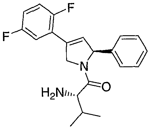


Claims
Priority Applications (4)
| Application Number | Priority Date | Filing Date | Title |
|---|---|---|---|
| JP2006527165A JP2007505634A (en) | 2003-09-22 | 2004-09-22 | Synthetic lethal screening using RNA interference |
| EP04816246A EP1670955A2 (en) | 2003-09-22 | 2004-09-22 | Synthetic lethal screen using rna interference |
| AU2004276823A AU2004276823A1 (en) | 2003-09-22 | 2004-09-22 | Synthetic lethal screen using RNA interference |
| CA002539651A CA2539651A1 (en) | 2003-09-22 | 2004-09-22 | Synthetic lethal screen using rna interference |
Applications Claiming Priority (6)
| Application Number | Priority Date | Filing Date | Title |
|---|---|---|---|
| US50522903P | 2003-09-22 | 2003-09-22 | |
| US60/505,229 | 2003-09-22 | ||
| US54856804P | 2004-02-27 | 2004-02-27 | |
| US60/548,568 | 2004-02-27 | ||
| US55428404P | 2004-03-17 | 2004-03-17 | |
| US60/554,284 | 2004-03-17 |
Publications (2)
| Publication Number | Publication Date |
|---|---|
| WO2005031002A2 true WO2005031002A2 (en) | 2005-04-07 |
| WO2005031002A3 WO2005031002A3 (en) | 2005-10-13 |
Family
ID=34397018
Family Applications (1)
| Application Number | Title | Priority Date | Filing Date |
|---|---|---|---|
| PCT/US2004/031629 WO2005031002A2 (en) | 2003-09-22 | 2004-09-22 | Synthetic lethal screen using rna interference |
Country Status (6)
| Country | Link |
|---|---|
| US (1) | US20050181385A1 (en) |
| EP (1) | EP1670955A2 (en) |
| JP (1) | JP2007505634A (en) |
| AU (1) | AU2004276823A1 (en) |
| CA (1) | CA2539651A1 (en) |
| WO (1) | WO2005031002A2 (en) |
Cited By (14)
| Publication number | Priority date | Publication date | Assignee | Title |
|---|---|---|---|---|
| EP1664274A2 (en) * | 2003-08-13 | 2006-06-07 | The Board Of Trustees Of The University Of Illinois | Silencing of tgf-beta receptor type ii expression by sirna |
| WO2006132907A3 (en) * | 2005-06-03 | 2007-05-18 | Novartis Vaccines & Diagnostic | Methods of treating, diagnosing or detecting cancer using an ephb3 modulator |
| WO2008030239A1 (en) * | 2006-09-05 | 2008-03-13 | Sirna Therapeutics, Inc. | RNA INTERFERENCE MEDIATED INHIBITION OF HISTONE DEACETYLASE (HDAC) GENE EXPRESSION USING SHORT INTERFERING NUCLEIC ACID (siNA) |
| WO2008137867A2 (en) | 2007-05-03 | 2008-11-13 | Rosetta Inpharmatics Llc | Compositions comprising mir34 therapeutic agents for treating cancer |
| WO2009039199A2 (en) * | 2007-09-17 | 2009-03-26 | Intradigm Corporation | Compositions comprising stat5 sirna and methods of use thereof |
| JP2010500006A (en) * | 2006-08-04 | 2010-01-07 | ノバルティス アーゲー | EphB3-specific antibodies and uses thereof |
| US7718629B2 (en) | 2006-03-31 | 2010-05-18 | Alnylam Pharmaceuticals, Inc. | Compositions and methods for inhibiting expression of Eg5 gene |
| WO2011038160A3 (en) * | 2009-09-23 | 2011-07-28 | Protiva Biotherapeutics, Inc. | Compositions and methods for silencing genes expressed in cancer |
| US8293719B2 (en) | 2004-03-12 | 2012-10-23 | Alnylam Pharmaceuticals, Inc. | iRNA agents targeting VEGF |
| US8530430B2 (en) | 2009-05-11 | 2013-09-10 | Oncotherapy Science, Inc. | TTK peptides and vaccines including the same |
| JP2013230154A (en) * | 2006-09-20 | 2013-11-14 | Inst Pasteur Korea | Method of detecting and/or quantifying expression of target protein candidate in cell, and method of identifying target protein of small molecule modulator |
| US8598333B2 (en) | 2006-05-26 | 2013-12-03 | Alnylam Pharmaceuticals, Inc. | SiRNA silencing of genes expressed in cancer |
| WO2017106964A1 (en) * | 2015-12-22 | 2017-06-29 | Sarissa Inc. | Methods of treating cancer by inhibition of dna repair proteins using antisense based therapies |
| US11041153B2 (en) * | 2017-04-19 | 2021-06-22 | Bio-Path Holdings, Inc. | P-ethoxy nucleic acids for STAT3 inhibition |
Families Citing this family (44)
| Publication number | Priority date | Publication date | Assignee | Title |
|---|---|---|---|---|
| KR101147147B1 (en) * | 2004-04-01 | 2012-05-25 | 머크 샤프 앤드 돔 코포레이션 | Modified polynucleotides for reducing off-target effects in rna interference |
| US8440610B2 (en) * | 2004-11-12 | 2013-05-14 | Massachusetts Institute Of Technology | Mapkap kinase-2 as a specific target for blocking proliferation of P53-defective cells |
| CA2586987A1 (en) * | 2004-11-12 | 2006-05-18 | Massachusetts Institute Of Technology | Methods and compositions for treating cellular proliferative diseases |
| US7923207B2 (en) | 2004-11-22 | 2011-04-12 | Dharmacon, Inc. | Apparatus and system having dry gene silencing pools |
| US7935811B2 (en) | 2004-11-22 | 2011-05-03 | Dharmacon, Inc. | Apparatus and system having dry gene silencing compositions |
| US7923206B2 (en) * | 2004-11-22 | 2011-04-12 | Dharmacon, Inc. | Method of determining a cellular response to a biological agent |
| US8447526B2 (en) * | 2005-05-23 | 2013-05-21 | Microsoft Corporation | Methods and computer systems for analyzing high-throughput assays |
| CN103820562B (en) | 2005-08-01 | 2015-05-13 | 俄亥俄州立大学研究基金会 | MicroRNA-based methods and compositions for the diagnosis, prognosis and treatment of breast cancer |
| EP2796554A3 (en) | 2005-09-12 | 2014-12-10 | The Ohio State University Research Foundation | Compositions for use in treating BCL2-associated cancers |
| WO2007041453A2 (en) * | 2005-09-30 | 2007-04-12 | Rosetta Inpharmatics Llc | Methods and compositions for treating cancer |
| WO2007044413A2 (en) * | 2005-10-05 | 2007-04-19 | The Ohio State University Research Foundation | Wwox gene, vectors containing the same, and uses in treatment of cancer |
| US7670840B2 (en) | 2006-01-05 | 2010-03-02 | The Ohio State University Research Foundation | Micro-RNA expression abnormalities of pancreatic, endocrine and acinar tumors |
| EP2514433B1 (en) | 2006-01-05 | 2015-10-21 | The Ohio State University Research Foundation | MicroRNA-based methods for the diagnosis, prognosis and treatment of lung cancer |
| ES2544713T3 (en) | 2006-01-05 | 2015-09-03 | The Ohio State University Research Foundation | MicroRNA based methods and compositions for the diagnosis and treatment of solid cancers |
| AU2007227423B2 (en) | 2006-03-20 | 2013-11-07 | The Ohio State University Research Foundation | MicroRNA fingerprints during human megakaryocytopoiesis |
| US8768629B2 (en) | 2009-02-11 | 2014-07-01 | Caris Mpi, Inc. | Molecular profiling of tumors |
| MX2008014608A (en) | 2006-05-18 | 2009-03-31 | Molecular Profiling Inst Inc | System and method for determining individualized medical intervention for a disease state. |
| EP2436786B1 (en) | 2006-07-13 | 2014-04-16 | The Ohio State University Research Foundation | MIR-10a for diagnosing poor survival prognosis colon adenocarcinoma. |
| AU2007346101B2 (en) | 2006-09-19 | 2013-08-15 | The Ohio State University Research Foundation | TCL1 expression in chronic lymphocytic leukemia (CLL) regulated by miR-29 and miR-181 |
| WO2008036825A2 (en) | 2006-09-22 | 2008-03-27 | Dharmacon, Inc. | Duplex oligonucleotide complexes and methods for gene silencing by rna interference |
| US8252538B2 (en) | 2006-11-01 | 2012-08-28 | The Ohio State University | MicroRNA expression signature for predicting survival and metastases in hepatocellular carcinoma |
| AU2008211142A1 (en) * | 2007-01-31 | 2008-08-07 | The Ohio State University Research Foundation | Mic orna-based methods and compositions for the treatment of acute myeloid leukemia |
| US8465917B2 (en) | 2007-06-08 | 2013-06-18 | The Ohio State University Research Foundation | Methods for determining heptocellular carcinoma subtype and detecting hepatic cancer stem cells |
| EP2719773A3 (en) | 2007-06-15 | 2014-07-30 | The Ohio State University Research Foundation | miRNA as marker for acute lamphomic leucemia |
| JP2010535782A (en) | 2007-07-31 | 2010-11-25 | ズィ、オハイオウ、ステイト、ユーニヴァーサティ、リサーチ、ファウンデイシャン | Method for reversing methylation by targeting DNMT3A and DNMT3B |
| ES2562077T3 (en) | 2007-08-03 | 2016-03-02 | The Ohio State University Research Foundation | Ultraconserved regions encoding RNAnc |
| CA2926831A1 (en) | 2007-08-22 | 2009-02-26 | The Ohio State University Research Foundation | Methods and compositions for inducing deregulation of epha7 and erk phosphorylation in human acute leukemias |
| WO2009055773A2 (en) | 2007-10-26 | 2009-04-30 | The Ohio State University Research Foundation | Methods for identifying fragile histidine triad (fhit) interaction and uses thereof |
| US8188060B2 (en) | 2008-02-11 | 2012-05-29 | Dharmacon, Inc. | Duplex oligonucleotides with enhanced functionality in gene regulation |
| WO2009126970A1 (en) * | 2008-04-11 | 2009-10-15 | The Translational Genomics Research Institute | Method of assessing sensitivity to brostallicin |
| EP2307028B1 (en) | 2008-06-11 | 2013-10-02 | The Government of the United States of America as represented by The Secretary of the Department of Health and Human Services | Use of mir-26 family as a predictive marker of hepatocellular carcinoma and responsiveness to therapy |
| EP3248618A1 (en) * | 2009-04-22 | 2017-11-29 | Massachusetts Institute Of Technology | Innate immune suppression enables repeated delivery of long rna molecules |
| US10837020B2 (en) | 2009-04-22 | 2020-11-17 | Massachusetts Institute Of Technology | Innate immune suppression enables repeated delivery of long RNA molecules |
| EP2421563B1 (en) * | 2009-04-22 | 2017-04-12 | Massachusetts Institute of Technology | Innate immune suppression enables repeated delivery of long rna molecules |
| CA2781547A1 (en) | 2009-11-23 | 2011-05-26 | The Ohio State University | Materials and methods useful for affecting tumor cell growth, migration and invasion |
| AU2011326032B2 (en) | 2010-11-12 | 2016-10-06 | The Ohio State University Research Foundation | Materials and methods related to microRNA-21, mismatch repair, and colorectal cancer |
| JP2014500258A (en) | 2010-11-15 | 2014-01-09 | ジ・オハイオ・ステイト・ユニバーシティ・リサーチ・ファウンデイション | Controlled release mucoadhesion system |
| WO2012083231A1 (en) * | 2010-12-16 | 2012-06-21 | Massachusetts Institute Of Technology | Rnai based method of drug screening and characterization |
| JP2014509852A (en) | 2011-03-07 | 2014-04-24 | ジ・オハイオ・ステート・ユニバーシティ | Mutagenic activity induced by microRNA-155 (miR-155) links inflammation and cancer |
| CN104364390B (en) | 2011-10-14 | 2016-08-24 | 俄亥俄州立大学 | The method relevant to ovarian cancer and material |
| WO2013090556A1 (en) | 2011-12-13 | 2013-06-20 | The Ohio State University | Methods and compositions related to mir-21 and mir-29a, exosome inhibition, and cancer metastasis |
| CN104685065B (en) | 2012-01-20 | 2017-02-22 | 俄亥俄州立大学 | Breast cancer biomarker signatures for invasiveness and prognosis |
| CN115813902A (en) | 2012-01-20 | 2023-03-21 | 德玛公司 | Use of substituted hexitols for the treatment of malignant tumors |
| CA3035315A1 (en) * | 2015-08-28 | 2017-03-09 | University Of Maryland, College Park | Computer system and methods for harnessing synthetic rescues and applications thereof |
Citations (3)
| Publication number | Priority date | Publication date | Assignee | Title |
|---|---|---|---|---|
| WO2002044321A2 (en) * | 2000-12-01 | 2002-06-06 | MAX-PLANCK-Gesellschaft zur Förderung der Wissenschaften e.V. | Rna interference mediating small rna molecules |
| WO2002072783A2 (en) * | 2001-03-12 | 2002-09-19 | Irm, Llc | Identification of cellular targets for biologically active molecules |
| US20030165945A1 (en) * | 2000-04-28 | 2003-09-04 | Bird Timothy A. | Human Pellino polypeptides |
Family Cites Families (15)
| Publication number | Priority date | Publication date | Assignee | Title |
|---|---|---|---|---|
| US5075217A (en) * | 1989-04-21 | 1991-12-24 | Marshfield Clinic | Length polymorphisms in (dC-dA)n ·(dG-dT)n sequences |
| US5364759B2 (en) * | 1991-01-31 | 1999-07-20 | Baylor College Medicine | Dna typing with short tandem repeat polymorphisms and identification of polymorphic short tandem repeats |
| US5659024A (en) * | 1994-01-14 | 1997-08-19 | The Burnham Institute | Promotors that regulate the expression of genes involved in cell death |
| US6335156B1 (en) * | 1997-12-18 | 2002-01-01 | The Johns Hopkins University School Of Medicine | 14-3-3σ arrests the cell cycle |
| US6506559B1 (en) * | 1997-12-23 | 2003-01-14 | Carnegie Institute Of Washington | Genetic inhibition by double-stranded RNA |
| DE19847779C1 (en) * | 1998-10-16 | 2000-02-03 | Deutsches Krebsforsch | Novel receptor DNA useful for identifying apoptosis-modulating substances potentially useful for cancer chemotherapy |
| EP1183389A2 (en) * | 1999-04-30 | 2002-03-06 | University of Florida | Adeno-associated virus-delivered ribozyme compositions and methods of use |
| US6617115B1 (en) * | 1999-10-27 | 2003-09-09 | Cytokinetics, Inc. | Methods of screening for modulators of cell proliferation |
| US20070026394A1 (en) * | 2000-02-11 | 2007-02-01 | Lawrence Blatt | Modulation of gene expression associated with inflammation proliferation and neurite outgrowth using nucleic acid based technologies |
| CA2404890C (en) * | 2000-03-30 | 2013-11-19 | Whitehead Institute For Biomedical Research | Rna sequence-specific mediators of rna interference |
| EP1322307B1 (en) * | 2000-07-28 | 2011-09-28 | Sloan-Kettering Institute For Cancer Research | Methods for treating cell proliferative disorders and viral infections |
| US20030148519A1 (en) * | 2001-11-14 | 2003-08-07 | Engelke David R. | Intracellular expression and delivery of siRNAs in mammalian cells |
| WO2003074662A2 (en) * | 2002-03-01 | 2003-09-12 | Exelixis, Inc. | SCDs AS MODIFIERS OF THE p53 PATHWAY AND METHODS OF USE |
| US20030229912A1 (en) * | 2002-03-28 | 2003-12-11 | Boehringer Ingelheim International Gmbh | Non-human transgenic mammals and their use as a disease model |
| WO2004074301A2 (en) * | 2003-02-14 | 2004-09-02 | Smithkline Beecham Corporation | Differentially expressed nucleic acids that correlate with ksp expression |
-
2004
- 2004-09-22 AU AU2004276823A patent/AU2004276823A1/en not_active Abandoned
- 2004-09-22 EP EP04816246A patent/EP1670955A2/en not_active Withdrawn
- 2004-09-22 JP JP2006527165A patent/JP2007505634A/en not_active Withdrawn
- 2004-09-22 CA CA002539651A patent/CA2539651A1/en not_active Abandoned
- 2004-09-22 WO PCT/US2004/031629 patent/WO2005031002A2/en active Application Filing
- 2004-09-22 US US10/947,637 patent/US20050181385A1/en not_active Abandoned
Patent Citations (4)
| Publication number | Priority date | Publication date | Assignee | Title |
|---|---|---|---|---|
| US20030165945A1 (en) * | 2000-04-28 | 2003-09-04 | Bird Timothy A. | Human Pellino polypeptides |
| WO2002044321A2 (en) * | 2000-12-01 | 2002-06-06 | MAX-PLANCK-Gesellschaft zur Förderung der Wissenschaften e.V. | Rna interference mediating small rna molecules |
| WO2002072783A2 (en) * | 2001-03-12 | 2002-09-19 | Irm, Llc | Identification of cellular targets for biologically active molecules |
| US20030170642A1 (en) * | 2001-03-12 | 2003-09-11 | Irm, Llc | Identification of cellular targets for biologically active molecules |
Non-Patent Citations (4)
| Title |
|---|
| DEVERAUX QUINN L ET AL: "Exposing oncogenic dependencies for cancer drug target discovery and validation using RNAi." SEMINARS IN CANCER BIOLOGY. AUG 2003, vol. 13, no. 4, August 2003 (2003-08), pages 293-300, XP002325031 ISSN: 1044-579X * |
| HANNON G J: "RNA interference" NATURE, MACMILLAN JOURNALS LTD. LONDON, GB, vol. 418, 11 July 2002 (2002-07-11), pages 244-251, XP002979088 ISSN: 0028-0836 * |
| HOLEN T ET AL: "Positional effects of short interfering RNAs targeting the human coagulation trigger Tissue Factor" NUCLEIC ACIDS RESEARCH, OXFORD UNIVERSITY PRESS, SURREY, GB, vol. 30, no. 8, 15 April 2002 (2002-04-15), pages 1757-1766, XP002232890 ISSN: 0305-1048 * |
| See also references of EP1670955A2 * |
Cited By (22)
| Publication number | Priority date | Publication date | Assignee | Title |
|---|---|---|---|---|
| EP1664274A2 (en) * | 2003-08-13 | 2006-06-07 | The Board Of Trustees Of The University Of Illinois | Silencing of tgf-beta receptor type ii expression by sirna |
| US8067389B2 (en) | 2003-08-13 | 2011-11-29 | The Broad of Trustees of the University of Illinois | Silencing of TGFβ type II receptor expression by siRNA |
| EP1664274A4 (en) * | 2003-08-13 | 2010-06-30 | Univ Illinois | Silencing of tgf-beta receptor type ii expression by sirna |
| US8293719B2 (en) | 2004-03-12 | 2012-10-23 | Alnylam Pharmaceuticals, Inc. | iRNA agents targeting VEGF |
| WO2006132907A3 (en) * | 2005-06-03 | 2007-05-18 | Novartis Vaccines & Diagnostic | Methods of treating, diagnosing or detecting cancer using an ephb3 modulator |
| US7718629B2 (en) | 2006-03-31 | 2010-05-18 | Alnylam Pharmaceuticals, Inc. | Compositions and methods for inhibiting expression of Eg5 gene |
| US8598333B2 (en) | 2006-05-26 | 2013-12-03 | Alnylam Pharmaceuticals, Inc. | SiRNA silencing of genes expressed in cancer |
| JP2010500006A (en) * | 2006-08-04 | 2010-01-07 | ノバルティス アーゲー | EphB3-specific antibodies and uses thereof |
| US9006398B2 (en) | 2006-08-04 | 2015-04-14 | Novartis Ag | EPHB3-specific antibody and uses thereof |
| KR101437188B1 (en) | 2006-08-04 | 2014-10-02 | 노바르티스 아게 | Ephb3-specific antibody and uses thereof |
| US8586716B2 (en) | 2006-08-04 | 2013-11-19 | Novartis Ag | EPHB3-specific antibody and uses thereof |
| WO2008030239A1 (en) * | 2006-09-05 | 2008-03-13 | Sirna Therapeutics, Inc. | RNA INTERFERENCE MEDIATED INHIBITION OF HISTONE DEACETYLASE (HDAC) GENE EXPRESSION USING SHORT INTERFERING NUCLEIC ACID (siNA) |
| JP2013230154A (en) * | 2006-09-20 | 2013-11-14 | Inst Pasteur Korea | Method of detecting and/or quantifying expression of target protein candidate in cell, and method of identifying target protein of small molecule modulator |
| WO2008137867A2 (en) | 2007-05-03 | 2008-11-13 | Rosetta Inpharmatics Llc | Compositions comprising mir34 therapeutic agents for treating cancer |
| WO2009039199A2 (en) * | 2007-09-17 | 2009-03-26 | Intradigm Corporation | Compositions comprising stat5 sirna and methods of use thereof |
| WO2009039199A3 (en) * | 2007-09-17 | 2009-05-14 | Intradigm Corp | Compositions comprising stat5 sirna and methods of use thereof |
| US8530430B2 (en) | 2009-05-11 | 2013-09-10 | Oncotherapy Science, Inc. | TTK peptides and vaccines including the same |
| WO2011038160A3 (en) * | 2009-09-23 | 2011-07-28 | Protiva Biotherapeutics, Inc. | Compositions and methods for silencing genes expressed in cancer |
| US9222086B2 (en) | 2009-09-23 | 2015-12-29 | Protiva Biotherapeutics, Inc. | Compositions and methods for silencing genes expressed in cancer |
| WO2017106964A1 (en) * | 2015-12-22 | 2017-06-29 | Sarissa Inc. | Methods of treating cancer by inhibition of dna repair proteins using antisense based therapies |
| EP3393479A4 (en) * | 2015-12-22 | 2019-10-23 | Sarissa Inc. | Methods of treating cancer by inhibition of dna repair proteins using antisense based therapies |
| US11041153B2 (en) * | 2017-04-19 | 2021-06-22 | Bio-Path Holdings, Inc. | P-ethoxy nucleic acids for STAT3 inhibition |
Also Published As
| Publication number | Publication date |
|---|---|
| WO2005031002A3 (en) | 2005-10-13 |
| AU2004276823A1 (en) | 2005-04-07 |
| US20050181385A1 (en) | 2005-08-18 |
| EP1670955A2 (en) | 2006-06-21 |
| CA2539651A1 (en) | 2005-04-07 |
| JP2007505634A (en) | 2007-03-15 |
Similar Documents
| Publication | Publication Date | Title |
|---|---|---|
| WO2005031002A2 (en) | Synthetic lethal screen using rna interference | |
| Sun et al. | Hsa-miR-326 targets CCND1 and inhibits non-small cell lung cancer development | |
| Voce et al. | Temozolomide treatment induces lncRNA MALAT1 in an NF-κB and p53 codependent manner in glioblastoma | |
| KR101791981B1 (en) | SiRNA-mediated silencing of genes for treating chemotherapeutic drug-resistant epithelial tumors | |
| Bell et al. | MYCN oncoprotein targets and their therapeutic potential | |
| Hu et al. | Negative regulation of tumor suppressor p53 by microRNA miR-504 | |
| JP4658936B2 (en) | Compositions and methods for treating lung cancer | |
| EP1458863B1 (en) | A system for stable expression of sirnas in mammalian cells | |
| JP5841332B2 (en) | Cell growth inhibitor | |
| Xu et al. | MicroRNA-31 functions as a tumor suppressor and increases sensitivity to mitomycin-C in urothelial bladder cancer by targeting integrin α5 | |
| EP3337509A1 (en) | Methods of treating autoimmune conditions in patients with genetic variations in dcr3 or in a dcr3 network gene | |
| Mallakin et al. | The Arf‐inducing transcription factor Dmp1 encodes a transcriptional activator of amphiregulin, thrombospondin‐1, JunB and Egr1 | |
| WO2005090572A2 (en) | Compositions and methods for treating pancreatic cancer | |
| Yu et al. | Retracted: Long noncoding RNA LINC00052 inhibits colorectal cancer metastasis by sponging microRNA‐574‐5p to modulate CALCOCO1 expression | |
| Jia et al. | Lung cancer cells expressing a shortened CDK16 3′ UTR escape senescence through impaired miR‐485‐5p targeting | |
| JP2009502113A (en) | Compositions and methods for treating breast cancer | |
| US20100166731A1 (en) | Methods and Compositions for Treating Cancer | |
| Liu et al. | miR-874 inhibits metastasis-relevant traits via targeting SH2B adaptor protein 1 (SH2B1) in gastric cancer | |
| US11674140B2 (en) | Compositions and methods for treating facioscapulohumeral dystrophy | |
| KR20230045386A (en) | COMPOSITION FOR ENHANCING SENSITIVITY TO ANTI-CANCER AGENT COMPRISING OF miR-4487 AS AN ACTIVE INGREDIENT | |
| Heminger | Loss of Hdmx Leads to Alterations in Gene Expression and Inhibition of Cell Growth in Tumor Cells with Wild-type P53 | |
| Church | Microprocessor Recruitment to RNA Polymerase II is Required for Differential Expression of MicroRNAs | |
| KR101171863B1 (en) | Pharmaceutical composition for the treatment of cancers or inhibition of metastasis containing the expression or activity inhibitors of PRPF4, a novel cancer therapeutic target | |
| Bellelli | NCOA4 depletion leads to replication stress and premature senescence |
Legal Events
| Date | Code | Title | Description |
|---|---|---|---|
| WWE | Wipo information: entry into national phase |
Ref document number: 200480034189.0 Country of ref document: CN |
|
| AK | Designated states |
Kind code of ref document: A2 Designated state(s): AE AG AL AM AT AU AZ BA BB BG BR BW BY BZ CA CH CN CO CR CU CZ DE DK DM DZ EC EE EG ES FI GB GD GE GH GM HR HU ID IL IN IS JP KE KG KP KR KZ LC LK LR LS LT LU LV MA MD MG MK MN MW MX MZ NA NI NO NZ OM PG PH PL PT RO RU SC SD SE SG SK SL SY TJ TM TN TR TT TZ UA UG US UZ VC VN YU ZA ZM ZW |
|
| AL | Designated countries for regional patents |
Kind code of ref document: A2 Designated state(s): BW GH GM KE LS MW MZ NA SD SL SZ TZ UG ZM ZW AM AZ BY KG KZ MD RU TJ TM AT BE BG CH CY CZ DE DK EE ES FI FR GB GR HU IE IT LU MC NL PL PT RO SE SI SK TR BF BJ CF CG CI CM GA GN GQ GW ML MR NE SN TD TG |
|
| 121 | Ep: the epo has been informed by wipo that ep was designated in this application | ||
| WWE | Wipo information: entry into national phase |
Ref document number: 2006527165 Country of ref document: JP |
|
| WWE | Wipo information: entry into national phase |
Ref document number: 2539651 Country of ref document: CA |
|
| WWE | Wipo information: entry into national phase |
Ref document number: 2004276823 Country of ref document: AU |
|
| WWE | Wipo information: entry into national phase |
Ref document number: 1356/CHENP/2006 Country of ref document: IN |
|
| WWE | Wipo information: entry into national phase |
Ref document number: 2004816246 Country of ref document: EP |
|
| WWP | Wipo information: published in national office |
Ref document number: 2004276823 Country of ref document: AU |
|
| WWP | Wipo information: published in national office |
Ref document number: 2004816246 Country of ref document: EP |