CROSS-REFERENCE TO RELATED APPLICATIONS
This application claims the benefit of U.S. Provisional Application No. 60/819,359, filed Jul. 10, 2006, which is incorporated herein by reference in its entirety.
BACKGROUND OF THE INVENTION
1. Field of the Invention
The present invention is in the fields of cell biology, immunology and oncology. The invention relates to the discovery that there is a relationship between the expression levels of the tumor suppressor gene smad4 (also known as dpc4) and integrin ανβ6, and the responsiveness of patient populations to ανβ6-active compounds and compositions (e.g., antibodies and other ligands that bind ανβ6), particularly in cancer cells from such patient populations, more particularly on carcinomas such as pancreatic carcinomas. The invention thus provides methods for determining the responsiveness of tumor cells (particularly those from pancreatic tumors) to such ανβ6-active compounds and compositions by examining the expression of ανβ6 and smad4 by the tumor cells, as well as methods of diagnosis and treatment/prevention of tumor progression using ligands, including antibodies and small molecule drugs, that bind to integrin ανβ6 on the surfaces of tumor cells and/or that block one or more components of the TGF-β pathway, particularly in smad4-deficient tumor cells.
2. Related Art
Integrins are cell surface glycoprotein receptors which bind extracellular matrix proteins and mediate cell-cell and cell-extracellular matrix interactions (generally referred to as cell adhesion events) (Ruoslahti, E., J Clin. Invest. 87:1-5 (1991); Hynes, R. O., Cell 69:11-25 (1992)). These receptors are composed of noncovalently associated alpha (α) and beta (β) chains which combine to give a variety of heterodimeric proteins with distinct cellular and adhesive specificities (Albeda, S. M., Lab. Invest. 68:4-14 (1993)). Recent studies have implicated certain integrins in the regulation of a variety of cellular processes including cellular adhesion, migration, invasion, differentiation, proliferation, apoptosis and gene expression (Albedo, S. M., Lab. Invest. 68:4-14 (1993); Juliano, R., Cancer Met. Rev. 13:25-30 (1994); Ruoslahti, E. and Reed, J. C., Cell 77:477-478 (1994); and Ruoslahti, E. and Giancotti, F. G., Cancer Cells 1:119-126 (1989); Plow, Haas et al. 2000; van der Flier and Sonnenberg 2001).
The ανβ6 receptor is one member of a family of integrins that are expressed as cell surface heterodimeric proteins (Busk, M. et al., J. Biol. Chem. 267(9):5790-5796 (1992)). While the αν subunit can form a heterodimer with a variety of β subunits (β1, β3, β5, β6 and β8), the β6 subunit can only be expressed as a heterodimer with the αν subunit. The ανβ6 integrin is known to be a fibronectin-, latency associated peptide (LAP)- and tenascin C-binding cell surface receptor, interacting with the extracellular matrix through the RGD tripeptide binding sites thereon (Busk, M. et al., J. Biol. Chem. 267:5790-5796 (1992); Weinacker, A. et al., J. Biol. Chem. 269:6940-6948 (1994); Prieto, A. L. et al., Proc. Natl. Acad. Sci. USA 90:10154-10158 (1993)). Although the ανβ6 integrin was first identified and sequenced more than 10 years ago, the biological significance of ανβ6, especially in disease, is still under investigation. The expression of ανβ6 is restricted to epithelial cells where it is expressed at relatively low levels in healthy tissue and significantly upregulated during development, injury, and wound healing (Breuss, J. M. et al., J. Histochem. Cytochem. 41:1521-1527 (1993); Breuss, J. M. et al., J Cell Sci. 108:2241-2251 (1995); Koivisto, L. et al., Cell Adhes. Communic. 7:245-257 (1999); Zambruno, G. et al., J. Cell Biol. 129(3):853-865 (1995); Hakkinen, L. et al., J. Histochem. Cytochem. 48(6):985-998 (2000)). An increasing number of recent reports demonstrate that ανβ6 is upregulated on cancers of epithelial origin, including colon carcinoma (Niu, J. et al, Int. J. Cancer 92:40-48 (2001); Bates, R. C. et al., J. Clin. Invest. 115:339-347 (2005)), ovarian cancer (Ahmed, N. et al., J. Cell. Biochem. 84:675-686 (2002); Ahmed, N. et al., J. Histochem. Cytochem. 50:1371-1379 (2002); Ahmed, N. et al., Carcinogen. 23:237-244 (2002)), squamous cell carcinoma (Koivisto, L. et al., Exp. Cell Res. 255:10-17 (2000); Xue, H. et al., Biochem. Biophys. Res. Comm. 288:610-618 (2001); Thomas, G. J. et al., J. Invest. Derinatol. 117:67-73 (2001); Thomas, G. J. et al., Int. J. Cancer 92:641-650 (2001); Ramos, D. M. et al., Matrix Biol. 21:297-307 (2002); (Agrez, M. et al., Br. J. Cancer 81:90-97 (1999); Hamidi, S. et al., Br. J. Cancer 82(8):1433-1440 (2000); Kawashima, A. et al., Pathol. Res. Pract. 99(2):57-64 (2003)), and breast cancer (Arihiro, K. et al., Breast Cancer 7:19-26 (2000)). It has also been reported that the cc subunit may be involved in tumor metastasis, and that blocking this subunit consequently may prevent metastasis (for review, see Imhof, B. A. et al., in: “Attempts to Understand Metastasis Formation I,” U. Günthert and W. Birchmeier, eds., Berlin: Springer-Verlag, pp. 195-203 (1996)).
The ανβ6 integrin may have multiple regulatory functions in tumor cell biology. Recent studies have demonstrated that the extracellular and cytoplasmic domains of the β6 subunit mediate different cellular activities. The extracellular and transmembrane domains have been shown to mediate TGF-β activation and adhesion (Sheppard, D., Cancer and Metastasis Rev. 24:395-402 (2005); Munger, J. S. et al., Cell 96:319-328 (1999)). The cytoplasmic domain of the β6 subunit contains a unique 11-amino acid sequence that is important in mediating ανβ6 regulated cell proliferation, MMP production, migration, and pro-survival (Li, X. et al., J. Biol. Chem. 278(43):41646-41653 (2003); Thomas, G. J. et al., J Invest. Derm. 117(1):67-73 (2001); Thomas, G. J. et al., Br. J. Cancer 87(8):859-867 (2002); Janes, S. M. and Watt, F. M., J. Cell Biol 166(3):419-431 (2004)). The β6 subunit has been cloned, expressed and purified (Sheppard et al., U.S. Pat. No. 6,787,322 B2, the disclosure of which is incorporated herein by reference in its entirety), and function-blocking antibodies that selectively bind to the ανβ6 integrin have been reported (Weinreb et al., J. Biol. Chem. 279:17875-17877 (2004), the disclosure of which is incorporated herein by reference in its entirety). Antagonists of (ανβ6 (including certain monoclonal antibodies) have also been suggested as possible treatments for certain forms of acute lung injury and fibrosis (see U.S. Pat. No. 6,692,741 B2 and WO 99/07405, the disclosures of which are incorporated herein by reference in their entireties).
ανβ6 can bind to several ligands including fibronectin, tenascin, and the latency associated peptide-1 and -3 (LAP1 and LAP3), the N-terminal 278 amino acids of the latent precursor form of TGF-β1 through a direct interaction with an arginine-glycine-aspartate (“RGD”) motif (Busk. M. et al., J. Biol. Chew. 267(9):5790-5796 (1992); Yokosaki, Y. et al., J. Biol. Chem. 271(39):24144-24150 (1996); Huang, X. Z. et al., J. Cell. Sci. 111:2189-2195 (1998); Munger, J. S. et al., Cell 96:319-328 (1999)). The TGF-β cytokine is synthesized as a latent complex which has the N-terminal LAP non-covalently associated with the mature active C-terminal TGF-β cytokine. The latent TGF-β complex cannot bind to its cognate receptor and thus is not biologically active until converted to an active form (Barcellos-Hoff, M. H., J. Mamm. Gland Biol. 1(4):353-363 (1996); Gleizes, P. E. et al., Stem Cells 15(3):190-197 (1997); Munger, J. S. et al., Kid. Int 51:1376-1382 (1997); Khalil, N., Microbes Infect. 1(15): 1255-1263 (1999)). ανβ6 binding to LAP1 or LAP3 leads to activation of the latent precursor form of TGF-β1 and TGF-β3 (Munger, J. S. et al., Cell 96:319-328 (1999)), proposed as a result of a conformational change in the latent complex allowing TGF-β to bind to its receptor. Thus, upregulated expression of ανβ6 can lead to local activation of TGF-β which in turn can activate a cascade of downstream events.
The TGF-β1 cytokine is a pleiotropic growth factor that regulates cell proliferation, differentiation, and immune responses (Wahl, S. M., J. Exp. Med. 180:1587-1590 (1994); Massague, J., Annu. Rev. Biochem. 67:753-791 (1998); Chen, W. and Wahl, S. M., TGF-β: Receptors, Signaling Pathways and Autoimmunity, Basel: Karger, pp. 62-91 (2002); Thomas, D. A. and Massague, J., Cancer Cell 8:369-380 (2005)). The role that TGF-β1 plays in cancer is two-sided. TGF-β is recognized to tumor suppressor and growth inhibitory activity yet, many tumors evolve a resistance to growth suppressive activities of TGF-β1 (Yingling, J. M. et al., Nature Rev. Drug Discov. 3(12):1011-1022 (2004); Akhurst, R. J. et al., Trends Cell Biol. 11(11):S44-S51 (2001); Balmain, A. and Akhurst, R. J., Nature 428(6980):271-272 (2004)). In established tumors, TGF-β1 expression and activity has been implicated in promoting tumor survival, progression, and metastases (Akhurst, R. J. et al., Trends Cell Biol. 11(11): S44-S51 (2001); Muraoka, R. S. et al., J. Clin. Invest. 109 (12):1551 (2002); Yang, Y. A. et al., J. Clin. Invest. 109(12): 1607-1615 (2002)). This is postulated to be mediated by both autocrine and paracrine effects in the local tumor-stromal environment including the effects of TGF-β on immune surveillance, angiogenesis, and increased tumor interstitial pressure. Several studies have now shown the anti-tumor and anti-metastatic effects of inhibiting TGF-β1 (Akhurst, R. J., J. Clin. Invest. 109(12):1533-1536 (2002); Muraoka, R. S. et al., J. Clin. Invest. 109(12):1551 (2002); Yingling, J. M. et al., Nat. Rev. Drug Discov. 3(12):1011-1022 (2004); Yang, Y. A. et al., J. Clin. Invest. 109(12):1607-1615 (2002); Halder, S. K. et al., Neoplasia 7(5):509-521 (2005); Tyer, S. et al., Cancer Biol. Ther. 4(3):261-266 (2005)).
Increased expression of ανβ6 on tumors, particularly at the tumor-stromal interface, may reflect a unique mechanism for local activation of TGF-β1 and the ability to promote tumor survival, invasion, and metastasis. The high level of expression in human metastases infers a potential role for ανβ6 in establishing metastases which is consistent with previous reports that ανβ6 can mediate epithelial to mesenchymal transition, tumor cell invasion in vitro, and expression correlated with metastases in a mouse model (Bates, R. C. et al., J. Clin. Invest. 115(2):339-347 (2005); Thomas, G. J. et al., Br. J. Cancer 87(8):859-867 (2002); Morgan, M. R. et al., J. Biol. Chem. 279(25):26533-26539 (2004)). We have previously described the generation of potent and selective anti-ανβ6 monoclonal antibodies (mAbs) that bind to both the human and murine forms of ανβ6 and block the binding of ανβ6 to its ligands and ανβ6 mediated activation of TGF-β1 (Weinreb, P. H. et al., J. Biol. Chem. 279(17):17875-17887 (2004)).
The generation of potent and selective anti-ανβ6 monoclonal antibodies (mAbs) that bind to both the human and murine forms of ανβ6 and block the binding of ανβ6 to its ligands and ανβ6 mediated activation of TGF-β1 has been previously described (Weinreb, P. H. et al., J. Biol. Chem. 279(17):17875-17887 (2004); see also U.S. patent application Ser. No. 11/483,190 by Violette et al., entitled “Anti-ανβ6 Antibodies and Uses Thereof,” filed on Jul. 10, 2006, which is incorporated herein by reference in its entirety). As also described in PCT Publication WO 03/100033, herein incorporated in its entirety by reference, high affinity antibodies against ανβ6, including the identification and analysis of key amino acid residues in the complementary determining regions (CDRs) of such antibodies, were discovered and characterized. In particular, these high affinity antibodies (a) specifically bind to ανβ6; (b) inhibit the binding of ανβ6 to its ligand such as LAP, fibronectin, vitronectin, and tenascin with an IC50 value lower than that of 10D5 (International Patent Application Publication WO 99/07405); (c) block activation of TGF-β; (d) contain certain amino acid sequences in the CDRs that provide binding specificity to ανβ6; (e) specifically bind to the β6 subunit; and/or (f) recognize ανβ6 in immunostaining procedures, such as immunostaining of paraffin-embedded tissues.
WO 03/100033 also describes the discovery that antibodies that bind to ανβ6 can be grouped into biophysically distinct classes and subclasses. One class of antibodies exhibits the ability to block binding of a ligand (e.g., LAP) to ανβ6 (blockers). This class of antibodies can be further divided into subclasses of cation-dependent blockers and cation-independent blockers. Some of the cation-dependent blockers contain an arginine-glycine-aspartate (RGD) peptide sequence, whereas the cation-independent blockers do not contain an RGD sequence. Another class of antibodies exhibits the ability to bind to ανβ6 and yet does not block binding of ανβ6 to a ligand (nonblockers).
Furthermore, WO 03/100033 discloses antibodies comprising heavy chains and light chains whose complementarity determining regions (CDR) 1, 2 and 3 consist of certain amino acid sequences that provide binding specificity to ανβ6. WO 03/100033 also provides for antibodies that specifically bind to ανβ6 but do not inhibit the binding of ανβ6 to latency associated peptide (LAP) as well as antibodies that bind to the same epitope.
WO 03/100033 further discloses cells of hybridomas 6.1A8, 6.2B10, 6.3G9, 6.8G6, 6.2B1, 6.2A1, 6.2E5, 7.1G10, 7.7G5, and 7.1C5, isolated nucleic acids comprising a coding sequences and isolated polypeptides comprising amino acid sequences of the anti-ανβ6 antibodies. In particular, WO 03/100033 discloses anti-ανβ6 antibodies comprising heavy and light chain polypeptide sequences as antibodies produced by hybridomas 6.1A8, 6.3G9, 6.8G6, 6.2B1, 6.2B10, 6.2A1, 6.2E5, 7.1G10, 7.7G5, or 7.1C5. Several of the hybridomas were deposited at the American Type Culture Collection (“ATCC”; P.O. Box 1549, Manassas, Va. 20108, USA) under the Budapest Treaty. In particular, hybridoma clones 6.3G9 and 6.8G6 were deposited on Aug. 16, 2001, and have accession numbers MCC PTA-3649 and PTA-3645, respectively. The murine antibodies produced by hybridomas 6.3G9 and 6.8G6 are being further explored in the present application for their potential development as humanized antibodies.
The murine monoclonal antibody 3G9 is a murine IgG1, kappa antibody isolated from the β6 integrin −/− mouse (Huang et al., J. Cell Biol. 133:921-928 (1996)) immunized with human soluble ανβ6. The 3G9 antibody specifically recognizes the ανβ6 integrin epitope which is expressed at upregulated levels during injury, fibrosis and cancer (see, e.g., Thomas et al., J. Invest. Dermatology 117:67-73 (2001); Brunton et al., Neoplasia 3: 215-226 (2001); Agrez et al., Int. J. Cancer 81:90-97 (1999); Breuss, J. Cell Science 108:2241-2251 (1995)). It does not bind to other αν integrins and is cross-reactive to both human and murine molecules. The murine monoclonal antibody 3G9 has been described to block the binding of ανβ6 to LAP as determined by blocking of ligand binding either to purified human soluble ανβ6 or to β6-expressing cells, thereby inhibiting the pro-fibrotic activity of TGF-β receptor activation (see WO 03/100033). It has also been shown to inhibit ανβ6-mediated activation of TGF-β with an IC50 value lower than one of the known ανβ6 antibodies, 10D5 (Huang et al., J. Cell Sci. 111:2189-2195 (1998)).
The murine monoclonal antibody 8G6 is a murine IgG1, kappa antibody which also recognizes the ανβ6 integrin epitope, as described in WO 03/100033. The murine monoclonal antibody 8G6 is a cation-dependent, high affinity blocker of ανβ6 displaying the ability to inhibit ανβ6—mediated activation of TGF-β with an IC50 value lower than 10D5 (see WO 03/100033).
Both the 3G9 and 8G6 murine antibodies were effective in preventing fibrosis of the kidney and lung, as described in WO 03/100033. Furthermore, the murine antibody 3G9 was able to effectively inhibit tumor growth in a human tumor xenograft model, suggesting the potential role of ανβ6 in cancer pathology and the effectiveness of such blockade using antibodies directed at ανβ6.
Smad4 is a component of the Smad pathway that is involved in signal transduction in the TGF-β pathway (Levy, L. and Hill, C. S., Molec. Cell. Biol. 25:8108-8125 (2005); Fukuchi, M. et al., Cancer 95:737-743 (2002)). This gene, also known as dpc4 (for “decreased in pancreatic carcinoma”), appears to be a tumor suppressor gene, and a decrease in smad4 expression has been observed in a variety of primary carcinomas, including pancreatic carcinomas (Luttges, J. et al., Am. J. Pathol. 158:1677-1683 (2001); Subramanian, G. et al., Cancer Res. 64:5200-5211 (2004)), esophageal carcinomas (Fukuchi, M. et al., Cancer 95:737-743 (2002), cervical carcinomas (Maliekal, T. T. et al., Oncogene 22:4889-4897 (2003), and other primary human cancers (Iacobuzio-Donahue, C. A. et al., Clin. Canc. Res. 10:1597-1604 (2004), as well as in cell line cancer models including of pancreatic cancers (Lohr, M. et al., Cancer Res. 61:550-555 (2001); Yasutome, M. et al., Clin. Exp. Metastasis 22:461-473 (2005)), and of colon cancers (Levy, L., and Hill, C. S., Molec. Cell. Biol. 25:8108-8125 (2005)). A reduced expression of smad4 in tumors has been associated with poor prognosis for patient survival, particularly in patients with smad4-deficient pancreatic adenocarcinomas (Liu, F., Clin. Cancer Res. 7:3853-3856 (2001); Tascilar, M. et al., Clin. Cancer Res. 7:4115-4121 (2001); Toga, T. et al., Anticancer Res. 24:1173-1178 (2004)). The mechanism of the tumor suppressive activity of the smad4 gene product is poorly understood, but it is thought that it may act as a “switch” regulating the growth-suppressive and growth-activating activities of certain components of the TGF-β signaling pathway (for reviews, see Akhurst, A. J., J. Clin. Invest. 109:1533-1536 (2002); Bachman, K. E., and Park, B. H., Curr. Opin. Oncol. 17:49-54 (2004); Bierie, B., and Moses, H. L., Nature Rev. Cancer 6:506-520 (2006)).
BRIEF SUMMARY OF THE INVENTION
The present invention relates to methods of cancer diagnosis, treatment and prevention using ανβ6-binding ligands, such as ανβ6-binding antibodies. In particular, the invention relates to the discovery of a correlation between reduced expression of smad4 and increased expression of integrin ανβ6 in tumor cells, and the propensity of tumor cells with such expression patterns to be more likely to respond to ανβ6-active compounds and to TGF-β-inhibitory ligands.
Thus, in one embodiment, the invention provides methods of characterizing a tumor, e.g., identifying a tumor that is more likely to progress to a metastatic or invasive tumor, or identifying a tumor for treatment with a preselected agent, e.g., an anti-ανβ6 agent described herein, comprising: (a) obtaining from a patient a cancerous tissue sample comprising a tumor or a portion thereof, and optionally a noncancerous tissue sample; (b) determining the level of expression of smad4 in cells from said cancerous tissue sample and optionally from said noncancerous tissue sample; (c) contacting cells from said tissue sample or samples with one or more ligands that bind to one or more subunits of integrin ανβ6; and (d) determining the level of expression of integrin ανβ6 in said cells from said cancerous tissue sample, and optionally from said cells from said noncancerous tissue sample, and comparing the level of expression in said cancerous tissue sample with a reference sample, e.g., the level of expression in said noncancerous tissue sample, thereby characterizing the tumor. A level of expression in the cancerous sample which meets or exceeds a reference value indicates the presence of a tumor more likely to progress to a metastatic or invasive tumor. For example, a decrease in smad4 expression and a concomitant increase in the level of expression of integrin ανβ6 in cells from said cancerous tissue sample, relative to the levels of expression of smad4 and integrin ανβ6 in said non-cancerous tissue sample, indicates the presence in said patient of a tumor that is more likely to progress to a metastatic or invasive tumor. In certain such embodiments of the invention, the tumor cell is a carcinoma, such as an adenocarcinoma. In more particular embodiments, the carcinoma is a pancreatic carcinoma, a colorectal carcinoma, a cervical carcinoma, a squamous cell carcinoma (such as an esophageal carcinoma), a head and neck carcinoma, a liver carcinoma, an ovarian carcinoma and a lung carcinoma. More particularly, the carcinoma is a pancreatic carcinoma, a colorectal carcinoma, a cervical carcinoma, or a head and neck carcinoma. Suitable embodiments according to this aspect of the invention use ανβ6 integrin-binding ligands which are ανβ6-binding antibodies or ανβ6 epitope-binding fragments thereof. According to certain such embodiments, the antibodies are monoclonal antibodies (which may be chimeric, primatized, human or humanized), including those disclosed in U.S. patent application publication no. U.S. 2005/0255102 A1, the disclosure of which is incorporated herein in its entirety. Suitable such antibodies include, but are not limited to, the ανβ6-binding monoclonal antibodies designated 1A8, 3G9, 8G6, 2B1, 2B10, 2A1, 2E5, 1G10, 7G5, 1C5, 10D5 (ATCC deposit no. HB12382) and CSβ6, as well as fragments, chimeras and hybrids thereof. Particularly suitable for use in such embodiments of the invention are monoclonal antibodies 3G9 and 8G6. Also particularly suitable for use in such embodiments of the invention are humanized monoclonal antibodies, such as the humanized 3G9 antibody designated hu3G9 (BG00011) and the humanized 8G6 antibody designated hu8G6. In certain such aspects of the invention, the ligand is conjugated with at least one detectable label, such as a chromogenic label (e.g., diaminobenzidine or 4-hydroxyazo-benzene-2-carboxylic acid), an enzyme label (e.g., malate dehydrogenase, staphylococcal nuclease, delta-5-steroid isomerase, yeast-alcohol dehydrogenase, alpha-glycerol phosphate dehydrogenase, those phosphate isomerase, peroxidase, alkaline phosphatase, asparaginase, glucose oxidase, β-galactosidase, ribonuclease, urease, catalase, glucose-6-phosphate dehydrogenase, glucoamylase and acetylcholine esterase), a radioisotopic label (e.g., 3H, 111In, 125I, 131I, 32P, 35S, 14C, 51Cr, 57To, 58Co, 59Fe, 75Se, 152Eu, 90Y, 67Cu, 217Ci, 211At, 212Pb, 47Sc and 109Pd), a non-radioactive isotopic label (e.g., 157Gd, 55Mn, 162Dy, 52Tr, 56Fe, 99mTc and 112In), a fluorescent label (e.g., a 152Eu label, a fluorescein label, an isothiocyanate label, a rhodamine label, a phycoerythrin label, a phycocyanin label, an allophycocyanin label, a Green Fluorescent Protein (GFP) label, an o-phthaldehyde label and a fluorescamine label), a toxic label (e.g., a diphtheria toxin label, a ricin label and a cholera toxin label), a chemiluminescent label (e.g., a luminol label, an isoluminol label, an aromatic acridinium ester label, an imidazole label, an acridinium salt label, an oxalate ester label, a luciferin label, a luciferase label and an aequorin label), an X-radiographic label (e.g., barium or cesium), a spin label (e.g., deuterium) or a nuclear magnetic resonance contrast agent label (e.g., Gd, Mn and iron).
In related embodiments, the invention provides therapeutic methods for eliminating tumors from patients, e.g, wherein the tumors have an increased potential to become metastatic or invasive. Such methods comprise, e.g., characterizing a tissue sample as described herein, for example: (a) identifying, in a tissue sample from a patient, a tumor that is more likely to progress to a metastatic or invasive tumor, according to the methods for identifying such tumors that are described above; and (b) administering to a patient, in which such a tumor has been identified, a therapeutically effective amount of one or more ligands that binds to one or more subunits of integrin ανβ6 on one or more ανβ6-positive metastatic tumor cells, wherein the binding of the ligand to the integrin results in the death, chemosensitization or decreased invasiveness of said metastatic tumor cell. In a preferred embodiment, the same agent, e.g., the same antibody is used to characterize the tumor and treat the patient. In other embodiments, different agents or antibodies are used. For example, characterizing can be performed with an antibody having a first label, and treating can be performed with an antibody having a second label, e.g., a therapeutic agent.
In related embodiments, the invention provides methods of reducing or preventing the progression of a pre-metastatic tumor to a metastatic tumor in a patient, comprising (a) characterizing a tissue sample as described herein, e.g., identifying, in a tissue sample from a patient, a tumor that is more likely to progress to a metastatic or invasive tumor, according to the methods for identifying such tumors that are described above; and (b) administering to the patient a therapeutically effective amount of one or more ligands that binds to one or more subunits of integrin ανβ6 on one or more cells in the pre-metastatic or pre-invasive tumor, wherein the binding of the ligand to the integrin results in the reduction or prevention of invasion of cells of the pre-metastatic cancer into tissue areas surrounding the primary tumor. In therapeutic embodiments according to this aspect of the invention, the ανβ6-binding ligands (e.g., ανβ6-binding antibodies) can be conjugated with or bound to one or more cytotoxic compounds or agents which lead to or cause the death of the cell or tissue upon binding of the ανβ6-binding ligand-toxic compound conjugate to one or more ανβ6 integrins on the cell or tissue. In some embodiments, the antibody used to characterize the tumor will lack the cytoxic compound or agent. In additional therapeutic embodiments of the invention, the ανβ6-binding ligands (e.g., ανβ6-binding antibodies) are administered to a patient in conjunction with one or more such cytotoxic compounds or agents. Cytotoxic compounds or agents which can be suitably used according to these aspects of the invention include, but are not limited to, cytotoxic agents (e.g., cisplatin, carboplatin, oxaliplatin, paclitaxel, melphalan, doxorubicin, methotrexate, 5-fluorouracil, etoposide, mechlorethamine, cyclophosphamide, bleomycin, a calicheamicin, a maytansine, a trichothene, CC1065, diphtheria A chain, Pseudomonas aeruginosa exotoxin A chain, ricin A chain, abrin A chain, modeccin A chain, alpha-sarcin, Aleurites fordii proteins, dianthin proteins, Phytolaca americana proteins, momordica charantia inhibitors, curcin, crotin, sapaonaria officinalis inhibitors, gelonin, mitogellin, restrictocin, adriamycin (also known as, and used interchangeably herein with, doxorubicin), gemcitabine, phenomycin, enomycin, tricothecenes, ribonucleases and deoxyribonucleases), radioisotopes (such as 211At, 131I, 125I, 90Y, 186Re, 188Re, 153Sm, 212Bi, 32P, and radioactive isotopes of Lu) and prodrug-activating enzymes (such as alkaline phosphatase, arylsulfatase, cytosine deaminase, proteases, D-alanylcarboxy-peptidases, carbohydrate-cleaving enzymes, P-lactamase and penicillin amidase. Cytotoxic agents can also be agents which recruit particular cells or generally increase the immune response to a tumor. In certain embodiments, the one or more ανβ6 integrin-binding ligands are administered to the patient in the form of a pharmaceutical composition comprising an effective amount of one or more of the ανβ6 integrin-binding ligands and one or more pharmaceutically acceptable carriers or excipients. The one or more ανβ6 integrin-binding ligands and/or one or more pharmaceutical compositions comprising the one or more ανβ6 integrin-binding ligands can be administered to the patient by any suitable mode of administering pharmaceutical compositions, including but not limited to oral administration, parenteral administration (including, for example, injection via an intramuscular, intravenous, intraarterial, intraperitoneal, or subcutaneous route), intracranial administration, transdermal administration, intrapulmonary administration and intranasal administration.
In additional embodiments, the present invention provides methods of diagnosing or identifying a tumor, such as a carcinoma (e.g., an adenocarcinoma), that is more likely to progress to an invasive carcinoma, and/or that is more likely to respond to treatment with a ligand that binds to one or more subunits of integrin ανβ6. Suitable such methods may comprise, for example, (a) obtaining from a patient a cancerous epithelial tissue sample comprising a tumor or a portion thereof, and a noncancerous epithelial tissue sample; (b) determining the level of expression of smad4 in cells from said tissue samples; (c) contacting the tissue samples with one or more ligands that binds to one or more subunits of integrin ανβ6; and (d) determining the level of expression of integrin ανβ6 in the tissue samples, wherein a decrease in the level of expression of smad4 and a concomitant increase in the level expression of integrin ανβ6 in the cancerous tissue sample, relative to the levels of expression of smad4 and integrin ανβ6 in the noncancerous tissue sample, indicates the presence in the patient of a tumor that: (a) has an increased likelihood of progressing from an in situ or noninvasive form, to an invasive, metastatic form; and/or (b) is more likely to respond to treatment with one or more of the above-referenced treatment methods that relies upon the binding of an ανβ6-binding ligand, particularly an ανβ6-binding ligand that is conjugated to or that is administered in conjunction with one or more cytotoxic compounds or agents such as those described above. Such methods are suitable for diagnosing or identifying a variety of tumors, including but not limited to those involving the epithelial tissues noted above. In certain such embodiments, the ligand that binds to one or more subunits of integrin ανβ6 is an ανβ6 integrin-binding antibody (which may be a monoclonal antibody such as those described above) or an ανβ6 epitope-binding fragment thereof. Particularly suitable for use in such diagnostic methods of the invention are ανβ6-binding ligands (e.g., antibodies) that are detectably labeled, i.e., that comprise, are conjugated to, or are bound with at least one detectable label such as a chromogenic label (e.g., diaminobenzidine or 4-hydroxyazobenzene-2-carboxylic acid), an enzyme label (e.g., malate dehydrogenase, staphylococcal nuclease, delta-5-steroid isomerase, yeast-alcohol dehydrogenase, alpha-glycerol phosphate dehydrogenase, triose phosphate isomerase, peroxidase, alkaline phosphatase, asparaginase, glucose oxidase, β-galactosidase, ribonuclease, urease, catalase, glucose-6-phosphate dehydrogenase, glucoamylase or acetylcholine esterase), a radioisotopic label g, 3H, 11In, 125I, 131I, 32P, 35S, 14C, 51Cr, 57To, 58Co, 59Fe, 75Se, 152Eu, 90Y, 67Cu, 217Ci, 211At, 212Pb, 47Sc or 109Pd), a non-radioactive isotopic label (e.g., 157Gd, 55Mn, 162Dy, 52Tr, 56Fe, 99mTc or 112In), a fluorescent label (e.g., a 152Eu label, a fluorescein label, an isothiocyanate label, a rhodamine label, a phycoerythrin label, a phycocyanin label, an allophycocyanin label, a Green Fluorescent Protein (GFP) label, an o-phthaldehyde label or a fluorescamine label), a toxic label (e.g., a diphtheria toxin label, a ricin label or a cholera toxin label), a chemiluminescent label (e.g., a luminol label, an isoluminol label, an aromatic acridinium ester label, an imidazole label, an acridinium salt label, an oxalate ester label, a luciferin label, a luciferase label or an aequorin label), an X-radiographic label (e.g., barium or cesium), a spin label (e.g., deuterium) and a nuclear magnetic resonance contrast agent label (e.g., Gd, Mn and iron).
In one aspect, the invention features a method of inhibiting growth of a cell from a tumor that is smad4 deficient. The method includes, for example, determining the level of expression of smad4 in a cell from the tumor, and treating a tumor cell that is deficient in smad4 expression with one or more agents that cause growth inhibition or death of the tumor cell. The one or more agents can be, for example, a ligand that binds to one or more subunits of integrin ανβ6, such as an anti-ανβ6 antibody, or an ανβ6 epitope-binding fragment thereof. In another embodiment, the one or more agents inhibits the TGF-β signaling pathway in the cell. Such agents include, e.g., protein kinase molecules, small molecule therapeutic compounds, or soluble TGF-β receptor polypeptides.
In another aspect, the invention features a method of chemosensitizing a smad-4-deficient tumor cell to treatment with a growth-inhibiting chemotherapeutic compound. The method includes, for example, determining the level of expression of smad4 in a cell from the tumor, and treating a tumor cell deficient for smad4 expression with one or more agents, such that the treatment results in increased responsiveness to one or more growth-inhibiting chemotherapeutic compounds. The one or more agents can be, for example, ligands that bind to one or more subunits of integrin ανβ6, or agents that inhibit the TGF-β signaling pathway.
Other preferred embodiments of the present invention will be apparent to one of ordinary skill in light of the following drawings and description of the invention, and of the claims.
BRIEF DESCRIPTION OF THE DRAWINGS
FIG. 1 is a composite of photomicrographs depicting the levels of ανβ6 expression (dark areas) in certain human carcinomas that have metastasized to either lymph node (FIGS. 1A-1D) or lung (FIGS. 1E-1F), from the indicated primary tumor sites.
FIG. 2 is a composite of photomicrographs depicting the levels of ανβ6 expression (dark areas) in certain human carcinomas that have metastasized from the indicated primary tumor site to the indicated metastatic tumor site.
FIG. 3 is a composite of two photomicrographs depicting the levels of αvβ6 expression (dark areas) observed in tissue sections of ductal carcinoma in situ (DCIS; BrCa 19) (FIG. 3A) and invasive breast carcinoma (BrCa 23) (FIG. 3B).
FIG. 4 is a composite of photomicrographs depicting the levels of αvβ6 expression (dark areas) observed in matched samples of primary and metastic pancreatic ductal adenocarcinoma tumors from three different patients. FIGS. 4A-4C: expression in primary tumor samples from three different patients.
FIGS. 4D-4F: expression in matched lymph node metastases from these same three patients. FIGS. 4G-4H:: expression in normal pancreatic tissue obtained from two of the three patients.
FIG. 5 is a composite of photomicrographs depicting the levels of αvβ6 expression (dark areas) observed in matched samples of primary and metastatic pancreatic adenocarcinoma tumors from five different patients. FIGS. 5A-5E: expression in primary tumor samples from five different patients, three with tumors characterized as adenosquamous (FIGS. 5A-5C), and two with tumors characterized as poorly differentiated (FIGS. 5D-5E). FIGS. 5F-5J: expression in matched lymph node metastases from these same five patients. FIGS. 5K-5L: expression in normal pancreatic tissue obtained from two of the five patients.
FIG. 6 demonstrates the ability of an anti-ανβ6 monoclonal antibody (3G9) to inhibit tumor growth in the BxPC-3 mouse xenograft model of human pancreatic cancer. FIG. 6A: photomicrograph of a section of xenograft tumor stained via immunohistochemistry with an anti-αvβ6 monoclonal antibody (3G9). FIG. 6B: BxPC-3 xenograft tumor growth curves during treatment with αvβ6 mAb 3G9 (▴), soluble TGFbRII-Fc-Ig fusion protein (▾), or vehicle PBS (▪). FIG. 6C: scatter plot of individual tumor sizes at the end of the study (day 66).
FIG. 7 is a series of photomicrographs of pancreatic tissue/tumor sections from three different patient samples, probed for expression of integrin αvβ6 (top three panels) or of smad4 (bottom three panels).
FIG. 8 is a series of photomicrographs of pancreatic tissue/tumor sections from three different patient samples (and different from those in FIG. 7), probed for expression of integrin αvβ6 (top three panels) or of smad4 (bottom three panels).
FIG. 9 is a series of photomicrographs of pancreatic tissue/tumor sections from three different patient samples (and different from those in FIGS. 7 and 8), probed for expression of integrin αvβ6 (top three panels) or of smad4 (bottom three panels).
FIG. 10 is a series of photomicrographs of pancreatic tissue/tumor sections from two different patient samples (and different from those in FIGS. 7-9), probed for expression of integrin αvβ6 (top two panels) or of smad4 (bottom two panels).
FIG. 11 is a series of photomicrographs of pancreatic tissue/tumor sections from two different patient samples (and different from those in FIGS. 7-10), which are heterogeneous (“+/−”) with respect to expression of integrin αvβ6, probed for expression of integrin αvβ6 (top two panels) or of smad4 (bottom two panels).
FIG. 12 is a series of photomicrographs of pancreatic tissue/tumor sections from a single patient sample (different from those in FIG. 11), which are heterogeneous (“+/−”) with respect to expression of integrin αvβ6, probed for expression of integrin αvβ6 or of smad4.
FIG. 13 is a Table summarizing the results depicted in FIGS. 7-12.
FIG. 14 is a summary of αvβ6 and smad4 expression levels in a panel of pancreatic cancer cell lines.
FIG. 15 is a composite of photomicrographs depicting the levels of αvβ6 and smad4 expression in pancreatic cancers as assayed by Folio array.
FIG. 16 is a schematic diagram summarizing αvβ6 and smad4 expression levels in 23 different pancreatic tumor tissue samples.
FIG. 17 is a schematic diagram summarizing αvβ6 and smad4 expression levels in 70 different pancreatic ductal adenocarcinoma (PDAC) samples assayed on Biomax tissue microarrays.
FIG. 18 is a schematic diagram summarizing αvβ6 and smad4 expression levels in 50 different PDAC samples assayed on Leiden arrays.
FIG. 19 is a schematic diagram summarizing αvβ6 and smad4 expression levels in 9 different PDAC metastases samples assayed on Leiden arrays.
FIGS. 20A and B are bar graphs summarizing the results of αvβ6 and smad4 expression levels in PDACs as measured by Biomax array and Leiden array, respectively.
FIG. 21 is a line graph demonstrating the response of BxPC 3 human pancreatic adenocarcinoma cells, subcutaneously implanted into athymic nude mice, to mAb 3G9 and gemcitabine, either as single agents or in combination.
FIG. 22 is a line graph demonstrating the response of BxPC 3 human pancreatic adenocarcinoma cells, subcutaneously implanted into athymic nude mice, to mAb 3G9 and gemcitabine, either as single agents or in combination, expressing the results of test animals as a percentage of control (vehicle) results.
FIG. 23 is a scatter plot demonstrating the response of BxPC 3 human pancreatic adenocarcinoma cells, subcutaneously implanted into athymic nude mice, to mAb 3G9 and gemcitabine, either as single agents or in combination, at day 45 of the treatment regimen.
FIG. 24 is a line graph demonstrating change in host animal body weight in response to treatment with mAb 3G9 or gemcitabine.
FIG. 25 is a line graph demonstrating the response of BxPC 3 human pancreatic adenocarcinoma cells, subcutaneously implanted into athymic nude mice, to mAb 3G9 and doxorubicin, either as single agents or in combination.
FIG. 26 is a line graph demonstrating the response of BxPC 3 human pancreatic adenocarcinoma cells, subcutaneously implanted into athymic nude mice, to mAb 3G9 and doxorubicin, either as single agents or in combination, expressing the results of test animals as a percentage of control (vehicle) results.
FIG. 27 is a scatter plot demonstrating the response of BxPC 3 human pancreatic adenocarcinoma cells, subcutaneously implanted into athymic nude mice, to mAb 3G9 and doxorubicin, either as single agents or in combination, at day 45 of the treatment regimen.
FIG. 28 is a line graph demonstrating change in host animal body weight in response to treatment with mAb 3G9 or doxorubicin.
FIG. 29A is a line graph demonstrating the response of Su86.86 human pancreatic adenocarcinoma cells, subcutaneously implanted into athymic nude mice, to mAb 3G9 and gemcitabine, either as single agents or in combination, and to soluble TGF-β receptor II/Fc fusion protein (TGF-β RII-Fc). FIG. 29B is a line graph demonstrating the response of Su86.86 human pancreatic adenocarcinoma cells, subcutaneously implanted into athymic nude mice, to mAb 3G9 and gemcitabine, either as single agents or in combination, and to TGF-β RII-Fc expressing the results of test animals as a percentage of control (vehicle) results.
FIG. 30A is a line graph demonstrating the response of Panc04 human pancreatic adenocarcinoma cells, subcutaneously implanted into athymic nude mice, to mAb 3G9 and TGF-β RII-Fc, and to either agent in combination with gemcitabine. FIG. 30B is a line graph demonstrating the response of Panc04 human pancreatic adenocarcinoma cells, subcutaneously implanted into athymic nude mice, to mAb 3G9 and TGF-β RII-Fc, and to either agent in combination with gemcitabine. FIG. 30B is a line graph demonstrating the response of Panc04 human pancreatic adenocarcinoma cells, subcutaneously implanted into athymic nude mice, to mAb 3G9 and TGF-β RII-Fc, and to either agent in combination with gemcitabine, expressing the results of test animals as a percentage of control (vehicle) results.
FIG. 31A is a line graph demonstrating the response of Capan-2 human pancreatic adenocarcinoma cells, subcutaneously implanted into athymic nude mice, to mAb 3G9, TGF-β RII-Fc, and gemcitabine, and to each of mAb 3G9 and TGF-β RII-Fc in combination with gemcitabine. FIG. 31B is a line graph demonstrating the response of Capan-2 human pancreatic adenocarcinoma cells, subcutaneously implanted into athymic nude mice, to mAb 3G9, TGF-β RII-Fc, and gemcitabine, and to each of mAb 3G9 and TGF-β RII-Fc in combination with gemcitabine, expressing the results of test animals as a percentage of control (vehicle) results.
FIGS. 32A-32C are line graphs demonstrating the response of SW1990 human pancreatic adenocarcinoma cells, subcutaneously implanted into athymic nude mice, to mAb 3G9 and gemcitabine, each alone and in combination, expressing the results of test animals as a percentage of control (vehicle) results.
FIG. 33A is a line graph demonstrating the response of Capan-1 human pancreatic adenocarcinoma cells, subcutaneously implanted into athymic nude mice, to mAb 3G9, TGF-β RII-Fc, and gemcitabine, and to each of mAb 3G9 and TGF-β RII-Fc in combination with gemcitabine. FIG. 33B is a line graph demonstrating the response of Capan-1 human pancreatic adenocarcinoma cells, subcutaneously implanted into athymic nude mice, to mAb 3G9, TGF-β RIT-Fc, and gemcitabine, and to each of mAb 3G9 and TGF-β RII-Fc in combination with gemcitabine, expressing the results of test animals as a percentage of control (vehicle) results.
FIG. 34 is a bar graph demonstrating the response of ASPC-1 human pancreatic adenocarcinoma cells orthotopically implanted into athymic nude mice to mAb 3G9, TGF-β RII-Fc, and gemcitabine, and to each of mAb 3G9 and TGF-β RII -Fc in combination with gemcitabine.
FIG. 35 is a Table summarizing the results depicted in FIGS. 29-34.
FIGS. 36A-36D are Kaplan-Meier curves representing the cumulative survival of 26 patients from Leiden University Medical Center with pancreatic ductal adenocarcinoma in function of retention of avb6 or deficiencies of avb6 expression, or retention of smad4 or deficiencies of smad4 expression, and combinations of these phenotypes. Only data from primary tumors was analyzed.
FIG. 37 is a Kaplan-Meier curve representing the cumulative survival of patients with pancreatic ductal adenocarcinoma in function of retention of smad4 or deficiencies of smad4 expression. Only data from primary tumors was analyzed. Log Rank (Mantel-Cox).
FIG. 38 is a line graph demonstrating the response of SW1990 human pancreatic adenocarcinoma cells, subcutaneously implanted into athymic nude mice (metastatic site: spleen), to mAb 3G9 and soluble TGF-β receptor II/Fc fusion protein (TGF-βRII-Fc).
FIG. 39 is a line graph demonstrating the response of SW1990 human pancreatic adenocarcinoma cells, subcutaneously implanted into athymic nude mice (metastatic site: spleen), to mAb 3G9 and soluble TGF-βRII-Fc, expressing the results of test animals as a percentage of control (vehicle) results.
FIG. 40 is a scatter plot demonstrating the response of SW1990 human pancreatic adenocarcinoma cells, subcutaneously implanted into athymic nude mice (metastatic site: spleen), to mAb 3G9 and soluble TGF-βRII-Fc, at day 42 of the treatment regimen.
FIG. 41 is a composite figure, showing: A: an autoradiograph of a Western blot showing smad4 expression in SW1990 cell subpopulations sorted based on αvβ6 expression; B: immunohistochemistry of αvβ6 expression in implanted SW1990 tumors; and C-E: FACS-mediated cell sorting of SW1990 cell populations by smad4 and αvβ6 expression.
FIG. 42 is a line graph demonstrating the response of Detroit 562 human pharyngeal carcinoma cells, subcutaneously implanted into athymic nude mice, to murine mAb 3G9 and soluble TGF-β receptor II/Fc fusion protein (TGF-βRII-Fc).
FIG. 43 is a scatter plot demonstrating the response of Detroit 562 human pharyngeal carcinoma cells, subcutaneously implanted into athymic nude mice, to murine mAb 3G9, mAb 4B4, and soluble TGF-β receptor II/Fc fusion protein (TGF-βRII-Fc), at day 39 of the treatment regimen.
FIG. 44 is a line graph demonstrating the response of Detroit 562 human pharyngeal carcinoma cells, subcutaneously implanted into athymic nude mice, to murine mAb 3G9, mAb 4B4, and soluble TGF-β receptor II/Fc fusion protein (TGF-βRII-Fc).
FIG. 45 is a line graph demonstrating the response of Detroit 562 human pharyngeal carcinoma cells, subcutaneously implanted into athymic nude mice, to murine mAb 3G9, mAb 4B4, mAb 8G6, and soluble TGF-β receptor II/Fc fusion protein (TGF-βRII-Fc).
FIG. 46 is a scatter plot demonstrating the response of Detroit 562 human pharyngeal carcinoma cells, subcutaneously implanted into athymic nude mice, to murine mAb 3G9, mAb 4B4, mAb 8G6, and soluble TGF-β receptor II/Fc fusion protein (TGF-βRII-Fc), at day 34 of the treatment regimen.
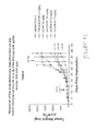
FIG. 47 is a pair of line graphs demonstrating the response of Detroit 562 human pharyngeal carcinoma cells, subcutaneously implanted into athymic nude mice, to murine mAb 3G9 (top) and to humanized mAb 3G9 (BG00011) (bottom).
FIG. 48 is a scatter plot demonstrating the response of Detroit 562 human pharyngeal carcinoma cells, subcutaneously implanted into athymic nude mice, to murine mAb 3G9 (top) and to humanized mAb 3G9 (BG00011) (bottom).
FIG. 49 is a line graph demonstrating the response of Detroit 562 human pharyngeal carcinoma cells, subcutaneously implanted into athymic nude mice, to murine mAb 3G9 and to humanized mAb 3G9 (BG00011), expressing the results of test animals as a percentage of control (vehicle as negative; TGF-β RII-Fc as positive) results.
FIG. 50 is a line graph demonstrating the response of Detroit 562 human pharyngeal carcinoma cells, subcutaneously implanted into athymic nude mice, to wildtype murine mAb 3G9.
FIG. 51 is a line graph demonstrating the response of Detroit 562 human pharyngeal carcinoma cells, subcutaneously implanted into athymic nude mice, to aglycosyl murine mAb 3G9.
FIG. 52 is a scatter plot demonstrating the response of Detroit 562 human pharyngeal carcinoma cells, subcutaneously implanted into athymic nude mice, to wildtype murine mAb 3G9 and to algycosyl murine mAb 3G9.
FIG. 53 is a line graph demonstrating the response of Detroit 562 human pharyngeal carcinoma cells, subcutaneously implanted into athymic nude mice, to wildtype murine mAb 3G9 and to algycosyl murine mAb 3G9, expressing the results of test animals as a percentage of control (vehicle) results.
FIG. 54 is a Table summarizing the results of FIGS. 43-52.
DETAILED DESCRIPTION OF THE INVENTION
Unless defined otherwise, all technical and scientific terms used herein have the same meanings as commonly understood by one of ordinary skill in the art to which this invention belongs. Although any methods and materials similar or equivalent to those described herein can be used in the practice or testing of the present invention, the preferred methods and materials are described hereinafter.
Definitions
About: As used herein when referring to any numerical value, the term “about” means a value of ±10% of the stated value (e.g., “about 50° C.” encompasses a range of temperatures from 45° C. to 55° C., inclusive; similarly, “about 100 mM” encompasses a range of concentrations from 90 mM to 110 mM, inclusive).
Antagonist: As used herein, the term “antagonist” refers to a compound, molecule, moiety or complex that reduces, substantially reduces or completely inhibits the biological and/or physiological effects of the ανβ6 integrin in a cell, tissue or organism. Antagonists, which may be ligands for ανβ6, may carry out such effects in a variety of ways, including but not limited to competing with another ligand for binding to ανβ6 on the cell surface; interacting with ανβ6 in such a way as to reduce, substantially reduce or inhibit the ability of the integrin to bind other ligands; binding to and inducing a conformational change in cell surface ανβ6 such that the integrin assumes a structure to which other ligands can no longer bind (or can bind only with reduced or substantially reduced affinity and/or efficiency); inducing a physiological change (e.g., increase in intracellular signaling complexes; increase in transcriptional inhibitors; reduction in cell surface ανβ6 expression; etc.) in cells, tissues or organisms such that the binding of other ligands, or the physiological signal induced by such ligands upon binding to the ανβ6 on the cell, is reduced, substantially reduced or completely inhibited; and other mechanisms by which antagonists may carry out their activities, that will be familiar to the ordinarily skilled artisan. As the ordinarily skilled artisan will understand, an antagonist may have a similar structure to another ανβ6-binding moiety (e.g., an ανβ6-binding ligand) that it antagonizes (e.g., the antagonist may be a mutein, variant, fragment or derivative of the agonist), or may have a wholly unrelated structure.
Bound: As used herein, the term “bound” refers to binding or attachment that may be covalent, e.g., by chemically coupling, or non-covalent, e.g., ionic interactions, hydrophobic interactions, hydrogen bonds, etc. Covalent bonds can be, for example, ester, ether, phosphoester, thioester, thioether, urethane, amide, amine, peptide, imide, hydrazone, hydrazide, carbon-sulfur bonds, carbon-phosphorus bonds, and the like. The term “bound” is broader than and includes terms such as “coupled,” “conjugated” and “attached.”
Conjugate/conjugation: As used herein, “conjugate” refers to the product of covalent attachment of a moiety, e.g., a chemical or radioisotope, to a ligand that binds to ανβ6, e.g., an ανβ6-binding antibody or fragment thereof. “Conjugation” refers to the formation of a conjugate as defined in the previous sentence. Any method normally used by those skilled in the art of conjugation of chemicals or radioisotopes to biologically active materials, such as proteins or polypeptides (including antibodies) can be used in the present invention.
Disease, disorder, condition: As used herein, the terms “disease” or “disorder” refer to any adverse condition of a human or animal including tumors, cancer, allergies, addiction, autoimmunity, infection, poisoning or impairment of optimal mental or bodily function. “Conditions” as used herein includes diseases and disorders but also refers to physiologic states. For example, fertility is a physiologic state but not a disease or disorder. Compositions of the invention suitable for preventing pregnancy by decreasing fertility would therefore be described as a treatment of a condition (fertility), but not a treatment of a disorder or disease. Other conditions are understood by those of ordinary skill in the art.
Effective Amount: As used herein, the term “effective amount” refers to an amount of a given compound, conjugate or composition that is necessary or sufficient to realize a desired biologic effect. An effective amount of a given compound, conjugate or composition in accordance with the methods of the present invention would be the amount that achieves this selected result, and such an amount can be determined as a matter of routine by a person skilled in the art, using assays that are known in the art and/or that are described herein, without the need for undue experimentation. For example, an effective amount for treating or preventing cancer metastasis could be that amount necessary to prevent migration and invasion of a tumor cell across the basement membrane or across an endothelial layer in vivo. The term is also synonymous with “sufficient amount.” The effective amount for any particular application can vary depending on such factors as the disease, disorder or condition being treated, the particular composition being administered, the route of administration, the size of the subject, and/or the severity of the disease or condition. One of ordinary skill in the art can determine empirically the effective amount of a particular compound, conjugate or composition of the present invention, in accordance with the guidance provided herein, without necessitating undue experimentation.
One, a, or an: When the terms “one,” “a,” or “an” are used in this disclosure, they mean “at least one” or “one or more,” unless otherwise indicated. An such, the terms “a” (or “an”), “one or more,” and “at least one” can be used interchangeably herein.
Peptide, polypeptide, protein: As used herein, the term “polypeptide” is intended to encompass a singular “polypeptide” as well as plural “polypeptides,” and refers to a molecule composed of monomers (amino acids) linearly linked by amide bonds (also known as peptide bonds). The term “polypeptide” refers to any chain or chains of two or more amino acids, and does not refer to a specific length of the product. Thus, peptides, dipeptides, tripeptides, oligopeptides, “protein,” “amino acid chain,” or any other term used to refer to a chain or chains of two or more amino acids, are included within the definition of “polypeptide,” and the term “polypeptide” may be used instead of, or interchangeably with any of these terms. The term “polypeptide” is also intended to refer to the products of post-expression modifications of the polypeptide, including without limitation glycosylation, acetylation, phosphorylation, amidation, derivatization by known protecting/blocking groups, proteolytic cleavage, or modification by non-naturally occurring amino acids. A polypeptide may be derived from a natural biological source or produced by recombinant technology, but is not necessarily translated from a designated nucleic acid sequence. It may be generated in any manner, including by chemical synthesis. In accordance with this definition, polypeptides used in the present invention may be of a size of about 3 or more, 5 or more, 10 or more, 20 or more, 25 or more, 50 or more, 75 or more, 100 or more, 200 or more, 500 or more, 1,000 or more, or 2,000 or more amino acids. Polypeptides may have a defined three-dimensional structure, although they do not necessarily have such structure. Polypeptides with a defined three-dimensional structure are referred to as folded, and polypeptides which do not possess a defined three-dimensional structure, but rather can adopt a large number of different conformations, and are referred to at unfolded. As used herein, the term glycoprotein refers to a protein coupled to at least one carbohydrate moiety that is attached to the protein via an oxygen-containing or a nitrogen-containing side chain of an amino acid residue, e.g., a serine residue or an asparagine residue. Preferred polypeptides used in accordance with the invention include polypeptides that are ligands or that bind to an ανβ6 integrin on the surface of a cell, including but not limited to antibodies (especially monoclonal antibodies) that recognize and bind to one or more epitopes on ανβ6.
By an “isolated” polypeptide or a fragment, variant, or derivative thereof is intended a polypeptide that is not in its natural milieu. No particular level of purification is required. For example, an isolated polypeptide can be removed from its native or natural environment. Recombinantly produced polypeptides and proteins expressed in host cells are considered isolated for purposed of the invention, as are native or recombinant polypeptides which have been separated, fractionated, or partially or substantially purified by any suitable technique.
Also included as polypeptides of the present invention are fragments, derivatives, analogs, or variants of the foregoing polypeptides, and any combination thereof. The terms “fragment,” “variant,” “derivative” and “analog” when referring to anti-ανβ6 antibodies or antibody polypeptides include any polypeptides which retain at least some of the antigen-binding properties of the corresponding native antibody or polypeptide, i.e., those polypeptides that retain the ability to bind to one or more epitopes on an ανβ6 integrin. Fragments of polypeptides of the present invention include proteolytic fragments, as well as deletion fragments, in addition to specific antibody fragments discussed elsewhere herein. Variants of anti-ανβ6 antibodies and antibody polypeptides useful in accordance with the present invention include fragments as described above, and also polypeptides with altered amino acid sequences due to amino acid substitutions, deletions, or insertions. Variants may occur naturally or be non-naturally occurring Non-naturally occurring variants may be produced using art-known mutagenesis techniques. Variant polypeptides may comprise conservative or non-conservative amino acid substitutions, deletions or additions. Derivatives of anti-ανβ6 antibodies and antibody polypeptides useful in accordance with the present invention are polypeptides which have been altered so as to exhibit additional features not found on the native polypeptide. Examples include fusion proteins. Variant polypeptides may also be referred to herein as “polypeptide analogs.” As used herein a “derivative” of an anti-ανβ6 antibody or antibody polypeptide refers to a subject polypeptide having one or more residues chemically derivatized by reaction of a functional side group. Also included as “derivatives” are those peptides which contain one or more naturally occurring amino acid derivatives of the twenty standard amino acids. For example, 4-hydroxyproline may be substituted for proline; 5-hydroxylysine may be substituted for lysine; 3-methylhistidine may be substituted for histidine; homoserine may be substituted for serine; and ornithine may be substituted for lysine.
smad4: As used herein, the tumor suppressor gene smad4 is synonymous with other designations for the same tumor suppressor gene that are known to those of skill in the art, including but not limited to madh4 and dpc4. As is conventional, the products of the expression of this gene is designated herein as SMAD4, which is synonymous with the corresponding other designations for the expression product of this gene that are known to those of skill in the art, including but not limited to MADH4 and DPC4.
Substantially, substantial: As used herein, conjugation of a protein is said not to interfere “substantially” with the ability of the protein to bind to its receptor(s) if the rate and/or amount of binding of a conjugated protein to a receptor is not less than about 40%, about 50%, about 60%, about 65%, about 70%, about 75%, about 80%, about 85%, about 90%, about 91%, about 92%, about 93%, about 94%, about 95%, about 96%, about 97%, about 98%, about 99% or about 100% or more, of the binding rate and/or amount of the corresponding cytokine, chemokine, growth factor or polypeptide hormone that has not been conjugated.
Treatment: As used herein, the terms “treatment,” “treat,” “treated” or “treating” refer to prophylaxis and/or therapy, particularly wherein the object is to prevent or slow down (lessen) an undesired physiological change or disorder, such as the progression of multiple sclerosis. Beneficial or desired clinical results include, but are not limited to, alleviation of symptoms, diminishment of extent of disease, stabilized (i.e., not worsening) state of disease, delay or slowing of disease progression, amelioration or palliation of the disease state, and remission (whether partial or total), whether detectable or undetectable. “Treatment” can also mean prolonging survival as compared to expected survival if not receiving treatment. Those in need of treatment include those already with the condition or disorder as well as those prone to have the condition or disorder or those in which the condition or disorder is to be prevented. By “subject” or “individual” or “animal” or “patient” or “mammal,” is meant any subject, particularly a mammalian subject, for whom diagnosis, prognosis, or therapy is desired. Mammalian subjects include humans and other primates, domestic animals, farm animals, and zoo, sports, or pet animals such as dogs, cats, guinea pigs, rabbits, rats, mice, horses, cattle, cows, and the like.
Overview
The present invention is based at least in part upon the findings that there is an inverse relationship between the level of expression of the tumor suppressor gene smad4 and the integrin ανβ6 in certain tumor cells, and that the levels of expression of these two markers can be used to determine or predict the susceptibility to treatment with anti-integrin ligands, and with agents that antagonize the TGF-β signalling pathway, of such tumor cells. Specifically, it has been found that tumor cells that display decreased levels of expression of smad4 concomitantly express increased amounts of the integrin ανβ6; therefore, smad-4-deficient tumor cells have a higher propensity to be responsive to ligands that bind to integrin ανβ6, compared to tumor cells that display higher expression levels of smad4 (and, concomitantly, lower levels of surface integrin ανβ6). In related embodiments, it has also been found by the present inventors that smad-4-deficient tumor cells are also more likely to be responsive to ligands that antagonize one or more components of the TGF-β signalling pathway, compared to tumor cells that display higher expression levels of smad4. As used herein, a tumor cell that is “responsive” to a ligand that binds to integrin ανβ6, or to a ligand that antagonizes one or more components of the TGF-β pathway, refers to a tumor cell in which the growth, division, and/or other metabolic pathways are adversely affected such that the cell is inhibited from growing or dividing and/or undergoes apoptosis or other forms of cell death.
In other embodiments, the invention also provides methods using identification of this differential expression in determining the invasive and/or metastatic potential of tumor cells and in identifying those carcinomas, such as certain adeno-carcinomas (including pancreatic carcinomas), that may be more likely to rapidly progress and which should be aggressively treated in a patient. The invention also provides methods of identifying those tumors in which the cells making up the tumor may be more likely to respond to treatment with one or more ligands that bind to integrin ανβ6. The invention also provides methods of diagnosis and treatment/prevention of tumor metastasis.
Determination of Expression of smad4 and Integrin ανβ6
In one embodiment, the present invention is directed to methods for identifying tumors and tumor cells, particularly carcinoma cells such as pancreatic carcinoma cells, that are more likely to be responsive to ligands that bind to integrin αvβ6, and/or to ligands that antagonize one or more components of the TGF-β signaling pathway. Such methods include, for example, determining the levels of expression of the tumor suppressor gene smad4 by the tumor cells, wherein a reduced expression of smad4 indicates that the tumor cell is more likely to be responsive to ligands that bind to integrin αvβ6, and/or to ligands that antagonize one or more components of the TGF-β signalling pathway. In certain such embodiments, the level of expression of smad4 in cells of a tumor, e.g., a carcinoma, is determined in tissue sections obtained from a patient suffering from such a tumor, wherein a decrease in the expression of smad4 in the tumor cells relative to that in non-tumor tissue samples (ideally, from the same organ in the same patient) indicates that the tumor is more likely to be responsive to ligands that bind to integrin αvβ6, and/or to ligands that antagonize one or more components of the TGF-β signalling pathway. In this way, appropriate and aggressive protocols for treating such tumors in a patient can rapidly be identified and implemented, thereby providing an increased likelihood of positive treatment outcomes for cancer patients.
In each such embodiment, the invention relies upon identification or exploitation of the decreased expression of smad4, and the concomitant increased expression of ανβ6, in certain tumor cells. The level of expression of smad4 can be readily determined using methods that are well-known in the art for measuring gene expression, including, for example, hybridization or PCR/RT-PCR using known genetic probes or primers specific for the smad4 gene (see, e.g., Maliekal, T. T. et al., Oncogene 22:4889-4897 (2003); Iacobuzio-Donahue, C. A. et al., Clin. Cancer Res. 10:1597-1604 (2004)), northern blotting (see, e.g., Yasutome, M. et al., Clin. Exper. Metast. 22:461-473 (2005)), and immunohistochemical analysis with anti-SMAD4 antibodies (see, e.g., Lüttges, J. et al., Am. J. Pathol. 158:1677-1683 (2001); Fukuchi, M. et al., Cancer 95:737-743 (2002); Subramanian, G. et al., Cancer Res. 64:5200-5211 (2004); Levy, L. and Hill, C. S., Mol. Cell. Biol. 25:8108-8125 (2005); Toga, T. et al., Anticancer Res. 24:1173-1178 (2004)). Other methods suitable for detecting the level of smad4 gene expression in a given cell, tissue, organ or biological sample will be familiar to those of ordinary skill in the art. Similarly, the level of expression of ανβ6 can be readily determined using methods that are well-known in the art for measuring integrin expression. In certain such embodiments, such determinations are accomplished by contacting the tissue, tumor or tumor cells with one or more ligands that binds to integrin ανβ6 in the tissue, tumor or tumor cells.
In certain such embodiments, the tissue, tumor or tumor cells are carcinoma tissues, tumors or tumor cells, including those from carcinomas such as adenocarcinomas. In more particular embodiments, the carcinoma is pancreatic carcinoma, a colorectal carcinoma, a cervical carcinoma, a squamous cell carcinoma (such as an esophageal carcinoma), a head and neck carcinoma, a liver carcinoma, an ovarian carcinoma and a lung carcinoma. More particularly, the carcinoma is a pancreatic carcinoma, a colorectal carcinoma, a cervical carcinoma, or a head and neck carcinoma.
In certain embodiments of the invention, the ligands that bind to ανβ6 are antagonists of ανβ6. Such antagonists include but are not limited to antibodies which specifically bind to ανβ6; antibodies which specifically bind to β6; antibodies that bind to αν; antibodies that bind to ligands for ανβ6; ligands for ανβ6; antisense nucleic acids; and peptide, non-peptide, and peptidomimetic analogs of such ligands.
In certain such embodiments of the present invention, the ligand that binds to integrin ανβ6 is an antibody that binds to integrin ανβ6, or integrin ανβ6-binding fragments, variants, or derivatives thereof. Such antibodies may bind to one subunit of the integrin (e.g., antibodies that bind to an epitope located on the αν subunit or to an epitope that is located on the β6 subunit), or to both subunits (e.g., antibodies that bind to an epitope that is located in a region of the integrin heterodimer that bridges both the αν and β6 subunits). Unless specifically referring to full-sized antibodies such as naturally occurring antibodies, the term “ανβ6 antibodies” encompasses full-sized antibodies as well as ανβ6-binding fragments, variants, analogs, or derivatives of such antibodies, e.g., naturally occurring antibody or immunoglobulin molecules or engineered antibody molecules or fragments that bind antigen in a manner similar to antibody molecules. Antibodies can be synthetic, monoclonal, or polyclonal and can be made by techniques well known in the art. For therapeutic applications, “human” monoclonal antibodies having human constant and variable regions are often preferred so as to minimize the immune response of a patient against the antibody. Such antibodies can be generated by immunizing transgenic animals which contain human immunoglobulin genes (see, e.g., Jakobovits et al., Ann. N.Y. Acad. Sci. 764:525-535 (1995)). In connection with synthetic and semi-synthetic antibodies, such terms are intended to cover but are not limited to antibody fragments, isotype switched antibodies, humanized antibodies (e.g., mouse-human, human-mouse, and the like), hybrids, antibodies having plural specificities, fully synthetic antibody-like molecules, and the like.
The terms “antibody” and “immunoglobulin” are used interchangeably herein. An antibody or immunoglobulin comprises at least the variable domain of a heavy chain, and normally comprises at least the variable domains of a heavy chain and a light chain. Basic immunoglobulin structures in vertebrate systems are relatively well understood. See, e.g., Harlow et al., Antibodies: A Laboratory Manual, (Cold Spring Harbor Laboratory Press, 2nd ed. 1988). As will be understood by those of ordinary skill, the terms “antibody” and “immunoglobulin” comprise various broad classes of polypeptides that can be distinguished biochemically. Those skilled in the art will appreciate that heavy chains are classified as gamma, mu, alpha, delta, or epsilon, (γ, μ, α, δ, ε) with some subclasses among them (e.g., γ1-γ4). It is the nature of this chain that determines the “class” of the antibody as IgG, IgM, IgA IgG, or IgE, respectively. The immunoglobulin subclasses (isotypes) e.g., IgG1, IgG2, IgG3, IgG4, IgA1, etc. are well characterized and are known to confer functional specialization. Modified versions of each of these classes and isotypes are readily discernable to the skilled artisan in view of the instant disclosure and, accordingly, are within the scope of the instant invention.
Antibodies that bind to ανβ6 , or ανβ6-binding fragments, variants, or derivatives thereof, that are suitable for use in the present invention include but are not limited to polyclonal, monoclonal, multispecific, human, humanized, primatized, or chimeric antibodies, single chain antibodies, epitope-binding fragments, e.g., Fab, Fab′ and F(ab′)2, Fd, Fvs, single-chain Fvs (scFv), single-chain antibodies, disulfide-linked Fvs (sdFv), fragments comprising either a VL or VH domain, fragments produced by a Fab expression library, and anti-idiotypic (anti-Id) antibodies (including, e.g., anti-Id antibodies to anti-ανβ6 antibodies disclosed herein). ScFv molecules are known in the art and are described, e.g., in U.S. Pat. No. 5,892,019. Immunoglobulin or antibody molecules of the invention can be of any type (e.g., IgG, IgE, IgM, IgD, IgA, and IgY), class (e.g., IgG1, IgG2, IgG3, IgG4, IgA1 and IgA2) or subclass of immunoglobulin molecule.
Antibody fragments, including single-chain antibodies, may comprise the variable region(s) alone or in combination with the entirety or a portion of the following: hinge region, C H1, C H2, and C H3 domains. Also included in the invention are antigen-binding fragments also comprising any combination of variable region(s) with a hinge region, C H1, C H2, and C H3 domains. Antibodies or immunospecific fragments thereof for use in the diagnostic and therapeutic methods disclosed herein may be from any animal origin including birds and mammals. Preferably, the antibodies are human, murine, rat, donkey, rabbit, goat, guinea pig, camel, llama, horse, bovine or chicken antibodies. Most preferably, the antibodies are human, humanized or primatized antibodies, or chimeric antibodies, particularly monoclonal antibodies. As used herein, “human” antibodies include antibodies having the amino acid sequence of a human immunoglobulin and include antibodies isolated from human immunoglobulin libraries or from animals transgenic for one or more human immunoglobulins and that do not express endogenous immunoglobulins, as described infra and, for example in, U.S. Pat. No. 5,939,598 by Kucherlapati et al. As used herein, the term “chimeric antibody” will be held to mean any antibody wherein the immunoreactive region or site is obtained or derived from a first species and the constant region (which may be intact, partial or modified in accordance with the instant invention) is obtained from a second species. In preferred embodiments the target binding region or site will be from a non-human source (e.g. mouse or primate) and the constant region is human.
Particularly preferred antibodies for use in accordance with the present invention are anti-ανβ6 monoclonal antibodies such as those disclosed in Weinreb et al., J. Biol. Chem. 279(17):17875-17877 (2004) (the disclosure of which is incorporated herein by reference in its entirety), including monoclonal antibodies 6.8G6 (“8G6”) and 6.3G9 (“3G9”) disclosed therein. Additional antibodies that bind to ανβ6 and that therefore are suitable for use in accordance with the present invention include antibodies (or fragments, variants or derivatives thereof) that bind to the β6 subunit of integrin ανβ6 (and that are therefore considered “anti-β6 antibodies”), such as those disclosed in Weinacker et al., J. Cell Biol. 269:1-9 (1994), which is incorporated herein by reference in its entirety; and in U.S. Pat. No. 6,692,741 B2, which is incorporated herein by reference in its entirety, particularly at columns 2-3 and 7-8 thereof, including the monoclonal antibody designated 10D5 (ATCC deposit no. HB12382, deposited Aug. 6, 1997, American Type Culture Collection, P.O. Box 1549, Manassas, Va. 20108) (see U.S. Pat. No. 6,692,741 at col. 3, lines 7-13, and at cols. 7-8) and CSβ6 (see U.S. Pat. No. 6,692,741 at cols. 7-8). Suitable embodiments according to this aspect of the invention use ανβ6 integrin-binding ligands which are ανβ6-binding antibodies or ανβ6 epitope-binding fragments thereof. Additional antibodies suitable for use in accordance with this aspect of the invention include, but are not limited to, the ανβ6-binding monoclonal antibodies disclosed in U.S. patent application publication no. US 2005/0255102 A1, the disclosure of which is incorporated herein in its entirety, including those designated therein as 3G9, 8G6, 1A8, 2B1, 2B10, 2A1, 2E5, 1G10, 7G5, 1C5, as well as fragments, chimeras and hybrids thereof. Particularly suitable antibodies for use in accordance with the present invention are monoclonal antibodies 2B1, 3G9 and 8G6.
In some embodiments, the antibodies comprise the same heavy and light chain polypeptide sequences as an antibody produced by hybridoma 6.1A8, 6.3G9, 6.8G6, 6.2B1, 6.2B10, 6.2A1, 6.2E5, 7.1G10, 7.7G5, or 7.1C5. Particularly suitable antibodies for use in accordance with the present invention are monoclonal antibodies that comprise the same heavy and light chain polypeptide sequences as 2B1 antibodies produced by hybridoma 6.2B1 (ATCC deposit no. PTA-3646, deposited Aug. 16, 2001, American Type Culture Collection, P.O. Box 1549, Manassas, Va. 20108), 8G6 antibodies produced by hybridoma 6.8G6 (ATCC deposit no. PTA-3645, deposited Aug. 16, 2001, American Type Culture Collection, P.O. Box 1549, Manassas, Va. 20108) and 3G9 antibodies produced by hybridoma 6.3G9 (ATCC deposit no. PTA-3649, deposited Aug. 16, 2001, American Type Culture Collection, P.O. Box 1549, Manassas, Va. 20108) (see published U.S. Appl. No. U.S. 2005/0255102 A1, the disclosure of which is incorporated herein by reference in its entirety, particularly at page 1, paragraph 0008; at page 2, paragraphs 0032 and 0036; and in the Examples at pages 6-14), and the antibody designated as 10D5 (the hybridoma secreting which antibody was deposited on Aug. 6, 1997, as ATCC deposit no. HB12382, American Type Culture Collection, P.O. Box 1549, Manassas, Va. 20108) (see U.S. Pat. No. 6,692,741, the disclosure of which is incorporated herein by reference in its entirety, particularly at col. 3, lines 7-13, and at cols. 7-8).
In some embodiments, the antibodies comprise a heavy chain whose complementarity determining regions (CDR) 1, 2 and 3 consist essentially (i.e., with the exception of some conservative variations) of the sequences shown in Table 1 below. In certain such embodiments, the antibodies comprise a heavy chain whose CDR1 consists essentially of any one of SEQ ID NOs:1-5; whose CDR2 consists essentially of any one of SEQ ID NOs: 6-11; and whose CDR3 consists essentially of any one of SEQ ID NOs:12-17; and/or a light chain whose CDRs 1, 2 and 3 consist essentially of any one of the sequences of SEQ ID NOs:18-23, 24-27, and 28-33, respectively.
| TABLE 1 |
| |
| Antibody |
Amino Acid Sequence |
SEQ ID NO: |
| |
| |
| Heavy Chain CDR1 Sequences |
| |
1 |
| 1A8 | SYTFTDYTMH | |
2 |
| 2B1 | GFTFSRYVMS | |
3 |
| 3G9 | GFTFSRYVMS | |
3 |
| 2A1 | GYDFNNDLIE | |
4 |
| 2G2 | GYAFTNYLIE | |
5 |
| Heavy Chain CDR2 Sequences |
| |
6 |
| 1A8 |
VIDTYYGKTNYNQKFEG |
7 |
| 2B1 |
SISSG-GSTYYPDSVKG |
8 |
| 3G9 |
SISSG-GRMYYPDTVKG |
9 |
| 2A1 | VINPGSGRTNYNEKFKG | |
10 |
| 2G2 |
VISPGSGIINYNEKFKG |
11 |
| Heavy Chain CDR3 Sequences |
| 8G6 |
GGLRRGDRPSLRYAMDY |
12 |
| 1A8 | GGFRRGDRPSLRYAMDS | |
13 |
| 2B1 |
GAIYDG-----YYVFAY |
14 |
| 3G9 |
GSIYDG-----YYVFPY |
15 |
| 2A1 |
IYYGPH-----SYAMDY |
16 |
| 2G2 |
ID-YSG-----PYAVDD |
17 |
| Light Chain CDR1 Sequences |
| 8G6 |
RASQSVSTSS-YSYMY |
18 |
| 1A8 |
RASQSVSIST-YSYIH |
19 |
| 2B1 |
SASSSVSSS----YLY |
20 |
| 3G9 |
SANSSVSSS----YLY |
21 |
| 2A1 |
KASLDVRTAVA |
22 |
| 2G2 | KASQAVNTAVA | |
23 |
| Light Chain CDR2 Sequences |
| 8G6 |
YASNLES |
24 |
| 1A8 |
YASNLES |
24 |
| 2B1 | STSNLAS | |
25 |
| 3G9 | STSNLAS | |
25 |
| 2A1 | SASYRYT | |
26 |
| 2G2 | SASYQYT | |
27 |
| Light Chain CDR3 Sequences |
| 8G6 |
QHNWEIPFT |
28 |
| 1A8 | QHSWEIPYT | |
29 |
| 2B1 | HQWSSYPPT | |
30 |
| 3G9 |
HQWSTYPPT |
31 |
| 2A1 | QQHYGTPWT | |
32 |
| 2G2 |
QHHYGVPWT |
33 |
| |
In other related embodiments, the monoclonal antibodies used in accordance with the present invention are chimeric antibodies, i.e., those in which a cognate antibody from one species (e.g., murine, rat or rabbit) is altered by recombinant DNA technology such that part or all of the hinge and/or constant regions of the heavy and/or light chains are replaced with the corresponding components of an antibody from another species (e.g., human). Generally, the variable domains of the engineered antibody remain identical or substantially so to the variable domains of the cognate antibody. Such an engineered antibody is called a chimeric antibody and is less antigenic than the cognate antibody when administered to an individual of the species from which the hinge and/or constant region is derived (e.g., a human). Methods of making chimeric antibodies are well known in the art.
In other related embodiments, the monoclonal antibodies used in accordance with the present invention are fully human antibodies. Methods for producing such fully human monoclonal antibodies are well known in the art (see, e.g., U.S. 2005/0255102 A1 at page 4, paragraphs 0069-0070, which are incorporated herein by reference).
In other related embodiments, the monoclonal antibodies used in accordance with the present invention are humanized versions of cognate anti-α84 β6 antibodies derived from other species. A humanized antibody is an antibody produced by recombinant DNA technology, in which some or all of the amino acids of a human immunoglobulin light or heavy chain that are not required for antigen binding (e.g., the constant regions and the framework regions of the variable domains) are used to substitute for the corresponding amino acids from the light or heavy chain of the cognate, nonhuman antibody. By way of example, a humanized version of a murine antibody to a given antigen has, on both of its heavy and light chain: (a) constant regions of a human antibody, (b) framework regions from the variable domains of a human antibody; and (c) CDRs from the murine antibody. When necessary, one or more residues in the human framework regions can be changed to residues at the corresponding positions in the murine antibody so as to preserve the binding affinity of the humanized antibody to the antigen. This change is sometimes called “back mutation.” Humanized antibodies generally are less likely to elicit an immune response in humans as compared to chimeric human antibodies because the former contain considerably fewer non-human components. Methods for producing such humanized monoclonal antibodies are well known in the art (see, e.g., U.S. 2005/0255102 A1 at pages 4-5, paragraphs 0072-0077, which are incorporated herein by reference).
In additional such embodiments, the humanized antibodies comprise one or more CDRs in the heavy and/or light chain that are derived from the corresponding CDRs in the heavy and/or light chain of a different antibody. One suitable non-limiting example of such an antibody is a humanized 3G9 antibody comprising a light chain CDR1 that has the sequence of the light chain CDR1 derived from the 2B1 antibody (SEQ ID NO:20) instead of the sequence of the light chain CDR1 for the deposited 3G9 antibody (SEQ ID NO:21). Such a humanized 3G9 antibody having a light chain CDR1 sequence set forth in SEQ ID NO:20 is designated herein as hu3G9 (or BG00011). Another suitable non-limiting example of such an antibody is a humanized 8G6 antibody comprising a light chain CDR1 that has the sequence of the light chain CDR1 derived from the 2B1 antibody (SEQ ID NO:20) instead of the sequence of the light chain CDR1 for the deposited 8G6 antibody (SEQ ID NO: 18) Such a humanized 8G6 antibody having a light chain CDR1 sequence set forth in SEQ ID NO:20 is designated herein as hu8G9. Additional examples of such derivative antibodies, in which one or more heavy chain and/or light chain CDRs has been replaced with one or more corresponding heavy chain and/or light chain CDRs from another antibody, and which are suitable for use in accordance with the present invention, will be readily apparent to those of ordinary skill in view of the sequences depicted in Table 1 and the guidance provided herein. Suitable methods for preparing such humanized antibodies, including such derivative humanized antibodies, are familiar to those of ordinary skill and are set forth, for example, in U.S. published application no. 2005/0255102 A1, the disclosure of which is incorporated herein by reference in its entirety.
In certain embodiments, the ligands, e.g., the antibodies, that bind to ανβ6 can be used in unconjugated form. In such embodiments, the tumor cells are contacted with the ligand binding to ανβ6, under conditions favoring binding of the ligand to ανβ6 on the cell surface. Suitable such conditions are well-known in the art, and are described in detail in the Examples hereinbelow. In certain such embodiments, the binding of the ligand sensitizes the tumor cell to which the ligand has bound to the action of one or more toxic compounds, i.e., the binding of the ligand to αvβ6 on the tumor cell surface induces the tumor cell to become more sensitive to the growth-inhibiting and/or death-inducing actions of one or more toxins Thus, in additional embodiments, the invention provides methods of inhibiting the growth of a tumor cell, or of killing a tumor cell, comprising (a) contacting the tumor cell with one or more ligands (e.g., one or more antibodies or fragments thereof that bind to ανβ6 on the cell surface, under conditions favoring the binding of the ligand ανβ6 on the cell surface; and then (b) contacting the tumor cell with one or more toxic compounds that inhibits the growth of and/or kills the tumor cell. Suitable toxic compounds for use in accordance with this aspect of the invention, and appropriate dosages and administration regimens therefor, are known in the art and are described in detail elsewhere herein.
In related embodiments, the invention provides methods of sensitizing tumor cells to one or more toxic compounds by treating the tumor cells with one or more agents that modulates (e.g., inhibits or activates) one or more components of the TGF-β signaling pathway. Suitable such agents include, for example, TGF-β or a fragment thereof, a soluble TGF-β receptor or a fragment thereof, or a fusion protein comprising a soluble TGF-β receptor or a fragment thereof (e.g., a TGF-β type II receptor fragment fused to the Fc region of an antibody, to produce a soluble “TGF-β RII-Fc” polypeptide). In use, the binding or internalization of the TGF-β-modulating agent sensitizes the tumor cell by which the agent has been bound or internalized to the action of one or more toxic compounds, i.e., induces the tumor cell to become more sensitive to the growth-inhibiting and/or death-inducing actions of one or more toxins. Thus, in additional embodiments, the invention provides methods of inhibiting the growth of a tumor cell, or of killing a tumor cell, comprising (a) contacting the tumor cell with one or more agents that modulates (e.g., inhibits or activates) one or more components of the TGF-β signaling pathway, under conditions favoring the binding or internalization of the agent by the tumor cell; and then (b) contacting the tumor cell with one or more toxic compounds that inhibits the growth of and/or kills the tumor cell. Suitable toxic compounds for use in accordance with this aspect of the invention, and appropriate dosages and administration regimens therefor, are known in the art and are described in detail elsewhere herein.
Conjugates an Other Modifications of ανβ6-Binding Ligands
As noted above, in certain embodiments, the ligands, e.g., the antibodies, that bind to ανβ6 can be used in unconjugated form. In other embodiments, the ligands, e.g., the antibodies, that bind to ανβ6 can be conjugated, e.g., to a detectable label, a drug, a prodrug or an isotope.
In certain methods of the invention described in more detail below, such as methods of detecting ανβ6 expression in cells or tissues in conjunction with detection of smad4 expression as a measure of the potential of tumor cells to be responsive to ανβ6-binding ligands and/or TGF-β-blocking agents, the ανβ6-binding ligands (e.g., antibodies) are conjugated to one or more detectable labels. For such uses, the ανβ6-binding ligands, e.g., ανβ6-binding antibodies, may be detectably labeled by covalent or non-covalent attachment of a chromogenic, enzymatic, radioisotopic, isotopic, fluorescent, toxic, chemiluminescent, nuclear magnetic resonance contrast agent or other label.
Examples of suitable chromogenic labels include diaminobenzidine and 4-hydroxyazo-benzene-2-carboxylic acid.
Examples of suitable enzyme labels include malate dehydrogenase, staphylococcal nuclease, Δ-5-steroid isomerase, yeast-alcohol dehydrogenase, α-glycerol phosphate dehydrogenase, triose phosphate isomerase, peroxidase, alkaline phosphatase, asparaginase, glucose oxidase, β-galactosidasce, ribonuclease, urease, catalase, glucose-6-phosphate dehydrogenase, glucoamylase, and acetylcholine esterase.
Examples of suitable radioisotopic labels include 3H, 111In, 125I, 131I, 32P, 35S, 14C, 51Cr, 57To, 58Co, 59Fe, 75Se, 152Eu, 90Y, 67Cu, 217Ci, 211At, 212Pb, 47Sc, 109Pd, etc. 111In is a preferred isotope where in vivo imaging is used since its avoids the problem of dehalogenation of the 125I or 131I-labeled ανβ6-binding ligands by the liver. In addition, this radionucleotide has a more favorable gamma emission energy for imaging (Perkins et al., Eur. J. Nucl. Med. 10:296-301 (1985); Carasquillo et al., J. Nucl. Med. 28:281-287 (1987)). For example, 111In coupled to monoclonal antibodies with 1-(P-isothiocyanatobenzyl)-DPTA has shown little uptake in non-tumorous tissues, particularly the liver, and therefore enhances specificity of tumor localization (Esteban et al., J. Nucl. Med. 28:861-870 (1987)).
Examples of suitable non-radioactive isotopic labels include 157Gd, 55Mn, 162Dy, 52Tr, 56Fe.
Examples of suitable fluorescent labels include an 152Eu label, a fluorescein label, an isothiocyanate label, a rhodamine label, a phycoerythrin label, a phycocyanin label, an allophycocyanin label, a Green Fluorescent Protein (GFP) label, an o-phthaldehyde label, and a fluorescamine label. Examples of suitable toxin labels include diphtheria toxin, ricin, and cholera toxin.
Examples of chemiluminescent labels include a luminol label, an isoluminol label, an aromatic acridinium ester label, an imidazole label, an acridinium salt label, an oxalate ester label, a luciferin label, a luciferase label, and an aequorin label.
Examples of nuclear magnetic resonance contrasting agents include heavy metal nuclei such as Gd, Mn, and iron.
Typical techniques for binding the above-described labels to ανβ6-binding ligands, e.g., ανβ6-binding antibodies, are provided by Kennedy et al., Clin. Chim. Acta 70:1-31 (1976), and Schurs et al., Clin. Chim. Acta 81:1-40 (1977). Coupling techniques mentioned in the latter are the glutaraldehyde method, the periodate method, the dimaleimide method, the m-maleimidobenzyl-N-hydroxy-succinimide ester method, all of which methods are incorporated by reference herein.
For use in certain therapeutic approaches of the invention such as ablation of tumor cells, or prevention of tumor cell growth, invasion or metastasis, the ανβ6-binding ligands can be conjugated to one or more drugs, prodrugs or isotopes. Preferred such conjugates comprise one or more ligands, e.g., one or more antibodies or fragments, derivatives or variants thereof, that bind to ανβ6, conjugated to one or more cytotoxic agents; such conjugates are useful in the methods of treatment and prevention of tumor metastasis provided by the invention. According to certain such embodiments of the invention, the ανβ6-binding ligand, e.g., antibody, is conjugated to a cytotoxic agent. Cytotoxic, e.g., chemotherapeutic, agents useful in the generation of ανβ6-binding ligand-cytotoxic agent conjugates are well known in the art, and include but are not limited to cisplatin, carboplatin, oxaliplatin, paclitaxel, gemcitabine, adriamycin (doxorubicin), melphalan, methotrexate, 5-fluorouracil, etoposide, mechlorethamine, cyclophosphamide, bleomycin, a calicheamicin, a maytansine, a trichothene. Particularly suitable for use in accordance with this aspect of the invention are paclitaxel, gemcitabine and adriamycin (doxorubicin). Other chemotherapeutic agents suitable for use in accordance with this aspect of the invention are well-known and will be familiar to the ordinarily skilled artisan.
Thus, the use of conjugates of one or more ανβ6-binding ligand, e.g., one or more ανβ6-binding antibodies, and one or more small molecule toxins, such as paclitaxel, adriamycin (doxorubicin), gemcitabine, a calicheamicin, a maytansine (U.S. Pat. No. 5,208,020), a trichothene, and CC1065, is also contemplated herein. In one embodiment of the invention, the ανβ6-binding ligand is conjugated to one or more maytansine molecules (e.g. about 1 to about 10 maytansine molecules per ανβ6-binding ligand). Maytansine may, for example, be converted to May-SS-Me which may be reduced to May-SH3 and reacted with modified ανβ6-binding ligands (Chari et al. Cancer Research 52: 127-131 (1992)) to generate a maytansinoid-ανβ6-binding ligand conjugate.
Alternatively, the ανβ6-binding ligand can be conjugated to one or more calicheamicin molecules. The calicheamicin family of antibiotics are capable of producing double-stranded DNA breaks at sub-picomolar concentrations. Structural analogues of calicheamicin which may be used include, but are not limited to, γ1 I, α2 I, α3 I, N-acetyl-γ1 I, PSAG and Φ1 I (Hinman et al. Cancer Research 53: 3336-3342 (1993) and Lode et al. Cancer Research 58: 2925-2928 (1998)).
Enzymatically active toxins and fragments thereof which can be used to produce conjugates with one or more ανβ6-binding ligands, e.g., one or more ανβ6-binding antibodies, include diphtheria A chain, nonbinding active fragments of diphtheria toxin, exotoxin A chain (from Pseudomonas aeruiginosa), ricin A chain, abrin A chain, modeccin A chain, alpha-sarcin, Aleuiritesfordii proteins, dianthin proteins, Phytolaca americana proteins (PAPI, PAPII, and PAP-S), momordica charantia inhibitor, curcin, crotin, sapaonaria officinalis inhibitor, gelonin, mitogellin, restrictocin, phenomycin, enomycin and the tricothecenes. See, for example, WO 93/21232 published in the English language on Oct. 28, 1993, the disclosure of which is incorporated herein by reference in its entirety. Mytansinoids may also be conjugated to one or more αvβ6-binding ligands, e.g., one or more ανβ6-binding antibodies. The present invention further contemplates ανβ6-binding ligands conjugated with a compound with nucleolytic activity (e.g., a ribonuclease or a DNA endonuclease such as a deoxyribonuclease; DNase).
A variety of radioactive isotopes are also available for the production of radioconjugated ανβ6-binding ligands for use in therapeutic methods of the invention. Examples include 211At, 131I, 125I, 90Y, 186Re, 188Re, 153Sm, 212Bi, 32P and radioactive isotopes of Lu.
Conjugates of the ανβ6-binding ligands and cytotoxic agents may be made using a variety of bifunctional protein coupling agents such as N-succinimidyl-3-(2-pyridyldithiol) propionate (SPDP), succinimidyl-4-(N-maleimidomethyl) cyclohexane-1-carboxylate, iminothiolane (IT), bifunctional derivatives of imidoesters (such as dimethyl adipimidate HCl), active esters (such as disuccinimidyl suberate), aldehydes (such as glutaraldehyde), his-azido compounds (such as bis(p-azidobenzoyl) hexanediamine), bis-diazonium derivatives (such as bis-(p-diazoniumbenzoyl)-ethylenediamine), diusocyanates (such as tolyene 2,6-diisocyanate), and bis-active fluorine compounds (such as 1,5-difluoro-2,4-dinitrobenzene). For example, a ricin immunotoxin can be prepared as described in Vitetta et al., Science 238: 1098 (1987). 14Carbon-labeled 1-isothiocyanatobenzyl-3-methyldiethylene triaminepentaacetic acid (MX-DTPA) is an exemplary chelating agent for conjugation of radionucleotide to the ανβ6-binding ligand. See WO 94/11026. The linker may be a “cleavable linker” facilitating release of the cytotoxic drug in the cell. For example, an acid-labile linker, peptidase-sensitive linker, dimethyl linker or disulfide-containing linker (Chari et al. Cancer Research 52:127-131 (1992)) may be used.
Alternatively, a fusion protein comprising the αvβ6-binding ligand and cytotoxic agent may be made, e.g. by recombinant techniques or peptide synthesis.
In yet another embodiment, the ανβ6-binding ligand may be conjugated to a “receptor” (such streptavidin) for utilization in “pretargeting” wherein the ανβ6-binding ligand-receptor conjugate is administered to the patient, followed by removal of unbound conjugate from the circulation using a clearing agent and then administration of a “ligand” (e.g. avidin) which is conjugated to a cytotoxic agent (e.g., a radionucleotide).
The ανβ6-binding ligands of the present invention may also be conjugated with a prodrug-activating enzyme which converts a prodrug (e.g. a peptidyl chemotherapeutic agent, see WO 81/01145) to an active drug. See, for example, WO 88/07378 and U.S. Pat. No. 4,975,278. The enzyme component of such conjugates includes any enzyme capable of acting on a prodrug in such a way so as to covert it into its more active, cytotoxic form.
Enzymes that are useful in the method of this invention include, but are not limited to, alkaline phosphatase useful for converting phosphate-containing prodrugs into free drugs; arylsulfatase useful for converting sulfate-containing prodrugs into free drugs; cytosine deaminase useful for converting non-toxic 5-fluorocytosine into the anti-cancer drug, 5-fluorouracil; proteases, such as Serratia protease, thermolysin, subtilisin, carboxypeptidases and cathepsins (such as cathepsins B and L), that are useful for converting peptide-containing prodrugs into free drugs; D-alanylcarboxypeptidases, useful for converting prodrugs that contain D-amino acid substituents; carbohydrate-cleaving enzymes such as O-galactosidase and neuraminidase useful for converting glycosylated prodrugs into free drugs; P-lactamase useful for converting drugs derivatized with P-lactams into free drugs; and penicillin amidases, such as penicillin V amidase or penicillin G amidase, useful for converting drugs derivatized at their amine nitrogens with phenoxyacetyl or phenylacetyl groups, respectively, into free drugs.
Enzymes can be covalently bound to the ανβ6-binding ligand by techniques well known in the art such as the use of the heterobifunctional crosslinking reagents. Alternatively, fusion proteins comprising at least the antigen binding region of a ανβ6-binding ligand of the invention linked to at least a functionally active portion of an enzyme can be constructed using recombinant DNA techniques well known in the art (see, e.g., Neuberger et al., Nature 312: 604-608 (1984)).
Disease Diagnosis and Prognosis
As noted above, it has now been found that cells from certain tumors that display significantly decreased levels of smad4 gene expression and significantly enhanced levels of integrin ανβ6 are more likely to be responsive to ligands that bind to ανβ6, and to agents that block one or more components of the TGF-β signalling pathway, when compared to cells that are display higher levels of smad4 gene expression. Thus, in one aspect, the invention provides a method useful in determining the likelihood of a tumor cell to respond to or to be chemosensitized by treatment with anti-αvβ6 ligands (e.g., antibodies) or with TGF-β-blocking agents, including tumors from carcinomas such as an adenocarcinoma. In more particular embodiments, the carcinoma is a pancreatic carcinoma, a colorectal carcinoma, a cervical carcinoma, a squamous cell carcinoma (such as an esophageal carcinoma), a head and neck carcinoma, a liver carcinoma, an ovarian carcinoma and a lung carcinoma. More particularly, the carcinoma is a pancreatic carcinoma, a colorectal carcinoma, a cervical carcinoma, or a head and neck carcinoma.
Methods according to this aspect of the invention involve assaying the level of expression of smad4 in the tumor cells or in a tumor tissue sample, and comparing these expression levels with a standard smad4 expression level (e.g., in normal cells or tissues, preferably obtained from the same animal, such as a human patient), wherein a decrease in the expression of smad4 in a tumor or in the cells thereof indicates that the tumor is more likely to be responsive to the growth-inhibiting actions of anti-αvβ6 ligands (e.g., antibodies) or with TGF-β-blocking agents, or to be chemosensitized by such ligands and agents to subsequent (or simultaneous) co-treatment with one or more chemotherapeutic compounds or compositions. As noted above, decreased expression of smad4 in tumors is often associated with a poor patient prognosis, so the methods of the invention provide a way to rapidly diagnose and aggressively treat such tumors with compounds and compositions that are most likely to provide a specifically targeted therapeutic approach, and thereby providing for a more favorable clinical outcome for the patient.
By “assaying the levels of expression of smad4” is intended qualitatively or quantitatively measuring or estimating the levels of smad4 in a first biological sample (e.g., a tumor sample, a tissue biopsy or aspirate, etc.) either directly (e.g., by determining or estimating absolute level of expression of ανβ6 in the sample) or relatively (e.g., by comparing the level of expression of smad4 in a first biological sample to that in a second biological sample). Preferably, the level of smad4 in the first biological sample is measured or estimated and compared to that in a standard taken from a second biological sample obtained from an individual not having a cancer or pre-cancerous lesion. The levels of smad4 can be measured in a given cell, tissue, organ or biological sample according to art-known methods of measuring gene expression, including, for example, hybridization or PCR/RT-PCR detection of the smad4 gene using known genetic probes or primers (see, e.g., Maliekal, T. T. et al., Oncogene 22:4889-4897 (2003); Iacobuzio-Donahue, C. A. et al., Clin. Cancer Res. 10:1597-1604 (2004)), northern blotting (see, e.g., Yasutome, M. et al., Clin. Exper. Metast. 22:461-473 (2005)), and immunohistochemical analysis with anti-SMAD4 antibodies (see, e.g., Lüttges, J. et al., Am. J. Pathol. 158:1677-1683 (2001); Fukuchi, M. et al., Cancer 95:737-743 (2002); Subramanian, G. et al., Cancer Res. 64:5200-5211 (2004); Levy, L. and Hill, C. S., Mol. Cell. Biol. 25:8108-8125 (2005)). As will be appreciated by one of ordinary skill in the art, once a standard smad4 expression level is known for a given non-cancerous tissue, it can be used repeatedly as a standard for comparison.
Similarly, by “assaying the levels of expression of ανβ6” is intended qualitatively or quantitatively measuring or estimating the levels of ανβ6 in a first biological sample (e.g., a tumor sample, a tissue biopsy or aspirate, etc.) either directly (e.g., by determining or estimating absolute amount of ανβ6 in the sample) or relatively (e.g., by comparing the level of expression of ανβ6 in a first biological sample to that in a second biological sample). Preferably, the level of ανβ6 in the first biological sample is measured or estimated and compared to that in a standard taken from a second biological sample obtained from an individual not having a cancer or pre-cancerous lesion. As will be appreciated by one of ordinary skill in the art, once a standard ανβ6 expression level is known for a given non-cancerous tissue, it can be used repeatedly as a standard for comparison.
By “biological sample” is intended any biological sample obtained from an individual (such as a patient), cell line, tissue culture, or other source which may contain cells or cellular products such as extracellular matrix. Such biological samples include mammalian body tissues and cells, including leukocyte, ovary, prostate, heart, placenta, pancreas, liver, spleen, lung, breast, head and neck tissues (e.g., oral, pharyngeal, lingual and laryngeal tissues), squamous cells (e.g., esophageal), endometrium, colon (or colorectal), cervix, stomach and umbilical tissues which may express ανβ6. Methods for obtaining tissue biopsies and body fluids from mammals are well known in the art. Preferred mammals include monkeys, apes, cats, dogs, cows, pigs, horses, rabbits and humans. Particularly preferred are humans.
Assaying ανβ6 expression levels in a biological sample can occur using any art-known method. Preferred for assaying ανβ6 expression levels in a biological sample are immunological techniques. For example, ανβ6 expression in tissues can be studied with classical immunohistological methods. In these, the specific recognition is provided by a primary ligand, e.g., an antibody (polyclonal or monoclonal), that binds to ανβ6. This primary ligand can be labeled, e.g., with a fluorescent, chemiluminescent, phosphorescent, enzymatic or radioisotopic label. Alternatively, these methods of the invention can use a secondary detection system in which a second ligand that recognizes and binds to the ανβ6-binding ligand, e.g., a so-called “secondary” antibody which recognizes and binds to a first ανβ6-binding antibody, is detectably labeled as described above. As a result, an immunohistological staining of tissue section for pathological examination is obtained. Alternatively, tissues and cell samples can also be extracted, e.g., with urea and neutral detergent, for the liberation of ανβ6 protein for Western-blot or dot/slot assay (Jalkanen, M., et al., J. Cell. Biol. 101:976-985 (1985); Jalkanen, M., et al., J. Cell. Biol. 105:3087-3096 (1987)) for direct quantitation, relative to a standard tissue or cell sample known to have lower levels of expression of ανβ6.
As noted above, the methods of the present invention are useful for detecting cancers in mammals, for determining the likelihood that a given tumor cell will be responsive to treatment and/or chemosensitization with anti-αvβ6 ligands and/or with TGF-β), and for treating carcinomas of a variety of types. In particular the methods of the invention are useful in detecting and/or treating cancers of epithelial tissues (i.e., carcinomas), including of the pancreas, colon/rectum, cervix, esophagus (and other squamous epithelial tissues), head and neck, liver, ovary and lung. Particularly amenable to detection by the methods of the present invention are invasive and/or metastatic adenocarcinomas, including but not limited to pancreatic carcinomas, colorectal carcinomas, cervical carcinomas, and head and neck carcinomas. Early identification and treatment of such carcinomas is associated with a better long-term prognosis for patients. For example, it has been reported that if left untreated, a significant proportion of noninvasive breast tumors become invasive and can lead to metastatic cancers which have a much poorer prognosis (see Sakorafas, G. H., and Tsiotou, A. G. H., Cancer Treatment Rev. 26:103-125 (2000)). In addition, as noted above, reduced expression of smad4 in primary pancreatic adenocarcinomas has been associated with poor prognosis for patients having such tumors (Liu, F., Clin. Cancer Res. 7:3853-3856 (2001); Tascilar, M. et al., Clin. Cancer Res. 7:4115-4121 (2001); Toga, T. et al., Anticancer Res. 24:1173-1178 (2004)). Thus, early diagnosis and initiation of treatment, preferably according to the methods described herein, is key to long-term survival of patients having smad4-deficient cancers.
Accordingly, the present invention contemplates methods of treating or preventing cancers by identifying smad4-deficient pre-cancerous lesions or carcinomas in patients, and treating the patient to eliminate the pre-cancerous lesion before it has the opportunity to evolve into a more advance form of cancer. Such methods comprise, for example, (a) obtaining a tissue sample that is suspected of containing a cancer or a pre-cancerous lesion, and a tissue sample that does not contain a cancer or pre-cancerous lesion (preferably from the same tissue or organ as that suspected of containing a cancer or pre-invasive lesion); and (b) determining the level of smad4 expression in the tumor or tissue samples, or cells therefrom, wherein a decrease in the level of smad4 expression in the tissue sample containing the cancerous or pre-invasive lesion, relative to the levels of smad4 expression in the non-cancerous tissue sample (or cells thereof), is indicative of a cancer or a precancerous lesion that is more likely to progressive to a more invasive or aggressive form of cancer. In other related embodiments, the invention contemplates methods of reducing or preventing the progression of a pre-cancerous lesion to a tumor in a patient, comprising identifying in a patient tumors (or cells thereof) that have a reduced level of smad4 expression (preferably using the methods described hereinabove), and administering to the patient a therapeutically effective amount of one or more ligands that binds to one or more subunits of integrin ανβ6 on one or more cells in the pre-cancerous, wherein the binding of the ligand to the integrin results in the death or inhibition of growth of the pre-cancerous lesion, or in the chemosensitization of the pre-cancerous lesion to the death-inducing or growth-inhibiting actions of one or more chemotherapeutic compounds.
Suitable tissues and organs from which samples can be obtained for use in accordance with these methods of the invention include, but are not limited to, the epithelial tissues described elsewhere herein. Cancers and tumors that may be advantageously treated or prevented according to such methods of the invention include, but are not necessarily limited to, carcinomas, particularly adenocarcinomas, including the carcinomas and adenocarcinomas described in detail elsewhere herein. Once such a carcinoma has been detected according to the methods of the invention, it can then be eliminated from the patient via surgical, chemotherapeutic, radiological or other methods of cancer therapy that are well-known in the art and that therefore will be familiar to those of ordinary skill. Alternatively, such a carcinoma can be eliminated using the methods of treatment of the present invention, by administering to the patient, or to the organs or tissues of the patient, one or more ανβ6-binding ligands, such as one or more ανβ6-binding antibodies or fragments thereof. In certain non-limiting examples of such embodiments, the one or more ανβ6-binding ligands have been conjugated with one or more cytotoxic compounds or agents as described in detail hereinabove. In additional non-limiting examples of such embodiments, the one or more ανβ6-binding ligands, such as one or more ανβ6-binding antibodies or fragments thereof, are administered to a subject, such as a patient, in conjunction with one or more cytotoxic compounds or agents as described in detail hereinabove. In other related embodiments, such methods of the invention comprise administering one or more TGF-β-blocking agents, such as those described herein, which inhibit the growth of the tumor cells and/or chemosensitize the tumor cells to the actions of one or more toxic chermotherapeutic compounds.
In related embodiments, the invention contemplates determining the metastatic potential of a tumor or cancer cell by measuring the levels of expression of smad4 and ανβ6 by the tumor or cancer cell. In such embodiments, tumor or cell samples are obtained from a patient as described above and are assayed according to the methods described herein for the levels of expression of smad4 and ανβ6 by the tumor or cancer cell. According to these methods of the invention, there is an inverse correlation between the level of expression of smad4 by the tumor or cancer cell and the level of expression of ανβ6 by the tumor or cancer cell that provides a useful clinical picture that can be used to determine the metastatic potential of a tumor or cancer cell. Specifically, a decrease in the expression of smad4 by a tumor or cancer cell and a concomitant increase in the expression of ανβ6 by the tumor or cancer cell direct correlation between the level of expression of ανβ6 by the tumor or cancer cell indicates that that tumor or cancer cell is more likely to metastasize to a secondary locus from the primary tumor site. Hence, the levels of expression of smad4 and of ανβ6 by a tumor or cancer cell can be used as a prognostic indicator of the metastatic potential of a tumor or cancer cell, which can assist cancer patients and their physicians in making appropriate treatment decisions based on the present or predicted future aggressiveness or invasiveness of the cancer.
In addition to assaying smad4 and ανβ6 expression levels in a biological sample obtained from an individual, such as a tissue or tumor cell sample, the level and pattern of expression of ανβ6 can also be detected in vivo by imaging. In such methods of the invention, one or more SMAD4-binding ligands and ανβ6-binding ligands, e.g., one or more SMAD4-binding antibodies and one or more ανβ6-binding antibodies, are detectably labeled with one or more labels suitable for in vivo imaging. Suitable labels or markers for in vivo imaging include those detectable by X-radiography, NMR or ESR. For X-radiography, suitable labels include radioisotopes such as barium or cesium, which emit detectable radiation but are not overtly harmful to the subject. Suitable markers for NMR and ESR include those with a detectable characteristic spin, such as deuterium.
A ligand binding to SMAD4 or ανβ6, e.g., a SMAD4− or an ανβ6-binding antibody or antibody fragment, which has been labeled with an appropriate detectable imaging moiety, such as a radioisotope (for example, 131I, 112In, 99mTc), a radio-opaque substance, or a material detectable by nuclear magnetic resonance, is introduced (for example, parenterally, subcutaneously or intraperitoneally) into the mammal to be examined for cancer or carcinoma in situ. It will be understood in the art that the size of the subject and the imaging system used will determine the quantity of imaging moiety needed to produce diagnostic images. In the case of a radio-isotope moiety, for a human subject, the quantity of radioactivity injected will normally range from about 5 to 20 millicuries of 99mTc. The labeled ligand, e.g., SMAD4− or ανβ6-binding antibody or antibody fragment, will then preferentially accumulate at the location of cells or tissues which contain or express SMAD4 or ανβ6 integrin. In vivo tumor imaging is then accomplished as described in S. W. Burchiel et al., “Immunopharmacokinetics of Radiolabelled Antibodies and Their Fragments” (Chapter 13 in Tumor Imaging: The Radiochemical Detection of Cancer, S. W. Burchiel and B. A. Rhodes, eds., Masson Publishing Inc. (1982)).
Therapeutic Methods
In additional embodiments of the invention, the methods of the present invention can be used therapeutically regimens for treating mammals afflicted with certain diseases, particularly with certain carcinomas such as those described elsewhere herein. Such methods of the invention are useful in treating cancer and associated events, including tumor growth, metastasis and angiogenesis. Particularly amenable to such an approach are those diseases or cancers that are characterized by decreased levels of smad4 expression, and concomitantly increased levels of ανβ6 expression, in the tissues or cells of a mammal suffering from the disease, and which are responsive to treatments which target the tissues or cells expressing increased levels of ανβ6 and eliminate those tissues or cells. Methods according to this aspect of the invention comprise, for example, (a) identifying tumors or tumor cells with decreased smad4 expression in a patient, and (b) treating the patient with one or more ανβ6-binding ligands, such as one or more ανβ6-binding antibodies or fragments thereof, or with one or more TGF-β-blocking agents or fragments thereof. Alternative such methods comprise, for example, (a) identifying tumors or tumor cells with decreased smad4 expression in a patient; (b) treating the patient with one or more ανβ6-binding ligands, such as one or more ανβ6-binding antibodies or fragments thereof, or with one or more TGF-β-blocking agents or fragments thereof, wherein the binding of the ανβ6-binding ligands or the TGF-β-blocking agents to the tumor cells chemosensitizes the tumor cells to the growth-inhibiting and/or death-inducing actions of one or more chemotherapeutic compounds; and (c) treating the patient with one or more chemotherapeutic compounds that inhibits the growth of, or kills, the tumor cells.
Diseases that are particularly treatable by these methods include metastatic cancers of epithelial tissues (i.e., metastatic carcinomas and/or adenocarcinomas), including of the pancreas, colon/rectum, cervix, esophagus (and other squamous epithelial tissues and organs), head and neck, liver, ovary and lung. Particularly suitable for treatment by these methods of the present invention are carcinomas of the pancreas, colon/rectum, cervix, head and neck and esophagus. Preferred mammals for treatment include monkeys, apes, cats, dogs, cows, pigs, horses, rabbits and humans. Particularly preferred are humans.
In related embodiments, as described above, the invention provides methods of reducing or preventing the progression of a pre-metastatic tumor to a metastatic tumor in a patient, comprising administering to the patient a therapeutically effective amount of one or more ligands that binds to one or more subunits of integrin ανβ6 on one or more cells in the pre-metastatic tumor, wherein the binding of the ligand to the integrin results in the reduction or prevention of invasion of cells of the pre-metastatic cancer into tissue areas surrounding the primary tumor.
In carrying out these therapeutic methods of the invention, ανβ6-binding ligands, such as ανβ6-binding antibodies or fragments thereof, or TGF-β-blocking agents or fragments thereof, may be administered to patients in the form of therapeutic formulations (which are also referred to herein interchangeably and equivalently as pharmaceutical compositions). Therapeutic formulations of the ανβ6-binding ligands or TGF-β-blocking agents or fragments thereof used in accordance with the present invention are prepared for storage by mixing a ανβ6-binding ligand or TGF-β-blocking agent or fragment thereof having the desired degree of purity with optional pharmaceutically acceptable carriers, excipients or stabilizers (Remington's Pharmaceutical Sciences 16th edition, Osol, A. Ed. (1980)), for example in the form of lyophilized formulations or aqueous solutions. In addition to the pharmacologically active compounds such as the ανβ6-binding ligands or the TGF-β-blocking agents or fragments thereof, the compositions used in the therapeutic methods of the invention can contain one or more suitable pharmaceutically acceptable carriers comprising excipients and auxiliaries that facilitate processing of the active compounds into preparations that can be used pharmaceutically. The pharmaceutical preparations of the present invention are manufactured in a manner that is, itself, known, for example, by means of conventional mixing, granulating, dragee-making, dissolving, or lyophilizing processes. Thus, pharmaceutical preparations for oral use can be obtained by combining the active compounds with solid excipients, optionally grinding the resulting mixture and processing the mixture of granules, after adding suitable auxiliaries, if desired or necessary, to obtain tablets or dragee cores.
Suitable excipients are, in particular, fillers such as saccharides, for example, lactose or sucrose, mannitol or sorbitol, cellulose preparations and/or calcium phosphates, for example, tricalcium phosphate or calcium hydrogen phosphate, as well as binders, such as starch paste, using, for example, maize starch, wheat starch, rice starch, potato starch, gelatin, tragacanth, methyl cellulose, hydroxypropylmethylcellulose, sodium carboxy-methylcellulose, and/or polyvinyl pyrrolidone. If desired, disintegrating agents can be added, such as the above-mentioned starches and also carboxymethyl-starch, cross-linked polyvinyl pyrrolidone, agar, or alginic acid or a salt thereof, such as sodium alginate. Auxiliaries are, above all, flow-regulating agents and lubricants, for example silica, talc, stearic acid or salts thereof, such as magnesium stearate or calcium stearate, and/or polyethylene glycol. Dragee cores are provided with suitable coatings, that, if desired, are resistant to gastric juices. For this purpose, concentrated saccharide solutions can be used, which may optionally contain gum arabic, talc, polyvinyl pyrrolidone, polyethylene glycol, and/or titanium dioxide, lacquer solutions and suitable organic solvents or solvent mixtures. In order to produce coatings resistant to gastric juices, solutions of suitable cellulose preparations, such as acetylcellulose phthalate or hydroxypropylmethylcellulose phthalate, are used. Dye stuffs or pigments can be added to the tablets or dragee coatings, for example, for identification or in order to characterize combinations of active compound doses.
Other pharmaceutical preparations that can be used orally include push-fit capsules made of gelatin, as well as soft, sealed capsules made of gelatin and a plasticizer such as glycerol or sorbitol. The push-fit capsules can contain the active compounds in the form of granules that may be mixed with fillers such as lactose, binders such as starches, and/or lubricants such as talc or magnesium stearate and, optionally, stabilizers. In soft capsules, the active compounds are preferably dissolved or suspended in suitable liquids such as fatty oils or liquid paraffin. In addition, stabilizers may be added.
Suitable formulations for parenteral administration include aqueous solutions of the active compounds in water-soluble form, for example water-soluble salts and alkaline solutions. Alkaline salts can include ammonium salts prepared, for example, with Tris, choline hydroxide, bis-Tris propane, N-methylglucamine, or arginine. In addition, suspensions of the active compounds as appropriate oily injection suspensions can be administered. Suitable lipophilic solvents or vehicles include fatty oils, for example, sesame oil, or synthetic fatty acid esters, for example, ethyl oleate or triglycerides or polyethylene glycol-400 (the compounds are soluble in PEG-400). Aqueous injection suspensions can contain substances that increase the viscosity of the suspension, for example sodium carboxymethyl cellulose, sorbitol, and/or dextran. Optionally, the suspension may also contain stabilizers.
The compounds of the present invention may be administered to the eye in animals and humans as a drop, or within ointments, gels, liposomes, or biocompatible polymer discs, pellets or carried within contact lenses. The intraocular composition may also contain a physiologically compatible ophthalmic vehicle as those skilled in the art can select using conventional criteria. The vehicles may be selected from the known ophthalmic vehicles which include but are not limited to water, polyethers such as polyethylene glycol 400, polyvinyls such as polyvinyl alcohol, povidone, cellulose derivatives such as carboxymethylcellulose, methylcellulose and hydroxypropyl methylcellulose, petroleum derivatives such as mineral oil and white petrolatum, animal fats such as lanolin, vegetable fats such as peanut oil, polymers of acrylic acid such as carboxylpolymethylene gel, polysaccharides such as dextrans and glycosaminoglycans such as sodium chloride and potassium, chloride, zinc chloride and buffer such as sodium bicarbonate or sodium lactate. High molecular weight molecules can also be used. Physiologically compatible preservatives which do not inactivate the compounds of the present invention in the composition include alcohols such as chlorobutanol, benzalkonium chloride and EDTA, or any other appropriate preservative known to those skilled in the art.
Lyophilized formulations of antibodies adapted for subcutaneous administration are described in U.S. Pat. No. 6,267,958, the disclosure of which is incorporated herein by reference in its entirety. Such lyophilized formulations may be reconstituted with a suitable diluent to a high protein concentration and the reconstituted formulation may be administered subcutaneously to the patient to be treated herein.
The ανβ6-binding ligands may also be entrapped in microcapsules prepared, for example, by coacervation techniques or by interfacial polymerization, for example, hydroxymethylcellulose or gelatin-microcapsules and poly-(methylmethacylate) microcapsules, respectively, in colloidal drug delivery systems (for example, liposomes, albumin microspheres, microemulsions, nano-particles and nanocapsules) or in macroemulsions. Such techniques are disclosed in Remington's Pharmaceutical Sciences 16th edition, Osol, A. Ed. (1980).
Sustained-release preparations of ανβ6-binding ligands may be prepared. Suitable examples of sustained-release preparations include semipermeable matrices of solid hydrophobic polymers containing the ανβ6-binding ligand, which matrices are in the form of shaped articles, e.g. films, or microcapsules. Examples of sustained-release matrices include polyesters, hydrogels (for example, poly(2-hydroxyethyl-methacrylate), or poly(vinylalcohol)), polylactides (U.S. Pat. No. 3,773,919), copolymers of L-glutamic acid and γ-ethyl-L-glutamate, non-degradable ethylene-vinyl acetate, degradable lactic acid-glycolic acid copolymers such as the LUPRON DEPOT™ (injectable microspheres composed of lactic acid-glycolic acid copolymer and leuprolide acetate), and poly-D-(−)-3-hydroxybutyric acid.
The formulations to be used for in vivo administration must be sterile. This is readily accomplished by filtration through sterile filtration membranes.
The ανβ6-binding ligand or TGF-β-blocking agents or fragments thereof may be administered to the subject or patient by any suitable means, including parenteral, intrapulmonary, intracranial, transdermal and intranasal. Parenteral infusions include intramuscular, intravenous, intraarterial, intraperitoneal, or subcutaneous administration. In addition, the ανβ6-binding ligand or TGF-β-blocking agents or fragments thereof may suitably be administered by pulse infusion, e.g., with declining doses of the ανβ6-binding ligand or TGF-β-blocking agent or fragments thereof. Preferably the dosing is given by injections, most preferably intravenous or subcutaneous injections, depending in part on whether the administration is brief or chronic.
In certain exemplary embodiments of the invention, the ανβ6-binding ligands or TGF-β-blocking agents or fragments thereof are administered to the patient (e.g., intravenously) in a dosage of between about 1 mg/m2 and about 500 mg/m2. For instance, the ανβ6-binding ligand or TGF-β-blocking agent or fragments thereof may be administered in a dosage of about 1 mg/m2, 2 mg/m2, 3 mg/m2, 4 mg/m2, 5 mg/m2, 10 mg/m2, 15 mg/m2, 20 mg/m2, 25 mg/m2, 30 mg/m2, 35 mg/m2, 40 mg/m2, 45 mg/m2, 50 mg/m2, 55 mg/m2, 60 mg/m2, 65 mg/m2, 70 mg/m2, 75 mg/m2, 80 mg/m2, 85 mg/m2, 90 mg/m2, 95 mg/m2, 100 mg/m2, 105 mg/m2, 110 mg/m2, 115 mg/m2, 120 mg/m2, 125 mg/m2, 130 mg/m2, 135 mg/m2, 140 mg/m2, 145 mg/m2, 150 mg/m2, 155 mg/m2, 160 mg/m2, 165 mg/m2, 170 mg/m2, 175 mg/m2, 180 mg/m2, 185 mg/m2, 190 mg/m2, 195 mg/m2, 200 mg/m2, 205 mg/m2, 210 mg/m2, 215 mg/m2, 220 mg/m2, 225 mg/m2, 230 mg/m2, 235 mg/m2, 240 mg/m2, 245 mg/m2, 250 mg/m2, 255 mg/m2, 260 mg/m2, 265 mg/m2, 270 mg/m2, 275 mg/m2, 280 mg/m2, 285 mg/m2, 290 mg/m2, 295 mg/m2, 300 mg/m2, 305 mg/m2, 310 mg/m2, 315 mg/m2, 320 mg/m2, 325 mg/m2, 330 mg/m2, 335 mg/m2, 340 mg/m2, 345 mg/m2, 350 mg/m2, 355 mg/m2, 360 mg/m2, 365 mg/m2, 370 mg/m2, 375 mg/m2, 380 mg/m2, 385 mg/m2, 390 mg/m2, 395 mg/m2 or 400 mg/m2.
The ανβ6-binding ligand or TGF-β-blocking agents or fragments thereof can be administered according to a wide variety of dosing schedules. For example, the ανβ6-binding ligand or TGF-β-blocking agents or fragments thereof can be administered once daily for a predetermined amount of time (e.g., four to eight weeks, or more), or according to a weekly schedule (e.g., one day per week, two days per week, three days per week, four days per week, five days per week, six days per week or seven days per week) for a predetermined amount of time (e.g., four to eight weeks, or more). A specific example of a “once weekly” dosing schedule is administration of the ανβ6-binding ligand or TGF-β-blocking agents or fragments thereof on days 1, 8, 15 and 22 of the treatment period. In alternative embodiments the ανβ6-binding ligand or TGF-β-blocking agents or fragments thereof may be administered intermittently over a period of months. For example, the ανβ6-binding ligand or TGF-β-blocking agents or fragments thereof may be administered weekly for three consecutive weeks biannually (i.e., repeat the weekly dosing schedule every six months). It will be appreciated that such administration regimens may be continued for extended periods (on the order of years) to maintain beneficial therapeutic effects provided by initial treatments. In yet other embodiments such maintenance therapy may be effected following an acute dosing regimen designed to reduce the immediate symptoms of the cancerous, metastatic or in situ carcinoma condition.
The amount of ανβ6-binding ligand or TGF-β-blocking agents or fragments thereof administered each time throughout the treatment period can be the same; alternatively, the amount administered each time during the treatment period can vary (e.g., the amount administered at a given time can be more or less than the amount administered previously). For example, doses given during maintenance therapy may be lower than those administered during the acute phase of treatment. Appropriate dosing schedules depending on the specific circumstances will be apparent to persons of ordinary skill in the art.
In certain embodiments of the invention, multiple types or species of ανβ6-binding ligands are combined with one another and administered to a patient to treat one or more cancerous, metastatic or in situ carcinoma conditions. For example, the invention contemplates the administration of two or more different ανβ6-binding antibodies and/or TGF-β-blocking agents or fragments thereof to a patient, such as those disclosed herein. When multiple ανβ6-binding ligands and/or TGF-β-blocking agents or fragments thereof are administered to a patient, the different ανβ6-binding ligands and/or TGF-β-blocking agents or fragments thereof can be administered together in a single pharmaceutical composition, or, more preferably, can be administered sequentially in separate dosages. The effective amount of such other agents depends on the amount of ανβ6-binding ligand and/or TGF-β-blocking agents or fragments thereof present in the formulation, the type of disease or disorder or treatment, and other factors.
The present invention also includes methods for treating cancerous or metastatic conditions that comprise administering to a patient a first agent in conjunction with a second agent, wherein the first agent is a ανβ6-binding ligand and the second agent is an agent that is useful for treating one or more cancerous, metastatic or in situ carcinoma conditions but that is not necessarily a ανβ6-binding ligand. In other aspects, the invention provides methods for treating cancerous or metastatic conditions that comprise administering to a patient a first agent in conjunction with a second agent, wherein the first agent is a TGF-β-blocking agent or fragment thereof and the second agent is an agent that is useful for treating one or more cancerous, metastatic or in situ carcinoma conditions but that is not necessarily a TGF-β-blocking agent. By administering a first agent “in conjunction with” a second agent is meant that the first agent can be administered to the patient prior to, simultaneously with, or after, administering the second agent to the patient, such that both agents are administered to the patient during the therapeutic regimen. For example, according to certain such embodiments of the invention, a ανβ6-binding ligand is administered to a patient in conjunction (i.e., before, simultaneously with, or after) administration of an antagonist of one or more other integrin receptors (e.g., α1β1, α4β1, ανβ8, ανβ5, α5β1, etc.) to the patient, including antibodies, polypeptide antagonists and/or small molecule antagonists specific for one or more integrin receptors (e.g., α1β1, α4β1, ανβ8, ανβ5, α5β1, etc.) which are known in the art.
In certain embodiments of this aspect of the invention, the second agent that is administered in conjunction with an ανβ6-binding ligand or TGF-β-blocking agents or fragments thereof is, e.g., a steroid, a cytotoxic compound (including those described elsewhere herein, and particularly paclitaxel, gemicitabine or adriamycin (doxorubicin), a radioisotope (including those described elsewhere herein), a prodrug-activating enzyme (including those described elsewhere herein), colchicine, oxygen, an antioxidant (e.g., N-acetylcysteine), a metal chelator (e.g., terathiomolybdate), IFN-β, IFN-γ, alpha-antitrypsin and the like. Additional second agents or compounds that can be administered to a patient in conjunction with one or more first agents, such as one or more ανβ6-binding ligands, for therapeutic purposes according to this aspect of the invention, will be familiar to those of ordinary skill in the art; the use of such additional second agents or compounds is therefore considered to be encompassed by the present invention.
Diagnostic Kits
In additional embodiments, the present invention provides kits, particularly kits useful in diagnosis and/or prognosis of diseases or disorders such as cancers. Kits according to this aspect the present invention may comprise at least one container containing one or more of the above-described ligands, such as antibodies, that bind to or recognize integrin αvβ6. These kits of the invention may optionally further comprise at least one additional container which may contain, for example, a reagent (such as a buffered salt solution) for delivering the ligand (e.g., antibody) to a test sample such as an organ, tissue or cell sample from a patient; at least one additional container containing, for example, a reagent (including but not limited to the nucleic acid reagents described above) suitable for determining the expression of smad4 in a biological sample; TGF-β; one or more detergents, lysis agents, or other solutions suitable for preparation of a biological sample for testing; and the like. Other suitable additional components of such kits of the invention will be familiar to those of ordinary skill in the art.
It will be readily apparent to one of ordinary skill in the relevant arts that other suitable modifications and adaptations to the methods and applications described herein are obvious and may be made without departing from the scope of the invention or any embodiment thereof. Having now described the present invention in detail, the same will be more clearly understood by reference to the following examples, which are included herewith for purposes of illustration only and are not intended to be limiting of the invention.
EXAMPLES
Example 1
ανβ6 is Highly Expressed in Metastases Relative to Primary Tumors
In the present experiments, we set out to study the expression of ανβ6 in a variety of cancers of epithelial origin and on metastatic lesions and to determine if function blocking ανβ6 mAbs could inhibit the growth of ανβ6 expressing tumors in vivo. We evaluated the in vitro and in vivo anti-tumor activity of our anti-human ανβ6 mAbs on a human pharyngeal carcinoma, Detroit62, and compared this to the in vivo anti-tumor activity of TGFβRII:Fc. Our data support a role in human cancer for ανβ6 and potential for therapeutic intervention with a function-blocking ανβ6 mAb.
A. Materials and Methods
For immunohistochemistry, tissue sections were deparaffinized in xylene and ethanol, rehydrated in distilled water, and then immersed in methanol containing 0.45% H2O. Tissues were incubated with pepsin (00-3009, Zymed, San Francisco, Calif.) and blocked with avidin and biotin (SP-2001; Vector Laboratories, Burlingame, Calif.). Primary antibody was diluted in phosphate-buffered saline (PBS) containing 0.1% bovine serum albumin (BSA) and tissues were incubated overnight at 4° C. For immunostaining β6 on mouse xenograft tissue, sections were incubated with a human/mouse chimeric form of the anti-ανβ6 mAb, 2A1 (Weinreb, P. H. et al., J. Biol. Chem. 279(17):17875-17887 (2004)), and an anti-human biotinylated secondary antibody (PK-6103, Vector Laboratories, Burlingame, Calif.). For immunostaining β6 on human tissue, sections were incubated with murine 2A1 and an anti-mouse-biotinylated secondary antibody (PK-6102, Vector Laboratories). Avidin-biotin complex-horseradish peroxidase (Vector Kit, PK-6102) was applied to sections, incubated for 30 minutes at room temperature, and 3,3′-diaminobenzidine (DAB) substrate was prepared as directed (SK-4100, Vector Laboratories) and applied to sections for five minutes at room temperature. Tissue sections were stained with Mayer's Hematoxylin for 1 minute and rinsed in water and PBS.
B. Results
1. ανβ6 Expression in Metastases
ανβ6 immunostaining was evaluated on a variety of tumor metastases. 78% of the metastases (43/55) were positively immunostained showing intense staining over a majority of the metastases (FIGS. 1A-F; FIGS. 2A-I). This result was found to be an increase in percent positive immunostaining in that only head and neck, cervical and pancreatic tumors were found to have an equivalent level of expression (Table 2):
| TABLE 2 |
| |
| avβ6 Expression in Human Tumors (immunohistochemistry) |
| |
Tissue |
αvβ6 +/total |
% αvβ6 + |
| |
|
| |
Oral |
4/4 |
100% |
| |
Cervical |
79/98 |
81% |
| |
Pancreas |
31/39 |
80% |
| |
Skin |
| |
41/53 |
77% |
| |
Larynx & pharynx |
43/64 |
67% |
| |
Esophageal |
| |
35/56 |
63% |
| |
Endometrium |
| |
17/32 |
53% |
| |
Lung |
28/70 |
40% |
| |
Breast |
| |
41/101 |
41% |
| |
Colorectal |
181/488 |
37% |
| |
|
2. ανβ6 Expression in Human Pancreatic Tumor Samples, Patient-Matched Metastases and Mouse Xenograft Model of Invasive Pancreatic Tumor
As noted above in Table 2, ανβ6 immunostaining was positive on 80% of pancreatic tumors examined. When samples from primary pancreatic tumors from eight different patients were examined by immunohistochemistry, staining was prominent in invasive regions of high grade tumors (FIGS. 4A-4C; 5A-5E). The primary tumor samples also had matched lymph node metastases (FIGS. 4D-4F; 5F-5J), which also demonstrated strong ανβ6 staining, supporting the notion that ανβ6-positive cells have disseminated from the primary tumor site. In normal pancreas (FIGS. 4G-4H; 5A-5E), staining was confined to occasional cells on the surface layer.
To further examine the influence of ανβ6 expression on tumor cell invasion, we used a BxPC-3 mouse tumor xenograft as a model of invasive human pancreatic adenocarcinoma. Animals were implanted (day 0) subcutaneously on the flank with 5×10⁶ cells/mouse, suspended in sterile saline, using a injection of 0.1 ml/mouse. At day 30, mice with established tumors (˜60-100 mm3) were pair-matched to each of three treatment groups (PBS; mAb 3G9; soluble TGF-β receptor II-Ig fusion protein (solTGFβRII -Fc)) for all studies. Test agents were administered to mice intraperitoneally on a 3 times per week treatment schedule. Mice were injected with 3G9 at 10 mg/kg, solTGFβRII-Fc at 2 mg/kg, or PBS (negative control). Tumor growth was measured twice a week and tumor volume estimated according to the formula: [(width)2×length]/2. Tumors from treatment groups were excised, fixed in 10% paraformaldehyde, paraffin-embedded and sectioned for immunohistochemical analysis using the non-blocking v6 chimeric mAb 6.2A1.
Treatment with anti-ανβ6 mAb 3G9 had a direct effect on tumor growth (FIGS. 6B, 6C), with significantly reduced tumor growth observed after about 48 days of treatment with the antibody. The level of growth inhibition observed with solTGFβRII-Fc was somewhat lower than that observed for 3G9. These results indicate that the anti-ανβ6 mAb 3G9 inhibits tumor growth in a xenograft model of human pancreatic cancer, and suggest that such blocking antibodies could be useful in inhibiting tumor growth, and by extension tumor invasion, in primary human pancreatic adenocarcinomas.
Example 2
Inverse Relationship Between smad4 Gene Expression and Integrin Expression in Primary Human Pancreatic Tumors
To examine the relationship between smad4 expression and αvβ6 expression in primary tumor cells, sections of human primary pancreatic tumors were obtained and stained via immunohistochemistry for SMAD4 protein and for αvβ6. Fourteen tumor samples from different patients were examined. Results are shown in FIGS. 7-13.
Of the 14 tumor samples, 7 (50%) were found to have clearly αvβ6+ tumor areas, while 3 of these showed heterogeneity in expression (i.e., some sections of the tumor that were αvβ6+, other sections in the same tumor that were αvβ6−; FIGS. 8-9). Seven of the 14 tumors were found to be αvβ6−negative.
In the 11 tumors (excluding the 3 that were heterogeneous in αvβ6 expression), the following results were observed FIGS. 7-10, 13:
(A) four were αvβ6+, and of these, three were smad4− and one was smad4+; and
(B) seven were αvβ6−, and all seven of these were smad4+.
Of the three tumors in which αvβ6 expression was heterogeneous, the following results were observed (FIGS. 11-13):
(A) those areas of the tumors that were αvβ6+ were smad4−; and
(B) those areas of the tumors that were αvβ6− were smad4+.
These results support the conclusion that there is an inverse relationship between the expression of smad4 and of integrin αvβ6 by primary pancreatic tumor cells.
Example 3
Expression of αvβ6 and smad4 on Pancreatic Cancer Cell Lines
Expression of ανβ6 and smad4 were determined for a panel of pancreatic cell lines. The results are summarized in FIG. 14.
In a preliminary analyses of ανβ6 and smad4 expression on primary pancreatic ductal adenocarcinomas (PDACs), three sample sets were examined. In the first set, 10 PDAC samples were tested by BioCat array, and all 10 (100%) were found to express ανβ6. In the second set, 14 pancreatic adenocarcinoma samples were tested by Cytomix assay, and 7 (50%) of these were ανβ6 positive. In the third set, 11 PDAC samples were tested by a Folio array (FIG. 15), and of these 10 (91%) were αvβ6 positive.
For the second and third sample sets, smad4 expression was also assayed. Fifteen lines were found to be αvβ6 positive, and 11 of the lines were smad4 positive. Twelve lines were αvβ6+/smad4−; 8 lines were αvβ6−/smad4+; and 3 lines were αvβ6+/smad4+. Two of the lines were heterogeneous and had areas that stained as αvβ6+/smad4− and αvβ6−/smad4+ (FIG. 16).
Seventy PDACs were examined by immunohistochemistry on Biomax Tissue MicroArray (FIG. 17). 89% of these samples were determined to be αvβ6 positive, 97% of which were smad4 downregulated. In total, 86% had a αvβ6+/smad4− phenotype.
In a separate study of 50 PDACs examined by immunohistochemistry on tissue arrays prepared at the Leiden University Medical Center (LUMC) (hereafter, “Leiden arrays”), 88% were determined to be αvβ6 positive, 59% of which were smad4 downregulated (FIG. 18). In total, 52% had a αvβ6+/smad4− phenotype.
In a study of 9 PDAC metastases examined using the Leiden arrays, all were found to be αvβ6 positive (most were strongly positive), and 78% had a αvβ6+/smad4− phenotype (FIG. 19).
The above results indicate that an αvβ6+/smad4− phenotype is the predominant phenotype in primary human pancreatic cancers.
A graphical representation of the results of αvβ6 and smad4 expression analysis on PDAC as scored in the Leiden and Biomax arrays is shown in FIG. 20. The differences in results observed between the two different methods may be due to the lower intensities of smad4 expression observed using the Biomax arrays.
Example 4
Response of the Subcutaneously Implanted BxPC-3, Human Pancreatic Adenocarcinoma, to ανβ mAb 3G9 and the Chemotherapeutics, Adriamycin (Doxorubicin) and Gemcitabine, both as Single Agents and in Combination
As a model for human tumor treatment, single or combination chemotherapy was carried out in the BxPC-3 xenogeneic mouse model of human pancreatic tumors. BxPC-3 is known to be smad4-deficient due to an apparent homozygous deletion of the smad4 gene in these cells (Subramanian, G. et al., Cancer Res. 64:5200-5211 (2004); Yasutome, M. et al., Clin. Exp. Metastasis 22:461-473 (2005).
Materials and Methods:
1. Animals. 220 athymic nude female mice from Harlan Sprague Dawley (Madison, Wis.), Rec. Mar. 13, 2006, DOB Jan. 30, 2006. Mice started on study at 6-7 weeks of age. Animals are acclimated to the lab for at least 5 days prior to implantation of tumor. Animals are individually marked with BioMedic implantable ID chips. Data collection performed using Onco Pharmacology Informatics (OPI).
2. Tumor. BXPC-3, cell line originally obtained from the University of Arizona (Tucson, Ariz.). The cell line is a human pancreatic adenocarcinoma. Donor line was passed for 5 generations in athymic nude female mice prior to implantation into this study. Animals implanted with a 3 mm3 fragment of tissue via trochar subcutaneously into the right flank area. Treatments were initiated on an established tumor with a minimum size of 100 milligrams and three progressive growths.
| |
| Group |
|
# of |
| # |
Treatment ** |
Mice |
| |
| |
| 1. |
Vehicle Control, Pyrogen-free PBS, 10 ml/kg/ini, i.p., |
18 |
| |
2x/wk plus 0.9% Sterile Saline, 10 ml/kg/ini, i.v., Q4Dx3 |
|
| 2. |
3G9, 10 mg/kg/inj., i.p., 2x/wk |
10 |
| 3. |
Gemcitabine, 100 mg/kg/inj, i.p., Q4Dx3 |
10 |
| 4. |
Gemcitabine, 140 mg/kg/inj, i.p., Q4Dx3 |
10 |
| 5. |
Adriamycin (doxorubicin), 3 mg/kg/inj, i.v., Q4Dx3 |
10 |
| 6. |
Adriamycin (doxorubicin), 6 mg/kg/inj, i.v., Q4Dx3 |
10 |
| 7. |
3G9, 10 mg/kg/inj., i.p., 2x/wk |
10 |
| |
plus Gemcitabine, 100 mg/kg/inj, i.p., Q4Dx3 |
|
| 8. |
3G9, 10 mg/kg/inj., i.p., 2x/wk |
10 |
| |
plus Gemcitabine, 140 mg/kg/inj, i.p., Q4Dx3 |
|
| 9. |
3G9, 10 mg/kg/inj., i.p., 2x/wk |
10 |
| |
plus Adriamycin (Dox), 3 mg/kg/inj, i.v., Q4Dx3 |
|
| 10. |
3G9, 10 mg/kg/inj., i.p., 2x/wk |
10 |
| |
plus Adriamycin (Dox), 6 mg/kg/inj, i.v., Q4Dx3 |
| |
3. Testing Schedule.
Day—1: Implant ID transponders subcutaneously in the left flank.
Day 0: Implant tumor as described above. Run bacterial cultures on the tumor implanted into mice. Record initial body weight of animals.
Day2: Begin recording tumor size and body weight measurements and continue every other day until staging day.
Staging Day (Treatment Day 1) (approximately Day 11-13 post implantation): Select mice with tumors measuring a minimum size of 100 milligrams determined using vernier calipers. Randomize mice into control and treatment groups and record body weights. Administer i.p. doses on the schedule listed above. Doses are blinded within each dosing regimen. Continue to record tumor size and body weights on mice 2×/week. Monitor the study daily and make notations of any unusual observation on animals. Study will be blinded until the termination of the study.
Midpoint Bleed: (Day 34/35 post implantation): Before first dose of 3G9 for the week collect serum from 5 animals/group from all groups. 24 hours post dosing, collect serum from the remaining 5 animals/group from all groups. Use the 5 remaining animals/group not used for the trough serum collection 24 hours earlier.
Final week of dosing (Day 44/45 post implantation): Before final dose of 3G9 collect serum from 5 animals/group from all groups. 24 hours post dosing, collect serum from the remaining 5 animals/group from all groups. Use the 5 remaining animals/group not used for the trough serum collection 24 hours earlier.
Endpoints:
(a) Initial body weight
(b) Tumor size and body weight measurements twice weekly
(c) Serum 3G9 levels at midpoint and final week of dosing
At sacrifice:
7 tumors from each of the following groups are harvested; ½ of each tumor preserved in 10% NBF, the other ½ frozen in OCT:
Group 1: Vehicle; Group 2: 3G9; Group 4: Gemcitabine (Gem) @140 mg/kg; Group 6: Doxorubicin (Dox) @ 6 mg/kg; Group 8: 3G9 plus Dox @ 6 mg/kg; and Group 10: 3G9 plus Gem at 140 mg/kg.
Results
The results of these experiments are depicted in FIGS. 21-28. These results demonstrate that 3G9 in combination with gemcitabine leads to significantly increased growth inhibition, which seems to be synergistic. The combined effect is even greater when 3G9 is combined with doxorubicin (adriamycin), about 60% growth inhibition. This combination chemotherapy was chosen because data obtained earlier with this xenograft model by oncopharmacology (not shown) showed that BxPC-3 was resistant to gemcitabine. Together, these results demonstrate that the smad4-deficient cell line BxPC-3 is (a) responsive to treatment (measured as growth inhibition) with anti-αvβ6 mAbs as single agents; and (b) is chemosensitized by treatment with anti-αvβ6 mAbs to subsequent (or simultaneous) treatment with small molecule agents such as gemcitabine and doxorubicin.
Similar synergistic effects were observed in mice transplanted with the pancreatic cancer cell lines Su86.6 (FIG. 29), Panc04 (FIG. 30), and Capan-2 (FIG. 31), when treated with a combination of 3G9 and gemcitabine (140 mg/kg). However, when mice transplanted with the pancreatic cell line SW1990 were treated with the same combination therapy, there was no significant difference in therapeutic effect as compared with treatment with gemcitabine alone (see FIG. 32C). When the gemcitabine concentration was lowered to 100 mg/kg, treatment with gemcitabine alone was significantly better than when with the combination therapy (FIG. 32C). When the gemcitabine concentration was decreased even further to 80 mg/kg, treatment with the combination therapy was significantly better than treatment with either gemcitabine or 3G9 alone (FIG. 32A), as observed with the other cell lines at the higher gemcitabine concentration of 140 mg/kg. In a Capan-1 xenograft mode, a combination therapy of antibody 3G9 and gemcitabine was only as effective as gemcitabine alone, and a combination therapy of TGF-β RII-Fc and gemcitabine (FIG. 33).
Gemcitabine and 3G9 significantly decreased tumor volume in an orthotopic model of pancreatic cancer using cells from the cell line ASPC-1. Treatment with gemcitabine alone or 3G9 alone did not have a significant effect on tumor volume (FIG. 34).
Signaling studies were performed on treated xenograft tumors of BxPC-3, Panc04 and Capan-2. Tumors were harvested at different timepoints following treatment with 3G9 and gemcitabine for the following analyses:
| TABLE 3 |
| |
| Summary of signaling studies |
| |
Read-out |
Western |
IHC |
| |
| Tumor Interstitial Fluid Pressure |
Fibronectin |
X |
|
| |
Collagen I |
|
X |
| |
SmaI |
|
X |
| |
CD31 |
|
X |
| Apoptosis |
Caspase |
|
X |
| |
TUNEL |
|
X |
| Survival |
p-AKT |
X |
|
| Proliferation |
p-ERK |
X |
|
| |
Ki67 |
|
X |
| Pathway |
p-smad-2 |
X |
|
| |
FAK |
X |
| |
The results of the above studies are summarized in FIG. 35.
Example 5
Survival Phenotypes of a Cohort of 26 Patients of Leiden University Medical Center
Survival data for patients diagnosed with pancreatic cancer is shown in FIGS. 36 and 37. The four panels in FIGS. 36 and 37 express the data in terms of avb6 and smad4 expression.
Generally, patients with tumors that express smad4 survive longer than patients with tumors that do not express smad4. Further, patients with an αvβ6+/smad4− phenotype have a significantly reduced survival.
Example 6
Reverse of Growth Inhibition of smad4+/ανβ6+ Tumor Cells by anti-ανβ6 Ligands
The human pancreatic cell line SW1990 was tested in the same xenogeneic mouse system as described in Example 4. Results are depicted in FIGS. 38-41.
The SW1990 cell line showed heterogeneity of αvβ6 expression. The cell line was sorted and enriched for αvβ6 positive and αvβ6 negative cells (FIG. 41 C-E). Western blot analysis of the resulting variants showed that the αvβ6 positive population was smad4+, while the αvβ6 negative population was smad4− (FIG. 41A). Thus, the αvβ6 positive SW1990 cell line does not fit the profile of the BxPC-3 line in which we saw inhibition of tumor growth by αvβ6 blocking mAb 3G9, especially in combination with chemotreatment (Example 4). Since tissue sections of the SW1990 xenograft showed αvβ6 expression (FIG. 41B), it is therefore expected that these cells in vivo are in fact smad4+.
Mice implanted with SW1990 cells were then treated in vivo with murine mAb 3G9 or with soluble TGF-β RII-Fc as described in Example 4, and were assessed for tumor growth over time. The results are shown in FIGS. 39-41, which demonstrate that in contrast to the pancreatic tumor line BxPC-3, SW1990 tumors are not inhibited from growth by treatment with the anti-αvβ6 3G9 mAb, but instead demonstrate a slight increase in cell growth. The inverse effect of treatment with 3G9 or sol TGF-β RII on growth in SW 1990 can be explained by reasoning that in the presence of smad4, TGF-β can induce a growth inhibitory signal, which is abrogated when TGF-β (activation) is blocked by soluble TGF-β RII and/or 3G9.
These results suggest that in order to be responsive to αvβ6 ligands, tumor cells must be deficient in smad4 expression, since the smad4+ cell line SW 1990 was not growth-inhibited by anti-αvβ6 mAbs, while the smad4−cell line BxPC-3 was. These results thus are consistent with the hypothesis and support the responsiveness in a selected set of pancreatic cancer patients (smad4−/αvβ6+), indicating that a useful clinical marker to predict the sensitivity or responsiveness of tumor cells, particularly pancreatic tumor cells, to αvβ6-active ligands or chemosenstitization therewith, would be to determine the level of expression of smad4 in those tumor cells.
Example 7
Growth Inhibition of Detroit 562 Human Pharyngeal Carcinoma Cells by ανβ6 mAbs
In a xenogeneic study, Detroit 562 human pharyngeal carcinoma cells were implanted subcutaneously into mice according to the protocol described in Example 4. Tumor growth was then assessed over time as described in the earlier Examples. Results are shown in FIGS. 42-54. These results indicate that in the Detroit 562 model, murine and humanized 3G9 anti-αvβ6 mAbs were able to significantly inhibit the growth of tumors, while other anti-αvβ6 mAbs (notably murine 4B4) were less effective.
Example 8
Relevance of αvβ6 Blockade in Pancreatic Cancer
Smad4 is inactivated in about 55% of pancreatic adenocarcinomas (Tascilar et al., Clin. Cancer Res. 4:4115-4121, 2001). Löhr et al. (Cancer Res. 61:550-555, 2001) reported that TGFβ confers desmoplastic potential in pancreatic cancer, which causes fibrosis. This desmoplastic potential, however, correlated with smad4 expression. We observed that human pancreatic cell lines that were smad4 null (AsPC-2, BxPC-3, Capan-1, and Capan-2), induced desmoplasia when transplanted into mice, while cell lines that carried wildtype smad4 (PaCa-2, PaCa-3, PaCa-44, and PANC-1) did not induce desmoplasia (see Table 1 of Lohr et al.).
Summary of results: We have discovered that αvβ6 is highly upregulated in pancreatic cancer. We also obtained efficacy data with αvβ6 mAb (3G9) in vivo, determining single agent efficacy in a pharyngeal cancer model and in two pancreatic cancer models. We also observed that treatment with a combination of 3G9 and gemcitabine demonstrated increased efficacy over gemcitabine alone in 5 of 7 pancreatic xenograft models (FIG. 35).
Having now fully described the present invention in some detail by way of illustration and example for purposes of clarity of understanding, it will be obvious to one of ordinary skill in the art that the same can be performed by modifying or changing the invention within a wide and equivalent range of conditions, formulations and other parameters without affecting the scope of the invention or any specific embodiment thereof, and that such modifications or changes are intended to be encompassed within the scope of the appended claims.
All publications, patents and patent applications mentioned in this specification are indicative of the level of skill of those skilled in the art to which this invention pertains, and are herein incorporated by reference to the same extent as if each individual publication, patent or patent application was specifically and individually indicated to be incorporated by reference.