US9526570B2 - Tissue cutting cap - Google Patents
Tissue cutting cap Download PDFInfo
- Publication number
- US9526570B2 US9526570B2 US14/039,699 US201314039699A US9526570B2 US 9526570 B2 US9526570 B2 US 9526570B2 US 201314039699 A US201314039699 A US 201314039699A US 9526570 B2 US9526570 B2 US 9526570B2
- Authority
- US
- United States
- Prior art keywords
- cap
- tissue
- cutting
- cutting portion
- distal end
- Prior art date
- Legal status (The legal status is an assumption and is not a legal conclusion. Google has not performed a legal analysis and makes no representation as to the accuracy of the status listed.)
- Active, expires
Links
- 239000000463 material Substances 0.000 claims description 16
- 239000012780 transparent material Substances 0.000 claims 1
- 238000000034 method Methods 0.000 abstract description 13
- 238000002679 ablation Methods 0.000 description 8
- 230000003287 optical effect Effects 0.000 description 3
- 238000012323 Endoscopic submucosal dissection Methods 0.000 description 2
- 239000004812 Fluorinated ethylene propylene Substances 0.000 description 2
- 238000000576 coating method Methods 0.000 description 2
- 229920009441 perflouroethylene propylene Polymers 0.000 description 2
- -1 polyethylene Polymers 0.000 description 2
- 229920000642 polymer Polymers 0.000 description 2
- 239000000243 solution Substances 0.000 description 2
- 239000004677 Nylon Substances 0.000 description 1
- 229920001774 Perfluoroether Polymers 0.000 description 1
- 239000004952 Polyamide Substances 0.000 description 1
- 239000004698 Polyethylene Substances 0.000 description 1
- 239000000654 additive Substances 0.000 description 1
- 230000000740 bleeding effect Effects 0.000 description 1
- 150000001875 compounds Chemical class 0.000 description 1
- 201000010099 disease Diseases 0.000 description 1
- 208000037265 diseases, disorders, signs and symptoms Diseases 0.000 description 1
- 239000013536 elastomeric material Substances 0.000 description 1
- 230000002708 enhancing effect Effects 0.000 description 1
- HQQADJVZYDDRJT-UHFFFAOYSA-N ethene;prop-1-ene Chemical group C=C.CC=C HQQADJVZYDDRJT-UHFFFAOYSA-N 0.000 description 1
- RTZKZFJDLAIYFH-UHFFFAOYSA-N ether Substances CCOCC RTZKZFJDLAIYFH-UHFFFAOYSA-N 0.000 description 1
- 239000000945 filler Substances 0.000 description 1
- 239000012530 fluid Substances 0.000 description 1
- 239000011521 glass Substances 0.000 description 1
- 229920001903 high density polyethylene Polymers 0.000 description 1
- 239000004700 high-density polyethylene Substances 0.000 description 1
- 239000000017 hydrogel Substances 0.000 description 1
- 238000002347 injection Methods 0.000 description 1
- 239000007924 injection Substances 0.000 description 1
- 239000010985 leather Substances 0.000 description 1
- 230000003902 lesion Effects 0.000 description 1
- 229920001684 low density polyethylene Polymers 0.000 description 1
- 239000004702 low-density polyethylene Substances 0.000 description 1
- 238000004519 manufacturing process Methods 0.000 description 1
- 229920001778 nylon Polymers 0.000 description 1
- 229920000052 poly(p-xylylene) Polymers 0.000 description 1
- 229920002647 polyamide Polymers 0.000 description 1
- 229920000573 polyethylene Polymers 0.000 description 1
- 239000002952 polymeric resin Substances 0.000 description 1
- 229920001296 polysiloxane Polymers 0.000 description 1
- 239000004814 polyurethane Substances 0.000 description 1
- 229920002635 polyurethane Polymers 0.000 description 1
- 238000000926 separation method Methods 0.000 description 1
- 229920003002 synthetic resin Polymers 0.000 description 1
Images


Classifications
-
- A—HUMAN NECESSITIES
- A61—MEDICAL OR VETERINARY SCIENCE; HYGIENE
- A61B—DIAGNOSIS; SURGERY; IDENTIFICATION
- A61B18/00—Surgical instruments, devices or methods for transferring non-mechanical forms of energy to or from the body
- A61B18/04—Surgical instruments, devices or methods for transferring non-mechanical forms of energy to or from the body by heating
- A61B18/12—Surgical instruments, devices or methods for transferring non-mechanical forms of energy to or from the body by heating by passing a current through the tissue to be heated, e.g. high-frequency current
- A61B18/14—Probes or electrodes therefor
- A61B18/1485—Probes or electrodes therefor having a short rigid shaft for accessing the inner body through natural openings
-
- A—HUMAN NECESSITIES
- A61—MEDICAL OR VETERINARY SCIENCE; HYGIENE
- A61B—DIAGNOSIS; SURGERY; IDENTIFICATION
- A61B18/00—Surgical instruments, devices or methods for transferring non-mechanical forms of energy to or from the body
- A61B2018/00571—Surgical instruments, devices or methods for transferring non-mechanical forms of energy to or from the body for achieving a particular surgical effect
- A61B2018/00577—Ablation
-
- A—HUMAN NECESSITIES
- A61—MEDICAL OR VETERINARY SCIENCE; HYGIENE
- A61B—DIAGNOSIS; SURGERY; IDENTIFICATION
- A61B18/00—Surgical instruments, devices or methods for transferring non-mechanical forms of energy to or from the body
- A61B2018/00571—Surgical instruments, devices or methods for transferring non-mechanical forms of energy to or from the body for achieving a particular surgical effect
- A61B2018/00601—Cutting
-
- A—HUMAN NECESSITIES
- A61—MEDICAL OR VETERINARY SCIENCE; HYGIENE
- A61B—DIAGNOSIS; SURGERY; IDENTIFICATION
- A61B18/00—Surgical instruments, devices or methods for transferring non-mechanical forms of energy to or from the body
- A61B2018/00982—Surgical instruments, devices or methods for transferring non-mechanical forms of energy to or from the body combined with or comprising means for visual or photographic inspections inside the body, e.g. endoscopes
Definitions
- This invention generally relates to medical devices and in particular to an apparatus and method for cutting tissue.
- Minimally invasive medical procedures are performed in various passageways in the body using elongated instruments inserted through natural orifices or small surgical openings.
- Electrocautery devices such as needle knives, that are currently available all operate in a monopolar fashion through the accessory channel of the endoscope.
- an apparatus and a method to provide an electrocautery device that reduces the risk of complications, such as perforations, and allows for cutting of specific layers of tissue.
- an electrocautery device that allows for an open accessory channel on an endoscope is advantageous.
- a tissue cutting cap in one aspect, includes a body having a proximal portion, a distal portion, a distal end, and a lumen extending at least partially therethrough.
- the tissue cutting cap also includes a cutting portion operably connected to the body where the cutting portion has at least one position where the cutting portion is positioned proximal to the distal end of the body and the cutting portion has a first side, a second side, an end and an opening defined by the first side and the second side.
- the proximal portion of the body is sized and shaped to fit on a distal end of an endoscope and the distal portion of the body extends distal to the distal end of the endoscope.
- a method of delivering energy to a tissue site within a patient's lumen using the tissue cutting cap includes positioning the tissue cutting cap within a patient's lumen with the tissue cutting cap positioned on a distal end of an endoscope.
- the tissue cutting cap includes a body having a proximal portion, a distal portion, a distal end, and a lumen extending at least partially therethrough.
- the tissue cutting cap also includes a cutting portion operably connected to the body where the cutting portion has at least one position where the cutting portion is positioned proximal to the distal end of the body and the cutting portion has a first side, a second side, an end and an opening defined by the first side and the second side.
- the method further includes contacting the tissue with the cutting portion, supplying energy to the cutting portion from an energy source and cutting the tissue.
- FIG. 1 is a perspective view showing a first side of an tissue cutting cap in accordance with an embodiment of the present invention
- FIG. 2 is a perspective view showing a second side of the tissue cutting cap shown in FIG. 1 ;
- FIG. 3A is a perspective view of an embodiment of a tissue cutting cap positioned on a distal end of an endoscope
- FIG. 3B is a perspective view of an embodiment of a tissue cutting cap positioned on a distal end of an endoscope using a band;
- FIG. 3C is a sectional view through the band shown in FIG. 3B ;
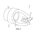
- FIG. 4 is a perspective view of an embodiment of a tissue cutting cap
- FIG. 5 is a perspective view of an embodiment of a tissue cutting cap with the cutting portion recessed within the body
- FIG. 6 is a perspective view of the embodiment of the tissue cutting cap shown in FIG. 5 with the cutting portion exposed;
- FIG. 7 is a perspective view of an alternative embodiment of a tissue cutting cap with the cutting portion recessed;
- FIG. 8 is a perspective view of the embodiment of the tissue cutting cap shown in FIG. 7 with the cutting portion exposed;
- FIGS. 9-11 illustrate operation of an embodiment of a tissue cutting device
- FIGS. 12-14 illustrate operation of an embodiment of a tissue cutting device.
- proximal and distal should be understood as being in the terms of a physician delivering the tissue cutting cap to a patient.
- distal means the portion of the tissue cutting cap that is farthest from the physician and the term “proximal” means the portion of the tissue cutting cap that is nearest to the physician.
- FIGS. 1 and 2 illustrate an embodiment of a tissue cutting cap 10 in accordance with the present invention.
- the tissue cutting cap 10 includes a generally tubular body 12 at a proximal portion 14 of the cap 10 and having a lumen 16 formed therein.
- the ablation cap 10 includes a distal portion 18 having a cutting portion 20 .
- the proximal portion 14 of the cap 10 is sized and shaped to fit over a distal end 24 of an endoscope 26 as shown in FIG. 3A .
- the proximal portion 14 of the ablation cap 10 may include an elastomeric portion 28 that is connected to the tubular body 12 and that fits over the distal end 24 of the endoscope 26 to secure the cap 10 to the endoscope 26 .
- the proximal portion 14 may be made of a hard material that is sized and shaped to friction fit over the distal end 24 of the endoscope 26 .
- the cap 10 may be secured to the endoscope 26 using a band 80 shown in FIG. 3B that surrounds the proximal portion 14 of the cap 10 and a portion of the distal end 24 of the endoscope 26 .
- the cap 10 may be positioned over the distal end 24 of the endoscope 26 and the band 80 may be pulled around the proximal portion 14 of the cap 10 .
- the band 80 may include one or more openings 84 that are secured over one or more corresponding notches 86 on the proximal portion 14 of the cap 10 .
- the band 80 may include a recessed portion 88 to fit against the proximal portion 14 of the cap 10 and a second portion 90 to fit against the distal end 24 of the endoscope 26 so that the cap 10 is secured to the endoscope 26 .
- the band 80 may be tape, leather, elastomeric material or other material suitable for securing the cap 10 to the endoscope 26 .
- the cap 10 may also be secured to the endoscope 26 using other methods, for example, using a hinged proximal portion that allows the proximal portion to open for attachment to the endoscope 26 and then be secured around the distal end of the endoscope 26 (not shown).
- the distal portion 18 of the ablation cap 10 may extend beyond the distal end 24 of the endoscope 26 .
- the distal portion 18 may be formed from a material having sufficient transparency so that the operator using an optical port of the endoscope 26 may observe a portion of the tissue to be treated by viewing the tissue through the lumen 16 of the tubular body 12 or through a wall 32 of the distal portion 18 of the ablation cap 10 .
- the cap 10 or portions thereof may be dear, translucent or opaque or any combination thereof.
- the distal portion 18 may also include a portion that is formed from a material for magnifying the tissue under observation.
- the distal portion 18 of the cap 10 does not extend 360° circumferentially around the cap 10 at a distal end 34 of the cap 10 .
- the distal portion 18 may extend less than 180° of the circumference of the cap 10 and in some embodiments less than about 135°, or less than about 90°.
- the distal portion 18 at the distal end 34 may extend 360° circumferentially around the cap 10 as shown in FIG. 7 and discussed in more detail below.
- the cap 10 having a distal portion 18 extending less than 360° circumferentially around the cap 10 includes an open portion 40 defined by the wall 32 of the distal portion 18 .
- the open portion 40 connects to the lumen 16 extending though the body 12 of the cap 10 .
- the open portion 40 may be configured to allow tissue to be exposed to the cutting portion 20 .
- An edge 42 of the wall 32 may be curvilinear to facilitate advancement of the cap 10 toward the tissue to be treated.
- the cutting portion 20 of the cap 10 may be recessed from the distal end 34 of the cap 10 so that the cutting portion 20 is proximal to the distal end 34 when the cutting portion 20 is exposed.
- the cutting portion 20 may include a first side 21 , a second side 22 and an end portion 25 .
- the first and second sides 21 , 22 form an opening 27 into which the tissue to be cut can be inserted.
- the end portion 25 is generally opposite and proximal to the opening 27 .
- the cutting portion 20 may be v-shaped.
- the cutting portion 20 may be u-shaped or polygonal-shaped or any other shape that allows specific layers of the tissue to be cut, but prevents perforation of the entire tissue.
- a proximal portion 44 of the cuffing portion 20 may be positioned distal to the distal end 24 of the endoscope 26 so that one or more medical devices may be extended distally from the endoscope 26 to facilitate manipulation of the tissue to be cut and to help position the tissue with respect to the cutting portion 20 as described in more detail below.
- the size of the cutting portion 20 may vary depending on the size of the tissue to be sampled, the size of the lumen from which the sample is cut, the size of the endoscope and the like.
- the cutting portion may include at least one electrode.
- the cutting portion may include a blade having a cutting edge.
- the distal end 34 of the cap 10 includes an opening 46 in the wall 34 that is sized and shaped to accommodate the cutting portion 20 .
- the opening 46 may be v-shaped to accommodate a v-shaped cutting portion 20 .
- a distal end 48 of the opening 46 may be wider than a proximal end 52 of the opening 48 to help facilitate cutting of the tissue.
- the distal end 34 of the cap 10 adjacent to the opening 46 is provided with tips 54 on either side of the opening 46 .
- the tips 54 may be curvilinear to facilitate passage of the cap 10 through a body lumen.
- the one or both tips 54 may be sharpened so that the tip 54 may penetrate the tissue to facilitate positioning the cutting portion 20 against the tissue.
- the wall 32 may surround and insulate the cutting portion 20 .
- the cutting portion 20 includes at least one electrode and the cap 10 may operate as a monopolar device or a bipolar device.
- the cutting portion 20 may be monopolar and a return pad (not shown) may be placed elsewhere on the patient.
- the cap 10 shown in FIG. 1 may be a bipolar device where the cutting portion 20 is an active electrode 58 and the cap 10 or a portion thereof is a return electrode 62 .
- the cutting portion 20 may be provided as a blade having a cutting edge.
- the cap 10 may be a bipolar device with an active electrode 66 at the base of the v of the cutting portion 20 and a return electrode 68 adjacent to the active electrode 66 .
- the bipolar device would eliminate the need for a return pad and also may advantageously provide lower bleeding rates.
- the active (and return if bipolar) electrode(s) may be connected to one or more wires that extend from the electrode(s) to a connector that allows for connection to an electrosurgical unit or an active cord (not shown).
- the wire may be external to the endoscope 26 as shown in FIG. 7 or internal to the endoscope 26 .
- the cutting portion is sized to limit the amount of active cutting portion 20 that is exposed to the tissue so that the cutting portion 20 has an increased current density to cut the tissue rather than burn the tissue.
- the surface area of the active portion is less than the surface area of the return portion to cut the tissue.
- the exposed cutting portion 20 may be about 0.01-0.02 inches extending from the cap 10 . Other sizes for the cutting portion may be used and will depend in part on the surface area of the cutting portion.
- FIGS. 5 and 6 illustrate an embodiment of a tissue cutting cap 100 in accordance with the present invention.
- the tissue cutting cap 100 is similar to the cap 10 described above with the exception that the cap 100 includes a cutting portion 120 that is movably positionable relative to a generally tubular body 112 of the cap 100 .
- the shape of the cutting portion 120 is similar to the shape of the cutting portion 20 described above.
- the tissue cutting cap 100 includes a proximal portion 114 and a distal portion 118 .
- a lumen 116 extends through at least a portion of the body 112 .
- the proximal portion 114 of the cap 100 is sized and shaped to fit over a distal end 24 of an endoscope 26 and may be secured to the endoscope 26 similar to the cap 10 as described above.
- the distal portion 118 of the ablation cap 100 may extend beyond the distal end 24 of the endoscope 26 .
- the distal portion 118 may be formed from a material having sufficient transparency so that the operator using an optical port of the endoscope 26 may observe a portion of the tissue to be treated by viewing the tissue through the lumen 116 of the tubular body 112 or through a wall 132 of the distal portion 118 of the ablation cap 100 .
- the cap 100 or portions thereof may be dear, translucent or opaque or any combination thereof.
- the distal portion 118 may also include a portion that is formed from a material for magnifying the tissue under observation. Similar to the cap 10 , the distal portion 118 of the cap 100 may extend less than 360° circumferentially around the cap 100 at a distal end 134 of the cap 100 .
- the cap 100 having a distal portion 118 extending less than 360° circumferentially around the cap 100 includes an open portion 140 defined by the wall 132 of the distal portion 118 .
- the open portion 140 connects to the lumen 116 extending though the body 112 of the cap 100 .
- the open portion 140 may be configured to allow tissue to be exposed to the cutting portion 120 when the cutting portion 120 is extended distally from the body 112 .
- An edge 158 of the wall 132 may be curvilinear to facilitate advancement of the cap 100 toward the tissue to be treated.
- the cutting portion 120 is movably positional relative to the body 112 . As shown in FIG. 5 , the cutting portion 120 is positioned within a recess 131 formed in the body 112 . In some embodiments, the cutting portion 120 may be entirely withdrawn in to the recess 131 of the body 112 in a recessed position 144 .
- the cutting portion 120 may be connected to a drive catheter 141 that extends proximally from the cutting portion 120 to a proximal control handle (not shown).
- the drive catheter 141 is distally movable to extend the cutting portion 120 from the recess 131 in a cutting configuration 147 as shown in FIG. 6 and proximally movable to re-position the cutting portion 120 within the recess 131 as shown in FIG.
- the cutting portion 120 is positioned within the recess 131 when the tissue cutting cap 100 is being delivered to a treatment site or being repositioned within a patient's lumen for additional treatment at one or more additional sites. Positioning of the cutting portion 120 within the recess 131 also helps to prevent accidental energy delivery, for example to healthy tissue.
- the cutting portion 120 is at least partially distally extended from the recess 131 for treatment at a site and energy is delivered to the tissue to ablate the diseased tissue as described in more detail below.
- the cutting portion 120 may be configured for monopolar or bipolar energy delivery as discussed above with reference to the cap 10 .
- One or more wires 139 may extend from the cutting portion 120 to a connector (not shown) for connection to an electrosurgical unit or active cord (not shown) and may co-extend with the drive catheter 141 .
- the drive catheter 141 and/or the wires 139 are positioned external to the endoscope 26 .
- the drive catheter 141 and/or the wires 139 extend within a channel of the endoscope 26 .
- the cutting portion 120 of the cap 100 may be recessed from the distal end 134 of the cap 100 when the cutting portion 120 is in the cutting configuration 147 shown in FIG. 6 so that the cutting portion 120 is proximal to the distal end 134 .
- the cutting portion 120 may be v-shaped.
- the cutting portion 120 may be u-shaped or polygonal-shaped or other shape that allows specific layers of the tissue to be cut, but prevents full-thickness cutting of the tissue.
- the cutting portion 120 may be sized and shape similarly to the cutting portion 20 described above.
- a proximal portion 144 of the cutting portion 120 may be positioned distal to the distal end 24 of the endoscope 26 so that one or more medical devices may be extended distally from the endoscope 26 to facilitate manipulation of the tissue to be cut and to help position the tissue with respect to the cutting portion 120 as described in more detail below.
- the size of the cutting portion 120 may vary depending on the size of the tissue to be sampled, the size of the lumen from which the sample is cut, the size of the endoscope and the like.
- the distal end 134 of the cap 100 includes an opening 146 in the wall 134 that is sized and shaped to accommodate the cutting portion 120 .
- the opening 146 may be sized and shaped according to the size and shape of the cutting portion 120 positioned within the opening 146 .
- a distal end 148 of the opening 146 may be wider than a proximal end 152 of the opening 148 to help facilitate cutting of the tissue.
- the distal end 134 of the cap 100 adjacent to the opening 146 is provided with tips 154 on either side of the opening 146 .
- the tips 154 may be curvilinear to facilitate passage of the cap 100 through a body lumen.
- the one or both tips 154 may be sharpened so that the tip 154 may penetrate the tissue to facilitate positioning the cutting portion 120 against the tissue.
- the wall 132 may surround and insulate the cutting portion 120 .
- FIGS. 7 and 8 illustrate an embodiment of a tissue cutting cap 200 in accordance with the present invention.
- the tissue cutting cap 200 is similar to the embodiments described above with the exception that the cap 200 includes a cutting portion 220 that is movably positionable relative to a generally tubular body 212 of the cap 200 and is extendable from a cover portion 230 of the cap 200 .
- the tissue cutting cap 200 includes a proximal portion 214 and a distal portion 218 .
- a lumen 216 extends through the body 212 .
- the proximal portion 214 of the cap 200 is sized and shaped to fit over a distal end 24 of an endoscope 26 and may be secured to the endoscope 26 similar to the cap 10 as described above.
- the distal portion 218 of the ablation cap 200 may extend beyond the distal end 24 of the endoscope 26 .
- the distal portion 218 may be formed from a material having sufficient transparency so that the operator using an optical port of the endoscope 26 may observe a portion of the tissue to be treated by viewing the tissue through the lumen 216 of the tubular body 212 or through a wall 232 of the distal portion 218 of the ablation cap 200 .
- the cap 200 or portions thereof may be dear, translucent or opaque or any combination thereof.
- the distal portion 218 may also include a portion that is formed from a material for magnifying the tissue under observation. As shown in FIG. 7 , the distal portion 218 of the cap 200 may extend 360° circumferentially around the cap 200 at a distal end 234 of the cap 200 .
- the tissue cutting cap 200 may further include the cover portion 230 that includes a recess 231 formed as part of the cap 200 .
- the cover portion 230 may be integrally formed with the cap 200 or provided as a separate portion and connected to the cap 200 .
- the cover portion 230 is at least partially spaced apart from the tubular body 212 to form the recess 231 .
- the recess 231 may be sized and shaped to hold an extendable cutting portion 220 within the recess 231 in a covered position 244 as shown in FIG. 7 so that the cutting portion 220 is proximal to a distal end 234 of the tissue cutting cap 200 .
- the cutting portion 220 is slidably positionable within the recess 231 of the cover portion 230 and may be completely covered by the cover portion 230 . As shown in FIG. 8 , the cutting portion 220 may be extended distally from the recess 231 so that at least a portion of the cutting portion 220 is exposed and can contact the tissue to be treated.
- the cutting portion 220 may be mounted in a slider member 221 that supports the cutting portion 220 .
- the slider member 221 may be curved and may include an opening 246 in a distal end 247 of the slider member 221 that is sized and shaped to accommodate the cutting portion 220 .
- the cutting portion 220 may be v-shaped, u-shaped or polygonal and the like, and the opening 246 may be similarly shaped to accommodate the shape of the cutting portion 220 .
- a distal end 248 of the opening 246 may be wider than a proximal end 252 of the opening 248 to help facilitate cutting of the tissue.
- the distal end 247 of the slider member 221 adjacent to the opening 246 is provided with tips 254 on either side of the opening 246 .
- the one or both tips 254 may be sharpened so that the tip 254 may penetrate the tissue to facilitate positioning the cutting portion 220 against the tissue.
- the cutting portion 220 on the slider member 221 is movably positional relative to the body 212 . As shown in FIG. 7 , the cutting portion 220 and the slider member 221 are positioned within the recess 231 formed in the body 212 . In some embodiments, the cutting portion 220 may be entirely withdrawn in to the recess 231 of the body 212 in a recessed position 244 shown in FIG. 7 .
- the slider member 221 may be connected to a drive catheter 241 that extends proximally from the slider member 221 to a proximal control handle (not shown).
- the drive catheter 241 is distally movable to extend the slider member 221 and the cutting portion 220 from the recess 231 in a cutting configuration 247 as shown in FIG. 8 and proximally movable to re-position the cutting portion 221 within the recess 231 as shown in FIG. 7 .
- the cutting portion 220 and the slider member 221 are positioned within the recess 231 when the tissue cutting cap 200 is being delivered to a treatment site or being repositioned within a patient's lumen for additional treatment at one or more additional sites. Positioning of the cutting portion 220 within the recess 231 also helps to prevent accidental energy delivery, for example to healthy tissue.
- the cutting portion 220 is at least partially distally extended from the recess 231 for treatment at a site and energy is delivered to the tissue to ablate the diseased tissue as described in more detail below,
- the cutting portion 220 may be configured for monopolar or bipolar energy delivery as discussed above with reference to the cap 10 and may be sized and shaped similar to the cutting portion 20 .
- One or more wires 239 may extend from the cutting portion 220 to a connector (not shown) for connection to an electrosurgical unit or active cord (not shown) and may co-extend with the drive catheter 241 .
- the drive catheter 241 and/or the wires 239 are positioned external to the endoscope 26 .
- the drive catheter 241 and/or the wires 239 extend within a channel of the endoscope 26 .
- the tissue cutting cap may be made primarily of a substantially transparent or translucent polymer such as polytetrafluorothylene (PTFE). Additional possible materials include, but are not limited to the following, polyethylene ether ketone (PEEK), fluorinated ethylene propylene (FEP), perfluoroalkoxy polymer resin (PFA), polyamide, polyurethane, high density or low density polyethylene, and nylon.
- PTFE polytetrafluorothylene
- Additional possible materials include, but are not limited to the following, polyethylene ether ketone (PEEK), fluorinated ethylene propylene (FEP), perfluoroalkoxy polymer resin (PFA), polyamide, polyurethane, high density or low density polyethylene, and nylon.
- the tissue cutting cap may be formed from a lubricious material such as PTFE and the like for easy slidability within the patient's lumen for delivery to the treatment site.
- the tissue cutting cap or a portion thereof may be formed from magnif
- the tissue cutting cap or a portion thereof may also be coated or impregnated with other compounds and materials to achieve the desired properties.
- Exemplary coatings or additives include, but are not limited to, parylene, glass fillers, silicone hydrogel polymers and hydrophilic coatings.
- the tissue cutting cap or portions thereof may be made of different durometers of material. By way of non-limiting example, the tissue cutting cap or a distal portion thereof may be made of a softer material to allow for easier advancement of the cap to the target tissue site.
- FIGS. 9 and 10 illustrate the tissue cutting cap 10 positioned on the distal of end 24 of the endoscope 26 that is been positioned near a tissue treatment site,
- a forceps device 49 is extended distally from a working channel 51 of the endoscope 26 .
- the forceps device 49 grasps a first layer of tissue 101 and pulls the first layer of tissue 101 away from a second layer of tissue 102 .
- the layers 101 , 102 may be any adjacent layers of tissue, for example, the muscularis and submucosal layers. As shown in FIG.
- the cutting portion 20 of the cap 10 is advanced to the first layer of tissue 101 and power is supplied to the cutting portion 20 so that the cutting portion 20 cuts the first tissue layer 101 and leaves the second layer 102 intact.
- a sample of the first tissue layer 101 may be cut away from and removed from the second tissue layer 102 .
- the forceps 49 may be used to hold onto the first tissue layer 101 as the layer 101 is being cut and to remove the sample of the first tissue layer 101 for testing.
- the forceps 49 may be proximally withdrawn from the site after the first tissue layer 101 has been cut away from the second tissue layer 102 so that the forceps 49 delivers the tissue 101 to the physician.
- the endoscope 26 may include a suction port 27 to facilitate removal of the tissue 101 and/or to facilitate elevation of the first layer of tissue 101 away from the second tissue layer 102 to facilitate cutting with the cutting portion 20 .
- FIG. 11 illustrates the site after the first tissue layer 101 has been removed and the second tissue layer 102 remains intact.
- FIGS. 12-14 illustrate an alternative method of operation of the device 10 .
- a needle 103 is extended distally and inserted between the first tissue layer 101 and the second tissue layer 102 .
- the injection of the solution between the first tissue layer 101 and the second tissue layer 102 forms a fluid-filled pocket 115 that forces separation between the first and second tissue layers 101 , 102 , breaking the attachments between the tissue layers 101 , 102 .
- the elevated portion of the first tissue layer 101 may then be resected by the physician using the cutting portion 20 of the cap 10 as shown in FIG. 13 .
- the cutting portion 20 of the cap 10 is advanced to the first layer of tissue 101 and power is supplied to the cutting portion 20 so that the cutting portion 20 cuts the first tissue layer 101 and leaves the second layer 102 intact.
- a sample of the first tissue layer 101 may be cut away from and removed from the second tissue layer 102 . Similar to the method described with reference to FIGS. 9-11 , the sample of the first tissue layer 101 may be removed by forceps or suction.
- the first tissue layer 101 is removed and the second tissue layer 102 remains intact as shown in FIG. 13 .
- FIG. 14 illustrates the site after the first tissue layer 101 has been removed and the second tissue layer 102 remains in position at the site.
Abstract
Description
Claims (14)
Priority Applications (1)
| Application Number | Priority Date | Filing Date | Title |
|---|---|---|---|
| US14/039,699 US9526570B2 (en) | 2012-10-04 | 2013-09-27 | Tissue cutting cap |
Applications Claiming Priority (2)
| Application Number | Priority Date | Filing Date | Title |
|---|---|---|---|
| US201261709455P | 2012-10-04 | 2012-10-04 | |
| US14/039,699 US9526570B2 (en) | 2012-10-04 | 2013-09-27 | Tissue cutting cap |
Publications (2)
| Publication Number | Publication Date |
|---|---|
| US20140100570A1 US20140100570A1 (en) | 2014-04-10 |
| US9526570B2 true US9526570B2 (en) | 2016-12-27 |
Family
ID=50433274
Family Applications (1)
| Application Number | Title | Priority Date | Filing Date |
|---|---|---|---|
| US14/039,699 Active 2034-05-21 US9526570B2 (en) | 2012-10-04 | 2013-09-27 | Tissue cutting cap |
Country Status (1)
| Country | Link |
|---|---|
| US (1) | US9526570B2 (en) |
Cited By (3)
| Publication number | Priority date | Publication date | Assignee | Title |
|---|---|---|---|---|
| US20140276790A1 (en) * | 2013-03-13 | 2014-09-18 | Boston Scientific Scimed, Inc. | Devices for tissue separation and related methods of use |
| US20150133926A1 (en) * | 2013-11-08 | 2015-05-14 | The Cleveland Clinic Foundation | Excising endocap |
| US20220225983A1 (en) * | 2021-01-21 | 2022-07-21 | Ethicon, Inc. | Suture needle adaptors for delivering suture needles through cannulas while simultaneously visualizing the delivery of the suture needles through the cannulas |
Families Citing this family (2)
| Publication number | Priority date | Publication date | Assignee | Title |
|---|---|---|---|---|
| EP2967727B1 (en) * | 2013-03-15 | 2020-09-09 | Cook Medical Technologies LLC | Electrosurgical system with electrically active outer surface |
| US20170119234A1 (en) | 2015-10-14 | 2017-05-04 | Cook Medical Technologies Llc | Endoscope cap with separable arms |
Citations (119)
| Publication number | Priority date | Publication date | Assignee | Title |
|---|---|---|---|---|
| US1056336A (en) | 1909-09-24 | 1913-03-18 | Allan G Hurdman | Bipolar intragastric electrode. |
| US4074718A (en) | 1976-03-17 | 1978-02-21 | Valleylab, Inc. | Electrosurgical instrument |
| US4386752A (en) | 1981-03-13 | 1983-06-07 | General Motors Corporation | Hinged collar clip |
| US4522205A (en) | 1980-09-03 | 1985-06-11 | The University Court Of The University Of Edinburgh | Therapeutic device and method of inducing thrombosis in a blood vessel |
| US4532924A (en) | 1980-05-13 | 1985-08-06 | American Hospital Supply Corporation | Multipolar electrosurgical device and method |
| US4706667A (en) | 1984-06-25 | 1987-11-17 | Berchtold Medizin-Elektronik Gmbh & Co. | Electro surgical high frequency cutting instrument |
| US4765331A (en) | 1987-02-10 | 1988-08-23 | Circon Corporation | Electrosurgical device with treatment arc of less than 360 degrees |
| US4823791A (en) | 1987-05-08 | 1989-04-25 | Circon Acmi Division Of Circon Corporation | Electrosurgical probe apparatus |
| US4936842A (en) | 1987-05-08 | 1990-06-26 | Circon Corporation | Electrosurgical probe apparatus |
| US5100402A (en) | 1990-10-05 | 1992-03-31 | Megadyne Medical Products, Inc. | Electrosurgical laparoscopic cauterization electrode |
| US5197491A (en) | 1990-09-28 | 1993-03-30 | Brunswick Biomedical Technologies, Inc. | Esophageal-stomach displacement electrode |
| US5254121A (en) | 1992-05-22 | 1993-10-19 | Meditron Devices, Inc. | Method and device for removing concretions within human ducts |
| US5443470A (en) | 1992-05-01 | 1995-08-22 | Vesta Medical, Inc. | Method and apparatus for endometrial ablation |
| US5454809A (en) | 1989-01-06 | 1995-10-03 | Angioplasty Systems, Inc. | Electrosurgical catheter and method for resolving atherosclerotic plaque by radio frequency sparking |
| US5494483A (en) | 1992-09-30 | 1996-02-27 | Adair; Edwin L. | Stereoscopic endoscope with miniaturized electronic imaging chip |
| US5514130A (en) | 1994-10-11 | 1996-05-07 | Dorsal Med International | RF apparatus for controlled depth ablation of soft tissue |
| US5562703A (en) | 1994-06-14 | 1996-10-08 | Desai; Ashvin H. | Endoscopic surgical instrument |
| US5573008A (en) * | 1993-10-29 | 1996-11-12 | Boston Scientific Corporation | Multiple biopsy sampling coring device |
| US5575788A (en) | 1994-06-24 | 1996-11-19 | Stuart D. Edwards | Thin layer ablation apparatus |
| US5681282A (en) | 1992-01-07 | 1997-10-28 | Arthrocare Corporation | Methods and apparatus for ablation of luminal tissues |
| US5683385A (en) | 1995-09-19 | 1997-11-04 | Symbiosis Corporation | Electrocautery connector for a bipolar push rod assembly |
| US5707355A (en) | 1995-11-15 | 1998-01-13 | Zimmon Science Corporation | Apparatus and method for the treatment of esophageal varices and mucosal neoplasms |
| US5718702A (en) | 1992-08-12 | 1998-02-17 | Somnus Medical Technologies, Inc. | Uvula, tonsil, adenoid and sinus tissue treatment device and method |
| US5743870A (en) | 1994-05-09 | 1998-04-28 | Somnus Medical Technologies, Inc. | Ablation apparatus and system for removal of soft palate tissue |
| WO1998022184A1 (en) | 1996-11-21 | 1998-05-28 | Boston Scientific Corporation | Mucosal ablation using light |
| US5766168A (en) | 1996-01-11 | 1998-06-16 | Northgate Technologies, Inc. | Perforated resectoscope electrode assembly |
| US5836906A (en) | 1996-02-23 | 1998-11-17 | Somnus Medical Technologies, Inc. | Method and apparatus for treatment of air way obstructions |
| US5925044A (en) | 1996-07-01 | 1999-07-20 | Gebrueder Berchtold Gmbh & Co. | Trocar for laparoscopic operations |
| WO1999035987A1 (en) | 1998-01-14 | 1999-07-22 | Conway-Stuart Medical, Inc. | Gerd treatment apparatus and method |
| US5957863A (en) | 1995-02-28 | 1999-09-28 | Boston Scientific Corporation | Deflectable biopsy catheter |
| US5993446A (en) | 1996-10-01 | 1999-11-30 | Select Medizin-Technik Herman Sutter Gmbh | Coagulation instrument |
| US5994717A (en) | 1996-05-17 | 1999-11-30 | Fujitsu Limited | Thin-film transistor and method for fabricating same and liquid crystal display device |
| US6009877A (en) | 1994-06-24 | 2000-01-04 | Edwards; Stuart D. | Method for treating a sphincter |
| US6015406A (en) * | 1996-01-09 | 2000-01-18 | Gyrus Medical Limited | Electrosurgical instrument |
| US6027499A (en) | 1997-05-23 | 2000-02-22 | Fiber-Tech Medical, Inc. (Assignee Of Jennifer B. Cartledge) | Method and apparatus for cryogenic spray ablation of gastrointestinal mucosa |
| US6044846A (en) | 1994-06-24 | 2000-04-04 | Edwards; Stuart D. | Method to treat esophageal sphincters |
| US6050993A (en) | 1998-07-27 | 2000-04-18 | Quantum Therapeutics Corp. | Medical device and methods for treating hemorrhoids |
| US6053172A (en) | 1995-06-07 | 2000-04-25 | Arthrocare Corporation | Systems and methods for electrosurgical sinus surgery |
| US6056744A (en) | 1994-06-24 | 2000-05-02 | Conway Stuart Medical, Inc. | Sphincter treatment apparatus |
| US6059719A (en) * | 1997-08-06 | 2000-05-09 | Olympus Optical Co., Ltd. | Endoscope system |
| US6073052A (en) | 1996-11-15 | 2000-06-06 | Zelickson; Brian D. | Device and method for treatment of gastroesophageal reflux disease |
| US6077257A (en) | 1996-05-06 | 2000-06-20 | Vidacare, Inc. | Ablation of rectal and other internal body structures |
| US6086585A (en) | 1995-06-07 | 2000-07-11 | Arthrocare Corporation | System and methods for electrosurgical treatment of sleep obstructive disorders |
| US6091995A (en) | 1996-11-08 | 2000-07-18 | Surx, Inc. | Devices, methods, and systems for shrinking tissues |
| US6112123A (en) | 1998-07-28 | 2000-08-29 | Endonetics, Inc. | Device and method for ablation of tissue |
| US6149647A (en) | 1999-04-19 | 2000-11-21 | Tu; Lily Chen | Apparatus and methods for tissue treatment |
| US6156032A (en) | 1998-09-30 | 2000-12-05 | Scimed Life Systems, Inc. | Method for causing a stricture of a body passageway |
| US6190381B1 (en) | 1995-06-07 | 2001-02-20 | Arthrocare Corporation | Methods for tissue resection, ablation and aspiration |
| US6197022B1 (en) | 1996-07-30 | 2001-03-06 | James A. Baker | Medical instruments and techniques for treatment of gastro-esophageal reflux disease |
| US6210409B1 (en) | 1999-05-03 | 2001-04-03 | Alan G. Ellman | Electrosurgical handpiece for treating tissue |
| US6231571B1 (en) | 1999-05-03 | 2001-05-15 | Alan G. Ellman | Electrosurgical handpiece for treating tissue |
| US6245067B1 (en) | 1997-04-16 | 2001-06-12 | Irvine Biomedical, Inc. | Ablation device and methods having perpendicular electrodes |
| US6248081B1 (en) * | 1999-09-28 | 2001-06-19 | Scimed Life Systems, Inc. | Endoscopic submucosal core biopsy device |
| US6258084B1 (en) | 1997-09-11 | 2001-07-10 | Vnus Medical Technologies, Inc. | Method for applying energy to biological tissue including the use of tumescent tissue compression |
| WO2001068015A1 (en) | 2000-03-13 | 2001-09-20 | Curon Medical, Inc. | Operative devices that can be removably fitted on catheter bodies to treat tissue regions in the body |
| WO2000019926A9 (en) | 1998-10-05 | 2001-12-20 | Scimed Life Systems Inc | Large area thermal ablation |
| US6346105B1 (en) | 1998-07-27 | 2002-02-12 | Quantum Cor Incorporated | Device for treating tissue and methods thereof |
| US6352533B1 (en) | 1999-05-03 | 2002-03-05 | Alan G. Ellman | Electrosurgical handpiece for treating tissue |
| US6355032B1 (en) | 1995-06-07 | 2002-03-12 | Arthrocare Corporation | Systems and methods for selective electrosurgical treatment of body structures |
| US6363937B1 (en) | 1995-06-07 | 2002-04-02 | Arthrocare Corporation | System and methods for electrosurgical treatment of the digestive system |
| US6402744B2 (en) | 1998-02-19 | 2002-06-11 | Curon Medical, Inc. | Systems and methods for forming composite lesions to treat dysfunction in sphincters and adjoining tissue regions |
| US6405732B1 (en) | 1994-06-24 | 2002-06-18 | Curon Medical, Inc. | Method to treat gastric reflux via the detection and ablation of gastro-esophageal nerves and receptors |
| US6419673B1 (en) | 1996-05-06 | 2002-07-16 | Stuart Edwards | Ablation of rectal and other internal body structures |
| US6464697B1 (en) | 1998-02-19 | 2002-10-15 | Curon Medical, Inc. | Stomach and adjoining tissue regions in the esophagus |
| US20020177847A1 (en) | 2001-03-30 | 2002-11-28 | Long Gary L. | Endoscopic ablation system with flexible coupling |
| US20020183739A1 (en) | 2001-03-30 | 2002-12-05 | Long Gary L. | Endoscopic ablation system with sealed sheath |
| US6494881B1 (en) | 1997-09-30 | 2002-12-17 | Scimed Life Systems, Inc. | Apparatus and method for electrode-surgical tissue removal having a selectively insulated electrode |
| US6535764B2 (en) | 2001-05-01 | 2003-03-18 | Intrapace, Inc. | Gastric treatment and diagnosis device and method |
| US6544261B2 (en) | 1995-06-07 | 2003-04-08 | Arthrocare Corporation | Systems and methods for electrosurgical treatment of submucosal tissue |
| US6589238B2 (en) | 1998-01-14 | 2003-07-08 | Curon Medical, Inc. | Sphincter treatment device |
| US6591838B2 (en) | 1998-07-06 | 2003-07-15 | Scimed Life Systems, Inc. | Implant system and method for bulking tissue |
| US20030181900A1 (en) | 2002-03-25 | 2003-09-25 | Long Gary L. | Endoscopic ablation system with a plurality of electrodes |
| US6632193B1 (en) | 1995-06-07 | 2003-10-14 | Arthrocare Corporation | Systems and methods for electrosurgical tissue treatment |
| US20030216727A1 (en) * | 2001-03-30 | 2003-11-20 | Long Gary L. | Medical device with improved wall construction |
| US6659106B1 (en) | 1995-06-07 | 2003-12-09 | Arthrocare Corporation | System and methods for electrosurgical treatment of turbinates |
| US6669687B1 (en) | 1999-06-25 | 2003-12-30 | Vahid Saadat | Apparatus and methods for treating tissue |
| US6685713B1 (en) | 1993-02-22 | 2004-02-03 | Dabegran Technologies, Inc. | Endoscopic ligating apparatus |
| US6692490B1 (en) | 1999-05-18 | 2004-02-17 | Novasys Medical, Inc. | Treatment of urinary incontinence and other disorders by application of energy and drugs |
| US6740082B2 (en) | 1998-12-29 | 2004-05-25 | John H. Shadduck | Surgical instruments for treating gastro-esophageal reflux |
| US6746447B2 (en) | 1993-05-10 | 2004-06-08 | Arthrocare Corporation | Methods for ablating tissue |
| US6763836B2 (en) | 1998-06-02 | 2004-07-20 | Arthrocare Corporation | Methods for electrosurgical tendon vascularization |
| US20050049454A1 (en) * | 2003-08-27 | 2005-03-03 | Pentax Corporation | Endoscopic high-frequency knife |
| US20050080412A1 (en) * | 2003-10-14 | 2005-04-14 | Pentax Corporation | High-frequency tool for endoscope |
| US6994705B2 (en) | 2003-09-29 | 2006-02-07 | Ethicon-Endo Surgery, Inc. | Endoscopic mucosal resection device with conductive tissue stop |
| US7022105B1 (en) | 1996-05-06 | 2006-04-04 | Novasys Medical Inc. | Treatment of tissue in sphincters, sinuses and orifices |
| US7025768B2 (en) | 2003-05-06 | 2006-04-11 | Boston Scientific Scimed, Inc. | Systems and methods for ablation of tissue |
| US20060167451A1 (en) | 2005-01-26 | 2006-07-27 | Ethicon Endo-Surgery, Inc. | Medical instrument including an end effector having a medical-treatment electrode |
| US20060217698A1 (en) | 2003-04-25 | 2006-09-28 | Medtronic, Inc. | Ablation of stomach lining to treat obesity |
| WO2006122279A2 (en) | 2005-05-11 | 2006-11-16 | Mayo Foundation For Medical Education And Research | Apparatus and methods for internal surgical procedures |
| US7137981B2 (en) | 2002-03-25 | 2006-11-21 | Ethicon Endo-Surgery, Inc. | Endoscopic ablation system with a distally mounted image sensor |
| US7232438B2 (en) | 2004-07-09 | 2007-06-19 | Ethicon Endo-Surgery, Inc. | Ablation device with clear probe |
| US7252665B2 (en) | 2003-10-31 | 2007-08-07 | Medtronic, Inc | Ablation of stomach lining to reduce stomach acid secretion |
| US20070203395A1 (en) * | 2006-02-28 | 2007-08-30 | Takayasu Mikkaichi | Cap installable on distal end portion of endoscope |
| US20070212926A1 (en) | 2006-03-10 | 2007-09-13 | Olympus Corporation | Protection hood for endoscope and endoscope including the same |
| US20080058586A1 (en) * | 2006-09-05 | 2008-03-06 | Wilson-Cook Medical Inc. | Hood member for use with an endoscope |
| US7344535B2 (en) | 2004-01-09 | 2008-03-18 | Barrx Medical, Inc. | Devices and methods for treatment of luminal tissue |
| US7347860B2 (en) * | 2003-10-08 | 2008-03-25 | Pentax Corporation | Endoscope for high-frequency treatment |
| US20080103357A1 (en) * | 2006-10-31 | 2008-05-01 | Zeiner Mark S | Attachment apparatus for an endoscope |
| US20080200912A1 (en) | 2007-02-15 | 2008-08-21 | Long Gary L | Electroporation ablation apparatus, system, and method |
| US20080242932A1 (en) | 2007-03-30 | 2008-10-02 | Wilson-Cook Medical Inc. | Endoscopic Securing System |
| US7530979B2 (en) | 1999-11-16 | 2009-05-12 | BÂRRX Medical, Inc. | Method of treating abnormal tissue in the human esophagus |
| US20090177030A1 (en) * | 2007-11-28 | 2009-07-09 | Hiroaki Goto | Endoscopic incision system |
| US7566300B2 (en) | 2004-04-15 | 2009-07-28 | Wilson-Cook Medical, Inc. | Endoscopic surgical access devices and methods of articulating an external accessory channel |
| JP2009183581A (en) | 2008-02-08 | 2009-08-20 | Hoya Corp | Hood for endoscope |
| US20090221872A1 (en) | 2008-02-28 | 2009-09-03 | Md Idiz, Llc | Endoscope Tip Protector |
| US20090247823A1 (en) * | 2005-09-26 | 2009-10-01 | Hironori Yamamoto | Instrument for Endoscopic Treatment |
| US20090270856A1 (en) | 2004-03-09 | 2009-10-29 | Usgi Medical, Inc. | Apparatus and methods for performing mucosectomy |
| US7648500B2 (en) | 1998-02-19 | 2010-01-19 | Mederi Therapeutics, Inc. | Sphincter treatment apparatus |
| US7658738B2 (en) | 2004-05-14 | 2010-02-09 | Ethicon Endo-Surgery, Inc. | Medical devices for use with endoscope |
| US7662151B2 (en) | 2006-02-15 | 2010-02-16 | Boston Scientific Scimed, Inc. | Contact sensitive probes |
| US7678069B1 (en) | 1995-11-22 | 2010-03-16 | Arthrocare Corporation | System for electrosurgical tissue treatment in the presence of electrically conductive fluid |
| US7691101B2 (en) | 2006-01-06 | 2010-04-06 | Arthrocare Corporation | Electrosurgical method and system for treating foot ulcer |
| US20100174283A1 (en) * | 2009-01-05 | 2010-07-08 | Mcnall Iii Ralph I | Electrosurgical devices for tonsillectomy and adenoidectomy |
| US7758087B2 (en) | 2000-10-16 | 2010-07-20 | Weatherford/Lamb, Inc. | Coupling apparatus |
| US20130046138A1 (en) * | 2011-08-19 | 2013-02-21 | Cook Medical Technologies Llc | Cap for Attachment to an Endoscope |
| US20130046300A1 (en) * | 2011-08-19 | 2013-02-21 | Cook Medical Technologies Llc | Ablation Cap |
| US9204782B2 (en) * | 2011-10-27 | 2015-12-08 | Boston Scientific Scimed, Inc. | Mucosal resection device and related methods of use |
| US9216057B2 (en) * | 2013-03-15 | 2015-12-22 | Kyphon Sarl | Steerable catheter system and method of using a steerable catheter system to dissect and evacuate tissue |
| US20160106497A1 (en) * | 2011-04-11 | 2016-04-21 | Iogyn, Inc. | Tissue extraction devices and methods |
-
2013
- 2013-09-27 US US14/039,699 patent/US9526570B2/en active Active
Patent Citations (135)
| Publication number | Priority date | Publication date | Assignee | Title |
|---|---|---|---|---|
| US1056336A (en) | 1909-09-24 | 1913-03-18 | Allan G Hurdman | Bipolar intragastric electrode. |
| US4074718A (en) | 1976-03-17 | 1978-02-21 | Valleylab, Inc. | Electrosurgical instrument |
| US4532924A (en) | 1980-05-13 | 1985-08-06 | American Hospital Supply Corporation | Multipolar electrosurgical device and method |
| US4522205A (en) | 1980-09-03 | 1985-06-11 | The University Court Of The University Of Edinburgh | Therapeutic device and method of inducing thrombosis in a blood vessel |
| US4386752A (en) | 1981-03-13 | 1983-06-07 | General Motors Corporation | Hinged collar clip |
| US4706667A (en) | 1984-06-25 | 1987-11-17 | Berchtold Medizin-Elektronik Gmbh & Co. | Electro surgical high frequency cutting instrument |
| US4765331A (en) | 1987-02-10 | 1988-08-23 | Circon Corporation | Electrosurgical device with treatment arc of less than 360 degrees |
| US4936842A (en) | 1987-05-08 | 1990-06-26 | Circon Corporation | Electrosurgical probe apparatus |
| US4823791A (en) | 1987-05-08 | 1989-04-25 | Circon Acmi Division Of Circon Corporation | Electrosurgical probe apparatus |
| US5454809A (en) | 1989-01-06 | 1995-10-03 | Angioplasty Systems, Inc. | Electrosurgical catheter and method for resolving atherosclerotic plaque by radio frequency sparking |
| US5197491A (en) | 1990-09-28 | 1993-03-30 | Brunswick Biomedical Technologies, Inc. | Esophageal-stomach displacement electrode |
| US5100402A (en) | 1990-10-05 | 1992-03-31 | Megadyne Medical Products, Inc. | Electrosurgical laparoscopic cauterization electrode |
| US5681282A (en) | 1992-01-07 | 1997-10-28 | Arthrocare Corporation | Methods and apparatus for ablation of luminal tissues |
| US5443470A (en) | 1992-05-01 | 1995-08-22 | Vesta Medical, Inc. | Method and apparatus for endometrial ablation |
| US5254121A (en) | 1992-05-22 | 1993-10-19 | Meditron Devices, Inc. | Method and device for removing concretions within human ducts |
| US5718702A (en) | 1992-08-12 | 1998-02-17 | Somnus Medical Technologies, Inc. | Uvula, tonsil, adenoid and sinus tissue treatment device and method |
| US5494483A (en) | 1992-09-30 | 1996-02-27 | Adair; Edwin L. | Stereoscopic endoscope with miniaturized electronic imaging chip |
| US6685713B1 (en) | 1993-02-22 | 2004-02-03 | Dabegran Technologies, Inc. | Endoscopic ligating apparatus |
| US6746447B2 (en) | 1993-05-10 | 2004-06-08 | Arthrocare Corporation | Methods for ablating tissue |
| US5573008A (en) * | 1993-10-29 | 1996-11-12 | Boston Scientific Corporation | Multiple biopsy sampling coring device |
| US5743870A (en) | 1994-05-09 | 1998-04-28 | Somnus Medical Technologies, Inc. | Ablation apparatus and system for removal of soft palate tissue |
| US5562703A (en) | 1994-06-14 | 1996-10-08 | Desai; Ashvin H. | Endoscopic surgical instrument |
| US5575788A (en) | 1994-06-24 | 1996-11-19 | Stuart D. Edwards | Thin layer ablation apparatus |
| US6405732B1 (en) | 1994-06-24 | 2002-06-18 | Curon Medical, Inc. | Method to treat gastric reflux via the detection and ablation of gastro-esophageal nerves and receptors |
| US6056744A (en) | 1994-06-24 | 2000-05-02 | Conway Stuart Medical, Inc. | Sphincter treatment apparatus |
| US6044846A (en) | 1994-06-24 | 2000-04-04 | Edwards; Stuart D. | Method to treat esophageal sphincters |
| US6009877A (en) | 1994-06-24 | 2000-01-04 | Edwards; Stuart D. | Method for treating a sphincter |
| US5514130A (en) | 1994-10-11 | 1996-05-07 | Dorsal Med International | RF apparatus for controlled depth ablation of soft tissue |
| US5957863A (en) | 1995-02-28 | 1999-09-28 | Boston Scientific Corporation | Deflectable biopsy catheter |
| US7442191B2 (en) | 1995-06-07 | 2008-10-28 | Arthrocare Corporation | Systems and methods for electrosurgical treatment of turbinates |
| US6355032B1 (en) | 1995-06-07 | 2002-03-12 | Arthrocare Corporation | Systems and methods for selective electrosurgical treatment of body structures |
| US6632193B1 (en) | 1995-06-07 | 2003-10-14 | Arthrocare Corporation | Systems and methods for electrosurgical tissue treatment |
| US6190381B1 (en) | 1995-06-07 | 2001-02-20 | Arthrocare Corporation | Methods for tissue resection, ablation and aspiration |
| US6659106B1 (en) | 1995-06-07 | 2003-12-09 | Arthrocare Corporation | System and methods for electrosurgical treatment of turbinates |
| US6544261B2 (en) | 1995-06-07 | 2003-04-08 | Arthrocare Corporation | Systems and methods for electrosurgical treatment of submucosal tissue |
| US6363937B1 (en) | 1995-06-07 | 2002-04-02 | Arthrocare Corporation | System and methods for electrosurgical treatment of the digestive system |
| US6086585A (en) | 1995-06-07 | 2000-07-11 | Arthrocare Corporation | System and methods for electrosurgical treatment of sleep obstructive disorders |
| US6053172A (en) | 1995-06-07 | 2000-04-25 | Arthrocare Corporation | Systems and methods for electrosurgical sinus surgery |
| US5683385A (en) | 1995-09-19 | 1997-11-04 | Symbiosis Corporation | Electrocautery connector for a bipolar push rod assembly |
| US5707355A (en) | 1995-11-15 | 1998-01-13 | Zimmon Science Corporation | Apparatus and method for the treatment of esophageal varices and mucosal neoplasms |
| US5906587A (en) | 1995-11-15 | 1999-05-25 | Zimmon; David S. | Apparatus and method for the treatment of esophageal varices and mucosal neoplasms |
| US7678069B1 (en) | 1995-11-22 | 2010-03-16 | Arthrocare Corporation | System for electrosurgical tissue treatment in the presence of electrically conductive fluid |
| US6015406A (en) * | 1996-01-09 | 2000-01-18 | Gyrus Medical Limited | Electrosurgical instrument |
| US5766168A (en) | 1996-01-11 | 1998-06-16 | Northgate Technologies, Inc. | Perforated resectoscope electrode assembly |
| US5836906A (en) | 1996-02-23 | 1998-11-17 | Somnus Medical Technologies, Inc. | Method and apparatus for treatment of air way obstructions |
| US6419673B1 (en) | 1996-05-06 | 2002-07-16 | Stuart Edwards | Ablation of rectal and other internal body structures |
| US6077257A (en) | 1996-05-06 | 2000-06-20 | Vidacare, Inc. | Ablation of rectal and other internal body structures |
| US7022105B1 (en) | 1996-05-06 | 2006-04-04 | Novasys Medical Inc. | Treatment of tissue in sphincters, sinuses and orifices |
| US5994717A (en) | 1996-05-17 | 1999-11-30 | Fujitsu Limited | Thin-film transistor and method for fabricating same and liquid crystal display device |
| US5925044A (en) | 1996-07-01 | 1999-07-20 | Gebrueder Berchtold Gmbh & Co. | Trocar for laparoscopic operations |
| US6197022B1 (en) | 1996-07-30 | 2001-03-06 | James A. Baker | Medical instruments and techniques for treatment of gastro-esophageal reflux disease |
| US6535768B1 (en) | 1996-08-30 | 2003-03-18 | James A. Baker | Medical instruments and techniques for treatment of gastro-esophageal reflux disease |
| US5993446A (en) | 1996-10-01 | 1999-11-30 | Select Medizin-Technik Herman Sutter Gmbh | Coagulation instrument |
| US6091995A (en) | 1996-11-08 | 2000-07-18 | Surx, Inc. | Devices, methods, and systems for shrinking tissues |
| US6587731B1 (en) | 1996-11-08 | 2003-07-01 | Surx, Inc. | Devices, methods, and systems for shrinking tissues |
| US6073052A (en) | 1996-11-15 | 2000-06-06 | Zelickson; Brian D. | Device and method for treatment of gastroesophageal reflux disease |
| WO1998022184A1 (en) | 1996-11-21 | 1998-05-28 | Boston Scientific Corporation | Mucosal ablation using light |
| US6245067B1 (en) | 1997-04-16 | 2001-06-12 | Irvine Biomedical, Inc. | Ablation device and methods having perpendicular electrodes |
| US6027499A (en) | 1997-05-23 | 2000-02-22 | Fiber-Tech Medical, Inc. (Assignee Of Jennifer B. Cartledge) | Method and apparatus for cryogenic spray ablation of gastrointestinal mucosa |
| US6059719A (en) * | 1997-08-06 | 2000-05-09 | Olympus Optical Co., Ltd. | Endoscope system |
| US6258084B1 (en) | 1997-09-11 | 2001-07-10 | Vnus Medical Technologies, Inc. | Method for applying energy to biological tissue including the use of tumescent tissue compression |
| US6494881B1 (en) | 1997-09-30 | 2002-12-17 | Scimed Life Systems, Inc. | Apparatus and method for electrode-surgical tissue removal having a selectively insulated electrode |
| WO1999035987A1 (en) | 1998-01-14 | 1999-07-22 | Conway-Stuart Medical, Inc. | Gerd treatment apparatus and method |
| US6589238B2 (en) | 1998-01-14 | 2003-07-08 | Curon Medical, Inc. | Sphincter treatment device |
| US6846312B2 (en) | 1998-01-14 | 2005-01-25 | Curon Medical, Inc. | GERD treatment apparatus and method |
| US7648500B2 (en) | 1998-02-19 | 2010-01-19 | Mederi Therapeutics, Inc. | Sphincter treatment apparatus |
| US6464697B1 (en) | 1998-02-19 | 2002-10-15 | Curon Medical, Inc. | Stomach and adjoining tissue regions in the esophagus |
| US6872206B2 (en) | 1998-02-19 | 2005-03-29 | Curon Medical, Inc. | Methods for treating the cardia of the stomach |
| US6402744B2 (en) | 1998-02-19 | 2002-06-11 | Curon Medical, Inc. | Systems and methods for forming composite lesions to treat dysfunction in sphincters and adjoining tissue regions |
| US6712814B2 (en) | 1998-02-19 | 2004-03-30 | Curon Medical, Inc. | Method for treating a sphincter |
| US6763836B2 (en) | 1998-06-02 | 2004-07-20 | Arthrocare Corporation | Methods for electrosurgical tendon vascularization |
| US6591838B2 (en) | 1998-07-06 | 2003-07-15 | Scimed Life Systems, Inc. | Implant system and method for bulking tissue |
| US6346105B1 (en) | 1998-07-27 | 2002-02-12 | Quantum Cor Incorporated | Device for treating tissue and methods thereof |
| US6050993A (en) | 1998-07-27 | 2000-04-18 | Quantum Therapeutics Corp. | Medical device and methods for treating hemorrhoids |
| US6112123A (en) | 1998-07-28 | 2000-08-29 | Endonetics, Inc. | Device and method for ablation of tissue |
| US6156032A (en) | 1998-09-30 | 2000-12-05 | Scimed Life Systems, Inc. | Method for causing a stricture of a body passageway |
| US7749159B2 (en) | 1998-10-05 | 2010-07-06 | Boston Scientific Scimed, Inc. | Large area thermal ablation |
| WO2000019926A9 (en) | 1998-10-05 | 2001-12-20 | Scimed Life Systems Inc | Large area thermal ablation |
| US20100256632A1 (en) | 1998-10-05 | 2010-10-07 | Boston Scientific Scimed, Inc. | Large area thermal ablation |
| US6932812B2 (en) | 1998-10-05 | 2005-08-23 | Scimed Life Systems, Inc. | Large area thermal ablation |
| US6394949B1 (en) * | 1998-10-05 | 2002-05-28 | Scimed Life Systems, Inc. | Large area thermal ablation |
| US6740082B2 (en) | 1998-12-29 | 2004-05-25 | John H. Shadduck | Surgical instruments for treating gastro-esophageal reflux |
| US6149647A (en) | 1999-04-19 | 2000-11-21 | Tu; Lily Chen | Apparatus and methods for tissue treatment |
| US6210409B1 (en) | 1999-05-03 | 2001-04-03 | Alan G. Ellman | Electrosurgical handpiece for treating tissue |
| US6231571B1 (en) | 1999-05-03 | 2001-05-15 | Alan G. Ellman | Electrosurgical handpiece for treating tissue |
| US6352533B1 (en) | 1999-05-03 | 2002-03-05 | Alan G. Ellman | Electrosurgical handpiece for treating tissue |
| US6692490B1 (en) | 1999-05-18 | 2004-02-17 | Novasys Medical, Inc. | Treatment of urinary incontinence and other disorders by application of energy and drugs |
| US6669687B1 (en) | 1999-06-25 | 2003-12-30 | Vahid Saadat | Apparatus and methods for treating tissue |
| US6248081B1 (en) * | 1999-09-28 | 2001-06-19 | Scimed Life Systems, Inc. | Endoscopic submucosal core biopsy device |
| US7530979B2 (en) | 1999-11-16 | 2009-05-12 | BÂRRX Medical, Inc. | Method of treating abnormal tissue in the human esophagus |
| WO2001068015A1 (en) | 2000-03-13 | 2001-09-20 | Curon Medical, Inc. | Operative devices that can be removably fitted on catheter bodies to treat tissue regions in the body |
| US7758087B2 (en) | 2000-10-16 | 2010-07-20 | Weatherford/Lamb, Inc. | Coupling apparatus |
| US20020177847A1 (en) | 2001-03-30 | 2002-11-28 | Long Gary L. | Endoscopic ablation system with flexible coupling |
| US7097644B2 (en) | 2001-03-30 | 2006-08-29 | Ethicon Endo-Surgery, Inc. | Medical device with improved wall construction |
| US6918906B2 (en) | 2001-03-30 | 2005-07-19 | Gary L. Long | Endoscopic ablation system with improved electrode geometry |
| US20030216727A1 (en) * | 2001-03-30 | 2003-11-20 | Long Gary L. | Medical device with improved wall construction |
| US20020183739A1 (en) | 2001-03-30 | 2002-12-05 | Long Gary L. | Endoscopic ablation system with sealed sheath |
| US6535764B2 (en) | 2001-05-01 | 2003-03-18 | Intrapace, Inc. | Gastric treatment and diagnosis device and method |
| US7137981B2 (en) | 2002-03-25 | 2006-11-21 | Ethicon Endo-Surgery, Inc. | Endoscopic ablation system with a distally mounted image sensor |
| US20030181900A1 (en) | 2002-03-25 | 2003-09-25 | Long Gary L. | Endoscopic ablation system with a plurality of electrodes |
| US20060217698A1 (en) | 2003-04-25 | 2006-09-28 | Medtronic, Inc. | Ablation of stomach lining to treat obesity |
| US7025768B2 (en) | 2003-05-06 | 2006-04-11 | Boston Scientific Scimed, Inc. | Systems and methods for ablation of tissue |
| US20050049454A1 (en) * | 2003-08-27 | 2005-03-03 | Pentax Corporation | Endoscopic high-frequency knife |
| US6994705B2 (en) | 2003-09-29 | 2006-02-07 | Ethicon-Endo Surgery, Inc. | Endoscopic mucosal resection device with conductive tissue stop |
| US7347860B2 (en) * | 2003-10-08 | 2008-03-25 | Pentax Corporation | Endoscope for high-frequency treatment |
| US20050080412A1 (en) * | 2003-10-14 | 2005-04-14 | Pentax Corporation | High-frequency tool for endoscope |
| US7252665B2 (en) | 2003-10-31 | 2007-08-07 | Medtronic, Inc | Ablation of stomach lining to reduce stomach acid secretion |
| US7344535B2 (en) | 2004-01-09 | 2008-03-18 | Barrx Medical, Inc. | Devices and methods for treatment of luminal tissue |
| US7703459B2 (en) | 2004-03-09 | 2010-04-27 | Usgi Medical, Inc. | Apparatus and methods for mapping out endoluminal gastrointestinal surgery |
| US20090270856A1 (en) | 2004-03-09 | 2009-10-29 | Usgi Medical, Inc. | Apparatus and methods for performing mucosectomy |
| US7566300B2 (en) | 2004-04-15 | 2009-07-28 | Wilson-Cook Medical, Inc. | Endoscopic surgical access devices and methods of articulating an external accessory channel |
| US7658738B2 (en) | 2004-05-14 | 2010-02-09 | Ethicon Endo-Surgery, Inc. | Medical devices for use with endoscope |
| US7232438B2 (en) | 2004-07-09 | 2007-06-19 | Ethicon Endo-Surgery, Inc. | Ablation device with clear probe |
| US20060167451A1 (en) | 2005-01-26 | 2006-07-27 | Ethicon Endo-Surgery, Inc. | Medical instrument including an end effector having a medical-treatment electrode |
| WO2006122279A2 (en) | 2005-05-11 | 2006-11-16 | Mayo Foundation For Medical Education And Research | Apparatus and methods for internal surgical procedures |
| US20090247823A1 (en) * | 2005-09-26 | 2009-10-01 | Hironori Yamamoto | Instrument for Endoscopic Treatment |
| US7691101B2 (en) | 2006-01-06 | 2010-04-06 | Arthrocare Corporation | Electrosurgical method and system for treating foot ulcer |
| US7662151B2 (en) | 2006-02-15 | 2010-02-16 | Boston Scientific Scimed, Inc. | Contact sensitive probes |
| US20070203395A1 (en) * | 2006-02-28 | 2007-08-30 | Takayasu Mikkaichi | Cap installable on distal end portion of endoscope |
| US20070212926A1 (en) | 2006-03-10 | 2007-09-13 | Olympus Corporation | Protection hood for endoscope and endoscope including the same |
| US20080058586A1 (en) * | 2006-09-05 | 2008-03-06 | Wilson-Cook Medical Inc. | Hood member for use with an endoscope |
| US20080103357A1 (en) * | 2006-10-31 | 2008-05-01 | Zeiner Mark S | Attachment apparatus for an endoscope |
| US7655004B2 (en) | 2007-02-15 | 2010-02-02 | Ethicon Endo-Surgery, Inc. | Electroporation ablation apparatus, system, and method |
| US20080200912A1 (en) | 2007-02-15 | 2008-08-21 | Long Gary L | Electroporation ablation apparatus, system, and method |
| US20100087813A1 (en) | 2007-02-15 | 2010-04-08 | Ethicon Endo-Surgery, Inc. | Electroporation ablation apparatus, system, and method |
| US20080242932A1 (en) | 2007-03-30 | 2008-10-02 | Wilson-Cook Medical Inc. | Endoscopic Securing System |
| US20090177030A1 (en) * | 2007-11-28 | 2009-07-09 | Hiroaki Goto | Endoscopic incision system |
| JP2009183581A (en) | 2008-02-08 | 2009-08-20 | Hoya Corp | Hood for endoscope |
| US20090221872A1 (en) | 2008-02-28 | 2009-09-03 | Md Idiz, Llc | Endoscope Tip Protector |
| US20100174283A1 (en) * | 2009-01-05 | 2010-07-08 | Mcnall Iii Ralph I | Electrosurgical devices for tonsillectomy and adenoidectomy |
| US20160106497A1 (en) * | 2011-04-11 | 2016-04-21 | Iogyn, Inc. | Tissue extraction devices and methods |
| US20130046138A1 (en) * | 2011-08-19 | 2013-02-21 | Cook Medical Technologies Llc | Cap for Attachment to an Endoscope |
| US20130046300A1 (en) * | 2011-08-19 | 2013-02-21 | Cook Medical Technologies Llc | Ablation Cap |
| US9204782B2 (en) * | 2011-10-27 | 2015-12-08 | Boston Scientific Scimed, Inc. | Mucosal resection device and related methods of use |
| US9216057B2 (en) * | 2013-03-15 | 2015-12-22 | Kyphon Sarl | Steerable catheter system and method of using a steerable catheter system to dissect and evacuate tissue |
Non-Patent Citations (11)
Cited By (4)
| Publication number | Priority date | Publication date | Assignee | Title |
|---|---|---|---|---|
| US20140276790A1 (en) * | 2013-03-13 | 2014-09-18 | Boston Scientific Scimed, Inc. | Devices for tissue separation and related methods of use |
| US20150133926A1 (en) * | 2013-11-08 | 2015-05-14 | The Cleveland Clinic Foundation | Excising endocap |
| US10398461B2 (en) * | 2013-11-08 | 2019-09-03 | The Cleveland Clinic Foundation | Excising endocap |
| US20220225983A1 (en) * | 2021-01-21 | 2022-07-21 | Ethicon, Inc. | Suture needle adaptors for delivering suture needles through cannulas while simultaneously visualizing the delivery of the suture needles through the cannulas |
Also Published As
| Publication number | Publication date |
|---|---|
| US20140100570A1 (en) | 2014-04-10 |
Similar Documents
| Publication | Publication Date | Title |
|---|---|---|
| KR100595803B1 (en) | High-frequency knife and endoscopic apparatus | |
| US20170224199A1 (en) | Method and apparatus for steerable, rotatable, microendoscope with tool for cutting, coagulating, desiccating and fulgurating tissue | |
| US10390806B2 (en) | Devices, systems, and methods for obtaining a tissue sample using a biopsy tool | |
| US20080119846A1 (en) | Methods and apparatus for percutaneous patient access and subcutaneous tissue tunneling | |
| US9526570B2 (en) | Tissue cutting cap | |
| EP2438876B1 (en) | Electrosurgical Cobb Elevator Instrument | |
| US20100292533A1 (en) | Endoscopic Cutter with Reconfigurable Guides | |
| JP2010022838A (en) | High frequency electrosurgical set for intervertebral disc operation | |
| JP2008535591A (en) | Endoscopic surgical instrument | |
| US20090076412A1 (en) | Apparatus and Methods for Obtaining a Sample of Tissue | |
| KR101830085B1 (en) | High frequency knife for endoscopic submucosal dissection with multiple tips | |
| US20230116333A1 (en) | Medical device capable of injection, cutting and coagulation | |
| JP5547736B2 (en) | Cylindrical system for use with endoscopes | |
| US20190167343A1 (en) | Devices, kits and methods relating to treatment of facet joints | |
| JP2015504726A (en) | Tissue excision apparatus and related method of use | |
| KR20210007811A (en) | A medical dispensing mechanism capable of combining multiple of treatment tools having an independent driving range | |
| US9119900B2 (en) | Unitary endoscopic vessel harvesting devices | |
| JP6559889B2 (en) | Retractable tissue cutting device | |
| US10980402B2 (en) | Diathermic endotherapeutic device | |
| US10765444B2 (en) | Medical instrument for ablation of tissue | |
| WO2021105131A1 (en) | Electrosurgical resector tool | |
| US11730940B2 (en) | System, method and apparatus for providing multiple functions in a surgical procedure | |
| US20140276814A1 (en) | Tissue resection device and related methods of use | |
| JP2006095336A (en) | Treating instrument for cutting living body tissue |
Legal Events
| Date | Code | Title | Description |
|---|---|---|---|
| AS | Assignment |
Owner name: WILSON-COOK MEDICAL INC., NORTH CAROLINA Free format text: ASSIGNMENT OF ASSIGNORS INTEREST;ASSIGNOR:MCLAWHORN, TYLER E.;REEL/FRAME:031301/0542 Effective date: 20130108 |
|
| AS | Assignment |
Owner name: COOK MEDICAL TECHNOLOGIES LLC, INDIANA Free format text: ASSIGNMENT OF ASSIGNORS INTEREST;ASSIGNOR:WILSON-COOK MEDICAL INC.;REEL/FRAME:031322/0664 Effective date: 20130111 |
|
| STCF | Information on status: patent grant |
Free format text: PATENTED CASE |
|
| MAFP | Maintenance fee payment |
Free format text: PAYMENT OF MAINTENANCE FEE, 4TH YEAR, LARGE ENTITY (ORIGINAL EVENT CODE: M1551); ENTITY STATUS OF PATENT OWNER: LARGE ENTITY Year of fee payment: 4 |
|
| AS | Assignment |
Owner name: WILMINGTON TRUST, NATIONAL ASSOCIATION, AS COLLATERAL AGENT, DELAWARE Free format text: SECURITY INTEREST;ASSIGNOR:COOK MEDICAL TECHNOLOGIES LLC;REEL/FRAME:066700/0277 Effective date: 20240227 |