US9129431B2 - Motion-tracking X-ray CT image processing method and motion-tracking X-ray CT image processing device - Google Patents
Motion-tracking X-ray CT image processing method and motion-tracking X-ray CT image processing device Download PDFInfo
- Publication number
- US9129431B2 US9129431B2 US14/366,037 US201214366037A US9129431B2 US 9129431 B2 US9129431 B2 US 9129431B2 US 201214366037 A US201214366037 A US 201214366037A US 9129431 B2 US9129431 B2 US 9129431B2
- Authority
- US
- United States
- Prior art keywords
- image
- probability distribution
- ray
- probability
- expressed
- Prior art date
- Legal status (The legal status is an assumption and is not a legal conclusion. Google has not performed a legal analysis and makes no representation as to the accuracy of the status listed.)
- Expired - Fee Related
Links
Images
Classifications
-
- G—PHYSICS
- G06—COMPUTING; CALCULATING OR COUNTING
- G06T—IMAGE DATA PROCESSING OR GENERATION, IN GENERAL
- G06T11/00—2D [Two Dimensional] image generation
- G06T11/003—Reconstruction from projections, e.g. tomography
- G06T11/008—Specific post-processing after tomographic reconstruction, e.g. voxelisation, metal artifact correction
-
- A—HUMAN NECESSITIES
- A61—MEDICAL OR VETERINARY SCIENCE; HYGIENE
- A61B—DIAGNOSIS; SURGERY; IDENTIFICATION
- A61B6/00—Apparatus for radiation diagnosis, e.g. combined with radiation therapy equipment
- A61B6/02—Devices for diagnosis sequentially in different planes; Stereoscopic radiation diagnosis
- A61B6/03—Computerised tomographs
- A61B6/032—Transmission computed tomography [CT]
-
- A—HUMAN NECESSITIES
- A61—MEDICAL OR VETERINARY SCIENCE; HYGIENE
- A61B—DIAGNOSIS; SURGERY; IDENTIFICATION
- A61B6/00—Apparatus for radiation diagnosis, e.g. combined with radiation therapy equipment
- A61B6/52—Devices using data or image processing specially adapted for radiation diagnosis
- A61B6/5258—Devices using data or image processing specially adapted for radiation diagnosis involving detection or reduction of artifacts or noise
-
- G—PHYSICS
- G06—COMPUTING; CALCULATING OR COUNTING
- G06T—IMAGE DATA PROCESSING OR GENERATION, IN GENERAL
- G06T11/00—2D [Two Dimensional] image generation
- G06T11/003—Reconstruction from projections, e.g. tomography
- G06T11/005—Specific pre-processing for tomographic reconstruction, e.g. calibration, source positioning, rebinning, scatter correction, retrospective gating
-
- G—PHYSICS
- G06—COMPUTING; CALCULATING OR COUNTING
- G06T—IMAGE DATA PROCESSING OR GENERATION, IN GENERAL
- G06T7/00—Image analysis
- G06T7/0002—Inspection of images, e.g. flaw detection
- G06T7/0012—Biomedical image inspection
-
- G—PHYSICS
- G06—COMPUTING; CALCULATING OR COUNTING
- G06T—IMAGE DATA PROCESSING OR GENERATION, IN GENERAL
- G06T7/00—Image analysis
- G06T7/20—Analysis of motion
-
- G06T7/208—
-
- G—PHYSICS
- G06—COMPUTING; CALCULATING OR COUNTING
- G06T—IMAGE DATA PROCESSING OR GENERATION, IN GENERAL
- G06T7/00—Image analysis
- G06T7/20—Analysis of motion
- G06T7/277—Analysis of motion involving stochastic approaches, e.g. using Kalman filters
-
- A—HUMAN NECESSITIES
- A61—MEDICAL OR VETERINARY SCIENCE; HYGIENE
- A61B—DIAGNOSIS; SURGERY; IDENTIFICATION
- A61B6/00—Apparatus for radiation diagnosis, e.g. combined with radiation therapy equipment
- A61B6/50—Clinical applications
- A61B6/503—Clinical applications involving diagnosis of heart
-
- G—PHYSICS
- G06—COMPUTING; CALCULATING OR COUNTING
- G06T—IMAGE DATA PROCESSING OR GENERATION, IN GENERAL
- G06T2207/00—Indexing scheme for image analysis or image enhancement
- G06T2207/10—Image acquisition modality
- G06T2207/10072—Tomographic images
- G06T2207/10081—Computed x-ray tomography [CT]
-
- G—PHYSICS
- G06—COMPUTING; CALCULATING OR COUNTING
- G06T—IMAGE DATA PROCESSING OR GENERATION, IN GENERAL
- G06T2207/00—Indexing scheme for image analysis or image enhancement
- G06T2207/20—Special algorithmic details
- G06T2207/20076—Probabilistic image processing
-
- G—PHYSICS
- G06—COMPUTING; CALCULATING OR COUNTING
- G06T—IMAGE DATA PROCESSING OR GENERATION, IN GENERAL
- G06T2207/00—Indexing scheme for image analysis or image enhancement
- G06T2207/30—Subject of image; Context of image processing
- G06T2207/30004—Biomedical image processing
- G06T2207/30048—Heart; Cardiac
-
- G—PHYSICS
- G06—COMPUTING; CALCULATING OR COUNTING
- G06T—IMAGE DATA PROCESSING OR GENERATION, IN GENERAL
- G06T2211/00—Image generation
- G06T2211/40—Computed tomography
- G06T2211/412—Dynamic
Definitions
- the present invention relates to an X-ray CT algorithm capable of tracking a motion of a measuring target, a program having the algorithm, and an X-ray CT device on which the program is installed.
- An X-ray CT is a powerful technique capable of acquiring an internal structure in a noninvasive and nondestructive manner and is being used for a wide range of medical purposes and industrial purposes such as defect inspection.
- the X-ray CT has been used for reconstructing an internal structure (CT image) based on the assumption that an object targeted for acquisition of its internal structure is at a standstill.
- objects targeted for X-ray CT for medical purposes include hearts, blood vessels, lungs and infants that find difficulty in stopping their motions completely.
- Applying an X-ray CT algorithm assuming a standstill to such active objects like in a conventional way is known to generate false images called motion artifacts.
- Cardiac diseases have been requested to be dealt with in Japan and countries except Japan: cardiac diseases such as cardiac infarction and cardiac angina have been the second leading cause for deaths in Japan and heart diseases have been the leading cause for deaths in the U.S. in these 10 years. Finding lesions of hearts in an early stage is considered to be one of the most effective countermeasures. Accordingly, reconstructing accurate CT images has especially been desired in the field of X-ray CT on hearts.
- a technique of reducing motion artifacts conventionally suggested has been targeted mainly for hearts in terms of significance of its application.
- known prior-art 1 see non-patent literature 1, for example
- an imaging target is scanned at high speed to shorten imaging time, thereby reducing motion artifacts.
- known prior-art 2 shown in FIG. 15
- a CT image is reconstructed by synchronizing a cardiac phase based on an electrocardiogram.
- known prior-art 3 see non-patent literatures 5-9, for example
- a CT image is reconstructed by correcting a motion based on estimation of the motion of a heart.
- Techniques of acquiring a reconstructed image having an SN ratio substantially the same as conventional one with a lower dose of exposure with an X-ray include: a technique intended to enhance a resolution capable of acquiring an image of a higher resolution with an X-ray intensity and the number of captured images same as a conventional intensity and a conventional number; a technique intended to shorten imaging time capable of shortening imaging time by reducing the number of captured images with an X-ray intensity and a resolution same as a conventional intensity and a conventional resolution; a technique intended to reduce an exposure dose capable of reducing an exposure dose by reducing an X-ray intensity with a resolution same as a conventional resolution; a technique intended to remove noise capable of reducing noise in a reconstructed image with an X-ray intensity and a resolution same as a conventional intensity and a
- a filtered back projection (FBP; Filtered Back Projection) method forming the mainstream of an image reconstruction method is known as the conventional technique that does not perform statistical estimation.
- FBP Filtered Back Projection
- Projection images are obtained through the irradiation of X-ray to the imaging target from various directions.
- the resultant projection images are projected in reverse directions to the irradiation direction in order to reconstruct an X-ray absorption image.
- the sinogram is filtered in a frequency range to remove blur.
- a MAP (maximum a posteriori) estimation method and a Bayesian estimation method are known as conventional techniques to perform statistical estimation belonging to image reconstruction methods of restoring a cross-sectional image of an original object using a projection image.
- Non-patent literature 2 C. C. Morehouse et al., “Gated cardiac computed tomography with a motion phantom,” Radiology 134, 134-137, 1980.
- Non-patent literature 8 AA. Isola et al., “Fully automatic nonrigid registration-based local motion estimation for motion-corrected iterative cardiac CT reconstruction,” Med Phys. 37(3), 1093-1109 2010.
- Rotating an X-ray source at high speed like in the aforementioned prior-art 1 increases centrifugal force. This makes it hard to design the X-ray source such that the X-ray source can withstand such physical force, so that increasing the rotation speed of the X-ray source has actually plateaued.
- a speed achieved at present is such that the X-ray source finishes a half cycle in about 2.5 seconds (one cycle in about 0.5 seconds). Meanwhile, one cycle of the heart rate is about one second and the volume of a heart changes largely in about 0.25 seconds. Accordingly, making an assumption that a heart is an inactive part like in the aforementioned prior-art 1 should always become unfeasible.
- parts such as blood vessels may shift their positions in response to each cardiac beat.
- change in the volume of a heart is known to follow a course differing between the case where the heart beats short and the case where the heart beats long.
- the prior-art 3 suffers from artifacts due to these factors.
- the prior-art 3 further suffers from a problem in that motion artifacts cannot be prevented as a motion responsive to breathing becomes unavoidable for a patient who cannot hold his or her breath.
- the motion tracking X-ray CT image processing method of the present invention makes statistical estimation based on prior knowledge about a motion and X-ray absorption coefficients of a measuring target.
- the method includes: a step of defining a first probability model relating to a motion (prior knowledge about the measuring target in every instant of time) and a second probability model relating to observation (observed model of a projection image in every instant of time), the first and second probability models being defined on the assumption that the measuring target changes smoothly with time; and a step of making statistical estimation depending on both the first and second probability models.
- This structure defines the first probability model relating to a motion of the measuring target (prior knowledge about the measuring target in every instant of time) and the second probability model relating to observation (observed model of a projection image in every instant of time).
- the first and second probability models are defined as probability models to be inherently used for statistical estimation. Then, an inference is drawn based on these probability models. This makes it possible to estimate both a motion and a CT image simultaneously without requiring synchronization with an electrocardiogram, for example.
- Estimation based on a probability model enables us to use a reliable estimation framework established in statistics where the convergent and consistent estimators are easily constructable.
- a motion is estimated depending on a model relating to a motion based only on a resultant CT reconstructed image (conventional technique employs a deterministic model), and a CT reconstructed image is estimated depending only on an observed model based on the resultant motion.
- the present invention achieves estimation of higher accuracy by estimating both a CT reconstructed image and a motion simultaneously while considering both a probability model about a motion and a probability model about observation.
- the first probability model (prior knowledge about the measuring target in every instant of time) be expressed by a first probability distribution characterized by a parameter defined for each pixel of a reconstructed image.
- the parameter expresses a degree of temporal continuity of the reconstructed image.
- the second probability model (observed model of a projection image in every instant of time) be expressed by a second probability distribution that describes a projection image expected to be observed from the reconstructed image.
- the first probability model is expressed by the first probability distribution characterized by the parameter expressing a degree of temporal continuity of the reconstructed image. This means that different reconstructed images are assumed in different instants of time and temporally smooth deformations of these reconstructed image in different instants of time are expressed in the form of a probability distribution.
- the first probability distribution be further characterized by adding a term expressing spatial correlation between neighboring pixels of the reconstructed image.
- the first probability distribution be further characterized by a probability distribution showing that each pixel value of the reconstructed image is likely to take on a specific value depending on a tissue and a probability distribution showing that a tissue is also displaced temporally continuously.
- the second probability model is expressed in the form of a probability distribution including information about a projection image expected to be observed from the reconstructed image.
- the first probability distribution be further characterized by a probability distribution showing that each pixel value of the reconstructed image is likely to take on a specific value depending on a tissue and a probability distribution showing that a tissue is also displaced temporally continuously.
- the first probability distribution be further characterized by expressing a tissue class with a hidden state variable given to each pixel of the reconstructed image.
- the first probability distribution is further characterized with a probability variable that shows a tissue class to characterize the reconstructed image further.
- the probability variable showing a tissue class is given for each pixel of the reconstructed image and shows X-ray absorption coefficients of a tissue (normal cell such as muscle, soft cell such as fat, bone or the like) if an imaging target is a human body, for example.
- Temporally smooth deformation of the reconstructed image is expressed by the first probability distribution.
- This deformation can be expressed in the form of a probability distribution assuming that reconstructed images in corresponding instants of time have direct temporal correlation.
- this deformation can be expressed indirectly through a tissue class by assuming that a tissue class in each instant of time is displaced temporally smoothly.
- distribution information is expressed in the form of a probability distribution indicating a ratio of distribution of each tissue and indicating how easily each tissue can be distributed spatially continuously.
- Prior knowledge about a tissue distribution can use a fixed average parameter. Meanwhile, changing this prior knowledge appropriately according to differences among individuals in physique, preexisting physical condition, sex, or age or according to imaging areas can contribute to further enhancement of accuracy in image reconstruction and tissue estimation.
- the first probability distribution be further characterized with a hidden state variable that expresses a tissue class to which each pixel belongs.
- the hidden stage variable to control a distribution of a tissue class to which each pixel belongs, the following cases become scarcely influential: the case where an assumed value of X-ray absorption coefficients of a previously assumed tissue class largely shifts from an actual X-ray absorption coefficient of the tissue class, and the case where there is a tissue class not having been assumed.
- Using the hidden state variable for statistical estimation can control a degree of influence exerted by prior knowledge of the existence of a tissue-dependent CT value on a reconstructed image as a solution to be estimated.
- the Boltzmann distribution be used as a prior distribution of the aforementioned tissue class.
- the Boltzmann distribution is used as a prior distribution of each tissue for the reasons as follows.
- the measuring target is assumed to change smoothly with time, so that a tissue should also be assumed to change smoothly with time between neighboring frames.
- the Boltzmann distribution has additional characteristics that simultaneously enable expression of spatial continuity of a tissue to be satisfied by the tissue and expression of a likelihood of a ratio of a tissue.
- a parameter indicating the strength of temporal correlation, the strength of spatial correlation, or a standard existing ratio can be adjusted for each tissue class manually by an expert or through study conducted using ease of application to actual projection data as a reference.
- the Gaussian distribution can be used appropriately as a parameter distribution expressing the value of X-ray absorption coefficients typical of each tissue.
- the Boltzmann distribution can be used appropriately as a parameter distribution expressing a degree of spatial continuity of each tissue. If the hidden state variable to control a distribution of a tissue class is introduced, the mixture Gaussian distribution corresponding to a weighted sum of the Gaussian distributions can be used appropriately as a parameter distribution expressing X-ray absorption coefficients of each tissue.
- the statistical estimation is conducted by estimation using a value to maximize a posterior probability (MAP estimation) or by the Bayesian estimation using an expectation of a posterior probability.
- the MAP estimation estimates a combination of a maximum likelihood in a posterior distribution relating to both a tissue to which each pixel region of a reconstructed image belongs and X-ray absorption coefficients.
- Estimating a combination with the tissue to which each pixel region of the reconstructed image belongs means inclusion of tissue class dependence.
- Including the tissue class dependence can include knowledge depending on a tissue class showing that pixels in an air tissue class can be coupled easily (no tissue can mix into air), for example. This enables distribution expression having a better fit to an actual situation and eventually, this is expected to enhance accuracy in estimating a solution.
- the MAP estimation allows omission of integration calculation required to obtain a posterior probability of a reconstructed image. This can facilitate optimization, so that a reconstructed image can be obtained at a higher speed.
- the Bayesian estimation For estimation of a posterior distribution relating to both X-ray absorption coefficients and a tissue to which each pixel region of a reconstructed image belongs, the Bayesian estimation (approximate Bayesian estimation) makes approximation using a test distribution that can approximate a posterior distribution satisfactorily. Like the MAP estimation, the Bayesian estimation can include tissue class dependence. This enables more flexible distribution expression. The Bayesian estimation enables an inference drawn in consideration of uncertainty of estimation and facilitates parameter study, thereby allowing more accurate X-ray CT image processing.
- the motion tracking X-ray CT image processing method of the present invention includes the following steps 1) to 5).
- a step of entering measuring conditions including a projection image and at least an X-ray intensity
- a step of reconstructing an image and estimating a tissue class by estimating an expectation of a posterior probability (Bayesian estimation) or by estimating maximization of a posterior probability (MAP estimation) depending on both the first and second probability models
- the probability distribution defining the physical process about photons mentioned in the aforementioned step 2 is specifically expressed for example by the Poisson distribution defining a probability model about photons observed in a projection image. This enables expression of the physical process more approximate to actual observation process, so that uncertainty of observation due, for example, to photon noise can be expressed.
- the motion tracking X-ray CT image processing device of the present invention is described next.
- the motion tracking X-ray CT image processing device of the present invention makes statistical estimation based on prior knowledge about a motion and X-ray absorption coefficients of a measuring target.
- the device includes: model defining means that defines a first probability model relating to a motion (prior knowledge about the measuring target in every instant of time) and a second probability model relating to observation (observed model of a projection image in every instant of time), the first and second probability models being defined on the assumption that the measuring target changes smoothly with time; and statistically estimating means that makes statistical estimation depending on both the first and second probability models.
- the first probability model (prior knowledge about the measuring target in every instant of time) be expressed by a first probability distribution characterized by a parameter defined for each pixel of a reconstructed image.
- the parameter expresses a degree of temporal continuity of the reconstructed image.
- the second probability model (observed model of a projection image in every instant of time) be expressed by a second probability distribution that describes a projection image expected to be observed from the reconstructed image.
- the first probability model is expressed by the first probability distribution characterized by the parameter expressing a degree of temporal continuity of the reconstructed image. This means that different reconstructed images are assumed in different instants of time and temporally smooth deformations of these reconstructed image in different instants of time are expressed in the form of a probability distribution.
- the first probability distribution be further characterized by adding a term expressing spatial correlation between neighboring pixels of the reconstructed image.
- the first probability distribution be further characterized by a probability distribution showing that each pixel value of the reconstructed image is likely to take on a specific value depending on a tissue and a probability distribution showing that a tissue is also displaced temporally continuously.
- the second probability model is expressed in the form of a probability distribution including information about a projection image expected to be observed from the reconstructed image.
- the first probability distribution be further characterized by a probability distribution showing that each pixel value of the reconstructed image is likely to take on a specific value depending on a tissue and a probability distribution showing that a tissue is also displaced temporally continuously.
- the first probability distribution be further characterized by expressing a tissue class with a hidden state variable given to each pixel of the reconstructed image.
- the first probability distribution is further characterized with a probability variable that shows a tissue class to characterize the reconstructed image further.
- the probability variable showing a tissue class is given for each pixel of the reconstructed image and shows X-ray absorption coefficients of a tissue (normal cell such as muscle, soft cell such as fat, bone or the like) if an imaging target is a human body, for example.
- Temporally smooth deformation of the reconstructed image is expressed by the first probability distribution.
- This deformation can be expressed in the form of a probability distribution assuming that reconstructed images in corresponding instants of time have direct temporal correlation.
- this deformation can be expressed indirectly through a tissue class by assuming that a tissue class in each instant of time is displaced temporally smoothly.
- distribution information is expressed in the form of a probability distribution indicating a ratio of distribution of each tissue and indicating how easily each tissue can be distributed spatially continuously.
- Prior knowledge about a tissue distribution can use a fixed average parameter. Meanwhile, changing this prior knowledge appropriately according to differences among individuals in physique, preexisting physical condition, sex, or age or according to imaging areas can contribute to further enhancement of accuracy in image reconstruction and tissue estimation.
- the motion tracking X-ray CT image processing device of the present invention includes the following means 1) to 5).
- Means that enters measuring conditions including a projection image and at least an X-ray intensity
- the probability distribution defining the physical process about photons mentioned in the aforementioned means 2) is specifically expressed for example by the Poisson distribution defining a probability model about photons observed in a projection image. This enables expression of the physical process more approximate to actual observation process, so that uncertainty of observation due, for example, to photon noise can be expressed.
- a motion tracking X-ray CT image processing program of the present invention makes statistical estimation based on prior knowledge about a motion and X-ray absorption coefficients of a measuring target.
- the program makes a computer execute the following steps 1) to 5).
- a step of entering measuring conditions including a projection image and at least an X-ray intensity
- a step of reconstructing an image and estimating a tissue class by estimating an expectation of a posterior probability (Bayesian estimation) or by estimating maximization of a posterior probability (MAP estimation) depending on both the first and second probability models
- the present invention can further provide an X-ray CT image processing device on which the aforementioned motion tracking X-ray CT image processing program is installed.
- the motion tracking X-ray CT image processing method and the motion tracking X-ray CT image processing device of the present invention achieve the effect of reducing motion artifacts compared to image reconstruction using prior art by estimating a motion and a CT image simultaneously.
- This further enables estimation using a projection image in every instant of time, so that reduction in an exposure dose is expected.
- FIG. 1 shows an explanatory view showing a general CT model and a CT model assuming a motion.
- FIG. 2 shows an explanatory view showing a motion of pixels between neighboring frames.
- FIG. 3 shows a flow diagram ( 1 ) of a motion tracking X-ray CT image processing method of the present invention.
- FIG. 4 shows a flow diagram ( 2 ) of a motion tracking X-ray CT image processing method of the present invention.
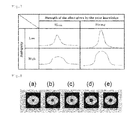
- FIG. 5 shows a distribution diagram of X-ray absorption coefficient for each tissue class.
- FIG. 6 shows an explanatory view showing dependence of probability variables in a CT probability model assuming a hidden state variable to control a motion, a tissue class, and a prior distribution of a tissue class.
- FIG. 7 shows an explanatory view showing an effect achieved by introducing the hidden state variable B.
- FIG. 8 shows an explanatory view showing an object to be deformed over time.
- FIG. 9 shows an explanatory view showing results of reconstruction of CT images relative to an object to be deformed over time.
- FIG. 10 shows an explanatory view showing a chest model involving the motion of a heart.
- FIG. 11 shows an explanatory view showing results of reconstruction of CT images relative to a chest model involving the motion of a heart.
- FIG. 12 shows an effect achieved by introducing the hidden state variable b that controls a distribution of tissue class.
- FIG. 13 shows results of reconstruction of CT images.
- FIG. 14 shows an explanatory view showing prior-art 1.
- FIG. 15 shows an explanatory view showing prior-art 2.
- a motion tracking X-ray CT image processing method and an X-ray CT image processing program of the present invention are to reconstruct an image using statistical estimation based on prior knowledge about a motion and X-ray absorption coefficients.
- FIG. 1 A general CT model and a CT model assuming a motion are described by referring to FIG. 1 .
- FIG. 1 ( 1 ) in the case of a CT model of a conventional filtered back projection (FBP) method, even if a measuring target is an active object such as a heart, acquiring projection is considered to be permitted from one target (CT image) on the assumption that the target is at a standstill.
- the present invention starts from the assumption that a measuring target is a moving object and considers that projections can be achieved from different moving targets as shown in FIG. 1 ( 2 ).
- a motion (deformation) parameter for each pixel of a CT image and estimating a motion (deformation) of each pixel between neighboring frames. More specifically, as shown in FIG. 2 , a motion of a j-th pixel between reconstructed images of different instants of time ((k ⁇ 1)-th frame and k-th frame) is expressed by a vector m j (k) .
- the following describes projection performed on a measuring target that changes smoothly with time in the form of a statistical model and explains that an image can be reconstructed by making the Bayesian estimation based on this statistical model.
- FIGS. 3 and 4 each show a flow of an embodiment of the motion tracking X-ray CT image processing method of the present invention.
- measuring conditions such as a projection image and an X-ray intensity are entered (Step S 11 ).
- Observed photons of the projection image are expressed in a physical model (Step S 12 ).
- a first probability model prior knowledge about the measuring target in every instant of time
- a second probability model observed model of a projection image in every instant of time
- the aforementioned first probability distribution is characterized further by a hidden state variable that expresses a tissue class to which each pixel belongs (Step S 24 ).
- Image construction has an ill-posed problem. This is solved by imposing some limitation on a reconstructed image (X-ray absorption coefficient) as a solution using appropriate prior knowledge.
- This limitation includes temporal limitation to become a significant part of the present invention to be imposed on reconstructed images of neighboring frames.
- the measuring target is assumed to be deformed smoothly with time, so that reconstructed images of neighboring frames make motions responsive to the motion m j (k) of each pixel j, as shown in FIG. 2 . Accordingly, if the reconstructed image in the (k ⁇ 1)-th frame is deformed according to the motion m j (k) a resultant image should have strong temporal correlation with the reconstructed image in the k-th frame.
- This temporal limitation on a reconstructed image is expressed in the form of a probability distribution by both direct temporal correlation between reconstructed images and indirect temporal correlation described later determined through a substance or a tissue assumed to exist potentially behind a reconstructed image, or by either of these temporal correlations.
- a human body is formed of limited substances and tissues such as fat, water, blood, muscle, bone and metal (such as implant).
- tissues can be characterized in the form of a probability distribution showing that each of these tissues is subjected to limitation as it is likely to take on a typical X-ray absorption coefficient.
- Deformation of a tissue in compliance with a motion of a measuring target can be expressed in the same way as that of expressing temporal correlation between reconstructed images of neighboring frames.
- This limitation can be characterized in the form of a probability distribution.
- Knowledge that each tissue has its spatial continuity and a ratio of each tissue to a human body is roughly known can be characterized further as limitation on the tissue in the form of a probability distribution.
- FIG. 6 shows the particulars of FIG. 1 ( 2 ) showing causal relationships between variables according to the probability model of the present invention.
- a scanning time is divided into frames of a significantly short temporal width and a target in each frame is expressed in the same CT image (in FIG. 6 , this is expressed as x (1) , x (2) , . . . ).
- Some hundreds to some thousands of projections are obtained in a short time such as 0.3 seconds as a result of actual scanning. Accordingly, multiple projections y are obtained from one x in each frame.
- a tissue class Z is introduced. While Z is given, the X-ray absorption coefficient x is assumed to take on a value fixed to a certain degree.
- B is a hidden state variable to control the shape of a prior distribution and is given to make the prior distribution more robust.
- FIG. 7 shows effect achieved by introducing the hidden state variable B.
- the prior distribution can be expressed as a mixture distribution by introducing B, so that the vagueness of the prior knowledge and the strength of the effect can be controlled separately.
- MAP estimation a value that maximizes the posterior distribution about the reconstructed image
- Bayesian estimation a value that maximizes the posterior distribution about the reconstructed image
- Bayesian estimation intended to determine the expectation of a posterior probability has higher resistance to disturbance.
- calculation of the posterior probability involves integration calculation of a high-dimensional hidden state variable, so that making the calculation analytically becomes difficult. The difficulty in the calculation is overcome by applying an approximation technique of determining the posterior probability approximately.
- the aforementioned difficulty in the calculation may alternatively be overcome by applying the MAP estimation that maximizes a joint posterior probability containing both a hidden state variable and a reconstructed image without making integration calculation of a hidden state variable.
- y i (t) means the number of photons detected by an i-th detector.
- each projection is divided into K frames and it is assumed that a projection image can be obtained from the same X-ray CT image in each frame.
- a set of indexes about projection data belonging to a k-th frame is called S (k) and an X-ray CT image in the k-th frame is called X (k) .
- An X-ray is exponentially attenuated while it passes through a substance. Accordingly, an average y i (t) or X-ray photons expected to be observed can be expressed by the following Equation 1.
- v is the number of photons emitted from an X-ray source (the number of photons that can be observed in the absence of any object)
- l ij (t) is proportionate to a crossover distance between a projection line projected at an angle ⁇ (t) and detected by the i-th detector and the j-th pixel
- l ij (t) x j is an effective X-ray absorption coefficient of a j-th pixel region regarding an X-ray corresponding to a t-th projection and entering the i-th detector.
- the following describes a physical model relating to photons observed in a projection image, a prior distribution about a tissue functioning as prior knowledge, specifically a parameter distribution showing an existing ratio of each tissue of an imaging target, a parameter distribution showing a typical X-ray absorption coefficient each tissue is likely to take on, a parameter distribution showing a degree of spatial continuity of each tissue, and a parameter distribution showing a degree of temporal continuity of each tissue that are adopted in the motion tracking X-ray CT image processing method and the motion tracking X-ray CT image processing program of the present invention.
- the Poisson distribution is given not for particular limitation but a different physical model capable of showing physical process satisfactorily is also applicable.
- Equation 1 shows an average of the Poisson distribution.
- Equation 2 is established using a result of the Equation 1. Noise is independent for each projection and for each pixel, so that the Equation 2 is expressed as the product of the entirely independent Poisson distributions.
- S (k) is a set of indexes of projections in the k-th frame.
- Equation 3 A 1 is a normalizing constant, and H 1 (Z) is an energy function showing which combination of tissue classes has a maximum likelihood.
- Equation 4 The energy function H 1 (Z) is expressed by the following Equation 4.
- z is a vector showing a tissue class to which the j-th pixel belongs in the k-th frame. If the j-th pixel belongs to a tissue class c, z jc (k) becomes 1 and the other components become 0.
- Z j (k) [z j1 ⁇ k ⁇ , . . . , z jc ⁇ k ⁇ ] ⁇ 0,1 ⁇ c in uppercase means a set of variables z j (k) where z is in lowercase.
- Equation 4 ⁇ (j) means four neighboring pixels of the j-th pixel, and (N)(j) means surrounding 9 ⁇ 9 (pixels) including the j-th pixel.
- the first term shows a ratio of a tissue
- the second term shows spatial smoothness (continuity) of a tissue in the same frame
- the third term shows temporally smooth displacement or deformation of a tissue between neighboring frames.
- G(i,j,m ⁇ k ⁇ ( ⁇ )) is the weight of the third term of H 1 (Z) and is expressed by the following Equation 5.
- G ⁇ ( i , j , m ( k ) ⁇ ( ⁇ ) ) G c time ⁇ exp ⁇ ( - ⁇ ⁇ ⁇ r j - ( r i + m i ( k ) ⁇ ( ⁇ ) ) ⁇ ) ⁇ i ⁇ N ⁇ ( j ) ⁇ ⁇ exp ⁇ ( - ⁇ ⁇ ⁇ r j - ( r i + m i ( k ) ⁇ ( ⁇ ) ⁇ ) ( 5 )
- Equation 6 shows a prior distribution about a reconstructed image X.
- tissue class Z Given the tissue class Z and the hidden state variable B, X-ray absorption coefficients is expressed as the Gaussian distribution. As described below, an average of the Gaussian distribution is set for each tissue class. A degree of approximation to the value of X-ray absorption coefficients typical of each tissue class is controlled by a hidden state variable b.
- b ij (k) and b j2 (k) are variables either of which takes on 1 and the other takes on 0.
- Dispersion parameters ⁇ c1 2 and ⁇ c2 2 have values fixed for each tissue class c: one of the values takes on a large positive value and the other value takes on a small positive value.
- Equation 6 is to determine a degree of approximation of X-ray absorption coefficients given by a reconstructed image to the value of a typical X-ray absorption coefficient. Either of the hidden state variables b j1 ⁇ k ⁇ and b jz ⁇ k ⁇ takes on 1 and the other takes on 0.
- a degree of approximation to the value of X-ray absorption coefficients typical of the tissue class c of a reconstructed image is determined not only by the tissue class c but is also controlled by the values of b j1 ⁇ k ⁇ and b j2 ⁇ k ⁇ for each frame k and each pixel j.
- the hidden state variables b j1 ⁇ k ⁇ and b j2 ⁇ k ⁇ estimated from a given projection image are prepared and used for estimation.
- Equation 6 The second and third terms of the Equation 6 respectively show that a reconstructed image changes spatially smoothly and that the reconstructed image is deformed temporally smoothly based on the motion vector m.
- G x spatial and G x temporal are parameters used to control the strength of spatial correlation and the strength of temporal correlation respectively.
- Equation 8 an energy function H 2 (B) showing a combination of the hidden state variables B of a maximum likelihood is expressed by the following Equation 8.
- the first term of the Equation 8 shows which one of two values is to be taken on more easily by a set B of binary hidden variables, and the second term of the Equation 8 is to control spatial smoothness of a distribution (showing how easy for neighboring pixels to take on the same distribution).
- r i is the position vector of an i-th pixel.
- the X-ray absorption coefficient x, the tissue class z, and the hidden state variable b are estimated as a posterior distribution p(X,Z,B
- D, ⁇ ) can be understood from the Bayes' theorem and is obtained in proportion to the product of a corresponding prior distribution and a likelihood as shown by the following Equation 10.
- D, ⁇ ) eliminates the need for making calculation to obtain a sum of high-dimensional hidden state variables.
- the Bayesian estimation evaluates the posterior distribution p having difficulty in calculation with a test distribution q(X,Z,B) approximate to the posterior distribution p(X,Z,B
- the Bayesian estimation itself is a method of calculating a posterior distribution and using the expectation thereof as an estimate value. A test distribution is not required if a posterior distribution and the expectation thereof can be calculated analytically.
- the test distribution q(X,Z,B) is determined so as to minimize an index called a KL distance indicating approximation of a distribution.
- the KL distance mentioned herein is defined by the following Equation 11.
- Equation 11 ⁇ > q(X,Z,B) is an operator showing integration calculation made on the test distribution q(X,Z,B), meaning that the expectation of the test distribution q(X,Z,B) is to be given.
- a set of distributions that can be calculated is searched for to find a distribution most approximate to the posterior distribution.
- the test distribution q(X,Z,B) can be selected in anyway such that the selected test distribution q(X,Z,B) allows minimization of the KL distance (aforementioned Equation 11) or minimization of approximation. Accordingly, for simple calculation, the test distribution q(X,Z,B) is given assumption of independence among the variables x, z and b called factored assumption as shown by the following Equation 12. Specifically, it is assumed that X, Z and B are independent of each other and are also independent for each frame and for each pixel. On the assumption that a distribution q(x j ) about x j becomes the Gaussian distribution, the test distribution q is modeled as shown by the following Equation 13. Under the foregoing assumptions, alternate optimization of optimizing q(X), q(Z) and q(B) alternately is conducted to obtain an optimum test distribution.
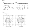
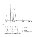
- Example 1 an object to be deformed was formed artificially and a screen was reconstructed based on a projection image of the object by calculator simulation using the motion tracking X-ray CT image processing method of the present invention. A result of the simulation is given below.
- Images in FIG. 8 show an artificially formed image to be deformed.
- a central area of each round screen shows the object (64 ⁇ 64 pixels).
- the deformation of the object is such that the object expanded at a rate differing between the vertical and horizontal directions.
- FIG. 9 shows results of reconstruction of X-ray CT images.
- FIG. 9 is given to compare true images, results of image reconstruction obtained by a filtered back projection (FBP) method employed most frequently as a conventional method not assuming a motion, and results obtained by the image reconstruction of the present invention (suggested method).
- FBP filtered back projection
- PSNR Peak signal to noise ratio
- MSE is a mean squared error between pixel values of the true images and those of the reconstructed images
- max i y i is the maximum pixel value of the true images.
- PSNR is given below each image of FIG. 9 .
- the results of FIG. 9 show that the present invention achieves enhancement of about 3.8 (dB) on average in terms of PSNR.
- the enhancement of about 3.8 (dB) corresponds to an error reduction of about 42% in terms of the mean squared error.
- the resultant images further show that the motion of the object in the center of these images can be expressed satisfactorily.
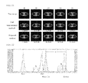
- FIG. 10 shows images about a chest model involving the motion of a heart. These images are given to simulate a chest involving the motion of a heart.
- an ellipse corresponding to the heart above the center expanded such that the ellipse increased gradually, specifically such that the ellipse expanded from (a) to (e) with time.
- the deformation of the ellipse is such that the ellipse expanded at a rate differing between the vertical and horizontal directions.
- the image size of the chest model was 256 ⁇ 256 (pixels) and the number of projections was 360 within 360 degrees.
- FIG. 11 shows results of reconstruction of X-ray CT images.
- FIG. 11 is given to compare true images, results of image reconstruction obtained by a filtered back projection (FBP) method employed most frequently as a conventional method not assuming a motion, and results obtained by the image reconstruction of the present invention (suggested method).
- FBP filtered back projection
- the results of FIG. 11 show that the present invention achieves enhancement of about 1.1 (dB) on average in terms of PSNR.
- the enhancement of about 1.1 (dB) corresponds to an error reduction of about 22% in terms of the mean squared error.
- the resultant images also show that the motion of the heart can be expressed satisfactorily.
- the motion tracking X-ray CT image processing method of the present invention uses a probability model that makes statistical estimation as described above, so that it can incorporate other prior knowledge easily and can increase the quality of image reconstruction.
- making estimation in consideration of X-ray absorption coefficients representative of each tissue can enhance estimation accuracy.
- incorporating a hidden state variable to control the distribution of each tissue class can enhance estimation accuracy and can achieve robust estimation. The following describes experiment conducted to check the effect thereof.
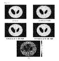
- FIG. 12 shows the X-ray absorption coefficient of an assumed tissue class and the value of the X-ray absorption coefficient of an object actually used in experiment.
- tissue classes including air, fat, muscle and bone, and that there is X-ray absorption coefficients representative of each tissue class.
- the X-ray absorption coefficient of each of the fat class and the bone class was made higher by 10% as indicated by lines with sign a in FIG. 12 .
- Lines with sign b in FIG. 12 show other outliers of X-ray absorption coefficients.
- X-ray CT was conducted on an object taking on a value higher by 5% than an assumed representative X-ray absorption coefficient about fat and muscle belonging to the aforementioned tissue classes.
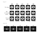
- FIG. 13 is given to compare results of X-ray CT reconstructions given under the aforementioned condition setting.
- ( 1 ) shows a true image
- ( 2 ) shows an image reconstructed by FBP
- ( 3 ) shows an image reconstructed by the present invention in the absence of introduction of the hidden state variable B
- ( 4 ) shows an image reconstructed by the present invention in the presence of introduction of the hidden state variable B
- ( 5 ) shows the estimated hidden state variable B.
- Example 3 an X-ray CT algorithm is described different from that of Examples 1 and 2 described above.
- Equations 15 and 16 can be established in consideration of the situation where noise caused by photoelectric conversion complies with an independent Poisson distribution.
- L (t) is a matrix of m ⁇ n, and the element (i,j) indicates a degree of intersection of a beam of an emitted X-ray to enter a detector i with a pixel j of a reconstructed image.
- b is the number of photons (the number of emitted photons) during blank scanning.
- the Gaussian distribution is applicable as a noise distribution.
- Z and B are each composed of a variable corresponding to each pixel of a CT image:
- the element z jC (k) becomes 1 only if it is applied to the tissue class c to which the pixel j in the k-th frame belongs whereas the other elements become zero. Accordingly, the limitation in the following Equation 18 is satisfied.
- the variable b j (k) to control the distribution shape of the pixel j in the k-th frame is a two-dimensional vector shown in the following Equation 19. A more complicated distribution can be expressed by increasing the number of values that can be taken on by d to any value higher than 2.
- a parameter ⁇ is defined as a parameter indicating a motion.
- a prior distribution is expressed by the following Equation 20.
- Equation 20 includes E(X,Z,B, ⁇ ) that is expressed by the following Equation 21.
- changing G x,cd spatial can control a level of smoothness the pixel value x should acquire if neighboring pixels are in the same tissue class and have the same distribution shape control variable.
- Changing G x,c spatial can also control a level of smoothness the pixel value x should acquire if neighboring pixels are in the same tissue class without depending on their distribution shape control variables.
- G z,c spatial controls how likely neighboring pixels are in the same tissue class. If the neighboring pixels are in the same tissue class, G b,cd spatial further controls the similarity of distribution shape control variables of the neighboring pixels.
- G cd self indicates a combination of tissue class variables and distribution shape control variables that is to be generated easily.
- Variables G x temporal and G z temporal are to respectively control how easily variables x and z makes time transitions in compliance with the motion parameter ⁇ . Further, ⁇ (j) and N(j) each show a set of pixel indexes near the pixel j. Each of ⁇ (j) and N(j) is to determine a range of smoothness and preferably, should be small for saving of a calculation amount.
- Equation 24 The function G(I,j,m ⁇ k ⁇ ,( ⁇ )) is defined by the following Equation 24.
- r i is the position vector of a pixel i
- m i (k) ( ⁇ ) is a motion vector indicating the amount of movement of the pixel i from a frame k to a frame k+1.
- the motion vector m i (k) ( ⁇ ) indicates that the pixel i in the k-th frame is expected to move from a position r i to a position r i +m i (k) ( ⁇ ) in the (k+1)-th frame.
- y is a given positive constant.
- the function G(I,j,m ⁇ k ⁇ ,( ⁇ )) acts as a weighting function to take on a larger value for a pixel of an movement amount more approximate to an expected amount.
- the amount of movement is expressed by the motion vector m i (k) defined by the following Equation 25.
- the Equation 25 shows that a motion vector m i (k) can be obtained by interpolation with the parameter ⁇ i (1) .
- Parameters ⁇ and ⁇ are to control the strength of spatial interpolation and that of temporal interpolation respectively.
- Equation 26 is expressed as a conditional independent expression on condition that P(X
- Z,B, ⁇ ) can be expressed by the following Equation 28 formulated by varying the Equation 27.
- Equation ⁇ ⁇ 26 ⁇ p ⁇ ( X , Z , B
- ⁇ ) 1 A 0 ⁇ exp ⁇ ( - 1 2 ⁇ E ⁇ ( X , Z , B , ⁇ ) ) ( 26 ) ⁇ [ Equation ⁇ ⁇ 27 ]
- ⁇ ) 1 A 0 ⁇ p ⁇ ( X
- Equation 27 includes p(Z,B
- Equation 26 includes E(X,Z,B, ⁇ ) defined by the following Equation 30.
- Equation ⁇ ⁇ 29 ⁇ p ⁇ ( Z , B
- ⁇ ) 1 A 0 ⁇ exp ⁇ ( - 1 2 ⁇ E ⁇ ( Z , B , ⁇ ) ) ( 29 ) ⁇ [ Equation ⁇ ⁇ 30 ]
- the gamma distribution can be used for P(x j ⁇ k ⁇
- the Equation 31 includes u cd and v cd each functioning as a parameter of the gamma distribution.
- the following describes a method that estimates the CT image X, the tissue class variable Z, and the distribution shape control variable B while estimating the motion parameter ⁇ using the aforementioned probability model.
- Equation 32 includes a distribution q (x) called a test distribution.
- ⁇ ) can be expressed by the following Equation 33 using the free energy F.
- the Equation 33 includes a function KL[q(x)
- p(x)] that is defined by Equation 34 if x is a real number. If x is a discrete variable, the function KL is expressed by Equation 35 and is used as a scale for measuring approximation of probability distributions. The function KL always takes on a negative value and becomes zero only if q(x) p(x) is established.
- Equation 37 a log-likelihood can be expressed by the following Equation 37 using free energy.
- the left side of the Equation 37 shows an amount that does not depend on the test distribution q(x). Accordingly, the right side of the Equation 37 does not depend on the formation of a test distribution.
- ⁇ ) - F ⁇ ( q ⁇ ( x ) , ⁇ ) + KL ⁇ [ q ⁇ ( x )
- Equation 39 is established from Equation 38.
- the right side of the Equation 39 shows that a minimum ⁇ log p(y
- a parameter to maximize a log-likelihood can be expressed by the following Equation 40.
- maximum likelihood estimation can be made by minimizing the free energy in the left side with respect to the test distribution q(x) and then minimizing the free energy with respect to the parameter ⁇ .
- a method can be devised from this fact that maximizes free energy with respect to the test distribution q(x) and the parameter ⁇ instead of maximizing a log-likelihood.
- Equation 41 two steps shown in Equation 42 are repeated.
- test distribution q(x) An optimum value of the test distribution q(x) is given from the posterior distribution p(x
- a candidate for q(x) is limited to enable optimization.
- Equation 45 can be obtained analytically by the variational method.
- a solution of Equation 46 cannot be obtained analytically, so that the Gaussian distribution given in the following Equation 47 or the gamma distribution given in the following Equation 48 is assumed as q(x j ⁇ k ⁇ ).
- Equation 49 can be evaluated analytically.
- a parameter (q(x i ⁇ k ⁇ ) of q(x) can be optimized by the non-linear optimization method such as the conjugate gradient method with respect to average ⁇ and dispersion ⁇ 2 in compliance with the Gaussian distribution, or with respect to a parameter ( ⁇ cd , ⁇ cd ) in compliance with the gamma distribution.
- the gamma distribution has preferable characteristics that enable expression of the non-negativity of x.
- the present invention is usefully applicable for X-ray CT devices for medical purposes and industrial purposes.
- One important application of the present invention relates to medical images, particularly, X-ray CT on hearts.
- minute motion of a stage becomes troublesome or deformation of a fluid becomes troublesome in the presence of such a fluid.
- the reduction in motion artifacts achieved by the present invention is expected to be applied usefully for various purposes.
Abstract
Description
p(X,Z,B|D,θ)∝p(D|X)p(X|Z,B)p(Z|θ)p(B) (10)
z jcε{0,1},Σc=1 C z jc=1 (18)
[Equation 19]
b j (k) =[b j1 (k) ,b j2 (k)]T ,b jd (k)ε{0,1}(d=1,2),Σd=1 2 b jd=1 (19)
F(q(x),θ)=−∫q(x)log p(x,y|θ)dx+∫q(x)log q(x)dx (32)
F(q(X)
Claims (13)
Applications Claiming Priority (3)
| Application Number | Priority Date | Filing Date | Title |
|---|---|---|---|
| JP2011-276545 | 2011-12-18 | ||
| JP2011276545 | 2011-12-18 | ||
| PCT/JP2012/008088 WO2013094186A1 (en) | 2011-12-18 | 2012-12-18 | Motion-tracking x-ray ct image processing method and motion-tracking x-ray ct image processing device |
Publications (2)
| Publication Number | Publication Date |
|---|---|
| US20140334705A1 US20140334705A1 (en) | 2014-11-13 |
| US9129431B2 true US9129431B2 (en) | 2015-09-08 |
Family
ID=48668104
Family Applications (1)
| Application Number | Title | Priority Date | Filing Date |
|---|---|---|---|
| US14/366,037 Expired - Fee Related US9129431B2 (en) | 2011-12-18 | 2012-12-18 | Motion-tracking X-ray CT image processing method and motion-tracking X-ray CT image processing device |
Country Status (4)
| Country | Link |
|---|---|
| US (1) | US9129431B2 (en) |
| EP (1) | EP2792303A4 (en) |
| JP (1) | JP6044046B2 (en) |
| WO (1) | WO2013094186A1 (en) |
Cited By (3)
| Publication number | Priority date | Publication date | Assignee | Title |
|---|---|---|---|---|
| US20150279031A1 (en) * | 2014-04-01 | 2015-10-01 | Case Western Reserve University | Imaging control to facilitate tracking objects and/or perform real-time intervention |
| US20160213341A1 (en) * | 2015-01-27 | 2016-07-28 | Septimiu Edmund Salcudean | Dynamic computed tomography imaging of elasticity |
| US9662081B2 (en) | 2013-05-15 | 2017-05-30 | Kyoto University | X-ray CT image processing method, X-ray CT image processing program, and X-ray CT image device |
Families Citing this family (7)
| Publication number | Priority date | Publication date | Assignee | Title |
|---|---|---|---|---|
| US9495770B2 (en) * | 2013-08-14 | 2016-11-15 | University Of Utah Research Foundation | Practical model based CT construction |
| US9848844B2 (en) * | 2015-10-09 | 2017-12-26 | Carestream Health, Inc. | Iterative reconstruction process |
| US11295449B2 (en) | 2016-11-21 | 2022-04-05 | Asto CT, Inc. | Three-dimensional tracking of a target in a body |
| CN108257196A (en) * | 2018-01-11 | 2018-07-06 | 苏州润心医疗器械有限公司 | A kind of blood vessel based on cardiac CT image stretches method for reconstructing |
| JP7273416B2 (en) | 2018-05-01 | 2023-05-15 | 国立大学法人東北大学 | Image processing device, image processing method and image processing program |
| US11204409B2 (en) * | 2018-10-11 | 2021-12-21 | University Of Virginia Patent Foundation | Systems and methods for motion-compensated reconstruction of magnetic resonance images |
| DE102019201079B4 (en) * | 2019-01-29 | 2021-06-17 | Siemens Healthcare Gmbh | Method and imaging device for generating a motion-compensated image, computer program and storage medium |
Citations (7)
| Publication number | Priority date | Publication date | Assignee | Title |
|---|---|---|---|---|
| US20050105682A1 (en) * | 2003-11-15 | 2005-05-19 | Heumann John M. | Highly constrained tomography for automated inspection of area arrays |
| US20060067461A1 (en) * | 2004-09-30 | 2006-03-30 | Zhye Yin | Method and system for CT reconstruction with pre-correction |
| US20100138379A1 (en) * | 2007-05-29 | 2010-06-03 | Mott Christopher | Methods and systems for circadian physiology predictions |
| US20110268335A1 (en) * | 2010-05-03 | 2011-11-03 | Siemens Aktiengesellschaft | Increased Temporal Resolution In The Case Of CT Images By Means Of Iterative View Reconstruction With Limiting Conditions |
| US8401252B2 (en) * | 2006-09-29 | 2013-03-19 | Google Inc. | Video retrieval system for human face content |
| US20130287278A1 (en) * | 2011-01-05 | 2013-10-31 | Koninklijke Philips Electronics N.V. | Method and apparatus to detect and correct motion in list-mode pet data with a gated signal |
| US8824756B2 (en) | 2009-06-30 | 2014-09-02 | University Of Utah Research Foundation | Image reconstruction incorporating organ motion |
Family Cites Families (4)
| Publication number | Priority date | Publication date | Assignee | Title |
|---|---|---|---|---|
| DE10231061A1 (en) * | 2002-07-10 | 2004-01-22 | Philips Intellectual Property & Standards Gmbh | Process and system for improving the information content in an image |
| EP1622085A1 (en) * | 2004-07-19 | 2006-02-01 | Deutsches Krebsforschungszentrum | Method of producing x-ray computer tomography images from limited data of an image object |
| ATE492006T1 (en) * | 2005-07-08 | 2011-01-15 | Wisconsin Alumni Res Found | BACK PROJECTION RECONSTRUCTION METHOD FOR CT IMAGING |
| JP5590548B2 (en) * | 2010-02-03 | 2014-09-17 | 国立大学法人京都大学 | X-ray CT image processing method, X-ray CT program, and X-ray CT apparatus equipped with the program |
-
2012
- 2012-12-18 WO PCT/JP2012/008088 patent/WO2013094186A1/en active Application Filing
- 2012-12-18 US US14/366,037 patent/US9129431B2/en not_active Expired - Fee Related
- 2012-12-18 JP JP2013550113A patent/JP6044046B2/en not_active Expired - Fee Related
- 2012-12-18 EP EP12859055.1A patent/EP2792303A4/en not_active Withdrawn
Patent Citations (7)
| Publication number | Priority date | Publication date | Assignee | Title |
|---|---|---|---|---|
| US20050105682A1 (en) * | 2003-11-15 | 2005-05-19 | Heumann John M. | Highly constrained tomography for automated inspection of area arrays |
| US20060067461A1 (en) * | 2004-09-30 | 2006-03-30 | Zhye Yin | Method and system for CT reconstruction with pre-correction |
| US8401252B2 (en) * | 2006-09-29 | 2013-03-19 | Google Inc. | Video retrieval system for human face content |
| US20100138379A1 (en) * | 2007-05-29 | 2010-06-03 | Mott Christopher | Methods and systems for circadian physiology predictions |
| US8824756B2 (en) | 2009-06-30 | 2014-09-02 | University Of Utah Research Foundation | Image reconstruction incorporating organ motion |
| US20110268335A1 (en) * | 2010-05-03 | 2011-11-03 | Siemens Aktiengesellschaft | Increased Temporal Resolution In The Case Of CT Images By Means Of Iterative View Reconstruction With Limiting Conditions |
| US20130287278A1 (en) * | 2011-01-05 | 2013-10-31 | Koninklijke Philips Electronics N.V. | Method and apparatus to detect and correct motion in list-mode pet data with a gated signal |
Non-Patent Citations (9)
Cited By (5)
| Publication number | Priority date | Publication date | Assignee | Title |
|---|---|---|---|---|
| US9662081B2 (en) | 2013-05-15 | 2017-05-30 | Kyoto University | X-ray CT image processing method, X-ray CT image processing program, and X-ray CT image device |
| US20150279031A1 (en) * | 2014-04-01 | 2015-10-01 | Case Western Reserve University | Imaging control to facilitate tracking objects and/or perform real-time intervention |
| US10026015B2 (en) * | 2014-04-01 | 2018-07-17 | Case Western Reserve University | Imaging control to facilitate tracking objects and/or perform real-time intervention |
| US20160213341A1 (en) * | 2015-01-27 | 2016-07-28 | Septimiu Edmund Salcudean | Dynamic computed tomography imaging of elasticity |
| US10085703B2 (en) * | 2015-01-27 | 2018-10-02 | Septimiu Edmund Salcudean | Dynamic computed tomography imaging of elasticity |
Also Published As
| Publication number | Publication date |
|---|---|
| JP6044046B2 (en) | 2016-12-14 |
| EP2792303A4 (en) | 2015-08-05 |
| EP2792303A1 (en) | 2014-10-22 |
| US20140334705A1 (en) | 2014-11-13 |
| WO2013094186A1 (en) | 2013-06-27 |
| JPWO2013094186A1 (en) | 2015-04-27 |
Similar Documents
| Publication | Publication Date | Title |
|---|---|---|
| US9129431B2 (en) | Motion-tracking X-ray CT image processing method and motion-tracking X-ray CT image processing device | |
| Wolterink et al. | Generative adversarial networks for noise reduction in low-dose CT | |
| Isola et al. | Fully automatic nonrigid registration‐based local motion estimation for motion‐corrected iterative cardiac CT reconstruction | |
| JP6855223B2 (en) | Medical image processing device, X-ray computer tomographic imaging device and medical image processing method | |
| JP4170767B2 (en) | Image processing device | |
| JP2020168352A (en) | Medical apparatus and program | |
| EP2150918B1 (en) | Methods and systems for improving spatial and temporal resolution of computed images of moving objects | |
| JP5833637B2 (en) | Dynamic perfusion CT image data alignment | |
| CN102947861B (en) | For reducing the method and system of noise in low dosage computer tomography | |
| JP5590548B2 (en) | X-ray CT image processing method, X-ray CT program, and X-ray CT apparatus equipped with the program | |
| US8355555B2 (en) | System and method for multi-image based virtual non-contrast image enhancement for dual source CT | |
| US20150154766A1 (en) | Image noise reduction and/or image resolution improvement | |
| US20160055658A1 (en) | Iterative reconstruction for x-ray computed tomography using prior-image induced nonlocal regularization | |
| JP2021013725A (en) | Medical apparatus | |
| Qi et al. | 4-D reconstruction with respiratory correction for gated myocardial perfusion SPECT | |
| Isola et al. | Cardiac motion-corrected iterative cone-beam CT reconstruction using a semi-automatic minimum cost path-based coronary centerline extraction | |
| Qi et al. | A quantitative study of motion estimation methods on 4D cardiac gated SPECT reconstruction | |
| Liang et al. | A self-supervised deep learning network for low-dose CT reconstruction | |
| US8548568B2 (en) | Methods and apparatus for motion compensation | |
| Zhu et al. | Iterative CT reconstruction via minimizing adaptively reweighted total variation | |
| JP4864909B2 (en) | Image processing device | |
| CN117203671A (en) | Iterative image reconstruction improvement based on machine learning | |
| Taubmann et al. | Convex temporal regularizers in cardiac C-arm CT | |
| Taguchi et al. | Toward time resolved 4D cardiac CT imaging with patient dose reduction: estimating the global heart motion | |
| Kamasak | Clustering dynamic PET images on the Gaussian distributed sinogram domain |
Legal Events
| Date | Code | Title | Description |
|---|---|---|---|
| AS | Assignment |
Owner name: NATIONAL UNIVERSITY CORPORATION KYOTO UNIVERSITY, Free format text: ASSIGNMENT OF ASSIGNORS INTEREST;ASSIGNORS:MAEDA, SHINICHI;TANAKA, TAKUMI;ISHII, SHIN;SIGNING DATES FROM 20140616 TO 20140617;REEL/FRAME:033125/0068 |
|
| STCF | Information on status: patent grant |
Free format text: PATENTED CASE |
|
| FEPP | Fee payment procedure |
Free format text: MAINTENANCE FEE REMINDER MAILED (ORIGINAL EVENT CODE: REM.); ENTITY STATUS OF PATENT OWNER: SMALL ENTITY |
|
| LAPS | Lapse for failure to pay maintenance fees |
Free format text: PATENT EXPIRED FOR FAILURE TO PAY MAINTENANCE FEES (ORIGINAL EVENT CODE: EXP.); ENTITY STATUS OF PATENT OWNER: SMALL ENTITY |
|
| STCH | Information on status: patent discontinuation |
Free format text: PATENT EXPIRED DUE TO NONPAYMENT OF MAINTENANCE FEES UNDER 37 CFR 1.362 |
|
| FP | Lapsed due to failure to pay maintenance fee |
Effective date: 20190908 |