US8073210B2 - Methods of smoothing segmented regions and related devices - Google Patents
Methods of smoothing segmented regions and related devices Download PDFInfo
- Publication number
- US8073210B2 US8073210B2 US11/355,321 US35532106A US8073210B2 US 8073210 B2 US8073210 B2 US 8073210B2 US 35532106 A US35532106 A US 35532106A US 8073210 B2 US8073210 B2 US 8073210B2
- Authority
- US
- United States
- Prior art keywords
- lung
- automated method
- boundary
- images
- segmented
- Prior art date
- Legal status (The legal status is an assumption and is not a legal conclusion. Google has not performed a legal analysis and makes no representation as to the accuracy of the status listed.)
- Expired - Fee Related, expires
Links
- 238000000034 method Methods 0.000 title claims abstract description 98
- 238000009499 grossing Methods 0.000 title claims abstract description 81
- 210000004072 lung Anatomy 0.000 claims abstract description 176
- 210000000621 bronchi Anatomy 0.000 claims description 15
- 238000002591 computed tomography Methods 0.000 claims description 15
- 238000004458 analytical method Methods 0.000 claims description 10
- 230000000877 morphologic effect Effects 0.000 claims description 8
- 210000003437 trachea Anatomy 0.000 claims description 6
- 230000011218 segmentation Effects 0.000 description 18
- 210000001370 mediastinum Anatomy 0.000 description 13
- 238000004422 calculation algorithm Methods 0.000 description 7
- 238000012545 processing Methods 0.000 description 6
- 238000007373 indentation Methods 0.000 description 5
- 238000003384 imaging method Methods 0.000 description 4
- 230000007704 transition Effects 0.000 description 4
- 210000003484 anatomy Anatomy 0.000 description 3
- 238000013459 approach Methods 0.000 description 3
- 230000008859 change Effects 0.000 description 3
- 230000000694 effects Effects 0.000 description 3
- 210000001147 pulmonary artery Anatomy 0.000 description 3
- 230000002685 pulmonary effect Effects 0.000 description 3
- 238000010989 Bland-Altman Methods 0.000 description 2
- 230000008901 benefit Effects 0.000 description 2
- 210000004204 blood vessel Anatomy 0.000 description 2
- 238000004364 calculation method Methods 0.000 description 2
- 210000000038 chest Anatomy 0.000 description 2
- 238000004040 coloring Methods 0.000 description 2
- 238000013500 data storage Methods 0.000 description 2
- 238000005516 engineering process Methods 0.000 description 2
- 230000001788 irregular Effects 0.000 description 2
- 238000002372 labelling Methods 0.000 description 2
- 238000002595 magnetic resonance imaging Methods 0.000 description 2
- 230000008569 process Effects 0.000 description 2
- 230000002792 vascular Effects 0.000 description 2
- 108010028984 3-isopropylmalate dehydratase Proteins 0.000 description 1
- 238000012935 Averaging Methods 0.000 description 1
- 230000009471 action Effects 0.000 description 1
- 238000007792 addition Methods 0.000 description 1
- 230000006399 behavior Effects 0.000 description 1
- 238000012512 characterization method Methods 0.000 description 1
- 238000010276 construction Methods 0.000 description 1
- 230000001054 cortical effect Effects 0.000 description 1
- 238000001514 detection method Methods 0.000 description 1
- 238000002059 diagnostic imaging Methods 0.000 description 1
- 238000000605 extraction Methods 0.000 description 1
- 230000006870 function Effects 0.000 description 1
- 230000036541 health Effects 0.000 description 1
- 238000010191 image analysis Methods 0.000 description 1
- 238000003709 image segmentation Methods 0.000 description 1
- 238000001727 in vivo Methods 0.000 description 1
- 230000002452 interceptive effect Effects 0.000 description 1
- 238000005304 joining Methods 0.000 description 1
- 238000012729 kappa analysis Methods 0.000 description 1
- 230000004807 localization Effects 0.000 description 1
- 239000000463 material Substances 0.000 description 1
- 238000012986 modification Methods 0.000 description 1
- 230000004048 modification Effects 0.000 description 1
- 238000009206 nuclear medicine Methods 0.000 description 1
- 230000003287 optical effect Effects 0.000 description 1
- 238000002600 positron emission tomography Methods 0.000 description 1
- 238000007781 pre-processing Methods 0.000 description 1
- 210000003492 pulmonary vein Anatomy 0.000 description 1
- 238000011002 quantification Methods 0.000 description 1
- 238000004445 quantitative analysis Methods 0.000 description 1
- 238000002601 radiography Methods 0.000 description 1
- 230000008707 rearrangement Effects 0.000 description 1
- 230000009467 reduction Effects 0.000 description 1
- 238000011160 research Methods 0.000 description 1
- 230000000717 retained effect Effects 0.000 description 1
- 238000005096 rolling process Methods 0.000 description 1
- 238000000926 separation method Methods 0.000 description 1
- 238000011524 similarity measure Methods 0.000 description 1
- 238000002603 single-photon emission computed tomography Methods 0.000 description 1
- 238000006467 substitution reaction Methods 0.000 description 1
- 238000003325 tomography Methods 0.000 description 1
Images


Classifications
-
- G—PHYSICS
- G06—COMPUTING; CALCULATING OR COUNTING
- G06T—IMAGE DATA PROCESSING OR GENERATION, IN GENERAL
- G06T5/00—Image enhancement or restoration
- G06T5/20—Image enhancement or restoration by the use of local operators
- G06T5/30—Erosion or dilatation, e.g. thinning
-
- G06T5/70—
-
- G—PHYSICS
- G06—COMPUTING; CALCULATING OR COUNTING
- G06T—IMAGE DATA PROCESSING OR GENERATION, IN GENERAL
- G06T7/00—Image analysis
- G06T7/10—Segmentation; Edge detection
- G06T7/12—Edge-based segmentation
-
- G—PHYSICS
- G06—COMPUTING; CALCULATING OR COUNTING
- G06T—IMAGE DATA PROCESSING OR GENERATION, IN GENERAL
- G06T7/00—Image analysis
- G06T7/10—Segmentation; Edge detection
- G06T7/155—Segmentation; Edge detection involving morphological operators
-
- G—PHYSICS
- G06—COMPUTING; CALCULATING OR COUNTING
- G06T—IMAGE DATA PROCESSING OR GENERATION, IN GENERAL
- G06T2207/00—Indexing scheme for image analysis or image enhancement
- G06T2207/10—Image acquisition modality
- G06T2207/10072—Tomographic images
- G06T2207/10081—Computed x-ray tomography [CT]
-
- G—PHYSICS
- G06—COMPUTING; CALCULATING OR COUNTING
- G06T—IMAGE DATA PROCESSING OR GENERATION, IN GENERAL
- G06T2207/00—Indexing scheme for image analysis or image enhancement
- G06T2207/20—Special algorithmic details
- G06T2207/20036—Morphological image processing
- G06T2207/20044—Skeletonization; Medial axis transform
-
- G—PHYSICS
- G06—COMPUTING; CALCULATING OR COUNTING
- G06T—IMAGE DATA PROCESSING OR GENERATION, IN GENERAL
- G06T2207/00—Indexing scheme for image analysis or image enhancement
- G06T2207/30—Subject of image; Context of image processing
- G06T2207/30004—Biomedical image processing
- G06T2207/30061—Lung
Abstract
Description
A1=Atrachea∪ARMB∪ALMB,
A2=A1⊕B4,
L1=L|A2.
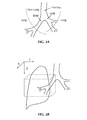
3.0 3-D Bounding Structure Around the Mediastinal Boundary of the Segmented Lung Volume for Localization of Smoothing
where RZ is the radius of a given disc and can be expressed in pixels, Z is the current slice (image) number or position and can be expressed in mm, and ΔZ is the distance between slices (images) and can be expressed in mm.
where Conncomp(.) processes an image and returns labeled, 3-D connected components R1, R2, R3, . . . RN.
5.0 Comparison with Manual Analysis
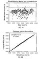
where Amanual and Aautomatic represent manually traced and automatically-detected regions, respectively, and ∥ represents the area of a region. The area overlap index κ is derived from the kappa coefficient calculation, which is used in reliability studies, and has been used in the area of image segmentation to measure the agreement between different classifications [7]. We computed κ for all transverse slices on which a manual segmentation was performed. The results are shown in
where (xi M, yi M) is the manually-defined border pixel location and (xj A, yj A) is a computer-defined border pixel location [8].
where l is the number of points on the manually-defined borders. The results are shown in
Thus in order to calculate the curvature we calculated the first and second derivatives of the signals x(t) and y(t). We used a multiscale curvature estimation technique using the Fourier transform of the complex contour representation u(t)=x(t)+jy(t), described in [10]. This technique allows the calculation of curvature of a contour at multiple scales, given by
where Im refers to the imaginary part. The parameter a describes the scale factor, which is actually the standard deviation term of the Gaussian function which is convolved with u(t) to give
where * is a convolution. By varying the parameter a, we can get the value of curvature at points on the contour at different scales.
- 1. M. S. Brown, M. F. McNitt-Gray, N. J. Mankovich, J. G. Goldin, J. Hiller, L. S. Wilson, and D. R. Aberle, Method for segmenting chest CT image data using an anatomic model: Preliminary results, IEEE Trans. Medical Imaging December 1997; 16: 828-839.
- 2. S. Hu, J. M. Reinhardt, and E. A. Hoffman, Automatic lung segmentation for accurate quantitation of volumetric X-ray CT images, IEEE Trans. Medical Imaging 2001; 20(6): 490-498.
- 3. S. G. Armato, M. L. Giger, C. J. Moran, J. T. Blackburn, K. Doi, and H. MacMahon, Computerized detection of pulmonary nodules on CT scans, Radiographics 1999; 19(5): 1301-1311.
- 4. Y. Li, Y. Zheng, M. Kallergi, and R. A. Clark, Improved method for automatic detection of lung regions in chest radiographs, Acad. Radiology 2001; 8: 629-638.
- 5.
Appendix 1 to the '184 provisional application.Appendix 1 constitutes aspects of J. Tschirren, Segmentation, Anatomical Labeling, Branchpoint Matching, and Quantitative Analysis of Human Airway Trees in Volumetric CT Images. Ph.D. thesis, The University of Iowa, 2003. - 6. K. Palagyi, J. Tschirren, and M. Sonka, Quantitative analysis of intrathoracic airway trees: methods and validation, in Proc. 18th Int. Conf. Information Processing in Medical Imaging, IPMI 2003, Ambleside, UK, Lecture Notes in Computer Science 2003; 2732(7): 222-233.
- 7. B. M. Dawant, S. L. Hartmann, J. P. Thirion, F. Maes, D. Vandermeulen, and P. Demaerel, Automatic 3-D segmentation of internal structures of the head in MR images using a combination of similarity and free-form transformation: Part I, methodology and validation on normal subjects, IEEE Trans. Medical Imaging 1999; 18(10): 909-916.
- 8. V. Chalana and Y. Kim, A methodology for evaluation of boundary detection algorithms on medical images, IEEE Trans. Medical Imaging 1997; 16(5): 642-652.
- 9. J. M. Bland and D. G. Altman, Statistical methods for assessing agreement between two methods of clinical measurement, Lancet 1986; 1(8476): 307-310.
- 10. L. da Fontoura Costa and R. M. Cesar Jr., Shape analysis and classification, CRC Press, Boca Raton, Fla., 2001.
- 11. S. Ukil and J. M. Reinhardt, Smoothing Lung Segmentation Surfaces in 3-D X-ray CT images using Anatomic Guidance, SPIE Conf. Medical Imaging 2004, 5370: 1066-1075.
Claims (52)
Priority Applications (1)
| Application Number | Priority Date | Filing Date | Title |
|---|---|---|---|
| US11/355,321 US8073210B2 (en) | 2005-02-14 | 2006-02-14 | Methods of smoothing segmented regions and related devices |
Applications Claiming Priority (2)
| Application Number | Priority Date | Filing Date | Title |
|---|---|---|---|
| US65269505P | 2005-02-14 | 2005-02-14 | |
| US11/355,321 US8073210B2 (en) | 2005-02-14 | 2006-02-14 | Methods of smoothing segmented regions and related devices |
Publications (2)
| Publication Number | Publication Date |
|---|---|
| US20070053562A1 US20070053562A1 (en) | 2007-03-08 |
| US8073210B2 true US8073210B2 (en) | 2011-12-06 |
Family
ID=37830077
Family Applications (1)
| Application Number | Title | Priority Date | Filing Date |
|---|---|---|---|
| US11/355,321 Expired - Fee Related US8073210B2 (en) | 2005-02-14 | 2006-02-14 | Methods of smoothing segmented regions and related devices |
Country Status (1)
| Country | Link |
|---|---|
| US (1) | US8073210B2 (en) |
Cited By (2)
| Publication number | Priority date | Publication date | Assignee | Title |
|---|---|---|---|---|
| US20130169674A1 (en) * | 2012-01-04 | 2013-07-04 | Samsung Medison Co., Ltd. | Method and apparatus for displaying medical image |
| US20140035916A1 (en) * | 2012-08-03 | 2014-02-06 | Sean Murphy | Preparation and display of derived series of medical images |
Families Citing this family (14)
| Publication number | Priority date | Publication date | Assignee | Title |
|---|---|---|---|---|
| WO2006085254A1 (en) * | 2005-02-11 | 2006-08-17 | Koninklijke Philips Electronics N.V. | Method of automatic extraction of the pulmonary artery tree from 3d medical images |
| US7756316B2 (en) * | 2005-12-05 | 2010-07-13 | Siemens Medicals Solutions USA, Inc. | Method and system for automatic lung segmentation |
| US11275242B1 (en) | 2006-12-28 | 2022-03-15 | Tipping Point Medical Images, Llc | Method and apparatus for performing stereoscopic rotation of a volume on a head display unit |
| US11228753B1 (en) | 2006-12-28 | 2022-01-18 | Robert Edwin Douglas | Method and apparatus for performing stereoscopic zooming on a head display unit |
| US10795457B2 (en) | 2006-12-28 | 2020-10-06 | D3D Technologies, Inc. | Interactive 3D cursor |
| US11315307B1 (en) | 2006-12-28 | 2022-04-26 | Tipping Point Medical Images, Llc | Method and apparatus for performing rotating viewpoints using a head display unit |
| US8219179B2 (en) | 2008-03-06 | 2012-07-10 | Vida Diagnostics, Inc. | Systems and methods for navigation within a branched structure of a body |
| JP2012048392A (en) * | 2010-08-25 | 2012-03-08 | Canon Inc | Image processing apparatus and image processing method |
| EP2639763B1 (en) * | 2012-03-15 | 2015-10-21 | Agfa Healthcare | Method, Apparatus and System for Localizing a Spine |
| WO2017136781A1 (en) * | 2016-02-05 | 2017-08-10 | Pulmonx Corporation | Methods, systems, and devices for analyzing lung imaging data |
| US10643333B2 (en) | 2018-04-12 | 2020-05-05 | Veran Medical Technologies | Apparatuses and methods for navigation in and Local segmentation extension of anatomical treelike structures |
| WO2020097425A2 (en) | 2018-11-09 | 2020-05-14 | Vida Diagnostics, Inc. | Cut-surface display of tubular structures |
| CN110853011B (en) * | 2019-11-11 | 2022-05-27 | 河北工业大学 | Method for constructing convolutional neural network model for pulmonary nodule detection |
| US11875459B2 (en) | 2020-04-07 | 2024-01-16 | Vida Diagnostics, Inc. | Subject specific coordinatization and virtual navigation systems and methods |
Citations (15)
| Publication number | Priority date | Publication date | Assignee | Title |
|---|---|---|---|---|
| US6246784B1 (en) * | 1997-08-19 | 2001-06-12 | The United States Of America As Represented By The Department Of Health And Human Services | Method for segmenting medical images and detecting surface anomalies in anatomical structures |
| US6282307B1 (en) * | 1998-02-23 | 2001-08-28 | Arch Development Corporation | Method and system for the automated delineation of lung regions and costophrenic angles in chest radiographs |
| US20020114503A1 (en) | 2001-02-17 | 2002-08-22 | Siemens Aktiengesellschaft | Method and apparatus for processing a computed tomography image of a lung obtained using contrast agent |
| US20030022367A1 (en) | 2001-07-12 | 2003-01-30 | Chunhui Xu | Cardiomyocyte precursors from human embryonic stem cells |
| US20030095696A1 (en) | 2001-09-14 | 2003-05-22 | Reeves Anthony P. | System, method and apparatus for small pulmonary nodule computer aided diagnosis from computed tomography scans |
| US20030099390A1 (en) | 2001-11-23 | 2003-05-29 | Xiaolan Zeng | Lung field segmentation from CT thoracic images |
| US20030099389A1 (en) | 2001-11-23 | 2003-05-29 | Xiaolan Zeng | Pleural nodule detection from CT thoracic images |
| US20030099384A1 (en) | 2001-11-23 | 2003-05-29 | Xiaolan Zeng | Detection and analysis of lesions in contact with a structural boundary |
| US20030223627A1 (en) | 2001-10-16 | 2003-12-04 | University Of Chicago | Method for computer-aided detection of three-dimensional lesions |
| US6813375B2 (en) * | 2001-06-15 | 2004-11-02 | University Of Chicago | Automated method and system for the delineation of the chest wall in computed tomography scans for the assessment of pleural disease |
| US20050207630A1 (en) * | 2002-02-15 | 2005-09-22 | The Regents Of The University Of Michigan Technology Management Office | Lung nodule detection and classification |
| US20060030958A1 (en) | 2004-05-05 | 2006-02-09 | University Of Iowa Research Foundation | Methods and devices for labeling and/or matching |
| US7315639B2 (en) * | 2004-03-03 | 2008-01-01 | Mevis Gmbh | Method of lung lobe segmentation and computer system |
| US7483023B2 (en) * | 2005-03-17 | 2009-01-27 | Siemens Medical Solutions Usa, Inc. | Model based adaptive multi-elliptical approach: a one click 3D segmentation approach |
| US7548649B2 (en) * | 2005-01-25 | 2009-06-16 | Siemens Medical Solutions Usa, Inc. | Multidimensional segmentation based on adaptive bounding box and ellipsoid models |
-
2006
- 2006-02-14 US US11/355,321 patent/US8073210B2/en not_active Expired - Fee Related
Patent Citations (18)
| Publication number | Priority date | Publication date | Assignee | Title |
|---|---|---|---|---|
| US6246784B1 (en) * | 1997-08-19 | 2001-06-12 | The United States Of America As Represented By The Department Of Health And Human Services | Method for segmenting medical images and detecting surface anomalies in anatomical structures |
| US6282307B1 (en) * | 1998-02-23 | 2001-08-28 | Arch Development Corporation | Method and system for the automated delineation of lung regions and costophrenic angles in chest radiographs |
| US6724925B2 (en) * | 1998-02-23 | 2004-04-20 | Arch Development Corporation | Method and system for the automated delineation of lung regions and costophrenic angles in chest radiographs |
| US20020114503A1 (en) | 2001-02-17 | 2002-08-22 | Siemens Aktiengesellschaft | Method and apparatus for processing a computed tomography image of a lung obtained using contrast agent |
| US6813375B2 (en) * | 2001-06-15 | 2004-11-02 | University Of Chicago | Automated method and system for the delineation of the chest wall in computed tomography scans for the assessment of pleural disease |
| US20030022367A1 (en) | 2001-07-12 | 2003-01-30 | Chunhui Xu | Cardiomyocyte precursors from human embryonic stem cells |
| US20030095696A1 (en) | 2001-09-14 | 2003-05-22 | Reeves Anthony P. | System, method and apparatus for small pulmonary nodule computer aided diagnosis from computed tomography scans |
| US20030223627A1 (en) | 2001-10-16 | 2003-12-04 | University Of Chicago | Method for computer-aided detection of three-dimensional lesions |
| US20030099384A1 (en) | 2001-11-23 | 2003-05-29 | Xiaolan Zeng | Detection and analysis of lesions in contact with a structural boundary |
| US20030099389A1 (en) | 2001-11-23 | 2003-05-29 | Xiaolan Zeng | Pleural nodule detection from CT thoracic images |
| US6766043B2 (en) * | 2001-11-23 | 2004-07-20 | R2 Technology, Inc. | Pleural nodule detection from CT thoracic images |
| US20030099390A1 (en) | 2001-11-23 | 2003-05-29 | Xiaolan Zeng | Lung field segmentation from CT thoracic images |
| US20050207630A1 (en) * | 2002-02-15 | 2005-09-22 | The Regents Of The University Of Michigan Technology Management Office | Lung nodule detection and classification |
| US20090252395A1 (en) * | 2002-02-15 | 2009-10-08 | The Regents Of The University Of Michigan | System and Method of Identifying a Potential Lung Nodule |
| US7315639B2 (en) * | 2004-03-03 | 2008-01-01 | Mevis Gmbh | Method of lung lobe segmentation and computer system |
| US20060030958A1 (en) | 2004-05-05 | 2006-02-09 | University Of Iowa Research Foundation | Methods and devices for labeling and/or matching |
| US7548649B2 (en) * | 2005-01-25 | 2009-06-16 | Siemens Medical Solutions Usa, Inc. | Multidimensional segmentation based on adaptive bounding box and ellipsoid models |
| US7483023B2 (en) * | 2005-03-17 | 2009-01-27 | Siemens Medical Solutions Usa, Inc. | Model based adaptive multi-elliptical approach: a one click 3D segmentation approach |
Non-Patent Citations (28)
| Title |
|---|
| Armato et al., "Computerized detection of pulmonary nodules on CT scans," Radiographics, 19(5): 1301-1311, 1999. |
| Bland and Altman, "Statistical methods for assessing agreement between two methods of clinical measurement," Lancet, 1(8476): 307-310, 1986. |
| Brown et al, Method for Segmenting Chest CT Image Data Using an Anatomical Model: Preliminary Results, IEEE Transactions on Medical Imaging, Vol. 16, No. 6, December 1997. * |
| Brown et al., "Method for segmenting chest CT image data using an anatomic model: Preliminary results," IEEE Trans. Medical Imaging, 16: 828-839, 1997. |
| Chalana and Kim, "A methodology for evaluation of boundary detection algorithms on medical images," IEEE Trans. Medical Imaging,;16(5): 642-652, 1997. |
| da Fontoura Costa and Cesar Jr., "Table of Contents," In: Shape Analysis and Classification: Theory and Practice, CRC Press, Boca Raton, Florida, 2001. |
| Dawant et al., "Automatic 3-D segmentation of internal structures of the head in MR images using a combination of similarity and free-form transformation: Part I, methodology and validation on normal subjects," IEEE Trans. Medical Imaging 18(10):909-916, 1999. |
| Dogdas et al., "Segmentation of skull in 3D human MR images using mathematical morphology," Proc. SPIE Medical Imaging 2002: Image Processing, 4684:1553-1562, 2002. |
| Fetita et al , 3D Automated Lung Nodule Segmentation in HRCT, MICCAI 2003, LNCS 2878, pp. 626-634, Springer-Verlag Berlin Heidelberg 2003. * |
| Höhne and Hanson, "Interactive 3D segmentation of mri and ct volumes using morphological operations," Computer Assisted Tomography, 16:258-294, 1992. |
| Hu et al., "Automatic lung segmentation for accurate quantitation of volumetric X-ray CT images," IEEE Trans. Medical Imaging, 20(6): 490-498, 2001. |
| Hunter, "Gray-scale morphology," Nov. 29, 2002. |
| Kuhnigk et al., "Lung lobe segmentation by anatomy-guided 3D watershed transform," Medical Imaging 2003, 4, 2003. |
| Li et al., "Improved method for automatic detection of lung regions in chest radiographs," Acad. Radiology, 8: 629-638, 2001. |
| Lohmann and von Cramon, "Austomatic labelling of the human cortical surface using sulcal basins," Proc.of SPIE, 5032:1482-1490, 2003. |
| Lürig and Ertl, "Hierarchical volume analysis and visualization based on morphological operators," IEEE Visualization Archive, Proceedings of the Conference on Visualization '98, pp. 335-341, 1998. |
| Megalooikonomou et al., "Fast and effective characterization of 3D region data," Proceedings of the International Conference on Image Processing, 1:I-424, 2002. |
| Pal'agyi et al, Quantitative Analysis of Intrathoracic Airway Trees: Methods and Validation, IPMI 2003, LNCS 2732, pp. 222-233, 2003, Springer-Verlag Berlin Heidelberg 2003. * |
| Palágyi et al., "Quantitative analysis of intrathoracic airway trees: methods and validation, in Proc. 18th Int. Conf. Information Processing in Medical Imaging," IPMI 2003, Ambleside, UK, Lecture Notes in Computer Science, 2732(7): 222-233, 2003. |
| Ruetter et al., "Nonlinear edge preserving smoothing and segmentation of 4-D medical images via scale-space fingerprint analysis," IPMI 2001: 17th International Conference, pp. 431-437. |
| Salfity et al., "A computer-aided diagnosis method for automated detection and classification of clustered microcalcifications in mammograms," Proceedings of the Argentine Symposium on Healthcare Informatics, Tandil, pp. 41-47, 2000. |
| Silva et al., "Segmentation and reconstruction of the pulmonary parenchyma," Technical Report, Vision and Graphics Laboratory, Institute of Pure and Applied Mathematics, Rio de Janeiro, 2002. |
| Tschirren et al., "Airway tree segmentation using adaptive regions of interest," Medical Imaging 2004: Physiology, Function, and Structure from Medical Images, Proceedings of the SPIE, vol. 5369, pp. 117-124 (2004). |
| Tschirren, "Segmentation, anatomical labeling, branchpoint matching, and quantitative analysis of human airway trees in volumetric CT images," Ph.D. Thesis, University of Iowa, Aug. 2003. |
| Tschirren, "Segmentation, branchpoint matching and anatomical labeling of human airway trees in volumetric CT images," slides presented at Ph.D. defense, which occurred on Jul. 10, 2003. |
| U.S. Appl. No. 60/568,184, filed May 5, 2004, Tschirren et al. |
| Ukil and Reinhardt, "Smoothing Lung Segmentation Surfaces in 3-D X-ray CT images using Anatomic Guidance," SPIE Conf. Medical Imaging, 5370: 1066-1075, 2004. |
| Yang and Hansell, "CT image enhancement with wavelet analysis for the detection of small airways disease," IEEE Transaction on Medical Imaging, 16:953-961, 1997. |
Cited By (3)
| Publication number | Priority date | Publication date | Assignee | Title |
|---|---|---|---|---|
| US20130169674A1 (en) * | 2012-01-04 | 2013-07-04 | Samsung Medison Co., Ltd. | Method and apparatus for displaying medical image |
| US20140035916A1 (en) * | 2012-08-03 | 2014-02-06 | Sean Murphy | Preparation and display of derived series of medical images |
| US9147280B2 (en) * | 2012-08-03 | 2015-09-29 | Kabushiki Kaisha Toshiba | Preparation and display of derived series of medical images |
Also Published As
| Publication number | Publication date |
|---|---|
| US20070053562A1 (en) | 2007-03-08 |
Similar Documents
| Publication | Publication Date | Title |
|---|---|---|
| US8073210B2 (en) | Methods of smoothing segmented regions and related devices | |
| US9996922B2 (en) | Image processing of organs depending on organ intensity characteristics | |
| Gonçalves et al. | Hessian based approaches for 3D lung nodule segmentation | |
| JP6877868B2 (en) | Image processing equipment, image processing method and image processing program | |
| Pu et al. | A computational geometry approach to automated pulmonary fissure segmentation in CT examinations | |
| Van Rikxoort et al. | Automated segmentation of pulmonary structures in thoracic computed tomography scans: a review | |
| US9117259B2 (en) | Method and system for liver lesion detection | |
| Azhari et al. | Brain tumor detection and localization in magnetic resonance imaging | |
| Wang et al. | Pulmonary fissure segmentation on CT | |
| Wan et al. | Automated coronary artery tree segmentation in X-ray angiography using improved Hessian based enhancement and statistical region merging | |
| Xiao et al. | Pulmonary fissure detection in CT images using a derivative of stick filter | |
| Lassen et al. | Lung and lung lobe segmentation methods at Fraunhofer MEVIS | |
| Doel et al. | Pulmonary lobe segmentation from CT images using fissureness, airways, vessels and multilevel B-splines | |
| Tozaki et al. | Pulmonary organs analysis for differential diagnosis based on thoracic thin-section CT images | |
| Szymczak et al. | Coronary vessel trees from 3d imagery: a topological approach | |
| US20030099384A1 (en) | Detection and analysis of lesions in contact with a structural boundary | |
| Park | Connectivity-based local adaptive thresholding for carotid artery segmentation using MRA images | |
| Van Dongen et al. | Automatic segmentation of pulmonary vasculature in thoracic CT scans with local thresholding and airway wall removal | |
| Sakellarios et al. | Novel methodology for 3D reconstruction of carotid arteries and plaque characterization based upon magnetic resonance imaging carotid angiography data | |
| Gao et al. | Accurate lung segmentation for X-ray CT images | |
| Ukil et al. | Smoothing lung segmentation surfaces in three-dimensional x-ray ct images using anatomic guidance1 | |
| Ukil et al. | Smoothing lung segmentation surfaces in 3D X-ray CT images using anatomic guidance | |
| Peng et al. | Pulmonary lobe segmentation in CT images based on lung anatomy knowledge | |
| Kuhnigk et al. | Fast automated segmentation and reproducible volumetry of pulmonary metastases in CT-scans for therapy monitoring | |
| Kiraly et al. | 3D human airway segmentation for virtual bronchoscopy |
Legal Events
| Date | Code | Title | Description |
|---|---|---|---|
| AS | Assignment |
Owner name: NATIONAL SCIENCE FOUNDATION, VIRGINIA Free format text: CONFIRMATORY LICENSE;ASSIGNOR:UNIVERSITY OF IOWA;REEL/FRAME:017748/0824 Effective date: 20060505 |
|
| AS | Assignment |
Owner name: UNIVERSITY OF IOWA RESEARCH FOUNDATION, IOWA Free format text: ASSIGNMENT OF ASSIGNORS INTEREST;ASSIGNORS:REINHARDT, JOSEPH;SONKA, MILAN;MCLENNAN, GEOFFREY;AND OTHERS;SIGNING DATES FROM 20061006 TO 20061101;REEL/FRAME:018485/0084 Owner name: UNIVERSITY OF IOWA RESEARCH FOUNDATION, IOWA Free format text: ASSIGNMENT OF ASSIGNORS INTEREST;ASSIGNORS:REINHARDT, JOSEPH;SONKA, MILAN;MCLENNAN, GEOFFREY;AND OTHERS;REEL/FRAME:018485/0084;SIGNING DATES FROM 20061006 TO 20061101 |
|
| CC | Certificate of correction | ||
| AS | Assignment |
Owner name: NATIONAL SCIENCE FOUNDATION, VIRGINIA Free format text: CONFIRMATORY LICENSE;ASSIGNOR:UNIVERSITY OF IOWA;REEL/FRAME:028548/0594 Effective date: 20120521 |
|
| REMI | Maintenance fee reminder mailed | ||
| LAPS | Lapse for failure to pay maintenance fees | ||
| STCH | Information on status: patent discontinuation |
Free format text: PATENT EXPIRED DUE TO NONPAYMENT OF MAINTENANCE FEES UNDER 37 CFR 1.362 |
|
| FP | Lapsed due to failure to pay maintenance fee |
Effective date: 20151206 |