US7846096B2 - Method for monitoring of medical treatment using pulse-echo ultrasound - Google Patents
Method for monitoring of medical treatment using pulse-echo ultrasound Download PDFInfo
- Publication number
- US7846096B2 US7846096B2 US10/721,034 US72103403A US7846096B2 US 7846096 B2 US7846096 B2 US 7846096B2 US 72103403 A US72103403 A US 72103403A US 7846096 B2 US7846096 B2 US 7846096B2
- Authority
- US
- United States
- Prior art keywords
- ultrasound
- anatomical tissue
- medical treatment
- signals
- signal
- Prior art date
- Legal status (The legal status is an assumption and is not a legal conclusion. Google has not performed a legal analysis and makes no representation as to the accuracy of the status listed.)
- Expired - Fee Related
Links
Images


Classifications
-
- A—HUMAN NECESSITIES
- A61—MEDICAL OR VETERINARY SCIENCE; HYGIENE
- A61B—DIAGNOSIS; SURGERY; IDENTIFICATION
- A61B8/00—Diagnosis using ultrasonic, sonic or infrasonic waves
- A61B8/08—Detecting organic movements or changes, e.g. tumours, cysts, swellings
-
- A—HUMAN NECESSITIES
- A61—MEDICAL OR VETERINARY SCIENCE; HYGIENE
- A61B—DIAGNOSIS; SURGERY; IDENTIFICATION
- A61B8/00—Diagnosis using ultrasonic, sonic or infrasonic waves
- A61B8/12—Diagnosis using ultrasonic, sonic or infrasonic waves in body cavities or body tracts, e.g. by using catheters
-
- A—HUMAN NECESSITIES
- A61—MEDICAL OR VETERINARY SCIENCE; HYGIENE
- A61B—DIAGNOSIS; SURGERY; IDENTIFICATION
- A61B8/00—Diagnosis using ultrasonic, sonic or infrasonic waves
- A61B8/13—Tomography
- A61B8/14—Echo-tomography
-
- A—HUMAN NECESSITIES
- A61—MEDICAL OR VETERINARY SCIENCE; HYGIENE
- A61B—DIAGNOSIS; SURGERY; IDENTIFICATION
- A61B8/00—Diagnosis using ultrasonic, sonic or infrasonic waves
- A61B8/44—Constructional features of the ultrasonic, sonic or infrasonic diagnostic device
- A61B8/4483—Constructional features of the ultrasonic, sonic or infrasonic diagnostic device characterised by features of the ultrasound transducer
- A61B8/4488—Constructional features of the ultrasonic, sonic or infrasonic diagnostic device characterised by features of the ultrasound transducer the transducer being a phased array
-
- A—HUMAN NECESSITIES
- A61—MEDICAL OR VETERINARY SCIENCE; HYGIENE
- A61B—DIAGNOSIS; SURGERY; IDENTIFICATION
- A61B8/00—Diagnosis using ultrasonic, sonic or infrasonic waves
- A61B8/52—Devices using data or image processing specially adapted for diagnosis using ultrasonic, sonic or infrasonic waves
- A61B8/5207—Devices using data or image processing specially adapted for diagnosis using ultrasonic, sonic or infrasonic waves involving processing of raw data to produce diagnostic data, e.g. for generating an image
-
- A—HUMAN NECESSITIES
- A61—MEDICAL OR VETERINARY SCIENCE; HYGIENE
- A61B—DIAGNOSIS; SURGERY; IDENTIFICATION
- A61B8/00—Diagnosis using ultrasonic, sonic or infrasonic waves
- A61B8/52—Devices using data or image processing specially adapted for diagnosis using ultrasonic, sonic or infrasonic waves
- A61B8/5215—Devices using data or image processing specially adapted for diagnosis using ultrasonic, sonic or infrasonic waves involving processing of medical diagnostic data
- A61B8/5238—Devices using data or image processing specially adapted for diagnosis using ultrasonic, sonic or infrasonic waves involving processing of medical diagnostic data for combining image data of patient, e.g. merging several images from different acquisition modes into one image
-
- A—HUMAN NECESSITIES
- A61—MEDICAL OR VETERINARY SCIENCE; HYGIENE
- A61B—DIAGNOSIS; SURGERY; IDENTIFICATION
- A61B8/00—Diagnosis using ultrasonic, sonic or infrasonic waves
- A61B8/52—Devices using data or image processing specially adapted for diagnosis using ultrasonic, sonic or infrasonic waves
- A61B8/5269—Devices using data or image processing specially adapted for diagnosis using ultrasonic, sonic or infrasonic waves involving detection or reduction of artifacts
- A61B8/5276—Devices using data or image processing specially adapted for diagnosis using ultrasonic, sonic or infrasonic waves involving detection or reduction of artifacts due to motion
-
- A—HUMAN NECESSITIES
- A61—MEDICAL OR VETERINARY SCIENCE; HYGIENE
- A61N—ELECTROTHERAPY; MAGNETOTHERAPY; RADIATION THERAPY; ULTRASOUND THERAPY
- A61N7/00—Ultrasound therapy
- A61N7/02—Localised ultrasound hyperthermia
- A61N7/022—Localised ultrasound hyperthermia intracavitary
-
- G—PHYSICS
- G01—MEASURING; TESTING
- G01S—RADIO DIRECTION-FINDING; RADIO NAVIGATION; DETERMINING DISTANCE OR VELOCITY BY USE OF RADIO WAVES; LOCATING OR PRESENCE-DETECTING BY USE OF THE REFLECTION OR RERADIATION OF RADIO WAVES; ANALOGOUS ARRANGEMENTS USING OTHER WAVES
- G01S15/00—Systems using the reflection or reradiation of acoustic waves, e.g. sonar systems
- G01S15/88—Sonar systems specially adapted for specific applications
- G01S15/89—Sonar systems specially adapted for specific applications for mapping or imaging
- G01S15/8906—Short-range imaging systems; Acoustic microscope systems using pulse-echo techniques
- G01S15/899—Combination of imaging systems with ancillary equipment
-
- A—HUMAN NECESSITIES
- A61—MEDICAL OR VETERINARY SCIENCE; HYGIENE
- A61B—DIAGNOSIS; SURGERY; IDENTIFICATION
- A61B10/00—Other methods or instruments for diagnosis, e.g. instruments for taking a cell sample, for biopsy, for vaccination diagnosis; Sex determination; Ovulation-period determination; Throat striking implements
- A61B10/02—Instruments for taking cell samples or for biopsy
- A61B10/0233—Pointed or sharp biopsy instruments
-
- A—HUMAN NECESSITIES
- A61—MEDICAL OR VETERINARY SCIENCE; HYGIENE
- A61B—DIAGNOSIS; SURGERY; IDENTIFICATION
- A61B17/00—Surgical instruments, devices or methods, e.g. tourniquets
- A61B17/22—Implements for squeezing-off ulcers or the like on the inside of inner organs of the body; Implements for scraping-out cavities of body organs, e.g. bones; Calculus removers; Calculus smashing apparatus; Apparatus for removing obstructions in blood vessels, not otherwise provided for
- A61B17/22004—Implements for squeezing-off ulcers or the like on the inside of inner organs of the body; Implements for scraping-out cavities of body organs, e.g. bones; Calculus removers; Calculus smashing apparatus; Apparatus for removing obstructions in blood vessels, not otherwise provided for using mechanical vibrations, e.g. ultrasonic shock waves
- A61B17/22012—Implements for squeezing-off ulcers or the like on the inside of inner organs of the body; Implements for scraping-out cavities of body organs, e.g. bones; Calculus removers; Calculus smashing apparatus; Apparatus for removing obstructions in blood vessels, not otherwise provided for using mechanical vibrations, e.g. ultrasonic shock waves in direct contact with, or very close to, the obstruction or concrement
- A61B17/2202—Implements for squeezing-off ulcers or the like on the inside of inner organs of the body; Implements for scraping-out cavities of body organs, e.g. bones; Calculus removers; Calculus smashing apparatus; Apparatus for removing obstructions in blood vessels, not otherwise provided for using mechanical vibrations, e.g. ultrasonic shock waves in direct contact with, or very close to, the obstruction or concrement the ultrasound transducer being inside patient's body at the distal end of the catheter
-
- A—HUMAN NECESSITIES
- A61—MEDICAL OR VETERINARY SCIENCE; HYGIENE
- A61B—DIAGNOSIS; SURGERY; IDENTIFICATION
- A61B17/00—Surgical instruments, devices or methods, e.g. tourniquets
- A61B17/28—Surgical forceps
- A61B17/29—Forceps for use in minimally invasive surgery
-
- A—HUMAN NECESSITIES
- A61—MEDICAL OR VETERINARY SCIENCE; HYGIENE
- A61B—DIAGNOSIS; SURGERY; IDENTIFICATION
- A61B90/00—Instruments, implements or accessories specially adapted for surgery or diagnosis and not covered by any of the groups A61B1/00 - A61B50/00, e.g. for luxation treatment or for protecting wound edges
- A61B90/36—Image-producing devices or illumination devices not otherwise provided for
- A61B90/37—Surgical systems with images on a monitor during operation
- A61B2090/378—Surgical systems with images on a monitor during operation using ultrasound
- A61B2090/3782—Surgical systems with images on a monitor during operation using ultrasound transmitter or receiver in catheter or minimal invasive instrument
- A61B2090/3784—Surgical systems with images on a monitor during operation using ultrasound transmitter or receiver in catheter or minimal invasive instrument both receiver and transmitter being in the instrument or receiver being also transmitter
-
- A—HUMAN NECESSITIES
- A61—MEDICAL OR VETERINARY SCIENCE; HYGIENE
- A61B—DIAGNOSIS; SURGERY; IDENTIFICATION
- A61B90/00—Instruments, implements or accessories specially adapted for surgery or diagnosis and not covered by any of the groups A61B1/00 - A61B50/00, e.g. for luxation treatment or for protecting wound edges
- A61B90/39—Markers, e.g. radio-opaque or breast lesions markers
- A61B2090/3925—Markers, e.g. radio-opaque or breast lesions markers ultrasonic
- A61B2090/3929—Active markers
-
- A—HUMAN NECESSITIES
- A61—MEDICAL OR VETERINARY SCIENCE; HYGIENE
- A61B—DIAGNOSIS; SURGERY; IDENTIFICATION
- A61B90/00—Instruments, implements or accessories specially adapted for surgery or diagnosis and not covered by any of the groups A61B1/00 - A61B50/00, e.g. for luxation treatment or for protecting wound edges
- A61B90/39—Markers, e.g. radio-opaque or breast lesions markers
- A61B2090/397—Markers, e.g. radio-opaque or breast lesions markers electromagnetic other than visible, e.g. microwave
- A61B2090/3975—Markers, e.g. radio-opaque or breast lesions markers electromagnetic other than visible, e.g. microwave active
-
- A—HUMAN NECESSITIES
- A61—MEDICAL OR VETERINARY SCIENCE; HYGIENE
- A61N—ELECTROTHERAPY; MAGNETOTHERAPY; RADIATION THERAPY; ULTRASOUND THERAPY
- A61N7/00—Ultrasound therapy
- A61N2007/0078—Ultrasound therapy with multiple treatment transducers
Definitions
- the present invention relates generally to ultrasound, and more particularly, to an ultrasound medical imaging method.
- Ultrasound medical systems and methods include ultrasound imaging of anatomical tissue to identify tissue for medical treatment. Ultrasound may also be used to medically treat and destroy unwanted tissue by heating the tissue. Imaging is done using low-intensity ultrasound waves, while medical treatment is performed with high-intensity ultrasound waves. High-intensity ultrasound waves, when focused at a focal zone a distance away from the ultrasound source, will substantially medically affect tissue in the focal zone. However, the high-intensity ultrasound will not substantially affect patient tissue outside the focal zone, such as tissue located between the ultrasound source and the focal zone. Other treatment regimes of interest include unfocused high-intensity ultrasound, wherein the ultrasound energy is distributed over a relatively broad region of tissue rather than being generally concentrated within a focal zone.
- Ultrasound waves may be emitted and received by a transducer assembly.
- the transducer assembly may include a single element, or an array of elements acting together, to image the anatomical tissue and to ultrasonically ablate identified tissue.
- Transducer elements may employ a concave shape or an acoustic lens to focus ultrasound energy.
- Transducer arrays may include planar, concave or convex elements to focus or otherwise direct ultrasound energy. Further, such array elements may be electronically or mechanically controlled to steer and focus the ultrasound waves emitted by the array to a focal zone to provide three-dimensional medical ultrasound treatment of anatomical tissue.
- the transducer is placed on the surface of the tissue for imaging and/or treatment of areas within the tissue.
- the transducer is surrounded with a balloon which is expanded to contact the surface of the tissue by filling the balloon with a fluid such as a saline solution to provide acoustic coupling between the transducer and the tissue.
- Examples of ultrasound medical systems and methods include: deploying an end effector having an ultrasound transducer outside the body to break up kidney stones inside the body; endoscopically inserting an end effector having an ultrasound transducer into the rectum to medically destroy prostate cancer; laparoscopically inserting an end effector having an ultrasound transducer into the abdominal cavity to destroy a cancerous liver tumor; intravenously inserting a catheter end effector having an ultrasound transducer into a vein in the arm and moving the catheter to the heart to medically destroy diseased heart tissue; and interstitially inserting a needle end effector having an ultrasound transducer into the tongue to medically destroy tissue to reduce tongue volume as a treatment for snoring.
- Methods for guiding an end effector to the target tissue include x-rays, Magnetic Resonance Images (“MRI”) and images produced using the ultrasound transducer itself.
- MRI Magnetic Resonance Images
- Low-intensity ultrasound energy may be applied to unexposed anatomical tissue for the purpose of examining the tissue.
- Ultrasound pulses are emitted, and returning echoes are measured to determine the characteristics of the unexposed tissue. Variations in tissue structure and tissue boundaries have varying acoustic impedances, resulting in variations in the strength of ultrasound echoes.
- a common ultrasound imaging technique is known in the art as “B-Mode” wherein either a single ultrasound transducer is articulated or an array of ultrasound transducers is moved or electronically scanned to generate a two-dimensional image of an area of tissue. The generated image is comprised of a plurality of pixels, each pixel corresponding to a portion of the tissue area being examined.
- the varying strength of the echoes is preferably translated to a proportional pixel brightness.
- a cathode ray tube or liquid crystal display can be used to display a two-dimensional pixellated image of the tissue area being examined varying strength of the echoes is preferably translated to a proportional pixel brightness.
- a cathode ray tube or liquid crystal display can be used to display a two-dimensional pixellated image of the tissue area being examined.
- anatomical tissue When high-intensity ultrasound energy is applied to anatomical tissue, significant beneficial physiological effects may be produced in the tissue. For example, undesired anatomical tissue may be ablated by heating the tissue with high-intensity ultrasound energy. By focusing the ultrasound energy at one or more specific focusing zones within the tissue, thermal effects can be confined to a defined region that may be remote from the ultrasound transducer.
- high-intensity focused ultrasound to ablate tissue presents many advantages, including: reduced patient trauma and pain; potentially reduced patient recovery time; elimination of the need for some surgical incisions and stitches; reduced or obviated need for general anesthesia; reduced exposure of non-targeted internal tissue; reduced risk of infection and other complications; avoidance of damage to non-targeted tissue; avoidance of harmful cumulative effects from the ultrasound energy on the surrounding non-target tissue; reduced treatment costs; minimal blood loss; and the ability for ultrasound treatments to be performed at non-hospital sites and/or on an out-patient basis.
- Ultrasound treatment of anatomical tissue may involve the alternating use of both low-intensity imaging ultrasound and high-intensity treatment ultrasound.
- imaging is first performed to identify and locate the tissue to be treated.
- the identified tissue is then medically treated with high-intensity ultrasound energy for the purpose of ablating the tissue.
- a subsequent image of the tissue is generated using low-intensity ultrasound energy to determine the results of the ultrasound treatment and provide visual guidance to the user to aid in subsequent treatments.
- This process of applying low-energy ultrasound to assist in guiding the position and focal point of the transducer, followed by high-energy ultrasound to ablate the undesired anatomical tissue may continue until the undesired tissue has been completely ablated.
- this conventional B-Mode ultrasound imaging provides an effective means for imaging tissue that is in a static state
- imaging of the tissue becomes more problematic when used in conjunction with thermal high-intensity ultrasound treatment.
- the heating effects of ultrasound upon the tissue often result in qualitative changes in echo strength, causing brightness variations in the pixel display that do not consistently correspond spatially to the tissue being treated.
- These brightness variations result in an image display that does not represent the actual shape and size of the region of tissue that is being thermally modified by the treatment, introducing some visual ambiguity to the image.
- the present invention overcomes the limitations of the known art by mapping differences between a first and second echo signal, each signal being obtained at different instants of time.
- the first and second signals are typically separated by a small time interval.
- the first and second signals are processed, then a measure of the amplitude of the differences between the first and second signals is made (as contrasted with a measure of the differences in signal amplitude).
- This difference signal is then spatially filtered and scaled to quantify echo changes associated with changes in tissue state.
- Difference signals may be summed over multiple time periods to obtain a cumulative map of the changes in the tissue.
- the resulting signals may be used to generate an ultrasound image that is more representative of the tissue as treatment progresses, providing additional information about where thermal effects are occurring. This allows for verification of successful treatment and modification of unsuccessful treatment.
- Known ultrasound imaging and treatment transducers may be used, providing users with increased accuracy without a need for special end effectors.
- FIG. 1 is a flow diagram providing an overview of an ultrasound treatment method according to an embodiment of the present invention
- FIG. 2 illustrates the relative amplitude and timing of ultrasound image frames and ultrasound treatments of the method of FIG. 1 ;
- FIG. 3 is a flow diagram of a method for monitoring medical treatment of anatomical tissue using pulse-echo ultrasound according to an embodiment of the present invention
- FIG. 4 is a view of a first ultrasound signal on a time scale
- FIG. 5 is a view of a second ultrasound signal on a time scale
- FIG. 6 is a composite view of the signals of FIG. 4 and FIG. 5 ;
- FIG. 7 is a view showing the difference signal computed from FIG. 4 and FIG. 5 ;
- FIG. 8 is a view showing the absolute value of the difference signal of FIG. 7 ;
- FIG. 9 is a view of the signal of FIG. 8 after filtering
- FIG. 10 is a flow diagram depicting a method for monitoring medical treatment of anatomical tissue using pulse-echo ultrasound according to an alternate embodiment of the present invention
- FIG. 11 illustrates the relative amplitude and timing of ultrasound image frames, image frame sets and ultrasound treatments of the method of FIG. 10 ;
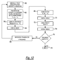
- FIG. 12 depicts a flow diagram of a method for monitoring medical treatment of anatomical tissue using pulse-echo ultrasound according to another alternate embodiment of the present invention
- FIG. 13 shows the relative amplitude and timing of ultrasound image frame sets, difference signals and ultrasound treatments of the method of FIG. 12 ;
- FIG. 14 is a flow diagram of a method for monitoring medical treatment of anatomical tissue using pulse-echo ultrasound according to yet another alternate embodiment of the present invention.
- FIG. 15 shows the relative amplitude and timing of ultrasound image frames and ultrasound treatments of the method of FIG. 14 .
- FIG. 1 An overview of an ultrasound treatment method 10 according to an embodiment of the present invention is shown in FIG. 1 .
- the method begins at step 12 by positioning proximate the targeted anatomical tissue to be medically treated a transducer capable of transmitting and receiving ultrasound signals. Once the transducer is in position, treatment begins at step 14 by emitting a high-intensity ultrasound signal from the transducer.
- the high-intensity ultrasound signal medically treats the targeted tissue, such as (but not limited to) heating the tissue to ablate the material.
- a low-intensity ultrasound signal such as a B-Mode signal, is emitted from the transducer and the reflected signals are received to form a first image frame F 1 .
- image includes, without limitation, creating an image in a visual form and displayed, for example, on a monitor, screen or display, and creating an image in electronic form which, for example, can be used by a computer without first being displayed in visual form.
- a predetermined waiting period is executed at step 18 before proceeding further. It is to be understood that the predetermined waiting period may vary in value from zero seconds upwardly to a maximum of several seconds, but preferably is in the range of milliseconds.
- a low intensity ultrasound signal is again emitted from the transducer and a second image frame F 2 is received at step 20 .
- a difference signal is derived from the image frames F 1 and F 2 , as will be discussed in greater detail below.
- the difference signal of step 22 is displayed as an image at step 24 to obtain a visual indication of the tissue change as a result of the medical treatment of step 14 .
- the visual indication of the tissue change provided by the present invention differs from the post-treatment image of the prior art in that the present invention provides an image showing echo differences in contrast to echos from the target tissue.
- the image of echo differences can indicate whether treatment is complete. If treatment is complete at step 26 (for example, the targeted tissue has been fully ablated), the method is ended at step 28 . However, if the tissue requires additional treatment, the transducer may be re-positioned at step 30 . The method then returns to step 14 to continue medical treatment of the targeted tissue.
- FIG. 1 the method of FIG. 1 is illustrated in relation to a time scale t.
- the targeted tissue is medically treated with a relatively high-intensity ultrasound signal as at step 14 .
- a relatively low-intensity B-Mode image scan frame F 1 is received.
- a second image frame F2 is received, as at step 20 .
- the image frames F 1 and F 2 each contain a signal representing the same portion of the target tissue.
- a number of A-lines of raw echo signal data are received, the number of each line corresponding to azimuthal position and the signal time corresponding to depth.
- FIG. 3 a method for monitoring medical treatment of anatomical tissue including, but not limited to, thermal ablation according to an embodiment of the present invention is depicted.
- An ultrasound transducer is positioned proximate the targeted anatomical tissue.
- the tissue may then be medically treated such as by ablation using high-intensity ultrasound waves for a period of time as at step 14 .
- a first image frame F 1 (such as is illustrated in FIG. 4 ) is received.
- the image frame may optionally be stored electronically, such as in a computer, magnetic media and solid-state memory.
- a second image frame F 2 separated from F 1 by a fixed time interval of step 18 , is received at step 20 .
- An example image frame F 2 is illustrated in FIG. 5 .
- a difference signal is then derived at step 22 by means of steps 32 - 38 .
- the raw echo signals of frames F 1 and F 2 may be processed at step 32 , such as to obtain complex analytic signals by means of a Hilbert transformation.
- a difference signal may then be derived by subtracting the signal of image frame F 2 from the signal of F 1 at step 34 .
- the difference signal of step 34 may take into account both phase and amplitude differences between F 1 and F 2 , computing the amplitude of the signal differences (as opposed to differences in signal amplitude) of F 1 and F 2 .
- a composite illustration of image frames F 1 and F 2 is shown in FIG. 6 , while the derived difference signal is depicted in FIG. 7 .
- the difference signal may be scaled to a convenient value, such as the mean squared value of the difference signal, the mean squared value of one of the original echo signals, or a mathematical constant.
- a convenient value such as the mean squared value of the difference signal, the mean squared value of one of the original echo signals, or a mathematical constant.
- a signal representing the scaled absolute value of the difference signal of FIG. 7 is shown in FIG. 8 .
- Other functions of the difference signal such as its squared absolute value or logarithm, may similarly be employed.
- Still other scaling algorithms may use the amplitude and/or phase of the first and second signals to enhance differences between the first and second signals. Details of such algorithms are left to the skilled artisan.
- the difference signal is spatially filtered, as depicted in FIG. 9 , to smooth small-scale random variations. Spatial filtering of the scaled difference signal is represented by Equation 1.
- ⁇ ⁇ ( x , z ) ⁇ - ⁇ ⁇ ⁇ ⁇ - ⁇ ⁇ ⁇ w ⁇ ( x - x , z - z 0 ) ⁇ ⁇ p 0 ⁇ ( x 0 , z 0 ) - p 1 ⁇ ( x 0 , z 0 ) ⁇ 2 ⁇ ⁇ d x 0 ⁇ ⁇ d z 0 Equation 1
- Equation 1 ⁇ is a spatial difference map (image) of the scaled and filtered difference signal.
- the filtering may be performed by convolution of the scaled difference signal with a two-dimensional window w. This convolution may be efficiently performed through the use of two-dimensional Fast Fourier Transform (“FFT”) operations.
- FFT Fast Fourier Transform
- the difference signal may be normalized to have a maximum value of 1. This approach would result in a spatial map of the echo decorrelation, similar to measures of turbulence in color Doppler imaging systems. However, instead of examining echo decorrelation (a normalized measure of echo differences), a non-normalized map is considered preferable for the present invention because the echo difference is then enhanced in regions of greater echogenicity. Since hyperechoicity is one correlate to tissue ablation, this feature increases the specificity of the method for monitoring thermal ablative medical treatment by providing an image with greater detail.
- the spatially filtered signal of FIG. 9 is then displayed as an image at step 24 (see FIG. 3 ), in any manner previously discussed.
- ultrasound images may be generated as depicted in FIGS. 10 and 11 .
- the tissue is medically treated with high-intensity ultrasound waves.
- a succession of image frames, depicted as F 1 , F 2 , . . . F n are received.
- the image frames F 1 , F 2 , . . . F n each contain a signal representing the same portion of the target tissue.
- the image frames F 1 , F 2 , . . . F n are mathematically grouped, such as by summing or averaging, to form a first image frame set labeled FS 1 , as shown in FIG. 11 .
- a second set of image frames F 1 , F 2 , . . . F n are received at step 48 .
- the second set of image frames F 1 , F 2 , . . . F n are mathematically grouped, such as by summing or averaging, to form a second image frame set FS 2 as shown in FIG. 11 .
- the raw echo signals of image frame sets FS 1 and FS 2 may be processed at step 52 , such as to derive complex analytic signals by means of a Hilbert transformation.
- the signal of image frame set FS 2 is then subtracted from the signal of image frame set FS 1 at step 54 to derive a difference signal.
- the difference signal may take into account both phase and amplitude differences between FS 1 and FS 2 , computing the amplitude of the signal differences (as opposed to differences in signal amplitude) of FS 1 and FS 2 .
- the difference signal may be scaled to a convenient value using any scaling methods and algorithms, previously described or otherwise.
- the difference signal is spatially filtered to smooth small-scale random variations before being displayed as an image at step 59 . This embodiment of the present invention may provide a more robust map of the backscatter changes by reducing the influence of random signal variations caused by rapid transient effects such as violent bubble activity produced during tissue ablation.
- smoothing of the image signal may alternatively be accomplished by using a plurality of image frames, as illustrated in FIGS. 12 and 13 .
- the tissue is medically treated at step 60 , then a set of image frames F 1 , F 2 , . . . F n are received at step 62 .
- a plurality of difference signals D 1 , D 2 , . . . D n are computed at step 64 .
- the difference signals may be computed using various arrangements of pairs of image frames. For example, difference signal D 1 may be formed by subtracting F 2 from F 1 ; likewise, D 2 may be formed by subtracting F 3 from F 2 , as shown in FIG. 13 .
- image frame pairs may also be used, including (but not limited to) odd-numbered image frames (i.e., subtracting F 3 from F 1 , etc.) and even-numbered image frames (i.e., subtracting F 4 from F 2 , etc.).
- the pairings may be interlaced (i.e., subtracting F 2 from F 1 , subtracting F 3 from F 2 , etc.) or sequential (i.e., subtracting F 2 from F 1 , F 4 from F 3 , etc.).
- An indication or image may be displayed at step 66 , showing at least one of the difference signals D 1 , D 2 , . . . D n .
- D n may be further processed, such as by averaging, to reduce artifactual content.
- the averaged signal, denoted as D may also be displayed as an image, as at step 70 .
- the averaged signals may themselves be cumulatively summed, as at step 72 , to provide a view of the results of successive medical treatments 60 .
- the summed averages may be displayed at step 74 . If treatment is determined to be complete at step 76 , the method is ended at step 78 . However, if the tissue appears to require additional treatment, the transducer may be re-positioned at step 80 . The method is then repeated beginning at step 60 to continue treatment of the targeted tissue.
- FIGS. 14 and 15 A fourth embodiment of the present invention is shown in FIGS. 14 and 15 wherein a difference signal is derived using imaging frames generated both before and after medical treatment.
- the ultrasound transducer is positioned proximate the targeted tissue to be medically treated.
- a pre-treatment image frame F 1 is generated from received ultrasound signals.
- the tissue is subject to a quantum of medical treatment, such as by ablating the tissue.
- a second image frame F 2 is generated at step 88 .
- a difference signal is derived at step 90 , using the data contained in image frames F 1 and F 2 in the same manner as previously described.
- An indication or image of the difference signal may be displayed at step 92 .
- the method is ended at step 96 . However, if the targeted tissue is determined to require additional treatment, the transducer may be re-positioned as necessary at step 98 . The method is then repeated and a subsequent quantum of treatment is administered beginning at step 84 .
- An expected difficulty for the present invention is artifactual backscatter change due to tissue motion artifacts.
- This difficulty can be largely overcome by several features of the method.
- First, backscatter differences can be computed between image frames closely spaced in time. If the tissue moves only a small amount during the interval, motion artifacts are then small.
- Second, artifacts due to axial tissue motion can be removed effectively by phase compensation during signal processing. That is, before computation of the signal difference, one of the complex image frames is multiplied by a phase compensation function e ⁇ i ⁇ , where ⁇ is the low-pass filtered phase of the conjugate product of the two complex image frames. The resulting signal difference is then computed, for example, using Equation 2:
- any of the ultrasound transducers may be used with other methods of medical treatment, such as producing images to aid in tissue ablation by means of Radio Frequency (RF) and laser energy, various non-ablative ultrasound medical treatments, and various ultrasound imaging applications.
- RF Radio Frequency
Abstract
Description
Claims (32)
Priority Applications (6)
| Application Number | Priority Date | Filing Date | Title |
|---|---|---|---|
| US10/721,034 US7846096B2 (en) | 2001-05-29 | 2003-11-24 | Method for monitoring of medical treatment using pulse-echo ultrasound |
| EP04811946A EP1691689A4 (en) | 2003-11-24 | 2004-11-23 | Method for monitoring of medical treatment using pulse-echo ultrasound |
| JP2006541653A JP2007512111A (en) | 2003-11-24 | 2004-11-23 | Method of monitoring medical treatment using pulse echo ultrasound |
| PCT/US2004/039318 WO2005072084A2 (en) | 2003-11-24 | 2004-11-23 | Medical treatment monitoring using pulse-echo ultrasound |
| US12/915,358 US9261596B2 (en) | 2001-05-29 | 2010-10-29 | Method for monitoring of medical treatment using pulse-echo ultrasound |
| US14/989,883 US20160113620A1 (en) | 2001-05-29 | 2016-01-07 | Method for Monitoring of Medical Treatment Using Pulse-Echo Ultrasound |
Applications Claiming Priority (3)
| Application Number | Priority Date | Filing Date | Title |
|---|---|---|---|
| US29413501P | 2001-05-29 | 2001-05-29 | |
| US10/153,241 US20030069502A1 (en) | 2001-05-29 | 2002-05-22 | Ultrasound feedback in medically-treated patients |
| US10/721,034 US7846096B2 (en) | 2001-05-29 | 2003-11-24 | Method for monitoring of medical treatment using pulse-echo ultrasound |
Related Parent Applications (1)
| Application Number | Title | Priority Date | Filing Date |
|---|---|---|---|
| US10/153,241 Continuation-In-Part US20030069502A1 (en) | 2001-05-29 | 2002-05-22 | Ultrasound feedback in medically-treated patients |
Related Child Applications (1)
| Application Number | Title | Priority Date | Filing Date |
|---|---|---|---|
| US12/915,358 Continuation US9261596B2 (en) | 2001-05-29 | 2010-10-29 | Method for monitoring of medical treatment using pulse-echo ultrasound |
Publications (2)
| Publication Number | Publication Date |
|---|---|
| US20040106870A1 US20040106870A1 (en) | 2004-06-03 |
| US7846096B2 true US7846096B2 (en) | 2010-12-07 |
Family
ID=34826381
Family Applications (3)
| Application Number | Title | Priority Date | Filing Date |
|---|---|---|---|
| US10/721,034 Expired - Fee Related US7846096B2 (en) | 2001-05-29 | 2003-11-24 | Method for monitoring of medical treatment using pulse-echo ultrasound |
| US12/915,358 Expired - Fee Related US9261596B2 (en) | 2001-05-29 | 2010-10-29 | Method for monitoring of medical treatment using pulse-echo ultrasound |
| US14/989,883 Abandoned US20160113620A1 (en) | 2001-05-29 | 2016-01-07 | Method for Monitoring of Medical Treatment Using Pulse-Echo Ultrasound |
Family Applications After (2)
| Application Number | Title | Priority Date | Filing Date |
|---|---|---|---|
| US12/915,358 Expired - Fee Related US9261596B2 (en) | 2001-05-29 | 2010-10-29 | Method for monitoring of medical treatment using pulse-echo ultrasound |
| US14/989,883 Abandoned US20160113620A1 (en) | 2001-05-29 | 2016-01-07 | Method for Monitoring of Medical Treatment Using Pulse-Echo Ultrasound |
Country Status (4)
| Country | Link |
|---|---|
| US (3) | US7846096B2 (en) |
| EP (1) | EP1691689A4 (en) |
| JP (1) | JP2007512111A (en) |
| WO (1) | WO2005072084A2 (en) |
Cited By (45)
| Publication number | Priority date | Publication date | Assignee | Title |
|---|---|---|---|---|
| US20080242998A1 (en) * | 2007-03-30 | 2008-10-02 | Yoichi Suzuki | Ultrasonic contrast imaging method and ultrasonic diagnostic apparatus |
| US20110040184A1 (en) * | 2001-05-29 | 2011-02-17 | Mast T Douglas | Method for monitoring of medical treatment using pulse-echo ultrasound |
| US8232801B2 (en) | 2011-06-30 | 2012-07-31 | General Electric Company | Nuclear quadrupole resonance system and method for structural health monitoring |
| US8857438B2 (en) | 2010-11-08 | 2014-10-14 | Ulthera, Inc. | Devices and methods for acoustic shielding |
| US8915853B2 (en) | 2004-10-06 | 2014-12-23 | Guided Therapy Systems, Llc | Methods for face and neck lifts |
| US8915854B2 (en) | 2004-10-06 | 2014-12-23 | Guided Therapy Systems, Llc | Method for fat and cellulite reduction |
| US8915870B2 (en) | 2004-10-06 | 2014-12-23 | Guided Therapy Systems, Llc | Method and system for treating stretch marks |
| US8920324B2 (en) | 2004-10-06 | 2014-12-30 | Guided Therapy Systems, Llc | Energy based fat reduction |
| US8932224B2 (en) | 2004-10-06 | 2015-01-13 | Guided Therapy Systems, Llc | Energy based hyperhidrosis treatment |
| US9005144B2 (en) | 2001-05-29 | 2015-04-14 | Michael H. Slayton | Tissue-retaining systems for ultrasound medical treatment |
| US9011336B2 (en) | 2004-09-16 | 2015-04-21 | Guided Therapy Systems, Llc | Method and system for combined energy therapy profile |
| US9011337B2 (en) | 2011-07-11 | 2015-04-21 | Guided Therapy Systems, Llc | Systems and methods for monitoring and controlling ultrasound power output and stability |
| US9039617B2 (en) | 2009-11-24 | 2015-05-26 | Guided Therapy Systems, Llc | Methods and systems for generating thermal bubbles for improved ultrasound imaging and therapy |
| US9039619B2 (en) | 2004-10-06 | 2015-05-26 | Guided Therapy Systems, L.L.C. | Methods for treating skin laxity |
| US9114247B2 (en) | 2004-09-16 | 2015-08-25 | Guided Therapy Systems, Llc | Method and system for ultrasound treatment with a multi-directional transducer |
| US9132287B2 (en) | 2004-06-14 | 2015-09-15 | T. Douglas Mast | System and method for ultrasound treatment using grating lobes |
| US9149658B2 (en) | 2010-08-02 | 2015-10-06 | Guided Therapy Systems, Llc | Systems and methods for ultrasound treatment |
| US9216276B2 (en) | 2007-05-07 | 2015-12-22 | Guided Therapy Systems, Llc | Methods and systems for modulating medicants using acoustic energy |
| US9263663B2 (en) | 2012-04-13 | 2016-02-16 | Ardent Sound, Inc. | Method of making thick film transducer arrays |
| US9272162B2 (en) | 1997-10-14 | 2016-03-01 | Guided Therapy Systems, Llc | Imaging, therapy, and temperature monitoring ultrasonic method |
| US9320537B2 (en) | 2004-10-06 | 2016-04-26 | Guided Therapy Systems, Llc | Methods for noninvasive skin tightening |
| US9452302B2 (en) | 2011-07-10 | 2016-09-27 | Guided Therapy Systems, Llc | Systems and methods for accelerating healing of implanted material and/or native tissue |
| US9504446B2 (en) | 2010-08-02 | 2016-11-29 | Guided Therapy Systems, Llc | Systems and methods for coupling an ultrasound source to tissue |
| US9510802B2 (en) | 2012-09-21 | 2016-12-06 | Guided Therapy Systems, Llc | Reflective ultrasound technology for dermatological treatments |
| US9694212B2 (en) | 2004-10-06 | 2017-07-04 | Guided Therapy Systems, Llc | Method and system for ultrasound treatment of skin |
| US9700340B2 (en) | 2004-10-06 | 2017-07-11 | Guided Therapy Systems, Llc | System and method for ultra-high frequency ultrasound treatment |
| US9827449B2 (en) | 2004-10-06 | 2017-11-28 | Guided Therapy Systems, L.L.C. | Systems for treating skin laxity |
| US10039938B2 (en) | 2004-09-16 | 2018-08-07 | Guided Therapy Systems, Llc | System and method for variable depth ultrasound treatment |
| US20180291803A1 (en) * | 2015-11-11 | 2018-10-11 | General Electric Company | Ultrasonic cleaning system and method |
| US10420960B2 (en) | 2013-03-08 | 2019-09-24 | Ulthera, Inc. | Devices and methods for multi-focus ultrasound therapy |
| US10537304B2 (en) | 2008-06-06 | 2020-01-21 | Ulthera, Inc. | Hand wand for ultrasonic cosmetic treatment and imaging |
| US10561862B2 (en) | 2013-03-15 | 2020-02-18 | Guided Therapy Systems, Llc | Ultrasound treatment device and methods of use |
| US10603521B2 (en) | 2014-04-18 | 2020-03-31 | Ulthera, Inc. | Band transducer ultrasound therapy |
| US20200289095A1 (en) * | 2019-03-14 | 2020-09-17 | Fujifilm Corporation | Ultrasound diagnostic system and method of operating ultrasound diagnostic system |
| US10864385B2 (en) | 2004-09-24 | 2020-12-15 | Guided Therapy Systems, Llc | Rejuvenating skin by heating tissue for cosmetic treatment of the face and body |
| US11207548B2 (en) | 2004-10-07 | 2021-12-28 | Guided Therapy Systems, L.L.C. | Ultrasound probe for treating skin laxity |
| US11224895B2 (en) | 2016-01-18 | 2022-01-18 | Ulthera, Inc. | Compact ultrasound device having annular ultrasound array peripherally electrically connected to flexible printed circuit board and method of assembly thereof |
| US11235179B2 (en) | 2004-10-06 | 2022-02-01 | Guided Therapy Systems, Llc | Energy based skin gland treatment |
| US11241218B2 (en) | 2016-08-16 | 2022-02-08 | Ulthera, Inc. | Systems and methods for cosmetic ultrasound treatment of skin |
| US11338156B2 (en) | 2004-10-06 | 2022-05-24 | Guided Therapy Systems, Llc | Noninvasive tissue tightening system |
| US11717661B2 (en) | 2007-05-07 | 2023-08-08 | Guided Therapy Systems, Llc | Methods and systems for ultrasound assisted delivery of a medicant to tissue |
| US11724133B2 (en) | 2004-10-07 | 2023-08-15 | Guided Therapy Systems, Llc | Ultrasound probe for treatment of skin |
| US11883688B2 (en) | 2004-10-06 | 2024-01-30 | Guided Therapy Systems, Llc | Energy based fat reduction |
| US11944849B2 (en) | 2018-02-20 | 2024-04-02 | Ulthera, Inc. | Systems and methods for combined cosmetic treatment of cellulite with ultrasound |
| US11969609B2 (en) | 2022-12-05 | 2024-04-30 | Ulthera, Inc. | Devices and methods for multi-focus ultrasound therapy |
Families Citing this family (21)
| Publication number | Priority date | Publication date | Assignee | Title |
|---|---|---|---|---|
| US7211044B2 (en) * | 2001-05-29 | 2007-05-01 | Ethicon Endo-Surgery, Inc. | Method for mapping temperature rise using pulse-echo ultrasound |
| US7617005B2 (en) * | 2002-04-08 | 2009-11-10 | Ardian, Inc. | Methods and apparatus for thermally-induced renal neuromodulation |
| US7662114B2 (en) * | 2004-03-02 | 2010-02-16 | Focus Surgery, Inc. | Ultrasound phased arrays |
| US20070219448A1 (en) * | 2004-05-06 | 2007-09-20 | Focus Surgery, Inc. | Method and Apparatus for Selective Treatment of Tissue |
| EP1807146A4 (en) * | 2004-09-29 | 2013-07-03 | Tel Hashomer Medical Res Infrastructure & Services Ltd | Composition for improving efficiency of drug delivery |
| US7367944B2 (en) * | 2004-12-13 | 2008-05-06 | Tel Hashomer Medical Research Infrastructure And Services Ltd. | Method and system for monitoring ablation of tissues |
| US8038631B1 (en) | 2005-06-01 | 2011-10-18 | Sanghvi Narendra T | Laparoscopic HIFU probe |
| US20070038096A1 (en) * | 2005-07-06 | 2007-02-15 | Ralf Seip | Method of optimizing an ultrasound transducer |
| US20070010805A1 (en) | 2005-07-08 | 2007-01-11 | Fedewa Russell J | Method and apparatus for the treatment of tissue |
| US7871406B2 (en) | 2006-08-04 | 2011-01-18 | INTIO, Inc. | Methods for planning and performing thermal ablation |
| US8556888B2 (en) | 2006-08-04 | 2013-10-15 | INTIO, Inc. | Methods and apparatuses for performing and monitoring thermal ablation |
| US20080033417A1 (en) * | 2006-08-04 | 2008-02-07 | Nields Morgan W | Apparatus for planning and performing thermal ablation |
| US8155416B2 (en) * | 2008-02-04 | 2012-04-10 | INTIO, Inc. | Methods and apparatuses for planning, performing, monitoring and assessing thermal ablation |
| US20080039724A1 (en) * | 2006-08-10 | 2008-02-14 | Ralf Seip | Ultrasound transducer with improved imaging |
| US7559905B2 (en) * | 2006-09-21 | 2009-07-14 | Focus Surgery, Inc. | HIFU probe for treating tissue with in-line degassing of fluid |
| US20080154131A1 (en) * | 2006-12-20 | 2008-06-26 | General Electric Company | Methods for enhancement of visibility of ablation regions |
| US8235902B2 (en) * | 2007-09-11 | 2012-08-07 | Focus Surgery, Inc. | System and method for tissue change monitoring during HIFU treatment |
| JP5318646B2 (en) * | 2009-04-17 | 2013-10-16 | ルネサスエレクトロニクス株式会社 | DIFFERENTIAL IMAGE GENERATION DEVICE, DIFFERENTIAL IMAGE GENERATION METHOD, AND PROGRAM |
| JP6055679B2 (en) * | 2013-01-08 | 2016-12-27 | 日本光電工業株式会社 | Biological signal averaging processor |
| WO2014133208A1 (en) * | 2013-02-28 | 2014-09-04 | 알피니언메디칼시스템 주식회사 | Method for focal point compensation, and ultrasonic medical apparatus therefor |
| CN110575627B (en) * | 2019-09-24 | 2021-04-06 | 黄晶 | Physical mapping device for rapidly acquiring target nerve treatment energy delivery site |
Citations (303)
| Publication number | Priority date | Publication date | Assignee | Title |
|---|---|---|---|---|
| US3168659A (en) | 1960-01-11 | 1965-02-02 | Gen Motors Corp | Variable focus transducer |
| US3779234A (en) | 1971-06-30 | 1973-12-18 | Intersc Res Inst | Ultrasonic catheter with rotating transducers |
| US3902501A (en) | 1973-06-21 | 1975-09-02 | Medtronic Inc | Endocardial electrode |
| US3927557A (en) | 1974-05-30 | 1975-12-23 | Gen Electric | Acoustic imaging apparatus with liquid-filled acoustic corrector lens |
| US4211948A (en) | 1978-11-08 | 1980-07-08 | General Electric Company | Front surface matched piezoelectric ultrasonic transducer array with wide field of view |
| US4315514A (en) | 1980-05-08 | 1982-02-16 | William Drewes | Method and apparatus for selective cell destruction |
| US4323077A (en) | 1980-03-12 | 1982-04-06 | General Electric Company | Acoustic intensity monitor |
| US4484569A (en) | 1981-03-13 | 1984-11-27 | Riverside Research Institute | Ultrasonic diagnostic and therapeutic transducer assembly and method for using |
| US4646756A (en) | 1982-10-26 | 1987-03-03 | The University Of Aberdeen | Ultra sound hyperthermia device |
| US4748985A (en) | 1985-05-10 | 1988-06-07 | Olympus Optical Co., Ltd. | Ultrasonic imaging apparatus having circulating cooling liquid for cooling ultrasonic transducers thereof |
| US4757820A (en) | 1985-03-15 | 1988-07-19 | Kabushiki Kaisha Toshiba | Ultrasound therapy system |
| US4787394A (en) | 1986-04-24 | 1988-11-29 | Kabushiki Kaisha Toshiba | Ultrasound therapy apparatus |
| US4790329A (en) | 1987-06-12 | 1988-12-13 | Trustees Of Beth Israel Hospital | Adjustable biopsy localization device |
| US4798215A (en) | 1984-03-15 | 1989-01-17 | Bsd Medical Corporation | Hyperthermia apparatus |
| US4818954A (en) | 1986-02-15 | 1989-04-04 | Karl Storz Endoscopy-America, Inc. | High-frequency generator with automatic power-control for high-frequency surgery |
| US4844080A (en) | 1987-02-19 | 1989-07-04 | Michael Frass | Ultrasound contact medium dispenser |
| US4858613A (en) | 1988-03-02 | 1989-08-22 | Laboratory Equipment, Corp. | Localization and therapy system for treatment of spatially oriented focal disease |
| US4932414A (en) | 1987-11-02 | 1990-06-12 | Cornell Research Foundation, Inc. | System of therapeutic ultrasound and real-time ultrasonic scanning |
| US4937767A (en) | 1986-12-24 | 1990-06-26 | Hewlett-Packard Company | Method and apparatus for adjusting the intensity profile of an ultrasound beam |
| US4951653A (en) | 1988-03-02 | 1990-08-28 | Laboratory Equipment, Corp. | Ultrasound brain lesioning system |
| US4955366A (en) | 1987-11-27 | 1990-09-11 | Olympus Optical Co., Ltd. | Ultrasonic therapeutical apparatus |
| US4955365A (en) | 1988-03-02 | 1990-09-11 | Laboratory Equipment, Corp. | Localization and therapy system for treatment of spatially oriented focal disease |
| US4960109A (en) | 1988-06-21 | 1990-10-02 | Massachusetts Institute Of Technology | Multi-purpose temperature sensing probe for hyperthermia therapy |
| US4960107A (en) | 1987-09-30 | 1990-10-02 | Kabushiki Kaisha Toshiba | Ultrasonic medical treatment apparatus |
| US4984575A (en) | 1987-04-16 | 1991-01-15 | Olympus Optical Co., Ltd. | Therapeutical apparatus of extracorporeal type |
| US4986275A (en) | 1987-08-05 | 1991-01-22 | Kabushiki Kaisha Toshiba | Ultrasonic therapy apparatus |
| US5005580A (en) * | 1988-04-22 | 1991-04-09 | Kabushiki Kaisha Toshiba | Destroying wave treatment apparatus |
| US5015929A (en) | 1987-09-07 | 1991-05-14 | Technomed International, S.A. | Piezoelectric device with reduced negative waves, and use of said device for extracorporeal lithotrity or for destroying particular tissues |
| USRE33590E (en) | 1983-12-14 | 1991-05-21 | Edap International, S.A. | Method for examining, localizing and treating with ultrasound |
| US5031626A (en) | 1988-08-17 | 1991-07-16 | Siemens Aktiengesellschaft | Extracorporeal lithotripsy apparatus with an ultrasound locating system |
| US5036855A (en) | 1988-03-02 | 1991-08-06 | Laboratory Equipment, Corp. | Localization and therapy system for treatment of spatially oriented focal disease |
| US5042486A (en) | 1989-09-29 | 1991-08-27 | Siemens Aktiengesellschaft | Catheter locatable with non-ionizing field and method for locating same |
| US5054470A (en) | 1988-03-02 | 1991-10-08 | Laboratory Equipment, Corp. | Ultrasonic treatment transducer with pressurized acoustic coupling |
| US5065740A (en) | 1986-12-26 | 1991-11-19 | Kabushiki Kaisha Toshiba | Ultrasonic medical treatment apparatus |
| US5078144A (en) | 1988-08-19 | 1992-01-07 | Olympus Optical Co. Ltd. | System for applying ultrasonic waves and a treatment instrument to a body part |
| US5080102A (en) | 1983-12-14 | 1992-01-14 | Edap International, S.A. | Examining, localizing and treatment with ultrasound |
| US5095906A (en) * | 1987-09-30 | 1992-03-17 | Kabushiki Kaisha Toshiba | Image processing system |
| US5095907A (en) | 1989-06-21 | 1992-03-17 | Kabushiki Kaisha Toshiba | Acoustic wave therapy apparatus |
| US5117832A (en) | 1990-09-21 | 1992-06-02 | Diasonics, Inc. | Curved rectangular/elliptical transducer |
| US5143074A (en) | 1983-12-14 | 1992-09-01 | Edap International | Ultrasonic treatment device using a focussing and oscillating piezoelectric element |
| US5149319A (en) | 1990-09-11 | 1992-09-22 | Unger Evan C | Methods for providing localized therapeutic heat to biological tissues and fluids |
| US5148809A (en) * | 1990-02-28 | 1992-09-22 | Asgard Medical Systems, Inc. | Method and apparatus for detecting blood vessels and displaying an enhanced video image from an ultrasound scan |
| US5150712A (en) | 1983-12-14 | 1992-09-29 | Edap International, S.A. | Apparatus for examining and localizing tumors using ultra sounds, comprising a device for localized hyperthermia treatment |
| US5150711A (en) * | 1983-12-14 | 1992-09-29 | Edap International, S.A. | Ultra-high-speed extracorporeal ultrasound hyperthermia treatment device |
| US5158071A (en) | 1988-07-01 | 1992-10-27 | Hitachi, Ltd. | Ultrasonic apparatus for therapeutical use |
| US5158070A (en) | 1983-12-14 | 1992-10-27 | Edap International, S.A. | Method for the localized destruction of soft structures using negative pressure elastic waves |
| US5203333A (en) | 1989-05-15 | 1993-04-20 | Kabushiki Kaisha Toshiba | Acoustic wave therapy apparatus |
| US5209221A (en) | 1988-03-01 | 1993-05-11 | Richard Wolf Gmbh | Ultrasonic treatment of pathological tissue |
| US5238007A (en) | 1991-12-12 | 1993-08-24 | Vitatron Medical B.V. | Pacing lead with improved anchor mechanism |
| US5240005A (en) | 1990-11-22 | 1993-08-31 | Dornier Medizintechnik Gmbh | Acoustic focussing device |
| US5242437A (en) | 1988-06-10 | 1993-09-07 | Trimedyne Laser Systems, Inc. | Medical device applying localized high intensity light and heat, particularly for destruction of the endometrium |
| US5295484A (en) | 1992-05-19 | 1994-03-22 | Arizona Board Of Regents For And On Behalf Of The University Of Arizona | Apparatus and method for intra-cardiac ablation of arrhythmias |
| US5304115A (en) | 1991-01-11 | 1994-04-19 | Baxter International Inc. | Ultrasonic angioplasty device incorporating improved transmission member and ablation probe |
| US5311869A (en) | 1990-03-24 | 1994-05-17 | Kabushiki Kaisha Toshiba | Method and apparatus for ultrasonic wave treatment in which medical progress may be evaluated |
| US5345940A (en) | 1991-11-08 | 1994-09-13 | Mayo Foundation For Medical Education And Research | Transvascular ultrasound hemodynamic and interventional catheter and method |
| US5348017A (en) | 1993-01-19 | 1994-09-20 | Cardiovascular Imaging Systems, Inc. | Drive shaft for an intravascular catheter system |
| US5354258A (en) * | 1992-01-07 | 1994-10-11 | Edap International | Ultra-high-speed extracorporeal ultrasound hyperthermia treatment method |
| US5370121A (en) * | 1992-09-07 | 1994-12-06 | Siemens Aktiengesellschaft | Method and apparatus for non-invasive measurement of a temperature change in a subject |
| US5391197A (en) | 1992-11-13 | 1995-02-21 | Dornier Medical Systems, Inc. | Ultrasound thermotherapy probe |
| US5391140A (en) | 1993-01-29 | 1995-02-21 | Siemens Aktiengesellschaft | Therapy apparatus for locating and treating a zone in the body of a life form with acoustic waves |
| US5398691A (en) | 1993-09-03 | 1995-03-21 | University Of Washington | Method and apparatus for three-dimensional translumenal ultrasonic imaging |
| US5398690A (en) | 1994-08-03 | 1995-03-21 | Batten; Bobby G. | Slaved biopsy device, analysis apparatus, and process |
| US5402792A (en) | 1993-03-30 | 1995-04-04 | Shimadzu Corporation | Ultrasonic medical apparatus |
| US5409002A (en) | 1989-07-12 | 1995-04-25 | Focus Surgery Incorporated | Treatment system with localization |
| US5419335A (en) | 1992-09-04 | 1995-05-30 | Siemens Aktiengesellschaft | Acoustic lens |
| US5421338A (en) | 1988-03-21 | 1995-06-06 | Boston Scientific Corporation | Acoustic imaging catheter and the like |
| US5431663A (en) | 1990-12-10 | 1995-07-11 | Coraje, Inc. | Miniature ultrasonic transducer for removal of intravascular plaque and clots |
| US5435311A (en) | 1989-06-27 | 1995-07-25 | Hitachi, Ltd. | Ultrasound therapeutic system |
| US5435304A (en) | 1992-04-24 | 1995-07-25 | Siemens Aktiengesellschaft | Method and apparatus for therapeutic treatment with focussed acoustic waves switchable between a locating mode and a therapy mode |
| US5443069A (en) | 1992-11-16 | 1995-08-22 | Siemens Aktiengesellschaft | Therapeutic ultrasound applicator for the urogenital region |
| US5448994A (en) | 1990-02-28 | 1995-09-12 | Kabushiki Kaisha Toshiba | Apparatus for performing medical treatment by using electroacoustic transducer element |
| US5458597A (en) | 1993-11-08 | 1995-10-17 | Zomed International | Device for treating cancer and non-malignant tumors and methods |
| US5465724A (en) | 1993-05-28 | 1995-11-14 | Acuson Corporation | Compact rotationally steerable ultrasound transducer |
| US5471988A (en) | 1993-12-24 | 1995-12-05 | Olympus Optical Co., Ltd. | Ultrasonic diagnosis and therapy system in which focusing point of therapeutic ultrasonic wave is locked at predetermined position within observation ultrasonic scanning range |
| US5474071A (en) | 1991-03-05 | 1995-12-12 | Technomed Medical Systems | Therapeutic endo-rectal probe and apparatus constituting an application thereof for destroying cancer tissue, in particular of the prostate, and preferably in combination with an imaging endo-cavitary-probe |
| US5485839A (en) | 1992-02-28 | 1996-01-23 | Kabushiki Kaisha Toshiba | Method and apparatus for ultrasonic wave medical treatment using computed tomography |
| US5492126A (en) | 1994-05-02 | 1996-02-20 | Focal Surgery | Probe for medical imaging and therapy using ultrasound |
| US5500012A (en) | 1992-07-15 | 1996-03-19 | Angeion Corporation | Ablation catheter system |
| US5501655A (en) | 1992-03-31 | 1996-03-26 | Massachusetts Institute Of Technology | Apparatus and method for acoustic heat generation and hyperthermia |
| US5514130A (en) | 1994-10-11 | 1996-05-07 | Dorsal Med International | RF apparatus for controlled depth ablation of soft tissue |
| US5514085A (en) | 1990-07-24 | 1996-05-07 | Yoon; Inbae | Multifunctional devices for use in endoscopic surgical procedures and methods therefor |
| US5520188A (en) | 1994-11-02 | 1996-05-28 | Focus Surgery Inc. | Annular array transducer |
| US5522869A (en) | 1994-05-17 | 1996-06-04 | Burdette; Everette C. | Ultrasound device for use in a thermotherapy apparatus |
| US5524620A (en) | 1991-11-12 | 1996-06-11 | November Technologies Ltd. | Ablation of blood thrombi by means of acoustic energy |
| US5526822A (en) | 1994-03-24 | 1996-06-18 | Biopsys Medical, Inc. | Method and apparatus for automated biopsy and collection of soft tissue |
| US5526816A (en) * | 1994-09-22 | 1996-06-18 | Bracco Research S.A. | Ultrasonic spectral contrast imaging |
| US5526815A (en) | 1993-01-29 | 1996-06-18 | Siemens Aktiengesellschat | Therapy apparatus for locating and treating a zone located in the body of a life form with acoustic waves |
| US5545195A (en) | 1994-08-01 | 1996-08-13 | Boston Scientific Corporation | Interstitial heating of tissue |
| US5547459A (en) | 1994-10-25 | 1996-08-20 | Orthologic Corporation | Ultrasonic bone-therapy apparatus and method |
| US5553618A (en) | 1993-03-12 | 1996-09-10 | Kabushiki Kaisha Toshiba | Method and apparatus for ultrasound medical treatment |
| US5558092A (en) | 1995-06-06 | 1996-09-24 | Imarx Pharmaceutical Corp. | Methods and apparatus for performing diagnostic and therapeutic ultrasound simultaneously |
| US5569241A (en) | 1994-06-24 | 1996-10-29 | Vidacare, Inc. | Thin layer ablation apparatus |
| US5571088A (en) | 1993-07-01 | 1996-11-05 | Boston Scientific Corporation | Ablation catheters |
| US5573497A (en) | 1994-11-30 | 1996-11-12 | Technomed Medical Systems And Institut National | High-intensity ultrasound therapy method and apparatus with controlled cavitation effect and reduced side lobes |
| US5575789A (en) | 1994-10-27 | 1996-11-19 | Valleylab Inc. | Energizable surgical tool safety device and method |
| US5582588A (en) | 1993-04-19 | 1996-12-10 | Olympus Optical Co., Ltd. | Ultrasonic therapeutic apparatus |
| US5588432A (en) | 1988-03-21 | 1996-12-31 | Boston Scientific Corporation | Catheters for imaging, sensing electrical potentials, and ablating tissue |
| US5590657A (en) | 1995-11-06 | 1997-01-07 | The Regents Of The University Of Michigan | Phased array ultrasound system and method for cardiac ablation |
| US5596991A (en) | 1994-04-07 | 1997-01-28 | Fuji Photo Optical Co., Ltd. | Catheter type ultrasound probe |
| US5601526A (en) | 1991-12-20 | 1997-02-11 | Technomed Medical Systems | Ultrasound therapy apparatus delivering ultrasound waves having thermal and cavitation effects |
| US5603326A (en) | 1993-03-22 | 1997-02-18 | Siemens Aktiengesellschaft | Method and apparatus for displaying an image obtained by echo signals |
| US5606975A (en) | 1994-09-19 | 1997-03-04 | The Board Of Trustees Of The Leland Stanford Junior University | Forward viewing ultrasonic imaging catheter |
| US5620479A (en) | 1992-11-13 | 1997-04-15 | The Regents Of The University Of California | Method and apparatus for thermal therapy of tumors |
| US5624382A (en) | 1992-03-10 | 1997-04-29 | Siemens Aktiengesellschaft | Method and apparatus for ultrasound tissue therapy |
| US5626607A (en) | 1995-04-03 | 1997-05-06 | Heartport, Inc. | Clamp assembly and method of use |
| US5628743A (en) | 1994-12-21 | 1997-05-13 | Valleylab Inc. | Dual mode ultrasonic surgical apparatus |
| US5630837A (en) | 1993-07-01 | 1997-05-20 | Boston Scientific Corporation | Acoustic ablation |
| US5643179A (en) | 1993-12-28 | 1997-07-01 | Kabushiki Kaisha Toshiba | Method and apparatus for ultrasonic medical treatment with optimum ultrasonic irradiation control |
| US5649547A (en) | 1994-03-24 | 1997-07-22 | Biopsys Medical, Inc. | Methods and devices for automated biopsy and collection of soft tissue |
| US5657760A (en) | 1994-05-03 | 1997-08-19 | Board Of Regents, The University Of Texas System | Apparatus and method for noninvasive doppler ultrasound-guided real-time control of tissue damage in thermal therapy |
| US5665054A (en) | 1994-01-27 | 1997-09-09 | Technomed Medical Systems S.A. | Control method for hyperthermia treatment apparatus using ultrasound |
| US5676692A (en) | 1996-03-28 | 1997-10-14 | Indianapolis Center For Advanced Research, Inc. | Focussed ultrasound tissue treatment method |
| US5687729A (en) | 1994-06-22 | 1997-11-18 | Siemens Aktiengesellschaft | Source of therapeutic acoustic waves introducible into the body of a patient |
| US5694936A (en) | 1994-09-17 | 1997-12-09 | Kabushiki Kaisha Toshiba | Ultrasonic apparatus for thermotherapy with variable frequency for suppressing cavitation |
| US5697897A (en) | 1994-01-14 | 1997-12-16 | Siemens Aktiengesellschaft | Endoscope carrying a source of therapeutic ultrasound |
| US5699805A (en) | 1996-06-20 | 1997-12-23 | Mayo Foundation For Medical Education And Research | Longitudinal multiplane ultrasound transducer underfluid catheter system |
| US5699804A (en) | 1995-06-07 | 1997-12-23 | Siemens Aktiengesellschaft | Therapy apparatus having a source of acoustic waves |
| US5703922A (en) | 1995-06-07 | 1997-12-30 | Siemens Aktiengesellschaft | Therapy apparatus with a radiation source |
| US5720287A (en) | 1993-07-26 | 1998-02-24 | Technomed Medical Systems | Therapy and imaging probe and therapeutic treatment apparatus utilizing it |
| US5728062A (en) | 1995-11-30 | 1998-03-17 | Pharmasonics, Inc. | Apparatus and methods for vibratory intraluminal therapy employing magnetostrictive transducers |
| US5733315A (en) | 1992-11-13 | 1998-03-31 | Burdette; Everette C. | Method of manufacture of a transurethral ultrasound applicator for prostate gland thermal therapy |
| US5735280A (en) | 1995-05-02 | 1998-04-07 | Heart Rhythm Technologies, Inc. | Ultrasound energy delivery system and method |
| US5735796A (en) | 1995-11-23 | 1998-04-07 | Siemens Aktiengesellschaft | Therapy apparatus with a source of acoustic waves |
| US5738635A (en) | 1993-01-22 | 1998-04-14 | Technomed Medical Systems | Adjustable focusing therapeutic apparatus with no secondary focusing |
| US5743862A (en) | 1994-09-19 | 1998-04-28 | Kabushiki Kaisha Toshiba | Ultrasonic medical treatment apparatus |
| US5746224A (en) | 1994-06-24 | 1998-05-05 | Somnus Medical Technologies, Inc. | Method for ablating turbinates |
| US5759154A (en) | 1996-12-23 | 1998-06-02 | C. R. Bard, Inc. | Print mask technique for echogenic enhancement of a medical device |
| US5762066A (en) | 1992-02-21 | 1998-06-09 | Ths International, Inc. | Multifaceted ultrasound transducer probe system and methods for its use |
| US5769086A (en) | 1995-12-06 | 1998-06-23 | Biopsys Medical, Inc. | Control system and method for automated biopsy device |
| US5769790A (en) * | 1996-10-25 | 1998-06-23 | General Electric Company | Focused ultrasound surgery system guided by ultrasound imaging |
| US5776092A (en) | 1994-03-23 | 1998-07-07 | Erbe Elektromedizin Gmbh | Multifunctional surgical instrument |
| US5779643A (en) | 1996-11-26 | 1998-07-14 | Hewlett-Packard Company | Imaging guidewire with back and forth sweeping ultrasonic source |
| US5782764A (en) | 1995-11-07 | 1998-07-21 | Iti Medical Technologies, Inc. | Fiber composite invasive medical instruments and methods for use in interventional imaging procedures |
| US5785705A (en) | 1994-10-11 | 1998-07-28 | Oratec Interventions, Inc. | RF method for controlled depth ablation of soft tissue |
| US5788636A (en) | 1997-02-25 | 1998-08-04 | Acuson Corporation | Method and system for forming an ultrasound image of a tissue while simultaneously ablating the tissue |
| US5800379A (en) | 1996-02-23 | 1998-09-01 | Sommus Medical Technologies, Inc. | Method for ablating interior sections of the tongue |
| US5807308A (en) | 1996-02-23 | 1998-09-15 | Somnus Medical Technologies, Inc. | Method and apparatus for treatment of air way obstructions |
| US5810742A (en) | 1994-10-24 | 1998-09-22 | Transcan Research & Development Co., Ltd. | Tissue characterization based on impedance images and on impedance measurements |
| US5817049A (en) | 1994-05-09 | 1998-10-06 | Somnus Medical Technologies, Inc. | Method for treatment of airway obstructions |
| US5817021A (en) | 1993-04-15 | 1998-10-06 | Siemens Aktiengesellschaft | Therapy apparatus for treating conditions of the heart and heart-proximate vessels |
| US5820580A (en) | 1996-02-23 | 1998-10-13 | Somnus Medical Technologies, Inc. | Method for ablating interior sections of the tongue |
| US5823962A (en) | 1996-09-02 | 1998-10-20 | Siemens Aktiengesellschaft | Ultrasound transducer for diagnostic and therapeutic use |
| US5836896A (en) | 1996-08-19 | 1998-11-17 | Angiosonics | Method of inhibiting restenosis by applying ultrasonic energy |
| US5840031A (en) | 1993-07-01 | 1998-11-24 | Boston Scientific Corporation | Catheters for imaging, sensing electrical potentials and ablating tissue |
| US5840022A (en) | 1993-03-22 | 1998-11-24 | Siemens Aktiengesellschaft | Method for imaging display of a part of the human body |
| US5860974A (en) | 1993-07-01 | 1999-01-19 | Boston Scientific Corporation | Heart ablation catheter with expandable electrode and method of coupling energy to an electrode on a catheter shaft |
| US5873845A (en) | 1997-03-17 | 1999-02-23 | General Electric Company | Ultrasound transducer with focused ultrasound refraction plate |
| US5873902A (en) | 1995-03-31 | 1999-02-23 | Focus Surgery, Inc. | Ultrasound intensity determining method and apparatus |
| US5873828A (en) | 1994-02-18 | 1999-02-23 | Olympus Optical Co., Ltd. | Ultrasonic diagnosis and treatment system |
| US5876399A (en) | 1997-05-28 | 1999-03-02 | Irvine Biomedical, Inc. | Catheter system and methods thereof |
| US5882302A (en) | 1992-02-21 | 1999-03-16 | Ths International, Inc. | Methods and devices for providing acoustic hemostasis |
| US5893835A (en) | 1997-10-10 | 1999-04-13 | Ethicon Endo-Surgery, Inc. | Ultrasonic clamp coagulator apparatus having dual rotational positioning |
| US5895356A (en) | 1995-11-15 | 1999-04-20 | American Medical Systems, Inc. | Apparatus and method for transurethral focussed ultrasound therapy |
| US5897495A (en) | 1993-03-10 | 1999-04-27 | Kabushiki Kaisha Toshiba | Ultrasonic wave medical treatment apparatus suitable for use under guidance of magnetic resonance imaging |
| US5906580A (en) | 1997-05-05 | 1999-05-25 | Creare Inc. | Ultrasound system and method of administering ultrasound including a plurality of multi-layer transducer elements |
| US5928169A (en) | 1994-12-23 | 1999-07-27 | Siemens Aktiengesellschaft | Apparatus for treating a subject with focused ultrasound waves |
| US5931848A (en) | 1996-12-02 | 1999-08-03 | Angiotrax, Inc. | Methods for transluminally performing surgery |
| US5931805A (en) | 1997-06-02 | 1999-08-03 | Pharmasonics, Inc. | Catheters comprising bending transducers and methods for their use |
| US5938608A (en) | 1995-03-03 | 1999-08-17 | Siemens Aktiengesellschaft | Therapy apparatus for carrying out treatment with focused ultrasound |
| US5938600A (en) | 1995-12-14 | 1999-08-17 | U.S. Philips Corporation | Method and device for heating by means of ultrasound |
| US5944663A (en) | 1995-04-28 | 1999-08-31 | Siemens Aktiengesellschaft | Apparatus for treatment with acoustic waves |
| US5964755A (en) | 1994-06-24 | 1999-10-12 | Vioacare International, Inc. | Thin layer ablation apparatus |
| US5976105A (en) | 1997-03-05 | 1999-11-02 | Marcove; Ralph C. | Intra annular ultrasound disc apparatus and method |
| US5979453A (en) | 1995-11-09 | 1999-11-09 | Femrx, Inc. | Needle myolysis system for uterine fibriods |
| US5984882A (en) | 1996-08-19 | 1999-11-16 | Angiosonics Inc. | Methods for prevention and treatment of cancer and other proliferative diseases with ultrasonic energy |
| US5987523A (en) | 1997-06-04 | 1999-11-16 | International Business Machines Corporation | Applet redirection for controlled access to non-orginating hosts |
| US5984881A (en) | 1995-03-31 | 1999-11-16 | Kabushiki Kaisha Toshiba | Ultrasound therapeutic apparatus using a therapeutic ultrasonic wave source and an ultrasonic probe |
| US5997534A (en) | 1998-06-08 | 1999-12-07 | Tu; Hosheng | Medical ablation device and methods thereof |
| US6001069A (en) | 1997-05-01 | 1999-12-14 | Ekos Corporation | Ultrasound catheter for providing a therapeutic effect to a vessel of a body |
| US6004269A (en) | 1993-07-01 | 1999-12-21 | Boston Scientific Corporation | Catheters for imaging, sensing electrical potentials, and ablating tissue |
| US6007499A (en) | 1997-10-31 | 1999-12-28 | University Of Washington | Method and apparatus for medical procedures using high-intensity focused ultrasound |
| US6010531A (en) | 1993-02-22 | 2000-01-04 | Heartport, Inc. | Less-invasive devices and methods for cardiac valve surgery |
| US6013031A (en) | 1998-03-09 | 2000-01-11 | Mendlein; John D. | Methods and devices for improving ultrasonic measurements using anatomic landmarks and soft tissue correction |
| US6022319A (en) | 1991-05-23 | 2000-02-08 | Scimed Life Systems, Inc. | Intravascular device such as introducer sheath or balloon catheter or the like and methods for use thereof |
| US6024740A (en) | 1997-07-08 | 2000-02-15 | The Regents Of The University Of California | Circumferential ablation device assembly |
| US6024718A (en) | 1996-09-04 | 2000-02-15 | The Regents Of The University Of California | Intraluminal directed ultrasound delivery device |
| US6030344A (en) * | 1996-12-04 | 2000-02-29 | Acuson Corporation | Methods and apparatus for ultrasound image quantification |
| US6039689A (en) | 1998-03-11 | 2000-03-21 | Riverside Research Institute | Stripe electrode transducer for use with therapeutic ultrasonic radiation treatment |
| US6042556A (en) | 1998-09-04 | 2000-03-28 | University Of Washington | Method for determining phase advancement of transducer elements in high intensity focused ultrasound |
| US6050943A (en) | 1997-10-14 | 2000-04-18 | Guided Therapy Systems, Inc. | Imaging, therapy, and temperature monitoring ultrasonic system |
| US6053868A (en) | 1997-04-24 | 2000-04-25 | Sulzer Osypka Gmbh | Apparatus for a cardiological therapy |
| US6059731A (en) | 1998-08-19 | 2000-05-09 | Mayo Foundation For Medical Education And Research | Simultaneous side-and-end viewing underfluid catheter |
| US6063050A (en) | 1996-10-04 | 2000-05-16 | United States Surgical Corp. | Ultrasonic dissection and coagulation system |
| US6066123A (en) | 1998-04-09 | 2000-05-23 | The Board Of Trustees Of The Leland Stanford Junior University | Enhancement of bioavailability by use of focused energy delivery to a target tissue |
| US6071238A (en) | 1996-06-28 | 2000-06-06 | Technomed Medical Systems | Therapy probe |
| US6071239A (en) | 1997-10-27 | 2000-06-06 | Cribbs; Robert W. | Method and apparatus for lipolytic therapy using ultrasound energy |
| US6071279A (en) | 1996-12-19 | 2000-06-06 | Ep Technologies, Inc. | Branched structures for supporting multiple electrode elements |
| US6086539A (en) * | 1996-12-04 | 2000-07-11 | Acuson Corporation | Methods and apparatus for ultrasound image quantification |
| US6088613A (en) | 1989-12-22 | 2000-07-11 | Imarx Pharmaceutical Corp. | Method of magnetic resonance focused surgical and therapeutic ultrasound |
| US6106517A (en) | 1994-06-23 | 2000-08-22 | Situs Corporation | Surgical instrument with ultrasound pulse generator |
| US6106470A (en) * | 1998-03-20 | 2000-08-22 | General Electric Company | Method and appartus for calculating distance between ultrasound images using sum of absolute differences |
| US6112123A (en) | 1998-07-28 | 2000-08-29 | Endonetics, Inc. | Device and method for ablation of tissue |
| US6113558A (en) | 1997-09-29 | 2000-09-05 | Angiosonics Inc. | Pulsed mode lysis method |
| US6117101A (en) | 1997-07-08 | 2000-09-12 | The Regents Of The University Of California | Circumferential ablation device assembly |
| US6128958A (en) | 1997-09-11 | 2000-10-10 | The Regents Of The University Of Michigan | Phased array system architecture |
| US6135963A (en) | 1998-12-07 | 2000-10-24 | General Electric Company | Imaging system with transmit apodization using pulse width variation |
| US6135971A (en) | 1995-11-09 | 2000-10-24 | Brigham And Women's Hospital | Apparatus for deposition of ultrasound energy in body tissue |
| US6138513A (en) | 1999-01-09 | 2000-10-31 | Barabash; Leonid S. | Method and apparatus for fast acquisition of ultrasound images |
| US6149598A (en) | 1998-03-31 | 2000-11-21 | Fuji Photo Optical Co., Ltd. | Ultrasound endoscope |
| US6156029A (en) | 1997-11-25 | 2000-12-05 | Eclipse Surgical Technologies, Inc. | Selective treatment of endocardial/myocardial boundary |
| US6159207A (en) | 1997-07-31 | 2000-12-12 | Yoon; Inbae | Protected ablation method and apparatus |
| US6171248B1 (en) | 1997-02-27 | 2001-01-09 | Acuson Corporation | Ultrasonic probe, system and method for two-dimensional imaging or three-dimensional reconstruction |
| US6176842B1 (en) | 1995-03-08 | 2001-01-23 | Ekos Corporation | Ultrasound assembly for use with light activated drugs |
| US6183469B1 (en) | 1997-08-27 | 2001-02-06 | Arthrocare Corporation | Electrosurgical systems and methods for the removal of pacemaker leads |
| US6193709B1 (en) | 1998-05-13 | 2001-02-27 | Olympus Optical Co., Ltd. | Ultrasonic treatment apparatus |
| US6206843B1 (en) | 1999-01-28 | 2001-03-27 | Ultra Cure Ltd. | Ultrasound system and methods utilizing same |
| US6210330B1 (en) | 1999-08-04 | 2001-04-03 | Rontech Medical Ltd. | Apparatus, system and method for real-time endovaginal sonography guidance of intra-uterine, cervical and tubal procedures |
| US6217576B1 (en) | 1997-05-19 | 2001-04-17 | Irvine Biomedical Inc. | Catheter probe for treating focal atrial fibrillation in pulmonary veins |
| US6216704B1 (en) | 1997-08-13 | 2001-04-17 | Surx, Inc. | Noninvasive devices, methods, and systems for shrinking of tissues |
| US6221014B1 (en) | 1996-11-22 | 2001-04-24 | Richard Wolf Gmbh | Device for tracking the focus position for a therapy apparatus |
| US6231834B1 (en) | 1995-06-07 | 2001-05-15 | Imarx Pharmaceutical Corp. | Methods for ultrasound imaging involving the use of a contrast agent and multiple images and processing of same |
| US6246898B1 (en) | 1995-03-28 | 2001-06-12 | Sonometrics Corporation | Method for carrying out a medical procedure using a three-dimensional tracking and imaging system |
| US20010007940A1 (en) | 1999-06-21 | 2001-07-12 | Hosheng Tu | Medical device having ultrasound imaging and therapeutic means |
| US6266552B1 (en) | 1996-06-28 | 2001-07-24 | Siemens-Elema Ab | Method and arrangement for locating a measurement and/or treatment catheter in a vessel or organ of a patient |
| US20010014805A1 (en) | 1998-12-08 | 2001-08-16 | Fred Burbank | Devices for occlusion of the uterine arteries |
| US20010037073A1 (en) | 1998-09-08 | 2001-11-01 | David White | System and method for intraluminal imaging |
| US6315723B1 (en) * | 1999-10-08 | 2001-11-13 | Atl Ultrasound | Ultrasonic diagnostic imaging system with synthesized transmit focus |
| US6340348B1 (en) | 1999-07-02 | 2002-01-22 | Acuson Corporation | Contrast agent imaging with destruction pulses in diagnostic medical ultrasound |
| US6340352B1 (en) | 1995-04-06 | 2002-01-22 | Olympus Optical Co., Ltd. | Ultrasound treatment system |
| US6352532B1 (en) | 1999-12-14 | 2002-03-05 | Ethicon Endo-Surgery, Inc. | Active load control of ultrasonic surgical instruments |
| US6361531B1 (en) | 2000-01-21 | 2002-03-26 | Medtronic Xomed, Inc. | Focused ultrasound ablation devices having malleable handle shafts and methods of using the same |
| US6371903B1 (en) | 2000-06-22 | 2002-04-16 | Technomed Medical Systems, S.A. | Therapy probe |
| US6371973B1 (en) | 1999-08-04 | 2002-04-16 | Ron-Tech Medical Ltd. | Forceps useful for intrabody guiding and/or positioning of a medical instrument |
| US6379320B1 (en) | 1997-06-11 | 2002-04-30 | Institut National De La Santa Et De La Recherche Medicale I.N.S.E.R.M. | Ultrasound applicator for heating an ultrasound absorbent medium |
| US20020065512A1 (en) | 2000-07-13 | 2002-05-30 | Todd Fjield | Thermal treatment methods and apparatus with focused energy application |
| US20020068934A1 (en) | 1999-06-23 | 2002-06-06 | Edwards Stuart D. | Thin layer ablation apparatus |
| US20020087803A1 (en) | 2000-12-29 | 2002-07-04 | Jones Phillip M. | Apparatus for identifying memory requests originating on remote I/O devices as noncacheable |
| US20020087081A1 (en) | 2001-01-04 | 2002-07-04 | Manuel Serrano | Method of mounting a transducer to a driveshaft |
| US6425867B1 (en) | 1998-09-18 | 2002-07-30 | University Of Washington | Noise-free real time ultrasonic imaging of a treatment site undergoing high intensity focused ultrasound therapy |
| US6428477B1 (en) | 2000-03-10 | 2002-08-06 | Koninklijke Philips Electronics, N.V. | Delivery of theraputic ultrasound by two dimensional ultrasound array |
| US20020111662A1 (en) | 2001-02-09 | 2002-08-15 | Iaizzo Paul A. | System and method for placing an implantable medical device within a body |
| US6440147B1 (en) | 1998-09-03 | 2002-08-27 | Rubicor Medical, Inc. | Excisional biopsy devices and methods |
| US6461314B1 (en) | 1999-02-02 | 2002-10-08 | Transurgical, Inc. | Intrabody hifu applicator |
| US20020165579A1 (en) | 2001-03-28 | 2002-11-07 | Burbank Fred H. | Multi-axial uterine artery identification, characterization, and occlusion devices and methods |
| US6482178B1 (en) | 1999-05-21 | 2002-11-19 | Cook Urological Incorporated | Localization device with anchoring barbs |
| US6488630B1 (en) | 1999-02-19 | 2002-12-03 | Imperial College Innovations Limited | Arrays of quasi-randomly distributed ultrasound transducers |
| US6488626B1 (en) | 1999-04-07 | 2002-12-03 | Riverside Research Institute | Ultrasonic sensing by induced tissue motion |
| US20020183742A1 (en) | 2001-06-04 | 2002-12-05 | Yuval Carmel | Vibrating electrosurgical ablator |
| US20020183371A1 (en) | 1998-01-23 | 2002-12-05 | Gordeev Mikhail F. | Oxazolidinone combinatorial libraries, compositions and methods of preparation |
| US20020193731A1 (en) | 2001-06-19 | 2002-12-19 | Whisper Wear, Inc. | System for a portable hands-free breast pump and method of using the same |
| US20030004434A1 (en) | 2001-06-29 | 2003-01-02 | Francesco Greco | Catheter system having disposable balloon |
| US20030014093A1 (en) | 2001-05-29 | 2003-01-16 | Makin Inder Raj. S. | Excisional and ultrasound medical treatment system |
| US6508774B1 (en) * | 1999-03-09 | 2003-01-21 | Transurgical, Inc. | Hifu applications with feedback control |
| US20030018358A1 (en) | 1999-06-25 | 2003-01-23 | Vahid Saadat | Apparatus and methods for treating tissue |
| US6512957B1 (en) | 1999-06-25 | 2003-01-28 | Biotronik Mess-Und Therapiegeraete Gmbh & Co. Ingenieurburo Berlin | Catheter having a guide sleeve for displacing a pre-bent guidewire |
| US6521211B1 (en) | 1995-06-07 | 2003-02-18 | Bristol-Myers Squibb Medical Imaging, Inc. | Methods of imaging and treatment with targeted compositions |
| US20030040698A1 (en) | 2001-06-29 | 2003-02-27 | Makin Inder Raj S. | Ultrasonic surgical instrument for intracorporeal sonodynamic therapy |
| US20030047582A1 (en) | 2000-11-20 | 2003-03-13 | Elazar Sonnenschein | Stapler for endoscopes |
| US6533726B1 (en) * | 1999-08-09 | 2003-03-18 | Riverside Research Institute | System and method for ultrasonic harmonic imaging for therapy guidance and monitoring |
| US6540700B1 (en) * | 1998-10-26 | 2003-04-01 | Kabushiki Kaisha Toshiba | Ultrasound treatment apparatus |
| US20030073907A1 (en) | 2001-10-16 | 2003-04-17 | Taylor James D. | Scanning probe |
| US6562033B2 (en) | 2001-04-09 | 2003-05-13 | Baylis Medical Co. | Intradiscal lesioning apparatus |
| US6575969B1 (en) | 1995-05-04 | 2003-06-10 | Sherwood Services Ag | Cool-tip radiofrequency thermosurgery electrode system for tumor ablation |
| US20030109786A1 (en) | 1999-11-26 | 2003-06-12 | Kazuyoshi Irioka | Ultrasonic probe |
| US20030120306A1 (en) | 2000-04-21 | 2003-06-26 | Vascular Control System | Method and apparatus for the detection and occlusion of blood vessels |
| US20030120270A1 (en) | 2001-04-23 | 2003-06-26 | Transurgical, Inc. | Ablation therapy |
| US6599245B1 (en) | 2000-06-27 | 2003-07-29 | Siemens Medical Solutions Usa, Inc. | Ultrasound transmission method and system for simulating a transmit apodization |
| US20030144593A1 (en) | 2001-07-09 | 2003-07-31 | Whitmore Willet F. | Tissue warming device and method |
| US6613004B1 (en) | 2000-04-21 | 2003-09-02 | Insightec-Txsonics, Ltd. | Systems and methods for creating longer necrosed volumes using a phased array focused ultrasound system |
| US6618620B1 (en) | 2000-11-28 | 2003-09-09 | Txsonics Ltd. | Apparatus for controlling thermal dosing in an thermal treatment system |
| US6626855B1 (en) | 1999-11-26 | 2003-09-30 | Therus Corpoation | Controlled high efficiency lesion formation using high intensity ultrasound |
| US6638235B2 (en) | 2000-11-06 | 2003-10-28 | Suros Surgical Systems, Inc. | Biopsy apparatus |
| US6641579B1 (en) | 2000-09-29 | 2003-11-04 | Spectrasonics Imaging, Inc. | Apparatus and method for ablating cardiac tissue |
| US6645202B1 (en) | 1996-10-22 | 2003-11-11 | Epicor Medical, Inc. | Apparatus and method for ablating tissue |
| US20030212331A1 (en) | 2002-05-13 | 2003-11-13 | Paul Fenton | Ultrasonic soft tissue cutting and coagulation systems having multiple superposed vibrational modes |
| US20030212332A1 (en) | 2002-05-13 | 2003-11-13 | Paul Fenton | Disposable ultrasonic soft tissue cutting and coagulation systems |
| US20030220568A1 (en) | 2001-12-14 | 2003-11-27 | Hansmann Douglas R. | Blood flow reestablishment determination |
| US6659949B1 (en) | 1997-08-19 | 2003-12-09 | Philipp Lang | Technique to measure capillary related interstitial fluid using ultra-sonic methods and devices |
| US6669638B1 (en) | 2002-10-10 | 2003-12-30 | Koninklijke Philips Electronics N.V. | Imaging ultrasound transducer temperature control system and method |
| US6673019B2 (en) * | 2000-01-31 | 2004-01-06 | Kabushiki Kaisha Toshiba | Diagnostic ultrasound imaging based on rate subtraction imaging (RSI) |
| US20040006336A1 (en) | 2002-07-02 | 2004-01-08 | Scimed Life Systems, Inc. | Apparatus and method for RF ablation into conductive fluid-infused tissue |
| US6682483B1 (en) | 1999-05-28 | 2004-01-27 | Vuesonix Sensors, Inc. | Device and method for mapping and tracking blood flow and determining parameters of blood flow |
| US20040041880A1 (en) | 2002-08-30 | 2004-03-04 | Canon Kabushiki Kaisha | Recording apparatus |
| US6702745B1 (en) | 1999-01-21 | 2004-03-09 | David Smythe | 3D/4D ultrasound imaging system |
| US6719694B2 (en) | 1999-12-23 | 2004-04-13 | Therus Corporation | Ultrasound transducers for imaging and therapy |
| US6726677B1 (en) | 1995-10-13 | 2004-04-27 | Transvascular, Inc. | Stabilized tissue penetrating catheters |
| US20040127791A1 (en) | 2001-05-29 | 2004-07-01 | Mast T. Douglas | Method for mapping temperature rise using pulse-echo ultrasound |
| US20040143252A1 (en) | 2003-01-16 | 2004-07-22 | Charlotte-Mecklenburg Hospital Authority D/B/A Carolinas Medical Center | Echogenic needle for transvaginal ultrasound directed reduction of uterine fibroids and an associated method |
| US6770070B1 (en) | 2000-03-17 | 2004-08-03 | Rita Medical Systems, Inc. | Lung treatment apparatus and method |
| US20050015107A1 (en) | 2003-07-14 | 2005-01-20 | O'brien Dennis | Anchored PTCA balloon |
| US6887239B2 (en) | 2002-04-17 | 2005-05-03 | Sontra Medical Inc. | Preparation for transmission and reception of electrical signals |
| US6902536B2 (en) | 2002-02-28 | 2005-06-07 | Milsonix Incorporated | Ultrasonic medical treatment device for RF cauterization and related method |
| US20050131298A1 (en) | 2003-12-10 | 2005-06-16 | Siemens Medical Solutions Usa, Inc. | Steering angle varied pattern for ultrasound imaging with a two-dimensional array |
| US20050137520A1 (en) | 2003-10-29 | 2005-06-23 | Rule Peter R. | Catheter with ultrasound-controllable porous membrane |
| US6921371B2 (en) | 2002-10-14 | 2005-07-26 | Ekos Corporation | Ultrasound radiating members for catheter |
| US6936024B1 (en) | 1995-01-23 | 2005-08-30 | Russell A. Houser | Percutaneous transmyocardial revascularization (PTMR) system |
| US20050228286A1 (en) | 2004-04-07 | 2005-10-13 | Messerly Jeffrey D | Medical system having a rotatable ultrasound source and a piercing tip |
| US20050240125A1 (en) | 2004-04-16 | 2005-10-27 | Makin Inder Raj S | Medical system having multiple ultrasound transducers or an ultrasound transducer and an RF electrode |
| US20050261610A1 (en) | 2004-05-21 | 2005-11-24 | Mast T D | Transmit apodization of an ultrasound transducer array |
| US20050261588A1 (en) | 2004-05-21 | 2005-11-24 | Makin Inder Raj S | Ultrasound medical system |
| US20050261585A1 (en) | 2004-05-20 | 2005-11-24 | Makin Inder Raj S | Ultrasound medical system |
| US20050261587A1 (en) | 2004-05-20 | 2005-11-24 | Makin Inder R S | Ultrasound medical system and method |
| US20050267488A1 (en) | 2004-05-13 | 2005-12-01 | Omnisonics Medical Technologies, Inc. | Apparatus and method for using an ultrasonic medical device to treat urolithiasis |
| US6974417B2 (en) | 2001-10-05 | 2005-12-13 | Queen's University At Kingston | Ultrasound transducer array |
| US20060052701A1 (en) | 1998-09-18 | 2006-03-09 | University Of Washington | Treatment of unwanted tissue by the selective destruction of vasculature providing nutrients to the tissue |
| US7035166B2 (en) | 2002-10-21 | 2006-04-25 | Farsounder, Inc. | 3-D forward looking sonar with fixed frame of reference for navigation |
| US7037306B2 (en) | 2003-06-30 | 2006-05-02 | Ethicon, Inc. | System for creating linear lesions for the treatment of atrial fibrillation |
| US7078015B2 (en) | 1989-12-22 | 2006-07-18 | Imarx Therapeutics, Inc. | Ultrasound imaging and treatment |
| US20060173348A1 (en) | 2004-12-14 | 2006-08-03 | Siemens Medical Solutions Usa, Inc. | Array rotation for ultrasound catheters |
| US20060235306A1 (en) | 2005-04-15 | 2006-10-19 | Integra Lifesciences (Ireland) | Ultrasonic horn for removal of hard tissue |
| US20070004984A1 (en) | 1997-10-31 | 2007-01-04 | University Of Washington | Method and apparatus for preparing organs and tissues for laparoscopic surgery |
| US20070021691A1 (en) | 2002-08-26 | 2007-01-25 | Flowcardia, Inc. | Ultrasound catheter for disrupting blood vessel obstructions |
| US7410469B1 (en) * | 1999-05-21 | 2008-08-12 | Exogen, Inc. | Apparatus and method for ultrasonically and electromagnetically treating tissue |
Family Cites Families (104)
| Publication number | Priority date | Publication date | Assignee | Title |
|---|---|---|---|---|
| US4396019A (en) * | 1978-03-06 | 1983-08-02 | Perry Jr John D | Vaginal myograph method and apparatus |
| US4884080A (en) * | 1985-01-31 | 1989-11-28 | Kabushiki Kaisha Toshiba | Color image printing apparatus |
| US4765331A (en) * | 1987-02-10 | 1988-08-23 | Circon Corporation | Electrosurgical device with treatment arc of less than 360 degrees |
| US6027449A (en) | 1988-05-11 | 2000-02-22 | Lunar Corporation | Ultrasonometer employing distensible membranes |
| JPH01288242A (en) | 1988-05-17 | 1989-11-20 | Nippon Oil Co Ltd | Ultrasonic probe for diagnosis in body cavity |
| JPH04307044A (en) | 1991-04-02 | 1992-10-29 | Olympus Optical Co Ltd | Forceps apparatus |
| DE4136004C1 (en) * | 1991-10-31 | 1993-01-28 | Siemens Ag, 8000 Muenchen, De | |
| JP3369504B2 (en) | 1992-02-28 | 2003-01-20 | 株式会社東芝 | Ultrasound therapy equipment |
| US5443470A (en) | 1992-05-01 | 1995-08-22 | Vesta Medical, Inc. | Method and apparatus for endometrial ablation |
| JPH06181923A (en) | 1992-12-21 | 1994-07-05 | Fujitsu Ltd | Ultrasonic diagnostic device |
| US5413550A (en) * | 1993-07-21 | 1995-05-09 | Pti, Inc. | Ultrasound therapy system with automatic dose control |
| US5462521A (en) | 1993-12-21 | 1995-10-31 | Angeion Corporation | Fluid cooled and perfused tip for a catheter |
| EP0671221B1 (en) | 1994-03-11 | 2000-04-26 | Intravascular Research Limited | Ultrasonic transducer array and method of manufacturing the same |
| JP3257908B2 (en) | 1994-09-08 | 2002-02-18 | オリジン電気株式会社 | DC high voltage generator |
| JPH0884740A (en) | 1994-09-16 | 1996-04-02 | Toshiba Corp | Treatment apparatus |
| US6334846B1 (en) * | 1995-03-31 | 2002-01-01 | Kabushiki Kaisha Toshiba | Ultrasound therapeutic apparatus |
| JPH09192139A (en) | 1996-01-17 | 1997-07-29 | Olympus Optical Co Ltd | Ultrasonic wave probe |
| US6033397A (en) | 1996-03-05 | 2000-03-07 | Vnus Medical Technologies, Inc. | Method and apparatus for treating esophageal varices |
| DE19626408A1 (en) | 1996-07-01 | 1998-01-08 | Berchtold Gmbh & Co Geb | Trocar for laparoscopic operations |
| DE69710725T2 (en) | 1996-07-02 | 2002-11-21 | B K Medical As Gentofte | DEVICE FOR DETERMINING MOVEMENTS AND SPEEDS OF MOVING OBJECTS |
| US7052493B2 (en) | 1996-10-22 | 2006-05-30 | Epicor Medical, Inc. | Methods and devices for ablation |
| US6719755B2 (en) | 1996-10-22 | 2004-04-13 | Epicor Medical, Inc. | Methods and devices for ablation |
| US5820564A (en) | 1996-12-16 | 1998-10-13 | Albatross Technologies, Inc. | Method and apparatus for surface ultrasound imaging |
| JP3850094B2 (en) | 1997-02-12 | 2006-11-29 | オリンパス株式会社 | Ultrasound diagnostic treatment system and treatment adapter |
| US5792140A (en) | 1997-05-15 | 1998-08-11 | Irvine Biomedical, Inc. | Catheter having cooled multiple-needle electrode |
| US6086583A (en) | 1997-06-05 | 2000-07-11 | Asahi Kogaku Kogyo Kabushiki Kaisha | Electric cautery for endoscope |
| US5842994A (en) | 1997-07-02 | 1998-12-01 | Boston Scientific Technology, Inc. | Multifunction intraluminal ultrasound catheter having a removable core with maximized transducer aperture |
| US6547788B1 (en) | 1997-07-08 | 2003-04-15 | Atrionx, Inc. | Medical device with sensor cooperating with expandable member |
| US5876343A (en) * | 1997-09-23 | 1999-03-02 | Scimed Life Systems, Inc. | Methods and apparatus for blood speckle detection in an intravascular ultrasound imaging system |
| US6049159A (en) | 1997-10-06 | 2000-04-11 | Albatros Technologies, Inc. | Wideband acoustic transducer |
| US6500121B1 (en) | 1997-10-14 | 2002-12-31 | Guided Therapy Systems, Inc. | Imaging, therapy, and temperature monitoring ultrasonic system |
| US6623430B1 (en) | 1997-10-14 | 2003-09-23 | Guided Therapy Systems, Inc. | Method and apparatus for safety delivering medicants to a region of tissue using imaging, therapy and temperature monitoring ultrasonic system |
| US6575956B1 (en) | 1997-12-31 | 2003-06-10 | Pharmasonics, Inc. | Methods and apparatus for uniform transcutaneous therapeutic ultrasound |
| WO1999033500A2 (en) | 1997-12-31 | 1999-07-08 | Pharmasonics, Inc. | Methods, systems, and kits for intravascular nucleic acid delivery |
| US6432057B1 (en) | 1998-03-31 | 2002-08-13 | Lunar Corporation | Stabilizing acoustic coupler for limb densitometry |
| US5897523A (en) | 1998-04-13 | 1999-04-27 | Ethicon Endo-Surgery, Inc. | Articulating ultrasonic surgical instrument |
| US6066096A (en) * | 1998-05-08 | 2000-05-23 | Duke University | Imaging probes and catheters for volumetric intraluminal ultrasound imaging and related systems |
| US6740082B2 (en) | 1998-12-29 | 2004-05-25 | John H. Shadduck | Surgical instruments for treating gastro-esophageal reflux |
| US6036646A (en) | 1998-07-10 | 2000-03-14 | Guided Therapy Systems, Inc. | Method and apparatus for three dimensional ultrasound imaging |
| US6022362A (en) | 1998-09-03 | 2000-02-08 | Rubicor Medical, Inc. | Excisional biopsy devices and methods |
| JP3330092B2 (en) | 1998-09-30 | 2002-09-30 | 松下電器産業株式会社 | Ultrasound diagnostic equipment |
| WO2000019926A1 (en) | 1998-10-05 | 2000-04-13 | Scimed Life Systems, Inc. | Large area thermal ablation |
| JP4091179B2 (en) | 1998-10-26 | 2008-05-28 | 株式会社東芝 | Ultrasonic therapy device |
| JP4095729B2 (en) | 1998-10-26 | 2008-06-04 | 株式会社日立製作所 | Therapeutic ultrasound system |
| US6010456A (en) * | 1998-12-30 | 2000-01-04 | General Electric Company | Method and apparatus for acoustic subtraction imaging using linear and nonlinear ultrasonic images |
| US6296619B1 (en) | 1998-12-30 | 2001-10-02 | Pharmasonics, Inc. | Therapeutic ultrasonic catheter for delivering a uniform energy dose |
| JP2000237205A (en) | 1999-02-17 | 2000-09-05 | Toshiba Corp | Ultrasonic therapeutic apparatus |
| JP2000336604A (en) | 1999-05-31 | 2000-12-05 | Nippon Hodo Co Ltd | Vehicle test course road surface |
| JP2001104358A (en) | 1999-10-08 | 2001-04-17 | Toshiba Corp | Ultrasonic therapy apparatus |
| US6440071B1 (en) | 1999-10-18 | 2002-08-27 | Guided Therapy Systems, Inc. | Peripheral ultrasound imaging system |
| US6361500B1 (en) * | 2000-02-07 | 2002-03-26 | Scimed Life Systems, Inc. | Three transducer catheter |
| US6398735B1 (en) * | 2000-03-07 | 2002-06-04 | Koninklijke Philips Electronics N.V. | Detecting a relative level of an ultrasound imaging contrast agent |
| US6454715B2 (en) * | 2000-04-11 | 2002-09-24 | Scimed Life Systems, Inc. | Methods and apparatus for blood speckle detection in an intravascular ultrasound imaging system |
| US6540679B2 (en) | 2000-12-28 | 2003-04-01 | Guided Therapy Systems, Inc. | Visual imaging system for ultrasonic probe |
| US7914453B2 (en) | 2000-12-28 | 2011-03-29 | Ardent Sound, Inc. | Visual imaging system for ultrasonic probe |
| US6918906B2 (en) | 2001-03-30 | 2005-07-19 | Gary L. Long | Endoscopic ablation system with improved electrode geometry |
| US20030181900A1 (en) | 2002-03-25 | 2003-09-25 | Long Gary L. | Endoscopic ablation system with a plurality of electrodes |
| US6783524B2 (en) | 2001-04-19 | 2004-08-31 | Intuitive Surgical, Inc. | Robotic surgical tool with ultrasound cauterizing and cutting instrument |
| US7846096B2 (en) * | 2001-05-29 | 2010-12-07 | Ethicon Endo-Surgery, Inc. | Method for monitoring of medical treatment using pulse-echo ultrasound |
| US20020193781A1 (en) | 2001-06-14 | 2002-12-19 | Loeb Marvin P. | Devices for interstitial delivery of thermal energy into tissue and methods of use thereof |
| JP2003010178A (en) * | 2001-07-03 | 2003-01-14 | Toshiba Corp | Ultrasonograph |
| EP1465529A4 (en) | 2002-01-15 | 2005-04-20 | Bruce K Redding Jr | A wearable, portable sonic applicator for inducing the release of bioactive compounds from internal organs |
| EP1503685B1 (en) | 2002-01-23 | 2012-10-31 | The Regents of The University of California | Implantable thermal treatment apparatus |
| IL148299A (en) | 2002-02-21 | 2014-04-30 | Technion Res & Dev Foundation | Ultrasound cardiac stimulator |
| JP3875581B2 (en) * | 2002-03-18 | 2007-01-31 | 独立行政法人科学技術振興機構 | Ultrasound diagnostic system |
| US7066893B2 (en) | 2002-06-06 | 2006-06-27 | Ethicon Endo-Surgery, Inc. | Biopsy method |
| US7063698B2 (en) | 2002-06-14 | 2006-06-20 | Ncontact Surgical, Inc. | Vacuum coagulation probes |
| US20040064050A1 (en) * | 2002-09-20 | 2004-04-01 | Jun Liu | System and method for screening tissue |
| US7628785B2 (en) | 2003-06-13 | 2009-12-08 | Piezo Technologies | Endoscopic medical treatment involving acoustic ablation |
| US7186252B2 (en) | 2003-09-29 | 2007-03-06 | Ethicon Endo-Surgery, Inc. | Endoscopic mucosal resection device and method of use |
| US7247141B2 (en) | 2004-03-08 | 2007-07-24 | Ethicon Endo-Surgery, Inc. | Intra-cavitary ultrasound medical system and method |
| US20050240123A1 (en) | 2004-04-14 | 2005-10-27 | Mast T D | Ultrasound medical treatment system and method |
| US20050234438A1 (en) | 2004-04-15 | 2005-10-20 | Mast T D | Ultrasound medical treatment system and method |
| US20050240124A1 (en) | 2004-04-15 | 2005-10-27 | Mast T D | Ultrasound medical treatment system and method |
| US8235909B2 (en) | 2004-05-12 | 2012-08-07 | Guided Therapy Systems, L.L.C. | Method and system for controlled scanning, imaging and/or therapy |
| US20050256405A1 (en) | 2004-05-17 | 2005-11-17 | Makin Inder Raj S | Ultrasound-based procedure for uterine medical treatment |
| US7883468B2 (en) | 2004-05-18 | 2011-02-08 | Ethicon Endo-Surgery, Inc. | Medical system having an ultrasound source and an acoustic coupling medium |
| US7473250B2 (en) | 2004-05-21 | 2009-01-06 | Ethicon Endo-Surgery, Inc. | Ultrasound medical system and method |
| US7474224B2 (en) | 2004-09-08 | 2009-01-06 | Smart Caregiver Corporation | Patient monitor with magnetic disarming circuit |
| US7824348B2 (en) | 2004-09-16 | 2010-11-02 | Guided Therapy Systems, L.L.C. | System and method for variable depth ultrasound treatment |
| US9011336B2 (en) | 2004-09-16 | 2015-04-21 | Guided Therapy Systems, Llc | Method and system for combined energy therapy profile |
| US7393325B2 (en) | 2004-09-16 | 2008-07-01 | Guided Therapy Systems, L.L.C. | Method and system for ultrasound treatment with a multi-directional transducer |
| US7530958B2 (en) | 2004-09-24 | 2009-05-12 | Guided Therapy Systems, Inc. | Method and system for combined ultrasound treatment |
| US20120165668A1 (en) | 2010-08-02 | 2012-06-28 | Guided Therapy Systems, Llc | Systems and methods for treating acute and/or chronic injuries in soft tissue |
| US8535228B2 (en) | 2004-10-06 | 2013-09-17 | Guided Therapy Systems, Llc | Method and system for noninvasive face lifts and deep tissue tightening |
| US7758524B2 (en) | 2004-10-06 | 2010-07-20 | Guided Therapy Systems, L.L.C. | Method and system for ultra-high frequency ultrasound treatment |
| KR20130080477A (en) | 2004-10-06 | 2013-07-12 | 가이디드 테라피 시스템스, 엘.엘.씨. | System of ultrasound treatment |
| US20120016239A1 (en) | 2004-10-06 | 2012-01-19 | Guided Therapy Systems, Llc | Systems for cosmetic treatment |
| KR20120088861A (en) | 2004-10-06 | 2012-08-08 | 가이디드 테라피 시스템스, 엘.엘.씨. | Method and system for controlled thermal treatment of human superficial tissue |
| KR101328103B1 (en) | 2004-10-06 | 2013-11-13 | 가이디드 테라피 시스템스, 엘.엘.씨. | Method and system for noninvasive cosmetic enhancement |
| US7530356B2 (en) | 2004-10-06 | 2009-05-12 | Guided Therapy Systems, Inc. | Method and system for noninvasive mastopexy |
| US8133180B2 (en) | 2004-10-06 | 2012-03-13 | Guided Therapy Systems, L.L.C. | Method and system for treating cellulite |
| US8663112B2 (en) | 2004-10-06 | 2014-03-04 | Guided Therapy Systems, Llc | Methods and systems for fat reduction and/or cellulite treatment |
| JP4695188B2 (en) | 2005-04-25 | 2011-06-08 | アーデント サウンド, インコーポレイテッド | Method and apparatus for improving the safety of computer peripherals |
| US20070016184A1 (en) | 2005-07-14 | 2007-01-18 | Ethicon Endo-Surgery, Inc. | Medical-treatment electrode assembly and method for medical treatment |
| US8025672B2 (en) | 2006-08-29 | 2011-09-27 | Misonix, Incorporated | Ultrasonic wound treatment method and apparatus |
| US9566454B2 (en) | 2006-09-18 | 2017-02-14 | Guided Therapy Systems, Llc | Method and sysem for non-ablative acne treatment and prevention |
| ES2579765T3 (en) | 2006-09-19 | 2016-08-16 | Guided Therapy Systems, L.L.C. | System for the treatment of muscle, tendon, ligamentous and cartilaginous tissue |
| US9241683B2 (en) | 2006-10-04 | 2016-01-26 | Ardent Sound Inc. | Ultrasound system and method for imaging and/or measuring displacement of moving tissue and fluid |
| TWI526233B (en) | 2007-05-07 | 2016-03-21 | 指導治療系統股份有限公司 | Methods and systems for modulating medicants using acoustic energy |
| ES2699477T3 (en) | 2007-05-07 | 2019-02-11 | Guided Therapy Systems Llc | Methods and systems for coupling and focusing acoustic energy using a coupling member |
| CA3206234A1 (en) | 2008-06-06 | 2009-12-10 | Ulthera, Inc. | A system and method for cosmetic treatment and imaging |
| US8715186B2 (en) | 2009-11-24 | 2014-05-06 | Guided Therapy Systems, Llc | Methods and systems for generating thermal bubbles for improved ultrasound imaging and therapy |
| US8857438B2 (en) | 2010-11-08 | 2014-10-14 | Ulthera, Inc. | Devices and methods for acoustic shielding |
-
2003
- 2003-11-24 US US10/721,034 patent/US7846096B2/en not_active Expired - Fee Related
-
2004
- 2004-11-23 JP JP2006541653A patent/JP2007512111A/en not_active Abandoned
- 2004-11-23 EP EP04811946A patent/EP1691689A4/en not_active Withdrawn
- 2004-11-23 WO PCT/US2004/039318 patent/WO2005072084A2/en active Application Filing
-
2010
- 2010-10-29 US US12/915,358 patent/US9261596B2/en not_active Expired - Fee Related
-
2016
- 2016-01-07 US US14/989,883 patent/US20160113620A1/en not_active Abandoned
Patent Citations (338)
| Publication number | Priority date | Publication date | Assignee | Title |
|---|---|---|---|---|
| US3168659A (en) | 1960-01-11 | 1965-02-02 | Gen Motors Corp | Variable focus transducer |
| US3779234A (en) | 1971-06-30 | 1973-12-18 | Intersc Res Inst | Ultrasonic catheter with rotating transducers |
| US3902501A (en) | 1973-06-21 | 1975-09-02 | Medtronic Inc | Endocardial electrode |
| US3927557A (en) | 1974-05-30 | 1975-12-23 | Gen Electric | Acoustic imaging apparatus with liquid-filled acoustic corrector lens |
| US4211948A (en) | 1978-11-08 | 1980-07-08 | General Electric Company | Front surface matched piezoelectric ultrasonic transducer array with wide field of view |
| US4323077A (en) | 1980-03-12 | 1982-04-06 | General Electric Company | Acoustic intensity monitor |
| US4315514A (en) | 1980-05-08 | 1982-02-16 | William Drewes | Method and apparatus for selective cell destruction |
| US4484569A (en) | 1981-03-13 | 1984-11-27 | Riverside Research Institute | Ultrasonic diagnostic and therapeutic transducer assembly and method for using |
| US4646756A (en) | 1982-10-26 | 1987-03-03 | The University Of Aberdeen | Ultra sound hyperthermia device |
| US5143073A (en) | 1983-12-14 | 1992-09-01 | Edap International, S.A. | Wave apparatus system |
| US5080102A (en) | 1983-12-14 | 1992-01-14 | Edap International, S.A. | Examining, localizing and treatment with ultrasound |
| US5080101A (en) | 1983-12-14 | 1992-01-14 | Edap International, S.A. | Method for examining and aiming treatment with untrasound |
| US5143074A (en) | 1983-12-14 | 1992-09-01 | Edap International | Ultrasonic treatment device using a focussing and oscillating piezoelectric element |
| US5158070A (en) | 1983-12-14 | 1992-10-27 | Edap International, S.A. | Method for the localized destruction of soft structures using negative pressure elastic waves |
| US5150711A (en) * | 1983-12-14 | 1992-09-29 | Edap International, S.A. | Ultra-high-speed extracorporeal ultrasound hyperthermia treatment device |
| USRE33590E (en) | 1983-12-14 | 1991-05-21 | Edap International, S.A. | Method for examining, localizing and treating with ultrasound |
| US5150712A (en) | 1983-12-14 | 1992-09-29 | Edap International, S.A. | Apparatus for examining and localizing tumors using ultra sounds, comprising a device for localized hyperthermia treatment |
| US4798215A (en) | 1984-03-15 | 1989-01-17 | Bsd Medical Corporation | Hyperthermia apparatus |
| US4757820A (en) | 1985-03-15 | 1988-07-19 | Kabushiki Kaisha Toshiba | Ultrasound therapy system |
| US4748985A (en) | 1985-05-10 | 1988-06-07 | Olympus Optical Co., Ltd. | Ultrasonic imaging apparatus having circulating cooling liquid for cooling ultrasonic transducers thereof |
| US4818954A (en) | 1986-02-15 | 1989-04-04 | Karl Storz Endoscopy-America, Inc. | High-frequency generator with automatic power-control for high-frequency surgery |
| US4787394A (en) | 1986-04-24 | 1988-11-29 | Kabushiki Kaisha Toshiba | Ultrasound therapy apparatus |
| US4937767A (en) | 1986-12-24 | 1990-06-26 | Hewlett-Packard Company | Method and apparatus for adjusting the intensity profile of an ultrasound beam |
| US5065740A (en) | 1986-12-26 | 1991-11-19 | Kabushiki Kaisha Toshiba | Ultrasonic medical treatment apparatus |
| US4844080A (en) | 1987-02-19 | 1989-07-04 | Michael Frass | Ultrasound contact medium dispenser |
| US4984575A (en) | 1987-04-16 | 1991-01-15 | Olympus Optical Co., Ltd. | Therapeutical apparatus of extracorporeal type |
| US4790329A (en) | 1987-06-12 | 1988-12-13 | Trustees Of Beth Israel Hospital | Adjustable biopsy localization device |
| US4986275A (en) | 1987-08-05 | 1991-01-22 | Kabushiki Kaisha Toshiba | Ultrasonic therapy apparatus |
| US5015929A (en) | 1987-09-07 | 1991-05-14 | Technomed International, S.A. | Piezoelectric device with reduced negative waves, and use of said device for extracorporeal lithotrity or for destroying particular tissues |
| US4960107A (en) | 1987-09-30 | 1990-10-02 | Kabushiki Kaisha Toshiba | Ultrasonic medical treatment apparatus |
| US5095906A (en) * | 1987-09-30 | 1992-03-17 | Kabushiki Kaisha Toshiba | Image processing system |
| US4932414A (en) | 1987-11-02 | 1990-06-12 | Cornell Research Foundation, Inc. | System of therapeutic ultrasound and real-time ultrasonic scanning |
| US4955366A (en) | 1987-11-27 | 1990-09-11 | Olympus Optical Co., Ltd. | Ultrasonic therapeutical apparatus |
| US5209221A (en) | 1988-03-01 | 1993-05-11 | Richard Wolf Gmbh | Ultrasonic treatment of pathological tissue |
| US4858613A (en) | 1988-03-02 | 1989-08-22 | Laboratory Equipment, Corp. | Localization and therapy system for treatment of spatially oriented focal disease |
| US4955365A (en) | 1988-03-02 | 1990-09-11 | Laboratory Equipment, Corp. | Localization and therapy system for treatment of spatially oriented focal disease |
| US5036855A (en) | 1988-03-02 | 1991-08-06 | Laboratory Equipment, Corp. | Localization and therapy system for treatment of spatially oriented focal disease |
| US4951653A (en) | 1988-03-02 | 1990-08-28 | Laboratory Equipment, Corp. | Ultrasound brain lesioning system |
| US5054470A (en) | 1988-03-02 | 1991-10-08 | Laboratory Equipment, Corp. | Ultrasonic treatment transducer with pressurized acoustic coupling |
| US5715825A (en) | 1988-03-21 | 1998-02-10 | Boston Scientific Corporation | Acoustic imaging catheter and the like |
| US5588432A (en) | 1988-03-21 | 1996-12-31 | Boston Scientific Corporation | Catheters for imaging, sensing electrical potentials, and ablating tissue |
| US5421338A (en) | 1988-03-21 | 1995-06-06 | Boston Scientific Corporation | Acoustic imaging catheter and the like |
| US5005580A (en) * | 1988-04-22 | 1991-04-09 | Kabushiki Kaisha Toshiba | Destroying wave treatment apparatus |
| US5242437A (en) | 1988-06-10 | 1993-09-07 | Trimedyne Laser Systems, Inc. | Medical device applying localized high intensity light and heat, particularly for destruction of the endometrium |
| US4960109A (en) | 1988-06-21 | 1990-10-02 | Massachusetts Institute Of Technology | Multi-purpose temperature sensing probe for hyperthermia therapy |
| US5158071A (en) | 1988-07-01 | 1992-10-27 | Hitachi, Ltd. | Ultrasonic apparatus for therapeutical use |
| US5031626A (en) | 1988-08-17 | 1991-07-16 | Siemens Aktiengesellschaft | Extracorporeal lithotripsy apparatus with an ultrasound locating system |
| US5078144A (en) | 1988-08-19 | 1992-01-07 | Olympus Optical Co. Ltd. | System for applying ultrasonic waves and a treatment instrument to a body part |
| US5203333A (en) | 1989-05-15 | 1993-04-20 | Kabushiki Kaisha Toshiba | Acoustic wave therapy apparatus |
| US5095907A (en) | 1989-06-21 | 1992-03-17 | Kabushiki Kaisha Toshiba | Acoustic wave therapy apparatus |
| US5435311A (en) | 1989-06-27 | 1995-07-25 | Hitachi, Ltd. | Ultrasound therapeutic system |
| US5409002A (en) | 1989-07-12 | 1995-04-25 | Focus Surgery Incorporated | Treatment system with localization |
| US5042486A (en) | 1989-09-29 | 1991-08-27 | Siemens Aktiengesellschaft | Catheter locatable with non-ionizing field and method for locating same |
| US6088613A (en) | 1989-12-22 | 2000-07-11 | Imarx Pharmaceutical Corp. | Method of magnetic resonance focused surgical and therapeutic ultrasound |
| US7078015B2 (en) | 1989-12-22 | 2006-07-18 | Imarx Therapeutics, Inc. | Ultrasound imaging and treatment |
| US5148809A (en) * | 1990-02-28 | 1992-09-22 | Asgard Medical Systems, Inc. | Method and apparatus for detecting blood vessels and displaying an enhanced video image from an ultrasound scan |
| US5448994A (en) | 1990-02-28 | 1995-09-12 | Kabushiki Kaisha Toshiba | Apparatus for performing medical treatment by using electroacoustic transducer element |
| US5311869A (en) | 1990-03-24 | 1994-05-17 | Kabushiki Kaisha Toshiba | Method and apparatus for ultrasonic wave treatment in which medical progress may be evaluated |
| US5514085A (en) | 1990-07-24 | 1996-05-07 | Yoon; Inbae | Multifunctional devices for use in endoscopic surgical procedures and methods therefor |
| US5149319A (en) | 1990-09-11 | 1992-09-22 | Unger Evan C | Methods for providing localized therapeutic heat to biological tissues and fluids |
| US5117832A (en) | 1990-09-21 | 1992-06-02 | Diasonics, Inc. | Curved rectangular/elliptical transducer |
| US5240005A (en) | 1990-11-22 | 1993-08-31 | Dornier Medizintechnik Gmbh | Acoustic focussing device |
| US5431663A (en) | 1990-12-10 | 1995-07-11 | Coraje, Inc. | Miniature ultrasonic transducer for removal of intravascular plaque and clots |
| US5540656A (en) | 1991-01-11 | 1996-07-30 | Baxter International, Inc. | Ultrasonic angioplasty device having surface disruptions |
| US5304115A (en) | 1991-01-11 | 1994-04-19 | Baxter International Inc. | Ultrasonic angioplasty device incorporating improved transmission member and ablation probe |
| US5666954A (en) | 1991-03-05 | 1997-09-16 | Technomed Medical Systems Inserm-Institut National De La Sante Et De La Recherche Medicale | Therapeutic endo-rectal probe, and apparatus constituting an application thereof for destroying cancer tissue, in particular of the prostate, and preferably in combination with an imaging endo-cavitary-probe |
| US5474071A (en) | 1991-03-05 | 1995-12-12 | Technomed Medical Systems | Therapeutic endo-rectal probe and apparatus constituting an application thereof for destroying cancer tissue, in particular of the prostate, and preferably in combination with an imaging endo-cavitary-probe |
| US6022319A (en) | 1991-05-23 | 2000-02-08 | Scimed Life Systems, Inc. | Intravascular device such as introducer sheath or balloon catheter or the like and methods for use thereof |
| US5345940A (en) | 1991-11-08 | 1994-09-13 | Mayo Foundation For Medical Education And Research | Transvascular ultrasound hemodynamic and interventional catheter and method |
| US5524620A (en) | 1991-11-12 | 1996-06-11 | November Technologies Ltd. | Ablation of blood thrombi by means of acoustic energy |
| US5238007A (en) | 1991-12-12 | 1993-08-24 | Vitatron Medical B.V. | Pacing lead with improved anchor mechanism |
| US5601526A (en) | 1991-12-20 | 1997-02-11 | Technomed Medical Systems | Ultrasound therapy apparatus delivering ultrasound waves having thermal and cavitation effects |
| US5354258A (en) * | 1992-01-07 | 1994-10-11 | Edap International | Ultra-high-speed extracorporeal ultrasound hyperthermia treatment method |
| US5762066A (en) | 1992-02-21 | 1998-06-09 | Ths International, Inc. | Multifaceted ultrasound transducer probe system and methods for its use |
| US5882302A (en) | 1992-02-21 | 1999-03-16 | Ths International, Inc. | Methods and devices for providing acoustic hemostasis |
| US5485839A (en) | 1992-02-28 | 1996-01-23 | Kabushiki Kaisha Toshiba | Method and apparatus for ultrasonic wave medical treatment using computed tomography |
| US5759162A (en) | 1992-03-10 | 1998-06-02 | Siemens Aktiengesellschaft | Method and apparatus for ultrasound tissue therapy |
| US5624382A (en) | 1992-03-10 | 1997-04-29 | Siemens Aktiengesellschaft | Method and apparatus for ultrasound tissue therapy |
| US5501655A (en) | 1992-03-31 | 1996-03-26 | Massachusetts Institute Of Technology | Apparatus and method for acoustic heat generation and hyperthermia |
| US5435304A (en) | 1992-04-24 | 1995-07-25 | Siemens Aktiengesellschaft | Method and apparatus for therapeutic treatment with focussed acoustic waves switchable between a locating mode and a therapy mode |
| US5295484A (en) | 1992-05-19 | 1994-03-22 | Arizona Board Of Regents For And On Behalf Of The University Of Arizona | Apparatus and method for intra-cardiac ablation of arrhythmias |
| US5500012A (en) | 1992-07-15 | 1996-03-19 | Angeion Corporation | Ablation catheter system |
| US5419335A (en) | 1992-09-04 | 1995-05-30 | Siemens Aktiengesellschaft | Acoustic lens |
| US5370121A (en) * | 1992-09-07 | 1994-12-06 | Siemens Aktiengesellschaft | Method and apparatus for non-invasive measurement of a temperature change in a subject |
| US5391197A (en) | 1992-11-13 | 1995-02-21 | Dornier Medical Systems, Inc. | Ultrasound thermotherapy probe |
| US5620479A (en) | 1992-11-13 | 1997-04-15 | The Regents Of The University Of California | Method and apparatus for thermal therapy of tumors |
| US5733315A (en) | 1992-11-13 | 1998-03-31 | Burdette; Everette C. | Method of manufacture of a transurethral ultrasound applicator for prostate gland thermal therapy |
| US5443069A (en) | 1992-11-16 | 1995-08-22 | Siemens Aktiengesellschaft | Therapeutic ultrasound applicator for the urogenital region |
| US5348017A (en) | 1993-01-19 | 1994-09-20 | Cardiovascular Imaging Systems, Inc. | Drive shaft for an intravascular catheter system |
| US5743863A (en) | 1993-01-22 | 1998-04-28 | Technomed Medical Systems And Institut National | High-intensity ultrasound therapy method and apparatus with controlled cavitation effect and reduced side lobes |
| US5738635A (en) | 1993-01-22 | 1998-04-14 | Technomed Medical Systems | Adjustable focusing therapeutic apparatus with no secondary focusing |
| US5526815A (en) | 1993-01-29 | 1996-06-18 | Siemens Aktiengesellschat | Therapy apparatus for locating and treating a zone located in the body of a life form with acoustic waves |
| US5391140A (en) | 1993-01-29 | 1995-02-21 | Siemens Aktiengesellschaft | Therapy apparatus for locating and treating a zone in the body of a life form with acoustic waves |
| US6010531A (en) | 1993-02-22 | 2000-01-04 | Heartport, Inc. | Less-invasive devices and methods for cardiac valve surgery |
| US5897495A (en) | 1993-03-10 | 1999-04-27 | Kabushiki Kaisha Toshiba | Ultrasonic wave medical treatment apparatus suitable for use under guidance of magnetic resonance imaging |
| US5722411A (en) | 1993-03-12 | 1998-03-03 | Kabushiki Kaisha Toshiba | Ultrasound medical treatment apparatus with reduction of noise due to treatment ultrasound irradiation at ultrasound imaging device |
| US5553618A (en) | 1993-03-12 | 1996-09-10 | Kabushiki Kaisha Toshiba | Method and apparatus for ultrasound medical treatment |
| US5603326A (en) | 1993-03-22 | 1997-02-18 | Siemens Aktiengesellschaft | Method and apparatus for displaying an image obtained by echo signals |
| US5840022A (en) | 1993-03-22 | 1998-11-24 | Siemens Aktiengesellschaft | Method for imaging display of a part of the human body |
| US5402792A (en) | 1993-03-30 | 1995-04-04 | Shimadzu Corporation | Ultrasonic medical apparatus |
| US5817021A (en) | 1993-04-15 | 1998-10-06 | Siemens Aktiengesellschaft | Therapy apparatus for treating conditions of the heart and heart-proximate vessels |
| US5582588A (en) | 1993-04-19 | 1996-12-10 | Olympus Optical Co., Ltd. | Ultrasonic therapeutic apparatus |
| US5465724A (en) | 1993-05-28 | 1995-11-14 | Acuson Corporation | Compact rotationally steerable ultrasound transducer |
| US5575288A (en) | 1993-05-28 | 1996-11-19 | Acuson Corporation | Compact rotationally steerable ultrasound transducer |
| US5771896A (en) | 1993-05-28 | 1998-06-30 | Acuson Corporation | Compact rotationally steerable ultrasound transducer |
| US5571088A (en) | 1993-07-01 | 1996-11-05 | Boston Scientific Corporation | Ablation catheters |
| US5575772A (en) | 1993-07-01 | 1996-11-19 | Boston Scientific Corporation | Albation catheters |
| US5840031A (en) | 1993-07-01 | 1998-11-24 | Boston Scientific Corporation | Catheters for imaging, sensing electrical potentials and ablating tissue |
| US6004269A (en) | 1993-07-01 | 1999-12-21 | Boston Scientific Corporation | Catheters for imaging, sensing electrical potentials, and ablating tissue |
| US5630837A (en) | 1993-07-01 | 1997-05-20 | Boston Scientific Corporation | Acoustic ablation |
| US5860974A (en) | 1993-07-01 | 1999-01-19 | Boston Scientific Corporation | Heart ablation catheter with expandable electrode and method of coupling energy to an electrode on a catheter shaft |
| US5720287A (en) | 1993-07-26 | 1998-02-24 | Technomed Medical Systems | Therapy and imaging probe and therapeutic treatment apparatus utilizing it |
| US5398691A (en) | 1993-09-03 | 1995-03-21 | University Of Washington | Method and apparatus for three-dimensional translumenal ultrasonic imaging |
| US5458597A (en) | 1993-11-08 | 1995-10-17 | Zomed International | Device for treating cancer and non-malignant tumors and methods |
| US5471988A (en) | 1993-12-24 | 1995-12-05 | Olympus Optical Co., Ltd. | Ultrasonic diagnosis and therapy system in which focusing point of therapeutic ultrasonic wave is locked at predetermined position within observation ultrasonic scanning range |
| US5643179A (en) | 1993-12-28 | 1997-07-01 | Kabushiki Kaisha Toshiba | Method and apparatus for ultrasonic medical treatment with optimum ultrasonic irradiation control |
| US5697897A (en) | 1994-01-14 | 1997-12-16 | Siemens Aktiengesellschaft | Endoscope carrying a source of therapeutic ultrasound |
| US5665054A (en) | 1994-01-27 | 1997-09-09 | Technomed Medical Systems S.A. | Control method for hyperthermia treatment apparatus using ultrasound |
| US5873828A (en) | 1994-02-18 | 1999-02-23 | Olympus Optical Co., Ltd. | Ultrasonic diagnosis and treatment system |
| US5776092A (en) | 1994-03-23 | 1998-07-07 | Erbe Elektromedizin Gmbh | Multifunctional surgical instrument |
| US5649547A (en) | 1994-03-24 | 1997-07-22 | Biopsys Medical, Inc. | Methods and devices for automated biopsy and collection of soft tissue |
| US5526822A (en) | 1994-03-24 | 1996-06-18 | Biopsys Medical, Inc. | Method and apparatus for automated biopsy and collection of soft tissue |
| US5596991A (en) | 1994-04-07 | 1997-01-28 | Fuji Photo Optical Co., Ltd. | Catheter type ultrasound probe |
| US5492126A (en) | 1994-05-02 | 1996-02-20 | Focal Surgery | Probe for medical imaging and therapy using ultrasound |
| US5657760A (en) | 1994-05-03 | 1997-08-19 | Board Of Regents, The University Of Texas System | Apparatus and method for noninvasive doppler ultrasound-guided real-time control of tissue damage in thermal therapy |
| US5817049A (en) | 1994-05-09 | 1998-10-06 | Somnus Medical Technologies, Inc. | Method for treatment of airway obstructions |
| US5549638A (en) | 1994-05-17 | 1996-08-27 | Burdette; Everette C. | Ultrasound device for use in a thermotherapy apparatus |
| US5522869A (en) | 1994-05-17 | 1996-06-04 | Burdette; Everette C. | Ultrasound device for use in a thermotherapy apparatus |
| US5687729A (en) | 1994-06-22 | 1997-11-18 | Siemens Aktiengesellschaft | Source of therapeutic acoustic waves introducible into the body of a patient |
| US6106517A (en) | 1994-06-23 | 2000-08-22 | Situs Corporation | Surgical instrument with ultrasound pulse generator |
| US5569241A (en) | 1994-06-24 | 1996-10-29 | Vidacare, Inc. | Thin layer ablation apparatus |
| US5746224A (en) | 1994-06-24 | 1998-05-05 | Somnus Medical Technologies, Inc. | Method for ablating turbinates |
| US5964755A (en) | 1994-06-24 | 1999-10-12 | Vioacare International, Inc. | Thin layer ablation apparatus |
| US5545195A (en) | 1994-08-01 | 1996-08-13 | Boston Scientific Corporation | Interstitial heating of tissue |
| US5398690A (en) | 1994-08-03 | 1995-03-21 | Batten; Bobby G. | Slaved biopsy device, analysis apparatus, and process |
| US5694936A (en) | 1994-09-17 | 1997-12-09 | Kabushiki Kaisha Toshiba | Ultrasonic apparatus for thermotherapy with variable frequency for suppressing cavitation |
| US5743862A (en) | 1994-09-19 | 1998-04-28 | Kabushiki Kaisha Toshiba | Ultrasonic medical treatment apparatus |
| US5606975A (en) | 1994-09-19 | 1997-03-04 | The Board Of Trustees Of The Leland Stanford Junior University | Forward viewing ultrasonic imaging catheter |
| US5526816A (en) * | 1994-09-22 | 1996-06-18 | Bracco Research S.A. | Ultrasonic spectral contrast imaging |
| US5514130A (en) | 1994-10-11 | 1996-05-07 | Dorsal Med International | RF apparatus for controlled depth ablation of soft tissue |
| US5785705A (en) | 1994-10-11 | 1998-07-28 | Oratec Interventions, Inc. | RF method for controlled depth ablation of soft tissue |
| US5810742A (en) | 1994-10-24 | 1998-09-22 | Transcan Research & Development Co., Ltd. | Tissue characterization based on impedance images and on impedance measurements |
| US5547459A (en) | 1994-10-25 | 1996-08-20 | Orthologic Corporation | Ultrasonic bone-therapy apparatus and method |
| US5575789A (en) | 1994-10-27 | 1996-11-19 | Valleylab Inc. | Energizable surgical tool safety device and method |
| US5520188A (en) | 1994-11-02 | 1996-05-28 | Focus Surgery Inc. | Annular array transducer |
| US5573497A (en) | 1994-11-30 | 1996-11-12 | Technomed Medical Systems And Institut National | High-intensity ultrasound therapy method and apparatus with controlled cavitation effect and reduced side lobes |
| US5628743A (en) | 1994-12-21 | 1997-05-13 | Valleylab Inc. | Dual mode ultrasonic surgical apparatus |
| US5928169A (en) | 1994-12-23 | 1999-07-27 | Siemens Aktiengesellschaft | Apparatus for treating a subject with focused ultrasound waves |
| US6936024B1 (en) | 1995-01-23 | 2005-08-30 | Russell A. Houser | Percutaneous transmyocardial revascularization (PTMR) system |
| US5938608A (en) | 1995-03-03 | 1999-08-17 | Siemens Aktiengesellschaft | Therapy apparatus for carrying out treatment with focused ultrasound |
| US6176842B1 (en) | 1995-03-08 | 2001-01-23 | Ekos Corporation | Ultrasound assembly for use with light activated drugs |
| US6246898B1 (en) | 1995-03-28 | 2001-06-12 | Sonometrics Corporation | Method for carrying out a medical procedure using a three-dimensional tracking and imaging system |
| US5873902A (en) | 1995-03-31 | 1999-02-23 | Focus Surgery, Inc. | Ultrasound intensity determining method and apparatus |
| US6086535A (en) | 1995-03-31 | 2000-07-11 | Kabushiki Kaisha Toshiba | Ultrasound therapeutic apparataus |
| US5984881A (en) | 1995-03-31 | 1999-11-16 | Kabushiki Kaisha Toshiba | Ultrasound therapeutic apparatus using a therapeutic ultrasonic wave source and an ultrasonic probe |
| US5626607A (en) | 1995-04-03 | 1997-05-06 | Heartport, Inc. | Clamp assembly and method of use |
| US6340352B1 (en) | 1995-04-06 | 2002-01-22 | Olympus Optical Co., Ltd. | Ultrasound treatment system |
| US5944663A (en) | 1995-04-28 | 1999-08-31 | Siemens Aktiengesellschaft | Apparatus for treatment with acoustic waves |
| US5735280A (en) | 1995-05-02 | 1998-04-07 | Heart Rhythm Technologies, Inc. | Ultrasound energy delivery system and method |
| US6575969B1 (en) | 1995-05-04 | 2003-06-10 | Sherwood Services Ag | Cool-tip radiofrequency thermosurgery electrode system for tumor ablation |
| US6083159A (en) | 1995-05-22 | 2000-07-04 | Ths International, Inc. | Methods and devices for providing acoustic hemostasis |
| US5993389A (en) | 1995-05-22 | 1999-11-30 | Ths International, Inc. | Devices for providing acoustic hemostasis |
| US5558092A (en) | 1995-06-06 | 1996-09-24 | Imarx Pharmaceutical Corp. | Methods and apparatus for performing diagnostic and therapeutic ultrasound simultaneously |
| US5703922A (en) | 1995-06-07 | 1997-12-30 | Siemens Aktiengesellschaft | Therapy apparatus with a radiation source |
| US6521211B1 (en) | 1995-06-07 | 2003-02-18 | Bristol-Myers Squibb Medical Imaging, Inc. | Methods of imaging and treatment with targeted compositions |
| US6231834B1 (en) | 1995-06-07 | 2001-05-15 | Imarx Pharmaceutical Corp. | Methods for ultrasound imaging involving the use of a contrast agent and multiple images and processing of same |
| US5699804A (en) | 1995-06-07 | 1997-12-23 | Siemens Aktiengesellschaft | Therapy apparatus having a source of acoustic waves |
| US6726677B1 (en) | 1995-10-13 | 2004-04-27 | Transvascular, Inc. | Stabilized tissue penetrating catheters |
| US5590657A (en) | 1995-11-06 | 1997-01-07 | The Regents Of The University Of Michigan | Phased array ultrasound system and method for cardiac ablation |
| US5782764A (en) | 1995-11-07 | 1998-07-21 | Iti Medical Technologies, Inc. | Fiber composite invasive medical instruments and methods for use in interventional imaging procedures |
| US5979453A (en) | 1995-11-09 | 1999-11-09 | Femrx, Inc. | Needle myolysis system for uterine fibriods |
| US6135971A (en) | 1995-11-09 | 2000-10-24 | Brigham And Women's Hospital | Apparatus for deposition of ultrasound energy in body tissue |
| US5895356A (en) | 1995-11-15 | 1999-04-20 | American Medical Systems, Inc. | Apparatus and method for transurethral focussed ultrasound therapy |
| US5735796A (en) | 1995-11-23 | 1998-04-07 | Siemens Aktiengesellschaft | Therapy apparatus with a source of acoustic waves |
| US5728062A (en) | 1995-11-30 | 1998-03-17 | Pharmasonics, Inc. | Apparatus and methods for vibratory intraluminal therapy employing magnetostrictive transducers |
| US5769086A (en) | 1995-12-06 | 1998-06-23 | Biopsys Medical, Inc. | Control system and method for automated biopsy device |
| US5938600A (en) | 1995-12-14 | 1999-08-17 | U.S. Philips Corporation | Method and device for heating by means of ultrasound |
| US5807308A (en) | 1996-02-23 | 1998-09-15 | Somnus Medical Technologies, Inc. | Method and apparatus for treatment of air way obstructions |
| US5820580A (en) | 1996-02-23 | 1998-10-13 | Somnus Medical Technologies, Inc. | Method for ablating interior sections of the tongue |
| US5800379A (en) | 1996-02-23 | 1998-09-01 | Sommus Medical Technologies, Inc. | Method for ablating interior sections of the tongue |
| US5676692A (en) | 1996-03-28 | 1997-10-14 | Indianapolis Center For Advanced Research, Inc. | Focussed ultrasound tissue treatment method |
| US5699805A (en) | 1996-06-20 | 1997-12-23 | Mayo Foundation For Medical Education And Research | Longitudinal multiplane ultrasound transducer underfluid catheter system |
| US6266552B1 (en) | 1996-06-28 | 2001-07-24 | Siemens-Elema Ab | Method and arrangement for locating a measurement and/or treatment catheter in a vessel or organ of a patient |
| US6071238A (en) | 1996-06-28 | 2000-06-06 | Technomed Medical Systems | Therapy probe |
| US5984882A (en) | 1996-08-19 | 1999-11-16 | Angiosonics Inc. | Methods for prevention and treatment of cancer and other proliferative diseases with ultrasonic energy |
| US5836896A (en) | 1996-08-19 | 1998-11-17 | Angiosonics | Method of inhibiting restenosis by applying ultrasonic energy |
| US5823962A (en) | 1996-09-02 | 1998-10-20 | Siemens Aktiengesellschaft | Ultrasound transducer for diagnostic and therapeutic use |
| US6024718A (en) | 1996-09-04 | 2000-02-15 | The Regents Of The University Of California | Intraluminal directed ultrasound delivery device |
| US6063050A (en) | 1996-10-04 | 2000-05-16 | United States Surgical Corp. | Ultrasonic dissection and coagulation system |
| US6645202B1 (en) | 1996-10-22 | 2003-11-11 | Epicor Medical, Inc. | Apparatus and method for ablating tissue |
| US5769790A (en) * | 1996-10-25 | 1998-06-23 | General Electric Company | Focused ultrasound surgery system guided by ultrasound imaging |
| US6546934B1 (en) | 1996-11-08 | 2003-04-15 | Surx, Inc. | Noninvasive devices and methods for shrinking of tissues |
| US6221014B1 (en) | 1996-11-22 | 2001-04-24 | Richard Wolf Gmbh | Device for tracking the focus position for a therapy apparatus |
| US5779643A (en) | 1996-11-26 | 1998-07-14 | Hewlett-Packard Company | Imaging guidewire with back and forth sweeping ultrasonic source |
| US5931848A (en) | 1996-12-02 | 1999-08-03 | Angiotrax, Inc. | Methods for transluminally performing surgery |
| US6193664B1 (en) * | 1996-12-04 | 2001-02-27 | Acuson Corporation | Method and apparatus for ultrasound image quantification |
| US6030344A (en) * | 1996-12-04 | 2000-02-29 | Acuson Corporation | Methods and apparatus for ultrasound image quantification |
| US6110118A (en) * | 1996-12-04 | 2000-08-29 | Acuson Corporation | Method and apparatus for ultrasound image quantification |
| US6258029B1 (en) * | 1996-12-04 | 2001-07-10 | Acuson Corporation | Methods and apparatus for ultrasound image quantification |
| US6464640B1 (en) * | 1996-12-04 | 2002-10-15 | Acuson Corporation | Methods and apparatus for ultrasound imaging with automatic color image positioning |
| US6093149A (en) * | 1996-12-04 | 2000-07-25 | Acuson Corporation | Method and apparatus for setting the integration interval for time integrated surface integral in an ultrasound imaging system |
| US6086539A (en) * | 1996-12-04 | 2000-07-11 | Acuson Corporation | Methods and apparatus for ultrasound image quantification |
| US6241677B1 (en) * | 1996-12-04 | 2001-06-05 | Acuson Corporation | Method and apparatus for ultrasound image quantification |
| US6322511B1 (en) * | 1996-12-04 | 2001-11-27 | Acuson Corporation | Methods and apparatus for ultrasound image quantification |
| US6071279A (en) | 1996-12-19 | 2000-06-06 | Ep Technologies, Inc. | Branched structures for supporting multiple electrode elements |
| US5759154A (en) | 1996-12-23 | 1998-06-02 | C. R. Bard, Inc. | Print mask technique for echogenic enhancement of a medical device |
| US5788636A (en) | 1997-02-25 | 1998-08-04 | Acuson Corporation | Method and system for forming an ultrasound image of a tissue while simultaneously ablating the tissue |
| US6171248B1 (en) | 1997-02-27 | 2001-01-09 | Acuson Corporation | Ultrasonic probe, system and method for two-dimensional imaging or three-dimensional reconstruction |
| US5976105A (en) | 1997-03-05 | 1999-11-02 | Marcove; Ralph C. | Intra annular ultrasound disc apparatus and method |
| US5873845A (en) | 1997-03-17 | 1999-02-23 | General Electric Company | Ultrasound transducer with focused ultrasound refraction plate |
| US6053868A (en) | 1997-04-24 | 2000-04-25 | Sulzer Osypka Gmbh | Apparatus for a cardiological therapy |
| US6001069A (en) | 1997-05-01 | 1999-12-14 | Ekos Corporation | Ultrasound catheter for providing a therapeutic effect to a vessel of a body |
| US5906580A (en) | 1997-05-05 | 1999-05-25 | Creare Inc. | Ultrasound system and method of administering ultrasound including a plurality of multi-layer transducer elements |
| US6217576B1 (en) | 1997-05-19 | 2001-04-17 | Irvine Biomedical Inc. | Catheter probe for treating focal atrial fibrillation in pulmonary veins |
| US5876399A (en) | 1997-05-28 | 1999-03-02 | Irvine Biomedical, Inc. | Catheter system and methods thereof |
| US5931805A (en) | 1997-06-02 | 1999-08-03 | Pharmasonics, Inc. | Catheters comprising bending transducers and methods for their use |
| US5987523A (en) | 1997-06-04 | 1999-11-16 | International Business Machines Corporation | Applet redirection for controlled access to non-orginating hosts |
| US6379320B1 (en) | 1997-06-11 | 2002-04-30 | Institut National De La Santa Et De La Recherche Medicale I.N.S.E.R.M. | Ultrasound applicator for heating an ultrasound absorbent medium |
| US6117101A (en) | 1997-07-08 | 2000-09-12 | The Regents Of The University Of California | Circumferential ablation device assembly |
| US6024740A (en) | 1997-07-08 | 2000-02-15 | The Regents Of The University Of California | Circumferential ablation device assembly |
| US6159207A (en) | 1997-07-31 | 2000-12-12 | Yoon; Inbae | Protected ablation method and apparatus |
| US6216704B1 (en) | 1997-08-13 | 2001-04-17 | Surx, Inc. | Noninvasive devices, methods, and systems for shrinking of tissues |
| EP0897696B1 (en) | 1997-08-14 | 2007-01-10 | United States Surgical Corporation | Ultrasonic dissection and coagulation system |
| US6659949B1 (en) | 1997-08-19 | 2003-12-09 | Philipp Lang | Technique to measure capillary related interstitial fluid using ultra-sonic methods and devices |
| US6183469B1 (en) | 1997-08-27 | 2001-02-06 | Arthrocare Corporation | Electrosurgical systems and methods for the removal of pacemaker leads |
| US6128958A (en) | 1997-09-11 | 2000-10-10 | The Regents Of The University Of Michigan | Phased array system architecture |
| US6113558A (en) | 1997-09-29 | 2000-09-05 | Angiosonics Inc. | Pulsed mode lysis method |
| US5893835A (en) | 1997-10-10 | 1999-04-13 | Ethicon Endo-Surgery, Inc. | Ultrasonic clamp coagulator apparatus having dual rotational positioning |
| US6050943A (en) | 1997-10-14 | 2000-04-18 | Guided Therapy Systems, Inc. | Imaging, therapy, and temperature monitoring ultrasonic system |
| US6071239A (en) | 1997-10-27 | 2000-06-06 | Cribbs; Robert W. | Method and apparatus for lipolytic therapy using ultrasound energy |
| US6007499A (en) | 1997-10-31 | 1999-12-28 | University Of Washington | Method and apparatus for medical procedures using high-intensity focused ultrasound |
| US20070004984A1 (en) | 1997-10-31 | 2007-01-04 | University Of Washington | Method and apparatus for preparing organs and tissues for laparoscopic surgery |
| US6156029A (en) | 1997-11-25 | 2000-12-05 | Eclipse Surgical Technologies, Inc. | Selective treatment of endocardial/myocardial boundary |
| US20020183371A1 (en) | 1998-01-23 | 2002-12-05 | Gordeev Mikhail F. | Oxazolidinone combinatorial libraries, compositions and methods of preparation |
| US6013031A (en) | 1998-03-09 | 2000-01-11 | Mendlein; John D. | Methods and devices for improving ultrasonic measurements using anatomic landmarks and soft tissue correction |
| US6039689A (en) | 1998-03-11 | 2000-03-21 | Riverside Research Institute | Stripe electrode transducer for use with therapeutic ultrasonic radiation treatment |
| US6106470A (en) * | 1998-03-20 | 2000-08-22 | General Electric Company | Method and appartus for calculating distance between ultrasound images using sum of absolute differences |
| US6149598A (en) | 1998-03-31 | 2000-11-21 | Fuji Photo Optical Co., Ltd. | Ultrasound endoscope |
| US6066123A (en) | 1998-04-09 | 2000-05-23 | The Board Of Trustees Of The Leland Stanford Junior University | Enhancement of bioavailability by use of focused energy delivery to a target tissue |
| US6193709B1 (en) | 1998-05-13 | 2001-02-27 | Olympus Optical Co., Ltd. | Ultrasonic treatment apparatus |
| US5997534A (en) | 1998-06-08 | 1999-12-07 | Tu; Hosheng | Medical ablation device and methods thereof |
| US6112123A (en) | 1998-07-28 | 2000-08-29 | Endonetics, Inc. | Device and method for ablation of tissue |
| US6059731A (en) | 1998-08-19 | 2000-05-09 | Mayo Foundation For Medical Education And Research | Simultaneous side-and-end viewing underfluid catheter |
| US6440147B1 (en) | 1998-09-03 | 2002-08-27 | Rubicor Medical, Inc. | Excisional biopsy devices and methods |
| US6042556A (en) | 1998-09-04 | 2000-03-28 | University Of Washington | Method for determining phase advancement of transducer elements in high intensity focused ultrasound |
| US20010037073A1 (en) | 1998-09-08 | 2001-11-01 | David White | System and method for intraluminal imaging |
| US6716184B2 (en) | 1998-09-18 | 2004-04-06 | University Of Washington | Ultrasound therapy head configured to couple to an ultrasound imaging probe to facilitate contemporaneous imaging using low intensity ultrasound and treatment using high intensity focused ultrasound |
| US20060052701A1 (en) | 1998-09-18 | 2006-03-09 | University Of Washington | Treatment of unwanted tissue by the selective destruction of vasculature providing nutrients to the tissue |
| US6425867B1 (en) | 1998-09-18 | 2002-07-30 | University Of Washington | Noise-free real time ultrasonic imaging of a treatment site undergoing high intensity focused ultrasound therapy |
| US20030028111A1 (en) | 1998-09-18 | 2003-02-06 | The University Of Washington | Noise-free real time ultrasonic imaging of a treatment site undergoing high intensity focused ultrasound therapy |
| US6540700B1 (en) * | 1998-10-26 | 2003-04-01 | Kabushiki Kaisha Toshiba | Ultrasound treatment apparatus |
| US6135963A (en) | 1998-12-07 | 2000-10-24 | General Electric Company | Imaging system with transmit apodization using pulse width variation |
| US6764488B1 (en) | 1998-12-08 | 2004-07-20 | Vascular Control Systems, Inc. | Devices and methods for occlusion of the uterine arteries |
| US20010014805A1 (en) | 1998-12-08 | 2001-08-16 | Fred Burbank | Devices for occlusion of the uterine arteries |
| US6602251B2 (en) | 1998-12-08 | 2003-08-05 | Vascular Control Systems, Inc. | Device and methods for occlusion of the uterine artieries |
| US6138513A (en) | 1999-01-09 | 2000-10-31 | Barabash; Leonid S. | Method and apparatus for fast acquisition of ultrasound images |
| US6702745B1 (en) | 1999-01-21 | 2004-03-09 | David Smythe | 3D/4D ultrasound imaging system |
| US6206843B1 (en) | 1999-01-28 | 2001-03-27 | Ultra Cure Ltd. | Ultrasound system and methods utilizing same |
| US6461314B1 (en) | 1999-02-02 | 2002-10-08 | Transurgical, Inc. | Intrabody hifu applicator |
| US6488630B1 (en) | 1999-02-19 | 2002-12-03 | Imperial College Innovations Limited | Arrays of quasi-randomly distributed ultrasound transducers |
| US6508774B1 (en) * | 1999-03-09 | 2003-01-21 | Transurgical, Inc. | Hifu applications with feedback control |
| US6488626B1 (en) | 1999-04-07 | 2002-12-03 | Riverside Research Institute | Ultrasonic sensing by induced tissue motion |
| US6482178B1 (en) | 1999-05-21 | 2002-11-19 | Cook Urological Incorporated | Localization device with anchoring barbs |
| US7410469B1 (en) * | 1999-05-21 | 2008-08-12 | Exogen, Inc. | Apparatus and method for ultrasonically and electromagnetically treating tissue |
| US6682483B1 (en) | 1999-05-28 | 2004-01-27 | Vuesonix Sensors, Inc. | Device and method for mapping and tracking blood flow and determining parameters of blood flow |
| US20010007940A1 (en) | 1999-06-21 | 2001-07-12 | Hosheng Tu | Medical device having ultrasound imaging and therapeutic means |
| US20020068934A1 (en) | 1999-06-23 | 2002-06-06 | Edwards Stuart D. | Thin layer ablation apparatus |
| US20030018358A1 (en) | 1999-06-25 | 2003-01-23 | Vahid Saadat | Apparatus and methods for treating tissue |
| US6512957B1 (en) | 1999-06-25 | 2003-01-28 | Biotronik Mess-Und Therapiegeraete Gmbh & Co. Ingenieurburo Berlin | Catheter having a guide sleeve for displacing a pre-bent guidewire |
| US6340348B1 (en) | 1999-07-02 | 2002-01-22 | Acuson Corporation | Contrast agent imaging with destruction pulses in diagnostic medical ultrasound |
| US6210330B1 (en) | 1999-08-04 | 2001-04-03 | Rontech Medical Ltd. | Apparatus, system and method for real-time endovaginal sonography guidance of intra-uterine, cervical and tubal procedures |
| US6371973B1 (en) | 1999-08-04 | 2002-04-16 | Ron-Tech Medical Ltd. | Forceps useful for intrabody guiding and/or positioning of a medical instrument |
| US6533726B1 (en) * | 1999-08-09 | 2003-03-18 | Riverside Research Institute | System and method for ultrasonic harmonic imaging for therapy guidance and monitoring |
| US6315723B1 (en) * | 1999-10-08 | 2001-11-13 | Atl Ultrasound | Ultrasonic diagnostic imaging system with synthesized transmit focus |
| US20040030268A1 (en) | 1999-11-26 | 2004-02-12 | Therus Corporation (Legal) | Controlled high efficiency lesion formation using high intensity ultrasound |
| US20030109786A1 (en) | 1999-11-26 | 2003-06-12 | Kazuyoshi Irioka | Ultrasonic probe |
| US6626855B1 (en) | 1999-11-26 | 2003-09-30 | Therus Corpoation | Controlled high efficiency lesion formation using high intensity ultrasound |
| US6352532B1 (en) | 1999-12-14 | 2002-03-05 | Ethicon Endo-Surgery, Inc. | Active load control of ultrasonic surgical instruments |
| US7063666B2 (en) | 1999-12-23 | 2006-06-20 | Therus Corporation | Ultrasound transducers for imaging and therapy |
| US6719694B2 (en) | 1999-12-23 | 2004-04-13 | Therus Corporation | Ultrasound transducers for imaging and therapy |
| US6361531B1 (en) | 2000-01-21 | 2002-03-26 | Medtronic Xomed, Inc. | Focused ultrasound ablation devices having malleable handle shafts and methods of using the same |
| US6673019B2 (en) * | 2000-01-31 | 2004-01-06 | Kabushiki Kaisha Toshiba | Diagnostic ultrasound imaging based on rate subtraction imaging (RSI) |
| US6428477B1 (en) | 2000-03-10 | 2002-08-06 | Koninklijke Philips Electronics, N.V. | Delivery of theraputic ultrasound by two dimensional ultrasound array |
| US6770070B1 (en) | 2000-03-17 | 2004-08-03 | Rita Medical Systems, Inc. | Lung treatment apparatus and method |
| US20030120306A1 (en) | 2000-04-21 | 2003-06-26 | Vascular Control System | Method and apparatus for the detection and occlusion of blood vessels |
| US6613004B1 (en) | 2000-04-21 | 2003-09-02 | Insightec-Txsonics, Ltd. | Systems and methods for creating longer necrosed volumes using a phased array focused ultrasound system |
| US6371903B1 (en) | 2000-06-22 | 2002-04-16 | Technomed Medical Systems, S.A. | Therapy probe |
| US6599245B1 (en) | 2000-06-27 | 2003-07-29 | Siemens Medical Solutions Usa, Inc. | Ultrasound transmission method and system for simulating a transmit apodization |
| US6635054B2 (en) | 2000-07-13 | 2003-10-21 | Transurgical, Inc. | Thermal treatment methods and apparatus with focused energy application |
| US20020065512A1 (en) | 2000-07-13 | 2002-05-30 | Todd Fjield | Thermal treatment methods and apparatus with focused energy application |
| US6641579B1 (en) | 2000-09-29 | 2003-11-04 | Spectrasonics Imaging, Inc. | Apparatus and method for ablating cardiac tissue |
| US6638235B2 (en) | 2000-11-06 | 2003-10-28 | Suros Surgical Systems, Inc. | Biopsy apparatus |
| US20030047582A1 (en) | 2000-11-20 | 2003-03-13 | Elazar Sonnenschein | Stapler for endoscopes |
| US6618620B1 (en) | 2000-11-28 | 2003-09-09 | Txsonics Ltd. | Apparatus for controlling thermal dosing in an thermal treatment system |
| US20020087803A1 (en) | 2000-12-29 | 2002-07-04 | Jones Phillip M. | Apparatus for identifying memory requests originating on remote I/O devices as noncacheable |
| US20020087081A1 (en) | 2001-01-04 | 2002-07-04 | Manuel Serrano | Method of mounting a transducer to a driveshaft |
| US20020111662A1 (en) | 2001-02-09 | 2002-08-15 | Iaizzo Paul A. | System and method for placing an implantable medical device within a body |
| US20020165579A1 (en) | 2001-03-28 | 2002-11-07 | Burbank Fred H. | Multi-axial uterine artery identification, characterization, and occlusion devices and methods |
| US6562033B2 (en) | 2001-04-09 | 2003-05-13 | Baylis Medical Co. | Intradiscal lesioning apparatus |
| US20030120270A1 (en) | 2001-04-23 | 2003-06-26 | Transurgical, Inc. | Ablation therapy |
| US20030013960A1 (en) | 2001-05-29 | 2003-01-16 | Makin Inder Raj. S. | Guiding ultrasound end effector for medical treatment |
| US20030014093A1 (en) | 2001-05-29 | 2003-01-16 | Makin Inder Raj. S. | Excisional and ultrasound medical treatment system |
| US20030018266A1 (en) | 2001-05-29 | 2003-01-23 | Makin Inder Raj. S. | Faceted ultrasound medical transducer assembly |
| US20040127791A1 (en) | 2001-05-29 | 2004-07-01 | Mast T. Douglas | Method for mapping temperature rise using pulse-echo ultrasound |
| US20030013971A1 (en) | 2001-05-29 | 2003-01-16 | Makin Inder Raj. S. | Ultrasound-based occlusive procedure for medical treatment |
| US20020183742A1 (en) | 2001-06-04 | 2002-12-05 | Yuval Carmel | Vibrating electrosurgical ablator |
| US20020193731A1 (en) | 2001-06-19 | 2002-12-19 | Whisper Wear, Inc. | System for a portable hands-free breast pump and method of using the same |
| US20030040698A1 (en) | 2001-06-29 | 2003-02-27 | Makin Inder Raj S. | Ultrasonic surgical instrument for intracorporeal sonodynamic therapy |
| US20030004434A1 (en) | 2001-06-29 | 2003-01-02 | Francesco Greco | Catheter system having disposable balloon |
| US20030144593A1 (en) | 2001-07-09 | 2003-07-31 | Whitmore Willet F. | Tissue warming device and method |
| US6974417B2 (en) | 2001-10-05 | 2005-12-13 | Queen's University At Kingston | Ultrasound transducer array |
| US20030073907A1 (en) | 2001-10-16 | 2003-04-17 | Taylor James D. | Scanning probe |
| US20030220568A1 (en) | 2001-12-14 | 2003-11-27 | Hansmann Douglas R. | Blood flow reestablishment determination |
| US6902536B2 (en) | 2002-02-28 | 2005-06-07 | Milsonix Incorporated | Ultrasonic medical treatment device for RF cauterization and related method |
| US6887239B2 (en) | 2002-04-17 | 2005-05-03 | Sontra Medical Inc. | Preparation for transmission and reception of electrical signals |
| US20030212331A1 (en) | 2002-05-13 | 2003-11-13 | Paul Fenton | Ultrasonic soft tissue cutting and coagulation systems having multiple superposed vibrational modes |
| US20030212332A1 (en) | 2002-05-13 | 2003-11-13 | Paul Fenton | Disposable ultrasonic soft tissue cutting and coagulation systems |
| US20040006336A1 (en) | 2002-07-02 | 2004-01-08 | Scimed Life Systems, Inc. | Apparatus and method for RF ablation into conductive fluid-infused tissue |
| US20070021691A1 (en) | 2002-08-26 | 2007-01-25 | Flowcardia, Inc. | Ultrasound catheter for disrupting blood vessel obstructions |
| US20040041880A1 (en) | 2002-08-30 | 2004-03-04 | Canon Kabushiki Kaisha | Recording apparatus |
| US6669638B1 (en) | 2002-10-10 | 2003-12-30 | Koninklijke Philips Electronics N.V. | Imaging ultrasound transducer temperature control system and method |
| US6921371B2 (en) | 2002-10-14 | 2005-07-26 | Ekos Corporation | Ultrasound radiating members for catheter |
| US7035166B2 (en) | 2002-10-21 | 2006-04-25 | Farsounder, Inc. | 3-D forward looking sonar with fixed frame of reference for navigation |
| US6936048B2 (en) | 2003-01-16 | 2005-08-30 | Charlotte-Mecklenburg Hospital Authority | Echogenic needle for transvaginal ultrasound directed reduction of uterine fibroids and an associated method |
| US20040143252A1 (en) | 2003-01-16 | 2004-07-22 | Charlotte-Mecklenburg Hospital Authority D/B/A Carolinas Medical Center | Echogenic needle for transvaginal ultrasound directed reduction of uterine fibroids and an associated method |
| US7037306B2 (en) | 2003-06-30 | 2006-05-02 | Ethicon, Inc. | System for creating linear lesions for the treatment of atrial fibrillation |
| US20050015107A1 (en) | 2003-07-14 | 2005-01-20 | O'brien Dennis | Anchored PTCA balloon |
| US20050137520A1 (en) | 2003-10-29 | 2005-06-23 | Rule Peter R. | Catheter with ultrasound-controllable porous membrane |
| US20050131298A1 (en) | 2003-12-10 | 2005-06-16 | Siemens Medical Solutions Usa, Inc. | Steering angle varied pattern for ultrasound imaging with a two-dimensional array |
| US20050228286A1 (en) | 2004-04-07 | 2005-10-13 | Messerly Jeffrey D | Medical system having a rotatable ultrasound source and a piercing tip |
| US20050240125A1 (en) | 2004-04-16 | 2005-10-27 | Makin Inder Raj S | Medical system having multiple ultrasound transducers or an ultrasound transducer and an RF electrode |
| US20050267488A1 (en) | 2004-05-13 | 2005-12-01 | Omnisonics Medical Technologies, Inc. | Apparatus and method for using an ultrasonic medical device to treat urolithiasis |
| US20050261585A1 (en) | 2004-05-20 | 2005-11-24 | Makin Inder Raj S | Ultrasound medical system |
| US20050261587A1 (en) | 2004-05-20 | 2005-11-24 | Makin Inder R S | Ultrasound medical system and method |
| US20050261588A1 (en) | 2004-05-21 | 2005-11-24 | Makin Inder Raj S | Ultrasound medical system |
| US20050261610A1 (en) | 2004-05-21 | 2005-11-24 | Mast T D | Transmit apodization of an ultrasound transducer array |
| US20060173348A1 (en) | 2004-12-14 | 2006-08-03 | Siemens Medical Solutions Usa, Inc. | Array rotation for ultrasound catheters |
| US20060235306A1 (en) | 2005-04-15 | 2006-10-19 | Integra Lifesciences (Ireland) | Ultrasonic horn for removal of hard tissue |
Non-Patent Citations (58)
| Title |
|---|
| AU, Examiners Report, Australian Application No. 2002303862 (Nov. 25, 2005). |
| AU, Examiners Report, Australian Application No. 2002305713 (Jan. 31, 2006). |
| AU, Examiners Report, Australian Application No. 2002310089 (Oct. 20, 2005). |
| AU, Examiners Report, Australian Application No. 2002312084 (Sep. 26, 2005). |
| AU, Examiners Report, Australian Application No. 2002314817 (Sep. 22, 2005). |
| CA, Office Action, Canadian Application No. 2,448,906 (Jan. 31, 2008). |
| CA, Office Action, Canadian Application No. 2,449,062 (Jan. 31, 2008). |
| CA, Office Action, Canadian Application No. 2,449,568 (Jan. 31, 2008). |
| Cain, C.A. et al., "Concentric-Ring and Sector-Vortex Phased-Array Applicators for Ultrasound Hyperthermia," IEEE Transactions on Microwave Theory and Techniques, vol. MTT-34, No. 5, pp. 542-551 (May 1986). |
| Chavrier, F. et al., "Modeling of high-intensity focused ultrasound-induced lesions in the presence of cavitation bubbles," J. Acoust. Soc. Am., 108 (1), pp. 432-440 (Jul. 2000). |
| Christensen, D.A., Ultrasonic Bioinstrumentation, 1988, John Wiley and Sons, p. 105, p. 155. |
| Clare, M.C. et al., "MRI Guided Focused Ultrasound Surgery (FUS) of uterine leiomyomas: A Feasibility Study," workshop of MRI Guided: Focused Ultrasound Surgery, 2002, Syllabus, International Society for Magnetic Resonance in Medicine. |
| Cool-tip(TM) RF Radio Frequency Ablation System, web page from radionics.com. |
| Cool-tip™ RF Radio Frequency Ablation System, web page from radionics.com. |
| Daum, D.R. et al., "Thermal Dose Optimization Via Temporal Switching in Ultrasound Surgery," IEEE Transactions on Ultrasonics, Ferroelectrics, and Frequency Control, vol. 45, No. 1, pp. 208-215 (Jan. 1998). |
| Electrosurgical Devices, RF Generator and RITA Base Software, web page of Rita Medical Systems, www.ritamedical.com. |
| EP, Supplementary Partial European Search Report, European Application No. 04811946.5 (Oct. 30, 2008). |
| Hill, C.R. et al., "Lesion Development in Focused Ultrasound Surgery: A General," Ultrasound in Med. & Biol., 1994, pp. 259-269, vol. 20, No. 3, Elsevier Science ltd., New York, USA. |
| International Preliminary Examination Report dated Mar. 30, 2004 for corresponding International patent application No. PCT/US02/16695. |
| Maass-Moreno, R. et al., "Noninvasive temperature estimation in tissue via ultrasound echo-shifts. Part I Analytical model," J. Acoust. Soc. Am., 100 (4), Pt. 1, pp. 2514-2521 (Oct. 1996). |
| Maass-Moreno, R. et al., "Noninvasive temperature estimation in tissue via ultrasound echo-shifts. Part II. In vitro study," J. Acoust Soc. Am., 100 (4), Pt. 1, pp. 2522-2530 (Oct. 1996). |
| McGough, R.J. et al., "Mode scanning: heating pattern synthesis with ultrasound phased arrays," Int. J. Hyperthermia, 1994, vol. 10, No. 3, pp. 433-442. |
| McGough, R.J. et al., "Mode scanning: heating pattern synthesis with ultrasound phased arrays," Int. J. Hyperthermia, 1994, vol. 10, No. 3, pp. 433-442. |
| PCT, International Preliminary Examination Report, International Application No. PCT/US02/16417 (May 2, 2003). |
| PCT, International Preliminary Examination Report, International Application No. PCT/US02/16421 (May 19, 2003). |
| PCT, International Preliminary Examination Report, International Application No. PCT/US02/16512 (Sep. 8, 2003). |
| PCT, International Preliminary Examination Report, International Application No. PCT/US02/16689 (Feb. 19, 2003). |
| PCT, International Preliminary Examination Report, International Application No. PCT/US02/16695 (Nov. 13, 2002). |
| PCT, International Preliminary Examination Report, International Application No. PCT/US02/16696 (May 5, 2003). |
| PCT, International Preliminary Examination Report, International Application No. PCT/US02/16697 (Apr. 23, 2003). |
| PCT, International Preliminary Examination Report, International Application No. PCT/US02/16699 (Aug. 25, 2003). |
| PCT, International Preliminary Examination Report, International Application No. PCT/US02/16700 (May 30, 2004). |
| PCT, International Search Report, International Application No. PCT/US02/16417 (dated Mar. 26, 2003; published Mar. 11, 2004). |
| PCT, International Search Report, International Application No. PCT/US02/16421 (dated Apr. 4, 2003; published Mar. 18, 2004). |
| PCT, International Search Report, International Application No. PCT/US02/16512 (dated Jul. 17, 2003; published Sep. 25, 2003). |
| PCT, International Search Report, International Application No. PCT/US02/16689 (dated Dec. 18, 2002; published Apr. 17, 2003). |
| PCT, International Search Report, International Application No. PCT/US02/16695 (dated Sep. 24, 2002; published Dec. 5, 2002). |
| PCT, International Search Report, International Application No. PCT/US02/16696 (dated Apr. 4, 2003; published Nov. 6, 2003). |
| PCT, International Search Report, International Application No. PCT/US02/16697 (dated Mar. 31, 2003; published Nov. 6, 2003). |
| PCT, International Search Report, International Application No. PCT/US02/16699 (dated Jul. 17, 2003; published Nov. 20, 2003). |
| PCT, International Search Report, International Application No. PCT/US02/16700 (dated Apr. 15, 2003; published 70/24/2003). |
| Seip R. et al., "Noninvasive Real-Time Multipoint Temperature Control for Ultrasound Phased Array Treatments," IEEE Transactions on Ultrasonics, Ferroelectrics, and Frequency Control, vol. 43, No. 6, pp. 1063-1073 (Nov. 1996). |
| Seip, R. et al., "Comparison of Transducer Geometries and Excitation Configurations for Transrectal Prostate HIFU Treatments," 2001 IEEE Ultrasonics Symposium, pp. 1343-1346. |
| Simon, C. et al., "Two-Dimensional Temperature Estimation Using Diagnostic Ultrasound," IEEE Transactions of Ultrasonics, Ferroelectrics, and Frequency Control, vol. 45, No. 4, pp. 1088-1099 (Apr. 1998). |
| Supplementary Partial European Search Report, European Application No. 04811946.5 (Oct. 30, 2008). |
| Supplementary Partial European Search Report, European Application No. 04814036.2 (Apr. 3, 2009). |
| U.S. Appl. No. 10/152,766 and the prosecution history thereof. |
| U.S. Appl. No. 10/152,769 and the prosecution history thereof. |
| U.S. Appl. No. 10/153,122 and the prosecution history thereof. |
| U.S. Appl. No. 10/153,241 and the prosecution history thereof. |
| U.S. Appl. No. 10/153,245 and the prosecution history thereof. |
| U.S. Appl. No. 10/153,510 and the prosecution history thereof. |
| U.S. Appl. No. 10/735,045 and the prosecution history thereof. |
| U.S. Appl. No. 10/795,680 and the prosecution history thereof. |
| U.S. Appl. No. 10/850,984 and the prosecution history thereof. |
| U.S. Appl. No. 10/867,170 and the prosecution history thereof. |
| U.S. Appl. No. 12/145,635 and the prosecution history thereof. |
| Vaezy, S. et al., Treatment of Uterine Fibroid Tumors in a Nude Mouse Model Using High-Intensity Focused Ultrasound, Am. J. Obstet. Gynecol., 2000, pp. 6-11, vol. 183, No. 1. |
Cited By (101)
| Publication number | Priority date | Publication date | Assignee | Title |
|---|---|---|---|---|
| US9272162B2 (en) | 1997-10-14 | 2016-03-01 | Guided Therapy Systems, Llc | Imaging, therapy, and temperature monitoring ultrasonic method |
| US9005144B2 (en) | 2001-05-29 | 2015-04-14 | Michael H. Slayton | Tissue-retaining systems for ultrasound medical treatment |
| US20110040184A1 (en) * | 2001-05-29 | 2011-02-17 | Mast T Douglas | Method for monitoring of medical treatment using pulse-echo ultrasound |
| US9261596B2 (en) * | 2001-05-29 | 2016-02-16 | T. Douglas Mast | Method for monitoring of medical treatment using pulse-echo ultrasound |
| US20160113620A1 (en) * | 2001-05-29 | 2016-04-28 | Guided Therapy Systems, Llc | Method for Monitoring of Medical Treatment Using Pulse-Echo Ultrasound |
| US9132287B2 (en) | 2004-06-14 | 2015-09-15 | T. Douglas Mast | System and method for ultrasound treatment using grating lobes |
| US9114247B2 (en) | 2004-09-16 | 2015-08-25 | Guided Therapy Systems, Llc | Method and system for ultrasound treatment with a multi-directional transducer |
| US10039938B2 (en) | 2004-09-16 | 2018-08-07 | Guided Therapy Systems, Llc | System and method for variable depth ultrasound treatment |
| US9011336B2 (en) | 2004-09-16 | 2015-04-21 | Guided Therapy Systems, Llc | Method and system for combined energy therapy profile |
| US10864385B2 (en) | 2004-09-24 | 2020-12-15 | Guided Therapy Systems, Llc | Rejuvenating skin by heating tissue for cosmetic treatment of the face and body |
| US9895560B2 (en) | 2004-09-24 | 2018-02-20 | Guided Therapy Systems, Llc | Methods for rejuvenating skin by heating tissue for cosmetic treatment of the face and body |
| US10328289B2 (en) | 2004-09-24 | 2019-06-25 | Guided Therapy Systems, Llc | Rejuvenating skin by heating tissue for cosmetic treatment of the face and body |
| US9095697B2 (en) | 2004-09-24 | 2015-08-04 | Guided Therapy Systems, Llc | Methods for preheating tissue for cosmetic treatment of the face and body |
| US11590370B2 (en) | 2004-09-24 | 2023-02-28 | Guided Therapy Systems, Llc | Rejuvenating skin by heating tissue for cosmetic treatment of the face and body |
| US10010724B2 (en) | 2004-10-06 | 2018-07-03 | Guided Therapy Systems, L.L.C. | Ultrasound probe for treating skin laxity |
| US10245450B2 (en) | 2004-10-06 | 2019-04-02 | Guided Therapy Systems, Llc | Ultrasound probe for fat and cellulite reduction |
| US11883688B2 (en) | 2004-10-06 | 2024-01-30 | Guided Therapy Systems, Llc | Energy based fat reduction |
| US11717707B2 (en) | 2004-10-06 | 2023-08-08 | Guided Therapy Systems, Llc | System and method for noninvasive skin tightening |
| US11697033B2 (en) | 2004-10-06 | 2023-07-11 | Guided Therapy Systems, Llc | Methods for lifting skin tissue |
| US11400319B2 (en) | 2004-10-06 | 2022-08-02 | Guided Therapy Systems, Llc | Methods for lifting skin tissue |
| US11338156B2 (en) | 2004-10-06 | 2022-05-24 | Guided Therapy Systems, Llc | Noninvasive tissue tightening system |
| US8932224B2 (en) | 2004-10-06 | 2015-01-13 | Guided Therapy Systems, Llc | Energy based hyperhidrosis treatment |
| US9283409B2 (en) | 2004-10-06 | 2016-03-15 | Guided Therapy Systems, Llc | Energy based fat reduction |
| US9283410B2 (en) | 2004-10-06 | 2016-03-15 | Guided Therapy Systems, L.L.C. | System and method for fat and cellulite reduction |
| US9320537B2 (en) | 2004-10-06 | 2016-04-26 | Guided Therapy Systems, Llc | Methods for noninvasive skin tightening |
| US8920324B2 (en) | 2004-10-06 | 2014-12-30 | Guided Therapy Systems, Llc | Energy based fat reduction |
| US11235180B2 (en) | 2004-10-06 | 2022-02-01 | Guided Therapy Systems, Llc | System and method for noninvasive skin tightening |
| US9421029B2 (en) | 2004-10-06 | 2016-08-23 | Guided Therapy Systems, Llc | Energy based hyperhidrosis treatment |
| US9427600B2 (en) | 2004-10-06 | 2016-08-30 | Guided Therapy Systems, L.L.C. | Systems for treating skin laxity |
| US9427601B2 (en) | 2004-10-06 | 2016-08-30 | Guided Therapy Systems, Llc | Methods for face and neck lifts |
| US9440096B2 (en) | 2004-10-06 | 2016-09-13 | Guided Therapy Systems, Llc | Method and system for treating stretch marks |
| US11235179B2 (en) | 2004-10-06 | 2022-02-01 | Guided Therapy Systems, Llc | Energy based skin gland treatment |
| US11207547B2 (en) | 2004-10-06 | 2021-12-28 | Guided Therapy Systems, Llc | Probe for ultrasound tissue treatment |
| US11179580B2 (en) | 2004-10-06 | 2021-11-23 | Guided Therapy Systems, Llc | Energy based fat reduction |
| US9522290B2 (en) | 2004-10-06 | 2016-12-20 | Guided Therapy Systems, Llc | System and method for fat and cellulite reduction |
| US9533175B2 (en) | 2004-10-06 | 2017-01-03 | Guided Therapy Systems, Llc | Energy based fat reduction |
| US9694212B2 (en) | 2004-10-06 | 2017-07-04 | Guided Therapy Systems, Llc | Method and system for ultrasound treatment of skin |
| US9694211B2 (en) | 2004-10-06 | 2017-07-04 | Guided Therapy Systems, L.L.C. | Systems for treating skin laxity |
| US9700340B2 (en) | 2004-10-06 | 2017-07-11 | Guided Therapy Systems, Llc | System and method for ultra-high frequency ultrasound treatment |
| US9707412B2 (en) | 2004-10-06 | 2017-07-18 | Guided Therapy Systems, Llc | System and method for fat and cellulite reduction |
| US9713731B2 (en) | 2004-10-06 | 2017-07-25 | Guided Therapy Systems, Llc | Energy based fat reduction |
| US11167155B2 (en) | 2004-10-06 | 2021-11-09 | Guided Therapy Systems, Llc | Ultrasound probe for treatment of skin |
| US9827449B2 (en) | 2004-10-06 | 2017-11-28 | Guided Therapy Systems, L.L.C. | Systems for treating skin laxity |
| US9827450B2 (en) | 2004-10-06 | 2017-11-28 | Guided Therapy Systems, L.L.C. | System and method for fat and cellulite reduction |
| US9833639B2 (en) | 2004-10-06 | 2017-12-05 | Guided Therapy Systems, L.L.C. | Energy based fat reduction |
| US9833640B2 (en) | 2004-10-06 | 2017-12-05 | Guided Therapy Systems, L.L.C. | Method and system for ultrasound treatment of skin |
| US8915870B2 (en) | 2004-10-06 | 2014-12-23 | Guided Therapy Systems, Llc | Method and system for treating stretch marks |
| US9974982B2 (en) | 2004-10-06 | 2018-05-22 | Guided Therapy Systems, Llc | System and method for noninvasive skin tightening |
| US10010726B2 (en) | 2004-10-06 | 2018-07-03 | Guided Therapy Systems, Llc | Ultrasound probe for treatment of skin |
| US10010721B2 (en) | 2004-10-06 | 2018-07-03 | Guided Therapy Systems, L.L.C. | Energy based fat reduction |
| US10960236B2 (en) | 2004-10-06 | 2021-03-30 | Guided Therapy Systems, Llc | System and method for noninvasive skin tightening |
| US10010725B2 (en) | 2004-10-06 | 2018-07-03 | Guided Therapy Systems, Llc | Ultrasound probe for fat and cellulite reduction |
| US8915854B2 (en) | 2004-10-06 | 2014-12-23 | Guided Therapy Systems, Llc | Method for fat and cellulite reduction |
| US10046181B2 (en) | 2004-10-06 | 2018-08-14 | Guided Therapy Systems, Llc | Energy based hyperhidrosis treatment |
| US10046182B2 (en) | 2004-10-06 | 2018-08-14 | Guided Therapy Systems, Llc | Methods for face and neck lifts |
| US10888718B2 (en) | 2004-10-06 | 2021-01-12 | Guided Therapy Systems, L.L.C. | Ultrasound probe for treating skin laxity |
| US10888717B2 (en) | 2004-10-06 | 2021-01-12 | Guided Therapy Systems, Llc | Probe for ultrasound tissue treatment |
| US10238894B2 (en) | 2004-10-06 | 2019-03-26 | Guided Therapy Systems, L.L.C. | Energy based fat reduction |
| US9039619B2 (en) | 2004-10-06 | 2015-05-26 | Guided Therapy Systems, L.L.C. | Methods for treating skin laxity |
| US10252086B2 (en) | 2004-10-06 | 2019-04-09 | Guided Therapy Systems, Llc | Ultrasound probe for treatment of skin |
| US10265550B2 (en) | 2004-10-06 | 2019-04-23 | Guided Therapy Systems, L.L.C. | Ultrasound probe for treating skin laxity |
| US8915853B2 (en) | 2004-10-06 | 2014-12-23 | Guided Therapy Systems, Llc | Methods for face and neck lifts |
| US10888716B2 (en) | 2004-10-06 | 2021-01-12 | Guided Therapy Systems, Llc | Energy based fat reduction |
| US10525288B2 (en) | 2004-10-06 | 2020-01-07 | Guided Therapy Systems, Llc | System and method for noninvasive skin tightening |
| US10532230B2 (en) | 2004-10-06 | 2020-01-14 | Guided Therapy Systems, Llc | Methods for face and neck lifts |
| US10610705B2 (en) | 2004-10-06 | 2020-04-07 | Guided Therapy Systems, L.L.C. | Ultrasound probe for treating skin laxity |
| US10610706B2 (en) | 2004-10-06 | 2020-04-07 | Guided Therapy Systems, Llc | Ultrasound probe for treatment of skin |
| US10603523B2 (en) | 2004-10-06 | 2020-03-31 | Guided Therapy Systems, Llc | Ultrasound probe for tissue treatment |
| US10603519B2 (en) | 2004-10-06 | 2020-03-31 | Guided Therapy Systems, Llc | Energy based fat reduction |
| US11724133B2 (en) | 2004-10-07 | 2023-08-15 | Guided Therapy Systems, Llc | Ultrasound probe for treatment of skin |
| US11207548B2 (en) | 2004-10-07 | 2021-12-28 | Guided Therapy Systems, L.L.C. | Ultrasound probe for treating skin laxity |
| US20080242998A1 (en) * | 2007-03-30 | 2008-10-02 | Yoichi Suzuki | Ultrasonic contrast imaging method and ultrasonic diagnostic apparatus |
| US9216276B2 (en) | 2007-05-07 | 2015-12-22 | Guided Therapy Systems, Llc | Methods and systems for modulating medicants using acoustic energy |
| US11717661B2 (en) | 2007-05-07 | 2023-08-08 | Guided Therapy Systems, Llc | Methods and systems for ultrasound assisted delivery of a medicant to tissue |
| US11723622B2 (en) | 2008-06-06 | 2023-08-15 | Ulthera, Inc. | Systems for ultrasound treatment |
| US11123039B2 (en) | 2008-06-06 | 2021-09-21 | Ulthera, Inc. | System and method for ultrasound treatment |
| US10537304B2 (en) | 2008-06-06 | 2020-01-21 | Ulthera, Inc. | Hand wand for ultrasonic cosmetic treatment and imaging |
| US9039617B2 (en) | 2009-11-24 | 2015-05-26 | Guided Therapy Systems, Llc | Methods and systems for generating thermal bubbles for improved ultrasound imaging and therapy |
| US9345910B2 (en) | 2009-11-24 | 2016-05-24 | Guided Therapy Systems Llc | Methods and systems for generating thermal bubbles for improved ultrasound imaging and therapy |
| US10183182B2 (en) | 2010-08-02 | 2019-01-22 | Guided Therapy Systems, Llc | Methods and systems for treating plantar fascia |
| US9149658B2 (en) | 2010-08-02 | 2015-10-06 | Guided Therapy Systems, Llc | Systems and methods for ultrasound treatment |
| US9504446B2 (en) | 2010-08-02 | 2016-11-29 | Guided Therapy Systems, Llc | Systems and methods for coupling an ultrasound source to tissue |
| US8857438B2 (en) | 2010-11-08 | 2014-10-14 | Ulthera, Inc. | Devices and methods for acoustic shielding |
| US8232801B2 (en) | 2011-06-30 | 2012-07-31 | General Electric Company | Nuclear quadrupole resonance system and method for structural health monitoring |
| US9452302B2 (en) | 2011-07-10 | 2016-09-27 | Guided Therapy Systems, Llc | Systems and methods for accelerating healing of implanted material and/or native tissue |
| US9011337B2 (en) | 2011-07-11 | 2015-04-21 | Guided Therapy Systems, Llc | Systems and methods for monitoring and controlling ultrasound power output and stability |
| US9263663B2 (en) | 2012-04-13 | 2016-02-16 | Ardent Sound, Inc. | Method of making thick film transducer arrays |
| US9510802B2 (en) | 2012-09-21 | 2016-12-06 | Guided Therapy Systems, Llc | Reflective ultrasound technology for dermatological treatments |
| US9802063B2 (en) | 2012-09-21 | 2017-10-31 | Guided Therapy Systems, Llc | Reflective ultrasound technology for dermatological treatments |
| US11517772B2 (en) | 2013-03-08 | 2022-12-06 | Ulthera, Inc. | Devices and methods for multi-focus ultrasound therapy |
| US10420960B2 (en) | 2013-03-08 | 2019-09-24 | Ulthera, Inc. | Devices and methods for multi-focus ultrasound therapy |
| US10561862B2 (en) | 2013-03-15 | 2020-02-18 | Guided Therapy Systems, Llc | Ultrasound treatment device and methods of use |
| US10603521B2 (en) | 2014-04-18 | 2020-03-31 | Ulthera, Inc. | Band transducer ultrasound therapy |
| US11351401B2 (en) | 2014-04-18 | 2022-06-07 | Ulthera, Inc. | Band transducer ultrasound therapy |
| US20180291803A1 (en) * | 2015-11-11 | 2018-10-11 | General Electric Company | Ultrasonic cleaning system and method |
| US11286849B2 (en) * | 2015-11-11 | 2022-03-29 | General Electric Company | Ultrasonic cleaning system and method |
| US11224895B2 (en) | 2016-01-18 | 2022-01-18 | Ulthera, Inc. | Compact ultrasound device having annular ultrasound array peripherally electrically connected to flexible printed circuit board and method of assembly thereof |
| US11241218B2 (en) | 2016-08-16 | 2022-02-08 | Ulthera, Inc. | Systems and methods for cosmetic ultrasound treatment of skin |
| US11944849B2 (en) | 2018-02-20 | 2024-04-02 | Ulthera, Inc. | Systems and methods for combined cosmetic treatment of cellulite with ultrasound |
| US20200289095A1 (en) * | 2019-03-14 | 2020-09-17 | Fujifilm Corporation | Ultrasound diagnostic system and method of operating ultrasound diagnostic system |
| US11969609B2 (en) | 2022-12-05 | 2024-04-30 | Ulthera, Inc. | Devices and methods for multi-focus ultrasound therapy |
Also Published As
| Publication number | Publication date |
|---|---|
| US20110040184A1 (en) | 2011-02-17 |
| WO2005072084A2 (en) | 2005-08-11 |
| EP1691689A4 (en) | 2008-12-03 |
| US20160113620A1 (en) | 2016-04-28 |
| JP2007512111A (en) | 2007-05-17 |
| US20040106870A1 (en) | 2004-06-03 |
| EP1691689A2 (en) | 2006-08-23 |
| WO2005072084A3 (en) | 2005-12-01 |
| US9261596B2 (en) | 2016-02-16 |
Similar Documents
| Publication | Publication Date | Title |
|---|---|---|
| US7846096B2 (en) | Method for monitoring of medical treatment using pulse-echo ultrasound | |
| US7211044B2 (en) | Method for mapping temperature rise using pulse-echo ultrasound | |
| US7722539B2 (en) | Treatment of unwanted tissue by the selective destruction of vasculature providing nutrients to the tissue | |
| EP1803403B1 (en) | Ultrasound diagnostic system of detecting a lesion | |
| US6361500B1 (en) | Three transducer catheter | |
| US7425198B2 (en) | Method and apparatus for automatic location of blood flow with Doppler ultrasound | |
| Fahey et al. | Liver ablation guidance with acoustic radiation force impulse imaging: challenges and opportunities | |
| Miller et al. | Imaging of temperature-induced echo strain: preliminary in vitro study to assess feasibility for guiding focused ultrasound surgery | |
| US20040210135A1 (en) | Shear mode diagnostic ultrasound | |
| EP3897391B1 (en) | Echo-based focusing correction | |
| US20230000466A1 (en) | Adaptive single-bubble-based autofocusing and power adjustment in ultrasound procedures | |
| US20100241001A1 (en) | Ultrasound Methods, Systems and Computer Program Products for Imaging Fluids | |
| Nguyen et al. | Visualization of the intensity field of a focused ultrasound source in situ | |
| Ballard et al. | Adaptive transthoracic refocusing of dual-mode ultrasound arrays | |
| Thies et al. | Real-time visualization of a focused ultrasound beam using ultrasonic backscatter | |
| Gunabushanam et al. | Ultrasound Image Optimization for the Interventional Radiologist | |
| JP2012245092A (en) | Ultrasonic diagnostic apparatus | |
| Marquet et al. | In-vivo non-invasive motion tracking and correction in High Intensity Focused Ultrasound therapy | |
| Tchacarski et al. | Atlas of diagnostic Ultrasound: Ultrasonography | |
| JP3779410B2 (en) | Ultrasonic irradiation device | |
| Pernot et al. | Reduction of the thermo-acoustic lens effect during ultrasound-based temperature estimation | |
| Ebbini et al. | Self-guided ultrasound phased arrays for noninvasive surgery | |
| Gallastegui | Ultrasonography (US) | |
| WO2023084307A1 (en) | Ultrasound autofocusing for short-pulse procedures | |
| Thies | Real-time beam visualization and therapy planning for focused ultrasound therapies using a diagnostic imaging array |
Legal Events
| Date | Code | Title | Description |
|---|---|---|---|
| AS | Assignment |
Owner name: ETHICON ENDO-SURGERY, INC., OHIO Free format text: ASSIGNMENT OF ASSIGNORS INTEREST;ASSIGNOR:MAST, T. DOUGLAS;REEL/FRAME:014746/0589 Effective date: 20031119 |
|
| AS | Assignment |
Owner name: ETHICON ENDO-SURGERY, INC., OHIO Free format text: ASSIGNMENT OF ASSIGNORS INTEREST;ASSIGNORS:FAIDI, WASEEM;MAKIN, INDER RAJ S.;BARTHE, PETER G.;AND OTHERS;REEL/FRAME:015958/0537;SIGNING DATES FROM 20040914 TO 20040920 Owner name: ETHICON ENDO-SURGERY, INC., OHIO Free format text: ASSIGNMENT OF ASSIGNORS INTEREST;ASSIGNORS:FAIDI, WASEEM;MAKIN, INDER RAJ S.;BARTHE, PETER G.;AND OTHERS;SIGNING DATES FROM 20040914 TO 20040920;REEL/FRAME:015958/0537 |
|
| STCF | Information on status: patent grant |
Free format text: PATENTED CASE |
|
| CC | Certificate of correction | ||
| FPAY | Fee payment |
Year of fee payment: 4 |
|
| MAFP | Maintenance fee payment |
Free format text: PAYMENT OF MAINTENANCE FEE, 8TH YEAR, LARGE ENTITY (ORIGINAL EVENT CODE: M1552) Year of fee payment: 8 |
|
| FEPP | Fee payment procedure |
Free format text: MAINTENANCE FEE REMINDER MAILED (ORIGINAL EVENT CODE: REM.); ENTITY STATUS OF PATENT OWNER: LARGE ENTITY |
|
| LAPS | Lapse for failure to pay maintenance fees |
Free format text: PATENT EXPIRED FOR FAILURE TO PAY MAINTENANCE FEES (ORIGINAL EVENT CODE: EXP.); ENTITY STATUS OF PATENT OWNER: LARGE ENTITY |
|
| STCH | Information on status: patent discontinuation |
Free format text: PATENT EXPIRED DUE TO NONPAYMENT OF MAINTENANCE FEES UNDER 37 CFR 1.362 |
|
| FP | Lapsed due to failure to pay maintenance fee |
Effective date: 20221207 |