US7179659B2 - Sensor surfaces for detecting analytes and methods of use - Google Patents
Sensor surfaces for detecting analytes and methods of use Download PDFInfo
- Publication number
- US7179659B2 US7179659B2 US10/115,721 US11572102A US7179659B2 US 7179659 B2 US7179659 B2 US 7179659B2 US 11572102 A US11572102 A US 11572102A US 7179659 B2 US7179659 B2 US 7179659B2
- Authority
- US
- United States
- Prior art keywords
- interaction
- binding pair
- detecting
- biological binding
- boronic acid
- Prior art date
- Legal status (The legal status is an assumption and is not a legal conclusion. Google has not performed a legal analysis and makes no representation as to the accuracy of the status listed.)
- Expired - Fee Related, expires
Links
- 0 *C1=C(C(=*)[W])C(O)=CC=C1.CCC(C)=O Chemical compound *C1=C(C(=*)[W])C(O)=CC=C1.CCC(C)=O 0.000 description 15
- BBYMLSQHQHPEOD-UHFFFAOYSA-N BCSCCC.OB(O)C1=CC=CC=C1 Chemical compound BCSCCC.OB(O)C1=CC=CC=C1 BBYMLSQHQHPEOD-UHFFFAOYSA-N 0.000 description 1
- IKJNLXJPIIRQCJ-UHFFFAOYSA-N BCSCCC1=CC(B(O)O)=C(B(O)O)C=C1 Chemical compound BCSCCC1=CC(B(O)O)=C(B(O)O)C=C1 IKJNLXJPIIRQCJ-UHFFFAOYSA-N 0.000 description 1
- UDZLKOHJKHGYOH-UHFFFAOYSA-N CC(C)(C)C1=CC=CC=C1.CC(C)(C)[Si](OCCOCCOCCOCCCCCCCCCCCC(=O)O[SiH2]C1=CC=CC=C1)(C1=CC=CC=C1)C1=CC=CC=C1 Chemical compound CC(C)(C)C1=CC=CC=C1.CC(C)(C)[Si](OCCOCCOCCOCCCCCCCCCCCC(=O)O[SiH2]C1=CC=CC=C1)(C1=CC=CC=C1)C1=CC=CC=C1 UDZLKOHJKHGYOH-UHFFFAOYSA-N 0.000 description 1
- HPTPZJBSQUULAV-UHFFFAOYSA-N CC(C)(C)OC(=O)NCCCCCCCCCCC(=O)O Chemical compound CC(C)(C)OC(=O)NCCCCCCCCCCC(=O)O HPTPZJBSQUULAV-UHFFFAOYSA-N 0.000 description 1
- YJFBFKGVUBJYMV-UHFFFAOYSA-N CC(C)(C)OC(=O)NCCCCCCCCCCCO Chemical compound CC(C)(C)OC(=O)NCCCCCCCCCCCO YJFBFKGVUBJYMV-UHFFFAOYSA-N 0.000 description 1
- NBADIHKYPDYBBI-UHFFFAOYSA-N CC(C)(C)OC(=O)NCCCCCCCCCCCOCCOCCO Chemical compound CC(C)(C)OC(=O)NCCCCCCCCCCCOCCOCCO NBADIHKYPDYBBI-UHFFFAOYSA-N 0.000 description 1
- GQSYRUJBSFJMCA-UHFFFAOYSA-N CC(C)(C)OC(=O)NCCCCCCCCCCCOCCOCCOCCO Chemical compound CC(C)(C)OC(=O)NCCCCCCCCCCCOCCOCCOCCO GQSYRUJBSFJMCA-UHFFFAOYSA-N 0.000 description 1
- KEKKPKOTSMPSJN-UHFFFAOYSA-N CC(C)(C)OC(=O)NCCCCCCCCCCCOCCOCCOCCOCCOCCOCCO Chemical compound CC(C)(C)OC(=O)NCCCCCCCCCCCOCCOCCOCCOCCOCCOCCO KEKKPKOTSMPSJN-UHFFFAOYSA-N 0.000 description 1
- LGBLAQBAFGZIPV-UHFFFAOYSA-N CC(C)(C)OC(=O)NCCCCCCCCCCCOCCOCCOCCOS(C)(=O)=O Chemical compound CC(C)(C)OC(=O)NCCCCCCCCCCCOCCOCCOCCOS(C)(=O)=O LGBLAQBAFGZIPV-UHFFFAOYSA-N 0.000 description 1
- JRCPSYZKDICHQN-UHFFFAOYSA-N CC(C)(C)OC(=O)NCCCCCCCCCCCOS(C)(=O)=O Chemical compound CC(C)(C)OC(=O)NCCCCCCCCCCCOS(C)(=O)=O JRCPSYZKDICHQN-UHFFFAOYSA-N 0.000 description 1
- NCFSZIOPVWJBBX-UHFFFAOYSA-N CC(C)(C)ONC(=O)C1=CC=C(CNC(=O)OCC2=CC=CC=C2)C=C1OCC1=CC=CC=C1 Chemical compound CC(C)(C)ONC(=O)C1=CC=C(CNC(=O)OCC2=CC=CC=C2)C=C1OCC1=CC=CC=C1 NCFSZIOPVWJBBX-UHFFFAOYSA-N 0.000 description 1
- WCPJHSGAJMDXNM-UHFFFAOYSA-N CC(C)(C)[Si](OCCOCCOCCOCCCCCCCCCCCC(=O)NCCSSCCNC(=O)CCCCCCCCCCCOCCOCCOCCO[Si](C1=CC=CC=C1)(C1=CC=CC=C1)C(C)(C)C)(C1=CC=CC=C1)C1=CC=CC=C1 Chemical compound CC(C)(C)[Si](OCCOCCOCCOCCCCCCCCCCCC(=O)NCCSSCCNC(=O)CCCCCCCCCCCOCCOCCOCCO[Si](C1=CC=CC=C1)(C1=CC=CC=C1)C(C)(C)C)(C1=CC=CC=C1)C1=CC=CC=C1 WCPJHSGAJMDXNM-UHFFFAOYSA-N 0.000 description 1
- AEPONNNEXAKPND-UHFFFAOYSA-N CC(C)(C)[Si](OCCOCCOCCOCCCCCCCCCCCC(=O)O)(C1=CC=CC=C1)C1=CC=CC=C1 Chemical compound CC(C)(C)[Si](OCCOCCOCCOCCCCCCCCCCCC(=O)O)(C1=CC=CC=C1)C1=CC=CC=C1 AEPONNNEXAKPND-UHFFFAOYSA-N 0.000 description 1
- DJTHMPHDAHWSOC-UHFFFAOYSA-N CCOCCOCCOC(=O)NCC1=CC=C(C2(=O)NO2C(C)(C)C)C(O)=C1.CCOCCOCCOC(=O)NCC1=CC=C(C2(=O)NO2C(C)(C)C)C(O)=C1.CCOCCOCCOCCOCCCCCCCCCCCNC(=O)CSSCC(=O)NCCCCCCCCCCCOCCOCCOCCOCC Chemical compound CCOCCOCCOC(=O)NCC1=CC=C(C2(=O)NO2C(C)(C)C)C(O)=C1.CCOCCOCCOC(=O)NCC1=CC=C(C2(=O)NO2C(C)(C)C)C(O)=C1.CCOCCOCCOCCOCCCCCCCCCCCNC(=O)CSSCC(=O)NCCCCCCCCCCCOCCOCCOCCOCC DJTHMPHDAHWSOC-UHFFFAOYSA-N 0.000 description 1
- WUSJPOQNYLPUBH-UHFFFAOYSA-N CCOCCOCCOC(=O)NCC1=CC=C(C2(=O)NO2C(C)(C)C)C(O)=C1.CCOCCOCCOCCOCCCCCCCCCCCNC(=O)OC(C)(C)C Chemical compound CCOCCOCCOC(=O)NCC1=CC=C(C2(=O)NO2C(C)(C)C)C(O)=C1.CCOCCOCCOCCOCCCCCCCCCCCNC(=O)OC(C)(C)C WUSJPOQNYLPUBH-UHFFFAOYSA-N 0.000 description 1
- QTIPBZRKFQLLLD-UHFFFAOYSA-N COC(=O)C1=CC=C(CNC(=O)OCC2=CC=CC=C2)C=C1O Chemical compound COC(=O)C1=CC=C(CNC(=O)OCC2=CC=CC=C2)C=C1O QTIPBZRKFQLLLD-UHFFFAOYSA-N 0.000 description 1
- XHGJYJYZLLCFCH-UHFFFAOYSA-N COC(=O)C1=CC=C(CNC(=O)OCC2=CC=CC=C2)C=C1OCC1=CC=CC=C1 Chemical compound COC(=O)C1=CC=C(CNC(=O)OCC2=CC=CC=C2)C=C1OCC1=CC=CC=C1 XHGJYJYZLLCFCH-UHFFFAOYSA-N 0.000 description 1
- CCOCWESFKVZHCH-UHFFFAOYSA-N COC(=O)C1=CC=C(CNC(=O)OCCOCCOCCOCCOCCOCCOCCCCCCCCCCCNC(=O)OC(C)(C)C)C=C1O Chemical compound COC(=O)C1=CC=C(CNC(=O)OCCOCCOCCOCCOCCOCCOCCCCCCCCCCCNC(=O)OC(C)(C)C)C=C1O CCOCWESFKVZHCH-UHFFFAOYSA-N 0.000 description 1
- MJDOFMBCSCPCKK-UHFFFAOYSA-N O=C(CCCCCCCCCCCOCCOCCOCCO)NCCS Chemical compound O=C(CCCCCCCCCCCOCCOCCOCCO)NCCS MJDOFMBCSCPCKK-UHFFFAOYSA-N 0.000 description 1
- UGUHPQGUGIPVGK-UHFFFAOYSA-N O=C(CCCCCCCCCCCOCCOCCOCCO)NCCSSCCNC(=O)CCCCCCCCCCCOCCOCCOCCO Chemical compound O=C(CCCCCCCCCCCOCCOCCOCCO)NCCSSCCNC(=O)CCCCCCCCCCCOCCOCCOCCO UGUHPQGUGIPVGK-UHFFFAOYSA-N 0.000 description 1
- LNEPMKQRLIJNIU-UHFFFAOYSA-N O=C(CCS)NCCCCCCCCCCCOCCOCCOC(=O)NCC1=CC=C(C(=O)NO)C(O)=C1 Chemical compound O=C(CCS)NCCCCCCCCCCCOCCOCCOC(=O)NCC1=CC=C(C(=O)NO)C(O)=C1 LNEPMKQRLIJNIU-UHFFFAOYSA-N 0.000 description 1
- CESJDZSZBHIMAT-UHFFFAOYSA-N O=C(CCS)NCCCCCCCCCCCOCCOCCOCCO Chemical compound O=C(CCS)NCCCCCCCCCCCOCCOCCOCCO CESJDZSZBHIMAT-UHFFFAOYSA-N 0.000 description 1
- KUUYAPXKNRDVJR-UHFFFAOYSA-N O=C(CCS)NCCCCCCCCCCCOCCOCCOCCOCCOCCOCCO Chemical compound O=C(CCS)NCCCCCCCCCCCOCCOCCOCCOCCOCCOCCO KUUYAPXKNRDVJR-UHFFFAOYSA-N 0.000 description 1
- XLZWLKXIIDKQBD-UHFFFAOYSA-N O=C(CCS)NCCCCCCCCCCCOCCOCCOCCOCCOCCOCCOC(=O)NCC1=CC=C(C(=O)NO)C(O)=C1 Chemical compound O=C(CCS)NCCCCCCCCCCCOCCOCCOCCOCCOCCOCCOC(=O)NCC1=CC=C(C(=O)NO)C(O)=C1 XLZWLKXIIDKQBD-UHFFFAOYSA-N 0.000 description 1
- GNCVDRBKXJIYOE-UHFFFAOYSA-N O=C(CCSSCCC(=O)NCCCCCCCCCCCOCCOCCOCCO)NCCCCCCCCCCCOCCOCCOCCO Chemical compound O=C(CCSSCCC(=O)NCCCCCCCCCCCOCCOCCOCCO)NCCCCCCCCCCCOCCOCCOCCO GNCVDRBKXJIYOE-UHFFFAOYSA-N 0.000 description 1
- OZEIWXOALUTJOW-UHFFFAOYSA-N O=C(CS)NCCCCCCCCCCCOCCOCCOCCO Chemical compound O=C(CS)NCCCCCCCCCCCOCCOCCOCCO OZEIWXOALUTJOW-UHFFFAOYSA-N 0.000 description 1
- RKTJZBWZJLRKPE-UHFFFAOYSA-N O=C(CS)NCCCCCCCCCCCOCCOOCCOCCOCCOCCOC(=O)NCC1=CC=C(C(=O)NO)C(O)=C1 Chemical compound O=C(CS)NCCCCCCCCCCCOCCOOCCOCCOCCOCCOC(=O)NCC1=CC=C(C(=O)NO)C(O)=C1 RKTJZBWZJLRKPE-UHFFFAOYSA-N 0.000 description 1
- IEJMSKVLLKILBK-UHFFFAOYSA-N O=C(NCC1=CC=C(C(=O)O)C(OCC2=CC=CC=C2)=C1)OCC1=CC=CC=C1 Chemical compound O=C(NCC1=CC=C(C(=O)O)C(OCC2=CC=CC=C2)=C1)OCC1=CC=CC=C1 IEJMSKVLLKILBK-UHFFFAOYSA-N 0.000 description 1
- QRBNSFOARDMYID-UHFFFAOYSA-N O=C(O)CCCCCCCCCCCOCCOCCOCCO Chemical compound O=C(O)CCCCCCCCCCCOCCOCCOCCO QRBNSFOARDMYID-UHFFFAOYSA-N 0.000 description 1
- QYKSUHRPPSCIFK-UHFFFAOYSA-N OCCOCCOCCOCCOCCOCCOCCCCCCCCCCCS Chemical compound OCCOCCOCCOCCOCCOCCOCCCCCCCCCCCS QYKSUHRPPSCIFK-UHFFFAOYSA-N 0.000 description 1
Images


Classifications
-
- G—PHYSICS
- G01—MEASURING; TESTING
- G01N—INVESTIGATING OR ANALYSING MATERIALS BY DETERMINING THEIR CHEMICAL OR PHYSICAL PROPERTIES
- G01N33/00—Investigating or analysing materials by specific methods not covered by groups G01N1/00 - G01N31/00
- G01N33/48—Biological material, e.g. blood, urine; Haemocytometers
- G01N33/50—Chemical analysis of biological material, e.g. blood, urine; Testing involving biospecific ligand binding methods; Immunological testing
- G01N33/53—Immunoassay; Biospecific binding assay; Materials therefor
- G01N33/543—Immunoassay; Biospecific binding assay; Materials therefor with an insoluble carrier for immobilising immunochemicals
- G01N33/54366—Apparatus specially adapted for solid-phase testing
- G01N33/54373—Apparatus specially adapted for solid-phase testing involving physiochemical end-point determination, e.g. wave-guides, FETS, gratings
-
- B—PERFORMING OPERATIONS; TRANSPORTING
- B82—NANOTECHNOLOGY
- B82Y—SPECIFIC USES OR APPLICATIONS OF NANOSTRUCTURES; MEASUREMENT OR ANALYSIS OF NANOSTRUCTURES; MANUFACTURE OR TREATMENT OF NANOSTRUCTURES
- B82Y15/00—Nanotechnology for interacting, sensing or actuating, e.g. quantum dots as markers in protein assays or molecular motors
-
- B—PERFORMING OPERATIONS; TRANSPORTING
- B82—NANOTECHNOLOGY
- B82Y—SPECIFIC USES OR APPLICATIONS OF NANOSTRUCTURES; MEASUREMENT OR ANALYSIS OF NANOSTRUCTURES; MANUFACTURE OR TREATMENT OF NANOSTRUCTURES
- B82Y30/00—Nanotechnology for materials or surface science, e.g. nanocomposites
-
- G—PHYSICS
- G01—MEASURING; TESTING
- G01N—INVESTIGATING OR ANALYSING MATERIALS BY DETERMINING THEIR CHEMICAL OR PHYSICAL PROPERTIES
- G01N33/00—Investigating or analysing materials by specific methods not covered by groups G01N1/00 - G01N31/00
- G01N33/48—Biological material, e.g. blood, urine; Haemocytometers
- G01N33/50—Chemical analysis of biological material, e.g. blood, urine; Testing involving biospecific ligand binding methods; Immunological testing
- G01N33/53—Immunoassay; Biospecific binding assay; Materials therefor
- G01N33/543—Immunoassay; Biospecific binding assay; Materials therefor with an insoluble carrier for immobilising immunochemicals
- G01N33/54353—Immunoassay; Biospecific binding assay; Materials therefor with an insoluble carrier for immobilising immunochemicals with ligand attached to the carrier via a chemical coupling agent
-
- G—PHYSICS
- G01—MEASURING; TESTING
- G01N—INVESTIGATING OR ANALYSING MATERIALS BY DETERMINING THEIR CHEMICAL OR PHYSICAL PROPERTIES
- G01N33/00—Investigating or analysing materials by specific methods not covered by groups G01N1/00 - G01N31/00
- G01N33/48—Biological material, e.g. blood, urine; Haemocytometers
- G01N33/50—Chemical analysis of biological material, e.g. blood, urine; Testing involving biospecific ligand binding methods; Immunological testing
- G01N33/53—Immunoassay; Biospecific binding assay; Materials therefor
- G01N33/543—Immunoassay; Biospecific binding assay; Materials therefor with an insoluble carrier for immobilising immunochemicals
- G01N33/551—Immunoassay; Biospecific binding assay; Materials therefor with an insoluble carrier for immobilising immunochemicals the carrier being inorganic
- G01N33/553—Metal or metal coated
-
- G—PHYSICS
- G01—MEASURING; TESTING
- G01N—INVESTIGATING OR ANALYSING MATERIALS BY DETERMINING THEIR CHEMICAL OR PHYSICAL PROPERTIES
- G01N21/00—Investigating or analysing materials by the use of optical means, i.e. using sub-millimetre waves, infrared, visible or ultraviolet light
- G01N21/17—Systems in which incident light is modified in accordance with the properties of the material investigated
- G01N21/55—Specular reflectivity
- G01N21/552—Attenuated total reflection
- G01N21/553—Attenuated total reflection and using surface plasmons
-
- G—PHYSICS
- G01—MEASURING; TESTING
- G01N—INVESTIGATING OR ANALYSING MATERIALS BY DETERMINING THEIR CHEMICAL OR PHYSICAL PROPERTIES
- G01N2610/00—Assays involving self-assembled monolayers [SAMs]
-
- Y—GENERAL TAGGING OF NEW TECHNOLOGICAL DEVELOPMENTS; GENERAL TAGGING OF CROSS-SECTIONAL TECHNOLOGIES SPANNING OVER SEVERAL SECTIONS OF THE IPC; TECHNICAL SUBJECTS COVERED BY FORMER USPC CROSS-REFERENCE ART COLLECTIONS [XRACs] AND DIGESTS
- Y10—TECHNICAL SUBJECTS COVERED BY FORMER USPC
- Y10S—TECHNICAL SUBJECTS COVERED BY FORMER USPC CROSS-REFERENCE ART COLLECTIONS [XRACs] AND DIGESTS
- Y10S436/00—Chemistry: analytical and immunological testing
- Y10S436/806—Electrical property or magnetic property
-
- Y—GENERAL TAGGING OF NEW TECHNOLOGICAL DEVELOPMENTS; GENERAL TAGGING OF CROSS-SECTIONAL TECHNOLOGIES SPANNING OVER SEVERAL SECTIONS OF THE IPC; TECHNICAL SUBJECTS COVERED BY FORMER USPC CROSS-REFERENCE ART COLLECTIONS [XRACs] AND DIGESTS
- Y10—TECHNICAL SUBJECTS COVERED BY FORMER USPC
- Y10S—TECHNICAL SUBJECTS COVERED BY FORMER USPC CROSS-REFERENCE ART COLLECTIONS [XRACs] AND DIGESTS
- Y10S436/00—Chemistry: analytical and immunological testing
- Y10S436/829—Liposomes, e.g. encapsulation
-
- Y—GENERAL TAGGING OF NEW TECHNOLOGICAL DEVELOPMENTS; GENERAL TAGGING OF CROSS-SECTIONAL TECHNOLOGIES SPANNING OVER SEVERAL SECTIONS OF THE IPC; TECHNICAL SUBJECTS COVERED BY FORMER USPC CROSS-REFERENCE ART COLLECTIONS [XRACs] AND DIGESTS
- Y10—TECHNICAL SUBJECTS COVERED BY FORMER USPC
- Y10T—TECHNICAL SUBJECTS COVERED BY FORMER US CLASSIFICATION
- Y10T436/00—Chemistry: analytical and immunological testing
- Y10T436/14—Heterocyclic carbon compound [i.e., O, S, N, Se, Te, as only ring hetero atom]
- Y10T436/142222—Hetero-O [e.g., ascorbic acid, etc.]
- Y10T436/143333—Saccharide [e.g., DNA, etc.]
Definitions
- the present invention relates, inter alia, to surface chemistries for immobilizing biomolecules on sensor surfaces. More particularly, it relates to novel boronic compound complexing agents useful for the preparation of a self-assembled molecular monolayer on a metal surface, and the method of making and using such reagents.
- Bioconjugation is a descriptive term for the joining of two or more different molecular species by chemical or biological means, in which at least one of the molecular species is preferably a biological macromolecule.
- biological macromolecules include, but are not limited to, conjugation of proteins, peptides, polysaccharides, hormones, nucleic acids, lipid bilayers, liposomes and cells, with each other or with any other molecular species that add useful properties, including, but not limited to, drugs, radionuclides, toxins, haptens, inhibitors, chromophores, fluorophores, ligands, and the like.
- Immobilization of biological macromolecules is also considered a special case of bioconjugation in which the macromolecule is conjugated, either reversibly or irreversibly, to an insoluble support, such as a chromatographic matrix, microwell plate, porous membrane, polymer bead, glass microscope slide, silicon chip, and the like.
- Bioconjugation is utilized extensively in biochemical, immunochemical and molecular biological research. Major applications of bioconjugation include, but are not limited to, detection of gene probes, enzyme-linked immunological solid-phase assays, affinity purification, monoclonal antibody-drug targeting and medical imaging.
- Bioconjugates are generally classed either as direct or indirect conjugates.
- Direct conjugates encompass those in which two or more components are joined by direct covalent chemical linkages.
- indirect conjugates encompass those in which two or more components are joined via an intermediary complex involving a biological macromolecule.
- the avidin-biotin system has been utilized extensively for enzyme-linked immunological solid-phase assays (ELISA), in which an enzyme-avidin conjugate (useful for sensitive detection by reaction with a substrate of the enzyme to produce a colored or luminescent product) is employed to detect the presence of biotin-conjugated antibody, after first binding that antibody conjugate to an immobilized hapten or antigen.
- ELISA enzyme-linked immunological solid-phase assays
- avidin-biotin system While utilized extensively, several limitations are known to be associated with avidin-biotin system. These include nonspecific binding of assay components due predominantly to the basicity of the avidin molecule or to the presence of carbohydrate chains on the avidin molecule, and background interference associated with the presence of endogenous biotin, which is ubiquitous in both eukaryotic and prokaryotic cells.
- the high specificity of the various ⁇ -digoxigenin antibodies for digoxigenin affords low backgrounds and eliminates the nonspecific binding often observed in the avidin-biotin system.
- Digoxigenin-conjugated DNA and RNA probes can detect single-copy sequences in human genomic Southern blots.
- the development of the digoxigenin- ⁇ -digoxigenin system has been reviewed (see, Kessler, C. (1990) in Advances in Mutagenesis Research (Obe, G., ed.) pp. 105–152, Springer-Verlag, Berlin and Heidelberg).
- the digoxigenin- ⁇ -digoxigenin system is the most recent representative of several hapten-antibody-based indirect conjugation systems now routinely used for bioconjugation.
- Phenylboronic acids are known to interact with a wide range of polar organic molecules having certain requisite functionalities. Complexes of varying stabilities involving 1,2-diols, 1,3-diols, 1,2-hydroxy acids, 1,3-hydroxy acids, 1,2-hydroxylamines, 1,3-hydroxylamines, 1,2-diketones and 1,3-diketones are known to form with either neutral phenylboronic acid or phenylboronate anion. Consequently, immobilized phenylboronic acids have been exploited as chromatographic supports to selectively retain, from complex biological samples, those molecular species having the requisite functionalities.
- Molecular species having cis or coaxial 1,2- or 1,3-diol functionalities are known to complex with immobilized phenylboronate anions, forming cyclic esters, under alkaline aqueous conditions (see, Lorand, J. P. and Edwards, J. O. (1959) J. Org. Chem. 24, 769). Acidification of 1,2- and 1,3-diol complexes with phenylboronic acid to neutral pH is known to release the diol-containing species, presumably due to hydrolysis of the cyclic ester.
- Coplanar aromatic 1,3-diols such as 1,8-dihydroxynaphthalene
- Coplanar aromatic 1,3-diols are known to form stable complexes even under acidic conditions, due to the hydrolytic stability of six-membered cyclic boronic acid esters (see, Sienkiewicz, P. A. and Roberts, D. C. (1980), J. Inorg. Nucl. Chem. 42, 1559–1571).
- 2-Acetamnidophenylboronic acids have been proposed as potential linkers for selective bioconjugation via the vicinal diol moieties of the carbohydrate residues associated with glycoproteins (see, Cai, S. X. and Keana, J. F. W. (1991) Bioconjugate Chem. 2, 317–322).
- Phenylboronic acid bioconjugates derived from 3-isothiocyanatophenylboronic acid have been successfully utilized for appending radioactive technetium dioxime complexes to monoclonal antibodies for use in medical imaging (see, Linder, K. E., Wen, M. D., Nowotnik, D. P., Malley, M.
- 3-Aminophenylboronic acid has been covalently appended to proteins by a variety of chemical methods and the resulting phenylboronic acid bioconjugates tested for their binding of D-sorbitol, D-mannose and glycated hemoglobin (GHb).
- the interactions proved to be reversible and of very low affinity, rendering the bioconjugates of very limited practical use.
- an alkaline phosphatase-phenylboronate used in an attempted enzyme-linked assay for the detection of GHb failed to detect the presence of glycated protein (see, Frantzen, F., Grimsrud, K., Heggli, D. and Sunfitagen, E. (1995) Journal of Chromatography B 670, 37–45).
- a novel class of phenylboronic acid reagents and boronic acid compound complexing reagents have been developed for conjugating biologically active species (BAS) (also known as “bioactive species”) and exploiting indirect bioconjugation through reversible formation of a boronic acid complex.
- BAS biologically active species
- bioactive species also known as “bioactive species”
- These reagents and associated conjugates can be used in a manner analogous to the avidin-biotin and digoxigenin- ⁇ -digoxigenin systems.
- the bioconjugate formed through the boron complex is generally insensitive to significant variations in ionic strength, temperature, the presence of organic co-solvents, and the presence of chaotropic agents (protein and nucleic acid denaturants). Additionally, the complex between the boronic acid and the boronic compound complexing reagent is facilely reversible, using a combination of low or high pH, elevated temperature, and/or competitive releasing reagents.
- Phosphoramidite reagents containing protected phenylboronic acid and 1,3-phenyldiboronic acid moieties for the preparation of modified synthetic oligonucleotides are the subject of U.S. Pat. No. 6,031,117.
- Oligonucleotides and polynucleotides containing phenylboronic acid and 1,3-phenyldiboronic acid, prepared from the aforementioned phosphoramidite reagents, are the subject of U.S. Pat. No. 6,013,783.
- BIA real-time biomolecular interaction analysis
- Molecules that have been studied using this technique include proteins, peptides, nucleic acids, oligonucleotides, carbohydrates, lipids, small molecule metabolites and pharmaceuticals. Additionally, BIA has been used to study the binding of biomolecules to cell surfaces. A recent review of BIA technologies and commercial instrumentation has been published (see, Baird, C. L.
- SPR surface plasmon resonance
- the general method for use of SPR in BIA is to immobilize one member of a biological binding pair (the ligand) on the surface of the metal film, and then to introduce the other member of the binding pair (the analyte) in solution. As the interaction progresses, mass accumulates on the sensor surface due to binding of the analyte to the ligand.
- An essential part of BIA using SPR detection is the means by which the ligand is tethered to the metal film surface.
- Common metals for SPR detection include gold, silver and copper, as well as others; the most common metal is gold, which is usually deposited in a thin layer on a material having a high index of refraction, such as glass.
- a common and extremely useful technique for the chemical modification of gold surfaces for the immobilization of biomolecules is the use of co-functionalized alkanethiols to form self-assembled monolayers presenting reactive sites for the attachment of biological ligands.
- long-chain hydroxyalkanethiols are used to form a monolayer of exposed hydroxyl groups on a gold biosensor surface; carboxymethyldextran polymers are then covalently attached to the hydroxyl groups to provide a matrix in which ligands may be chemically immobilized.
- carboxymethyldextran polymers are then covalently attached to the hydroxyl groups to provide a matrix in which ligands may be chemically immobilized.
- a binary mixed monolayer composed of hydroxyl-terminated and biotin-terminated alkanethiols is deposited on a gold biosensor surface; streptavidin is next bound to the exposed biotin moieties to provide an intermediary proteinaceous layer; and finally, biotinylated ligands are contacted with the streptavidin-modified surface and immobilized.
- streptavidin is next bound to the exposed biotin moieties to provide an intermediary proteinaceous layer
- biotinylated ligands are contacted with the streptavidin-modified surface and immobilized.
- a self-assembled monolayer of alkanethiols terminally modified to provide a metal chelating functionality for the coordination of a metal ion is prepared on the gold biosensor surface; ligands having an accessible, partially coordinated metal ion are then immobilized following contact with the chelating monolayer surface.
- the metal chelate technique requires that an appropriate metal chelating functionality be introduced into the ligand; for genetically engineered proteins, this can be accomplished using known recombinant DNA techniques to insert a stretch of multiple histidine amino acids at a desired location in the polypeptide chain, but for non-protein ligands, incorporation of a metal coordinating functionality into the ligand is considerably challenging.
- Biomolecular interaction analysis is the measurement of the interaction or binding of one or more analytes to a biologically active species or ligand.
- One method for measuring such biomolecular interactions is to immobilize the biologically active species on the surface of a sensor, and monitor changes at the surface due to interaction with the analyte using some suitable detection methodology.
- the present invention provides an immobilization chemistry for use in BIA that exploits the benefits associated with phenylboronic acid reagents and boronic acid compound complexing reagents, which have been developed for conjugating biologically active species and for exploiting indirect bioconjugation through reversible formation of a boronic acid complex.
- the present invention provides a sensor surface, the sensor surface comprising: a substrate coated with a free electron metal; and a matrix layer disposed on the free electron metal, wherein the matrix layer comprises an organic compound having a boronic acid complexing moiety.
- the matrix can be for example, a self-assembled monolayer (SAM) or, a mixed self-assembled monolayer (mSAM).
- the organic compound having the boronic acid complexing moiety is of the formula: X—R—Y (I)
- X is an anchor group that forms a complex with a free electron metal.
- R in Formula I, is an optionally substituted alkylene group optionally interrupted by one or more members selected from the group of a heteroatom, an amido group, and combinations thereof.
- Y in Formula I, is a boronic acid complexing moiety.
- the organic compound having the boronic acid complexing moiety is of the formula: X—(CH 2 ) n C(O)NHR′—(NSBI) m —Y (IIa) or X—(CH 2 ) n NHC(O)R′—(NSBI) m —Y (IIb)
- X is an anchor group that forms a complex with a free electron metal.
- the index “n”, in Formulae IIa and IIb, is an integer from 1 to about 5.
- R′, in Formulae IIa and IIb, is an optionally substituted alkylene group optionally interrupted by a heteroatom.
- NSBI in Formulae IIa and IIb, is a nonspecific binding inhibitor including, but not limited to, an oligo(ethylene glycol) [OEG], a poly(ethylene glycol) [PEG], a branched OEG, a branched PEG, an oligo(peptide), an oligo(propylene sulfoxide), a sugar, a sugar alcohol, and a dendrimer.
- the index “m”, in Formulae IIa and IIb, is 0 or 1.
- Y, in Formulae IIa and IIb is a boronic acid complexing moiety.
- the boronic acid complexing moiety has the formula:
- W in Formula III, is a functional group including, but not limited to, H, OH, NH 2 , NHCH 3 , NHOH and NHOCH 3 .
- Q in Formula III, is a functional group including, but not limited to, O, S and NH.
- X in Formula III, is either H or OH.
- the index “p”, in Formula III, is an integer from 0 to 3.
- Q is O (oxygen)
- X is H (hydrogen)
- W is NHOH.
- the organic compound having the boronic acid complexing moiety is of the formula: HS(CH 2 ) n C(O)NHR′—NSBI—SHA (IV)
- R′ in Formula IV, is an optionally substituted alkylene group optionally interrupted by a heteroatom.
- NSBI in Formula IV, is a nonspecific binding inhibitor including, but not limited to, an oligo(ethylene glycol), a poly(ethylene glycol), a branched OEG, a branched PEG, an oligo(propylene sulfoxide), a sugar, a sugar alcohol, and a dendrimer.
- SHA in Formula IV, is salicylhydroxamic acid, which is a boronic acid complexing moiety.
- the organic compound having the boronic acid complexing moiety is of the formula: HS(CH 2 ) n C(O)NHR′—NSBI—DHBHA (V)
- R′ in Formula V, is an optionally substituted alkylene group optionally interrupted by a heteroatom.
- NSBI in Formula V, is a nonspecific binding inhibitor including, but not limited to, an oligo(ethylene glycol), a poly(ethylene glycol), a branched OEG, a branched PEG, an oligo(propylene sulfoxide), a sugar, a sugar alcohol, and a dendrimer.
- DHBHA in Formula V, is dihydroxybenzohydroxamic acid, which is a boronic acid complexing moiety.
- the matrix comprises a mixture of organic compounds, one of Formulae I, IIa, or IIb and an organic compound having the formula: X—(CH 2 ) n C(O)NHR′—(NSBI) m —Z (VIa) or X—(CH 2 ) n NHC(O)R′—(NSBI) m —Z (VIb)
- X is an anchor group that forms a complex with a free electron metal.
- the index “n”, in Formulae VIa and VIb, is an integer from 1 to about 5.
- R′, in Formulae VIa and VIb, is an optionally substituted alkylene group optionally interrupted by a heteroatom.
- NSBI in Formulae VIa and VIb, is a nonspecific binding inhibitor including, but not limited to, an oligo(ethylene glycol), a poly(ethylene glycol), a branched OEG, a branched PEG, an oligo(propylene sulfoxide), a sugar, a sugar alcohol, and a dendrimer.
- the index “m”, in Formulae VIa and VIb, is 0 or 1.
- Z, in Formulae VIa and VIb, is an unreactive, uncharged chain-terminating group including, but not limited to, H, CH 3 , OH, OCH 3 , CO 2 CH 3 , C(O)NH 2 , C(O)NHCH 3 , C(O)N(CH 3 ) 2 or SO 2 NH 2 .
- the sensor surface farther comprises: a substrate coated with a free electron metal; a matrix layer disposed on the free electron metal, wherein the matrix layer comprises an organic compound having a boronic acid complexing moiety; and a boronic acid moiety complexed to the boronic acid complexing moiety.
- the boronic acid moiety comprises a boronic acid reagent conjugated to a biologically active species.
- the boronic acid moiety has the formula:
- BAS is a bioactive species including, but not limited to, a protein, a polypeptide, a polypeptide fragment, a nucleic acid, a carbohydrate, a receptor, a hormone, a toxin, a vesicle, a liposome, and a cell.
- Z 1 if present, in Formula VII, is a spacer group including, but not limited to, a saturated or unsaturated aliphatic chain of 0 to about 6 carbon equivalents in length, an unbranched saturated or unsaturated aliphatic chain of from about 6 to about 18 carbon equivalents in length with at least one intermediate amide or disulfide moiety, and an oligo(ethylene glycol) chain of from about 3 to about 12 carbon equivalents in length.
- Z 1 is 0 it is absent.
- Q 1 in Formula VII, is a linkage including, but not limited to, an amide, methyl amide, methylene, ether, thioether, methylene ether and methylene thioether moiety.
- the index “t”, in Formula VII, is an integer from 1 to 3.
- the present invention provides a process for immobilizing a biologically active species or ligand on the sensor surface, the method comprising: admixing a boronic acid reagent having a reactive group with a ligand to form a boronic acid-ligand conjugate; and contacting the boronic acid-ligand conjugate with the sensor surface; and incubating the boronic acid-ligand conjugate and the sensor surface, thereby immobilizing the ligand on the sensor surface.
- the present invention provides a method for detecting an analyte, the method comprising: providing a sensor comprising a substrate coated with a free electron metal, a matrix layer disposed on the free electron metal, the matrix layer comprising an organic compound, wherein the organic compound has a boronic acid complexing moiety and, a boronic acid moiety complexed to the boronic acid complexing moiety; contacting the sensor with the analyte to elicit a response; and measuring the response, thereby detecting the analyte.
- FIG. 1 illustrates one embodiment of a sensor substrate coated with a thin coating of gold, which is then reacted with a pair of thiol-containing compounds.
- the treatment results in the formation of a binary self-assembled monolayer on the gold coating.
- FIG. 2 illustrates one embodiment of a phenylenediboronic acid (PDBA) conjugated protein immobilized on a sensor surface.
- PDBA phenylenediboronic acid
- FIG. 3 depicts a reaction scheme for the preparation of a compound of Formula IV.
- FIG. 4 depicts a reaction scheme for the preparation of a compound of Formula V.
- FIG. 5 presents a summary of contact angle data obtained in Example 12.
- FIG. 6 presents ultraviolet spectra obtained for PDBA-conjugated alkaline phosphatase and unmodified alkaline phosphatase, used to determine the extent of modification of PDBA-alkaline phosphatase as described in Example 14.
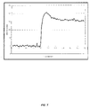
- FIG. 7 presents the sensorgram data for the immobilization of PDBA-conjugated alkaline phosphatase on the surface of a Spreeta 2000 sensor according to the compositions and methods of the present invention, as described in Example 15.
- FIG. 8 presents the sensorgram data for the sequential immobilization of increasing amounts of PDBA-conjugated alkaline phosphatase on the surface of a Spreeta 2000 sensor according to the compositions and methods of the present invention, as described in Example 17.
- FIG. 9 presents the sensorgram data for the sequential immobilization and release of PDBA-conjugated alkaline phosphatase on the surface of a Spreeta 2000 sensor according to the compositions and methods of the present invention, as described in Example 18.
- FIG. 10 presents the sensorgram data for the binding of an antibody to alkaline phosphatase to immobilized PDBA-conjugated alkaline phosphatase on the surface of a Spreeta 2000 sensor, as described in Example 19.
- FIG. 11 presents the sensorgram data for the binding of a human IgG to immobilized PDBA-conjugated Protein A on the surface of a Spreeta 2000 sensor, as described in Example 20.
- biological binding pair refers to any two molecules that exhibit mutual affinity or binding capability, including biochemical, physiological and/or pharmaceutical interactions. Often the pair will be referred to as a “ligand-analyte” pair in the context of BIA.
- Typical examples of biological binding pairs include: antibody/antigen, antibody/hapten, enzyme/substrate, enzyme/inhibitor, enzyme/cofactor, DNA/DNA, DNA/RNA, RNA/RNA, protein/nucleic acid, lectin/carbohydrate, receptor/hormone, receptor/effector, ligand/cell surface receptor, ligand/virus, and the like.
- BAS refers to a bioactive species or a biologically active species or a ligand including, but not limited to, a protein, a polypeptide, a polypeptide fragment, a nucleic acid, a carbohydrate, a receptor, a hormone, a toxin, a vesicle, a liposome, and a cell.
- ACM refers to any member of a broad class of boronic acid complexing moieties.
- PBA refers to a broad class of boron-containing compounds which complex with a BACM. In certain preferred aspects, it refers to a phenylboronic acid moiety.
- self-assembled monolayer refers to a relatively ordered assembly of molecules spontaneously chemisorbed on a surface, in which the molecules are ordered roughly parallel to each other and roughly perpendicular to the surface.
- Each of the molecules includes a functional group that adheres to the surface, and a portion that interacts with the neighboring molecules in the monolayer to form the relatively ordered array.
- SAM refers to a self-assembled monolayer of a single organic compound.
- mSAM refers to a self-assembled monolayer of two or more organic compounds.
- OEG refers to an oligo(ethylene glycol), which is a polymer of about 1 to about 50 ethylene oxide units.
- PEG refers to a poly(ethylene glycol), which is a polymer of greater than about 50 ethylene oxide units.
- immobilized used with respect to a species, has an art recognized meaning and refers to a condition in which the species is attached to a surface with an attractive force stronger than the attractive forces that are present in the intended environment of use of the surface and that act on the species, for example, solvating and turbulent forces. Coordinate and covalent bonds are representative of attractive forces stronger than typical environmental forces.
- non-specific binding refers to interaction between any species, present in a medium from which a biological molecule or analyte is desirably captured, and a binding partner or other species immobilized at a surface, or the surface itself, other than the desired biological binding between the biological molecule and the binding partner.
- Biomolecular interaction analysis is the measurement of interaction or binding of one or more analytes to a biologically active species or ligand on a sensor surface. If the sensor is based on surface plasmon resonance, the interaction of an analyte with a ligand changes the refractive index at the surface. This change is detected for example, as a shift in the angle or wavelength of light at which surface plasmon resonance occurs. The magnitude of the shift in angle or wavelength is an indication of the strength of the interaction (affinity). The rate-of-change of the angle or wavelength with respect to time is used to determine the rate at which the binding occurs.
- the present invention provides a sensor surface, the sensor surface comprising: a substrate coated with a free electron metal; and a matrix layer disposed on the free electron metal, wherein the matrix layer comprises an organic compound having a boronic acid complexing moiety.
- the organic compound “presents” a boronic acid complexing moiety for complexation with a phenylboronic acid.
- the matrix is preferably a self-assembled monolayer (SAM) or, more preferably, a mixed self-assembled monolayer (mSAM).
- the matrix layer comprises an organic compound having a boronic acid complexing moiety (BACM).
- the BACM complexes with a boronic acid, such as aphenylboronic acid or derivatives thereof.
- a boronic acid such as aphenylboronic acid or derivatives thereof.
- phenylboronic acid (or “PBA”) is used herein to include a broad class of boron-containing compounds which complex with the BACM.
- boronic acid complexing moieties are utilized with a boronic acid moiety, such as PBA, to facilitate chemical immobilization of a biologically active species (BAS) to a sensor surface.
- BACM biologically active species
- Suitable BAS include, but are not limited to, a protein, a polypeptide, a polypeptide fragment, a nucleic acid, a carbohydrate, a receptor, a hormone, a toxin, a vesicle, a liposome, a cell, and the like.
- the BAS has an affinity for an analyte of interest.
- FIG. 1 depicts a typical embodiment of the present invention. This diagram is merely an illustration and should not be assumed to limit the scope of the claims herein. One of ordinary skill in the art will recognize other variations, modifications, and alternatives to the specifics of this diagram.
- the sensor substrate 100 is coated with a thin coating of free electron metal such as gold 110 .
- the gold coating is then reacted, such as simultaneously, sequentially, or the like, with a pair of thiol-containing compounds, one of which terminates in a hydroxyl group 120 and the other in a boronic acid complexing moiety 130 (e.g., salicylhydroxamic acid (SHA)).
- a boronic acid complexing moiety 130 e.g., salicylhydroxamic acid (SHA)
- This treatment results in the formation of a self-assembled monolayer 140 (such as a binary SAM) on the gold coating and provides a matrix for immobilization of a biologically active species.
- a self-assembled monolayer 140 such as a binary SAM
- either of the two thiol-containing compounds, 120 or 130 may be used by itself to form a unitary self-assembled monolayer. Additionally, it is possible to use mixtures of three or more thiol-containing compounds to form ternary, quaternary, and the like, SAMs.
- the sensor surface of the present invention comprises a matrix layer, preferably a self-assembled monolayer, of one or more organic compounds, at least one of which, in turn, comprises a boronic acid complexing moiety (BACM).
- the organic compound having the BACM is of the formula: X—R—Y (I) wherein X, R and Y are as defined previously.
- the anchor group X forms a stable coordination complex with the free electron metal (e.g., Au coating on the sensor substrate).
- Suitable free electron metals include, but are not limited to, gold, silver, aluminum, copper, palladium and platinum. Those of skill in the art will know of other free electron metals suitable for use in the present invention.
- the choice of metal depends upon the particular detection technology employed with the sensor.
- the free electron metal is preferably gold.
- Anchor groups for free electron metals include, but are not limited to, thiol, disulfide and phosphine functional groups. The adsorption of the organic compound to the free electron metal is dependent upon the correct pairing of anchor group X with the free electron metal of choice.
- thiol or disulfide anchor groups are particularly suited for gold- or silver-coated surfaces, while phosphine anchor groups are suitable for a palladium-coated surface. If the free electron metal has an oxide coating, then a carboxylate, a sulfonate, a phosphate, an alkoxysilane or a chlorosilane group is suitable as an anchor group.
- the anchor group is not attached directly to the BACM, but can be linked via a spacer molecule, R.
- the spacer molecule is flexible.
- R preferably contains an alkylene group of formula (CH 2 ) n , optionally interrupted by a heteroatom, and is at least about 8 carbon equivalents in length. More preferably, R is an optionally substituted alkylene group, optionally interrupted by a heteroatom, which is about 8 to about 40 carbon equivalents in length.
- one end of the spacer group R comprises the anchor group X.
- the other end of the spacer group R which faces away from the free electron metal following self-assembled monolayer formation, comprises one or several linking groups by which the BACM or a component thereof, is attached to R.
- linking groups include, but are not limited to, groups that inhibit nonspecific binding of ligands or analytes to the matrix layer, such as an OEG, a PEG, a branched OEG, a branched PEG, an oligo(peptide), an oligo(propylene sulfoxide), a sugar (e.g., monosaccharide, disaccharide, and the like), a sugar alcohol (e.g., an alditol), and a dendrimer.
- groups that inhibit nonspecific binding of ligands or analytes to the matrix layer such as an OEG, a PEG, a branched OEG, a branched PEG, an oligo(peptide), an oligo(propylene sulfoxide), a sugar (e.g., monosaccharide, disaccharide, and the like), a sugar alcohol (e.g., an alditol), and a dendrimer.
- R includes an amide bond, as in the formula: X—(CH 2 ) n —NHC(O)—R′—Y (Ia) or X—(CH 2 ) n —C(O)NH—R′—Y (Ib) wherein X and Y are as defined previously.
- R′ in Formulae Ia and Ib, is an optionally substituted alkylene group, optionally interrupted by a heteroatom, which is about 8 to about 30 carbon equivalents in length.
- the index “n” of the oligo(methylene) group in Formulae IIa and IIb is an integer from 1 to 5.
- the presence of this amide bond substantially increases the thermal stability of matrix layer.
- the organic compound comprising a BACM is of the formula: X—R—NSBI—Y (II) wherein n, R, NSBI and Y are as previously defined.
- R includes the presence of an amide bond, as in the formula: X—(CH 2 ) n —NHC(O)—R′—NSBI—Y (IIa) or X—(CH 2 ) n —C(O)NH—R′—NSBI—Y (IIb)
- the R of Formula II comprises an amide bond, an oligo(methylene) group and R′.
- the index “n” of the oligo(methylene) group in Formulae IIa and IIb is an integer from 1 to about 5.
- R′ in Formulae Ia and Ib is an optionally substituted alkylene group, optionally interrupted by a heteroatom, which is about 8 to about 30 carbon equivalents in length.
- the nonspecific binding inhibitor, NSBI is preferably an oligo(ethylene glycol) moiety containing form about 1 to about 15 ethylene oxide units, and is terminated in a hydroxyl or amino group to facilitate attachment of Y, the BACM.
- the BACM interacts with a boronic acid, such as PBA or derivatives thereof, to form a boronic acid complex. This is accomplished by contacting the BACM and the boronic acid and incubating for a period of time.
- a boronic acid such as PBA or derivatives thereof
- the ligand will be in aqueous solution.
- the complexation reaction is insensitive to variations in the ionic strength and the pH of the solution, as well as to the presence of organic co-solvents (e.g., used to help dissolve analytes of low water solubility), detergents (e.g., used to solubilize and stabilize solutions of hydrophobic analytes such as membrane proteins), and chaotropic agents (e.g., protein and nucleic acid denaturants).
- organic co-solvents e.g., used to help dissolve analytes of low water solubility
- detergents e.g., used to solubilize and stabilize solutions of hydrophobic analytes such as membrane proteins
- chaotropic agents e.g., protein and nucleic acid denaturants
- the BACM has the formula:
- p, Q, W and X are as previously defined.
- Compounds of Formula III can be made by a variety of methods.
- Q is O (oxygen)
- X is H or OH
- W is NHOH
- the index “p” is 1.
- the compounds of Formula III may be derived from either 4- or 5-methylsalicylic acid (X ⁇ H) or 3- or 4-methyl-2,6-dihydroxybenzoic acid (X ⁇ OH).
- the compound may be prepared from a synthetic intermediate which is either a lower alkyl 4- or 5-methylsalicylate (X ⁇ H), or a lower alkyl 3- or 4-methyl-2,6-dihydroxybenzoate (for detailed preparation of these compounds, see, U.S. Pat. Nos.
- the matrix layer is made up of a binary mixture of organic compounds terminated in a BACM such as one of the type depicted in Formulae I, Ia, Ib, II, IIa or IIb, and an organic compound of similar and related structure terminated not in a BACM, but rather in an uncharged, unreactive group, as in the formula: X—R—Z (VI) or X—(CH 2 ) n C(O)NHR′—(NSBI) m —Z (VIa) or X—(CH 2 ) n NHC(O)R′—(NSBI) m —Z (VIb) wherein X, n, R′, NSBI, m and Z are as defined previously.
- Such uncharged, unreactive groups include, but are not limited to, H, CH 3 , OH, OCH 3 , CO 2 CH 3 , C(O)NH 2 , C(O)NHCH 3 , C(O)N(CH 3 ) 2 or SO 2 NH 2 .
- Preferred groups in this context include, for example, hydrophilic groups such as OH and OCH 3 , in order to minimize or eliminate nonspecific binding.
- the matrix comprising the mixed self-assembled monolayer from such a binary mixture of organic compounds contains from about 1 mole % to about 30 mole % of the BACM-terminated compound; preferably, it contains from about 2 mole % to about 20 mole %; and most preferably, it contains from about 4 mole % to about 10 mole %.
- the sensor surface comprises SCH 2 C(O)NH(CH 2 ) 11 (OCH 2 CH 2 ) 11 OC(O)Y (Polymer A) and —SCH 2 C(O)NH—(CH 2 ) 11 (OCH 2 CH 2 ) 3 OH (Polymer B).
- the mole percent of Polymer A is less than about 10% of the total mole percent of Polymer A and Polymer B together. In another aspect, the mole percent of the Polymer A is less than about 5% of the total mole percent of Polymer A and Polymer B.
- a binary SAM is advantageous as it provides minimum nonspecific binding to the matrix layer, and allows for optimization of immobilized ligand density for particular applications of the sensor.
- the organic compound having a BACM attached thereto is of the formula: HS(CH 2 ) n C(O)NHR′—NSBI—SHA (IV) wherein n, R′, NSBI and SHA are as defined previously.
- n is 2
- R′ is (CH 2 ) 11
- NSBI is (OCH 2 CH 2 ) 6
- FIG. 3 sets forth a preferred compound of Formula IV.
- the organic compound having a BACM attached thereto is of the formula: HS(CH 2 ) n C(O)NHR′—NSBI—DHBHA (V) wherein n, R′, NSBI and DHBHA are as defined previously.
- n is 2
- R′ is (CH 2 ) 11
- NSBI is (OCH 2 CH 2 ) 6
- FIG. 4 sets forth a preferred compound of Formula V.
- the matrix layer comprises a binary mSAM of organic compound of Formula IV having a BACM bound thereto and a compound of formula: HS(CH 2 ) n C(O)NHR′—NSBI—Z (VIII) wherein n, R′, NSBI and Z are as defined previously.
- n is 2, R′ is (CH 2 ) 11 , NSBI is (OCH 2 CH 2 ) 6 , and Z is OH.
- the matrix layer comprises a binary mSAM of organic compound of Formula V having a BACM thereto affixed and a compound of Formula VIII.
- n is 2, R′ is (CH 2 ) 11 , NSBI is (OCH 2 CH 2 ) 6 , and Z is OH.
- the present invention provides a sensor surface comprising: a substrate coated with a free electron metal; a matrix layer disposed on the free electron metal, wherein the matrix layer comprises an organic compound having a boronic acid complexing moiety; and a boronic acid moiety complexed to the boronic acid complexing moiety.
- the boronic acid moiety comprises a boronic acid reagent conjugated to a biologically active species.
- the boronic acid moiety has the formula:
- BAS is a bioactive species including, but not limited to, a protein, a polypeptide, a polypeptide fragment, a nucleic acid, a carbohydrate, a receptor, a hormone, a toxin, a vesicle, a liposome, and a cell.
- Z 1 in Formula VII, is a spacer group including, but not limited to, a saturated or unsaturated aliphatic chain of 0 to about 6 carbon equivalents in length, an unbranched saturated or unsaturated aliphatic chain of from about 6 to about 18 carbon equivalents in length with at least one intermediate amide or disulfide moiety, and an oligo(ethylene glycol) chain of from about 3 to about 12 carbon equivalents in length.
- Q 1 in Formula VII, is a linkage including, but not limited to, an amide, methyl amide, methylene, ether, thioether, methylene ether and methylene thioether moiety.
- the index “t”, in Formula VII, is an integer from 1 to 3.
- t is 1.
- the boronic acid functional group is preferably meta or para to Q 1 .
- the present invention provides boronic acid moieties of the following formulae:
- t is 2.
- the boronic acid functional groups are preferably oriented 1,2 or 1,3 on the phenyl ring with respect to Q 1 .
- the present invention also provides boronic acid moieties of the following formulae:
- transition metal-catalyzed cross-coupling reactions have been developed to produce phenylboronic acids from aryl halides and alkoxydiboron (Ishiyama, T.; Murata, M. and Miyaura, N. (1995) J. Org. Chem. 60, 7508–7510; and Giroux, A., Han, Y. and Prasit, P. (1997) Tetrahedron Lett. 38, 3841–3844) or bialkoxyhydroborane (Murata, M., Watanabe, S., and Masuda, Y. (1997) J. Org. Chem. 62, 6458–6459) using PdCl 2 (dppf) as the catalyst.
- 1,2-phenylenediboronic acid has been previously described (see, Clement, R. and Thiais, F. (1966) Hebd. Seances Acad. Sci., Ser. C 263, 1398–1400).
- FIG. 2 illustrates one embodiment of the present invention, wherein a phenylenediboronic acid (PDBA) conjugated protein 250 is immobilized on a sensor surface by virtue of formation of boronic acid complexes 260a, 260b with boronic acid complexing moieties (e.g., 130 in FIG. 1 ).
- a boronic acid complexing moiety preferably results in a reversible, non-covalent coordination complex.
- boronic acid moieties comprising a boronic acid reagent 270a, 270b conjugated to a bioactive species 250 is complexed or conjugated to the boronic acid complexing moiety (e.g., 130 in FIG. 1 ) of the matrix of the sensor surface.
- the boronic acid will typically attach to more than one reactive site on the BAS. If, for example, the BAS is a protein 250, multiple boronic acids will react with the protein, each boronic acid reacting at one of several sites on the protein.
- a complex can be with at least one bioactive species 250 having one or more pendant phenylboronic acid or phenylenediboronic acid moieties comprising boronic acid reagents 270a, 270b, complexed with one or more boronic acid complexing moieties.
- the chemical structure of the complex will, of course, depend upon the particular boronic acid moiety conjugated to the bioactive species.
- the bioconjugate formed by complexation of the boronic acid moiety comprising a phenylboronic acid moiety and a BAS is of the formula:
- the bioconjugate formed by complexation of the boronic acid moiety comprising a 1,3- or 1,4-phenylenediboronic acid moiety and a BAS is of the formula:
- the bioconjugate formed by complexation of the boronic acid moiety comprising a 1,2-phenylenediboronic acid moiety and a BAS is of the formula:
- the bioconjugate complex can be prepared in buffered aqueous solution, or in organic solvents, or in mixtures of aqueous buffers and water-miscible organic solvents.
- the bioconjugate is formed within a few minutes over a range of temperatures, for example, from about 4° C. to about 70° C.
- the bioconjugate can be formed over a wide range of pH and ionic strength, and in the presence of detergents or chaotropes (e.g., protein and nucleic acid denaturants). Once formed, the bioconjugate is stable to an even broader range of pH, ionic strength, detergent concentrations and chaotrope concentrations.
- BAS represents a biologically active species which may or may not contain a portion of a reactive moiety used to attach the bioactive species to a boronic acid moiety, such as a phenylboronic acid moiety.
- the BAS is typically a ligand for an analyte of interest. The BAS and the analyte of interest specifically interact with one another, thereby forming a biological binding pair.
- Specific binding of the first member pair, e.g., the BAS, to the second member of the binding pair, e.g., the analyte, in a sample is evidenced by the binding of the first member to the second member, or vice versa, with greater affinity (and/or specificity) than to the other components of the sample.
- the binding between the members of the binding pair is typically non-covalent, although the present invention is not so limited. In sensor applications, any BAS that has an affinity for an analyte of interest can be used with the methods of the present invention.
- attachment of the boronic acid to the BAS involves the stacking of ligands.
- a boronic acid moiety is covalently attached to avidin or streptavidin which, in turn, is non-covalently attached to a biotinylated ligand that is capable of selectively binding an analyte of interest (e.g., a biotinylated antibody).
- analyte of interest e.g., a biotinylated antibody
- the BAS can be any small molecule that is capable of binding to an analyte of interest.
- Exemplary biological binding pairs suitable for use with the methods of the present invention include, but are not limited to, any haptenic or antigenic compound in combination with a corresponding antibody or binding portion or fragment thereof (e.g., digoxigenin and anti-digoxigenin; fluorescein and anti-fluorescein; dinitrophenol and anti-dinitrophenol; bromodeoxyuridine and anti-bromodeoxyuridine; mouse immunoglobulin and goat anti-mouse immunoglobulin) and nonimmunological binding pairs (e.g., biotin and avidin; biotin and streptavidin; hormone (e.g., thyroxine or cortisol) and hormone binding protein; receptor and receptor agonist or antagonist; immunoglobulin G and protein A; lectin and carbohydrate; enzyme and enzyme cofactor; enzyme and enzyme inhibitor; complementary polynucleotide pairs capable of forming nucleic acid duplexes or triplexes; and the like.
- a haptenic or antigenic compound in combination with
- test assay generally refers to any procedure in which a member of a biological binding pair is to be specifically captured from a medium in which it is dispersed or is expected to be dispersed.
- test assay can be used to describe a diagnostic procedure, analytical procedure, microanalytical procedure, forensic analysis, pharmacokinetic study, cell sorting procedure, affinity chromatographic separation, laboratory or industrial characterization or recovery of one or more species such as toxins, catalysts, starting materials or products, and the like.
- Biosensing applications include those such as drug screening, environmental monitoring, medical diagnostics, quality control in the pharmaceutical and food industries, and other areas in which it is advantageous to sensitively measure the interaction between biological binding partners.
- the present invention provides a method for detecting an analyte, the method comprising: providing a sensor comprising a substrate coated with a free electron metal, a matrix layer disposed on the free electron metal, the matrix layer comprising an organic compound, wherein the organic compound has a boronic acid complexing moiety and, a boronic acid moiety complexed to the boronic acid complexing moiety; and contacting the sensor with the analyte to elicit a response; and measuring the response, thereby detecting the analyte.
- the boronic acid moiety comprises a boronic acid conjugated to a BAS that is capable of specifically binding an analyte of interest.
- Various responses include, for example, an electrical response, an optical response, a refractive index change, a shift in the angle or wavelength of light such as at which surface plasmon resonance occurs, a fluorescence response, a color change, and combinations thereof.
- Information that can be obtained using the sensors of the present invention includes, but is not limited to, kinetic measurements, which provide, for example, information on the rates at which interacting molecules bind to each other and break apart; affinity determinations, which provide details on the way in which a biological process occurs and is controlled; specificity studies, which examine, for example, the ability of one molecule to bind to another molecule to the exclusion of others; and concentration measurements, which determine, for example, the amount of an active molecule that is present in a sample.
- Advantages of the sensors of the present invention include, but are not limited to, tailored immobilization of ligands; label-free detection; analysis without purification; continuous monitoring; real-time monitoring; high sensitivity of detection; facile preparation and regeneration; stability at increased temperature; minimal non-specific binding; and the like.
- the present invention provides for arrays of sensors (e.g., at least two sensors).
- the sensor array may be a cartridge containing eight individual sensors operating individually or simultaneously.
- the sensor array may be made up of 96 individual sensors disposed in a multi-well plate.
- the array of sensors contains the same ligand immobilized on the surface of all sensors in the array; the array is then contacted with a plurality of analytes to analyze their various interactions with the ligand.
- a plurality of ligands are immobilized on the surface of the array of sensors, wherein at least two sensors have a different ligand; the array is then contacted with a single analyte of interest to analyze its interaction with the various ligands.
- any combination of ligands and analytes can be utilized with the array, in particular, to make replicate measurements on multiple analytes and ligands.
- certain sensors in the array can be employed as references to determine the level of non-specific responses. As such, the sensor array can be tailored to specific analytes or ligands and to specific assays and applications.
- a single sensor may have an array of differently mixed monolayers patterned on its surface in a spatially distinct manner.
- this patterning can be used on a single sensor to detect a plurality of analytes by immobilizing different ligands on the spatially distinct elements of the array.
- the array of sensors are compositionally different.
- the sensor array may contain a pH sensor, a temperature probe sensor, a humidity sensor, a color sensor, a sensor as described herein, and the like.
- 11-Aminoundecanoic acid (20.4 g, 101.3 mmol) and di-tert-butyldicarbonate (24.3 g, 111.0 mmol) are suspended in water (300 mL) and methanol (150 mL).
- Aqueous sodium hydroxide (3.13 M, 65 mL) is added, and the mixture is stirred at room temperature. After 30 minutes, the mixture became homogenous. After stirring for a total of 6 hours, the methanol was removed in vacuo. Ethyl acetate (500 mL) was added to the aqueous solution.
- Aqueous hydrochloric acid (1 N, 300 mL) was added slowly to the stirred mixture until the evolution of gas ceased.
- Tri(ethylene glycol) (47.0 mL, 352.0 mmol) was added to a round bottom flask (100 mL) and heated to 110° C.
- Aqueous sodium hydroxide solution (50% [w/v], 2.80 mL, 35.0 mmol) was added, and the solution was stirred for 45 minutes at 110° C.
- Compound 3 (12.8 g, 35.0 mmol) was added to the hot solution, and stirring was continued for 8 hours at 110° C.
- the reaction mixture was then cooled to room temperature, and diluted with water (300 mL). The aqueous solution was extracted three times with ethyl acetate (150 mL portions).
- Dithiopropionic acid (1.65 g, 7.85 mmol) was dissolved in anhydrous 1,4-dioxane (60 mL). The solution was stirred and N-hydroxysuccinimide (1.99 g, 17.3 mmol) was added, followed by N,N′-dicyclohexylcarbodiimide (3.57 g, 17.3 mmol). The reaction mixture was stirred for 16 hours at room temperature, then filtered.
- compound 4 (6.60 g, 15.7 mmol) was dissolved in 1,4-dioxane (40 mL) and a solution of hydrogen chloride in 1,4-dioxane (4 M, 38 mL, 152 mmol) was added. The mixture was stirred for 16 hours at room temperature.
- the crude reaction mixture containing compound 5 was treated with water (20 mL) and aqueous hydrochloric acid (4 M, 10 mL). Tris-carboxyethylphosphine hydrochloride (4.10 g, 14.3 mmol) was added, and the solution was stirred for 24 hours at room temperature. The organic solvents were removed in vacuo, and the residue was diluted with water (200 mL). The aqueous mixture was extracted three times with ethyl acetate (150 mL portions). The combined ethyl acetate extracts were washed sequentially with water (100 mL) and brine (75 mL), dried over anhydrous magnesium sulfate, filtered, and evaporated in vacuo.
- Hexa(ethylene glycol) (75.0 g, 267 mmol was added to a round bottom flask (100 mL) and heated to 110° C.
- Aqueous sodium hydroxide solution (50% [w/v], 1.80 mL, 22.5 mmol) was added, and the solution was stirred for 45 minutes at 110° C.
- Compound 3 (12.8 g, 35.0 mmol) was added to the hot solution, and stirring was continued for 8 hours at 110° C.
- the reaction mixture was then cooled to room temperature, and diluted with water (300 mL). The aqueous solution was extracted three times with ethyl acetate (150 mL portions).
- the reaction mixture was diluted with water (150 mL) and extracted three times with ethyl acetate (100 mL portions). The combined organic solutions were washed with aqueous hydrochloric acid (1 N, 50 mL) and then twice with brine (50 mL portions). The organic layer was dried over anhydrous magnesium sulfate and filtered. The filtrate was concentrated in vacuo and the crude product was purified by chromatography on silica gel (200 g, gradient of dichloromethane to 5% [v/v] methanol in dichloromethane). The product containing fractions were pooled and evaporated in vacuo to give a yellowish oil (9.8 g, 69% yield).
- Dithiopropionic acid (1.05 g, 5.0 mmol) was dissolved in anhydrous 1,4-dioxane (20 mL). The solution was stirred and N-hydroxysuccinimide (1.15 g, 10.0 mmol) was added, followed by N,N′-dicyclohexylcarbodiimide (2.06 g, 10.0 mmol). The reaction mixture was stirred for 16 hours at room temperature, then filtered.
- compound 12 (7.68 g, 10.0 mmol) was dissolved in anhydrous 1,4-dioxane (20 mL) and hydrogen chloride in 1,4-dioxane (4 N, 10 mL, 40.0 mmol) was added. The solution was stirred at room temperature for 16 hours. The mixture was then cooled to 4° C., and N,N-diisopropylethylamine (10 mL, 57.0 mmol) was added slowly.
- Compound 16 was prepared from compound 4 using methods analogous to the preparation of compound 3 (Example 1, step C).
- Compound 17 was prepared from compound 16 using methods analogous to the preparation of compound 11 (Example 2, step A).
- HRMS (ESI-pos) calcd for C 34 H 69 NO 12 683.4820, obsd: 683.4829.
- Compound 19 was prepared from compound 4 using methods analogous to the preparation of compound 4 (Example 1, steps E and F), substituting dithiodiglycolic acid for dithiopropionic acid.
- a hydrogenation bottle (1 L) was charged with compound 23 (6.20 g, 12.7 mmol) dissolved in methanol (300 mL). The atmosphere in the bottle was replaced with nitrogen, and palladium on carbon (10% [w/w], 0.62 g) is added.
- the bottle was affixed to a Parr hydrogenation apparatus, the bottle evacuated then re-filled with hydrogen to 40 psi, and the reaction was shaken at room temperature for 2 hours. The bottle was then removed from the Parr apparatus, the catalyst removed by filtration, and the methanol evaporated in vacuo. The residue was dissolved in N,N-dimethylformamide (20 mL) and transferred to the reaction flask containing activated compound 11.
- N,N-Diisopropylethylamine (4.0 mL, 23.0 mmol) was added, and the solution was heated to 70° C. The reaction was allowed to proceed for 24 hours. The solvent was then removed in vacuo, and the residue was dissolved in ethyl acetate (250 mL). The ethyl acetate solution was washed sequentially with aqueous hydrochloric acid (1 N, 150 mL), water (100 mL) and brine (100 mL). The organic layer was dried over anhydrous magnesium sulfate, filtered, and the filtrate was evaporated in vacuo.
- Dithioglycolic acid (0.65 g, 3.54 mmol) was dissolved in tetrahydrofuran (50 mL). The solution was stirred and N-hydroxysuccinimide (0.90 g, 7.79 mmol) was added, followed by N,N′-dicyclohexylcarbodiimide (1.61 g, 7.79 mmol). The reaction mixture was stirred for 16 hours at room temperature, then filtered.
- compound 24 (5.78 g, 7.08 mmol) was dissolved in dichloromethane (25 mL) and trifluoroacetic acid (25 mL) was added. The solution was stirred for 30 minutes at room temperature, and then the solvents were evaporated in vacuo. The resulting oil was dissolved in tetrahydrofuran (50 mL) and N,N-diisopropylethylamine (12.0 mL, 68.9 mmol) was added.
- the crude compound 25 (3.54 mmol) was dissolved in trifluoroacetic acid (50 mL) and the solution was stirred for 3 days at room temperature. The solvent was then removed in vacuo. The resulting oil was dissolved in methanol (100 mL) and water (10 mL). Tris-carboxyethylphosphine hydrochloride (1.15 g, 4.01 mmol) was added, and the solution was stirred for 2 hours at room temperature. The methanol was removed in vacuo, and ethyl acetate (300 mL) was added. The mixture was washed three times with water (100 mL).
- Tri(ethylene glycol) (96.0 mL, 719.0 mmol) was added to a round bottom flask (250 mL) and heated to 110° C.
- Aqueous sodium hydroxide solution (50% [w/v], 5.75 mL, 71.9 mmol) was added, and the solution was stirred for 45 minutes at 110° C.
- 12-Bromoundecanoic acid (10.0 g, 35.8 mmol) was added to the hot solution, and stirring was continued for 16 hours at 110° C.
- the reaction mixture was then cooled to room temperature, and diluted with water (500 mL). The aqueous solution was extracted three times with ethyl acetate (150 mL portions).
- the reaction mixture was diluted with ethyl acetate (300 mL) and washed twice with water (150 mL portions) and then brine (75 mL). The organic layer was dried over anhydrous magnesium sulfate, filtered, and evaporated in vacuo.
- the crude product was purified by chromatography on silica gel (150 g, gradient of dichloromethane to 3% [v/v] methanol in dichloromethane). The product containing fractions were pooled and evaporated in vacuo to give an oil (2.73 g, 75% yield).
- Compound 33 was prepared by the method of Pale-Grosdemange et al. (Pale-Grosdemange, C., Simon, E. S., Prime, K. L. and Whitesides, G. M. (1991) J. Am. Chem. Soc. 113, 12–20).
- a solution of thiol compound was prepared at a total thiol concentration of 1.0 mM in absolute ethanol.
- a cleaned wafer was placed gold-side up in a polypropylene tub of sufficient diameter to allow the wafer to sit horizontally at the bottom.
- the thiol solution was added (150 to 200 mL) and the tub was covered.
- the wafer was allowed to sit in the thiol solution for 5 days in the dark.
- the wafer was then removed from the solution and washed in a vigorous spray of absolute ethanol or N,N-dimethylformamide.
- the wafer was sonicated in a covered polypropylene container in three changes of either absolute ethanol or N,N-dimethylformamide for 15 minutes each. Finally, the wafer was washed with absolute ethanol and dried under a stream of nitrogen.
- the wafer was sawed or scribed into small chips (9.46 mm ⁇ 4.46 mm) prior to deposition of the mixed SAM.
- the procedure used for cleaning the chips and depositing the mixed SAM was the same as that for the wafer.
- a solution of thiol compound containing the boronic acid complexing moiety (e.g., compounds of Formulae IIa and IIb such as compound 14, compound 18 and compound 26) and thiol compound terminating in an uncharged, hydrophilic group (e.g., compounds of Formula VIa and VIb, such as compound 4 and compound 32) in the desired molar ratio was prepared at a total thiol concentration of 1.0 mM in absolute ethanol.
- a cleaned wafer was placed gold-side up in a polypropylene tub of sufficient diameter to allow the wafer to sit horizontally at the bottom.
- the mixed thiol solution was added (150 to 200 mL) and the tub was covered. The wafer was allowed to sit in the thiol solution for 5 days in the dark.
- the wafer was then removed from the solution and washed in a vigorous spray of absolute ethanol or N,N-dimethylformamide.
- the wafer was sonicated in a covered polypropylene container in three changes of either absolute ethanol or N,N-dimethylformamide for 15 minutes each.
- the wafer was washed with absolute ethanol and dried under a stream of nitrogen.
- the wafer was sawed or scribed into small chips (9.46 mm ⁇ 4.46 mm) prior to deposition of the mixed SAM.
- the procedure used for cleaning the chips and depositing the mixed SAM was the same as that for the wafer.
- a solution of thiol compound containing the boronic acid complexing moiety (e.g., compounds of Formulae IIa and IIb such as compound 14, compound 18 and compound 26) was prepared at the desired concentration (“X mM)”in 10:90 [v/v] water:N,N-dimethylformamide.
- X mM concentration
- a cleaned wafer was placed gold-side up in a polypropylene tub of sufficient diameter to allow the wafer to sit horizontally at the bottom.
- the thiol solution was added (150 to 200 mL) and the tub was covered. The wafer was allowed to sit in the thiol solution for 15–24 hours in the dark. The wafer was then removed from the solution and rinsed twice with 10:90 [v/v] water:N,N-dimethylformamide.
- a solution of the thiol compound terminating in an uncharged, hydrophilic group (e.g., compounds of Formula VIa and VIb, such as compound 4 and compound A2) in the desired concentration (“1.0–X mM”) was prepared in 10:90 [v/v] water:N,N-dimethylformamide.
- the once-treated wafer was placed gold-side up in a polypropylene tub of sufficient diameter to allow the wafer to sit horizontally at the bottom.
- the second thiol solution was added (150 to 200 mL) and the tub was covered.
- the wafer was allowed to sit in the thiol solution for 2–5 days in the dark.
- the wafer was then removed from the solution and sonicated in a covered polypropylene container in three changes of N,N-dimethylformamide for 15 minutes each. Finally, the wafer was washed with absolute ethanol and dried under a stream of nitrogen.
- the wafer was sawed or scribed into small chips (9.46 mm ⁇ 4.46 mm) prior to deposition of the mixed SAM.
- the procedure used for cleaning the chips and depositing the mixed SAM was the same as that for the wafer.
- SAMs were prepared on gold-on-glass chips (mm ⁇ mm) according to the procedure of Example 9 using a 1.0 mM solution of either compound 15 (amide-containing; 25 chips) or compound 33 (amide-lacking; 25 chips) in absolute ethanol. Following SAM deposition, the chips were gently wiped with a non-abrasive, lint-free pad moistened with aqueous sodium hydroxide (1 N), then rinsed well with water. The SAM-coated chips were then air-dried. The cleaned, coated chips (5 of each type) were then analyzed for surface wettability using water contact angle measurements obtained with a VCA Optima (AST Products, Inc.; Billerica, Mass., USA).
- Water contact angle is an indicator of the wettability of surface. Smaller values of water contact angle indicate a more wettable (i.e., hydrophilic) surface, while larger values indicate a less wettable (i.e., hydrophobic) surface. Intitially, chips with SAMs prepared from either the amide-containing monomer or amide-lacking monomer showed good wettability (contact angle of 40–43 degrees), consistent with the surface presentation of hydrophilic oligo(ethylene glycol) groups (see FIG. 1 ). However, significant differences were observed between the two SAMs on heating to 60° C.
- the amide-containing SAMs showed little change in wettability until between 2 and 4 days of incubation at elevated temperature, and even modest wettability after 4 days (contact angle of 51–54 degrees), indicating full to partial retention of the hydrophilic SAM layer. Conversely, the amide-lacking SAMs showed poor wettability (contact angle of 62 degrees) within 1 day of treatment at elevated temperature, indicating significant if not total loss of the hydrophilic SAM layer.
- the pure gold film exhibited a water contact angle of greater than 60 degrees.
- the amide-containing SAMs (unitary or mixed) that are the subject of the present invention have a significant advantage over amide-lacking SAMs in terms of their stability at elevated temperature. This is presumably due to the ability of the amide-containing SAM monomers to form a strong network of hydrogen bonds among the various amide groups near the surface of the gold film, stabilizing the SAM toward thermal degradation (loss of SAM due to cleavage of the gold-sulfur bond). Formation of such a network of hydrogen bonds is not possible in the prior art amide-lacking SAMs.
- the gold-on-glass chips with the immobilization matrix were affixed to the Spreeta 2000 sensors using optical epoxy (OS1102; Dexter Corporation; Olean, N.Y., USA). Resin (100 parts by weight) and hardener (35 parts by weight) were measured in to a glass beaker and mixed well (10 minutes minimum) using a wooden spatula. The thoroughly mixed epoxy was then degassed under high vacuum (oil pump) in a vacuum desiccator for 20–30 minutes. A clean 30 mL polypropylene syringe was filled with the degassed epoxy. The syringe was placed in the vacuum desiccator and the epoxy degassed under high vacuum for an additional 20–30 minutes.
- optical epoxy OS1102; Dexter Corporation; Olean, N.Y., USA.
- PDBA-AP purified phenylenediboronic acid-alkaline phosphatase conjugate
- PDBA has negligible absorbance at 280 nm, but measurable absorbance at 260 nm.
- Ultraviolet spectra from 240 nm to 450 nm were obtained for each solution. These spectra are shown in FIG.
- a Spreeta 2000 sensor was assembled as described in Example 13, using a gold-on-glass chip with a mixed SAM immobilization matrix prepared as in Example 10.
- the matrix was prepared using a 1.0 mM solution of compound 14 (8 mole %) and compound 15 (92 mole %) in absolute ethanol.
- the sensor was then fitted with an aluminum flow cell equipped with a silicone gasket to provide a flow channel obtained from Texas Instruments.
- the inlet to the flow cell was attached to a six-port liquid chromatography switching valve (Model 9725; Rheodyne. L.
- the sensor was initiated in air prior to use according to instructions received from Texas Instruments.
- the syringe was then filled with buffer (phosphate-buffered saline [PBS]: 0.14 M sodium chloride, 8 mM disodium hydrogen phosphate, 2 mM sodium dihydrogen phosphate, pH 7.3) and the flow started at 6.0 mL/hour.
- PBS phosphate-buffered saline
- Data were collected from the sensor using an LED intensity setting of 1 and an integration time of 1.62 msec.
- SPR Surface plasmon resonance
- FIG. 7 presents the sensorgram data obtained from this experiment.
- the buffer baseline prior to sample injection was mathematically adjusted to zero by subtracting an average of the raw signal over the time period 5.0 to 7.0 minutes from the entire data set.
- the y-axis of the plot is given in Resonance Units (RU), which are equivalent to the change in refractive index times 10 6 .
- Injection of the sample occurred just prior to 8 minutes, where the signal rises quickly in the data. This is due to the increased bulk refractive index of the solution containing protein as well as binding of the PDBA-conjugated protein to the surface matrix.
- the sensor After the 100 ⁇ L bolus of protein solution is cleared from the sensor flow cell, the sensor establishes a new buffer baseline position (at approximately 150 RU), due to immobilization of protein on the sensor surface.
- Each sensor was initiated in air prior to use according to instructions received from Texas Instruments.
- the syringe was then filled with buffer (phosphate-buffered saline [PBS]: 0.14 M sodium chloride, 8 mM disodium hydrogen phosphate, 2 mM sodium dihydrogen phosphate, pH 7.3) and the flow started at 6.0 mL/hour.
- PBS phosphate-buffered saline
- Data were collected from the sensor using an LED intensity setting of 1.0 and an integration time of 1.62 msec.
- SPR Surface plasmon resonance
- Table II below presents representative data for the variety of sensor surface matrices. Bare gold is included for reference, since bare gold exhibits significant non-specific binding of proteins. The non-specific binding is calculated as the difference in refractive index (in RU) between the buffer baseline established prior to the injection of the protein solution and the buffer baseline established following injection of the protein solution.
- Each sensor was initiated in air prior to use according to instructions received from Texas Instruments.
- the syringe was then filled with buffer (phosphate-buffered saline [PBS]: 0.14 M sodium chloride, 8 mM disodium hydrogen phosphate, 2 mM sodium dihydrogen phosphate, pH 7.3) and the flow started at 6.0 mL/hour.
- PBS phosphate-buffered saline
- Data were collected from the sensor using an LED intensity setting of 1.0 and an integration time of 1.62 msec.
- SPR Surface plasmon resonance
- FIG. 8 presents the sensorgram data from this experiment.
- the buffer baseline prior to the initial sample injection on each sensor was mathematically adjusted to zero by subtracting an average of the raw signal over the time period 8.0 to 10.0 minutes from the entire data set.
- the y-axis of the plot is given in Resonance Units (RU), which are equivalent to the change in refractive index times 10 6 .
- the open circles represent data obtained from the binding (non-specific) of AP to the sensor surface matrix. Even at very high protein loads (0.4 mg/mL), non-specific binding is small ( ⁇ 50 RU).
- the closed circles represent data obtained from the immobilization (specific binding) of PDBA-AP to the sensor surface matrix. A titration is observed, as increasing amounts of protein are added to the surface.
- the spikes around 32, 47 and 60 minutes are due to the increased bulk refractive index of the solution containing protein.
- the sensor After each 100 ⁇ L bolus of protein solution is cleared from the sensor flow cell, the sensor establishes a new buffer baseline position (at approximately 40, 75, 150 and 225 RU), due to immobilization of increasing amounts protein on the sensor surface.
- a Spreeta 2000 sensor was assembled as described in Example 13, using a gold-on-glass chip with a mixed SAM immobilization matrix prepared as in Example 10.
- the matrix was prepared using a 1.0 mM solution of compound 14 (4 mole %) and compound 15 (96 mole %) in 10:90 (v/v) water:N,N-dimethylformamide.
- the sensor was then fitted with an aluminum flow cell equipped with a silicone gasket to provide a flow channel obtained from Texas Instruments.
- the inlet to the flow cell was attached to a six-port liquid chromatography switching valve (Model 9725; Rheodyne. L.
- the sensor was initiated in air prior to use according to instructions received from Texas Instruments.
- the syringe was then filled with buffer (phosphate-buffered saline [PBS]: 0.14 M sodium chloride, 8 mM disodium hydrogen phosphate, 2 mM sodium dihydrogen phosphate, pH 7.3) and the flow started at 6.0 mL/hour.
- PBS phosphate-buffered saline
- Data were collected from the sensor using an LED intensity setting of 1 and an integration time of 1.62 msec.
- SPR Surface plasmon resonance
- FIG. 9 presents the sensorgram data obtained from this experiment.
- the buffer baseline prior to sample injection was mathematically adjusted to zero by subtracting an average of the raw signal over the time period 2.0 to 5.0 minutes from the entire data set.
- the y-axis of the plot is given in Resonance Units (RU), which are equivalent to the change in refractive index times 10 6 .
- Injection of the sample occurred just prior to 7 minutes, where the signal rises quickly in the data. This is due to the increased bulk refractive index of the solution containing protein as well as binding of the PDBA-conjugated protein to the surface matrix.
- the sensor After the 100 ⁇ L bolus of protein solution is cleared from the sensor flow cell, the sensor establishes a new buffer baseline position (at approximately 65 RU), due to immobilization of protein on the sensor surface. Injection of aqueous sodium hydroxide (1 N) serves to break the bonds holding the boronic acid complex together, removing the immobilized protein from the sensor surface. This happens essentially quantitatively, as following buffer re-equilibration of the sensor surface matrix (time points 18 to 25 minutes), an equivalent amount of PDBA-AP can again be immobilized (time points 25 to 35 minutes; approximately 80 RU).
- a Spreeta 2000 sensor was assembled as described in Example 13, using a gold-on-glass chip with a mixed SAM immobilization matrix prepared as in Example 10.
- the matrix was prepared using a 1.0 mM solution of compound 14 (8 mole %) and compound 15 (92 mole %) in absolute ethanol.
- the sensor was then fitted with an aluminum flow cell equipped with a silicone gasket to provide a flow channel obtained from Texas Instruments.
- the inlet to the flow cell was attached to a six-port liquid chromatography switching valve (Model 9725; Rheodyne. L.
- the sensor was initiated in air prior to use according to instructions received from Texas Instruments.
- the syringe was then filled with buffer (phosphate-buffered saline [PBS]: 0.14 M sodium chloride, 8 mM disodium hydrogen phosphate, 2 mM sodium dihydrogen phosphate, pH 7.3) and the flow started at 6.0 mL/hour.
- PBS phosphate-buffered saline
- Data were collected from the sensor using an LED intensity setting of 1.0 and an integration time of 1.62 msec.
- SPR Surface plasmon resonance
- an aliquot (100 ⁇ L) of PDBA-AP (prepared as in Example 14; 0.25 mg/mL in PBS) was loaded into the sample loop and injected into the flow stream.
- the PDBA-AP was immobilized to the surface matrix as described in Example 15. Approximately 140 RU of PDBA-AP was immobilized. Data collection was started, and the sensor was washed with PBS buffer until a stable baseline was re-established. Then, an aliquot (100 ⁇ L) of a polyclonal antibody against AP (anti-AP; Rockland, Inc.; Gilbertsville, Pa., USA; 3.0 ⁇ M in PBS) was injected into the flow stream.
- a polyclonal antibody against AP anti-AP; Rockland, Inc.; Gilbertsville, Pa., USA; 3.0 ⁇ M in PBS
- the sensor was washed with PBS buffer until a stable baseline was re-established. Then, an aliquot (100 ⁇ L) of aqueous sodium hydroxide (0.1 N) was injected to dissociate the bound antibody from the AP, and the sensor was washed with PBS buffer until a stable baseline was re-established. This procedure (injection of antibody solution, washing, dissociation with sodium hydroxide and washing) was repeated once more.
- FIG. 10 presents the sensorgram data from this experiment.
- the buffer baseline prior to antibody injection was mathematically adjusted to zero by subtracting an average of the raw signal over the time period 5.0 to 7.0 minutes from the entire data set.
- the y-axis of the plot is given in Resonance Units (RU), which are equivalent to the change in refractive index times 10 6 .
- Injection of the first antibody aliquot occurred at about 4.5 minutes, where the signal rises quickly in the data.
- the sensor After the 100 ⁇ L bolus of antibody solution is cleared from the sensor flow cell, the sensor establishes a new buffer baseline position (at approximately 110 RU), due to binding of the analyte (antibody) to the immobilized ligand (AP) on the sensor surface.
- RU Resonance Units
- the response due to antibody binding is expected to be similar to the initial amount of PDBA-AP immobilized (140 RU), assuming a 1:1 interaction between the two proteins.
- the fact that the antibody binding response is slightly less than anticipated may mean either that some fraction of antibody species is binding simultaneously to two PDBA-AP molecules (each antibody has two potential binding sites), or that some fraction of the PDBA-AP is immobilized in such a way that the antibody recognition site (epitope) is not available for antibody binding.
- the first injection of sodium hydroxide occurred at about 10 minutes, as indicated by the rapid drop in refractive index to a point below zero (this is due to the lower refractive index of the sodium hydroxide solution relative to the PBS buffer).
- the baseline re-stabilized at about 5–10 RU, indicating that the antibody had been nearly completely stripped from the immobilized PDBA-AP. Repetition of the cycle yielded the same result within experimental accuracy.
- immobilization of ligand on a sensor surface by means of the present invention provides an active ligand capable of binding analyte with good efficiency. Additionally, the immobilization of ligand on a sensor surface by means of the present invention is sufficiently stable to allow multiple ligand regeneration and analyte binding cycles.
- a Spreeta 2000 sensor was assembled as described in Example 13, using a gold-on-glass chip with a mixed SAM immobilization matrix prepared as in Example 11.
- the matrix was prepared using a 1.0 mM solution of compound 14 (4 mole %) and compound 15 (96 mole %) in 10:90 (v/v) water:N,N-dimethylformamide.
- the sensor was then fitted with an aluminum flow cell equipped with a silicone gasket to provide a flow channel obtained from Texas Instruments.
- the inlet to the flow cell was attached to a six-port liquid chromatography switching valve (Model 9725; Rheodyne. L.
- the sensor was initiated in air prior to use according to instructions received from Texas Instruments.
- the syringe was then filled with buffer (phosphate-buffered saline plus surfactant [PBSS]: 0.14 M sodium chloride, 8 mM disodium hydrogen phosphate, 2 mM sodium dihydrogen phosphate, pH 7.3 plus 0.1% [v/v] lauryl dimethylpropyl betaine) and the flow started at 6.0 mL/hour.
- PBSS phosphate-buffered saline plus surfactant
- Data were collected from the sensor using an LED intensity setting of 1.0 and an integration time of 1.62 msec.
- SPR Surface plasmon resonance
- PDBA-Protein A (Protein A from Prozyme, San Leandro, Calif., USA; prepared as in Example 14, using a 15:1 molar input ratio of PDBA-X-NHS to Protein A, final conjugation ratio 4:1 PDBA:Protein A; 1.0 mg/mL in PBSS) was loaded into the sample loop and injected into the flow stream.
- the PDBA-Protein A was immobilized to the surface matrix as described in Example 15. Approximately 38 RU of PDBA-Protein A was immobilized. Data collection was started, and the sensor was washed with PBSS buffer until a stable baseline was re-established.
- FIG. 11 presents the sensorgram data from this experiment.
- the buffer baseline prior to antibody injection was mathematically adjusted to zero by subtracting an average of the raw signal over the time period 20.0 to 22.0 minutes from the entire data set.
- the y-axis of the plot is given in Resonance Units (RU), which are equivalent to the change in refractive index times 10 6 .
- Injection of the antibody aliquot occurred at about 23.0 minutes, where the signal rises quickly in the data.
- the sensor After the 100 ⁇ L bolus of antibody solution is cleared from the sensor flow cell, the sensor establishes a new buffer baseline position (at approximately 115 RU), due to binding of the analyte (IgG) to the immobilized ligand (Protein A) on the sensor surface.
- IgG analyte
- Protein A immobilized ligand
Abstract
Description
X—R—Y (I)
X—(CH2)nC(O)NHR′—(NSBI)m—Y (IIa)
or
X—(CH2)nNHC(O)R′—(NSBI)m—Y (IIb)

HS(CH2)nC(O)NHR′—NSBI—SHA (IV)
HS(CH2)nC(O)NHR′—NSBI—DHBHA (V)
X—(CH2)nC(O)NHR′—(NSBI)m—Z (VIa)
or
X—(CH2)nNHC(O)R′—(NSBI)m—Z (VIb)

X—R—Y (I)
wherein X, R and Y are as defined previously. The anchor group X forms a stable coordination complex with the free electron metal (e.g., Au coating on the sensor substrate). Suitable free electron metals include, but are not limited to, gold, silver, aluminum, copper, palladium and platinum. Those of skill in the art will know of other free electron metals suitable for use in the present invention.
X—(CH2)n—NHC(O)—R′—Y (Ia)
or
X—(CH2)n—C(O)NH—R′—Y (Ib)
wherein X and Y are as defined previously. R′, in Formulae Ia and Ib, is an optionally substituted alkylene group, optionally interrupted by a heteroatom, which is about 8 to about 30 carbon equivalents in length. The index “n” of the oligo(methylene) group in Formulae IIa and IIb is an integer from 1 to 5. Advantageously, the presence of this amide bond substantially increases the thermal stability of matrix layer.
X—R—NSBI—Y (II)
wherein n, R, NSBI and Y are as previously defined. As discussed above, in a preferred embodiment, R includes the presence of an amide bond, as in the formula:
X—(CH2)n—NHC(O)—R′—NSBI—Y (IIa)
or
X—(CH2)n—C(O)NH—R′—NSBI—Y (IIb)

wherein p, Q, W and X are as previously defined. Compounds of Formula III can be made by a variety of methods. In preferred embodiments, Q is O (oxygen), X is H or OH, W is NHOH, and the index “p” is 1. The compounds of Formula III may be derived from either 4- or 5-methylsalicylic acid (X═H) or 3- or 4-methyl-2,6-dihydroxybenzoic acid (X═OH). In each instance, the compound may be prepared from a synthetic intermediate which is either a lower alkyl 4- or 5-methylsalicylate (X═H), or a lower alkyl 3- or 4-methyl-2,6-dihydroxybenzoate (for detailed preparation of these compounds, see, U.S. Pat. Nos. 5,774,627; 5,777,148; 5,837,878; 5,847,192; 5,859,210; 5,869,623; 5,872,224; and 5,877,297; the teachings of each of which are incorporated herein by reference in their entirety for all purposes).
X—R—Z (VI)
or
X—(CH2)nC(O)NHR′—(NSBI)m—Z (VIa)
or
X—(CH2)nNHC(O)R′—(NSBI)m—Z (VIb)
wherein X, n, R′, NSBI, m and Z are as defined previously. Such uncharged, unreactive groups include, but are not limited to, H, CH3, OH, OCH3, CO2CH3, C(O)NH2, C(O)NHCH3, C(O)N(CH3)2 or SO2NH2. Preferred groups in this context include, for example, hydrophilic groups such as OH and OCH3, in order to minimize or eliminate nonspecific binding. In general, the matrix comprising the mixed self-assembled monolayer from such a binary mixture of organic compounds contains from about 1 mole % to about 30 mole % of the BACM-terminated compound; preferably, it contains from about 2 mole % to about 20 mole %; and most preferably, it contains from about 4 mole % to about 10 mole %.
HS(CH2)nC(O)NHR′—NSBI—SHA (IV)
wherein n, R′, NSBI and SHA are as defined previously. In an especially preferred embodiment, n is 2, R′ is (CH2)11, NSBI is (OCH2CH2)6, and SHA is bound to NSBI through a carbamate linkage as in Formula III (p=1).
HS(CH2)nC(O)NHR′—NSBI—DHBHA (V)
wherein n, R′, NSBI and DHBHA are as defined previously. In an especially preferred embodiment, n is 2, R′ is (CH2)11, NSBI is (OCH2CH2)6, and DHBHA is bound to NSBI through a carbamate linkage as in Formula III (p=1).
HS(CH2)nC(O)NHR′—NSBI—Z (VIII)
wherein n, R′, NSBI and Z are as defined previously. In an especially preferred embodiment, n is 2, R′ is (CH2)11, NSBI is (OCH2CH2)6, and Z is OH. In another preferred embodiment, the matrix layer comprises a binary mSAM of organic compound of Formula V having a BACM thereto affixed and a compound of Formula VIII. In an especially preferred embodiment, n is 2, R′ is (CH2)11, NSBI is (OCH2CH2)6, and Z is OH.


wherein X, Q, W, Q1, Z1, p and BAS are as previously defined. In other aspects, the bioconjugate formed by complexation of the boronic acid moiety comprising a 1,3- or 1,4-phenylenediboronic acid moiety and a BAS is of the formula:

wherein X, Q, W, Q1, Z1, p and BAS are as previously defined. In other aspects, the bioconjugate formed by complexation of the boronic acid moiety comprising a 1,2-phenylenediboronic acid moiety and a BAS is of the formula:

wherein X, Q, W, Q1, Z1, p and BAS are as previously defined.
- A) Synthesis of 11-tert-Butoxycarbonylamino-undecanoic acid (1)
- B) Synthesis of (11-Hydroxy-undecyl)carbamic acid tert-butyl ester (2)
- C) Synthesis of Methanesulfonic acid 11-tert-butoxycarbonylamino-undecyl ester (3)
- D) Synthesis of (11-{2-[2-(2-hydroxy-ethoxy)-ethoxy]-ethoxy}-undecyl)-carbamic acid tert-butyl ester (4)
- E) Synthesis of N-(11-{2-[2-(-hydroxy-ethoxy)-ethoxy]-ethoxy}-undecyl)-3-[2-(11-{2-[2-(2-hydroxy-ethoxy)-ethoxy]-ethoxy}-undecylcarbamoyl)-ethyldisulfanyl]-propionamide (5)

- F) Synthesis of N-(11-{2-[2-(-hydroxy-ethoxy)-ethoxy]-ethoxy}-undecyl)-3-mercaptopropionamide (6)
- A) Synthesis of (11-{2-[2-(2-t2-[2-{2-hydroxy-ethoxy)-ethoxy]-ethoxy}-ethoxy)-ethoxy]-ethoxy}-undecyl)-carbamic acid tert-butyl ester (11)
- B) Synthesis of (tert-Butoxy)-N-{11-[2-(2-{2-[2-(2-{2-[N-[{4-[N-(tert-butoxy)carbamoyl]-3-hydroxyphenyl}-methyl)carbamoyloxy]ethoxy}ethoxy)ethoxy]ethoxy}ethoxy)ethoxy]undecyl}-carboxamide (12)

- C) Synthesis of N-(11-{2-[2-(2-{2-[2-(2-{methyl-2-hydroxy-4-carbamoylmethylbenzoyl}-ethoxy)-ethoxy)-ethoxy]-ethoxy}-ethoxy)ethoxy]-ethoxy}-undecyl)-3-[2-(11-{2-[2-(2-{2-[2-(2-{methyl-2-hydroxy-4-carbamoylmethylbenzoyl}-ethoxy)-ethoxy]-ethoxy}-ethoxy)-ethoxy]-ethoxy}-undecylcarbamoyl)-ethyldisulfanyl]-propionamide (13)

- D) Synthesis of (3-Hydroxy-4-hydroxycarbamoyl-benzyl)-carbamic acid 2-(2-{2-[2-(2-{2-[11-(3-mercapto-propionylamino)-undecyloxy]-ethoxy}-ethoxy)-ethoxy]-ethoxy}-ethoxy)-ethyl ester (14)

- A) Synthesis of Methanesulfomic acid 2-{2-[2-(11-tert-butoxycarbonylamino-undecyloxy)-ethoxy]ethoxy}-ethyl ester (16)
- B) Synthesis of (tert-Butoxy)-N-(11-{2-(2-[2-{2-[2-(2-{2-[2-{2-hydroxy-ethoxy)-ethoxy]-ethoxy}-ethoxy)-ethoxy]-ethoxy}-ethoxy]-ethoxy)-ethoxy}undecyl)-carbamic acid tert-butyl ester (17)
- C) Synthesis of (3-Hydroxy-4-hydroxycarbamoyl-benzyl)-carbamic acid 2-(2-{2-[2-(2-{2-[2-(2-{2-[11-(3-mercapto-propionylamino)-undecyloxy}-ethoxy)-ethoxy]-ethoxy}-ethoxy)-ethoxy]-ethoxy}-ethoxy]-ethoxy)-ethyl ester (18)

- A) Synthesis of Methyl 2-hydroxy-4-{(benzyloxy)carbonylamino]methyl}benzoate (20)

- B) Synthesis of Methyl 2-(benzyloxy)-4-{(benzyloxy)carbonylamino]methyl}benzoate (21)

- C) Synthesis of Methyl 2-(benzyloxy)-4-{(benzyloxy)carbonylamino]methyl}benzoic acid (22)

- D) Synthesis of N-(tert-Butoxy)-2-(benzyloxy)-4-{(benzyloxy)carbonylamino]methyl}phenyl)-carboxamide (23)

- E) Synthesis of (tert-Butoxy)-N-{11-[2-(2-{2-[2-(2-{2-[N-[}4-[N-(tert-butoxy)carbamoyl]-3-hydroxyphenyl}-methyl)carbamoyloxy]ethoxy}ethoxy)ethoxy]ethoxy}ethoxy)ethoxy]undecyl}-carboxamide (24)

- F) Synthesis of N-(11-{2-[2-(2-{2-[2-(2-{N-[(4-({N-tert-butoxy}carbamoyl)-3-hydroxyphenyl)methyl]carbamoyloxy}ethoxy)ethoxy]ethoxy}ethoxy)ethoxy]ethoxy}undecyl)-3-({2-[N-(11-{2-[2-(2-{2-[2-(2-{N-[(4-({N-tert-butoxy}carbamoyl)-3-hydroxyphenyl)methyl]-carbamoyloxy}ethoxy)ethoxy]ethoxy}ethoxy)ethoxy]ethoxy}undecyl)carbamoyl]disulfanyl)-acetamide (25)

- G) Synthesis of (3-Hydroxy-4-hydroxycarbamoyl-benzyl)-carbamic acid 2-(2-{2-[2-(2-{2-[11-(3-mercapto-acetylamino)-undecyloxy]-ethoxy}-ethoxy)-ethoxy]-ethoxy}-ethoxy)-ethyl ester (26)

- A) Synthesis of 12-{2-[2-(2-Hydroxy-ethoxy)-ethoxy]-ethoxy}-dodecanoic acid (27)
- B) Synthesis of 12-(2-{2-[2-(2,2-dimethyl-1,1-diphenyl-1-silapropoxy)-ethoxy]-ethoxy}-ethoxy)-
dodecanoic acid 2,2-dimethyl-1,1-dipehnyl-1-silapropyl ester (28)
- C) Synthesis of 12-(2-{2-[2-(2,2-dimethyl-1,1-diphenyl-1-silapropoxy)-ethoxy]-ethoxy}-ethoxy)-dodecanoic acid (29)
- D) Synthesis of 12-(2-{2-[2-(2,2-Dimethyl-1,1diphenyl-1-silapropoxy)-ethoxy]-ethoxy}-ethoxy)-N-[2-({2-[12-(2-{2-[2-(2,2-dimethyl-1,1-diphenyl-1-silapropoxy)-ethoxy]-ethoxy}-ethoxy)-dodecanoylamino]-ethyl}-disulfanyl)-ethyl]-dodecanamide (30)

- E) Synthesis of 12-(2-{2-[2-Hydroxy-ethoxy]-ethoxy}-ethoxy)-N-[2-({2-[12-(2-{2-[2-hydroxy-ethoxy]-ethoxy}-ethoxy)-dodecanoylamino]-ethyl}-disulfanyl)-ethyl]-dodecanamide (31)

- F) Synthesis of 12-{2-[2-(2-Hydroxy-ethoxy)-ethoxy]-ethoxy}-dodecanoic acid (2-mercapto-ethyl)-amide (32)
| TABLE I | ||||
| Amide-containing SAM | Amide-lacking SAM | |||
| (water contact angle, degrees) | (water contact angle, degrees) | |||
| 0 | 1 | 2 | 4 | 7 | 0 | 1 | 2 | 4 | 7 | ||
| day | day | days | days | days | day | day | days | days | days | ||
| 37.8 | 40.0 | 39.7 | 55.4 | 49.2 | 43.4 | 61.8 | 62.5 | 66.7 | 81.9 | |
| 39.7 | 38.6 | 41.2 | 53.4 | 54.1 | 42.5 | 59.3 | 68.3 | 71.8 | 76.8 | |
| 40.8 | 38.8 | 42.2 | 59.0 | 51.7 | 42.6 | 66.1 | 62.8 | 72.0 | 79.7 | |
| 42.5 | 38.3 | 41.3 | 50.4 | 52.7 | 44.4 | 63.1 | 55.8 | 68.4 | 79.8 | |
| 39.7 | 38.8 | 42.5 | 53.5 | 49.3 | 43.1 | 59.8 | 61.6 | 63.7 | 71.8 | |
| Average | 40.1 | 38.9 | 41.4 | 54.3 | 51.4 | 43.1 | 62.0 | 62.2 | 68.5 | 78.0 |
| Std. Dev. | 1.5 | 0.6 | 1.1 | 3.2 | 2.1 | 0.7 | 2.7 | 4.4 | 3.5 | 3.9 |
| TABLE II | ||
| Non-Specific | % NSB | |
| Binding | (relative to bare | |
| Sensor Surface Matrix | (RU) | gold) |
| Bare gold | 469 | 100 |
| 100 mole % Compound 6 | 32 | 6.8 |
| 100 |
38 | 8.1 |
| 2 mole % Compound 14 + 98 mole % | 19 | 4.1 |
| Compound 15 (two-step procedure) | ||
| 4 mole % Compound 14 + 96 mole % | 64 | 13.6 |
| Compound 15 (one-step procedure) | ||
| 4 mole % Compound 14 + 96 mole % | 16 | 3.4 |
| Compound 15 (two-step procedure) | ||
| 8 mole % Compound 14 + 92 mole % | 31 | 6.6 |
| Compound 15 (one-step procedure) | ||
| 4 mole % Compound 14 + 96 mole % | 26 | 5.5 |
| Compound 15 (one-step procedure) | ||
| 20 mole % Compound 14 + 80 |
100 | 21.3 |
| Compound 15 (one-step procedure) | ||
Claims (33)
X—R—Y (I)
X(CH2)nC(O)NHR′—(NSBI)m—Y (IIa)
X(CH2)nNHC(O)R′—(NSBI)m—Y (IIb)

HS(CH2)nC(O)NHR′—(NSBI)—SHA (IV)
X—(CH2)nC(O)NHR′—(NSBI)m—Z (VIa)
X—(CH2)nC(O)NHR′—(NSBI)m—Z (VIb)


Priority Applications (1)
| Application Number | Priority Date | Filing Date | Title |
|---|---|---|---|
| US10/115,721 US7179659B2 (en) | 2001-04-02 | 2002-04-02 | Sensor surfaces for detecting analytes and methods of use |
Applications Claiming Priority (5)
| Application Number | Priority Date | Filing Date | Title |
|---|---|---|---|
| US28109301P | 2001-04-02 | 2001-04-02 | |
| US28109401P | 2001-04-02 | 2001-04-02 | |
| US28108501P | 2001-04-02 | 2001-04-02 | |
| US36079802P | 2002-03-01 | 2002-03-01 | |
| US10/115,721 US7179659B2 (en) | 2001-04-02 | 2002-04-02 | Sensor surfaces for detecting analytes and methods of use |
Publications (2)
| Publication Number | Publication Date |
|---|---|
| US20030032202A1 US20030032202A1 (en) | 2003-02-13 |
| US7179659B2 true US7179659B2 (en) | 2007-02-20 |
Family
ID=27501293
Family Applications (2)
| Application Number | Title | Priority Date | Filing Date |
|---|---|---|---|
| US10/115,721 Expired - Fee Related US7179659B2 (en) | 2001-04-02 | 2002-04-02 | Sensor surfaces for detecting analytes and methods of use |
| US10/116,013 Expired - Fee Related US7208322B2 (en) | 2001-04-02 | 2002-04-02 | Sensor surfaces for detecting analytes |
Family Applications After (1)
| Application Number | Title | Priority Date | Filing Date |
|---|---|---|---|
| US10/116,013 Expired - Fee Related US7208322B2 (en) | 2001-04-02 | 2002-04-02 | Sensor surfaces for detecting analytes |
Country Status (3)
| Country | Link |
|---|---|
| US (2) | US7179659B2 (en) |
| EP (1) | EP1392504A4 (en) |
| WO (1) | WO2002078947A1 (en) |
Cited By (22)
| Publication number | Priority date | Publication date | Assignee | Title |
|---|---|---|---|---|
| US20130172207A1 (en) * | 2011-12-28 | 2013-07-04 | The Board Of Trustees Of The Leland Stanford Junior University | Fluorescence enhancing plasmonic nanoscopic gold films and assays based thereon |
| US9555388B2 (en) | 2013-08-05 | 2017-01-31 | Twist Bioscience Corporation | De novo synthesized gene libraries |
| US9677067B2 (en) | 2015-02-04 | 2017-06-13 | Twist Bioscience Corporation | Compositions and methods for synthetic gene assembly |
| US9895673B2 (en) | 2015-12-01 | 2018-02-20 | Twist Bioscience Corporation | Functionalized surfaces and preparation thereof |
| US9981239B2 (en) | 2015-04-21 | 2018-05-29 | Twist Bioscience Corporation | Devices and methods for oligonucleic acid library synthesis |
| US10053688B2 (en) | 2016-08-22 | 2018-08-21 | Twist Bioscience Corporation | De novo synthesized nucleic acid libraries |
| US10417457B2 (en) | 2016-09-21 | 2019-09-17 | Twist Bioscience Corporation | Nucleic acid based data storage |
| US10669304B2 (en) | 2015-02-04 | 2020-06-02 | Twist Bioscience Corporation | Methods and devices for de novo oligonucleic acid assembly |
| US10696965B2 (en) | 2017-06-12 | 2020-06-30 | Twist Bioscience Corporation | Methods for seamless nucleic acid assembly |
| US10844373B2 (en) | 2015-09-18 | 2020-11-24 | Twist Bioscience Corporation | Oligonucleic acid variant libraries and synthesis thereof |
| US10894959B2 (en) | 2017-03-15 | 2021-01-19 | Twist Bioscience Corporation | Variant libraries of the immunological synapse and synthesis thereof |
| US10894242B2 (en) | 2017-10-20 | 2021-01-19 | Twist Bioscience Corporation | Heated nanowells for polynucleotide synthesis |
| US10907274B2 (en) | 2016-12-16 | 2021-02-02 | Twist Bioscience Corporation | Variant libraries of the immunological synapse and synthesis thereof |
| US10936953B2 (en) | 2018-01-04 | 2021-03-02 | Twist Bioscience Corporation | DNA-based digital information storage with sidewall electrodes |
| US11332738B2 (en) | 2019-06-21 | 2022-05-17 | Twist Bioscience Corporation | Barcode-based nucleic acid sequence assembly |
| US11377676B2 (en) | 2017-06-12 | 2022-07-05 | Twist Bioscience Corporation | Methods for seamless nucleic acid assembly |
| US11407837B2 (en) | 2017-09-11 | 2022-08-09 | Twist Bioscience Corporation | GPCR binding proteins and synthesis thereof |
| US11492727B2 (en) | 2019-02-26 | 2022-11-08 | Twist Bioscience Corporation | Variant nucleic acid libraries for GLP1 receptor |
| US11492728B2 (en) | 2019-02-26 | 2022-11-08 | Twist Bioscience Corporation | Variant nucleic acid libraries for antibody optimization |
| US11492665B2 (en) | 2018-05-18 | 2022-11-08 | Twist Bioscience Corporation | Polynucleotides, reagents, and methods for nucleic acid hybridization |
| US11512347B2 (en) | 2015-09-22 | 2022-11-29 | Twist Bioscience Corporation | Flexible substrates for nucleic acid synthesis |
| US11550939B2 (en) | 2017-02-22 | 2023-01-10 | Twist Bioscience Corporation | Nucleic acid based data storage using enzymatic bioencryption |
Families Citing this family (55)
| Publication number | Priority date | Publication date | Assignee | Title |
|---|---|---|---|---|
| US6664407B2 (en) * | 2000-12-04 | 2003-12-16 | Beckman Coulter, Inc. | Electrochemical saccharide sensor |
| JP4435454B2 (en) * | 2001-09-03 | 2010-03-17 | 富士フイルム株式会社 | Biosensor surface |
| EP1413886A1 (en) * | 2002-10-25 | 2004-04-28 | Interuniversitair Microelektronica Centrum ( Imec) | Sensor surface |
| SE0201874D0 (en) * | 2002-06-19 | 2002-06-19 | Biosensor Applications Sweden Ab Publ | Coated metal surface on solid support for displacement reactions |
| US20050221081A1 (en) * | 2004-03-23 | 2005-10-06 | Liu Gang-Yu | Stabilization of self-assembled monolayers |
| GB0406841D0 (en) | 2004-03-26 | 2004-04-28 | Univ St Andrews | Surface patterning system |
| WO2005118129A1 (en) * | 2004-05-27 | 2005-12-15 | Stratos Biosystems, Llc | Solid-phase affinity-based method for preparing and manipulating an analyte-containing solution |
| KR20070061808A (en) * | 2004-08-04 | 2007-06-14 | 악셀라 바이오센서스 인코포레이티드 | Patterned surfaces with chemical crosslinkers for use in diffraction-based sensing |
| GB2420849B (en) * | 2004-12-02 | 2007-06-27 | Schlumberger Holdings | Optical pH sensor |
| WO2006116362A2 (en) | 2005-04-25 | 2006-11-02 | The Trustees Of Boston University | Structured substrates for optical surface profiling |
| JP2006313091A (en) * | 2005-05-06 | 2006-11-16 | Fuji Photo Film Co Ltd | Bonding material recovery method and device thereof |
| EP1724584A1 (en) * | 2005-05-19 | 2006-11-22 | Agilent Technologies, Inc. | Evanescent wave sensor with attached ligand |
| JP4833679B2 (en) * | 2006-01-31 | 2011-12-07 | 富士通株式会社 | Method and apparatus for producing molecular film with adjusted density |
| US20100285094A1 (en) * | 2006-04-20 | 2010-11-11 | University Of Utah Research Foundation | Polymeric compositions and methods of making and using thereof |
| US8747870B2 (en) | 2006-04-20 | 2014-06-10 | University Of Utah Research Foundation | Polymeric compositions and methods of making and using thereof |
| US20100279422A1 (en) * | 2006-08-21 | 2010-11-04 | Cmed Technologies Ltd. | Method of surface plasmon resonance (spr) technology to detect genomic disorders for prenatal diagnosis |
| KR100853104B1 (en) | 2006-09-04 | 2008-08-21 | 주식회사 휴온스 | Method for Detecting Proteins Using the SPR Chip on Which Dendrimer is Attached |
| US20100086920A1 (en) * | 2006-09-18 | 2010-04-08 | Cmed Technologies Ltd. | Method to assess cancer susceptibility and differential diagnosis of metastases of unknown primary tumors |
| GB2454852B (en) * | 2006-09-19 | 2011-06-29 | Limited Cmed Technologies | A method for screening of infectious agents in blood |
| US20100047815A1 (en) * | 2006-09-21 | 2010-02-25 | Cmed Technologies Ltd. | Method to detect tumor markers and diagnosis of undifferentiated tumors |
| WO2008070223A2 (en) * | 2006-09-21 | 2008-06-12 | Cmed Technologies Ltd. | A method to remove repetitive sequences from human dna |
| WO2008057652A2 (en) * | 2006-09-25 | 2008-05-15 | Cmed Technologies Ltd. | A method to detect virus related immunological markers for the diagnosis of hepatitis c virus infection |
| WO2008085554A2 (en) * | 2006-09-25 | 2008-07-17 | Cmed Technologies Ltd. | A method to detect virus related immunologigal markers for the diagnosis of hepatitis b virus infection |
| US20090311699A1 (en) * | 2006-09-25 | 2009-12-17 | Cmed Technologies Ltd. | Method of surface plasmon resonance (spr) to detect genomic aberrations in patients with chronic lymphocytic leukemia |
| WO2008094312A2 (en) * | 2006-09-25 | 2008-08-07 | Cmed Technologies Ltd. | A method of surface plasmon resonance (spr) technology to detect genomic disorders for postnatal diagnosis |
| US8158342B2 (en) * | 2006-09-25 | 2012-04-17 | Cmed Technologies Ltd. | Method for the identification of human immunodeficiency virus related antibodies in blood |
| WO2008082710A2 (en) * | 2006-09-25 | 2008-07-10 | Cmed Technologies Ltd. | A method of surface plasmon resonance (spr) technology to detect genomic aberrations in patients with multiple myeloma |
| WO2008067003A2 (en) * | 2006-09-27 | 2008-06-05 | Cmed Technologies Ltd. | A method to detect virus related immunological markers for the diagnosis of respiratory tract infections |
| WO2008067008A2 (en) * | 2006-09-27 | 2008-06-05 | Cmed Technologies Ltd. | A method for the quantitative evaluation of sex hormones in a serum sample |
| US8110409B2 (en) * | 2006-09-27 | 2012-02-07 | Cmed Technologies Ltd. | Method to measure serum biomarkers for the diagnosis of liver fibrosis |
| US20100086937A1 (en) * | 2006-09-27 | 2010-04-08 | Cmed Technologies Ltd. | method to detect treponema pallidum immunological markers for the diagnosis of syphilis |
| WO2008066994A2 (en) * | 2006-09-27 | 2008-06-05 | Cmed Technologies Ltd. | Application of surface plasmon resonance technology to maternal serum screening for congenital birth defects |
| WO2008066997A2 (en) * | 2006-09-27 | 2008-06-05 | Cmed Technologies Ltd. | A method for quantitative measurement of cardiac biochemical markers |
| WO2008067007A2 (en) * | 2006-09-28 | 2008-06-05 | Cmed Technologies Ltd. | A method for quantitative measurement of thyroid homrones and related antibodies in a serum sample |
| US8110408B2 (en) * | 2006-09-28 | 2012-02-07 | Cmed Technologies Ltd. | Method for quantitative detection of diabetes related immunological markers |
| US8168379B2 (en) | 2007-10-04 | 2012-05-01 | Cmed Technologies Ltd. | Application of surface plasmon resonance technology for detecting and genotyping HPV |
| EP2233920B1 (en) * | 2009-03-27 | 2017-08-16 | ams international AG | A sensor device and a method of manufacturing the same |
| WO2010120293A1 (en) * | 2009-04-15 | 2010-10-21 | Nomadics, Inc. | Non-planar biosensing surfaces |
| KR20110057416A (en) * | 2009-11-24 | 2011-06-01 | 삼성전자주식회사 | Microfluidic structure for detecting biomolecule and microfluidic device comprising same |
| WO2011120048A2 (en) | 2010-03-26 | 2011-09-29 | The Regents Of The University Of California | Nanomotors and motion-based detection of biomolecular interactions |
| KR101528154B1 (en) * | 2010-07-13 | 2015-06-11 | 삼성전자 주식회사 | Apparatus for Measuring Cholesterol and Method thereof |
| US8920857B2 (en) | 2010-12-22 | 2014-12-30 | Michael T. Abramson | System and method for detection of a contaminated beverage |
| US9285352B2 (en) | 2010-12-22 | 2016-03-15 | Drinksavvy, Inc. | System and method for detection of a contaminated beverage |
| WO2012148516A2 (en) * | 2011-01-28 | 2012-11-01 | The Regents Of The University Of California | Bioaffinity sensors based on surface monolayers |
| US20150093291A1 (en) * | 2011-08-15 | 2015-04-02 | Receptors Llc | Competitive binding dendrimer-based system for analyte detection |
| CA2863551C (en) * | 2012-01-31 | 2023-08-01 | The University Of Toledo | Methods and devices for detection and measurement of analytes |
| TWI458732B (en) | 2012-06-27 | 2014-11-01 | Univ Nat Chiao Tung | Borate moiety-contained linker and bio-sensing element containing the same |
| CN103293293B (en) * | 2013-06-24 | 2015-01-14 | 浙江大学 | Preparation method of electrochemistry immunosensor for unmarked carcinoembryonic antigen detection |
| EP3353528B1 (en) | 2015-09-22 | 2023-11-29 | Trustees of Boston University | Multiplexed phenotyping of nanovesicles |
| WO2017053586A1 (en) * | 2015-09-22 | 2017-03-30 | Cedars-Sinai Medical Center | Device, system and method for using the device or system for assaying an analyte in a sample |
| JP7033082B2 (en) | 2016-02-05 | 2022-03-09 | ナノビュー バイオサイエンシズ インコーポレイテッド | Detection of exosomes with surface markers |
| US20190285624A1 (en) * | 2016-07-19 | 2019-09-19 | Bio-Techne Corporation | Surface plasmon resonance sensor chip having sensor surface capable of capturing multiple species of antibodies and method of making |
| US10752834B2 (en) * | 2018-05-17 | 2020-08-25 | Chung Yuan Christian University | Composite fluorescent gold nanoclusters with high quantum yield and method for manufacturing the same |
| US10756243B1 (en) * | 2019-03-04 | 2020-08-25 | Chung Yuan Christian University | Light-emitting diode package structure and method for manufacturing the same |
| JP7297644B2 (en) * | 2019-11-08 | 2023-06-26 | 富士フイルム株式会社 | Optical electric field enhancing substrate and manufacturing method |
Citations (34)
| Publication number | Priority date | Publication date | Assignee | Title |
|---|---|---|---|---|
| US4889427A (en) | 1987-04-10 | 1989-12-26 | Nederlandse Organisatie Voor Toegepastnatuurwetenschappelijk Onderzoek Tno | Method and apparatus for detecting low concentrations of (bio) chemical components present in a test medium using surface plasmon resonance |
| US5164589A (en) | 1988-11-10 | 1992-11-17 | Pharmacia Biosensor Ab | Reusable optical interface for non-permanent passive light coupling |
| US5242828A (en) | 1988-11-10 | 1993-09-07 | Pharmacia Biosensor Ab | Sensing surfaces capable of selective biomolecular interactions, to be used in biosensor systems |
| US5492840A (en) | 1988-11-10 | 1996-02-20 | Pharmacia Biosensor Ab | Surface plasmon resonance sensor unit and its use in biosensor systems |
| US5554541A (en) | 1988-11-10 | 1996-09-10 | Pharmacia Biosensor Ab | Characterizing macromolecules interacting with at least three ligands on a sensor |
| US5561069A (en) | 1993-07-06 | 1996-10-01 | Smithkline Beecham Corporation | Surface plasmon resonance detector having collector for eluted ligate |
| US5641640A (en) | 1992-06-29 | 1997-06-24 | Biacore Ab | Method of assaying for an analyte using surface plasmon resonance |
| US5663790A (en) | 1993-04-08 | 1997-09-02 | Pharmacia Biosensor Ab | Method and apparatus for determination of refractive index |
| US5716854A (en) | 1990-10-01 | 1998-02-10 | Pharmacia Biosensor Ab | Solid phase binding assay |
| WO1998022606A1 (en) | 1996-11-15 | 1998-05-28 | Rhone-Poulenc Rorer S.A. | Recombinant bicistron adenovirus for treating pathological conditions linked with dyslipoproteinemia |
| EP0863395A2 (en) | 1997-02-07 | 1998-09-09 | Fuji Photo Film Co., Ltd. | Surface plasmon sensor |
| US5898503A (en) | 1997-03-19 | 1999-04-27 | Texas Instruments Incorporated | Surface plasmon resonance sensor with interchangeable optical element |
| US5912456A (en) | 1996-03-19 | 1999-06-15 | Texas Instruments Incorporated | Integrally formed surface plasmon resonance sensor |
| US5946083A (en) | 1997-10-01 | 1999-08-31 | Texas Instruments Incorporated | Fixed optic sensor system and distributed sensor network |
| US5955729A (en) | 1995-09-08 | 1999-09-21 | Biacore Ab | Surface plasmon resonance-mass spectrometry |
| US5965456A (en) | 1992-06-11 | 1999-10-12 | Biacore Ab | Analyte detection |
| US5972612A (en) | 1995-07-14 | 1999-10-26 | Biacore Ab | Surface-sensitive detection of hybridization at equilibrium |
| US6002954A (en) | 1995-11-22 | 1999-12-14 | The Regents Of The University Of California | Detection of biological molecules using boronate-based chemical amplification and optical sensors |
| US6008893A (en) | 1999-03-22 | 1999-12-28 | Biacore Ab | Reversible-flow conduit system |
| US6011984A (en) | 1995-11-22 | 2000-01-04 | Minimed Inc. | Detection of biological molecules using chemical amplification and optical sensors |
| EP0973023A1 (en) | 1998-07-14 | 2000-01-19 | Texas Instruments Incorporated | High throughput surface plasmon resonance analysis system |
| WO2000043534A1 (en) | 1999-01-19 | 2000-07-27 | Somalogic, Inc. | Method and apparatus for the automated generation of nucleic acid ligands |
| US6111248A (en) | 1996-10-01 | 2000-08-29 | Texas Instruments Incorporated | Self-contained optical sensor system |
| US6127183A (en) | 1995-09-01 | 2000-10-03 | Biacore Ab | Monitoring of refractive index of optical sensor for determination of surface structure changes |
| US6143513A (en) | 1999-06-23 | 2000-11-07 | Biacore Ab | Method and kit for detecting betalactam-containing compounds |
| US6143574A (en) | 1995-11-14 | 2000-11-07 | Biacore Ab | Method of determining affinity or kinetic properties in solution |
| WO2000075644A1 (en) | 1999-06-05 | 2000-12-14 | Zeptosens Ag | Sensor platform and method for analysing multiple analytes |
| US6183696B1 (en) | 1997-01-22 | 2001-02-06 | Texas Instruments Incorporated | Optically based miniaturized sensor with integrated fluidics |
| US6200814B1 (en) | 1998-01-20 | 2001-03-13 | Biacore Ab | Method and device for laminar flow on a sensing surface |
| US6207381B1 (en) | 1996-04-04 | 2001-03-27 | Biacore Ab | Method for nucleic acid analysis |
| WO2001063256A1 (en) | 2000-02-22 | 2001-08-30 | Graffinity Pharmaceutical Design Gmbh | Spr sensor system |
| US6289286B1 (en) | 1998-05-29 | 2001-09-11 | Biacore Ab | Surface regeneration of biosensors and characterization of biomolecules associated therewith |
| EP1186881A1 (en) | 2000-03-16 | 2002-03-13 | Fuji Photo Film Co., Ltd. | Measuring method and instrument utilizing total reflection attenuation |
| US20040046126A1 (en) | 1994-07-11 | 2004-03-11 | Fisher Steven M. | Ion sampling for APPI mass spectrometry |
Family Cites Families (2)
| Publication number | Priority date | Publication date | Assignee | Title |
|---|---|---|---|---|
| CA2181252A1 (en) * | 1994-01-28 | 1995-08-03 | Mark L. Stolowitz | Phenylboronic acid complexes |
| GB9623820D0 (en) | 1996-11-16 | 1997-01-08 | Secr Defence | Surface plasma resonance sensor |
-
2002
- 2002-04-02 WO PCT/US2002/010541 patent/WO2002078947A1/en not_active Application Discontinuation
- 2002-04-02 US US10/115,721 patent/US7179659B2/en not_active Expired - Fee Related
- 2002-04-02 US US10/116,013 patent/US7208322B2/en not_active Expired - Fee Related
- 2002-04-02 EP EP02725512A patent/EP1392504A4/en not_active Ceased
Patent Citations (39)
| Publication number | Priority date | Publication date | Assignee | Title |
|---|---|---|---|---|
| US4889427A (en) | 1987-04-10 | 1989-12-26 | Nederlandse Organisatie Voor Toegepastnatuurwetenschappelijk Onderzoek Tno | Method and apparatus for detecting low concentrations of (bio) chemical components present in a test medium using surface plasmon resonance |
| US5164589A (en) | 1988-11-10 | 1992-11-17 | Pharmacia Biosensor Ab | Reusable optical interface for non-permanent passive light coupling |
| US5242828A (en) | 1988-11-10 | 1993-09-07 | Pharmacia Biosensor Ab | Sensing surfaces capable of selective biomolecular interactions, to be used in biosensor systems |
| US5436161A (en) | 1988-11-10 | 1995-07-25 | Pharmacia Biosensor Ab | Matrix coating for sensing surfaces capable of selective biomolecular interactions, to be used in biosensor systems |
| US5492840A (en) | 1988-11-10 | 1996-02-20 | Pharmacia Biosensor Ab | Surface plasmon resonance sensor unit and its use in biosensor systems |
| US5554541A (en) | 1988-11-10 | 1996-09-10 | Pharmacia Biosensor Ab | Characterizing macromolecules interacting with at least three ligands on a sensor |
| US5716854A (en) | 1990-10-01 | 1998-02-10 | Pharmacia Biosensor Ab | Solid phase binding assay |
| US5965456A (en) | 1992-06-11 | 1999-10-12 | Biacore Ab | Analyte detection |
| US5641640A (en) | 1992-06-29 | 1997-06-24 | Biacore Ab | Method of assaying for an analyte using surface plasmon resonance |
| US5663790A (en) | 1993-04-08 | 1997-09-02 | Pharmacia Biosensor Ab | Method and apparatus for determination of refractive index |
| US5561069A (en) | 1993-07-06 | 1996-10-01 | Smithkline Beecham Corporation | Surface plasmon resonance detector having collector for eluted ligate |
| US20040046126A1 (en) | 1994-07-11 | 2004-03-11 | Fisher Steven M. | Ion sampling for APPI mass spectrometry |
| US5972612A (en) | 1995-07-14 | 1999-10-26 | Biacore Ab | Surface-sensitive detection of hybridization at equilibrium |
| US6127183A (en) | 1995-09-01 | 2000-10-03 | Biacore Ab | Monitoring of refractive index of optical sensor for determination of surface structure changes |
| US5955729A (en) | 1995-09-08 | 1999-09-21 | Biacore Ab | Surface plasmon resonance-mass spectrometry |
| US6143574A (en) | 1995-11-14 | 2000-11-07 | Biacore Ab | Method of determining affinity or kinetic properties in solution |
| US6002954A (en) | 1995-11-22 | 1999-12-14 | The Regents Of The University Of California | Detection of biological molecules using boronate-based chemical amplification and optical sensors |
| US6011984A (en) | 1995-11-22 | 2000-01-04 | Minimed Inc. | Detection of biological molecules using chemical amplification and optical sensors |
| US5912456A (en) | 1996-03-19 | 1999-06-15 | Texas Instruments Incorporated | Integrally formed surface plasmon resonance sensor |
| US6207381B1 (en) | 1996-04-04 | 2001-03-27 | Biacore Ab | Method for nucleic acid analysis |
| US6111248A (en) | 1996-10-01 | 2000-08-29 | Texas Instruments Incorporated | Self-contained optical sensor system |
| US6191847B1 (en) | 1996-10-02 | 2001-02-20 | Texas Instruments Incorporated | Fixed optic sensor system and distributed sensor network |
| WO1998022606A1 (en) | 1996-11-15 | 1998-05-28 | Rhone-Poulenc Rorer S.A. | Recombinant bicistron adenovirus for treating pathological conditions linked with dyslipoproteinemia |
| US6183696B1 (en) | 1997-01-22 | 2001-02-06 | Texas Instruments Incorporated | Optically based miniaturized sensor with integrated fluidics |
| EP0863395A2 (en) | 1997-02-07 | 1998-09-09 | Fuji Photo Film Co., Ltd. | Surface plasmon sensor |
| US5898503A (en) | 1997-03-19 | 1999-04-27 | Texas Instruments Incorporated | Surface plasmon resonance sensor with interchangeable optical element |
| US5946083A (en) | 1997-10-01 | 1999-08-31 | Texas Instruments Incorporated | Fixed optic sensor system and distributed sensor network |
| US6200814B1 (en) | 1998-01-20 | 2001-03-13 | Biacore Ab | Method and device for laminar flow on a sensing surface |
| US6289286B1 (en) | 1998-05-29 | 2001-09-11 | Biacore Ab | Surface regeneration of biosensors and characterization of biomolecules associated therewith |
| EP0973023A1 (en) | 1998-07-14 | 2000-01-19 | Texas Instruments Incorporated | High throughput surface plasmon resonance analysis system |
| US6111652A (en) | 1998-07-14 | 2000-08-29 | Texas Instruments Incorporated | High throughput surface plasmon resonance analysis system |
| WO2000043534A1 (en) | 1999-01-19 | 2000-07-27 | Somalogic, Inc. | Method and apparatus for the automated generation of nucleic acid ligands |
| US6008893A (en) | 1999-03-22 | 1999-12-28 | Biacore Ab | Reversible-flow conduit system |
| WO2000075644A1 (en) | 1999-06-05 | 2000-12-14 | Zeptosens Ag | Sensor platform and method for analysing multiple analytes |
| US20020074513A1 (en) | 1999-06-05 | 2002-06-20 | Abel Andreas Peter | Sensor platform and method for the determination of multiple analytes |
| US6143513A (en) | 1999-06-23 | 2000-11-07 | Biacore Ab | Method and kit for detecting betalactam-containing compounds |
| WO2001063256A1 (en) | 2000-02-22 | 2001-08-30 | Graffinity Pharmaceutical Design Gmbh | Spr sensor system |
| US20010026943A1 (en) | 2000-02-22 | 2001-10-04 | Stefan Dickopf | SPR sensor system |
| EP1186881A1 (en) | 2000-03-16 | 2002-03-13 | Fuji Photo Film Co., Ltd. | Measuring method and instrument utilizing total reflection attenuation |
Non-Patent Citations (10)
| Title |
|---|
| Baird, C. and Myszka, D., "Current and emerging commercial optical biosensors" J. Mol. Recognition 14:261-268 (2001). |
| English Language Abstract of Japanese Patent Publication No. JP 09 257606 A, "Surface Plasmon Resonance Sensor Apparatus", Inventor: Osada Taiji, Applicant: Toto Ltd., published Oct. 3, 1997. |
| LofÅs, S. et al., "A novel hydrogel matrix on gold surfaces in surface plasmon resonance sensors for fast and efficient covalent immobilization of ligands" J. Chem. Soc., Chem. Commun., pp. 1526-1528 (1990). |
| M. Lee et al, Analytical Biochemistry (2002), vol. 310, No. 2, pp. 163-170. * |
| M. Valina-Saba et al, Materials Science and Engineering C: Biomimetic and Supramolecular Systems (1999), C8-9, pp. 205-209. * |
| N. Kanayama et al, Langmuir (2000), vol. 16, No. 2, pp. 577-583. * |
| R. Carey et al, Langmuir (1994), vol. 10, No. 7, pp. 2228-2234. * |
| Rich, R. and Myszka, D., "Survey of the year 2000 commerical optical biosensor literature" J. Mol. Recognition 14:273-294 (2001). |
| Shumaker-Parry, J. et al., "Probing Protein: DNA interactions using a uniform monolayer of DNA and surface plasmon resonance" Proceedings of the SPIE 3922:158-166 (2000). |
| Spinke, J. et al., "Molecular recognition at self-assembled monolayers: Optimization of surface functionalization" J. Chem. Phys. 99:7012-7019 (1993). |
Cited By (50)
| Publication number | Priority date | Publication date | Assignee | Title |
|---|---|---|---|---|
| US9823246B2 (en) * | 2011-12-28 | 2017-11-21 | The Board Of Trustees Of The Leland Stanford Junior University | Fluorescence enhancing plasmonic nanoscopic gold films and assays based thereon |
| US20130172207A1 (en) * | 2011-12-28 | 2013-07-04 | The Board Of Trustees Of The Leland Stanford Junior University | Fluorescence enhancing plasmonic nanoscopic gold films and assays based thereon |
| US10088478B2 (en) | 2011-12-28 | 2018-10-02 | The Board Of Trustees Of The Leland Stanford Junior University | Fluorescence enhancing plasmonic nanoscopic gold films and assays based thereon |
| US9839894B2 (en) | 2013-08-05 | 2017-12-12 | Twist Bioscience Corporation | De novo synthesized gene libraries |
| US10618024B2 (en) | 2013-08-05 | 2020-04-14 | Twist Bioscience Corporation | De novo synthesized gene libraries |
| US10773232B2 (en) | 2013-08-05 | 2020-09-15 | Twist Bioscience Corporation | De novo synthesized gene libraries |
| US9889423B2 (en) | 2013-08-05 | 2018-02-13 | Twist Bioscience Corporation | De novo synthesized gene libraries |
| US9555388B2 (en) | 2013-08-05 | 2017-01-31 | Twist Bioscience Corporation | De novo synthesized gene libraries |
| US11185837B2 (en) | 2013-08-05 | 2021-11-30 | Twist Bioscience Corporation | De novo synthesized gene libraries |
| US10639609B2 (en) | 2013-08-05 | 2020-05-05 | Twist Bioscience Corporation | De novo synthesized gene libraries |
| US10632445B2 (en) | 2013-08-05 | 2020-04-28 | Twist Bioscience Corporation | De novo synthesized gene libraries |
| US10272410B2 (en) | 2013-08-05 | 2019-04-30 | Twist Bioscience Corporation | De novo synthesized gene libraries |
| US11559778B2 (en) | 2013-08-05 | 2023-01-24 | Twist Bioscience Corporation | De novo synthesized gene libraries |
| US10384188B2 (en) | 2013-08-05 | 2019-08-20 | Twist Bioscience Corporation | De novo synthesized gene libraries |
| US11452980B2 (en) | 2013-08-05 | 2022-09-27 | Twist Bioscience Corporation | De novo synthesized gene libraries |
| US10583415B2 (en) | 2013-08-05 | 2020-03-10 | Twist Bioscience Corporation | De novo synthesized gene libraries |
| US9833761B2 (en) | 2013-08-05 | 2017-12-05 | Twist Bioscience Corporation | De novo synthesized gene libraries |
| US9677067B2 (en) | 2015-02-04 | 2017-06-13 | Twist Bioscience Corporation | Compositions and methods for synthetic gene assembly |
| US10669304B2 (en) | 2015-02-04 | 2020-06-02 | Twist Bioscience Corporation | Methods and devices for de novo oligonucleic acid assembly |
| US11697668B2 (en) | 2015-02-04 | 2023-07-11 | Twist Bioscience Corporation | Methods and devices for de novo oligonucleic acid assembly |
| US11691118B2 (en) | 2015-04-21 | 2023-07-04 | Twist Bioscience Corporation | Devices and methods for oligonucleic acid library synthesis |
| US9981239B2 (en) | 2015-04-21 | 2018-05-29 | Twist Bioscience Corporation | Devices and methods for oligonucleic acid library synthesis |
| US10744477B2 (en) | 2015-04-21 | 2020-08-18 | Twist Bioscience Corporation | Devices and methods for oligonucleic acid library synthesis |
| US10844373B2 (en) | 2015-09-18 | 2020-11-24 | Twist Bioscience Corporation | Oligonucleic acid variant libraries and synthesis thereof |
| US11807956B2 (en) | 2015-09-18 | 2023-11-07 | Twist Bioscience Corporation | Oligonucleic acid variant libraries and synthesis thereof |
| US11512347B2 (en) | 2015-09-22 | 2022-11-29 | Twist Bioscience Corporation | Flexible substrates for nucleic acid synthesis |
| US9895673B2 (en) | 2015-12-01 | 2018-02-20 | Twist Bioscience Corporation | Functionalized surfaces and preparation thereof |
| US10987648B2 (en) | 2015-12-01 | 2021-04-27 | Twist Bioscience Corporation | Functionalized surfaces and preparation thereof |
| US10384189B2 (en) | 2015-12-01 | 2019-08-20 | Twist Bioscience Corporation | Functionalized surfaces and preparation thereof |
| US10053688B2 (en) | 2016-08-22 | 2018-08-21 | Twist Bioscience Corporation | De novo synthesized nucleic acid libraries |
| US10975372B2 (en) | 2016-08-22 | 2021-04-13 | Twist Bioscience Corporation | De novo synthesized nucleic acid libraries |
| US10417457B2 (en) | 2016-09-21 | 2019-09-17 | Twist Bioscience Corporation | Nucleic acid based data storage |
| US11263354B2 (en) | 2016-09-21 | 2022-03-01 | Twist Bioscience Corporation | Nucleic acid based data storage |
| US11562103B2 (en) | 2016-09-21 | 2023-01-24 | Twist Bioscience Corporation | Nucleic acid based data storage |
| US10754994B2 (en) | 2016-09-21 | 2020-08-25 | Twist Bioscience Corporation | Nucleic acid based data storage |
| US10907274B2 (en) | 2016-12-16 | 2021-02-02 | Twist Bioscience Corporation | Variant libraries of the immunological synapse and synthesis thereof |
| US11550939B2 (en) | 2017-02-22 | 2023-01-10 | Twist Bioscience Corporation | Nucleic acid based data storage using enzymatic bioencryption |
| US10894959B2 (en) | 2017-03-15 | 2021-01-19 | Twist Bioscience Corporation | Variant libraries of the immunological synapse and synthesis thereof |
| US11332740B2 (en) | 2017-06-12 | 2022-05-17 | Twist Bioscience Corporation | Methods for seamless nucleic acid assembly |
| US10696965B2 (en) | 2017-06-12 | 2020-06-30 | Twist Bioscience Corporation | Methods for seamless nucleic acid assembly |
| US11377676B2 (en) | 2017-06-12 | 2022-07-05 | Twist Bioscience Corporation | Methods for seamless nucleic acid assembly |
| US11407837B2 (en) | 2017-09-11 | 2022-08-09 | Twist Bioscience Corporation | GPCR binding proteins and synthesis thereof |
| US10894242B2 (en) | 2017-10-20 | 2021-01-19 | Twist Bioscience Corporation | Heated nanowells for polynucleotide synthesis |
| US11745159B2 (en) | 2017-10-20 | 2023-09-05 | Twist Bioscience Corporation | Heated nanowells for polynucleotide synthesis |
| US10936953B2 (en) | 2018-01-04 | 2021-03-02 | Twist Bioscience Corporation | DNA-based digital information storage with sidewall electrodes |
| US11492665B2 (en) | 2018-05-18 | 2022-11-08 | Twist Bioscience Corporation | Polynucleotides, reagents, and methods for nucleic acid hybridization |
| US11732294B2 (en) | 2018-05-18 | 2023-08-22 | Twist Bioscience Corporation | Polynucleotides, reagents, and methods for nucleic acid hybridization |
| US11492728B2 (en) | 2019-02-26 | 2022-11-08 | Twist Bioscience Corporation | Variant nucleic acid libraries for antibody optimization |
| US11492727B2 (en) | 2019-02-26 | 2022-11-08 | Twist Bioscience Corporation | Variant nucleic acid libraries for GLP1 receptor |
| US11332738B2 (en) | 2019-06-21 | 2022-05-17 | Twist Bioscience Corporation | Barcode-based nucleic acid sequence assembly |
Also Published As
| Publication number | Publication date |
|---|---|
| US20030032202A1 (en) | 2003-02-13 |
| US7208322B2 (en) | 2007-04-24 |
| EP1392504A4 (en) | 2008-03-05 |
| WO2002078947A1 (en) | 2002-10-10 |
| EP1392504A1 (en) | 2004-03-03 |
| US20020192722A1 (en) | 2002-12-19 |
Similar Documents
| Publication | Publication Date | Title |
|---|---|---|
| US7179659B2 (en) | Sensor surfaces for detecting analytes and methods of use | |
| Delamarche et al. | Immobilization of antibodies on a photoactive self-assembled monolayer on gold | |
| EP0589867B1 (en) | Sensing surfaces capable of selective biomolecular interactions, to be used in biosensor systems | |
| US8759008B2 (en) | Robust, self-assembled, biocompatible films | |
| US20230102277A1 (en) | Detection of symmetrical dimethylarginine | |
| JP2002501198A (en) | sensor | |
| US6485984B1 (en) | Calixcrown derivatives, a process for the preparation thereof, a self-assembled mono-layer of the calixcrown derivatives prepared by using the same and a process for immobilizing a protein mono-layer by using the self-assembled mono-layer of the calixcrown derivatives | |
| US20090168068A1 (en) | Coated metal surface on solid support for displacement reactions | |
| US20110212511A1 (en) | System for detecting biomolecule with high sensitivity using micro-cantilever | |
| Pollheimer et al. | Reversible biofunctionalization of surfaces with a switchable mutant of avidin | |
| CN102307885B (en) | Method for introducing functional group to surface of material | |
| Liu et al. | Rapid and regenerable surface plasmon resonance determinations of biomarker concentration and biomolecular interaction based on tris-nitrilotriacetic acid chips | |
| WO2004001417A1 (en) | Coated metal surface on solid support for displacement reactions | |
| EP1514109B1 (en) | Coated metal surface on solid support useful in analyte detection by displacement | |
| Jorde et al. | Biofunctionalization of carbon nanotubes for reversible site-specific protein immobilization | |
| US20080194798A1 (en) | Novel Iminecalixarene Derivatives, Method for Preparing the Same, Self-Assembled Monolayer Prepared by Using the Iminecalixarene Derivatives or Aminocalixarene, and Protein Chip Using the Self-Assembled Monolayer | |
| KR100965244B1 (en) | A preparation method of biosensor or gas-sensor using liposome | |
| KR100853104B1 (en) | Method for Detecting Proteins Using the SPR Chip on Which Dendrimer is Attached | |
| KR100785655B1 (en) | Self-assembled monolayers prepared by using a aminocalixarene derivatives, and protein chip using the self-assembled monolayers | |
| Leblanc et al. | Langmuir and Langmuir-Blodgett films of proteins and enzymes | |
| KR101044922B1 (en) | A method for introducing a functional group on surface of material | |
| Schalkhammer et al. | Ion channels in artificial bolaamphiphile membranes deposited on sensor chips: optical detection in an ion-channel-based biosensor | |
| Wood | Cytochrome c films formed on silane self-assembled monolayer derivatized surfaces |
Legal Events
| Date | Code | Title | Description |
|---|---|---|---|
| AS | Assignment |
Owner name: PROLINX INCORPORATED, WASHINGTON Free format text: ASSIGNMENT OF ASSIGNORS INTEREST;ASSIGNORS:STOLOWITZ, MARK L.;WILEY, JEAN P.;LI, GUISHENG;AND OTHERS;REEL/FRAME:013072/0140;SIGNING DATES FROM 20020617 TO 20020620 |
|
| AS | Assignment |
Owner name: TULLIS DICKERSON CAPITAL FOCUS II, L.P., CONNECTIC Free format text: INTELLECTUAL PROPERTY SECURITY AGREEMENT;ASSIGNOR:PROLINX, INC.;REEL/FRAME:013645/0352 Effective date: 20021231 Owner name: BA VENTURE PARTNERS IV, CALIFORNIA Free format text: INTELLECTUAL PROPERTY SECURITY AGREEMENT;ASSIGNOR:PROLINX, INC.;REEL/FRAME:013645/0352 Effective date: 20021231 Owner name: BANK OF AMERICA VENTURES, CALIFORNIA Free format text: INTELLECTUAL PROPERTY SECURITY AGREEMENT;ASSIGNOR:PROLINX, INC.;REEL/FRAME:013645/0352 Effective date: 20021231 |
|
| AS | Assignment |
Owner name: PROLINX, INC., WASHINGTON Free format text: RELEASE OF SECURITY INTEREST;ASSIGNORS:BANK OF AMERICA VENTURES;BA VENTURE PARTNERS IV., L.P.;TULLIS DICKERSON CAPITAL FOCUS II, L.P.;REEL/FRAME:014624/0662 Effective date: 20030904 |
|
| AS | Assignment |
Owner name: AGILENT TECHNOLOGIES, INC., COLORADO Free format text: ASSIGNMENT OF ASSIGNORS INTEREST;ASSIGNOR:PROLINX, INC.;REEL/FRAME:018741/0305 Effective date: 20030915 |
|
| REMI | Maintenance fee reminder mailed | ||
| LAPS | Lapse for failure to pay maintenance fees | ||
| STCH | Information on status: patent discontinuation |
Free format text: PATENT EXPIRED DUE TO NONPAYMENT OF MAINTENANCE FEES UNDER 37 CFR 1.362 |
|
| FP | Lapsed due to failure to pay maintenance fee |
Effective date: 20110220 |