US6372899B1 - Purified genes encoding mammalian cell surface antigens; proteins and antibodies - Google Patents
Purified genes encoding mammalian cell surface antigens; proteins and antibodies Download PDFInfo
- Publication number
- US6372899B1 US6372899B1 US09/199,955 US19995598A US6372899B1 US 6372899 B1 US6372899 B1 US 6372899B1 US 19995598 A US19995598 A US 19995598A US 6372899 B1 US6372899 B1 US 6372899B1
- Authority
- US
- United States
- Prior art keywords
- slam
- leu
- ser
- thr
- val
- Prior art date
- Legal status (The legal status is an assumption and is not a legal conclusion. Google has not performed a legal analysis and makes no representation as to the accuracy of the status listed.)
- Expired - Fee Related
Links
Images


Classifications
-
- C—CHEMISTRY; METALLURGY
- C07—ORGANIC CHEMISTRY
- C07K—PEPTIDES
- C07K14/00—Peptides having more than 20 amino acids; Gastrins; Somatostatins; Melanotropins; Derivatives thereof
- C07K14/435—Peptides having more than 20 amino acids; Gastrins; Somatostatins; Melanotropins; Derivatives thereof from animals; from humans
- C07K14/705—Receptors; Cell surface antigens; Cell surface determinants
- C07K14/70503—Immunoglobulin superfamily
-
- C—CHEMISTRY; METALLURGY
- C07—ORGANIC CHEMISTRY
- C07K—PEPTIDES
- C07K16/00—Immunoglobulins [IGs], e.g. monoclonal or polyclonal antibodies
- C07K16/18—Immunoglobulins [IGs], e.g. monoclonal or polyclonal antibodies against material from animals or humans
- C07K16/28—Immunoglobulins [IGs], e.g. monoclonal or polyclonal antibodies against material from animals or humans against receptors, cell surface antigens or cell surface determinants
- C07K16/2803—Immunoglobulins [IGs], e.g. monoclonal or polyclonal antibodies against material from animals or humans against receptors, cell surface antigens or cell surface determinants against the immunoglobulin superfamily
-
- A—HUMAN NECESSITIES
- A61—MEDICAL OR VETERINARY SCIENCE; HYGIENE
- A61K—PREPARATIONS FOR MEDICAL, DENTAL OR TOILETRY PURPOSES
- A61K38/00—Medicinal preparations containing peptides
-
- C—CHEMISTRY; METALLURGY
- C07—ORGANIC CHEMISTRY
- C07K—PEPTIDES
- C07K2317/00—Immunoglobulins specific features
- C07K2317/70—Immunoglobulins specific features characterized by effect upon binding to a cell or to an antigen
- C07K2317/74—Inducing cell proliferation
-
- C—CHEMISTRY; METALLURGY
- C07—ORGANIC CHEMISTRY
- C07K—PEPTIDES
- C07K2319/00—Fusion polypeptide
-
- C—CHEMISTRY; METALLURGY
- C07—ORGANIC CHEMISTRY
- C07K—PEPTIDES
- C07K2319/00—Fusion polypeptide
- C07K2319/30—Non-immunoglobulin-derived peptide or protein having an immunoglobulin constant or Fc region, or a fragment thereof, attached thereto
-
- Y—GENERAL TAGGING OF NEW TECHNOLOGICAL DEVELOPMENTS; GENERAL TAGGING OF CROSS-SECTIONAL TECHNOLOGIES SPANNING OVER SEVERAL SECTIONS OF THE IPC; TECHNICAL SUBJECTS COVERED BY FORMER USPC CROSS-REFERENCE ART COLLECTIONS [XRACs] AND DIGESTS
- Y02—TECHNOLOGIES OR APPLICATIONS FOR MITIGATION OR ADAPTATION AGAINST CLIMATE CHANGE
- Y02A—TECHNOLOGIES FOR ADAPTATION TO CLIMATE CHANGE
- Y02A50/00—TECHNOLOGIES FOR ADAPTATION TO CLIMATE CHANGE in human health protection, e.g. against extreme weather
- Y02A50/30—Against vector-borne diseases, e.g. mosquito-borne, fly-borne, tick-borne or waterborne diseases whose impact is exacerbated by climate change
Definitions
- the present invention pertains to compositions related to proteins which function in controlling activation of mammalian cells, e.g., cells of a mammalian immune system.
- mammalian cells e.g., cells of a mammalian immune system.
- it provides purified genes, proteins, antibodies, and related reagents useful, e.g., to regulate activation, development, differentiation, and function of various cell types, including hematopoietic cells.
- T cells The activation of resting T cells is critical to most immune responses and allows these cells to exert their regulatory or effector capabilities.
- APC antigen presenting cells
- mAb immobilized monoclonal antibodies
- T-cell activation and T cell expansion depends upon engagement of the T-cell receptor (TCR) and co-stimulatory signals provided by accessory cells.
- TCR T-cell receptor
- the inability to modulate activation signals prevents control of inappropriate developmental or physiological responses in the immune system.
- the present invention provides at least one alternative costimulatory molecule, agonists and antagonists of which will be useful in modulating a plethora of immune responses.
- the present invention is based, in part, upon the discovery of an antigen which acts as a costimulator of T cell activation.
- an antigen which acts as a costimulator of T cell activation.
- it provides a gene encoding a glycosylated 70 kDa protein, designated SLAM, which is expressed on CD4 + , CD8 + thymocytes and peripheral blood CD45RO high memory T cells, and is rapidly induced on naive T cells following activation.
- SLAM glycosylated 70 kDa protein
- SLAM is a novel T-cell co-stimulatory molecule which, when engaged, potentiates T cell expansion and induces a Th0/Th1 cytokine production profile.
- Th2 T helper type 2
- SLAM-can function as its binding partner to stimulate other cells expressing the antigen in a homophilic interaction.
- the present invention provides a substantially pure or recombinant SLAM protein or peptide fragment thereof.
- Various embodiments include a protein or peptide selected from a protein or peptide from a warm blooded animal selected from the group of birds and mammals, including a human or mouse; a protein or peptide comprising at least one polypeptide segment of SEQ ID NO: 2, 4, 6, 8, 10, or 12; a protein or peptide which exhibits a post-translational modification pattern distinct from natural SLAM; or a protein or peptide which is capable of co-stimulating a T cell with another signal.
- the protein or peptide can comprise a sequence from the extracellular or the intracellular portion of a SLAM; or be a fusion protein.
- Another embodiment is a composition comprising a SLAM protein and a pharmaceutically acceptable carrier.
- the invention also embraces an antibody which specifically binds a SLAM protein or peptide, e.g., wherein the SLAM is a mammalian protein, including a human or mouse; the antibody is raised against a purified SLAM peptide sequence of SEQ ID NO; 2, 4, 6, 8, 10, or 12; the antibody is a monoclonal antibody; or the antibody is labeled.
- the antibodies also make available a method of purifying a SLAM protein or peptide from other materials in a mixture comprising contacting the mixture to an anti-SLAM antibody, and separating bound SLAM from other materials.
- nucleic acid capable of encoding a SLAM protein or peptide, including a nucleic acid which encodes a sequence of SEQ ID NO: 2, 4, 6, 8, 10, or 12; which includes a sequence of SEQ ID NO: 1, 3, 5, 7, 9, or 11; which encodes a sequence from an extracellular domain of a natural SLAM; or which encodes a sequence from an intracellular domain of a natural SLAM.
- nucleic acid embodiments also include an expression or replicating vector.
- the invention also provides a kit containing a substantially pure SLAM or fragment; an antibody or receptor which specifically binds a SLAM; or a nucleic acid, or its complement, encoding a SLAM or peptide.
- This kit also provides methods for detecting in a sample the presence of a nucleic acid, protein, or antibody, comprising testing said sample with such a kit.
- the invention also supplies methods of modulating the. physiology of a cell comprising contacting said cell with a. substantially pure SLAM or fragment; an antibody or binding partner which specifically binds a SLAM; or a nucleic acid encoding a SLAM or peptide.
- Certain preferred embodiments include a method where the cell is a T cell and the modulating of physiology is activation of the T cell; or where the cell is in a tissue and/or in an organism.
- the invention also provides a cell, tissue, organ, or organism comprising a nucleic acid encoding a SLAM peptide.
- the invention also provides a recombinant nucleic acid comprising sequence at least about 70% identity over a stretch of at least about 30 nucleotides to a SLAM nucleic acid sequence of SEQ ID NO: 1, 3, 5, 7, 9, or 11, useful, e.g., as a probe or PCR primer for a related gene.
- Another embodiment encodes a polypeptide comprising at least about 60% identity over a stretch of at least about 20 amino acids to a SLAM sequence of SEQ ID NO: 2, 4, 6, 8, 10, or 12.
- Monoclonal antibodies were raised to molecules expressed in the early phase of T-cell activation.
- A12 had unique agonistic effects on T cell clones and recognized a previously unidentified early activation molecule designated SLAM.
- A12 directly induced proliferation of CD4 + T cell clones belonging to the TH0, TH1, and Th2-like subsets.
- A12 or its F(Ab′) 2 induced proliferation of T cell clones B21, ChT38, HY06, and TA23, whereas consistent with previous studies, see June, et al. (1990) Immunol. Today 11:211-216, engagement of CD28 was ineffective.
- a cDNA encoding SLAM was isolated from a T-cell cDNA library by expression cloning using A12 for selection.
- the SLAM cDNA was 1860 bp in length and contained one large open reading frame encoding a type I transmembrane protein with a 27 amino-acid N-terminal hydrophobic leader sequence, a 202 amino-acid extracellular region which contains 8 potential N-glycosylation sites, a 22 amino-acid hydrophobic membrane spanning portion, and a 77 amino-acid cytoplasmic domain. See SEQ. ID. NO: 1.
- SLAM Three of the four potential tyr phosphorylation sites in the cytoplasmic domain of SLAM conform to the consensus sequence phosphotyrosine-hydrophobic-x-hydrophobic, determined for binding to one class of SH2 domains. See Zhou, et al. (1993) Cell 72:767-778. Antisera raised against recombinant SLAM precipitated a 70 kD glycoprotein from an activated CD4 + T-cell clone. N-glycanase treatment of the SLAM immunoprecipitate revealed a protein core of 40 kDa, which correlates with the predicted molecular size.
- SLAM exhibits characteristics of a member of the immunoglobulin (Ig) supergene family, with one variable and one constant domain, and shows some degree of homology with CD48 (26% homology; see Staunton and Thorley-Lawson (1987) EMBO J . 6:3695-3701), LFA-3/CD58 (17%. homology; see Seed (1987) Nature 329:840-842), and a recently cloned signaling molecule expressed on murine NK and cytotoxic T cells called 2B4 (28% homology; see Mathew, et al. (1993) J. Immunol . 151:5328-5337).
- Ig immunoglobulin
- SLAM is expressed primarily in lymphoid cells.
- Activated peripheral blood mononuclear cells contain a 1.9 kb transcript, corresponding to the size of the cloned SLAM cDNA and also a 4 kb transcript.
- the 4 kb mRNA is composed of at least two different transcripts, including one encoding a secreted form of SLAM lacking 30 amino-acids, including the entire 22 amino-acid transmembrane region, and another which encodes transmembrane SLAM.
- An alternatively spliced 2 kb cDNA clone was also identified, encoding a form of SLAM with a truncated cytoplasmic domain.
- SLAM mRNA is induced within 2 h after activation, which correlates with its rapid appearance on the T-cell surface.
- SLAM is not expressed on CD45RA 30 naive T cells, but can be detected at low levels on CD45Ro high memory T cells in the absence of in vitro activation.
- SLAM expression is rapidly induced (within 3 h) on naive CD45RA 30 T cells and enhanced on CD45RO high T cells following activation, and maximal expression occurs at 6-8 h.
- Immature CD3 low , CD4 + , CD8 + fetal thymocytes express SLAM, whereas the more mature CD3high single CD4 + or CD8 + thymocytes are mostly negative.
- SLAM is expressed at very low levels on peripheral B cells and is upregulated with activation but is not present on monocytes.
- SLAM acts as a co-stimulatory molecule for T-cell activation.
- the optimal antigen-specific proliferative responses of peripheral blood T cells of donors immunized with tetanus toxoid (TT) or purified protein derivative (PPD) were further enhanced in a dose dependent fashion by the addition of A12 F(ab′) 2 , indicating that specific engagement of SLAM is responsible for the enhanced T-cell responses. Generally a 2-3 fold increase in proliferation was observed.
- the optimal antigen-specific proliferation of CD4 + T-cell clones were enhanced in the presence of A12 or A12 F(ab′) 2 in a dose-dependent manner.
- Cytokine production by a panel of CD4 + T-cell clones belonging to different subsets stimulated by their respective antigens was upregulated following SLAM engagement by A12.
- IFN- ⁇ production was strongly enhanced by A12 and A12 F(Ab′) 2 .
- Co-stimulation of Th2 clones with A12 or its F(Ab′) 2 strongly upregulated (5-17 fold) IFN- ⁇ production, whereas there were little (less than 2 fold), or no, enhancing effects on IL-4 production by four clones tested.
- the levels of IFN- ⁇ production induced in the presence of A12 by Th2 clones were comparable to those induced by antigen in Th1 and Th0 clones.
- A12 co-stimulation also preferentially enhanced IFN- ⁇ production by Th0 and Th1 clones. In contrast to its strong IFN- ⁇ -inducing effects on Th2 clones, costimulation via SLAM did not induce IL-4 or IL-5 production by Th1 clones.
- T cell co-stimulation via SLAM results in a preferential induction of IFN- ⁇ production, even in allergen-specific CD4 + T-cell clones of the Th2-subset, thereby reversing the phenotype of these cells to a clear Th0 cytokine production profile.
- the cytokine production pattern defining established Th1 clones is not altered by co-stimulation via SLAM.
- SLAM-Ig SLAM-immunoglobulin fusion protein
- the SLAM portion of SLAM-Ig bound specifically to L cells stably transfected with SLAM.
- SLAM-Ig interacted homophilically in solution demonstrating that SLAM can serve as a self-ligand.
- SLAM-Ig binding to various cell types also correlated with their SLAM expression.
- SLAM expressed on L cells provided a direct proliferative signal for human T cell clones in the absence of any other stimuli. This novel stimulatory activity provided by homophilic interaction of SLAM was resistant to cyclosporin.
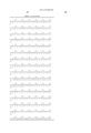
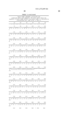
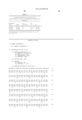
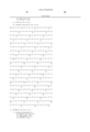
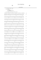
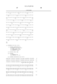
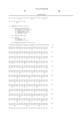
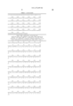
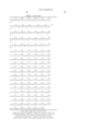
- Human SLAM1 (pSURslaml) nucleotide and predicted amino-acid sequence.Predicted leader sequence and the transmembrane sequence are underlined,though natural boundaries may be different, also depending upon cell type.
- An exon encoding the transmembrane domain which is not present in human SLAM3 (pSECslam) is delineated by two •s and the bases bordering this exon are in bold type (nucleotides numbered 761 and 850). Cysteines are found at amino acid residues numbered 32, 132, 158, 164, 209, 228, and 303.
- N-linked glycosylation sites are found at residues numbered 53, 57, 102, 125, 150, 155, 189, and 217. Fragments between cysteines and/or N-linked glycosylation sites are particularly useful in generating antibodies. SEQ ID NO: 1 and 2.
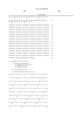
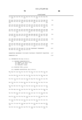
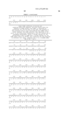
- the human SLAM2 apparently differs from human SLAM1 by a differential splicing event resulting in a different C-terminal sequence beginning at the point indicated by •(nucleotide 924).
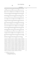
- SEQ ID NO: 3 and 4. 10 20 30 40 50 60 tggcatctgtgagcagctgccaggctccggccaggatcccttccttcctcattggctg 70 80 90 100 110 120 atggatcccaaggggctcctctctgaccttcgtgctgtttttctctctctcccctggctttggg M D P K G L L S L T F V L F L S L A F G 1 130 140 150 160 170 180 gcaagctacggaacaggtgggcgcatgatgaactgcccaaagattctccggcagttggga A S Y G T G G R M M N
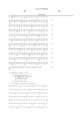
- SLAM3 is secreted by COS cells transfected with pSECslam, confirming that SLAM3 encodes a soluble form of SLAM.
- primers specific for this soluble form of SLAM for RT-PCR the SLAM3 transcript has been detected in different cell types, confirming that it is a bonafide mRNA. SEQ ID NO: 5 and 6.
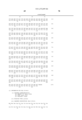
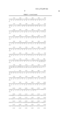
- mouse SLAM The nucleotide and predicted amino acid sequence of mouse SLAM is shown in Table 2.
- One version of mouse SLAM is a type I transmembrane protein containing 9 potential N-linked glycosylation sites. The predicted unglycosylated MW is 40,000.
- the sequence shown is for mouse SLAM1 (in the plasmid pMSLAM1) which is the most abundant 1.8 kb SLAM cDNA, however, another 1.8 kb cDNA SLAM2 (in pMSLAM2), representing about 25% of the cDNA's was also isolated.
- SLAM2 shares about the first 1 kb of sequence with the SLAM1 sequence, but has different sequence at its 3′ end.
- This SLAM2 cDNA in pMSLAM2 encodes a SLAM protein with a different cytoplasmic domain.
- the sequence of SLAM2 cDNA is shown in Table 2 and the position after which SLAM2 sequence varies from SLAM1 is indicated.
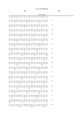
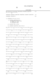
- Table 3 shows an alignment of selected human and mouse SLAM protein sequences.
- mouse SLAM typically has one V and one C immunoglobulin domain and shares extensive amino-acid homology with human SLAM over the entire molecule, this being 88% counting conservative substitutions. The homology at the nucleotide level is about 70%.
- This mouse protein contains eight separate amino acid insertions relative to that human SLAM.
- pMSLAM2 The alternate sequence in pMSLAM2 is not homologous to the unique sequence of the human SLAM2 (pSURslam2), however, the position in the nucleotide sequence where the alternative exon is spliced is identical in both sequences (Table 2).
- the natural antigens are capable of mediating various biochemical responses which lead to biological or physiological responses in target cells.
- the best characterized embodiment was initially described in human, but human and mouse variants are also described herein. Additional sequences for proteins in other mammalian species, e.g., primates and rodents, should also be available. See below. The descriptions below are directed, for exemplary purposes, to a human SLAM, but are likewise applicable to related embodiments from other species.
- Isolated human SLAM protein is a protein which exhibits structural features characteristic of a cell surface antigen. The protein is easily detected on particular cell types, others express lesser amounts. See Table 4.
- the SLAM mediates a biochemical response to binding of an antibody, or other yet unidentified ligands, leading to signal transduction and cellular response.
- the SLAM antigen has been isolated by expression cloning using a specific antibody.
- the SLAM antigen was isolated and characterized as a protein which migrates on polyacrylamide gel electrophoresis with a mobility characteristic of a protein of about 70 kD.
- the core protein after treatment with N-glycanase, has a mobility of about a 40 kd protein.
- RNA from various cells and tissues was subject to reverse transcription and PCR using SLAM specific primers. Rough qualitative determinations are provided, though a negative merely means below threshold detection levels. Thymus also expresses the message.
- the SLAM antigen should be present in the identified tissue types and the interaction of the antigen with its binding partner should be important for mediating various aspects of cellular physiology or development.
- Human and mouse SLAM amino acid sequences are shown in SEQ ID NO: 2, 4, 6, 8, 10, and 12. These amino acid sequences, provided amino to carboxy, are important in providing sequence information in the antigen allowing for distinguishing the protein from other proteins and exemplifying numerous variants. Moreover, the peptide sequences allow preparation of peptides to generate antibodies to recognize such segments, and allow preparation of oligonucleotide probes, both of which are strategies for detection or isolation, e.g., cloning, of genes encoding such sequences.
- human SLAM shall encompass, when used in a protein context, a protein having amino acid sequences shown in SEQ ID NO: 2, 4, 6, or 8, or a significant fragment of such a protein, or another highly homologous protein derived from human.
- mRNA species representing splicing variants. It also refers to a human derived polypeptide which exhibits similar biological function or interacts with SLAM specific binding components.
- binding components e.g., antibodies, typically bind to a SLAM with high affinity, e.g., at least about 100 nM, usually better than about 30 nM, preferably better than about 10 nm, and more preferably at better than about 3 nM.
- Homologous proteins would be found in mammalian species other than human, e.g., primates or rodents. Non-mammalian species should also possess structurally or functionally related genes and proteins, e.g., birds or amphibians.
- polypeptide as used herein includes a significant fragment or segment, and encompasses a stretch of amino acid residues of at least about 8 amino acids, generally at least about 12 amino acids, typically at least about 16 amino acids, preferably at least about 20 amino acids, and, in particularly preferred embodiments, at least about 30 or more amino acids.
- binding composition refers to molecules that bind with specificity to SLAM, e.g., in a cell adhesion pairing type fashion, or an antibody-antigen interaction. It also includes compounds, e.g., proteins, which specifically associate with SLAM, including in a natural physiologically relevant protein-protein interaction, either covalent or non-covalent.
- the molecule may be a polymer, or chemical reagent.
- a functional analog may be an antigen with structural modifications, or it may be a molecule which has a molecular shape which interacts with the appropriate binding determinants.
- the compounds may serve as agonists or antagonists of the binding interaction, see, e.g., Goodman, et al. (eds.) (1990) Goodman & Gilman's: The Pharmacological Bases of Therapeutics (8th ed.), Pergamon Press.
- Substantially pure typically means that the protein is free from other contaminating proteins, nucleic acids, or other biologicals derived from the original source organism. Purity may be assayed by standard methods, typically by weight, and will ordinarily be at least about 40% pure, generally at least about 50% pure, often at least about 60% pure, typically at least about 80% pure, preferably at least about 90% pure, and in most preferred embodiments, at least about 95% pure. Carriers or excipients will often be added.
- Solubility of a polypeptide or fragment depends upon the environment and the polypeptide. Many parameters affect polypeptide solubility, including temperature, electrolyte environment, size and molecular characteristics of the polypeptide, and nature of the solvent. Typically, the temperature at which the polypeptide is used ranges from about 4° C. to about 65° C. Usually the temperature at use is greater than about 18° C. For diagnostic purposes, the temperature will usually be about room temperature or warmer, but less than the denaturation temperature of components in the assay. For therapeutic purposes, the temperature will usually be body temperature, typically about 37° C. for humans and mice, though under certain situations the temperature may be raised or lowered in situ or in vitro.
- the size and structure of the polypeptide should generally be in a substantially stable state, and usually not in a denatured state.
- the polypeptide may be associated with other polypeptides in a quaternary structure, e.g., to confer solubility, or associated with lipids or detergents in a manner which approximates natural lipid bilayer interactions.
- the solvent and electrolytes will usually be a biologically compatible buffer, of a type used for preservation of biological activities, and will usually approximate a physiological aqueous solvent.
- the solvent will have a neutral pH, typically between about 5 and 10, and preferably about 7.5.
- one or more detergents will be added, typically a mild non-denaturing one, e.g., CHS (cholesteryl hemisuccinate) or CHAPS (3-[3-cholamidopropyl)dimethylammonio]-1-propane sulfonate), or a low enough concentration as to avoid significant disruption of structural or physiological properties of the protein.
- This invention also encompasses proteins or peptides having substantial amino acid sequence identity with the amino acid sequence of the SLAM.
- the variants include species or allelic variants.
- Amino acid sequence homology, or sequence identity is determined by optimizing residue matches, if necessary, by introducing gaps as required. See also Needleham, et al. (1970) J. Mol. Biol . 48:443-453; Sankoff, et al. (1983) Chapter One in Time Warps, String Edits, and Macromolecules: The Theory and Practice of Sequence Comparison , Addison-Wesley, Reading, Mass.; and software packages from IntelliGenetics, Mountain View, Calif.; and the University of Wisconsin Genetics Computer Group, Madison, Wis. Sequence identity changes when considering conservative substitutions as matches.
- Conservative substitutions typically include substitutions within the following groups: glycine, alanine; valine, isoleucine, leucine; aspartic acid, glutamic acid; asparagine, glutamine; serine, threonine; lysine, arginine; and phenylalanine, tyrosine.
- Homologous amino acid sequences are typically intended to include natural allelic and interspecies variations in each respective protein sequence. Typical homologous proteins or peptides will have from 25-100% identity (if gaps can be introduced), to 50-100% identity (if conservative substitutions are included) with the amino acid sequence of the SLAM.
- Identity measures will be at least about 35%, generally at least about 40%, often at least about 50%, typically at least about 60%, usually at least about 70%, preferably at least about 80%, and more preferably at least about 90%.
- the isolated SLAM DNA can be readily modified by nucleotide substitutions, nucleotide deletions, nucleotide insertions, and inversions of nucleotide stretches. These modifications result in novel DNA sequences which encode these antigens, their derivatives, or proteins having similar physiological, immunogenic, antigenic, or other functional activity. These modified sequences can be used to produce mutant antigens or to enhance expression. Enhanced expression may involve gene amplification, increased transcription, increased translation, and other mechanisms. “Mutant SLAM” encompasses a polypeptide otherwise falling within the sequence identity definition of the SLAM as set forth above, but having an amino acid sequence which differs from that of SLAM as normally found in nature, whether by way of deletion, substitution, or insertion.
- SLAM mutagenesis can also be conducted by making amino acid insertions or deletions. Substitutions, deletions, insertions, or any combinations may be generated to arrive at a final construct. Insertions include amino- or carboxy- terminal fusions. Random mutagenesis can be conducted at a target codon and the expressed mutants can then be screened for the desired activity. Methods for making substitution mutations at predetermined sites in DNA having a known sequence are well known in the art, e.g., by M13 primer mutagenesis or polymerase chain reaction (PCR) techniques. See, e.g., Sambrook, et al. (1989); Ausubel, et al. (1987 and Supplements); and Kunkel, et al. (1987) Methods in Enzymol . 154:367-382.
- PCR polymerase chain reaction
- the present invention also provides recombinant proteins, e.g., heterologous fusion proteins using segments from these proteins.
- a heterologous fusion protein is a fusion of proteins or segments which are naturally not normally fused in the same manner.
- a similar concept applies to heterologous nucleic acid sequences.
- new constructs may be made from combining similar functional domains from other proteins.
- target-binding or other segments may be “swapped” between different new fusion polypeptides or fragments. See, e.g., Cunningham, et al. (1989) Science 243:1330-1336; and O'Dowd, et al. (1988) J. Biol. Chem . 263:15985-15992.
- SLAM The blocking of physiological response to SLAMs may result from the inhibition of binding of the antigen to its binding partner, e.g., another of itself, likely through competitive inhibition.
- binding partner e.g., another of itself
- in vitro assays of the present invention will often use isolated protein, membranes from cells expressing a membrane associated recombinant SLAM, soluble fragments comprising antigen binding segments of these proteins, or fragments attached to solid phase substrates. These assays will also allow for the diagnostic determination of the effects of either binding segment mutations and modifications, or antigen mutations and modifications, e.g., SLAM analogues.
- This invention also contemplates the use of competitive drug screening assays, e.g., where neutralizing antibodies to antigen or binding fragments compete with a test compound for binding to the protein.
- glycosylation alterations are included, e.g., made by modifying the glycosylation patterns of a polypeptide during its synthesis and processing, or in further processing steps. See, e.g., Elbein (1987) Ann. Rev. Biochem . 56:497-534. Also embraced are versions of the peptides with the same primary amino acid sequence which have other minor modifications, including phosphorylated amino acid residues, e.g., phosphotyrosine, phosphoserine, or phosphothreonine.
- phosphorylated amino acid residues e.g., phosphotyrosine, phosphoserine, or phosphothreonine.
- Fusion polypeptides between SLAMs and other homologous or heterologous proteins are also provided.
- Many cytokine receptors or other surface proteins are multimeric, e.g., homodimeric entities, and a repeat construct may have various advantages, including lessened susceptibility to proteolytic cleavage.
- Typical examples are fusions of a reporter polypeptide, e.g., luciferase, with a segment or domain of a protein, e.g., a receptor-binding segment, so that the presence or location of the fused ligand may be easily determined. See, e.g., Dull, et al., U.S. Pat. No. 4,859,609.
- Fusion peptides will typically be made by either recombinant nucleic acid methods or by synthetic polypeptide methods. Techniques for nucleic acid manipulation and expression are described generally, e.g., in Sambrook, et al. (1989) Molecular Cloning: A Laboratory Manual (2d ed.), vols. 1-3, Cold Spring Harbor Laboratory; and Ausubel, et al. (eds.) (1993) Current Protocols in Molecular Biology , Greene and Wiley, NY. Techniques for synthesis of polypeptides are described, e.g., in Merrifield (1963) J. Amer. Chem. Soc . 85:2149-2156; Merrifield (1986) Science 232: 341-347; Atherton, et al. (1989) Solid Phase Peptide Synthesis: A Practical Approach , IRL Press, Oxford; and Grant (1992) Synthetic Peptides: A User's Guide , W.H. Freeman, NY.
- This invention also contemplates the use of derivatives of SLAMs other than variations in amino acid sequence or glycosylation.
- Such derivatives may involve covalent or aggregative association with chemical moieties.
- Covalent or aggregative derivatives will be useful as immunogens, as reagents in immunoassays, or in purification methods such as for affinity purification of binding partners, e.g., other antigens.
- a SLAM can be immobilized by covalent bonding to a solid support such as cyanogen bromide-activated SEPHAROSE, by methods which are well known in the art, or adsorbed onto polyolefin surfaces, with or without glutaraldehyde cross-linking, for use in the assay or purification of anti-SLAM antibodies or an alternative binding composition.
- the SLAMs can also be labeled with a detectable group, e.g., for use in diagnostic assays.
- Purification of SLAM may be effected by an immobilized antibody or complementary binding partner.
- a solubilized SLAM or fragment of this invention can be used as an immunogen for the production of antisera or antibodies specific for binding to the antigen or fragments thereof.
- Purified antigen can be used to screen monoclonal antibodies or antigen-binding fragments, encompassing antigen binding fragments of natural antibodies.
- Purified SLAMs can also be used as a reagent to detect antibodies generated in response to the presence of elevated levels of the antigen or cell fragments containing the antigen, both of which may be diagnostic of an abnormal or specific physiological or disease condition.
- This invention contemplates antibodies raised against amino acid sequences encoded by nucleotide sequences shown in SEQ ID NO: 1, 3, 5, 7, 9, or 11, or fragments of proteins containing them.
- this invention contemplates antibodies having binding affinity to or being raised against specific fragments which are predicted to lie outside of the lipid bilayer, both extracellular or intracellular.
- the present invention contemplates the isolation of additional closely related species variants. Southern and Northern blot analysis should establish that similar genetic entities exist in other mammals. It is likely that SLAMs are widespread in species variants, e.g.,.rodents, lagomorphs, carnivores, artiodactyla, perissodactyla, and primates.
- the invention also provides means to isolate a group of related antigens displaying both distinctness and similarities in structure, expression, and function. Elucidation of many of the physiological effects of the molecules will be greatly accelerated by the isolation and characterization of additional distinct species variants of them.
- the present invention provides useful probes for identifying additional homologous genetic entities in different species.
- Intracellular functions would probably involve segments of the antigen which are normally accessible to the cytosol. However, protein internalization may occur under certain circumstances, and interaction between intracellular components and “extracellular” segments may occur.
- the specific segments of interaction of SLAM with other intracellular components may be identified by mutagenesis or direct biochemical means, e.g., cross-linking or affinity methods. Structural analysis by crystallographic or other physical methods will also be applicable. Further investigation of the mechanism of signal transduction will include study of associated components which may be isolatable by affinity methods or by genetic means, e.g., complementation analysis of mutants.
- the controlling elements associated with the antigens should exhibit differential physiological, developmental, tissue specific, or other expression patterns. Upstream or downstream genetic regions, e.g., control elements, are of interest. In particular, physiological or developmental variants, e.g., multiple alternatively processed forms of the antigen have been found. See, e.g., SEQ ID NO: 1 and 3. Thus, differential splicing of message may lead to an assortment of membrane bound forms, soluble forms, and modified versions of antigen.
- Antigen fragments may be joined to other materials, particularly polypeptides, as fused or covalently joined polypeptides to be used as immunogens.
- An antigen and its fragments may be fused or covalently linked to a variety of immunogens, such as keyhole limpet hemocyanin, bovine serum albumin, tetanus toxoid, etc. See Microbioloy , Hoeber Medical Division, Harper and Row, 1969; Landsteiner (1962) Specificity of Serological Reactions , Dover Publications, New York; Williams, et al. (1967) Methods in Immunology and Immunochemistry , vol. 1, Academic Press., New York; and Harlow and Lane (1988) Antibodies: A Laboratory Manual , CSH Press, NY, for descriptions of methods of preparing polyclonal antisera.
- monoclonal antibodies from various mammalian hosts, such as mice, rodents, primates, humans, etc. Description of techniques for preparing such monoclonal antibodies may be found in, e.g., Stites, et al.
- labels and conjugation techniques are known and are reported extensively in both the scientific and patent literature. Suitable labels include radionuclides, enzymes, substrates, cofactors, inhibitors, fluorescent moieties, chemiluminescent moieties, magnetic particles, and the like. Patents, teaching the use of such labels include U.S. Pat. Nos. 3,817,837; 3,850,752; 3,939,350; 3,996,345; 4,277,437; 4,275,149; and 4,366,241. Also, recombinant immunoglobulins may be produced, see Cabilly, U.S. Pat. No. 4,816,567; Moore, et al., U.S. Pat. No. 4,642,334; and Queen, et al. 1989) Proc. Nat'l Acad. Sci. USA 86:10029-10033.
- the antibodies of this invention can also be used for affinity chromatography in isolating the protein.
- Columns can be prepared where the antibodies are linked to a solid support. See, e.g., Wilchek et al. (1984) Meth. Enzymol . 104:3-55.
- the described peptide sequences and the related reagents are useful in detecting, isolating, or identifying a DNA clone encoding SLAM, e.g., from a natural source.
- a DNA clone encoding SLAM e.g., from a natural source.
- it will be useful in isolating a gene from mammal, and similar procedures will be applied to isolate genes from other species, e.g., warm blooded animals, such as birds and mammals.
- Cross hybridization will allow isolation of SLAM from other species.
- a number of different approaches should be available to successfully isolate a suitable nucleic acid clone.
- the purified protein or defined peptides are useful for generating antibodies by standard methods, as described above.
- Synthetic peptides or purified protein can be presented to an immune system to generate monoclonal or polyclonal antibodies. See, e.g., Coligan (1991) Current Protocols in Immunology Wiley/Greene; and Harlow and Lane (1989) Antibodies: A Laboratory Manual , Cold Spring Harbor Press.
- the SLAM can be used as a specific binding reagent, and advantage can be taken of its specificity of binding, much like an antibody would be used.
- An isolated nucleic acid will generally be a homogeneous composition of molecules, but will, in some embodiments, contain minor heterogeneity. This heterogeneity is typically found at the polymer ends or portions not critical to a desired biological function or activity.
- a “recombinant” nucleic acid is defined either by its method of production or its structure. In reference to its method of production, e.g., a product made by a process, the process is use of recombinant nucleic acid techniques, e.g., involving human intervention in the nucleotide sequence, typically selection or production. Alternatively, it can be a nucleic acid made by generating a sequence comprising fusion of two fragments which are not naturally contiguous to each other, but is meant to exclude products of nature, e.g., naturally occurring mutants.
- a DNA which codes for a SLAM protein will be particularly useful to identify genes, mRNA, and cDNA species which code for related or homologous proteins, as well as DNAs which code for homologous proteins from different species. There are likely homologues in other species, including primates, rodents, and birds. Various SLAM proteins should be homologous and are encompassed herein. However, even proteins that have a more distant evolutionary relationship to the antigen can readily be isolated under appropriate conditions using these sequences if they are sufficiently homologous. Primate SLAM proteins are of particular interest.
- Substantial homology in the nucleic acid sequence comparison context means either that the segments, or their complementary strands, when compared, are identical when optimally aligned, with appropriate nucleotide insertions or deletions, in at least about 50% of the nucleotides, generally at least about 58%, ordinarily at least about 65%, often at least about 71%, typically at least about 77%, usually at least about 85%, preferably at least about 95 to 98% or more, and in particular embodiments, as high as about 99% or more of the nucleotides.
- SLAM sequence of SLAM
- selective hybridization will occur when there is at least about 55% homology over a stretch of at least about 30 nucleotides, preferably at least about 75% over a stretch of about 25 nucleotides, and most preferably at least about 90% over about 20 nucleotides. See, Kanehisa (1984) Nuc. Acids Res . 12:203-213.
- the length of homology comparison may be over longer stretches, and in certain embodiments will be over a stretch of at least about 17 nucleotides, usually at least about 28 nucleotides, typically at least about 40 nucleotides, and preferably at least about 75 to 100 or more nucleotides.
- DNA which encodes the SLAM or fragments thereof can be obtained by chemical synthesis, screening cDNA libraries, or screening genomic libraries prepared from a wide variety of cell lines or tissue samples. See, e.g., Okayama and Berg (1982) Mol. Cell. Biol . 2:161-170; Gubler and Hoffman (1983) Gene 25:263-269; and Glover (ed.) (1984) DNA Cloning: A Practical, Approach , IRL Press, Oxford.
- the sequences provided herein provide useful PCR primers or allow synthetic or other preparation of suitable genes encoding a SLAM.
- This DNA can be expressed in a wide variety of host cells for the synthesis of a full-length SLAM or fragments which can in turn, e.g., be used to generate polyclonal or monoclonal antibodies; for binding studies; for construction and expression of modified molecules; and for structure/function studies.
- Vectors as used herein, comprise plasmids, viruses, bacteriophage, integratable DNA fragments, and other vehicles which enable the integration of DNA fragments into the genome of the host. See, e.g., Pouwels, et al. (1985 and Supplements) Cloning Vectors: A Laboratory Manual , Elsevier, N.Y.; and Rodriguez, et al. (1988) (eds.) Vectors: A Survey of Molecular Cloning Vectors and Their Uses , Buttersworth, Boston, Mass.
- DNA sequences are operably linked when they are functionally related to each other.
- DNA for a presequence or secretory leader is operably linked to a polypeptide if it is expressed as a preprotein or participates in directing the polypeptide to the cell membrane or in secretion of the polypeptide.
- a promoter is operably linked to a coding sequence if it controls the transcription of the polypeptide;
- a ribosome binding site is operably linked to a coding sequence if it is positioned to permit translation.
- operably linked means contiguous and in reading frame, however, certain genetic elements such as repressor genes are not contiguously linked but still bind to operator sequences that in turn control expression.
- Suitable expression vectors include pCDNA 1 ; pCD, see Okayama, et al. (1985) Mol. Cell Biol . 5:1136-1142; pMc 1 neo Poly-A, see Thomas, et al. (1987) Cell 51:503-512; and a baculovirus vector such as pAC 373 or pAC 610. See, e.g., Miller (1988) Ann. Rev. Microbiol . 42:177-199.
- SLAM polypeptide in a system which provides a specific or defined glycosylation pattern. See, e.g., Luckow and Summers (1988) Bio/Technology 6:47-55; and Kaufman (1990) Meth. Enzymol . 185:487-511.
- the SLAM may be engineered to be phosphatidyl inositol (PI) linked to a cell membrane, but can be removed from membranes by treatment with a phosphatidyl inositol cleaving enzyme, e.g., phosphatidyl inositol phospholipase-C.
- PI phosphatidyl inositol
- a phosphatidyl inositol cleaving enzyme e.g., phosphatidyl inositol phospholipase-C.
- the present invention provides reagents which will find use in diagnostic applications as described elsewhere herein, e.g., in the general description for T cell mediated conditions, or below in the description of kits for diagnosis.
- This invention also provides reagents with significant therapeutic value.
- the SLAM naturally occurring or recombinant
- fragments thereof, and antibodies thereto, along with compounds identified as having binding affinity to SLAM, should be useful in the treatment of conditions associated with abnormal physiology or development, including abnormal proliferation, e.g., cancerous conditions, or degenerative conditions.
- modulation of development of lymphoid cells will be achieved by appropriate therapeutic treatment using the compositions provided herein.
- a disease or disorder associated with abnormal expression or abnormal signaling by a SLAM should be a likely target for an agonist or antagonist of the antigen.
- the antigen plays a role in regulation or development of hematopoietic cells, e.g., lymphoid cells, which affect immunological responses, e.g., autoimmune disorders.
- the antigen has been demonstrated to provide a costimulatory signal to T cell activation.
- the SLAM has a role in T cell to T cell interactions. These interactions lead, in particular contexts, to cell proliferation, enhanced cytokine synthesis by the cells, and consequential amplification of T cell proliferation.
- SLAM induced production of interferon- ⁇ suggests that certain agonists to SLAM could direct T cell responses towards a Th0/Th1 pathway, and thus suppress a Th2 type response.
- these agonists should be various antibodies which recognize the appropriate epitopes, e.g., which mimic binding of SLAM to its ligand.
- antagonists of SLAM may provide a selective and powerful way to block immune responses in abnormal situations, e.g., autoimmune disorders, including rheumatoid arthritis, systemic lupus erythematosis (SLE), Hashimoto's autoimmune thyroiditis, as well as acute and chronic inflammatory responses in which T cell activation, expansion, and/or immunological T cell memory play an important role.
- autoimmune disorders including rheumatoid arthritis, systemic lupus erythematosis (SLE), Hashimoto's autoimmune thyroiditis
- SLE systemic lupus erythematosis
- Hashimoto's autoimmune thyroiditis as well as acute and chronic inflammatory responses in which T cell activation, expansion, and/or immunological T cell memory play an important role.
- the SLAM appears to be coexpressed with CD45RO, which is a marker for primed, or memory, T cells.
- SLAM is also absent in the CD45RA cells, which represent the naive T cell subset. As such, the SLAM can also serve as a diagnostic marker for memory T cells.
- SLAM antibodies can be purified and then administered to a patient, veterinary or human.
- These reagents can be combined for therapeutic use with additional active or inert ingredients, e.g., in conventional pharmaceutically acceptable carriers or diluents, e.g., immunogenic adjuvants, along with physiologically innocuous stabilizers, excipients, or preservatives.
- additional active or inert ingredients e.g., in conventional pharmaceutically acceptable carriers or diluents, e.g., immunogenic adjuvants, along with physiologically innocuous stabilizers, excipients, or preservatives.
- These combinations can be sterile filtered and placed into dosage forms as by lyophilization in dosage vials or storage in stabilized aqueous preparations.
- This invention also contemplates use of antibodies or binding fragments thereof, including forms which are not complement binding.
- Drug screening using SLAM or fragments thereof can be performed to identify compounds having binding affinity to or other relevant biological effects on SLAM functions, including isolation of associated components. Subsequent biological assays can then be utilized to determine if the compound has intrinsic stimulating activity and is therefore a blocker or antagonist in that it blocks the activity of the antigen. Likewise, a compound having intrinsic stimulating activity can activate the signal pathway and is thus an agonist in that it simulates the activity of SLAM.
- This invention further contemplates the therapeutic use of blocking antibodies to SLAM as antagonists and of stimulatory antibodies, e.g., A12, as agonists. This approach should be particularly useful with other SLAM species variants.
- reagents necessary for effective therapy will depend upon many different factors, including means of administration, target site, physiological state of the patient, and other medicants administered. Thus, treatment dosages should be titrated to optimize safety and efficacy. Typically, dosages used in vitro may provide useful guidance in the amounts useful for in situ administration of these reagents. Animal testing of effective doses for treatment of particular disorders will provide further predictive indication of human dosage. Various considerations are described, e.g., in Gilman, et al. (eds.) (1990) Goodman and Gilman's: The Pharmacological Bases of Therapeutics , 8th Ed., Pergamon Press; and Remington's Pharmaceutical Sciences , 17th ed.
- Pharmaceutically acceptable carriers will include water, saline, buffers, and other compounds described, e.g., in the Merck Index , Merck & Co., Rahway, N.J. Dosage ranges would ordinarily be expected to be in amounts lower than 1 mm concentrations, typically less than about 10 ⁇ M concentrations, usually less than about 100 nM, preferably less than about 10 pM (picomolar), and most preferably less than about 1 fM (femtomolar), with an appropriate carrier. Slow release formulations, or a slow release apparatus will often be utilized for continuous or long term administration. See, e.g., Langer (1990) Science 249:1527-1533.
- Formulations include those suitable for oral, rectal, nasal, topical, or parenteral (including subcutaneous, intramuscular, intravenous and intradermal) administration.
- the formulations may conveniently be presented in unit dosage form and may be prepared by any methods well known in the art of pharmacy. See, e.g., Gilman, et al. (eds.) (1990) Goodman and Gilman's: The Pharmacological Bases of Therapeutics , 8th Ed., Pergamon Press; and Remington's Pharmaceutical Sciences , 17th ed. (1990), Mack Publishing Co., Easton, Pa.; Avis, et al.
- SLAMs of this invention are particularly useful in kits and assay methods which are capable of screening compounds for binding activity to the proteins.
- Several methods of automating assays have been developed in recent years so as to permit screening of tens of thousands of compounds in a short period. See, e.g., Fodor, et al. (1991) Science 251:767-773, which describes means for testing of binding affinity by a plurality of defined polymers synthesized on a solid substrate.
- suitable assays can be greatly facilitated by the availability of large amounts of purified, soluble SLAM as provided by this invention.
- antagonists can normally be found once the antigen has been structurally defined, e.g., by tertiary structure data. Testing of potential interacting analogues is now possible upon the development of highly automated assay methods using a purified SLAM. In particular, new agonists and antagonists will be discovered by using screening techniques described herein. Of particular importance are compounds found to have a combined binding affinity for a spectrum of SLAM molecules, e.g., compounds which can serve as antagonists for species variants of SLAM.
- One method of drug screening utilizes eukaryotic or prokaryotic host cells which are stably transformed with recombinant DNA molecules expressing a SLAM.
- Cells may be isolated which express a SLAM in isolation from other molecules.
- Such cells either in viable or fixed form, can be used for standard binding partner binding assays. See also, Parce, et al. (1989) Science 246:243-247; and Owicki, et al. (1990) Proc. Nat'l Acad. Sci. USA 87:4007-4011, which describe sensitive methods to detect cellular responses.
- Another technique for drug screening involves an approach which provides high throughput screening for compounds having suitable binding affinity to a SLAM and is described in detail in Geysen, European Patent Application 84/03564, published on Sep. 13, 1984.
- a solid substrate e.g., plastic pins or some other appropriate surface, see Fodor, et al. (1991).
- all the pins are reacted with solubilized, unpurified or solubilized, purified SLAM, and washed.
- the next step involves detecting bound SLAM.
- Rational drug design may also be based upon structural studies of the molecular shapes of the SLAM and other effectors or analogues. Effectors may be other proteins which mediate other functions in response to binding, or other proteins which normally interact with SLAM.
- Effectors may be other proteins which mediate other functions in response to binding, or other proteins which normally interact with SLAM.
- One means for determining which sites interact with specific other proteins is a physical structure determination, e.g., x-ray crystallography or 2 dimensional NMR techniques. These will provide guidance as to which amino acid residues form molecular contact regions.
- x-ray crystallography or 2 dimensional NMR techniques.
- kits and methods for detecting the presence of another SLAM or binding partner typically the kit will have a compartment containing either a defined SLAM peptide or gene segment or a reagent which recognizes one or the other, e.g., SLAM fragments or antibodies.
- a kit for determining the binding affinity of a test compound to a SLAM would typically comprise a test compound; a labeled compound, for example a binding partner or antibody having known binding affinity for SLAM; a source of SLAM (naturally occurring or recombinant); and a means for separating bound from free labeled compound, such as a solid phase for immobilizing the molecule.
- a test compound for example a binding partner or antibody having known binding affinity for SLAM
- a source of SLAM naturally occurring or recombinant
- a means for separating bound from free labeled compound such as a solid phase for immobilizing the molecule.
- a preferred kit for determining the concentration of, e.g., a SLAM in a sample would typically comprise a labeled compound, e.g., binding partner or antibody, having known binding affinity for the antigen, a source of antigen (naturally occurring or recombinant) and a means for separating the bound from free labeled compound, e.g., a solid phase for immobilizing the SLAM. Compartments containing reagents, and instructions, will normally be provided.
- Antibodies including antigen binding fragments, specific for the SLAM or fragments are useful in diagnostic applications to detect the presence of elevated levels of SLAM and/or its fragments.
- diagnostic assays can employ lysates, live cells, fixed cells, immunofluorescence, cell cultures, body fluids, and further can involve the detection of antigens related to the antigen in serum, or the like. Diagnostic assays may be homogeneous (without a separation step between free reagent and antigen-binding partner complex) or heterogeneous (with a separation step).
- RIA radioimmunoassay
- ELISA enzyme-linked immunosorbent assay
- EIA enzyme immunoassay
- EMIT enzyme-multiplied immunoassay technique
- SFIA substrate-labeled fluorescent immunoassay
- Anti-idiotypic antibodies may have similar use to diagnose presence of antibodies against a SLAM, as such may be diagnostic of various abnormal states.
- overproduction of SLAM may result in production of various immunological reactions which may be diagnostic of abnormal physiological states, particularly in proliferative cell conditions such as cancer or abnormal activation or differentiation.
- the reagents for diagnostic assays are supplied in kits, so as to optimize the sensitivity of the assay.
- the protocol, and the label either labeled or unlabeled antibody or binding partner, or labeled SLAM is provided. This is usually in conjunction with other additives, such as buffers, stabilizers, materials necessary for signal production such as substrates for enzymes, and the like.
- the kit will also contain instructions for proper use and disposal of the contents after use.
- the kit has compartments for each useful reagent.
- the reagents are provided as a dry lyophilized powder, where the reagents may be reconstituted in an aqueous medium providing appropriate concentrations of reagents for performing the assay.
- labeling may be achieved by covalently or non-covalently joining a moiety which directly or indirectly provides a detectable signal.
- the binding partner, test compound, SLAM, or antibodies thereto can be labeled either directly or indirectly.
- Possibilities for direct labeling include label groups: radiolabels such as 125 I, enzymes (U.S. Pat. No. 3,645,090) such as peroxidase and alkaline phosphatase, and fluorescent labels (U.S. Pat. No. 3,940,475) capable of monitoring the change in fluorescence intensity, wavelength shift, or fluorescence polarization.
- Possibilities for indirect labeling include biotinylation of one constituent followed by binding to avidin coupled to one of the above label groups.
- the SLAM can be immobilized on various matrixes followed by washing. Suitable matrixes include plastic such as an ELISA plate, filters, and beads. See, e.g., Coligan, et al. (eds.) (1993) Current Protocols in Immunology, Vol. 1, Chapter 2, Greene and Wiley, NY.
- suitable separation techniques include, without limitation, the fluorescein antibody magnetizable particle method described in Rattle, et al. (1984) Clin. Chem . 30:1457-1461, and the double antibody magnetic particle separation as described in U.S. Pat. No. 4,659,678.
- Another diagnostic aspect of this invention involves use of oligonucleotide or polynucleotide sequences taken from the sequence of a SLAM. These sequences can be used as probes for detecting levels of the SLAM message in samples from patients suspected of having an abnormal condition, e.g., cancer or developmental problem.
- the preparation of both RNA and DNA nucleotide sequences, the labeling of the sequences, and the preferred size of the sequences has received ample description and discussion in the literature. See, e.g., Langer-Safer, et al. (1982) Proc. Nat'l. Acad. Sci . 79:4381-4385; Caskey (1987) Science 236:962-967; and Wilchek et al. (1988) Anal. Biochem . 171:1-32.
- kits which also test for the qualitative or quantitative presence of other markers are also contemplated. Diagnosis or prognosis may depend on the combination of multiple indications used as markers. Thus, kits may test for combinations of markers. See, e.g., Viallet, et al. (1989) Progress in Growth Factor Res . 1:89-97.
- SLAM-Ig The binding of SLAM-Ig to SLAM transfected L cells demonstrates that SLAM can interact with itself as a ligand.
- L cells transfected with SLAM could provide a direct co-stimulatory signal for CD4 + T-cell clones.
- Engagement of SLAM with the mAb A12 provides a significant co-stimulatory signal for T-cell activation.
- activation of CD4 + T-cell clones via SLAM expressed on L cells, in combination with anti-CD3 leads to large increases in proliferation.
- Co-stimulation of proliferation with suboptimal doses of anti-CD3 was observed with SLAM-transfectants. The stimulation provided by SLAM transfected L cells was substantial enough to lead directly to T-cell proliferation in the absence of other stimuli.
- the direct stimulatory signal provided by SLAM expressed on L cells is unique, and is not observed even for the classical co-stimulatory molecules B7 (Jenkins and Johnson (1993) Curr. Opin. Immunol . 5:361-367) and B70 (Azuma, et al. (1993) Nature 366:76-79).
- the ligand for B7 is CD28, and anti-CD28 mAbs do not directly stimulate proliferation of T-cell clones.
- the anti-SLAM mAb A12, or its F(ab) 2 fragments can directly induce T-cell proliferation.
- the consequences of engagement of SLAM on T-cell clones by SLAM on transfected L cells, or by mAb A12 or its F(ab) 2 fragments are concordant. Thus, direct engagement of SLAM, without the involvement of other molecules in the interaction, is sufficient to induce the functional effects observed.
- the SLAM gene was localized to the interface of bands q21.3 and q22 on human chromosome 1. This region of chromosome 1 appears to be an important locus for genes involved in cell-cell interactions.
- the genes for selectins (Watson, et al. (1990) J. Exp. Med . 172:263-272), molecules involved in leucocyte adhesion and trafficking, also localize to 1q22-23.
- Another gene at this locus (1q21.3-23) is the gene for myelin Po (Pham-Dinh, et al. (1993) Hum. Mol. Genet . 2:2051-2054), the most abundant protein in myelin (Filbin, et al.
- myelin Po is a member of the Ig-superfamily (Williams and Barclay (1988) Annu. Rev. Immunol . 6:381-405) and also interacts homophilically. Normal myelin structure relies upon the self-interaction of myelin Po, and inherited mutations in myelin Po are responsible for the Charcot-Marie-Tooth neuropathy, type 1b. (Kulkens, et al. (1993) Nat. Genet . 5:35-39; Hayasaka, et al. (1993) Nat. Genet . 5:31-34).
- CD2 with LFA-3 (Selvaraj, et al. (1987) Nature 326:400-403) or CD48 (van der Merwe, et al. (1993) EMBO J . 12:4945-4954); CD28 with B7-1 (Linsley,et al. (1990) Proc. Natl. Acad. Sci. USA 87:5031-5035) or B7-2 (Freeman, et al. (1993) Science 262:909-911; Azuma, et al. (1993) Nature 366:76-79); and the TCR with MHC class II (Matsui, et al.
- Ig-superfamily members may interact in this way by evolution after gene duplication of a homophilically interacting precursor (Williams and Barclay (1988) Annu. Rev. Immunol . 6:381-405).
- SLAM and myelin Po may have retained a primordial function of Ig-superfamily members to interact homophilically.
- CD48 localizes to the same part of chromosome 1 as SLAM at 1q21-23 (Staunton et al. (1989) J. Exp. Med . 169:1087-1099).
- CD48 reported to be a weak ligand for CD2 (vander Merwe, et al. (1993) EMBO J . 12:4945-4954), and 2B4, a signaling molecule expressed on murine NK cells and cytotoxic T cells (Mathew, et al. (1993) J. Immunol . 151:5328-5337) for which a ligand has not been reported, are the most closely related molecules to SLAM.
- SLAM, CD48, and 2B4 all have one V and one C domain and can be distinguished from other members of the Ig-superfamily by the conservation of the sequence CXLXLXC, the second cysteine being the tether for the C-domain and the first cysteine a conserved residue probably between the V- and C-domains.
- CD48 and 2B4 have not yet been directly assessed for their ability to interact homophilically, however it has been reported that a recombinant soluble form CD48 tends to aggregate in solution. The relatedness and chromosomal co-localization of CD48 and SLAM is indicative of evolutionary divergence following gene duplication.
- Ig-superfamily members with multiple domains have been reported to interact homophilically, and these include platelet-endothelial cell adhesion molecule (CD31) (Watt, et al. (1993) Blood 82:2649-2663), neuron-glia cell adhesion molecule (Grumet and Edelman (1988) J. Cell Biol . 106:487-503), neuron-glia-related cell adhesion molecule (Mauro, et al. (1992) J. Cell Biol . 119:191-202), neural cell adhesion molecule or CD56 (Rao, et al. (1992) J. Cell Biol . 118:937-949), and the carcinoembryonic antigen (Zhou, et al. (1993) J. Cell Biol. 122:951-960).
- CD31 platelet-endothelial cell adhesion molecule
- CD56 neuron-glia cell adhesion molecule
- CD56 neural cell adhesion
- SLAM immunospliced form of SLAM lacking a 90 bp exon, corresponding to and precisely encompassing the transmembrane region of SLAM encodes a secreted form of SLAM.
- This naturally produced molecule expressed by activated T cells may suppress T-cell function and may be part of a negative feedback loop to attenuate, or locally restrict SLAM mediated activation upon cell-cell interaction.
- SLAM mediated T-cell activation is resistant to cyclosporin, consistent with the inability of anti-IL-2 antibodies to inhibit SLAM induced T-cell clone proliferation.
- soluble SLAM may make it an effective adjunct for inhibiting ongoing immune responses relatively resistant to cyclosporin such as that seen in allograft rejection (Pereira, et al. (1990) J. Immunol . 144:2109-2116; Zeevi, et al. (1988) Hum. Immunol . 21:143-153).
- SLAM engagement has unique consequences for T-cell activation in terms of its ability to modulate cytokine production profiles toward a Th0/Th1 subtype and, under some circumstances, to directly induce T-cell proliferation.
- the newly described SLAM appears to be a member of the Ig-superfamily in addition to the TCR, CD28, CTLA-4, CD4, and CD2, and its engagement regulates T-Cell responses.
- the presence of SLAM on lymphocytes indicates that activated lymphocytes themselves can provide a significant co-stimulus. This is not unexpected, as the most predominant cell type in lymphoid organs are lymphocytes, which are statistically ever present collaborators, and the major source of autocrine T-cell growth factors such as IL-2.
- SLAM may not only provide strong co-stimulatory signals, but could also be involved in maintaining the relative segregation and lymphocyte accumulation within lymphoid organs.
- Most work on T-cell co-stimulation has focused on different antigen-presenting cells and the molecules they express, particularly B7 and B70, the ligands for CD28 and CTLA-4 (Jenkins (1994) Immunity 1:443-446).
- B cells are an antigen-presenting cell which when activated express SLAM, which may support B-T cell collaboration leading to Ig production. Consistent with the co-stimulatory functions described herein for SLAM, recent studies on CD28 deficient mice have invoked a role for other co-stimulatory molecules in T cell activation (Green, et al.
- Anti-SLAM monoclonal antibodies inhibit IL-4 indued IgE synthesis, which indicates that signaling through SLAM either at the T helper cell or at the B cell levels, inhibits productive T-B cell interaction, which result in IL-4 driven IgE switching and IgE production.
- This effect can be direct, e.g., through interactions between SLAM on T cells and SLAM on B cells, or indirect, e.g., by inducing cytokine production by the T-helper cell, which inhibits IL-4 driven IgE synthesis. Interferon- ⁇ is the primary example of such a cytokine.
- SLAM agonists may have general clinical utility in redirecting Th2 responses to Th1 responses in diseases in which a clear Th2 profile has been implicated, such as allergy, certain autoimmune diseases, or certain inflammatory diseases. This includes Hashimoto's thyroiditis.
- SLAM antagonists will have an opposite effect; that is, blocking of Th1 responses in the disease situations caused by Th1 cells and Th1 cell derived cytokines, such as infectious diseases, including, e.g., tuberculosis and leprosy, or autoimmune diseases, e.g., rheumatoid arthritis and autoimmune uveitis.
- infectious diseases including, e.g., tuberculosis and leprosy
- autoimmune diseases e.g., rheumatoid arthritis and autoimmune uveitis.
- therapeutic reagents will be useful also in modulating such responses as to parasitic infections, to modulate a vaccine reaction, or in
- Methods for protein purification include such methods as ammonium sulfate precipitation, column chromatography, electrophoresis, centrifugation, crystallization, and others. See, e.g., Ausubel, et al. (1987 and periodic supplements); Deutscher (1990) “Guide to Protein Purification” in Methods in Enzymology vol. 182, and other volumes in this series; and manufacturer's literature on use of protein purification products, e.g., Pharmacia, Piscataway, N.J., or Bio-Rad, Richmond, Calif. Combination with recombinant techniques allow fusion to appropriate segments, e.g., to a FLAG sequence or an equivalent which can be fused via a protease-removable sequence.
- the anti-SLAM monoclonal antibody A12 (IgG1) was generated in a fusion of splenocytes from a BALB/c mouse immunized with peripheral blood mononuclear cells activated for 5 hours with 12-0-tetradecanoyl-13 Acetate (TPA) (1 ng/ml) and the Ca 2+ ionophore A23187 (500 ng/ml) (Calbiochem-Behring Corporation).
- TPA 12-0-tetradecanoyl-13 Acetate
- Ca 2+ ionophore A23187 500 ng/ml
- Standard procedures were used to screen for appropriate producing clones, and the A12 hybridoma was clonally isolated and subjected to normal analysis, e.g., determination of producing capacity and immunoglobulin type.
- the A12 hybridoma cell line was deposited with the ATCC on Nov. 10, 1994, and has been assigned ATCC (American Type Culture Collection), 10801 University Boulevard, Manassas, Va. 20110-2209 ) Accession Number HB11760.
- COS-7 cells were transfected by electroporation as described in Cocks, et al. (1993) Int. Immunol . 5:657-663, with an A10 CD8 + T-cell library DNA prepared as described in McKinney and Parkinson (1987) J. Immunol. Methods 96:271-278. Transfected cells were stained with FITC-conjugated anti-SLAM mAb A12 and sorted with a FACStar plus (Becton Dickinson) cell sorting instrument. Plasmid DNA was isolated from sorted cells using a Wizard miniprep kit (Promega Corporation).
- Plasmid DNA was transformed in Escherichia coli (ElectroMAX, BRL) by electroporation for amplification and then introduced into COS-7 cells. After two rounds of sorting SLAM cDNA clones were enriched to 45% of the total cDNA clones. A 1.8 kb insert in one of these clones (pSURslam1) was sequenced in both strands using the dideoxy chain termination method. This plasmid was deposited with the ATCC on Nov. 10, 1994, and has been assigned ATCC Accession number 69713. Other cDNA clones encoding SLAM variants were isolated and characterized using standard methods. In particular, constructs encoding an extracellular portion of SLAM, or an intracellular portion were prepared by use of appropriate PCR primers and pSURslam1 as template.
- the mouse SLAM cDNA was cloned from an early thymocyte cDNA library, i.e., ⁇ , CD4 ⁇ , CD8 ⁇ thymocytes, using DNA representing the extracellular domain of human SLAM as a hybridization probe.
- Thymocytes were isolated and stained with primary antibody for 30 min at 4° C., washed twice, and then incubated with FITC-conjugated secondary antibody for 30 min at 4° C. before washing three times. Freshly isolated thymocytes were stained with anti-SLAM monoclonal antibody or IgG, followed by an FITC-conjugated sheep anti-mouse antibody. Cells were assessed for staining using a FACScan (Becton-Dickinson) instrument.
- FACScan Becton-Dickinson
- mice were immunized with L-cells stably transfected with pSURslam1.
- Hybridomas were generated by fusing splenocytes with the NJ1 myeloma line. Detection of the hybridoma cells producing appropriate monoclonal antibodies to human SLAM was by indirect immunofluorescence and flow cytometry. The hybridoma supernatants were screened for reactivity with pSURslam1 transfected L cells, compared to untransfected L cells as control.
- Rats were immunized with 10 7 COS cells transfected with pMSLAM1. Hybridomas were prepared by fusing rat popliteal lymph node cells with mouse myeloma cells. Polyclonal serum was also isolated from the rats.
- SLAM was immuno-precipitated using PansorbinTM (Calbiochem) coated with rabbit anti-mouse Ig and the anti-SLAM anti-serum. The immunoprecipitates were run on a 10% acrylamide minigel (ISS) under reducing conditions, and the dried gel was scanned and analyzed with a Phosphorimager (Molecular Dynamics).
- the natural SLAM migrated in a diffuse manner characteristic of glycoproteins, at a mobility characteristic of about 70 kd. If the SLAM was treated overnight with 1.2 ⁇ l N-glycanase (Genzyme), the protein migrated at a mobility characteristic of a protein of about 40 kd.
- SLAM SLAM expression on human PBMC is induced by exposure of the cells to anti-CD3 antibodies for differing time periods. Peripheral blood lymphocytes were incubated with anti-CD3 antibodies (1 ⁇ Rg/ml) for 0, 1, 2, 4, 8, or 24 hours. RNA was extracted and subjected to Northern analysis using SLAM and actin probes, successively. For PCR analysis, appropriate primers were selected for SLAM and for HPRT. 5 ng of cDNA primers was subject to 30 cycles of PCR. The actin signal serves as a normalization factor.
- a 4 kb species is apparent at the 2 and 4 hour time points, and is much less detectable at the 0, 1, 8, and 24 hour time points.
- a 2 kb species is less detectable at the 0 and 1 hour time points, is high at the 2 hour point, decreases at the 4 hour, and stabilizes at the 8 and 24 hour points.
- peripheral blood mononuclear cells from a healthy donor were incubated for 6 h with or without TPA and A23187 Ca 2+ ionophore and stained with anti-CD3 cychrome conjugated (Pharmingen), PE-conjugated A12 mAb, and FITC-labeled CD45RO (Pharmingen).
- fetal thymocytes were stained for 30 minutes with PE-conjugated A12 and FITC-conjugated anti-CD3 (Becton Dickinson) and analysed with a FACScan (Becton Dickinson).
- Unstimulated peripheral blood T cells and activated T cells were stained with mAbs to CD45RO and A12. Similarly, fetal thymocytes were stained with anti-CD3 and A12.
- the unstimulated T cells had two significant subpopulations: one with little or no SLAM and no CD45RO, this comprising about 49% of the cells; and one with low SLAM and high CD45RO, this subpopulation comprising about 51% of the cells.
- the CD45RO is a marker for memory T cells, and the SLAM seems to positively correlate with its expression.
- Naive T cells, which are CD45RO ⁇ also lack SLAM.
- the SLAM seems to be a useful marker for a memory T cell phenotype.
- the activated T cells had two major subpopulations: both with high SLAM, but one had low CD45RO, this making up about 46% of the cells, and the second had high CD45RO. A minor subpopulation, about 4% of the cells, expressed neither CD45RO nor SLAM.
- Fetal thymocytes had a pattern which seems to suggest a developmental progression. There is a minor subpopulation of cells, about 2%, which exhibit neither SLAM nor CD3. About 13% of the cells, presumably early development cells, which exhibht low CD3, and high SLAM. About 80% of the cells, presumably at an intermediate stage of development, which express both CD3 and SLAM. A small subpopulation, about 5% of the cells, are mature thymocytes whcih exhibit low SLAM but high CD3. This probably reflects a progression of SLAM expression with thymocyte maturation. At the eariest maturation stages, SLAM is highly expressed, but eventurlaly disappears.
- RNA from various cells and tissues was subject to reverse transcription and PCR using SLAM specific primers. See Table 4 for tissue distribution of human SLAM.
- a probe specific for DNA encoding a portion of the extracellular domain of mouse SLAM1 was used to determne tissue distribution of the antigen.
- a 600 bp probe DNA for murine SLAM was generated by a XhoI/PstI limit digest of the plasmid pMSLAM1 (containing the mouse SLAM cDNA) and purified after gel electrophoresis using a Promega (Madison, Wis.) DNA Clean Up system. All probes were labeled by random priming.
- the multiple tissue Northern blot was purchased from Clontech and probed using Quick Hyb (Stratagene, La Jolla, Calif.).
- SLAM was expressed far more abundantly in spleen than in heart, brain, lung, liver, skeletal muscle, kidney, or testes. Testes appeared to have more expression than other tissues but not as much as thymus. Although thymus was not one of the tissues on the Northern blot, SLAM must be expressed there.
- the mouse SLAM cDNA was cloned from ⁇ , CD4 ⁇ , CD8 ⁇ thymocytes and, in addition, a monoclonal antibody recognizing mouse SLAM bound specifically to 90% of freshly isolated thymocytes. The frequency of SLAM clones in 15 the thymocyte library was about 1 in 5000.
- DNA was obtained from the various species, digested with EcoRI, electrophoresed, blotted, and transferred, then hybridized with a 32 p labelled human SLAM probe at 68° C. inclusive of nucleotides 291-901. The blot was washed in 0.2 ⁇ SSC at 60° C. Southern analysis of genomic DNA from different species indicated that the SLAM gene is well conserved among mammals, e.g., human, monkey, mouse, dog, cow, rabbit, rabbit, but was not detected in chicken or yeast. It was also not detected in rat, but no positive control was provided.
- T cell clones including the CD4 + T cell clones MoT72 (Th2) and MoT81 (Th0) specific for tetanus toxoid, were cultured in similar conditions as for the proliferative assays, with the following modifications: cultures were performed in 24 well plates culturing 10 6 T cells with 10 6 irradiated autologous EBV-transformed B cells, antigen, and mAbs as described for the proliferative assays, in 1 ml Yssel's medium. The supernatants were harvested 24 hours later and the cytokine content was determined by ELISA as described by Chretien, et al. (1989) J. Immunol.
- Th type/cell line no antibody control
- A12 Ab IFN- ⁇ Th2/NP12 962 902 8303 Th2/NP44 1073 1319 7660 Th2/MoT72 496 170 8585 Th0/ChT38 5207 7463 20569 Tho/MoT81 5423 6596 18176 Th1/HY06 5982 5904 21946 Th1/TA20 8374 8070 15414 IL-4
- Th2/NP12 6636 6486 11104
- Th2/MoT72 8805 8542 16548 Th0/ChT38 12907 10102 15039 Tho/MoT81 8455 8451 11070 Th1/HY06 48
- Peripheral blood mononuclear cells (10 5 /well) from recently boosted donors were stimulated with 1 ⁇ g/ml of tetanus toxoid or purified protein derivative (PPD) in flat-bottom 96-well plates in 200 ⁇ l Yssel's medium in triplicate wells. The cultures were harvested five days later. 1 ⁇ Ci of 3 H-Tdr was added to each well in the last 16 h of culture, and proliferation was measured by 3 H-Tdr uptake using a ⁇ -counter.
- PPD purified protein derivative
- Th0 B21 (Bacchetta, et al. (1990) J. Immunol . 144:902-908); MoT72 specific for tetanus toxoid fragment 947-960, and ChT38 specific for tetanus toxoid fragment 16-35 (prepared according to Carballido, et al. (1993) J. Immunol . 150:3582-3591.
- Th1 HY-06 (Haanen, et al. (1991) J. Exp. Med . 174:583-592) specific for heat shock protein; TA20 and TA23 specific for purified protein derivative (PPD).
- Th2 NP12 and NP44 (Th2) specific for the Der-p 1 (Yssel, et al. (1992) J. Immunol . 148:738-745). All T cell clones were harvested 5-7 days following restimulation with PHA and irradiated PBMC as feeder cells and cultured in Yssel's medium (5 ⁇ 10 4 /well) in the presence or absence of specific antigen (1 ⁇ g/ml) or tetanus peptides (100 ng/ml), and 2.5 ⁇ 10 5 autologous irradiated (5000 rads) EBV-transformed B cells and mAbs as indicated. Proliferation was measured 3 days later.
- T cell clone proliferation by the anti-SLAM mAb A12 The T cell clones B21 and ChT38 were cultured in Yssells medium in the presence or absence of mAbs and their F(Ab′) 2 . Proliferation was measured 3 days later. Dose dependent proliferation of the two cell lines was observed.
- Antigen-specific T-cell proliferation of peripheral blood lymphocytes is enhanced by the anti-SLAM antibody A12: Fab fragments of A12 induced a dose dependent proliferation.
- PBMC from immunized donors were stimulated with tetanus toxoid or purified protein derivative, with or without mAb fragments.
- T cell clones NP12, AR142, ChT38, or HY06 were stimulated with their specific antigen with or without mAbs. All results are consistent with an interpretation that either A12, or Fab fragments, can induce proliferation in a dose dependent manner.
- Co-stimulation of anti-CD3-induced T-cell clone proliferation by A12 antibody The T cell clones B21 or TA20 were stimulated with anti-CD3 mAb in the presence or absence of A12 mAb or control IgG1 mAb. In each case, there appeared a dose dependent proliferation with the A12, but not with control antibody. The proliferation was also dependent upon the anti-CD3 amount.
- SLAM-Ig a recombinant protein comprising the entire extracellular domain of SLAM fused to the Fc portion of human IgG.
- SLAM-Ig was made by fusing DNA encoding SLAM to DNA encoding the Fc portion of IgG.
- DNA encoding the extracellular domain of SLAM was generated by PCR using the plasmid pSURslam1 as template and appropriate primers. After digestion with XhoI the fragment was fused to cDNA encoding the Fc proportion of the IgG 1 heavy chain.
- the SLAM-Ig expression vector was transfected into COS cells and SLAM-Ig affinity purified from the supernatants using protein G-sepharose (Sigma).
- SLAM-Ig Recombinant SLAM-Ig was effective in neutralizing the SLAM-specific monoclonal antibody A12, indicating that SLAM-Ig had retained a native conformation similar to transmembrane SLAM. Fluorescein conjugated SLAM-Ig was used for fluorocytometric analysis of various cell types and did not bind to many cell types tested. However, SLAM-Ig did bind to cell types which have been shown to express SLAM.
- T-cell clones B21 (Bacchetta, et al. (1990) J. Immunol . 144:902-908) and HY06 (Haanen, et al. (1991) J. Exp. Med . 174:583-592) have been described.
- Thymic epithelial cell lines were generated as described by Galy and Spits (1991) J. Immunol . 147:3823-3830, by culture from fetal thymus and the lines TEC, TEC, U937 have been described also by Galy and Spits (1991).
- L cells carried in RPMI were stably transfected with pSURslam1. Monocytes were purified by negative depletion, and CD32 L cells were provided by Dr. K. Moore (DNAX, Palo Alto).
- PBMC were freshly isolated from peripheral blood by centrifugation over ficoll (Histopaque-1077, Sigma).
- SLAM-Ig did bind to L cells transfected with SLAM (SLAM/L cells), and not to untransfected L cells, indicating that SLAM interacts homophilically.
- the binding of SLAM-Ig to SLAM transfectants is specific for the SLAM portion of the molecule and not the Ig, as the staining was performed in the presence of excess IgG in the 30% human serum added.
- SLAM transfectants were not stained by other Fc containing molecules such as CD40-Ig.
- the binding of SLAM-Ig was about 5-fold lower than the binding to SLAM/L cells observed using an equivalent concentration of the mAb A12.
- the interaction of SLAM-Ig with cell surface SLAM could be specifically inhibited by an excess of a monoclonal antibody to SLAM.
- SLAM-Ig binding to transfected cells was not inhibited by EDTA.
- the A12 anti-SLAM mAb has been described.
- Phycoerythrin conjugated CD45RO and CD3 mAbs were purchased from Becton-Dickinson. Cells stained with mAbs, SLAM-Ig or CD40-Ig were washed three times with PBS, 2% FCS and analyzed using a FACScan (Becton-Dickinson).
- Fluorescein conjugated SLAM-Ig was used for fluorocytometric analysis of various cell types and did not bind to many cell types tested, including monocytes or thymic epithelial cell lines. However, SLAM-Ig did bind to EBV-transformed B-cell lines and CD4 + T-cell clones, both cell types which we have shown to express SLAM. In no cell types tested did SLAM-Ig bind to cells not expressing SLAM. In addition, the levels of SLAM-Ig binding co-modulated with SLAM expression on CD45RO + T cells following activation with anti-CD3. The level of SLAM-Ig staining relative to A12 staining on different cell-types was consistent with that observed on L-cell transfectants being 5-fold lower and Ca ++ independent.
- Gel electrophoresis was performed using gels purchased from Integrated Separation Systems and a BioRad multigel apparatus. SDS-electrophoresis was performed under conditions described using a 10% gel and native gel electrophoresis according to manufacturers instructions using a 2-25% gradient gel. Gels were stained with Coomassie Blue. MW standards were purchased from Sigma.
- SLAM-Ig a soluble form of SLAM
- SLAM-Ig a soluble form of SLAM
- Purified SLAM-Ig migrates to a position consistent with its size under SDS-gel electrophoresis and forms one discrete band under reducing or non-reducing conditions.
- SLAM-Ig runs anomolously large under native gel electrophoresis, indicative of aggregation of SLAM-Ig molecules in solution.
- CD40-Ig and other proteins band sharply and according to their size, whereas under the same conditions SLAM-Ig forms a smear beginning at its predicted size of 160,000 without aggregation to over 500,000.
- SLAM expressed on activated T cells is a significant co-stimulatory molecule. Engagement of SLAM by the mAb A12 leads to increases in T-cell proliferation and cytokine production.
- the natural ligand for SLAM should also provide such a co-stimulatory signal.
- SLAM/L cells were also capable of directly supporting T-cell proliferation in the absence of anti-CD3 or other stimulatory signals. This ability to directly stimulate T cells in the absence of other stimuli distinguishes SLAM from other co-stimulatory molecules including LFA-3 (Bierer and Hahn (1993) Semin. Immunol . 5:249-261), B7 (Jenkins and Johnson (1993) Curr. Opin. Immunol . 5:361-367), and B70 (Azuma, et al. (1993) Nature 366:76-79), each of which requires additional signals to induce T cell proliferation.
- L cells transfected with SLAM could be distinguished from untransfected L cells by at least three criteria.
- SLAM + L cells are resistant to detachment with EDTA requiring over 30 min at 37° C., compared with normal L cells and other L cell transfectants, which become detached within 5 min.
- the SLAM transfectants are strictly contact inhibited whereas, untransfected L cells, although contact inhibited, do continue to proliferate to some extent after confluency has been reached.
- SLAM transfectants have a more elongated morphology, evident in confluent monolayer cultures where the cells are intertwined, in contrast to normal L cells, which have a more cobblestone appearance. Detached SLAM/L cells did not appear to adhere more readily in suspension.
- T cell activation mediated via the TCR is inhibited by cyclosporin.
- the T cell clone B21 was activated directly with SLAM/L cells in the presence of various concentrations of cyclosporin.
- SLAM/L cells were capable of directly supporting T-cell proliferation even in the presence of 1 ⁇ g cyclosporin.
- cyclosporin actually enhanced T cell proliferation induced by homophilic interaction of SLAM at concentrations greater than 100 ng/ml.
- cyclosporin enhanced T cell proliferation induced by SLAM/L cells by 2 fold.
- the probe (pSURslam1) was nick-translated with biotin-14 DATP and hybridized in situ at a final concentration of 5 ng/ ⁇ l to metaphases from two normal males.
- the fluorescence in situ hybridization (FISH) method was modified from that described by Callen, et al. (1990). Ann. Genet . 33:219-221, in that chromosomes were stained before analysis with both prodidium iodide (as counter stain) and DAPI (for chromosome identification). Images of metaphase preparations were captured by a CCD camera and computer enhanced.
- the gene maps to the same region as one which correlates with systemic lupus erythematosis susceptibility.
- the two genes may be the same, e.g., SLAM reagents may be useful either as a direct therapeutic for the condition, or the gene may be a useful genetic marker for mapping such gene.
- SLAM-Ig/SLAM-Ig binding kinetics were measured in a similar manner. After immobilization of SLAM-Ig to the chip, solutions of SLAM-Ig at concentrations ranging from 100 nM to 1500 nM were passed through the flow cell at a flow rate of 5 ⁇ l/min. From the association and dissociation phases, the corresponding rate constants and thus the equilibrium binding constant were obtained.
- the K d is some 10-100 times higher than other cell-cell adhesion interactions, e.g., CD2 interaction with CD48 (about 60-80 ⁇ M).
Abstract
Purified genes encoding a T cell surface antigen from a mammal, reagents related thereto including purified proteins, specific antibodies, and nucleic acids encoding said antigen. Methods of using said reagents and diagnostic kits are also provided.
Description
This application is a continuation of application Ser. No. 08/461,473, filed Jun. 5, 1995, now abandoned, which was a divisional application of application Ser. No. 08/348,792, filed Dec. 2, 1994, now U.S. Pat. No. 5,576,423. The disclosure of these earlier filed applications is incorporated herein by reference in their entirety.
The present invention pertains to compositions related to proteins which function in controlling activation of mammalian cells, e.g., cells of a mammalian immune system. In particular, it provides purified genes, proteins, antibodies, and related reagents useful, e.g., to regulate activation, development, differentiation, and function of various cell types, including hematopoietic cells.
The activation of resting T cells is critical to most immune responses and allows these cells to exert their regulatory or effector capabilities. See Paul (ed; 1993) Fundamental Immunology 3d ed., Raven Press, N.Y. Increased adhesion between T cells and antigen presenting cells (APC) or other forms of primary stimuli, e.g., immobilized monoclonal antibodies (mAb), can potentiate the T-cell receptor signals. T-cell activation and T cell expansion depends upon engagement of the T-cell receptor (TCR) and co-stimulatory signals provided by accessory cells. See, e.g., Jenkins and Johnson (1993) Curr. Opin. Immunol. 5:361-367; Bierer and Hahn (1993) Semin. Immunol. 5:249-261; June, et al. (1990) Immunol. Today 11:211-216; and Jenkins (1994) Immunity 1:443-446. A major, and well-studied, co-stimulatory interaction for T cells involves either CD28 or CTLA-4 on T cells with either B7 or B70 (Jenkins (1994) Immunity 1:443-446). Recent studies on CD28 deficient mice (Shahinian, et al. (1993) Science 261:609-612; Green, et al. (1994) Immunity 1:501-508) and CTLA-4 immunoglobulin expressing transgenic mice (Ronchese, et al. (1994) J. Exp. Med. 179:809-817) have revealed deficiencies in some T-cell responses though these mice have normal primary immune responses and normal CTL responses to lymphocytic choriomeningitis virus and vesicular stomatitis virus. As a result, both these studies conclude that other co-stimulatory molecules must be supporting T-cell function. However, identification of these molecules which mediate distinct costimulatory signals has been difficult.
The inability to modulate activation signals prevents control of inappropriate developmental or physiological responses in the immune system. The present invention provides at least one alternative costimulatory molecule, agonists and antagonists of which will be useful in modulating a plethora of immune responses.
The present invention is based, in part, upon the discovery of an antigen which acts as a costimulator of T cell activation. In particular, it provides a gene encoding a glycosylated 70 kDa protein, designated SLAM, which is expressed on CD4+, CD8+ thymocytes and peripheral blood CD45ROhigh memory T cells, and is rapidly induced on naive T cells following activation. Engagement of SLAM directly stimulates proliferation of CD4+ T cell clones and enhances antigen-specific proliferation and cytokine production by CD4+ T cells. Particularly the production of IFN-γ is strongly upregulated, even in T helper type 2 (Th2) CD4+ T cell clones, whereas no induction of IL-4 or IL-5 production was observed in Th1 clones. These data indicate SLAM is a novel T-cell co-stimulatory molecule which, when engaged, potentiates T cell expansion and induces a Th0/Th1 cytokine production profile. Both human and mouse embodiments are described, enabling mammalian genes, proteins, antibodies, and uses thereof. Functional equivalents exhibiting significant sequence homology are available from non-mammalian species. Moreover, SLAM-can function as its binding partner to stimulate other cells expressing the antigen in a homophilic interaction.
More particularly, the present invention provides a substantially pure or recombinant SLAM protein or peptide fragment thereof. Various embodiments include a protein or peptide selected from a protein or peptide from a warm blooded animal selected from the group of birds and mammals, including a human or mouse; a protein or peptide comprising at least one polypeptide segment of SEQ ID NO: 2, 4, 6, 8, 10, or 12; a protein or peptide which exhibits a post-translational modification pattern distinct from natural SLAM; or a protein or peptide which is capable of co-stimulating a T cell with another signal. The protein or peptide can comprise a sequence from the extracellular or the intracellular portion of a SLAM; or be a fusion protein. Another embodiment is a composition comprising a SLAM protein and a pharmaceutically acceptable carrier.
The invention also embraces an antibody which specifically binds a SLAM protein or peptide, e.g., wherein the SLAM is a mammalian protein, including a human or mouse; the antibody is raised against a purified SLAM peptide sequence of SEQ ID NO; 2, 4, 6, 8, 10, or 12; the antibody is a monoclonal antibody; or the antibody is labeled. The antibodies also make available a method of purifying a SLAM protein or peptide from other materials in a mixture comprising contacting the mixture to an anti-SLAM antibody, and separating bound SLAM from other materials.
Another aspect of the invention is an isolated or recombinant nucleic acid capable of encoding a SLAM protein or peptide, including a nucleic acid which encodes a sequence of SEQ ID NO: 2, 4, 6, 8, 10, or 12; which includes a sequence of SEQ ID NO: 1, 3, 5, 7, 9, or 11; which encodes a sequence from an extracellular domain of a natural SLAM; or which encodes a sequence from an intracellular domain of a natural SLAM. Such nucleic acid embodiments also include an expression or replicating vector.
The invention also provides a kit containing a substantially pure SLAM or fragment; an antibody or receptor which specifically binds a SLAM; or a nucleic acid, or its complement, encoding a SLAM or peptide. This kit also provides methods for detecting in a sample the presence of a nucleic acid, protein, or antibody, comprising testing said sample with such a kit.
The invention also supplies methods of modulating the. physiology of a cell comprising contacting said cell with a. substantially pure SLAM or fragment; an antibody or binding partner which specifically binds a SLAM; or a nucleic acid encoding a SLAM or peptide. Certain preferred embodiments include a method where the cell is a T cell and the modulating of physiology is activation of the T cell; or where the cell is in a tissue and/or in an organism.
Also provided are a method of expressing a SLAM peptide by expressing a nucleic acid encoding a SLAM polypeptide. The invention also provides a cell, tissue, organ, or organism comprising a nucleic acid encoding a SLAM peptide.
The invention also provides a recombinant nucleic acid comprising sequence at least about 70% identity over a stretch of at least about 30 nucleotides to a SLAM nucleic acid sequence of SEQ ID NO: 1, 3, 5, 7, 9, or 11, useful, e.g., as a probe or PCR primer for a related gene. Another embodiment encodes a polypeptide comprising at least about 60% identity over a stretch of at least about 20 amino acids to a SLAM sequence of SEQ ID NO: 2, 4, 6, 8, 10, or 12.
All references cited herein are incorporated herein by reference to the same extent as if each individual publication or patent application was specifically and individually indicated to be incorporated by reference.
I. General
II. Purified SLAM
A. physical properties
B. biological properties
III. Physical Variants
A. sequence variants, fragments
B. post-translational variants
1. glycosylation
2. others
IV. Functional Variants
A. analogs, fragments
1. agonists
2. antagonists
B. mimetics
1. protein
2. chemicals
C. species variants
V. Antibodies
A. polyclonal
B. monoclonal
C. fragments, binding compositions
VI. Nucleic Acids
A. matural isolates; methods
B. synthetic genes
C. methods to isolate
VII. Making SLAM, mimetics
A. recombinat methods
B. synthetic methods
C. natural purification
VIII. Uses
A. diagnostic
B. therapeutic
IX. Kits
A. nucleic acid reagents
B. protein reagents
C. antibody reagents
I. General
The present invention provides amino acid sequences and DNA sequences encoding various mammalian proteins which are antigens found in the early stages of T cell activation, e.g., which can activate a T cell. Among these proteins are antigens which induce proliferation of T cells, among other physiological effects. The full length antigens, and fragments, will be useful in both physiological modulation of cells expressing the antigen. The proteins will also be useful as antigens, e.g., immunogens, for raising antibodies to various epitopes on the protein, both linear and conformational epitopes.
Monoclonal antibodies (mAb) were raised to molecules expressed in the early phase of T-cell activation. One antibody designated A12 had unique agonistic effects on T cell clones and recognized a previously unidentified early activation molecule designated SLAM. A12 directly induced proliferation of CD4+ T cell clones belonging to the TH0, TH1, and Th2-like subsets. In the absence of any other stimuli, A12 or its F(Ab′)2 induced proliferation of T cell clones B21, ChT38, HY06, and TA23, whereas consistent with previous studies, see June, et al. (1990) Immunol. Today 11:211-216, engagement of CD28 was ineffective. These data indicate that SLAM acts independently of CD28 and that it plays a novel and important role in T cell activation.
A cDNA encoding SLAM was isolated from a T-cell cDNA library by expression cloning using A12 for selection. The SLAM cDNA was 1860 bp in length and contained one large open reading frame encoding a type I transmembrane protein with a 27 amino-acid N-terminal hydrophobic leader sequence, a 202 amino-acid extracellular region which contains 8 potential N-glycosylation sites, a 22 amino-acid hydrophobic membrane spanning portion, and a 77 amino-acid cytoplasmic domain. See SEQ. ID. NO: 1. Three of the four potential tyr phosphorylation sites in the cytoplasmic domain of SLAM conform to the consensus sequence phosphotyrosine-hydrophobic-x-hydrophobic, determined for binding to one class of SH2 domains. See Zhou, et al. (1993) Cell 72:767-778. Antisera raised against recombinant SLAM precipitated a 70 kD glycoprotein from an activated CD4+ T-cell clone. N-glycanase treatment of the SLAM immunoprecipitate revealed a protein core of 40 kDa, which correlates with the predicted molecular size. SLAM exhibits characteristics of a member of the immunoglobulin (Ig) supergene family, with one variable and one constant domain, and shows some degree of homology with CD48 (26% homology; see Staunton and Thorley-Lawson (1987) EMBO J. 6:3695-3701), LFA-3/CD58 (17%. homology; see Seed (1987) Nature 329:840-842), and a recently cloned signaling molecule expressed on murine NK and cytotoxic T cells called 2B4 (28% homology; see Mathew, et al. (1993) J. Immunol. 151:5328-5337).
Using PCR to detect transcripts in various tissues and cell types, it is clear that SLAM is expressed primarily in lymphoid cells. Activated peripheral blood mononuclear cells (PBMC) contain a 1.9 kb transcript, corresponding to the size of the cloned SLAM cDNA and also a 4 kb transcript. The 4 kb mRNA is composed of at least two different transcripts, including one encoding a secreted form of SLAM lacking 30 amino-acids, including the entire 22 amino-acid transmembrane region, and another which encodes transmembrane SLAM. An alternatively spliced 2 kb cDNA clone was also identified, encoding a form of SLAM with a truncated cytoplasmic domain.
SLAM mRNA is induced within 2 h after activation, which correlates with its rapid appearance on the T-cell surface. SLAM is not expressed on CD45RA30 naive T cells, but can be detected at low levels on CD45Rohigh memory T cells in the absence of in vitro activation. SLAM expression is rapidly induced (within 3 h) on naive CD45RA30 T cells and enhanced on CD45ROhigh T cells following activation, and maximal expression occurs at 6-8 h. Immature CD3low, CD4+, CD8+ fetal thymocytes express SLAM, whereas the more mature CD3high single CD4+ or CD8+ thymocytes are mostly negative. SLAM is expressed at very low levels on peripheral B cells and is upregulated with activation but is not present on monocytes.
The presence of SLAM on B cells and CD45ROhigh memory T cells, and the natural occurrence of a soluble form of SLAM, suggest a broad function of this molecule. The findings that co-stimulation via SLAM enhances Ag-specific proliferative responses and induces Th0/Th1 cytokine production profiles in T cell clones, including Th2 clones, suggests that the interaction between SLAM and its ligand will contribute to T cell expansion and the generation of TH0 or TH1 responses.
In addition to its direct stimulatory effects on T cell clones, SLAM acts as a co-stimulatory molecule for T-cell activation. The optimal antigen-specific proliferative responses of peripheral blood T cells of donors immunized with tetanus toxoid (TT) or purified protein derivative (PPD) were further enhanced in a dose dependent fashion by the addition of A12 F(ab′)2, indicating that specific engagement of SLAM is responsible for the enhanced T-cell responses. Generally a 2-3 fold increase in proliferation was observed. Similarly, the optimal antigen-specific proliferation of CD4+ T-cell clones were enhanced in the presence of A12 or A12 F(ab′)2 in a dose-dependent manner. This enhancement was observed with CD4+ T cell clones belonging to the Th2, TH0, and TH1 subsets. The co-stimulatory effects mediated through SLAM on T cells were not restricted to Ag-specific stimulation, as T-cell proliferation induced by anti-CD3 mAb was also enhanced by A12. Even at optimal anti-CD3 concentrations, a further 2-3-fold increase in the proliferation was observed upon engagement of SLAM by A12.
Cytokine production by a panel of CD4+ T-cell clones belonging to different subsets stimulated by their respective antigens was upregulated following SLAM engagement by A12. In particular, IFN-γ production was strongly enhanced by A12 and A12 F(Ab′)2.
Co-stimulation of Th2 clones with A12 or its F(Ab′)2 strongly upregulated (5-17 fold) IFN-γ production, whereas there were little (less than 2 fold), or no, enhancing effects on IL-4 production by four clones tested. The levels of IFN-γ production induced in the presence of A12 by Th2 clones were comparable to those induced by antigen in Th1 and Th0 clones. A12 co-stimulation also preferentially enhanced IFN-γ production by Th0 and Th1 clones. In contrast to its strong IFN-γ-inducing effects on Th2 clones, costimulation via SLAM did not induce IL-4 or IL-5 production by Th1 clones.
These results indicate that T cell co-stimulation via SLAM results in a preferential induction of IFN-γ production, even in allergen-specific CD4+ T-cell clones of the Th2-subset, thereby reversing the phenotype of these cells to a clear Th0 cytokine production profile. The cytokine production pattern defining established Th1 clones, however, is not altered by co-stimulation via SLAM.
In order to identify the natural ligand for SLAM, a SLAM-immunoglobulin fusion protein (SLAM-Ig) was generated. The SLAM portion of SLAM-Ig bound specifically to L cells stably transfected with SLAM. In addition, SLAM-Ig interacted homophilically in solution demonstrating that SLAM can serve as a self-ligand. SLAM-Ig binding to various cell types also correlated with their SLAM expression. Unlike other described ligands for T cells, SLAM expressed on L cells provided a direct proliferative signal for human T cell clones in the absence of any other stimuli. This novel stimulatory activity provided by homophilic interaction of SLAM was resistant to cyclosporin.
| TABLE 1 | |
| Human SLAM sequences. | |
| Human SLAM1 (pSURslaml) nucleotide and predicted amino-acid | |
| sequence.Predicted leader sequence and the transmembrane | |
| sequence are underlined,though natural boundaries may be | |
| different, also depending upon cell type. An exon encoding the | |
| transmembrane domain which is not present in human SLAM3 | |
| (pSECslam) is delineated by two •s and the bases bordering this | |
| exon are in bold type (nucleotides numbered 761 and 850). | |
| Cysteines are found at amino acid residues numbered 32, 132, | |
| 158, 164, 209, 228, and 303. Potential N-linked glycosylation | |
| sites are found at residues numbered 53, 57, 102, 125, | 150, 155, |
| 189, and 217. Fragments between cysteines and/or N-linked | |
| glycosylation sites are particularly useful in generating | |
| antibodies. SEQ ID NO: 1 and 2. | |
| 10 20 30 40 50 60 |
| aggcatctgtgagcagctgccaggctccggccaggatcccttccttctcctcattggctg |
| 70 80 90 100 110 120 |
| atggatcccaaggggctcctctccttgaccttcgtgctgtttctctccctggcttttggg |
| M D P K G L L S L T F V L F L S L A F G |
| 1 |
| 130 140 150 160 170 180 |
| gcaagctacggaacaggtgggcgcatgatgaactgcccaaagattctccggcagttggga |
| A S Y G T G G R M M N C P K I L R Q L G |
| 21 |
| 190 200 210 220 230 240 |
| agcaaagtgctgctgcccctgacatatgaaaggataaataagagcatgaacaaaagcatc |
| S K V L L P L T Y E R I N K S M N K S I |
| 41 |
| 250 260 270 280 290 300 |
| cacattgtcgtcacaatggcaaaatcactggagaacagtgtcgagaacaaaatagtgtct |
| H I V V T M A K S L E N S V E N K I V S |
| 61 |
| 310 320 330 340 350 360 |
| cttgatccatccgaagcaggccctccacgttatctaggagatcgctacaagttttatctg |
| L D P S E A G P P R Y L G D R Y K F Y L |
| 81 |
| 370 380 390 400 410 420 |
| gagaatctcaccctggggatacgggaaagcaggaaggaggatgagggatggtaccttatg |
| E N L T L G I R E S R K E D E G W Y L M |
| 101 |
| 430 440 450 460 470 480 |
| accctggagaaaaatgtttcagttcagcgcttttgcctgcagttgaggctttatgagcag |
| T L E K N V S V Q R F C L Q L R L Y E Q |
| 121 |
| 490 500 510 520 530 540 |
| gtctccactccagaaattaaagttttaaacaagacccaggagaacgggacctgcaccttg |
| V S T P E I K V L N K T Q E N G T C T L |
| 141 |
| 550 560 570 580 590 600 |
| atactgggctgcacagtggagaagggggaccatgtggcttacagctggagtgaaaaggcg |
| I L G C T V E K G D H V A Y S W S E K A |
| 161 |
| 610 620 630 640 650 660 |
| ggcacccacccactgaacccagccaacagctcccacctcctgtccctcaccctcggcccc |
| G T H P L N P A N S S H L L S L T L G P |
| 181 |
| 670 680 690 700 710 720 |
| cagcatgctgacaatatctacatctgcaccgtgagcaaccctatcagcaacaattcccag |
| Q H A D N I Y I C T V S N P I S N N S Q |
| 201 |
| 730 740 750 760• 770 780 |
| accttcagcccgtggcccggatgcaggacagacccctcagaaacaaaaccatgggcagtg |
| T F S P W P G C R T D P S E T K P W A V |
| 221 |
| 790 800 810 820 830 840 |
| tatgctgggctgttagggggtgtcatcatgattctcatcatggtggtaatactacagttg |
| Y A G L L G G V I M I L I M V V I L Q L |
| 241 |
| • 860 870 880 890 900 |
| agaagaagaggtaaaacgaaccattaccagacaacagtggaaaaaaaaagccttacgatc |
| R R R G K T N H Y Q T T V E K K S L T I |
| 261 |
| 910 920 930 940 950 960 |
| tatgcccaagtccagaaaccaggtcctcttcagaagaaacttgactccttcccagctcag |
| Y A Q V Q K F G F L Q K K L D S F P A Q |
| 281 |
| 970 980 990 1000 1010 1020 |
| gacccttgcaccaccatatatgttgctgccacagagcctgtcccagagtctgtccaggaa |
| D P C T T I Y V A A T E P V P E S V Q E |
| 301 |
| 1030 1040 1050 1060 1070 1080 |
| acaaattccatcacagtctatgctagtgtgacacttccagagagctgacaccagagacca |
| T N S I T V Y A S V T L P E S |
| 321 |
| 1090 1100 1110 1120 1130 1140 |
| acaaagggactttctgaaggaaaatggaaaaaccaaaatgaacactgaacttggccacag |
| 1150 1160 1170 1180 1190 1200 |
| gcccaagtttcctctggcagacatgctgcacgtctgtacccttctcagatcaactccctg |
| 1210 1220 1230 1240 1250 1260 |
| gtgatgtttcttccacatacatctgtgaaatgaacaaggaagtgaggcttcccaagaatt |
| 1270 1280 1290 1300 1310 1320 |
| tagcttgctgtgcagtggctgcaggcgcagaacagagcgttacttgataacagcgttcca |
| 1330 1340 1350 1360 1370 1380 |
| tctttgtgttgtagcagatgaaatggacagtaatgtgagttcagactttgggcatcttgc |
| 1390 1400 1410 1420 1430 1440 |
| tcttggctggaactgataataaaaatcagactgaaagccaggacatctgagtacctatct |
| 1450 1460 1470 1480 1490 1500 |
| cacacactgaccaccagtcacaaagtctggaaaagtttacattttggctatctttacttt |
| 1510 1520 1530 1540 1550 1560 |
| gttctgggagctgatcatgataacctgcagacctgatcaagcctctgtgcctcagtttct |
| 1570 1580 1590 1600 1610 1620 |
| ctctcaggataaagagtgaatagaggccgaagggtgaatttcttattatacataaaacac |
| 1630 1640 1650 1660 1670 1680 |
| tctgatattattgtataaaggaagctaagaatattattttatttgcaaaacccagaagct |
| 1690 1700 1710 |
| aaaaagtcaataaacagaaagaatgattttgagaaa |
| Human SLAM2 (pSURslam2) nucleotide and |
| predicted amino acid sequence. The human SLAM2 apparently |
| differs from human SLAM1 by a differential splicing event |
| resulting in a different C-terminal sequence beginning at the |
| point indicated by •(nucleotide 924). SEQ ID NO: 3 and 4. |
| 10 20 30 40 50 60 |
| tggcatctgtgagcagctgccaggctccggccaggatcccttccttctcctcattggctg |
| 70 80 90 100 110 120 |
| atggatcccaaggggctcctctccttgaccttcgtgctgtttctctccctggcttttggg |
| M D P K G L L S L T F V L F L S L A F G |
| 1 |
| 130 140 150 160 170 180 |
| gcaagctacggaacaggtgggcgcatgatgaactgcccaaagattctccggcagttggga |
| A S Y G T G G R M M N C P K I L R Q L G |
| 21 |
| 190 200 210 220 230 240 |
| agcaaagtgctgctgcccctgacatatgaaaggataaataagagcatgaacaaaagcatc |
| S K V L L P L T Y E R I N K S M N K S I |
| 41 |
| 250 260 270 280 290 300 |
| cacattgtcgtcacaatggcaaaatcactggagaacagtgtcgagaacaaaatagtgtct |
| H I V V T M A K S L E N S V E N K I V S |
| 61 |
| 310 320 330 340 350 360 |
| cttgatccatccgaagcaggccctccacgttatctaggagatcgctacaagttttatctg |
| L D P S E A G P P R Y L G D R Y K F Y L |
| 81 |
| 370 380 390 400 410 420 |
| gagaatctcaccctggggatacgggaaagcaggaaggaggatgagggatggtaccttatg |
| E N L T L G I R E S R K E D E G W Y L M |
| 101 |
| 430 440 450 460 470 480 |
| accctggagaaaaatgtttcagttcagcgcttttgcctgcagttgaggctttatgagcag |
| T L E K N V S V Q R F C L Q L R L Y E Q |
| 121 |
| 490 500 510 520 530 540 |
| gtctccactccagaaattaaagttttaaacaagacccaggagaacgggacctgcaccttg |
| V S T P E I K V L N K T Q E N G T C T L |
| 141 |
| 550 560 570 580 590 600 |
| atactgggctgcacagtggagaagggggaccatgtggcttacagctggagtgaaaaggcg |
| I L G C T V E K G D H V A Y S W S E K A |
| 161 |
| 610 620 630 640 650 660 |
| ggcacccacccactgaacccagccaacagctcccacctcctgtccctcaccctcggcccc |
| G T H P L N P A N S S H L L S L T L G P |
| 181 |
| 670 680 690 700 710 720 |
| cagcatgctgacaatatctacatctgcaccgtgagcaaccctatcagcaacaattcccag |
| Q H A D N I Y I C T V S N P I S N N S Q |
| 201 |
| 730 740 750 760 770 780 |
| accttcagcccgtggcccggatgcaggacagacccctcagaaacaaaaccatgggcagtg |
| T F S P W P G C R T D P S E T K P W A V |
| 221 |
| 790 800 810 820 830 840 |
| tatgctgggctgttagggggtgtcatcatgattctcatcatggtggtaatactacagttg |
| Y A G L L G G V I M I L I M V V I L Q L |
| 241 |
| 850 860 870 880 890 900 |
| agaagaagaggtaaaacgaaccattaccagacaacagtggaaaaaaaaagccttacgatc |
| R R R G K T N H Y Q T T V E K K S L T I |
| 261 |
| 910 920 • 930 940 950 960 |
| tatgcccaagtccagaaaccaggtgacactcatcatcagacttcggacttattctaatcc |
| Y A Q V Q K P G D T H H Q T S D L F |
| 281 |
| 970 980 990 1000 1010 1020 |
| aggatgaccttattttgaaatccttatcttgacatctgtgaagacctttattcaaataaa |
| 1030 1040 1050 1060 1070 1080 |
| gtcacattttgacattctgcgaggggctggagccgggccggggcgatgtggagcgcgggc |
| 1090 1100 1110 1120 1130 1140 |
| cgcggcggggctgcctggccggtgctgttggggctgctgctggcgctgttagtgccgggc |
| 1150 1160 1170 1180 1190 1200 |
| ggtggtgccgccaagaccggtgcggagctcgtgactgcgggtcggtgctgaagctgctca |
| 1210 1220 1230 1240 1250 1260 |
| atacgcaccaccggtgcggctgcactcgcacgacatcaaatacggatccggcagcggcca |
| 1270 1280 1290 1300 1310 1320 |
| gcaatcggtgaccggcgtagaggtcggagcgacgaatagctactggcggatccgcggcgg |
| 1330 1340 1350 1360 1370 1380 |
| ctcggaggggggtgcccgcgcgggtccccggtgcgctgcgggcaggcggtgaggtcacac |
| 1390 1400 1410 1420 1430 1440 |
| atgtgcttacgggcaagaacctgcacacgcaccacttcccgtcgccgctgtccaacaacc |
| 1450 1460 1470 1480 1490 1500 |
| aggaagtgagtgccaaaggggaagacggcgagggcgacgacctggacctatggacagtgc |
| 1510 1520 1530 1540 1550 1560 |
| gctgctctgctctggacagcactgggagcgtgaggctgctgtggcgccttccagcatgtg |
| 1570 1580 1590 1600 1610 1620 |
| gcacctctgtggttcctgtcagtcacggtagcagtatggaagccccatccgtgggcagca |
| 1630 1640 1650 1660 1670 1680 |
| tgaggtccacgcatgcccagtgccaacacgcacaatacgtggaaggccatggaaggcatc |
| 1690 1700 1710 1720 1730 1740 |
| ttcatcaagcctagtgtggagccctctgcaggtcacgatgaactctgagtgtgtggatgg |
| 1750 1760 1770 1780 1790 1800 |
| atgggtggatggagggtggcaggtggggcgtctgcagggccactcttggcagagactttg |
| 1810 1820 1830 1840 1850 |
| ggtttgtaggggtcctcaagtgcctttgtgattaaagaatgttggtctatga |
| Human SLAM3 (pSECslam) nucleotide and |
| predicted amino-acid sequence. The splice junction where the |
| transmembrane domain sequence of SLAM1 was deleted is indicated |
| by •(nucleotide 761). SLAM3 is secreted by COS cells |
| transfected with pSECslam, confirming that SLAM3 encodes a |
| soluble form of SLAM. Using primers specific for this soluble |
| form of SLAM for RT-PCR, the SLAM3 transcript has been detected |
| in different cell types, confirming that it is a bonafide mRNA. |
| SEQ ID NO: 5 and 6. |
| 10 20 30 40 50 60 |
| aggcatctgtgagcagctgccaggctccggccaggatcccttccttctcctcattggctg |
| 70 80 90 100 110 120 |
| atggatcccaaggggctcctctccttgaccttcgtgctgtttctctccctggcttttggg |
| M D P K G L L S L T F V L F L S L A F G |
| 1 |
| 130 140 150 160 170 180 |
| gcaagctacggaacaggtgggcgcatgatgaactgcccaaagattctccggcagttggga |
| A S Y G T G G R M M N C P K I L R Q L G |
| 21 |
| 190 200 210 220 230 240 |
| agcaaagtgctgctgcccctgacatatgaaaggataaataagagcatgaacaaaagcatc |
| S K V L L P L T Y E R I N K S M N K S I |
| 41 |
| 250 260 270 280 290 300 |
| cacattgtcgtcacaatggcaaaatcactggagaacagtgtcgagaacaaaatagtgtct |
| H I V V T M A K S L E N S V E N K I V S |
| 61 |
| 310 320 330 340 350 360 |
| cttgatccatccgaagcaggccctccacgttatctaggagatcgctacaagttttatctg |
| L D P S E A G P P R Y L G D R Y K F Y L |
| 81 |
| 370 380 390 400 410 420 |
| gagaatctcaccctggggatacgggaaagcaggaaggaggatgagggatggtaccttatg |
| E N L T L G I R E S R K E D E G W Y L M |
| 101 |
| 430 440 450 460 470 480 |
| accctggagaaaaatgtttcagttcagcgcttttgcctgcagttgaggctttatgagcag |
| T L E K N V S V Q R F C L Q L R L Y E Q |
| 121 |
| 490 500 510 520 530 540 |
| gtctccactccagaaattaaagttttaaacaagacccaggagaacgggacctgcaccttg |
| V S T P E I K V L N K T Q E N G T C T L |
| 141 |
| 550 560 570 580 590 600 |
| atactgggctgcacagtggagaagggggaccatgtggcttacagctggagtgaaaaggcg |
| I L G C T V E K G D H V A Y S W S E K A |
| 161 |
| 610 620 630 640 650 660 |
| ggcacccacccactgaacccagccaacagctcccacctcctgtccctcaccctcggcccc |
| G T H P L N P A N S S H L L S L T L G P |
| 181 |
| 670 680 690 700 710 720 |
| cagcatgctgacaatatctacatctgcaccgtgagcaaccctatcagcaacaattcccag |
| Q H A D N I Y I C T V S N P I S N N S Q |
| 201 |
| 730 740 750 • 770 780 |
| accttcagcccgtggcccggatgcaggacagacccctcaggtaaaacgaaccattaccag |
| T F S P W P G C R T D P S G K T N H Y Q |
| 221 |
| 790 800 810 820 830 840 |
| acaacagtggaaaaaaaaagccttacgatctatgcccaagtccagaaaccaggtcctctt |
| T T V E K K S L T I Y A Q V Q K P G P L |
| 241 |
| 850 860 870 880 890 900 |
| cagaagaaacttgactccttcccagctcaggacccttgcaccaccatatatgttgctgcc |
| Q K K L D S F P A Q D P C T T I Y V A A |
| 261 |
| 910 920 930 940 950 960 |
| acagagcctgtcccagagtctgtccaggaaacaaattccatcacagtctatgctagtgtg |
| T E P V P E S V Q E T N S I T V Y A S V |
| 281 |
| 970 980 990 1000 1010 1020 |
| acacttccagagagctgacaccagagaccaacaaagggactttctgaaggaaaatggaaa |
| T L P E S |
| 301 |
| Human SLAM4 (pCYTslam) nucleotide and- |
| predicted amino-acid sequence. The point before which the |
| sequence of SLAM4 differs from SLAM1 is indicated by • |
| (nucleotide 145) and the base in bold type. The presence of |
| this alternate exon at the 5′end predicts that SLAM4 lacks a |
| leader sequence. The SLAM4 molecule, when expressed in COS |
| cells, is not effectively transferred to the cell surface and is |
| presumably cytoplasmic. Using a 5′primer specific for the |
| untranslated 5′exon of SLAM4 and a 3′primer specific for the |
| SLAM coding region for RT-PCR, this transcript has been detected |
| in different cell types, confirming that it is a bonafide mRNA. |
| SEQ ID NO: 7 and 8. |
| 10 20 30 40 50 60 |
| ggactctgttcctgtctttctgtctatcttcttcccaaggcaggctattgctttctgttt |
| 70 80 90 100 110 120 |
| agaagtatcagggctatgagaaaaggtatttgagaaagaaaaagccaagcaagaagtgg |
| 130 140 • 150 160 170 180 |
| actttggactgcctgtgtgagtggggtgggcgcatgatgaactgcccaaagattctccgg |
| M M N C P K I L R |
| 190 200 210 220 230 240 |
| cagttgggaagcaaagtgctgctgcccctgacatatgaaaggataaataagagcatgaac |
| Q L G S K V L L P L T Y E R I N K S M N |
| 10 |
| 250 260 270 280 290 300 |
| aaaagcatccacattgtcgtcacaatggcaaaatcactggagaacagtgtcgagaacaaa |
| K S I H I V V T M A K S L E N S V E N K |
| 30 |
| 310 320 330 340 350 360 |
| atagtgtctcttgatccatccgaagcaggccctccacgttatctaggagatcgctacaag |
| I V S L D P S E A G P P R Y L G D R Y K |
| 50 |
| 370 380 390 400 410 420 |
| ttttatctggagaatctcaccctggggatacgggaaagcaggaaggaggatgagggatgg |
| F Y L E N L T L G I R E S R K E D E G W |
| 70 |
| 430 440 450 460 470 480 |
| taccttatgaccctggagaaaaatgtttcagttcagcgcttttgcctgcagttgaggctt |
| Y L M T L E K N V S V Q R F C L Q L R L |
| 90 |
| 490 500 510 520 530 540 |
| tatgagcaggtctccactccagaaattaaagttttaaacaagacccaggagaacgggacc |
| Y E Q V S T P E I K V L N K T Q E N G T |
| 110 |
| 550 560 570 580 590 600 |
| tgcaccttgatactgggctgcacagtggagaagggggaccatgtggcttacagctggagt |
| C T L I L G C T V E K G D H V A Y S W S |
| 130 |
| 610 620 630 640 650 660 |
| gaaaaggcgggcacccacccactgaacccagccaacagctcccacctcctgtccctcacc |
| E K A G T H P L N P A N S S H L L S L T |
| 150 |
| 670 680 690 700 710 720 |
| ctcggcccccagcatgctgacaatatctacatctgcaccgtgagcaaccctatcagcaac |
| L G P Q H A D N I Y I C T V S N P I S N |
| 170 |
| 730 740 750 760 770 780 |
| aattcccagaccttcagcccgtggcccggatgcaggacagacccctcagaaacaaaacca |
| N S Q T F S P W P G C R T D P S E T K P |
| 190 |
| 790 800 810 820 830 840 |
| tgggcagtgtatgctgggctgttagggggtgtcatcatgattctcatcatggtggtaata |
| W A V Y A G L L G G V I M I L I M V V I |
| 210 |
| 850 860 870 880 890 900 |
| ctacagttgagaagaagaggtaaaacgaaccattaccagacaacagtggaaaaaaaaagc |
| L Q L R R R G K T N H Y Q T T V E K K S |
| 230 |
| 910 920 930 940 950 960 |
| cttacgatctatgcccaagtccagaaaccaggtcctcttcagaagaaacttgactccttc |
| L T I Y A Q V Q K P G P L Q K K L D S F |
| 970 980 990 1000 1010 1020 |
| ccagctcaggacccttgcaccaccatatatgttgctgccacagagcctgtcccagagtct |
| P A Q D P C T T I Y V A A T E P V P E S |
| 1030 1040 1050 1060 1070 1080 |
| gtccaggaaacaaattccatcacagtctatgctagtgtgacacttccagagagctgacac |
| V Q E T N S I T V Y A S V T L P E S |
The nucleotide and predicted amino acid sequence of mouse SLAM is shown in Table 2. One version of mouse SLAM is a type I transmembrane protein containing 9 potential N-linked glycosylation sites. The predicted unglycosylated MW is 40,000. The sequence shown is for mouse SLAM1 (in the plasmid pMSLAM1) which is the most abundant 1.8 kb SLAM cDNA, however, another 1.8 kb cDNA SLAM2 (in pMSLAM2), representing about 25% of the cDNA's was also isolated. SLAM2 shares about the first 1 kb of sequence with the SLAM1 sequence, but has different sequence at its 3′ end. This SLAM2 cDNA in pMSLAM2 encodes a SLAM protein with a different cytoplasmic domain. The sequence of SLAM2 cDNA is shown in Table 2 and the position after which SLAM2 sequence varies from SLAM1 is indicated. Table 3 shows an alignment of selected human and mouse SLAM protein sequences. As is the case for human SLAM, mouse SLAM typically has one V and one C immunoglobulin domain and shares extensive amino-acid homology with human SLAM over the entire molecule, this being 88% counting conservative substitutions. The homology at the nucleotide level is about 70%. This mouse protein contains eight separate amino acid insertions relative to that human SLAM. The cysteines in the extracellular domain are all conserved and the context of three tyrosines in the cytoplasmic domain are perfectly retained. The two distal tyrosines in the cytoplasmic domain are not present in the alternatively spliced mouse SLAM2 molecule encoded by pSLAM2 (Table 2) and the unique portion of this cytoplasmic domain does not share high homology with human SLAM. There is an alternatively spliced form of human SLAM with a different cytoplasmic tail. The alternate sequence in pMSLAM2 is not homologous to the unique sequence of the human SLAM2 (pSURslam2), however, the position in the nucleotide sequence where the alternative exon is spliced is identical in both sequences (Table 2).
| TABLE 2 |
| Mouse SLAM sequences. |
| Mouse SLAM1 (pMSLAM1) nucleotide and predicted amino-acid |
| sequence. Predicted leader sequence and the transmembrane |
| sequence are underlined, though natural boundaries may be |
| different, also depending upon cell type. Cysteines are found |
| at amino acid residues numbered 32, 133, 161, 167, 212, 232, |
| 276, and 310. Potential N-linked glycosylation sites are found |
| at residues numbered 54, 58, 103, 126, 151, 158, 192, 210, and |
| 226. Fragments between cysteines and/or N-linked glycosylation |
| sites are particularly useful in generating antibodies. SEQ ID |
| NO: 9 and 10. |
| 10 20 30 40 50 60 |
| tcctgccgagctgagctgagctgagctcacagctgggaccctgtctgcgattgctggcta |
| 70 80 90 100 110 120 |
| atggatcccaaaggatccctttcctggagaatacttctgtttctctccctggcttttgag |
| M D P K G S L S W R I L L F L S L A F E |
| 1 |
| 130 140 150 160 170 180 |
| ttgagctacggaacaggtggaggtgtgatggattgcccagtgattctccagaagctggga |
| L S Y G T G G G V M D C P V I L Q K L G |
| 21 |
| 190 200 210 220 230 240 |
| caggacacgtggctgcccctgacgaatgaacatcagataaataagagcgtgaacaaaagt |
| Q D T W L P L T N E H Q I N K S V N K S |
| 41 |
| 250 260 270 280 290 300 |
| gtccgcatcctcgtcaccatggcgacgtccccaggaagcaaatccaacaagaaaattgtg |
| V R I L V T M A T S P G S K S N K K I V |
| 61 |
| 310 320 330 340 350 360 |
| tcttttgatctctctaaagggagctatccagatcacctggaggatggctaccactttcaa |
| S F D L S K G S Y P D H L E D G Y H F Q |
| 81 |
| 370 380 390 400 410 420 |
| tcgaaaaacctgagcctgaagatcctcgggaacaggcgggagagtgaaggatggtacttg |
| 101 |
| 430 440 450 460 470 480 |
| gtgagcgtggaggagaacgtttctgttcagcaattctgcaagcagctgaagctttatgaa |
| V S V E E N V S V Q Q F C K Q L K L Y E |
| 121 |
| 490 500 510 520 530 540 |
| caggtctcccctccagagattaaagtgctaaacaaaacccaggagaacgagaatgggacc |
| Q V S P P E I K V L N K T Q E N E N G T |
| 141 |
| 550 560 570 580 590 600 |
| tgcagcttgctgttggcctgcacagtgaagaaaggggaccatgtgacttacagctggagt |
| C S L L L A C T V K K G D H V T Y S W S |
| 161 |
| 610 620 630 640 650 660 |
| gatgaggcaggcacccacctgctgagccgagccaaccgctcccacctcctgcacatcact |
| D E A G T H L L S R A N R S H L L H I T |
| 181 |
| 670 680 690 700 710 720 |
| cttagcaaccagcatcaagacagcatctacaactgcaccgcaagcaaccctgtcagcagt |
| L S N Q H Q D S I Y N C T A S N P V S S |
| 201 |
| 730 740 750 760 770 780 |
| atctctaggaccttcaacctatcatcgcaagcatgcaagcaggaatcctcctcagaatcg |
| I S R T F N L S S Q A C K Q E S S S E S |
| 221 |
| 790 800 810 820 830 840 |
| agtccatggatgcaatatactcttgtaccactgggggtcgttataatcttcatcctggtt |
| S P W M O Y T L V P L G V V I I F I L V |
| 241 |
| 850 860 870 880 890 900 |
| ttcacggcaataataatgatgaaaagacaaggtaaatcaaatcactgccagccaccagtg |
| F T A I I M M K R Q G K S N H C Q P P V |
| 261 |
| 910 920 930 940 950 960 |
| gaagaaaaaagccttactatttatgcccaagtacagaaatcagggcctcaagagaagaaa |
| E E K S L T I Y A Q V Q K S G P Q E K K |
| 281 |
| 970 980 990 1000 1010 1020 |
| cttcatgatgccctaacagatcaggacccctgcacaaccatttatgtggctgccacagag |
| L H D A L T D Q D P C T T I Y V A A T E |
| 301 |
| 1030 1040 1050 1060 1070 1080 |
| cctgccccagagtctgtccaggaaccaaaccccaccacagtttatgccagtgtgacactg |
| P A P E S V Q E P N P T T V Y A S V T L |
| 321 |
| 1090 1100 1110 1120 1130 1140 |
| ccagagagctgacccatatacccagtgaaaggactttttgaaggaggatagaagaaccaa |
| P E S |
| 341 |
| 1150 1160 1170 1180 1190 1200 |
| aatccacactgaactggaccccggggtccaagttctctgtgacagaaactgcacatctgt |
| Mouse SLAM2 | (pMSLAM2) nucleotide and |
| predicted amino acid sequence. The point after which the | |
| sequence of mouse SLAM2 differs from mouse SLAM1 is indicated by | |
| •(nucleotide 944). SEQ ID NO: 11 and 12. | |
| 10 20 30 40 50 60 |
| tcctgccgagctgagctgagctgagctcacagctgggaccctgtctgcgattgctggcta |
| 70 80 90 100 110 120 |
| atggatcccaaaggatccctttcctggagaatacttctgtttctctccctggcttttgag |
| M D P K G S L S W R I L L F L S L A F E |
| 1 |
| 130 140 150 160 170 180 |
| ttgagctacggaacaggtggaggtgtgatggattgcccagtgattctccagaagctggga |
| L S Y G T G G G V M D C P V I L Q K L G |
| 21 |
| 190 200 210 220 230 240 |
| caggacacgtggctgcccctgacgaatgaacatcagataaataagagcgtgaacaaaagt |
| Q D T W L P L T N E H Q I N K S V N K S |
| 41 |
| 250 260 270 280 290 300 |
| gtccgcatcctcgtcaccatggcgacgtccccaggaagcaaatccaacaagaaaattgtg |
| V R I L V T M A T S P G S K S N K K I V |
| 61 |
| 310 320 330 340 350 360 |
| tcttttgatctctctaaagggagctatccagatcacctggaggatggctaccactttcaa |
| S F D L S K G S Y P D H L E D G Y H F Q |
| 81 |
| 370 380 390 400 410 420 |
| tcgaaaaacctgagcctgaagatcctcgggaacaggcgggagagtgaaggatggtacttg |
| S K N L S L K I L G N R R E S E G W Y L |
| 101 |
| 430 440 450 460 470 480 |
| gtgagcgtggaggagaacgtttctgttcagcaattctgcaagcagctgaagctttatgaa |
| V S V E E N V S V Q Q F C K Q L K L Y E |
| 121 |
| 490 500 510 520 530 540 |
| caggtctcccctccagagattaaagtgctaaacaaaacccaggagaacgagaatgggacc |
| Q V S P P E I K V L N K T Q E N E N G T |
| 141 |
| 550 560 570 580 590 600 |
| tgcagcttgctgttggcctgcacagtgaagaaaggggaccatgtgacttacagctggagt |
| C S L L L A C T V K K G D H V T Y S W S |
| 161 |
| 610 620 630 640 650 660 |
| gatgaggcaggcacccacctgctgagccgagccaaccgctcccacctcctgcacatcact |
| D E A G T H L L S R A N R S H L L H I T |
| 181 |
| 670 680 690 700 710 720 |
| cttagcaaccagcatcaagacagcatctacaactgcaccgcaagcaaccctgtcagcagt |
| L S N Q H Q D S I Y N C T A S N P V S S |
| 201 |
| 730 740 750 760 770 780 |
| atctctaggaccttcaacctatcatcgcaagcatgcaagcaggaatcctcctcagaatcg |
| I S R T F N L S S Q A C K Q E S S S E S |
| 221 |
| 790 800 810 820 830 840 |
| agtccatggatgcaatatactcttgtaccactgggggtcgttataatcttcatcctggtt |
| S P W M Q Y T L V P L G V V I I F I L V |
| 241 |
| 850 860 870 880 890 900 |
| ttcacggcaataataatgatgaaaagacaaggtaaatcaaatcactgccagccaccagtg |
| F T A I I M M K R Q G K S N H C Q P P V |
| 261 |
| 910 920 930 940 • 950 960 |
| gaagaaaaaagccttactatttatgcccaagtacagaaatcaggggtacgttctatgcct |
| E E K S L T I Y A Q V Q K S G V R S M P |
| 281 |
| 970 980 990 1000 1010 1020 |
| caccttgcgggagtgtctgtcatatttcgcacaggatttctgatagctgccttgcacaca |
| H L A G V S V I F R T G F L I A A L H T |
| 301 |
| 1030 1040 1050 1060 1070 1080 |
| accatggtcctgcagggactcctagagtagatgaacttaagaaagcagaaaagtcaagaa |
| T M V L Q G L L E |
| 321 |
| 1090 1100 1110 1120 1130 1140 |
| caagagctcccccagtgtcactgacccttatattgtttgaacttgtagaaaacagtgaca |
| TABLE 3 |
| Alignment of mouse SLAM1 to human SLAM1. * indicates |
| a conserved cysteine; • indicates identica1 amino acids; |
| indicates a conserved amino acid; conserved cysteines in the |
| cytoplasmic domain are underlined. |
| • | ||
| M | 1′ | MDPKGSLSWRILLFLSLAFELSYGTGGGVMDCPVILQKLGQDTWLPLTNEHQINKSVNKS |
| ***** ** ..*******. ****** .*.** **..** .. **** *. ****.*** | ||
| H | 1″ | MDPKGLLSLTFVLFLSLAFGASYGTGGRMMNcPKILRQLGSKVLLPLTYER-INKSMNKS |
| M | 61′ | VRILVTMATSPGSKSNKKIVSFDLSKGSYPDHLEDGYHFQSKNLSLKILGNRRESEGWYL |
| ..*.****.* ... ..****.* *... * .*.* *.* .**.* * ..*.*.***** | ||
| H | 60″ | IHIVVTMAKSLENSVENKIVSLDPSEAGPPRYLGDRYKFYLENLTLGIRESRKEDEGWYL |
| • • • | ||
| M | 121′ | VSVEENVSVQQFCKQLKLYEQVSPPEIKVLNKTQENENGTCSLLLACTVKKGDHVTYSWS |
| ...*.*****.** **.******.********** *****.*.*.***.*****.**** | ||
| H | 120″ | MTLEKNVSVQRFCLQLRLYEQVSTPEIKVLNKTQ--ENGTCTLILGCTVEKGDHVAYSWS |
| M | 181′ | DEAGTHLLSRANRSHLLHITLSNQHQDSIYNCTASNPVSSISRTFNLSSQACKQESSSES |
| ..**** *..**.**** .**. **.*.** **.***.*. *.**. . ..*. . .**. | ||
| H | 178″ | EKAGTHPLNPANSSHLLSLTLGPQHADNIYICTVSNPISNNSQTFS-PWPGCRTD-PSET |
| M | 241′ | SPWMQYTLVPLGVVIIFILVFTAIIMMKRQGKSNHCQPPVEEKSLTIYAQVQKSGPQEKK |
| .** *. . ** **. **....*. ..*.**.**.*..**.***********.** .** | ||
| H | 236″ | KPWAVYAGL-LGGVIM-ILIMVVILQLRRRGKTNHYQTTVEKKSLTIYAQVQKPGPLQKK |
| • | ||
| M | 301′ | LHDALTDQDPCTTIYVAATEPAPESVQEPNPTTVYASVTLPES |
| * *....**************.******.*..******** | ||
| H | 294″ | L-DSFPAQDPCTTIYVAATEPVPESVQETNSITVYASVTLQRADTRDQQRDFLKENGKTK |
Some homology is apparent in the extracellular domains of human SLAM with mouse 2B4, human CD48, and human LFA-3 (CD58) protein sequences. Alignment of the sequences reveals portions of shared homology, disparate homology, common motifs, and partly shared features.
The natural antigens are capable of mediating various biochemical responses which lead to biological or physiological responses in target cells. The best characterized embodiment was initially described in human, but human and mouse variants are also described herein. Additional sequences for proteins in other mammalian species, e.g., primates and rodents, should also be available. See below. The descriptions below are directed, for exemplary purposes, to a human SLAM, but are likewise applicable to related embodiments from other species.
Isolated human SLAM protein is a protein which exhibits structural features characteristic of a cell surface antigen. The protein is easily detected on particular cell types, others express lesser amounts. See Table 4. The SLAM mediates a biochemical response to binding of an antibody, or other yet unidentified ligands, leading to signal transduction and cellular response. In particular, the SLAM antigen has been isolated by expression cloning using a specific antibody. The SLAM antigen was isolated and characterized as a protein which migrates on polyacrylamide gel electrophoresis with a mobility characteristic of a protein of about 70 kD. The core protein, after treatment with N-glycanase, has a mobility of about a 40 kd protein.
| TABLE 4 |
| Cellular expression of SLAM. RNA from various |
| cells and tissues was subject to reverse transcription and |
| PCR using SLAM specific primers. Rough qualitative |
| determinations are provided, though a negative merely means |
| below threshold detection levels. Thymus also expresses the |
| message. |
| cell type | expression | ||
| JY EBV transformed B cells | + | ||
| purified B cells CD20+ | + | ||
| CD4+ T-cell clone S11 | + | ||
| CD4+ T-cell clone S40 | + | ||
| CD4+ T-cell clone B21 | + | ||
| CD4+ T-cell clone B21 activated | + | ||
| purified NK cells | + | ||
| purified NK cells | + | ||
| fetal liver | − | ||
| fetal bone marrow | − | ||
| fetal thymus | + | ||
| small intestine | − | ||
| brain | − | ||
| kidney | − | ||
| heart | − | ||
| FL508 pre-T cell line | + | ||
| TN92 pre-T cell line | + | ||
The SLAM antigen should be present in the identified tissue types and the interaction of the antigen with its binding partner should be important for mediating various aspects of cellular physiology or development.
II. Purified SLAM
Human and mouse SLAM amino acid sequences are shown in SEQ ID NO: 2, 4, 6, 8, 10, and 12. These amino acid sequences, provided amino to carboxy, are important in providing sequence information in the antigen allowing for distinguishing the protein from other proteins and exemplifying numerous variants. Moreover, the peptide sequences allow preparation of peptides to generate antibodies to recognize such segments, and allow preparation of oligonucleotide probes, both of which are strategies for detection or isolation, e.g., cloning, of genes encoding such sequences.
As used herein, the term “human SLAM” shall encompass, when used in a protein context, a protein having amino acid sequences shown in SEQ ID NO: 2, 4, 6, or 8, or a significant fragment of such a protein, or another highly homologous protein derived from human. Clearly, there are mRNA species representing splicing variants. It also refers to a human derived polypeptide which exhibits similar biological function or interacts with SLAM specific binding components. These binding components, e.g., antibodies, typically bind to a SLAM with high affinity, e.g., at least about 100 nM, usually better than about 30 nM, preferably better than about 10 nm, and more preferably at better than about 3 nM. Homologous proteins would be found in mammalian species other than human, e.g., primates or rodents. Non-mammalian species should also possess structurally or functionally related genes and proteins, e.g., birds or amphibians.
The term “polypeptide” as used herein includes a significant fragment or segment, and encompasses a stretch of amino acid residues of at least about 8 amino acids, generally at least about 12 amino acids, typically at least about 16 amino acids, preferably at least about 20 amino acids, and, in particularly preferred embodiments, at least about 30 or more amino acids.
The term “binding composition” refers to molecules that bind with specificity to SLAM, e.g., in a cell adhesion pairing type fashion, or an antibody-antigen interaction. It also includes compounds, e.g., proteins, which specifically associate with SLAM, including in a natural physiologically relevant protein-protein interaction, either covalent or non-covalent. The molecule may be a polymer, or chemical reagent. A functional analog may be an antigen with structural modifications, or it may be a molecule which has a molecular shape which interacts with the appropriate binding determinants. The compounds may serve as agonists or antagonists of the binding interaction, see, e.g., Goodman, et al. (eds.) (1990) Goodman & Gilman's: The Pharmacological Bases of Therapeutics (8th ed.), Pergamon Press.
Substantially pure typically means that the protein is free from other contaminating proteins, nucleic acids, or other biologicals derived from the original source organism. Purity may be assayed by standard methods, typically by weight, and will ordinarily be at least about 40% pure, generally at least about 50% pure, often at least about 60% pure, typically at least about 80% pure, preferably at least about 90% pure, and in most preferred embodiments, at least about 95% pure. Carriers or excipients will often be added.
Solubility of a polypeptide or fragment depends upon the environment and the polypeptide. Many parameters affect polypeptide solubility, including temperature, electrolyte environment, size and molecular characteristics of the polypeptide, and nature of the solvent. Typically, the temperature at which the polypeptide is used ranges from about 4° C. to about 65° C. Usually the temperature at use is greater than about 18° C. For diagnostic purposes, the temperature will usually be about room temperature or warmer, but less than the denaturation temperature of components in the assay. For therapeutic purposes, the temperature will usually be body temperature, typically about 37° C. for humans and mice, though under certain situations the temperature may be raised or lowered in situ or in vitro.
The size and structure of the polypeptide should generally be in a substantially stable state, and usually not in a denatured state. The polypeptide may be associated with other polypeptides in a quaternary structure, e.g., to confer solubility, or associated with lipids or detergents in a manner which approximates natural lipid bilayer interactions.
The solvent and electrolytes will usually be a biologically compatible buffer, of a type used for preservation of biological activities, and will usually approximate a physiological aqueous solvent. Usually the solvent will have a neutral pH, typically between about 5 and 10, and preferably about 7.5. On some occasions, one or more detergents will be added, typically a mild non-denaturing one, e.g., CHS (cholesteryl hemisuccinate) or CHAPS (3-[3-cholamidopropyl)dimethylammonio]-1-propane sulfonate), or a low enough concentration as to avoid significant disruption of structural or physiological properties of the protein.
III. Physical Variants
This invention also encompasses proteins or peptides having substantial amino acid sequence identity with the amino acid sequence of the SLAM. The variants include species or allelic variants.
Amino acid sequence homology, or sequence identity, is determined by optimizing residue matches, if necessary, by introducing gaps as required. See also Needleham, et al. (1970) J. Mol. Biol. 48:443-453; Sankoff, et al. (1983) Chapter One in Time Warps, String Edits, and Macromolecules: The Theory and Practice of Sequence Comparison, Addison-Wesley, Reading, Mass.; and software packages from IntelliGenetics, Mountain View, Calif.; and the University of Wisconsin Genetics Computer Group, Madison, Wis. Sequence identity changes when considering conservative substitutions as matches. Conservative substitutions typically include substitutions within the following groups: glycine, alanine; valine, isoleucine, leucine; aspartic acid, glutamic acid; asparagine, glutamine; serine, threonine; lysine, arginine; and phenylalanine, tyrosine. Homologous amino acid sequences are typically intended to include natural allelic and interspecies variations in each respective protein sequence. Typical homologous proteins or peptides will have from 25-100% identity (if gaps can be introduced), to 50-100% identity (if conservative substitutions are included) with the amino acid sequence of the SLAM. Identity measures will be at least about 35%, generally at least about 40%, often at least about 50%, typically at least about 60%, usually at least about 70%, preferably at least about 80%, and more preferably at least about 90%.
The isolated SLAM DNA can be readily modified by nucleotide substitutions, nucleotide deletions, nucleotide insertions, and inversions of nucleotide stretches. These modifications result in novel DNA sequences which encode these antigens, their derivatives, or proteins having similar physiological, immunogenic, antigenic, or other functional activity. These modified sequences can be used to produce mutant antigens or to enhance expression. Enhanced expression may involve gene amplification, increased transcription, increased translation, and other mechanisms. “Mutant SLAM” encompasses a polypeptide otherwise falling within the sequence identity definition of the SLAM as set forth above, but having an amino acid sequence which differs from that of SLAM as normally found in nature, whether by way of deletion, substitution, or insertion. This generally includes proteins having significant identity with a protein having sequences of SEQ ID NO: 2, 4, 6, 8, 10, or 12, and as sharing various biological activities, e.g., antigenic or immunogenic, with those sequences, and in preferred embodiments contain most of the full length disclosed sequences. Full length sequences will typically be preferred, though truncated versions will also be useful. Similar concepts apply to different SLAM proteins, particularly those found in various warm blooded animals, e.g., mammals and birds. These descriptions are generally meant to encompass all SLAM proteins, not limited to the particular human or mouse embodiments specifically discussed.
SLAM mutagenesis can also be conducted by making amino acid insertions or deletions. Substitutions, deletions, insertions, or any combinations may be generated to arrive at a final construct. Insertions include amino- or carboxy- terminal fusions. Random mutagenesis can be conducted at a target codon and the expressed mutants can then be screened for the desired activity. Methods for making substitution mutations at predetermined sites in DNA having a known sequence are well known in the art, e.g., by M13 primer mutagenesis or polymerase chain reaction (PCR) techniques. See, e.g., Sambrook, et al. (1989); Ausubel, et al. (1987 and Supplements); and Kunkel, et al. (1987) Methods in Enzymol. 154:367-382.
The present invention also provides recombinant proteins, e.g., heterologous fusion proteins using segments from these proteins. A heterologous fusion protein is a fusion of proteins or segments which are naturally not normally fused in the same manner. A similar concept applies to heterologous nucleic acid sequences.
In addition, new constructs may be made from combining similar functional domains from other proteins. For example, target-binding or other segments may be “swapped” between different new fusion polypeptides or fragments. See, e.g., Cunningham, et al. (1989) Science 243:1330-1336; and O'Dowd, et al. (1988) J. Biol. Chem. 263:15985-15992.
The phosphoramidite method described by Beaucage and Carruthers (1981) Tetra. Letts. 22:1859-1862, will produce suitable synthetic DNA fragments. A double stranded fragment will often be obtained either by synthesizing the complementary strand and annealing the strand together under appropriate conditions or by adding the complementary strand using DNA polymerase with an appropriate primer sequence, e.g., PCR techniques.
IV. Functional Variants
The blocking of physiological response to SLAMs may result from the inhibition of binding of the antigen to its binding partner, e.g., another of itself, likely through competitive inhibition. Thus, in vitro assays of the present invention will often use isolated protein, membranes from cells expressing a membrane associated recombinant SLAM, soluble fragments comprising antigen binding segments of these proteins, or fragments attached to solid phase substrates. These assays will also allow for the diagnostic determination of the effects of either binding segment mutations and modifications, or antigen mutations and modifications, e.g., SLAM analogues.
This invention also contemplates the use of competitive drug screening assays, e.g., where neutralizing antibodies to antigen or binding fragments compete with a test compound for binding to the protein.
“Derivatives” of SLAM antigens include amino acid sequence mutants, glycosylation variants, and covalent or aggregate conjugates with other chemical moieties. Covalent derivatives can be prepared by linkage of functionalities to groups which are found in SLAM amino acid side chains or at the N- or C-termini, e.g., by standard means. See, e.g., Lundblad and Noyes (1988) Chemical Reagents for Protein Modification, vols. 1-2, CRC Press, Inc., Boca Raton, FL; Hugli (ed.) (1989) Techniques in Protein Chemistry, Academic Press, San Diego, Calif.; and Wong (1991) Chemistry of Protein Conjugation and Cross Linking, CRC Press, Boca Raton, Fla.
In particular, glycosylation alterations are included, e.g., made by modifying the glycosylation patterns of a polypeptide during its synthesis and processing, or in further processing steps. See, e.g., Elbein (1987) Ann. Rev. Biochem. 56:497-534. Also embraced are versions of the peptides with the same primary amino acid sequence which have other minor modifications, including phosphorylated amino acid residues, e.g., phosphotyrosine, phosphoserine, or phosphothreonine.
Fusion polypeptides between SLAMs and other homologous or heterologous proteins are also provided. Many cytokine receptors or other surface proteins are multimeric, e.g., homodimeric entities, and a repeat construct may have various advantages, including lessened susceptibility to proteolytic cleavage. Typical examples are fusions of a reporter polypeptide, e.g., luciferase, with a segment or domain of a protein, e.g., a receptor-binding segment, so that the presence or location of the fused ligand may be easily determined. See, e.g., Dull, et al., U.S. Pat. No. 4,859,609. Other gene fusion partners include bacterial β-galactosidase, trpE, Protein A, β-lactamase, alpha amylase, alcohol dehydrogenase, yeast alpha mating factor, and detection or purification tags such as a FLAG sequence of His6 sequence. See, e.g., Godowski, et al. (1988) Science 241:812-816.
Fusion peptides will typically be made by either recombinant nucleic acid methods or by synthetic polypeptide methods. Techniques for nucleic acid manipulation and expression are described generally, e.g., in Sambrook, et al. (1989) Molecular Cloning: A Laboratory Manual (2d ed.), vols. 1-3, Cold Spring Harbor Laboratory; and Ausubel, et al. (eds.) (1993) Current Protocols in Molecular Biology, Greene and Wiley, NY. Techniques for synthesis of polypeptides are described, e.g., in Merrifield (1963) J. Amer. Chem. Soc. 85:2149-2156; Merrifield (1986) Science 232: 341-347; Atherton, et al. (1989) Solid Phase Peptide Synthesis: A Practical Approach, IRL Press, Oxford; and Grant (1992) Synthetic Peptides: A User's Guide, W.H. Freeman, NY.
This invention also contemplates the use of derivatives of SLAMs other than variations in amino acid sequence or glycosylation. Such derivatives may involve covalent or aggregative association with chemical moieties. Covalent or aggregative derivatives will be useful as immunogens, as reagents in immunoassays, or in purification methods such as for affinity purification of binding partners, e.g., other antigens. A SLAM can be immobilized by covalent bonding to a solid support such as cyanogen bromide-activated SEPHAROSE, by methods which are well known in the art, or adsorbed onto polyolefin surfaces, with or without glutaraldehyde cross-linking, for use in the assay or purification of anti-SLAM antibodies or an alternative binding composition. The SLAMs can also be labeled with a detectable group, e.g., for use in diagnostic assays.
Purification of SLAM may be effected by an immobilized antibody or complementary binding partner.
A solubilized SLAM or fragment of this invention can be used as an immunogen for the production of antisera or antibodies specific for binding to the antigen or fragments thereof. Purified antigen can be used to screen monoclonal antibodies or antigen-binding fragments, encompassing antigen binding fragments of natural antibodies. Purified SLAMs can also be used as a reagent to detect antibodies generated in response to the presence of elevated levels of the antigen or cell fragments containing the antigen, both of which may be diagnostic of an abnormal or specific physiological or disease condition. This invention contemplates antibodies raised against amino acid sequences encoded by nucleotide sequences shown in SEQ ID NO: 1, 3, 5, 7, 9, or 11, or fragments of proteins containing them. In particular, this invention contemplates antibodies having binding affinity to or being raised against specific fragments which are predicted to lie outside of the lipid bilayer, both extracelular or intracellular.
The present invention contemplates the isolation of additional closely related species variants. Southern and Northern blot analysis should establish that similar genetic entities exist in other mammals. It is likely that SLAMs are widespread in species variants, e.g.,.rodents, lagomorphs, carnivores, artiodactyla, perissodactyla, and primates.
The invention also provides means to isolate a group of related antigens displaying both distinctness and similarities in structure, expression, and function. Elucidation of many of the physiological effects of the molecules will be greatly accelerated by the isolation and characterization of additional distinct species variants of them. In particular, the present invention provides useful probes for identifying additional homologous genetic entities in different species.
The isolated genes will allow transformation of cells lacking expression of a corresponding SLAM, e.g., either species types or cells which lack corresponding antigens and exhibit negative background activity. This should allow analysis of the function of SLAM in comparison to untransformed control cells.
Dissection of critical structural elements which effect the various activation or differentiation functions mediated through these antigens is possible using standard techniques of modern molecular biology, particularly in comparing members of the related class. See, e.g., the homolog-scanning mutagenesis technique described in Cunningham, et al. (1989) Science 243:1339-1336; and approaches used in O'Dowd, et al. (1988) J. Biol. Chem. 263:15985-15992; and Lechleiter, et al. (1990) EMBO J. 9:4381-4390.
Intracellular functions would probably involve segments of the antigen which are normally accessible to the cytosol. However, protein internalization may occur under certain circumstances, and interaction between intracellular components and “extracellular” segments may occur. The specific segments of interaction of SLAM with other intracellular components may be identified by mutagenesis or direct biochemical means, e.g., cross-linking or affinity methods. Structural analysis by crystallographic or other physical methods will also be applicable. Further investigation of the mechanism of signal transduction will include study of associated components which may be isolatable by affinity methods or by genetic means, e.g., complementation analysis of mutants.
Further study of the expression and control of SLAM will be pursued. The controlling elements associated with the antigens should exhibit differential physiological, developmental, tissue specific, or other expression patterns. Upstream or downstream genetic regions, e.g., control elements, are of interest. In particular, physiological or developmental variants, e.g., multiple alternatively processed forms of the antigen have been found. See, e.g., SEQ ID NO: 1 and 3. Thus, differential splicing of message may lead to an assortment of membrane bound forms, soluble forms, and modified versions of antigen.
Structural studies of the antigens will lead to design of new antigens, particularly analogs exhibiting agonist or antagonist properties on the molecule. This can be combined with previously described screening methods to isolate antigens exhibiting desired spectra of activities.
V. Antibodies
Antibodies can be raised to various SLAMs, including species or allelic variants, and fragments thereof, both in their naturally occurring forms and in their recombinant forms. Additionally, antibodies can be raised to SLAMS in either their active forms or in their inactive forms, including native or denatured versions. Anti-idiotypic antibodies are also contemplated.
Antibodies, including binding fragments and single chain versions, against predetermined fragments of the antigens can be raised by immunization of animals with conjugates of the fragments with immunogenic proteins. Monoclonal antibodies are prepared from cells secreting the desired antibody. These antibodies can be screened for binding to normal or defective SLAMs, or screened for agonistic or antagonistic activity, e.g., mediated through the antigen or its binding partner. These monoclonal antibodies will usually bind with at least a KD of about 1 mM, more usually at least about 300 μM, typically at least about 100 μM, more typically at least about 30 μM, preferably at least about 10 μM, and more preferably at least about 3 μM or better.
The antibodies of this invention can also be useful in diagnostic applications. As capture or non-neutralizing antibodies, they can be screened for ability to bind to the antigens without inhibiting binding by a partner. As neutralizing antibodies, they can be useful in competitive binding assays. They will also be useful in detecting or quantifying SLAM protein or its binding partners. See, e.g., Chan (ed.) (1987) Immunology: A Practical Guide, Academic Press, Orlando, Fla.; Price and Newman (eds.) (1991) Principles and Practice of Immunoassay, Stockton Press, N.Y.; and Ngo (ed.) (1988) Nonisotopic Immunoassay, Plenum Press, N.Y. Cross absorptions or other tests will identify antibodies which exhibit various spectra of specificities, e.g., unique or shared species specificities.
Further, the antibodies, including antigen binding fragments, of this invention can be potent antagonists that bind to the antigen and inhibit functional binding or inhibit the ability of a binding partner to elicit a biological response. They also can be useful as non-neutralizing antibodies and can be coupled to toxins or radionuclides so that when the antibody binds to antigen, a cell expressing it, e.g., on its surface, is killed. Further, these antibodies can be conjugated to drugs or other therapeutic agents, either directly or indirectly by means of a linker, and may effect drug targeting.
Antigen fragments may be joined to other materials, particularly polypeptides, as fused or covalently joined polypeptides to be used as immunogens. An antigen and its fragments may be fused or covalently linked to a variety of immunogens, such as keyhole limpet hemocyanin, bovine serum albumin, tetanus toxoid, etc. See Microbioloy, Hoeber Medical Division, Harper and Row, 1969; Landsteiner (1962) Specificity of Serological Reactions, Dover Publications, New York; Williams, et al. (1967) Methods in Immunology and Immunochemistry, vol. 1, Academic Press., New York; and Harlow and Lane (1988) Antibodies: A Laboratory Manual, CSH Press, NY, for descriptions of methods of preparing polyclonal antisera.
In some instances, it is desirable to prepare monoclonal antibodies from various mammalian hosts, such as mice, rodents, primates, humans, etc. Description of techniques for preparing such monoclonal antibodies may be found in, e.g., Stites, et al. (eds.) Basic and Clinical Immunology (4th ed.), Lange Medical Publications, Los Altos, Calif., and references cited therein; Harlow and Lane (1988) Antibodies: A Laboratory Manual, CSH Press; Goding (1986) Monoclonal Antibodies: Principles and Practice (2d ed.), Academic Press, New York; and particularly in Kohler and Milstein (1975) in Nature 256:495-497, which discusses one method of generating monoclonal antibodies.
Other suitable techniques involve in vitro exposure of lymphocytes to the antigenic polypeptides or alternatively to selection of libraries of antibodies in phage or similar vectors. See, Huse, et al. (1989) “Generation of a Large Combinatorial Library of the Immunoglobulin Repertoire in Phage Lambda,” Science 246:1275-1281; and Ward, et al. (1989) Nature 341:544-546. The polypeptides and antibodies of the present invention may be used with or without modification, including chimeric or humanized antibodies. Frequently, the polypeptides and antibodies will be labeled by joining, either covalently or non-covalently, a substance which provides for a detectable signal. A wide variety of labels and conjugation techniques are known and are reported extensively in both the scientific and patent literature. Suitable labels include radionuclides, enzymes, substrates, cofactors, inhibitors, fluorescent moieties, chemiluminescent moieties, magnetic particles, and the like. Patents, teaching the use of such labels include U.S. Pat. Nos. 3,817,837; 3,850,752; 3,939,350; 3,996,345; 4,277,437; 4,275,149; and 4,366,241. Also, recombinant immunoglobulins may be produced, see Cabilly, U.S. Pat. No. 4,816,567; Moore, et al., U.S. Pat. No. 4,642,334; and Queen, et al. 1989) Proc. Nat'l Acad. Sci. USA 86:10029-10033.
The antibodies of this invention can also be used for affinity chromatography in isolating the protein. Columns can be prepared where the antibodies are linked to a solid support. See, e.g., Wilchek et al. (1984) Meth. Enzymol. 104:3-55.
Antibodies raised against each SLAM will also be useful to raise anti-idiotypic antibodies. These will be useful in detecting or diagnosing various immunological conditions related to expression of the respective antigens.
VI. Nucleic Acids
The described peptide sequences and the related reagents are useful in detecting, isolating, or identifying a DNA clone encoding SLAM, e.g., from a natural source. Typically, it will be useful in isolating a gene from mammal, and similar procedures will be applied to isolate genes from other species, e.g., warm blooded animals, such as birds and mammals. Cross hybridization will allow isolation of SLAM from other species. A number of different approaches should be available to successfully isolate a suitable nucleic acid clone.
The purified protein or defined peptides are useful for generating antibodies by standard methods, as described above. Synthetic peptides or purified protein can be presented to an immune system to generate monoclonal or polyclonal antibodies. See, e.g., Coligan (1991) Current Protocols in Immunology Wiley/Greene; and Harlow and Lane (1989) Antibodies: A Laboratory Manual, Cold Spring Harbor Press. Alternatively, the SLAM can be used as a specific binding reagent, and advantage can be taken of its specificity of binding, much like an antibody would be used.
For example, the specific binding composition could be used for screening of an expression library made from a cell line which expresses a SLAM. The screening can be standard staining of surface expressed antigen, or by panning. Screening of intracellular expression can also be performed by various staining or immunofluorescence procedures. The binding compositions could be used to affinity purify or sort out cells expressing the protein.
The peptide segments can also be used to predict appropriate oligonucleotides to screen a library. The genetic code can be used to select appropriate oligonucleotides useful as probes for screening. See, e.g., SEQ ID NO: 1 or 3. In combination with polymerase chain reaction (PCR) techniques, synthetic oligonucleotides will be useful in selecting correct clones from a library. Complementary sequences will also be used as probes, primers, or antisense strands. Based upon identification of the likely extracellular domain, various fragments should be particularly useful, e.g., coupled with anchored vector or poly-A complementary PCR techniques or with complementary DNA of other peptides.
This invention contemplates use of isolated DNA or fragments to encode a biologically active corresponding SLAM polypeptide. In addition, this invention covers isolated or recombinant DNA which encodes a biologically active protein or polypeptide which is capable of hybridizing under appropriate conditions with the DNA sequences described herein. Said biologically active protein or polypeptide can be an intact antigen, or fragment, and have an amino acid sequence disclosed in, e.g., SEQ ID NO: 1 or 3. Further, this invention covers the use of isolated or recombinant DNA, or fragments thereof, which encode proteins which are homologous to a SLAM or which was isolated using cDNA encoding a SLAM as a probe. The isolated DNA can have the respective regulatory sequences in the 5′ and 3′ flanks, e.g., promoters, enhancers, poly-A addition signals, and others.
An “isolated” nucleic acid is a nucleic acid, e.g., an RNA, DNA, or a mixed polymer, which is substantially separated from other components which naturally accompany a native sequence, e.g., ribosomes, polymerases, and/or flanking genomic sequences from the originating species. The term embraces a nucleic acid sequence which has been removed from its naturally occurring environment, and includes recombinant or cloned DNA isolates and chemically synthesized analogs or analogs biologically synthesized by heterol6gous systems. A substantially pure molecule includes isolated forms of the molecule. Generally, the nucleic acid will be in a vector or fragment less than about 50 kb, usually less than about 30 kb, typically less than about 10 kb, and preferably less than about 6 kb.
An isolated nucleic acid will generally be a homogeneous composition of molecules, but will, in some embodiments, contain minor heterogeneity. This heterogeneity is typically found at the polymer ends or portions not critical to a desired biological function or activity.
A “recombinant” nucleic acid is defined either by its method of production or its structure. In reference to its method of production, e.g., a product made by a process, the process is use of recombinant nucleic acid techniques, e.g., involving human intervention in the nucleotide sequence, typically selection or production. Alternatively, it can be a nucleic acid made by generating a sequence comprising fusion of two fragments which are not naturally contiguous to each other, but is meant to exclude products of nature, e.g., naturally occurring mutants. Thus, e.g., products made by transforming cells with any unnaturally occurring vector is encompassed, as are nucleic acids comprising sequence derived using any synthetic oligonucleotide process. Such is often done to replace a codon with a redundant codon encoding the same or a conservative amino acid, while typically introducing or removing a sequence recognition site.
Alternatively, it is performed to join together nucleic acid segments of desired functions to generate a single genetic entity comprising a desired combination of functions not found in the commonly available natural forms. Restriction enzyme recognition sites are often the target of such artificial manipulations, but other site specific targets, e.g., promoters, DNA replication sites, regulation sequences, control sequences, or other useful features may be incorporated by design. A similar concept is intended for a recombinant, e.g., fusion, polypeptide. Specifically included are synthetic nucleic acids which, by genetic code redundancy, encode polypeptides similar to fragments of these antigens, and fusions of sequences from various different species variants.
A significant “fragment” in a nucleic acid context is a contiguous segment of at least about 17 nucleotides, generally at least about 22 nucleotides, ordinarily at least about 29 nucleotides, more often at least about 35 nucleotides, typically at least about 41 nucleotides, usually at least about 47 nucleotides, preferably at least about 55 nucleotides, and in particularly preferred embodiments will be at least about 60 or more nucleotides.
A DNA which codes for a SLAM protein will be particularly useful to identify genes, mRNA, and cDNA species which code for related or homologous proteins, as well as DNAs which code for homologous proteins from different species. There are likely homologues in other species, including primates, rodents, and birds. Various SLAM proteins should be homologous and are encompassed herein. However, even proteins that have a more distant evolutionary relationship to the antigen can readily be isolated under appropriate conditions using these sequences if they are sufficiently homologous. Primate SLAM proteins are of particular interest.
Recombinant clones derived from the genomic sequences, e.g., containing introns, will be useful for transgenic studies, including, e.g., transgenic cells and organisms, and for gene therapy. See, e.g., Goodnow (1992) “Transgenic Animals” in Roitt (ed.) Encyclopedia of Immunology, Academic Press, San Diego, pp. 1502-1504; Travis (1992) Science 256:1392-1394; Kuhn, et al. (1991) Science 254:707-710; Capecchi (1989) Science 244:1288; Robertson (1987)(ed.) Teratocarcinomas and Embryonic Stem Cells: A Practical Approach, IRL Press, Oxford; and Rosenberg (1992) J. Clinical Oncology 10:180-199.
Substantial homology in the nucleic acid sequence comparison context means either that the segments, or their complementary strands, when compared, are identical when optimally aligned, with appropriate nucleotide insertions or deletions, in at least about 50% of the nucleotides, generally at least about 58%, ordinarily at least about 65%, often at least about 71%, typically at least about 77%, usually at least about 85%, preferably at least about 95 to 98% or more, and in particular embodiments, as high as about 99% or more of the nucleotides. Alternatively, substantial homology exists when the segments will hybridize under selective hybridization conditions, to a strand, or its complement, typically using a sequence of SLAM, e.g., in SEQ ID NO: 1, 3, 5, 7, 9, or 11. Typically, selective hybridization will occur when there is at least about 55% homology over a stretch of at least about 30 nucleotides, preferably at least about 75% over a stretch of about 25 nucleotides, and most preferably at least about 90% over about 20 nucleotides. See, Kanehisa (1984) Nuc. Acids Res. 12:203-213. The length of homology comparison, as described, may be over longer stretches, and in certain embodiments will be over a stretch of at least about 17 nucleotides, usually at least about 28 nucleotides, typically at least about 40 nucleotides, and preferably at least about 75 to 100 or more nucleotides.
Stringent conditions, in referring to homology in the hybridization context, will be stringent combined conditions of salt, temperature, organic solvents, and other parameters, typically those controlled in hybridization reactions. Stringent temperature conditions will usually include temperatures in excess of about 30° C., usually in excess of about 37° C., typically in excess of about 55° C., preferably in excess of about 70° C. Stringent salt conditions will ordinarily be less than about 1000 mm, usually less than about 400 mM, typically less than about 250 mM, preferably less than about 150 mM. However, the combination of parameters is much more important than the measure of any single parameter. See, e.g., Wetmur and Davidson (1968) J. Mol. Biol. 31:349-370.
SLAM from other mammalian species can be cloned and isolated by cross-species hybridization of closely related species. Homology may be relatively low between distantly related species, and thus hybridization of relatively closely related species is advisable. Alternatively, preparation of an antibody preparation which exhibits less species specificity may be useful in expression cloning approaches.
VII. Making SLAM; Mimetics
DNA which encodes the SLAM or fragments thereof can be obtained by chemical synthesis, screening cDNA libraries, or screening genomic libraries prepared from a wide variety of cell lines or tissue samples. See, e.g., Okayama and Berg (1982) Mol. Cell. Biol. 2:161-170; Gubler and Hoffman (1983) Gene 25:263-269; and Glover (ed.) (1984) DNA Cloning: A Practical, Approach, IRL Press, Oxford. Alternatively, the sequences provided herein provide useful PCR primers or allow synthetic or other preparation of suitable genes encoding a SLAM.
This DNA can be expressed in a wide variety of host cells for the synthesis of a full-length SLAM or fragments which can in turn, e.g., be used to generate polyclonal or monoclonal antibodies; for binding studies; for construction and expression of modified molecules; and for structure/function studies.
Vectors, as used herein, comprise plasmids, viruses, bacteriophage, integratable DNA fragments, and other vehicles which enable the integration of DNA fragments into the genome of the host. See, e.g., Pouwels, et al. (1985 and Supplements) Cloning Vectors: A Laboratory Manual, Elsevier, N.Y.; and Rodriguez, et al. (1988) (eds.) Vectors: A Survey of Molecular Cloning Vectors and Their Uses, Buttersworth, Boston, Mass.
For purposes of this invention, DNA sequences are operably linked when they are functionally related to each other. For example, DNA for a presequence or secretory leader is operably linked to a polypeptide if it is expressed as a preprotein or participates in directing the polypeptide to the cell membrane or in secretion of the polypeptide. A promoter is operably linked to a coding sequence if it controls the transcription of the polypeptide; a ribosome binding site is operably linked to a coding sequence if it is positioned to permit translation. Usually, operably linked means contiguous and in reading frame, however, certain genetic elements such as repressor genes are not contiguously linked but still bind to operator sequences that in turn control expression. See e.g., Rodriguez, et al., Chapter 10, pp. 205-236; Balbas and Bolivar (1990) Methods in Enzymology 185:14-37; and Ausubel, et al. (1993) Current Protocols in Molecular Biology, Greene and Wiley, NY.
Representative examples of suitable expression vectors include pCDNA1; pCD, see Okayama, et al. (1985) Mol. Cell Biol. 5:1136-1142; pMc1neo Poly-A, see Thomas, et al. (1987) Cell 51:503-512; and a baculovirus vector such as pAC 373 or pAC 610. See, e.g., Miller (1988) Ann. Rev. Microbiol. 42:177-199.
It will often be desired to express a SLAM polypeptide in a system which provides a specific or defined glycosylation pattern. See, e.g., Luckow and Summers (1988) Bio/Technology 6:47-55; and Kaufman (1990) Meth. Enzymol. 185:487-511.
The SLAM, or a fragment thereof, may be engineered to be phosphatidyl inositol (PI) linked to a cell membrane, but can be removed from membranes by treatment with a phosphatidyl inositol cleaving enzyme, e.g., phosphatidyl inositol phospholipase-C. This releases the antigen in a biologically active form, and allows purification by standard procedures of protein chemistry. See, e.g., Low (1989) Biochim. Biophys. Acta 988:427-454; Tse, et al. (1985) Science 230:1003-1008; and Brunner, et al. (1991) J. Cell Biol. 114:1275-1283.
Now that the SLAM has been characterized, fragments or derivatives thereof can be prepared by conventional processes for synthesizing peptides. These include processes such as are described in Stewart and Young (1984) Solid Phase Peptide Synthesis, Pierce Chemical Co., Rockford, Ill.; Bodanszky and Bodanszky (1984) The Practice of Peptide Synthesis, Springer-Verlag, New York; Bodanszky (1984) The Principles of Peptide Synthesis, Springer-Verlag, New York; and Villafranca (ed.) (1991) Techniques in Protein Chemistry II, Academic Press, San Diego, Calif.
VIII. Uses
The present invention provides reagents which will find use in diagnostic applications as described elsewhere herein, e.g., in the general description for T cell mediated conditions, or below in the description of kits for diagnosis.
This invention also provides reagents with significant therapeutic value. The SLAM (naturally occurring or recombinant), fragments thereof, and antibodies thereto, along with compounds identified as having binding affinity to SLAM, should be useful in the treatment of conditions associated with abnormal physiology or development, including abnormal proliferation, e.g., cancerous conditions, or degenerative conditions. In particular, modulation of development of lymphoid cells will be achieved by appropriate therapeutic treatment using the compositions provided herein. For example, a disease or disorder associated with abnormal expression or abnormal signaling by a SLAM should be a likely target for an agonist or antagonist of the antigen. The antigen plays a role in regulation or development of hematopoietic cells, e.g., lymphoid cells, which affect immunological responses, e.g., autoimmune disorders.
In particular, the antigen has been demonstrated to provide a costimulatory signal to T cell activation. Thus, the SLAM has a role in T cell to T cell interactions. These interactions lead, in particular contexts, to cell proliferation, enhanced cytokine synthesis by the cells, and consequential amplification of T cell proliferation.
Moreover, the SLAM induced production of interferon-γ suggests that certain agonists to SLAM could direct T cell responses towards a Th0/Th1 pathway, and thus suppress a Th2 type response. Among these agonists should be various antibodies which recognize the appropriate epitopes, e.g., which mimic binding of SLAM to its ligand.
Conversely, antagonists of SLAM, such as the naturally occurring secreted form of SLAM or blocking antibodies, may provide a selective and powerful way to block immune responses in abnormal situations, e.g., autoimmune disorders, including rheumatoid arthritis, systemic lupus erythematosis (SLE), Hashimoto's autoimmune thyroiditis, as well as acute and chronic inflammatory responses in which T cell activation, expansion, and/or immunological T cell memory play an important role. See also Samter, et al. (eds) Immunological Diseases vols. 1 and 2, Little, Brown and Co: Suppression of T cell activation, expansion, and/or cytokine release by the naturally occurring secreted form of SLAM, which can be produced in large quantities by recombinant methods, or by blocking antibodies, should be effective in many disorders in which abnormal T cell responses are of importance.
The SLAM appears to be coexpressed with CD45RO, which is a marker for primed, or memory, T cells. SLAM is also absent in the CD45RA cells, which represent the naive T cell subset. As such, the SLAM can also serve as a diagnostic marker for memory T cells.
Various abnormal conditions are known in each of the cell types shown to possess SLAM mRNA by Northern blot analysis. See Berkow (ed.) The Merck Manual of Diagnosis and Therapy, Merck & Co., Rahway, N.J.; Thorn, et al. Harrison's Principles of Internal Medicine, McGraw-Hill, N.Y.; and Weatherall, et al. (eds.) Oxford Textbook of Medicine, Oxford University Press, Oxford. Many other medical conditions and diseases involve T cells or are T cell mediated, and many of these will be responsive to treatment by an agonist or antagonist provided herein. See, e.g., Stites and Terr (eds; 1991) Basic and Clinical Immunology Appleton and Lange, Norwalk, Conn.; and Samter, et al. (eds) Immunological Diseases Little, Brown and Co. These problems should be susceptible to prevention or treatment using compositions provided herein.
SLAM antibodies can be purified and then administered to a patient, veterinary or human. These reagents can be combined for therapeutic use with additional active or inert ingredients, e.g., in conventional pharmaceutically acceptable carriers or diluents, e.g., immunogenic adjuvants, along with physiologically innocuous stabilizers, excipients, or preservatives. These combinations can be sterile filtered and placed into dosage forms as by lyophilization in dosage vials or storage in stabilized aqueous preparations. This invention also contemplates use of antibodies or binding fragments thereof, including forms which are not complement binding.
Drug screening using SLAM or fragments thereof can be performed to identify compounds having binding affinity to or other relevant biological effects on SLAM functions, including isolation of associated components. Subsequent biological assays can then be utilized to determine if the compound has intrinsic stimulating activity and is therefore a blocker or antagonist in that it blocks the activity of the antigen. Likewise, a compound having intrinsic stimulating activity can activate the signal pathway and is thus an agonist in that it simulates the activity of SLAM. This invention further contemplates the therapeutic use of blocking antibodies to SLAM as antagonists and of stimulatory antibodies, e.g., A12, as agonists. This approach should be particularly useful with other SLAM species variants.
The quantities of reagents necessary for effective therapy will depend upon many different factors, including means of administration, target site, physiological state of the patient, and other medicants administered. Thus, treatment dosages should be titrated to optimize safety and efficacy. Typically, dosages used in vitro may provide useful guidance in the amounts useful for in situ administration of these reagents. Animal testing of effective doses for treatment of particular disorders will provide further predictive indication of human dosage. Various considerations are described, e.g., in Gilman, et al. (eds.) (1990) Goodman and Gilman's: The Pharmacological Bases of Therapeutics, 8th Ed., Pergamon Press; and Remington's Pharmaceutical Sciences, 17th ed. (1990), Mack Publishing Co., Easton, Pa. Methods for administration are discussed therein and below, e.g., for oral, intravenous, intraperitoneal, or intramuscular administration, transdermal diffusion, and others. Pharmaceutically acceptable carriers will include water, saline, buffers, and other compounds described, e.g., in the Merck Index, Merck & Co., Rahway, N.J. Dosage ranges would ordinarily be expected to be in amounts lower than 1 mm concentrations, typically less than about 10 μM concentrations, usually less than about 100 nM, preferably less than about 10 pM (picomolar), and most preferably less than about 1 fM (femtomolar), with an appropriate carrier. Slow release formulations, or a slow release apparatus will often be utilized for continuous or long term administration. See, e.g., Langer (1990) Science 249:1527-1533.
SLAM, fragments thereof, and antibodies to it or its fragments, antagonists, and agonists, may be administered directly to the host to be treated or, depending on the size of the compounds, it may be desirable to conjugate them to carrier proteins such as ovalbumin or serum albumin prior to their administration. Therapeutic formulations may be administered in any conventional dosage formulation. While it is possible for the active ingredient to be administered alone, it is preferable to present it as a pharmaceutical formulation. Formulations typically comprise at least one active ingredient, as defined above, together with one or more acceptable carriers thereof. Each carrier should be both pharmaceutically and physiologically acceptable in the sense of being compatible with the other ingredients and not injurious to the patient. Formulations include those suitable for oral, rectal, nasal, topical, or parenteral (including subcutaneous, intramuscular, intravenous and intradermal) administration. The formulations may conveniently be presented in unit dosage form and may be prepared by any methods well known in the art of pharmacy. See, e.g., Gilman, et al. (eds.) (1990) Goodman and Gilman's: The Pharmacological Bases of Therapeutics, 8th Ed., Pergamon Press; and Remington's Pharmaceutical Sciences, 17th ed. (1990), Mack Publishing Co., Easton, Pa.; Avis, et al. (eds.) (1993) Pharmaceutical Dosage Forms: Parenteral Medications, Dekker, N.Y.; Lieberman, et al. (eds.) (1990) Pharmaceutical Dosage Forms: Tablets, Dekker, N.Y.; and Lieberman, et al. (eds.) (1990) Pharmaceutical Dosage Forms: Disperse Systems, Dekker, N.Y. The therapy of this invention may be combined with or used in association with other agents.
Both the naturally occurring and the recombinant form of the SLAMs of this invention are particularly useful in kits and assay methods which are capable of screening compounds for binding activity to the proteins. Several methods of automating assays have been developed in recent years so as to permit screening of tens of thousands of compounds in a short period. See, e.g., Fodor, et al. (1991) Science 251:767-773, which describes means for testing of binding affinity by a plurality of defined polymers synthesized on a solid substrate. The development of suitable assays can be greatly facilitated by the availability of large amounts of purified, soluble SLAM as provided by this invention.
Other methods can be used to determine the critical residues in the SLAM-SLAM interactions. Mutational analysis can be performed, e.g., see Somoza, et al. (1993) J. Expt1. Med. 178:549-558, to determine specific residues critical in the interaction and/or signallying. Both extracellular domains, involved in the homophilic interaction, or intracellular domain, which provides interactions important in intracellular signaling.
For example, antagonists can normally be found once the antigen has been structurally defined, e.g., by tertiary structure data. Testing of potential interacting analogues is now possible upon the development of highly automated assay methods using a purified SLAM. In particular, new agonists and antagonists will be discovered by using screening techniques described herein. Of particular importance are compounds found to have a combined binding affinity for a spectrum of SLAM molecules, e.g., compounds which can serve as antagonists for species variants of SLAM.
One method of drug screening utilizes eukaryotic or prokaryotic host cells which are stably transformed with recombinant DNA molecules expressing a SLAM. Cells may be isolated which express a SLAM in isolation from other molecules. Such cells, either in viable or fixed form, can be used for standard binding partner binding assays. See also, Parce, et al. (1989) Science 246:243-247; and Owicki, et al. (1990) Proc. Nat'l Acad. Sci. USA 87:4007-4011, which describe sensitive methods to detect cellular responses.
Another technique for drug screening involves an approach which provides high throughput screening for compounds having suitable binding affinity to a SLAM and is described in detail in Geysen, European Patent Application 84/03564, published on Sep. 13, 1984. First, large numbers of different small peptide test compounds are synthesized on a solid substrate, e.g., plastic pins or some other appropriate surface, see Fodor, et al. (1991). Then all the pins are reacted with solubilized, unpurified or solubilized, purified SLAM, and washed. The next step involves detecting bound SLAM.
Rational drug design may also be based upon structural studies of the molecular shapes of the SLAM and other effectors or analogues. Effectors may be other proteins which mediate other functions in response to binding, or other proteins which normally interact with SLAM. One means for determining which sites interact with specific other proteins is a physical structure determination, e.g., x-ray crystallography or 2 dimensional NMR techniques. These will provide guidance as to which amino acid residues form molecular contact regions. For a detailed description of protein structural determination, see, e.g., Blundell and Johnson (1976) Protein Crystallography, Academic Press, New York.
IX. Kits
This invention also contemplates use of SLAM proteins, fragments thereof, peptides, and their fusion products in a variety of diagnostic kits and methods for detecting the presence of another SLAM or binding partner. Typically the kit will have a compartment containing either a defined SLAM peptide or gene segment or a reagent which recognizes one or the other, e.g., SLAM fragments or antibodies.
A kit for determining the binding affinity of a test compound to a SLAM would typically comprise a test compound; a labeled compound, for example a binding partner or antibody having known binding affinity for SLAM; a source of SLAM (naturally occurring or recombinant); and a means for separating bound from free labeled compound, such as a solid phase for immobilizing the molecule. Once compounds are screened, those having suitable binding affinity to the antigen can be evaluated in suitable biological assays, as are well known in the art, to determine whether they act as agonists or antagonists to the SALM signaling pathway. The availability of recombinant SLAM polypeptides also provide well defined standards for calibrating such assays.
A preferred kit for determining the concentration of, e.g., a SLAM in a sample would typically comprise a labeled compound, e.g., binding partner or antibody, having known binding affinity for the antigen, a source of antigen (naturally occurring or recombinant) and a means for separating the bound from free labeled compound, e.g., a solid phase for immobilizing the SLAM. Compartments containing reagents, and instructions, will normally be provided.
Antibodies, including antigen binding fragments, specific for the SLAM or fragments are useful in diagnostic applications to detect the presence of elevated levels of SLAM and/or its fragments. Such diagnostic assays can employ lysates, live cells, fixed cells, immunofluorescence, cell cultures, body fluids, and further can involve the detection of antigens related to the antigen in serum, or the like. Diagnostic assays may be homogeneous (without a separation step between free reagent and antigen-binding partner complex) or heterogeneous (with a separation step). Various commercial assays exist, such as radioimmunoassay (RIA), enzyme-linked immunosorbent assay (ELISA), enzyme immunoassay (EIA), enzyme-multiplied immunoassay technique (EMIT), substrate-labeled fluorescent immunoassay (SLFIA), and the like. See, e.g., Van Vunakis, et al. (1980) Meth Enzymol. 70:1-525; Harlow and Lane (1980) Antibodies: A Laboratory Manual, CSH Press, NY; and Coligan, et al. (eds.) (1993) Current Protocols in Immunology, Greene and Wiley, NY.
Anti-idiotypic antibodies may have similar use to diagnose presence of antibodies against a SLAM, as such may be diagnostic of various abnormal states. For example, overproduction of SLAM may result in production of various immunological reactions which may be diagnostic of abnormal physiological states, particularly in proliferative cell conditions such as cancer or abnormal activation or differentiation.
Frequently, the reagents for diagnostic assays are supplied in kits, so as to optimize the sensitivity of the assay. For the subject invention, depending upon the nature of the assay, the protocol, and the label, either labeled or unlabeled antibody or binding partner, or labeled SLAM is provided. This is usually in conjunction with other additives, such as buffers, stabilizers, materials necessary for signal production such as substrates for enzymes, and the like. Preferably, the kit will also contain instructions for proper use and disposal of the contents after use. Typically the kit has compartments for each useful reagent. Desirably, the reagents are provided as a dry lyophilized powder, where the reagents may be reconstituted in an aqueous medium providing appropriate concentrations of reagents for performing the assay.
Any of the aforementioned constituents of the drug screening and the diagnostic assays may be used without modification or may be modified in a variety of ways. For example, labeling may be achieved by covalently or non-covalently joining a moiety which directly or indirectly provides a detectable signal. In any of these assays, the binding partner, test compound, SLAM, or antibodies thereto can be labeled either directly or indirectly. Possibilities for direct labeling include label groups: radiolabels such as 125I, enzymes (U.S. Pat. No. 3,645,090) such as peroxidase and alkaline phosphatase, and fluorescent labels (U.S. Pat. No. 3,940,475) capable of monitoring the change in fluorescence intensity, wavelength shift, or fluorescence polarization. Possibilities for indirect labeling include biotinylation of one constituent followed by binding to avidin coupled to one of the above label groups.
There are also numerous methods of separating the bound from the free SLAM, or alternatively the bound from the free test compound. The SLAM can be immobilized on various matrixes followed by washing. Suitable matrixes include plastic such as an ELISA plate, filters, and beads. See, e.g., Coligan, et al. (eds.) (1993) Current Protocols in Immunology, Vol. 1, Chapter 2, Greene and Wiley, NY. Other suitable separation techniques include, without limitation, the fluorescein antibody magnetizable particle method described in Rattle, et al. (1984) Clin. Chem. 30:1457-1461, and the double antibody magnetic particle separation as described in U.S. Pat. No. 4,659,678.
Methods for linking proteins or their fragments to the various labels have been extensively reported in the literature and do not require detailed discussion here. Many of the techniques involve the use of activated carboxyl groups either through the use of carbodiimide or active esters to form peptide bonds, the formation of thioethers by reaction of a mercapto group with an activated halogen such as chloroacetyl, or an activated olefin such as maleimide, for linkage, or the like. Fusion proteins will also find use in these applications.
Another diagnostic aspect of this invention involves use of oligonucleotide or polynucleotide sequences taken from the sequence of a SLAM. These sequences can be used as probes for detecting levels of the SLAM message in samples from patients suspected of having an abnormal condition, e.g., cancer or developmental problem. The preparation of both RNA and DNA nucleotide sequences, the labeling of the sequences, and the preferred size of the sequences has received ample description and discussion in the literature. See, e.g., Langer-Safer, et al. (1982) Proc. Nat'l. Acad. Sci. 79:4381-4385; Caskey (1987) Science 236:962-967; and Wilchek et al. (1988) Anal. Biochem. 171:1-32.
Diagnostic kits which also test for the qualitative or quantitative presence of other markers are also contemplated. Diagnosis or prognosis may depend on the combination of multiple indications used as markers. Thus, kits may test for combinations of markers. See, e.g., Viallet, et al. (1989) Progress in Growth Factor Res. 1:89-97.
The binding of SLAM-Ig to SLAM transfected L cells demonstrates that SLAM can interact with itself as a ligand. Native gel electrophoresis of purified SLAM-Ig indicated directly, with the existence of high molecular weight forms, that SLAM-Ig molecules were also capable of homophilic interaction in solution. Although monomeric and dimeric forms of SLAM-Ig were predominant on the native gel they were not distinct bands, indicative of a fairly weak molecular interaction susceptible to dissociation during electrophoresis. Indeed the level of SLAM-Ig binding to SLAM expressing L cells was lower than that observed using an equivalent concentration of monoclonal antibody, suggesting that SLAM-SLAM interaction is weaker than the interaction of the mAb A12 with SLAM. Interactions between other Ig superfamily members are substantially weaker than the interaction of antibodies (van der Merwe and Barclay (1994) TIBS 19:354-358). Consistent with SLAM being a ligand for itself, SLAM-Ig binding was observed on T-cell clones and EBV-transformed B cells, both cell types which express significant levels of SLAM. The levels of SLAM on CD45RO+ T cells from PBMC corelated with SLAM-Ig binding,levels following activation. These data do not exclude that there may be another ligand for SLAM, but there is no evidence for another ligand since no SLAM-negative cell-type tested so far has shown SLAM-Ig binding, and when SLAM-Ig binding was observed it was proportional to the level of SLAM expression.
Consistent with the biochemical evidence that SLAM is a natural ligand for itself, L cells transfected with SLAM could provide a direct co-stimulatory signal for CD4+ T-cell clones. Engagement of SLAM with the mAb A12 provides a significant co-stimulatory signal for T-cell activation. As observed with the agonistic mAb A12, activation of CD4+ T-cell clones via SLAM expressed on L cells, in combination with anti-CD3, leads to large increases in proliferation. Co-stimulation of proliferation with suboptimal doses of anti-CD3 was observed with SLAM-transfectants. The stimulation provided by SLAM transfected L cells was substantial enough to lead directly to T-cell proliferation in the absence of other stimuli. In this respect, the direct stimulatory signal provided by SLAM expressed on L cells is unique, and is not observed even for the classical co-stimulatory molecules B7 (Jenkins and Johnson (1993) Curr. Opin. Immunol. 5:361-367) and B70 (Azuma, et al. (1993) Nature 366:76-79).
The ligand for B7 is CD28, and anti-CD28 mAbs do not directly stimulate proliferation of T-cell clones. However, the anti-SLAM mAb A12, or its F(ab)2 fragments can directly induce T-cell proliferation. The consequences of engagement of SLAM on T-cell clones by SLAM on transfected L cells, or by mAb A12 or its F(ab)2 fragments are concordant. Thus, direct engagement of SLAM, without the involvement of other molecules in the interaction, is sufficient to induce the functional effects observed. This does not preclude the likely interaction of SLAM with signal-transducing molecules, or diminish the importance of other cell-surface molecule interactions in achieving the most potent functional effects of SLAM engagement, such as the tremendous co-stimulatory effects via SLAM on T cells stimulated in an antigen-specific manner.
The SLAM gene was localized to the interface of bands q21.3 and q22 on human chromosome 1. This region of chromosome 1 appears to be an important locus for genes involved in cell-cell interactions. The genes for selectins (Watson, et al. (1990) J. Exp. Med. 172:263-272), molecules involved in leucocyte adhesion and trafficking, also localize to 1q22-23. Another gene at this locus (1q21.3-23) is the gene for myelin Po (Pham-Dinh, et al. (1993) Hum. Mol. Genet. 2:2051-2054), the most abundant protein in myelin (Filbin, et al. (1990) Nature 344:871-872). Like SLAM, myelin Po is a member of the Ig-superfamily (Williams and Barclay (1988) Annu. Rev. Immunol. 6:381-405) and also interacts homophilically. Normal myelin structure relies upon the self-interaction of myelin Po, and inherited mutations in myelin Po are responsible for the Charcot-Marie-Tooth neuropathy, type 1b. (Kulkens, et al. (1993) Nat. Genet. 5:35-39; Hayasaka, et al. (1993) Nat. Genet. 5:31-34). Many members of the Ig-superfamily interact heterophilically with related family members, prominent examples being CD2 with LFA-3 (Selvaraj, et al. (1987) Nature 326:400-403) or CD48 (van der Merwe, et al. (1993) EMBO J. 12:4945-4954); CD28 with B7-1 (Linsley,et al. (1990) Proc. Natl. Acad. Sci. USA 87:5031-5035) or B7-2 (Freeman, et al. (1993) Science 262:909-911; Azuma, et al. (1993) Nature 366:76-79); and the TCR with MHC class II (Matsui, et al. (1991) Science 254:1788-1791). That many Ig-superfamily members can interact in this way may be the result of evolution after gene duplication of a homophilically interacting precursor (Williams and Barclay (1988) Annu. Rev. Immunol. 6:381-405). SLAM and myelin Po may have retained a primordial function of Ig-superfamily members to interact homophilically.
The gene for CD48 localizes to the same part of chromosome 1 as SLAM at 1q21-23 (Staunton et al. (1989) J. Exp. Med. 169:1087-1099). CD48, reported to be a weak ligand for CD2 (vander Merwe, et al. (1993) EMBO J. 12:4945-4954), and 2B4, a signaling molecule expressed on murine NK cells and cytotoxic T cells (Mathew, et al. (1993) J. Immunol. 151:5328-5337) for which a ligand has not been reported, are the most closely related molecules to SLAM. Interestingly, SLAM, CD48, and 2B4 all have one V and one C domain and can be distinguished from other members of the Ig-superfamily by the conservation of the sequence CXLXLXC, the second cysteine being the tether for the C-domain and the first cysteine a conserved residue probably between the V- and C-domains. CD48 and 2B4 have not yet been directly assessed for their ability to interact homophilically, however it has been reported that a recombinant soluble form CD48 tends to aggregate in solution. The relatedness and chromosomal co-localization of CD48 and SLAM is indicative of evolutionary divergence following gene duplication.
Other large Ig-superfamily members with multiple domains have been reported to interact homophilically, and these include platelet-endothelial cell adhesion molecule (CD31) (Watt, et al. (1993) Blood 82:2649-2663), neuron-glia cell adhesion molecule (Grumet and Edelman (1988) J. Cell Biol. 106:487-503), neuron-glia-related cell adhesion molecule (Mauro, et al. (1992) J. Cell Biol. 119:191-202), neural cell adhesion molecule or CD56 (Rao, et al. (1992) J. Cell Biol. 118:937-949), and the carcinoembryonic antigen (Zhou, et al. (1993) J. Cell Biol. 122:951-960).
An alternatively spliced form of SLAM lacking a 90 bp exon, corresponding to and precisely encompassing the transmembrane region of SLAM encodes a secreted form of SLAM. This naturally produced molecule expressed by activated T cells may suppress T-cell function and may be part of a negative feedback loop to attenuate, or locally restrict SLAM mediated activation upon cell-cell interaction. SLAM mediated T-cell activation is resistant to cyclosporin, consistent with the inability of anti-IL-2 antibodies to inhibit SLAM induced T-cell clone proliferation. Given the potent co-stimulatory effects of SLAM engagement on T-cell proliferation and Th1 cytokine production, the potential immunosuppressive activity of soluble SLAM may make it an effective adjunct for inhibiting ongoing immune responses relatively resistant to cyclosporin such as that seen in allograft rejection (Pereira, et al. (1990) J. Immunol. 144:2109-2116; Zeevi, et al. (1988) Hum. Immunol. 21:143-153).
SLAM engagement has unique consequences for T-cell activation in terms of its ability to modulate cytokine production profiles toward a Th0/Th1 subtype and, under some circumstances, to directly induce T-cell proliferation. The newly described SLAM appears to be a member of the Ig-superfamily in addition to the TCR, CD28, CTLA-4, CD4, and CD2, and its engagement regulates T-Cell responses. The presence of SLAM on lymphocytes indicates that activated lymphocytes themselves can provide a significant co-stimulus. This is not unexpected, as the most predominant cell type in lymphoid organs are lymphocytes, which are statistically ever present collaborators, and the major source of autocrine T-cell growth factors such as IL-2. SLAM may not only provide strong co-stimulatory signals, but could also be involved in maintaining the relative segregation and lymphocyte accumulation within lymphoid organs. Most work on T-cell co-stimulation has focused on different antigen-presenting cells and the molecules they express, particularly B7 and B70, the ligands for CD28 and CTLA-4 (Jenkins (1994) Immunity 1:443-446). B cells are an antigen-presenting cell which when activated express SLAM, which may support B-T cell collaboration leading to Ig production. Consistent with the co-stimulatory functions described herein for SLAM, recent studies on CD28 deficient mice have invoked a role for other co-stimulatory molecules in T cell activation (Green, et al. (1994) Immunity 1:501-508; Shahinian, et al. (1993) Science 261:609-612) and have indicated that co-stimulation provided by other T cells can contribute to T cell activation (Green, et al. (1994) Immunity 1:501-508; Jenkins (1994) Immunity 1:443-446) In addition to SLAM, activated human T cells do express MHC class II and B7 and have been shown to be able to present antigen (Azuma, et al. (1993) J. Exp. Med. 177:845-850), emphasizing the potential of interactions between T cells, which may alleviate the requirement for the constant presence of antigen-presenting cells during the clonal expansion of T cells. Naturally produced soluble SLAM should provide a useful antagonist to further assess the importance of SLAM-SLAM interactions in the development of human immune reactions.
Anti-SLAM monoclonal antibodies inhibit IL-4 indued IgE synthesis, which indicates that signaling through SLAM either at the T helper cell or at the B cell levels, inhibits productive T-B cell interaction, which result in IL-4 driven IgE switching and IgE production. This effect can be direct, e.g., through interactions between SLAM on T cells and SLAM on B cells, or indirect, e.g., by inducing cytokine production by the T-helper cell, which inhibits IL-4 driven IgE synthesis. Interferon-γ is the primary example of such a cytokine.
These results also suggest that soluble forms of SLAM with agonist activities may be able to prevent IL-4 and/or IL-13 driven IgE synthesis in atopic patients, and thereby will have therapeutic utility in downregulating IgE-mediated allergic diseases. In addition, the fact that engagement of SLAM induces preferentially Th1 cytokine production, SLAM agonists may have general clinical utility in redirecting Th2 responses to Th1 responses in diseases in which a clear Th2 profile has been implicated, such as allergy, certain autoimmune diseases, or certain inflammatory diseases. This includes Hashimoto's thyroiditis.
On the other hand, SLAM antagonists will have an opposite effect; that is, blocking of Th1 responses in the disease situations caused by Th1 cells and Th1 cell derived cytokines, such as infectious diseases, including, e.g., tuberculosis and leprosy, or autoimmune diseases, e.g., rheumatoid arthritis and autoimmune uveitis.
These therapeutic reagents will be useful also in modulating such responses as to parasitic infections, to modulate a vaccine reaction, or in
Many modifications and variations of this invention can be made without departing from its spirit and scope, as will be apparent to those skilled in the art. The specific embodiments described herein are offered by way of example only, and the invention is to be limited only by the terms of the appended claims, along with the full scope of equivalents to which such claims are entitled.
General Methods
Some of the standard methods, are described or referenced, e.g., in Maniatis, et al. (1982) Molecular Cloning, A Laboratory Manual, Cold Spring Harbor Laboratory, Cold Spring Harbor Press; Sambrook, et al. (1989) Molecular Cloning: A Laboratory Manual (2d ed.), vols 1-3, CSH Press, NY; Ausubel, et al., Biology, Greene Publishing Associates, Brooklyn, N.Y.; or Ausubel, et al. (1987 and Supplements) Current Protocols in Molecular Biology, Greene and Wiley,. New York; Innis, et al. (eds.)(1990) PCR Protocols: A Guide to Methods and Applications, Academic Press, N.Y. Methods for protein purification include such methods as ammonium sulfate precipitation, column chromatography, electrophoresis, centrifugation, crystallization, and others. See, e.g., Ausubel, et al. (1987 and periodic supplements); Deutscher (1990) “Guide to Protein Purification” in Methods in Enzymology vol. 182, and other volumes in this series; and manufacturer's literature on use of protein purification products, e.g., Pharmacia, Piscataway, N.J., or Bio-Rad, Richmond, Calif. Combination with recombinant techniques allow fusion to appropriate segments, e.g., to a FLAG sequence or an equivalent which can be fused via a protease-removable sequence. See, e.g., Hochuli (1989) Chemische Industrie 12:69-70; Hochuli (1990) “Purification of Recombinant Proteins with Metal Chelate Absorbent” in Setlow (ed.) Genetic Engineering, Principle and Methods 12:87-98, Plenum Press, N.Y.; and Crowe, et al. (1992) OIAexpress: The High Level Expression & Protein Purification System QUIAGEN, Inc., Chatsworth, Calif. Cell culture techniques are described in Doyle, et al. (eds.) (1994) Cell and Tissue Culture: Laboratory Procedures, John Wiley and Sons, NY.
FACS analyses are described in Melamed, et al. (1990) Flow Cytometry and Sorting Wiley-Liss, Inc., New York, NY; Shapiro (1988) Practical Flow Cytometry Liss, New York, N.Y.; and Robinson, et al. (1993) Handbook of Flow Cytometry Methods Wiley-Liss, New York, N.Y. Fluorescent labeling of appropriate reagents was performed by standard methods.
The anti-SLAM monoclonal antibody A12 (IgG1) was generated in a fusion of splenocytes from a BALB/c mouse immunized with peripheral blood mononuclear cells activated for 5 hours with 12-0-tetradecanoyl-13 Acetate (TPA) (1 ng/ml) and the Ca2+ ionophore A23187 (500 ng/ml) (Calbiochem-Behring Corporation).
Standard procedures were used to screen for appropriate producing clones, and the A12 hybridoma was clonally isolated and subjected to normal analysis, e.g., determination of producing capacity and immunoglobulin type. The A12 hybridoma cell line was deposited with the ATCC on Nov. 10, 1994, and has been assigned ATCC (American Type Culture Collection), 10801 University Boulevard, Manassas, Va. 20110-2209) Accession Number HB11760.
COS-7 cells were transfected by electroporation as described in Cocks, et al. (1993) Int. Immunol. 5:657-663, with an A10 CD8+ T-cell library DNA prepared as described in McKinney and Parkinson (1987) J. Immunol. Methods 96:271-278. Transfected cells were stained with FITC-conjugated anti-SLAM mAb A12 and sorted with a FACStar plus (Becton Dickinson) cell sorting instrument. Plasmid DNA was isolated from sorted cells using a Wizard miniprep kit (Promega Corporation). Plasmid DNA was transformed in Escherichia coli (ElectroMAX, BRL) by electroporation for amplification and then introduced into COS-7 cells. After two rounds of sorting SLAM cDNA clones were enriched to 45% of the total cDNA clones. A 1.8 kb insert in one of these clones (pSURslam1) was sequenced in both strands using the dideoxy chain termination method. This plasmid was deposited with the ATCC on Nov. 10, 1994, and has been assigned ATCC Accession number 69713. Other cDNA clones encoding SLAM variants were isolated and characterized using standard methods. In particular, constructs encoding an extracellular portion of SLAM, or an intracellular portion were prepared by use of appropriate PCR primers and pSURslam1 as template.
The mouse SLAM cDNA was cloned from an early thymocyte cDNA library, i.e., αβ, CD4−, CD8− thymocytes, using DNA representing the extracellular domain of human SLAM as a hybridization probe.
Thymocytes were isolated and stained with primary antibody for 30 min at 4° C., washed twice, and then incubated with FITC-conjugated secondary antibody for 30 min at 4° C. before washing three times. Freshly isolated thymocytes were stained with anti-SLAM monoclonal antibody or IgG, followed by an FITC-conjugated sheep anti-mouse antibody. Cells were assessed for staining using a FACScan (Becton-Dickinson) instrument.
C3H mice were immunized with L-cells stably transfected with pSURslam1. Hybridomas were generated by fusing splenocytes with the NJ1 myeloma line. Detection of the hybridoma cells producing appropriate monoclonal antibodies to human SLAM was by indirect immunofluorescence and flow cytometry. The hybridoma supernatants were screened for reactivity with pSURslam1 transfected L cells, compared to untransfected L cells as control.
Rats were immunized with 107 COS cells transfected with pMSLAM1. Hybridomas were prepared by fusing rat popliteal lymph node cells with mouse myeloma cells. Polyclonal serum was also isolated from the rats.
Cell-surface proteins of the Th0 T-cell clone B21 were radiolabeled with 125I-Na (Amersham) using the lactoperoxidose-catalyzed reaction. SLAM was immuno-precipitated using Pansorbin™ (Calbiochem) coated with rabbit anti-mouse Ig and the anti-SLAM anti-serum. The immunoprecipitates were run on a 10% acrylamide minigel (ISS) under reducing conditions, and the dried gel was scanned and analyzed with a Phosphorimager (Molecular Dynamics).
The natural SLAM migrated in a diffuse manner characteristic of glycoproteins, at a mobility characteristic of about 70 kd. If the SLAM was treated overnight with 1.2 μl N-glycanase (Genzyme), the protein migrated at a mobility characteristic of a protein of about 40 kd.
SLAM. expression on human PBMC is induced by exposure of the cells to anti-CD3 antibodies for differing time periods. Peripheral blood lymphocytes were incubated with anti-CD3 antibodies (1 μRg/ml) for 0, 1, 2, 4, 8, or 24 hours. RNA was extracted and subjected to Northern analysis using SLAM and actin probes, successively. For PCR analysis, appropriate primers were selected for SLAM and for HPRT. 5 ng of cDNA primers was subject to 30 cycles of PCR. The actin signal serves as a normalization factor.
A 4 kb species is apparent at the 2 and 4 hour time points, and is much less detectable at the 0, 1, 8, and 24 hour time points. A 2 kb species is less detectable at the 0 and 1 hour time points, is high at the 2 hour point, decreases at the 4 hour, and stabilizes at the 8 and 24 hour points.
For FACS analysis, peripheral blood mononuclear cells from a healthy donor were incubated for 6 h with or without TPA and A23187 Ca2+ ionophore and stained with anti-CD3 cychrome conjugated (Pharmingen), PE-conjugated A12 mAb, and FITC-labeled CD45RO (Pharmingen). In addition, fetal thymocytes were stained for 30 minutes with PE-conjugated A12 and FITC-conjugated anti-CD3 (Becton Dickinson) and analysed with a FACScan (Becton Dickinson).
Unstimulated peripheral blood T cells and activated T cells (CD3+ cells) were stained with mAbs to CD45RO and A12. Similarly, fetal thymocytes were stained with anti-CD3 and A12.
The unstimulated T cells had two significant subpopulations: one with little or no SLAM and no CD45RO, this comprising about 49% of the cells; and one with low SLAM and high CD45RO, this subpopulation comprising about 51% of the cells. The CD45RO is a marker for memory T cells, and the SLAM seems to positively correlate with its expression. Naive T cells, which are CD45RO−, also lack SLAM. The SLAM seems to be a useful marker for a memory T cell phenotype.
The activated T cells had two major subpopulations: both with high SLAM, but one had low CD45RO, this making up about 46% of the cells, and the second had high CD45RO. A minor subpopulation, about 4% of the cells, expressed neither CD45RO nor SLAM.
Fetal thymocytes had a pattern which seems to suggest a developmental progression. There is a minor subpopulation of cells, about 2%, which exhibit neither SLAM nor CD3. About 13% of the cells, presumably early development cells, which exhibht low CD3, and high SLAM. About 80% of the cells, presumably at an intermediate stage of development, which express both CD3 and SLAM. A small subpopulation, about 5% of the cells, are mature thymocytes whcih exhibit low SLAM but high CD3. This probably reflects a progression of SLAM expression with thymocyte maturation. At the eariest maturation stages, SLAM is highly expressed, but eventurlaly disappears.
RNA from various cells and tissues was subject to reverse transcription and PCR using SLAM specific primers. See Table 4 for tissue distribution of human SLAM.
A probe specific for DNA encoding a portion of the extracellular domain of mouse SLAM1 was used to determne tissue distribution of the antigen. A 600 bp probe DNA for murine SLAM was generated by a XhoI/PstI limit digest of the plasmid pMSLAM1 (containing the mouse SLAM cDNA) and purified after gel electrophoresis using a Promega (Madison, Wis.) DNA Clean Up system. All probes were labeled by random priming. The multiple tissue Northern blot was purchased from Clontech and probed using Quick Hyb (Stratagene, La Jolla, Calif.).
The results showed that SLAM was expressed far more abundantly in spleen than in heart, brain, lung, liver, skeletal muscle, kidney, or testes. Testes appeared to have more expression than other tissues but not as much as thymus. Although thymus was not one of the tissues on the Northern blot, SLAM must be expressed there. The mouse SLAM cDNA was cloned from αβ, CD4−, CD8− thymocytes and, in addition, a monoclonal antibody recognizing mouse SLAM bound specifically to 90% of freshly isolated thymocytes. The frequency of SLAM clones in 15 the thymocyte library was about 1 in 5000.
DNA was obtained from the various species, digested with EcoRI, electrophoresed, blotted, and transferred, then hybridized with a 32p labelled human SLAM probe at 68° C. inclusive of nucleotides 291-901. The blot was washed in 0.2×SSC at 60° C. Southern analysis of genomic DNA from different species indicated that the SLAM gene is well conserved among mammals, e.g., human, monkey, mouse, dog, cow, rabbit, rabbit, but was not detected in chicken or yeast. It was also not detected in rat, but no positive control was provided.
The indicated T cell clones, including the CD4+ T cell clones MoT72 (Th2) and MoT81 (Th0) specific for tetanus toxoid, were cultured in similar conditions as for the proliferative assays, with the following modifications: cultures were performed in 24 well plates culturing 106 T cells with 106 irradiated autologous EBV-transformed B cells, antigen, and mAbs as described for the proliferative assays, in 1 ml Yssel's medium. The supernatants were harvested 24 hours later and the cytokine content was determined by ELISA as described by Chretien, et al. (1989) J. Immunol. Methods 117:67-81; or Favre, et al. (1989) Mol. Immunol. 26:17-25. The CD4+ T cell clones MoT72 (Th2) and MoT81 (Th0) are specific for tetanus toxoid, and were cultured as described. See Table 5. The mAbs used in this and the costimulation functional studies were purified from ascites by caprilic acid fractionation, see McKinney and Parkinson (1987) J. Immunol. Methods 96:271-278, followed by ammonium sulphate precipitation. F(Ab′)2 were produced by standard methods digesting the mAbs with pepsin. The control mAbs used were IgG1 from MOPC-21 and IgG1 control mAb (Pharmingen).
| TABLE 5 |
| Cytokine production, IFN-γ or IL-4. |
| Th type/cell line | no antibody | control Ab | A12 Ab | ||
| IFN-γ |
| Th2/NP12 | 962 | 902 | 8303 | |
| Th2/NP44 | 1073 | 1319 | 7660 | |
| Th2/MoT72 | 496 | 170 | 8585 | |
| Th0/ChT38 | 5207 | 7463 | 20569 | |
| Tho/MoT81 | 5423 | 6596 | 18176 | |
| Th1/HY06 | 5982 | 5904 | 21946 | |
| Th1/TA20 | 8374 | 8070 | 15414 |
| IL-4 |
| Th2/NP12 | 6636 | 6486 | 11104 | ||
| Th2/NP44 | 11617 | 11738 | 10373 | ||
| Th2/MoT72 | 8805 | 8542 | 16548 | ||
| Th0/ChT38 | 12907 | 10102 | 15039 | ||
| Tho/MoT81 | 8455 | 8451 | 11070 | ||
| Th1/HY06 | 48 | 40 | 90 | ||
| Th1/TA20 | 62 | 69 | 97 | ||
Peripheral blood mononuclear cells (105/well) from recently boosted donors were stimulated with 1 μg/ml of tetanus toxoid or purified protein derivative (PPD) in flat-bottom 96-well plates in 200 μl Yssel's medium in triplicate wells. The cultures were harvested five days later. 1 μCi of 3H-Tdr was added to each well in the last 16 h of culture, and proliferation was measured by 3H-Tdr uptake using a β-counter.
The following CD4+ T cell clones were used: Th0: B21 (Bacchetta, et al. (1990) J. Immunol. 144:902-908); MoT72 specific for tetanus toxoid fragment 947-960, and ChT38 specific for tetanus toxoid fragment 16-35 (prepared according to Carballido, et al. (1993) J. Immunol. 150:3582-3591. Th1: HY-06 (Haanen, et al. (1991) J. Exp. Med. 174:583-592) specific for heat shock protein; TA20 and TA23 specific for purified protein derivative (PPD). Th2: NP12 and NP44 (Th2) specific for the Der-p1 (Yssel, et al. (1992) J. Immunol. 148:738-745). All T cell clones were harvested 5-7 days following restimulation with PHA and irradiated PBMC as feeder cells and cultured in Yssel's medium (5×10⁴/well) in the presence or absence of specific antigen (1 μg/ml) or tetanus peptides (100 ng/ml), and 2.5×10⁵ autologous irradiated (5000 rads) EBV-transformed B cells and mAbs as indicated. Proliferation was measured 3 days later.
Direct induction of T cell clone proliferation by the anti-SLAM mAb A12: The T cell clones B21 and ChT38 were cultured in Yssells medium in the presence or absence of mAbs and their F(Ab′)2. Proliferation was measured 3 days later. Dose dependent proliferation of the two cell lines was observed.
Antigen-specific T-cell proliferation of peripheral blood lymphocytes is enhanced by the anti-SLAM antibody A12: Fab fragments of A12 induced a dose dependent proliferation. PBMC from immunized donors were stimulated with tetanus toxoid or purified protein derivative, with or without mAb fragments.
Co-stimulation of antigen-induced T-cell clone proliferation by A12 antibody: T cell clones NP12, AR142, ChT38, or HY06, were stimulated with their specific antigen with or without mAbs. All results are consistent with an interpretation that either A12, or Fab fragments, can induce proliferation in a dose dependent manner.
Co-stimulation of anti-CD3-induced T-cell clone proliferation by A12 antibody: The T cell clones B21 or TA20 were stimulated with anti-CD3 mAb in the presence or absence of A12 mAb or control IgG1 mAb. In each case, there appeared a dose dependent proliferation with the A12, but not with control antibody. The proliferation was also dependent upon the anti-CD3 amount.
In order to identify a potential ligand for the T-cell co-stimulatory molecule SLAM, a recombinant protein (SLAM-Ig) comprising the entire extracellular domain of SLAM fused to the Fc portion of human IgG was generated. SLAM-Ig was made by fusing DNA encoding SLAM to DNA encoding the Fc portion of IgG. DNA encoding the extracellular domain of SLAM was generated by PCR using the plasmid pSURslam1 as template and appropriate primers. After digestion with XhoI the fragment was fused to cDNA encoding the Fc proportion of the IgG1 heavy chain. The SLAM-Ig expression vector was transfected into COS cells and SLAM-Ig affinity purified from the supernatants using protein G-sepharose (Sigma).
Recombinant SLAM-Ig was effective in neutralizing the SLAM-specific monoclonal antibody A12, indicating that SLAM-Ig had retained a native conformation similar to transmembrane SLAM. Fluorescein conjugated SLAM-Ig was used for fluorocytometric analysis of various cell types and did not bind to many cell types tested. However, SLAM-Ig did bind to cell types which have been shown to express SLAM.
The T-cell clones B21 (Bacchetta, et al. (1990) J. Immunol. 144:902-908) and HY06 (Haanen, et al. (1991) J. Exp. Med. 174:583-592) have been described. Thymic epithelial cell lines were generated as described by Galy and Spits (1991) J. Immunol. 147:3823-3830, by culture from fetal thymus and the lines TEC, TEC, U937 have been described also by Galy and Spits (1991). L cells carried in RPMI were stably transfected with pSURslam1. Monocytes were purified by negative depletion, and CD32 L cells were provided by Dr. K. Moore (DNAX, Palo Alto). PBMC were freshly isolated from peripheral blood by centrifugation over ficoll (Histopaque-1077, Sigma).
SLAM-Ig did bind to L cells transfected with SLAM (SLAM/L cells), and not to untransfected L cells, indicating that SLAM interacts homophilically. The binding of SLAM-Ig to SLAM transfectants is specific for the SLAM portion of the molecule and not the Ig, as the staining was performed in the presence of excess IgG in the 30% human serum added. Furthermore, SLAM transfectants were not stained by other Fc containing molecules such as CD40-Ig. The binding of SLAM-Ig was about 5-fold lower than the binding to SLAM/L cells observed using an equivalent concentration of the mAb A12. The interaction of SLAM-Ig with cell surface SLAM could be specifically inhibited by an excess of a monoclonal antibody to SLAM. SLAM-Ig binding to transfected cells was not inhibited by EDTA.
The A12 anti-SLAM mAb has been described. Phycoerythrin conjugated CD45RO and CD3 mAbs were purchased from Becton-Dickinson. Cells stained with mAbs, SLAM-Ig or CD40-Ig were washed three times with PBS, 2% FCS and analyzed using a FACScan (Becton-Dickinson).
Fluorescein conjugated SLAM-Ig was used for fluorocytometric analysis of various cell types and did not bind to many cell types tested, including monocytes or thymic epithelial cell lines. However, SLAM-Ig did bind to EBV-transformed B-cell lines and CD4+ T-cell clones, both cell types which we have shown to express SLAM. In no cell types tested did SLAM-Ig bind to cells not expressing SLAM. In addition, the levels of SLAM-Ig binding co-modulated with SLAM expression on CD45RO+ T cells following activation with anti-CD3. The level of SLAM-Ig staining relative to A12 staining on different cell-types was consistent with that observed on L-cell transfectants being 5-fold lower and Ca++ independent.
Gel electrophoresis was performed using gels purchased from Integrated Separation Systems and a BioRad multigel apparatus. SDS-electrophoresis was performed under conditions described using a 10% gel and native gel electrophoresis according to manufacturers instructions using a 2-25% gradient gel. Gels were stained with Coomassie Blue. MW standards were purchased from Sigma.
Since a soluble form of SLAM (SLAM-Ig) can interact with cell surface SLAM, it was tested whether SLAM-Ig would interact homophilically, e.g., self recognizing, in solution. Purified SLAM-Ig migrates to a position consistent with its size under SDS-gel electrophoresis and forms one discrete band under reducing or non-reducing conditions. However SLAM-Ig runs anomolously large under native gel electrophoresis, indicative of aggregation of SLAM-Ig molecules in solution. CD40-Ig and other proteins band sharply and according to their size, whereas under the same conditions SLAM-Ig forms a smear beginning at its predicted size of 160,000 without aggregation to over 500,000. Within this range of molecular weights there are two more predominant bands; one at ˜160,000 and the other at ˜300,000 corresponding to one and two molecules of SLAM-Ig, respectively. Gel filtration of SLAM-Ig confirmed the existence of SLAM-Ig aggregates. Under these conditions, although the monomeric form was more predominant, -a peak corresponding to dimeric SLAM was also prominent among the higher molecular weight material.
It was also shown that SLAM expressed on activated T cells is a significant co-stimulatory molecule. Engagement of SLAM by the mAb A12 leads to increases in T-cell proliferation and cytokine production. The natural ligand for SLAM should also provide such a co-stimulatory signal. These results suggest that the natural ligand for SLAM is SLAM itself. Thus, the ability of surface SLAM to provide stimulatory signals to T cells was tested. At suboptimal doses of anti-CD3, L cells expressing SLAM provided a direct co-stimulatory signal for T cells to proliferate, whereas, untransfected L cells were ineffective. SLAM/L cells were also capable of directly supporting T-cell proliferation in the absence of anti-CD3 or other stimulatory signals. This ability to directly stimulate T cells in the absence of other stimuli distinguishes SLAM from other co-stimulatory molecules including LFA-3 (Bierer and Hahn (1993) Semin. Immunol. 5:249-261), B7 (Jenkins and Johnson (1993) Curr. Opin. Immunol. 5:361-367), and B70 (Azuma, et al. (1993) Nature 366:76-79), each of which requires additional signals to induce T cell proliferation.
Since SLAM-SLAM interactions between L cells and T cells have clear functional effects, it was not surprising that L cells transfected with SLAM could be distinguished from untransfected L cells by at least three criteria. First, SLAM+ L cells are resistant to detachment with EDTA requiring over 30 min at 37° C., compared with normal L cells and other L cell transfectants, which become detached within 5 min. Secondly, the SLAM transfectants are strictly contact inhibited whereas, untransfected L cells, although contact inhibited, do continue to proliferate to some extent after confluency has been reached. Thirdly, SLAM transfectants have a more elongated morphology, evident in confluent monolayer cultures where the cells are intertwined, in contrast to normal L cells, which have a more cobblestone appearance. Detached SLAM/L cells did not appear to adhere more readily in suspension.
T cell activation mediated via the TCR is inhibited by cyclosporin. To test whether SLAM-mediated T cell activation was susceptible to cyclosporin, the T cell clone B21 was activated directly with SLAM/L cells in the presence of various concentrations of cyclosporin. SLAM/L cells were capable of directly supporting T-cell proliferation even in the presence of 1 μg cyclosporin. Interestingly, cyclosporin actually enhanced T cell proliferation induced by homophilic interaction of SLAM at concentrations greater than 100 ng/ml. At 2 μg/ml, cyclosporin enhanced T cell proliferation induced by SLAM/L cells by 2 fold.
The probe (pSURslam1) was nick-translated with biotin-14 DATP and hybridized in situ at a final concentration of 5 ng/μl to metaphases from two normal males. The fluorescence in situ hybridization (FISH) method was modified from that described by Callen, et al. (1990). Ann. Genet. 33:219-221, in that chromosomes were stained before analysis with both prodidium iodide (as counter stain) and DAPI (for chromosome identification). Images of metaphase preparations were captured by a CCD camera and computer enhanced.
Twenty metaphases from the first normal male were examined for fluorescent signal. Nineteen of these metaphases showed signal on one or both chromatids of chromosome 1 in the region 1q21.2-1q23; 34% of this signal was at 1q21.3 and 59% was at 1q22. This indicated a probable location close to the interface of these two bands. There was a total of 4 non-specific background dots observed in these 20 metaphases. A similar result was obtained for hybridization of the probe to 20 metaphases from the second normal male.
The gene maps to the same region as one which correlates with systemic lupus erythematosis susceptibility. The two genes may be the same, e.g., SLAM reagents may be useful either as a direct therapeutic for the condition, or the gene may be a useful genetic marker for mapping such gene.
The equilibrium constants for SLAM/SLAM interactions were analysed by surface plasmon resonance using a BIAcore™ (Pharmacia) instrument. An anti-SLAM Ab 7D4 was used.
Ab 7D4/SLAM-Ig and SLAM-Ig/SLAM-Ig binding kinetics and affinity were measured. About 8000 resonance units (RUs) of SLAM-Ig were covalently attached to the dextran matrix in the sensor chip via its lysines, according to the manufacturer's protocol (BIAcore™ manual, Pharmacia Biosensor). Buffer (phosphate-buffer saline, PBS, pH 7.0) was passed through the flow cell until all of the dissociable protein was removed and the baseline remained stable. Solutions containing various concentrations of the antibody 7D4 in PBS (ranging from 10 nM to 500 nM) were then passed through the flow cell. An increase in mass of protein bound was observed, followed by a decrease when the protein solution was replaced with buffer. A non-linear data analysis protocol, O'Shannessy, et al. (1993) Anal. Biochem. 212:457-468, was used to analyse the data to determine the association (ka) and dissociation (kd) rate constants. See Table 6. The equilibrium dissociation constant Kd was then calculated from the ratio kd/ka.
SLAM-Ig/SLAM-Ig binding kinetics were measured in a similar manner. After immobilization of SLAM-Ig to the chip, solutions of SLAM-Ig at concentrations ranging from 100 nM to 1500 nM were passed through the flow cell at a flow rate of 5 μl/min. From the association and dissociation phases, the corresponding rate constants and thus the equilibrium binding constant were obtained.
Both the kon and koff rates are slower than other cell-cell adhesion interactions. The Kd is some 10-100 times higher than other cell-cell adhesion interactions, e.g., CD2 interaction with CD48 (about 60-80 μM).
| TABLE 6 |
| Association and dissociation rate constants1 and the |
| apparent equilibrium constant Kd for SLAM/SLAM and SLAM/Ab 7D4 |
| interactions |
| Immobilized | kon | koff | ||
| surface | Ligand | (×104 M−1s−1) | (s−1) | Kd |
| SLAM-Ig | Ab 7D4 | 1.32 | 5.5 × 10⁻⁵ | 4.2 nM |
| SLAM-Ig | SLAM-Ig | 1.2 | 0.011 | 0.92 mM |
| 1The standard errors in the parameters were estimated to be: kon, 20%; koff, 10%; Kd, 24%. | ||||
Claims (14)
1. An isolated or recombinant nucleic acid molecule which encodes a SLAM protein or fragment thereof, wherein said fragment comprises the extracellular domain of the SLAM protein and co-stimulates T-cells, and wherein said SLAM protein has an amino acid sequence set forth in SEQ ID NO. 2, 4, 6, 8, 10, or 12.
2. An isolated or recombinant nucleic acid molecule, wherein said nucleic acid molecule:
a) encodes a sequence of SEQ ID NO: 2, 4, 6, 8, 10, or 12;
b) comprises a sequence of SEQ ID NO: 1, 3, 5, 7, 9, or 11; or
c) encodes an extracellular domain of SEQ ID NO: 2, 4, 6, 8, 10, or 12 or
d) encodes an intracellular domain of SEQ ID NO: 2, 4, 6, 8, 10, or 12.
3. An expression or replicating vector comprising the nucleic acid molecule of claim 1 .
4. A kit comprising the nucleic acid molecule of claim 1 .
5. A method of expressing a SLAM polypeptide comprising expressing a nucleic acid molecule of claim 1 , wherein said nucleic acid molecule is operably lined to a promoter sequence.
6. An isolated cell comprising a nucleic acid molecule of claim 1 .
7. A vector comprising the nucleic acid molecule of claim 2 .
8. The nucleic acid molecule of claim 2 that encodes a polypeptide with the sequence of SEQ ID NO: 2, 4, 6, 8, 10, or 12.
9. An expression or replicating vector comprising the nucleic acid molecule of claim 2 .
10. The nucleic acid molecule of claim 1 that encodes a soluble protein.
11. The nucleic acid molecule of claim 2 that encodes a soluble protein.
12. The nucleic acid molecule of claim 1 that encodes a SLAM fusion protein.
13. The nucleic acid molecule of claim 2 that encodes a SLAM fusion protein.
14. A method of preparing a SLAM polypeptide comprising expressing a nucleic acid molecule of claim 1 in a cell, said nucleic acid molecule comprising a polypeptide-encoding sequence operably linked to a promoter, and purifying the polypeptide from the cell or culture medium.
Priority Applications (1)
| Application Number | Priority Date | Filing Date | Title |
|---|---|---|---|
| US09/199,955 US6372899B1 (en) | 1994-12-02 | 1998-11-25 | Purified genes encoding mammalian cell surface antigens; proteins and antibodies |
Applications Claiming Priority (3)
| Application Number | Priority Date | Filing Date | Title |
|---|---|---|---|
| US08/348,792 US5576423A (en) | 1994-12-02 | 1994-12-02 | Antibodies to the slam protein expressed on activated T cells |
| US46147395A | 1995-06-05 | 1995-06-05 | |
| US09/199,955 US6372899B1 (en) | 1994-12-02 | 1998-11-25 | Purified genes encoding mammalian cell surface antigens; proteins and antibodies |
Related Parent Applications (1)
| Application Number | Title | Priority Date | Filing Date |
|---|---|---|---|
| US46147395A Continuation | 1994-12-02 | 1995-06-05 |
Publications (1)
| Publication Number | Publication Date |
|---|---|
| US6372899B1 true US6372899B1 (en) | 2002-04-16 |
Family
ID=23369555
Family Applications (3)
| Application Number | Title | Priority Date | Filing Date |
|---|---|---|---|
| US08/348,792 Expired - Lifetime US5576423A (en) | 1994-12-02 | 1994-12-02 | Antibodies to the slam protein expressed on activated T cells |
| US08/462,738 Expired - Fee Related US5977303A (en) | 1994-12-02 | 1995-06-05 | Mammalian cell surface antigens |
| US09/199,955 Expired - Fee Related US6372899B1 (en) | 1994-12-02 | 1998-11-25 | Purified genes encoding mammalian cell surface antigens; proteins and antibodies |
Family Applications Before (2)
| Application Number | Title | Priority Date | Filing Date |
|---|---|---|---|
| US08/348,792 Expired - Lifetime US5576423A (en) | 1994-12-02 | 1994-12-02 | Antibodies to the slam protein expressed on activated T cells |
| US08/462,738 Expired - Fee Related US5977303A (en) | 1994-12-02 | 1995-06-05 | Mammalian cell surface antigens |
Country Status (1)
| Country | Link |
|---|---|
| US (3) | US5576423A (en) |
Families Citing this family (52)
| Publication number | Priority date | Publication date | Assignee | Title |
|---|---|---|---|---|
| US6010861A (en) * | 1994-08-03 | 2000-01-04 | Dgi Biotechnologies, Llc | Target specific screens and their use for discovering small organic molecular pharmacophores |
| US6066322A (en) * | 1995-03-03 | 2000-05-23 | Millennium Pharmaceuticals, Inc. | Methods for the treatment of immune disorders |
| DE69728037T2 (en) * | 1996-03-04 | 2004-11-11 | Université Catholique de Louvain | DIAGNOSIS OF AUTOIMMUNE DISEASES OF THE INNER EAR BY DETERMINING MYELIN PROTEIN P ZERO |
| US6001965A (en) * | 1996-11-21 | 1999-12-14 | The Regents Of The University Of Michigan | Anticancer compounds and methods |
| US6025150A (en) * | 1996-11-21 | 2000-02-15 | The Regents Of The University Of Michigan | Methods and compositions for wound healing |
| US6331409B1 (en) | 1996-11-21 | 2001-12-18 | The Regents Of The University Of Michigan | Methods and compositions for wound healing |
| US5840514A (en) * | 1996-11-21 | 1998-11-24 | The Regents Of The University Of Michigan | Methods of testing cancer and anticancer drugs |
| US6274305B1 (en) | 1996-12-19 | 2001-08-14 | Tufts University | Inhibiting proliferation of cancer cells |
| US6127520A (en) * | 1997-04-15 | 2000-10-03 | Regents Of The University Of Michigan | Compositions and methods for the inhibition of neurotransmitter uptake of synaptic vesicles |
| US6620912B2 (en) | 1998-01-26 | 2003-09-16 | Human Genome Sciences, Inc. | Dendritic enriched secreted lymphocyte activation molecule |
| US7012134B2 (en) * | 1998-01-26 | 2006-03-14 | Human Genome Sciences, Inc. | Dendritic enriched secreted lymphocyte activation molecule |
| JP2002502601A (en) * | 1998-01-26 | 2002-01-29 | ヒューマン ジノーム サイエンシーズ, インコーポレイテッド | Dendritic enriched secretory lymphocyte activating molecule |
| US7166475B2 (en) * | 1999-02-26 | 2007-01-23 | Cyclacel Ltd. | Compositions and methods for monitoring the modification state of a pair of polypeptides |
| US6693176B1 (en) * | 1999-07-23 | 2004-02-17 | University Of Massachusetts | Antitumor antibodies, proteins, and uses thereof |
| JP2003506087A (en) * | 1999-08-05 | 2003-02-18 | ヒューマン ジノーム サイエンシーズ, インコーポレイテッド | Dendritic enriched secretory lymphocyte activating molecule |
| US6673346B1 (en) | 1999-08-31 | 2004-01-06 | The Regents Of The University Of Michigan | Compositions and methods for the treatment of sepsis |
| WO2001038527A1 (en) * | 1999-11-19 | 2001-05-31 | Kunihiro Matsumoto | Novel protein tab2 |
| US20030054002A1 (en) * | 2000-05-17 | 2003-03-20 | Board Of Regents, The University Of Texas System | Isolation of genes within SLE-1B that mediate a break in immune tolerance |
| US20050054051A1 (en) * | 2001-04-12 | 2005-03-10 | Human Genome Sciences, Inc. | Albumin fusion proteins |
| US20030096748A1 (en) * | 2001-06-04 | 2003-05-22 | The Regents Of The University Of Michigan | Methods and compositions for the treatment of diseases associated with signal transduction aberrations |
| US20040091473A1 (en) * | 2001-07-27 | 2004-05-13 | Dubose Robert F. | Metalloproteinase-disintegrin polypeptides and methods of making and use thereof |
| WO2003049704A2 (en) * | 2001-12-11 | 2003-06-19 | University Of Massachusetts | Antibodies to treat cancer |
| WO2003070892A2 (en) * | 2002-02-15 | 2003-08-28 | The Regents Of The University Of Michigan | Inhibitors of rgs proteins |
| US7074893B2 (en) * | 2002-06-03 | 2006-07-11 | Regents Of The University Of Michigan | Methods and compositions for the treatment of diseases associated with signal transduction aberrations |
| US20040115204A1 (en) * | 2002-12-11 | 2004-06-17 | Fanger Gary R. | Antibodies to treat cancer |
| US8557957B2 (en) | 2003-01-03 | 2013-10-15 | Elizabeth Kornecki | Methods of treating disorders by administration of F11 receptor antagonists |
| US20050265992A1 (en) * | 2003-01-03 | 2005-12-01 | The Research Foundation Of State University Of New York | F11 receptor (F11R) antagonists as therapeutic agents |
| EP1522858A1 (en) * | 2003-10-10 | 2005-04-13 | Jean-Christophe Roegel | Methods for selecting compounds using antibodies with activity as agonist, antagonist or allosteric modulators |
| WO2005090570A1 (en) * | 2004-03-24 | 2005-09-29 | Supratek Pharma Inc. | Therapeutic compositions and methods for treating diseases that involve angiogenesis |
| US20060078535A1 (en) * | 2004-10-13 | 2006-04-13 | The Regents Of The University Of Michigan | Anticancer compounds and methods |
| KR20180067735A (en) | 2005-08-05 | 2018-06-20 | 아라임 파마슈티칼즈, 인크. | Tissue protective peptides and uses thereof |
| US9150850B2 (en) | 2005-08-22 | 2015-10-06 | Cornell Research Foundation, Inc. | Compositions and methods for analyzing protein interactions |
| CA2624900A1 (en) * | 2005-10-04 | 2007-04-19 | The Research Foundation Of State University Of New York | Fibronectin polypeptides and methods of use |
| EP2674440B1 (en) | 2005-12-16 | 2019-07-03 | IBC Pharmaceuticals, Inc. | Multivalent immunoglobulin-based bioactive assemblies |
| US20100136537A1 (en) * | 2006-10-10 | 2010-06-03 | The Regents Of The University Of Michigan | Photoreceptor precursor cells |
| CA2690734A1 (en) * | 2007-06-14 | 2008-12-24 | Richard A. Clark | Polypeptides and methods of use |
| WO2009036092A2 (en) | 2007-09-10 | 2009-03-19 | University Of Massachusetts | Mitochondria-targeted anti-tumor agents |
| US8198406B2 (en) * | 2007-11-07 | 2012-06-12 | Northwestern University | Methods and compositions for inhibiting angiogenesis |
| US9096689B2 (en) | 2007-11-07 | 2015-08-04 | Northwestern University | Methods and compositions for inhibiting angiogenesis |
| NZ587105A (en) | 2008-01-22 | 2012-09-28 | Araim Pharmaceuticals Inc | Tissue protective peptides and peptide analogs for preventing and treating diseases and disorders associated with tissue damage |
| WO2009129220A2 (en) | 2008-04-14 | 2009-10-22 | The Gereral Hospital Corporation | Plectin-1 targeted agents for detection and treatment of pancreatic ductal adenocarcinoma |
| PT2411410E (en) | 2009-03-27 | 2015-10-09 | Gojo Ind Inc | Compositions and methods for screening and using compounds antagonizing spore-surface interactions |
| WO2010132537A1 (en) | 2009-05-13 | 2010-11-18 | The Regents Of The University Of Michigan | Compounds for, and methods of, treating cancer and inhibiting invasion and metastases |
| AR083044A1 (en) | 2010-09-27 | 2013-01-30 | Regeneron Pharma | ANTI-CD48 ANTIBODIES AND USES OF THE SAME |
| US9533039B2 (en) * | 2010-09-27 | 2017-01-03 | Regeneron Pharmaceuticals, Inc. | Methods of treating systemic lupus erythematosus (SLE) using anti-CD48 antibodies |
| WO2013119923A1 (en) | 2012-02-09 | 2013-08-15 | The Regents Of The University Of Michigan | Different states of cancer stem cells |
| CN108064166A (en) * | 2014-10-23 | 2018-05-22 | 戊瑞治疗有限公司 | SLAMF1 antagonists and application thereof |
| EP3448415A4 (en) | 2016-04-29 | 2019-11-06 | Araim Pharmaceuticals, Inc. | Tissue protective peptides for preventing and treating diseases and disorders associated with tissue damage |
| US11548935B2 (en) | 2016-10-14 | 2023-01-10 | Neomatrix Therapeutics Inc. | Peptides derived from fibronectin with improved bioactivity and reduced susceptibility to neutrophil elastase degradation |
| US10729741B2 (en) | 2017-03-27 | 2020-08-04 | Neomatrix Therapeutics Inc. | Methods of treating burns with i.v. cP12 in a window from 2 to 6 hours after injury |
| MX2021002761A (en) | 2018-09-10 | 2021-05-31 | Cold Spring Harbor Laboratory | Methods for treating pancreatitis. |
| US20220372137A1 (en) * | 2019-07-03 | 2022-11-24 | Oxford Biotherapeutics Ltd | Antibodies and methods of use |
Citations (3)
| Publication number | Priority date | Publication date | Assignee | Title |
|---|---|---|---|---|
| WO1992000092A1 (en) | 1990-07-02 | 1992-01-09 | Bristol-Myers Squibb Company | Ligand for cd28 receptor on b cells and methods |
| WO1993000431A1 (en) | 1991-06-27 | 1993-01-07 | Bristol-Myers Squibb Company | Ctl4a receptor, fusion proteins containing it and uses thereof |
| WO1993019782A1 (en) | 1992-04-02 | 1993-10-14 | Dana-Farber Cancer Institute, Inc. | Specific detection of cell surface receptor leukocyte adhesion molecule-1 |
Family Cites Families (2)
| Publication number | Priority date | Publication date | Assignee | Title |
|---|---|---|---|---|
| US5274087A (en) * | 1986-08-13 | 1993-12-28 | Molecular Diagnostics, Inc. | cDNA coding for carcinoembryonic antigen (CEA) |
| DE4127775A1 (en) * | 1991-08-22 | 1993-02-25 | Henkel Kgaa | SLEEVE TO CONNECT PIPE END BY INJECTING ADHESIVE |
-
1994
- 1994-12-02 US US08/348,792 patent/US5576423A/en not_active Expired - Lifetime
-
1995
- 1995-06-05 US US08/462,738 patent/US5977303A/en not_active Expired - Fee Related
-
1998
- 1998-11-25 US US09/199,955 patent/US6372899B1/en not_active Expired - Fee Related
Patent Citations (3)
| Publication number | Priority date | Publication date | Assignee | Title |
|---|---|---|---|---|
| WO1992000092A1 (en) | 1990-07-02 | 1992-01-09 | Bristol-Myers Squibb Company | Ligand for cd28 receptor on b cells and methods |
| WO1993000431A1 (en) | 1991-06-27 | 1993-01-07 | Bristol-Myers Squibb Company | Ctl4a receptor, fusion proteins containing it and uses thereof |
| WO1993019782A1 (en) | 1992-04-02 | 1993-10-14 | Dana-Farber Cancer Institute, Inc. | Specific detection of cell surface receptor leukocyte adhesion molecule-1 |
Non-Patent Citations (51)
| Title |
|---|
| A. Swaroop, et al., EMBL Sequence Database Release 32, Accession No. M91394, Jul. 21, 1992. "Human mRNA sequence' sequence identity <97% in 184bp overlap to reverse complement of sequence ID No. 3 from pos. 1849". |
| A. Zeevi, et al., "Sensitivity of Activated Human Lymphocytes to Cyclosporin and Its Metabolities," Hum. Immunol. 21:142-153, 1988. |
| Alan F. Williams, et al., "The Immunoglobulin Superfamily-Domains for Cell Surface Recognition," Ann. Rev. Immunol. 6:381-405, 1988. |
| Arda Shahinian, et al., Differential T Cell Costimulatory Requirements in CD28-Deficient Mice, ' Science 261:609-612, Jul. 1993. |
| Barbacid et al., GENBANK(TM) Sequence Database (Accession Q28668), National Center for Biotechnology Information, National Library of Medicine, Bethesda, Maryland, publicly available Feb. 22, 1993.* * |
| Barbacid et al., GENBANK™ Sequence Database (Accession Q28668), National Center for Biotechnology Information, National Library of Medicine, Bethesda, Maryland, publicly available Feb. 22, 1993.* |
| Barbara E. Bierer, et al., "T Cell Adhesion, Avidity Regulation and Signalinmg: A Molecular Analysis of CD2," Semin. Immunol. 5:249-261, 1993. |
| Barton et al Development vol. 104:165-173, 1988.* * |
| Benjamin G. Cocks, et al., Nature, 376:260-263, Jul. 20, 1995. "A novel receptor involved in T-cell activation". |
| Bowie et al. Science, 247:1306-1310, 1990, p. 1306, col. 2.* * |
| Brian Seed, "An LFA-3 cDNA Encodes a Phosphlipid-Linked Membrane Protein Homologous to Its Receptor CD2," Nature 329:840-842, Oct. 1992. |
| Burgess et al., J of Cell Bio. 111:2129-2138, 1990.* * |
| Carl H. June, et al., "Role of the CD28 Receptor in T-Cell Activation," Immunol. Today 11:211-216, 1990. |
| Danielle Pham-Dinh, et al., "The Major Peripheral Myelin Protein Zero Gene: Structure and Localization in the Cluster of Fcgamma Receptor Genes on Human Chromosome 1q21.3-q23," Human Molec. Genet. 2:2051-2054, 1993. |
| Danielle Pham-Dinh, et al., "The Major Peripheral Myelin Protein Zero Gene: Structure and Localization in the Cluster of Fcγ Receptor Genes on Human Chromosome 1q21.3-q23," Human Molec. Genet. 2:2051-2054, 1993. |
| Dominique Schols, et al., Journal of Cellular Biochemistry, Supplement 21A, p. 83, Astract No. C2-181, 1995. Keystone Symposium on Control and Manipulation of the Immune System, Taos, New Mexico, USA, Mar. 16-22, 1995. "A Role for SLAM, A Novel T Cell Costimulatory Molecule, in Regulating Human Thymocyte Development". |
| Donald E. Staunton, et al., "Blast-1 Possesses a Glycoyl-Phosphatidylinositol (GPI) Membrane Anchor, Is Related to LFA-3 and OX-45 and Maps to Chromosome 1q21-23," J. Exp. Med. 169:1087-1099, Mar. 1989. |
| Donald E. Staunton, et al., "Molecular Cloning of the Lymphocyte Activation Marker Blast-1," EMBO J. 6:3695-3701, 1987. |
| Franca Ronchese, et al., "Mice Transgenic for a Soluble Form of Murine CTLA-4 Show Enhanced Expansion of Antigen-Specific CD4+ T Cell and Defective Anibody Production In Vivo," J. Exp. Med. 179:809-817, Mar. 1994. |
| Geraldo M.B. Pereira, et al., "Mechanism of Action of Cyclosporin A in Vivo: II. T Cell Priming in Vivo to Alloantigen Can Be Mediated by an IL-2-Independent Cyclosporine A-Resistant Pathway," J. Immunol. 144:2109-2116, Mar. 1990. |
| Gordon J. Freeman, et al., "Cloning of B7-2: A CTLA Counter-Receptor that Costimualtes Human T Cell Proliferation," Science 262:909-911, Nov. 1993. |
| Hua Zhou, et al., "Homophilic Adhesion between Ig Superfamily Carcinoembyonic Antigen Molecules Involves Double Reciprocal Bonds," J. Cell Biol. 122:951-960, Aug. 1993. |
| Jan E. de Vries, et al., Journal of Cellular Biochemistry, Supplement 21A, p. 67, Astract No. C2-118, 1995. Keystone Symposium on Control and Manipulation of the Immune System, Taos, New Mexico, USA, Mar. 16-22, 1995. "SLAM: A Novel Co-Stimulatory Molecule for Human T Cells". |
| Jonathan M. Green, et al., "Absence of B7-Dependent Responses in CD28-Deficient Mice," Immunity 1:501-508, Sep. 1994. |
| Kiyoshi Matsui, et al., "Low Affinity Interaction of Peptide-MHC Complexes with T Cell Receptors," Science, 254:1788-1791, Dec. 1991. |
| Lazar et al. Molecular and Cellular Biology 8: 1247-1252, 1988.* * |
| Marc K. Jenkins, "The Ups and Downs of T Cell Costimulation," Immunity 1:443-446, Sep. 1994. |
| Marc K. Jenkins, et al., "Molecules Involved in T-Cell Costimulation," Curr. Opin. Immunol. 5:361-367, 1993. |
| Marie T. Filbin, et al., "Role of Myelin Po Protein as a Homophilic Adhesion Molecule," Nature 344:871-872, Apr. 1990. |
| Mark L. Watson, et al., "Genomic Organization of the Selectin Family of Leukocyte Adhesion Moleucles on Human and Mouse Chromosome 1," J. Exp. Med. 172:263-272, Jul. 1990. |
| Martin Grumet, et al., "Neuron-Glia Cell Adhesion Molecule Interacts with Neurons and Astroglia via Different Binding Mechanisms," J. Cell Biol. 106:487-503, Feb. 1988. |
| Miyuki Azuma, et al. "B70 Anitgen is a Second Ligand for CTLA-4 and CD28," Nature 366:76-79, Nov. 1993. |
| Miyuki Azuma, et al. "Functional Expression of B7/BB1 on Activated T Lymphocytes," J. Exp. Med., 177:845-850, Mar. 1993. |
| Nigel Kileen, et al., EMBO Journal, 7(10):3087-3091, 1988. "The MRC OX-45 antigen of rat leukocytes and endothelium is in a subset of the immunoglobulin superfamily with CD2, LFA-3 and carcinoembryonic antigens". |
| Owens, T. Current Biology VOl 6 No. 1: 32-35, 1996.* * |
| P. Anton van der Merwe, et al., "Affinity and Kinetic Analysis of the Interaction of the Cell Adhesion Molecules Rat CD2 and CD48," EMBO J. 12:4045-4954 1993. |
| Periasamy Selvaraj, et al., "The T Lymphocyte Glycoprotein CD2 Binds The Cell Surface Ligand LFA-3," Nature 326:400-403, Mar. 1987. |
| Peter S. Linsley, et al "T-Cell Antigen CD28 Mediates Adhesion with B Cells by Interacting with Activiation Antigen B7/BB1," Proc. Natl. Acad Sci. 87:5031-5035, Jul. 1990. |
| Pinchouk et al., 1988, "Monoclonal Antibodies IPO-3 and IPO-10 Against Human B Cell Differentiation Antigens", Anticancer Research 8:1377-1380. |
| Porunellor A. Mathew, et al., "Cloning and Characterization of the 2B4 Gene Encoding a Molecule Associated with Non-MHC-Restricted Killing Mediated by Activated Natural Killer Cells and T Cells," J. Immunol. 151:5328-5337, Nov. 1993. |
| Rosen et al Cell vol. 35: 243-251, 1983. * |
| Sambrook et al Molecular Cloning, A laboratory Manual, second Edition Chapter 11, p 47, 1989.* * |
| Sidorenko et al., 1992, "Monoclonal antibodies of IPO series against B cell differentiation antigens in leukemia and lymphoma immunophenotyping", Neoplasma 39,1, pp. 3-9. |
| Sidorenko et al., 1993, "Characterization of a Cell Surface Glycoprotein IPO-3, Expressed on Activated Human B and T Lymphocytes", Journal of Immunology vol. 151, 4614-4624, No. 9. |
| Suzanne M. Watt, et al., "The Heparin Binding PECAM-1 Adhesion Molecule Is Expressed by CD34+ Hematopoietic Precursor Cells with Early Myeloid and B-Lymphoid Cell Phenotypes," Blood 9:2649-2663, Nov. 1993. |
| VI International Workshop and Conference on Human Leukocyte Differentiation Antigens Japan 1996 UPDATE http://phoenix.jr2.ox.ac.uk/BcellWork/update.html (downloaded Aug. 15, 2000). |
| Vincent P. Mauro, et al., "Homophilic and Heterophilic Binding Activities of Nr-CAM, a Nervous System Cell Adhesion Molecule," J. Cell Biol. 119:191-202, Oct. 1992. |
| Vladimir Korinek, et al., Immunogenetics, 33:108-112, 1991. "The human leucocyte antigen CD48 (MEM-102) is closely related to the activation marker Blast-1". |
| Wallace et al Methods in Enzymology VOl 152:432, 1987.* * |
| Yong Rao, et al., "Identification of a Peptide Sequence Involved in Homophilic Binding in the Neural Cell Adhesion Molecule NCAM," J. Cell Biol. 118:937-949, Aug. 1992. |
| Zhou Songyang, et al., "SH2 Domains Recognize Specific Phosphopetide Sequences," Cell 72:767-778, Mar. 1993. |
Also Published As
| Publication number | Publication date |
|---|---|
| US5576423A (en) | 1996-11-19 |
| US5977303A (en) | 1999-11-02 |
Similar Documents
| Publication | Publication Date | Title |
|---|---|---|
| US6372899B1 (en) | Purified genes encoding mammalian cell surface antigens; proteins and antibodies | |
| CA2263129C (en) | Mammalian cell surface antigens; related reagents | |
| US20130123200A1 (en) | Mammalian cell surface antigens; related reagents | |
| US20090092597A1 (en) | Mammalian cell surface antigens; related reagents | |
| JP2010099077A (en) | Mammalian cell membrane protein; related reagents | |
| WO1995006738A1 (en) | B70(b7-2):ctla-4 bonding protein | |
| JP2007008958A (en) | Purified gene encoding mammalian cell surface antigen; protein and antibody | |
| WO1995002611A1 (en) | Cell surface protein present on nk (natural killer cells) named dx1 | |
| MXPA97003956A (en) | Purified genes that codify antigens of surface of mammali cells, proteins and antibody |
Legal Events
| Date | Code | Title | Description |
|---|---|---|---|
| FPAY | Fee payment |
Year of fee payment: 4 |
|
| FPAY | Fee payment |
Year of fee payment: 8 |
|
| REMI | Maintenance fee reminder mailed | ||
| LAPS | Lapse for failure to pay maintenance fees | ||
| STCH | Information on status: patent discontinuation |
Free format text: PATENT EXPIRED DUE TO NONPAYMENT OF MAINTENANCE FEES UNDER 37 CFR 1.362 |
|
| FP | Lapsed due to failure to pay maintenance fee |
Effective date: 20140416 |