US5480968A - Isolated polypeptide erbB-3, related to the epidermal growth factor receptor and antibody thereto - Google Patents

Isolated polypeptide erbB-3, related to the epidermal growth factor receptor and antibody thereto Download PDF

Info

Publication number
US5480968A
US5480968A US07/978,895 US97889592A US5480968A US 5480968 A US5480968 A US 5480968A US 97889592 A US97889592 A US 97889592A US 5480968 A US5480968 A US 5480968A
Authority
US
United States
Prior art keywords
erbb
egfr
human
protein
seq
Prior art date
Legal status (The legal status is an assumption and is not a legal conclusion. Google has not performed a legal analysis and makes no representation as to the accuracy of the status listed.)
Expired - Lifetime
Application number
US07/978,895
Inventor
Matthias H. Kraus
Stuart A. Aaronson
Current Assignee (The listed assignees may be inaccurate. Google has not performed a legal analysis and makes no representation or warranty as to the accuracy of the list.)
US Department of Health and Human Services
Original Assignee
US Department of Health and Human Services
Priority date (The priority date is an assumption and is not a legal conclusion. Google has not performed a legal analysis and makes no representation as to the accuracy of the date listed.)
Filing date
Publication date
Application filed by US Department of Health and Human Services filed Critical US Department of Health and Human Services
Priority to US07/978,895 priority Critical patent/US5480968A/en
Assigned to GOVERNMENT OF THE UNITED STATES OF AMERICA, THE, AS REPRESENTED BY THE SECRETARY, DEPARTMENT OF HEALTH AND HUMAN SERVICES reassignment GOVERNMENT OF THE UNITED STATES OF AMERICA, THE, AS REPRESENTED BY THE SECRETARY, DEPARTMENT OF HEALTH AND HUMAN SERVICES ASSIGNMENT OF ASSIGNORS INTEREST. Assignors: AARONSON, STUART A., KRAUS, MATTHIAS H.
Priority to US08/473,119 priority patent/US5820859A/en
Priority to US08/475,352 priority patent/US5916755A/en
Application granted granted Critical
Publication of US5480968A publication Critical patent/US5480968A/en
Priority to US09/170,699 priority patent/US6639060B1/en
Priority to US10/693,030 priority patent/US20040063140A1/en
Anticipated expiration legal-status Critical
Expired - Lifetime legal-status Critical Current

Links

Images

Classifications

    • CCHEMISTRY; METALLURGY
    • C07ORGANIC CHEMISTRY
    • C07KPEPTIDES
    • C07K16/00Immunoglobulins [IGs], e.g. monoclonal or polyclonal antibodies
    • C07K16/18Immunoglobulins [IGs], e.g. monoclonal or polyclonal antibodies against material from animals or humans
    • C07K16/32Immunoglobulins [IGs], e.g. monoclonal or polyclonal antibodies against material from animals or humans against translation products of oncogenes
    • AHUMAN NECESSITIES
    • A61MEDICAL OR VETERINARY SCIENCE; HYGIENE
    • A61KPREPARATIONS FOR MEDICAL, DENTAL OR TOILETRY PURPOSES
    • A61K47/00Medicinal preparations characterised by the non-active ingredients used, e.g. carriers or inert additives; Targeting or modifying agents chemically bound to the active ingredient
    • A61K47/50Medicinal preparations characterised by the non-active ingredients used, e.g. carriers or inert additives; Targeting or modifying agents chemically bound to the active ingredient the non-active ingredient being chemically bound to the active ingredient, e.g. polymer-drug conjugates
    • A61K47/51Medicinal preparations characterised by the non-active ingredients used, e.g. carriers or inert additives; Targeting or modifying agents chemically bound to the active ingredient the non-active ingredient being chemically bound to the active ingredient, e.g. polymer-drug conjugates the non-active ingredient being a modifying agent
    • A61K47/68Medicinal preparations characterised by the non-active ingredients used, e.g. carriers or inert additives; Targeting or modifying agents chemically bound to the active ingredient the non-active ingredient being chemically bound to the active ingredient, e.g. polymer-drug conjugates the non-active ingredient being a modifying agent the modifying agent being an antibody, an immunoglobulin or a fragment thereof, e.g. an Fc-fragment
    • A61K47/6835Medicinal preparations characterised by the non-active ingredients used, e.g. carriers or inert additives; Targeting or modifying agents chemically bound to the active ingredient the non-active ingredient being chemically bound to the active ingredient, e.g. polymer-drug conjugates the non-active ingredient being a modifying agent the modifying agent being an antibody, an immunoglobulin or a fragment thereof, e.g. an Fc-fragment the modifying agent being an antibody or an immunoglobulin bearing at least one antigen-binding site
    • A61K47/6843Medicinal preparations characterised by the non-active ingredients used, e.g. carriers or inert additives; Targeting or modifying agents chemically bound to the active ingredient the non-active ingredient being chemically bound to the active ingredient, e.g. polymer-drug conjugates the non-active ingredient being a modifying agent the modifying agent being an antibody, an immunoglobulin or a fragment thereof, e.g. an Fc-fragment the modifying agent being an antibody or an immunoglobulin bearing at least one antigen-binding site the antibody targeting a material from animals or humans
    • AHUMAN NECESSITIES
    • A61MEDICAL OR VETERINARY SCIENCE; HYGIENE
    • A61PSPECIFIC THERAPEUTIC ACTIVITY OF CHEMICAL COMPOUNDS OR MEDICINAL PREPARATIONS
    • A61P35/00Antineoplastic agents
    • CCHEMISTRY; METALLURGY
    • C07ORGANIC CHEMISTRY
    • C07KPEPTIDES
    • C07K14/00Peptides having more than 20 amino acids; Gastrins; Somatostatins; Melanotropins; Derivatives thereof
    • C07K14/82Translation products from oncogenes
    • GPHYSICS
    • G01MEASURING; TESTING
    • G01NINVESTIGATING OR ANALYSING MATERIALS BY DETERMINING THEIR CHEMICAL OR PHYSICAL PROPERTIES
    • G01N33/00Investigating or analysing materials by specific methods not covered by groups G01N1/00 - G01N31/00
    • G01N33/48Biological material, e.g. blood, urine; Haemocytometers
    • G01N33/50Chemical analysis of biological material, e.g. blood, urine; Testing involving biospecific ligand binding methods; Immunological testing
    • G01N33/53Immunoassay; Biospecific binding assay; Materials therefor
    • G01N33/574Immunoassay; Biospecific binding assay; Materials therefor for cancer
    • G01N33/5748Immunoassay; Biospecific binding assay; Materials therefor for cancer involving oncogenic proteins
    • GPHYSICS
    • G01MEASURING; TESTING
    • G01NINVESTIGATING OR ANALYSING MATERIALS BY DETERMINING THEIR CHEMICAL OR PHYSICAL PROPERTIES
    • G01N33/00Investigating or analysing materials by specific methods not covered by groups G01N1/00 - G01N31/00
    • G01N33/48Biological material, e.g. blood, urine; Haemocytometers
    • G01N33/50Chemical analysis of biological material, e.g. blood, urine; Testing involving biospecific ligand binding methods; Immunological testing
    • G01N33/53Immunoassay; Biospecific binding assay; Materials therefor
    • G01N33/574Immunoassay; Biospecific binding assay; Materials therefor for cancer
    • G01N33/57484Immunoassay; Biospecific binding assay; Materials therefor for cancer involving compounds serving as markers for tumor, cancer, neoplasia, e.g. cellular determinants, receptors, heat shock/stress proteins, A-protein, oligosaccharides, metabolites
    • AHUMAN NECESSITIES
    • A61MEDICAL OR VETERINARY SCIENCE; HYGIENE
    • A61KPREPARATIONS FOR MEDICAL, DENTAL OR TOILETRY PURPOSES
    • A61K38/00Medicinal preparations containing peptides
    • CCHEMISTRY; METALLURGY
    • C07ORGANIC CHEMISTRY
    • C07KPEPTIDES
    • C07K2317/00Immunoglobulins specific features
    • C07K2317/30Immunoglobulins specific features characterized by aspects of specificity or valency
    • C07K2317/34Identification of a linear epitope shorter than 20 amino acid residues or of a conformational epitope defined by amino acid residues
    • GPHYSICS
    • G01MEASURING; TESTING
    • G01NINVESTIGATING OR ANALYSING MATERIALS BY DETERMINING THEIR CHEMICAL OR PHYSICAL PROPERTIES
    • G01N2500/00Screening for compounds of potential therapeutic value
    • G01N2500/02Screening involving studying the effect of compounds C on the interaction between interacting molecules A and B (e.g. A = enzyme and B = substrate for A, or A = receptor and B = ligand for the receptor)
    • GPHYSICS
    • G01MEASURING; TESTING
    • G01NINVESTIGATING OR ANALYSING MATERIALS BY DETERMINING THEIR CHEMICAL OR PHYSICAL PROPERTIES
    • G01N2500/00Screening for compounds of potential therapeutic value
    • G01N2500/04Screening involving studying the effect of compounds C directly on molecule A (e.g. C are potential ligands for a receptor A, or potential substrates for an enzyme A)

Definitions

  • the present invention relates to genes which encode novel proteins related to a family of receptor proteins typified by two related membrane spanning tyrosinc kinases: the Epidermal Growth Factor receptor (EGFR), which is encoded by the erbB gene, the normal human counterpart of an oncogene (v-erbB) that was first recognized in the proviral DNA of avian erythroblastosis virus; and the receptor encoded by the related gene erbB-2.
  • the present invention relates to a DNA segment encoding the coding sequence, or a unique portion thereof, for a third member of this receptor gene family, herein designated erbB-3.
  • Proto-oncogcnes encoding growth factor receptors constitute several distinct families with close overall structural homology. The highest degree of hornology is observed in their catalytic domains, essential for the intrinsic tyrosine kinase activity of these proteins.
  • Examples of such receptor families include: the EGFR and the related product of the erbB-2 oncogene; the Colony Stimulating Factor 1 receptor (CSF-1-R) and the related Platelet-Derived Growth Factor receptor (PDGF-R); the insulin receptor (IR) and the related Insulin-like Growth factor 1 receptor (IGF-1R); and the receptors encoded by the related oncogenes eph and elk.
  • Drosophila Drosophila EGFR homologue
  • DER The genetic locus of the Drosophila EGFR homologue, designated DER, has recently been identified as being allelic to the zygotic embryonic lethal faint little ball exhibiting a complex phenotype with deterioration of multiple tissue components of ectodermal origin.
  • other mutants appear to lack DER function either in the egg or the surrounding maternal tissue.
  • the DER receptor may play an important role in the ligand-receptor interaction between egg and follicle cells necessary for determination of correct shape of eggshell and embryo. It is not yet known whether DER represents the sole Drosophila counterpart of known mammalian erbB-related genes.
  • both the erbB and erbB-2 genes have been shown to be activated as oncogenes by mechanisms involving overexpression or mutations that constitutively activate the catalytic activity of their encoded receptor proteins (Bargmann, C. I., Hung, M. C. & Weinberg, R. A., 1986, Cell 45:649-657; Di Fiore, P. P., Pierce, J. H., Kraus, M. H., Segatto, O., King, C. R. & Aaronson, S. A., 1987, Science 237:178-182; Di Fiore, P. P., Pierce, J.
  • further objects of this invention include providing antibodies, either polyclonal or monoclonal, specific to a unique portion of the receptor protein; a method for detecting the presence of an erbB-3 ligand that is capable of either activating or down-regulating the receptor protein as well as procedures for purifying the resultant ligand; a method of screening potential ligand analogs for their ability to activate the receptor protein; and procedures for targeting a therapeutic drug to cells having a high level of the receptor protein.
  • a human genomic DNA fragment that is produced by cleavage with the SacI restriction enzyme, has a size of about 9 kbp, and is detectable by nucleic acid hybridization with a probe derived from the v-erbB gene only under reduced stringency hybridization conditions.
  • this DNA fragment is distinct from those known to encode the epidermal growth factor receptor (EGFR) (i.e., the erbB gene) and from the related erbB-2 gene.
  • EGFR epidermal growth factor receptor
  • This gene was mapped to human chromosome 12q11-13 and was shown to be expressed as a 6.2 kb transcript in a variety of normal tissues of epithelial origin. Markedly elevated erbB-3 mRNA levels were demonstrated in certain human mammary tumor cell lines.
  • the predicted human erbB-3 gene product is closely related to EGFR and erbB-2, which have been implicated as oncogenes in model systems and human neoplasia.
  • the erbB-3 coding sequence was expressed in NIH/3T3 fibroblasts and its product was identified as a 180 kDa glycoprotein, gp180 erbB-3 .
  • Tunicamycin and pulse-chase experiments revealed that the mature protein was processed by N-linked glycosylation of a 145 kDa erbB-3 core polypeptide.
  • the intrinsic catalytic function of gp180 erbB-3 was uncovered by its ability to autophosphorylate in vitro.
  • the present invention relates to a DNA segment having a nucleotide sequence that encodes an erbB-3gene or a unique portion thereof.
  • This portion of an erbB-3 gene includes at least about 12 to 14 nucleotides which are sufficient to allow formation of a stable duplex with a DNA or RNA segment having sequences complementary to those in this portion of an erbB-3 gene.
  • this unique portion of an erbB-3 gene has a sequence not present in an erbB or an erbB-2 gene. In other words, the sequence of this portion of an erbB-3 gene differs in at least one nucleotide from the sequence of any other DNA segment.
  • this DNA segment is exemplified by a human genomic DNA fragment that is produced by cleavage with the ScI restriction enzyme, has a size of about 9 kbp, and is detectable by nucleic acid hybridization with a probe derived from the v-erbB gene only under reduced stringency hybridization conditions, as described in Example 1.
  • genomic DNA fragment of the erbB-3 gene as a probe in hybridization methods also enables one of ordinary skill in the art to obtain an entire erbB-3 gene, by sequential isolation of overlapping fragments adjoining the present fragment, i.e., by an approach known in the art as chromosome walking.
  • the present disclosure describes the partial nucleotide sequence of the human genomic 9 kbp SacI DNA fragment, within the region of homology to the v-erbB gene; however, the methods in the present disclosure further enable the isolation and determination of the sequence of the entire 9 kbp human genomic DNA fragment according to the present invention. Accordingly, the present invention further relates to a DNA segment having the nucleotide sequence, or a unique portion thereof, of a human genomic DNA fragment that is produced by cleavage with the SacI restriction enzyme, has a size of about 9 kbp, and is detectable by nucleic acid hybridization with a probe derived from the v-erbB gene only under reduced stringency hybridization conditions, as described in Example 1.
  • the present invention further enables one of ordinary skill in the art to determine the sequences of related DNA fragments comprising the complete human erbB-3 gene as well as erbB-3 genes of, for example, mammals other than human.
  • the fragment is amplified, for example, by the in vitro polymerase chain reaction method (PCR; see U.S. Pat. No. 4,683,202; U.S. Pat. No. 4,683,195; and Saiki et al., 1985, Science 230:1350-54) or by standard methods of molecular cloning.
  • PCR polymerase chain reaction
  • a clone of the human erbB-3 gene DNA segment according to the present invention is exemplified by a recombinant clone of a normal human thymus DNA fragment, herein designated as the E3-1 genomic clone, having the partial restriction enzyme map defined in FIG. 2 and the partial DNA sequence defined in FIG. 3 and SEQ ID NO:1 of the present application. Isolation and characterization of genomic clone E3-1 is described in Example 2, below.
  • nucleotide sequences of the human genomic DNA segment reveals that the nucleotide sequence encodes three open reading frames bordered by splice junction consensus sequences which define the boundaries between nontranslated intron sequences and the translated exons (shown in FIG. 2 and SEQ ID NO:1).
  • the predicted amino acid sequences of the three exons (SEQ ID NOS:1 and 2) are highly similar to three regions which are contiguous in the tyrosine kinase domains of v-erbB, as well as human EGFR and erbB-2 proteins.
  • the predicted amino acid sequences of this human genomic clone are included in a larger open reading frame in complementary DNA (cDNA) clones of an mRNA species that is detected by hybridization of a probe derived from the human genomic DNA clone.
  • cDNA complementary DNA
  • the present invention also relates to a DNA segment having a nucleotide sequence of an erbB-3 gene in which that nucleotide sequence encodes the amino acid sequence of an erbB-3 protein or a unique portion thereof.
  • the sequence of this portion of an erbB-3 amino acid sequence differs in at least one amino acid residue from the amino acid sequence encoded by any other DNA segment.
  • This portion of an erbB-3 amino acid sequence includes at least about 4 to 6 amino acids which are sufficient to provide a binding site for an antibody specific for this portion of the erbB-3 polypeptide.
  • this unique portion of an erbB-3 amino acid sequence includes sequences not present in an erbB or an erbB-2 gene.
  • the present invention relates to such a DNA segment for which this amino acid sequence or unique portion thereof is that of the polypeptide product of the human erbB-3 gene.
  • This DNA segment is exemplified by the human genomic DNA clone E3-1, above, as well as by human cDNA cones designated E3-6, E3-8, E3-9, E3-11 and E3-16, which are described in Example 3 below.
  • a preferred embodiment of this DNA segment that encodes the amino acid sequence of the entire polypeptide product of the human erbB-3 gene is human cDNA clone E3-16 having the nucleotide sequence defined in SEQ ID NO:3 and having the predicted amino acid sequence defined in SEQ ID NOS:3 and 4.
  • the present invention relates to a bioassay for determining the amount of erbB-3 mRNA in a biological sample comprising the steps of: i) contacting that biological sample with a nucleic acid isolate consisting essentially of a nucleotide sequence that encodes erbB-3 or a unique portion thereof under conditions such that a nucleic acid:RNA hybrid molecule, such as a DNA:RNA hybrid molecule, can be formed; and ii) determining the amount of hybrid molecule present, the amount of hybrid molecule indicating the amount of erbB-3 mRNA in the sample.
  • Findings described in Example 5, below indicate that increased erbB-3 expression, as detected by this method of this invention, plays a role in some human malignantics, as is the case for the EGFR (erbB) and erbB-2 genes.
  • the present invention also includes DNA segments having DNA sequences other than those in the present examples that also encode the amino acid sequence of the polypeptide product of an erbB-3 gene.
  • DNA segments having DNA sequences other than those in the present examples that also encode the amino acid sequence of the polypeptide product of an erbB-3 gene.
  • standard genetic engineering methods can be used to produce synthetic DNA segments having various sequences that encode any given amino acid sequence.
  • Such synthetic DNA segments encoding at least a portion of the amino acid sequence of the polypeptide product of the human erbB-3 gene also fall within the scope of the present invention.
  • the present invention further relates to a DNA segment having a nucleotide sequence that encodes an amino acid sequence differing in at least one amino acid from the amino acid sequence of human erbB-3, or a unique portion thereof, and having greater overall similarity to the amino acid sequence of human erbB-3 than to that of any other polypeptide.
  • the amino acid sequence of this DNA segment includes at least about 4 to 6 amino acids which are sufficient to provide a binding site for an antibody specific for the portion of a polypeptide containing this sequence.
  • this DNA segment encodes an amino acid sequence having substantially the function of the human erbB-3 polypeptide.
  • the predicted erbB-3 polypeptide is a 148 kd transmembrane polypeptide with structural features identifying it as a member of the erbB receptor family.
  • the similarity of the amino acid sequence of the present invention with that of an erbB-3 amino acid sequence is determined by the method of analysis defined by the sequence alignment and comparison algorithms described by Pearson and Lipman (Pearson, W. R. & Lipman, D. J., 1988, Proc. Nat. Acad. Sci. U.S.A. 85:2444-48). This comparison contemplates not only precise homology of amino acid sequences, but also substitutions of one residue for another which are known to occur frequently in families of evolutionarily related proteins sharing a conserved function.
  • the present invention further relates to a recombinant DNA molecule comprising a DNA segment of this invention and a vector.
  • the present invention relates to a culture of cells transformed with a DNA segment according to this invention.
  • These host cells transformed with DNAs of the invention include both higher eukaryotes, including animal, plant and insect cells, and lower eukaryotes, such as yeast cells, as well as prokaryotic hosts including bacterial cells such as those of E. coli and Bacillus subtills.
  • One particular embodiment of this aspect of this invention comprises a cell, preferably a mammalian cell, transformed with a DNA of the invention, wherein the transforming DNA is capable of being expressed to produce the functional polypeptide of an erbB-3 gene.
  • mammalian cells COS-1 transformed with the pSV2 gpt vector carrying the E3-16 cDNA are prepared according to well-known methods, such as those described in U.S. patent application Ser. No. 07/308,302 of Matsui et al., filed Feb. 9, 1989; see also Pierce, J. H.
  • cDNA expression plasmids are constructed by introducing the erbB-3-related cDNA encompassing all the nucleotides in the open reading frame into the pSV2 gpt vector into which the simian sarcoma virus long-terminal-repeat (LTR) had been engineered as the promotor, as previously described in detail. Transient expression of the erbB-3 gene in such recombinant vectors is achieved by transfection into COS-1 cells.
  • LTR simian sarcoma virus long-terminal-repeat
  • Stable expression of an erbB-3 gene can also be obtained with mammalian expression vectors such as the pZIPNEOSVX vector (Cepko, C. L., Roberts B. E. and Mulligan, R. C., 1984, Cell 37:1053-62).
  • mammalian expression vectors such as the pZIPNEOSVX vector (Cepko, C. L., Roberts B. E. and Mulligan, R. C., 1984, Cell 37:1053-62).
  • a eukaryotic expression vector was engineered by cloning the full-length erbB-3 coding sequence derived from cDNA clone E3-16 into the BamHI site of the pZIPNEOSVX vector DNA adapting the DNA fragments with synthetic oligonucleotides.
  • NIH/3T3 cells were transfected with 1 ⁇ g of recombinant expression vector DNA (LTRerbB-3) and selected with the resistance marker antibiotic G418.
  • LTRerbB-3 recombinant expression vector DNA
  • polyclonal rabbit antiserum was raised against a synthetic peptide (such as amino acid (aa) positions 1191-1205 (SEQ ID NO:5); aa 1254-1268 (SEQ ID NO:6); aa 478-492 (SEQ ID NO:7); aa 1116-1130 (SEQ ID NO:8) and aa 1199-1213 (SEQ ID NO:9)).
  • peptide epitopes are located intracellularly within the predicted carboxyl terminus of the erbB-3 coding sequence with the exception of aa 478-492, which resides in the extracellular domain of the erbB-3 protein.
  • immunoblotting analysis using antiserum raised against aa 1191-1205 led to detection of the erbB-3 protein (panel A).
  • the specificity of erbB-3 protein detection was demonstrated by preincubating the antiserum with the homologous peptide (panel B).
  • the normal 180 kD erbB-3 protein was specifically detected with the polyclonal antiserum only in cell transfected with the recombinant erbB-3 expression vector, while control NIH3T3 cells that were not transfected with the vector were negative. There was no cross-reactivity of the above-listed antisera with the related EGFR or erbB-2 proteins overexpressed in NIH/3T3 cells.
  • the stably transfected NIH3T3 cells are useful as erbB-3 receptor protein sources for testing potential candidates for an erbB-3-specific ligand, analysis of the biological actMty, as well as generation of monoclonal antibodies raised against the native erbB-3 protein.
  • An erbB-3-specific ligand is identified by detection of autophosphorylation of the erbB-3 receptor protein, stimulation of DNA synthesis or induction of the transformed phenotype of the LTRerbB-3 transfected NIH3T3 cells.
  • erbB receptor family a system based on the 32D cell line, a mouse hematopoietic cell line normally dependent on interleukin-3 (I1-3) for survival and proliferation.
  • I1-3 interleukin-3
  • Recent studies have established that introduction of an expression vector for the EGFR in these cells leads to effective coupling with EGF mitogenic signal transduction pathways, thereby allowing a ligand of the EGFR to replace 11-3 in supporting survival and growth of the 32D cells.
  • EGF mitogenic signal transduction pathways thereby allowing a ligand of the EGFR to replace 11-3 in supporting survival and growth of the 32D cells.
  • the E3-16 cDNA of the present invention is expressed to produce functional receptors in 32D cells which are then useful for examining the biological function of these erbB-3 receptors, for instance, the specificity of their ligand binding capacity and coupling capacities to secondary messenger systems.
  • the DNAs of the present invention especially the preferred E3-16 cDNA clone, one of ordinary skill in the art, without undue experimentation, can construct cell systems which fall within the scope of this invention, for determining the mechanisms of erbB-3 regulatory processes.
  • the present invention also relates to a bioassay for screening potential analogs of ligands of erbB-3 receptors for the ability to affect an activity mediated by erbB-3 receptors, comprising the steps of: i) contacting a molecule suspected of being a ligand with erbB-3 receptors produced by a cell that yields functional erbB-3 receptors; ii) determining the amount of a biological activity mediated by those erbB-3 receptors; and iii) selecting those analogs which affect the biological activity mediated by the erbB-3 receptors.
  • a compound can be added to a cell having normal or low level erbB-3 phosphorylation.
  • the amount of erbB-3 phosphorylation is then measured and compared to the level prior to adding the compound. The presence of increased activity can then be selected. Alternatively, a cell with high or constitutive erbB-3 phosphorylation can be used to screen for compounds which decrease activity. In addition, an erbB-3 ligand or analogs can be used in this system to screen for the amount of ligand which is necessary to promote or inhibit phosphorylation.
  • the present invention also encompasses an isolated polypeptide having at least a portion of the amino acid sequence defined in FIG. 4 (SEQ ID NO:4), such as those polypeptides given by SEQ ID NOS:5-9.
  • the invention further presents results undertaken in an effort to identify and characterize the normal erbB-3 gene product (Examples 6-8).
  • this invention demonstrates that EGF-dependent activation of the erbB-3 catalytic domain results in a proliferative response in transfected NIH/3T3 cells.
  • the invention shows that some human mammary tumor cell lines exhibit a dramatic elevation of steady state erbB-3 tyrosine phosphorylation, implying functional erbB-3 activation in these tumor cells.
  • erbB-3 ligands The identification of erbB-3 ligands is of great importance because, for instance, the availability of these ligands will facilitate the complete characterization of erbB-3 biological function as well as development of therapeutic strategies involving the ligands.
  • the instant observation of functional erbB-3 activation in mammary tumor cells at steady state raises the possibility that a role of erbB-3 in human tumors involves autotrine activation. That is, the simultaneous expression of the ligand by the tumor cell may constitutively activate erbB-3, leading to an uncontrolled proliferative growth response.
  • this invention provides for the detection, purification and characterization of erbB-3 ligands, particularly erbB-3 ligands that are capable of either activating or down-regulating (blocking the activation of) the erbB-3 protein.
  • the ligand detection and purification method of this invention capitalizes on the erbB-3 expression and activation characteristics in certain cell lines as well as the common property of growth factor receptor tyrosine kinases to rapidly autophosphorylate on tyrosine residues in response to ligand triggering to detect activating or blocking ligand from sources containing potential erbB-3 ligands, as described in Example 9.
  • the present invention relates to a method for detecting the presence of an erbB-3 ligand in a source containing a potential erbB-3 ligand, comprising the steps of a) contacting a first sample of cells from a cell line that expresses erbB-3 protein with the source containing a potential erbB-3 ligand for a time and under conditions sufficient to allow erbB-3 ligand contained in the source to bind to erbB-3 protein to form a triggered sample, wherein the cell line expresses erbB-3 protein having low level intrinsic tyrosine phosphorylation; b) contacting a second sample of cells from the cell line with a control medium (unconditioned serum free medium) for the time and under the conditions as given in step a) above to form a control sample; c) determining the level of erbB-3 activation in the triggered sample and in the control sample; and d) comparing the level of erbB-3 activation
  • chinaeric receptors can be utilized to screen for erbB-3 ligands.
  • the erbB-3 activation can be ascertained by measuring the level of erbB-3 tyrosine phosphorylation in the triggered sample and in the control sample (an increase in the level of erbB-3 tyrosine phosphorylation correlates with an increase in the level of erbB-3 protein activation); measuring the level of cell growth in the triggered sample and in the control sample (wherein an increase in the level of cell growth correlates with an increase in the level of erbB-3 activation) or measuring the level of DNA synthesis for the cells in the triggered sample and in the control sample (an increase in the level of DNA synthesis for the cells correlates with an increase in the level of erbB-3 activation).
  • the presence of an erbB-3 blocking or inhibiting ligand in a source containing a potential erbB-3 ligand can be detected by a) contacting a first sample of a cell line that expresses erbB-3 protein with the source containing a potential erbB-3 ligand for a time and under conditions sufficient to allow erbB-3 ligand contained in the source to bind to erbB-3 protein to form a blocked sample, wherein the cell line expresses erbB-3 protein having high level intrinsic tyrosine phosphorylation; b) contacting a second sample of the cell line with a control medium for the time and under the conditions as given in step a) to form a control sample; c) determining the level of erbB-3 activation in the blocked sample and in the control sample; and d) comparing the level of erbB-3 activation in the blocked sample with the level of erbB-3 activation in the control sample, wherein a decrease in activation
  • concentration of various ligands can be utilized to affect the erbB-3 activity.
  • a ligand which promotes erbB-3 activity at low concentrations can be administered or promoted to high concentrations which can inhibit erbB-3 activity.
  • This invention additionally provides a method of decreasing a biochemical or biological activity mediated by the erbB-3 receptor, comprising blocking the binding of an erbB-3 activating ligand with the erbB-3 receptor.
  • the blocking can be accomplished by an antibody reactive with the ligand binding domain of the erbB-3 receptor or by an erbB-3 blocking ligand.
  • a method of promoting a biochemical or biological activity mediated by the erbB-3 receptor comprising contacting an erbB-3 activating ligand with the erbB-3 receptor is provided.
  • This invention also provides a method of detecting the overexpression of erbB-3 in a sample from a subject.
  • the method comprises detecting the amount of erbB-3 in the sample and comparing the amount in the sample to the amount in an equivalent sample having normal expression, the presence of erbB-3 in a greater amount indicating overexpression of erbB-3.
  • greater amount is meant a statistically significant amount. Such amount depends on the conditions utilized and can readily be determined given the teachings set forth herein. Generally, a two-fold or greater increase would be predictive of overexpression.
  • erbB-3 can be detected, for example, by detecting mRNA utilizing Northern hybridization, RNA dot blot, RNA slot blot, or in situ hybridization.
  • erbB-3 can also be detected at the protein level utilizing, for example, Western blots, imrnunoprecipitation, immunohistochemistry, ELISA, and radioimmunoassay. Once overexpression is detected, the overexpression of erbB-3 can be correlated to a tumor. Such correlation can be used to diagnose a tumor or monitor the progression of a previously diagnosed tumor.
  • Also provided is a method of detecting the activation of erbB-3 in a test sample from a subject comprising detecting the presence of phosphorylation of erbB-3, the presence of phosphorylation of erbB-3 indicating the presence of erbB-3 activation in the sample.
  • This method can further comprise comparing the amount of erbB-3 phosphorylation in the test sample to the amount of erbB-3 phosphorylation in a sample from a normal subject and correlating an increase in phosphorylation in the test sample, with the presence of a neoplastic condition in the subject. Such correlation can be used to diagnose a tumor or monitor the progression of a previously diagnosed tumor.
  • This invention further comprises a purified antibody specific for the human erbB-3 polypeptide having the amino acid sequence defined in FIG. 4 (SEQ ID NO:4) or the mature gp180 erbB-3 protein or a unique portion thereof, such as those polypeptides given by SEQ ID NOS:5-9.
  • the antibodies are monoclonal or polyclonal in origin, and are generated using erbB-3 receptor-related polypeptides or peptides from natural, recombinant or synthetic chemistry sources.
  • the term "specific” refers to an erbB-3 antibody capable of binding or otherwise associating nonrandomly with an antigen of erbB-3 such that it does not cross react substantially with other antigens.
  • antibodies specifically bind to an erbB-3 protein which includes the sequence of such polypeptide.
  • these antibodies bind substantially only to erbB-3 receptor proteins and not to erbB (EGFR) or erbB-2 proteins.
  • preferred antibodies of this invention bind to an erbB-3 protein when that protein is in its native (biologically active) conformation. For instance, MAb E-31 has been shown to detect the native erbB-3 protein.
  • this invention comprises a pharmaceutical composition of the antibodies of this invention, or an active fragment thereof, which can be prepared using materials and methods for preparing pharmaceutical compositions for administration of polypeptides that are well known in the art and can be adapted readily for administration of the present antibodies without undue experimentation.
  • antibodies and active fragments thereof can be used, for example, for specific detection or purification of the novel erbB-3 receptor.
  • Such antibodies could also be used in various methods known in the art for targeting therapeutic drugs, including cytotoxic agents, to tissues with high levels of erbB-3 receptors, for example, in the treatment of appropriate tumors with conjugates of such antibodies and cell killing agents.
  • the present invention further relates to a method for targeting a therapeutic drug to cells having high levels of erbB-3 receptors, comprising the steps of i) conjugating an antibody specific for an erbB-3 receptor, or an active fragment of that antibody, to the therapeutic drug; and ii) administering the resulting conjugate to an individual with cells having high levels of erbB-3 receptors in an effective amount and by an effective route such that the antibody is able to bind to the erbB-3 receptors on those cells.
  • the antibody of this invention is exemplified by rabbit antisera containing antibodies which specifically bind to erbB-3 protein.
  • receptor specific antisera are raised to synthetic peptides representing a unique portion of the erbB-3 amino acid sequence, having six or more amino acids in sequences which are sufficient to provide a binding site for an antibody specific for this portion of the erbB-3 polypeptide.
  • this unique portion of an erbB-3 amino acid sequence includes sequences not present in an erbB or an erbB-2 amino acid sequence, as predicted by the respective cDNA sequences.
  • the erbB-3 specific anti-peptide antibody of the present invention is exemplified by an anti-peptide antibody in polyclonal rabbit antiserum raised against any of the synthetic peptides given in SEQ ID NOS:5-9, which are derived from the predicted sequence of the erbB-3 polypeptide.
  • the specific detection of erbB-3 polypeptide with antiserum raised against the peptide given in SEQ ID NO:5 is illustrated in mammalian cells transformed with an expression vector carrying a human erbB-3 cDNA (see FIG. 7).
  • the antibody of this invention is further exemplified by erbB-3-specific monoclonal antibodies, such as the monoclonal antibody MAb E3-1, which was raised against the recombinantly expressed protein and is capable of detecting the native erbB-3 protein.
  • MAb E3-1 specifically immunoprecipitated the mature 180 kDa erbB-3 protein from LTR-erbB-3 transfectants (FIG. 9A) and did not exhibit cross-reactivity with the EGFR or erbB-2 proteins.
  • Antibodies to peptides are prepared by chemically synthesizing the peptides, conjugating them to a carrier protein, and injecting the conjugated peptides into rabbits with complete Freund's adjuvant, according to standard methods of peptide immunization.
  • the peptide is synthesized by standard methods (Merrifield, R. B., 1963, J. Amer. Soc., 85:2149) on a solid phase synthesizer.
  • the crude peptide is purified by HPLC and conjugated to the carrier, keyhole limpet hemocyanin or bovine thyroglobulin, for example, by coupling the amino terminal cysteine to the carrier through a maleimido linkage according to well-known methods (e.g., Lerner R. A. et al., 1981, Proc. Nat. Acad. Sci. USA, 78:3403).
  • rabbits are immunized with 100 ⁇ g of the erbB-3 peptide-carrier conjugate (1 mg/ml) in an equal volume of complete Freund's adjuvant and then boosted at 10-14 day intervals with 100 ⁇ g of conjugated peptide in incomplete Freund's adjuvant. Additional boosts with similar doses at 10-14 day intervals are continued until anti-peptic antibody titer, as determined, for example, by routine ELISA assays, reaches a plateau.
  • the antibody can be labeled with a detectable moiety or attached to a solid support by methods known in the art to facilitate detection of an antibody/antigen complex.
  • a detectable moiety will allow visual detection of a precipitate or a color change, visual detection by microscopy, or automated detection by spectrometry or radiometric measurement or the like.
  • detectable moieties include fluorescein and rhodamine (for fluorescence microscopy), horseradish peroxidase (for either light microscopy or electron microscopy and biochemical detection), biotin-strepavidin (for light or electron microscopy) and alkaline phosphatase (for biochemical detection by color change).
  • the detection methods and moieties used can be selected, for example, from the list above or other suitable examples by the standard criteria applied to such selections (Harlow and Lane, Antibodies: A Laboratory Manual, Cold Spring Harbor Laboratory, Cold Spring Harbor, N.Y., 1988).
  • the present invention also relates to a bioassay for detecting an erbB-3 antigen in a biological sample comprising the steps of: i) contacting that sample with an antibody of the present invention specific for an erbB-3 polypeptide, under conditions such that a specific complex of that antibody and that antigen can be formed; and ii) determining the amount of that antibody present in the form of those complexes.
  • FIG. 1 Detection of v-erbB-related DNA fragments in DNAs from normal human thymus (lane 1), human mammary tumor lines MDA-MB468 (lane 2), and SK-BR-3 (lane 3). Hybridization was conducted at reduced (panel A) or intermediate (panel B) stringency conditions.
  • the arrow denotes a novel 9 kilobase pair (kbp) erbB-related restriction fragment distinct from those of the EGFR gene (erbB) and erbB-2;
  • FIG. 2 Genomic and cDNA cloning of erbB-3.
  • the region of (v-erbB) hornology within the genomic 9 kbp SacI insert of ⁇ E3-1 was subcloned into the plasmid pUC (pE3-1) and subjected to nucleotide sequence analysis. The three predicted exons are depicted as solid boxes.
  • erbB-3 cDNA clones were isolated from oligo dT-primed libraries of mRNAs from normal human placenta (shaded bars) and the breast tumor cell line MCF-7 (open bar).
  • the entire nucleotide sequence was determined for both strands on erbB-3 complementary DNA from normal human placenta and upstream of the 5'XhoI site on pE3-16.
  • FIG. 3 Nucleotide sequence of the region of v-erbB homology in the human erbB-3 gene derived from human genomic DNA clone E3-1, in the 1.5 kbp region from the EcoRI to the PstI sites. This region contains three open reading frames bordered by splice junction consensus sequences (underlined). The predicted amino acid sequences of the three exons are shown in three letter code below the relevant DNA sequences;
  • FIG. 4A Comparison of the predicted amino acid sequence of the erbB-3 polypeptide with other receptor-like tyrosine kinases.
  • the amino acid sequence is shown in single letter code and is numbered on the right.
  • the putative extracellular domain extends between the predicted signal sequence (bold-lined box) at the amino-terminus and a single hydrophobic transmembrane region (bold-lined box) within the polypeptide.
  • the two cysteine clusters (Cys) in the extracellular domain and the predicted tyrosine kinase domain (TK) within the cytoplasmic portion of the polypeptide are outlined by the second and third thin-lined boxes, respectively.
  • the putative ATP-binding site at the amino-terminus of the TK domain is circled. Potential autophosphorylation sites within the carboxyl-terminal domain (COOH) are indicated by asterisks. Potential N-linked glycosylation sites ( ⁇ ) are marked above the amino acid sequence. The percentage of amino acid homology of erbB-3 in individual domains with erbB-2, EGFR, met, eph, insulin receptor (IR), and fms is shown in FIB. 4B. Less than 16% identity is denoted by (-);
  • FIG. 5 Assignment of the genomic locus of erbB-3 was assigned to human chromosomal locus 12q13. A total of 142 grains were localized on the 400-band ideogram. As depicted in the diagram, specific labeling of chromosome 12 was observed, where 38 out of 51 grains were localized to band q13;
  • FIG. 6 Elevated erbB-3 transcript levels in human mammary tumor cell lines.
  • FIG. 7 Expression of a human erbB-3 polypeptide in cells transformed by a cDNA segment as detected by an erbB-3-specific anti-peptide antiserum.
  • Cellular lysates 100 ⁇ g of each sample) were electrophoresed and transferred to nitrocellulosc membranes for analysis by Western blotting.
  • Panel A Detection of erbB-3 polypeptide with the antiserum.
  • Panel B Preincubation of the antiserum with homologous peptide.
  • Antibody blocking indicates binding specificity.
  • Lane 1 Selected cultures of NIH3T3 cells transfected with 1 ⁇ g LTRerbB-3 expression vector.
  • Lane 2 control NIH3T3 cells;
  • FIG. 8 Characterization of gp180 erB-3 recombinantly expressed in NIH/3T3 cells.
  • A Immunoblot analysis of transfectants with erbB-3 peptide antisera MK4 and MK5 and peptide competition.
  • B Immunoprecipitation with MK5 antiserum of LTR-erbB-3 transfectants metabolically labeled for 2 h in the presence or absence of glycosylation inhibitor tunicamycin (1 ⁇ g/ml).
  • Pulse-chase analysis LTR-erbB-3 transfectants were pulse labeled for 15 min with 0.5 mCi each of [ 35 S] methionine and [ 35 S] cysteine and immediately lysed (0) or chased with 100 ⁇ g/ml each of unlabeled methionine and cysteine for the indicated time periods. 1 ⁇ 10 7 TCA-precipitable counts were immunoprecipitated from total lysates using MK5 antiserum;
  • A Immunoprecipitation analysis of metabolically labeled LTR-erbB-3 transfectants with monoclonal antibody E3-1 and a non-immune control.
  • B-D Indirect immunofluorescence: Formalin-fixed LTR-erbB-3 transfectants were incubated with MAb E3-1 (B) or non-immune IgG (lower left) and stained with a fluorescein-conjugated secondary antibody (100 ⁇ original magnification). Indirect immunofluorescence with MAb E3-1 of native LTR-erbB-3 cells (C; 1000 ⁇ original magnification);
  • FIG. 10 Autophosphorylation in vitro and chronic tyrosine phosphorylation in vivo of gp180 erbB-3 .
  • LTR-erbB-3 or control lysates were immunoprecipitated with erbB-3 monoclonal antibody (E3-1) or non-immune IgG (NI).
  • Parallel immunoprecipitates were subjected either to immunoblot analysis with MK4 antiserum (A) or to an immunocomplex kinase assay in the presence of [ 32 P]- ⁇ ATP (B).
  • Tyrosine Phosphorylation in vivo was assayed by immunoprecipitation with monoclonal anti-P-Tyr antibodies followed by immunoblotting with MK4 antiserum (C);
  • FIG. 11 EGF-dependent tyrosine phosphorylation of an EGFR/erbB-3 chimerio receptor, gp180 EGFR/erbB-3 .
  • Serum-starved LTR-erbB-3, LTR-EGFR/erbB-3, and LTR-EGFR transfectants were triggered with 100 ng/ml EGF.
  • Similar amounts of gp180 erbB-3 , gp180 EGFR/erB-3 , and EGFR were immunoprecipitated with erbB-3 (E3-1) or EGFR (AB-1) monoclonal antibodies followed by immunoblot analysis with anti-P-Tyr antibodies or peptide antisera; and
  • FIG. 12 Activation of gp180 erbB-3 signaling function in human breast tumor cells.
  • the erbB-3 protein was immunoprecipitated with MAb E3-1 from 1 mg total protein lysate and subjected to immunoblot analysis with erbB-3 peptide antiserum or phosphotyrosine antibodies as indicated.
  • polypeptide As used herein, the terms “polypeptide”, “protein”, “gene product”, “antigen”, “receptor”, “receptor protein” and the like, when used in reference to erbB-3, encompass the erbB-3 amino acid functional sequence as given in SEQ ID NO:4, the mature erbB-3 glycoprotein, gp180 erbB-3 , and these entities modified by other post-translational modifications, such as glycosylation or tyrosine phosphorylation.
  • the term “erbB-3 polypeptide” typically refers to the sequence as given in SEQ ID NO:4 while the remaining terms typically refer to gp180 erbB-3 .
  • tyrosine kinase domain which represents the most conserved region of the predicted erbB-3 protein, a short stretch of 29 amino acids closer to the carboxyl terminus than the ATP binding site differed from regions of the predicted erbB-2 and EGFR coding sequence in 28 and 25 positions, respectively. Such regions of higher divergence in their cytoplasmic domains are likely to confer different functional specificity to these closely related receptor-like molecules. Thus, mutations or other alterations in expression of the erbB-3 gene are likely to cause cancers or genetic disorders different from those associated with such defects in the erbB and erbB-2 genes.
  • Chromosomal mapping localized erbB-3 to human chromosome 12 at the q11-13 locus whereas the related EGFR and erbB-2 genes are located at chromosomal sites 7p12-13 and 17p12-21.3, respectively.
  • each gene appears to be localized to a region containing a different homeobox and a different collagen chain gene locus. Keratin type I and type II genes also map to regions of 12 and 17, respectively, consistent with the different localizations of erbB-3 and erbB-2, respectively.
  • the DNA segments of the present invention represent novel probes to aid in genetic mapping of any heritable diseases which are associated with chromosomal aberrations in the vicinity of the 12q11-13 locus.
  • melanocytes and stromal fibroblasts specifically lacked EGFR and erbB-3 transcripts, respectively.
  • melanocytes and stromal fibroblasts may be sources of paracrine growth factors for EGFR and erbB-3 products, respectively, that are expressed by the other cell types residing in dose proximity in epidermal tissues.
  • Characterization of the human erbB-3 gene product, gp180 erbB-3 shows that it is a transmembrane glycoprotein exhibiting properties characteristic of a receptor-like tyrosine kinase.
  • the recombinant human erbB-3 protein shared identical electrophoretic mobility with the natural erbB-3 product expressed in human breast rumor cell lines.
  • both recombinant and endogenously expressed gp180 erbB-3 were recognized by different antibodies directed against distinct epitopes, such as monodonal (i.e., Mab E3-1) and peptide antibodies directed against epitopes in the extracellular and carboxyl-terminal domains.
  • codon 834 within the tyrosine kinase domain predicts asparagine in erbB-3, while aspartate is present at this position in essentially all known protein kinases. Moreover, substitution of asparagine for aspartate in this position abolishes c-kit and v-fps tyrosine kinase activity.
  • the present characterization of the erbB-3 cytoplasmic domain demonstrates not only its catalytic function but also the ability to transduce a mitogenic signal as well. gp181 erbB-3 demonstrated autokinase activity in vitro and, in some cell lines, tyrosine phosphorylation in vivo.
  • erbB-3 protein represents a biologically active membrane spanning receptor capable of transducing a mitogenic signal in a ligand-dependent manner.
  • the erbB-3 gene encodes a membrane spanning molecule possessing all the properties of a functional growth receptor.
  • Additional ligands of the EGF family including TGF ⁇ , amphiregulin, and HB-EGF, have also failed to stimulate gp180 erbB- 3 tyrosine phosphorylation or DNA synthesis in LTR-erbB-3 cells. While a low affinity interaction of known EGF-related ligands for gp180 erbB-3 cannot be excluded, these findings indicated that erbB-3 and EGFR proteins possess distinct ligand specificities. The ability to trigger the erbB-3 catalytic domain in the EGFR/erbB-3 chimeric molecule should make it possible to more readily identify its substrates as well as to compare them with those of its closely related family members.
  • NHEM human epidermal melanocytes
  • NHEK keratinocytes
  • Sources for human embryo fibroblasts (Rubin, J. S., Osada, H., Finch, P. W., Taylor, W. G., Rudikoff, S., & Aaronson, S. A., 1989, Proc. Nat. Aced. Sci. USA 86:802-806) or mammary tumor cell lines SK-BR-3, MDA-MB468, MDA-MB453, and MDA-MB415 (Kraus, M. H., Vietnamese, N. C., Amsbaugh, S.C. & King, C. R., 1987, EMBO. J.
  • RNA hybridization DNA and RNA were transferred to nitrocellulose membranes as previously described (Kraus, M. H., et al., 1987, supra). High stringency hybridization was conducted in 50% formamide and 5 ⁇ SSC at 42° C. Filters were washed at 50° C. in 0.1 ⁇ SSC. Reduced stringency hybridization of DNA was carded out in 30% formamide followed by washes in 0.6 ⁇ SSC, while intermediate stringency was achieved by hybridization in 40% formamide and washing in 0.25 ⁇ SSC. For the specific results depicted in FIG.
  • SacI restriction fragments were detected. Two were identified as EGFR gene fragments by their amplification in the mammary tumor cell line MDA-MB468 (FIG. 1A, lane 1,2) known to contain EGFR gene amplification and one as an erbB-2 specific gene fragment due to its increased signal intensity in another mammary tumor cell line, SK-BR-3, known to have erbB-2 amplified (FIG. 1A, lane 1,3).
  • SK-BR-3 mammary tumor cell line
  • a single 9 kbp SacI fragment exhibited equal signal intensities in DNAs from normal human thymus, SK.BR-3 and a line with high levels of EGFR, A431 (FIG. 1A).
  • a normal human genomic library was prepared from SacI cleaved thymus DNA enriched for 8 to 12 kbp fragments.
  • bacteriophage ⁇ sep-lac5 was obtained from L. Prestidge and D. Hogness (Stanford University); many other cloning vectors derived from phage ⁇ or other genomes can be used for cloning this DNA fragment according to standard recombinant DNA methods that are well known in the art.
  • Purified phage DNA was subjected to cos-end ligation, restriction with SacI, and fractionation in a continuous 10-40% sucrose gradient.
  • a genomic library was prepared by ligating SacI restriction fragments of normal human thymus DNA in the molecular weight range of 8 kbp to 12 kbp (isolated by sucrose gradient sedimentation) with the purified phage arms. Ten recombinant clones detected by v-erbB under reduced stringency conditions did not hybridize with human EGFR or erbB-2 cDNA probes at high stringency. As shown in the restriction map of a representative clone with 9 kbp insert, the region of v-erbB homology was localized by hybridization analysis to a 1.5 kbp segment spanning from the EcoRI to the downstream PstI site.
  • the nucleotide sequence of a portion of a clone of the novel human genomic DNA fragment related to erbB was determined for both DNA strands by the dideoxy chain termination method (Sanger, F., Nicklen, S. & Coulson, A. R., 1977, Proc. Nat. Acad. Sci. USA. 74:5463-67) using supercoiled plasmid DNA as template.
  • the nucleotide sequence was assembled and translated using IntelliGenetics software. Amino acid sequence comparison was performed with the alignment program by Pearson and Lipman (Pearson, W. R. & Lipman, D. J., 1988, supra) as implemented on the computers of the NCI Advanced Scientific Computing Laboratory.
  • oligo dT-primed human placenta cDNA library in ⁇ gt11 was obtained from Clontech.
  • MCF-7 cDNA was prepared by first strand synthesis from 5 ⁇ g poly A+ RNA using an oligo dT containing linker-primer and Mo-MuLV reverse transcriptase, followed by second strand synthesis with DNA polymerase I, RNaseH, and subsequent T4 DNA polymerase treatment.
  • Double-stranded cDNA was directionally cloned into the SfI site of ⁇ pCEV9 using specific linker-adapter oligonucleotides (Miki, T., Matusi, T., Heinies, M. A. & Aaronson, S. A., 1989, Gene 83:137-146; see also, U.S. application Ser. No. 07/386,053 of Miki et al., filed Jul. 28, 1989).
  • phage DNA inserts were subcloned into pUC-based plasmid vectors for further characterization. The clones were initially characterized by restriction analysis and hybridization to the mRNA, and were subsequently subjected to nucleotide sequence analysis.
  • Clones designated pE3-6, pE3-8, pE-9, and pE3-11 carrying inserts with molecular weights ranging from 1.3 kbp to 4.3 kbp were isolated from a human placenta library, whereas the pE3-16 clone containing a 5 kbp insert was obtained by screening the MCF-7 cDNA library with the upstream most coding sequence of pE3-11 as a probe.
  • the clones pE3-8, pE3-9, pE3-11, and pE3-16 contained identical 3' ends terminating in a poly A stretch (FIG. 2).
  • the complete coding sequence of erbB-3 was contained within a single long open reading frame of 4080 nucleotides extending from position 46 to an in-frame termination codon at position 4126.
  • the most upstream ATG codon at position 100 was the likely initiation codon, as it was preceded by an in-frame stop codon at nucleotide position 43 and fulfilled the criteria of Kozak for an authentic initiation codon.
  • the open reading frame comprised 1342 codons predicting a 148 kd polypeptide. Downstream from the termination codon, multiple stop codons were present in all frames.
  • the deduced amino acid sequence of the erbB-3 polypeptide predicted a transmembrane receptor tyrosine kinase most closely related to EGFR and erbB-2.
  • a hydrophobic signal sequence of erbB-3 was predicted to comprise the 19 amino-terminal amino acid residues. Cleavage of this signal sequence between glycine at position 19 and serine at position 20 would generate a processed polypeptide of 1323 amino acids with an estimated molecular weight of 145 kd.
  • a single hydrophobic membrane spanning domain encompassing 21 amino acids was identified within the coding sequence separating an extracellular domain of 624 amino acids from a cytoplasmic domain comprising 678 amino acids (SEQ ID NO:4).
  • the putative erbB-3 ligand-binding domain was 43% and 45% identical in amino acid residues with the predicted erbB-2 and EGFR protein, respectively.
  • all 50 cysteine residues of the processed erbB-3 polypeptide were conserved and similarly spaced when compared to the EGFR and erbB-2.
  • Forty-seven cysteine residues were organized in two clusters containing 22 and 25 cysteines respectively, a structural hallmark of this tyrosine kinase receptor subfamily (see, for example, Yamamoto, T., Ikawa, S., Aldyama, T., Semba, K., Nomura, N., Miyajima, N., Saito, T.
  • N-linked glycosylation sites were localized within the erbB-3 extracellular domain. In comparison with the EGFR and erbB-2 proteins, five and two of these glycosylation sites were conserved, respectively. Among these, the site proximal to the transmembrane domain was conserved among all three proteins (SEQ ID NO:4).
  • the most divergent region of erbB-3 compared to either EGFR or erbB-2 was its carboxyl terminus comprising 364 amino acids. This region showed a high degree of hydrophilicity and the frequent occurrence of proline and tyrosine residues. Among these tyrosine residues, those at positions 1197, 1199, and 1262 matched closest with the consensus sequence for putative phosphorylation sites.
  • the peptide sequence YEYMN SEQ ID NO:12
  • tyrosine 1197 and 1199 was repeated at positions 1260-1264 and was at both locations surrounded by charged residues, providing an environment of high local hydrophilicity.
  • the chromosomal location of the erbB-3 gene was determined by in situ hybridization (Popesou, N. C., King, C. R. & Kraus, M. H., 1989, Genomics 4:362-366) with a 3 H-labeled plasmid containing the amino-terminal erbB-3 coding sequence.
  • a total of 110 human chromosome spreads was examined prior and subsequent to G banding for identification of individual chromosomes.
  • a total of 142 grains was localized on a 400-band ideogram. Specific labeling of chromosome 12 was observed, where 38 out of 51 grains were localized to band q13 (FIG. 5).
  • the genomic locus of erbB-3 was assigned to 12q13.
  • chromosome 12 In this region of chromosome 12, several genes have previously been mapped including the melanoma-associated antigen ME491, histone genes and the gene for lactalbumin. In addition, two proto-oncogenes, int-1 and gli are located in close proximity to erbB-3.
  • erbB-3 transcript was expressed in term placenta, postnatal skin, stomach, lung, kidney, and brain, while it was not detectable in skin fibroblasts, skeletal muscle or lymphold cells.
  • the erbB-3 transcript was expressed in liver, kidney, and brain, but not in fetal heart or embryonic lung fibroblasts.
  • ErbB-3 expression was also investigated in individual cell populations derived from normal human epithelial tissues including keratinocytes, glandular epithelial cells, melanocytes, and fibroblasts. For comparison levels of EGFR and erbB-2 transcripts were analyzed. As shown in Table 1, erbB-3 mRNA levels were relatively high in keratinocytes, comparable with those of erbB-2 and EGFR in these cells. Lower, but similar expression levels of each transcript were detected in cells derived from glandular epithelium. These findings are consistent with growth regulatory roles of all three receptor-like molecules in squamous and glandular epithelium.
  • erbB-3 mRNA levels in a series of human tumor cell lines were surveyed.
  • the erbB-3 transcript was detected in 36 and in 38 carcinomas of 2 of 12 sarcomas while 7 tumor cell lines of hematopoietic origin lacked measurable erbB-3 mRNA.
  • Markedly elevated levels of a normal-sized transcript were observed in 6 out of 17 tumor cell lines derived from human mammary carcinomas.
  • FIG. 6A shows the results of Northern blot analysis with control AB589 nonmalignant human mammary epithelial cells (lane 1) and two representative human mammary tumor lines, MDA-MB415 (lane 2) and MDA-MB453 (lane 3).
  • Hybridization of the same filter with human ⁇ -actin probe (FIG. 6B) verified actual levels of mRNA in each lane. Densitometric scanning indicated that the erbB-3 transcript in each tumor cell line was elevated more than 100 fold above that of the control cell line. Thus, overexpression of this new member of the erbB family, as in the case of the EGFR and erbB-2 genes, is likely to play an important role in some human malignancies.
  • the pZIPneo expression vector (Cepko et al, Cell 37:1053 (1984)) was modified by introduction of a unique Sal I cloning site. Following deletion of the Sal I site in the tetracyclin resistance gene, the synthetic oligonucleotides 5'-GATCTCGAGTCGAC-3'(SEQ ID NO:10) and 5'-GATCGTCGACTCGA-3'(SEQ ID NO:11) were annealed and ligated into the single Bam HI site to generate pZIPneo Sal .
  • the erbB-3 open reading frame including 7 nucleotides upstream of the initiation codon and the termination codon (nucleotides 93-4128) was linkered with Sal I ends, employing the polymerase chain reaction (PCR) and cloned into pZIPneo Sal (LTR-erbB). Sense orientation and integrity of the open reading frame were confirmed by restriction analysis as well as nucleotide sequence analyds of cloning boundaries and PCR-amplified regions.
  • the complete erbB-3 open reading-frame was inserted as given above into the modified ZIPneo vector, placing the cDNA under the transcriptional control of the Moloney murine leukemia virus long-terminal-repeat sequence (LTR-erbB-3).
  • LTR-erbB-3 Moloney murine leukemia virus long-terminal-repeat sequence
  • NIH/3T3 fibroblasts were transfected with LTR-erbB-3 or LTR-neo control DNA and cultured in the presence or absence of the selective drug G418. Under conditions in which efficient drug resistance (6 ⁇ 10 3 colonies/pmol) was conferred by LTR-erbB-3, no transformed foci were detectable.
  • EFR-erbB-2 or EGF-triggered LTR-EGFR induced morphological transformation of NIH-3T3 cells with efficiencies of around 1.2 ⁇ 10 4 /pmol and 2.3 ⁇ 10 2 /pmol, respectively.
  • polyclonal rabbit antisera including MK4 and MK5
  • MK4 and MK5 were raised against peptides that encompass the residues given in SEQ ID NO:5 and SEQ ID NO:6, respectively, which are within the carboxyl terminus of the predicted erbB-3 product.
  • peptides were coupled to thyroglobulin using glutaraldehyde.
  • Immunoblot analysis of lysates from marker-selected LTR-neo and LTR-erbB-3 transfectants revealed a major 180 kDa band only in LTR-erbB-3 cells. This band was independently recognized by both antisera (FIG.
  • erbB-3-specific monoclonal antibodies including MAb E3-1, were raised against the recombinantly expressed protein.
  • BALB/c mice were immunized with live LTR-erbB-3 cells.
  • Somatic cell hybrids were prepared by fusion of immune splenocytes with murine non-secreting myeloma cells NS-1.
  • Hybridoma supernatants were screened for differential immunoreactivity with LTR-erbB-3 but not LTR-neo transfectants by enzyme-linked immunosorbent assay (ELISA) using both live cells or cell extracts as antigen source.
  • ELISA enzyme-linked immunosorbent assay
  • MAb E3-1 IgG2a isotype
  • MAb E3-1 immunoprecipitated gp180 erbB-3 from LTR-erbB-3 transfectants
  • FIG. 9A Immunofluorescence analysis using a labeled second antibody revealed heterogeneous membrane immunostaining of formalin-fixed LTR-erbB-3 cells using MAb E3-1 (FIG.
  • FIG. 9B shows that MAb E3-1-specific membrane fluorescence of native LTR-erbB-3 cells (FIG. 9D) indicated that gp180 erbB-3 was expressed at the cell surface, as expected for a membrane-anchored protein.
  • LTR-erbB-3 lysates were subjected to immunoprecipitation with phosphotyrosine-specific toonotional antibodies (anti-P-Tyr) followed by immunoblotting with MK4 antiserum.
  • anti-P-Tyr phosphotyrosine-specific toonotional antibodies
  • FIG. 10C the erbB-3 protein was recovered from anti-P-Tyr immunoprecipitates, and immunodetection was competed either by phenyl phosphate in the hnmunoprecipitation or the erbB-3 peptide in Western blot analysis.
  • the protein lysates were prepared in Staph A buffer containing the protease inhibitors phenylmethyl sulfonyl fluoride (1 mM) and aprotinin (10 ⁇ g/ml; Boehringer Mannheim).
  • the protease inhibitors phenylmethyl sulfonyl fluoride (1 mM) and aprotinin (10 ⁇ g/ml; Boehringer Mannheim).
  • the phosphatase inhibitors sodium orthovanadate (2 mM) and sodium pyrophosphate (10 mM) were added. Immunoblot analysis using peptide antisera was essentially conducted as previously reported.
  • membranes were blocked in PBS containing 5% BSA and immunostained with a mixture of monoclonal anti-P-Tyr antibodies (PY20 and PY69; ICN) diluted 1:500 in PBS containing 1% BSA. Filters were washed with PBS containing 0.05 % Tween20. Immunoprecipitation was conducted using gammabind G agarose (Pharmacia) to collect the immunocomplexes. The beads were coupled with goat anti-mouse-IgG second antibody (Boehringer Mannhelm) in immunoprecipitations using erbB-3 or EGFR monoclonal antibodies.
  • Example 7 EGF-dependent mitogenic signaling by an EGFR/erb B-3 chimeric receptor
  • a chimeric receptor, LTR-EGFR/erbB-3, containing the ligand-binding domain of the closely related EGF receptor (aa 1-682) and the intracellular portion of erbB-3 (aa 681-1342) was engineered.
  • Linearized expression constructs (0.01-10 ⁇ g/plate) were transfected into NIH/3T3 cells by calcium phosphate precipitation using 40 ⁇ g of calf thymus DNA as carrier.
  • Mass cultures expressing the recombinant proteins were obtained by selection with 750 ⁇ g/ml G418.
  • Selected LTR-EGFR/erbB-3 transfectants were enriched for expression of the chimeric protein by preparative FACS sorting using EGFR monoclonal antibody AB-1 (Oncogene Sciences).
  • the high-affinity sites were in the minority (2.3 ⁇ 10 4 ), and there were 5.5 ⁇ 10 5 low-affinity binding sites. Similar results were obtained with the wild-type EGFR in LTR-EGFR transfectants, which displayed 1.1 ⁇ 10 5 high affinity (0.13 nM) and 2.4 ⁇ 10 6 low affinity receptors (7 nM).
  • the chimefie EGFR/erbB-3 receptor which was expressed as a 180 kDa protein, gp180 EGFR/erbB-3 displayed low, but detectable level of tyrosine phosphorylation in serum-free medium (lane 3).
  • EGF triggering of the chimera resulted in a substantial increase in tyrosine phosphorylation, demonstrating EGF-dependent activation of erbB-3 catalytic function (lane 4).
  • the wild-type EGFR showed a somewhat higher level of EGF-dependent tyrosine phosphorylation under the: same conditions lane 6).
  • the biological effects of activated erbB-3 catalytic function were assessed by testing the transfectants for anchorage-independent growth.
  • cell suspensions were seeded at 10-fold serial dilutions in semisolid agarose medium containing growth medium and 0.45% seaplaque agarose (FMC Corp.). Visible colonies comprising>100 cells were scored at 14 days.
  • EGF was added at a concentration of 20 ng/ml.
  • Human mammary tumor cell lines were obtained from the American Type Culture Collection and propagated in Dulbecco's modified Eagle medium containing 10% fetal calf serum.
  • LTR-neo transfectants failed to exhibit significant soft agar growth in the presence or absence of EGF.
  • EGF induced soft agar colony formation with both LTR-EGFR/erbB-3 and LTR-EGFR transfectants.
  • the latter showed a larger colony number (Table 2) as well as colony size (data not shown).
  • the LTR-erbB-3 transfectant displayed EGF-independent colony formation with an efficiency similar to that of EGF-activated LTR-EGFR/erbB-3 transfectant (Table 2).
  • Example 8 Evidence for activated erbB-3 signaling function in human breast tumor cells
  • erbB-3 specific antibodies made it possible to explore expression and activity of gp180 erbB-3 in human tumor cells. Based upon our previous evidence for erbB-3 mRNA overexpression in certain breast cancer cell lines, we measured erbB-3 protein levels and tyrosine phosphorylation in such tumor lines, using the procedures given above. Following immunoprecipitation with Mab E3-1, immunoblot analysis with MK4 antiserum revealed the natural human erbB-3 product as a 180 kDa protein.
  • erbB-3 protein expression varied markedly along the tumor lines analyzed, with highest expression in BT483, MDA-MB453, MDA-MB134, MDA-MB361, SK-BR-3, and MDA-MB468 (FIG. 12). The lowest levels were observed in BT20 and MDA-MB175 cell lines (FIG. 12), comparable with that expressed by nonmalignant 184B5 mammary epithelial cells (data not shown). Thus, erbB-3 protein expression varied by at least 20-30 fold among the lines tested, consistent with results of transcript analysis (data not shown).
  • the gp180 erbB-3 that is overexpressed in some human breast tumor cell lines can be either functionally activated or not, depending on the cell line. Further, in some other human breast tumor cell lines, the erbB-3 polypeptide is not overexpressed and, again, can be either activated or not activated.
  • erbB-3 ligand refers to a molecule that binds to the erbB-3 protein, particularly to the extracellular domain of the erbB-3 protein, and can activate (“erbB-3 activating ligand”) or down-regulate (“erbB-3 blocking ligand”) the biochemical and/or biological activity of the erbB-3 protein. Depending on the concentration of the ligand, a ligand can both activate and down-regulate activity.
  • a source containing a potential erbB-3 ligand such as conditioned medium, body fluid extracts, cell extracts, tissue extracts or the like, with or without agents which can modify erbB-3 activity, can be screened for the presence of such a ligand by the ability of the solution, in the case of an activating ligand, to enhance erbB-3 phosphorylation.
  • cells from a cell line whose expressed erbB-3 protein contains nonexistent or low level intrinsic tyrosine phosphorylation can be contacted with potential ligand sources or control medium for a time and under conditions sufficient to allow binding of an erbB-3 ligand, if present, to bind to erbB-3.
  • potential ligand sources or control medium for, preferably, no longer than 30 minutes, most preferably, 10 minutes or less.
  • Appropriate conditions to allow binding of the ligand can be determined by one skilled in the art, such as physiological conditions at 37° C.
  • erbB-3 modifying agents if administered, can be present in a concentration between 10 -2 pM and 10 5 pM.
  • the cell line employed can overexpress erbB-3, such as the mammary tumor cell lines MDA-MB415, MDA-MB134, MDA-MB468, BT483, MDA-MB361, MCF-7 and ZR-75-1, which express an increased amount of the erbB-3 protein with low level intrinsic tyrosine phosphorylation compared to protein amounts and activation levels for corresponding nonmalignant cells.
  • One such potential activating ligand source can be derived from cell lines that not only overexpress erbB-3 but also exhibit high level intrinsic tyrosine phosphorylation, such as MDA-MB453, SK-BR-3, and BT474.
  • any normal cell which does not overexpress erbB-3 can be utilized, e.g., fibroblasts.
  • a cell line whose expressed erbB-3 protein contains high level intrinsic tyrosine phosphorylation can be exposed to potential ligand sources or control medium for a time and under conditions sufficient to allow binding of an erbB-3 ligand, if present, to bind to erbB-3.
  • the cell line employed preferably expresses activated erbB-3, such as the mammary tumor cell lines MDA-MB453, SK-BR-3 and BT474.
  • an activating ligand at higher concentration can be down-regulating. Such activity can be routinely screened given the teaching herein.
  • the triggering or blocking of erbB-3 activation can be detected by comparing the level of erbB-3 tyrosine phosphorylation in the cell line after exposure to the potential ligand source with the normal level, e.g., the level obtained after exposure to the control medium.
  • the cells can be in serum free medium and for activating ligand the conditioned medium is from cell lines with increased erbB-3 that don't have phosphorylation.
  • the control cells are lysed.
  • the erbB-3 protein is immunoprecipitated with an erbB-3 specific antibody, preferably a monoclonal such as MAb E3-1.
  • the immunoprecipitates are dMded and subjected to immunoblot analysis with either antiphosphotyrosine or erbB-3 antibodies.
  • the presence of an erbB-3 activating or blocking ligand can be monitored by a relative increase or decrease, respectively, of phosphotyrosine levels in comparison to the untriggered control. Any increase can be significant, especially a two-fold or greater increase.
  • This ligand-detection system can be used repeatedly throughout the ligand purification procedures so as to monitor protein purification of the erbB-3 ligand to homogeneity.
  • detection of an erbB-3 activating ligand or blocking ligand can be accomplished by measurement of cell growth and/or mitogenic signals resulting from the activation or inhibition of erbB-3 catalytic activity, using, for example, the procedures given in Example 7 above.
  • An increase in colony number or colony size and/or a dose-dependent increase of DNA synthesis for the cells exposed to the potential ligand relative to those exposed to the control medium correlates with the presence of an activating ligand in the potential ligand source.
  • respective decreases correlate with the presence of a blocking ligand in the potential ligand source.
  • the identity of the ligand can be determined by protein identification methods known in the art, such as amino acid sequencing. Further, the erbB-3 ligand can be molecularly characterized. For instance, similar to the procedures outlined in Holmes et al., Science 256:1205 (1992), the nucleic acid sequence that corresponds to the ligand's amino acid sequence, or a partial amino acid sequence corresponding to a portion of the ligand, can be used to design degenerate oligonucleotide probes corresponding to the amino acid sequence or partial sequence.
  • oligonucleotides can be used to screen a cDNA library and generate a clone that encodes the precursor of the erbB-3 ligand. Following determination of the coding sequence, related coding sequences can be discovered by screening other libraries.

Abstract

A DNA fragment distinct from the epidermal growth factor receptor (EGFR) and erbB-2 genes was detected by reduced stringency hybridization of v-erbB to normal genomic human DNA. Characterization of the cloned DNA fragment mapped the region of v-erbB homology to three exons with closest homology of 64% and 67% to a contiguous region within the tyrosine kinase domains of the EGFR and erbB-2 proteins, respectively. cDNA cloning revealed a predicted 148 kd transmembrane polypeptide with structural features identifying it as a member of the erbB family, prompting designation of the new gene as erbB-3. It was mapped to human chromosome 12 ql 11-13 and was shown to be expressed as a 6.2 kb transcript in a variety of normal tissues of epithelial origin. Markedly elevated erbB-3 mRNA levels were demonstrated in certain human mammary tumor cell lines. These findings indicate that increased erbB-3 expression, as in the case of EGFR and erbB-2, plays a role in some human malignancies. Using erbB-3 specific antibodies (polyclonal or monoclonal), the erbB-3 protein was identified as a 180 kDa glycoprotein, gp180EGFR/erbB-3. The intrinsic catalytic function of gp180erbB-3 was uncovered by its ability to autophosphorylate in vitro. Ligand-dependent signaling of its cytoplasmic domain was established employing transfectants which express a chimeric EGFR/erbB-3 protein, gp180EGFR/erbB-3. EGF induced tyrosine phosphorylation of the chimera and promoted soft agar colony formation of such transfectants. These findings, combined with the detection of constitutive tyrosine phosphorylation of gp180erbB-3 in 4 out of 12 human mammary tumor cell lines, implicate the activated erbB-3 product in the pathogenesis of some human malignancies.

Description

This application is a continuation-in-part of applicant's U.S. Ser. No. 07/444,406, which was filed on Dec. 4, 1989, and issued on Feb. 2, 1993 as U.S. Pat. No. 5,183,884.
FIELD OF THE INVENTION
The present invention relates to genes which encode novel proteins related to a family of receptor proteins typified by two related membrane spanning tyrosinc kinases: the Epidermal Growth Factor receptor (EGFR), which is encoded by the erbB gene, the normal human counterpart of an oncogene (v-erbB) that was first recognized in the proviral DNA of avian erythroblastosis virus; and the receptor encoded by the related gene erbB-2. In particular, the present invention relates to a DNA segment encoding the coding sequence, or a unique portion thereof, for a third member of this receptor gene family, herein designated erbB-3.
BACKGROUND OF THE INVENTION
Proto-oncogcnes encoding growth factor receptors constitute several distinct families with close overall structural homology. The highest degree of hornology is observed in their catalytic domains, essential for the intrinsic tyrosine kinase activity of these proteins. Examples of such receptor families include: the EGFR and the related product of the erbB-2 oncogene; the Colony Stimulating Factor 1 receptor (CSF-1-R) and the related Platelet-Derived Growth Factor receptor (PDGF-R); the insulin receptor (IR) and the related Insulin-like Growth factor 1 receptor (IGF-1R); and the receptors encoded by the related oncogenes eph and elk.
It is well established that growth factor receptors in several of these families play critical roles in regulation of normal growth and development. Recent studies in Drosophila have emphasized how critical and multifunctional are developmental processes mediated by ligand-receptor interactions. An increasing number of Drosophila mutants with often varying phenotypes have now been identified as being due to lesions in genes encoding such proteins. The genetic locus of the Drosophila EGFR homologue, designated DER, has recently been identified as being allelic to the zygotic embryonic lethal faint little ball exhibiting a complex phenotype with deterioration of multiple tissue components of ectodermal origin. Furthermore, other mutants appear to lack DER function either in the egg or the surrounding maternal tissue. Thus, the DER receptor may play an important role in the ligand-receptor interaction between egg and follicle cells necessary for determination of correct shape of eggshell and embryo. It is not yet known whether DER represents the sole Drosophila counterpart of known mammalian erbB-related genes.
Some of these receptor molecules have been implicated in the neoplastic process as well. In particular, both the erbB and erbB-2 genes have been shown to be activated as oncogenes by mechanisms involving overexpression or mutations that constitutively activate the catalytic activity of their encoded receptor proteins (Bargmann, C. I., Hung, M. C. & Weinberg, R. A., 1986, Cell 45:649-657; Di Fiore, P. P., Pierce, J. H., Kraus, M. H., Segatto, O., King, C. R. & Aaronson, S. A., 1987, Science 237:178-182; Di Fiore, P. P., Pierce, J. H., Fleming, T. P., Hazan, R., Ullrich, A., King, C. R., Schlessinger, J. & Aaronson, S. A., 1987, Cell 51:1063-1070; Velu, T. J., Beguinot, L., Vass, W. C., Willingham, M. C., Merlino, G. T., Pastan, I. & Lowy, D. R., 1987, Science 238:1408-1410). Both erbB and erbB-2 have been causally implicated in human malignancy. erbB gene amplification or overexpression, or a combination of both, has been demonstrated in squamous cell carcinomas and glioblastomas (Libermann, T. A., Nusbaum, H. R., Razon, N., Kris, R., Lax, I., Soreq, H., Whittle, N., Waterfield, M.D., Ullrich, A. & Schlessinger, J., 1985, Nature 313:144-147). erbB-2 amplification and overexpression have been obsen, ed in human breast and ovarian carcinomas (King, C. R., Kraus, M. H. & Aaronson, S. A., 1985, Science 229:974-976; Slamon, D. J., Godolphin, W., Jones, L. A., Holt, J. A., Wong, S. G., Keith, D. E., Levin, W. J., Smart, S. G., Udove, J., Ullrich, A. & Press, M. F., 1989, Science 244:707-712), and erbB-2 overexpression has been reported to be an important prognostic indicator of particularly aggressive tumors (Slamon, D. J., et al., 1989, supra). Yet, not all such tumors have been found to overexpress erbB-2, and many human tumors have not yet been associated with any known oncogene. Thus, there has been a continuing need to search for additional oncogenes which would provide knowledge and methods for diagnosis and, ultimately, for rational molecular therapy of human cancers.
Throughout this application, various publications are referenced. The disclosures of these publications in their entireties are hereby incorporated by reference into this application in order to more fully desentre the state of the art to which this invention pertains.
SUMMARY OF THE INVENTION
It is an object of present invention to provide a DNA segment encoding a receptor protein related to the erbB proto-oncogene family which previously has not been known or even suspected to exist. Further, it is an object of the present invention to develop assays for expression of the RNA and protein products of such genes to enable determining whether abnormal expression of such genes is involved in human cancers. Thus, further objects of this invention include providing antibodies, either polyclonal or monoclonal, specific to a unique portion of the receptor protein; a method for detecting the presence of an erbB-3 ligand that is capable of either activating or down-regulating the receptor protein as well as procedures for purifying the resultant ligand; a method of screening potential ligand analogs for their ability to activate the receptor protein; and procedures for targeting a therapeutic drug to cells having a high level of the receptor protein.
In pursuit of the above objects, the present inventors have discovered a human genomic DNA fragment that is produced by cleavage with the SacI restriction enzyme, has a size of about 9 kbp, and is detectable by nucleic acid hybridization with a probe derived from the v-erbB gene only under reduced stringency hybridization conditions. Thus, this DNA fragment is distinct from those known to encode the epidermal growth factor receptor (EGFR) (i.e., the erbB gene) and from the related erbB-2 gene. Characterization of this DNA fragment after partial purification and molecular cloning showed that the region of v-erbB homology mapped to three exons that encode amino acid sequences having homologics of 64% and 67% to contiguous regions within the tyrosine kinase domains of the EGFR and erbB-2 proteins, respectively. A probe derived from the genomic DNA clone identified cDNA clones of the related mRNA which encode a predicted 148 kd transmembrane polypeptide with structural features identifying it as a member of the erbB family, prompting designation of the new gene as erbB-3. This gene was mapped to human chromosome 12q11-13 and was shown to be expressed as a 6.2 kb transcript in a variety of normal tissues of epithelial origin. Markedly elevated erbB-3 mRNA levels were demonstrated in certain human mammary tumor cell lines.
The predicted human erbB-3 gene product is closely related to EGFR and erbB-2, which have been implicated as oncogenes in model systems and human neoplasia. The erbB-3 coding sequence was expressed in NIH/3T3 fibroblasts and its product was identified as a 180 kDa glycoprotein, gp180erbB-3. Tunicamycin and pulse-chase experiments revealed that the mature protein was processed by N-linked glycosylation of a 145 kDa erbB-3 core polypeptide. The intrinsic catalytic function of gp180erbB-3 was uncovered by its ability to autophosphorylate in vitro. Ligand-dependent signaling of its cytoplasmic domain was established employing transfectants which express a chimerio EGFR/erbB-3 protein, gp180EGFR/erbB-3. EGF induced tyrosine phosphorylation of the chimera and promoted soft agar colony formation of such transfectants. These findings, combined with the detection of constitutive tyrosine phosphorylation of gp180erbB-3 in 4 out of 12 human mammary tumor cell lines, implicates the activated erbB-3 product in the pathogenesis of some human malignancies.
Accordingly, in a principal embodiment, the present invention relates to a DNA segment having a nucleotide sequence that encodes an erbB-3gene or a unique portion thereof. This portion of an erbB-3 gene includes at least about 12 to 14 nucleotides which are sufficient to allow formation of a stable duplex with a DNA or RNA segment having sequences complementary to those in this portion of an erbB-3 gene. Further, this unique portion of an erbB-3 gene, of course, has a sequence not present in an erbB or an erbB-2 gene. In other words, the sequence of this portion of an erbB-3 gene differs in at least one nucleotide from the sequence of any other DNA segment. In one embodiment, this DNA segment is exemplified by a human genomic DNA fragment that is produced by cleavage with the ScI restriction enzyme, has a size of about 9 kbp, and is detectable by nucleic acid hybridization with a probe derived from the v-erbB gene only under reduced stringency hybridization conditions, as described in Example 1. By application of the nucleic acid hybridization and cloning methods described in the present disclosure, without undue experimentation, one of ordinary skill in the art of recombinant DNA is enabled to identify and isolate DNA fragments related to the present human DNA fragment comprising a nucleotide sequence that encodes at least a portion of a mammalian erbB-3 gene other than the human erbB-3 gene. Application of the genomic DNA fragment of the erbB-3 gene as a probe in hybridization methods also enables one of ordinary skill in the art to obtain an entire erbB-3 gene, by sequential isolation of overlapping fragments adjoining the present fragment, i.e., by an approach known in the art as chromosome walking.
The present disclosure describes the partial nucleotide sequence of the human genomic 9 kbp SacI DNA fragment, within the region of homology to the v-erbB gene; however, the methods in the present disclosure further enable the isolation and determination of the sequence of the entire 9 kbp human genomic DNA fragment according to the present invention. Accordingly, the present invention further relates to a DNA segment having the nucleotide sequence, or a unique portion thereof, of a human genomic DNA fragment that is produced by cleavage with the SacI restriction enzyme, has a size of about 9 kbp, and is detectable by nucleic acid hybridization with a probe derived from the v-erbB gene only under reduced stringency hybridization conditions, as described in Example 1. By extension of the chromosome walking approach noted above, the present invention further enables one of ordinary skill in the art to determine the sequences of related DNA fragments comprising the complete human erbB-3 gene as well as erbB-3 genes of, for example, mammals other than human.
In the application of the present SacI DNA fragment or any portion thereof as a probe for nucleic acid hybridization, the fragment is amplified, for example, by the in vitro polymerase chain reaction method (PCR; see U.S. Pat. No. 4,683,202; U.S. Pat. No. 4,683,195; and Saiki et al., 1985, Science 230:1350-54) or by standard methods of molecular cloning. For example, a clone of the human erbB-3 gene DNA segment according to the present invention is exemplified by a recombinant clone of a normal human thymus DNA fragment, herein designated as the E3-1 genomic clone, having the partial restriction enzyme map defined in FIG. 2 and the partial DNA sequence defined in FIG. 3 and SEQ ID NO:1 of the present application. Isolation and characterization of genomic clone E3-1 is described in Example 2, below.
Analysis of the nucleotide sequences of the human genomic DNA segment according to the present invention reveals that the nucleotide sequence encodes three open reading frames bordered by splice junction consensus sequences which define the boundaries between nontranslated intron sequences and the translated exons (shown in FIG. 2 and SEQ ID NO:1). The predicted amino acid sequences of the three exons (SEQ ID NOS:1 and 2) are highly similar to three regions which are contiguous in the tyrosine kinase domains of v-erbB, as well as human EGFR and erbB-2 proteins. Moreover, the predicted amino acid sequences of this human genomic clone are included in a larger open reading frame in complementary DNA (cDNA) clones of an mRNA species that is detected by hybridization of a probe derived from the human genomic DNA clone.
Accordingly, the present invention also relates to a DNA segment having a nucleotide sequence of an erbB-3 gene in which that nucleotide sequence encodes the amino acid sequence of an erbB-3 protein or a unique portion thereof. In other words, the sequence of this portion of an erbB-3 amino acid sequence differs in at least one amino acid residue from the amino acid sequence encoded by any other DNA segment. This portion of an erbB-3 amino acid sequence includes at least about 4 to 6 amino acids which are sufficient to provide a binding site for an antibody specific for this portion of the erbB-3 polypeptide. Further, this unique portion of an erbB-3 amino acid sequence, of course, includes sequences not present in an erbB or an erbB-2 gene. In particular, the present invention relates to such a DNA segment for which this amino acid sequence or unique portion thereof is that of the polypeptide product of the human erbB-3 gene. This DNA segment is exemplified by the human genomic DNA clone E3-1, above, as well as by human cDNA cones designated E3-6, E3-8, E3-9, E3-11 and E3-16, which are described in Example 3 below. A preferred embodiment of this DNA segment that encodes the amino acid sequence of the entire polypeptide product of the human erbB-3 gene is human cDNA clone E3-16 having the nucleotide sequence defined in SEQ ID NO:3 and having the predicted amino acid sequence defined in SEQ ID NOS:3 and 4.
The DNA segments according to this invention are useful for detection of expression of erbB-3 genes in normal and tumor tissues, as described in Example 5 below. Therefore, in yet another aspect, the present invention relates to a bioassay for determining the amount of erbB-3 mRNA in a biological sample comprising the steps of: i) contacting that biological sample with a nucleic acid isolate consisting essentially of a nucleotide sequence that encodes erbB-3 or a unique portion thereof under conditions such that a nucleic acid:RNA hybrid molecule, such as a DNA:RNA hybrid molecule, can be formed; and ii) determining the amount of hybrid molecule present, the amount of hybrid molecule indicating the amount of erbB-3 mRNA in the sample. Findings described in Example 5, below, indicate that increased erbB-3 expression, as detected by this method of this invention, plays a role in some human malignantics, as is the case for the EGFR (erbB) and erbB-2 genes.
Of course, it will be understood by one skilled in the art of genetic engineering that in relation to production of erbB-3 polypeptide products, the present invention also includes DNA segments having DNA sequences other than those in the present examples that also encode the amino acid sequence of the polypeptide product of an erbB-3 gene. For example, it is known that by reference to the universal genetic code, standard genetic engineering methods can be used to produce synthetic DNA segments having various sequences that encode any given amino acid sequence. Such synthetic DNA segments encoding at least a portion of the amino acid sequence of the polypeptide product of the human erbB-3 gene also fall within the scope of the present invention. Further, it is known that different individuals may have slightly different DNA sequences for any given human gene and, in some cases, such mutant or variant genes encode polypeptide products having amino acid sequences which differ among individuals without affecting the essential function of the polypeptide product. Still further, it is also known that many amino acid substitutions can be made in a polypeptide product by genetic engineering methods without affecting the essential function of that polypeptide. Accordingly, the present invention further relates to a DNA segment having a nucleotide sequence that encodes an amino acid sequence differing in at least one amino acid from the amino acid sequence of human erbB-3, or a unique portion thereof, and having greater overall similarity to the amino acid sequence of human erbB-3 than to that of any other polypeptide. The amino acid sequence of this DNA segment includes at least about 4 to 6 amino acids which are sufficient to provide a binding site for an antibody specific for the portion of a polypeptide containing this sequence. In a preferred embodiment, this DNA segment encodes an amino acid sequence having substantially the function of the human erbB-3 polypeptide. As noted above, the predicted erbB-3 polypeptide is a 148 kd transmembrane polypeptide with structural features identifying it as a member of the erbB receptor family.
The similarity of the amino acid sequence of the present invention with that of an erbB-3 amino acid sequence is determined by the method of analysis defined by the sequence alignment and comparison algorithms described by Pearson and Lipman (Pearson, W. R. & Lipman, D. J., 1988, Proc. Nat. Acad. Sci. U.S.A. 85:2444-48). This comparison contemplates not only precise homology of amino acid sequences, but also substitutions of one residue for another which are known to occur frequently in families of evolutionarily related proteins sharing a conserved function.
The present invention further relates to a recombinant DNA molecule comprising a DNA segment of this invention and a vector. In yet another aspect, the present invention relates to a culture of cells transformed with a DNA segment according to this invention. These host cells transformed with DNAs of the invention include both higher eukaryotes, including animal, plant and insect cells, and lower eukaryotes, such as yeast cells, as well as prokaryotic hosts including bacterial cells such as those of E. coli and Bacillus subtills. These aspects of the invention are exemplified by recombinant DNAs and cells described in Examples 2, 3 and 6, below.
One particular embodiment of this aspect of this invention comprises a cell, preferably a mammalian cell, transformed with a DNA of the invention, wherein the transforming DNA is capable of being expressed to produce the functional polypeptide of an erbB-3 gene. For example, mammalian cells (COS-1) transformed with the pSV2 gpt vector carrying the E3-16 cDNA are prepared according to well-known methods, such as those described in U.S. patent application Ser. No. 07/308,302 of Matsui et al., filed Feb. 9, 1989; see also Pierce, J. H. et al., 1988, Science 239:628-631; and Matsui, T., Heidaran, M., Miki, T., Popescu, N., La Rochelle, W., Kraus, M., Pierce, J. & Aaronson, S., 1989 Science 243:800-804). Briefly, cDNA expression plasmids are constructed by introducing the erbB-3-related cDNA encompassing all the nucleotides in the open reading frame into the pSV2 gpt vector into which the simian sarcoma virus long-terminal-repeat (LTR) had been engineered as the promotor, as previously described in detail. Transient expression of the erbB-3 gene in such recombinant vectors is achieved by transfection into COS-1 cells.
Stable expression of an erbB-3 gene can also be obtained with mammalian expression vectors such as the pZIPNEOSVX vector (Cepko, C. L., Roberts B. E. and Mulligan, R. C., 1984, Cell 37:1053-62). For example, a eukaryotic expression vector was engineered by cloning the full-length erbB-3 coding sequence derived from cDNA clone E3-16 into the BamHI site of the pZIPNEOSVX vector DNA adapting the DNA fragments with synthetic oligonucleotides. NIH/3T3 cells were transfected with 1 μg of recombinant expression vector DNA (LTRerbB-3) and selected with the resistance marker antibiotic G418. To detect expression of erbB-3, polyclonal rabbit antiserum was raised against a synthetic peptide (such as amino acid (aa) positions 1191-1205 (SEQ ID NO:5); aa 1254-1268 (SEQ ID NO:6); aa 478-492 (SEQ ID NO:7); aa 1116-1130 (SEQ ID NO:8) and aa 1199-1213 (SEQ ID NO:9)). These peptide epitopes are located intracellularly within the predicted carboxyl terminus of the erbB-3 coding sequence with the exception of aa 478-492, which resides in the extracellular domain of the erbB-3 protein. For example, as shown in FIG. 7, immunoblotting analysis using antiserum raised against aa 1191-1205 led to detection of the erbB-3 protein (panel A). The specificity of erbB-3 protein detection was demonstrated by preincubating the antiserum with the homologous peptide (panel B). Moreover, the normal 180 kD erbB-3 protein was specifically detected with the polyclonal antiserum only in cell transfected with the recombinant erbB-3 expression vector, while control NIH3T3 cells that were not transfected with the vector were negative. There was no cross-reactivity of the above-listed antisera with the related EGFR or erbB-2 proteins overexpressed in NIH/3T3 cells. The stably transfected NIH3T3 cells are useful as erbB-3 receptor protein sources for testing potential candidates for an erbB-3-specific ligand, analysis of the biological actMty, as well as generation of monoclonal antibodies raised against the native erbB-3 protein. An erbB-3-specific ligand is identified by detection of autophosphorylation of the erbB-3 receptor protein, stimulation of DNA synthesis or induction of the transformed phenotype of the LTRerbB-3 transfected NIH3T3 cells.
Alternatively, other transformed cell systems are available for functional expression of receptors of the erbB receptor family, for example, a system based on the 32D cell line, a mouse hematopoietic cell line normally dependent on interleukin-3 (I1-3) for survival and proliferation. Recent studies have established that introduction of an expression vector for the EGFR in these cells leads to effective coupling with EGF mitogenic signal transduction pathways, thereby allowing a ligand of the EGFR to replace 11-3 in supporting survival and growth of the 32D cells. By employing the known methods described for the EGFR, for example (Pierce, J. H. et al., 1988, supra), the E3-16 cDNA of the present invention is expressed to produce functional receptors in 32D cells which are then useful for examining the biological function of these erbB-3 receptors, for instance, the specificity of their ligand binding capacity and coupling capacities to secondary messenger systems. Thus, by so using gene expression methods described herein with the DNAs of the present invention, especially the preferred E3-16 cDNA clone, one of ordinary skill in the art, without undue experimentation, can construct cell systems which fall within the scope of this invention, for determining the mechanisms of erbB-3 regulatory processes. Accordingly, the present invention also relates to a bioassay for screening potential analogs of ligands of erbB-3 receptors for the ability to affect an activity mediated by erbB-3 receptors, comprising the steps of: i) contacting a molecule suspected of being a ligand with erbB-3 receptors produced by a cell that yields functional erbB-3 receptors; ii) determining the amount of a biological activity mediated by those erbB-3 receptors; and iii) selecting those analogs which affect the biological activity mediated by the erbB-3 receptors. For example, a compound can be added to a cell having normal or low level erbB-3 phosphorylation. The amount of erbB-3 phosphorylation is then measured and compared to the level prior to adding the compound. The presence of increased activity can then be selected. Alternatively, a cell with high or constitutive erbB-3 phosphorylation can be used to screen for compounds which decrease activity. In addition, an erbB-3 ligand or analogs can be used in this system to screen for the amount of ligand which is necessary to promote or inhibit phosphorylation.
Various standard recombinant systems, such as those cited above as well as others known in the art, are suitable as well for production of large amounts of the novel erbB-3 receptor protein using methods of isolation for receptor proteins that are well known in the art. Therefore, the present invention also encompasses an isolated polypeptide having at least a portion of the amino acid sequence defined in FIG. 4 (SEQ ID NO:4), such as those polypeptides given by SEQ ID NOS:5-9.
The invention further presents results undertaken in an effort to identify and characterize the normal erbB-3 gene product (Examples 6-8). By analysis of an EGFR/erbB-3 chimefie receptor, this invention demonstrates that EGF-dependent activation of the erbB-3 catalytic domain results in a proliferative response in transfected NIH/3T3 cells. Further, the invention shows that some human mammary tumor cell lines exhibit a dramatic elevation of steady state erbB-3 tyrosine phosphorylation, implying functional erbB-3 activation in these tumor cells.
The identification of erbB-3 ligands is of great importance because, for instance, the availability of these ligands will facilitate the complete characterization of erbB-3 biological function as well as development of therapeutic strategies involving the ligands. In particular, the instant observation of functional erbB-3 activation in mammary tumor cells at steady state raises the possibility that a role of erbB-3 in human tumors involves autotrine activation. That is, the simultaneous expression of the ligand by the tumor cell may constitutively activate erbB-3, leading to an uncontrolled proliferative growth response. Accordingly, this invention provides for the detection, purification and characterization of erbB-3 ligands, particularly erbB-3 ligands that are capable of either activating or down-regulating (blocking the activation of) the erbB-3 protein.
The ligand detection and purification method of this invention capitalizes on the erbB-3 expression and activation characteristics in certain cell lines as well as the common property of growth factor receptor tyrosine kinases to rapidly autophosphorylate on tyrosine residues in response to ligand triggering to detect activating or blocking ligand from sources containing potential erbB-3 ligands, as described in Example 9. Therefore, in yet another aspect, the present invention relates to a method for detecting the presence of an erbB-3 ligand in a source containing a potential erbB-3 ligand, comprising the steps of a) contacting a first sample of cells from a cell line that expresses erbB-3 protein with the source containing a potential erbB-3 ligand for a time and under conditions sufficient to allow erbB-3 ligand contained in the source to bind to erbB-3 protein to form a triggered sample, wherein the cell line expresses erbB-3 protein having low level intrinsic tyrosine phosphorylation; b) contacting a second sample of cells from the cell line with a control medium (unconditioned serum free medium) for the time and under the conditions as given in step a) above to form a control sample; c) determining the level of erbB-3 activation in the triggered sample and in the control sample; and d) comparing the level of erbB-3 activation in the triggered sample with the level of erbB-3 activation in the control sample, wherein an increase in activation in the triggered sample over the control sample indicates the presence of an erbB-3 activating ligand in the source containing a potential erbB-3 ligand. Alternatively, chinaeric receptors, as shown in FIG. 11, can be utilized to screen for erbB-3 ligands. The erbB-3 activation can be ascertained by measuring the level of erbB-3 tyrosine phosphorylation in the triggered sample and in the control sample (an increase in the level of erbB-3 tyrosine phosphorylation correlates with an increase in the level of erbB-3 protein activation); measuring the level of cell growth in the triggered sample and in the control sample (wherein an increase in the level of cell growth correlates with an increase in the level of erbB-3 activation) or measuring the level of DNA synthesis for the cells in the triggered sample and in the control sample (an increase in the level of DNA synthesis for the cells correlates with an increase in the level of erbB-3 activation).
Similarly, the presence of an erbB-3 blocking or inhibiting ligand in a source containing a potential erbB-3 ligand can be detected by a) contacting a first sample of a cell line that expresses erbB-3 protein with the source containing a potential erbB-3 ligand for a time and under conditions sufficient to allow erbB-3 ligand contained in the source to bind to erbB-3 protein to form a blocked sample, wherein the cell line expresses erbB-3 protein having high level intrinsic tyrosine phosphorylation; b) contacting a second sample of the cell line with a control medium for the time and under the conditions as given in step a) to form a control sample; c) determining the level of erbB-3 activation in the blocked sample and in the control sample; and d) comparing the level of erbB-3 activation in the blocked sample with the level of erbB-3 activation in the control sample, wherein a decrease in activation in the blocked sample over the control sample indicates the presence of an erbB-3 blocking ligand in the source containing a potential erbB- 3 ligand. Alternatively, chimaric receptors, as shown in FIG. 11, can be utilized to screen for erbB-3 blocking ligands.
In addition, the concentration of various ligands can be utilized to affect the erbB-3 activity. For example, a ligand which promotes erbB-3 activity at low concentrations can be administered or promoted to high concentrations which can inhibit erbB-3 activity.
This invention additionally provides a method of decreasing a biochemical or biological activity mediated by the erbB-3 receptor, comprising blocking the binding of an erbB-3 activating ligand with the erbB-3 receptor. The blocking can be accomplished by an antibody reactive with the ligand binding domain of the erbB-3 receptor or by an erbB-3 blocking ligand. Furthermore, a method of promoting a biochemical or biological activity mediated by the erbB-3 receptor, comprising contacting an erbB-3 activating ligand with the erbB-3 receptor is provided.
This invention also provides a method of detecting the overexpression of erbB-3 in a sample from a subject. The method comprises detecting the amount of erbB-3 in the sample and comparing the amount in the sample to the amount in an equivalent sample having normal expression, the presence of erbB-3 in a greater amount indicating overexpression of erbB-3. By "greater amount" is meant a statistically significant amount. Such amount depends on the conditions utilized and can readily be determined given the teachings set forth herein. Generally, a two-fold or greater increase would be predictive of overexpression. erbB-3 can be detected, for example, by detecting mRNA utilizing Northern hybridization, RNA dot blot, RNA slot blot, or in situ hybridization. erbB-3 can also be detected at the protein level utilizing, for example, Western blots, imrnunoprecipitation, immunohistochemistry, ELISA, and radioimmunoassay. Once overexpression is detected, the overexpression of erbB-3 can be correlated to a tumor. Such correlation can be used to diagnose a tumor or monitor the progression of a previously diagnosed tumor.
Also provided is a method of detecting the activation of erbB-3 in a test sample from a subject comprising detecting the presence of phosphorylation of erbB-3, the presence of phosphorylation of erbB-3 indicating the presence of erbB-3 activation in the sample. This method can further comprise comparing the amount of erbB-3 phosphorylation in the test sample to the amount of erbB-3 phosphorylation in a sample from a normal subject and correlating an increase in phosphorylation in the test sample, with the presence of a neoplastic condition in the subject. Such correlation can be used to diagnose a tumor or monitor the progression of a previously diagnosed tumor.
This invention further comprises a purified antibody specific for the human erbB-3 polypeptide having the amino acid sequence defined in FIG. 4 (SEQ ID NO:4) or the mature gp180erbB-3 protein or a unique portion thereof, such as those polypeptides given by SEQ ID NOS:5-9. In this embodiment of the invention, the antibodies are monoclonal or polyclonal in origin, and are generated using erbB-3 receptor-related polypeptides or peptides from natural, recombinant or synthetic chemistry sources. The term "specific" refers to an erbB-3 antibody capable of binding or otherwise associating nonrandomly with an antigen of erbB-3 such that it does not cross react substantially with other antigens. These antibodies specifically bind to an erbB-3 protein which includes the sequence of such polypeptide. In other words, these antibodies bind substantially only to erbB-3 receptor proteins and not to erbB (EGFR) or erbB-2 proteins. Also, preferred antibodies of this invention bind to an erbB-3 protein when that protein is in its native (biologically active) conformation. For instance, MAb E-31 has been shown to detect the native erbB-3 protein.
Fragments of antibodies of this invention, such as Fab or F(ab)' fragments, which retain antigen binding activity and can be prepared by methods well known in the art, also fall within the scope of the present invention. Further, this invention comprises a pharmaceutical composition of the antibodies of this invention, or an active fragment thereof, which can be prepared using materials and methods for preparing pharmaceutical compositions for administration of polypeptides that are well known in the art and can be adapted readily for administration of the present antibodies without undue experimentation.
These antibodies and active fragments thereof, can be used, for example, for specific detection or purification of the novel erbB-3 receptor. Such antibodies could also be used in various methods known in the art for targeting therapeutic drugs, including cytotoxic agents, to tissues with high levels of erbB-3 receptors, for example, in the treatment of appropriate tumors with conjugates of such antibodies and cell killing agents. Accordingly, the present invention further relates to a method for targeting a therapeutic drug to cells having high levels of erbB-3 receptors, comprising the steps of i) conjugating an antibody specific for an erbB-3 receptor, or an active fragment of that antibody, to the therapeutic drug; and ii) administering the resulting conjugate to an individual with cells having high levels of erbB-3 receptors in an effective amount and by an effective route such that the antibody is able to bind to the erbB-3 receptors on those cells.
The antibody of this invention is exemplified by rabbit antisera containing antibodies which specifically bind to erbB-3 protein. Such receptor specific antisera are raised to synthetic peptides representing a unique portion of the erbB-3 amino acid sequence, having six or more amino acids in sequences which are sufficient to provide a binding site for an antibody specific for this portion of the erbB-3 polypeptide. Further, this unique portion of an erbB-3 amino acid sequence, of course, includes sequences not present in an erbB or an erbB-2 amino acid sequence, as predicted by the respective cDNA sequences. The erbB-3 specific anti-peptide antibody of the present invention is exemplified by an anti-peptide antibody in polyclonal rabbit antiserum raised against any of the synthetic peptides given in SEQ ID NOS:5-9, which are derived from the predicted sequence of the erbB-3 polypeptide. The specific detection of erbB-3 polypeptide with antiserum raised against the peptide given in SEQ ID NO:5 is illustrated in mammalian cells transformed with an expression vector carrying a human erbB-3 cDNA (see FIG. 7). The antibody of this invention is further exemplified by erbB-3-specific monoclonal antibodies, such as the monoclonal antibody MAb E3-1, which was raised against the recombinantly expressed protein and is capable of detecting the native erbB-3 protein. MAb E3-1 specifically immunoprecipitated the mature 180 kDa erbB-3 protein from LTR-erbB-3 transfectants (FIG. 9A) and did not exhibit cross-reactivity with the EGFR or erbB-2 proteins.
Antibodies to peptides are prepared by chemically synthesizing the peptides, conjugating them to a carrier protein, and injecting the conjugated peptides into rabbits with complete Freund's adjuvant, according to standard methods of peptide immunization. For example, the peptide is synthesized by standard methods (Merrifield, R. B., 1963, J. Amer. Soc., 85:2149) on a solid phase synthesizer. The crude peptide is purified by HPLC and conjugated to the carrier, keyhole limpet hemocyanin or bovine thyroglobulin, for example, by coupling the amino terminal cysteine to the carrier through a maleimido linkage according to well-known methods (e.g., Lerner R. A. et al., 1981, Proc. Nat. Acad. Sci. USA, 78:3403). In one standard method of peptide immunology, rabbits are immunized with 100 μg of the erbB-3 peptide-carrier conjugate (1 mg/ml) in an equal volume of complete Freund's adjuvant and then boosted at 10-14 day intervals with 100 μg of conjugated peptide in incomplete Freund's adjuvant. Additional boosts with similar doses at 10-14 day intervals are continued until anti-peptic antibody titer, as determined, for example, by routine ELISA assays, reaches a plateau.
The antibody can be labeled with a detectable moiety or attached to a solid support by methods known in the art to facilitate detection of an antibody/antigen complex. Such a detectable moiety will allow visual detection of a precipitate or a color change, visual detection by microscopy, or automated detection by spectrometry or radiometric measurement or the like. Examples of detectable moieties include fluorescein and rhodamine (for fluorescence microscopy), horseradish peroxidase (for either light microscopy or electron microscopy and biochemical detection), biotin-strepavidin (for light or electron microscopy) and alkaline phosphatase (for biochemical detection by color change). The detection methods and moieties used can be selected, for example, from the list above or other suitable examples by the standard criteria applied to such selections (Harlow and Lane, Antibodies: A Laboratory Manual, Cold Spring Harbor Laboratory, Cold Spring Harbor, N.Y., 1988).
Thus, by following the teachings of the present disclosure, including application of generally known immunological methods cited herein, one of ordinary skill in the art is able to obtain erbB-3-specific antibodies and use them in a variety of immunological assays, for example, for diagnostic detection of unusually high or low expression in normal or tumor tissues. Thus, the present invention also relates to a bioassay for detecting an erbB-3 antigen in a biological sample comprising the steps of: i) contacting that sample with an antibody of the present invention specific for an erbB-3 polypeptide, under conditions such that a specific complex of that antibody and that antigen can be formed; and ii) determining the amount of that antibody present in the form of those complexes.
The present invention may be understood more readily by reference to the following detailed description of specific embodiments and the Examples and Figures included therein.
DESCRIPTION OF THE FIGURES
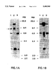
FIG. 1. Detection of v-erbB-related DNA fragments in DNAs from normal human thymus (lane 1), human mammary tumor lines MDA-MB468 (lane 2), and SK-BR-3 (lane 3). Hybridization was conducted at reduced (panel A) or intermediate (panel B) stringency conditions. The arrow denotes a novel 9 kilobase pair (kbp) erbB-related restriction fragment distinct from those of the EGFR gene (erbB) and erbB-2;
FIG. 2. Genomic and cDNA cloning of erbB-3. The region of (v-erbB) hornology within the genomic 9 kbp SacI insert of λE3-1 was subcloned into the plasmid pUC (pE3-1) and subjected to nucleotide sequence analysis. The three predicted exons are depicted as solid boxes. erbB-3 cDNA clones were isolated from oligo dT-primed libraries of mRNAs from normal human placenta (shaded bars) and the breast tumor cell line MCF-7 (open bar). The entire nucleotide sequence was determined for both strands on erbB-3 complementary DNA from normal human placenta and upstream of the 5'XhoI site on pE3-16. The coding sequence is shown as a solid bar and splice junctions of the three characterized genomic exons are indicated by vertical white lines. Solid lines in the cDNA map represent untranslated sequences. Restriction sites: A=AccI, Av=AvaI, B=BamHI, Bg=BglII, E=EcoRI, H=HindIII, K=KpnI, M=MstII, P=PstI, S=SacI, Sm=SmaI, Sp=SpeI;
FIG. 3. (SEQ ID NO:1) Nucleotide sequence of the region of v-erbB homology in the human erbB-3 gene derived from human genomic DNA clone E3-1, in the 1.5 kbp region from the EcoRI to the PstI sites. This region contains three open reading frames bordered by splice junction consensus sequences (underlined). The predicted amino acid sequences of the three exons are shown in three letter code below the relevant DNA sequences;
FIG. 4A. (SEQ ID NO:4) Comparison of the predicted amino acid sequence of the erbB-3 polypeptide with other receptor-like tyrosine kinases. The amino acid sequence is shown in single letter code and is numbered on the right. The putative extracellular domain (thin-lined boxes) extends between the predicted signal sequence (bold-lined box) at the amino-terminus and a single hydrophobic transmembrane region (bold-lined box) within the polypeptide. The two cysteine clusters (Cys) in the extracellular domain and the predicted tyrosine kinase domain (TK) within the cytoplasmic portion of the polypeptide are outlined by the second and third thin-lined boxes, respectively. The putative ATP-binding site at the amino-terminus of the TK domain is circled. Potential autophosphorylation sites within the carboxyl-terminal domain (COOH) are indicated by asterisks. Potential N-linked glycosylation sites (→) are marked above the amino acid sequence. The percentage of amino acid homology of erbB-3 in individual domains with erbB-2, EGFR, met, eph, insulin receptor (IR), and fms is shown in FIB. 4B. Less than 16% identity is denoted by (-);
FIG. 5. Assignment of the genomic locus of erbB-3 was assigned to human chromosomal locus 12q13. A total of 142 grains were localized on the 400-band ideogram. As depicted in the diagram, specific labeling of chromosome 12 was observed, where 38 out of 51 grains were localized to band q13;
FIG. 6. Elevated erbB-3 transcript levels in human mammary tumor cell lines. A Northern blot containing 10 μg total cellular RNA from AB589 mammary epithelial cells (lane 1), as well as mammary tumor cell lines MDA-MB415 (lane 2) and MDA-MB453 (lane 3) was hybridized with an erbB-3 cDNA probe (panel A). Following signal decay the same blot was rehybridized with a human β-actin cDNA probe (panel B) (Gunning, P., Ponte, P., Okayama, H., Engel, J., Blau, H. & Kedes, L., 1983, Mol. Cell Biol. 3:787-795);
FIG. 7. Expression of a human erbB-3 polypeptide in cells transformed by a cDNA segment as detected by an erbB-3-specific anti-peptide antiserum. Cellular lysates (100 μg of each sample) were electrophoresed and transferred to nitrocellulosc membranes for analysis by Western blotting. Panel A. Detection of erbB-3 polypeptide with the antiserum. Panel B. Preincubation of the antiserum with homologous peptide. Antibody blocking indicates binding specificity. Lane 1: Selected cultures of NIH3T3 cells transfected with 1 μg LTRerbB-3 expression vector. Lane 2: control NIH3T3 cells;
FIG. 8. Characterization of gp180erB-3 recombinantly expressed in NIH/3T3 cells. A: Immunoblot analysis of transfectants with erbB-3 peptide antisera MK4 and MK5 and peptide competition. B: Immunoprecipitation with MK5 antiserum of LTR-erbB-3 transfectants metabolically labeled for 2 h in the presence or absence of glycosylation inhibitor tunicamycin (1 μg/ml). C: Pulse-chase analysis: LTR-erbB-3 transfectants were pulse labeled for 15 min with 0.5 mCi each of [35 S] methionine and [35 S] cysteine and immediately lysed (0) or chased with 100 μg/ml each of unlabeled methionine and cysteine for the indicated time periods. 1×10⁷ TCA-precipitable counts were immunoprecipitated from total lysates using MK5 antiserum;
FIG. 9. Immunolocalization of gp180 erbB- 3 on the surface of LTR-erbB-3 cells. A: Immunoprecipitation analysis of metabolically labeled LTR-erbB-3 transfectants with monoclonal antibody E3-1 and a non-immune control. B-D: Indirect immunofluorescence: Formalin-fixed LTR-erbB-3 transfectants were incubated with MAb E3-1 (B) or non-immune IgG (lower left) and stained with a fluorescein-conjugated secondary antibody (100×original magnification). Indirect immunofluorescence with MAb E3-1 of native LTR-erbB-3 cells (C; 1000×original magnification);
FIG. 10. Autophosphorylation in vitro and chronic tyrosine phosphorylation in vivo of gp180erbB-3. LTR-erbB-3 or control lysates were immunoprecipitated with erbB-3 monoclonal antibody (E3-1) or non-immune IgG (NI). Parallel immunoprecipitates were subjected either to immunoblot analysis with MK4 antiserum (A) or to an immunocomplex kinase assay in the presence of [32 P]-γATP (B). Tyrosine Phosphorylation in vivo was assayed by immunoprecipitation with monoclonal anti-P-Tyr antibodies followed by immunoblotting with MK4 antiserum (C);
FIG. 11. EGF-dependent tyrosine phosphorylation of an EGFR/erbB-3 chimerio receptor, gp180EGFR/erbB-3. Serum-starved LTR-erbB-3, LTR-EGFR/erbB-3, and LTR-EGFR transfectants were triggered with 100 ng/ml EGF. Similar amounts of gp180erbB-3, gp180EGFR/erB-3, and EGFR were immunoprecipitated with erbB-3 (E3-1) or EGFR (AB-1) monoclonal antibodies followed by immunoblot analysis with anti-P-Tyr antibodies or peptide antisera; and
FIG. 12. Activation of gp180erbB-3 signaling function in human breast tumor cells. The erbB-3 protein was immunoprecipitated with MAb E3-1 from 1 mg total protein lysate and subjected to immunoblot analysis with erbB-3 peptide antiserum or phosphotyrosine antibodies as indicated.
DESCRIPTION OF SPECIFIC EMBODIMENTS
As used herein, the terms "polypeptide", "protein", "gene product", "antigen", "receptor", "receptor protein" and the like, when used in reference to erbB-3, encompass the erbB-3 amino acid functional sequence as given in SEQ ID NO:4, the mature erbB-3 glycoprotein, gp180erbB-3, and these entities modified by other post-translational modifications, such as glycosylation or tyrosine phosphorylation. However, as is common in the art, the term "erbB-3 polypeptide" typically refers to the sequence as given in SEQ ID NO:4 while the remaining terms typically refer to gp180erbB-3.
The identification of a third member of the erbB-EGF receptor family of membrane spanning tyrosine kinases and the cloning of its full length coding sequence is described the Examples herein. The presence of apparent structural domains resembling those of the EGF receptor suggests the existence of an extracellular binding site for a ligand. The structural relatedness of the extracellular domain of the erbB-3 receptor with that of the EGF receptor indicates that one or more of an increasing number of EGF-like ligands (Shoyab, M., Plowman, G. D., McDonald, V. L. Bradley, J. G. & Todaro, G. J., 1989, Science 243:1074-1076) interacts with the erbB-3 product. Accordingly, the erbB-3 gene is expected to play important roles in both normal and neoplastic processes, as is known for the EGFR and erbB-2 genes.
Despite extensive collinear homology with the EGF receptor and erbB-2, distinct regions within the predicted erbB-3 coding sequence revealed relatively higher degrees of divergence. For example, its carboxyl terminal domain failed to exhibit significant collinear identity scores with either erbB-2 or EGFR. The divergence at the carboxyl terminus also accounts for minor size differences among the three polypeptides of erbB-3, erbB-2, and EGFR, which possess estimated molecular weights of 148 kilodaltons (kd), 138 kd, and 131 kd, respectively. Within the tyrosine kinase domain, which represents the most conserved region of the predicted erbB-3 protein, a short stretch of 29 amino acids closer to the carboxyl terminus than the ATP binding site differed from regions of the predicted erbB-2 and EGFR coding sequence in 28 and 25 positions, respectively. Such regions of higher divergence in their cytoplasmic domains are likely to confer different functional specificity to these closely related receptor-like molecules. Thus, mutations or other alterations in expression of the erbB-3 gene are likely to cause cancers or genetic disorders different from those associated with such defects in the erbB and erbB-2 genes.
Chromosomal mapping localized erbB-3 to human chromosome 12 at the q11-13 locus, whereas the related EGFR and erbB-2 genes are located at chromosomal sites 7p12-13 and 17p12-21.3, respectively. Thus, each gene appears to be localized to a region containing a different homeobox and a different collagen chain gene locus. Keratin type I and type II genes also map to regions of 12 and 17, respectively, consistent with the different localizations of erbB-3 and erbB-2, respectively. Thus, the DNA segments of the present invention represent novel probes to aid in genetic mapping of any heritable diseases which are associated with chromosomal aberrations in the vicinity of the 12q11-13 locus.
There is evidence for autocrine as well as paracrine effectors of normal cell proliferation. The former are factors that are produced by the same cells upon which they stimulate cell proliferation, whereas the latter factors are secreted by cells other than those that are affected by those factors. However, the inherent transforming potential of autocrine growth factors suggests that growth factors most commonly act on their target cell populations by a paracrine route. The present survey of erbB-3 gene expression indicates its normal expression in cells of epithelial and neuroectodermal derivation. Comparative analysis of the three erbB receptor-like genes in different cell types of epidermal tissue revealed that keratinocytes expressed all three genes. In contrast, melanocytes and stromal fibroblasts specifically lacked EGFR and erbB-3 transcripts, respectively. Thus, melanocytes and stromal fibroblasts may be sources of paracrine growth factors for EGFR and erbB-3 products, respectively, that are expressed by the other cell types residing in dose proximity in epidermal tissues.
Given that both erbB and erbB-2 have been causally implicated in human malignancy, the present findings (Example 5) that the erbB-3 transcript is overexpressed in a significant fraction of human mammary tumor cell lines indicates that this new member of the EGFR receptor family also plays an important role in some human malignancies.
Characterization of the human erbB-3 gene product, gp180erbB-3, shows that it is a transmembrane glycoprotein exhibiting properties characteristic of a receptor-like tyrosine kinase. The recombinant human erbB-3 protein shared identical electrophoretic mobility with the natural erbB-3 product expressed in human breast rumor cell lines. Moreover, both recombinant and endogenously expressed gp180erbB-3 were recognized by different antibodies directed against distinct epitopes, such as monodonal (i.e., Mab E3-1) and peptide antibodies directed against epitopes in the extracellular and carboxyl-terminal domains. The 145 kDa erbB-3 polypeptide precursor conformed with predicted erbB-3 encoded protein following cleavage of its signal sequence. Finally, demonstration of its inherent signaling properties established functional integrity of recombinantly expressed gp 180erbB-3.
Although the erbB-3 tyrosine kinase domain shares greater than 60% amino acid identity with the EGFR and erbB-2 proteins, single amino acid substitutions differences in highly consented residues shared among known tyrosine kinases raised a question as to whether erbB-3 harbors intrinsic catalytic activity involved in signal propagation instead of signal attenuation as has been postulated for certain receptor tyrosine kinase-like molecules (Chou et al., Proc. Natl. Acad. Sci. USA 88:4897 (1991)). Most notably, codon 834 within the tyrosine kinase domain predicts asparagine in erbB-3, while aspartate is present at this position in essentially all known protein kinases. Moreover, substitution of asparagine for aspartate in this position abolishes c-kit and v-fps tyrosine kinase activity. The present characterization of the erbB-3 cytoplasmic domain demonstrates not only its catalytic function but also the ability to transduce a mitogenic signal as well. gp181erbB-3 demonstrated autokinase activity in vitro and, in some cell lines, tyrosine phosphorylation in vivo. Moreover, EGF-dependent activation of gp180EGFR/erbB-3 was associated both with mitogenic signaling and in vivo tyrosine phosphorylation of the chimefie receptor. All of these findings imply that the erbB-3 protein represents a biologically active membrane spanning receptor capable of transducing a mitogenic signal in a ligand-dependent manner. Thus, the erbB-3 gene encodes a membrane spanning molecule possessing all the properties of a functional growth receptor.
Constitutive activation of erbB-3 catalytic activity was demonstrated in LTR-erbB-3 transfectants. These results raise the possibility that NIH/3T3 cells may express an erbB-3 ligand. If so, this putative ligand would unlikely interact with the EGFR, since overexpression of the latter in NIH/3T3 cells is not associated with its chronic tyrosine phosphorylation in the absence of exogenous EGF. This invention further established that EGF neither enhanced in vivo tyrosine phosphorylation of gp180erbB-3 nor elicited a mitogenic response in LTR-erbB-3 cells. Additional ligands of the EGF family, including TGFα, amphiregulin, and HB-EGF, have also failed to stimulate gp180 erbB- 3 tyrosine phosphorylation or DNA synthesis in LTR-erbB-3 cells. While a low affinity interaction of known EGF-related ligands for gp180erbB-3 cannot be excluded, these findings indicated that erbB-3 and EGFR proteins possess distinct ligand specificities. The ability to trigger the erbB-3 catalytic domain in the EGFR/erbB-3 chimeric molecule should make it possible to more readily identify its substrates as well as to compare them with those of its closely related family members.
Based upon this invention's demonstration that the erbB-3 protein is both catalytically active and can elicit a proliferative response in NIH/3T3 cells, the instant findings of its chronic activation in some human breast tumor cells suggest its contribution to the malignant phenotype in such tumors. Analogous evidence has implicated overexpression associated with gene amplification of both EGFR and erbB-2 in a variety of tumors as well. In such tumors, there is precedence for activation of receptor kinase activity by mechanisms involving autocrine loops as well as genetic alterations affecting regulatory or coding sequences.
Both EGFR and erbB-2 genes have been implicated as oncogenes based upon demonstration of their overexpression and constitutive activation in various human tumors. The results of this invention argue strongly that the most recently identified family member, erbB-3, is activated in some human breast tumors. Overexpression of the erbB-3 protein did not invariably correlate with its chronic tyrosine phosphorylation. Hence, erbB-3 activation may involve autotrine stimulation or subtle genetic alterations. In addition to breast tumors, expression of the erbB-3 transcript has been observed in a wide range of human carcinomas, including colon, lung, kidney, pancreas, and skin. These findings prompt the search for evidence of erbB-3 activation as an oncogene in these other common human cancers.
Example 1. Identification of a Human DNA Fragment Related to the erbB-3 proto-oncogene Family
In an effort to detect novel erbB-related genes, human genomic DNA was cleaved with a variety of restriction endonucleases and subjected to Southern blot analysis with v-erbB as a probe. Normal mammary epithelial cells AB589 (Walen, K. H. & Stampfer, M. R., 1989, Cancer. Genet. Cytogenet. 37:249-261) and immortalized keratinocytes RHEK have been described previously (Rhim, J. S., Jay, G., Arestein, P., Price, F. M., Sanford, K. K. & Aaronson, S. A., 1985, Science 227:1250-52). Normal human epidermal melanocytes (NHEM) and keratinocytes (NHEK) were obtained from Clonetics. Sources for human embryo fibroblasts (Rubin, J. S., Osada, H., Finch, P. W., Taylor, W. G., Rudikoff, S., & Aaronson, S. A., 1989, Proc. Nat. Aced. Sci. USA 86:802-806) or mammary tumor cell lines SK-BR-3, MDA-MB468, MDA-MB453, and MDA-MB415 (Kraus, M. H., Popescu, N. C., Amsbaugh, S.C. & King, C. R., 1987, EMBO. J. 6:605-610) have been described. For nudeic acid RNA hybridization, DNA and RNA were transferred to nitrocellulose membranes as previously described (Kraus, M. H., et al., 1987, supra). High stringency hybridization was conducted in 50% formamide and 5×SSC at 42° C. Filters were washed at 50° C. in 0.1×SSC. Reduced stringency hybridization of DNA was carded out in 30% formamide followed by washes in 0.6×SSC, while intermediate stringency was achieved by hybridization in 40% formamide and washing in 0.25×SSC. For the specific results depicted in FIG. 1, DNAs were restricted with SacI and hybridized with probe specific for an oncogenic vital form of the erbB gene, v-erbB, spanning from the upstream BamHi site to the EcoRI site in the avian erythroblastosis proviral DNA (Vennstrom, B., Fanshier, L., Moscovici, C. & Bishop, J. M., 1980, J. Virol. 36:575-585).
Under reduced stringency hybridization, four SacI restriction fragments were detected. Two were identified as EGFR gene fragments by their amplification in the mammary tumor cell line MDA-MB468 (FIG. 1A, lane 1,2) known to contain EGFR gene amplification and one as an erbB-2 specific gene fragment due to its increased signal intensity in another mammary tumor cell line, SK-BR-3, known to have erbB-2 amplified (FIG. 1A, lane 1,3). However, a single 9 kbp SacI fragment exhibited equal signal intensities in DNAs from normal human thymus, SK.BR-3 and a line with high levels of EGFR, A431 (FIG. 1A). When the hybridization stringency was raised by 7° C., this fragment did not hybridize, whereas EGFR and erbB-2 specific restriction fragments were still detected with v-erbB as a probe (FIG. 1B). Taken together, these findings suggested the specific detection of a novel v-erbB-related DNA sequence within the 9 kbp SacI fragment.
Example 2. Cloning of the Human DNA Fragment Related to erbB
For further characterization, a normal human genomic library was prepared from SacI cleaved thymus DNA enriched for 8 to 12 kbp fragments. For convenience, bacteriophage λsep-lac5 was obtained from L. Prestidge and D. Hogness (Stanford University); many other cloning vectors derived from phage λ or other genomes can be used for cloning this DNA fragment according to standard recombinant DNA methods that are well known in the art. Purified phage DNA was subjected to cos-end ligation, restriction with SacI, and fractionation in a continuous 10-40% sucrose gradient. A genomic library was prepared by ligating SacI restriction fragments of normal human thymus DNA in the molecular weight range of 8 kbp to 12 kbp (isolated by sucrose gradient sedimentation) with the purified phage arms. Ten recombinant clones detected by v-erbB under reduced stringency conditions did not hybridize with human EGFR or erbB-2 cDNA probes at high stringency. As shown in the restriction map of a representative clone with 9 kbp insert, the region of v-erbB homology was localized by hybridization analysis to a 1.5 kbp segment spanning from the EcoRI to the downstream PstI site.
The nucleotide sequence of a portion of a clone of the novel human genomic DNA fragment related to erbB was determined for both DNA strands by the dideoxy chain termination method (Sanger, F., Nicklen, S. & Coulson, A. R., 1977, Proc. Nat. Acad. Sci. USA. 74:5463-67) using supercoiled plasmid DNA as template. The nucleotide sequence was assembled and translated using IntelliGenetics software. Amino acid sequence comparison was performed with the alignment program by Pearson and Lipman (Pearson, W. R. & Lipman, D. J., 1988, supra) as implemented on the computers of the NCI Advanced Scientific Computing Laboratory. Hydrophobic and hydrophilic regions in the predicted protein were identified according to Kyte and Doolittle (Kyte, J. & Doolittle, R. F., 1982, J. Mol. Biol. 157:105-132). Nucleotide sequence analysis revealed that the region of v-erbB homology in the 1.5 kbp segment from the EcorI to the PstI contained three open reading frames bordered by splice junction consensus sequences (FIG. 2). Computerized comparisons of the predicted amino acid sequence of these three open reading frames with other known proteins revealed the highest identity scores of 64% to 67% to three regions which are contiguous in the tyrosine kinase domains of v-erbB, as well as human EGFR and erbB-2 proteins. Furthermore, all splice junctions of the three characterized exons in the new gene were conserved with erbB-2. Amino acid sequence homology to other known tyrosine kinases was significantly lower, ranging from 39% to 46%.
A single 6.2 kb specific mRNA was identified by Northern blot analysis of human epithelial cells using the 150 bp SpeI-AccI exon-containing fragment as probe (FIG. 2). Under the stringent hybridization conditions employed, this probe detected neither the 5 kb erbB-2 mRNA nor the 6 kb and 10 kb EGFR mRNAs. All of these findings suggested that the present work has identified a new functional member of the erbB proto-oncogene family, which tentatively has been designated as erbB-3.
Example 3. Cloning and Characterization Of cDNAS for the mRNA of the Human erbB-3 Gene
In an effort to characterize the entire erbB-3 coding sequence, overlapping cDNA clones were isolated from oligo dT-primed cDNA libraries from sources with known erbB-3 expression, utilizing gene-specific genomic exons or cDNA fragments as probes. In brief, an oligo dT-primed human placenta cDNA library in λgt11 was obtained from Clontech. MCF-7 cDNA was prepared by first strand synthesis from 5 μg poly A+ RNA using an oligo dT containing linker-primer and Mo-MuLV reverse transcriptase, followed by second strand synthesis with DNA polymerase I, RNaseH, and subsequent T4 DNA polymerase treatment. Double-stranded cDNA was directionally cloned into the SfI site of λpCEV9 using specific linker-adapter oligonucleotides (Miki, T., Matusi, T., Heidaran, M. A. & Aaronson, S. A., 1989, Gene 83:137-146; see also, U.S. application Ser. No. 07/386,053 of Miki et al., filed Jul. 28, 1989). Following plaque purification, phage DNA inserts were subcloned into pUC-based plasmid vectors for further characterization. The clones were initially characterized by restriction analysis and hybridization to the mRNA, and were subsequently subjected to nucleotide sequence analysis. Clones designated pE3-6, pE3-8, pE-9, and pE3-11 carrying inserts with molecular weights ranging from 1.3 kbp to 4.3 kbp were isolated from a human placenta library, whereas the pE3-16 clone containing a 5 kbp insert was obtained by screening the MCF-7 cDNA library with the upstream most coding sequence of pE3-11 as a probe. The clones pE3-8, pE3-9, pE3-11, and pE3-16 contained identical 3' ends terminating in a poly A stretch (FIG. 2).
The complete coding sequence of erbB-3 was contained within a single long open reading frame of 4080 nucleotides extending from position 46 to an in-frame termination codon at position 4126. The most upstream ATG codon at position 100 was the likely initiation codon, as it was preceded by an in-frame stop codon at nucleotide position 43 and fulfilled the criteria of Kozak for an authentic initiation codon. The open reading frame comprised 1342 codons predicting a 148 kd polypeptide. Downstream from the termination codon, multiple stop codons were present in all frames. As shown in SEQ ID NO:4, the deduced amino acid sequence of the erbB-3 polypeptide predicted a transmembrane receptor tyrosine kinase most closely related to EGFR and erbB-2. A hydrophobic signal sequence of erbB-3 was predicted to comprise the 19 amino-terminal amino acid residues. Cleavage of this signal sequence between glycine at position 19 and serine at position 20 would generate a processed polypeptide of 1323 amino acids with an estimated molecular weight of 145 kd. A single hydrophobic membrane spanning domain encompassing 21 amino acids was identified within the coding sequence separating an extracellular domain of 624 amino acids from a cytoplasmic domain comprising 678 amino acids (SEQ ID NO:4).
The putative erbB-3 ligand-binding domain was 43% and 45% identical in amino acid residues with the predicted erbB-2 and EGFR protein, respectively. Within the extaracellular domain, all 50 cysteine residues of the processed erbB-3 polypeptide were conserved and similarly spaced when compared to the EGFR and erbB-2. Forty-seven cysteine residues were organized in two clusters containing 22 and 25 cysteines respectively, a structural hallmark of this tyrosine kinase receptor subfamily (see, for example, Yamamoto, T., Ikawa, S., Aldyama, T., Semba, K., Nomura, N., Miyajima, N., Saito, T. and Toyoshima, K., 1986, Nature 319:230-234). Ten potential N-linked glycosylation sites were localized within the erbB-3 extracellular domain. In comparison with the EGFR and erbB-2 proteins, five and two of these glycosylation sites were conserved, respectively. Among these, the site proximal to the transmembrane domain was conserved among all three proteins (SEQ ID NO:4).
Within the cytoplasmic domain, a core of 277 amino acids from position 702 through 978 revealed the most extensive homology with the tyrosine kinase domains of EGFR and erbB-2. In this region 60% or 62% of amino acid residues were identical and 90% or 89% were conserved, respectively. This stretch of amino acid homology coincides with the minimal catalytic domain of tyrosine kinases (Hanks, S. K., Quinn, A. M. & Hunter, T., 1988, Science 241:42-52).
There was significantly lower homology with other tyrosine kinases (FIG. 4). The consensus sequence for an ATP-binding site (GxGxxG, Hanks, S. K. et al., 1988, supra) was identified at amino acid positions 716 through 721. This sequence as well as a lysine residue located 21 amino acid residues further toward the carboxyl terminus was conserved between the three erbB-related receptors. Taken together these findings defined the region between amino acid position 702 and 978 as the putative catalytic domain of the erbB-3 protein (SEQ ID NO:4).
The most divergent region of erbB-3 compared to either EGFR or erbB-2 was its carboxyl terminus comprising 364 amino acids. This region showed a high degree of hydrophilicity and the frequent occurrence of proline and tyrosine residues. Among these tyrosine residues, those at positions 1197, 1199, and 1262 matched closest with the consensus sequence for putative phosphorylation sites. The peptide sequence YEYMN (SEQ ID NO:12), encompassing tyrosine 1197 and 1199, was repeated at positions 1260-1264 and was at both locations surrounded by charged residues, providing an environment of high local hydrophilicity. These observations render tyrosines 1197, 1199 and 1262 likely candidates for autophosphorylation sites of the erbB-3 protein.
Example 4. Chromosomeal Mapping of the Human erbB-3 Gene.
The chromosomal location of the erbB-3 gene was determined by in situ hybridization (Popesou, N. C., King, C. R. & Kraus, M. H., 1989, Genomics 4:362-366) with a 3 H-labeled plasmid containing the amino-terminal erbB-3 coding sequence. A total of 110 human chromosome spreads was examined prior and subsequent to G banding for identification of individual chromosomes. A total of 142 grains was localized on a 400-band ideogram. Specific labeling of chromosome 12 was observed, where 38 out of 51 grains were localized to band q13 (FIG. 5). Thus, the genomic locus of erbB-3 was assigned to 12q13. In this region of chromosome 12, several genes have previously been mapped including the melanoma-associated antigen ME491, histone genes and the gene for lactalbumin. In addition, two proto-oncogenes, int-1 and gli are located in close proximity to erbB-3.
Example 5. erbB-3 Expression in Normal and Malignant Human Cells
To investigate its pattern of expression, a number of human tissues were surveyed for the erbB-3 transcript. The 6.2 kb erbB-3 specific mRNA was observed in term placenta, postnatal skin, stomach, lung, kidney, and brain, while it was not detectable in skin fibroblasts, skeletal muscle or lymphold cells. Among the fetal tissues analyzed, the erbB-3 transcript was expressed in liver, kidney, and brain, but not in fetal heart or embryonic lung fibroblasts. These observations indicate the preferential expression of erbB-3 in epithelial tissues and brain.
ErbB-3 expression was also investigated in individual cell populations derived from normal human epithelial tissues including keratinocytes, glandular epithelial cells, melanocytes, and fibroblasts. For comparison levels of EGFR and erbB-2 transcripts were analyzed. As shown in Table 1, erbB-3 mRNA levels were relatively high in keratinocytes, comparable with those of erbB-2 and EGFR in these cells. Lower, but similar expression levels of each transcript were detected in cells derived from glandular epithelium. These findings are consistent with growth regulatory roles of all three receptor-like molecules in squamous and glandular epithelium. Whereas erbB-2 and EGFR transcripts were also readily observed in normal fibroblasts, the same cells lacked detectable erbB-3 mRNA. In contrast, normal human melanocytes, which expressed both erbB-3 and erbB-2 at levels comparable with human keratinocytes, lacked detectable EGFR transcripts. Thus, the expression patterns of these receptor-dike molecules were different in specialized cell populations derived from epidermal tissues.
              TABLE 1                                                     
______________________________________                                    
Normal expression pattern of human erbB gene family members               
                             Relative                                     
Cell Source of Transcripts                                                
                   Gene      RNA levels                                   
______________________________________                                    
Embryonic fibroblast (M426)                                               
                   erbB-3    -                                            
                   erbB-2    +                                            
                   EGF-R     +                                            
Skin fibroblast (501T)                                                    
                   erbB-3    -                                            
                   erbB-2    +                                            
                   EGF-R     +                                            
Immortal keratinocyte (RHEK)                                              
                   erbB-3    ++                                           
                   erbB-2    ++                                           
                   EGF-R     ++                                           
Primary keratinocyte (NHEK)                                               
                   erbB-3    +                                            
                   erbB-2    +                                            
                   EGF-R     ++                                           
Glandular epithelium (AB589)                                              
                   erbB-3    (+)                                          
                   erbB-2    (+)                                          
                   EGF-R     (+)                                          
Melanocyte (NHEM)  erbB-3    ++                                           
                   erbB-2    ++                                           
                   EGF-R     -                                            
______________________________________                                    
 Replicate Northern blots were hybridized with equal amounts (in cpm) of  
 probes of similar specific activities for erbB3, erbB2, and EGFR,        
 respectively. Relative signal intensities were estimated: - not          
 detectable, (+) weakly positive, + positive, ++ strongly positive.       
To search for evidence of erbB-3 involvement in the neoplastic process, erbB-3 mRNA levels in a series of human tumor cell lines were surveyed. The erbB-3 transcript was detected in 36 and in 38 carcinomas of 2 of 12 sarcomas while 7 tumor cell lines of hematopoietic origin lacked measurable erbB-3 mRNA. Markedly elevated levels of a normal-sized transcript were observed in 6 out of 17 tumor cell lines derived from human mammary carcinomas. By Southern blot analysis, neither gross gene rearrangement nor amplification were detected in the cell lines. FIG. 6A shows the results of Northern blot analysis with control AB589 nonmalignant human mammary epithelial cells (lane 1) and two representative human mammary tumor lines, MDA-MB415 (lane 2) and MDA-MB453 (lane 3). Hybridization of the same filter with human β-actin probe (FIG. 6B) verified actual levels of mRNA in each lane. Densitometric scanning indicated that the erbB-3 transcript in each tumor cell line was elevated more than 100 fold above that of the control cell line. Thus, overexpression of this new member of the erbB family, as in the case of the EGFR and erbB-2 genes, is likely to play an important role in some human malignancies.
Example 6. Further Characterization of the normal erbB-3 gene product
The pZIPneo expression vector (Cepko et al, Cell 37:1053 (1984)) was modified by introduction of a unique Sal I cloning site. Following deletion of the Sal I site in the tetracyclin resistance gene, the synthetic oligonucleotides 5'-GATCTCGAGTCGAC-3'(SEQ ID NO:10) and 5'-GATCGTCGACTCGA-3'(SEQ ID NO:11) were annealed and ligated into the single Bam HI site to generate pZIPneoSal. The erbB-3 open reading frame including 7 nucleotides upstream of the initiation codon and the termination codon (nucleotides 93-4128) was linkered with Sal I ends, employing the polymerase chain reaction (PCR) and cloned into pZIPneoSal (LTR-erbB). Sense orientation and integrity of the open reading frame were confirmed by restriction analysis as well as nucleotide sequence analyds of cloning boundaries and PCR-amplified regions.
For structural and functional characterization of the erbB-3 gene product, the complete erbB-3 open reading-frame was inserted as given above into the modified ZIPneo vector, placing the cDNA under the transcriptional control of the Moloney murine leukemia virus long-terminal-repeat sequence (LTR-erbB-3). NIH/3T3 fibroblasts were transfected with LTR-erbB-3 or LTR-neo control DNA and cultured in the presence or absence of the selective drug G418. Under conditions in which efficient drug resistance (6×10³ colonies/pmol) was conferred by LTR-erbB-3, no transformed foci were detectable. In contrast, EFR-erbB-2 or EGF-triggered LTR-EGFR induced morphological transformation of NIH-3T3 cells with efficiencies of around 1.2×10⁴ /pmol and 2.3×10² /pmol, respectively.
To test for expression of the erbB-3 protein, polyclonal rabbit antisera, including MK4 and MK5, were developed against synthetic peptides. MK4 and MK5 were raised against peptides that encompass the residues given in SEQ ID NO:5 and SEQ ID NO:6, respectively, which are within the carboxyl terminus of the predicted erbB-3 product. For immunization, peptides were coupled to thyroglobulin using glutaraldehyde. Immunoblot analysis of lysates from marker-selected LTR-neo and LTR-erbB-3 transfectants revealed a major 180 kDa band only in LTR-erbB-3 cells. This band was independently recognized by both antisera (FIG. 8A). There was no cross-reactivity of either antiserum with the related EGFR or erbB-2 proteins overexpressed in NIH/3T3 cells. Immunoreactivity of either antiserum with the 180 kDa band in LTR-erbB-3 transfectants was competed by the antigenic peptide, while MK4 reactivity with a faint 125 kDa band was not affected by preincubation with peptide (FIG. 8A). These results established specificity of erbB-3 protein detection by both polyclonal antisera. By comparison, the 180 kDa erbB-3protein migrated distinctly slower than the 170 kDa EGFR and slightly faster than the 185 kDa erbB-2 protein.
To characterize processing of the erbB-3 protein, we performed immunoprecipitation experiments with MK5 antiserum. Metabolic labeling of LTR-erbB-3 transfectants in the presence or absence of tunicamycin demonstrated that the 145 kDa erbB-3 core polypeptide is modified by N-linked glycosylation (FIG. 8B). Pulse-chase analysis further indicated cotranslational processing, resulting in a predominant 170 kDa precursor protein in addition to faint erbB-3 specific bands of 150 kDa and 160 kDa, following 15 min of pulse-labeling (FIG. 8C). The mature 180 kDa erbB-3 protein appeared after 0.5 h of chase, and the majority was converted into gp180erbB-3 by 1 h. By analysis of further time points, we estimate an approximate half-life of 2-3 h (FIG. 8C). Thus, in NIH/3T3 cells, gp180erbB-3 exhibits an apparently faster turn-over than the EGFR, for which a biosynthesis time of 3 h and approximate half-life of 3-6 h has been reported.
For immunolocalization of the erbB-3 protein, erbB-3-specific monoclonal antibodies, including MAb E3-1, were raised against the recombinantly expressed protein. BALB/c mice were immunized with live LTR-erbB-3 cells. Somatic cell hybrids were prepared by fusion of immune splenocytes with murine non-secreting myeloma cells NS-1. Hybridoma supernatants were screened for differential immunoreactivity with LTR-erbB-3 but not LTR-neo transfectants by enzyme-linked immunosorbent assay (ELISA) using both live cells or cell extracts as antigen source. Positive hybridoma cell lines were cloned twice by limiting dilution and further characterized by immunoprecipitation and immunofluorescence analysis. One monoclonal antibody, MAb E3-1 (IgG2a isotype), specifically immunoprecipitated gp180erbB-3 from LTR-erbB-3 transfectants (FIG. 9A) and did not exhibit cross-reactivity with the EGFR or erbB-2 proteins overexpressed in an NIH/3T3 cell background. Immunofluorescence analysis using a labeled second antibody revealed heterogeneous membrane immunostaining of formalin-fixed LTR-erbB-3 cells using MAb E3-1 (FIG. 9B), but not with a non-specific immunoglobulin of matching isotype (FIG. 9C). MAb E3-1-specific membrane fluorescence of native LTR-erbB-3 cells (FIG. 9D) indicated that gp180erbB-3 was expressed at the cell surface, as expected for a membrane-anchored protein.
To investigate its function, we next analyzed the erbB-3 protein for in vitro kinase activity. LTR-erbB-3 and control LTR-neo cell lysates were first immunoprecipitated with E3-1 followed by immunoblot analysis with MK4 antiserum (FIG. 10A). When the same immunoprecipitates were incubated in autokinase buffer containing [32 P]-γATP, a predominant 180 kDa phosphoprotein was labeled only in immunoprecipitates containing the erbB-3 protein (FIG. 10B). These findings indicated that gp180erbB-3 possessed intrinsic protein kinase activity. To assess its enzymatic actMty in vivo, LTR-erbB-3 lysates were subjected to immunoprecipitation with phosphotyrosine-specific toonotional antibodies (anti-P-Tyr) followed by immunoblotting with MK4 antiserum. As shown in FIG. 10C, the erbB-3 protein was recovered from anti-P-Tyr immunoprecipitates, and immunodetection was competed either by phenyl phosphate in the hnmunoprecipitation or the erbB-3 peptide in Western blot analysis. These findings indicated that recombinant gp180erbB-3 expressed in NIH/3T3 cells was chronically phosphorylated on tyrosine residues.
The protein lysates were prepared in Staph A buffer containing the protease inhibitors phenylmethyl sulfonyl fluoride (1 mM) and aprotinin (10 μg/ml; Boehringer Mannheim). For the analysis of phospho-tyrosine proteins, the phosphatase inhibitors sodium orthovanadate (2 mM) and sodium pyrophosphate (10 mM) were added. Immunoblot analysis using peptide antisera was essentially conducted as previously reported. For the detection of phosphotyrosine proteins, membranes were blocked in PBS containing 5% BSA and immunostained with a mixture of monoclonal anti-P-Tyr antibodies (PY20 and PY69; ICN) diluted 1:500 in PBS containing 1% BSA. Filters were washed with PBS containing 0.05 % Tween20. Immunoprecipitation was conducted using gammabind G agarose (Pharmacia) to collect the immunocomplexes. The beads were coupled with goat anti-mouse-IgG second antibody (Boehringer Mannhelm) in immunoprecipitations using erbB-3 or EGFR monoclonal antibodies. For in vitro kinase assays, 4 mg total lysates were precleared with gammabind G agarose. Following immunoprecipitation, washed immunocomplexes were cquilibrated in autokinase buffer containing 40 mM Hepes 7.5, 10 mM MgCl2, and 0.05 % Triton. The immunocomplexes were subsequently divided for immunoblot analysis or immunocomplex kinase assay, respectively. Autokinase reactions were carried out in 40 μl autokinase buffer containing 20 Ci γATP (3000 ci/mmol) at 25° for 10 min and terminated by addition of SDS containing sample buffer.
Example 7. EGF-dependent mitogenic signaling by an EGFR/erb B-3 chimeric receptor
To explore erbB-3 signaling, a chimeric receptor, LTR-EGFR/erbB-3, containing the ligand-binding domain of the closely related EGF receptor (aa 1-682) and the intracellular portion of erbB-3 (aa 681-1342) was engineered. Linearized expression constructs (0.01-10 μg/plate) were transfected into NIH/3T3 cells by calcium phosphate precipitation using 40 μg of calf thymus DNA as carrier. Mass cultures expressing the recombinant proteins were obtained by selection with 750 μg/ml G418. Selected LTR-EGFR/erbB-3 transfectants were enriched for expression of the chimeric protein by preparative FACS sorting using EGFR monoclonal antibody AB-1 (Oncogene Sciences).
Transfection of NIH/3T3 cells with this construct did not result in detectable focus formation either in the presence or absence of EGF. To quantitate expression of the chimerio receptor, selected mass cultures were analyzed for EGF-binding in comparison to NIH/3T3 cells overexpressing the EGFR (LTR-EGFR). Scatchard analysis established around 5.7×10⁵ EGF binding sites/cell for the LTR-EGFR/erbB-3 transfectant as compared to 2.5×10⁶ binding sites/cell for LTR-EGFR transfectant. The LTR-EGFR/erbB-3 transfectant exhibited two populations of binding sites with affinities of 0.11 nM and 5 nM, respectively. The high-affinity sites were in the minority (2.3×10⁴), and there were 5.5×10⁵ low-affinity binding sites. Similar results were obtained with the wild-type EGFR in LTR-EGFR transfectants, which displayed 1.1×10⁵ high affinity (0.13 nM) and 2.4×10⁶ low affinity receptors (7 nM).
To investigate EGF responsiveness of erbB-3 enzymatic activity, the in vivo tyrosine phosphorylation of the chimefie receptor in the presence or absence of EGF was compared. This protein, as well as the EGFR and erbB-3 proteins from independent transfectants were enriched by immunoprecipitation and subjected to immunoblot analysis with either anti-P-Tyr or the appropriate specific antiserum. As shown in FIG. 11, the steady state level of tyrosine phosphorylated gp180erbB-3 in NIH/3T3 cells was not altered upon EGF exposure (lane 1,2). The chimefie EGFR/erbB-3 receptor, which was expressed as a 180 kDa protein, gp180EGFR/erbB-3 displayed low, but detectable level of tyrosine phosphorylation in serum-free medium (lane 3). However, EGF triggering of the chimera resulted in a substantial increase in tyrosine phosphorylation, demonstrating EGF-dependent activation of erbB-3 catalytic function (lane 4). The wild-type EGFR showed a somewhat higher level of EGF-dependent tyrosine phosphorylation under the: same conditions lane 6). Of note, the relative level of gp180EGFR/erbB-3 tyrosine phosphorylation was comparable to that of EGF-activated chimeric receptor expressed at a similar protein level, indicating constitutive activation of erbB-3 catalytic properties in LTR-erbB-3 transfectants.
Whether the erbB-3 catalytic domain was capable of transducing a mitogenic signal was then assessed. When the LTR-EGFR/erbB-3 transfectant was exposed to increasing EGF concentrations, there was a dose-dependent stimulation of DNA synthesis similar to that observed with EGFR overexpressing NIH-3T3 cells. Under the same conditions, neither LTR-neo nor LTR-erbB-3 transfectants showed a significant increase in DNA synthesis even at high EGF concentrations, consistent with previous observations. It should be noted that basal levels of DNA synthesis of the LTR-erbB-3 transfectant were 2-3 fold above those of the other transfectants. These findings that were reproducible with several independent selected mass cultures.
The biological effects of activated erbB-3 catalytic function were assessed by testing the transfectants for anchorage-independent growth. To test anchorage-independent growth, cell suspensions were seeded at 10-fold serial dilutions in semisolid agarose medium containing growth medium and 0.45% seaplaque agarose (FMC Corp.). Visible colonies comprising>100 cells were scored at 14 days. EGF was added at a concentration of 20 ng/ml. Human mammary tumor cell lines were obtained from the American Type Culture Collection and propagated in Dulbecco's modified Eagle medium containing 10% fetal calf serum.
As shown in Table 2, LTR-neo transfectants failed to exhibit significant soft agar growth in the presence or absence of EGF. In contrast, EGF induced soft agar colony formation with both LTR-EGFR/erbB-3 and LTR-EGFR transfectants. The latter showed a larger colony number (Table 2) as well as colony size (data not shown). By comparison, the LTR-erbB-3 transfectant displayed EGF-independent colony formation with an efficiency similar to that of EGF-activated LTR-EGFR/erbB-3 transfectant (Table 2). All of these findings establish that ligand activation of a chimetic EGFR/erbB-3 receptor causes mitogenic signaling in NIH/3T3 cells and suggest that chronic tyrosine phosphorylation of erbB-3 in LTR-erbB-3 transfectants is associated with constitutive signaling in these cells.
              TABLE 2                                                     
______________________________________                                    
Anchorage-independent growth of NIH/3T3 transfectants                     
                #colonies*/10.sup.4 cells                                 
NIH3T3 transfectants                                                      
                  -EGF     +EGF                                           
______________________________________                                    
LTR-neo           1 (±1)                                               
                           4 (±2)                                      
LTR-EGFR          2 (±2)                                               
                           206 (±49)                                   
LTR-EGFR/ergB-3   7 (±3)                                               
                           88 (±16)                                    
LTR-erbB-3        97 (±20)                                             
                           94 (±29)                                    
______________________________________                                    
 *mean (± standard error) of 3 independent assays                      
Example 8. Evidence for activated erbB-3 signaling function in human breast tumor cells
The availability of erbB-3 specific antibodies made it possible to explore expression and activity of gp180erbB-3 in human tumor cells. Based upon our previous evidence for erbB-3 mRNA overexpression in certain breast cancer cell lines, we measured erbB-3 protein levels and tyrosine phosphorylation in such tumor lines, using the procedures given above. Following immunoprecipitation with Mab E3-1, immunoblot analysis with MK4 antiserum revealed the natural human erbB-3 product as a 180 kDa protein. The levels of erbB-3 protein varied markedly along the tumor lines analyzed, with highest expression in BT483, MDA-MB453, MDA-MB134, MDA-MB361, SK-BR-3, and MDA-MB468 (FIG. 12). The lowest levels were observed in BT20 and MDA-MB175 cell lines (FIG. 12), comparable with that expressed by nonmalignant 184B5 mammary epithelial cells (data not shown). Thus, erbB-3 protein expression varied by at least 20-30 fold among the lines tested, consistent with results of transcript analysis (data not shown).
Immunoblot analysis of the same immunoprecipitates with anti-P-Tyr antibodies revealed that tyrosine phosphorylation of the native erbB-3 product was undetectable in 8 of the tumor cell lines, including MDA-MB134, MDA-MB361, and MDA-MB468, which harbored increased erbB-3 levels. In contrast, erbB-3 protein expressed by 4 cell lines, including MDA-MB453, BT474, MDA-MB175, and SK-BR-3, demonstrated readily detectable chronic tyrosine phosphorylation (FIG. 12, lanes 5, 7, 9 and 11). In MDA-MB175, there was no significantly elevated level of erbB-3 protein. Thus, in 4 out of 12 breast tumor cells lines, the gp180erbB-3 signaling function was activated at steady state. Whether chronic erbB-3 phosphorylation involves autocrine stimulation or subtle structural alterations, these findings provide evidence for constitutive gp180erbB-3 activation in these human breast tumor cells.
Example 9. Identification, purification and characterization of erbB-3 ligands
As shown in FIG. 12, the gp180erbB-3 that is overexpressed in some human breast tumor cell lines can be either functionally activated or not, depending on the cell line. Further, in some other human breast tumor cell lines, the erbB-3 polypeptide is not overexpressed and, again, can be either activated or not activated. These differences, and the common property of growth factor receptor-like tyrosine kinases to rapidly autophosphorylate on tyrosine residues in response to ligand triggering, can be exploited to identify, isolate and characterize ligands, preferably specific ligands, that can activate or down-regulate erbB-3. The term "erbB-3 ligand" refers to a molecule that binds to the erbB-3 protein, particularly to the extracellular domain of the erbB-3 protein, and can activate ("erbB-3 activating ligand") or down-regulate ("erbB-3 blocking ligand") the biochemical and/or biological activity of the erbB-3 protein. Depending on the concentration of the ligand, a ligand can both activate and down-regulate activity.
A source containing a potential erbB-3 ligand, such as conditioned medium, body fluid extracts, cell extracts, tissue extracts or the like, with or without agents which can modify erbB-3 activity, can be screened for the presence of such a ligand by the ability of the solution, in the case of an activating ligand, to enhance erbB-3 phosphorylation. With respect to screening for an erbB-3 activating ligand, cells from a cell line whose expressed erbB-3 protein contains nonexistent or low level intrinsic tyrosine phosphorylation can be contacted with potential ligand sources or control medium for a time and under conditions sufficient to allow binding of an erbB-3 ligand, if present, to bind to erbB-3. Typically, if an erbB-3 ligand is present, binding will occur within a short time. Thus, the cells are exposed to potential ligand sources or control medium for, preferably, no longer than 30 minutes, most preferably, 10 minutes or less. Appropriate conditions to allow binding of the ligand can be determined by one skilled in the art, such as physiological conditions at 37° C. or the conditions given in FIG. 5 of Holmes et al., Science 256:1205 (1992). erbB-3 modifying agents, if administered, can be present in a concentration between 10-2 pM and 105 pM. The cell line employed can overexpress erbB-3, such as the mammary tumor cell lines MDA-MB415, MDA-MB134, MDA-MB468, BT483, MDA-MB361, MCF-7 and ZR-75-1, which express an increased amount of the erbB-3 protein with low level intrinsic tyrosine phosphorylation compared to protein amounts and activation levels for corresponding nonmalignant cells. One such potential activating ligand source can be derived from cell lines that not only overexpress erbB-3 but also exhibit high level intrinsic tyrosine phosphorylation, such as MDA-MB453, SK-BR-3, and BT474. In addition, any normal cell which does not overexpress erbB-3 can be utilized, e.g., fibroblasts.
Similarly, with respect to screening for an erbB-3 inhibitory down-regulating ligand, a cell line whose expressed erbB-3 protein contains high level intrinsic tyrosine phosphorylation can be exposed to potential ligand sources or control medium for a time and under conditions sufficient to allow binding of an erbB-3 ligand, if present, to bind to erbB-3. The cell line employed preferably expresses activated erbB-3, such as the mammary tumor cell lines MDA-MB453, SK-BR-3 and BT474. In addition, an activating ligand at higher concentration can be down-regulating. Such activity can be routinely screened given the teaching herein.
The triggering or blocking of erbB-3 activation can be detected by comparing the level of erbB-3 tyrosine phosphorylation in the cell line after exposure to the potential ligand source with the normal level, e.g., the level obtained after exposure to the control medium. For example, in a negative control the cells can be in serum free medium and for activating ligand the conditioned medium is from cell lines with increased erbB-3 that don't have phosphorylation. For instance, to measure erbB-3 specific tyrosine phosphorylation, potentially triggered (or blocked) cells and the control cells are lysed. Using procedures such as those discussed above, the erbB-3 protein is immunoprecipitated with an erbB-3 specific antibody, preferably a monoclonal such as MAb E3-1. The immunoprecipitates are dMded and subjected to immunoblot analysis with either antiphosphotyrosine or erbB-3 antibodies. The presence of an erbB-3 activating or blocking ligand can be monitored by a relative increase or decrease, respectively, of phosphotyrosine levels in comparison to the untriggered control. Any increase can be significant, especially a two-fold or greater increase. This ligand-detection system can be used repeatedly throughout the ligand purification procedures so as to monitor protein purification of the erbB-3 ligand to homogeneity.
Alternatively, following exposure of the cell lines to the potential ligand source as discussed above, detection of an erbB-3 activating ligand or blocking ligand can be accomplished by measurement of cell growth and/or mitogenic signals resulting from the activation or inhibition of erbB-3 catalytic activity, using, for example, the procedures given in Example 7 above. An increase in colony number or colony size and/or a dose-dependent increase of DNA synthesis for the cells exposed to the potential ligand relative to those exposed to the control medium correlates with the presence of an activating ligand in the potential ligand source. Conversely, respective decreases correlate with the presence of a blocking ligand in the potential ligand source.
Following the isolation and purification of the erbB-3 ligand, the identity of the ligand can be determined by protein identification methods known in the art, such as amino acid sequencing. Further, the erbB-3 ligand can be molecularly characterized. For instance, similar to the procedures outlined in Holmes et al., Science 256:1205 (1992), the nucleic acid sequence that corresponds to the ligand's amino acid sequence, or a partial amino acid sequence corresponding to a portion of the ligand, can be used to design degenerate oligonucleotide probes corresponding to the amino acid sequence or partial sequence. These degenerate oligonucleotides can be used to screen a cDNA library and generate a clone that encodes the precursor of the erbB-3 ligand. Following determination of the coding sequence, related coding sequences can be discovered by screening other libraries.
For purposes of completing the background description and present disclosure, each of the published articles, patents and patent applications heretofore identified in this specification are hereby incorporated by reference into the specification.
The foregoing invention has been described in some detail for purposes of clarity and understanding. It will also be obvious that various changes and combinations in form and detail can be made without departing from the scope of the invention.
__________________________________________________________________________
SEQUENCE LISTING                                                          
(1) GENERAL INFORMATION:                                                  
(iii) NUMBER OF SEQUENCES: 12                                             
(2) INFORMATION FOR SEQ ID NO:1:                                          
(i) SEQUENCE CHARACTERISTICS:                                             
(A) LENGTH: 1542 base pairs                                               
(B) TYPE: nucleic acid                                                    
(C) STRANDEDNESS: single                                                  
(D) TOPOLOGY: linear                                                      
(ii) MOLECULE TYPE: DNA (genomic)                                         
(ix) FEATURE:                                                             
 (A) NAME/KEY: exon                                                       
(B) LOCATION: 66..221                                                     
(ix) FEATURE:                                                             
(A) NAME/KEY: exon                                                        
(B) LOCATION: 780..855                                                    
(ix) FEATURE:                                                             
(A) NAME/KEY: exon                                                        
(B) LOCATION: 1040..1185                                                  
(ix) FEATURE:                                                             
(A) NAME/KEY: intron                                                      
(B) LOCATION: 222..779                                                    
(ix) FEATURE:                                                             
 (A) NAME/KEY: intron                                                     
(B) LOCATION: 856..1039                                                   
(ix) FEATURE:                                                             
(A) NAME/KEY: CDS                                                         
(B) LOCATION: join(66..221, 780..855, 1040..1185)                         
(xi) SEQUENCE DESCRIPTION: SEQ ID NO:1:                                   
GAATTCCAGATCTCAGTGACTGATTCCCCCAACCTTAAGAATACTTTCTTCCCCTATACC60            
TACAGGGAATGTACTACCT TGAGGAACATGGTATGGTGCATAGAAAC107                       
GlyMetTyrTyrLeuGluGluHisGlyMetValHisArgAsn                                
1510                                                                      
CTGGCTGCCCGAAACGTGCTACTCAAG TCACCCAGTCAGGTTCAGGTG155                      
LeuAlaAlaArgAsnValLeuLeuLysSerProSerGlnValGlnVal                          
15202530                                                                  
GCAGATTTTGGTGTGGCTGAC CTGCTGCCTCCTGATGATAAGCAGCTG203                      
AlaAspPheGlyValAlaAspLeuLeuProProAspAspLysGlnLeu                          
354045                                                                    
CTATACAGTGAGGCCAAG GTGAGGAGACACAAAGGGTAAGGAGGCGGG251                      
LeuTyrSerGluAlaLys                                                        
50                                                                        
GGTGGAGTGAAGCATGGGGATAGGGAGCAGCCAGTGGTCTCTTCCAGAGGCAAGCAGATG311           
CTTCATGGTAAGTTCAAGGAGAGAAG GCTGCAGATGCCAGATATTTTAGTTCAGAGGGCA371          
ACAAAGAAAATAATGATCAAGAACTTGGGACTGGCCGGGCGCGGTGGCTCACGCCTGTAA431           
TCCCAACACTTCGGGAGGCCAAGGCGGGTGGATCACAAGGTCAGGAGATCAAGACCATCC491           
 TGGCTAGCACGGTGAAACCCCGTCTCTACTAAATATACAAAAAAAAAAAAATTAGCCAGG551          
CGTGGCGGCATGCATCTGTACTCCCAGCTACTCGGGAGGCTGAGGCAGGAGAATGGCGTG611           
AACCCAGGAGGCGGAGCTTGCAGTGGGCCGAGATCGCACCACTG CACTCCAGTCTGGGCG671          
ACAGAGCGAGACTCCGTCTCAAAAAAAAAAAAAAAAGAATTTGGGACTTGGAAATCCTAA731           
GAAAATTTGTGGAAATAAACTTGTGATACCTCTATCTTTAATCCGCAGACTCCAATT788              
 ThrProIle                                                                
55                                                                        
AAGTGGATGGCCCTTGAGAGTATCCACTTTGGGAAATACACACACCAG836                       
LysTrpMet AlaLeuGluSerIleHisPheGlyLysTyrThrHisGln                         
606570                                                                    
AGTGATGTCTGGAGCTATGGTCAGTGCATCTGGATGCCCTCTCTACCAT885                      
SerAsp ValTrpSerTyr                                                       
75                                                                        
CACTGGCCCCAGTTTCAAATTTACCTTTTGAGAGCCCCCTCTTAGAATCTCTAAGCACTT945           
CAGATTTTTGTGTTAGATCAGGTTCTGCCTTCCCTTCACTTCATGCCCATGTCTACTATT1005          
TTGCCAGTGA CTAGTCCATGTCTTCCTGCAACAGGTGTGACAGTTTGGGAG1056                  
GlyValThrValTrpGlu                                                        
80                                                                        
TTGATGACCTTCGG GGCAGAGCCCTATGCAGGGCTACGATTGGCTGAA1104                     
LeuMetThrPheGlyAlaGluProTyrAlaGlyLeuArgLeuAlaGlu                          
859095                                                                    
GTACCAGACCTGCTAGAGAA GGGGGAGCGGTTGGCACAGCCCCAGATC1152                     
ValProAspLeuLeuGluLysGlyGluArgLeuAlaGlnProGlnIle                          
100105110115                                                              
TGCACAATTGATGT CTACATGGTGATGGTCAAGTGTGAGTTACCTGCTGAGCC1205                
CysThrIleAspValTyrMetValMetValLys                                         
120125                                                                    
CAACCATTTTCTCTTTTTTTCTTTTTTTTTCTTTTTTTTTTTTTTTTGAG ACAGAGTCTC1265         
ACAATTGTCACCCAGGCTGGAGTGCAATGGTGCAATCAATCTTGGCTCACTACAACCTCC1325          
GCCTCTCGGGTTCAAGAGATTCTCCTGCTTCAGCTCCGGAGTAGCTGGGATTACAGCGCC1385          
CGCCACACCTGGATAACTGTTACA CTTTTAGTAGAGATGGGGTTTCACCATGTTGGCCAG1445         
GCTGGTCTCAAACTCCTGACCTCAGGTGATCCGCCTGCCTCAGCTTCCCAAAGTGCTGGG1505          
ATTACAGGTGTGAGCCATCATGCTCGCCTGACTGCAG1542                                 
(2) INFORMATION FOR SEQ ID NO:2:                                          
(i) SEQUENCE CHARACTERISTICS:                                             
(A) LENGTH: 126 amino acids                                               
(B) TYPE: amino acid                                                      
(D) TOPOLOGY: linear                                                      
(ii) MOLECULE TYPE: protein                                               
(xi) SEQUENCE DESCRIPTION: SEQ ID NO:2:                                   
GlyMetTyrTyrLeuGluGluHisGlyMetValHisArgAsnLeuAla                          
1 51015                                                                   
AlaArgAsnValLeuLeuLysSerProSerGlnValGlnValAlaAsp                          
202530                                                                    
PheGlyValAlaAspLeuLeu ProProAspAspLysGlnLeuLeuTyr                         
354045                                                                    
SerGluAlaLysThrProIleLysTrpMetAlaLeuGluSerIleHis                          
5055 60                                                                   
PheGlyLysTyrThrHisGlnSerAspValTrpSerTyrGlyValThr                          
65707580                                                                  
ValTrpGluLeuMetThrPheGlyAlaGluProTyrAlaGlyL euArg                         
859095                                                                    
LeuAlaGluValProAspLeuLeuGluLysGlyGluArgLeuAlaGln                          
100105110                                                                 
Pro GlnIleCysThrIleAspValTyrMetValMetValLys                               
115120125                                                                 
(2) INFORMATION FOR SEQ ID NO:3:                                          
(i) SEQUENCE CHARACTERISTICS:                                             
(A) LENGTH: 4905 base pairs                                               
(B) TYPE: nucleic acid                                                    
(C) STRANDEDNESS: single                                                  
 (D) TOPOLOGY: linear                                                     
(ii) MOLECULE TYPE: cDNA                                                  
(ix) FEATURE:                                                             
(A) NAME/KEY: CDS                                                         
(B) LOCATION: 100..4125                                                   
(xi) SEQUENCE DESCRIPTION: SEQ ID NO:3:                                   
ACCAATTCGCCAGCGGTTCAGGTGGCTCTTGCCTCGATGTCCTAGCCTAGGGGCCCCCGG60            
GCCGGACTTGGCTGGGCTCCCTTCACCCT CTGCGGAGTCATGAGGGCGAACGAC114                
MetArgAlaAsnAsp                                                           
15                                                                        
GCTCTGCAGGTGCTGGGCT TGCTTTTCAGCCTGGCCCGGGGCTCCGAG162                      
AlaLeuGlnValLeuGlyLeuLeuPheSerLeuAlaArgGlySerGlu                          
101520                                                                    
GTGGGCAACTCTCAGG CAGTGTGTCCTGGGACTCTGAATGGCCTGAGT210                      
ValGlyAsnSerGlnAlaValCysProGlyThrLeuAsnGlyLeuSer                          
253035                                                                    
GTGACCGGCGATGCTG AGAACCAATACCAGACACTGTACAAGCTCTAC258                      
ValThrGlyAspAlaGluAsnGlnTyrGlnThrLeuTyrLysLeuTyr                          
404550                                                                    
GAGAGGTGTGAGGTGGTGA TGGGGAACCTTGAGATTGTGCTCACGGGA306                      
GluArgCysGluValValMetGlyAsnLeuGluIleValLeuThrGly                          
556065                                                                    
CACAATGCCGACCTCTCCTTCCTGC AGTGGATTCGAGAAGTGACAGGC354                      
HisAsnAlaAspLeuSerPheLeuGlnTrpIleArgGluValThrGly                          
70758085                                                                  
TATGTCCTCGTGGCCATGA ATGAATTCTCTACTCTACCATTGCCCAAC402                      
TyrValLeuValAlaMetAsnGluPheSerThrLeuProLeuProAsn                          
9095100                                                                   
CTCCGCGTGGTGCGAG GGACCCAGGTCTACGATGGGAAGTTTGCCATC450                      
LeuArgValValArgGlyThrGlnValTyrAspGlyLysPheAlaIle                          
105110115                                                                 
TTCGTCATGTTGAACT ATAACACCAACTCCAGCCACGCTCTGCGCCAG498                      
PheValMetLeuAsnTyrAsnThrAsnSerSerHisAlaLeuArgGln                          
120125130                                                                 
CTCCGCTTGACTCAGCTCA CCGAGATTCTGTCAGGGGGTGTTTATATT546                      
LeuArgLeuThrGlnLeuThrGluIleLeuSerGlyGlyValTyrIle                          
135140145                                                                 
GAGAAGAACGATAAGCTTTGTCACA TGGACACAATTGACTGGAGGGAC594                      
GluLysAsnAspLysLeuCysHisMetAspThrIleAspTrpArgAsp                          
150155160165                                                              
ATCGTGAGGGACCGAGATG CTGAGATAGTGGTGAAGGACAATGGCAGA642                      
IleValArgAspArgAspAlaGluIleValValLysAspAsnGlyArg                          
170175180                                                                 
AGCTGTCCCCCCTGTC ATGAGGTTTGCAAGGGGCGATGCTGGGGTCCT690                      
SerCysProProCysHisGluValCysLysGlyArgCysTrpGlyPro                          
185190195                                                                 
GGATCAGAAGACTGCC AGACATTGACCAAGACCATCTGTGCTCCTCAG738                      
GlySerGluAspCysGlnThrLeuThrLysThrIleCysAlaProGln                          
200205210                                                                 
TGTAATGGTCACTGCTTTG GGCCCAACCCCAACCAGTGCTGCCATGAT786                      
CysAsnGlyHisCysPheGlyProAsnProAsnGlnCysCysHisAsp                          
215220225                                                                 
GAGTGTGCCGGGGGCTGCTCAGGCC CTCAGGACACAGACTGCTTTGCC834                      
GluCysAlaGlyGlyCysSerGlyProGlnAspThrAspCysPheAla                          
230235240245                                                              
TGCCGGCACTTCAATGACA GTGGAGCCTGTGTACCTCGCTGTCCACAG882                      
CysArgHisPheAsnAspSerGlyAlaCysValProArgCysProGln                          
250255260                                                                 
CCTCTTGTCTACAACA AGCTAACTTTCCAGCTGGAACCCAATCCCCAC930                      
ProLeuValTyrAsnLysLeuThrPheGlnLeuGluProAsnProHis                          
265270275                                                                 
ACCAAGTATCAGTATG GAGGAGTTTGTGTAGCCAGCTGTCCCCATAAC978                      
ThrLysTyrGlnTyrGlyGlyValCysValAlaSerCysProHisAsn                          
280285290                                                                 
TTTGTGGTGGATCAAACAT CCTGTGTCAGGGCCTGTCCTCCTGACAAG1026                     
PheValValAspGlnThrSerCysValArgAlaCysProProAspLys                          
295300305                                                                 
ATGGAAGTAGATAAAAATGGGCTCA AGATGTGTGAGCCTTGTGGGGGA1074                     
MetGluValAspLysAsnGlyLeuLysMetCysGluProCysGlyGly                          
310315320325                                                              
CTATGTCCCAAAGCCTGTG AGGGAACAGGCTCTGGGAGCCGCTTCCAG1122                     
LeuCysProLysAlaCysGluGlyThrGlySerGlySerArgPheGln                          
330335340                                                                 
ACTGTGGACTCGAGCA ACATTGATGGATTTGTGAACTGCACCAAGATC1170                     
ThrValAspSerSerAsnIleAspGlyPheValAsnCysThrLysIle                          
345350355                                                                 
CTGGGCAACCTGGACT TTCTGATCACCGGCCTCAATGGAGACCCCTGG1218                     
LeuGlyAsnLeuAspPheLeuIleThrGlyLeuAsnGlyAspProTrp                          
360365370                                                                 
CACAAGATCCCTGCCCTGG ACCCAGAGAAGCTCAATGTCTTCCGGACA1266                     
HisLysIleProAlaLeuAspProGluLysLeuAsnValPheArgThr                          
375380385                                                                 
GTACGGGAGATCACAGGTTACCTGA ACATCCAGTCCTGGCCGCCCCAC1314                     
ValArgGluIleThrGlyTyrLeuAsnIleGlnSerTrpProProHis                          
390395400405                                                              
ATGCACAACTTCAGTGTTT TTTCCAATTTGACAACCATTGGAGGCAGA1362                     
MetHisAsnPheSerValPheSerAsnLeuThrThrIleGlyGlyArg                          
410415420                                                                 
AGCCTCTACAACCGGG GCTTCTCATTGTTGATCATGAAGAACTTGAAT1410                     
SerLeuTyrAsnArgGlyPheSerLeuLeuIleMetLysAsnLeuAsn                          
425430435                                                                 
GTCACATCTCTGGGCT TCCGATCCCTGAAGGAAATTAGTGCTGGGCGT1458                     
ValThrSerLeuGlyPheArgSerLeuLysGluIleSerAlaGlyArg                          
440445450                                                                 
ATCTATATAAGTGCCAATA GGCAGCTCTGCTACCACCACTCTTTGAAC1506                     
IleTyrIleSerAlaAsnArgGlnLeuCysTyrHisHisSerLeuAsn                          
455460465                                                                 
TGGACCAAGGTGCTTCGGGGGCCTA CGGAAGAGCGACTAGACATCAAG1554                     
TrpThrLysValLeuArgGlyProThrGluGluArgLeuAspIleLys                          
470475480485                                                              
CATAATCGGCCGCGCAGAG ACTGCGTGGCAGAGGGCAAAGTGTGTGAC1602                     
HisAsnArgProArgArgAspCysValAlaGluGlyLysValCysAsp                          
490495500                                                                 
CCACTGTGCTCCTCTG GGGGATGCTGGGGCCCAGGCCCTGGTCAGTGC1650                     
ProLeuCysSerSerGlyGlyCysTrpGlyProGlyProGlyGlnCys                          
505510515                                                                 
TTGTCCTGTCGAAATT ATAGCCGAGGAGGTGTCTGTGTGACCCACTGC1698                     
LeuSerCysArgAsnTyrSerArgGlyGlyValCysValThrHisCys                          
520525530                                                                 
AACTTTCTGAATGGGGAGC CTCGAGAATTTGCCCATGAGGCCGAATGC1746                     
AsnPheLeuAsnGlyGluProArgGluPheAlaHisGluAlaGluCys                          
535540545                                                                 
TTCTCCTGCCACCCGGAATGCCAAC CCATGGAGGGCACTGCCACATGC1794                     
PheSerCysHisProGluCysGlnProMetGluGlyThrAlaThrCys                          
550555560565                                                              
AATGGCTCGGGCTCTGATA CTTGTGCTCAATGTGCCCATTTTCGAGAT1842                     
AsnGlySerGlySerAspThrCysAlaGlnCysAlaHisPheArgAsp                          
570575580                                                                 
GGGCCCCACTGTGTGA GCAGCTGCCCCCATGGAGTCCTAGGTGCCAAG1890                     
GlyProHisCysValSerSerCysProHisGlyValLeuGlyAlaLys                          
585590595                                                                 
GGCCCAATCTACAAGT ACCCAGATGTTCAGAATGAATGTCGGCCCTGC1938                     
GlyProIleTyrLysTyrProAspValGlnAsnGluCysArgProCys                          
600605610                                                                 
CATGAGAACTGCACCCAGG GGTGTAAAGGACCAGAGCTTCAAGACTGT1986                     
HisGluAsnCysThrGlnGlyCysLysGlyProGluLeuGlnAspCys                          
615620625                                                                 
TTAGGACAAACACTGGTGCTGATCG GCAAAACCCATCTGACAATGGCT2034                     
LeuGlyGlnThrLeuValLeuIleGlyLysThrHisLeuThrMetAla                          
630635640645                                                              
TTGACAGTGATAGCAGGAT TGGTAGTGATTTTCATGATGCTGGGCGGC2082                     
LeuThrValIleAlaGlyLeuValValIlePheMetMetLeuGlyGly                          
650655660                                                                 
ACTTTTCTCTACTGGC GTGGGCGCCGGATTCAGAATAAAAGGGCTATG2130                     
ThrPheLeuTyrTrpArgGlyArgArgIleGlnAsnLysArgAlaMet                          
665670675                                                                 
AGGCGATACTTGGAAC GGGGTGAGAGCATAGAGCCTCTGGACCCCAGT2178                     
ArgArgTyrLeuGluArgGlyGluSerIleGluProLeuAspProSer                          
680685690                                                                 
GAGAAGGCTAACAAAGTCT TGGCCAGAATCTTCAAAGAGACAGAGCTA2226                     
GluLysAlaAsnLysValLeuAlaArgIlePheLysGluThrGluLeu                          
695700705                                                                 
AGGAAGCTTAAAGTGCTTGGCTCGG GTGTCTTTGGAACTGTGCACAAA2274                     
ArgLysLeuLysValLeuGlySerGlyValPheGlyThrValHisLys                          
710715720725                                                              
GGAGTGTGGATCCCTGAGG GTGAATCAATCAAGATTCCAGTCTGCATT2322                     
GlyValTrpIleProGluGlyGluSerIleLysIleProValCysIle                          
730735740                                                                 
AAAGTCATTGAGGACA AGAGTGGACGGCAGAGTTTTCAAGCTGTGACA2370                     
LysValIleGluAspLysSerGlyArgGlnSerPheGlnAlaValThr                          
745750755                                                                 
GATCATATGCTGGCCA TTGGCAGCCTGGACCATGCCCACATTGTAAGG2418                     
AspHisMetLeuAlaIleGlySerLeuAspHisAlaHisIleValArg                          
760765770                                                                 
CTGCTGGGACTATGCCCAG GGTCATCTCTGCAGCTTGTCACTCAATAT2466                     
LeuLeuGlyLeuCysProGlySerSerLeuGlnLeuValThrGlnTyr                          
775780785                                                                 
TTGCCTCTGGGTTCTCTGCTGGATC ATGTGAGACAACACCGGGGGGCA2514                     
LeuProLeuGlySerLeuLeuAspHisValArgGlnHisArgGlyAla                          
790795800805                                                              
CTGGGGCCACAGCTGCTGC TCAACTGGGGAGTACAAATTGCCAAGGGA2562                     
LeuGlyProGlnLeuLeuLeuAsnTrpGlyValGlnIleAlaLysGly                          
810815820                                                                 
ATGTACTACCTTGAGG AACATGGTATGGTGCATAGAAACCTGGCTGCC2610                     
MetTyrTyrLeuGluGluHisGlyMetValHisArgAsnLeuAlaAla                          
825830835                                                                 
CGAAACGTGCTACTCA AGTCACCCAGTCAGGTTCAGGTGGCAGATTTT2658                     
ArgAsnValLeuLeuLysSerProSerGlnValGlnValAlaAspPhe                          
840845850                                                                 
GGTGTGGCTGACCTGCTGC CTCCTGATGATAAGCAGCTGCTATACAGT2706                     
GlyValAlaAspLeuLeuProProAspAspLysGlnLeuLeuTyrSer                          
855860865                                                                 
GAGGCCAAGACTCCAATTAAGTGGA TGGCCCTTGAGAGTATCCACTTT2754                     
GluAlaLysThrProIleLysTrpMetAlaLeuGluSerIleHisPhe                          
870875880885                                                              
GGGAAATACACACACCAGA GTGATGTCTGGAGCTATGGTGTGACAGTT2802                     
GlyLysTyrThrHisGlnSerAspValTrpSerTyrGlyValThrVal                          
890895900                                                                 
TGGGAGTTGATGACCT TCGGGGCAGAGCCCTATGCAGGGCTACGATTG2850                     
TrpGluLeuMetThrPheGlyAlaGluProTyrAlaGlyLeuArgLeu                          
905910915                                                                 
GCTGAAGTACCAGACC TGCTAGAGAAGGGGGAGCGGTTGGCACAGCCC2898                     
AlaGluValProAspLeuLeuGluLysGlyGluArgLeuAlaGlnPro                          
920925930                                                                 
CAGATCTGCACAATTGATG TCTACATGGTGATGGTCAAGTGTTGGATG2946                     
GlnIleCysThrIleAspValTyrMetValMetValLysCysTrpMet                          
935940945                                                                 
ATTGATGAGAACATTCGCCCAACCT TTAAAGAACTAGCCAATGAGTTC2994                     
IleAspGluAsnIleArgProThrPheLysGluLeuAlaAsnGluPhe                          
950955960965                                                              
ACCAGGATGGCCCGAGACC CACCACGGTATCTGGTCATAAAGAGAGAG3042                     
ThrArgMetAlaArgAspProProArgTyrLeuValIleLysArgGlu                          
970975980                                                                 
AGTGGGCCTGGAATAG CCCCTGGGCCAGAGCCCCATGGTCTGACAAAC3090                     
SerGlyProGlyIleAlaProGlyProGluProHisGlyLeuThrAsn                          
985990995                                                                 
AAGAAGCTAGAGGAAG TAGAGCTGGAGCCAGAACTAGACCTAGACCTA3138                     
LysLysLeuGluGluValGluLeuGluProGluLeuAspLeuAspLeu                          
100010051010                                                              
GACTTGGAAGCAGAGGAG GACAACCTGGCAACCACCACACTGGGCTCC3186                     
AspLeuGluAlaGluGluAspAsnLeuAlaThrThrThrLeuGlySer                          
101510201025                                                              
GCCCTCAGCCTACCAGTTGGAACA CTTAATCGGCCACGTGGGAGCCAG3234                     
AlaLeuSerLeuProValGlyThrLeuAsnArgProArgGlySerGln                          
1030103510401045                                                          
AGCCTTTTAAGTCCATC ATCTGGATACATGCCCATGAACCAGGGTAAT3282                     
SerLeuLeuSerProSerSerGlyTyrMetProMetAsnGlnGlyAsn                          
105010551060                                                              
CTTGGGGAGTCTT GCCAGGAGTCTGCAGTTTCTGGGAGCAGTGAACGG3330                     
LeuGlyGluSerCysGlnGluSerAlaValSerGlySerSerGluArg                          
106510701075                                                              
TGCCCCCGTCCA GTCTCTCTACACCCAATGCCACGGGGATGCCTGGCA3378                     
CysProArgProValSerLeuHisProMetProArgGlyCysLeuAla                          
108010851090                                                              
TCAGAGTCATCAGAG GGGCATGTAACAGGCTCTGAGGCTGAGCTCCAG3426                     
SerGluSerSerGluGlyHisValThrGlySerGluAlaGluLeuGln                          
109511001105                                                              
GAGAAAGTGTCAATGTGTAG AAGCCGGAGCAGGAGCCGGAGCCCACGG3474                     
GluLysValSerMetCysArgSerArgSerArgSerArgSerProArg                          
1110111511201125                                                          
CCACGCGGAGATA GCGCCTACCATTCCCAGCGCCACAGTCTGCTGACT3522                     
ProArgGlyAspSerAlaTyrHisSerGlnArgHisSerLeuLeuThr                          
113011351140                                                              
CCTGTTACC CCACTCTCCCCACCCGGGTTAGAGGAAGAGGATGTCAAC3570                     
ProValThrProLeuSerProProGlyLeuGluGluGluAspValAsn                          
114511501155                                                              
GGTTATGTC ATGCCAGATACACACCTCAAAGGTACTCCCTCCTCCCGG3618                     
GlyTyrValMetProAspThrHisLeuLysGlyThrProSerSerArg                          
116011651170                                                              
GAAGGCACCCT TTCTTCAGTGGGTCTTAGTTCTGTCCTGGGTACTGAA3666                     
GluGlyThrLeuSerSerValGlyLeuSerSerValLeuGlyThrGlu                          
117511801185                                                              
GAAGAAGATGAAGATG AGGAGTATGAATACATGAACCGGAGGAGAAGG3714                     
GluGluAspGluAspGluGluTyrGluTyrMetAsnArgArgArgArg                          
1190119512001205                                                          
CACAGTCCA CCTCATCCCCCTAGGCCAAGTTCCCTTGAGGAGCTGGGT3762                     
HisSerProProHisProProArgProSerSerLeuGluGluLeuGly                          
121012151220                                                              
TATGAG TACATGGATGTGGGGTCAGACCTCAGTGCCTCTCTGGGCAGC3810                     
TyrGluTyrMetAspValGlySerAspLeuSerAlaSerLeuGlySer                          
122512301235                                                              
ACACA GAGTTGCCCACTCCACCCTGTACCCATCATGCCCACTGCAGGC3858                     
ThrGlnSerCysProLeuHisProValProIleMetProThrAlaGly                          
124012451250                                                              
ACAACTC CAGATGAAGACTATGAATATATGAATCGGCAACGAGATGGA3906                     
ThrThrProAspGluAspTyrGluTyrMetAsnArgGlnArgAspGly                          
125512601265                                                              
GGTGGTCCTGGG GGTGATTATGCAGCCATGGGGGCCTGCCCAGCATCT3954                     
GlyGlyProGlyGlyAspTyrAlaAlaMetGlyAlaCysProAlaSer                          
1270127512801285                                                          
GAGCAA GGGTATGAAGAGATGAGAGCTTTTCAGGGGCCTGGACATCAG4002                     
GluGlnGlyTyrGluGluMetArgAlaPheGlnGlyProGlyHisGln                          
129012951300                                                              
GC CCCCCATGTCCATTATGCCCGCCTAAAAACTCTACGTAGCTTAGAG4050                     
AlaProHisValHisTyrAlaArgLeuLysThrLeuArgSerLeuGlu                          
130513101315                                                              
G CTACAGACTCTGCCTTTGATAACCCTGATTACTGGCATAGCAGGCTT4098                     
AlaThrAspSerAlaPheAspAsnProAspTyrTrpHisSerArgLeu                          
132013251330                                                              
TTC CCCAAGGCTAATGCCCAGAGAACGTAACTCCTGCTCCCTGTGGC4145                      
PheProLysAlaAsnAlaGlnArgThr                                               
13351340                                                                  
ACTCAGGGAGCATTTAATGGCAGCTAGTGCCTTTAGAGGGTACCGTCTTCTCCC TATTCC4205         
CTCTCTCTCCCAGGTCCCAGCCCCTTTTCCCCAGTCCCAGACAATTCCATTCAATCTTTG4265          
GAGGCTTTTAAACATTTTGACACAAAATTCTTATGGTATGTAGCCAGCTGTGCACTTTCT4325          
TCTCTTTCCCAACCCCAGGAAAGGTTTTC CTTATTTTGTGTGCTTTCCCAGTCCCATTCC4385         
TCAGCTTCTTCACAGGCACTCCTGGAGATATGAAGGATTACTCTCCATATCCCTTCCTCT4445          
CAGGCTCTTGACTACTTGGAACTAGGCTCTTATGTGTGCCTTTGTTTCCCATCAGACTGT4505          
CAA GAAGAGGAAAGGGAGGAAACCTAGCAGAGGAAAGTGTAATTTTGGTTTATGACTCTT4565         
AACCCCCTAGAAAGACAGAAGCTTAAAATCTGTGAAGAAAGAGGTTAGGAGTAGATATTG4625          
ATTACTATCATAATTCAGCACTTAACTATGAGCCAGGCATCATACTA AACTTCACCTACA4685         
TTATCTCACTTAGTCCTTTATCATCCTTAAAACAATTCTGTGACATACATATTATCTCAT4745          
TTTACACAAAGGGAAGTCGGGCATGGTGGCTCATGCCTGTAATCTCAGCACTTTGGGAGG4805          
CTGAGGCAGAAGGATTACCTG AGGCAAGGAGTTTGAGACCAGCTTAGCCAACATAGTAAG4865         
ACCCCCATCTCTTTAAAAAAAAAAAAAAAAAAAAAAAAAA4905                              
(2) INFORMATION FOR SEQ ID NO:4:                                          
(i) SEQUENCE CHARACTERISTICS:                                             
(A) LENGTH: 1342 amino acids                                              
(B) TYPE: amino acid                                                      
 (D) TOPOLOGY: linear                                                     
(ii) MOLECULE TYPE: protein                                               
(xi) SEQUENCE DESCRIPTION: SEQ ID NO:4:                                   
MetArgAlaAsnAspAlaLeuGlnValLeuGlyLeuLeuPheSerLeu                          
151015                                                                    
AlaArgGlySerGluValGl yAsnSerGlnAlaValCysProGlyThr                         
202530                                                                    
LeuAsnGlyLeuSerValThrGlyAspAlaGluAsnGlnTyrGlnThr                          
3540 45                                                                   
LeuTyrLysLeuTyrGluArgCysGluValValMetGlyAsnLeuGlu                          
505560                                                                    
IleValLeuThrGlyHisAsnAlaAspLeuSerPheLeuGlnTrpIle                          
65707580                                                                  
ArgGluValThrGlyTyrValLeuValAlaMetAsnGluPheSerThr                          
859095                                                                    
L euProLeuProAsnLeuArgValValArgGlyThrGlnValTyrAsp                         
100105110                                                                 
GlyLysPheAlaIlePheValMetLeuAsnTyrAsnThrAsnSerSer                          
11 5120125                                                                
HisAlaLeuArgGlnLeuArgLeuThrGlnLeuThrGluIleLeuSer                          
130135140                                                                 
GlyGlyValTyrIleGluLysAsnAspLy sLeuCysHisMetAspThr                         
145150155160                                                              
IleAspTrpArgAspIleValArgAspArgAspAlaGluIleValVal                          
165170 175                                                                
LysAspAsnGlyArgSerCysProProCysHisGluValCysLysGly                          
180185190                                                                 
ArgCysTrpGlyProGlySerGluAspCysGlnThrLeu ThrLysThr                         
195200205                                                                 
IleCysAlaProGlnCysAsnGlyHisCysPheGlyProAsnProAsn                          
210215220                                                                 
GlnCysCysH isAspGluCysAlaGlyGlyCysSerGlyProGlnAsp                         
225230235240                                                              
ThrAspCysPheAlaCysArgHisPheAsnAspSerGlyAlaCysVal                          
 245250255                                                                
ProArgCysProGlnProLeuValTyrAsnLysLeuThrPheGlnLeu                          
260265270                                                                 
GluProAsnProHisThrLy sTyrGlnTyrGlyGlyValCysValAla                         
275280285                                                                 
SerCysProHisAsnPheValValAspGlnThrSerCysValArgAla                          
290295 300                                                                
CysProProAspLysMetGluValAspLysAsnGlyLeuLysMetCys                          
305310315320                                                              
GluProCysGlyGlyLeuCysProLysAlaCysGluGlyThr GlySer                         
325330335                                                                 
GlySerArgPheGlnThrValAspSerSerAsnIleAspGlyPheVal                          
340345350                                                                 
A snCysThrLysIleLeuGlyAsnLeuAspPheLeuIleThrGlyLeu                         
355360365                                                                 
AsnGlyAspProTrpHisLysIleProAlaLeuAspProGluLysLeu                          
370 375380                                                                
AsnValPheArgThrValArgGluIleThrGlyTyrLeuAsnIleGln                          
385390395400                                                              
SerTrpProProHisMetHisAs nPheSerValPheSerAsnLeuThr                         
405410415                                                                 
ThrIleGlyGlyArgSerLeuTyrAsnArgGlyPheSerLeuLeuIle                          
420425 430                                                                
MetLysAsnLeuAsnValThrSerLeuGlyPheArgSerLeuLysGlu                          
435440445                                                                 
IleSerAlaGlyArgIleTyrIleSerAlaAsnArgGlnLeu CysTyr                         
450455460                                                                 
HisHisSerLeuAsnTrpThrLysValLeuArgGlyProThrGluGlu                          
465470475480                                                              
ArgL euAspIleLysHisAsnArgProArgArgAspCysValAlaGlu                         
485490495                                                                 
GlyLysValCysAspProLeuCysSerSerGlyGlyCysTrpGlyPro                          
 500505510                                                                
GlyProGlyGlnCysLeuSerCysArgAsnTyrSerArgGlyGlyVal                          
515520525                                                                 
CysValThrHisCysAsnPheLe uAsnGlyGluProArgGluPheAla                         
530535540                                                                 
HisGluAlaGluCysPheSerCysHisProGluCysGlnProMetGlu                          
545550555 560                                                             
GlyThrAlaThrCysAsnGlySerGlySerAspThrCysAlaGlnCys                          
565570575                                                                 
AlaHisPheArgAspGlyProHisCysValSerSerCys ProHisGly                         
580585590                                                                 
ValLeuGlyAlaLysGlyProIleTyrLysTyrProAspValGlnAsn                          
595600605                                                                 
GluC ysArgProCysHisGluAsnCysThrGlnGlyCysLysGlyPro                         
610615620                                                                 
GluLeuGlnAspCysLeuGlyGlnThrLeuValLeuIleGlyLysThr                          
62563 0635640                                                             
HisLeuThrMetAlaLeuThrValIleAlaGlyLeuValValIlePhe                          
645650655                                                                 
MetMetLeuGlyGlyThrPh eLeuTyrTrpArgGlyArgArgIleGln                         
660665670                                                                 
AsnLysArgAlaMetArgArgTyrLeuGluArgGlyGluSerIleGlu                          
675680 685                                                                
ProLeuAspProSerGluLysAlaAsnLysValLeuAlaArgIlePhe                          
690695700                                                                 
LysGluThrGluLeuArgLysLeuLysValLeuGlySerGlyValPhe                          
705710715720                                                              
GlyThrValHisLysGlyValTrpIleProGluGlyGluSerIleLys                          
725730735                                                                 
I leProValCysIleLysValIleGluAspLysSerGlyArgGlnSer                         
740745750                                                                 
PheGlnAlaValThrAspHisMetLeuAlaIleGlySerLeuAspHis                          
75 5760765                                                                
AlaHisIleValArgLeuLeuGlyLeuCysProGlySerSerLeuGln                          
770775780                                                                 
LeuValThrGlnTyrLeuProLeuGlySe rLeuLeuAspHisValArg                         
785790795800                                                              
GlnHisArgGlyAlaLeuGlyProGlnLeuLeuLeuAsnTrpGlyVal                          
805810 815                                                                
GlnIleAlaLysGlyMetTyrTyrLeuGluGluHisGlyMetValHis                          
820825830                                                                 
ArgAsnLeuAlaAlaArgAsnValLeuLeuLysSerPro SerGlnVal                         
835840845                                                                 
GlnValAlaAspPheGlyValAlaAspLeuLeuProProAspAspLys                          
850855860                                                                 
GlnLeuLeuT yrSerGluAlaLysThrProIleLysTrpMetAlaLeu                         
865870875880                                                              
GluSerIleHisPheGlyLysTyrThrHisGlnSerAspValTrpSer                          
 885890895                                                                
TyrGlyValThrValTrpGluLeuMetThrPheGlyAlaGluProTyr                          
900905910                                                                 
AlaGlyLeuArgLeuAlaGl uValProAspLeuLeuGluLysGlyGlu                         
915920925                                                                 
ArgLeuAlaGlnProGlnIleCysThrIleAspValTyrMetValMet                          
930935 940                                                                
ValLysCysTrpMetIleAspGluAsnIleArgProThrPheLysGlu                          
945950955960                                                              
LeuAlaAsnGluPheThrArgMetAlaArgAspProProArg TyrLeu                         
965970975                                                                 
ValIleLysArgGluSerGlyProGlyIleAlaProGlyProGluPro                          
980985990                                                                 
H isGlyLeuThrAsnLysLysLeuGluGluValGluLeuGluProGlu                         
99510001005                                                               
LeuAspLeuAspLeuAspLeuGluAlaGluGluAspAsnLeuAlaThr                          
1010 10151020                                                             
ThrThrLeuGlySerAlaLeuSerLeuProValGlyThrLeuAsnArg                          
1025103010351040                                                          
ProArgGlySerGlnSerLeu LeuSerProSerSerGlyTyrMetPro                         
104510501055                                                              
MetAsnGlnGlyAsnLeuGlyGluSerCysGlnGluSerAlaValSer                          
1060 10651070                                                             
GlySerSerGluArgCysProArgProValSerLeuHisProMetPro                          
107510801085                                                              
ArgGlyCysLeuAlaSerGluSerSerGluGlyHisV alThrGlySer                         
109010951100                                                              
GluAlaGluLeuGlnGluLysValSerMetCysArgSerArgSerArg                          
1105111011151120                                                          
SerArgSerProArgProArgGlyAspSerAlaTyrHisSerGlnArg                          
112511301135                                                              
HisSerLeuLeuThrProValThrProLeuSerProProGlyLeuGlu                          
 114011451150                                                             
GluGluAspValAsnGlyTyrValMetProAspThrHisLeuLysGly                          
115511601165                                                              
ThrProSerSerArg GluGlyThrLeuSerSerValGlyLeuSerSer                         
117011751180                                                              
ValLeuGlyThrGluGluGluAspGluAspGluGluTyrGluTyrMet                          
11851190 11951200                                                         
AsnArgArgArgArgHisSerProProHisProProArgProSerSer                          
120512101215                                                              
LeuGluGluLeuGlyTyrGluTyrMetA spValGlySerAspLeuSer                         
122012251230                                                              
AlaSerLeuGlySerThrGlnSerCysProLeuHisProValProIle                          
12351240 1245                                                             
MetProThrAlaGlyThrThrProAspGluAspTyrGluTyrMetAsn                          
125012551260                                                              
ArgGlnArgAspGlyGlyGlyProGlyGlyAspTyrAlaAlaMetGly                          
1265 127012751280                                                         
AlaCysProAlaSerGluGlnGlyTyrGluGluMetArgAlaPheGln                          
128512901295                                                              
GlyPro GlyHisGlnAlaProHisValHisTyrAlaArgLeuLysThr                         
130013051310                                                              
LeuArgSerLeuGluAlaThrAspSerAlaPheAspAsnProAspTyr                          
1315 13201325                                                             
TrpHisSerArgLeuPheProLysAlaAsnAlaGlnArgThr                                
133013351340                                                              
(2) INFORMATION FOR SEQ ID NO:5:                                          
(i) SEQUENCE CHARACTERISTICS:                                             
(A) LENGTH: 15 amino acids                                                
 (B) TYPE: amino acid                                                     
(D) TOPOLOGY: linear                                                      
(ii) MOLECULE TYPE: peptide                                               
(v) FRAGMENT TYPE: internal                                               
(xi) SEQUENCE DESCRIPTION: SEQ ID NO:5:                                   
GluAspGluAspGluGluTyrGluTyrMetAsnArgArgArgArg                             
1510 15                                                                   
(2) INFORMATION FOR SEQ ID NO:6:                                          
(i) SEQUENCE CHARACTERISTICS:                                             
(A) LENGTH: 15 amino acids                                                
(B) TYPE: amino acid                                                      
(D) TOPOLOGY: linear                                                      
(ii) MOLECULE TYPE: peptide                                               
(v) FRAGMENT TYPE: internal                                               
(xi) SEQUENCE DESCRIPTION: SEQ ID NO:6:                                   
ThrThrProAspGluAspTyrGluTyrMetAsnArg GlnArgAsp                            
151015                                                                    
(2) INFORMATION FOR SEQ ID NO:7:                                          
(i) SEQUENCE CHARACTERISTICS:                                             
(A) LENGTH: 15 amino acids                                                
(B) TYPE: amino acid                                                      
(D) TOPOLOGY: linear                                                      
(ii) MOLECULE TYPE: peptide                                               
(v) FRAGMENT TYPE: internal                                               
( xi) SEQUENCE DESCRIPTION: SEQ ID NO:7:                                  
ThrGluGluArgLeuAspIleLysHisAsnArgProArgArgAsp                             
151015                                                                    
(2) INFORMATION FOR SEQ ID NO:8:                                          
(i) SEQUENCE CHARACTERISTICS:                                             
(A) LENGTH: 15 amino acids                                                
 (B) TYPE: amino acid                                                     
(D) TOPOLOGY: linear                                                      
(ii) MOLECULE TYPE: peptide                                               
(v) FRAGMENT TYPE: internal                                               
(xi) SEQUENCE DESCRIPTION: SEQ ID NO:8:                                   
ArgSerArgSerArgSerArgSerProArgProArgGlyAspSer                             
1510 15                                                                   
(2) INFORMATION FOR SEQ ID NO:9:                                          
(i) SEQUENCE CHARACTERISTICS:                                             
(A) LENGTH: 15 amino acids                                                
(B) TYPE: amino acid                                                      
(D) TOPOLOGY: linear                                                      
(ii) MOLECULE TYPE: peptide                                               
(v) FRAGMENT TYPE: internal                                               
(xi) SEQUENCE DESCRIPTION: SEQ ID NO:9:                                   
TyrMetAsnArgArgArgArgHisSerProProHisProP roArg                            
151015                                                                    
(2) INFORMATION FOR SEQ ID NO:10:                                         
(i) SEQUENCE CHARACTERISTICS:                                             
(A) LENGTH: 14 base pairs                                                 
(B) TYPE: nucleic acid                                                    
(C) STRANDEDNESS: single                                                  
(D) TOPOLOGY: linear                                                      
(ii) MOLECULE TYPE: DNA (genomic)                                         
(x i) SEQUENCE DESCRIPTION: SEQ ID NO:10:                                 
GATCTCGAGTCGAC14                                                          
(2) INFORMATION FOR SEQ ID NO:11:                                         
(i) SEQUENCE CHARACTERISTICS:                                             
(A) LENGTH: 14 base pairs                                                 
(B) TYPE: nucleic acid                                                    
(C) STRANDEDNESS: single                                                  
(D) TOPOLOGY: linear                                                      
(ii) MOLECULE TYPE: DNA (genomic)                                         
(xi) SEQUENCE DESCRIPTION: SEQ ID NO:11:                                  
GATCGTCGACTCGA14                                                          
(2) INFORMATION FOR SEQ ID NO:12:                                         
(i) SEQUENCE CHARACTERISTICS:                                             
(A) LENGTH: 5 amino acids                                                 
(B) TYPE: amino acid                                                      
(D) TOPOLOGY: linear                                                      
(ii) MOLECULE TYPE: peptide                                               
 (v) FRAGMENT TYPE: internal                                              
(xi) SEQUENCE DESCRIPTION: SEQ ID NO:12:                                  
TyrGluTyrMetAsn                                                           
15                                                                        

Claims (7)

What is claimed is:
1. An isolated polypeptide having the amino acid sequence SEQ ID NO:4, or a portion of SEQ ID NO:4 sufficient to provide an erbB-3 receptor protein binding site for an antibody thereto which antibody is further characterized by not binding to erbB-2 or erbB.
2. The isolated polypeptide of claim 1, wherein the portion is selected from the group consisting of the amino acid sequence SEQ ID NO:5, SEQ ID NO:6, SEQ ID NO:7, SEQ ID NO:8 and SEQ ID NO:9.
3. A purified antibody specific for gp180erbB-3, having the amino acid sequence SEQ ID NO:4, wherein the antibody is further characterized by not binding to erbB-2 or erbB.
4. The antibody of claim 3, wherein the antibody is specific for the amino acid sequence selected from the group consisting of SEQ ID NO:5, SEQ ID NO:6, SEQ ID NO:7, SEQ ID NO:8 and SEQ ID NO:9.
5. The antibody of claim 3, wherein the antibody is conjugated to a cytotoxic agent.
6. The antibody of claim 3, wherein the antibody is labeled with a detectable moiety.
7. The antibody of claim 3, wherein the antibody is bound to a solid support.
US07/978,895 1989-12-01 1992-11-10 Isolated polypeptide erbB-3, related to the epidermal growth factor receptor and antibody thereto Expired - Lifetime US5480968A (en)

Priority Applications (5)

Application Number Priority Date Filing Date Title
US07/978,895 US5480968A (en) 1989-12-01 1992-11-10 Isolated polypeptide erbB-3, related to the epidermal growth factor receptor and antibody thereto
US08/473,119 US5820859A (en) 1989-12-01 1995-06-07 Method of targeting a therapeutic agent to cells expressing the erb B-3 receptor
US08/475,352 US5916755A (en) 1989-12-01 1995-06-07 Methods of characterizing ligands for the erbB-3 receptor, methods of influencing erbB-3 activities and methods of diagnosing erbB-3-related neoplasm
US09/170,699 US6639060B1 (en) 1989-12-01 1998-10-13 erbB-3 nucleic acids
US10/693,030 US20040063140A1 (en) 1989-12-01 2003-10-24 Methods for diagnosis of cancer using erbB-3

Applications Claiming Priority (2)

Application Number Priority Date Filing Date Title
US07/444,406 US5183884A (en) 1989-12-01 1989-12-01 Dna segment encoding a gene for a receptor related to the epidermal growth factor receptor
US07/978,895 US5480968A (en) 1989-12-01 1992-11-10 Isolated polypeptide erbB-3, related to the epidermal growth factor receptor and antibody thereto

Related Parent Applications (1)

Application Number Title Priority Date Filing Date
US07/444,406 Continuation-In-Part US5183884A (en) 1989-12-01 1989-12-01 Dna segment encoding a gene for a receptor related to the epidermal growth factor receptor

Related Child Applications (2)

Application Number Title Priority Date Filing Date
US08/473,119 Division US5820859A (en) 1989-12-01 1995-06-07 Method of targeting a therapeutic agent to cells expressing the erb B-3 receptor
US08/475,352 Division US5916755A (en) 1989-12-01 1995-06-07 Methods of characterizing ligands for the erbB-3 receptor, methods of influencing erbB-3 activities and methods of diagnosing erbB-3-related neoplasm

Publications (1)

Publication Number Publication Date
US5480968A true US5480968A (en) 1996-01-02

Family

ID=23764745

Family Applications (6)

Application Number Title Priority Date Filing Date
US07/444,406 Expired - Lifetime US5183884A (en) 1989-12-01 1989-12-01 Dna segment encoding a gene for a receptor related to the epidermal growth factor receptor
US07/978,895 Expired - Lifetime US5480968A (en) 1989-12-01 1992-11-10 Isolated polypeptide erbB-3, related to the epidermal growth factor receptor and antibody thereto
US08/473,119 Expired - Lifetime US5820859A (en) 1989-12-01 1995-06-07 Method of targeting a therapeutic agent to cells expressing the erb B-3 receptor
US08/475,352 Expired - Lifetime US5916755A (en) 1989-12-01 1995-06-07 Methods of characterizing ligands for the erbB-3 receptor, methods of influencing erbB-3 activities and methods of diagnosing erbB-3-related neoplasm
US09/170,699 Expired - Lifetime US6639060B1 (en) 1989-12-01 1998-10-13 erbB-3 nucleic acids
US10/693,030 Abandoned US20040063140A1 (en) 1989-12-01 2003-10-24 Methods for diagnosis of cancer using erbB-3

Family Applications Before (1)

Application Number Title Priority Date Filing Date
US07/444,406 Expired - Lifetime US5183884A (en) 1989-12-01 1989-12-01 Dna segment encoding a gene for a receptor related to the epidermal growth factor receptor

Family Applications After (4)

Application Number Title Priority Date Filing Date
US08/473,119 Expired - Lifetime US5820859A (en) 1989-12-01 1995-06-07 Method of targeting a therapeutic agent to cells expressing the erb B-3 receptor
US08/475,352 Expired - Lifetime US5916755A (en) 1989-12-01 1995-06-07 Methods of characterizing ligands for the erbB-3 receptor, methods of influencing erbB-3 activities and methods of diagnosing erbB-3-related neoplasm
US09/170,699 Expired - Lifetime US6639060B1 (en) 1989-12-01 1998-10-13 erbB-3 nucleic acids
US10/693,030 Abandoned US20040063140A1 (en) 1989-12-01 2003-10-24 Methods for diagnosis of cancer using erbB-3

Country Status (10)

Country Link
US (6) US5183884A (en)
EP (1) EP0502927B1 (en)
JP (2) JP3196079B2 (en)
AT (1) ATE163970T1 (en)
AU (1) AU654805B2 (en)
CA (1) CA2069900C (en)
DE (1) DE69032133T2 (en)
DK (1) DK0502927T3 (en)
ES (1) ES2113877T3 (en)
WO (1) WO1991008214A1 (en)

Cited By (145)

* Cited by examiner, † Cited by third party
Publication number Priority date Publication date Assignee Title
WO1997035885A1 (en) * 1996-03-27 1997-10-02 Genentech, Inc. ErbB3 ANTIBODIES
WO1998006441A1 (en) * 1996-08-16 1998-02-19 The Trustees Of Columbia University In The City Of New York USE OF A MELANOMA DIFFERENTIATION ASSOCIATED GENE (mda-7) FOR REVERSING A CANCEROUS PHENOTYPE
US5916755A (en) * 1989-12-01 1999-06-29 The United States Of America As Represented By The Department Of Health And Human Services Methods of characterizing ligands for the erbB-3 receptor, methods of influencing erbB-3 activities and methods of diagnosing erbB-3-related neoplasm
US5968511A (en) * 1996-03-27 1999-10-19 Genentech, Inc. ErbB3 antibodies
US5985598A (en) * 1994-10-27 1999-11-16 Thomas Jefferson University TCL-1 gene and protein and related methods and compositions
US6051221A (en) * 1993-07-09 2000-04-18 Glaxo Wellcome Inc. BRK protein tyrosine kinase and encoding nucleic acid
WO2002039121A2 (en) * 2000-11-03 2002-05-16 Board Of Regents, The University Of Texas System Methods for detecting the efficacy of anticancer treatments
US20030003377A1 (en) * 2001-06-28 2003-01-02 Torsten Franke Exposure mask with repaired dummy structure and method of repairing an exposure mask
EP1283053A1 (en) * 2001-08-09 2003-02-12 Max-Planck-Gesellschaft zur Förderung der Wissenschaften e.V. Inhibitors of HER3 activity
WO2003025130A2 (en) * 2001-09-14 2003-03-27 Incyte Genomics, Inc. Receptors and membrane-associated proteins
US20030086924A1 (en) * 1999-06-25 2003-05-08 Genentech, Inc. Treatment with anti-ErbB2 antibodies
US20030134344A1 (en) * 2000-05-19 2003-07-17 Genentech, Inc. Gene detection assay for improving the likelihood of an effective response to an ErbB antagonist cancer therapy
US20030147884A1 (en) * 1997-12-12 2003-08-07 Genentech, Inc. Treatment with anti-ErbB2 antibodies
US6627196B1 (en) 1999-08-27 2003-09-30 Genentech, Inc. Dosages for treatment with anti-ErbB2 antibodies
WO2003080835A1 (en) * 2002-03-26 2003-10-02 Zensun (Shanghai) Sci-Tech. Ltd. Erbb3 based methods and compositions for treating neoplasms
US20030202972A1 (en) * 1995-07-27 2003-10-30 Genentech, Inc. Protein formulation
US20030225025A1 (en) * 1993-10-27 2003-12-04 Fisher Paul B. MDA-7 proteins and methods of use thereof
US20040013667A1 (en) * 1999-06-25 2004-01-22 Genentech, Inc. Treatment with anti-ErbB2 antibodies
US20040082510A1 (en) * 2001-01-31 2004-04-29 Axel Ullrich Pyk2 phosphorylation by her3 induces tumor invasion
WO2004087887A2 (en) 2003-04-01 2004-10-14 Monogram Biosciences, Inc. Intracellular complexes as biomarkers
US6821515B1 (en) 1995-07-27 2004-11-23 Genentech, Inc. Protein formulation
US20040258685A1 (en) * 2002-11-21 2004-12-23 Genentech, Inc. Therapy of non-malignant diseases or disorders with anti-ErbB2 antibodies
US20050130238A1 (en) * 2003-04-01 2005-06-16 Po-Ying Chan-Hui ErbB surface receptor complexes as biomarkers
US20050131006A1 (en) * 2003-10-14 2005-06-16 Ali Mukherjee Receptor tyrosine kinase signaling pathway analysis for diagnosis and therapy
US20050208043A1 (en) * 1999-06-25 2005-09-22 Genentech, Inc. Humanized anti-ErbB2 antibodies and treatment with anti-ErbB2 antibodies
US20050239088A1 (en) * 2003-05-16 2005-10-27 Shepard H M Intron fusion proteins, and methods of identifying and using same
US20060013819A1 (en) * 2004-06-16 2006-01-19 Genentech, Inc. Therapy of platinum-resistant cancer
US20060018899A1 (en) * 2004-07-22 2006-01-26 Genentech, Inc. HER2 antibody composition
US20060025576A1 (en) * 2000-04-11 2006-02-02 Genentech, Inc. Multivalent antibodies and uses therefor
US20060029605A1 (en) * 1999-04-06 2006-02-09 Genentech, Inc. Use of ErbB receptor ligands in treating diabetes
US20060034840A1 (en) * 2004-04-08 2006-02-16 Agus David B ErbB antagonists for pain therapy
US20060083739A1 (en) * 1999-06-25 2006-04-20 Sliwkowski Mark X Treating prostate cancer with anti-ErbB2 antibodies
US20060088523A1 (en) * 2004-10-20 2006-04-27 Genentech, Inc. Antibody formulations
US7041292B1 (en) 1999-06-25 2006-05-09 Genentech, Inc. Treating prostate cancer with anti-ErbB2 antibodies
US7052692B1 (en) 1997-09-02 2006-05-30 Advanced Research & Technology Institute Role of tyrosine phosphorylation of a cellular protein in adeno-associated virus 2-mediated transgene expression
US20060121044A1 (en) * 2004-12-07 2006-06-08 Genentech, Inc. Selecting patients for therapy with a her inhibitor
US20060165702A1 (en) * 2005-01-21 2006-07-27 Genentech, Inc. Fixed dosing of HER antibodies
US20060177448A1 (en) * 2005-02-09 2006-08-10 Genentech, Inc. Inhibiting HER2 shedding with matrix metalloprotease antagonists
US20060188509A1 (en) * 2005-02-23 2006-08-24 Genentech, Inc. Extending time to disease progression or survival in cancer patients
US7097840B2 (en) 2000-03-16 2006-08-29 Genentech, Inc. Methods of treatment using anti-ErbB antibody-maytansinoid conjugates
US20060199231A1 (en) * 2004-11-04 2006-09-07 Moore Sean C Detection of activation of endothelial cells as surrogate marker for angiogenesis
US20060204505A1 (en) * 2005-03-08 2006-09-14 Sliwkowski Mark X Methods for identifying tumors responsive to treatment with HER dimerization inhibitors (HDIs)
US20070009433A1 (en) * 2000-04-28 2007-01-11 Millennium Pharmaceuticals, Inc. 14094, a novel human trypsin family member and uses thereof
US7175995B1 (en) 1994-10-27 2007-02-13 Thomas Jefferson University TCL-1 protein and related methods
WO2007077028A2 (en) 2005-12-30 2007-07-12 U3 Pharma Ag Antibodies directed to her-3 and uses thereof
US7371376B1 (en) 1996-10-18 2008-05-13 Genentech, Inc. Anti-ErbB2 antibodies
EP1941905A1 (en) 1998-03-27 2008-07-09 Genentech, Inc. APO-2 Ligand-anti-her-2 antibody synergism
US7402397B2 (en) 2002-05-21 2008-07-22 Monogram Biosciences, Inc. Detecting and profiling molecular complexes
US20080187948A1 (en) * 2002-05-21 2008-08-07 Monogram Biosciences Inc. Erbb heterodimers as biomarkers
US20080233602A1 (en) * 2003-08-11 2008-09-25 Po-Ying Chan-Yui Detecting and profiling molecular complexes
US20080261882A1 (en) * 2001-05-31 2008-10-23 Tumor Biology Investment Group, Inc. Methods for Inhibiting Heregulin and Treating Cancer
US20080267961A1 (en) * 2001-05-31 2008-10-30 Tumor Biology Investment Group, Inc. SOLUBLE ErbB3 AND TREATMENT OF CANCER
US20090155818A1 (en) * 2003-07-17 2009-06-18 Monogram Biosciences, Inc. Measuring Receptor Homodimerization
US20090170769A1 (en) * 2005-05-13 2009-07-02 Pei Jin Cell surface receptor isoforms and methods of identifying and using the same
US20090191559A1 (en) * 2007-12-20 2009-07-30 Monogram Biosciences, Inc. Her2 diagnostic methods
EP2112167A2 (en) 1999-06-25 2009-10-28 Genentech, Inc. Humanized ANTI-ERBB2 antibodies and treatment with ANTI-ERBB2 antibodies
US20090286737A1 (en) * 2001-04-06 2009-11-19 The Trustees Of The University Of Pennsylvania Erbb interface peptidomimetics and methods of use thereof
US20090291085A1 (en) * 2007-02-16 2009-11-26 Merrimack Pharmaceuticals, Inc. Antibodies against erbb3 and uses thereof
US20100047829A1 (en) * 2006-11-28 2010-02-25 U3 Pharma Gmbh Activated her3 as a marker for predicting therapeutic efficacy
WO2010083470A1 (en) * 2009-01-15 2010-07-22 Laboratory Corporation Of America Holdings Methods of determining patient response by measurement of her-3
WO2010108127A1 (en) 2009-03-20 2010-09-23 Genentech, Inc. Bispecific anti-her antibodies
WO2010136569A1 (en) 2009-05-29 2010-12-02 F. Hoffmann-La Roche Ag Modulators for her2 signaling in her2 expressing patients with gastric cancer
US20100310557A1 (en) * 2009-04-29 2010-12-09 Bruce Keyt Antibodies immunoreactive with heregulin-coupled her3
EP2260858A2 (en) 2003-11-06 2010-12-15 Seattle Genetics, Inc. Monomethylvaline compounds capable of conjugation to ligands
EP2263691A1 (en) 2002-07-15 2010-12-22 Genentech, Inc. Treatment of cancer with the recombinant humanized monoclonal anti-erbb2 antibody 2C4 (rhuMAb 2C4)
US20110027291A1 (en) * 2008-08-15 2011-02-03 Merrimack Pharmaceuticals, Inc. Methods, systems and products for predicting response of tumor cells to a therapeutic agent and treating a patient according to the predicted response
EP2283867A2 (en) 1999-06-25 2011-02-16 Genentech, Inc. Methods of treatment using anti-ERBB antibody-maytansinoid conjugates
EP2286844A2 (en) 2004-06-01 2011-02-23 Genentech, Inc. Antibody-drug conjugates and methods
WO2011047180A1 (en) 2009-10-14 2011-04-21 Merrimack Pharmaceuticals, Inc. Bispecific binding agents targeting igf-1r and erbb3 signalling and uses thereof
WO2011056983A1 (en) 2009-11-05 2011-05-12 Genentech, Inc. Zirconium-radiolabeled, cysteine engineered antibody conjugates
US7981418B2 (en) * 2007-03-02 2011-07-19 Genentech, Inc. Predicting response to a HER inhibitor
US20110180408A1 (en) * 2007-11-27 2011-07-28 Youssouf Badal Enhanced method for detecting and/or quantifying an analyte in a sample
WO2011112953A2 (en) 2010-03-11 2011-09-15 Merrimack Pharmaceuticals, Inc. Use of erbb3 inhibitors in the treatment of triple negative and basal-like breast cancers
US20110229406A1 (en) * 2009-11-13 2011-09-22 U3 Pharma Gmbh Material and methods for treating or preventing HER-3 associated diseases
WO2011146568A1 (en) 2010-05-19 2011-11-24 Genentech, Inc. Predicting response to a her inhibitor
US20120009596A1 (en) * 2010-07-07 2012-01-12 Sen-Yung Hsieh Protein markers for detecting liver cancer and method for identifying the markers thereof
WO2012022814A1 (en) 2010-08-20 2012-02-23 Novartis Ag Antibodies for epidermal growth factor receptor 3 (her3)
WO2012085111A1 (en) 2010-12-23 2012-06-28 F. Hoffmann-La Roche Ag Polypeptide-polynucleotide-complex and its use in targeted effector moiety delivery
WO2012125573A2 (en) 2011-03-11 2012-09-20 Merrimack Pharmaceuticals, Inc. Use of inhibitors of egfr-family receptors in the treatment of hormone refractory breast cancers
EP2511301A2 (en) 2006-08-04 2012-10-17 Medimmune Limited Human antibodies to ERBB2
WO2012145507A2 (en) 2011-04-19 2012-10-26 Merrimack Pharmaceuticals, Inc. Monospecific and bispecific anti-igf-1r and anti-erbb3 antibodies
WO2012156309A1 (en) 2011-05-13 2012-11-22 Millegen Antibodies against her3
WO2012159115A1 (en) 2011-05-19 2012-11-22 Laboratory Corporation Of America Holdings Methods for determining the likelihood of survival and for predicting likelihood of metastasis in cancer patients
WO2012176779A1 (en) 2011-06-20 2012-12-27 協和発酵キリン株式会社 Anti-erbb3 antibody
USRE43899E1 (en) 1999-10-01 2013-01-01 Immunogen Inc. Compositions and methods for treating cancer using immunoconjugates and chemotherapeutic agents
WO2013016714A1 (en) 2011-07-28 2013-01-31 Sea Lane Biotechnologies Sur-binding proteins against erbb3
WO2013033380A1 (en) 2011-08-31 2013-03-07 Genentech, Inc. Diagnostic markers
WO2013048883A2 (en) 2011-09-30 2013-04-04 Regeneron Pharmaceuticals, Inc. Anti-erbb3 antibodies and uses thereof
WO2013055874A2 (en) 2011-10-14 2013-04-18 Genentech, Inc. Uses for and article of manufacture including her2 dimerization inhibitor pertuzumab
EP2592156A2 (en) 2007-06-08 2013-05-15 Genentech, Inc. Gene expression markers of tumor resistance to HER2 inhibitor treatment
WO2013078191A1 (en) 2011-11-23 2013-05-30 Medimmune, Llc Binding molecules specific for her3 and uses thereof
WO2013081645A2 (en) 2011-11-30 2013-06-06 Genentech, Inc. Erbb3 mutations in cancer
WO2013084148A2 (en) 2011-12-05 2013-06-13 Novartis Ag Antibodies for epidermal growth factor receptor 3 (her3) directed to domain ii of her3
WO2013084147A2 (en) 2011-12-05 2013-06-13 Novartis Ag Antibodies for epidermal growth factor receptor 3 (her3)
WO2013083810A1 (en) 2011-12-09 2013-06-13 F. Hoffmann-La Roche Ag Identification of non-responders to her2 inhibitors
US8470542B2 (en) 2008-12-01 2013-06-25 Laboratory Corporation Of America Holdings Methods and assays for measuring p95 and/or p95 complexes in a sample and antibodies specific for p95
US8481687B2 (en) 2010-04-09 2013-07-09 Aveo Pharmaceuticals, Inc. Anti-ErbB3 antibodies
WO2013124419A1 (en) 2012-02-23 2013-08-29 U3 Pharma Gmbh Her3 inhibitor for modulating radiosensitivity
WO2013148315A1 (en) 2012-03-27 2013-10-03 Genentech, Inc. Diagnosis and treatments relating to her3 inhibitors
WO2013152034A1 (en) 2012-04-02 2013-10-10 Merrimack Pharmaceuticals, Inc. Dosage and administration of monospecific and bispecific anti-igf-1r and anti-erbb3 antibodies
US8591897B2 (en) 2005-05-13 2013-11-26 Genentech, Inc. Anti-ERBB2 antibody adjuvant therapy
US8652474B2 (en) 2008-01-30 2014-02-18 Genentech, Inc. Composition comprising antibody that binds to domain II of HER2 and acidic variants thereof
US8663643B2 (en) 2008-03-18 2014-03-04 Genentech, Inc. Combinations of an anti-HER2 antibody-drug conjugate and chemotherapeutic agents, and methods of use
US8680047B2 (en) 2002-05-23 2014-03-25 The Trustees Of The University Of Pennsylvania Fas peptide mimetics and uses thereof
WO2014083178A1 (en) 2012-11-30 2014-06-05 F. Hoffmann-La Roche Ag Identification of patients in need of pd-l1 inhibitor cotherapy
WO2014122582A1 (en) 2013-02-05 2014-08-14 Nestec S.A. Drug selection for non-small cell lung cancer therapy
WO2014165855A1 (en) 2013-04-05 2014-10-09 Laboratory Corporation Of America Holdings Systems and methods for facilitating diagnosis, prognosis and treatment of cancer based on detection of her3 activation
WO2014182970A1 (en) 2013-05-08 2014-11-13 Zymeworks Inc. Bispecific her2 and her3 antigen binding constructs
WO2015100459A2 (en) 2013-12-27 2015-07-02 Merrimack Pharmaceuticals, Inc. Biomarker profiles for predicting outcomes of cancer therapy with erbb3 inhibitors and/or chemotherapies
US9085622B2 (en) 2010-09-03 2015-07-21 Glaxosmithkline Intellectual Property Development Limited Antigen binding proteins
WO2015155998A1 (en) 2014-04-10 2015-10-15 Daiichi Sankyo Company, Limited Anti-her3 antibody-drug conjugate
WO2016011432A2 (en) 2014-07-17 2016-01-21 Czerniecki Brian J Identification of immunogenic mhc class ii peptides for immune-based therapy
US9273143B2 (en) 2011-09-30 2016-03-01 Regeneron Pharmaceuticals, Inc. Methods and compositions comprising a combination of an anti-ErbB3 antibody and an anti-EGFR antibody
WO2016092508A1 (en) 2014-12-12 2016-06-16 Massachusetts General Hospital Treatment of breast cancer brain metastases
EP3088004A1 (en) 2004-09-23 2016-11-02 Genentech, Inc. Cysteine engineered antibodies and conjugates
WO2016196377A1 (en) 2015-05-29 2016-12-08 Merrimack Pharmaceuticals, Inc. Combination cancer therapies
WO2017127545A1 (en) 2016-01-19 2017-07-27 Merrimack Pharmaceuticals, Inc. Dosage and administration of combination therapies comprising istiratumab, uses and methods of treatment
WO2017160990A1 (en) 2016-03-15 2017-09-21 Merrimack Pharmaceuticals, Inc. Methods for treating er+, her2-, hrg+ breast cancer using combination therapies comprising an anti-erbb3 antibody
US9783456B1 (en) 1999-06-18 2017-10-10 Zensun (Shanghai) Science & Technology, Co., Ltd. Method for inhibiting cell growth using anti-ErbB-3 and anti-ErbB-2 antibodies
WO2017194554A1 (en) 2016-05-10 2017-11-16 Inserm (Institut National De La Sante Et De La Recherche Medicale) Combinations therapies for the treatment of cancer
US9828635B2 (en) 2011-10-06 2017-11-28 Aveo Pharmaceuticals, Inc. Predicting tumor response to anti-ERBB3 antibodies
US9975956B2 (en) 2011-12-22 2018-05-22 I2 Pharmaceuticals, Inc. Surrogate binding proteins which bind DR4 and/or DR5
WO2018125589A1 (en) 2016-12-28 2018-07-05 Genentech, Inc. Treatment of advanced her2 expressing cancer
US10017561B2 (en) 2009-05-13 2018-07-10 I2 Pharmaceuticals, Inc. Neutralizing molecules to influenza viruses
WO2018136412A2 (en) 2017-01-17 2018-07-26 Genentech, Inc. Subcutaneous her2 antibody formulations
WO2018160654A2 (en) 2017-03-02 2018-09-07 Genentech, Inc. Adjuvant treatment of her2-positive breast cancer
WO2018200505A1 (en) 2017-04-24 2018-11-01 Genentech, Inc. Erbb2/her2 mutations in the transmbrane or juxtamembrane domain
US10184006B2 (en) 2015-06-04 2019-01-22 Merrimack Pharmaceuticals, Inc. Biomarkers for predicting outcomes of cancer therapy with ErbB3 inhibitors
US10214580B2 (en) 2007-03-27 2019-02-26 I2 Pharmaceuticals, Inc. Constructs and libraries comprising antibody surrogate light chain sequences
US10240207B2 (en) 2014-03-24 2019-03-26 Genentech, Inc. Cancer treatment with c-met antagonists and correlation of the latter with HGF expression
EP3539990A1 (en) 2014-07-16 2019-09-18 Dana-Farber Cancer Institute, Inc. Her3 inhibition in low-grade serous cancers
US10584181B2 (en) 2009-12-04 2020-03-10 Genentech, Inc. Methods of making and using multispecific antibody panels and antibody analog panels
US10689457B2 (en) 2008-06-16 2020-06-23 Genentech, Inc. Treatment of metastatic breast cancer
US10745490B2 (en) 2014-04-11 2020-08-18 Celldex Therapeutics, Inc. Anti-ErbB antibodies and methods of use thereof
US10844135B2 (en) 2003-10-10 2020-11-24 Immunogen, Inc. Method of targeting specific cell populations using cell-binding agent maytansinoid conjugates linked via a non-cleavable linker, said conjugates and methods of making said
US10906974B2 (en) 2017-01-17 2021-02-02 Daiichi Sankyo Company, Limited Anti-GPR20 antibody and anti-GPR20 antibody-drug conjugate
US11077202B2 (en) 2017-05-15 2021-08-03 Daiichi Sankyo Company, Limited Anti-CDH6 antibody and anti-CDH6 antibody-drug conjugate
US11173213B2 (en) 2015-06-29 2021-11-16 Daiichi Sankyo Company, Limited Method for selectively manufacturing antibody-drug conjugate
WO2022005499A1 (en) 2020-06-29 2022-01-06 Genentech, Inc. Pertuzumab plus trastuzumab fixed dose combination
US11273155B2 (en) 2016-12-12 2022-03-15 Daiichi Sankyo Company, Limited Combination of antibody-drug conjugate and immune checkpoint inhibitor
US11305012B2 (en) 2013-09-24 2022-04-19 Medimmune, Llc Binding molecules specific for HER3 and uses thereof
US11318212B2 (en) 2017-08-31 2022-05-03 Daiichi Sankyo Company, Limited Method for producing antibody-drug conjugate
EP4180061A1 (en) 2021-11-10 2023-05-17 Nerviano Medical Sciences S.r.l. Anthracycline derivative linker reagents, antibody-drug conjugates and methods
US11872289B2 (en) 2018-05-18 2024-01-16 Daiichi Sankyo Co., Ltd. Anti-MUC1 antibody-drug conjugate
US11945882B2 (en) 2017-08-31 2024-04-02 Daiichi Sankyo Company, Limited Method for producing antibody-drug conjugate

Families Citing this family (37)

* Cited by examiner, † Cited by third party
Publication number Priority date Publication date Assignee Title
DK0474727T3 (en) * 1989-05-19 1998-01-12 Genentech Inc HER2 extracellular domain
US5578482A (en) * 1990-05-25 1996-11-26 Georgetown University Ligand growth factors that bind to the erbB-2 receptor protein and induce cellular responses
US5776755A (en) * 1992-10-09 1998-07-07 Helsinki University Licensing, Ltd. FLT4, a receptor tyrosine kinase
US6824777B1 (en) * 1992-10-09 2004-11-30 Licentia Ltd. Flt4 (VEGFR-3) as a target for tumor imaging and anti-tumor therapy
US6107046A (en) * 1992-10-09 2000-08-22 Orion Corporation Antibodies to Flt4, a receptor tyrosine kinase and uses thereof
US5811098A (en) * 1992-11-24 1998-09-22 Bristol-Myers Squibb Company Antibodies to HER4, human receptor tyrosine kinase
US5518885A (en) * 1994-04-19 1996-05-21 The United States Of America As Represented By The Department Of Health & Human Services ERBB2 promoter binding protein in neoplastic disease
US5635351A (en) * 1995-03-14 1997-06-03 The Regents Of The University Of California Genetic gain and loss in gliomas
DE69732711T2 (en) * 1996-07-12 2006-03-16 Genentech, Inc., South San Francisco GAMMA-heregulin
ATE486937T1 (en) 1996-07-12 2010-11-15 Genentech Inc CHIMERIC HETEROMULTIMERIC ADHESINS
AU732027B2 (en) 1997-02-10 2001-04-12 Genentech Inc. Heregulin variants
US6136558A (en) * 1997-02-10 2000-10-24 Genentech, Inc. Heregulin variants
EP1690927A1 (en) * 1997-04-30 2006-08-16 Hans Klingemann Natural killer cell lines and methods of use
EP1027437B8 (en) 1997-10-29 2008-10-29 Genentech, Inc. Uses of the wnt-1 induced secreted polypeptide wisp-1
DK1029046T3 (en) 1997-10-29 2009-02-02 Genentech Inc WNT-1 inducible genes
US20010023241A1 (en) * 1998-02-04 2001-09-20 Sliwkowski Mark X. Use of heregulin as a growth factor
ES2357406T3 (en) * 1998-10-09 2011-04-26 Vegenics Limited Flt4 (VEGFR-3) AS A DIANA FOR OBTAINING IMAGES OF TUMORS AND ANTITUMORAL THERAPY.
US7390632B2 (en) * 1999-09-30 2008-06-24 Tumor Biology Investment Group, Inc. Soluble ErbB3 receptor isoforms
US7892541B1 (en) 1999-09-30 2011-02-22 Tumor Biology Investment Group, Inc. Soluble epidermal growth factor receptor isoforms
CN1204142C (en) * 2000-06-01 2005-06-01 冯宝章 Antigene V-erb B oligonucleotide and its use
US6277640B1 (en) 2000-07-31 2001-08-21 Isis Pharmaceuticals, Inc. Antisense modulation of Her-3 expression
US20020119148A1 (en) * 2000-09-01 2002-08-29 Gerritsen Mary E. ErbB4 antagonists
ATE359300T1 (en) 2001-01-19 2007-05-15 Ludwig Inst Cancer Res FLT4 (VEGFR-3) AS A TARGET FOR CANCER DETECTION AND ANTI-CANCER TREATMENT
EP1385962A2 (en) * 2001-05-02 2004-02-04 Bayer HealthCare AG Human prolyl 4-hydroxylases
US7744882B2 (en) 2001-05-31 2010-06-29 Tumor Biology Investment Group, Inc. Soluble ErbB3 methods of detection and antibodies
US7125680B2 (en) * 2001-07-27 2006-10-24 The Regents Of The University Of California Methods and materials for characterizing and modulating interaction between heregulin and HER3
JP2003110360A (en) * 2001-09-28 2003-04-11 Seiko Epson Corp Voltage-controlled oscillator, receiving apparatus, and communications equipment
US8505468B2 (en) * 2002-11-19 2013-08-13 Sharp Kabushiki Kaisha Substrate accommodating tray
WO2005113596A2 (en) * 2004-05-14 2005-12-01 Receptor Biologix, Inc. Cell surface receptor isoforms and methods of identifying and using the same
HUE030640T2 (en) 2008-01-18 2017-06-28 Natco Pharma Ltd 6,7-dialkoxy-quinazoline derivatives useful for treatment of cancer-related disorders
WO2009157919A1 (en) * 2008-06-23 2009-12-30 Tumor Biology Investment Group, Inc. SOLUBLE ErbB3 DETECTION, REGULATION AND TREATMENT OF CANCER
US9050341B2 (en) 2009-07-14 2015-06-09 Natco Pharma Limited Methods of treating drug resistant and other tumors by administering 6,7-dialkoxy quinazoline derivatives
MA33892B1 (en) 2009-12-22 2013-01-02 Roche Glycart Ag Antibodies against her3 and used
AR094403A1 (en) 2013-01-11 2015-07-29 Hoffmann La Roche ANTI-HER3 ANTIBODY COMBINATION THERAPY
AU2014218318B2 (en) 2013-02-18 2018-02-15 Vegenics Pty Limited Ligand binding molecules and uses thereof
EP3325005A4 (en) * 2015-07-17 2019-03-27 Brian J. Czerniecki Identification of immunogenic mhc class ii peptides for immune-based therapy
AU2018275147B2 (en) * 2017-06-02 2021-01-28 Etubics Corporation Compositions and methods for tumor vaccination and immunotherapy involving HER antigens

Citations (2)

* Cited by examiner, † Cited by third party
Publication number Priority date Publication date Assignee Title
US4867973A (en) * 1984-08-31 1989-09-19 Cytogen Corporation Antibody-therapeutic agent conjugates
EP0444961A1 (en) * 1990-03-02 1991-09-04 Bristol-Myers Squibb Company Her3: A novel EGF receptor homolog

Family Cites Families (6)

* Cited by examiner, † Cited by third party
Publication number Priority date Publication date Assignee Title
US4480968A (en) * 1984-04-05 1984-11-06 General Motors Corporation Two-cycle engine compressor
US4683195A (en) * 1986-01-30 1987-07-28 Cetus Corporation Process for amplifying, detecting, and/or-cloning nucleic acid sequences
US4683202A (en) * 1985-03-28 1987-07-28 Cetus Corporation Process for amplifying nucleic acid sequences
GB8810400D0 (en) * 1988-05-03 1988-06-08 Southern E Analysing polynucleotide sequences
GB8910977D0 (en) 1989-05-12 1989-06-28 Rolls Royce Plc Metal matrix composite material
US5183884A (en) * 1989-12-01 1993-02-02 United States Of America Dna segment encoding a gene for a receptor related to the epidermal growth factor receptor

Patent Citations (2)

* Cited by examiner, † Cited by third party
Publication number Priority date Publication date Assignee Title
US4867973A (en) * 1984-08-31 1989-09-19 Cytogen Corporation Antibody-therapeutic agent conjugates
EP0444961A1 (en) * 1990-03-02 1991-09-04 Bristol-Myers Squibb Company Her3: A novel EGF receptor homolog

Non-Patent Citations (23)

* Cited by examiner, † Cited by third party
Title
Coussens et al., Science 230:1132 1139, 1985. *
Coussens et al., Science 230:1132-1139, 1985.
Di Fiore et al., Science 237:178 182, 1987. *
Di Fiore et al., Science 237:178-182, 1987.
Drebin et al., Proc. Natl. Acad. Sci. USA 83:9129 9133 (1986). *
Drebin et al., Proc. Natl. Acad. Sci. USA 83:9129-9133 (1986).
King et al., Science 229:974 978, 1985. *
King et al., Science 229:974-978, 1985.
Kraus et al., EMBO Journal 6:605 610, 1987. *
Kraus et al., EMBO Journal 6:605-610, 1987.
Kraus et al., Proc. Natl. Acad. Sci. USA 86:9193 9197, 1989. *
Kraus et al., Proc. Natl. Acad. Sci. USA 86:9193-9197, 1989.
Kraus et al., UCLA Symposia on Molecular & Cellular Biology, 19th Annual Meetings, Abstract F226 (1990). *
Pearson and Lipman, Proc. Natl. Acad. Sci. USA 85:2444 2448, 1988. *
Pearson and Lipman, Proc. Natl. Acad. Sci. USA 85:2444-2448, 1988.
Pierce et al., Science 239:628 631, 1988. *
Pierce et al., Science 239:628-631, 1988.
Plowman et al., Proc. Natl. Acad. Sci. USA 87:4905 4909, 1990. *
Plowman et al., Proc. Natl. Acad. Sci. USA 87:4905-4909, 1990.
Semba et al., Proc. Natl. Acad. Sci. USA 82:6497 6501, 1985. *
Semba et al., Proc. Natl. Acad. Sci. USA 82:6497-6501, 1985.
Yamamoto et al., Nature 319:230 234, 1986. *
Yamamoto et al., Nature 319:230-234, 1986.

Cited By (378)

* Cited by examiner, † Cited by third party
Publication number Priority date Publication date Assignee Title
US20040063140A1 (en) * 1989-12-01 2004-04-01 Kraus Matthias H. Methods for diagnosis of cancer using erbB-3
US5916755A (en) * 1989-12-01 1999-06-29 The United States Of America As Represented By The Department Of Health And Human Services Methods of characterizing ligands for the erbB-3 receptor, methods of influencing erbB-3 activities and methods of diagnosing erbB-3-related neoplasm
US6639060B1 (en) * 1989-12-01 2003-10-28 The United States Of America As Represented By The Department Of Health And Human Services erbB-3 nucleic acids
US6051221A (en) * 1993-07-09 2000-04-18 Glaxo Wellcome Inc. BRK protein tyrosine kinase and encoding nucleic acid
US20110136224A1 (en) * 1993-10-27 2011-06-09 Fisher Paul B Mda-7 proteins and methods of use thereof
US20090246272A1 (en) * 1993-10-27 2009-10-01 Fisher Paul B Mda-7 proteins and methods of use thereof
US6720408B2 (en) 1993-10-27 2004-04-13 The Trustees Of Columbia University In The City Of New York MDA-7 nucleic acid molecules and pharmaceutical compositions thereof
US7507792B2 (en) 1993-10-27 2009-03-24 The Trustees Of Columbia University In The City Of New York MDA-7 proteins and methods of use thereof
US20030225025A1 (en) * 1993-10-27 2003-12-04 Fisher Paul B. MDA-7 proteins and methods of use thereof
US5985598A (en) * 1994-10-27 1999-11-16 Thomas Jefferson University TCL-1 gene and protein and related methods and compositions
US7175995B1 (en) 1994-10-27 2007-02-13 Thomas Jefferson University TCL-1 protein and related methods
US7749715B2 (en) 1994-10-27 2010-07-06 Thomas Jefferson University TCL-1 gene and protein and related methods and compositions
US20060188924A1 (en) * 1994-10-27 2006-08-24 Giandomenico Russo TCL-1 gene and protein and related methods and compositions
US7682609B2 (en) 1995-07-27 2010-03-23 Genentech, Inc. Protein formulation
US20030202972A1 (en) * 1995-07-27 2003-10-30 Genentech, Inc. Protein formulation
US7060268B2 (en) 1995-07-27 2006-06-13 Genentech, Inc. Protein formulation
US9283273B2 (en) 1995-07-27 2016-03-15 Genentech, Inc. Protein formulation
US6821515B1 (en) 1995-07-27 2004-11-23 Genentech, Inc. Protein formulation
US20100158899A1 (en) * 1995-07-27 2010-06-24 Genentech, Inc. Protein formulation
US6685940B2 (en) 1995-07-27 2004-02-03 Genentech, Inc. Protein formulation
US9180189B2 (en) 1995-07-27 2015-11-10 Genentech, Inc. Treating a mammal with a formulation comprising an antibody which binds IgE
US7285649B2 (en) 1996-03-27 2007-10-23 Genentech, Inc. Isolated nucleic acids, vectors and host cells encoding ErbB3 antibodies
US5968511A (en) * 1996-03-27 1999-10-19 Genentech, Inc. ErbB3 antibodies
WO1997035885A1 (en) * 1996-03-27 1997-10-02 Genentech, Inc. ErbB3 ANTIBODIES
US20050136494A1 (en) * 1996-03-27 2005-06-23 Genentech, Inc. Isolated nucleic acids, vectors and host cells encoding ErbB3 antibodies
US20020064805A1 (en) * 1996-03-27 2002-05-30 Genentech, Inc. Isolated nucleic acids, vectors and host cells encoding ErbB3 antibodies
US20080124334A1 (en) * 1996-03-27 2008-05-29 Robert Akita Isolated Nucleic Acids, Vectors and Host Cells Encoding ErbB3 Antibodies
EP1728802A2 (en) * 1996-03-27 2006-12-06 Genentech, Inc. ErbB3 antibodies
US20070122407A1 (en) * 1996-03-27 2007-05-31 Robert Akita ISOLATED NUCLEIC ACIDS, VECTORS AND HOST CELLS ENCODING ErbB3 ANTIBODIES
EP1728802A3 (en) * 1996-03-27 2006-12-13 Genentech, Inc. ErbB3 antibodies
AU727735B2 (en) * 1996-08-16 2000-12-21 Trustees Of Columbia University In The City Of New York, The Use of a melanoma differentiation associated gene (mda 7) for reversing cancerous phenotype
US20050191277A1 (en) * 1996-08-16 2005-09-01 Fisher Paul B. Methods of treating various cancers using melanoma differentiation associated protein-7
US6355622B1 (en) 1996-08-16 2002-03-12 The Trustees Of Columbia University In The City Of New York Use of a melanoma differentiation associated gene (mda-7) for inducing apoptosis of a tumor cell
US7291605B2 (en) 1996-08-16 2007-11-06 The Trustees Of Columbia University In The City Of New York Methods of treating various cancers using melanoma differentiation associated protein-7
US7579330B2 (en) 1996-08-16 2009-08-25 The Trustees Of Columbia University In The City Of New York Methods of treating various cancers using melanoma differentiation associated protein-7
WO1998006441A1 (en) * 1996-08-16 1998-02-19 The Trustees Of Columbia University In The City Of New York USE OF A MELANOMA DIFFERENTIATION ASSOCIATED GENE (mda-7) FOR REVERSING A CANCEROUS PHENOTYPE
CN100363057C (en) * 1996-08-16 2008-01-23 纽约市哥伦比亚大学信托人 Use of melanoma differentiation associated gene (mda-7) for reversing cancerous phenotype
US20080112957A1 (en) * 1996-10-18 2008-05-15 Board Of Regents, The University Of Texas System Anti-ErbB2 antibodies
US7371376B1 (en) 1996-10-18 2008-05-13 Genentech, Inc. Anti-ErbB2 antibodies
US7052692B1 (en) 1997-09-02 2006-05-30 Advanced Research & Technology Institute Role of tyrosine phosphorylation of a cellular protein in adeno-associated virus 2-mediated transgene expression
US20050002928A1 (en) * 1997-12-12 2005-01-06 Genentech, Inc. Treatment with anti-ErbB2 antibodies
US8642036B2 (en) 1997-12-12 2014-02-04 Genentech, Inc. Treatment with anti-ErbB2 antibodies
US8425908B2 (en) 1997-12-12 2013-04-23 Genentech, Inc. Treatment with anti-ErbB2 antibodies
US20080187533A1 (en) * 1997-12-12 2008-08-07 Genentech, Inc. Treatment with anti-erbb2 antibodies
US7846441B1 (en) 1997-12-12 2010-12-07 Genentech, Inc. Treatment with anti-ErbB2 antibodies
US8075892B2 (en) 1997-12-12 2011-12-13 Genentech, Inc. Treatment with anti-ErbB2 antibodies
US20030147884A1 (en) * 1997-12-12 2003-08-07 Genentech, Inc. Treatment with anti-ErbB2 antibodies
US20030170234A1 (en) * 1997-12-12 2003-09-11 Genentech, Inc. Treatment with anti-ErbB2 antibodies
US8309087B2 (en) 1997-12-12 2012-11-13 Genentech, Inc. Treatment with anti-ErbB2 antibodies
US20040037823A9 (en) * 1997-12-12 2004-02-26 Genentech, Inc. Treatment with anti-ErbB2 antibodies
US7892549B2 (en) 1997-12-12 2011-02-22 Genentech, Inc. Treatment with anti-ErbB2 antibodies
US20070292419A1 (en) * 1997-12-12 2007-12-20 Genentech, Inc. Treatment with anti-erbb2 antibodies
EP1941905A1 (en) 1998-03-27 2008-07-09 Genentech, Inc. APO-2 Ligand-anti-her-2 antibody synergism
US20060029605A1 (en) * 1999-04-06 2006-02-09 Genentech, Inc. Use of ErbB receptor ligands in treating diabetes
US20080031880A1 (en) * 1999-04-06 2008-02-07 Genentech, Inc. Use of ErbB receptor ligands in treating diabetes
US9783456B1 (en) 1999-06-18 2017-10-10 Zensun (Shanghai) Science & Technology, Co., Ltd. Method for inhibiting cell growth using anti-ErbB-3 and anti-ErbB-2 antibodies
EP2112167A2 (en) 1999-06-25 2009-10-28 Genentech, Inc. Humanized ANTI-ERBB2 antibodies and treatment with ANTI-ERBB2 antibodies
US20030086924A1 (en) * 1999-06-25 2003-05-08 Genentech, Inc. Treatment with anti-ErbB2 antibodies
EP2283867A2 (en) 1999-06-25 2011-02-16 Genentech, Inc. Methods of treatment using anti-ERBB antibody-maytansinoid conjugates
US7862817B2 (en) 1999-06-25 2011-01-04 Genentech, Inc. Humanized anti-ErbB2 antibodies and treatment with anti-ErbB2 antibodies
EP2283866A2 (en) 1999-06-25 2011-02-16 Genentech, Inc. Methods of treatment using anti-ERBB antibody-maytansinoid conjugates
US7498030B2 (en) 1999-06-25 2009-03-03 Genetech, Inc. Treatment with anti-ErbB2 antibodies and anti-hormonal compounds
US7501122B2 (en) 1999-06-25 2009-03-10 Genentech, Inc. Treatment with anti-ErbB2 antibody combinations
US20060193854A1 (en) * 1999-06-25 2006-08-31 Genentech, Inc. Humanized anti-ErbB2 antibodies and treatment with anti-ErbB2 antibodies
EP2803367A1 (en) 1999-06-25 2014-11-19 ImmunoGen, Inc. Methods of treatment using anti-ERBB antibody-maytansinoid conjugates
US20060198843A1 (en) * 1999-06-25 2006-09-07 Genentech, Inc. Treatment with anti-ErbB2 antibodies and chemotherapeutic agents
US7537931B2 (en) 1999-06-25 2009-05-26 Genentech, Inc. Humanized anti-ERBB2 antibodies and treatment with anti-ERBB2 antibodies
US20110129464A1 (en) * 1999-06-25 2011-06-02 Genentech, Inc. Humanized anti-erbb2 antibodies and treatment with anti-erbb2 antibodies
EP2977063A1 (en) 1999-06-25 2016-01-27 Genentech, Inc. Methods of treatment using anti-ErbB antibody-maytansinoid conjugates
US20050208043A1 (en) * 1999-06-25 2005-09-22 Genentech, Inc. Humanized anti-ErbB2 antibodies and treatment with anti-ErbB2 antibodies
US6949245B1 (en) 1999-06-25 2005-09-27 Genentech, Inc. Humanized anti-ErbB2 antibodies and treatment with anti-ErbB2 antibodies
US7041292B1 (en) 1999-06-25 2006-05-09 Genentech, Inc. Treating prostate cancer with anti-ErbB2 antibodies
US20050238640A1 (en) * 1999-06-25 2005-10-27 Genentech, Inc. Treatment with anti-ErbB2 antibodies and EGFR-targeted drugs
US7485302B2 (en) 1999-06-25 2009-02-03 Genentech, Inc. Treatment with anti-ErbB2 antibodies and chemotherapeutic agents
US7618631B2 (en) 1999-06-25 2009-11-17 Genentech, Inc. Treatment with anti-ErbB2 antibodies and EGFR-targeted drugs
US20040013667A1 (en) * 1999-06-25 2004-01-22 Genentech, Inc. Treatment with anti-ErbB2 antibodies
US20060083739A1 (en) * 1999-06-25 2006-04-20 Sliwkowski Mark X Treating prostate cancer with anti-ErbB2 antibodies
US20070269429A1 (en) * 1999-06-25 2007-11-22 Genentech, Inc. Treatment with anti-erbb2 antibodies
US20060210561A1 (en) * 1999-08-27 2006-09-21 Genentech, Inc. Dosages for treatment with anti-EGFR antibodies
US10280228B2 (en) 1999-08-27 2019-05-07 Genentech, Inc. Treatment with anti-ErbB2 antibodies
US7371379B2 (en) 1999-08-27 2008-05-13 Genentech, Inc. Dosages for treatment with anti-ErbB2 antibodies
US6627196B1 (en) 1999-08-27 2003-09-30 Genentech, Inc. Dosages for treatment with anti-ErbB2 antibodies
US20040037824A1 (en) * 1999-08-27 2004-02-26 Genentech, Inc. Dosages for treatment with anti-ErbB2 antibodies
US10160811B2 (en) 1999-08-27 2018-12-25 Genentech, Inc. Treatment with anti-ErbB2 antibodies
EP2111870A1 (en) 1999-08-27 2009-10-28 Genentech, Inc. Dosages for treatment of anti-erbB2 antibodies
EP2110138A1 (en) 1999-08-27 2009-10-21 Genentech, Inc. Dosages for treatment of anti-erbB2 antibodies
USRE43899E1 (en) 1999-10-01 2013-01-01 Immunogen Inc. Compositions and methods for treating cancer using immunoconjugates and chemotherapeutic agents
USRE44704E1 (en) 1999-10-01 2014-01-14 Immunogen Inc. Compositions and methods for treating cancer using immunoconjugates and chemotherapeutic agents
US7575748B1 (en) 2000-03-16 2009-08-18 Genentech, Inc. Methods of treatment using anti-ErbB antibody-maytansinoid conjugates
US7097840B2 (en) 2000-03-16 2006-08-29 Genentech, Inc. Methods of treatment using anti-ErbB antibody-maytansinoid conjugates
US8337856B2 (en) 2000-03-16 2012-12-25 Immunogen, Inc. Methods of treatment using anti-ERBB antibody-maytansinoid conjugates
US20110110852A1 (en) * 2000-04-11 2011-05-12 Genentech, Inc. Multivalent Antibodies and Uses Therefor
EP2857516A1 (en) 2000-04-11 2015-04-08 Genentech, Inc. Multivalent antibodies and uses therefor
US9493579B2 (en) 2000-04-11 2016-11-15 Genentech, Inc. Multivalent antibodies and uses therefor
US8722859B2 (en) 2000-04-11 2014-05-13 Genentech, Inc. Multivalent antibodies and uses therefor
US20080299120A1 (en) * 2000-04-11 2008-12-04 Miller Kathy L Multivalent antibodies and uses therefor
US20060025576A1 (en) * 2000-04-11 2006-02-02 Genentech, Inc. Multivalent antibodies and uses therefor
US20070009433A1 (en) * 2000-04-28 2007-01-11 Millennium Pharmaceuticals, Inc. 14094, a novel human trypsin family member and uses thereof
US8440402B2 (en) 2000-05-19 2013-05-14 Genentech, Inc. Gene detection assay for improving the likelihood of an effective response to a HER2 antibody cancer therapy
EP2116262A2 (en) 2000-05-19 2009-11-11 Genentech, Inc. Gene detection assay for improving the likelihood of an effective response to an ErbB antagonist cancer therapy
US7993834B2 (en) 2000-05-19 2011-08-09 Genentech, Inc. Detection of ErbB2 gene amplification to increase the likelihood of the effectiveness of ErbB2 antibody breast cancer therapy
US20070202516A1 (en) * 2000-05-19 2007-08-30 Genentech, Inc. Gene detection assay for improving the likelihood of an effective response to an egfr antagonist cancer therapy
US8592152B2 (en) 2000-05-19 2013-11-26 Genentech, Inc. Gene detection assay for improving the likelihood of an effective response to an EGFR antagonist cancer therapy
US20030134344A1 (en) * 2000-05-19 2003-07-17 Genentech, Inc. Gene detection assay for improving the likelihood of an effective response to an ErbB antagonist cancer therapy
US20080112958A1 (en) * 2000-05-19 2008-05-15 Genentech, Inc. GENE DETECTION ASSAY FOR IMPROVING THE LIKELIHOOD OF AN EFFECTIVE RESPONSE TO AN ErbB ANTAGONIST CANCER THERAPY
US20090239236A1 (en) * 2000-05-19 2009-09-24 Genentech, Inc. Gene detection assay for improving the likelihood of an effective response to an egfr antagonist cancer therapy
US8076066B2 (en) 2000-05-19 2011-12-13 Genentech, Inc. Gene detection assay for improving the likelihood of an effective response to a HER2 antibody cancer therapy
WO2002039121A2 (en) * 2000-11-03 2002-05-16 Board Of Regents, The University Of Texas System Methods for detecting the efficacy of anticancer treatments
US20020132275A1 (en) * 2000-11-03 2002-09-19 Board Of Regents, The University Of Texas System Methods for detecting the efficacy of anticancer treatments
WO2002039121A3 (en) * 2000-11-03 2002-09-06 Univ Texas Methods for detecting the efficacy of anticancer treatments
US20040082510A1 (en) * 2001-01-31 2004-04-29 Axel Ullrich Pyk2 phosphorylation by her3 induces tumor invasion
US20090286737A1 (en) * 2001-04-06 2009-11-19 The Trustees Of The University Of Pennsylvania Erbb interface peptidomimetics and methods of use thereof
US20100120688A1 (en) * 2001-04-06 2010-05-13 Greene Mark I ErbB Interface Peptidomimetics And Methods Of Use Thereof
US8962793B2 (en) 2001-04-06 2015-02-24 The Trustees Of The University Of Pennsylvania ErbB interface peptidomimetics and methods of use thereof
US20100239587A1 (en) * 2001-04-06 2010-09-23 The Trustees Of The University Of Pennsylvania ErbB Interface Peptidomimetics And Methods Of Use Thereof
US8057799B2 (en) 2001-04-06 2011-11-15 The Trustees Of The University Of Pennsylvania ErbB interface peptidomimetics and methods of use thereof
US7638598B2 (en) * 2001-04-06 2009-12-29 The Trustees Of The University Of Pennsylvania ErbB interface peptidomimetics and methods of use thereof
US8445644B2 (en) 2001-04-06 2013-05-21 The Trustees Of The University Of Pennsylvania ErbB interface peptidomimetics and methods of use thereof
US7718384B2 (en) * 2001-05-31 2010-05-18 Tumor Biology Investment Group, Inc. Soluble ErbB3 method of detection
US20080261882A1 (en) * 2001-05-31 2008-10-23 Tumor Biology Investment Group, Inc. Methods for Inhibiting Heregulin and Treating Cancer
US7612042B2 (en) * 2001-05-31 2009-11-03 Tumor Biology Investment Group, Inc. Methods for inhibiting heregulin and treating cancer
US20080267961A1 (en) * 2001-05-31 2008-10-30 Tumor Biology Investment Group, Inc. SOLUBLE ErbB3 AND TREATMENT OF CANCER
US20090017471A1 (en) * 2001-05-31 2009-01-15 Tumor Biology Investment Group, Inc. SOLUBLE ErbB3 METHOD OF DETECTION
US7745398B2 (en) * 2001-05-31 2010-06-29 Tumor Biology Investment Group, Inc. Soluble ErbB3 and treatment of cancer
US20030003377A1 (en) * 2001-06-28 2003-01-02 Torsten Franke Exposure mask with repaired dummy structure and method of repairing an exposure mask
WO2003013602A1 (en) * 2001-08-09 2003-02-20 MAX-PLANCK-Gesellschaft zur Förderung der Wissenschaften e.V. Inhibitors of her3 activity
US9011851B2 (en) 2001-08-09 2015-04-21 Max-Planck-Gesellschaft Zur Foerderung Der Wissenschaften E.V. Inhibitors of her3 activity
EP1283053A1 (en) * 2001-08-09 2003-02-12 Max-Planck-Gesellschaft zur Förderung der Wissenschaften e.V. Inhibitors of HER3 activity
WO2003025130A2 (en) * 2001-09-14 2003-03-27 Incyte Genomics, Inc. Receptors and membrane-associated proteins
WO2003025130A3 (en) * 2001-09-14 2003-10-23 Incyte Genomics Inc Receptors and membrane-associated proteins
EP2400021A3 (en) * 2002-03-26 2012-03-21 Zensun (Shanghai) Science and Technology Limited ErbB3 based methods and compositions for treating neoplasms
WO2003080835A1 (en) * 2002-03-26 2003-10-02 Zensun (Shanghai) Sci-Tech. Ltd. Erbb3 based methods and compositions for treating neoplasms
CN100424175C (en) * 2002-03-26 2008-10-08 上海泽生科技开发有限公司 Methods and compositions for treating neoplasms by ERBB3
AU2003218600C1 (en) * 2002-03-26 2009-12-17 Zensun (Shanghai) Science & Technology Co., Ltd. ERBB3 based methods and compositions for treating neoplasms
US20110229478A1 (en) * 2002-03-26 2011-09-22 Zensun (Shanghai) Science & Technology Limited Erbb3 based methods and compositions for treating neoplasms
US7919098B2 (en) 2002-03-26 2011-04-05 Zensun ( Shanghai ) Sci & Tech Co., Ltd. ErbB-3 based methods and compositions for treating neoplasms
AU2003218600B2 (en) * 2002-03-26 2009-05-21 Zensun (Shanghai) Science & Technology Co., Ltd. ERBB3 based methods and compositions for treating neoplasms
US7402397B2 (en) 2002-05-21 2008-07-22 Monogram Biosciences, Inc. Detecting and profiling molecular complexes
US20080187948A1 (en) * 2002-05-21 2008-08-07 Monogram Biosciences Inc. Erbb heterodimers as biomarkers
US8680047B2 (en) 2002-05-23 2014-03-25 The Trustees Of The University Of Pennsylvania Fas peptide mimetics and uses thereof
US20110117096A1 (en) * 2002-07-15 2011-05-19 Birgit Bossenmaier Methods for Identifying Tumors That are Responsive to Treatement With Anti-ErbB2 Antibodies
EP2263691A1 (en) 2002-07-15 2010-12-22 Genentech, Inc. Treatment of cancer with the recombinant humanized monoclonal anti-erbb2 antibody 2C4 (rhuMAb 2C4)
US20040258685A1 (en) * 2002-11-21 2004-12-23 Genentech, Inc. Therapy of non-malignant diseases or disorders with anti-ErbB2 antibodies
US7648828B2 (en) 2003-04-01 2010-01-19 Monogram Biosciences, Inc. Methods for detecting receptor complexes comprising PI3K
USRE44437E1 (en) 2003-04-01 2013-08-13 Monogram Biosciences, Inc. Methods for detecting receptor complexes comprising PI3K
WO2004087887A2 (en) 2003-04-01 2004-10-14 Monogram Biosciences, Inc. Intracellular complexes as biomarkers
US20050170439A1 (en) * 2003-04-01 2005-08-04 Po-Ying Chan-Hui Methods for Detecting Receptor Complexes Comprising PI3K
US20050170438A1 (en) * 2003-04-01 2005-08-04 Po-Ying Chan-Hui Methods for Detecting Receptor Complexes Comprising PDGFR
US20050130238A1 (en) * 2003-04-01 2005-06-16 Po-Ying Chan-Hui ErbB surface receptor complexes as biomarkers
US20050239088A1 (en) * 2003-05-16 2005-10-27 Shepard H M Intron fusion proteins, and methods of identifying and using same
US8247180B2 (en) 2003-07-17 2012-08-21 Monogram Biosciences, Inc. Measuring receptor homodimerization
US20090155818A1 (en) * 2003-07-17 2009-06-18 Monogram Biosciences, Inc. Measuring Receptor Homodimerization
US8198031B2 (en) 2003-08-11 2012-06-12 Monogram Biosciences, Inc. Detecting and profiling molecular complexes
US20080233602A1 (en) * 2003-08-11 2008-09-25 Po-Ying Chan-Yui Detecting and profiling molecular complexes
US10844135B2 (en) 2003-10-10 2020-11-24 Immunogen, Inc. Method of targeting specific cell populations using cell-binding agent maytansinoid conjugates linked via a non-cleavable linker, said conjugates and methods of making said
US7402399B2 (en) 2003-10-14 2008-07-22 Monogram Biosciences, Inc. Receptor tyrosine kinase signaling pathway analysis for diagnosis and therapy
US20050131006A1 (en) * 2003-10-14 2005-06-16 Ali Mukherjee Receptor tyrosine kinase signaling pathway analysis for diagnosis and therapy
EP2486933A1 (en) 2003-11-06 2012-08-15 Seattle Genetics, Inc. Monomethylvaline compounds conjugated with antibodies
EP2489364A1 (en) 2003-11-06 2012-08-22 Seattle Genetics, Inc. Monomethylvaline compounds onjugated to antibodies
EP3434275A1 (en) 2003-11-06 2019-01-30 Seattle Genetics, Inc. Assay for cancer cells based on the use of auristatin conjugates with antibodies
EP2478912A1 (en) 2003-11-06 2012-07-25 Seattle Genetics, Inc. Auristatin conjugates with anti-HER2 or anti-CD22 antibodies and their use in therapy
EP3858387A1 (en) 2003-11-06 2021-08-04 Seagen Inc. Monomethylvaline compounds capable of conjugation to ligands
EP3120861A1 (en) 2003-11-06 2017-01-25 Seattle Genetics, Inc. Intermediate for conjugate preparation comprising auristatin derivatives and a linker
EP2260858A2 (en) 2003-11-06 2010-12-15 Seattle Genetics, Inc. Monomethylvaline compounds capable of conjugation to ligands
US8333964B2 (en) 2004-04-08 2012-12-18 Genentech, Inc. ErbB antagonists for pain therapy
US20060034840A1 (en) * 2004-04-08 2006-02-16 Agus David B ErbB antagonists for pain therapy
US20110064737A1 (en) * 2004-04-08 2011-03-17 Agus David B ErbB ANTAGONISTS FOR PAIN THERAPY
EP2286844A2 (en) 2004-06-01 2011-02-23 Genentech, Inc. Antibody-drug conjugates and methods
US20060013819A1 (en) * 2004-06-16 2006-01-19 Genentech, Inc. Therapy of platinum-resistant cancer
US20110117097A1 (en) * 2004-07-22 2011-05-19 Genentech, Inc. Her2 antibody composition
US8241630B2 (en) 2004-07-22 2012-08-14 Genentech, Inc. HER2 antibody composition
US20060018899A1 (en) * 2004-07-22 2006-01-26 Genentech, Inc. HER2 antibody composition
US7879325B2 (en) 2004-07-22 2011-02-01 Genentech, Inc. HER2 antibody composition
US7560111B2 (en) 2004-07-22 2009-07-14 Genentech, Inc. HER2 antibody composition
EP3088004A1 (en) 2004-09-23 2016-11-02 Genentech, Inc. Cysteine engineered antibodies and conjugates
US20100015157A1 (en) * 2004-10-20 2010-01-21 Genentech, Inc. Antibody formulations
US20060088523A1 (en) * 2004-10-20 2006-04-27 Genentech, Inc. Antibody formulations
EP3498294A1 (en) 2004-10-20 2019-06-19 Genentech, Inc. Antibody formulations
EP2371388A2 (en) 2004-10-20 2011-10-05 Genentech, Inc. Antibody formulations
US9017671B2 (en) 2004-10-20 2015-04-28 Genentech, Inc. Method of treating cancer with a pharmaceutical formulation comprising a HER2 antibody
US8372396B2 (en) 2004-10-20 2013-02-12 Genetech, Inc. Antibody formulations
US7939267B2 (en) 2004-11-04 2011-05-10 Laboratory Corporation Of America Holdings Detection of activation of endothelial cells as surrogate marker for angiogenesis
US20060199231A1 (en) * 2004-11-04 2006-09-07 Moore Sean C Detection of activation of endothelial cells as surrogate marker for angiogenesis
US20060121044A1 (en) * 2004-12-07 2006-06-08 Genentech, Inc. Selecting patients for therapy with a her inhibitor
US20080317753A1 (en) * 2004-12-07 2008-12-25 Genentech, Inc. Selecting patients for therapy with a her inhibitor
US7449184B2 (en) 2005-01-21 2008-11-11 Genentech, Inc. Fixed dosing of HER antibodies
US20060165702A1 (en) * 2005-01-21 2006-07-27 Genentech, Inc. Fixed dosing of HER antibodies
US8404234B2 (en) 2005-01-21 2013-03-26 Genentech, Inc. Fixed dosing of HER antibodies
EP3698807A1 (en) 2005-01-21 2020-08-26 Genentech, Inc. Fixed dosing of her antibodies
US20060177448A1 (en) * 2005-02-09 2006-08-10 Genentech, Inc. Inhibiting HER2 shedding with matrix metalloprotease antagonists
US20060188509A1 (en) * 2005-02-23 2006-08-24 Genentech, Inc. Extending time to disease progression or survival in cancer patients
US8691232B2 (en) 2005-02-23 2014-04-08 Genentech, Inc. Extending time to disease progression or survival in cancer patients
EP2399605A1 (en) 2005-02-23 2011-12-28 Genentech, Inc. Extending time to disease progression or survival in cancer patients
US20060204505A1 (en) * 2005-03-08 2006-09-14 Sliwkowski Mark X Methods for identifying tumors responsive to treatment with HER dimerization inhibitors (HDIs)
US20090170769A1 (en) * 2005-05-13 2009-07-02 Pei Jin Cell surface receptor isoforms and methods of identifying and using the same
US8591897B2 (en) 2005-05-13 2013-11-26 Genentech, Inc. Anti-ERBB2 antibody adjuvant therapy
US8597654B2 (en) 2005-05-13 2013-12-03 Genentech, Inc. Adjuvant therapy with an anti-ERBB2 antibody conjugated to a maytansiniod
US11267900B2 (en) 2005-12-30 2022-03-08 Daiichi Sankyo Europe Gmbh Antibodies directed to HER-3 and uses thereof
US8771695B2 (en) 2005-12-30 2014-07-08 U3 Pharma Gmbh Antibodies directed to HER-3 and uses thereof
US9988462B2 (en) 2005-12-30 2018-06-05 Daiichi Sankyo Europe Gmbh Material and methods for treating or preventing HER-3 associated diseases
US10100124B2 (en) 2005-12-30 2018-10-16 Daiichi Sankyo Europe Gmbh Antibodies directed to HER-3 and uses thereof
EP2993187A2 (en) 2005-12-30 2016-03-09 U3 Pharma GmbH Antibodies directed to her-3 and uses thereof
EP3196213A2 (en) 2005-12-30 2017-07-26 Daiichi Sankyo Europe GmbH Antibodies directed to her-3 and uses thereof
US9249230B2 (en) 2005-12-30 2016-02-02 U3 Pharma Gmbh Antibodies directed to HER-3 and uses thereof
US20100183631A1 (en) * 2005-12-30 2010-07-22 Mike Rothe Antibodies Directed to HER-3 and Uses Thereof
EP3950715A1 (en) 2005-12-30 2022-02-09 Amgen Inc. Antibodies directed to her-3 and uses thereof
WO2007077028A2 (en) 2005-12-30 2007-07-12 U3 Pharma Ag Antibodies directed to her-3 and uses thereof
EP2511301A2 (en) 2006-08-04 2012-10-17 Medimmune Limited Human antibodies to ERBB2
US20100047829A1 (en) * 2006-11-28 2010-02-25 U3 Pharma Gmbh Activated her3 as a marker for predicting therapeutic efficacy
US10365283B2 (en) 2006-11-28 2019-07-30 Daiichi Sankyo Europe Gmbh Activated HER3 as a marker for predicting therapeutic efficacy
AU2007324868B2 (en) * 2006-11-28 2014-03-20 Daiichi Sankyo Europe Gmbh Activated HER3 as a marker for predicting therapeutic efficacy
US8691225B2 (en) 2007-02-16 2014-04-08 Merrimack Pharmaceuticals, Inc. Antibodies against the ectodomain of ErbB3 and uses thereof
EP2716301A2 (en) 2007-02-16 2014-04-09 Merrimack Pharmaceuticals, Inc. Antibodies against ERBB3 and uses thereof
US7846440B2 (en) 2007-02-16 2010-12-07 Merrimack Pharmaceuticals, Inc. Antibodies against ErbB3 and uses thereof
US9487588B2 (en) 2007-02-16 2016-11-08 Merrimack Pharmaceuticals, Inc. Antibodies against the ectodomain of ERBB3 and uses thereof
EP3248617A2 (en) 2007-02-16 2017-11-29 Merrimack Pharmaceuticals, Inc. Antibodies against erbb3 and uses thereof
US20090291085A1 (en) * 2007-02-16 2009-11-26 Merrimack Pharmaceuticals, Inc. Antibodies against erbb3 and uses thereof
US20110123523A1 (en) * 2007-02-16 2011-05-26 Merrimack Pharmaceuticals, Inc. Antibodies against erbb3 and uses thereof
EP2647388A1 (en) 2007-02-16 2013-10-09 Merrimack Pharmaceuticals, Inc. Antibodies Against ERBB3 and Uses Thereof
US8961966B2 (en) 2007-02-16 2015-02-24 Merrimack Pharmaceuticals, Inc. Antibodies against ERBB3 and uses thereof
US20100266584A1 (en) * 2007-02-16 2010-10-21 Merrimack Pharmaceuticals, Inc. Antibodies against the ectodomain of erbb3 and uses thereof
US7981418B2 (en) * 2007-03-02 2011-07-19 Genentech, Inc. Predicting response to a HER inhibitor
US8940302B2 (en) 2007-03-02 2015-01-27 Genentech, Inc. Predicting response to a HER inhibitor
EP2899541A1 (en) 2007-03-02 2015-07-29 Genentech, Inc. Predicting response to a HER dimerisation inhbitor based on low HER3 expression
US10214580B2 (en) 2007-03-27 2019-02-26 I2 Pharmaceuticals, Inc. Constructs and libraries comprising antibody surrogate light chain sequences
EP2592156A2 (en) 2007-06-08 2013-05-15 Genentech, Inc. Gene expression markers of tumor resistance to HER2 inhibitor treatment
US20110180408A1 (en) * 2007-11-27 2011-07-28 Youssouf Badal Enhanced method for detecting and/or quantifying an analyte in a sample
US8357277B2 (en) 2007-11-27 2013-01-22 Laboratory Corp. of America Holdings Enhanced method for detecting and/or quantifying an analyte in a sample
US10416162B2 (en) 2007-12-20 2019-09-17 Monogram Biosciences, Inc. Her2 diagnostic methods
US20090191559A1 (en) * 2007-12-20 2009-07-30 Monogram Biosciences, Inc. Her2 diagnostic methods
EP3401335A1 (en) 2008-01-30 2018-11-14 Genentech, Inc. Composition comprising antibody that binds to domain ii of her2 and acidic variants thereof
US11597776B2 (en) 2008-01-30 2023-03-07 Genentech, Inc. Composition comprising antibody that binds to domain II of HER2 and acidic variants thereof
EP4119583A1 (en) 2008-01-30 2023-01-18 Genentech, Inc. Composition comprising antibody that binds to domain ii of her2 and acidic variants thereof
US11414498B2 (en) 2008-01-30 2022-08-16 Genentech, Inc. Composition comprising antibody that binds to domain II of HER2 and acidic variants thereof
US8652474B2 (en) 2008-01-30 2014-02-18 Genentech, Inc. Composition comprising antibody that binds to domain II of HER2 and acidic variants thereof
US9181346B2 (en) 2008-01-30 2015-11-10 Genentech, Inc. Composition comprising antibody that binds to domain II of HER2 and acidic variants thereof
US8663643B2 (en) 2008-03-18 2014-03-04 Genentech, Inc. Combinations of an anti-HER2 antibody-drug conjugate and chemotherapeutic agents, and methods of use
US11655305B2 (en) 2008-06-16 2023-05-23 Genentech, Inc. Treatment of metastatic breast cancer
US10689457B2 (en) 2008-06-16 2020-06-23 Genentech, Inc. Treatment of metastatic breast cancer
US20110159513A1 (en) * 2008-08-15 2011-06-30 Merrimack Pharmaceuticals, Inc. Methods and systems for predicting response of cells to a therapeutic agent
US20110027291A1 (en) * 2008-08-15 2011-02-03 Merrimack Pharmaceuticals, Inc. Methods, systems and products for predicting response of tumor cells to a therapeutic agent and treating a patient according to the predicted response
US8623592B2 (en) 2008-08-15 2014-01-07 Merrimack Pharmaceuticals, Inc. Methods and systems for predicting response of cells to a therapeutic agent
US8470542B2 (en) 2008-12-01 2013-06-25 Laboratory Corporation Of America Holdings Methods and assays for measuring p95 and/or p95 complexes in a sample and antibodies specific for p95
US9081019B2 (en) 2008-12-01 2015-07-14 Laboratory Corporation Of America Holdings Methods and assays for measuring p95 and/or p95 complexes in a sample and antibodies specific for p95
US10273308B2 (en) 2008-12-01 2019-04-30 Laboratory Corporation Of America Holdings Methods of producing antibodies specific for p95
US9110066B2 (en) 2009-01-15 2015-08-18 Laboratory Corporation Of America Holdings HER-3 antibodies and methods of use
US9766242B2 (en) 2009-01-15 2017-09-19 Laboratory Corporation Of America Holdings Methods of determining patient response by measurement of HER-3 and P95
US10775382B2 (en) 2009-01-15 2020-09-15 Laboratory Corporation Of America Holdings Methods of determining patient response by measurement of HER-3
US8349574B2 (en) 2009-01-15 2013-01-08 Laboratory Corporation Of America Holdings Methods of determining patient response by measurement of Her-3
WO2010083470A1 (en) * 2009-01-15 2010-07-22 Laboratory Corporation Of America Holdings Methods of determining patient response by measurement of her-3
CN102460152B (en) * 2009-01-15 2014-11-26 美国控股实验室公司 Methods of determining patient response by measurement of her-3
EP3088420A1 (en) 2009-03-20 2016-11-02 F. Hoffmann-La Roche AG Bispecific anti-her antibodies
WO2010108127A1 (en) 2009-03-20 2010-09-23 Genentech, Inc. Bispecific anti-her antibodies
US20100255010A1 (en) * 2009-03-20 2010-10-07 Genentech, Inc. Anti-her antibodies
US8597652B2 (en) 2009-03-20 2013-12-03 Genentech, Inc. Multispecific anti-HER antibodies
US9327035B2 (en) 2009-03-20 2016-05-03 Genentech, Inc. Anti-HER antibodies
US8362215B2 (en) 2009-04-29 2013-01-29 Trellis Bioscience, Llc Antibodies immunoreactive with heregulin-coupled HER3
US9321839B2 (en) 2009-04-29 2016-04-26 Trellis Bioscience, Llc Antibodies immunoreactive with heregulin-coupled HER3
US20100310557A1 (en) * 2009-04-29 2010-12-09 Bruce Keyt Antibodies immunoreactive with heregulin-coupled her3
US8828388B2 (en) 2009-04-29 2014-09-09 Trellis Bioscience, Llc Antibodies immunoreactive with heregulin-coupled HER3
US10017561B2 (en) 2009-05-13 2018-07-10 I2 Pharmaceuticals, Inc. Neutralizing molecules to influenza viruses
WO2010136569A1 (en) 2009-05-29 2010-12-02 F. Hoffmann-La Roche Ag Modulators for her2 signaling in her2 expressing patients with gastric cancer
WO2011047180A1 (en) 2009-10-14 2011-04-21 Merrimack Pharmaceuticals, Inc. Bispecific binding agents targeting igf-1r and erbb3 signalling and uses thereof
WO2011056983A1 (en) 2009-11-05 2011-05-12 Genentech, Inc. Zirconium-radiolabeled, cysteine engineered antibody conjugates
US9101760B2 (en) 2009-11-13 2015-08-11 U3 Pharma Gmbh Material and methods for treating or preventing HER-3 associated diseases
US20110229406A1 (en) * 2009-11-13 2011-09-22 U3 Pharma Gmbh Material and methods for treating or preventing HER-3 associated diseases
US9803025B2 (en) 2009-11-13 2017-10-31 Amgen, Inc. Material and methods for treating or preventing HER-3 associated diseases
EP3778917A2 (en) 2009-12-04 2021-02-17 F. Hoffmann-La Roche AG Multispecific antibodies, antibody analogs, compositions, and methods
US10584181B2 (en) 2009-12-04 2020-03-10 Genentech, Inc. Methods of making and using multispecific antibody panels and antibody analog panels
US9518130B2 (en) 2010-03-11 2016-12-13 Merrimack Pharmaceuticals, Inc. Use of ERBB3 inhibitors in the treatment of triple negative and basal-like breast cancers
WO2011112953A2 (en) 2010-03-11 2011-09-15 Merrimack Pharmaceuticals, Inc. Use of erbb3 inhibitors in the treatment of triple negative and basal-like breast cancers
EP2859893A1 (en) 2010-03-11 2015-04-15 Merrimack Pharmaceuticals, Inc. Use of ERBB3 inhibitors in the treatment of triple negative and basal-like breast cancers
US8895001B2 (en) 2010-03-11 2014-11-25 Merrimack Pharmaceuticals, Inc. Use of ErbB3 inhibitors in the treatment of triple negative and basal-like breast cancers
US9598498B2 (en) 2010-04-09 2017-03-21 Aveo Pharmaceuticals, Inc. Anti-ErbB3 antibodies
US9228021B2 (en) 2010-04-09 2016-01-05 Aveo Pharmaceuticals, Inc. Anti-ErbB3 antibodies
US8481687B2 (en) 2010-04-09 2013-07-09 Aveo Pharmaceuticals, Inc. Anti-ErbB3 antibodies
US10494441B2 (en) 2010-04-09 2019-12-03 Aveo Pharmaceuticals, Inc. Anti-ERBB3 antibodies
US11680108B2 (en) 2010-04-09 2023-06-20 Aveo Pharmaceuticals, Inc. Anti-ErbB3 antibodies
WO2011146568A1 (en) 2010-05-19 2011-11-24 Genentech, Inc. Predicting response to a her inhibitor
US8741288B2 (en) * 2010-07-07 2014-06-03 Chang Gung Medical Foundation, Linkou Branch Protein markers for detecting liver cancer and method for identifying the markers thereof
US20120009596A1 (en) * 2010-07-07 2012-01-12 Sen-Yung Hsieh Protein markers for detecting liver cancer and method for identifying the markers thereof
WO2012022814A1 (en) 2010-08-20 2012-02-23 Novartis Ag Antibodies for epidermal growth factor receptor 3 (her3)
US9085622B2 (en) 2010-09-03 2015-07-21 Glaxosmithkline Intellectual Property Development Limited Antigen binding proteins
WO2012085111A1 (en) 2010-12-23 2012-06-28 F. Hoffmann-La Roche Ag Polypeptide-polynucleotide-complex and its use in targeted effector moiety delivery
WO2012125573A2 (en) 2011-03-11 2012-09-20 Merrimack Pharmaceuticals, Inc. Use of inhibitors of egfr-family receptors in the treatment of hormone refractory breast cancers
WO2012145507A2 (en) 2011-04-19 2012-10-26 Merrimack Pharmaceuticals, Inc. Monospecific and bispecific anti-igf-1r and anti-erbb3 antibodies
EP3214096A2 (en) 2011-04-19 2017-09-06 Merrimack Pharmaceuticals, Inc. Monospecific and bispecific anti-igf-1r and anti-erbb3 antibodies
WO2012156309A1 (en) 2011-05-13 2012-11-22 Millegen Antibodies against her3
EP3124975A1 (en) 2011-05-19 2017-02-01 Laboratory Corporation of America Holdings Methods for determining the likelihood of survival and for predicting likelihood of metastasis in cancer patients
WO2012159115A1 (en) 2011-05-19 2012-11-22 Laboratory Corporation Of America Holdings Methods for determining the likelihood of survival and for predicting likelihood of metastasis in cancer patients
WO2012176779A1 (en) 2011-06-20 2012-12-27 協和発酵キリン株式会社 Anti-erbb3 antibody
WO2013016714A1 (en) 2011-07-28 2013-01-31 Sea Lane Biotechnologies Sur-binding proteins against erbb3
US10300140B2 (en) 2011-07-28 2019-05-28 I2 Pharmaceuticals, Inc. Sur-binding proteins against ERBB3
WO2013033380A1 (en) 2011-08-31 2013-03-07 Genentech, Inc. Diagnostic markers
US11771762B2 (en) 2011-09-30 2023-10-03 Regeneron Pharmaceuticals, Inc. Anti-ErbB3 antibodies and uses thereof
US9827310B2 (en) 2011-09-30 2017-11-28 Regeneron Pharmaceuticals, Inc. Anti-ErbB3 antibodies and uses thereof
WO2013048883A2 (en) 2011-09-30 2013-04-04 Regeneron Pharmaceuticals, Inc. Anti-erbb3 antibodies and uses thereof
US8791244B2 (en) 2011-09-30 2014-07-29 Regeneron Pharmaceuticals, Inc. Anti-ErbB3 antibodies and uses thereof
US9284380B2 (en) 2011-09-30 2016-03-15 Regeneron Pharmaceuticals, Inc. Anti-ErbB3 antibodies and uses thereof
US9273143B2 (en) 2011-09-30 2016-03-01 Regeneron Pharmaceuticals, Inc. Methods and compositions comprising a combination of an anti-ErbB3 antibody and an anti-EGFR antibody
US10632194B2 (en) 2011-09-30 2020-04-28 Regeneron Pharmaceuticals, Inc. Anti-ErbB3 antibodies and uses thereof
US9828635B2 (en) 2011-10-06 2017-11-28 Aveo Pharmaceuticals, Inc. Predicting tumor response to anti-ERBB3 antibodies
WO2013055874A2 (en) 2011-10-14 2013-04-18 Genentech, Inc. Uses for and article of manufacture including her2 dimerization inhibitor pertuzumab
EP4241849A2 (en) 2011-10-14 2023-09-13 F. Hoffmann-La Roche AG Uses for and article of manufacture including her2 dimerization inhibitor pertuzumab
EP4234033A2 (en) 2011-10-14 2023-08-30 F. Hoffmann-La Roche AG Uses for and article of manufacture including her2 dimerization inhibitor pertuzumab
EP4234034A2 (en) 2011-10-14 2023-08-30 F. Hoffmann-La Roche AG Uses for and article of manufacture including her2 dimerization inhibitor pertuzumab
EP3598981A2 (en) 2011-10-14 2020-01-29 F. Hoffmann-La Roche AG Uses for and article of manufacture including her2 dimerization inhibitor pertuzumab
US11091554B2 (en) 2011-11-23 2021-08-17 Medlmmune, Llc Binding molecules specific for HER3 and uses thereof
WO2013078191A1 (en) 2011-11-23 2013-05-30 Medimmune, Llc Binding molecules specific for her3 and uses thereof
US10040857B2 (en) 2011-11-23 2018-08-07 Medimmune, Llc Binding molecules specific for HER3 and uses thereof
EP3608340A1 (en) 2011-11-23 2020-02-12 Medlmmune, LLC Binding molecules specific for her3 and uses thereof
US9220775B2 (en) 2011-11-23 2015-12-29 Medimmune Llc Binding molecules specific for HER3 and uses thereof
WO2013081645A2 (en) 2011-11-30 2013-06-06 Genentech, Inc. Erbb3 mutations in cancer
EP3590538A1 (en) 2011-12-05 2020-01-08 Novartis AG Antibodies for epidermal growth factor receptor 3 (her3)
WO2013084147A2 (en) 2011-12-05 2013-06-13 Novartis Ag Antibodies for epidermal growth factor receptor 3 (her3)
WO2013084148A2 (en) 2011-12-05 2013-06-13 Novartis Ag Antibodies for epidermal growth factor receptor 3 (her3) directed to domain ii of her3
WO2013083810A1 (en) 2011-12-09 2013-06-13 F. Hoffmann-La Roche Ag Identification of non-responders to her2 inhibitors
US9975956B2 (en) 2011-12-22 2018-05-22 I2 Pharmaceuticals, Inc. Surrogate binding proteins which bind DR4 and/or DR5
US10357566B2 (en) 2012-02-23 2019-07-23 Daiichi Sankyo Europe Gmbh HER3 inhibitor for modulating radiosensitivity
US11351259B2 (en) 2012-02-23 2022-06-07 Daiichi Sankyo Europe Gmbh HER3 inhibitor for modulating radiosensitivity
WO2013124419A1 (en) 2012-02-23 2013-08-29 U3 Pharma Gmbh Her3 inhibitor for modulating radiosensitivity
WO2013148315A1 (en) 2012-03-27 2013-10-03 Genentech, Inc. Diagnosis and treatments relating to her3 inhibitors
WO2013152034A1 (en) 2012-04-02 2013-10-10 Merrimack Pharmaceuticals, Inc. Dosage and administration of monospecific and bispecific anti-igf-1r and anti-erbb3 antibodies
WO2014083178A1 (en) 2012-11-30 2014-06-05 F. Hoffmann-La Roche Ag Identification of patients in need of pd-l1 inhibitor cotherapy
EP3511718A1 (en) 2012-11-30 2019-07-17 F. Hoffmann-La Roche AG Pd-l1 inhibitor
WO2014122582A1 (en) 2013-02-05 2014-08-14 Nestec S.A. Drug selection for non-small cell lung cancer therapy
WO2014165855A1 (en) 2013-04-05 2014-10-09 Laboratory Corporation Of America Holdings Systems and methods for facilitating diagnosis, prognosis and treatment of cancer based on detection of her3 activation
WO2014182970A1 (en) 2013-05-08 2014-11-13 Zymeworks Inc. Bispecific her2 and her3 antigen binding constructs
US11305012B2 (en) 2013-09-24 2022-04-19 Medimmune, Llc Binding molecules specific for HER3 and uses thereof
US9688761B2 (en) 2013-12-27 2017-06-27 Merrimack Pharmaceuticals, Inc. Biomarker profiles for predicting outcomes of cancer therapy with ERBB3 inhibitors and/or chemotherapies
US10273304B2 (en) 2013-12-27 2019-04-30 Merrimack Pharmaceuticals, Inc. Biomarker profiles for predicting outcomes of cancer therapy with ERBB3 inhibitors and/or chemotherapies
WO2015100459A2 (en) 2013-12-27 2015-07-02 Merrimack Pharmaceuticals, Inc. Biomarker profiles for predicting outcomes of cancer therapy with erbb3 inhibitors and/or chemotherapies
US10240207B2 (en) 2014-03-24 2019-03-26 Genentech, Inc. Cancer treatment with c-met antagonists and correlation of the latter with HGF expression
KR20230021173A (en) 2014-04-10 2023-02-13 다이이찌 산쿄 가부시키가이샤 Anti-her3 antibody-drug conjugate
KR20210115056A (en) 2014-04-10 2021-09-24 다이이찌 산쿄 가부시키가이샤 Anti-her3 antibody-drug conjugate
KR20200136052A (en) 2014-04-10 2020-12-04 다이이찌 산쿄 가부시키가이샤 Anti-her3 antibody-drug conjugate
KR20240008415A (en) 2014-04-10 2024-01-18 다이이찌 산쿄 가부시키가이샤 Anti-her3 antibody-drug conjugate
KR20200077620A (en) 2014-04-10 2020-06-30 다이이찌 산쿄 가부시키가이샤 Anti-her3 antibody-drug conjugate
EP3789042A1 (en) 2014-04-10 2021-03-10 Daiichi Sankyo Company, Limited Method for producing anti-her3 antibody-drug conjugate
KR20220068270A (en) 2014-04-10 2022-05-25 다이이찌 산쿄 가부시키가이샤 Anti-her3 antibody-drug conjugate
KR20210041126A (en) 2014-04-10 2021-04-14 다이이찌 산쿄 가부시키가이샤 Anti-her3 antibody-drug conjugate
KR20190004837A (en) 2014-04-10 2019-01-14 다이이찌 산쿄 가부시키가이샤 Anti-her3 antibody-drug conjugate
KR20160144396A (en) 2014-04-10 2016-12-16 다이이찌 산쿄 가부시키가이샤 Anti-her3 antibody-drug conjugate
US11298359B2 (en) 2014-04-10 2022-04-12 Daiichi Sankyo Company, Limited Anti-HER3 antibody-drug conjugate
KR20220012402A (en) 2014-04-10 2022-02-03 다이이찌 산쿄 가부시키가이샤 Anti-her3 antibody-drug conjugate
WO2015155998A1 (en) 2014-04-10 2015-10-15 Daiichi Sankyo Company, Limited Anti-her3 antibody-drug conjugate
KR20220132025A (en) 2014-04-10 2022-09-29 다이이찌 산쿄 가부시키가이샤 Anti-her3 antibody-drug conjugate
US10383878B2 (en) 2014-04-10 2019-08-20 Daiichi Sankyo Company, Limited Anti-HER3 antibody-drug conjugate
US10745490B2 (en) 2014-04-11 2020-08-18 Celldex Therapeutics, Inc. Anti-ErbB antibodies and methods of use thereof
EP3539990A1 (en) 2014-07-16 2019-09-18 Dana-Farber Cancer Institute, Inc. Her3 inhibition in low-grade serous cancers
WO2016011432A2 (en) 2014-07-17 2016-01-21 Czerniecki Brian J Identification of immunogenic mhc class ii peptides for immune-based therapy
EP3791891A1 (en) 2014-07-17 2021-03-17 Brian J. Czerniecki Identification of immunogenic mhc class ii peptides for immune-based therapy
WO2016092508A1 (en) 2014-12-12 2016-06-16 Massachusetts General Hospital Treatment of breast cancer brain metastases
WO2016196377A1 (en) 2015-05-29 2016-12-08 Merrimack Pharmaceuticals, Inc. Combination cancer therapies
US10184006B2 (en) 2015-06-04 2019-01-22 Merrimack Pharmaceuticals, Inc. Biomarkers for predicting outcomes of cancer therapy with ErbB3 inhibitors
US11173213B2 (en) 2015-06-29 2021-11-16 Daiichi Sankyo Company, Limited Method for selectively manufacturing antibody-drug conjugate
WO2017127545A1 (en) 2016-01-19 2017-07-27 Merrimack Pharmaceuticals, Inc. Dosage and administration of combination therapies comprising istiratumab, uses and methods of treatment
WO2017160990A1 (en) 2016-03-15 2017-09-21 Merrimack Pharmaceuticals, Inc. Methods for treating er+, her2-, hrg+ breast cancer using combination therapies comprising an anti-erbb3 antibody
WO2017194554A1 (en) 2016-05-10 2017-11-16 Inserm (Institut National De La Sante Et De La Recherche Medicale) Combinations therapies for the treatment of cancer
US11273155B2 (en) 2016-12-12 2022-03-15 Daiichi Sankyo Company, Limited Combination of antibody-drug conjugate and immune checkpoint inhibitor
WO2018125589A1 (en) 2016-12-28 2018-07-05 Genentech, Inc. Treatment of advanced her2 expressing cancer
EP3868404A1 (en) 2017-01-17 2021-08-25 F. Hoffmann-La Roche AG Subcutaneous her2 antibody formulations
US10906974B2 (en) 2017-01-17 2021-02-02 Daiichi Sankyo Company, Limited Anti-GPR20 antibody and anti-GPR20 antibody-drug conjugate
US11434289B2 (en) 2017-01-17 2022-09-06 Daiichi Sankyo Company, Limited Anti-GPR20 antibody and anti-GPR20 antibody-drug conjugate
WO2018136412A2 (en) 2017-01-17 2018-07-26 Genentech, Inc. Subcutaneous her2 antibody formulations
US11654105B2 (en) 2017-01-17 2023-05-23 Genentech, Inc. Subcutaneous HER2 antibody formulations
US11638756B2 (en) 2017-03-02 2023-05-02 Genentech, Inc. Adjuvant treatment of HER2-positive breast cancer
WO2018160654A2 (en) 2017-03-02 2018-09-07 Genentech, Inc. Adjuvant treatment of her2-positive breast cancer
US11077189B2 (en) 2017-03-02 2021-08-03 Genentech Inc. Adjuvant treatment of HER2-positive breast cancer
WO2018200505A1 (en) 2017-04-24 2018-11-01 Genentech, Inc. Erbb2/her2 mutations in the transmbrane or juxtamembrane domain
US11446386B2 (en) 2017-05-15 2022-09-20 Daiichi Sankyo Company, Limited Anti-CDH6 antibody and method of producing an anti-CDH6 antibody-drug conjugate
US11077202B2 (en) 2017-05-15 2021-08-03 Daiichi Sankyo Company, Limited Anti-CDH6 antibody and anti-CDH6 antibody-drug conjugate
US11945882B2 (en) 2017-08-31 2024-04-02 Daiichi Sankyo Company, Limited Method for producing antibody-drug conjugate
US11318212B2 (en) 2017-08-31 2022-05-03 Daiichi Sankyo Company, Limited Method for producing antibody-drug conjugate
US11872289B2 (en) 2018-05-18 2024-01-16 Daiichi Sankyo Co., Ltd. Anti-MUC1 antibody-drug conjugate
WO2022005499A1 (en) 2020-06-29 2022-01-06 Genentech, Inc. Pertuzumab plus trastuzumab fixed dose combination
WO2023083716A1 (en) 2021-11-10 2023-05-19 Nerviano Medical Sciences S.R.L. Anthracycline derivative linker reagents, antibody-drug conjugates and methods
EP4180061A1 (en) 2021-11-10 2023-05-17 Nerviano Medical Sciences S.r.l. Anthracycline derivative linker reagents, antibody-drug conjugates and methods

Also Published As

Publication number Publication date
US20040063140A1 (en) 2004-04-01
CA2069900A1 (en) 1991-06-02
JP3196079B2 (en) 2001-08-06
AU654805B2 (en) 1994-11-24
DK0502927T3 (en) 1998-06-02
JP2001299373A (en) 2001-10-30
ES2113877T3 (en) 1998-05-16
EP0502927B1 (en) 1998-03-11
AU6889991A (en) 1991-06-26
JPH06503467A (en) 1994-04-21
DE69032133T2 (en) 1998-07-02
CA2069900C (en) 2004-07-27
JP3632171B2 (en) 2005-03-23
EP0502927A1 (en) 1992-09-16
WO1991008214A1 (en) 1991-06-13
US5916755A (en) 1999-06-29
ATE163970T1 (en) 1998-03-15
EP0502927A4 (en) 1993-05-26
US5820859A (en) 1998-10-13
US6639060B1 (en) 2003-10-28
DE69032133D1 (en) 1998-04-16
US5183884A (en) 1993-02-02

Similar Documents

Publication Publication Date Title
US5480968A (en) Isolated polypeptide erbB-3, related to the epidermal growth factor receptor and antibody thereto
Fendly et al. Characterization of murine monoclonal antibodies reactive to either the human epidermal growth factor receptor or HER2/neu gene product
US5766860A (en) Screening method using a recombinant kinase insert domain containing receptor and gene encoding same
US5674691A (en) Method of screening for ligands to a receptor-type tyrosine kinase
JP3561268B2 (en) CDNA cloning method of receptor tyrosine kinase target protein and hGRB protein
CA2185656C (en) Protein tyrosine kinase agonist antibodies
US5891652A (en) Methods of using domains of extracellular region of human platelet-derived growth factor receptor polypeptides
EP0536350B1 (en) Identification of a novel human receptor tyrosine kinase gene
US6500941B1 (en) Gamma-heregulin
Modjtahedi et al. The human EGF receptor as a target for cancer therapy: six new rat mAbs against the receptor on the breast carcinoma MDA-MB 468
JP2004043486A (en) DETECTION OF neu GENE PRODUCT
JPH07132084A (en) Her4 human acceptor tyrosine kinase
US7744882B2 (en) Soluble ErbB3 methods of detection and antibodies
US8703917B2 (en) Epidermal growth factor receptor variants and pharmaceutical compositions thereof
Rubini et al. Characterization of an antibody that can detect an activated IGF-I receptor in human cancers
US8796421B2 (en) Human epidermal growth factor receptor variant lacking an exon
WO1995025166A1 (en) Methods for treatment or diagnosis of diseases or conditions associated with abnormal signal transduction
US5807989A (en) Methods for treatment or diagnosis of diseases or disorders associated with an APB domain
US20080267961A1 (en) SOLUBLE ErbB3 AND TREATMENT OF CANCER
Klapper The ErbB-2 receptor tyrosine kinase; molecular aspects of oncogenesis and cancer inhibition
Zhang Signalling pathways involved in cell transformation and conversion of phenotype

Legal Events

Date Code Title Description
AS Assignment

Owner name: GOVERNMENT OF THE UNITED STATES OF AMERICA, THE, A

Free format text: ASSIGNMENT OF ASSIGNORS INTEREST.;ASSIGNORS:KRAUS, MATTHIAS H.;AARONSON, STUART A.;REEL/FRAME:006507/0154

Effective date: 19930129

STCF Information on status: patent grant

Free format text: PATENTED CASE

FEPP Fee payment procedure

Free format text: PAYOR NUMBER ASSIGNED (ORIGINAL EVENT CODE: ASPN); ENTITY STATUS OF PATENT OWNER: LARGE ENTITY

FPAY Fee payment

Year of fee payment: 4

FPAY Fee payment

Year of fee payment: 8

FPAY Fee payment

Year of fee payment: 12