EP2572648A1 - Tensionable knotless anchors with splice and methods of tissue repair - Google Patents
Tensionable knotless anchors with splice and methods of tissue repair Download PDFInfo
- Publication number
- EP2572648A1 EP2572648A1 EP12185074A EP12185074A EP2572648A1 EP 2572648 A1 EP2572648 A1 EP 2572648A1 EP 12185074 A EP12185074 A EP 12185074A EP 12185074 A EP12185074 A EP 12185074A EP 2572648 A1 EP2572648 A1 EP 2572648A1
- Authority
- EP
- European Patent Office
- Prior art keywords
- suture
- anchor
- fixation device
- flexible strand
- surgical system
- Prior art date
- Legal status (The legal status is an assumption and is not a legal conclusion. Google has not performed a legal analysis and makes no representation as to the accuracy of the status listed.)
- Granted
Links
- 230000017423 tissue regeneration Effects 0.000 title claims abstract description 17
- 238000000034 method Methods 0.000 title description 26
- 210000004872 soft tissue Anatomy 0.000 claims abstract description 23
- 210000001519 tissue Anatomy 0.000 claims description 28
- HLXZNVUGXRDIFK-UHFFFAOYSA-N nickel titanium Chemical compound [Ti].[Ti].[Ti].[Ti].[Ti].[Ti].[Ti].[Ti].[Ti].[Ti].[Ti].[Ni].[Ni].[Ni].[Ni].[Ni].[Ni].[Ni].[Ni].[Ni].[Ni].[Ni].[Ni].[Ni].[Ni] HLXZNVUGXRDIFK-UHFFFAOYSA-N 0.000 claims description 10
- 229910001000 nickel titanium Inorganic materials 0.000 claims description 10
- 229920000785 ultra high molecular weight polyethylene Polymers 0.000 claims description 5
- 239000004699 Ultra-high molecular weight polyethylene Substances 0.000 claims description 4
- 210000000988 bone and bone Anatomy 0.000 abstract description 69
- 230000008439 repair process Effects 0.000 abstract description 10
- RVTZCBVAJQQJTK-UHFFFAOYSA-N oxygen(2-);zirconium(4+) Chemical compound [O-2].[O-2].[Zr+4] RVTZCBVAJQQJTK-UHFFFAOYSA-N 0.000 description 16
- 239000000463 material Substances 0.000 description 14
- 238000003780 insertion Methods 0.000 description 8
- 230000037431 insertion Effects 0.000 description 8
- 238000013461 design Methods 0.000 description 6
- 241001653121 Glenoides Species 0.000 description 5
- 239000007787 solid Substances 0.000 description 5
- 230000015572 biosynthetic process Effects 0.000 description 4
- 239000004696 Poly ether ether ketone Substances 0.000 description 3
- 229920010741 Ultra High Molecular Weight Polyethylene (UHMWPE) Polymers 0.000 description 3
- JUPQTSLXMOCDHR-UHFFFAOYSA-N benzene-1,4-diol;bis(4-fluorophenyl)methanone Chemical compound OC1=CC=C(O)C=C1.C1=CC(F)=CC=C1C(=O)C1=CC=C(F)C=C1 JUPQTSLXMOCDHR-UHFFFAOYSA-N 0.000 description 3
- 239000002184 metal Substances 0.000 description 3
- 229910052751 metal Inorganic materials 0.000 description 3
- 229920002530 polyetherether ketone Polymers 0.000 description 3
- JTIGKVIOEQASGT-UHFFFAOYSA-N proquazone Chemical compound N=1C(=O)N(C(C)C)C2=CC(C)=CC=C2C=1C1=CC=CC=C1 JTIGKVIOEQASGT-UHFFFAOYSA-N 0.000 description 3
- 241001504639 Alcedo atthis Species 0.000 description 2
- JVTAAEKCZFNVCJ-REOHCLBHSA-N L-lactic acid Chemical compound C[C@H](O)C(O)=O JVTAAEKCZFNVCJ-REOHCLBHSA-N 0.000 description 2
- 238000004873 anchoring Methods 0.000 description 2
- 239000000835 fiber Substances 0.000 description 2
- 239000007943 implant Substances 0.000 description 2
- 210000003041 ligament Anatomy 0.000 description 2
- 229920003023 plastic Polymers 0.000 description 2
- 239000004033 plastic Substances 0.000 description 2
- 229920001432 poly(L-lactide) Polymers 0.000 description 2
- 230000003068 static effect Effects 0.000 description 2
- 238000001356 surgical procedure Methods 0.000 description 2
- 239000003356 suture material Substances 0.000 description 2
- 210000002435 tendon Anatomy 0.000 description 2
- 230000037303 wrinkles Effects 0.000 description 2
- 208000005137 Joint instability Diseases 0.000 description 1
- 239000000560 biocompatible material Substances 0.000 description 1
- 239000011173 biocomposite Substances 0.000 description 1
- 239000007767 bonding agent Substances 0.000 description 1
- 239000003086 colorant Substances 0.000 description 1
- 238000005516 engineering process Methods 0.000 description 1
- 239000002657 fibrous material Substances 0.000 description 1
- 229910001092 metal group alloy Inorganic materials 0.000 description 1
- 150000002739 metals Chemical class 0.000 description 1
- 238000002156 mixing Methods 0.000 description 1
- 238000012986 modification Methods 0.000 description 1
- 230000004048 modification Effects 0.000 description 1
- 238000000465 moulding Methods 0.000 description 1
- 210000000513 rotator cuff Anatomy 0.000 description 1
- 210000000323 shoulder joint Anatomy 0.000 description 1
- 238000001228 spectrum Methods 0.000 description 1
- 238000006467 substitution reaction Methods 0.000 description 1
- 238000012800 visualization Methods 0.000 description 1
Images


Classifications
-
- A—HUMAN NECESSITIES
- A61—MEDICAL OR VETERINARY SCIENCE; HYGIENE
- A61B—DIAGNOSIS; SURGERY; IDENTIFICATION
- A61B17/00—Surgical instruments, devices or methods, e.g. tourniquets
- A61B17/04—Surgical instruments, devices or methods, e.g. tourniquets for suturing wounds; Holders or packages for needles or suture materials
- A61B17/0401—Suture anchors, buttons or pledgets, i.e. means for attaching sutures to bone, cartilage or soft tissue; Instruments for applying or removing suture anchors
-
- A—HUMAN NECESSITIES
- A61—MEDICAL OR VETERINARY SCIENCE; HYGIENE
- A61B—DIAGNOSIS; SURGERY; IDENTIFICATION
- A61B17/00—Surgical instruments, devices or methods, e.g. tourniquets
- A61B17/04—Surgical instruments, devices or methods, e.g. tourniquets for suturing wounds; Holders or packages for needles or suture materials
- A61B17/0466—Suture bridges
-
- A—HUMAN NECESSITIES
- A61—MEDICAL OR VETERINARY SCIENCE; HYGIENE
- A61B—DIAGNOSIS; SURGERY; IDENTIFICATION
- A61B17/00—Surgical instruments, devices or methods, e.g. tourniquets
- A61B17/04—Surgical instruments, devices or methods, e.g. tourniquets for suturing wounds; Holders or packages for needles or suture materials
- A61B17/0485—Devices or means, e.g. loops, for capturing the suture thread and threading it through an opening of a suturing instrument or needle eyelet
-
- A—HUMAN NECESSITIES
- A61—MEDICAL OR VETERINARY SCIENCE; HYGIENE
- A61B—DIAGNOSIS; SURGERY; IDENTIFICATION
- A61B17/00—Surgical instruments, devices or methods, e.g. tourniquets
- A61B17/04—Surgical instruments, devices or methods, e.g. tourniquets for suturing wounds; Holders or packages for needles or suture materials
- A61B17/0487—Suture clamps, clips or locks, e.g. for replacing suture knots; Instruments for applying or removing suture clamps, clips or locks
-
- A—HUMAN NECESSITIES
- A61—MEDICAL OR VETERINARY SCIENCE; HYGIENE
- A61F—FILTERS IMPLANTABLE INTO BLOOD VESSELS; PROSTHESES; DEVICES PROVIDING PATENCY TO, OR PREVENTING COLLAPSING OF, TUBULAR STRUCTURES OF THE BODY, e.g. STENTS; ORTHOPAEDIC, NURSING OR CONTRACEPTIVE DEVICES; FOMENTATION; TREATMENT OR PROTECTION OF EYES OR EARS; BANDAGES, DRESSINGS OR ABSORBENT PADS; FIRST-AID KITS
- A61F2/00—Filters implantable into blood vessels; Prostheses, i.e. artificial substitutes or replacements for parts of the body; Appliances for connecting them with the body; Devices providing patency to, or preventing collapsing of, tubular structures of the body, e.g. stents
- A61F2/02—Prostheses implantable into the body
- A61F2/08—Muscles; Tendons; Ligaments
- A61F2/0811—Fixation devices for tendons or ligaments
-
- A—HUMAN NECESSITIES
- A61—MEDICAL OR VETERINARY SCIENCE; HYGIENE
- A61B—DIAGNOSIS; SURGERY; IDENTIFICATION
- A61B17/00—Surgical instruments, devices or methods, e.g. tourniquets
- A61B17/04—Surgical instruments, devices or methods, e.g. tourniquets for suturing wounds; Holders or packages for needles or suture materials
- A61B17/06—Needles ; Sutures; Needle-suture combinations; Holders or packages for needles or suture materials
- A61B17/06166—Sutures
-
- A—HUMAN NECESSITIES
- A61—MEDICAL OR VETERINARY SCIENCE; HYGIENE
- A61B—DIAGNOSIS; SURGERY; IDENTIFICATION
- A61B17/00—Surgical instruments, devices or methods, e.g. tourniquets
- A61B17/04—Surgical instruments, devices or methods, e.g. tourniquets for suturing wounds; Holders or packages for needles or suture materials
- A61B17/0401—Suture anchors, buttons or pledgets, i.e. means for attaching sutures to bone, cartilage or soft tissue; Instruments for applying or removing suture anchors
- A61B2017/0412—Suture anchors, buttons or pledgets, i.e. means for attaching sutures to bone, cartilage or soft tissue; Instruments for applying or removing suture anchors having anchoring barbs or pins extending outwardly from suture anchor body
-
- A—HUMAN NECESSITIES
- A61—MEDICAL OR VETERINARY SCIENCE; HYGIENE
- A61B—DIAGNOSIS; SURGERY; IDENTIFICATION
- A61B17/00—Surgical instruments, devices or methods, e.g. tourniquets
- A61B17/04—Surgical instruments, devices or methods, e.g. tourniquets for suturing wounds; Holders or packages for needles or suture materials
- A61B17/0401—Suture anchors, buttons or pledgets, i.e. means for attaching sutures to bone, cartilage or soft tissue; Instruments for applying or removing suture anchors
- A61B2017/0414—Suture anchors, buttons or pledgets, i.e. means for attaching sutures to bone, cartilage or soft tissue; Instruments for applying or removing suture anchors having a suture-receiving opening, e.g. lateral opening
-
- A—HUMAN NECESSITIES
- A61—MEDICAL OR VETERINARY SCIENCE; HYGIENE
- A61B—DIAGNOSIS; SURGERY; IDENTIFICATION
- A61B17/00—Surgical instruments, devices or methods, e.g. tourniquets
- A61B17/04—Surgical instruments, devices or methods, e.g. tourniquets for suturing wounds; Holders or packages for needles or suture materials
- A61B17/0401—Suture anchors, buttons or pledgets, i.e. means for attaching sutures to bone, cartilage or soft tissue; Instruments for applying or removing suture anchors
- A61B2017/0445—Suture anchors, buttons or pledgets, i.e. means for attaching sutures to bone, cartilage or soft tissue; Instruments for applying or removing suture anchors cannulated, e.g. with a longitudinal through-hole for passage of an instrument
-
- A—HUMAN NECESSITIES
- A61—MEDICAL OR VETERINARY SCIENCE; HYGIENE
- A61B—DIAGNOSIS; SURGERY; IDENTIFICATION
- A61B17/00—Surgical instruments, devices or methods, e.g. tourniquets
- A61B17/04—Surgical instruments, devices or methods, e.g. tourniquets for suturing wounds; Holders or packages for needles or suture materials
- A61B17/0401—Suture anchors, buttons or pledgets, i.e. means for attaching sutures to bone, cartilage or soft tissue; Instruments for applying or removing suture anchors
- A61B2017/0446—Means for attaching and blocking the suture in the suture anchor
- A61B2017/0458—Longitudinal through hole, e.g. suture blocked by a distal suture knot
-
- A—HUMAN NECESSITIES
- A61—MEDICAL OR VETERINARY SCIENCE; HYGIENE
- A61B—DIAGNOSIS; SURGERY; IDENTIFICATION
- A61B17/00—Surgical instruments, devices or methods, e.g. tourniquets
- A61B17/04—Surgical instruments, devices or methods, e.g. tourniquets for suturing wounds; Holders or packages for needles or suture materials
- A61B17/0469—Suturing instruments for use in minimally invasive surgery, e.g. endoscopic surgery
- A61B2017/0475—Suturing instruments for use in minimally invasive surgery, e.g. endoscopic surgery using sutures having a slip knot
-
- A—HUMAN NECESSITIES
- A61—MEDICAL OR VETERINARY SCIENCE; HYGIENE
- A61B—DIAGNOSIS; SURGERY; IDENTIFICATION
- A61B17/00—Surgical instruments, devices or methods, e.g. tourniquets
- A61B17/04—Surgical instruments, devices or methods, e.g. tourniquets for suturing wounds; Holders or packages for needles or suture materials
- A61B2017/0496—Surgical instruments, devices or methods, e.g. tourniquets for suturing wounds; Holders or packages for needles or suture materials for tensioning sutures
-
- A—HUMAN NECESSITIES
- A61—MEDICAL OR VETERINARY SCIENCE; HYGIENE
- A61B—DIAGNOSIS; SURGERY; IDENTIFICATION
- A61B17/00—Surgical instruments, devices or methods, e.g. tourniquets
- A61B17/04—Surgical instruments, devices or methods, e.g. tourniquets for suturing wounds; Holders or packages for needles or suture materials
- A61B17/06—Needles ; Sutures; Needle-suture combinations; Holders or packages for needles or suture materials
- A61B17/06166—Sutures
- A61B2017/06185—Sutures hollow or tubular
-
- A—HUMAN NECESSITIES
- A61—MEDICAL OR VETERINARY SCIENCE; HYGIENE
- A61F—FILTERS IMPLANTABLE INTO BLOOD VESSELS; PROSTHESES; DEVICES PROVIDING PATENCY TO, OR PREVENTING COLLAPSING OF, TUBULAR STRUCTURES OF THE BODY, e.g. STENTS; ORTHOPAEDIC, NURSING OR CONTRACEPTIVE DEVICES; FOMENTATION; TREATMENT OR PROTECTION OF EYES OR EARS; BANDAGES, DRESSINGS OR ABSORBENT PADS; FIRST-AID KITS
- A61F2/00—Filters implantable into blood vessels; Prostheses, i.e. artificial substitutes or replacements for parts of the body; Appliances for connecting them with the body; Devices providing patency to, or preventing collapsing of, tubular structures of the body, e.g. stents
- A61F2/02—Prostheses implantable into the body
- A61F2/08—Muscles; Tendons; Ligaments
- A61F2/0811—Fixation devices for tendons or ligaments
- A61F2002/0847—Mode of fixation of anchor to tendon or ligament
- A61F2002/0852—Fixation of a loop or U-turn, e.g. eyelets, anchor having multiple holes
-
- A—HUMAN NECESSITIES
- A61—MEDICAL OR VETERINARY SCIENCE; HYGIENE
- A61F—FILTERS IMPLANTABLE INTO BLOOD VESSELS; PROSTHESES; DEVICES PROVIDING PATENCY TO, OR PREVENTING COLLAPSING OF, TUBULAR STRUCTURES OF THE BODY, e.g. STENTS; ORTHOPAEDIC, NURSING OR CONTRACEPTIVE DEVICES; FOMENTATION; TREATMENT OR PROTECTION OF EYES OR EARS; BANDAGES, DRESSINGS OR ABSORBENT PADS; FIRST-AID KITS
- A61F2/00—Filters implantable into blood vessels; Prostheses, i.e. artificial substitutes or replacements for parts of the body; Appliances for connecting them with the body; Devices providing patency to, or preventing collapsing of, tubular structures of the body, e.g. stents
- A61F2/02—Prostheses implantable into the body
- A61F2/08—Muscles; Tendons; Ligaments
- A61F2/0811—Fixation devices for tendons or ligaments
- A61F2002/0876—Position of anchor in respect to the bone
- A61F2002/0888—Anchor in or on a blind hole or on the bone surface without formation of a tunnel
Definitions
- the present invention relates to surgical devices and, in particular, to devices for repair or fixation of soft tissue to bone without the need for knots.
- knotless anchor which has a design that allows tensioning of the suture as necessary and after insertion into bone. Also needed is a tensionable anchor that does not require tying of knots and allows adjustment of both the tension of the suture and the location of the tissue with respect to the bone.
- the present invention fulfills the above needs and objectives by providing a knotless, tensionable suture anchor.
- the suture anchor of the present invention has a configuration which allows the suture to be spliced and passed through itself within the suture anchor, to create a construct that is tensionable after insertion in bone (to allow attached tissue to be brought proximate to bone) and does not require tying of any knots.
- FIG. 1 illustrates a tensionable knotless anchor according to an exemplary embodiment of the present invention.
- FIG. 2 is a cross-sectional view of a surgical construct according to an exemplary embodiment of the present invention (with the tensionable knotless anchor of FIG. 1 , a suture and a suture passing device attached to the suture, before tensioning of the suture).
- FIG. 3 illustrates the surgical construct of FIG. 2 with the suture threaded through the suture passing device.
- FIG. 4 illustrates the surgical construct of FIG. 3 during tensioning, wherein the suture has been pulled so that the suture passes through itself.
- FIG. 5 illustrates the surgical construct of FIG. 4 after tensioning, wherein the suture has been pulled through itself to create a splice and the tissue has been pulled towards the bone.
- FIG. 6 illustrates a partial cross-sectional, side view of a tensionable knotless anchor according to another embodiment of the present invention.
- FIG. 7 illustrates a cross-sectional view of the tensionable knotless anchor of FIG. 6 (taken along line A-A').
- FIG. 8 is a right side view of the tensionable knotless anchor of FIG. 6 .
- FIG. 9 is another side view of the tensionable knotless anchor of FIG. 6 .
- FIGS. 10-14 illustrate subsequent steps of a method of knotless SutureTakTM self-locking technology according to a method of the present invention (and with the surgical construct of FIGS. 2-5 ).
- FIG. 15 illustrates the surgical construct of the present invention employed in a knotless simple stitch.
- FIG. 16 illustrates the surgical construct of the present invention employed in a knotless mattress stitch.
- FIGS. 17-30 illustrate subsequent steps of an exemplary method of tissue repair (rotator cuff repair) with a surgical construct of the present invention.
- FIGS. 31-72 illustrate subsequent steps of a method of assembling a surgical construct of the present invention (with a tensionable knotless anchor (knotless SutureTak), suture and suture passing device attached to the suture).
- a tensionable knotless anchor notless SutureTak
- suture and suture passing device attached to the suture
- FIG. 73 illustrates a tensionable knotless anchor according to another exemplary embodiment of the present invention.
- FIG. 74 is a cross-sectional view of a surgical construct according to another exemplary embodiment of the present invention (with the knotless tensionable anchor of FIG. 73 and with a suture and a suture passing device attached to the suture, before tensioning of the suture).
- FIG. 75 illustrates the surgical construct of FIG. 74 with the suture threaded through the suture passing device.
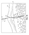
- FIG. 76 illustrates the surgical construct of FIG. 75 during tensioning, wherein the suture has been pulled so that the suture passes through itself.
- FIG. 77 illustrates the surgical construct of FIG. 76 after tensioning, wherein the suture has been pulled through itself to create a splice and the tissue has been pulled towards the bone.
- FIG. 78 illustrates a partial, cross-sectional view of a surgical construct according to another embodiment of the present invention (with a knotless tensionable anchor and a suture mechanism contained within the bone and outside of the body of the knotless tensionable anchor).
- the present invention provides surgical constructs, systems and techniques for knotless soft tissue repair and fixation, such as fixation of soft tissue (ligament, tendon, graft, etc.) to bone.
- the surgical constructs comprise fixation devices (tensionable knotless anchors) that are inserted into bone with a suture mechanism (tensionable construct) formed of a flexible strand (a suture) provided within the fixation device and a shuttle/pull device (a suture passing instrument) attached to the flexible strand.
- the flexible strand and the shuttle/pull device attached to it allow the formation of a splice within or outside the body of the anchor and during the tissue repair procedure (to finalize the construct).
- the shuttle/pull device is provided within the strand (inside of the strand) and forms the splice subsequent to the insertion of the fixation device within the bone (and subsequent to attachment to soft tissue to be repaired or fixated) to allow formation of the final fixation device with a knotless self-locking mechanism that allows the user (for example, the surgeon) to control the tension of the strand on the soft tissue to be attached to bone.
- At least one of the flexible strand and the shuttle/pull device may be made of any known suture material, such as ultrahigh molecular weight poly ethylene (UHMWPE) or the FiberWire ® suture (disclosed in U.S. Patent No. 6,716,234 .
- UHMWPE ultrahigh molecular weight poly ethylene
- FiberWire ® suture Dislosed in U.S. Patent No. 6,716,234 .
- the shuttle/pull device may be a shuttle/pull suture device such as a FiberLinkTM or a Nitinol loop.
- the present invention also provides methods of soft tissue repair which do not require tying of knots and allow adjustment of both the tension of the suture and the location of the tissue with respect to the bone.
- An exemplary method of the present invention comprises inter alia the steps of: (i) providing a surgical construct comprising a fixation device (for example, an anchor) with a flexible strand (for example, suture) and with a shuttle/pull device (a suture passing instrument) attached to the flexible strand; (ii) inserting the fixation device into bone; (iii) passing the flexible strand around or through tissue to be fixated (or reattached) to bone, and then through an eyelet/loop of the shuttle/pull device; (iv) subsequently, pulling on the shuttle/pull device to allow the flexible strand to pass through itself and to form a splice; and (v) pulling on the flexible strand to allow the soft tissue to achieve the desired location relative to the bone, and to allow proper tensioning of the final construct.
- the flexible strand may be passed through at least a portion of the body of the fixation device (for example, through a full cannulation of the fixation device, or through a transversal opening at a distal end of the fixation device).
- the flexible strand may be fixed to the fixation device (which may be solid or cannulated) by overmolding the suture to the anchor body or by compressing the suture against the bone (achieving an interference fit between the fixation device and the bone tunnel, compressing the flexible strand).
- the splice may be formed within the body of the fixation device or outside the body of the fixation device. Upon insertion into the bone and tensioning, the splice may reside within the body of the fixation device or outside the body of the fixation device (but within a bone tunnel).
- Another exemplary method of the present invention comprises inter alia the steps of: (i) providing a surgical construct comprising a fixation device (for example, an anchor) with a flexible strand (for example, suture) extending through the body of the fixation device and with a shuttle/pull device (a suture passing instrument) attached to the flexible strand; (ii) inserting the fixation device into bone; (iii) passing the flexible strand around or through tissue to be fixated (or reattached) to bone, and then through an eyelet/loop of the shuttle/pull device; (iv) subsequently, pulling on the shuttle/pull device to allow the flexible strand to pass through itself and to form a splice within the body of the fixation device (with the flexible strand passing through itself); and (v) pulling on the flexible strand to allow the soft tissue to achieve the desired location relative to the bone, and to allow proper tensioning of the final construct.
- a fixation device for example, an anchor
- a flexible strand for example, su
- a method of tissue repair comprises inter alia the steps of: (i) providing a surgical construct comprising a fixation device (for example, an anchor) with a flexible strand (for example, suture) fixed to the fixation device and with a shuttle/pull device (a suture passing instrument) attached to the flexible strand; (ii) inserting the fixation device into bone; (iii) passing the flexible strand around or through tissue to be fixated (or reattached) to bone, and then through an eyelet/loop of the shuttle/pull device; (iv) subsequently, pulling on the shuttle/pull device to allow the flexible strand to pass through itself and to form a splice outside the body of the fixation device (i.e., with the flexible strand passing through itself and the splice being located outside the body of the fixation device); and (v) pulling on the flexible strand to allow the soft tissue to achieve the desired location relative to the bone, and to allow proper tensioning of the final
- FIGS. 1-5 illustrate an exemplary fixation device 10 of the present invention employed to assemble surgical construct 100 ( FIG. 5 ).
- fixation device 10 is a tensionable knotless anchor having an anchor body 11 provided with a longitudinal axis 11a, a proximal end 13 and a distal end 12, and a plurality of ribs 15 extending circumferentially around it. Openings/channels 16 and 17 allow threading suture(s) and/or suture passing device(s) to pass around post 20, as detailed below.
- Cannulation 11b extends along the body 11 to allow passage of flexible strands and of suture passing devices, as detailed below.
- Cylindrical portion 14 is provided at the proximal end 13 of the anchor 10 and contains a socket 19 ( FIG. 2 ) configured to securely engage a tip of a driver.
- Openings/channels 16, 17 are positioned opposite to each other relative to the post 20 and also symmetrically located relative to the post 20, to allow flexible strand 30 (suture 30) and shuttle/pull device 40 (suture passing instrument 40) to pass and slide therethrough, as also detailed below. Openings/channels 16, 17 extend in a direction about perpendicular to the longitudinal axis 11a, and communicate through recesses 16a, 17a with the outer surfaces 11c of anchor body 11. Only recess 16a is shown in FIG. 1 (recess 17a is located on the opposite side of the recess 16a, i.e., on the anchor side facing away from the page).
- the position and size of the openings/channels 16, 17 and recesses 16a, 17a may be determined according to the characteristics of the flexible strand 30 and shuttle/pull device 40, and of the arthroscopic procedure, and the need to precisely orientate the anchor during insertion to optimize suture sliding characteristics.
- Anchor 10 may be a screw-in anchor or a push-in style anchor.
- Anchor 10 may be formed of metal, biocompatible plastic such as PEEK or a bioabsorbable PLLA material.
- Socket 19 at the distal end 13 of the anchor 10 is configured to securely engage a tip of a driver, as detailed below.
- the socket of the anchor 10 may have any shape adapted to receive a driver tip for pushing tap-in or screw-in style anchors.
- Tensionable knotless anchor 10 may be made of one or more pieces, or may be provided as an integrated device.
- FIGS. 2-5 illustrate the anchor 10 of FIG. 1 assembled with construct 99 (tensionable construct 99) formed of flexible strand or flexible material 30 (suture 30 or tie down suture 30) and shuttle/pull device 40 (suture passing instrument such as FiberLinkTM 40 or a nitinol loop 40) attached to the flexible strand 30.
- the flexible strand 30 is a suture strand 30
- the shuttle/pull device 40 is a suture passing device 40.
- Surgical construct 100 ( FIG. 2 ) comprises tensionable knotless anchor 10 provided with flexible strand 30 passing through the body of the tensionable knotless anchor 10 and with shuttle/pull device 40 attached to the flexible strand 30. Details on assembling the construct 100 of the present invention (i.e., integrated system, surgical construct or surgical system 100 consisting of anchor 10, suture 30 and suture passing device 40 attached to the suture 30) are set forth below with reference to FIGS. 31-72 .
- Suture 30, which is typically braided or multi-filament, is preloaded onto the anchor by tying static knot 31, which prevents suture 30 from passing through distal blind hole 12a.
- the suture may also be preloaded by insert molding or by any other means known in the art.
- Suture 30 passes around post 20, which is large enough to allow suture 30 to take gradual turns instead of sharp turns.
- Suture 30 then passes through cannulation 11b and proximal blind hole 13a.
- Tensionable knotless anchor 10 is loaded onto a driver (not shown in FIGS. 1-5 ), and suture 30 is tied to the driver (for example, wrapped around a cleft of the driver) to fasten tensionable knotless anchor 10 securely to the driver.
- suture passing device 40 Prior to the fastening of the anchor 10 to the driver, suture passing device 40 (for example, a FiberLinkTM or a nitinol loop) is threaded through suture 30 (i.e., attached to the suture 30 through splice region 39), as shown in FIG. 2 .
- Suture passing device 40 includes an eyelet/loop 44 for passing suture and, optionally, a pull-ring 41.
- Suture passing device 40 passes through an aperture of suture 30, located either proximal or distal to distal blind hole 12a. It then exits an aperture of suture 30, within the tensionable knotless anchor 10, traverses around post 20, and through proximal blind hole 13a.
- Tensionable knotless anchor 10 loaded with tensionable construct 99 formed of suture 30 attached to the suture passing device 40
- bone for example, into a hole/socket/tunnel formed in the bone
- FIG. 3 depicts the tensionable knotless anchor 10 after it has been inserted into a drilled hole in bone, the suture released from the driver, and the driver removed. Suture 30 is then passed through (or around) the tissue 50 which is to be reattached to bone. Suture 30 is subsequently passed through eyelet/loop 44 of the suture passing device 40. Suture passing device 40 is then pulled by ring 41, thereby pulling suture 30 towards tensionable knotless anchor 10.
- suture 30 has been further pulled towards tensionable knotless anchor 10 so that it doubles on itself inside tensionable knotless anchor 10.
- the suture passing device 40 has also been further pulled through the splice region of suture 30.
- FIG. 5 illustrates surgical construct 100 with suture 30 after it has been pulled through itself, creating splice 33.
- the suture passing device helps create splice 33 within tensionable knotless anchor 10 by facilitating suture 30 passing through itself.
- suture end 32 may be pulled until tissue 50 has been moved to the desired location, such as near a drilled hole in the bone. Once the desired tension and location is achieved, suture end 32 may be clipped off to complete the soft tissue repair or fixation.
- the surgical construct 100 with the knotless anchor 10 and tensionable construct 99 of the invention offers the following advantages:
- FIGS. 6-9 illustrate various views of another tensionable knotless anchor 210 of the present invention.
- Tensionable knotless anchor 210 is about similar to knotless anchor 10 described above with reference to FIGS. 1-5 in that it may be used with a tensionable construct (such as construct 99 described above), but differs in that the most distal end of tensionable knotless anchor 210 is provided with a cut slot 233 that allows loading of the flexible strand 30 and suture passing device 40 onto the anchor 210.
- Tensionable knotless anchor 210 is provided with anchor body 211 having longitudinal axis 211a, cannulation 211b, proximal end 213 and distal end 212.
- Openings 216 and 217 allow threading suture(s) and/or suture passing device(s) (not shown) around post 220.
- Cut slot 233 is provided at most distal end of the body 210, extending from the opening 217 to a most distal end surface 212b, as shown in FIG. 6 . Openings 216 and 217 are axially aligned with the cut slot 233 along longitudinal axis 211a, as shown in FIGS. 6, 7 and 9 .
- tensionable knotless anchor 210 is depicted in FIGS. 6, 7 and 9 as having ridges 215, and thus designed to be pushed into the bone, it could instead be fabricated with threads and thereby designed to be twisted or screwed into the bone.
- FIGS. 10-14 illustrate surgical system 100 of FIGS. 2-5 (with knotless tensionable anchor 10, 210, suture 30 and suture passing device 40 attached to the suture 30) employed in an exemplary method of tissue repair such as a Bankart or SLAP repair, wherein the knotless suture anchor (knotless SutureTakTM) simplifies arthroscopic glenohumeral joint instability repair by combining a proven and reproducible suture anchor insertion procedure with knotless soft tissue fixation.
- the knotless suture anchor knotless SutureTakTM
- FIG. 10 shows suture 30, preferably a UHMWPE suture, preloaded onto the anchor 10 by tying static knot 31, which prevents suture 30 from passing through distal blind hole 12a.
- Suture 30 is pre-attached to suture passing device 40 (for example, a FiberLinkTM or a Nitinol loop 40) which is threaded through suture 30 (as shown by spliced region 39 in FIG. 10 ).
- suture 30 is pre-loaded on anchor 10 which is loaded onto a driver (not shown in FIGS. 10-14 ).
- Suture 30 is tied to the driver (for example, wrapped around a cleft of the driver) to fasten tensionable knotless anchor 10 securely to the driver.
- the suture passing device 40 Prior to securing knotless anchor 10 to the driver, the suture passing device 40 is attached (threaded through splice 39) to the suture 30.
- the construct is inserted into bone, the suture 30 untied from the driver, and the driver removed.
- FIG. 10 depicts the tensionable knotless anchor 10 after it has been inserted into a drilled hole 88 in bone 80, the suture 30 released from the driver, and the driver removed. Suture 30 is passed through or around the tissue 50 which is to be reattached to bone 80.
- FIG. 11 depicts suture 30 passed around the tissue 50 and then threaded through eyelet/closed loop 44 of the suture passing device 40. Suture passing device 40 is pulled (as shown in FIG. 12 ), thereby pulling suture 30 towards tensionable knotless anchor 10.
- FIG. 13 suture 30 has been further pulled towards tensionable knotless anchor 10 so that it passed through itself inside tensionable knotless anchor 10.
- the suture passing device 40 has also been further pulled through suture 30.
- FIG. 13 illustrates surgical construct 100 with suture 30 after it has been pulled through itself, creating splice 33.
- the suture passing device 40 (not visible anymore in FIG. 13 as it has been completely pulled out of the suture 30) helps create splice 33 within tensionable knotless anchor 10 by facilitating suture 30 passing through itself.
- the suture end 32 may be pulled until tissue 50 has been moved to the desired location, such as near drilled hole 88 in the bone 80. Once the desired tension and location is achieved ( FIG. 14 ), suture end 32 may be clipped off to complete the soft tissue repair or fixation. In this manner, the suture 30 is shuttled and pulled (during the surgery) to a desired tension.
- FIG. 15 illustrates surgical system 100 (with tensionable knotless anchor 10 loaded with suture passing device 40 attached to the loaded suture 30) employed in a knotless simple stitch 300.
- FIG. 16 illustrates surgical system 100 (with tensionable knotless anchor 10 loaded with suture passing device 40 attached to the loaded suture 30) employed in a knotless mattress stitch 400.
- FIGS. 17-29 illustrate surgical system 100 of FIGS. 2-5 (with fixation device 10, 210, flexible strand 30 and shuttling/pulling device 40 attached to the flexible strand 30) employed in another exemplary method of soft tissue repair (a Bankart and SLAP repair).
- a guide and drill are used to create a pilot hole precisely on the glenoid rim and the suture anchor is inserted through the same guide maintaining the same portal and drill trajectory.
- the knotless self-locking suture function allows the user to control the tension of the suture on the soft tissue under direct visualization.
- FIGS. 17 and 18 an elevator is used to mobilize the labrum 50 on the glenoid 80.
- a bone socket is formed on the glenoid rim to allow subsequent insertion of surgical construct 100 of the present invention.
- an offset guide FIG. 18 ) may be employed to aid in the placement of the anchor onto the face of the glenoid.
- FIGS. 19 and 20 Surgical construct 100 is inserted into the socket in the glenoid by employing driver 90 (shown in Fig. 20 ). Suture 30 is released from the handle of the driver and the driver removed.
- FIGS. 21-24 illustrate the passing of the suture of the surgical construct 100 around the tissue by employing suture passing and retrieving instruments known in the art (for example, a KingFisher® Suture Retriever/Tissue Grasper instrument and a SutureLassoTM instrument).
- suture passing and retrieving instruments known in the art (for example, a KingFisher® Suture Retriever/Tissue Grasper instrument and a SutureLassoTM instrument).
- FIGS. 21 and 22 One limb of suture 30 is retrieved through the anterosuperior portal using a suture retrieval instrument 61 (for example, a KingFisher® Suture Retriever/Tissue Grasper 61).
- a curved SutureLassoTM instrument 62 is inserted into the anteroinferior cannula and passed through the capsulolabral tissue inferior to the anchor.
- Loop 63 (for example, a nitinol wire loop) is advanced into the joint. The loop is retrieved through the anterosuperior portal using the suture retrieval instrument 61.
- FIGS. 23 and 24 Suture 30 is loaded through the loop 63.
- the wire loop 63 is retracted through the SutureLassoTM instrument 62, to pull the suture to the distal end of the SutureLassoTM instrument 62 inside the joint.
- the SutureLassoTM instrument 62 and the wire loop are removed together to shuttle the suture 30 through the labral tissue 50.
- FIGS. 25 and 26 Suture 30 is passed through loop 44 of the suture passing device 40 ( FIG. 25 ). The nitinol wire loop 40 is pulled away from the surgical site, to allow the suture 30 to splice itself and form splice 33 within the body of the knotless tensionable anchor 10 of system 100 (as described above with reference to FIG. 5 , for example).
- FIGS. 27 and 28 The free end of suture 30 is pulled until the desired tension on the repair is achieved.
- a knot pusher may be used when applying tension on the repair to divert the force over the anchor and steer the tissue (labrum) 50 to the desired position.
- the suture is cut flush with a suture cutter instrument.
- FIG. 29 Final repair 500 is shown comprising a plurality of surgical constructs 100 of the present invention.
- FIG . 30 illustrates a kit 600 of the present invention including a surgical construct 100 of the present invention (for example, a 3mm knotless SutureTak) with a spinal needle, a 1.1mm Nitinol wire 40, a portal dilator and a SutureTak drill.
- a surgical construct 100 of the present invention for example, a 3mm knotless SutureTak
- spinal needle for example, a spinal needle
- 1.1mm Nitinol wire 40 1.1mm Nitinol wire 40
- portal dilator for example, a portal dilator and a SutureTak drill.
- FIGS. 31-72 illustrate subsequent steps of a method of assembling a surgical construct of the present invention such as surgical construct 100 of FIG. 5 (comprising a tensionable knotless anchor (knotless SutureTak) loaded with a suture and a suture passing device attached to the suture). Assembly instructions are provided below:
- FIG. 31 illustrates exemplary materials for the surgical construct 100: driver 90, suture anchor 10; nitinol wire 40 with closed loop 44; and UHMWPE braid 30.
- the suture component 30 is constructed from exemplary braided UHMWPE.
- FIG. 32 illustrates driver 90 assembled with suture anchor 10 of the present invention.
- FIG. 33 Tie an overhand knot 31 within few inches from one end of the braid 30.
- the sides of the knot 31 will be referred to as the short end and the long end (resembling the length of suture 30 on the particular side of the knot).
- FIG. 34 Pull knot 31 tight so that the knot will fit in hole of the anchor.
- FIG. 35 Optionally, place a small amount of bonding agent 31a on the knot 31.
- FIG. 36 Perform the next steps with a straight needle 45 with an attached nitinol loop 40.
- Any alternative suture passing device may be used as long as it allows the formation of the device 100 in FIG. 43 . Pierce the braid 30 with the needle 45 at a predetermined distance from the long end of the knot 31.
- FIG. 37 Advance the needle 45 through the center of the braid 30, taking care not to penetrate the sheath with the tip of the needle.
- FIG. 38 Allow needle 45 to exit the sheath 30 a distance from the knot 31. The needle must not be passed through glued portions of the braid 30.
- FIG. 39 Pass a small length of suture 71 through the open end 44 of the nitinol wire 40.
- FIG. 40 Pass both free ends of the suture 71 through the loop 45a on the needle 45 and fold the ends.
- FIG. 41 Advance the needle 45 through the sheath 30 so the folded suture ends are passed through the center of the braid 30.
- FIG. 42 Continue to pull the suture 71 through the braid 30 resulting in pulling the nitinol wire 40 through the center of the braid 30 as well.
- FIG. 43 Pull approximately half of the nitinol wire 40 through the braid splice 39. Ensure the shrink tube of the nitinol wire does not snag any portion of the splice 39.
- FIG. 44 Insert the long free end of the braid 30 into the side port of the anchor 10 that is on the same side as the cut slot (for example, cut slot 233).
- FIG. 45 Pull the braid 30 through the end hole.
- FIG. 46 Insert the non-looped end of the nitinol wire 40 through the same side port as the braid 30.
- FIG. 47 Pull the nitinol 40 and the braid 30 evenly through the anchor 10 so the splice passes through the side port. Pass the splice 39 until there is sufficient access to the through hole across the side ports.
- FIG. 48 Pass the looped end of the nitinol wire 40 through the side port access hole to the other side of the anchor. Pull until slack is removed.
- FIG. 49 Insert looped end of nitinol wire 40 back into side port and out the end hole.
- FIG. 50 Looped end of nitinol wire 40 should be on the opposite side of the post than the splice.
- FIG. 51 Feed wire to remove all slack within and around the anchor.
- FIG. 52 Pull short end of braid 30 and knot 31 and relocate it through the cut slot 233 of the anchor.
- FIG. 53 Pull long end of braid 30 to seat knot 31 within the counterbore of the tip. Cut the remainder of the short end.
- Nitinol wire 40 should pull freely in both directions through the anchor 10 and braid splice 39. Adjust wire so both ends are about even.
- FIG. 55 From the end of the suture tail, pinch the suture 30 and compress it, to loosen the yarns within the braid.
- FIG. 56 With a needle, separate one of the yarns.
- FIG. 57 Lightly pull some slack to form a small loop 30a.
- FIG. 58 Carefully pull the yarn from the direction of the free suture end out from the braided suture.
- the braid may wrinkle as a result.
- Limit the amount of wrinkling in the opposite direction of the free end by limiting pulling of the yarn from that direction.
- FIG. 59 Once the yarn is removed, the suture can be straightened out and smoothened, by pinching it with the finger and running it along the direction of the free end. This step is optional.
- FIG. 60 Using the needle, separate out a second yarn from the site of the first yarn.
- FIG. 61 Lightly pull some slack to form a small loop 30b.
- FIG. 62 Carefully pull the yarn from the direction of the free suture end out from the braided suture.
- the braid may wrinkle as a result.
- FIG. 63 Once the yarn is removed the suture can be straightened out and smoothened. The result should be similar to the picture with two loose yarns branching off from the larger.
- FIG. 64 The loose yarns shall be carefully trimmed close to the surface of the larger suture. The frayed edges should be pinched with the suture and brushed in the direction of the loose end, to limit how much it sticks out.
- step may be performed before smoothening the suture to facilitate blending the cut ends in.
- FIG. 65 Ensure there are no knots on the free end of the braid. Feed the free end of braid into the opening of the driver 90, until it can be pulled from the opposite side.
- FIG. 66 Insert both ends of the nitinol wire 40 into the opening of the driver 90.
- FIG. 67 Pull the slack of the braid 30 and the nitinol 40 so the anchor 10 seats in the counterbore of the driver 90.
- FIG. 68 Wrap the free end of the braid clockwise around keel 91 of the driver 90 once. Then pass it through the keel as shown.
- FIG. 69 Continue to pass the braid halfway around the keel counterclockwise.
- FIG. 70 Pass the braid back through the keel as shown.
- FIG. 71 Result should look as shown. A length of braid 30 should extend from keel 91. Trim excess braid (there should not be any tipped suture left on the end).
- FIG. 72 Completed final assembly.
- FIGS. 73-77 illustrate other exemplary embodiments of self-cinching tensionable knotless anchor 310, 310a of the present invention that allow for knotless soft tissue repairs.
- Tensionable knotless anchor 310, 310a has a new design that allows for a significantly smaller diameter anchor to be used (i.e., less than a 3mm anchor).
- This knotless anchor uses a mechanism similar to that of the SutureTakTM which is disclosed and described in U.S. Provisional Appl. No. 61/663,024 entitled "Tensionable Knotless Labral Anchor and Methods of Tissue Repair," filed on June 22, 2012.
- the final splice mechanism 221 ( Fig. 77 ) of surgical construct 200 ( FIG. 77 ) is located outside the anchor 310a but within the drill hole 88.
- the suture 30 does not travel around a post to lead into the splice (as in the previously-described embodiments) but rather passes through a cannulation of the anchor body and fixed to the anchor by knot 31.
- the anchor 310, 310a is significantly shorter in length and diameter.
- the final splice construct 221 ( Fig. 77 ) is contained in bone 80 (within bone socket or hole 88) but not within the anchor 310, 310a.
- Tensionable knotless anchor 310 of FIG. 73 is about similar to knotless anchors 10, 210 described above in that it may be used with a tensionable construct (such as construct 99 described above), but differs in that anchor body 311 of anchor 310 is very small (i.e., with outer diameter D of less than 3mm) and provided with only three exemplary ridges 315.
- Anchor body 311 is also provided with a longitudinal axis 311a, cannulation 311b, proximal end 313 and distal end 312. Opening 316 (located at the most proximal end) allows threading suture(s) and/or suture passing device(s) (not shown) around a post or similar structure (not shown) located within the body 311.
- Opening 316 extends along the longitudinal axis 311a, as shown in FIG. 73 and may have various geometries and configurations, for example, the rectangular shape shown in FIG. 73 (extending from one outer side of the anchor body to the diametrically-opposed outer side of the body).
- tensionable knotless anchor 310 is depicted in FIG. 73 as having ridges 315, and thus designed to be pushed into the bone, it could instead be fabricated with threads and thereby designed to be twisted or screwed into the bone.
- FIGS. 74-77 illustrate an exemplary method of anchoring surgical construct 200 of the present invention which includes tensionable anchor 310a assembled with construct 99 (tensionable construct 99) formed of flexible strand or flexible material 30 (suture 30 or tie down suture 30) and shuttle/pull device 40 (suture passing instrument such as FiberLinkTM 40 or a nitinol loop 40) attached to the flexible strand 30.
- Tensionable anchor 310a of FIGS. 74-77 is similar to tensionable anchor 310 of FIG. 73 but differs in that the anchor body is not provided with rectangular opening 316 and the flexible material does not pass around a post or a similar structure.
- the final splice 221 is located outside the anchor body of tensionable anchor 310a but within the bone tunnel or socket 88.
- An exemplary method of anchoring surgical construct 200 comprises the steps of:
- FIG. 74 Anchor 310a is implanted in stepped bone tunnel 88.
- the bone tunnel 88 may be larger than tunnel 88a where the anchor rests, to accommodate the suture splice construct.
- the anchor 310a is preloaded with splice making mechanism 221.
- FIG. 75 Similar to the previous design, the suture is passed around the tissue 50 and is loaded through the shuttling/pulling device 40 (Nitinol wire 40). Nitinol wire 40 is pulled to shuttle the suture 30.
- FIG. 76 Similar to the previous design, suture 30 is shuttled through itself to create a splice 221 with the nitinol loop 40. There is no “lead-in” from a post, but the suture can be tapered to help facilitate pulling it through.
- FIG. 77 Same as in the previous design, the suture 30 is pulled until the tissue 50 has been moved to the desired location relative to the bone 80, and the desired tension and location have been achieved. Tension makes mechanism work and suture 30 is trimmed.
- An exemplary method of tissue repair with surgical construct 200 comprises inter alia the steps of: (i) providing a surgical construct 99 comprising a fixation device 310a (for example, anchor) with a flexible strand 30 (for example, suture) fixed to the fixation device 310a (by knot 31, for example) and with a shuttle/pull device 40 (a suture passing instrument) attached to the flexible strand 30; (ii) inserting the fixation device 310 into bone; (iii) passing the flexible strand 30 around or through tissue to be fixated (or reattached) to bone, and then through an eyelet/loop of the shuttle/pull device 40; (iv) subsequently, pulling on the shuttle/pull device 40 to allow the flexible strand 30 to pass through itself and to form a splice 221 outside of the body of the fixation device (with the flexible strand passing through itself); and (v) pulling on the flexible strand to allow the soft tissue to
- FIG. 78 illustrates another exemplary embodiment of a surgical construct 200a according to yet another embodiment of the present invention.
- the surgical construct 200a comprises a knotless tensionable anchor 310b and a suture mechanism 499 (similar to tensionable construct 99) that is contained within the bone and outside of the body of the knotless tensionable anchor.
- the suture end is fixed to the anchor body (which may be solid or cannulated) by overmolding the suture to the anchor body or by compressing the suture against the bone (i.e., similarly to how a PushLock® anchor (disclosed in U.S. Patent No. 7,329,272 .
- the knotless tensionable anchor 310b of FIG. 78 is similar to the designs of the anchors 310, 310a of FIGS. 73-77 in that it is also a significantly smaller diameter anchor (i.e., less than a 3mm anchor) and uses a mechanism similar to that of the SutureTakTM which is disclosed and described in U.S. Provisional Appl. No. 61/663,024 entitled "Tensionable Knotless Labral Anchor and Methods of Tissue Repair,” filed on June 22, 2012.
- the suture mechanism 499 (formed of flexible strand 30 and shuttle/pull device 40 attached to the flexible strand 30) is not attached to the anchor by a knot, but rather it is affixed to the body of the anchor (fixed to it) by being trapped between the anchor and bone.
- the anchor body needs not be cannulated and could be instead a solid body (or a partially solid body).
- Tensionable anchor 310b of FIG. 78 is also provided with an anchor body 333 which is very small (i.e., with an outer D of less than 3mm) and with only three exemplary ridges 315.
- Anchor body 333 is also provided with a longitudinal axis 322, a proximal end 333a and distal end 333b. Opening 335 (located at the most distal end) allows threading suture(s)30 and/or suture passing device(s) 40 to pass therethrough and aid in the fixation of the suture 40 to the anchor body 333. Opening 335 may extend about perpendicular to the longitudinal axis 332 and may have a circular configuration, as shown in FIG. 78 , but may have other geometries and configurations, and may be located in other directions relative to the longitudinal axis of the anchor body.
- Anchor body 333 may be solid or cannulated.
- the suture mechanism Upon insertion into bone socket or tunnel 88 in bone 80, the suture mechanism forms the splice 221 similar to those formed in the above-described embodiments (i.e., with flexible strand 30 and shuttle/pull device 40 attached to the strand 30 and in a manner similar to the formation of the final constructs described above).
- splice 221 is contained within the bone 80 instead of the anchor body.
- suture 30 is fixed to anchor body 333 either by overmolding the suture to the anchor, or by compressing the suture against the walls of bone tunnel or socket 88 in a manner similar to how a PushLock® anchor (disclosed in U.S. Patent No. 7,329,272 ), fixes suture into a bone tunnel or socket.
- An exemplary method of tissue repair employing anchor 310b of FIG. 78 comprises inter alia the steps of: (i) providing a fixation device 310b (for example, an anchor) with a flexible strand 30 (for example, suture) fixed to the fixation device and with a shuttle/pull device 40 (a suture passing instrument) attached to the flexible strand 30; (ii) inserting the fixation device 310b into a tunnel 88a in bone 80; (iii) passing the flexible strand 30 around or through tissue to be fixated (or reattached) to bone, and then through an eyelet/loop of the shuttle/pull device 40; (iv) subsequently, pulling on the shuttle/pull device 40 to allow the flexible strand 30 to pass through itself and to form a splice 221 outside of the body of the fixation device 310b (with the flexible strand passing through itself) but within tunnel 88 of the bone 80; and (v) pulling on the flexible strand 30 to allow the soft tissue to achieve the desired location relative
- Anchor 310, 310a, 310b may be formed of metal, biocompatible plastic such as PEEK or a bioabsorbable PLLA material.
- the anchors may be provided with a socket at the distal end (such as socket 19 of the anchor 10) configured to securely engage a tip of a driver.
- the socket of the anchor 310, 310a, 310b may have any shape adapted to receive a driver tip for pushing the anchors, for example, tap-in or screw-in style anchors.
- Tensionable knotless anchor 310, 310a, 310b may be made of one or more pieces, or may be provided as an integrated device.
- the knotless suture constructs and systems of the present invention are used in conjunction with any knotless fixation devices which can allow a flexible strand and attached suture passing device to form a splice within the body of the fixation device.
- the fixation devices may be any of swivel and/or screw-in suture anchors and/or push-in suture anchors (such as an Arthrex SwiveLock® anchor, disclosed in U.S. Patent Application Publication No. 2008/0004659 or a PushLock® anchor, as disclosed in U.S. Patent No. 7,329,272 ).
- the fixation devices may be also any anchors, implants or screws (such as interference screws or tenodesis screws) or any fixation element that allows attachment/fixation of the knotless suture construct to bone.
- the fixation devices/implants may have various sizes (various diameters and/or lengths) and may be formed of biocompatible materials such as PEEK, biocomposite materials, metals and/or metal alloys, or combination of such materials, among others.
- the fixation devices may be unitary or may be multiple-piece constructs.
- the flexible strand 30 may be a high-strength suture, such as an ultrahigh molecular weight polyethylene (UHMWPE) suture which is the preferred material as this material allows easy splicing.
- the high strength suture may be a FiberWire® suture, which is disclosed and claimed in U.S. Pat. No. 6,716,234 .
- FiberWire® suture is formed of an advanced, high-strength fiber material, namely ultrahigh molecular weight polyethylene (UHMWPE), sold under the tradenames Spectra (Honeywell) and Dyneema (DSM) fibers, braided with at least one other fiber, natural or synthetic, to form lengths of suture material.
- the strands may also be formed of a stiff material, or combination of stiff and flexible materials, depending on the intended application.
- the strands may be also coated and/or provided in different colors.
- the knotless anchors of the present invention can be used with any type of flexible material or suture that forms a splice and a loop.
- the knotless suture constructs also include sutures that are spliced -- at least in part -- in a manner similar to an Arthrex ACL TightRope®, such as disclosed in U.S. Patent Application Publication Nos. 2010/0256677 and 2010/0268273 .
Abstract
Description
- The present invention relates to surgical devices and, in particular, to devices for repair or fixation of soft tissue to bone without the need for knots.
- When soft tissue such as a ligament or a tendon becomes detached from a bone, surgery is usually required to reattach or reconstruct the tissue. Often, a tissue graft is attached to the bone to facilitate regrowth and permanent attachment. Techniques and devices that have been developed generally involve tying the soft tissue with suture to an anchor or a hole provided in the bone tissue. Knotless suture anchors, such as the two piece Arthrex PushLock® anchor, disclosed in
U.S. Patent No. 7,329,272 , have been developed to facilitate tissue fixation to bone. - There is a need for a knotless anchor which has a design that allows tensioning of the suture as necessary and after insertion into bone. Also needed is a tensionable anchor that does not require tying of knots and allows adjustment of both the tension of the suture and the location of the tissue with respect to the bone.
- The present invention fulfills the above needs and objectives by providing a knotless, tensionable suture anchor. The suture anchor of the present invention has a configuration which allows the suture to be spliced and passed through itself within the suture anchor, to create a construct that is tensionable after insertion in bone (to allow attached tissue to be brought proximate to bone) and does not require tying of any knots.
- Other features and advantages of the present invention will become apparent from the following description of the invention.
-
FIG. 1 illustrates a tensionable knotless anchor according to an exemplary embodiment of the present invention. -
FIG. 2 is a cross-sectional view of a surgical construct according to an exemplary embodiment of the present invention (with the tensionable knotless anchor ofFIG. 1 , a suture and a suture passing device attached to the suture, before tensioning of the suture). -
FIG. 3 illustrates the surgical construct ofFIG. 2 with the suture threaded through the suture passing device. -
FIG. 4 illustrates the surgical construct ofFIG. 3 during tensioning, wherein the suture has been pulled so that the suture passes through itself. -
FIG. 5 illustrates the surgical construct ofFIG. 4 after tensioning, wherein the suture has been pulled through itself to create a splice and the tissue has been pulled towards the bone. -
FIG. 6 illustrates a partial cross-sectional, side view of a tensionable knotless anchor according to another embodiment of the present invention. -
FIG. 7 illustrates a cross-sectional view of the tensionable knotless anchor ofFIG. 6 (taken along line A-A'). -
FIG. 8 is a right side view of the tensionable knotless anchor ofFIG. 6 . -
FIG. 9 is another side view of the tensionable knotless anchor ofFIG. 6 . -
FIGS. 10-14 illustrate subsequent steps of a method of knotless SutureTak™ self-locking technology according to a method of the present invention (and with the surgical construct ofFIGS. 2-5 ). -
FIG. 15 illustrates the surgical construct of the present invention employed in a knotless simple stitch. -
FIG. 16 illustrates the surgical construct of the present invention employed in a knotless mattress stitch. -
FIGS. 17-30 illustrate subsequent steps of an exemplary method of tissue repair (rotator cuff repair) with a surgical construct of the present invention. -
FIGS. 31-72 illustrate subsequent steps of a method of assembling a surgical construct of the present invention (with a tensionable knotless anchor (knotless SutureTak), suture and suture passing device attached to the suture). -
FIG. 73 illustrates a tensionable knotless anchor according to another exemplary embodiment of the present invention. -
FIG. 74 is a cross-sectional view of a surgical construct according to another exemplary embodiment of the present invention (with the knotless tensionable anchor ofFIG. 73 and with a suture and a suture passing device attached to the suture, before tensioning of the suture). -
FIG. 75 illustrates the surgical construct ofFIG. 74 with the suture threaded through the suture passing device. -
FIG. 76 illustrates the surgical construct ofFIG. 75 during tensioning, wherein the suture has been pulled so that the suture passes through itself. -
FIG. 77 illustrates the surgical construct ofFIG. 76 after tensioning, wherein the suture has been pulled through itself to create a splice and the tissue has been pulled towards the bone. -
FIG. 78 illustrates a partial, cross-sectional view of a surgical construct according to another embodiment of the present invention (with a knotless tensionable anchor and a suture mechanism contained within the bone and outside of the body of the knotless tensionable anchor). - The present invention provides surgical constructs, systems and techniques for knotless soft tissue repair and fixation, such as fixation of soft tissue (ligament, tendon, graft, etc.) to bone. The surgical constructs comprise fixation devices (tensionable knotless anchors) that are inserted into bone with a suture mechanism (tensionable construct) formed of a flexible strand (a suture) provided within the fixation device and a shuttle/pull device (a suture passing instrument) attached to the flexible strand. The flexible strand and the shuttle/pull device attached to it allow the formation of a splice within or outside the body of the anchor and during the tissue repair procedure (to finalize the construct). The shuttle/pull device is provided within the strand (inside of the strand) and forms the splice subsequent to the insertion of the fixation device within the bone (and subsequent to attachment to soft tissue to be repaired or fixated) to allow formation of the final fixation device with a knotless self-locking mechanism that allows the user (for example, the surgeon) to control the tension of the strand on the soft tissue to be attached to bone.
- At least one of the flexible strand and the shuttle/pull device may be made of any known suture material, such as ultrahigh molecular weight poly ethylene (UHMWPE) or the FiberWire® suture (disclosed in
U.S. Patent No. 6,716,234 . Typically the suture will be UHWMPE suture without a core to permit ease of splicing. The shuttle/pull device may be a shuttle/pull suture device such as a FiberLink™ or a Nitinol loop. - The present invention also provides methods of soft tissue repair which do not require tying of knots and allow adjustment of both the tension of the suture and the location of the tissue with respect to the bone. An exemplary method of the present invention comprises inter alia the steps of: (i) providing a surgical construct comprising a fixation device (for example, an anchor) with a flexible strand (for example, suture) and with a shuttle/pull device (a suture passing instrument) attached to the flexible strand; (ii) inserting the fixation device into bone; (iii) passing the flexible strand around or through tissue to be fixated (or reattached) to bone, and then through an eyelet/loop of the shuttle/pull device; (iv) subsequently, pulling on the shuttle/pull device to allow the flexible strand to pass through itself and to form a splice; and (v) pulling on the flexible strand to allow the soft tissue to achieve the desired location relative to the bone, and to allow proper tensioning of the final construct.
- The flexible strand may be passed through at least a portion of the body of the fixation device (for example, through a full cannulation of the fixation device, or through a transversal opening at a distal end of the fixation device). Alternatively, the flexible strand may be fixed to the fixation device (which may be solid or cannulated) by overmolding the suture to the anchor body or by compressing the suture against the bone (achieving an interference fit between the fixation device and the bone tunnel, compressing the flexible strand). The splice may be formed within the body of the fixation device or outside the body of the fixation device. Upon insertion into the bone and tensioning, the splice may reside within the body of the fixation device or outside the body of the fixation device (but within a bone tunnel).
- Another exemplary method of the present invention comprises inter alia the steps of: (i) providing a surgical construct comprising a fixation device (for example, an anchor) with a flexible strand (for example, suture) extending through the body of the fixation device and with a shuttle/pull device (a suture passing instrument) attached to the flexible strand; (ii) inserting the fixation device into bone; (iii) passing the flexible strand around or through tissue to be fixated (or reattached) to bone, and then through an eyelet/loop of the shuttle/pull device; (iv) subsequently, pulling on the shuttle/pull device to allow the flexible strand to pass through itself and to form a splice within the body of the fixation device (with the flexible strand passing through itself); and (v) pulling on the flexible strand to allow the soft tissue to achieve the desired location relative to the bone, and to allow proper tensioning of the final construct.
- According to another exemplary method of the present invention, a method of tissue repair comprises inter alia the steps of: (i) providing a surgical construct comprising a fixation device (for example, an anchor) with a flexible strand (for example, suture) fixed to the fixation device and with a shuttle/pull device (a suture passing instrument) attached to the flexible strand; (ii) inserting the fixation device into bone; (iii) passing the flexible strand around or through tissue to be fixated (or reattached) to bone, and then through an eyelet/loop of the shuttle/pull device; (iv) subsequently, pulling on the shuttle/pull device to allow the flexible strand to pass through itself and to form a splice outside the body of the fixation device (i.e., with the flexible strand passing through itself and the splice being located outside the body of the fixation device); and (v) pulling on the flexible strand to allow the soft tissue to achieve the desired location relative to the bone, and to allow proper tensioning of the final construct.
- Referring now to the drawings, where like elements are designated by like reference numerals,
FIGS. 1-5 illustrate anexemplary fixation device 10 of the present invention employed to assemble surgical construct 100 (FIG. 5 ). In the particular exemplary embodiment illustrated inFIG. 1 ,fixation device 10 is a tensionable knotless anchor having ananchor body 11 provided with alongitudinal axis 11a, aproximal end 13 and adistal end 12, and a plurality ofribs 15 extending circumferentially around it. Openings/channels post 20, as detailed below.Cannulation 11b extends along thebody 11 to allow passage of flexible strands and of suture passing devices, as detailed below. -
Cylindrical portion 14 is provided at theproximal end 13 of theanchor 10 and contains a socket 19 (FIG. 2 ) configured to securely engage a tip of a driver. - Openings/
channels post 20 and also symmetrically located relative to thepost 20, to allow flexible strand 30 (suture 30) and shuttle/pull device 40 (suture passing instrument 40) to pass and slide therethrough, as also detailed below. Openings/channels longitudinal axis 11a, and communicate throughrecesses 16a, 17a with theouter surfaces 11c ofanchor body 11. Onlyrecess 16a is shown inFIG. 1 (recess 17a is located on the opposite side of therecess 16a, i.e., on the anchor side facing away from the page). The position and size of the openings/channels recesses 16a, 17a may be determined according to the characteristics of theflexible strand 30 and shuttle/pull device 40, and of the arthroscopic procedure, and the need to precisely orientate the anchor during insertion to optimize suture sliding characteristics. -
Anchor 10 may be a screw-in anchor or a push-in style anchor.Anchor 10 may be formed of metal, biocompatible plastic such as PEEK or a bioabsorbable PLLA material.Socket 19 at thedistal end 13 of theanchor 10 is configured to securely engage a tip of a driver, as detailed below. The socket of theanchor 10 may have any shape adapted to receive a driver tip for pushing tap-in or screw-in style anchors. Tensionableknotless anchor 10 may be made of one or more pieces, or may be provided as an integrated device. - Reference is now made to
FIGS. 2-5 which illustrate theanchor 10 ofFIG. 1 assembled with construct 99 (tensionable construct 99) formed of flexible strand or flexible material 30 (suture 30 or tie down suture 30) and shuttle/pull device 40 (suture passing instrument such asFiberLink™ 40 or a nitinol loop 40) attached to theflexible strand 30. In particular and exemplary-only embodiments, theflexible strand 30 is asuture strand 30 and the shuttle/pull device 40 is asuture passing device 40. Surgical construct 100 (FIG. 2 ) comprises tensionableknotless anchor 10 provided withflexible strand 30 passing through the body of the tensionableknotless anchor 10 and with shuttle/pull device 40 attached to theflexible strand 30. Details on assembling theconstruct 100 of the present invention (i.e., integrated system, surgical construct orsurgical system 100 consisting ofanchor 10,suture 30 andsuture passing device 40 attached to the suture 30) are set forth below with reference toFIGS. 31-72 . -
Suture 30, which is typically braided or multi-filament, is preloaded onto the anchor by tyingstatic knot 31, which preventssuture 30 from passing through distalblind hole 12a. The suture may also be preloaded by insert molding or by any other means known in the art.Suture 30 passes aroundpost 20, which is large enough to allowsuture 30 to take gradual turns instead of sharp turns.Suture 30 then passes throughcannulation 11b and proximal blind hole 13a.Tensionable knotless anchor 10 is loaded onto a driver (not shown inFIGS. 1-5 ), andsuture 30 is tied to the driver (for example, wrapped around a cleft of the driver) to fasten tensionableknotless anchor 10 securely to the driver. - Prior to the fastening of the
anchor 10 to the driver, suture passing device 40 (for example, a FiberLink™ or a nitinol loop) is threaded through suture 30 (i.e., attached to thesuture 30 through splice region 39), as shown inFIG. 2 . Suture passingdevice 40 includes an eyelet/loop 44 for passing suture and, optionally, a pull-ring 41. Suture passingdevice 40 passes through an aperture ofsuture 30, located either proximal or distal to distalblind hole 12a. It then exits an aperture ofsuture 30, within the tensionableknotless anchor 10, traverses aroundpost 20, and through proximal blind hole 13a.Tensionable knotless anchor 10 loaded with tensionable construct 99 (formed ofsuture 30 attached to the suture passing device 40) is then secured into bone (for example, into a hole/socket/tunnel formed in the bone) by using the driver. -
FIG. 3 depicts the tensionableknotless anchor 10 after it has been inserted into a drilled hole in bone, the suture released from the driver, and the driver removed.Suture 30 is then passed through (or around) thetissue 50 which is to be reattached to bone.Suture 30 is subsequently passed through eyelet/loop 44 of thesuture passing device 40. Suture passingdevice 40 is then pulled byring 41, thereby pullingsuture 30 towards tensionableknotless anchor 10. - In
FIG. 4 ,suture 30 has been further pulled towards tensionableknotless anchor 10 so that it doubles on itself inside tensionableknotless anchor 10. Thesuture passing device 40 has also been further pulled through the splice region ofsuture 30. -
FIG. 5 illustratessurgical construct 100 withsuture 30 after it has been pulled through itself, creatingsplice 33. Thus, the suture passing device (not visible) helps createsplice 33 within tensionableknotless anchor 10 by facilitatingsuture 30 passing through itself. Once thesuture 30 has been fully passed through itself,suture end 32 may be pulled untiltissue 50 has been moved to the desired location, such as near a drilled hole in the bone. Once the desired tension and location is achieved,suture end 32 may be clipped off to complete the soft tissue repair or fixation. - The
surgical construct 100 with theknotless anchor 10 and tensionable construct 99 of the invention offers the following advantages: - the tension and/or location of the tissue may be altered after the tensionable knotless anchor is implanted;
- no knots need to be tied in the suture during the repair or fixation procedure, which makes the procedure faster, easier, and less costly;
- there is no need to load the suture outside of the tensionable knotless anchor;
- the suture may be loaded or pre-loaded on the inside of the tensionable knotless anchor; and
- no additional fasteners need to be used.
-
FIGS. 6-9 illustrate various views of another tensionableknotless anchor 210 of the present invention. Tensionableknotless anchor 210 is about similar toknotless anchor 10 described above with reference toFIGS. 1-5 in that it may be used with a tensionable construct (such asconstruct 99 described above), but differs in that the most distal end of tensionableknotless anchor 210 is provided with acut slot 233 that allows loading of theflexible strand 30 andsuture passing device 40 onto theanchor 210. Tensionableknotless anchor 210 is provided withanchor body 211 havinglongitudinal axis 211a,cannulation 211b,proximal end 213 anddistal end 212.Openings post 220.Cut slot 233 is provided at most distal end of thebody 210, extending from theopening 217 to a mostdistal end surface 212b, as shown inFIG. 6 .Openings cut slot 233 alonglongitudinal axis 211a, as shown inFIGS. 6, 7 and 9 . - Although tensionable
knotless anchor 210 is depicted inFIGS. 6, 7 and 9 as havingridges 215, and thus designed to be pushed into the bone, it could instead be fabricated with threads and thereby designed to be twisted or screwed into the bone. -
FIGS. 10-14 illustratesurgical system 100 ofFIGS. 2-5 (with knotlesstensionable anchor suture 30 andsuture passing device 40 attached to the suture 30) employed in an exemplary method of tissue repair such as a Bankart or SLAP repair, wherein the knotless suture anchor (knotless SutureTak™) simplifies arthroscopic glenohumeral joint instability repair by combining a proven and reproducible suture anchor insertion procedure with knotless soft tissue fixation. -
FIG. 10 showssuture 30, preferably a UHMWPE suture, preloaded onto theanchor 10 by tyingstatic knot 31, which preventssuture 30 from passing through distalblind hole 12a.Suture 30 is pre-attached to suture passing device 40 (for example, a FiberLink™ or a Nitinol loop 40) which is threaded through suture 30 (as shown by splicedregion 39 inFIG. 10 ). As explained above,suture 30 is pre-loaded onanchor 10 which is loaded onto a driver (not shown inFIGS. 10-14 ).Suture 30 is tied to the driver (for example, wrapped around a cleft of the driver) to fasten tensionableknotless anchor 10 securely to the driver. Prior to securingknotless anchor 10 to the driver, thesuture passing device 40 is attached (threaded through splice 39) to thesuture 30. The construct is inserted into bone, thesuture 30 untied from the driver, and the driver removed. -
FIG. 10 depicts the tensionableknotless anchor 10 after it has been inserted into a drilledhole 88 inbone 80, thesuture 30 released from the driver, and the driver removed.Suture 30 is passed through or around thetissue 50 which is to be reattached tobone 80.FIG. 11 depictssuture 30 passed around thetissue 50 and then threaded through eyelet/closed loop 44 of thesuture passing device 40. Suture passingdevice 40 is pulled (as shown inFIG. 12 ), thereby pullingsuture 30 towards tensionableknotless anchor 10. - In
FIG. 13 ,suture 30 has been further pulled towards tensionableknotless anchor 10 so that it passed through itself inside tensionableknotless anchor 10. Thesuture passing device 40 has also been further pulled throughsuture 30.FIG. 13 illustratessurgical construct 100 withsuture 30 after it has been pulled through itself, creatingsplice 33. The suture passing device 40 (not visible anymore inFIG. 13 as it has been completely pulled out of the suture 30) helps createsplice 33 within tensionableknotless anchor 10 by facilitatingsuture 30 passing through itself. - Once the
suture 30 has been fully passed through itself, the suture end 32 (FIG. 13 ) may be pulled untiltissue 50 has been moved to the desired location, such as near drilledhole 88 in thebone 80. Once the desired tension and location is achieved (FIG. 14 ),suture end 32 may be clipped off to complete the soft tissue repair or fixation. In this manner, thesuture 30 is shuttled and pulled (during the surgery) to a desired tension. -
FIG. 15 illustrates surgical system 100 (with tensionableknotless anchor 10 loaded withsuture passing device 40 attached to the loaded suture 30) employed in a knotlesssimple stitch 300. -
FIG. 16 illustrates surgical system 100 (with tensionableknotless anchor 10 loaded withsuture passing device 40 attached to the loaded suture 30) employed in aknotless mattress stitch 400. -
FIGS. 17-29 illustratesurgical system 100 ofFIGS. 2-5 (withfixation device flexible strand 30 and shuttling/pullingdevice 40 attached to the flexible strand 30) employed in another exemplary method of soft tissue repair (a Bankart and SLAP repair). A guide and drill are used to create a pilot hole precisely on the glenoid rim and the suture anchor is inserted through the same guide maintaining the same portal and drill trajectory. The knotless self-locking suture function allows the user to control the tension of the suture on the soft tissue under direct visualization. -
FIGS. 17 and 18 : an elevator is used to mobilize thelabrum 50 on the glenoid 80. A bone socket is formed on the glenoid rim to allow subsequent insertion ofsurgical construct 100 of the present invention. If desired, an offset guide (FIG. 18 ) may be employed to aid in the placement of the anchor onto the face of the glenoid. -
FIGS. 19 and 20 :Surgical construct 100 is inserted into the socket in the glenoid by employing driver 90 (shown inFig. 20 ).Suture 30 is released from the handle of the driver and the driver removed. -
FIGS. 21-24 illustrate the passing of the suture of thesurgical construct 100 around the tissue by employing suture passing and retrieving instruments known in the art (for example, a KingFisher® Suture Retriever/Tissue Grasper instrument and a SutureLasso™ instrument). -
FIGS. 21 and 22 : One limb ofsuture 30 is retrieved through the anterosuperior portal using a suture retrieval instrument 61 (for example, a KingFisher® Suture Retriever/Tissue Grasper 61). A curvedSutureLasso™ instrument 62 is inserted into the anteroinferior cannula and passed through the capsulolabral tissue inferior to the anchor. Loop 63 (for example, a nitinol wire loop) is advanced into the joint. The loop is retrieved through the anterosuperior portal using thesuture retrieval instrument 61. -
FIGS. 23 and 24 :Suture 30 is loaded through theloop 63. Thewire loop 63 is retracted through theSutureLasso™ instrument 62, to pull the suture to the distal end of theSutureLasso™ instrument 62 inside the joint. TheSutureLasso™ instrument 62 and the wire loop are removed together to shuttle thesuture 30 through thelabral tissue 50. -
FIGS. 25 and 26 :Suture 30 is passed throughloop 44 of the suture passing device 40 (FIG. 25 ). Thenitinol wire loop 40 is pulled away from the surgical site, to allow thesuture 30 to splice itself and formsplice 33 within the body of the knotless tensionableanchor 10 of system 100 (as described above with reference toFIG. 5 , for example). -
FIGS. 27 and 28 : The free end ofsuture 30 is pulled until the desired tension on the repair is achieved. A knot pusher may be used when applying tension on the repair to divert the force over the anchor and steer the tissue (labrum) 50 to the desired position. The suture is cut flush with a suture cutter instrument. -
FIG. 29 :Final repair 500 is shown comprising a plurality ofsurgical constructs 100 of the present invention. -
FIG. 30 illustrates akit 600 of the present invention including asurgical construct 100 of the present invention (for example, a 3mm knotless SutureTak) with a spinal needle, a 1.1mm Nitinol wire 40, a portal dilator and a SutureTak drill. - Reference is now made to
FIGS. 31-72 , which illustrate subsequent steps of a method of assembling a surgical construct of the present invention such assurgical construct 100 ofFIG. 5 (comprising a tensionable knotless anchor (knotless SutureTak) loaded with a suture and a suture passing device attached to the suture). Assembly instructions are provided below: -
FIG. 31 illustrates exemplary materials for the surgical construct 100:driver 90,suture anchor 10;nitinol wire 40 with closedloop 44; andUHMWPE braid 30. Thesuture component 30 is constructed from exemplary braided UHMWPE. -
FIG. 32 illustratesdriver 90 assembled withsuture anchor 10 of the present invention. -
FIG. 33 : Tie anoverhand knot 31 within few inches from one end of thebraid 30. In further steps, the sides of theknot 31 will be referred to as the short end and the long end (resembling the length ofsuture 30 on the particular side of the knot). Preferably, there should be no tipped suture within the vicinity of the knot. -
FIG. 34 : Pullknot 31 tight so that the knot will fit in hole of the anchor. -
FIG. 35 : Optionally, place a small amount ofbonding agent 31a on theknot 31. -
FIG. 36 : Perform the next steps with astraight needle 45 with an attachednitinol loop 40. Any alternative suture passing device may be used as long as it allows the formation of thedevice 100 inFIG. 43 . Pierce thebraid 30 with theneedle 45 at a predetermined distance from the long end of theknot 31. -
FIG. 37 : Advance theneedle 45 through the center of thebraid 30, taking care not to penetrate the sheath with the tip of the needle. -
FIG. 38 : Allowneedle 45 to exit thesheath 30 a distance from theknot 31. The needle must not be passed through glued portions of thebraid 30. -
FIG. 39 : Pass a small length ofsuture 71 through theopen end 44 of thenitinol wire 40. -
FIG. 40 : Pass both free ends of thesuture 71 through theloop 45a on theneedle 45 and fold the ends. -
FIG. 41 : Advance theneedle 45 through thesheath 30 so the folded suture ends are passed through the center of thebraid 30. -
FIG. 42 : Continue to pull thesuture 71 through thebraid 30 resulting in pulling thenitinol wire 40 through the center of thebraid 30 as well. -
FIG. 43 : Pull approximately half of thenitinol wire 40 through thebraid splice 39. Ensure the shrink tube of the nitinol wire does not snag any portion of thesplice 39. -
FIG. 44 : Insert the long free end of thebraid 30 into the side port of theanchor 10 that is on the same side as the cut slot (for example, cut slot 233). -
FIG. 45 : Pull thebraid 30 through the end hole. -
FIG. 46 : Insert the non-looped end of thenitinol wire 40 through the same side port as thebraid 30. -
FIG. 47 : Pull thenitinol 40 and thebraid 30 evenly through theanchor 10 so the splice passes through the side port. Pass thesplice 39 until there is sufficient access to the through hole across the side ports. -
FIG. 48 : Pass the looped end of thenitinol wire 40 through the side port access hole to the other side of the anchor. Pull until slack is removed. -
FIG. 49 : Insert looped end ofnitinol wire 40 back into side port and out the end hole. -
FIG. 50 : Looped end ofnitinol wire 40 should be on the opposite side of the post than the splice. -
FIG. 51 : Feed wire to remove all slack within and around the anchor. -
FIG. 52 : Pull short end ofbraid 30 andknot 31 and relocate it through thecut slot 233 of the anchor. -
FIG. 53 : Pull long end ofbraid 30 toseat knot 31 within the counterbore of the tip. Cut the remainder of the short end. -
FIG. 54 :Nitinol wire 40 should pull freely in both directions through theanchor 10 andbraid splice 39. Adjust wire so both ends are about even. -
FIG. 55 : From the end of the suture tail, pinch thesuture 30 and compress it, to loosen the yarns within the braid. -
FIG. 56 : With a needle, separate one of the yarns. -
FIG. 57 : Lightly pull some slack to form asmall loop 30a. -
FIG. 58 : Carefully pull the yarn from the direction of the free suture end out from the braided suture. The braid may wrinkle as a result. Limit the amount of wrinkling in the opposite direction of the free end by limiting pulling of the yarn from that direction. -
FIG. 59 : Once the yarn is removed, the suture can be straightened out and smoothened, by pinching it with the finger and running it along the direction of the free end. This step is optional. -
FIG. 60 : Using the needle, separate out a second yarn from the site of the first yarn. -
FIG. 61 : Lightly pull some slack to form asmall loop 30b. -
FIG. 62 : Carefully pull the yarn from the direction of the free suture end out from the braided suture. The braid may wrinkle as a result. Limit the amount of wrinkling in the opposite direction of the free end, by limiting pulling of the yarn from that direction. -
FIG. 63 : Once the yarn is removed the suture can be straightened out and smoothened. The result should be similar to the picture with two loose yarns branching off from the larger. -
FIG. 64 : The loose yarns shall be carefully trimmed close to the surface of the larger suture. The frayed edges should be pinched with the suture and brushed in the direction of the loose end, to limit how much it sticks out. Optional: step may be performed before smoothening the suture to facilitate blending the cut ends in. -
FIG. 65 : Ensure there are no knots on the free end of the braid. Feed the free end of braid into the opening of thedriver 90, until it can be pulled from the opposite side. -
FIG. 66 : Insert both ends of thenitinol wire 40 into the opening of thedriver 90. -
FIG. 67 : Pull the slack of thebraid 30 and thenitinol 40 so theanchor 10 seats in the counterbore of thedriver 90. -
FIG. 68 : Wrap the free end of the braid clockwise aroundkeel 91 of thedriver 90 once. Then pass it through the keel as shown. -
FIG. 69 : Continue to pass the braid halfway around the keel counterclockwise. -
FIG. 70 : Pass the braid back through the keel as shown. -
FIG. 71 : Result should look as shown. A length ofbraid 30 should extend fromkeel 91. Trim excess braid (there should not be any tipped suture left on the end). -
FIG. 72 : Completed final assembly. -
FIGS. 73-77 illustrate other exemplary embodiments of self-cinchingtensionable knotless anchor knotless anchor U.S. Provisional Appl. No. 61/663,024 - The final splice mechanism 221 (
Fig. 77 ) of surgical construct 200 (FIG. 77 ) is located outside theanchor 310a but within thedrill hole 88. Thesuture 30 does not travel around a post to lead into the splice (as in the previously-described embodiments) but rather passes through a cannulation of the anchor body and fixed to the anchor byknot 31. Theanchor Fig. 77 ) is contained in bone 80 (within bone socket or hole 88) but not within theanchor - Tensionable
knotless anchor 310 ofFIG. 73 is about similar toknotless anchors construct 99 described above), but differs in thatanchor body 311 ofanchor 310 is very small (i.e., with outer diameter D of less than 3mm) and provided with only threeexemplary ridges 315.Anchor body 311 is also provided with alongitudinal axis 311a,cannulation 311b,proximal end 313 anddistal end 312. Opening 316 (located at the most proximal end) allows threading suture(s) and/or suture passing device(s) (not shown) around a post or similar structure (not shown) located within thebody 311.Opening 316 extends along thelongitudinal axis 311a, as shown inFIG. 73 and may have various geometries and configurations, for example, the rectangular shape shown inFIG. 73 (extending from one outer side of the anchor body to the diametrically-opposed outer side of the body). - Although tensionable
knotless anchor 310 is depicted inFIG. 73 as havingridges 315, and thus designed to be pushed into the bone, it could instead be fabricated with threads and thereby designed to be twisted or screwed into the bone. -
FIGS. 74-77 illustrate an exemplary method of anchoringsurgical construct 200 of the present invention which includestensionable anchor 310a assembled with construct 99 (tensionable construct 99) formed of flexible strand or flexible material 30 (suture 30 or tie down suture 30) and shuttle/pull device 40 (suture passing instrument such asFiberLink™ 40 or a nitinol loop 40) attached to theflexible strand 30.Tensionable anchor 310a ofFIGS. 74-77 is similar totensionable anchor 310 ofFIG. 73 but differs in that the anchor body is not provided withrectangular opening 316 and the flexible material does not pass around a post or a similar structure. - In this exemplary embodiment, and as shown in
FIGS. 74-77 , thefinal splice 221 is located outside the anchor body oftensionable anchor 310a but within the bone tunnel orsocket 88. An exemplary method of anchoringsurgical construct 200 comprises the steps of: -
FIG. 74 :Anchor 310a is implanted in steppedbone tunnel 88. Thebone tunnel 88 may be larger thantunnel 88a where the anchor rests, to accommodate the suture splice construct. Theanchor 310a is preloaded withsplice making mechanism 221. -
FIG. 75 : Similar to the previous design, the suture is passed around thetissue 50 and is loaded through the shuttling/pulling device 40 (Nitinol wire 40).Nitinol wire 40 is pulled to shuttle thesuture 30. -
FIG. 76 : Similar to the previous design,suture 30 is shuttled through itself to create asplice 221 with thenitinol loop 40. There is no "lead-in" from a post, but the suture can be tapered to help facilitate pulling it through. -
FIG. 77 : Same as in the previous design, thesuture 30 is pulled until thetissue 50 has been moved to the desired location relative to thebone 80, and the desired tension and location have been achieved. Tension makes mechanism work andsuture 30 is trimmed. - An exemplary method of tissue repair with surgical construct 200 (including
tensionable anchor flexible material 30 and passing device 40) comprises inter alia the steps of: (i) providing asurgical construct 99 comprising afixation device 310a (for example, anchor) with a flexible strand 30 (for example, suture) fixed to thefixation device 310a (byknot 31, for example) and with a shuttle/pull device 40 (a suture passing instrument) attached to theflexible strand 30; (ii) inserting thefixation device 310 into bone; (iii) passing theflexible strand 30 around or through tissue to be fixated (or reattached) to bone, and then through an eyelet/loop of the shuttle/pull device 40; (iv) subsequently, pulling on the shuttle/pull device 40 to allow theflexible strand 30 to pass through itself and to form asplice 221 outside of the body of the fixation device (with the flexible strand passing through itself); and (v) pulling on the flexible strand to allow the soft tissue to achieve the desired location relative to the bone, and to allow proper tensioning of the final construct. -
FIG. 78 illustrates another exemplary embodiment of asurgical construct 200a according to yet another embodiment of the present invention. Thesurgical construct 200a comprises a knotlesstensionable anchor 310b and a suture mechanism 499 (similar to tensionable construct 99) that is contained within the bone and outside of the body of the knotless tensionable anchor. According to this exemplary-only embodiment, the suture end is fixed to the anchor body (which may be solid or cannulated) by overmolding the suture to the anchor body or by compressing the suture against the bone (i.e., similarly to how a PushLock® anchor (disclosed inU.S. Patent No. 7,329,272 . - The knotless
tensionable anchor 310b ofFIG. 78 is similar to the designs of theanchors FIGS. 73-77 in that it is also a significantly smaller diameter anchor (i.e., less than a 3mm anchor) and uses a mechanism similar to that of the SutureTak™ which is disclosed and described inU.S. Provisional Appl. No. 61/663,024 flexible strand 30 and shuttle/pull device 40 attached to the flexible strand 30) is not attached to the anchor by a knot, but rather it is affixed to the body of the anchor (fixed to it) by being trapped between the anchor and bone. For this, the anchor body needs not be cannulated and could be instead a solid body (or a partially solid body). -
Tensionable anchor 310b ofFIG. 78 is also provided with ananchor body 333 which is very small (i.e., with an outer D of less than 3mm) and with only threeexemplary ridges 315.Anchor body 333 is also provided with alongitudinal axis 322, a proximal end 333a and distal end 333b. Opening 335 (located at the most distal end) allows threading suture(s)30 and/or suture passing device(s) 40 to pass therethrough and aid in the fixation of thesuture 40 to theanchor body 333. Opening 335 may extend about perpendicular to the longitudinal axis 332 and may have a circular configuration, as shown inFIG. 78 , but may have other geometries and configurations, and may be located in other directions relative to the longitudinal axis of the anchor body.Anchor body 333 may be solid or cannulated. - Upon insertion into bone socket or
tunnel 88 inbone 80, the suture mechanism forms thesplice 221 similar to those formed in the above-described embodiments (i.e., withflexible strand 30 and shuttle/pull device 40 attached to thestrand 30 and in a manner similar to the formation of the final constructs described above). However,splice 221 is contained within thebone 80 instead of the anchor body. As shown inFIG. 78 ,suture 30 is fixed to anchorbody 333 either by overmolding the suture to the anchor, or by compressing the suture against the walls of bone tunnel orsocket 88 in a manner similar to how a PushLock® anchor (disclosed inU.S. Patent No. 7,329,272 ), fixes suture into a bone tunnel or socket. - An exemplary method of tissue
repair employing anchor 310b ofFIG. 78 comprises inter alia the steps of: (i) providing afixation device 310b (for example, an anchor) with a flexible strand 30 (for example, suture) fixed to the fixation device and with a shuttle/pull device 40 (a suture passing instrument) attached to theflexible strand 30; (ii) inserting thefixation device 310b into atunnel 88a inbone 80; (iii) passing theflexible strand 30 around or through tissue to be fixated (or reattached) to bone, and then through an eyelet/loop of the shuttle/pull device 40; (iv) subsequently, pulling on the shuttle/pull device 40 to allow theflexible strand 30 to pass through itself and to form asplice 221 outside of the body of thefixation device 310b (with the flexible strand passing through itself) but withintunnel 88 of thebone 80; and (v) pulling on theflexible strand 30 to allow the soft tissue to achieve the desired location relative to the bone, and to allow proper tensioning of the final construct. -
Anchor socket 19 of the anchor 10) configured to securely engage a tip of a driver. The socket of theanchor knotless anchor - The knotless suture constructs and systems of the present invention are used in conjunction with any knotless fixation devices which can allow a flexible strand and attached suture passing device to form a splice within the body of the fixation device. The fixation devices may be any of swivel and/or screw-in suture anchors and/or push-in suture anchors (such as an Arthrex SwiveLock® anchor, disclosed in
U.S. Patent Application Publication No. 2008/0004659 or a PushLock® anchor, as disclosed inU.S. Patent No. 7,329,272 ). The fixation devices may be also any anchors, implants or screws (such as interference screws or tenodesis screws) or any fixation element that allows attachment/fixation of the knotless suture construct to bone. The fixation devices/implants may have various sizes (various diameters and/or lengths) and may be formed of biocompatible materials such as PEEK, biocomposite materials, metals and/or metal alloys, or combination of such materials, among others. The fixation devices may be unitary or may be multiple-piece constructs. - The
flexible strand 30 may be a high-strength suture, such as an ultrahigh molecular weight polyethylene (UHMWPE) suture which is the preferred material as this material allows easy splicing. Alternatively, the high strength suture may be a FiberWire® suture, which is disclosed and claimed inU.S. Pat. No. 6,716,234 . FiberWire® suture is formed of an advanced, high-strength fiber material, namely ultrahigh molecular weight polyethylene (UHMWPE), sold under the tradenames Spectra (Honeywell) and Dyneema (DSM) fibers, braided with at least one other fiber, natural or synthetic, to form lengths of suture material. - The strands may also be formed of a stiff material, or combination of stiff and flexible materials, depending on the intended application. The strands may be also coated and/or provided in different colors. The knotless anchors of the present invention can be used with any type of flexible material or suture that forms a splice and a loop.
- The knotless suture constructs also include sutures that are spliced -- at least in part -- in a manner similar to an Arthrex ACL TightRope®, such as disclosed in
U.S. Patent Application Publication Nos. 2010/0256677 and2010/0268273 . - While the present invention is described herein with reference to illustrative embodiments for particular applications, it should be understood that the invention is not limited thereto. Those having ordinary skill in the art and access to the teachings provided herein will recognize additional modifications, applications, embodiments and substitution of equivalents all fall within the scope of the invention. Accordingly, the invention is to be limited not by the specific disclosure herein, but only by the appended claims.
Claims (14)
- A surgical system for tissue repairs, comprising:a fixation device comprising a body, a longitudinal axis, a proximal end and a distal end; anda tensionable construct pre-loaded on the fixation device, the tensionable construct comprising a flexible strand and a shuttling device attached to the flexible strand.
- The surgical system of claim 1, wherein at least a portion of the body is cannulated and the tensionable construct extends through the body of the fixation device.
- The surgical system of claim 1, wherein the flexible construct is fixed to the body of the fixation device.
- The surgical system of claim 1, wherein the tensionable construct consists of the flexible strand and the shuttling device attached to the flexible strand
- The surgical system of claim 1, wherein the fixation device is an anchor with a post and a pair of openings symmetrically positioned relative to the post, the pair of openings extending in a direction about transversal to the longitudinal axis of the body, the pair of openings being configured to allow the tensionable construct to be passed through the body of the fixation device and around the post.
- The surgical system of claim 1, wherein the flexible strand is a suture formed of ultrahigh molecular weight polyethylene.
- The surgical system of claim 1, wherein the shuttling device is a suture passing instrument.
- The surgical system of claim 1, wherein the shuttling device is a nitinol loop.
- The surgical system of claim 1, wherein the body has an opening at a most proximal end configured to receive a driver head for driving the fixation device, and an opening at a most distal end configured to receive and house a knot of the flexible strand.
- The surgical system of claim 1, wherein the fixation device is an anchor with an anchor body provided with a plurality of circumferential ribs extending from an outer surface of the anchor body.
- The surgical system of claim 1, wherein the shuttling device is configured to be pulled out of the body of the fixation device to allow the flexible strand to pass through itself and form a splice within the body of the fixation device.
- The surgical system of claim 1, wherein the shuttling device is configured to be pulled out of the body of the fixation device to allow the flexible strand to pass through itself and form a splice outside the body of the fixation device.
- The surgical system of claim 1, wherein the body has an outer diameter less than about 3mm.
- The surgical system of claim 1, wherein the tissue is soft tissue.
Applications Claiming Priority (2)
| Application Number | Priority Date | Filing Date | Title |
|---|---|---|---|
| US201161537811P | 2011-09-22 | 2011-09-22 | |
| US201261663024P | 2012-06-22 | 2012-06-22 |
Publications (2)
| Publication Number | Publication Date |
|---|---|
| EP2572648A1 true EP2572648A1 (en) | 2013-03-27 |
| EP2572648B1 EP2572648B1 (en) | 2021-05-05 |
Family
ID=46888947
Family Applications (1)
| Application Number | Title | Priority Date | Filing Date |
|---|---|---|---|
| EP12185074.7A Active EP2572648B1 (en) | 2011-09-22 | 2012-09-19 | Tensionable knotless anchors with splice for tissue repair |
Country Status (2)
| Country | Link |
|---|---|
| US (2) | US9107653B2 (en) |
| EP (1) | EP2572648B1 (en) |
Cited By (17)
| Publication number | Priority date | Publication date | Assignee | Title |
|---|---|---|---|---|
| EP2724673A1 (en) * | 2012-10-26 | 2014-04-30 | Arthrex, Inc. | Systems for locking a cinch loop |
| EP2774546A1 (en) * | 2013-03-05 | 2014-09-10 | Arthrex, Inc. | Knotless tape/suture construct for fixation of tissue to bone |
| WO2014138398A1 (en) * | 2013-03-06 | 2014-09-12 | Smith & Nephew, Inc. | Microanchor |
| WO2014159058A1 (en) * | 2013-03-14 | 2014-10-02 | Kfx Medical Corporation | Tissue capturing bone anchor |
| WO2015017426A1 (en) * | 2013-07-29 | 2015-02-05 | Arthrex, Inc. | Suture anchor and methods of knotless tissue fixation |
| WO2015095524A3 (en) * | 2013-12-20 | 2015-08-27 | Arthrocare Corporation | Knotless all suture tissue repair |
| WO2016148941A1 (en) * | 2015-03-13 | 2016-09-22 | Smith & Nephew, Inc. | Microanchors for knotless tissue repair |
| WO2017031279A1 (en) * | 2015-08-20 | 2017-02-23 | Arthrex, Inc. | Tensionable constructs with multi-limb locking mechanism through single splice |
| US9687222B2 (en) | 2013-03-05 | 2017-06-27 | Arthrex, Inc. | Knotless repair technique using tape/suture hybrid |
| EP3235443A1 (en) * | 2016-04-20 | 2017-10-25 | Medos International S.à.r.l | Meniscal repair devices, systems, and methods |
| US9924939B1 (en) | 2017-07-06 | 2018-03-27 | Christian N. Anderson | Self-cinching suture construct apparatus |
| US9962149B2 (en) | 2011-11-14 | 2018-05-08 | Arthrocare Corporation | Tissue repair assembly |
| US10085740B1 (en) | 2017-07-06 | 2018-10-02 | Christian N. Anderson | Suture button construct for surgical procedures |
| US10265060B2 (en) | 2015-08-20 | 2019-04-23 | Arthrex, Inc. | Tensionable constructs with multi-limb locking mechanism through single splice and methods of tissue repair |
| US10893933B2 (en) | 2017-02-24 | 2021-01-19 | James Jastifer | Tissue anchors, kits, and associated methods |
| WO2021146593A3 (en) * | 2020-01-17 | 2021-08-26 | New York Society For The Relief Of The Ruptured And Crippled, Maintaining The Hospital For Special Surgery | Bone anchor |
| US11272921B2 (en) | 2017-07-06 | 2022-03-15 | Christian N. Anderson | Self-cinching suture construct apparatus |
Families Citing this family (92)
| Publication number | Priority date | Publication date | Assignee | Title |
|---|---|---|---|---|
| US6520964B2 (en) | 2000-05-01 | 2003-02-18 | Std Manufacturing, Inc. | System and method for joint resurface repair |
| US6610067B2 (en) | 2000-05-01 | 2003-08-26 | Arthrosurface, Incorporated | System and method for joint resurface repair |
| US8388624B2 (en) | 2003-02-24 | 2013-03-05 | Arthrosurface Incorporated | Trochlear resurfacing system and method |
| JP2008504107A (en) | 2004-06-28 | 2008-02-14 | アースロサーフィス・インコーポレーテッド | Joint surface replacement system |
| EP2136717B1 (en) | 2006-12-11 | 2013-10-16 | Arthrosurface Incorporated | Retrograde resection apparatus |
| WO2010121246A1 (en) | 2009-04-17 | 2010-10-21 | Arthrosurface Incorporated | Glenoid resurfacing system and method |
| US9662126B2 (en) | 2009-04-17 | 2017-05-30 | Arthrosurface Incorporated | Glenoid resurfacing system and method |
| WO2016154393A1 (en) | 2009-04-17 | 2016-09-29 | Arthrosurface Incorporated | Glenoid repair system and methods of use thereof |
| US8911474B2 (en) | 2009-07-16 | 2014-12-16 | Howmedica Osteonics Corp. | Suture anchor implantation instrumentation system |
| US11197663B2 (en) | 2009-07-17 | 2021-12-14 | Stryker Puerto Rico Limited | Method and apparatus for attaching tissue to bone, including the provision and use of a novel knotless suture anchor system |
| US9179905B2 (en) | 2009-07-17 | 2015-11-10 | Pivot Medical, Inc. | Method and apparatus for re-attaching the labrum to the acetabulum, including the provision and use of a novel suture anchor system |
| US10238379B2 (en) | 2009-07-17 | 2019-03-26 | Pivot Medical, Inc. | Method and apparatus for attaching tissue to bone, including the provision and use of a novel knotless suture anchor system |
| US9149268B2 (en) | 2009-07-17 | 2015-10-06 | Pivot Medical, Inc. | Method and apparatus for attaching tissue to bone, including the provision and use of a novel knotless suture anchor system |
| US10426456B2 (en) | 2009-07-17 | 2019-10-01 | Pivot Medical, Inc. | Method and apparatus for re-attaching the labrum to the acetabulum, including the provision and use of a novel suture anchor system |
| US9101355B2 (en) | 2009-07-17 | 2015-08-11 | Pivot Medical, Inc. | Method and apparatus for re-attaching the labrum to the acetabulum, including the provision and use of a novel suture anchor system |
| US10058319B2 (en) | 2009-07-17 | 2018-08-28 | Pivot Medical, Inc. | Method and apparatus for attaching tissue to bone, including the provision and use of a novel knotless suture anchor system, including a novel locking element |
| US11246585B2 (en) | 2009-07-17 | 2022-02-15 | Stryker Puerto Rico Limited | Method and apparatus for attaching tissue to bone, including the provision and use of a novel knotless suture anchor system |
| US10136884B2 (en) | 2009-07-17 | 2018-11-27 | Pivot Medical, Inc. | Method and apparatus for attaching tissue to bone, including the provision and use of a novel knotless suture anchor system, including a retractable sheath |
| US9232954B2 (en) | 2009-08-20 | 2016-01-12 | Howmedica Osteonics Corp. | Flexible ACL instrumentation, kit and method |
| CA2792048A1 (en) | 2010-03-05 | 2011-09-09 | Arthrosurface Incorporated | Tibial resurfacing system and method |
| CN105326533B (en) | 2010-09-24 | 2017-12-08 | 斯博特威尔丁股份有限公司 | Apparatus and method for suture holdfast to be fixed in sclerous tissues |
| JP6393477B2 (en) | 2010-09-24 | 2018-09-19 | スポートウェルディング・ゲゼルシャフト・ミット・ベシュレンクテル・ハフツングSportwelding Gmbh | Suture anchor and method for securing a suture to hard tissue |
| US9386976B2 (en) | 2011-01-28 | 2016-07-12 | Sportwelding Gmbh | Method and device for fixating a suture anchor with a suture in hard tissue |
| KR101998429B1 (en) | 2011-01-28 | 2019-07-09 | 스포트벨딩 게엠베하 | Device and method for fixating a suture anchor with a suture or a headed anchor in hard tissue |
| US11116498B2 (en) | 2011-02-02 | 2021-09-14 | Syntorr Inc. | Variable denier yarn and suture |
| US9782165B2 (en) | 2011-11-11 | 2017-10-10 | VentureMD Innovations, LLC | Transosseous attachment |
| US10675014B2 (en) | 2011-11-16 | 2020-06-09 | Crossroads Extremity Systems, Llc | Knotless soft tissue attachment |
| US10548585B2 (en) | 2011-11-16 | 2020-02-04 | VentureMD Innovations, LLC | Soft tissue attachment |
| US10470756B2 (en) | 2011-11-16 | 2019-11-12 | VentureMD Innovations, LLC | Suture anchor and method |
| US10136883B2 (en) | 2011-11-16 | 2018-11-27 | VentureMD Innovations, LLC | Method of anchoring a suture |
| US9445803B2 (en) | 2011-11-23 | 2016-09-20 | Howmedica Osteonics Corp. | Filamentary suture anchor |
| WO2013096746A1 (en) | 2011-12-22 | 2013-06-27 | Arthrosurface Incorporated | System and method for bone fixation |
| US9808242B2 (en) | 2012-04-06 | 2017-11-07 | Howmedica Osteonics Corp. | Knotless filament anchor for soft tissue repair |
| US9345471B2 (en) | 2012-06-22 | 2016-05-24 | Arthrex, Inc. | Tensionable knotless labral anchor and methods of tissue repair |
| WO2014008126A1 (en) | 2012-07-03 | 2014-01-09 | Arthrosurface Incorporated | System and method for joint resurfacing and repair |
| US20150272567A1 (en) | 2012-08-03 | 2015-10-01 | Stabilynx, Inc. | Devices, systems, and methods for attaching soft tissue to bone tissue |
| US9861353B2 (en) | 2012-08-03 | 2018-01-09 | Stabilynx, Inc. | Devices, systems, and methods for attaching soft tissue to bone tissue |
| US8821494B2 (en) | 2012-08-03 | 2014-09-02 | Howmedica Osteonics Corp. | Surgical instruments and methods of use |
| US9737294B2 (en) * | 2013-01-28 | 2017-08-22 | Cartiva, Inc. | Method and system for orthopedic repair |
| AU2014209124A1 (en) | 2013-01-28 | 2015-09-17 | Cartiva, Inc. | Systems and methods for orthopedic repair |
| US9687221B2 (en) | 2013-02-13 | 2017-06-27 | Venture MD Innovations, LLC | Method of anchoring a suture |
| US9402620B2 (en) | 2013-03-04 | 2016-08-02 | Howmedica Osteonics Corp. | Knotless filamentary fixation devices, assemblies and systems and methods of assembly and use |
| US9788826B2 (en) | 2013-03-11 | 2017-10-17 | Howmedica Osteonics Corp. | Filamentary fixation device and assembly and method of assembly, manufacture and use |
| US9463013B2 (en) | 2013-03-13 | 2016-10-11 | Stryker Corporation | Adjustable continuous filament structure and method of manufacture and use |
| US9492200B2 (en) | 2013-04-16 | 2016-11-15 | Arthrosurface Incorporated | Suture system and method |
| US10292694B2 (en) | 2013-04-22 | 2019-05-21 | Pivot Medical, Inc. | Method and apparatus for attaching tissue to bone |
| US10610211B2 (en) | 2013-12-12 | 2020-04-07 | Howmedica Osteonics Corp. | Filament engagement system and methods of use |
| AU2014362199B2 (en) | 2013-12-12 | 2019-07-11 | Stryker Puerto Rico Limited | Method and apparatus for attaching tissue to bone, including the provision and use of a novel knotless suture anchor system |
| US9693765B2 (en) * | 2014-01-27 | 2017-07-04 | Arthrex, Inc. | Surgical assembly for rotator cuff repair |
| US9931219B2 (en) | 2014-03-07 | 2018-04-03 | Arthrosurface Incorporated | Implant and anchor assembly |
| US11607319B2 (en) | 2014-03-07 | 2023-03-21 | Arthrosurface Incorporated | System and method for repairing articular surfaces |
| US10624748B2 (en) | 2014-03-07 | 2020-04-21 | Arthrosurface Incorporated | System and method for repairing articular surfaces |
| US9993332B2 (en) | 2014-07-09 | 2018-06-12 | Medos International Sarl | Systems and methods for ligament graft preparation |
| WO2016044053A1 (en) * | 2014-09-19 | 2016-03-24 | Crossroads Extremity Systems, Llc | Bone fixation implant and means of fixation |
| US9986992B2 (en) | 2014-10-28 | 2018-06-05 | Stryker Corporation | Suture anchor and associated methods of use |
| US10779811B2 (en) | 2014-12-11 | 2020-09-22 | Smith & Nephew, Inc. | Bone anchor having improved fixation strength |
| US10568616B2 (en) | 2014-12-17 | 2020-02-25 | Howmedica Osteonics Corp. | Instruments and methods of soft tissue fixation |
| US10512457B2 (en) * | 2015-01-16 | 2019-12-24 | Boston Scientific Scimed, Inc. | Delivery tools for medical implants and methods of using the same |
| US11684358B2 (en) | 2015-04-20 | 2023-06-27 | Boston Scientific Scimed, Inc. | Fusible biodegradable sutures utilizing tissue soldering technology |
| US10182808B2 (en) | 2015-04-23 | 2019-01-22 | DePuy Synthes Products, Inc. | Knotless suture anchor guide |
| US10820918B2 (en) | 2015-07-17 | 2020-11-03 | Crossroads Extremity Systems, Llc | Transosseous guide and method |
| US10258401B2 (en) | 2015-07-17 | 2019-04-16 | Kator, Llc | Transosseous guide |
| US9962174B2 (en) | 2015-07-17 | 2018-05-08 | Kator, Llc | Transosseous method |
| US10226243B2 (en) | 2015-08-04 | 2019-03-12 | Kator, Llc | Transosseous suture anchor |
| US10130354B2 (en) | 2015-08-17 | 2018-11-20 | Arthrex, Inc. | Limited sliding suture in suture anchor |
| US10368855B2 (en) * | 2015-10-20 | 2019-08-06 | Arthrex, Inc. | Surgical constructs for tissue repair |
| US10548586B2 (en) * | 2015-11-04 | 2020-02-04 | Zimmer, Inc. | Fixation devices methods and systems for soft tissue repair |
| US10383720B2 (en) | 2015-12-22 | 2019-08-20 | DePuy Synthes Products, Inc. | Graft preparation system |
| US10398426B2 (en) | 2016-01-06 | 2019-09-03 | Arthrex, Inc. | Point-loading knotless fixation devices |
| US11642120B2 (en) * | 2016-01-22 | 2023-05-09 | Arthrex, Inc. | Tensionable knotless anchors and methods of tissue repair |
| US10631845B2 (en) | 2016-01-22 | 2020-04-28 | Arthrex, Inc. | Tensionable knotless anchors and methods of tissue repair |
| US10172606B2 (en) * | 2016-01-22 | 2019-01-08 | Arthrex, Inc. | Tensionable knotless anchors and methods of tissue repair |
| US11684467B2 (en) | 2016-11-07 | 2023-06-27 | Acuitive Technologies, Inc. | In-situ curing biodegradable anchor with reinforcement |
| US10646327B2 (en) | 2017-02-09 | 2020-05-12 | Arthrex, Inc. | Self-locking suture constructs and methods of tissue fixation |
| US10575842B2 (en) * | 2017-02-09 | 2020-03-03 | Arthrex, Inc. | Knotless self-locking anchor constructs and methods of tissue fixation |
| US10603028B2 (en) | 2017-06-14 | 2020-03-31 | Medos International Sarl | Finger traps for collapsible suture loops |
| US10646215B2 (en) * | 2017-07-21 | 2020-05-12 | Arthrex, Inc. | Suture anchor systems with spring loaded suture anchors |
| US10786236B2 (en) * | 2017-08-04 | 2020-09-29 | Tigon Medical | Knotless anchor |
| US11160663B2 (en) | 2017-08-04 | 2021-11-02 | Arthrosurface Incorporated | Multicomponent articular surface implant |
| USD902405S1 (en) | 2018-02-22 | 2020-11-17 | Stryker Corporation | Self-punching bone anchor inserter |
| US10849734B2 (en) * | 2018-08-16 | 2020-12-01 | Arthrex, Inc. | Methods of tissue repairs |
| GB2616360B (en) | 2019-03-12 | 2023-11-29 | Arthrosurface Inc | Humeral and glenoid articular surface implant systems and methods |
| JP7375050B2 (en) | 2019-07-03 | 2023-11-07 | ボストン サイエンティフィック サイムド,インコーポレイテッド | Devices, systems, and methods for adjustable tensioning of artificial chordae tendineae between valve leaflets and papillary muscles or cardiac wall |
| US11576666B2 (en) * | 2019-10-04 | 2023-02-14 | Arthrex, Inc | Surgical constructs for tissue fixation and methods of tissue repairs |
| JP2022551330A (en) | 2019-10-14 | 2022-12-08 | リース メディカル, エルエルシー | Bone fixation system with fasteners and removal tools for decoupling the fasteners |
| US11826038B2 (en) * | 2019-10-23 | 2023-11-28 | Biomet Manufacturing, Llc | Suture anchor construct |
| US11337687B2 (en) | 2020-03-26 | 2022-05-24 | Medos International Sarl | Double row collapsible suture construct |
| WO2022056167A1 (en) * | 2020-09-09 | 2022-03-17 | Suture Concepts Inc. | Method and apparatus for closing a fissure in an intervertebral disc and/or other anatomical repairs |
| US20220167962A1 (en) * | 2020-12-01 | 2022-06-02 | Dunamis Medical Technologies, Llc | Re-Tensionable Suture Anchor System and Related Methods |
| US11375995B1 (en) * | 2021-04-08 | 2022-07-05 | Integrity Orthopaedics, Inc. | Locking suture construct for tensioned suture to suture stitches in anchor arrays for attaching soft tissue to bone |
| US11344290B1 (en) * | 2021-12-16 | 2022-05-31 | Christopher Ninh | Knotless labral repair techniques and related devices |
| US20230338019A1 (en) * | 2022-04-20 | 2023-10-26 | Arthrex, Inc. | Suture tensioner and methods of tensioning |
Citations (15)
| Publication number | Priority date | Publication date | Assignee | Title |
|---|---|---|---|---|
| US5156616A (en) * | 1992-02-10 | 1992-10-20 | Meadows Bruce F | Apparatus and method for suture attachment |
| US5534011A (en) * | 1994-10-27 | 1996-07-09 | Vesica Medical, Inc. | Method and apparatus for threading a suture anchor |
| WO2003022161A1 (en) * | 2001-09-07 | 2003-03-20 | Opus Medical, Inc. | Single-tailed suturing method and apparatus |
| US6716234B2 (en) | 2001-09-13 | 2004-04-06 | Arthrex, Inc. | High strength suture material |
| WO2006037131A2 (en) * | 2004-09-28 | 2006-04-06 | Surgical Solutions Llc | Suture anchor |
| US20070203508A1 (en) * | 2006-02-28 | 2007-08-30 | Arthrocare Corporation | Bone anchor suture-loading system, method and apparatus |
| WO2007109769A1 (en) * | 2006-03-22 | 2007-09-27 | C2M Medical, Inc. | Suture passer devices and uses thereof |
| US20080004659A1 (en) | 2006-05-18 | 2008-01-03 | Arthrex, Inc. | Swivel anchor and method for knotless fixation of tissue |
| US7329272B2 (en) | 2000-06-22 | 2008-02-12 | Arthrex, Inc. | Graft fixation using a plug against suture |
| US20080140092A1 (en) * | 2006-02-03 | 2008-06-12 | Stone Kevin T | Soft tissue repair device and associated methods |
| US20090082805A1 (en) * | 2006-09-29 | 2009-03-26 | Biomet Sports Medicine, Llc | Adjustable knotless loops |
| US20090312776A1 (en) * | 2006-02-03 | 2009-12-17 | Biomet Sports Medicine, Llc | Method and Apparatus for Coupling Soft Tissue to a Bone |
| US20100256677A1 (en) | 2009-03-31 | 2010-10-07 | Arthrex, Inc. | Integrated adjustable button-suture-graft construct with two fixation devices |
| US20100268273A1 (en) | 2009-03-31 | 2010-10-21 | Ricardo Albertorio | Adjustable suture button construct and methods of tissue reconstruction |
| US20110071545A1 (en) * | 2009-07-17 | 2011-03-24 | Chris Pamichev | Method and apparatus for re-attaching the labrum to the acetabulum, including the provision and use of a novel suture anchor system |
Family Cites Families (222)
| Publication number | Priority date | Publication date | Assignee | Title |
|---|---|---|---|---|
| US330087A (en) * | 1885-11-10 | Endless band or cord | ||
| US2698986A (en) * | 1951-03-08 | 1955-01-11 | Shepherd F Brown | Fishing line and process of making loops therein |
| US3176316A (en) | 1963-01-07 | 1965-04-06 | Bruce R Bodell | Plastic prosthetic tendon |
| US4099750A (en) * | 1977-09-02 | 1978-07-11 | Mcgrew James D | Method of forming eye splice in double braided line |
| US4187558A (en) | 1977-10-25 | 1980-02-12 | Cutter Laboratories, Inc. | Prosthetic ligament |
| CA1112401A (en) | 1979-05-24 | 1981-11-17 | Roland Dore | Deformable high energy storage tension spring |
| GB2084468B (en) | 1980-09-25 | 1984-06-06 | South African Inventions | Surgical implant |
| US4400833A (en) | 1981-06-10 | 1983-08-30 | Kurland Kenneth Z | Means and method of implanting bioprosthetics |
| US4792336A (en) | 1986-03-03 | 1988-12-20 | American Cyanamid Company | Flat braided ligament or tendon implant device having texturized yarns |
| US4731084A (en) | 1986-03-14 | 1988-03-15 | Richards Medical Company | Prosthetic ligament |
| US4776851A (en) | 1986-07-23 | 1988-10-11 | Bruchman William C | Mechanical ligament |
| GB8622563D0 (en) | 1986-09-19 | 1986-10-22 | Amis A A | Artificial ligaments |
| CH671686A5 (en) | 1987-01-07 | 1989-09-29 | Sulzer Ag | |
| US5263984A (en) | 1987-07-20 | 1993-11-23 | Regen Biologics, Inc. | Prosthetic ligaments |
| US5026398A (en) | 1988-07-01 | 1991-06-25 | The Minnesota Mining And Manufacturing Company | Abrasion resistant prosthetic device |
| US4917700A (en) | 1988-08-01 | 1990-04-17 | Zimmer, Inc. | Prosthetic ligament |
| US5024669A (en) | 1988-09-09 | 1991-06-18 | Baxter International Inc. | Artificial ligament of differential weave structure |
| US5217495A (en) | 1989-05-10 | 1993-06-08 | United States Surgical Corporation | Synthetic semiabsorbable composite yarn |
| US5139520A (en) | 1990-01-31 | 1992-08-18 | American Cyanamid Company | Method for acl reconstruction |
| US5062344A (en) * | 1990-04-12 | 1991-11-05 | Sparton Corporation | Bowline knot in a braided line |
| US5129902A (en) | 1990-04-20 | 1992-07-14 | Marlowe Goble E | Endosteal ligament retainer and process |
| US5258016A (en) * | 1990-07-13 | 1993-11-02 | American Cyanamid Company | Suture anchor and driver assembly |
| DE59105371D1 (en) | 1990-09-07 | 1995-06-08 | Sulzer Medizinaltechnik Ag | Implant used as a ligament or tendon replacement. |
| US5562669A (en) | 1994-01-13 | 1996-10-08 | Mcguire; David A. | Cruciate ligament reconstruction with tibial drill guide |
| US5320626A (en) | 1992-02-19 | 1994-06-14 | Arthrex Inc. | Endoscopic drill guide |
| US5211647A (en) | 1992-02-19 | 1993-05-18 | Arthrex Inc. | Interference screw and cannulated sheath for endosteal fixation of ligaments |
| US5250053A (en) * | 1992-05-29 | 1993-10-05 | Linvatec Corporation | Suture shuttle device |
| US5266075A (en) | 1992-10-05 | 1993-11-30 | Roy Clark | Tendon threader for endosteal ligament mounting |
| US5540703A (en) | 1993-01-06 | 1996-07-30 | Smith & Nephew Richards Inc. | Knotted cable attachment apparatus formed of braided polymeric fibers |
| US5306301A (en) | 1993-02-11 | 1994-04-26 | American Cyanamid Company | Graft attachment device and method of using same |
| US5397357A (en) | 1993-02-18 | 1995-03-14 | Arthrex, Inc. | Method for preparing a bone-tendon-bone core graft |
| US5632748A (en) | 1993-06-14 | 1997-05-27 | Linvatec Corporation | Endosteal anchoring device for urging a ligament against a bone surface |
| US5517542A (en) | 1995-03-06 | 1996-05-14 | Thomson Consumer Electronics, S.A. | Shift register with a transistor operating in a low duty cycle |
| US5643266A (en) | 1995-06-05 | 1997-07-01 | Li Medical Technologies, Inc. | Method and apparatus for securing ligaments |
| DE19616122C1 (en) | 1996-04-23 | 1997-08-14 | Aesculap Ag | Implant for fixture of tendon replacement plastic in channel in knee area of tibia |
| US5699657A (en) * | 1996-05-23 | 1997-12-23 | Paulson; William Thomas | Braided line splices and methods of splicing to form same |
| WO1998011839A1 (en) | 1996-09-20 | 1998-03-26 | Medicinelodge, Inc. | Adjustable length strap and footing for ligament mounting and method for its use |
| GB9620046D0 (en) | 1996-09-26 | 1996-11-13 | Neoligaments | Attachment device for use in the implantation of prosthetic ligament |
| US5931869A (en) | 1997-07-23 | 1999-08-03 | Arthrotek, Inc. | Apparatus and method for tibial fixation of soft tissue |
| US6159234A (en) | 1997-08-01 | 2000-12-12 | Peter M. Bonutti | Method and apparatus for securing a suture |
| AUPP000797A0 (en) | 1997-10-24 | 1997-11-20 | Cryptych Pty Ltd | Fixation of cruciate ligament grafts |
| US6099568A (en) | 1998-03-03 | 2000-08-08 | Linvatec Corporation | ACL graft fixation device and method |
| US5964764A (en) | 1998-03-24 | 1999-10-12 | Hugh S. West, Jr. | Apparatus and methods for mounting a ligament graft to a bone |
| US6099530A (en) | 1998-04-09 | 2000-08-08 | Smith & Nephew, Inc. | Soft-tissue intra-tunnel fixation device |
| US6203572B1 (en) | 1999-02-09 | 2001-03-20 | Linvatec Corporation | Device and method for ligament reconstruction |
| DE29910202U1 (en) | 1999-06-11 | 1999-08-05 | Aesculap Ag & Co Kg | Packing material for narrowing the cross section of a bone canal |
| US6499486B1 (en) | 1999-07-29 | 2002-12-31 | Ethicon, Inc. | Method for reconstructing a ligament |
| US6517542B1 (en) * | 1999-08-04 | 2003-02-11 | The Cleveland Clinic Foundation | Bone anchoring system |
| US6592609B1 (en) * | 1999-08-09 | 2003-07-15 | Bonutti 2003 Trust-A | Method and apparatus for securing tissue |
| DE19941574A1 (en) | 1999-09-01 | 2001-03-08 | Storz Karl Gmbh & Co Kg | Instruments for implanting a tendon replacement |
| US6737053B1 (en) | 1999-11-12 | 2004-05-18 | National University Of Singapore | Tissue-engineered ligament |
| US20050070906A1 (en) | 1999-11-30 | 2005-03-31 | Ron Clark | Endosteal tibial ligament fixation with adjustable tensioning |
| US7887551B2 (en) | 1999-12-02 | 2011-02-15 | Smith & Nephew, Inc. | Soft tissue attachment and repair |
| GB9929599D0 (en) | 1999-12-15 | 2000-02-09 | Atlantech Medical Devices Limi | A graft suspension device |
| DE59901090D1 (en) * | 1999-12-23 | 2002-05-02 | Storz Karl Gmbh & Co Kg | Decentralized drive screw |
| EP1272112A4 (en) | 2000-01-03 | 2003-05-14 | Yosef Freedland | Flip-wing tissue retainer |
| US6893462B2 (en) | 2000-01-11 | 2005-05-17 | Regeneration Technologies, Inc. | Soft and calcified tissue implants |
| US6296659B1 (en) | 2000-02-29 | 2001-10-02 | Opus Medical, Inc. | Single-tailed suturing method and apparatus |
| US6623524B2 (en) | 2000-06-09 | 2003-09-23 | Arthrex, Inc. | Method for anterior cruciate ligament reconstruction using cross-pin implant with eyelet |
| US6325804B1 (en) | 2000-06-28 | 2001-12-04 | Ethicon, Inc. | Method for fixing a graft in a bone tunnel |
| US6878166B2 (en) | 2000-08-28 | 2005-04-12 | Ron Clark | Method and implant for securing ligament replacement into the knee |
| US6923824B2 (en) * | 2000-09-12 | 2005-08-02 | Axya Medical, Inc. | Apparatus and method for securing suture to bone |
| GB0024903D0 (en) | 2000-10-11 | 2000-11-22 | Ellis Dev Ltd | A textile prothesis |
| DE10104658A1 (en) | 2001-02-02 | 2002-10-02 | Aesculap Ag & Co Kg | Implant for fixing a tendoplasty in a channel in the knee region of the tibia and/or femur comprises fixing the tendoplasty under tension in the channel by relative movement between a bearing element and a connecting element |
| US6533802B2 (en) | 2001-05-16 | 2003-03-18 | Smith & Nephew, Inc. | Endobutton continuous loop for bone-tendon-bone |
| US20050065533A1 (en) | 2001-05-31 | 2005-03-24 | Magen Hugh E. | Apparatus for assembling anterior cruciate ligament reconstruction system |
| US9005245B2 (en) | 2002-08-30 | 2015-04-14 | Arthrex, Inc. | Acromioclavicular joint fixation technique |
| US6773436B2 (en) * | 2001-09-28 | 2004-08-10 | Depuy Mitek, Inc. | Absorbable bone anchor |
| US6887271B2 (en) | 2001-09-28 | 2005-05-03 | Ethicon, Inc. | Expanding ligament graft fixation system and method |
| US7520898B2 (en) | 2001-10-01 | 2009-04-21 | Scandius Biomedical, Inc. | Apparatus and method for reconstructing a ligament |
| US6712849B2 (en) | 2001-10-01 | 2004-03-30 | Scandius Biomedical, Inc. | Apparatus and method for reconstructing a ligament |
| US6599319B2 (en) | 2001-12-14 | 2003-07-29 | Celanese Advanced Materials, Inc. | Prosthetic ligament |
| DE50200594D1 (en) | 2002-02-08 | 2004-08-12 | Storz Karl Gmbh & Co Kg | Anchor element for anchoring a ligament graft |
| US6890354B2 (en) | 2002-03-08 | 2005-05-10 | Musculoskeletal Transplant Foundation | Bone-tendon-bone assembly with allograft bone block and method for inserting same |
| AU2003226382A1 (en) | 2002-04-12 | 2003-10-27 | Eric S. Steenlage | Method and apparatus for reconstructing a ligament |
| US6991636B2 (en) * | 2002-08-26 | 2006-01-31 | Arthrex, Inc. | Nitinol loop suture passer |
| US7713286B2 (en) * | 2002-11-15 | 2010-05-11 | Linvatec Corporation | Knotless suture anchor |
| FR2847456B1 (en) | 2002-11-21 | 2005-01-28 | Michel Collette | SCREWS FOR FIXING A LIGAMENT GRAFT |
| DE102004025332A1 (en) | 2003-05-26 | 2004-12-16 | Huber, Jakob, Dipl.-Ing. | Alloplastic ligament, in particular, in the form of a crucial ligament consist of a plastic material which is at least partially coated with an aluminum oxide ceramic, a zirconium oxide ceramic or tantalum |
| US7320701B2 (en) * | 2003-06-02 | 2008-01-22 | Linvatec Corporation | Push-in suture anchor, insertion tool, and method for inserting a push-in suture anchor |
| US7326247B2 (en) | 2003-10-30 | 2008-02-05 | Arthrex, Inc. | Method for creating a double bundle ligament orientation in a single bone tunnel during knee ligament reconstruction |
| US7217279B2 (en) * | 2003-11-14 | 2007-05-15 | Ethicon, Inc. | Suture loop anchor |
| US7717929B2 (en) | 2003-12-19 | 2010-05-18 | Radi Medical Systems Ab | Technique for securing a suture |
| FR2865380B1 (en) | 2004-01-23 | 2006-03-03 | L A R S Laboratoire D Applic E | METHOD FOR ATTACHING TRACTION WIRES TO THE END OF A PROTHETIC LIGAMENT |
| US20060282081A1 (en) * | 2004-04-16 | 2006-12-14 | Fanton Gary S | Apparatus and method for securing tissue to bone with a suture |
| US7819898B2 (en) | 2004-06-09 | 2010-10-26 | Biomet Sports Medicine, Llc | Method and apparatus for soft tissue fixation |
| US7695503B1 (en) | 2004-06-09 | 2010-04-13 | Biomet Sports Medicine, Llc | Method and apparatus for soft tissue attachment |
| US8109965B2 (en) | 2004-06-09 | 2012-02-07 | Biomet Sports Medicine, LLP | Method and apparatus for soft tissue fixation |
| US20120046747A1 (en) | 2004-09-07 | 2012-02-23 | Medicinelodge, Inc. Dba Imds Co-Innovation | Systems and methods for zipknot acl fixation |
| US20060067971A1 (en) | 2004-09-27 | 2006-03-30 | Story Brooks J | Bone void filler |
| US7468074B2 (en) | 2004-10-29 | 2008-12-23 | Arthrex, Inc. | Ligament fixation using graft harness |
| US9017381B2 (en) * | 2007-04-10 | 2015-04-28 | Biomet Sports Medicine, Llc | Adjustable knotless loops |
| US7658751B2 (en) | 2006-09-29 | 2010-02-09 | Biomet Sports Medicine, Llc | Method for implanting soft tissue |
| US7857830B2 (en) | 2006-02-03 | 2010-12-28 | Biomet Sports Medicine, Llc | Soft tissue repair and conduit device |
| US7749250B2 (en) | 2006-02-03 | 2010-07-06 | Biomet Sports Medicine, Llc | Soft tissue repair assembly and associated method |
| US8118836B2 (en) | 2004-11-05 | 2012-02-21 | Biomet Sports Medicine, Llc | Method and apparatus for coupling soft tissue to a bone |
| US8128658B2 (en) * | 2004-11-05 | 2012-03-06 | Biomet Sports Medicine, Llc | Method and apparatus for coupling soft tissue to bone |
| US8137382B2 (en) | 2004-11-05 | 2012-03-20 | Biomet Sports Medicine, Llc | Method and apparatus for coupling anatomical features |
| US8840645B2 (en) | 2004-11-05 | 2014-09-23 | Biomet Sports Medicine, Llc | Method and apparatus for coupling soft tissue to a bone |
| US7905903B2 (en) | 2006-02-03 | 2011-03-15 | Biomet Sports Medicine, Llc | Method for tissue fixation |
| US7914539B2 (en) | 2004-11-09 | 2011-03-29 | Biomet Sports Medicine, Llc | Tissue fixation device |
| US7572275B2 (en) * | 2004-12-08 | 2009-08-11 | Stryker Endoscopy | System and method for anchoring suture to bone |
| JP4366306B2 (en) * | 2004-12-17 | 2009-11-18 | テルモ株式会社 | In vivo tissue closure device and in vivo tissue closure device |
| AU2006223357A1 (en) * | 2005-03-10 | 2006-09-21 | Tyco Healthcare Group Lp | Suture anchors |
| DE602006019016D1 (en) | 2005-03-30 | 2011-02-03 | Arthrex Inc | High-strength chain-like seam with loops for knotless attachment |
| US20090192546A1 (en) * | 2005-03-30 | 2009-07-30 | Reinhold Schmieding | Fenestrated suture anchor and method for knotless fixation of tissue |
| US20070083236A1 (en) | 2005-06-24 | 2007-04-12 | Smith & Nephew, Inc. | Methods and devices for tissue repair |
| US20060293709A1 (en) | 2005-06-24 | 2006-12-28 | Bojarski Raymond A | Tissue repair device |
| US20070021839A1 (en) | 2005-07-21 | 2007-01-25 | William Lowe | Trapezium prosthesis and method |
| US7306417B2 (en) * | 2005-09-26 | 2007-12-11 | Edward Dorstewitz | Rope tie-down |
| US8029536B2 (en) * | 2005-11-14 | 2011-10-04 | Tornier, Inc. | Multiple offset eyelet suture anchor |
| FR2893836B1 (en) | 2005-11-29 | 2008-10-10 | Pierre Imbert | SURGICAL IMPLANT WITH EXTRA CORTICAL SUPPORT FOR LIGAMENTARY TRANSPLANT |
| US20070162123A1 (en) | 2005-12-20 | 2007-07-12 | Whittaker Gregory R | Materials and methods for ligament reconstruction |
| US8323338B2 (en) | 2005-12-22 | 2012-12-04 | Smith & Nephew, Inc. | Tissue graft fixation |
| US20090306776A1 (en) | 2006-01-25 | 2009-12-10 | Children's Medical Center Corporation | Methods and procedures for ligament repair |
| US8936621B2 (en) * | 2006-02-03 | 2015-01-20 | Biomet Sports Medicine, Llc | Method and apparatus for forming a self-locking adjustable loop |
| US8652171B2 (en) * | 2006-02-03 | 2014-02-18 | Biomet Sports Medicine, Llc | Method and apparatus for soft tissue fixation |
| US8562647B2 (en) | 2006-09-29 | 2013-10-22 | Biomet Sports Medicine, Llc | Method and apparatus for securing soft tissue to bone |
| US8652172B2 (en) * | 2006-02-03 | 2014-02-18 | Biomet Sports Medicine, Llc | Flexible anchors for tissue fixation |
| US9078644B2 (en) | 2006-09-29 | 2015-07-14 | Biomet Sports Medicine, Llc | Fracture fixation device |
| US8968364B2 (en) | 2006-02-03 | 2015-03-03 | Biomet Sports Medicine, Llc | Method and apparatus for fixation of an ACL graft |
| US8801783B2 (en) | 2006-09-29 | 2014-08-12 | Biomet Sports Medicine, Llc | Prosthetic ligament system for knee joint |
| EP1832246B1 (en) | 2006-03-08 | 2019-06-12 | Arthrex, Inc. | Bundle graft and method of making same |
| US20080009904A1 (en) * | 2006-03-17 | 2008-01-10 | Bourque Barnard J | Soft Tissue Fixation |
| US7828820B2 (en) * | 2006-03-21 | 2010-11-09 | Biomet Sports Medicine, Llc | Method and apparatuses for securing suture |
| US8298284B2 (en) | 2006-03-21 | 2012-10-30 | Arthrex, Inc. | Whip stitched graft construct and method of making the same |
| US20070225805A1 (en) | 2006-03-21 | 2007-09-27 | Reinhold Schmieding | Ligament Fixation Using Graft Harness/Bolt Assembly |
| US8852250B2 (en) | 2006-05-18 | 2014-10-07 | Linvatec Corporation | Graft fixation implant |
| US8834496B2 (en) * | 2006-06-13 | 2014-09-16 | Bret A. Ferree | Soft tissue repair methods and apparatus |
| ATE450206T1 (en) | 2006-08-16 | 2009-12-15 | Arthrex Inc | BUTTON AND CONTINUOUS LOOP FOR FIXING BANDS |
| US7776039B2 (en) | 2006-09-25 | 2010-08-17 | Joseph Bernstein | Anterior cruciate ligament tether |
| US8500818B2 (en) | 2006-09-29 | 2013-08-06 | Biomet Manufacturing, Llc | Knee prosthesis assembly with ligament link |
| US8672969B2 (en) * | 2006-09-29 | 2014-03-18 | Biomet Sports Medicine, Llc | Fracture fixation device |
| US20080109037A1 (en) * | 2006-11-03 | 2008-05-08 | Musculoskeletal Transplant Foundation | Press fit suture anchor and inserter assembly |
| US7686838B2 (en) | 2006-11-09 | 2010-03-30 | Arthrocare Corporation | External bullet anchor apparatus and method for use in surgical repair of ligament or tendon |
| CA2921678C (en) | 2006-12-27 | 2018-04-24 | Shriners Hospitals For Children | Methods of making high-strength ndga polymerized collagen fibers and related collagen-prep methods, medical devices and constructs |
| US7901431B2 (en) | 2007-01-17 | 2011-03-08 | Arthrex, Inc. | Lisfranc repair using suture-button construct |
| US7875058B2 (en) | 2007-01-17 | 2011-01-25 | Arthrex, Inc. | Bunion repair using suture-button construct |
| US7875057B2 (en) | 2007-01-19 | 2011-01-25 | Arthrex, Inc. | Method and suture-button construct for stabilization of cranial cruciate ligament deficient stifle |
| WO2008091690A1 (en) | 2007-01-25 | 2008-07-31 | Arthrex, Inc. | Drill pin for fixation of ligaments using button/loop construct |
| JP2010517638A (en) | 2007-02-02 | 2010-05-27 | トアニエ, インコーポレイテッド | Systems and methods for repairing tendons and ligaments |
| US20080234819A1 (en) | 2007-02-15 | 2008-09-25 | Reinhold Schmieding | All-inside double-bundle acl reconstruction |
| US8758406B2 (en) * | 2007-03-05 | 2014-06-24 | Tomier, Inc. | Tack anchor systems, bone anchor systems, and methods of use |
| US8147546B2 (en) | 2007-03-13 | 2012-04-03 | Biomet Sports Medicine, Llc | Method and apparatus for graft fixation |
| CA2682701A1 (en) | 2007-03-20 | 2008-09-25 | Serica Technologies, Inc. | Prosthetic device and method of manufacturing the same |
| US7766964B2 (en) | 2007-03-30 | 2010-08-03 | Biomet Sports Medicine, Llc | In situ graft preparation for knee ligament reconstruction |
| US9770534B2 (en) | 2007-04-19 | 2017-09-26 | Smith & Nephew, Inc. | Graft fixation |
| EP1987803A3 (en) | 2007-05-01 | 2009-05-27 | Arthrex, Inc. | Expanding plug for tendon fixation |
| US8652139B2 (en) | 2007-05-02 | 2014-02-18 | Arthrex, Inc. | Flip retrograde cutting instrument |
| US8663324B2 (en) | 2007-06-29 | 2014-03-04 | Arthrex, Inc. | Double socket ACL reconstruction |
| US8500809B2 (en) * | 2011-01-10 | 2013-08-06 | Ceterix Orthopaedics, Inc. | Implant and method for repair of the anterior cruciate ligament |
| HUP0700524A2 (en) | 2007-08-10 | 2010-01-28 | Pecsi Tudomanyegyetem | Cartilage allograft for replacement of cartilage damages, and process and accessories for producing thereof |
| WO2009026078A2 (en) * | 2007-08-17 | 2009-02-26 | Wilson-Cook Medical, Inc. | Suture lock |
| US7905918B2 (en) | 2007-08-23 | 2011-03-15 | William Wayne Cimino | Elastic metallic replacement ligament |
| US8162997B2 (en) | 2007-11-05 | 2012-04-24 | Steven Struhl | Device for treatment of acromioclavicular joint dislocations |
| US8206445B2 (en) | 2008-01-21 | 2012-06-26 | Ion Surgical Technologies, Inc. | Method of arthroscopically assisted ligament reconstruction |
| GB0801956D0 (en) | 2008-02-02 | 2008-03-12 | Imp Innovations Ltd | Adjustable ligament graft fixation |
| US8062363B2 (en) | 2008-02-27 | 2011-11-22 | Xircon Limited | Tendon repair apparatus and method |
| US8110000B2 (en) | 2008-03-06 | 2012-02-07 | Collins Evan D | Ligament reconstruction system |
| US20090234451A1 (en) | 2008-03-12 | 2009-09-17 | Manderson Easton L | Method and system for graft ligament attachment |
| WO2009113076A1 (en) | 2008-03-13 | 2009-09-17 | Tavor [I.T.N] Ltd. | Ligament and tendon prosthesis |
| US7998203B2 (en) | 2008-06-06 | 2011-08-16 | Blum Michael F | Total knee prosthesis and method for total knee arthroplasty |
| US20100049258A1 (en) | 2008-08-19 | 2010-02-25 | Dougherty Christopher P | Single tunnel double bundle posterior cruciate ligament reconstruction |
| US8777990B2 (en) * | 2008-09-08 | 2014-07-15 | Howmedica Osteonics Corp. | Knotless suture anchor for soft tissue repair and method of use |
| US8470036B2 (en) | 2008-09-30 | 2013-06-25 | William S. Barnes | Method and apparatus for reconstructing a ligament |
| WO2010045207A2 (en) | 2008-10-13 | 2010-04-22 | The General Hospital Corporation | Single tunnel, double bundle anterior cruciate ligament reconstruction using bone-patellar tendon-bone grafts |
| GB2464952A (en) | 2008-10-30 | 2010-05-05 | Xiros Plc | Surgical cord |
| US8771315B2 (en) * | 2008-12-15 | 2014-07-08 | Smith & Nephew, Inc. | Composite anchor |
| EP2201900B1 (en) * | 2008-12-23 | 2018-10-03 | Arthrex, Inc. | Suturing construct with spliced tails |
| US20100274355A1 (en) | 2009-01-21 | 2010-10-28 | Mcguire David A | Bone-tendon-bone assembly with cancellous allograft bone block having cortical end portion |
| US8206446B1 (en) | 2009-03-10 | 2012-06-26 | Cayenne Medical, Inc. | Method for surgically repairing a damaged ligament |
| JP5457544B2 (en) | 2009-03-31 | 2014-04-02 | アイエムディーエス コーポレイション | Double bundle ACL repair |
| WO2010124088A1 (en) | 2009-04-22 | 2010-10-28 | The Cleveland Clinic Foundation | Apparatus and method for sequentially anchoring multiple graft ligaments in a bone tunnel |
| WO2010132309A1 (en) * | 2009-05-12 | 2010-11-18 | Foundry Newco Xi, Inc. | Knotless suture anchor and methods of use |
| US8801784B2 (en) | 2009-06-11 | 2014-08-12 | Linares Medical Devices, Llc | Clamping assemblies for securing ligaments to a bone |
| EP2263608B1 (en) | 2009-06-19 | 2016-09-07 | Arthrex, Inc. | Bone-tendon-bone suture button construct |
| EP2456399B1 (en) | 2009-06-25 | 2013-10-23 | Optonol Ltd. | Fiber matrix for maintaining space in soft tissues |
| CA2771555C (en) | 2009-08-25 | 2016-09-13 | Fellowship Of Orthopaedic Researchers, Inc. | Trochlear implants and methods of use |
| US20110184227A1 (en) | 2009-09-11 | 2011-07-28 | Allergan, Inc. | Prosthetic device and method of manufacturing the same |
| US20110118838A1 (en) | 2009-11-16 | 2011-05-19 | George Delli-Santi | Graft pulley and methods of use |
| EP2501299B1 (en) * | 2009-11-16 | 2016-03-23 | Tornier, Inc. | Bone implant with convertible suture attachment |
| US20110196432A1 (en) | 2010-01-30 | 2011-08-11 | Griffis Iii Jack C | Method and system for compression and fixation of tendon to bone |
| US9757132B2 (en) | 2010-03-24 | 2017-09-12 | Biorez, Inc. | Mechanically competent scaffold for rotator cuff and tendon augmentation |
| EP2552325A4 (en) * | 2010-03-31 | 2015-05-27 | Siesta Medical Inc | Suture passer systems and methods for tongue or other tissue suspension and compression |
| WO2011137159A1 (en) | 2010-04-27 | 2011-11-03 | Synthes Usa, Llc | Anchor assembly including expandable anchor |
| US8790394B2 (en) | 2010-05-24 | 2014-07-29 | Valtech Cardio, Ltd. | Adjustable artificial chordeae tendineae with suture loops |
| US9649189B2 (en) | 2010-08-23 | 2017-05-16 | Arthrex, Inc. | Reinforced biological construct and method of reinforcing biological construct |
| US20120053627A1 (en) * | 2010-08-30 | 2012-03-01 | DePuy Mikek, Inc. | Suture anchor and threader |
| US8460340B2 (en) * | 2010-08-30 | 2013-06-11 | Depuy Mitek, Llc | Knotless suture anchor |
| FR2965168A1 (en) * | 2010-09-23 | 2012-03-30 | Tornier Inc | SUTURE IMPLANT COMPONENT AND SUTURE IMPLANT DEVICE COMPRISING SUCH COMPONENT |
| US8409204B2 (en) | 2010-10-12 | 2013-04-02 | Compagnie Financiere et Medicale SARL | Surgical method for repairing a fractured shoulder joint |
| US9724188B2 (en) | 2010-10-27 | 2017-08-08 | The General Hospital Corporation | System and method for ligament reconstruction |
| US9241701B2 (en) * | 2010-11-11 | 2016-01-26 | Depuy Mitek, Inc. | Cannula system and method for partial thickness rotator cuff repair |
| EP2452632B1 (en) * | 2010-11-11 | 2016-05-11 | Arthrex, Inc. | Wedge knotless suture anchor |
| EP2455002B1 (en) | 2010-11-17 | 2019-04-03 | Arthrex, Inc. | Adjustable suture-button construct for ankle syndesmosis repair |
| EP2455001B1 (en) * | 2010-11-17 | 2020-07-22 | Arthrex, Inc. | Adjustable suture-button constructs for ligament reconstruction |
| EP2455040B1 (en) | 2010-11-17 | 2015-03-04 | Arthrex, Inc. | Adjustable suture-button construct for knotless stabilization of cranial cruciate deficient ligament stifle |
| US8821543B2 (en) * | 2010-12-23 | 2014-09-02 | Depuy Mitek, Llc | Adjustable anchor systems and methods |
| US8814905B2 (en) * | 2010-11-23 | 2014-08-26 | Depuy Mitek, Llc | Surgical filament snare assemblies |
| US9345468B2 (en) * | 2010-11-23 | 2016-05-24 | Medos International Sárl | Surgical filament snare assemblies |
| US20120158051A1 (en) * | 2010-12-17 | 2012-06-21 | Foerster Seth A | Re-tensionable knotless suture system |
| WO2012088496A2 (en) * | 2010-12-23 | 2012-06-28 | Depuy Mitek, Inc. | Adjustable anchor systems and methods |
| US8795334B2 (en) | 2011-01-28 | 2014-08-05 | Smith & Nephew, Inc. | Tissue repair |
| US9237887B2 (en) | 2011-05-19 | 2016-01-19 | Biomet Sports Medicine, Llc | Tissue engaging member |
| US20120330357A1 (en) * | 2011-06-24 | 2012-12-27 | Raymond Thal | Multi-loop adjustable knotless anchor assembly and method for repair |
| US9301745B2 (en) | 2011-07-21 | 2016-04-05 | Arthrex, Inc. | Knotless suture constructs |
| US9332979B2 (en) | 2011-07-22 | 2016-05-10 | Arthrex, Inc. | Tensionable knotless acromioclavicular repairs and constructs |
| US20130085528A1 (en) * | 2011-09-30 | 2013-04-04 | Depuy Mitek, Inc. | Knotless suture anchor |
| US10245016B2 (en) * | 2011-10-12 | 2019-04-02 | Arthrex, Inc. | Adjustable self-locking loop constructs for tissue repairs and reconstructions |
| US9370350B2 (en) * | 2011-11-10 | 2016-06-21 | Biomet Sports Medicine, Llc | Apparatus for coupling soft tissue to a bone |
| US9357992B2 (en) * | 2011-11-10 | 2016-06-07 | Biomet Sports Medicine, Llc | Method for coupling soft tissue to a bone |
| US9381013B2 (en) * | 2011-11-10 | 2016-07-05 | Biomet Sports Medicine, Llc | Method for coupling soft tissue to a bone |
| US10136883B2 (en) * | 2011-11-16 | 2018-11-27 | VentureMD Innovations, LLC | Method of anchoring a suture |
| US9345471B2 (en) * | 2012-06-22 | 2016-05-24 | Arthrex, Inc. | Tensionable knotless labral anchor and methods of tissue repair |
| US9737292B2 (en) * | 2012-06-22 | 2017-08-22 | Arthrex, Inc. | Knotless suture anchors and methods of tissue repair |
| US20140039551A1 (en) * | 2012-08-03 | 2014-02-06 | Joseph P. Donahue | Suture Anchor Device and Methods of Use |
| US9463011B2 (en) * | 2012-08-17 | 2016-10-11 | Arthrex, Inc. | Soft anchors with soft eyelets |
| US10258320B2 (en) * | 2012-10-26 | 2019-04-16 | Arthrex, Inc. | Systems for locking a cinch loop in tissue repair |
| US9687222B2 (en) * | 2013-03-05 | 2017-06-27 | Arthrex, Inc. | Knotless repair technique using tape/suture hybrid |
| US9757119B2 (en) * | 2013-03-08 | 2017-09-12 | Biomet Sports Medicine, Llc | Visual aid for identifying suture limbs arthroscopically |
| US9788826B2 (en) * | 2013-03-11 | 2017-10-17 | Howmedica Osteonics Corp. | Filamentary fixation device and assembly and method of assembly, manufacture and use |
| EP3027118B1 (en) * | 2013-07-29 | 2018-11-21 | Arthrex, Inc. | Suture anchor for knotless tissue fixation |
-
2012
- 2012-09-14 US US13/615,986 patent/US9107653B2/en active Active
- 2012-09-19 EP EP12185074.7A patent/EP2572648B1/en active Active
-
2015
- 2015-04-28 US US14/698,533 patent/US9855029B2/en active Active
Patent Citations (15)
| Publication number | Priority date | Publication date | Assignee | Title |
|---|---|---|---|---|
| US5156616A (en) * | 1992-02-10 | 1992-10-20 | Meadows Bruce F | Apparatus and method for suture attachment |
| US5534011A (en) * | 1994-10-27 | 1996-07-09 | Vesica Medical, Inc. | Method and apparatus for threading a suture anchor |
| US7329272B2 (en) | 2000-06-22 | 2008-02-12 | Arthrex, Inc. | Graft fixation using a plug against suture |
| WO2003022161A1 (en) * | 2001-09-07 | 2003-03-20 | Opus Medical, Inc. | Single-tailed suturing method and apparatus |
| US6716234B2 (en) | 2001-09-13 | 2004-04-06 | Arthrex, Inc. | High strength suture material |
| WO2006037131A2 (en) * | 2004-09-28 | 2006-04-06 | Surgical Solutions Llc | Suture anchor |
| US20080140092A1 (en) * | 2006-02-03 | 2008-06-12 | Stone Kevin T | Soft tissue repair device and associated methods |
| US20090312776A1 (en) * | 2006-02-03 | 2009-12-17 | Biomet Sports Medicine, Llc | Method and Apparatus for Coupling Soft Tissue to a Bone |
| US20070203508A1 (en) * | 2006-02-28 | 2007-08-30 | Arthrocare Corporation | Bone anchor suture-loading system, method and apparatus |
| WO2007109769A1 (en) * | 2006-03-22 | 2007-09-27 | C2M Medical, Inc. | Suture passer devices and uses thereof |
| US20080004659A1 (en) | 2006-05-18 | 2008-01-03 | Arthrex, Inc. | Swivel anchor and method for knotless fixation of tissue |
| US20090082805A1 (en) * | 2006-09-29 | 2009-03-26 | Biomet Sports Medicine, Llc | Adjustable knotless loops |
| US20100256677A1 (en) | 2009-03-31 | 2010-10-07 | Arthrex, Inc. | Integrated adjustable button-suture-graft construct with two fixation devices |
| US20100268273A1 (en) | 2009-03-31 | 2010-10-21 | Ricardo Albertorio | Adjustable suture button construct and methods of tissue reconstruction |
| US20110071545A1 (en) * | 2009-07-17 | 2011-03-24 | Chris Pamichev | Method and apparatus for re-attaching the labrum to the acetabulum, including the provision and use of a novel suture anchor system |
Cited By (39)
| Publication number | Priority date | Publication date | Assignee | Title |
|---|---|---|---|---|
| US9962149B2 (en) | 2011-11-14 | 2018-05-08 | Arthrocare Corporation | Tissue repair assembly |
| US10258320B2 (en) | 2012-10-26 | 2019-04-16 | Arthrex, Inc. | Systems for locking a cinch loop in tissue repair |
| US10898179B2 (en) | 2012-10-26 | 2021-01-26 | Arthrex, Inc. | Systems for locking a cinch loop in tissue repair |
| EP2724673A1 (en) * | 2012-10-26 | 2014-04-30 | Arthrex, Inc. | Systems for locking a cinch loop |
| US9687222B2 (en) | 2013-03-05 | 2017-06-27 | Arthrex, Inc. | Knotless repair technique using tape/suture hybrid |
| EP2774546A1 (en) * | 2013-03-05 | 2014-09-10 | Arthrex, Inc. | Knotless tape/suture construct for fixation of tissue to bone |
| US10492776B2 (en) | 2013-03-05 | 2019-12-03 | Arthrex, Inc. | Knotless repair technique using tape/suture hybrid |
| WO2014138398A1 (en) * | 2013-03-06 | 2014-09-12 | Smith & Nephew, Inc. | Microanchor |
| US10188378B2 (en) | 2013-03-06 | 2019-01-29 | Smith & Nephew, Inc. | Microanchor |
| WO2014159058A1 (en) * | 2013-03-14 | 2014-10-02 | Kfx Medical Corporation | Tissue capturing bone anchor |
| WO2015017426A1 (en) * | 2013-07-29 | 2015-02-05 | Arthrex, Inc. | Suture anchor and methods of knotless tissue fixation |
| US9504462B2 (en) | 2013-07-29 | 2016-11-29 | Arthrex, Inc. | Suture anchor and methods of knotless tissue fixation |
| US10058322B2 (en) | 2013-07-29 | 2018-08-28 | Arthrex, Inc. | Suture anchor and methods of knotless tissue fixation |
| US10966705B2 (en) | 2013-12-20 | 2021-04-06 | Arthrocare Corporation | Knotless all suture tissue repair |
| CN106028959A (en) * | 2013-12-20 | 2016-10-12 | 亚瑟罗凯尔公司 | Knotless all suture tissue repair |
| WO2015095524A3 (en) * | 2013-12-20 | 2015-08-27 | Arthrocare Corporation | Knotless all suture tissue repair |
| US10543075B2 (en) | 2015-03-13 | 2020-01-28 | Smith & Nephew, Inc. | Microanchors for knotless tissue repair |
| WO2016148941A1 (en) * | 2015-03-13 | 2016-09-22 | Smith & Nephew, Inc. | Microanchors for knotless tissue repair |
| WO2017031279A1 (en) * | 2015-08-20 | 2017-02-23 | Arthrex, Inc. | Tensionable constructs with multi-limb locking mechanism through single splice |
| US10335136B2 (en) | 2015-08-20 | 2019-07-02 | Arthrex, Inc. | Tensionable constructs with multi-limb locking mechanism through single splice and methods of tissue repair |
| US10265060B2 (en) | 2015-08-20 | 2019-04-23 | Arthrex, Inc. | Tensionable constructs with multi-limb locking mechanism through single splice and methods of tissue repair |
| US11672521B2 (en) | 2016-04-20 | 2023-06-13 | Medos International Sarl | Meniscal repair devices, systems, and methods |
| US10299782B2 (en) | 2016-04-20 | 2019-05-28 | Medos International Sarl | Meniscal repair devices, systems, and methods |
| US10390814B2 (en) | 2016-04-20 | 2019-08-27 | Medos International Sarl | Meniscal repair devices, systems, and methods |
| US11826037B2 (en) | 2016-04-20 | 2023-11-28 | Medos International Sarl | Meniscal repair devices, systems, and methods |
| US11419599B2 (en) | 2016-04-20 | 2022-08-23 | Medos International Sarl | Meniscal repair devices, systems, and methods |
| US10568617B2 (en) | 2016-04-20 | 2020-02-25 | Medos International Sarl | Meniscal repair devices, systems, and methods |
| US10709435B2 (en) | 2016-04-20 | 2020-07-14 | Medos International Sarl | Meniscal repair devices, systems, and methods |
| CN107440774A (en) * | 2016-04-20 | 2017-12-08 | 美多斯国际有限公司 | Meniscal repair device, system and method |
| EP3235443A1 (en) * | 2016-04-20 | 2017-10-25 | Medos International S.à.r.l | Meniscal repair devices, systems, and methods |
| EP4122409A1 (en) * | 2016-04-20 | 2023-01-25 | Medos International S.à.r.l | Meniscal repair devices, systems, and methods |
| US10893933B2 (en) | 2017-02-24 | 2021-01-19 | James Jastifer | Tissue anchors, kits, and associated methods |
| US10085740B1 (en) | 2017-07-06 | 2018-10-02 | Christian N. Anderson | Suture button construct for surgical procedures |
| US11266400B2 (en) | 2017-07-06 | 2022-03-08 | Christian N. Anderson | Self-cinching suture construct apparatus |
| US11272921B2 (en) | 2017-07-06 | 2022-03-15 | Christian N. Anderson | Self-cinching suture construct apparatus |
| US10993711B2 (en) | 2017-07-06 | 2021-05-04 | Christian N. Anderson | Suture button construct for surgical procedures |
| US9924939B1 (en) | 2017-07-06 | 2018-03-27 | Christian N. Anderson | Self-cinching suture construct apparatus |
| US10299784B2 (en) | 2017-07-06 | 2019-05-28 | Christian N. Anderson | Suture button construct for surgical procedures |
| WO2021146593A3 (en) * | 2020-01-17 | 2021-08-26 | New York Society For The Relief Of The Ruptured And Crippled, Maintaining The Hospital For Special Surgery | Bone anchor |
Also Published As
| Publication number | Publication date |
|---|---|
| US20150245831A1 (en) | 2015-09-03 |
| US9107653B2 (en) | 2015-08-18 |
| US20130096611A1 (en) | 2013-04-18 |
| US9855029B2 (en) | 2018-01-02 |
| EP2572648B1 (en) | 2021-05-05 |
Similar Documents
| Publication | Publication Date | Title |
|---|---|---|
| US9855029B2 (en) | Method of tissue repair using a tensionable knotless anchor with splice | |
| US10898179B2 (en) | Systems for locking a cinch loop in tissue repair | |
| US11684356B2 (en) | Point-loading knotless fixation devices | |
| US10492776B2 (en) | Knotless repair technique using tape/suture hybrid | |
| US9867607B2 (en) | Tensionable knotless labral anchor and methods of tissue repair | |
| US11497484B2 (en) | Knotless self-locking anchor constructs and methods of tissue fixation | |
| US9693765B2 (en) | Surgical assembly for rotator cuff repair | |
| EP2548520B1 (en) | Tensionable knotless acromioclavicular constructs | |
| EP2698117B1 (en) | Soft anchors with soft eyelets | |
| US20170049432A1 (en) | Tensionable contructs with multi-limb locking mechanism through single splice and methods of tissue repair | |
| EP2548519A2 (en) | Knotless suture constructs | |
| US20230248354A1 (en) | Tensionable knotless anchors and methods of tissue repair | |
| EP2774546B1 (en) | Knotless tape/suture construct for fixation of tissue to bone | |
| US20220175514A1 (en) | Tensionable knotless anchors and methods of tissue repair |
Legal Events
| Date | Code | Title | Description |
|---|---|---|---|
| PUAI | Public reference made under article 153(3) epc to a published international application that has entered the european phase |
Free format text: ORIGINAL CODE: 0009012 |
|
| AK | Designated contracting states |
Kind code of ref document: A1 Designated state(s): AL AT BE BG CH CY CZ DE DK EE ES FI FR GB GR HR HU IE IS IT LI LT LU LV MC MK MT NL NO PL PT RO RS SE SI SK SM TR |
|
| AX | Request for extension of the european patent |
Extension state: BA ME |
|
| 17P | Request for examination filed |
Effective date: 20130904 |
|
| RBV | Designated contracting states (corrected) |
Designated state(s): AL AT BE BG CH CY CZ DE DK EE ES FI FR GB GR HR HU IE IS IT LI LT LU LV MC MK MT NL NO PL PT RO RS SE SI SK SM TR |
|
| STAA | Information on the status of an ep patent application or granted ep patent |
Free format text: STATUS: EXAMINATION IS IN PROGRESS |
|
| 17Q | First examination report despatched |
Effective date: 20200212 |
|
| GRAP | Despatch of communication of intention to grant a patent |
Free format text: ORIGINAL CODE: EPIDOSNIGR1 |
|
| STAA | Information on the status of an ep patent application or granted ep patent |
Free format text: STATUS: GRANT OF PATENT IS INTENDED |
|
| RIC1 | Information provided on ipc code assigned before grant |
Ipc: A61F 2/08 20060101ALN20201202BHEP Ipc: A61B 17/06 20060101ALN20201202BHEP Ipc: A61B 17/04 20060101AFI20201202BHEP |
|
| INTG | Intention to grant announced |
Effective date: 20201215 |
|
| GRAS | Grant fee paid |
Free format text: ORIGINAL CODE: EPIDOSNIGR3 |
|
| GRAA | (expected) grant |
Free format text: ORIGINAL CODE: 0009210 |
|
| STAA | Information on the status of an ep patent application or granted ep patent |
Free format text: STATUS: THE PATENT HAS BEEN GRANTED |
|
| AK | Designated contracting states |
Kind code of ref document: B1 Designated state(s): AL AT BE BG CH CY CZ DE DK EE ES FI FR GB GR HR HU IE IS IT LI LT LU LV MC MK MT NL NO PL PT RO RS SE SI SK SM TR |
|
| REG | Reference to a national code |
Ref country code: GB Ref legal event code: FG4D |
|
| REG | Reference to a national code |
Ref country code: CH Ref legal event code: EP |
|
| REG | Reference to a national code |
Ref country code: AT Ref legal event code: REF Ref document number: 1388807 Country of ref document: AT Kind code of ref document: T Effective date: 20210515 |
|
| REG | Reference to a national code |
Ref country code: DE Ref legal event code: R096 Ref document number: 602012075454 Country of ref document: DE |
|
| REG | Reference to a national code |
Ref country code: IE Ref legal event code: FG4D |
|
| REG | Reference to a national code |
Ref country code: LT Ref legal event code: MG9D |
|
| REG | Reference to a national code |
Ref country code: AT Ref legal event code: MK05 Ref document number: 1388807 Country of ref document: AT Kind code of ref document: T Effective date: 20210505 |
|
| PG25 | Lapsed in a contracting state [announced via postgrant information from national office to epo] |
Ref country code: FI Free format text: LAPSE BECAUSE OF FAILURE TO SUBMIT A TRANSLATION OF THE DESCRIPTION OR TO PAY THE FEE WITHIN THE PRESCRIBED TIME-LIMIT Effective date: 20210505 Ref country code: LT Free format text: LAPSE BECAUSE OF FAILURE TO SUBMIT A TRANSLATION OF THE DESCRIPTION OR TO PAY THE FEE WITHIN THE PRESCRIBED TIME-LIMIT Effective date: 20210505 Ref country code: HR Free format text: LAPSE BECAUSE OF FAILURE TO SUBMIT A TRANSLATION OF THE DESCRIPTION OR TO PAY THE FEE WITHIN THE PRESCRIBED TIME-LIMIT Effective date: 20210505 Ref country code: BG Free format text: LAPSE BECAUSE OF FAILURE TO SUBMIT A TRANSLATION OF THE DESCRIPTION OR TO PAY THE FEE WITHIN THE PRESCRIBED TIME-LIMIT Effective date: 20210805 Ref country code: AT Free format text: LAPSE BECAUSE OF FAILURE TO SUBMIT A TRANSLATION OF THE DESCRIPTION OR TO PAY THE FEE WITHIN THE PRESCRIBED TIME-LIMIT Effective date: 20210505 |
|
| PG25 | Lapsed in a contracting state [announced via postgrant information from national office to epo] |
Ref country code: LV Free format text: LAPSE BECAUSE OF FAILURE TO SUBMIT A TRANSLATION OF THE DESCRIPTION OR TO PAY THE FEE WITHIN THE PRESCRIBED TIME-LIMIT Effective date: 20210505 Ref country code: GR Free format text: LAPSE BECAUSE OF FAILURE TO SUBMIT A TRANSLATION OF THE DESCRIPTION OR TO PAY THE FEE WITHIN THE PRESCRIBED TIME-LIMIT Effective date: 20210806 Ref country code: IS Free format text: LAPSE BECAUSE OF FAILURE TO SUBMIT A TRANSLATION OF THE DESCRIPTION OR TO PAY THE FEE WITHIN THE PRESCRIBED TIME-LIMIT Effective date: 20210905 Ref country code: SE Free format text: LAPSE BECAUSE OF FAILURE TO SUBMIT A TRANSLATION OF THE DESCRIPTION OR TO PAY THE FEE WITHIN THE PRESCRIBED TIME-LIMIT Effective date: 20210505 Ref country code: RS Free format text: LAPSE BECAUSE OF FAILURE TO SUBMIT A TRANSLATION OF THE DESCRIPTION OR TO PAY THE FEE WITHIN THE PRESCRIBED TIME-LIMIT Effective date: 20210505 Ref country code: NO Free format text: LAPSE BECAUSE OF FAILURE TO SUBMIT A TRANSLATION OF THE DESCRIPTION OR TO PAY THE FEE WITHIN THE PRESCRIBED TIME-LIMIT Effective date: 20210805 Ref country code: PL Free format text: LAPSE BECAUSE OF FAILURE TO SUBMIT A TRANSLATION OF THE DESCRIPTION OR TO PAY THE FEE WITHIN THE PRESCRIBED TIME-LIMIT Effective date: 20210505 Ref country code: PT Free format text: LAPSE BECAUSE OF FAILURE TO SUBMIT A TRANSLATION OF THE DESCRIPTION OR TO PAY THE FEE WITHIN THE PRESCRIBED TIME-LIMIT Effective date: 20210906 Ref country code: ES Free format text: LAPSE BECAUSE OF FAILURE TO SUBMIT A TRANSLATION OF THE DESCRIPTION OR TO PAY THE FEE WITHIN THE PRESCRIBED TIME-LIMIT Effective date: 20210505 |
|
| REG | Reference to a national code |
Ref country code: NL Ref legal event code: MP Effective date: 20210505 |
|
| PG25 | Lapsed in a contracting state [announced via postgrant information from national office to epo] |
Ref country code: NL Free format text: LAPSE BECAUSE OF FAILURE TO SUBMIT A TRANSLATION OF THE DESCRIPTION OR TO PAY THE FEE WITHIN THE PRESCRIBED TIME-LIMIT Effective date: 20210505 |
|
| PG25 | Lapsed in a contracting state [announced via postgrant information from national office to epo] |
Ref country code: DK Free format text: LAPSE BECAUSE OF FAILURE TO SUBMIT A TRANSLATION OF THE DESCRIPTION OR TO PAY THE FEE WITHIN THE PRESCRIBED TIME-LIMIT Effective date: 20210505 Ref country code: EE Free format text: LAPSE BECAUSE OF FAILURE TO SUBMIT A TRANSLATION OF THE DESCRIPTION OR TO PAY THE FEE WITHIN THE PRESCRIBED TIME-LIMIT Effective date: 20210505 Ref country code: CZ Free format text: LAPSE BECAUSE OF FAILURE TO SUBMIT A TRANSLATION OF THE DESCRIPTION OR TO PAY THE FEE WITHIN THE PRESCRIBED TIME-LIMIT Effective date: 20210505 Ref country code: SM Free format text: LAPSE BECAUSE OF FAILURE TO SUBMIT A TRANSLATION OF THE DESCRIPTION OR TO PAY THE FEE WITHIN THE PRESCRIBED TIME-LIMIT Effective date: 20210505 Ref country code: SK Free format text: LAPSE BECAUSE OF FAILURE TO SUBMIT A TRANSLATION OF THE DESCRIPTION OR TO PAY THE FEE WITHIN THE PRESCRIBED TIME-LIMIT Effective date: 20210505 Ref country code: RO Free format text: LAPSE BECAUSE OF FAILURE TO SUBMIT A TRANSLATION OF THE DESCRIPTION OR TO PAY THE FEE WITHIN THE PRESCRIBED TIME-LIMIT Effective date: 20210505 |
|
| REG | Reference to a national code |
Ref country code: DE Ref legal event code: R097 Ref document number: 602012075454 Country of ref document: DE |
|
| PLBE | No opposition filed within time limit |
Free format text: ORIGINAL CODE: 0009261 |
|
| STAA | Information on the status of an ep patent application or granted ep patent |
Free format text: STATUS: NO OPPOSITION FILED WITHIN TIME LIMIT |
|
| 26N | No opposition filed |
Effective date: 20220208 |
|
| REG | Reference to a national code |
Ref country code: CH Ref legal event code: PL |
|
| REG | Reference to a national code |
Ref country code: BE Ref legal event code: MM Effective date: 20210930 |
|
| PG25 | Lapsed in a contracting state [announced via postgrant information from national office to epo] |
Ref country code: IS Free format text: LAPSE BECAUSE OF FAILURE TO SUBMIT A TRANSLATION OF THE DESCRIPTION OR TO PAY THE FEE WITHIN THE PRESCRIBED TIME-LIMIT Effective date: 20210905 Ref country code: MC Free format text: LAPSE BECAUSE OF FAILURE TO SUBMIT A TRANSLATION OF THE DESCRIPTION OR TO PAY THE FEE WITHIN THE PRESCRIBED TIME-LIMIT Effective date: 20210505 Ref country code: AL Free format text: LAPSE BECAUSE OF FAILURE TO SUBMIT A TRANSLATION OF THE DESCRIPTION OR TO PAY THE FEE WITHIN THE PRESCRIBED TIME-LIMIT Effective date: 20210505 |
|
| PG25 | Lapsed in a contracting state [announced via postgrant information from national office to epo] |
Ref country code: LU Free format text: LAPSE BECAUSE OF NON-PAYMENT OF DUE FEES Effective date: 20210919 Ref country code: IE Free format text: LAPSE BECAUSE OF NON-PAYMENT OF DUE FEES Effective date: 20210919 Ref country code: BE Free format text: LAPSE BECAUSE OF NON-PAYMENT OF DUE FEES Effective date: 20210930 |
|
| PG25 | Lapsed in a contracting state [announced via postgrant information from national office to epo] |
Ref country code: LI Free format text: LAPSE BECAUSE OF NON-PAYMENT OF DUE FEES Effective date: 20210930 Ref country code: CH Free format text: LAPSE BECAUSE OF NON-PAYMENT OF DUE FEES Effective date: 20210930 |
|
| PG25 | Lapsed in a contracting state [announced via postgrant information from national office to epo] |
Ref country code: HU Free format text: LAPSE BECAUSE OF FAILURE TO SUBMIT A TRANSLATION OF THE DESCRIPTION OR TO PAY THE FEE WITHIN THE PRESCRIBED TIME-LIMIT; INVALID AB INITIO Effective date: 20120919 Ref country code: CY Free format text: LAPSE BECAUSE OF FAILURE TO SUBMIT A TRANSLATION OF THE DESCRIPTION OR TO PAY THE FEE WITHIN THE PRESCRIBED TIME-LIMIT Effective date: 20210505 |
|
| PGFP | Annual fee paid to national office [announced via postgrant information from national office to epo] |
Ref country code: IT Payment date: 20230810 Year of fee payment: 12 Ref country code: GB Payment date: 20230727 Year of fee payment: 12 |
|
| PGFP | Annual fee paid to national office [announced via postgrant information from national office to epo] |
Ref country code: FR Payment date: 20230710 Year of fee payment: 12 Ref country code: DE Payment date: 20230726 Year of fee payment: 12 |