EP2532745B1 - Method and Compositions for Detection and Enumeration of Genetic Variations - Google Patents
Method and Compositions for Detection and Enumeration of Genetic Variations Download PDFInfo
- Publication number
- EP2532745B1 EP2532745B1 EP12183074.9A EP12183074A EP2532745B1 EP 2532745 B1 EP2532745 B1 EP 2532745B1 EP 12183074 A EP12183074 A EP 12183074A EP 2532745 B1 EP2532745 B1 EP 2532745B1
- Authority
- EP
- European Patent Office
- Prior art keywords
- beads
- analyte dna
- bound
- species
- bead
- Prior art date
- Legal status (The legal status is an assumption and is not a legal conclusion. Google has not performed a legal analysis and makes no representation as to the accuracy of the status listed.)
- Active
Links
- 238000000034 method Methods 0.000 title claims description 30
- 239000000203 mixture Substances 0.000 title claims description 20
- 238000001514 detection method Methods 0.000 title description 8
- 230000007614 genetic variation Effects 0.000 title description 5
- 239000011324 bead Substances 0.000 claims description 238
- 108020004414 DNA Proteins 0.000 claims description 94
- 239000012491 analyte Substances 0.000 claims description 45
- 108091034117 Oligonucleotide Proteins 0.000 claims description 40
- 239000004530 micro-emulsion Substances 0.000 claims description 31
- 239000013615 primer Substances 0.000 claims description 31
- YBJHBAHKTGYVGT-ZKWXMUAHSA-N (+)-Biotin Chemical group N1C(=O)N[C@@H]2[C@H](CCCCC(=O)O)SC[C@@H]21 YBJHBAHKTGYVGT-ZKWXMUAHSA-N 0.000 claims description 20
- 102000053602 DNA Human genes 0.000 claims description 18
- 125000003729 nucleotide group Chemical group 0.000 claims description 18
- 239000002773 nucleotide Substances 0.000 claims description 17
- 239000003153 chemical reaction reagent Substances 0.000 claims description 14
- 239000003155 DNA primer Substances 0.000 claims description 12
- 229960002685 biotin Drugs 0.000 claims description 12
- 239000011616 biotin Substances 0.000 claims description 12
- 238000009396 hybridization Methods 0.000 claims description 11
- 235000020958 biotin Nutrition 0.000 claims description 10
- 239000007788 liquid Substances 0.000 claims description 10
- 102000040430 polynucleotide Human genes 0.000 claims description 9
- 108091033319 polynucleotide Proteins 0.000 claims description 9
- 239000002157 polynucleotide Substances 0.000 claims description 9
- 230000027455 binding Effects 0.000 claims description 7
- 239000003599 detergent Substances 0.000 claims description 7
- 238000001943 fluorescence-activated cell sorting Methods 0.000 claims description 7
- 108091028043 Nucleic acid sequence Proteins 0.000 claims description 6
- 108020005187 Oligonucleotide Probes Proteins 0.000 claims description 6
- 239000002299 complementary DNA Substances 0.000 claims description 6
- 239000002751 oligonucleotide probe Substances 0.000 claims description 6
- 239000005547 deoxyribonucleotide Substances 0.000 claims description 5
- 125000002637 deoxyribonucleotide group Chemical group 0.000 claims description 5
- 230000035772 mutation Effects 0.000 claims description 5
- 238000012217 deletion Methods 0.000 claims description 4
- 230000037430 deletion Effects 0.000 claims description 4
- 238000003780 insertion Methods 0.000 claims description 4
- 230000037431 insertion Effects 0.000 claims description 4
- 229920000936 Agarose Polymers 0.000 claims description 2
- 239000000047 product Substances 0.000 description 75
- 238000003752 polymerase chain reaction Methods 0.000 description 63
- 239000000523 sample Substances 0.000 description 49
- 108700028369 Alleles Proteins 0.000 description 46
- JLCPHMBAVCMARE-UHFFFAOYSA-N [3-[[3-[[3-[[3-[[3-[[3-[[3-[[3-[[3-[[3-[[3-[[5-(2-amino-6-oxo-1H-purin-9-yl)-3-[[3-[[3-[[3-[[3-[[3-[[5-(2-amino-6-oxo-1H-purin-9-yl)-3-[[5-(2-amino-6-oxo-1H-purin-9-yl)-3-hydroxyoxolan-2-yl]methoxy-hydroxyphosphoryl]oxyoxolan-2-yl]methoxy-hydroxyphosphoryl]oxy-5-(5-methyl-2,4-dioxopyrimidin-1-yl)oxolan-2-yl]methoxy-hydroxyphosphoryl]oxy-5-(6-aminopurin-9-yl)oxolan-2-yl]methoxy-hydroxyphosphoryl]oxy-5-(6-aminopurin-9-yl)oxolan-2-yl]methoxy-hydroxyphosphoryl]oxy-5-(6-aminopurin-9-yl)oxolan-2-yl]methoxy-hydroxyphosphoryl]oxy-5-(6-aminopurin-9-yl)oxolan-2-yl]methoxy-hydroxyphosphoryl]oxyoxolan-2-yl]methoxy-hydroxyphosphoryl]oxy-5-(5-methyl-2,4-dioxopyrimidin-1-yl)oxolan-2-yl]methoxy-hydroxyphosphoryl]oxy-5-(4-amino-2-oxopyrimidin-1-yl)oxolan-2-yl]methoxy-hydroxyphosphoryl]oxy-5-(5-methyl-2,4-dioxopyrimidin-1-yl)oxolan-2-yl]methoxy-hydroxyphosphoryl]oxy-5-(5-methyl-2,4-dioxopyrimidin-1-yl)oxolan-2-yl]methoxy-hydroxyphosphoryl]oxy-5-(6-aminopurin-9-yl)oxolan-2-yl]methoxy-hydroxyphosphoryl]oxy-5-(6-aminopurin-9-yl)oxolan-2-yl]methoxy-hydroxyphosphoryl]oxy-5-(4-amino-2-oxopyrimidin-1-yl)oxolan-2-yl]methoxy-hydroxyphosphoryl]oxy-5-(4-amino-2-oxopyrimidin-1-yl)oxolan-2-yl]methoxy-hydroxyphosphoryl]oxy-5-(4-amino-2-oxopyrimidin-1-yl)oxolan-2-yl]methoxy-hydroxyphosphoryl]oxy-5-(6-aminopurin-9-yl)oxolan-2-yl]methoxy-hydroxyphosphoryl]oxy-5-(4-amino-2-oxopyrimidin-1-yl)oxolan-2-yl]methyl [5-(6-aminopurin-9-yl)-2-(hydroxymethyl)oxolan-3-yl] hydrogen phosphate Polymers Cc1cn(C2CC(OP(O)(=O)OCC3OC(CC3OP(O)(=O)OCC3OC(CC3O)n3cnc4c3nc(N)[nH]c4=O)n3cnc4c3nc(N)[nH]c4=O)C(COP(O)(=O)OC3CC(OC3COP(O)(=O)OC3CC(OC3COP(O)(=O)OC3CC(OC3COP(O)(=O)OC3CC(OC3COP(O)(=O)OC3CC(OC3COP(O)(=O)OC3CC(OC3COP(O)(=O)OC3CC(OC3COP(O)(=O)OC3CC(OC3COP(O)(=O)OC3CC(OC3COP(O)(=O)OC3CC(OC3COP(O)(=O)OC3CC(OC3COP(O)(=O)OC3CC(OC3COP(O)(=O)OC3CC(OC3COP(O)(=O)OC3CC(OC3COP(O)(=O)OC3CC(OC3COP(O)(=O)OC3CC(OC3COP(O)(=O)OC3CC(OC3CO)n3cnc4c(N)ncnc34)n3ccc(N)nc3=O)n3cnc4c(N)ncnc34)n3ccc(N)nc3=O)n3ccc(N)nc3=O)n3ccc(N)nc3=O)n3cnc4c(N)ncnc34)n3cnc4c(N)ncnc34)n3cc(C)c(=O)[nH]c3=O)n3cc(C)c(=O)[nH]c3=O)n3ccc(N)nc3=O)n3cc(C)c(=O)[nH]c3=O)n3cnc4c3nc(N)[nH]c4=O)n3cnc4c(N)ncnc34)n3cnc4c(N)ncnc34)n3cnc4c(N)ncnc34)n3cnc4c(N)ncnc34)O2)c(=O)[nH]c1=O JLCPHMBAVCMARE-UHFFFAOYSA-N 0.000 description 31
- 238000004458 analytical method Methods 0.000 description 22
- 239000000872 buffer Substances 0.000 description 20
- 230000003321 amplification Effects 0.000 description 16
- 238000003199 nucleic acid amplification method Methods 0.000 description 16
- 239000003921 oil Substances 0.000 description 15
- 238000012163 sequencing technique Methods 0.000 description 15
- 238000006243 chemical reaction Methods 0.000 description 14
- 239000000839 emulsion Substances 0.000 description 14
- 108090000623 proteins and genes Proteins 0.000 description 13
- 150000007523 nucleic acids Chemical class 0.000 description 12
- 238000000684 flow cytometry Methods 0.000 description 10
- 102000039446 nucleic acids Human genes 0.000 description 10
- 108020004707 nucleic acids Proteins 0.000 description 10
- 239000008346 aqueous phase Substances 0.000 description 8
- 241000282414 Homo sapiens Species 0.000 description 7
- 230000001351 cycling effect Effects 0.000 description 7
- 238000002474 experimental method Methods 0.000 description 7
- 241000894007 species Species 0.000 description 7
- 238000003556 assay Methods 0.000 description 6
- 239000000463 material Substances 0.000 description 6
- 239000012071 phase Substances 0.000 description 6
- 108010090804 Streptavidin Proteins 0.000 description 5
- 238000005119 centrifugation Methods 0.000 description 5
- 238000001962 electrophoresis Methods 0.000 description 5
- 230000002068 genetic effect Effects 0.000 description 5
- 239000003068 molecular probe Substances 0.000 description 5
- 230000037452 priming Effects 0.000 description 5
- 239000007790 solid phase Substances 0.000 description 5
- QKNYBSVHEMOAJP-UHFFFAOYSA-N 2-amino-2-(hydroxymethyl)propane-1,3-diol;hydron;chloride Chemical compound Cl.OCC(N)(CO)CO QKNYBSVHEMOAJP-UHFFFAOYSA-N 0.000 description 4
- 241000282412 Homo Species 0.000 description 4
- FAPWRFPIFSIZLT-UHFFFAOYSA-M Sodium chloride Chemical compound [Na+].[Cl-] FAPWRFPIFSIZLT-UHFFFAOYSA-M 0.000 description 4
- 238000005516 engineering process Methods 0.000 description 4
- GNBHRKFJIUUOQI-UHFFFAOYSA-N fluorescein Chemical compound O1C(=O)C2=CC=CC=C2C21C1=CC=C(O)C=C1OC1=CC(O)=CC=C21 GNBHRKFJIUUOQI-UHFFFAOYSA-N 0.000 description 4
- 238000002372 labelling Methods 0.000 description 4
- 239000002245 particle Substances 0.000 description 4
- BZTDTCNHAFUJOG-UHFFFAOYSA-N 6-carboxyfluorescein Chemical compound C12=CC=C(O)C=C2OC2=CC(O)=CC=C2C11OC(=O)C2=CC=C(C(=O)O)C=C21 BZTDTCNHAFUJOG-UHFFFAOYSA-N 0.000 description 3
- HRPVXLWXLXDGHG-UHFFFAOYSA-N Acrylamide Chemical compound NC(=O)C=C HRPVXLWXLXDGHG-UHFFFAOYSA-N 0.000 description 3
- 102000004007 Calpain-10 Human genes 0.000 description 3
- 108090000451 Calpain-10 Proteins 0.000 description 3
- 241000283707 Capra Species 0.000 description 3
- KCXVZYZYPLLWCC-UHFFFAOYSA-N EDTA Chemical compound OC(=O)CN(CC(O)=O)CCN(CC(O)=O)CC(O)=O KCXVZYZYPLLWCC-UHFFFAOYSA-N 0.000 description 3
- 229920004890 Triton X-100 Polymers 0.000 description 3
- 230000008878 coupling Effects 0.000 description 3
- 238000010168 coupling process Methods 0.000 description 3
- 238000005859 coupling reaction Methods 0.000 description 3
- 239000005546 dideoxynucleotide Substances 0.000 description 3
- 239000007850 fluorescent dye Substances 0.000 description 3
- 230000001404 mediated effect Effects 0.000 description 3
- 238000012986 modification Methods 0.000 description 3
- 230000004048 modification Effects 0.000 description 3
- 239000003973 paint Substances 0.000 description 3
- 102000004169 proteins and genes Human genes 0.000 description 3
- 238000010791 quenching Methods 0.000 description 3
- 230000000171 quenching effect Effects 0.000 description 3
- 230000035945 sensitivity Effects 0.000 description 3
- 239000000243 solution Substances 0.000 description 3
- 230000009870 specific binding Effects 0.000 description 3
- 238000003756 stirring Methods 0.000 description 3
- 238000003260 vortexing Methods 0.000 description 3
- 108091032973 (ribonucleotides)n+m Proteins 0.000 description 2
- 206010069754 Acquired gene mutation Diseases 0.000 description 2
- XKRFYHLGVUSROY-UHFFFAOYSA-N Argon Chemical compound [Ar] XKRFYHLGVUSROY-UHFFFAOYSA-N 0.000 description 2
- 108010014303 DNA-directed DNA polymerase Proteins 0.000 description 2
- 102000016928 DNA-directed DNA polymerase Human genes 0.000 description 2
- 102100031780 Endonuclease Human genes 0.000 description 2
- TWRXJAOTZQYOKJ-UHFFFAOYSA-L Magnesium chloride Chemical compound [Mg+2].[Cl-].[Cl-] TWRXJAOTZQYOKJ-UHFFFAOYSA-L 0.000 description 2
- 241000699670 Mus sp. Species 0.000 description 2
- 206010028980 Neoplasm Diseases 0.000 description 2
- 239000004793 Polystyrene Substances 0.000 description 2
- 206010036790 Productive cough Diseases 0.000 description 2
- VYPSYNLAJGMNEJ-UHFFFAOYSA-N Silicium dioxide Chemical compound O=[Si]=O VYPSYNLAJGMNEJ-UHFFFAOYSA-N 0.000 description 2
- IQFYYKKMVGJFEH-XLPZGREQSA-N Thymidine Chemical compound O=C1NC(=O)C(C)=CN1[C@@H]1O[C@H](CO)[C@@H](O)C1 IQFYYKKMVGJFEH-XLPZGREQSA-N 0.000 description 2
- 239000013504 Triton X-100 Substances 0.000 description 2
- 238000013459 approach Methods 0.000 description 2
- 102000023732 binding proteins Human genes 0.000 description 2
- 108091008324 binding proteins Proteins 0.000 description 2
- 230000015572 biosynthetic process Effects 0.000 description 2
- 210000004369 blood Anatomy 0.000 description 2
- 239000008280 blood Substances 0.000 description 2
- 229910052799 carbon Inorganic materials 0.000 description 2
- 210000004027 cell Anatomy 0.000 description 2
- 238000001816 cooling Methods 0.000 description 2
- RGWHQCVHVJXOKC-SHYZEUOFSA-J dCTP(4-) Chemical compound O=C1N=C(N)C=CN1[C@@H]1O[C@H](COP([O-])(=O)OP([O-])(=O)OP([O-])([O-])=O)[C@@H](O)C1 RGWHQCVHVJXOKC-SHYZEUOFSA-J 0.000 description 2
- 238000013461 design Methods 0.000 description 2
- 230000004069 differentiation Effects 0.000 description 2
- 230000029087 digestion Effects 0.000 description 2
- 230000009977 dual effect Effects 0.000 description 2
- 230000005684 electric field Effects 0.000 description 2
- 239000000499 gel Substances 0.000 description 2
- 230000003993 interaction Effects 0.000 description 2
- 238000007834 ligase chain reaction Methods 0.000 description 2
- 239000007791 liquid phase Substances 0.000 description 2
- 238000005259 measurement Methods 0.000 description 2
- BASFCYQUMIYNBI-UHFFFAOYSA-N platinum Chemical compound [Pt] BASFCYQUMIYNBI-UHFFFAOYSA-N 0.000 description 2
- 229920002223 polystyrene Polymers 0.000 description 2
- 238000002360 preparation method Methods 0.000 description 2
- 238000011002 quantification Methods 0.000 description 2
- 108091008146 restriction endonucleases Proteins 0.000 description 2
- 238000003757 reverse transcription PCR Methods 0.000 description 2
- 238000005096 rolling process Methods 0.000 description 2
- 239000011780 sodium chloride Substances 0.000 description 2
- 230000037439 somatic mutation Effects 0.000 description 2
- 210000003802 sputum Anatomy 0.000 description 2
- 208000024794 sputum Diseases 0.000 description 2
- 239000006228 supernatant Substances 0.000 description 2
- 210000001519 tissue Anatomy 0.000 description 2
- -1 urine Substances 0.000 description 2
- 210000002700 urine Anatomy 0.000 description 2
- 230000003612 virological effect Effects 0.000 description 2
- KDELTXNPUXUBMU-UHFFFAOYSA-N 2-[2-[bis(carboxymethyl)amino]ethyl-(carboxymethyl)amino]acetic acid boric acid Chemical compound OB(O)O.OB(O)O.OB(O)O.OC(=O)CN(CC(O)=O)CCN(CC(O)=O)CC(O)=O KDELTXNPUXUBMU-UHFFFAOYSA-N 0.000 description 1
- FWMNVWWHGCHHJJ-SKKKGAJSSA-N 4-amino-1-[(2r)-6-amino-2-[[(2r)-2-[[(2r)-2-[[(2r)-2-amino-3-phenylpropanoyl]amino]-3-phenylpropanoyl]amino]-4-methylpentanoyl]amino]hexanoyl]piperidine-4-carboxylic acid Chemical compound C([C@H](C(=O)N[C@H](CC(C)C)C(=O)N[C@H](CCCCN)C(=O)N1CCC(N)(CC1)C(O)=O)NC(=O)[C@H](N)CC=1C=CC=CC=1)C1=CC=CC=C1 FWMNVWWHGCHHJJ-SKKKGAJSSA-N 0.000 description 1
- 241000251468 Actinopterygii Species 0.000 description 1
- 229920001817 Agar Polymers 0.000 description 1
- DWRXFEITVBNRMK-UHFFFAOYSA-N Beta-D-1-Arabinofuranosylthymine Natural products O=C1NC(=O)C(C)=CN1C1C(O)C(O)C(CO)O1 DWRXFEITVBNRMK-UHFFFAOYSA-N 0.000 description 1
- LSNNMFCWUKXFEE-UHFFFAOYSA-M Bisulfite Chemical compound OS([O-])=O LSNNMFCWUKXFEE-UHFFFAOYSA-M 0.000 description 1
- 241000283690 Bos taurus Species 0.000 description 1
- 241000195493 Cryptophyta Species 0.000 description 1
- 102000052510 DNA-Binding Proteins Human genes 0.000 description 1
- 108700020911 DNA-Binding Proteins Proteins 0.000 description 1
- 241000252212 Danio rerio Species 0.000 description 1
- 108010042407 Endonucleases Proteins 0.000 description 1
- 102000004190 Enzymes Human genes 0.000 description 1
- 108090000790 Enzymes Proteins 0.000 description 1
- 241000282326 Felis catus Species 0.000 description 1
- 241000287828 Gallus gallus Species 0.000 description 1
- 229920002884 Laureth 4 Polymers 0.000 description 1
- 241000124008 Mammalia Species 0.000 description 1
- 241001465754 Metazoa Species 0.000 description 1
- VVQNEPGJFQJSBK-UHFFFAOYSA-N Methyl methacrylate Chemical compound COC(=O)C(C)=C VVQNEPGJFQJSBK-UHFFFAOYSA-N 0.000 description 1
- 241000283973 Oryctolagus cuniculus Species 0.000 description 1
- 241000288906 Primates Species 0.000 description 1
- 108010092799 RNA-directed DNA polymerase Proteins 0.000 description 1
- 241000700159 Rattus Species 0.000 description 1
- 241000283984 Rodentia Species 0.000 description 1
- 240000004808 Saccharomyces cerevisiae Species 0.000 description 1
- 238000012300 Sequence Analysis Methods 0.000 description 1
- NWGKJDSIEKMTRX-AAZCQSIUSA-N Sorbitan monooleate Chemical compound CCCCCCCC\C=C/CCCCCCCC(=O)OC[C@@H](O)[C@H]1OC[C@H](O)[C@H]1O NWGKJDSIEKMTRX-AAZCQSIUSA-N 0.000 description 1
- PPBRXRYQALVLMV-UHFFFAOYSA-N Styrene Natural products C=CC1=CC=CC=C1 PPBRXRYQALVLMV-UHFFFAOYSA-N 0.000 description 1
- 241000282887 Suidae Species 0.000 description 1
- 230000002159 abnormal effect Effects 0.000 description 1
- 239000008272 agar Substances 0.000 description 1
- 238000013019 agitation Methods 0.000 description 1
- 230000009830 antibody antigen interaction Effects 0.000 description 1
- 239000007864 aqueous solution Substances 0.000 description 1
- 229910052786 argon Inorganic materials 0.000 description 1
- 230000009286 beneficial effect Effects 0.000 description 1
- IQFYYKKMVGJFEH-UHFFFAOYSA-N beta-L-thymidine Natural products O=C1NC(=O)C(C)=CN1C1OC(CO)C(O)C1 IQFYYKKMVGJFEH-UHFFFAOYSA-N 0.000 description 1
- 239000012496 blank sample Substances 0.000 description 1
- 239000001045 blue dye Substances 0.000 description 1
- 210000001185 bone marrow Anatomy 0.000 description 1
- 238000005251 capillar electrophoresis Methods 0.000 description 1
- 150000007942 carboxylates Chemical class 0.000 description 1
- CZPLANDPABRVHX-UHFFFAOYSA-N cascade blue Chemical compound C=1C2=CC=CC=C2C(NCC)=CC=1C(C=1C=CC(=CC=1)N(CC)CC)=C1C=CC(=[N+](CC)CC)C=C1 CZPLANDPABRVHX-UHFFFAOYSA-N 0.000 description 1
- 230000000295 complement effect Effects 0.000 description 1
- 238000012790 confirmation Methods 0.000 description 1
- 229920001577 copolymer Polymers 0.000 description 1
- OPTASPLRGRRNAP-UHFFFAOYSA-N cytosine Chemical group NC=1C=CNC(=O)N=1 OPTASPLRGRRNAP-UHFFFAOYSA-N 0.000 description 1
- SUYVUBYJARFZHO-RRKCRQDMSA-N dATP Chemical compound C1=NC=2C(N)=NC=NC=2N1[C@H]1C[C@H](O)[C@@H](COP(O)(=O)OP(O)(=O)OP(O)(O)=O)O1 SUYVUBYJARFZHO-RRKCRQDMSA-N 0.000 description 1
- SUYVUBYJARFZHO-UHFFFAOYSA-N dATP Natural products C1=NC=2C(N)=NC=NC=2N1C1CC(O)C(COP(O)(=O)OP(O)(=O)OP(O)(O)=O)O1 SUYVUBYJARFZHO-UHFFFAOYSA-N 0.000 description 1
- HAAZLUGHYHWQIW-KVQBGUIXSA-N dGTP Chemical compound C1=NC=2C(=O)NC(N)=NC=2N1[C@H]1C[C@H](O)[C@@H](COP(O)(=O)OP(O)(=O)OP(O)(O)=O)O1 HAAZLUGHYHWQIW-KVQBGUIXSA-N 0.000 description 1
- NHVNXKFIZYSCEB-XLPZGREQSA-N dTTP Chemical compound O=C1NC(=O)C(C)=CN1[C@@H]1O[C@H](COP(O)(=O)OP(O)(=O)OP(O)(O)=O)[C@@H](O)C1 NHVNXKFIZYSCEB-XLPZGREQSA-N 0.000 description 1
- 238000004925 denaturation Methods 0.000 description 1
- 230000036425 denaturation Effects 0.000 description 1
- 238000009792 diffusion process Methods 0.000 description 1
- 201000010099 disease Diseases 0.000 description 1
- 208000037265 diseases, disorders, signs and symptoms Diseases 0.000 description 1
- 238000006073 displacement reaction Methods 0.000 description 1
- 238000009826 distribution Methods 0.000 description 1
- 230000000694 effects Effects 0.000 description 1
- 238000010828 elution Methods 0.000 description 1
- 239000003995 emulsifying agent Substances 0.000 description 1
- 230000004076 epigenetic alteration Effects 0.000 description 1
- SFNALCNOMXIBKG-UHFFFAOYSA-N ethylene glycol monododecyl ether Chemical compound CCCCCCCCCCCCOCCO SFNALCNOMXIBKG-UHFFFAOYSA-N 0.000 description 1
- 238000011156 evaluation Methods 0.000 description 1
- 230000005284 excitation Effects 0.000 description 1
- 238000000605 extraction Methods 0.000 description 1
- 238000001914 filtration Methods 0.000 description 1
- 238000012921 fluorescence analysis Methods 0.000 description 1
- 239000012634 fragment Substances 0.000 description 1
- 238000012252 genetic analysis Methods 0.000 description 1
- 210000004602 germ cell Anatomy 0.000 description 1
- 239000011521 glass Substances 0.000 description 1
- 239000000383 hazardous chemical Substances 0.000 description 1
- 238000010438 heat treatment Methods 0.000 description 1
- 238000000338 in vitro Methods 0.000 description 1
- 239000003112 inhibitor Substances 0.000 description 1
- 238000011835 investigation Methods 0.000 description 1
- 238000002955 isolation Methods 0.000 description 1
- 238000011901 isothermal amplification Methods 0.000 description 1
- 229940061515 laureth-4 Drugs 0.000 description 1
- 239000003446 ligand Substances 0.000 description 1
- 239000006194 liquid suspension Substances 0.000 description 1
- 235000019689 luncheon sausage Nutrition 0.000 description 1
- 210000001165 lymph node Anatomy 0.000 description 1
- 229910001629 magnesium chloride Inorganic materials 0.000 description 1
- 238000007885 magnetic separation Methods 0.000 description 1
- 230000014759 maintenance of location Effects 0.000 description 1
- 239000003550 marker Substances 0.000 description 1
- 238000004949 mass spectrometry Methods 0.000 description 1
- 108020004999 messenger RNA Proteins 0.000 description 1
- 230000011987 methylation Effects 0.000 description 1
- 238000007069 methylation reaction Methods 0.000 description 1
- 239000011859 microparticle Substances 0.000 description 1
- 239000004005 microsphere Substances 0.000 description 1
- 239000002480 mineral oil Substances 0.000 description 1
- 235000010446 mineral oil Nutrition 0.000 description 1
- 210000005170 neoplastic cell Anatomy 0.000 description 1
- 230000009826 neoplastic cell growth Effects 0.000 description 1
- 230000003287 optical effect Effects 0.000 description 1
- 238000005191 phase separation Methods 0.000 description 1
- 229910052697 platinum Inorganic materials 0.000 description 1
- 102000054765 polymorphisms of proteins Human genes 0.000 description 1
- 235000010482 polyoxyethylene sorbitan monooleate Nutrition 0.000 description 1
- 229920001184 polypeptide Polymers 0.000 description 1
- 229920000053 polysorbate 80 Polymers 0.000 description 1
- 244000144977 poultry Species 0.000 description 1
- 239000002243 precursor Substances 0.000 description 1
- 102000004196 processed proteins & peptides Human genes 0.000 description 1
- 108090000765 processed proteins & peptides Proteins 0.000 description 1
- 238000004393 prognosis Methods 0.000 description 1
- 230000004850 protein–protein interaction Effects 0.000 description 1
- 238000000746 purification Methods 0.000 description 1
- 238000012175 pyrosequencing Methods 0.000 description 1
- 238000011084 recovery Methods 0.000 description 1
- 230000010076 replication Effects 0.000 description 1
- 238000011160 research Methods 0.000 description 1
- 238000010839 reverse transcription Methods 0.000 description 1
- 210000003296 saliva Anatomy 0.000 description 1
- 229920006395 saturated elastomer Polymers 0.000 description 1
- 235000015170 shellfish Nutrition 0.000 description 1
- 229910052710 silicon Inorganic materials 0.000 description 1
- 239000010703 silicon Substances 0.000 description 1
- 239000000377 silicon dioxide Substances 0.000 description 1
- 238000002764 solid phase assay Methods 0.000 description 1
- 238000000527 sonication Methods 0.000 description 1
- 230000003595 spectral effect Effects 0.000 description 1
- 238000010186 staining Methods 0.000 description 1
- 238000003860 storage Methods 0.000 description 1
- 238000006467 substitution reaction Methods 0.000 description 1
- 239000000725 suspension Substances 0.000 description 1
- 238000002560 therapeutic procedure Methods 0.000 description 1
- 125000003396 thiol group Chemical group [H]S* 0.000 description 1
- 229940104230 thymidine Drugs 0.000 description 1
- 238000013518 transcription Methods 0.000 description 1
- 230000035897 transcription Effects 0.000 description 1
- 238000013519 translation Methods 0.000 description 1
- 238000010200 validation analysis Methods 0.000 description 1
- 239000007762 w/o emulsion Substances 0.000 description 1
- 239000011534 wash buffer Substances 0.000 description 1
- XLYOFNOQVPJJNP-UHFFFAOYSA-N water Substances O XLYOFNOQVPJJNP-UHFFFAOYSA-N 0.000 description 1
- DGVVWUTYPXICAM-UHFFFAOYSA-N β‐Mercaptoethanol Chemical compound OCCS DGVVWUTYPXICAM-UHFFFAOYSA-N 0.000 description 1
Images


Classifications
-
- C—CHEMISTRY; METALLURGY
- C12—BIOCHEMISTRY; BEER; SPIRITS; WINE; VINEGAR; MICROBIOLOGY; ENZYMOLOGY; MUTATION OR GENETIC ENGINEERING
- C12Q—MEASURING OR TESTING PROCESSES INVOLVING ENZYMES, NUCLEIC ACIDS OR MICROORGANISMS; COMPOSITIONS OR TEST PAPERS THEREFOR; PROCESSES OF PREPARING SUCH COMPOSITIONS; CONDITION-RESPONSIVE CONTROL IN MICROBIOLOGICAL OR ENZYMOLOGICAL PROCESSES
- C12Q1/00—Measuring or testing processes involving enzymes, nucleic acids or microorganisms; Compositions therefor; Processes of preparing such compositions
- C12Q1/68—Measuring or testing processes involving enzymes, nucleic acids or microorganisms; Compositions therefor; Processes of preparing such compositions involving nucleic acids
- C12Q1/6844—Nucleic acid amplification reactions
- C12Q1/6858—Allele-specific amplification
-
- C—CHEMISTRY; METALLURGY
- C07—ORGANIC CHEMISTRY
- C07H—SUGARS; DERIVATIVES THEREOF; NUCLEOSIDES; NUCLEOTIDES; NUCLEIC ACIDS
- C07H21/00—Compounds containing two or more mononucleotide units having separate phosphate or polyphosphate groups linked by saccharide radicals of nucleoside groups, e.g. nucleic acids
- C07H21/04—Compounds containing two or more mononucleotide units having separate phosphate or polyphosphate groups linked by saccharide radicals of nucleoside groups, e.g. nucleic acids with deoxyribosyl as saccharide radical
-
- C—CHEMISTRY; METALLURGY
- C12—BIOCHEMISTRY; BEER; SPIRITS; WINE; VINEGAR; MICROBIOLOGY; ENZYMOLOGY; MUTATION OR GENETIC ENGINEERING
- C12N—MICROORGANISMS OR ENZYMES; COMPOSITIONS THEREOF; PROPAGATING, PRESERVING, OR MAINTAINING MICROORGANISMS; MUTATION OR GENETIC ENGINEERING; CULTURE MEDIA
- C12N15/00—Mutation or genetic engineering; DNA or RNA concerning genetic engineering, vectors, e.g. plasmids, or their isolation, preparation or purification; Use of hosts therefor
- C12N15/09—Recombinant DNA-technology
- C12N15/10—Processes for the isolation, preparation or purification of DNA or RNA
- C12N15/1034—Isolating an individual clone by screening libraries
- C12N15/1075—Isolating an individual clone by screening libraries by coupling phenotype to genotype, not provided for in other groups of this subclass
-
- C—CHEMISTRY; METALLURGY
- C12—BIOCHEMISTRY; BEER; SPIRITS; WINE; VINEGAR; MICROBIOLOGY; ENZYMOLOGY; MUTATION OR GENETIC ENGINEERING
- C12Q—MEASURING OR TESTING PROCESSES INVOLVING ENZYMES, NUCLEIC ACIDS OR MICROORGANISMS; COMPOSITIONS OR TEST PAPERS THEREFOR; PROCESSES OF PREPARING SUCH COMPOSITIONS; CONDITION-RESPONSIVE CONTROL IN MICROBIOLOGICAL OR ENZYMOLOGICAL PROCESSES
- C12Q1/00—Measuring or testing processes involving enzymes, nucleic acids or microorganisms; Compositions therefor; Processes of preparing such compositions
- C12Q1/68—Measuring or testing processes involving enzymes, nucleic acids or microorganisms; Compositions therefor; Processes of preparing such compositions involving nucleic acids
- C12Q1/6844—Nucleic acid amplification reactions
- C12Q1/686—Polymerase chain reaction [PCR]
Definitions
- the invention relates to the field of genetic analysis.
- it relates to methods and compositions for analyzing variations in individual genes or transcripts and separating variants.
- Nakano et al (2003) Journal of Biotechnology 102(2), 117-124 discloses a single molecule PCR in a water-in-oil emulsion. It does not disclose a liquid composition according to the present invention, which comprises a plurality of micro emulsions comprising primers bound to beads.
- a liquid composition comprising a plurality of microemulsions forming aqueous compartments. At least a portion of said aqueous compartments comprise a bead, a polynucleotide template, and oligonucleotide primers for amplifying the template. At least a portion of the oligonucleotide primers is bound to the bead.
- a second embodiment of the invention provides a method for analyzing nucleotide sequence variations.
- Microemulsions comprising one or more species of analyte DNA molecules are formed.
- the analyte DNA molecules in the microemulsions are amplified in the presence of reagent beads which are bound to a plurality of molecules of a primer for amplifying the analyte DNA molecules.
- Product beads are formed that are bound to a plurality of copies of a single species of analyte DNA molecule.
- the product beads are separated from analyte DNA molecules which are not bound to product beads.
- a sequence feature of the single species of analyte DNA molecule that is bound to the product beads is determined.
- a method for isolating nucleotide sequence variants.
- Microemulsions comprising one or more species of analyte DNA molecules are formed.
- Analyte DNA molecules in the microemulsions are amplified in the presence of reagent beads.
- the reagent beads are bound to a plurality of molecules of a primer for amplifying the analyte DNA molecules.
- Product beads are formed which are bound to a plurality of copies of one species of analyte DNA molecule.
- the product beads are separated from analyte DNA molecules which are not bound to product beads.
- the product beads which are bound to a plurality of copies of a first species of analyte DNA molecule are isolated from product beads which are bound to a plurality of copies of a second species of analyte DNA molecule.
- the inventors describe herein a digital technology, called BEAMing, that has the power to assess millions of molecules and can be generally applied to the study of genetic variation.
- BEAMing a digital technology
- the technology involves conversion of single DNA molecules to single beads each containing thousands of copies of the sequence of the original DNA molecule.
- the number of variant DNA molecules in the population can then be assessed, for example, by staining the beads with fluorescent probes and counting them using flow cytometry. Beads representing specific variants can be optionally recovered through flow sorting and used for subsequent confirmation and experimentation.
- Beads according to the present invention are also known as microspheres or microparticles. Particle sizes can vary between about 0.1 and 10 microns in diameter. Typically beads are made of a polymeric material, such as polystyrene, although nonpolymeric materials such as silica can also be used. Other materials which can be used include styrene copolymers, methyl methacrylate, functionalized polystyrene, glass, silicon, and carboxylate. Optionally the particles are superparamagnetic, which facilitates their purification after being used in reactions.
- Beads can be modified by covalent or non-covalent interactions with other materials, either to alter gross surface properties, such as hydrophobicity or hydrophilicity, or to attach molecules that impart binding specificity.
- molecules include without limitation, antibodies, ligands, members of a specific-binding protein pair, receptors, nucleic acids.
- Specific-binding protein pairs include avidin-biotin, streptavidin-biotin, and Factor VII-Tissue Factor.
- Beads after being prepared according to the present invention as product beads, have more than one copy of the same nucleic acid molecule bound to them.
- each bead is bound to at least 10, 50, 100, 500, or 1000 molecules of the same nucleic acid sequence.
- some of the product beads are bound to more than one type of nucleic acid molecule.
- These product beads are generally less useful in the analysis of ratios of genetic sequences in a population of genetic sequences. Such product beads can be readily discriminated and so will not distort the analysis.
- a population of product beads will often comprise two or more types of nucleic acids. Such a population is heterogeneous with respect to the nucleic acids. Desirably, a substantial proportion of the product beads comprise only one type of nucleic acid per bead. A substantial proportion can be for example, at least 1 %, at least 5 %, at least 10 %, or at least 50 %.
- a product bead with only one type of nucleic acid per bead is termed homogeneous. Homogeneous beads with only one type of nucleic acid per bead include those with nucleic acids containing errors due to errors in polymerase chain reaction.
- a product bead with two types of nucleic acid per bead is termed heterogeneous.
- heterogeneous product beads are thought to result from aqueous compartments which have more than two molecules of template of non-identical sequence.
- a population of product beads can be heterogeneous as a population but contain individual product beads that are homogeneous
- Individual product beads preferably comprise more than one copy of template analyte molecule.
- Each bead may comprise at least 10, at least 50, at least 100, at least 500, or at least 1000 copies of template analyte. If the bead is homogeneous, each of those copies will be identical.
- Populations of product beads can be maintained in a liquid suspension. Alternatively they can be sedimented and dried or frozen. The latter alternatives may be beneficial for storage stability.
- Polynucleotides can be distinguished which differ by as little as a single nucleotide polymorphism (SNP), by the presence or absence of a mutation, by the presence or absence of an insertion or deletion, by the presence or absence of a non-single nucleotide polymorphism.
- SNP single nucleotide polymorphism
- populations of product beads may be heterogeneous with regard to these genetic variations.
- One very convenient way for distinguishing genetic variants is by differentially labeling the variants with fluorescent dyes.
- labeling can be accomplished by hybridization of a fluorescently labeled oligonucleotide probe to one species of polynucleotide.
- a fluorescently labeled antibody can be used to specifically attach to one oligonucleotide probe that hybridizes to a particular genetic variant.
- Such antibody binding can be, for example, mediated by a protein or polypeptide which is attached to an oligonucleotide hybridization probe.
- primer extension Another means of labeling different polynucleotide species is by primer extension. Primers can be extended using labeled deoxyribonucleotides, such as fluorescently labeled deoxyribonucleotides.
- Template analyte molecules on the product beads can be analyzed to assess DNA sequence variations by hybridization, primer-extension methods, mass spectroscopy, and other methods commonly used in the art.
- Template analyte molecules on product beads can be employed for solid phase sequencing. In one solid phase sequencing technique, product beads are arrayed by placing them on slides spotted with complementary oligonucleotides. In another solid phase sequencing technique, product beads are placed into individual wells. In still another solid phase sequencing technique product beads are incorporated into acrylamide matrices (with or without subsequent polony formation).
- Sequencing reactions can be performed with any solid phase sequencing method, such as those using unlabeled nucleotide precursors (e.g ., pyrosequencing, as described in Ronaghi et al., Anal. Biochem. 267: 65-71, 1999 ) or labeled nucleotides (e . g ., photocleavable reagents described by Mitra et al., Anal. Biochem. 320:55-65, 2003 ).
- Product beads can thus be used for and facilitate multiple parallel sequencing.
- Product beads can also be used in sequencing employing Type IIS restriction endonucleases.
- Product beads can also be used to provide templates for conventional dideoxynucleotide sequencing.
- product beads can be diluted, separated, or otherwise isolated so that each sequencing reaction contains a single product bead.
- product beads can be sorted to provide populations of beads with a single species of template.
- Oligonucleotide primers can be bound to beads by any means known in the art. They can be bound covalently or non-covalently. They can be bound via an intermediary, such as via a protein-protein interaction, such as an antibody-antigen interaction or a biotin-avidin interaction. Other specific binding pairs as are known in the art can be used as well. To achieve optimum amplification, primers bound to the bead may be longer than necessary in a homogeneous, liquid phase reaction. Oligonucleotide primers may be at least 12, at least 15, at least 18, at least 25, at least 35, or at least 45 nucleotides in length.
- the length of the oligonucleotide primers which are bound to the beads need not be identical to that of the primers that are in the liquid phase.
- Primers can be used in any type of amplification reaction known in the art, including without limitation, polymerase chain reaction, isothermal amplification, rolling circle amplification, self-sustaining sequence replication (3SR), nucleic acid sequence-based amplification (NASBA), transcription-mediated amplification (TMA), strand-displacement amplification (SDA), and ligase chain reaction (LCR).
- Microemulsions are made by stirring or agitation of oil, aqueous phase, and detergent.
- the microemulsions form small aqueous compartments which have an average diameter of 0.5 to 50 microns.
- the compartments may be from 1 to 10 microns, inclusive, from 11 to 100 microns, inclusive, or about 5 microns, on average. All such compartments need not comprise a bead. Desirably, at least one in 10,000 of said aqueous compartments comprise a bead. Typically from 1/100 to 1/1 or from 1/50 to 1/1 of said aqueous compartments comprise a bead.
- each aqueous compartment contains less than 1 template molecule.
- Aqueous compartments will also desirably contain whatever reagents and enzymes are necessary to carry out amplification.
- the compartments will desirably contain a DNA polymerase and deoxyribonucleotides.
- PCR polymerase chain reaction
- the compartments will desirably contain a DNA polymerase and deoxyribonucleotides.
- a DNA polymerase and a generic DNA circle may be present.
- Emulsions can be "broken” or disrupted by any means known in the art.
- One particularly simple way to break the emulsions is to add more detergent.
- Detergents which can be used include, but are not limited to Triton X100, Laureth 4, Nonidet.
- Sample DNA for amplification and analysis according to the present invention can be genomic DNA, cDNA, PCR products of genomic DNA, or PCR products of cDNA, for example.
- Samples can be derived from a single individual, for example, from a body sample such as urine, blood, sputum, stool, tissue or saliva. Samples can also be derived from a population of individuals. The individuals can be humans, but can be any organism, plant or animal, eukaryotic or prokaryotic, viral or non-viral.
- any type of probe can be used for specific hybridization to the amplified polynucleotides which are bound to the beads.
- Fluorescently labeled probes are useful because their analysis can be automated and can achieve high throughput. Fluorescence activated cell sorting (FACS) permits both the analysis and the isolation of different populations of beads.
- FACS Fluorescence activated cell sorting
- One type of fluorescently labeled probe that can be used is a modified molecular beacon probe. These probes have stem-loop structures and an attached fluorescent moiety on the probe, typically on one end of the probe, sometimes attached through a linker. Unlike standard molecular beacon probes, modified molecular beacon probes do not have a quenching moiety. The modified molecular beacon probe can have the fluorescent moiety attached on either end of the probe, 5' or 3'. One such probe will hybridize better to a wild-type sequence than to a mutant. Another such probe will hybridize better to a mutant sequence than to the wild type. Still other probes will preferably hybrid
- the method of the present invention provides a reliable and sensitive assay for measuring variations in genes and transcripts. It requires no instrumentation other than machines that are widely available. There are several other advantages of this approach.
- Second, the data obtained can be used not only to demonstrate that a variant is present in a particular population of DNA molecules, but also quantifies the fraction of variant DNA molecules in that population ( Fig. 5A ). Such quantification is not possible with techniques that destroy or ignore the wild type molecules as part of the assay, such as those that use allele specific priming or endonuclease digestion during PCR.
- Third, the beads containing variant alleles can easily be purified through flow sorting. Such recovery is difficult with digital techniques that count molecules deposited on microscope slides. And finally, the method is automatable.
- microemulsions were made by stirring water/oil/detergent mixes.
- the sizes of the resultant aqueous compartments were somewhat heterogeneous, as illustrated in Fig. 2 .
- a relatively large number of beads containing PCR products of both alleles are obtained from large compartments because they are more likely to contain >1 template molecule than smaller compartments. Though this is not a problem for the analysis of uncommon variants, it does pose a problem when the variant to be analyzed is present in a substantial fraction of the DNA molecules.
- the methods of the invention can be applied to genes or transcripts of any organism or population of organisms. These include without limitation: humans, rodents, ungulates, mammals, primates, cows, goats, pigs, rats, mice, yeast, poultry, fish, shellfish, digs, cats, zebrafish, worms, algae. It can also be used to quantify epigenetic alterations, such as methylation, if DNA is first treated with bisulfite to convert methylated cytosine residues to thymidine. Beads generated from random fragments of whole genomes (24), rather than from individual genes as described above, could be used to identify gene segments that bind to specific DNA-binding proteins (25).
- variant proteins can be bound to beads containing the corresponding variant DNA sequences (23). This could allow facile flow cytometric evaluation of rare mutations using antibodies that distinguished between wild type and mutant gene products (26).
- Step 1 Coupling oligonucleotides to beads.
- Superparamagnetic beads of 1.05 +/- 0.1 um in diameter, covalently bound to streptavidin, were purchased from Dynal Biotech, Inc. (650.01, Lake Success, NY). Beads were washed once with 1x PCR buffer (53286, Invitrogen, Carlsbad, CA) then suspended in Bind and Wash Buffer (BWB) (5 mM Tris-HCl, 0.5 mM EDTA, 1.0 M NaCl, pH 7.5). Beads were incubated in BWB for 30 min at room temperature in the presence of 10 uM oligonucleotides ( Figure 8 ).
- oligonucleotides were modified with a dual biotin group at the 5' end with the biotin groups separated by a six-carbon linker (IDT, Coralville, IA). After binding, the beads were washed 3 times with 1x PCR buffer to thoroughly remove unbound oligonucleotides.
- Step 2 Preparing microemulsions.
- Microemulsions for PCR were prepared by slight modifications of previously described methods (14) (15).
- the oil phase was composed of 4.5% Span 80 (S6760, Sigma, St. Louis, MO), 0.40 % Tween 80 (Sigma S-8074), and 0.05% Triton X-100 (Sigma T-9284) in mineral oil (Sigma M-3516).
- the oil phase was freshly prepared each day.
- the aqueous phase consisted of 67 mM Tris-HCl (pH 8.8), 16.6 mM NH4SO4, 6.7 mM MgCl2, 10 mM ⁇ -mercaptoethanol, 1 mM dATP, 1 mM dCTP, 1 mM dGTP, 1 mM dTTP, 0.05 uM forward primer, 25 uM reverse primer, 45 units Platinum Taq (Invitrogen 10966-034), various amounts of template DNA (see results), and ⁇ 10 8 oligonucleotide-coupled beads in a total volume of 300 ul.
- the forward primer was an oligonucleotide whose sequence was identical to the 3' 20 - 22 nt of that described in step 1 and was not modified with biotin.
- Water-in-oil microemulsions were prepared by drop wise addition of 200 microliters of the aqueous phase to 400 microliters of the oil phase previously placed in a 2 ml round bottom cryogenic vial (430661, Coming, Corning, NY). The drop wise addition was performed over ⁇ one minute while the mixture was being stirred at 1400 RPM with a magnetic microstir bar (58948-353, VWR, Plainfield, NJ) on a VWR model 565 Magnetic Stirrer. After the addition of the aqueous phase, the mixture continued to be stirred for a total time of 30 minutes. Two emulsions were made at once by placing two tubes in a rack placed at the center of the magnetic stirrer.
- Step 3 PCR cycling.
- the emulsions were aliquotted into five wells of a 96 well PCR plate, each containing 100 ul.
- PCR was carried out under the following cycling conditions: 94°C for 2 minutes; 40 cycles of 94°C for 15 seconds, 57°C for 30 seconds, 70°C for 30 seconds.
- the PCR products analyzed in this study ranged from 189 to 239 bp.
- Step 4 Magnetic capture of beads.
- NX buffer 100 mM NaCl containing 1% Triton X-100, 10 mM Tris-HCl, pH 7.5, 1 mM EDTA
- the beads were pelleted by centrifugation in a microcentrifuge at 8000 rpm (5000 g) for 90 seconds.
- the top oil phase and all but ⁇ 300 microliters of the aqueous phase was removed from the tube and 600 microliters of NX buffer was added.
- Step 5 Sequence differentiation. Two oligonucleotide probes were used for each reaction. One was 5'-labeled with 6-carboxyfluorescein (6-FAM) and was specific for one allele while the second was 5'-labeled with biotin and was specific for the other alleles. Probes were synthesized by IDT. The 30 microliters hybridization reactions contained 10 uM of each probe and 5 - 25 million beads in1 x PCR buffer. Reactions were performed in PCR plates on a thermal cycler by heating to 94°C for 30 seconds then cooling to 75°C at a rate of 0.5°C per second, cooling to 45°C at 0.2°C per second, and finally cooled to 30°C at 1°C per second.
- 6-FAM 6-carboxyfluorescein
- the beads were then incubated for 10 minutes in a total volume of 100 microliters B-PCR buffer containing 3 ug of Alexa-488 rabbit anti-fluorescein antibody (Molecular Probes A-11090, Eugene, OR) and 3 ug of Nutravidin labeled with R-phycoerythrin (Molecular Probes A-2660) in B-PCR buffer.
- B-PCR buffer containing 3 ug of Alexa-488 rabbit anti-fluorescein antibody (Molecular Probes A-11090, Eugene, OR) and 3 ug of Nutravidin labeled with R-phycoerythrin (Molecular Probes A-2660) in B-PCR buffer.
- the beads were washed three times and resuspended in B-PCR buffer as described above.
- B-PCR buffer containing 6 ug of Alexa 488- conjugated chicken anti-rabbit antibody (Molecular Probes A-21441)and 3 ug of biotinylated goat anti-avidin antibody (BA-0300, Vector Laboratories, Burlingame, CA). The beads were washed three times and resuspended in B-PCR buffer as described above.
- Step 6 Flow Cytometry.
- the bead suspension was diluted to a concentration of ⁇ 106 -107 beads per ml in 10 mM Tris-HCl, 1 mM EDTA (351-010-131, Quality Biological, Inc., Gaithersburg, MD) and analyzed using a LSR instrument (BD Biosciences, Franklin Lakes, NJ).
- the instrument was set up for standard two-color analysis using an argon laser and optical filters that distinguished between the two fluorescent dyes. No spectral deconvolution was required as the major bead populations were well-separated. In some cases, scanning was performed with FACScan or FACSCalibur instruments (BD Biosciences), yielding equivalent results. Sorting was carried out with a FACS Vantage SE instrument (BD Biosciences).
- Step 1 Coupling oligonucleotides to beads.
- Oligonucleotides with just a single 5' biotin group were found to dissociate from the beads during temperature cycling, while oligonucleotides labeled with dual biotin groups at their 5' end (separated by a six-carbon linker) were stable to cycling.
- oligonucleotides doubly labeled with 6-FAM and biotin ⁇ 105 oligonucleotide molecules were bound to each bead.
- Step 2 Preparing microemulsions.
- the size of the individual aqueous compartments ranged from less than 1 micron to >10 microns in diameter ( Fig. 2 ).
- an emulsion comprising 200 microliters of aqueous solution and 400 microliters of oil would contain ⁇ 3 x 10 9 compartments with an average diameter of 5 microns.
- Approximately 10 8 beads were included in each emulsion, so that only one in ⁇ 30 compartments contained a bead.
- the optimal amount of template was experimentally determined to be ⁇ 5 x 10 8 molecules, so that one in ⁇ six compartments contained a template molecule.
- Step 3 PCR priming by oligonucleotides coupled to beads was found to be very inefficient compared to the priming by the same oligonucleotides when free in solution. For this reason, a small amount of non-biotinylated forward primer identical in sequence to the biotinylated oligonucleotide coupled to the beads was included in the reactions. This facilitated the first few rounds of amplification of the single template within each aqueous compartment. In the absence of additional primer, no detectable amplification on the beads was generated. Conversely, if too much additional primer was included, no amplification on the beads occurred because of competition with the primers in solution. An excess of the reverse primer was included in the aqueous compartment to maximize the probability that bead-bound oligonucleotides extended by polymerase would serve as templates for further amplification cycles.
- Step 4 Magnetic capture of beads.
- Step 5 Sequence differentiation.
- Most fluorescence-based methods for distinguishing alleles in homogeneous or two-phase assays can be used to assess allelic variation captured on beads. These methods include single nucleotide extension, allele specific priming, or hybridization.
- oligonucleotides we used differed from Molecular Beacons in that there was no need for a quenching group. Such quenching is required for homogeneous assays when unhybridized oligonucleotides cannot be removed from the reactions prior to assay but is not necessary for solid phase assays such as those employed with beads.
- Step 6 Flow Cytometry.
- Optimum results in flow cytometry depend on high fluorescent signals on the beads. We generally enhanced the fluorescence emanating from the hybridization probes with secondary reagents. For example, Alexa 488-labeled antibodies were used to enhance the signals emanating from fluorescein-coupled oligonucleotide probes. Similarly, R-phycoerythrin-labeled streptavidin was used to generate a signal from biotin-labeled oligonucleotide probes.
- Flow cytometers equipped with two or three lasers and appropriate filters have the capacity to distinguish multi-allelic loci and to perform multiplex analysis of several genes simultaneously. The newest generation of flow cytometers can also analyze >70,000 events per second. In addition to the analytical power of flow cytometry, FACS instruments can separate specific populations of beads for further analysis.
- aqueous compartment contained a blue dye and 1 micron magnetic beads that were labeled by binding to an oligonucleotide that was biotinylated at its 5' end and labeled with fluorescein at its' 3' end.
- the appearance of emulsions immediately after their formation is shown in Fig. 2 . As expected, this appearance was unchanged after temperature cycling during PCR (15).
- Most aqueous compartments contained no beads, as expected from the figures provided in the previous section.
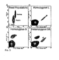
- Fig. 3 shows typical results obtained with human DNA samples.
- the MID42 marker used in this experiment was chosen from a collection of diallelic short insertion/deletion polymorphisms assembled by Weber and colleagues (17). These alleles are particularly simple to distinguish with hybridization probes because the two alleles at each locus differ by ⁇ 4 bases.
- the probe for the longer (L) allele was labeled with fluorescein (green) and the probe for the shorter (S) allele labeled with R-phycoerythrin (red).
- Fig. 3A shows a plot of the side scatter vs. forward scatter of beads following BEAMing.
- >75% of beads were dispersed as single particles, with the remainder aggregated in groups of two or more.
- Subsequent flow cytometric analysis was confined to the singlet beads, gated as outlined in Fig. 3A .
- Figs. 3B - D show density plots of gated beads generated with various templates.
- a template from an individual homozygous for the L allele was included in the emulsion.
- Two populations of beads were apparent 98 % of the beads contained no PCR product (black) and the remaining 2% fluoresced in the FL1 channel (colored green in Fig. 3).
- Fig. 3C represents the analysis of an individual homozygous for the S allele.
- Two populations of beads were again apparent, but this time the labeled population fluoresced in the FL2 channel (colored red in Fig. 3).
- Fig. 3D presents density plots from the analysis of an individual heterozygous at the MID42 locus.
- the black region represents beads without any PCR product
- the red region represents beads containing PCR products from the L allele
- the green region represents beads containing PCR products from the S allele
- the blue region represents beads containing PCR products from both alleles.
- Beads containing PCR products from both alleles were derived from aqueous compartments which contained more than one template molecule. The number of blue beads increased in a non-linear fashion as more template molecules were added. At the extreme, when all aqueous compartments are saturated, virtually all beads will register as blue. Operationally, we found that the bead populations were most distinct when the number of beads containing any PCR product was ⁇ 10% of the total beads analyzed.
- Fig. 3 The results shown in Fig. 3 were generated using PCR products made from human genomic DNA samples. As the ratio of the beads representing L alleles to those representing S alleles was 1.0 in this experiment, it was clear that the initial PCR did not preferentially amplify either allele.
- the use of PCR products rather than genomic DNA permitted large numbers of alleles to be amplified from even small quantities of starting DNA. In general, 10 to 100 picograms of PCR products of size 200 bp were found to be optimal for BEAMing, producing PCR-mediated extension of primers on ⁇ 1 to 10% labeled beads.
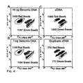
- Fig. 4A and B show flow cytometric data from an experiment wherein 10 ug or 1 ug of human genomic DNA was used as template for BEAMing at the MID42 locus. Patterns very similar to those shown in Fig. 3 were observed, though fewer beads were labeled than when PCR products were used as templates.
- BEAMing could also be used to analyze variations in expression from the two alleles of a heterozygous individual. Heritable variations in the expression from individual alleles of the same gene have been shown to occur often in humans (18) and mice (19) and can have significant phenotypic effects (20).
- the results shown in Fig. 4C and D show that PCR products made from reverse-transcribed mRNA can be used for BEAMing.
- calpain-10 transcripts differing by a single nucleotide polymorphism (SNP) were analyzed.
- SNPs single nucleotide polymorphism
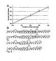
- Fig. 5A shows representative data from templates representing 1%, 2%, 3%, and 4% of the L allele of MID42.
- the linearity of these measurements with a correlation coefficient of 0.99, demonstrates the utility of this approach for such applications.
- the rare beads representing the mutant alleles could not only be quantified but could also be purified for subsequent analysis.
- samples of the beads enumerated in Fig. 5A were additionally assessed using a flow cytometer equipped with sorting capabilities. Beads were sorted and individual beads used as templates for conventional PCR using the same primers employed for BEAMing. As each bead contains thousands of bound template molecules, single beads were expected to generate robust PCR products (23) and this was experimentally confirmed. These PCR products were then subjected to sequencing. As shown in Fig. 5B and C , green and red beads generated PCR products exclusively of the L and S types, respectively.
- a 100 bp product was amplified on beads as described in Example 1, steps 1 through 4.
- Two FAM-labeled oligonucleotides (50 and 20 bases in length) were annealed to the 100 bp product on the beads.
- the beads were then embedded in an acrylamide gel (using conventional Tris-Borate-EDTA electrophoresis buffer) in an oval shaped configuration.
- An electric field 250 V was applied under denaturing conditions for 3 minutes.
- the labeled oligonucleotides migrated off the beads and migrated a distance related to their sizes. See Figure 7 . There was little diffusion, as evidenced by the retention of the oval shape of the beads.
- Sanger-type (dideoxynucleotide) sequencing is performed using as templates oligonucleotides which have been amplified on beads, as described in Example 1. Individual beads are subjected to primer extension conditions in the presence of dideoxynucleotide inhibitors. The beads are then subjected to electrophoresis under denaturing conditions to separate the dideoxynucleotide-terminated, primer extended oligonucleotides on the basis of length. A sequence is compiled based on the length of the primer extended oligonucleotides.
Description
- The invention relates to the field of genetic analysis. In particular, it relates to methods and compositions for analyzing variations in individual genes or transcripts and separating variants.
- The study of DNA sequence variation is essential for many areas of research. The study of germ-line variations is essential for assessing the role of inheritance in normal and abnormal physiologic states (1). Other variations, developed somatically, are responsible for neoplasia (2). The identification of such mutations in urine, sputum, and stool can therefore be used for the detection of presymptomatic cancers (3-5). Similarly, the detection of somatic mutations in lymph nodes, blood, or bone marrow can provide data about the stage of disease, prognosis, and appropriateness of various therapies (5). Somatic mutations in non-neoplastic cells also occur and appear to accumulate as humans age or are exposed to environmental hazards (6). Such mutations occur in only a small fraction of the cells in a tissue, thereby complicating their analysis.
- Central to the investigation of many of these issues is the detection and quantification of sequence variants within a population of DNA molecules. The number of molecules in each such collection is finite and therefore countable. Consider, for example, a collection of red and green balls. Counting these balls is simple in principle but subject to basic probability theory. If there is only one red ball for every 500 green balls, then it is necessary to count several thousand balls to get an accurate estimate of the proportion of red balls. If it is difficult to count enough balls to make a reliable estimate, one can elute the paint off all the balls and measure the color of the resultant paint mix.
- In analogous fashion, small numbers of DNA molecules that vary by subtle changes (single base pair substitutions or small deletions or insertions) can be directly counted by amplifying individual DNA molecules (single molecule PCR) (7-12). Such digital techniques have been shown to be extremely useful for measuring variation in genes or their transcripts. But digital technologies have so far been limited to counting tens to thousands of molecules, either in the wells of microtiter plates, on microscope slides, or after electrophoresis of individual PCR products. Analog techniques, analogous to the elution of paint from the balls described above, are generally easier to implement and can assess millions of molecules simultaneously (13). However, their accuracy and sensitivity is limited by instrumental and experimental noise. There is a continuing need in the art for methods which are accurate and sensitive for measuring variation in genes or their transcripts,
- Nakano et al, (2003) Journal of Biotechnology 102(2), 117-124 discloses a single molecule PCR in a water-in-oil emulsion. It does not disclose a liquid composition according to the present invention, which comprises a plurality of micro emulsions comprising primers bound to beads.
- In a first embodiment of the invention a liquid composition is provided. The liquid composition comprises a plurality of microemulsions forming aqueous compartments. At least a portion of said aqueous compartments comprise a bead, a polynucleotide template, and oligonucleotide primers for amplifying the template. At least a portion of the oligonucleotide primers is bound to the bead.
- A second embodiment of the invention provides a method for analyzing nucleotide sequence variations. Microemulsions comprising one or more species of analyte DNA molecules are formed. The analyte DNA molecules in the microemulsions are amplified in the presence of reagent beads which are bound to a plurality of molecules of a primer for amplifying the analyte DNA molecules. Product beads are formed that are bound to a plurality of copies of a single species of analyte DNA molecule. The product beads are separated from analyte DNA molecules which are not bound to product beads. A sequence feature of the single species of analyte DNA molecule that is bound to the product beads is determined.
- In a third embodiment of the invention a method is provided for isolating nucleotide sequence variants. Microemulsions comprising one or more species of analyte DNA molecules are formed. Analyte DNA molecules in the microemulsions are amplified in the presence of reagent beads. The reagent beads are bound to a plurality of molecules of a primer for amplifying the analyte DNA molecules. Product beads are formed which are bound to a plurality of copies of one species of analyte DNA molecule. The product beads are separated from analyte DNA molecules which are not bound to product beads. The product beads which are bound to a plurality of copies of a first species of analyte DNA molecule are isolated from product beads which are bound to a plurality of copies of a second species of analyte DNA molecule.
- These and other embodiments of the invention, which will be apparent from the entire description of the invention, provide the art with the ability to quantify genetic variations at a scale and ease heretofore unattainable.
-
-
Fig. 1 is a schematic drawing of the BEAMing method. Step 1- Magnetic beads covalently coated with streptavidin are bound to biotinylated oligonucleotides ("oligos"). Step 2 - An aqueous mix containing all the necessary components for PCR plus primer-bound beads and template DNA are stirred together with an oil/detergent mix to create microemulsions. The aqueous compartments (white circles in the gray oil layer) contain an average of <1 template molecule and <1 bead. Red and green templates represent two template molecules whose sequences differ by one or many nucleotides. Step 3 - The microemulsions are temperature cycled as in a conventional PCR. If a DNA template and a bead are present together in a single aqueous compartment, the bead bound oligonucleotides act as primers for amplification. The straight red and green lines connected to the beads represent extension products from the two different kinds of templates. Step 4 - The emulsions are broken and the beads are purified with a magnet. Step 5 - After denaturation, the beads are incubated with oligonucleotides that can distinguish between the sequences of the different kinds of templates. Fluorescently-labeled antibodies are then used to label the bound hybridization probes. This renders the beads containing PCR product as red or green upon appropriate laser excitation. Step 6 - Flow cytometry is used to count the red and green beads. -
Fig. 2 is a photograph of a typical microemulsion. Microemulsions were made as described infra with the exception that the aqueous compartments contained cascade blue-labeled dCTP and the beads were pre-labeled with R-phycoerythrin (red) or Alexa 488 (green). One microliter of microemulsion was deposited in 1 microliter of oil on a microscope slide prior to photography. Of the seven aqueous compartments visible in this picture, two contain beads. Note the heterogeneous size of the aqueous compartments (beads are 1.05 microns in. diameter). -
Fig. 3A to Fig. 3D show density plots of flow cytometric data obtained from BEAMing. The locus queried in this experiment was MID42 and PCR products generated from genomic DNA were used as templates in the microemulsions. (Fig. 3A ) Forward scatter (FSC) and side scatter (SSC) of all beads show that ~80% of the total beads are singlets, with most of the remaining beads aggregated as doublets. The "noise" is instrumental and is observed with blank samples containing no beads. The instrument output was gated so that only singlets were analyzed for fluorescence analysis. The patterns observed from an individual homozygous for the L allele (Fig. 3C ), homozygous for the S allele (Fig. 3B ), and heterozygous for L and S (Fig. 3D ) are shown. The regions containing beads hybridizing to the L and S allele probes are labeled green and red, respectively. The region containing beads that did not hybridize to any probe is black and the region containing beads that hybridized to both probes is blue. The blue beads arose from aqueous compartments in which both types of template molecules were present. The proportion of singlet beads that hybridized to at least one of the probes was 2.9%, 4.3%, and 20.3% in (Fig. 3B) to (Fig. 3D ), respectively. The FSC and SSC plots in (Fig. 3A ) represent the same beads analyzed in (Fig. 3D ). -
Fig. 4A to Fig. 4D show density plots of BEAMing using genomic DNA or RT-PCR products as templates. The data in (Fig. 4A) and (Fig. 4B ) were generated by including 10 and 1 ug of human genomic DNA, respectively, in the microemulsions, querying the MID42 locus. The data in (Fig. 4C) and (Fig. 4D ) were generated using emulsions that contained ~ 50 picograms of PCR products synthesized from cDNA of lymphoblastoid cells, querying the calpain-10 locus. The green and red regions correspond to the L and S alleles for MID42 and to the A and G alleles for calpain-10. The number of beads in the outlined regions containing red or green beads is shown in each case. The proportion of singlet beads that hybridized to at least one of the probes was 1.2%, 0.6%, 6.8% and 4.2% in (Fig. 4A) to (Fig. 4D ), respectively. The outlined regions used for counting in (Fig. 4A) and (Fig. 4B ) were identical, as were those used for (Fig. 4C) and (Fig. 4D ). Beads that did not hybridize to any probe were gated out and therefore not evident in the graphs, while the region containing beads that hybridized to both probes is labeled blue. -
Fig. 5A to Fig. 5C show detection and validation of variants present in a minor fraction of the DNA population. (Fig. 5A ) Mixtures of PCR products containing 0% to 4% L alleles of MID42 were used for BEAMing. Flow cytometry such as that shown inFig. 3 was used to determine the fraction of singlet beads that were red (y-axis). The proportion of singlet beads that hybridized to at least one of the probes varied from 3.2% to 4.3%. (Fig. 5B and Fig. 5C ) Beads were sorted with the FACS Vantage SE instrument and individual red or green beads were used as templates for conventional PCR employing the forward and reverse primers listed inFig. 8 . Red beads generated only the S allele sequence (Fig. 5B ; SEQ ID NO: 1) while green beads generated only the L allele sequence (Fig. 5C ; SEQ ID NO: 2). -
Fig. 6A to 6B demonstrate the use of agar in the aqueous phase of the microemulsions. Emulsion bubbles that were formed by including 1.5% agarose in the aqueous compartment are shown.Fig. 6A shows the bubbles that have fluorescents in them.Fig. 6B shows a darkfield image of the bubbles with one of the bubbles containing a bead in it. After breaking the emulsions, the droplets containing magnetic beads can be recovered by centrifugation and size fractionated through filtration or flow sorting. -
Fig. 7 shows denaturing electrophoresis of two FAM-labeled oligonucleotides, 50 and 20 bases in length, which had been hybridized to a 100 bp product on beads. The beads were embedded in an acrylamide gel in an oval shaped configuration and an electric field was applied The labeled oligonucleotides migrated off the beads and migrated a distance proportional to their size. -
Figure 8 shows oligonucleotides used. - The inventors describe herein a digital technology, called BEAMing, that has the power to assess millions of molecules and can be generally applied to the study of genetic variation. The technology involves conversion of single DNA molecules to single beads each containing thousands of copies of the sequence of the original DNA molecule. The number of variant DNA molecules in the population can then be assessed, for example, by staining the beads with fluorescent probes and counting them using flow cytometry. Beads representing specific variants can be optionally recovered through flow sorting and used for subsequent confirmation and experimentation.
- Beads according to the present invention are also known as microspheres or microparticles. Particle sizes can vary between about 0.1 and 10 microns in diameter. Typically beads are made of a polymeric material, such as polystyrene, although nonpolymeric materials such as silica can also be used. Other materials which can be used include styrene copolymers, methyl methacrylate, functionalized polystyrene, glass, silicon, and carboxylate. Optionally the particles are superparamagnetic, which facilitates their purification after being used in reactions.
- Beads can be modified by covalent or non-covalent interactions with other materials, either to alter gross surface properties, such as hydrophobicity or hydrophilicity, or to attach molecules that impart binding specificity. Such molecules include without limitation, antibodies, ligands, members of a specific-binding protein pair, receptors, nucleic acids. Specific-binding protein pairs include avidin-biotin, streptavidin-biotin, and Factor VII-Tissue Factor.
- Beads, after being prepared according to the present invention as product beads, have more than one copy of the same nucleic acid molecule bound to them. Preferably each bead is bound to at least 10, 50, 100, 500, or 1000 molecules of the same nucleic acid sequence. In some circumstances some of the product beads are bound to more than one type of nucleic acid molecule. These product beads are generally less useful in the analysis of ratios of genetic sequences in a population of genetic sequences. Such product beads can be readily discriminated and so will not distort the analysis.
- A population of product beads will often comprise two or more types of nucleic acids. Such a population is heterogeneous with respect to the nucleic acids. Desirably, a substantial proportion of the product beads comprise only one type of nucleic acid per bead. A substantial proportion can be for example, at least 1 %, at least 5 %, at least 10 %, or at least 50 %. A product bead with only one type of nucleic acid per bead is termed homogeneous. Homogeneous beads with only one type of nucleic acid per bead include those with nucleic acids containing errors due to errors in polymerase chain reaction. A product bead with two types of nucleic acid per bead is termed heterogeneous. Although not wishing to be bound by any particular theory, heterogeneous product beads are thought to result from aqueous compartments which have more than two molecules of template of non-identical sequence. A population of product beads can be heterogeneous as a population but contain individual product beads that are homogeneous
- Individual product beads preferably comprise more than one copy of template analyte molecule. Each bead may comprise at least 10, at least 50, at least 100, at least 500, or at least 1000 copies of template analyte. If the bead is homogeneous, each of those copies will be identical.
- Populations of product beads can be maintained in a liquid suspension. Alternatively they can be sedimented and dried or frozen. The latter alternatives may be beneficial for storage stability.
- Analysis of populations of product beads can be useful for distinguishing between many kinds of genetic variants. Polynucleotides can be distinguished which differ by as little as a single nucleotide polymorphism (SNP), by the presence or absence of a mutation, by the presence or absence of an insertion or deletion, by the presence or absence of a non-single nucleotide polymorphism. Thus populations of product beads may be heterogeneous with regard to these genetic variations.
- One very convenient way for distinguishing genetic variants, i.e., determining a sequence feature of the analyte, is by differentially labeling the variants with fluorescent dyes. Such labeling can be accomplished by hybridization of a fluorescently labeled oligonucleotide probe to one species of polynucleotide. Alternatively, a fluorescently labeled antibody can be used to specifically attach to one oligonucleotide probe that hybridizes to a particular genetic variant. Such antibody binding can be, for example, mediated by a protein or polypeptide which is attached to an oligonucleotide hybridization probe. Of course, other means of labeling polynucleotides as are known in the art can be used without limitation. Another means of labeling different polynucleotide species is by primer extension. Primers can be extended using labeled deoxyribonucleotides, such as fluorescently labeled deoxyribonucleotides.
- Populations of product beads can be used as templates. Template analyte molecules on the product beads can be analyzed to assess DNA sequence variations by hybridization, primer-extension methods, mass spectroscopy, and other methods commonly used in the art. Template analyte molecules on product beads can be employed for solid phase sequencing. In one solid phase sequencing technique, product beads are arrayed by placing them on slides spotted with complementary oligonucleotides. In another solid phase sequencing technique, product beads are placed into individual wells. In still another solid phase sequencing technique product beads are incorporated into acrylamide matrices (with or without subsequent polony formation). Sequencing reactions can be performed with any solid phase sequencing method, such as those using unlabeled nucleotide precursors (e.g., pyrosequencing, as described in Ronaghi et al., Anal. Biochem. 267: 65-71, 1999) or labeled nucleotides (e.g., photocleavable reagents described by Mitra et al., Anal. Biochem. 320:55-65, 2003). Product beads can thus be used for and facilitate multiple parallel sequencing. Product beads can also be used in sequencing employing Type IIS restriction endonucleases. Product beads can also be used to provide templates for conventional dideoxynucleotide sequencing. To obtain useful data upon sequence analysis, a homogeneous template population is desirable. To provide a homogenous template population, product beads can be diluted, separated, or otherwise isolated so that each sequencing reaction contains a single product bead. Alternatively, product beads can be sorted to provide populations of beads with a single species of template.
- Oligonucleotide primers can be bound to beads by any means known in the art. They can be bound covalently or non-covalently. They can be bound via an intermediary, such as via a protein-protein interaction, such as an antibody-antigen interaction or a biotin-avidin interaction. Other specific binding pairs as are known in the art can be used as well. To achieve optimum amplification, primers bound to the bead may be longer than necessary in a homogeneous, liquid phase reaction. Oligonucleotide primers may be at least 12, at least 15, at least 18, at least 25, at least 35, or at least 45 nucleotides in length. The length of the oligonucleotide primers which are bound to the beads need not be identical to that of the primers that are in the liquid phase. Primers can be used in any type of amplification reaction known in the art, including without limitation, polymerase chain reaction, isothermal amplification, rolling circle amplification, self-sustaining sequence replication (3SR), nucleic acid sequence-based amplification (NASBA), transcription-mediated amplification (TMA), strand-displacement amplification (SDA), and ligase chain reaction (LCR).
- Microemulsions are made by stirring or agitation of oil, aqueous phase, and detergent. The microemulsions form small aqueous compartments which have an average diameter of 0.5 to 50 microns. The compartments may be from 1 to 10 microns, inclusive, from 11 to 100 microns, inclusive, or about 5 microns, on average. All such compartments need not comprise a bead. Desirably, at least one in 10,000 of said aqueous compartments comprise a bead. Typically from 1/100 to 1/1 or from 1/50 to 1/1 of said aqueous compartments comprise a bead. In order to maximize the proportion of beads which are homogeneous with respect to oligonucleotide, it is desirable that on average, each aqueous compartment contains less than 1 template molecule. Aqueous compartments will also desirably contain whatever reagents and enzymes are necessary to carry out amplification. For example, for polymerase chain reaction (PCR) the compartments will desirably contain a DNA polymerase and deoxyribonucleotides. For rolling circle amplification a DNA polymerase and a generic DNA circle may be present.
- Emulsions can be "broken" or disrupted by any means known in the art. One particularly simple way to break the emulsions is to add more detergent. Detergents which can be used include, but are not limited to Triton X100,
Laureth 4, Nonidet. - Sample DNA for amplification and analysis according to the present invention can be genomic DNA, cDNA, PCR products of genomic DNA, or PCR products of cDNA, for example. Samples can be derived from a single individual, for example, from a body sample such as urine, blood, sputum, stool, tissue or saliva. Samples can also be derived from a population of individuals. The individuals can be humans, but can be any organism, plant or animal, eukaryotic or prokaryotic, viral or non-viral.
- Any type of probe can be used for specific hybridization to the amplified polynucleotides which are bound to the beads. Fluorescently labeled probes are useful because their analysis can be automated and can achieve high throughput. Fluorescence activated cell sorting (FACS) permits both the analysis and the isolation of different populations of beads. One type of fluorescently labeled probe that can be used is a modified molecular beacon probe. These probes have stem-loop structures and an attached fluorescent moiety on the probe, typically on one end of the probe, sometimes attached through a linker. Unlike standard molecular beacon probes, modified molecular beacon probes do not have a quenching moiety. The modified molecular beacon probe can have the fluorescent moiety attached on either end of the probe, 5' or 3'. One such probe will hybridize better to a wild-type sequence than to a mutant. Another such probe will hybridize better to a mutant sequence than to the wild type. Still other probes will preferably hybridize to one polymorphic variant over another.
- The method of the present invention provides a reliable and sensitive assay for measuring variations in genes and transcripts. It requires no instrumentation other than machines that are widely available. There are several other advantages of this approach. First, the sensitivity can be increased to meet the particular specifications of an assay simply by analyzing more beads. Such sensitivity is limited only by the error rate of the polymerases used for amplification. Second, the data obtained can be used not only to demonstrate that a variant is present in a particular population of DNA molecules, but also quantifies the fraction of variant DNA molecules in that population (
Fig. 5A ). Such quantification is not possible with techniques that destroy or ignore the wild type molecules as part of the assay, such as those that use allele specific priming or endonuclease digestion during PCR. Third, the beads containing variant alleles can easily be purified through flow sorting. Such recovery is difficult with digital techniques that count molecules deposited on microscope slides. And finally, the method is automatable. - Several modifications of the basic principles described here can be envisioned that will further simplify the technology or widen its applications. For example, microemulsions were made by stirring water/oil/detergent mixes. The sizes of the resultant aqueous compartments were somewhat heterogeneous, as illustrated in
Fig. 2 . A relatively large number of beads containing PCR products of both alleles are obtained from large compartments because they are more likely to contain >1 template molecule than smaller compartments. Though this is not a problem for the analysis of uncommon variants, it does pose a problem when the variant to be analyzed is present in a substantial fraction of the DNA molecules. For example, it is easy to distinguish a population containing 2% of allele A and 98% of allele B from one that contains 0% of allele A (Fig. 5A ). But it is more difficult to distinguish a population that contains 48% of allele A and 52% of allele B from a population that contains 50% of allele A; the large number of heterozygote beads formed in the latter analysis diffuse the boundaries of the pure red and green channels. This limit to accuracy can be overcome through the preparation of more uniformly sized aqueous compartments. Sonication or pressure-driven emulsifiers can make more uniform compartments. - Though flow cytometry requires only seconds to minutes per sample, multiple parallel analyses could facilitate throughput. Novel particle counting designs may prove useful for this purpose. Another way to increase throughput would be to physically separate the beads that contained PCR products prior to flow cytometry. This could be accomplished with proteins such as antibodies or streptavidin that bind to modified nucleotides incorporated into the PCR product during amplification.
- The methods of the invention can be applied to genes or transcripts of any organism or population of organisms. These include without limitation: humans, rodents, ungulates, mammals, primates, cows, goats, pigs, rats, mice, yeast, poultry, fish, shellfish, digs, cats, zebrafish, worms, algae. It can also be used to quantify epigenetic alterations, such as methylation, if DNA is first treated with bisulfite to convert methylated cytosine residues to thymidine. Beads generated from random fragments of whole genomes (24), rather than from individual genes as described above, could be used to identify gene segments that bind to specific DNA-binding proteins (25). And if the product beads are used in compartmentalized in vitro transcription-translation reactions, variant proteins can be bound to beads containing the corresponding variant DNA sequences (23). This could allow facile flow cytometric evaluation of rare mutations using antibodies that distinguished between wild type and mutant gene products (26).
- Step 1 - Coupling oligonucleotides to beads. Superparamagnetic beads of 1.05 +/- 0.1 um in diameter, covalently bound to streptavidin, were purchased from Dynal Biotech, Inc. (650.01, Lake Success, NY). Beads were washed once with 1x PCR buffer (53286, Invitrogen, Carlsbad, CA) then suspended in Bind and Wash Buffer (BWB) (5 mM Tris-HCl, 0.5 mM EDTA, 1.0 M NaCl, pH 7.5). Beads were incubated in BWB for 30 min at room temperature in the presence of 10 uM oligonucleotides (
Figure 8 ). These oligonucleotides were modified with a dual biotin group at the 5' end with the biotin groups separated by a six-carbon linker (IDT, Coralville, IA). After binding, the beads were washed 3 times with 1x PCR buffer to thoroughly remove unbound oligonucleotides. - Step 2 - Preparing microemulsions. Microemulsions for PCR were prepared by slight modifications of previously described methods (14) (15). The oil phase was composed of 4.5% Span 80 (S6760, Sigma, St. Louis, MO), 0.40 % Tween 80 (Sigma S-8074), and 0.05% Triton X-100 (Sigma T-9284) in mineral oil (Sigma M-3516). The oil phase was freshly prepared each day. The aqueous phase consisted of 67 mM Tris-HCl (pH 8.8), 16.6 mM NH4SO4, 6.7 mM MgCl2, 10 mM β-mercaptoethanol, 1 mM dATP, 1 mM dCTP, 1 mM dGTP, 1 mM dTTP, 0.05 uM forward primer, 25 uM reverse primer, 45 units Platinum Taq (Invitrogen 10966-034), various amounts of template DNA (see results), and ~108 oligonucleotide-coupled beads in a total volume of 300 ul. The forward primer was an oligonucleotide whose sequence was identical to the 3' 20 - 22 nt of that described in
step 1 and was not modified with biotin. - Water-in-oil microemulsions were prepared by drop wise addition of 200 microliters of the aqueous phase to 400 microliters of the oil phase previously placed in a 2 ml round bottom cryogenic vial (430661, Coming, Corning, NY). The drop wise addition was performed over ~ one minute while the mixture was being stirred at 1400 RPM with a magnetic microstir bar (58948-353, VWR, Plainfield, NJ) on a VWR model 565 Magnetic Stirrer. After the addition of the aqueous phase, the mixture continued to be stirred for a total time of 30 minutes. Two emulsions were made at once by placing two tubes in a rack placed at the center of the magnetic stirrer.
- Step 3 - PCR cycling. The emulsions were aliquotted into five wells of a 96 well PCR plate, each containing 100 ul. PCR was carried out under the following cycling conditions: 94°C for 2 minutes; 40 cycles of 94°C for 15 seconds, 57°C for 30 seconds, 70°C for 30 seconds. The PCR products analyzed in this study ranged from 189 to 239 bp.
- Step 4 - Magnetic capture of beads. After PCR cycling, the microemulsion from five wells of a PCR plate were pooled and broken by the addition 800 microliters of NX buffer (100 mM NaCl containing 1% Triton X-100, 10 mM Tris-HCl, pH 7.5, 1 mM EDTA) in a 1.5 ml tube (Corning 430909). After vortexing for ~20 sec. the beads were pelleted by centrifugation in a microcentrifuge at 8000 rpm (5000 g) for 90 seconds. The top oil phase and all but ~300 microliters of the aqueous phase was removed from the tube and 600 microliters of NX buffer was added. After vortexing for 20 sec. and centrifugation for 90 sec., the top oil phase and all but ~300 microliters of the aqueous phase was removed. The addition of 600 microliters NX buffer, vortexing, and centrifugation was repeated once more and the top oil portion and all but ~300 microliters of the aqueous phase was removed. The tube was then placed on a magnet (Dynal MPC-S) and the rest of the supernatant was carefully pipetted off. The beads were washed an additional 3 times with 1x PCR buffer using magnetic separation rather than centrifugation and finally resuspended in 100 microliters of 1x PCR buffer.
- Step 5 - Sequence differentiation. Two oligonucleotide probes were used for each reaction. One was 5'-labeled with 6-carboxyfluorescein (6-FAM) and was specific for one allele while the second was 5'-labeled with biotin and was specific for the other alleles. Probes were synthesized by IDT. The 30 microliters hybridization reactions contained 10 uM of each probe and 5 - 25 million beads in1 x PCR buffer. Reactions were performed in PCR plates on a thermal cycler by heating to 94°C for 30 seconds then cooling to 75°C at a rate of 0.5°C per second, cooling to 45°C at 0.2°C per second, and finally cooled to 30°C at 1°C per second. All subsequent steps were performed at room temperature. The reactions were transferred to a 96 well Costar plate (Corning 3797) and placed on a 96 well magnet. Beads were collected magnetically by exposing them to the magnet for 2 minutes. The supernatant was removed and the beads washed 3 times with Ix PCR buffer by pipetting them and collecting for two minutes. They were finally resuspended in 100 microliters B-PCR buffer (1mg/mL BSA in 1x PCR buffer). The beads were then incubated for 10 minutes in a total volume of 100 microliters B-PCR buffer containing 3 ug of Alexa-488 rabbit anti-fluorescein antibody (Molecular Probes A-11090, Eugene, OR) and 3 ug of Nutravidin labeled with R-phycoerythrin (Molecular Probes A-2660) in B-PCR buffer. The beads were washed three times and resuspended in B-PCR buffer as described above. They were then incubated for ten minutes in a total volume of 100 microliters B-PCR buffer containing 6 ug of Alexa 488- conjugated chicken anti-rabbit antibody (Molecular Probes A-21441)and 3 ug of biotinylated goat anti-avidin antibody (BA-0300, Vector Laboratories, Burlingame, CA). The beads were washed three times and resuspended in B-PCR buffer as described above. They were then incubated for ten minutes in a total volume of 100 microliters B-PCR buffer containing 3 ug of an Alexa 488-conjugated goat anti-chicken antibody (Molecular Probes A-11039) and 3 micrograms of R-phycoerythrin-labeled streptavidin (Molecular Probes S-866). This solution was then washed an additional 3 times with 1x PCR buffer and resuspended in 20 microliters of 1 X PCR buffer.
- Step 6 - Flow Cytometry. The bead suspension was diluted to a concentration of ~ 106 -107 beads per ml in 10 mM Tris-HCl, 1 mM EDTA (351-010-131, Quality Biological, Inc., Gaithersburg, MD) and analyzed using a LSR instrument (BD Biosciences, Franklin Lakes, NJ). The instrument was set up for standard two-color analysis using an argon laser and optical filters that distinguished between the two fluorescent dyes. No spectral deconvolution was required as the major bead populations were well-separated. In some cases, scanning was performed with FACScan or FACSCalibur instruments (BD Biosciences), yielding equivalent results. Sorting was carried out with a FACS Vantage SE instrument (BD Biosciences).
- Template preparation and sequence analyses. Human genomic DNA was purified with DNeasy (69504, Qiagen, Valencia, CA). RNA was purified with Quickprep (27-9255-01, Amersham Biosciences, Piscataway, NJ). Reverse transcription of RNA was performed using Superscript II reverse transcriptase (Invitrogen 18064014) according to the manufacturer's instructions. PCR using genomic DNA or reverse transcripts as templates was performed as described (7). PCR products to be used as templates for BEAMing or for sequencing were purified with QIAquick (Qiagen 28104). Sequencing reactions were performed using Big Dye v3.0 reagents (Applied Biosystems, Foster City, CA) and analyzed by capillary electrophoresis (Spectrumedix 9600, State College, PA).
- Step 1 - Coupling oligonucleotides to beads. We used streptavidin-beads because of the simplicity of coupling biotinylated oligonucleotides to them. Oligonucleotides with just a single 5' biotin group were found to dissociate from the beads during temperature cycling, while oligonucleotides labeled with dual biotin groups at their 5' end (separated by a six-carbon linker) were stable to cycling. As determined by fluoroscopic measurements of oligonucleotides doubly labeled with 6-FAM and biotin, ~105 oligonucleotide molecules were bound to each bead. We found that short oligonucleotides (20 bases) did not work as well for priming as longer ones (41 bp), perhaps because of steric hindrance at the bead surface. It is likely that amino-, sulfhydryl-, or carboxyl-modified oligonucleotides covalently coupled to beads modified with corresponding reactive groups could also function as bead-bound primers for BEAMing.
- Step 2 - Preparing microemulsions. The size of the individual aqueous compartments ranged from less than 1 micron to >10 microns in diameter (
Fig. 2 ). We estimated that an emulsion comprising 200 microliters of aqueous solution and 400 microliters of oil would contain ~3 x 10⁹ compartments with an average diameter of 5 microns. Approximately 108 beads were included in each emulsion, so that only one in ~30 compartments contained a bead. The optimal amount of template was experimentally determined to be ~ 5 x 10⁸ molecules, so that one in ~six compartments contained a template molecule. - Step 3 - PCR cycling. PCR priming by oligonucleotides coupled to beads was found to be very inefficient compared to the priming by the same oligonucleotides when free in solution. For this reason, a small amount of non-biotinylated forward primer identical in sequence to the biotinylated oligonucleotide coupled to the beads was included in the reactions. This facilitated the first few rounds of amplification of the single template within each aqueous compartment. In the absence of additional primer, no detectable amplification on the beads was generated. Conversely, if too much additional primer was included, no amplification on the beads occurred because of competition with the primers in solution. An excess of the reverse primer was included in the aqueous compartment to maximize the probability that bead-bound oligonucleotides extended by polymerase would serve as templates for further amplification cycles.
- Step 4 - Magnetic capture of beads. There are several ways to break water-in-oil emulsions, including extraction with organics (14). We found that simply adding non-ionic detergents produced phase separations without any detectable modification of the beads or DNA molecules bound to them. By measuring the amount of DNA that could be released from the beads following restriction endonuclease digestion, we estimate that >10,000 extended PCR products were present, on average, per bead.
- Step 5 - Sequence differentiation. Most fluorescence-based methods for distinguishing alleles in homogeneous or two-phase assays can be used to assess allelic variation captured on beads. These methods include single nucleotide extension, allele specific priming, or hybridization. We generally employed hybridization of fluorescein-conjugated or biotin-conjugated oligonucleotides for discrimination. As shown in
Fig. 1 andFig. 8 , these oligonucleotides had a stem-loop structure, with the middle of the loop containing the variant nucleotide(s). This design was based on studies of Molecular Beacons wherein a stem-loop structure was shown to markedly improve allelic discrimination (16). The oligonucleotides we used differed from Molecular Beacons in that there was no need for a quenching group. Such quenching is required for homogeneous assays when unhybridized oligonucleotides cannot be removed from the reactions prior to assay but is not necessary for solid phase assays such as those employed with beads. - Step 6 - Flow Cytometry. Optimum results in flow cytometry depend on high fluorescent signals on the beads. We generally enhanced the fluorescence emanating from the hybridization probes with secondary reagents. For example, Alexa 488-labeled antibodies were used to enhance the signals emanating from fluorescein-coupled oligonucleotide probes. Similarly, R-phycoerythrin-labeled streptavidin was used to generate a signal from biotin-labeled oligonucleotide probes. Flow cytometers equipped with two or three lasers and appropriate filters have the capacity to distinguish multi-allelic loci and to perform multiplex analysis of several genes simultaneously. The newest generation of flow cytometers can also analyze >70,000 events per second. In addition to the analytical power of flow cytometry, FACS instruments can separate specific populations of beads for further analysis.
- Pilot experiments demonstrated that simply stirring the water-oil mixtures described in Materials and Methods produced very stable microemulsions of a size compatible with that of the beads. In the experiment shown in
Fig. 2 , the aqueous compartment contained a blue dye and 1 micron magnetic beads that were labeled by binding to an oligonucleotide that was biotinylated at its 5' end and labeled with fluorescein at its' 3' end. The appearance of emulsions immediately after their formation is shown inFig. 2 . As expected, this appearance was unchanged after temperature cycling during PCR (15). Most aqueous compartments contained no beads, as expected from the figures provided in the previous section. Those compartments that did contain beads generally contained only one, though a fraction contained more, as expected from a Poisson distribution and non-uniform aqueous compartment sizes. "Heterozygous" beads containing PCR products representing both alleles are produced when two or more DNA template molecules are contained within a single aqueous compartment. Such heterozygotes can compromise the accuracy of the analyses under some circumstances (see Discussion). -
Fig. 3 shows typical results obtained with human DNA samples. The MID42 marker used in this experiment was chosen from a collection of diallelic short insertion/deletion polymorphisms assembled by Weber and colleagues (17). These alleles are particularly simple to distinguish with hybridization probes because the two alleles at each locus differ by ~ 4 bases. The probe for the longer (L) allele was labeled with fluorescein (green) and the probe for the shorter (S) allele labeled with R-phycoerythrin (red). -
Fig. 3A shows a plot of the side scatter vs. forward scatter of beads following BEAMing. In general, >75% of beads were dispersed as single particles, with the remainder aggregated in groups of two or more. Subsequent flow cytometric analysis was confined to the singlet beads, gated as outlined inFig. 3A . -
Figs. 3B - D show density plots of gated beads generated with various templates. InFig. 3B , a template from an individual homozygous for the L allele was included in the emulsion. Two populations of beads were apparent 98 % of the beads contained no PCR product (black) and the remaining 2% fluoresced in the FL1 channel (colored green inFig. 3). Fig. 3C represents the analysis of an individual homozygous for the S allele. Two populations of beads were again apparent, but this time the labeled population fluoresced in the FL2 channel (colored red inFig. 3). Fig. 3D presents density plots from the analysis of an individual heterozygous at the MID42 locus. Four populations of beads are evident: the black region represents beads without any PCR product, the red region represents beads containing PCR products from the L allele, the green region represents beads containing PCR products from the S allele, and the blue region represents beads containing PCR products from both alleles. Beads containing PCR products from both alleles were derived from aqueous compartments which contained more than one template molecule. The number of blue beads increased in a non-linear fashion as more template molecules were added. At the extreme, when all aqueous compartments are saturated, virtually all beads will register as blue. Operationally, we found that the bead populations were most distinct when the number of beads containing any PCR product was < 10% of the total beads analyzed. - The results shown in
Fig. 3 were generated using PCR products made from human genomic DNA samples. As the ratio of the beads representing L alleles to those representing S alleles was 1.0 in this experiment, it was clear that the initial PCR did not preferentially amplify either allele. The use of PCR products rather than genomic DNA permitted large numbers of alleles to be amplified from even small quantities of starting DNA. In general, 10 to 100 picograms of PCR products of size 200 bp were found to be optimal for BEAMing, producing PCR-mediated extension of primers on ~1 to 10% labeled beads. - In some situations it might be useful to use genomic DNA rather than PCR products as templates for BEAMing. The data in
Fig. 4A and B show flow cytometric data from an experiment wherein 10 ug or 1 ug of human genomic DNA was used as template for BEAMing at the MID42 locus. Patterns very similar to those shown inFig. 3 were observed, though fewer beads were labeled than when PCR products were used as templates. - BEAMing could also be used to analyze variations in expression from the two alleles of a heterozygous individual. Heritable variations in the expression from individual alleles of the same gene have been shown to occur often in humans (18) and mice (19) and can have significant phenotypic effects (20). The results shown in
Fig. 4C and D show that PCR products made from reverse-transcribed mRNA can be used for BEAMing. In this case, calpain-10 transcripts differing by a single nucleotide polymorphism (SNP) were analyzed. For SNPs like these, probes that incorporated an extra mismatched nucleotide adjacent to the polymorphic nucleotide (seeFig. 8 ) can enhance the distinction between alleles (21) (22). The results from two independent emulsions made with aliquots of the same RT-PCR product are shown to illustrate reproducibility. Though the number of beads that functioned as templates in BEAMing varied up to 3-fold among experiments with identical templates, the proportion of beads representing the two alleles was reproducible (775 A allele beads to 690 G allele beads inFig. 4C and 1380 A allele beads to 1227 G allele beads inFig. 4D ) respectively). - The analysis of uncommon variations is ideally suited for analysis via BEAMing because of the large number of molecules that can be independently analyzed while retaining a high signal-to-noise ratio.
Fig. 5A shows representative data from templates representing 1%, 2%, 3%, and 4% of the L allele of MID42. The linearity of these measurements, with a correlation coefficient of 0.99, demonstrates the utility of this approach for such applications. We also applied this analysis to the detection of KRAS and could easily observe 0.1% mutants when spiked into a population of wt molecules (data not shown). - The rare beads representing the mutant alleles could not only be quantified but could also be purified for subsequent analysis. As a demonstration, samples of the beads enumerated in
Fig. 5A were additionally assessed using a flow cytometer equipped with sorting capabilities. Beads were sorted and individual beads used as templates for conventional PCR using the same primers employed for BEAMing. As each bead contains thousands of bound template molecules, single beads were expected to generate robust PCR products (23) and this was experimentally confirmed. These PCR products were then subjected to sequencing. As shown inFig. 5B and C , green and red beads generated PCR products exclusively of the L and S types, respectively. - A 100 bp product was amplified on beads as described in Example 1,
steps 1 through 4. Two FAM-labeled oligonucleotides (50 and 20 bases in length) were annealed to the 100 bp product on the beads. The beads were then embedded in an acrylamide gel (using conventional Tris-Borate-EDTA electrophoresis buffer) in an oval shaped configuration. An electric field (250 V) was applied under denaturing conditions for 3 minutes. The labeled oligonucleotides migrated off the beads and migrated a distance related to their sizes. SeeFigure 7 . There was little diffusion, as evidenced by the retention of the oval shape of the beads. - Sanger-type (dideoxynucleotide) sequencing is performed using as templates oligonucleotides which have been amplified on beads, as described in Example 1. Individual beads are subjected to primer extension conditions in the presence of dideoxynucleotide inhibitors. The beads are then subjected to electrophoresis under denaturing conditions to separate the dideoxynucleotide-terminated, primer extended oligonucleotides on the basis of length. A sequence is compiled based on the length of the primer extended oligonucleotides.
-
- 1. Collins, F. S., Patrinos, A., Jordan, E., Chakravarti, A., Gesteland, R. & Walters, L. (1998) Science 282, 682-689.
- 2. Vogelstein, B. & Kinzler, K. W. (2002) The Genetic Basis of Human Cancer (McGraw-Hill, Toronto).
- 3. Sidransky, D., Von Eschenbach, A., Tsai, Y. C., Jones, P., Summerhayes, I., Marshall, F., Paul, M., Green, P., Hamilton, S. R., Frost, P. & et al. (1991) Science 252, 706-709.
- 4. Ahlquist, D. A. & Shuber, A. P. (2002) Clin Chim Acta 315,157-168.
- 5. Sidransky, D. (2002) .
- 6. Chomyn, A. & Attardi, G. (2003) Biochem Biophys Res Commun 304, 519-529.
- 7. Vogelstein, B. & Kinzler, K. W. (1999) Proc Natl Acad Sci USA 96, 9236-9241.
- 8. Mitra, R.D., Butty, V. L., Shendure, J., Williams, B. R., Housman, D. E. & Church, G. M. (2003) Proc Natl Acad Sci USA 100, 5926-5931.
- 9. Li, H. H., Gyllensten, U. B., Cui, X. F., Saiki, R. K., Erlich, H. A. & Arnheim, N. (1988) Nature 335, 14-417.
- 10. Ruano, G., Kidd, K. K. & Stephens, J. C. (1990) Proc Natl Acad Sci U S A 87, 6296-6300.
- 11. Jeffreys, A. J., Allen, M. J., Armour, J. A., Collick, A., Dubrova, Y., Fretwell, N., Guram, T., Jobling, M., May, C. A., Neil, D. L. & et al. (1995) Electrophoresis 16, 1577-1585.
- 12. Lizardi, P. M., Huang, X., Zhu, Z., Bray-Ward, P., Thomas, D. C. & Ward, D. C. (1998) Nat Genet 19, 225-232.
- 13. Jurinke, C., van den Boom, D., Cantor, C. R. & Koster, H. (2002) Adv Biochem Eng Biotechnol 77, 57-74.
- 14. Tawfik, D. S. & Griffiths, A. D. (1998) Nat Biotechnol 16, 652-656.
- 15. Ghadessy, F. J., Ong, J. L. & Holliger, P. (2001) Proc Natl Acad Sci USA 98, 4552-4557.
- 16. Tyagi, S., Bratu, D. P. & Kramer, F. R. (1998) Nat Biotechnol 16, 49-53.
- 17. Weber, J. L., David, D., Heil, J., Fan, Y., Zhao, C. & Marth, G. (2002) Am J Hum Genet 71, 854-862.
- 18. Yan, H., Yuan, W., Velculescu, V. E., Vogelstein, B. & Kinzler, K. W. (2002) Science 297, 1143.
- 19. Cowles, C. R., Joel, N. H., Altshuler, D. & Lander, E. S. (2002) Nat Genet 32, 432-437.
- 20. Yan, H., Dobbie, Z., Gruber, S. B., Markowitz, S., Romans, K., Giardiello, F. M., Kinzler, K. W. & Vogelstein, B. (2002) Nat Genet 30, 25-26.
- 21. Okimoto, R. & Dodgson, J. B. (1996) Biotechniques 21, 20-26.
- 22. Luo, J., Bergstrom, D. E. & Barany, F. (1996) Nucleic Acids Res 24, 3071-3078.
- 23. Sepp, A., Tawfik, D. S. & Griffiths, A. D. (2002) FEBSLett 532, 455-458.
- 24. Kinzler, K. W. & Vogelstein, B. (1989) Nucleic Acids Res 17, 3645-3653.
- 25. Yang, X., Li, X., Prow, T. W., Reece, L. M., Bassett, S. E., Luxon, B. A., Herzog, N. K., Aronson, J., Shope, R. E., Leary, J. F. & Gorenstein, D. G. (2003) Nucleic Acids Res 31, e54.
- 26. Gite, S., Lim, M., Carlson, R., Olejnik, J., Zehnbauer, B. & Rothschild, K. (2003) Nat Biotechnol 21, 194-197.
-
- <110> Dressman, Devin
Yan, Hai
Kinzler, Kenneth
Vogelstein, Bert - <120> Methods and compositions for detection
and enumeration of genetic variations - <130> 001107.00478
- <150>
60/485,301 - <150>
60/525,859 - <160> 17
- <170> FastSEQ for Windows Version 4.0
- <210> 1
<211> 20
<212> DNA
<213> Homo sapiens - <400> 1
ctttgtaact aactgtttaa 20 - <210> 2
<211> 16
<212> DNA
<213> Homo sapiens - <400> 2
ctttgtaact gtttaa 16 - <210> 3
<211> 40
<212> DNA
<213> Artificial Sequence - <220>
<223> probe - <400> 3
tactatgtat ttatagttaa gacctctatg aatgaatgta 40 - <210> 4
<211> 26
<212> DNA
<213> Artificial Sequence - <220>
<223> probe - <400> 4
cgttaagacc tctatgaatg aatgta 26 - <210> 5
<211> 22
<212> DNA
<213> Artificial Sequence - <220>
<223> probe - <400> 5
gaaaggtaag tacagggaaa gg 22 - <210> 6
<211> 40
<212> DNA
<213> Artificial Sequence - <220>
<223> probe - <400> 6
cacgcagatt gaattaaaca gttagttaca aagacacgtg 40 - <210> 7
<211> 36
<212> DNA
<213> Artificial Sequence - <220>
<223> probe - <400> 7
cacgcagatt gaattaaaca gttacaaaga cacgtg 36 - <210> 8
<211> 47
<212> DNA
<213> Artificial Sequence - <220>
<223> probe - <400> 8
aggtcccaga gggtggaagg agccaggacg cacccccact gctgctg 47 - <210> 9
<211> 19
<212> DNA
<213> Artificial Sequence - <220>
<223> probe - <400> 9
aggtcccaga gggtggaag 19 - <210> 10
<211> 20
<212> DNA
<213> Artificial Sequence - <220>
<223> probe - <400> 10
ttgcgatggt cactgtgaag 20 - <210> 11
<211> 26
<212> DNA
<213> Artificial Sequence - <220>
<223> probe - <400> 11
cacggtaggt gcttgcaggc agcgtg 26 - <210> 12
<211> 26
<212> DNA
<213> Artificial Sequence - <220>
<223> probe - <400> 12
cacggtaggt gcccgcaggc agcgtg 26 - <210> 13
<211> 41
<212> DNA
<213> Artificial Sequence - <220>
<223> probe - <400> 13
ttcgtccaca aaatgattct gaattagctg tatcgtcaag g 41 - <210> 14
<211> 21
<212> DNA
<213> Artificial Sequence - <220>
<223> probe - <400> 14
agaatggtcc tgcaccagta a 21 - <210> 15
<211> 26
<212> DNA
<213> Artificial Sequence - <220>
<223> probe - <400> 15
catgttctaa tatagtcaca ttttca 26 - <210> 16
<211> 24
<212> DNA
<213> Artificial Sequence - <220>
<223> probe - <400> 16
cacgggagct ggtggcgtag cgtg 24 - <210> 17
<211> 26
<212> DNA
<213> Artificial Sequence - <220>
<223> probe - <400> 17
ccacgggagc tgatggcgta gcgtgg 26
Claims (13)
- A liquid composition comprising a plurality of microemulsions forming aqueous compartments wherein at least a portion of said aqueous compartments comprise:a bead;a polynucleotide template; andoligonucleotide primers for amplifying said template;wherein at least a portion of the oligonucleotide primers is bound to the bead.
- The liquid composition of claim 1 which comprises forward and reverse oligonucleotide primers.
- The liquid composition of claim 1 wherein:(1) said aqueous compartments have an average diameter of 0.5 to 50 microns; tor(2) at least one in 10,000 of said aqueous compartments comprise a bead; or(3) from 1/100 to 1 of said aqueous compartments comprise a bead; or(4) from 1/50 to 1 of said aqueous compartments comprise a polynucleotide template molecule; or(5) the bead is magnetic; or(6) the average number of template molecules per aqueous compartment is less than 1; or(7) the average diameter of said aqueous compartments is from 1 to 10 microns, inclusive; or(8) the average diameter of said aqueous compartments is from 11 to 100 microns, inclusive; or(9) the average diameter of said aqueous compartments is about 5 microns; or(10) each oligonucleotide primer is at least 12 nucleotides in length; or(11) each oligonucleotide primer is from 25 to 55 nucleotides; or(12) binding of said oligonucleotide primers to said bead is covalent; or(13) binding of said oligonucleotide primers to said bead is via a biotin-streptavidin binding pair, wherein optionally said forward or reverse oligonucleotide primers that are bound to said bead comprise at least two biotin moieties; or(14) the aqueous compartments comprise agarose.
- The liquid composition of claim 1 further comprising a DNA, polymerase and deoxyribonucleotides.
- A method for analyzing nucleotide sequence variations, comprising:forming microemulsions comprising one or more species of analyte DNA molecules;amplifying analyte DNA molecules in the microemulsions in the presence of reagent beads, wherein the reagent beads are bound to a plurality of molecules of a primer for amplifying the analyte DNA molecules, whereby product beads are formed which are bound to a plurality of copies of one species of analyte DNA molecule;separating the product beads from analyte DNA molecules which are not bound to product beads;determining a sequence feature of the one species of analyte DNA, molecule which is bound to the product beads.
- The method of claim 5 further comprising the step of isolating product beads which are bound to a plurality of copies of a first species of analyte DNA molecule from product beads which are bound to a plurality of copies of a second species of analyte DNA molecule.
- The method of claim 6 wherein the step of isolating is performed using fluorescence activated cell sorting.
- The method of claim 6 further comprising the step of:(1) recovering the first species of analyte DNA molecule from the product beads and optionally further comprising the step of determining the sequence of the first species of analyte DNA molecule; or(2) amplifying the first species of analyte DNA molecule from the isolated product beads.
- The method of claim 5 wherein:(1) the step of amplifying converts less than 10 % of the reagent beads present in the microemulsions into product beads; or(2) prior to the step of separating, the microemulsions are broken by addition of one or more detergents; or(3) the step of determining is performed by hybridization to oligonucleotide probes which are differentially labeled; or(4) the relative or absolute amounts of product beads comprising one or more sequence features is determined, wherein optionally the relative or absolute amounts are determined using flow cytometery; or(5) the step of amplifying employs additional copies of the primer which are not bound to the reagent bead; or(6) the analyte DNA molecules are genomic DNA; or(7) the analyte DNA molecules are cDNA; or(8) the analyte DNA molecules are PCR products made from genomic DNA; or(9) the analyte DNA molecules are PCR products made from cDNA; or(10) the analyte DNA molecules are derived from a single individual; or(11) the analyte DNA molecules are derived from a population of individuals; or(12) the reagent beads are magnetic; or(13) the step of determining a sequence feature is performed by extension of a primer with one or more labeled deoxyribonucleotides.
- A method for isolating nucleotide sequences variants, comprising:forming microemulsions comprising one or more species of analyte DNA molecules;amplifying analyte DNA molecules in the microemulsions in the presence of reagent beads, wherein the reagent beads are bound to a plurality of molecules of a primer for amplifying the analyte DNA molecules, whereby product beads are formed which are bound to a plurality of copies of one species of analyte DNA molecule;separating the product beads from analyte DNA molecules which are not bound to product beads;isolating product beads which are bound to a plurality of copies of a first species of analyte DNA molecule from product beads which are bound to a plurality of copies of a second species of analyte DNA molecule,wherein optionally:(1) the step of isolating is performed using fluorescence activated cell sorting; or(2) the method further comprises the step of recovering the first species of analyte DNA molecule from the product beads; or(3) the method further comprises the step of amplifying the first species of analyte DNA molecule from the isolated product beads; or(4) the method further comprises the step of determining the sequence of the first species of analyte DNA molecule.
- The liquid composition of claim 1, wherein the polynucleotide templates in the plurality of aqueous compartments differ by a feature selected from the group consisting of a single nucleotide polymorphism (SNP), a mutation, an insertion, a deletion, and a polymorphism.
- The liquid composition of claim 1, wherein the portion of the oligonucleotide primers is bound to the bead via a biotin-streptavidin linkage.
- The liquid composition of claim 1, wherein the bead is a superparamagnetic bead[s].
Applications Claiming Priority (3)
| Application Number | Priority Date | Filing Date | Title |
|---|---|---|---|
| US48530103P | 2003-07-05 | 2003-07-05 | |
| US52585903P | 2003-12-01 | 2003-12-01 | |
| EP04752581.1A EP1641809B2 (en) | 2003-07-05 | 2004-06-09 | Method and compositions for detection and enumeration of genetic variations |
Related Parent Applications (3)
| Application Number | Title | Priority Date | Filing Date |
|---|---|---|---|
| EP04752581.1A Division-Into EP1641809B2 (en) | 2003-07-05 | 2004-06-09 | Method and compositions for detection and enumeration of genetic variations |
| EP04752581.1A Division EP1641809B2 (en) | 2003-07-05 | 2004-06-09 | Method and compositions for detection and enumeration of genetic variations |
| EP04752581.1 Division | 2004-06-09 |
Publications (3)
| Publication Number | Publication Date |
|---|---|
| EP2532745A2 EP2532745A2 (en) | 2012-12-12 |
| EP2532745A3 EP2532745A3 (en) | 2014-03-12 |
| EP2532745B1 true EP2532745B1 (en) | 2015-09-09 |
Family
ID=34107717
Family Applications (3)
| Application Number | Title | Priority Date | Filing Date |
|---|---|---|---|
| EP04752581.1A Active EP1641809B2 (en) | 2003-07-05 | 2004-06-09 | Method and compositions for detection and enumeration of genetic variations |
| EP15159069.2A Active EP2918595B1 (en) | 2003-07-05 | 2004-06-09 | Method and compositions for detection and enumeration of genetic variations |
| EP12183074.9A Active EP2532745B1 (en) | 2003-07-05 | 2004-06-09 | Method and Compositions for Detection and Enumeration of Genetic Variations |
Family Applications Before (2)
| Application Number | Title | Priority Date | Filing Date |
|---|---|---|---|
| EP04752581.1A Active EP1641809B2 (en) | 2003-07-05 | 2004-06-09 | Method and compositions for detection and enumeration of genetic variations |
| EP15159069.2A Active EP2918595B1 (en) | 2003-07-05 | 2004-06-09 | Method and compositions for detection and enumeration of genetic variations |
Country Status (6)
| Country | Link |
|---|---|
| US (4) | US8048627B2 (en) |
| EP (3) | EP1641809B2 (en) |
| JP (1) | JP5183063B2 (en) |
| CA (1) | CA2531105C (en) |
| HK (1) | HK1179304A1 (en) |
| WO (1) | WO2005010145A2 (en) |
Families Citing this family (412)
| Publication number | Priority date | Publication date | Assignee | Title |
|---|---|---|---|---|
| US7601497B2 (en) | 2000-06-15 | 2009-10-13 | Qiagen Gaithersburg, Inc. | Detection of nucleic acids by target-specific hybrid capture method |
| US7439016B1 (en) * | 2000-06-15 | 2008-10-21 | Digene Corporation | Detection of nucleic acids by type-specific hybrid capture method |
| CN101128601B (en) | 2003-01-29 | 2011-06-08 | 454生命科学公司 | Methods of amplifying and sequencing nucleic acids |
| US7041481B2 (en) | 2003-03-14 | 2006-05-09 | The Regents Of The University Of California | Chemical amplification based on fluid partitioning |
| US7968287B2 (en) | 2004-10-08 | 2011-06-28 | Medical Research Council Harvard University | In vitro evolution in microfluidic systems |
| EP2239342A3 (en) | 2005-02-01 | 2010-11-03 | AB Advanced Genetic Analysis Corporation | Reagents, methods and libraries for bead-based sequencing |
| EP2241637A1 (en) | 2005-02-01 | 2010-10-20 | AB Advanced Genetic Analysis Corporation | Nucleic acid sequencing by performing successive cycles of duplex extension |
| ES2313143T3 (en) | 2005-04-06 | 2009-03-01 | Maurice Stroun | METHOD FOR THE CANCER DIAGNOSIS THROUGH CIRCULATING DNA AND RNA DETECTION. |
| EP1760159A1 (en) * | 2005-08-31 | 2007-03-07 | Max-Planck-Gesellschaft zur Förderung der Wissenschaften e.V. | A method of electrophoresis of nucleic acid molecules |
| ATE536420T1 (en) | 2005-10-24 | 2011-12-15 | Univ Johns Hopkins | IMPROVED METHODS FOR BEAMING |
| WO2007081387A1 (en) * | 2006-01-11 | 2007-07-19 | Raindance Technologies, Inc. | Microfluidic devices, methods of use, and kits for performing diagnostics |
| DK3002338T3 (en) | 2006-02-02 | 2019-08-05 | Univ Leland Stanford Junior | NON-INVASIVE FEET GENETIC SCREENING BY DIGITAL ANALYSIS |
| US11237171B2 (en) | 2006-02-21 | 2022-02-01 | Trustees Of Tufts College | Methods and arrays for target analyte detection and determination of target analyte concentration in solution |
| US8492098B2 (en) | 2006-02-21 | 2013-07-23 | The Trustees Of Tufts College | Methods and arrays for target analyte detection and determination of reaction components that affect a reaction |
| WO2007111937A1 (en) | 2006-03-23 | 2007-10-04 | Applera Corporation | Directed enrichment of genomic dna for high-throughput sequencing |
| US9074242B2 (en) | 2010-02-12 | 2015-07-07 | Raindance Technologies, Inc. | Digital analyte analysis |
| US20080003142A1 (en) * | 2006-05-11 | 2008-01-03 | Link Darren R | Microfluidic devices |
| US9562837B2 (en) | 2006-05-11 | 2017-02-07 | Raindance Technologies, Inc. | Systems for handling microfludic droplets |
| US8399188B2 (en) | 2006-09-28 | 2013-03-19 | Illumina, Inc. | Compositions and methods for nucleotide sequencing |
| US7682791B2 (en) * | 2006-10-29 | 2010-03-23 | Macevicz Stephen C | Method of generating nested sets of double stranded DNA circles |
| US20080145898A1 (en) * | 2006-12-14 | 2008-06-19 | Applera Corporation | Sequencing methods |
| US8772046B2 (en) | 2007-02-06 | 2014-07-08 | Brandeis University | Manipulation of fluids and reactions in microfluidic systems |
| WO2008109176A2 (en) | 2007-03-07 | 2008-09-12 | President And Fellows Of Harvard College | Assays and other reactions involving droplets |
| WO2008130623A1 (en) | 2007-04-19 | 2008-10-30 | Brandeis University | Manipulation of fluids, fluid components and reactions in microfluidic systems |
| WO2009029728A1 (en) | 2007-08-29 | 2009-03-05 | Applied Biosystems Inc. | Alternative nucleic acid sequencing methods |
| US20110195852A1 (en) | 2007-08-30 | 2011-08-11 | Trustees Of Tufts College | Methods for determining the concentration of an analyte in solution |
| JP5738597B2 (en) | 2007-12-21 | 2015-06-24 | プレジデント アンド フェローズ オブ ハーバード カレッジ | Systems and methods for nucleic acid sequencing |
| EP2489744B1 (en) | 2008-02-04 | 2014-09-03 | Life Technologies Corporation | Composition and method for sequencing nucleic acid |
| US8709726B2 (en) * | 2008-03-11 | 2014-04-29 | Sequenom, Inc. | Nucleic acid-based tests for prenatal gender determination |
| AU2009238247B2 (en) * | 2008-04-17 | 2014-12-11 | Qiagen Gaithersburg, Inc. | Compositions, methods, and kits using synthetic probes for determining the presence of a target nucleic acid |
| EP2291533B2 (en) | 2008-07-02 | 2020-09-30 | Illumina Cambridge Limited | Using populations of beads for the fabrication of arrays on surfaces |
| EP4047367A1 (en) | 2008-07-18 | 2022-08-24 | Bio-Rad Laboratories, Inc. | Method for detecting target analytes with droplet libraries |
| EP2329021B1 (en) | 2008-09-16 | 2016-08-10 | Sequenom, Inc. | Processes and compositions for methylation-based enrichment of fetal nucleic acid from a maternal sample useful for non invasive prenatal diagnoses |
| US8962247B2 (en) * | 2008-09-16 | 2015-02-24 | Sequenom, Inc. | Processes and compositions for methylation-based enrichment of fetal nucleic acid from a maternal sample useful for non invasive prenatal diagnoses |
| US8476013B2 (en) * | 2008-09-16 | 2013-07-02 | Sequenom, Inc. | Processes and compositions for methylation-based acid enrichment of fetal nucleic acid from a maternal sample useful for non-invasive prenatal diagnoses |
| WO2010033200A2 (en) | 2008-09-19 | 2010-03-25 | President And Fellows Of Harvard College | Creation of libraries of droplets and related species |
| US9156010B2 (en) | 2008-09-23 | 2015-10-13 | Bio-Rad Laboratories, Inc. | Droplet-based assay system |
| US11130128B2 (en) | 2008-09-23 | 2021-09-28 | Bio-Rad Laboratories, Inc. | Detection method for a target nucleic acid |
| US9492797B2 (en) | 2008-09-23 | 2016-11-15 | Bio-Rad Laboratories, Inc. | System for detection of spaced droplets |
| US9132394B2 (en) | 2008-09-23 | 2015-09-15 | Bio-Rad Laboratories, Inc. | System for detection of spaced droplets |
| US8633015B2 (en) | 2008-09-23 | 2014-01-21 | Bio-Rad Laboratories, Inc. | Flow-based thermocycling system with thermoelectric cooler |
| US8709762B2 (en) | 2010-03-02 | 2014-04-29 | Bio-Rad Laboratories, Inc. | System for hot-start amplification via a multiple emulsion |
| WO2011120024A1 (en) | 2010-03-25 | 2011-09-29 | Quantalife, Inc. | Droplet generation for droplet-based assays |
| US8951939B2 (en) | 2011-07-12 | 2015-02-10 | Bio-Rad Laboratories, Inc. | Digital assays with multiplexed detection of two or more targets in the same optical channel |
| US10512910B2 (en) | 2008-09-23 | 2019-12-24 | Bio-Rad Laboratories, Inc. | Droplet-based analysis method |
| US9764322B2 (en) | 2008-09-23 | 2017-09-19 | Bio-Rad Laboratories, Inc. | System for generating droplets with pressure monitoring |
| US9417190B2 (en) | 2008-09-23 | 2016-08-16 | Bio-Rad Laboratories, Inc. | Calibrations and controls for droplet-based assays |
| US8541207B2 (en) | 2008-10-22 | 2013-09-24 | Illumina, Inc. | Preservation of information related to genomic DNA methylation |
| TWI417389B (en) | 2008-10-27 | 2013-12-01 | Qiagen Gaithersburg Inc | Fast results hybrid capture assay on an automated platform |
| WO2010062775A2 (en) | 2008-11-03 | 2010-06-03 | The Regents Of The University Of California | Methods for detecting modification resistant nucleic acids |
| US9528160B2 (en) | 2008-11-07 | 2016-12-27 | Adaptive Biotechnolgies Corp. | Rare clonotypes and uses thereof |
| US9506119B2 (en) | 2008-11-07 | 2016-11-29 | Adaptive Biotechnologies Corp. | Method of sequence determination using sequence tags |
| US9365901B2 (en) | 2008-11-07 | 2016-06-14 | Adaptive Biotechnologies Corp. | Monitoring immunoglobulin heavy chain evolution in B-cell acute lymphoblastic leukemia |
| SG195652A1 (en) | 2008-11-07 | 2013-12-30 | Sequenta Inc | Methods of monitoring conditions by sequence analysis |
| US8628927B2 (en) | 2008-11-07 | 2014-01-14 | Sequenta, Inc. | Monitoring health and disease status using clonotype profiles |
| US8748103B2 (en) | 2008-11-07 | 2014-06-10 | Sequenta, Inc. | Monitoring health and disease status using clonotype profiles |
| EP2373812B1 (en) | 2008-12-19 | 2016-11-09 | President and Fellows of Harvard College | Particle-assisted nucleic acid sequencing |
| EP2607496B1 (en) | 2008-12-23 | 2014-07-16 | Illumina, Inc. | Methods useful in nucleic acid sequencing protocols |
| DK3059337T3 (en) | 2009-01-15 | 2019-07-22 | Adaptive Biotechnologies Corp | Adaptive immunity profiling and methods for producing monoclonal antibodies |
| US8741558B2 (en) | 2009-02-04 | 2014-06-03 | Wake Forest University | Compositions, methods, and kits for identifying candidate molecules from encoded chemical libraries |
| JP5457222B2 (en) * | 2009-02-25 | 2014-04-02 | エフ.ホフマン−ラ ロシュ アーゲー | Miniaturized high-throughput nucleic acid analysis |
| JP5909095B2 (en) | 2009-03-13 | 2016-04-26 | プレジデント アンド フェローズ オブ ハーバード カレッジ | Scale up microfluidic devices |
| GB0904957D0 (en) | 2009-03-23 | 2009-05-06 | Univ Erasmus Medical Ct | Tumour gene profile |
| EP3327139A1 (en) * | 2009-04-06 | 2018-05-30 | The Johns Hopkins University | Digital quantification of dna methylation |
| CN102414327B (en) * | 2009-05-01 | 2015-04-08 | 奇亚根盖瑟斯堡股份有限公司 | A non-target amplification method for detection of RNA splice-forms in a sample |
| JP2012531202A (en) | 2009-06-25 | 2012-12-10 | フレッド ハチンソン キャンサー リサーチ センター | How to measure adaptive immunity |
| JP6155418B2 (en) | 2009-09-02 | 2017-07-05 | バイオ−ラッド・ラボラトリーズ・インコーポレーテッド | System for mixing fluids by combining multiple emulsions |
| BR112012004719A2 (en) | 2009-09-02 | 2016-04-05 | Harvard College | multiple emulsions created by blasting and other techniques |
| US20110053143A1 (en) * | 2009-09-03 | 2011-03-03 | Qiagen Gaithersburg Inc. | Methods and kits for determining viral load in clinical samples |
| JP5826752B2 (en) * | 2009-09-14 | 2015-12-02 | キアジェン ゲイサーズバーグ インコーポレイテッド | Compositions and methods for recovering nucleic acids or proteins from tissue samples fixed in cytological media |
| CN102648053B (en) | 2009-10-27 | 2016-04-27 | 哈佛学院院长等 | Drop formation technology |
| JP2013511991A (en) * | 2009-11-25 | 2013-04-11 | クアンタライフ, インコーポレイテッド | Methods and compositions for detecting genetic material |
| US9315857B2 (en) | 2009-12-15 | 2016-04-19 | Cellular Research, Inc. | Digital counting of individual molecules by stochastic attachment of diverse label-tags |
| US8835358B2 (en) | 2009-12-15 | 2014-09-16 | Cellular Research, Inc. | Digital counting of individual molecules by stochastic attachment of diverse labels |
| EP2516680B1 (en) | 2009-12-22 | 2016-04-06 | Sequenom, Inc. | Processes and kits for identifying aneuploidy |
| US8722328B2 (en) * | 2010-01-04 | 2014-05-13 | Qiagen Gaithersburg, Inc. | Methods, compositions, and kits for recovery of nucleic acids or proteins from fixed tissue samples |
| WO2011091046A1 (en) | 2010-01-19 | 2011-07-28 | Verinata Health, Inc. | Identification of polymorphic sequences in mixtures of genomic dna by whole genome sequencing |
| ES2615728T3 (en) * | 2010-01-29 | 2017-06-08 | Qiagen Gaithersburg, Inc. | Methods and compositions for purification and specific multiple nucleic acid sequence analysis |
| BR112012018545A2 (en) | 2010-01-29 | 2016-05-03 | Qiagen Gaithersburg Inc | method of determining and confirming the presence of an hpv in a sample |
| DK2531880T3 (en) | 2010-02-01 | 2016-11-07 | Illumina Inc | FOCUSING METHODS AND OPTICAL SYSTEMS AND DEVICES THEREOF |
| US10351905B2 (en) | 2010-02-12 | 2019-07-16 | Bio-Rad Laboratories, Inc. | Digital analyte analysis |
| US9399797B2 (en) | 2010-02-12 | 2016-07-26 | Raindance Technologies, Inc. | Digital analyte analysis |
| US8236574B2 (en) | 2010-03-01 | 2012-08-07 | Quanterix Corporation | Ultra-sensitive detection of molecules or particles using beads or other capture objects |
| JP5363663B2 (en) | 2010-03-01 | 2013-12-11 | クワンテリクス コーポレーション | Method or system for extending the dynamic range in an assay to detect molecules or particles |
| US8399198B2 (en) | 2010-03-02 | 2013-03-19 | Bio-Rad Laboratories, Inc. | Assays with droplets transformed into capsules |
| WO2011112465A1 (en) | 2010-03-06 | 2011-09-15 | Illumina, Inc. | Systems, methods, and apparatuses for detecting optical signals from a sample |
| EP2556170A4 (en) | 2010-03-25 | 2014-01-01 | Quantalife Inc | Droplet transport system for detection |
| CA2767113A1 (en) | 2010-03-25 | 2011-09-29 | Bio-Rad Laboratories, Inc. | Detection system for droplet-based assays |
| US20190300945A1 (en) | 2010-04-05 | 2019-10-03 | Prognosys Biosciences, Inc. | Spatially Encoded Biological Assays |
| RS54482B1 (en) | 2010-04-05 | 2016-06-30 | Prognosys Biosciences, Inc. | Spatially encoded biological assays |
| US10787701B2 (en) | 2010-04-05 | 2020-09-29 | Prognosys Biosciences, Inc. | Spatially encoded biological assays |
| AU2011255638B2 (en) | 2010-05-19 | 2016-08-25 | Qiagen Gaithersburg, Inc. | Methods and compositions for sequence-specific purification and multiplex analysis of nucleic acids |
| CA2799205A1 (en) | 2010-05-25 | 2011-12-01 | Qiagen Gaithersburg, Inc. | Fast results hybrid capture assay and associated strategically-truncated probes |
| CA2801039A1 (en) | 2010-06-07 | 2011-12-15 | Esoterix Genetic Laboratories, Llc | Enumeration of nucleic acids |
| US9353412B2 (en) | 2010-06-18 | 2016-05-31 | Illumina, Inc. | Conformational probes and methods for sequencing nucleic acids |
| SG186787A1 (en) | 2010-07-23 | 2013-02-28 | Esoterix Genetic Lab Llc | Identification of differentially represented fetal or maternal genomic regions and uses thereof |
| ES2690753T3 (en) | 2010-09-21 | 2018-11-22 | Agilent Technologies, Inc. | Increased confidence in allele identifications with molecular count |
| US8759038B2 (en) | 2010-09-29 | 2014-06-24 | Illumina Cambridge Limited | Compositions and methods for sequencing nucleic acids |
| WO2012045012A2 (en) | 2010-09-30 | 2012-04-05 | Raindance Technologies, Inc. | Sandwich assays in droplets |
| WO2012055929A1 (en) | 2010-10-26 | 2012-05-03 | Illumina, Inc. | Sequencing methods |
| EP2633076B1 (en) | 2010-10-28 | 2016-07-27 | Life Technologies Corporation | Chemically-enhanced primer compositions, methods and kits |
| EP3574990B1 (en) | 2010-11-01 | 2022-04-06 | Bio-Rad Laboratories, Inc. | System for forming emulsions |
| WO2012074855A2 (en) | 2010-11-22 | 2012-06-07 | The Regents Of The University Of California | Methods of identifying a cellular nascent rna transcript |
| US8951781B2 (en) | 2011-01-10 | 2015-02-10 | Illumina, Inc. | Systems, methods, and apparatuses to image a sample for biological or chemical analysis |
| US9952237B2 (en) | 2011-01-28 | 2018-04-24 | Quanterix Corporation | Systems, devices, and methods for ultra-sensitive detection of molecules or particles |
| CA2826748C (en) | 2011-02-09 | 2020-08-04 | Bio-Rad Laboratories, Inc. | Method of detecting variations in copy number of a target nucleic acid |
| EP3859011A1 (en) | 2011-02-11 | 2021-08-04 | Bio-Rad Laboratories, Inc. | Methods for forming mixed droplets |
| WO2012112804A1 (en) | 2011-02-18 | 2012-08-23 | Raindance Technoligies, Inc. | Compositions and methods for molecular labeling |
| AU2012231098B2 (en) | 2011-03-18 | 2016-09-29 | Bio-Rad Laboratories, Inc. | Multiplexed digital assays with combinatorial use of signals |
| US20140302532A1 (en) | 2011-04-12 | 2014-10-09 | Quanterix Corporation | Methods of determining a treatment protocol for and/or a prognosis of a patient's recovery from a brain injury |
| GB201106254D0 (en) | 2011-04-13 | 2011-05-25 | Frisen Jonas | Method and product |
| CA2834291A1 (en) | 2011-04-25 | 2012-11-01 | Biorad Laboratories, Inc. | Methods and compositions for nucleic acid analysis |
| CA2834218C (en) | 2011-04-29 | 2021-02-16 | Sequenom, Inc. | Quantification of a minority nucleic acid species using inhibitory oligonucleotides |
| JP6122843B2 (en) | 2011-05-23 | 2017-04-26 | プレジデント アンド フェローズ オブ ハーバード カレッジ | Emulsion control including multiple emulsions |
| WO2012167142A2 (en) | 2011-06-02 | 2012-12-06 | Raindance Technolgies, Inc. | Enzyme quantification |
| WO2012177792A2 (en) | 2011-06-24 | 2012-12-27 | Sequenom, Inc. | Methods and processes for non-invasive assessment of a genetic variation |
| US8658430B2 (en) | 2011-07-20 | 2014-02-25 | Raindance Technologies, Inc. | Manipulating droplet size |
| WO2013019751A1 (en) | 2011-07-29 | 2013-02-07 | Bio-Rad Laboratories, Inc., | Library characterization by digital assay |
| US10385475B2 (en) | 2011-09-12 | 2019-08-20 | Adaptive Biotechnologies Corp. | Random array sequencing of low-complexity libraries |
| HUE056246T2 (en) | 2011-09-23 | 2022-02-28 | Illumina Inc | Compositions for nucleic acid sequencing |
| US10152569B2 (en) | 2011-09-26 | 2018-12-11 | Gen-Probe Incorporated | Algorithms for sequence determinations |
| US10378051B2 (en) | 2011-09-29 | 2019-08-13 | Illumina Cambridge Limited | Continuous extension and deblocking in reactions for nucleic acids synthesis and sequencing |
| US9367663B2 (en) | 2011-10-06 | 2016-06-14 | Sequenom, Inc. | Methods and processes for non-invasive assessment of genetic variations |
| US10196681B2 (en) | 2011-10-06 | 2019-02-05 | Sequenom, Inc. | Methods and processes for non-invasive assessment of genetic variations |
| US9984198B2 (en) | 2011-10-06 | 2018-05-29 | Sequenom, Inc. | Reducing sequence read count error in assessment of complex genetic variations |
| JP6073902B2 (en) | 2011-10-06 | 2017-02-01 | セクエノム, インコーポレイテッド | Methods and processes for non-invasive assessment of genetic variation |
| US20140242588A1 (en) | 2011-10-06 | 2014-08-28 | Sequenom, Inc | Methods and processes for non-invasive assessment of genetic variations |
| US10424394B2 (en) | 2011-10-06 | 2019-09-24 | Sequenom, Inc. | Methods and processes for non-invasive assessment of genetic variations |
| DK2766496T3 (en) | 2011-10-11 | 2017-05-15 | Sequenom Inc | METHODS AND PROCESSES FOR NON-INVASIVE ASSESSMENT OF GENETIC VARIATIONS |
| US8688388B2 (en) | 2011-10-11 | 2014-04-01 | Sequenom, Inc. | Methods and processes for non-invasive assessment of genetic variations |
| CA2853088C (en) | 2011-10-21 | 2018-03-13 | Adaptive Biotechnologies Corporation | Quantification of adaptive immune cell genomes in a complex mixture of cells |
| AU2012336040B2 (en) | 2011-11-07 | 2015-12-10 | Illumina, Inc. | Integrated sequencing apparatuses and methods of use |
| GB201120711D0 (en) | 2011-12-01 | 2012-01-11 | Univ Erasmus Medical Ct | Method for classifying tumour cells |
| ES2867924T3 (en) | 2011-12-09 | 2021-10-21 | Adaptive Biotechnologies Corp | Diagnosis of Lymphoid Malignant Tumors and Detection of Minimal Residual Disease |
| DK2788499T3 (en) | 2011-12-09 | 2016-03-21 | Illumina Inc | Enhanced root for polymer tags |
| US9499865B2 (en) | 2011-12-13 | 2016-11-22 | Adaptive Biotechnologies Corp. | Detection and measurement of tissue-infiltrating lymphocytes |
| WO2013096777A2 (en) | 2011-12-23 | 2013-06-27 | Wake Forest University Health Sciences | Integrated compound discovery systems and methods |
| US9506113B2 (en) | 2011-12-28 | 2016-11-29 | Ibis Biosciences, Inc. | Nucleic acid ligation systems and methods |
| US9222115B2 (en) | 2011-12-30 | 2015-12-29 | Abbott Molecular, Inc. | Channels with cross-sectional thermal gradients |
| HUE059847T2 (en) | 2012-01-20 | 2023-01-28 | Sequenom Inc | Diagnostic processes that factor experimental conditions |
| EP2820174B1 (en) | 2012-02-27 | 2019-12-25 | The University of North Carolina at Chapel Hill | Methods and uses for molecular tags |
| WO2013130674A1 (en) | 2012-02-27 | 2013-09-06 | Cellular Research, Inc. | Compositions and kits for molecular counting |
| US9670529B2 (en) | 2012-02-28 | 2017-06-06 | Population Genetics Technologies Ltd. | Method for attaching a counter sequence to a nucleic acid sample |
| EP4155401A1 (en) | 2012-03-02 | 2023-03-29 | Sequenom, Inc. | Methods and processes for non-invasive assessment of genetic variations |
| EP2823060B1 (en) | 2012-03-05 | 2018-02-14 | Adaptive Biotechnologies Corporation | Determining paired immune receptor chains from frequency matched subunits |
| NO2694769T3 (en) | 2012-03-06 | 2018-03-03 | ||
| US20130261984A1 (en) | 2012-03-30 | 2013-10-03 | Illumina, Inc. | Methods and systems for determining fetal chromosomal abnormalities |
| CA2867665C (en) | 2012-04-03 | 2022-01-04 | Illumina, Inc. | Integrated optoelectronic read head and fluidic cartridge useful for nucleic acid sequencing |
| US20130274148A1 (en) | 2012-04-11 | 2013-10-17 | Illumina, Inc. | Portable genetic detection and analysis system and method |
| WO2013155531A2 (en) | 2012-04-13 | 2013-10-17 | Bio-Rad Laboratories, Inc. | Sample holder with a well having a wicking promoter |
| EP2831276B1 (en) | 2012-05-08 | 2016-04-20 | Adaptive Biotechnologies Corporation | Compositions and method for measuring and calibrating amplification bias in multiplexed pcr reactions |
| US9920361B2 (en) | 2012-05-21 | 2018-03-20 | Sequenom, Inc. | Methods and compositions for analyzing nucleic acid |
| US10504613B2 (en) | 2012-12-20 | 2019-12-10 | Sequenom, Inc. | Methods and processes for non-invasive assessment of genetic variations |
| DK2852680T3 (en) | 2012-05-21 | 2020-03-16 | Sequenom Inc | Methods and processes for non-invasive evaluation of genetic variations |
| US9012022B2 (en) | 2012-06-08 | 2015-04-21 | Illumina, Inc. | Polymer coatings |
| US8895249B2 (en) | 2012-06-15 | 2014-11-25 | Illumina, Inc. | Kinetic exclusion amplification of nucleic acid libraries |
| US10497461B2 (en) | 2012-06-22 | 2019-12-03 | Sequenom, Inc. | Methods and processes for non-invasive assessment of genetic variations |
| US20150167084A1 (en) | 2012-07-03 | 2015-06-18 | Sloan Kettering Institute For Cancer Research | Quantitative Assessment of Human T-Cell Repertoire Recovery After Allogeneic Hematopoietic Stem Cell Transplantation |
| CA2878979C (en) | 2012-07-13 | 2021-09-14 | Sequenom, Inc. | Processes and compositions for methylation-based enrichment of fetal nucleic acid from a maternal sample useful for non-invasive prenatal diagnoses |
| US9977861B2 (en) | 2012-07-18 | 2018-05-22 | Illumina Cambridge Limited | Methods and systems for determining haplotypes and phasing of haplotypes |
| CN111748607A (en) | 2012-08-14 | 2020-10-09 | 10X基因组学有限公司 | Microcapsule compositions and methods |
| US9951386B2 (en) | 2014-06-26 | 2018-04-24 | 10X Genomics, Inc. | Methods and systems for processing polynucleotides |
| US10273541B2 (en) | 2012-08-14 | 2019-04-30 | 10X Genomics, Inc. | Methods and systems for processing polynucleotides |
| US11591637B2 (en) | 2012-08-14 | 2023-02-28 | 10X Genomics, Inc. | Compositions and methods for sample processing |
| US10400280B2 (en) | 2012-08-14 | 2019-09-03 | 10X Genomics, Inc. | Methods and systems for processing polynucleotides |
| US9701998B2 (en) | 2012-12-14 | 2017-07-11 | 10X Genomics, Inc. | Methods and systems for processing polynucleotides |
| US10752949B2 (en) | 2012-08-14 | 2020-08-25 | 10X Genomics, Inc. | Methods and systems for processing polynucleotides |
| US10323279B2 (en) | 2012-08-14 | 2019-06-18 | 10X Genomics, Inc. | Methods and systems for processing polynucleotides |
| US10221442B2 (en) | 2012-08-14 | 2019-03-05 | 10X Genomics, Inc. | Compositions and methods for sample processing |
| NL2017959B1 (en) | 2016-12-08 | 2018-06-19 | Illumina Inc | Cartridge assembly |
| US20160040229A1 (en) | 2013-08-16 | 2016-02-11 | Guardant Health, Inc. | Systems and methods to detect rare mutations and copy number variation |
| US10876152B2 (en) | 2012-09-04 | 2020-12-29 | Guardant Health, Inc. | Systems and methods to detect rare mutations and copy number variation |
| CA3190199A1 (en) | 2012-09-04 | 2014-03-13 | Guardant Health, Inc. | Systems and methods to detect rare mutations and copy number variation |
| US11913065B2 (en) | 2012-09-04 | 2024-02-27 | Guardent Health, Inc. | Systems and methods to detect rare mutations and copy number variation |
| US20160002731A1 (en) | 2012-10-01 | 2016-01-07 | Adaptive Biotechnologies Corporation | Immunocompetence assessment by adaptive immune receptor diversity and clonality characterization |
| CA2887094C (en) | 2012-10-04 | 2021-09-07 | Sequenom, Inc. | Methods and processes for non-invasive assessment of genetic variations |
| US10482994B2 (en) | 2012-10-04 | 2019-11-19 | Sequenom, Inc. | Methods and processes for non-invasive assessment of genetic variations |
| US10150996B2 (en) | 2012-10-19 | 2018-12-11 | Adaptive Biotechnologies Corp. | Quantification of adaptive immune cell genomes in a complex mixture of cells |
| EP3567116A1 (en) | 2012-12-14 | 2019-11-13 | 10X Genomics, Inc. | Methods and systems for processing polynucleotides |
| US10533221B2 (en) | 2012-12-14 | 2020-01-14 | 10X Genomics, Inc. | Methods and systems for processing polynucleotides |
| US9932626B2 (en) | 2013-01-15 | 2018-04-03 | Quanterix Corporation | Detection of DNA or RNA using single molecule arrays and other techniques |
| US20130309666A1 (en) | 2013-01-25 | 2013-11-21 | Sequenom, Inc. | Methods and processes for non-invasive assessment of genetic variations |
| CA2900543C (en) | 2013-02-08 | 2023-01-31 | 10X Genomics, Inc. | Partitioning and processing of analytes and other species |
| US9512422B2 (en) | 2013-02-26 | 2016-12-06 | Illumina, Inc. | Gel patterned surfaces |
| EP3597774A1 (en) | 2013-03-13 | 2020-01-22 | Sequenom, Inc. | Primers for dna methylation analysis |
| GB2525568B (en) | 2013-03-15 | 2020-10-14 | Abvitro Llc | Single cell barcoding for antibody discovery |
| ES2946689T3 (en) | 2013-03-15 | 2023-07-24 | Univ Leland Stanford Junior | Identification and use of circulating nucleic acid tumor markers |
| WO2014142981A1 (en) | 2013-03-15 | 2014-09-18 | Illumina, Inc. | Enzyme-linked nucleotides |
| US10930368B2 (en) | 2013-04-03 | 2021-02-23 | Sequenom, Inc. | Methods and processes for non-invasive assessment of genetic variations |
| IL300163B1 (en) | 2013-05-24 | 2024-02-01 | Sequenom Inc | Methods and processes for non-invasive assessment of genetic variations |
| KR102299305B1 (en) | 2013-06-21 | 2021-09-06 | 시쿼넘, 인코포레이티드 | Methods and processes for non-invasive assessment of genetic variations |
| US9868979B2 (en) | 2013-06-25 | 2018-01-16 | Prognosys Biosciences, Inc. | Spatially encoded biological assays using a microfluidic device |
| US9708657B2 (en) | 2013-07-01 | 2017-07-18 | Adaptive Biotechnologies Corp. | Method for generating clonotype profiles using sequence tags |
| WO2015002813A1 (en) | 2013-07-01 | 2015-01-08 | Illumina, Inc. | Catalyst-free surface functionalization and polymer grafting |
| ES2628485T3 (en) | 2013-07-03 | 2017-08-03 | Illumina, Inc. | Sequencing by orthogonal synthesis |
| BR112016000456B1 (en) | 2013-08-08 | 2021-06-01 | Illumina, Inc | REAGENTS REUSE SYSTEM AND METHOD |
| WO2015031691A1 (en) | 2013-08-28 | 2015-03-05 | Cellular Research, Inc. | Massively parallel single cell analysis |
| US10395758B2 (en) | 2013-08-30 | 2019-08-27 | 10X Genomics, Inc. | Sequencing methods |
| US11901041B2 (en) | 2013-10-04 | 2024-02-13 | Bio-Rad Laboratories, Inc. | Digital analysis of nucleic acid modification |
| EP4258269A3 (en) | 2013-10-04 | 2024-01-10 | Sequenom, Inc. | Methods and processes for non-invasive assessment of genetic variations |
| EP3055427B1 (en) | 2013-10-07 | 2018-09-12 | Sequenom, Inc. | Methods and processes for non-invasive assessment of chromosome alterations |
| CN105745528A (en) | 2013-10-07 | 2016-07-06 | 赛卢拉研究公司 | Methods and systems for digitally counting features on arrays |
| DK3058092T3 (en) | 2013-10-17 | 2019-08-19 | Illumina Inc | METHODS AND PREPARATIONS FOR PREPARING NUCLEIC ACID LIBRARIES |
| US20160279068A1 (en) | 2013-11-08 | 2016-09-29 | President And Fellows Of Harvard College | Microparticles, methods for their preparation and use |
| WO2015088913A1 (en) | 2013-12-09 | 2015-06-18 | Illumina, Inc. | Methods and compositions for targeted nucleic acid sequencing |
| US9944977B2 (en) | 2013-12-12 | 2018-04-17 | Raindance Technologies, Inc. | Distinguishing rare variations in a nucleic acid sequence from a sample |
| US9824068B2 (en) | 2013-12-16 | 2017-11-21 | 10X Genomics, Inc. | Methods and apparatus for sorting data |
| ES2784450T3 (en) | 2013-12-28 | 2020-09-25 | Guardant Health Inc | Methods and systems to detect genetic variants |
| CA2941612A1 (en) | 2014-03-05 | 2015-09-11 | Adaptive Biotechnologies Corporation | Methods using randomer-containing synthetic molecules |
| WO2015138774A1 (en) | 2014-03-13 | 2015-09-17 | Sequenom, Inc. | Methods and processes for non-invasive assessment of genetic variations |
| US10066265B2 (en) | 2014-04-01 | 2018-09-04 | Adaptive Biotechnologies Corp. | Determining antigen-specific t-cells |
| US11390921B2 (en) | 2014-04-01 | 2022-07-19 | Adaptive Biotechnologies Corporation | Determining WT-1 specific T cells and WT-1 specific T cell receptors (TCRs) |
| US10619192B2 (en) * | 2014-04-08 | 2020-04-14 | University Of Washington Through Its Center For Commercialization | Methods and apparatus for performing digital assays using polydisperse droplets |
| MX2016013156A (en) | 2014-04-10 | 2017-02-14 | 10X Genomics Inc | Fluidic devices, systems, and methods for encapsulating and partitioning reagents, and applications of same. |
| EP3155127B1 (en) * | 2014-06-13 | 2020-07-22 | Life Technologies Corporation | Multiplex nucleic acid amplification |
| KR102531677B1 (en) | 2014-06-26 | 2023-05-10 | 10엑스 제노믹스, 인크. | Methods of analyzing nucleic acids from individual cells or cell populations |
| SG11201610691QA (en) | 2014-06-26 | 2017-01-27 | 10X Genomics Inc | Processes and systems for nucleic acid sequence assembly |
| WO2016019042A1 (en) | 2014-07-30 | 2016-02-04 | Sequenom, Inc. | Methods and processes for non-invasive assessment of genetic variations |
| EP3967771B1 (en) | 2014-08-11 | 2023-10-04 | Luminex Corporation | Probes for improved melt discrimination and multiplexing in nucleic acid assays |
| WO2016026924A1 (en) | 2014-08-21 | 2016-02-25 | Illumina Cambridge Limited | Reversible surface functionalization |
| EP3536786B1 (en) | 2014-09-15 | 2021-06-30 | AbVitro LLC | High-throughput nucleotide library sequencing |
| WO2016044233A1 (en) | 2014-09-18 | 2016-03-24 | Illumina, Inc. | Methods and systems for analyzing nucleic acid sequencing data |
| WO2016053881A1 (en) | 2014-10-03 | 2016-04-07 | Life Technologies Corporation | Genetic sequence verification compositions, methods and kits |
| EP3204512B1 (en) | 2014-10-10 | 2020-05-06 | Sequenom, Inc. | Methods for partitioning of genomic sequences |
| US10392663B2 (en) | 2014-10-29 | 2019-08-27 | Adaptive Biotechnologies Corp. | Highly-multiplexed simultaneous detection of nucleic acids encoding paired adaptive immune receptor heterodimers from a large number of samples |
| EP3212807B1 (en) | 2014-10-29 | 2020-09-02 | 10X Genomics, Inc. | Methods and compositions for targeted nucleic acid sequencing |
| EP3632944B1 (en) | 2014-10-31 | 2021-12-01 | Illumina Cambridge Limited | Polymers and dna copolymer coatings |
| US9975122B2 (en) | 2014-11-05 | 2018-05-22 | 10X Genomics, Inc. | Instrument systems for integrated sample processing |
| US9828627B2 (en) | 2014-11-05 | 2017-11-28 | Illumina Cambridge Limited | Reducing DNA damage during sample preparation and sequencing using siderophore chelators |
| US10246701B2 (en) | 2014-11-14 | 2019-04-02 | Adaptive Biotechnologies Corp. | Multiplexed digital quantitation of rearranged lymphoid receptors in a complex mixture |
| US11066705B2 (en) | 2014-11-25 | 2021-07-20 | Adaptive Biotechnologies Corporation | Characterization of adaptive immune response to vaccination or infection using immune repertoire sequencing |
| MX367432B (en) | 2015-01-12 | 2019-08-08 | 10X Genomics Inc | Processes and systems for preparing nucleic acid sequencing libraries and libraries prepared using same. |
| EP4092681A1 (en) | 2015-01-13 | 2022-11-23 | 10X Genomics, Inc. | Systems and methods for visualizing structural variation and phasing information |
| CN107208156B (en) | 2015-02-09 | 2021-10-08 | 10X基因组学有限公司 | System and method for determining structural variation and phasing using variation recognition data |
| EP3259371B1 (en) | 2015-02-19 | 2020-09-02 | Becton, Dickinson and Company | High-throughput single-cell analysis combining proteomic and genomic information |
| KR20170119710A (en) | 2015-02-24 | 2017-10-27 | 10엑스 제노믹스, 인크. | Targeted nucleic acid sequence coverage method |
| CA2976580A1 (en) | 2015-02-24 | 2016-09-01 | Adaptive Biotechnologies Corp. | Methods for diagnosing infectious disease and determining hla status using immune repertoire sequencing |
| WO2016137973A1 (en) | 2015-02-24 | 2016-09-01 | 10X Genomics Inc | Partition processing methods and systems |
| US9727810B2 (en) | 2015-02-27 | 2017-08-08 | Cellular Research, Inc. | Spatially addressable molecular barcoding |
| EP3270160B1 (en) | 2015-03-13 | 2020-03-18 | Sysmex Corporation | Method for detecting test substance and reagent kit used in said method |
| CA3225867A1 (en) | 2015-03-24 | 2016-09-29 | Illumina, Inc. | Methods, carrier assemblies, and systems for imaging samples for biological or chemical analysis |
| CN107406888A (en) | 2015-03-30 | 2017-11-28 | 赛卢拉研究公司 | For combining the method and composition of bar coding |
| US11041202B2 (en) | 2015-04-01 | 2021-06-22 | Adaptive Biotechnologies Corporation | Method of identifying human compatible T cell receptors specific for an antigenic target |
| CN107532207B (en) | 2015-04-10 | 2021-05-07 | 空间转录公司 | Spatially differentiated, multiplexed nucleic acid analysis of biological samples |
| EP3286326A1 (en) | 2015-04-23 | 2018-02-28 | Cellular Research, Inc. | Methods and compositions for whole transcriptome amplification |
| CA3160383A1 (en) | 2015-05-11 | 2016-11-17 | Illumina, Inc. | Platform for discovery and analysis of therapeutic agents |
| WO2016196229A1 (en) | 2015-06-01 | 2016-12-08 | Cellular Research, Inc. | Methods for rna quantification |
| JP6612539B2 (en) | 2015-06-30 | 2019-11-27 | シスメックス株式会社 | Sample processing equipment for genetic testing |
| WO2017007753A1 (en) | 2015-07-07 | 2017-01-12 | Illumina, Inc. | Selective surface patterning via nanoimrinting |
| CN107835857A (en) | 2015-07-17 | 2018-03-23 | 亿明达股份有限公司 | For the polymer sheet layer of application to be sequenced |
| CN107771223B (en) | 2015-07-30 | 2022-03-22 | 亿明达股份有限公司 | Orthogonal deblocking of nucleotides |
| EP3338096B1 (en) | 2015-08-24 | 2024-03-06 | Illumina, Inc. | In-line pressure accumulator and flow-control system for biological or chemical assays |
| EP3344389B1 (en) | 2015-09-02 | 2020-06-10 | Illumina Cambridge Limited | Method of fixing defects in a hydrophobic surface of a droplet actuator |
| US11302416B2 (en) | 2015-09-02 | 2022-04-12 | Guardant Health | Machine learning for somatic single nucleotide variant detection in cell-free tumor nucleic acid sequencing applications |
| JP6940484B2 (en) | 2015-09-11 | 2021-09-29 | セルラー リサーチ, インコーポレイテッド | Methods and compositions for library normalization |
| EP3362032A4 (en) | 2015-10-13 | 2019-05-01 | President and Fellows of Harvard College | Systems and methods for making and using gel microspheres |
| CA3002449A1 (en) | 2015-11-16 | 2017-05-26 | Sequenom, Inc. | Methods and processes for non-invasive assessment of genetic variations |
| US11371094B2 (en) | 2015-11-19 | 2022-06-28 | 10X Genomics, Inc. | Systems and methods for nucleic acid processing using degenerate nucleotides |
| WO2017093905A1 (en) | 2015-12-03 | 2017-06-08 | Novartis Ag | Treatment of cancer with a pi3k inhibitor in a patient preselected for having a pik3ca mutation in the ctdna |
| PT3882357T (en) | 2015-12-04 | 2022-09-05 | 10X Genomics Inc | Methods and compositions for nucleic acid analysis |
| WO2017106768A1 (en) | 2015-12-17 | 2017-06-22 | Guardant Health, Inc. | Methods to determine tumor gene copy number by analysis of cell-free dna |
| DE202017100081U1 (en) | 2016-01-11 | 2017-03-19 | Illumina, Inc. | Detection device with a microfluorometer, a fluidic system and a flow cell detent module |
| JP6685138B2 (en) | 2016-01-27 | 2020-04-22 | シスメックス株式会社 | Quality control method for nucleic acid amplification, quality control reagent and reagent kit therefor |
| WO2017138984A1 (en) | 2016-02-11 | 2017-08-17 | 10X Genomics, Inc. | Systems, methods, and media for de novo assembly of whole genome sequence data |
| JP2017143783A (en) * | 2016-02-17 | 2017-08-24 | 国立大学法人 筑波大学 | Suspension for parallel reaction, parallel reaction method, screening method and examination method |
| WO2017172798A1 (en) | 2016-03-28 | 2017-10-05 | Illumina, Inc. | Multi-plane microarrays |
| EP3452614B1 (en) | 2016-05-02 | 2023-06-28 | Becton, Dickinson and Company | Accurate molecular barcoding |
| WO2017197338A1 (en) | 2016-05-13 | 2017-11-16 | 10X Genomics, Inc. | Microfluidic systems and methods of use |
| SG11201809376WA (en) | 2016-05-18 | 2018-12-28 | Illumina Inc | Self assembled patterning using patterned hydrophobic surfaces |
| US10301677B2 (en) | 2016-05-25 | 2019-05-28 | Cellular Research, Inc. | Normalization of nucleic acid libraries |
| CN109074430B (en) | 2016-05-26 | 2022-03-29 | 贝克顿迪金森公司 | Molecular marker counting adjustment method |
| US10640763B2 (en) | 2016-05-31 | 2020-05-05 | Cellular Research, Inc. | Molecular indexing of internal sequences |
| US10202641B2 (en) | 2016-05-31 | 2019-02-12 | Cellular Research, Inc. | Error correction in amplification of samples |
| EP3491560A1 (en) | 2016-07-27 | 2019-06-05 | Sequenom, Inc. | Genetic copy number alteration classifications |
| US10428325B1 (en) | 2016-09-21 | 2019-10-01 | Adaptive Biotechnologies Corporation | Identification of antigen-specific B cell receptors |
| AU2017331459B2 (en) | 2016-09-26 | 2023-04-13 | Becton, Dickinson And Company | Measurement of protein expression using reagents with barcoded oligonucleotide sequences |
| WO2018064116A1 (en) | 2016-09-28 | 2018-04-05 | Illumina, Inc. | Methods and systems for data compression |
| US9850523B1 (en) | 2016-09-30 | 2017-12-26 | Guardant Health, Inc. | Methods for multi-resolution analysis of cell-free nucleic acids |
| CN109642250A (en) | 2016-09-30 | 2019-04-16 | 夸登特健康公司 | The method of multiresolution analysis for cell-free nucleic acid |
| TWI781669B (en) | 2016-10-14 | 2022-10-21 | 美商伊路米納有限公司 | Cartridge assembly |
| AU2017359047A1 (en) | 2016-11-08 | 2019-05-02 | Becton, Dickinson And Company | Methods for cell label classification |
| CN109952612B (en) | 2016-11-08 | 2023-12-01 | 贝克顿迪金森公司 | Method for classifying expression profiles |
| US10550429B2 (en) | 2016-12-22 | 2020-02-04 | 10X Genomics, Inc. | Methods and systems for processing polynucleotides |
| US10011872B1 (en) | 2016-12-22 | 2018-07-03 | 10X Genomics, Inc. | Methods and systems for processing polynucleotides |
| US10815525B2 (en) | 2016-12-22 | 2020-10-27 | 10X Genomics, Inc. | Methods and systems for processing polynucleotides |
| GB201704754D0 (en) | 2017-01-05 | 2017-05-10 | Illumina Inc | Kinetic exclusion amplification of nucleic acid libraries |
| US10722880B2 (en) | 2017-01-13 | 2020-07-28 | Cellular Research, Inc. | Hydrophilic coating of fluidic channels |
| EP3574424A1 (en) | 2017-01-24 | 2019-12-04 | Sequenom, Inc. | Methods and processes for assessment of genetic variations |
| WO2018140966A1 (en) | 2017-01-30 | 2018-08-02 | 10X Genomics, Inc. | Methods and systems for droplet-based single cell barcoding |
| GB201701686D0 (en) | 2017-02-01 | 2017-03-15 | Illunina Inc | System & method with fiducials having offset layouts |
| GB201701689D0 (en) | 2017-02-01 | 2017-03-15 | Illumia Inc | System and method with fiducials of non-closed shapes |
| GB201701688D0 (en) | 2017-02-01 | 2017-03-15 | Illumia Inc | System and method with fiducials in non-recliner layouts |
| CN110382708A (en) | 2017-02-01 | 2019-10-25 | 赛卢拉研究公司 | Selective amplification is carried out using blocking property oligonucleotides |
| US10995333B2 (en) | 2017-02-06 | 2021-05-04 | 10X Genomics, Inc. | Systems and methods for nucleic acid preparation |
| WO2018170511A1 (en) | 2017-03-17 | 2018-09-20 | Sequenom, Inc. | Methods and processes for assessment of genetic mosaicism |
| WO2018187013A1 (en) | 2017-04-04 | 2018-10-11 | Omniome, Inc. | Fluidic apparatus and methods useful for chemical and biological reactions |
| US10161003B2 (en) | 2017-04-25 | 2018-12-25 | Omniome, Inc. | Methods and apparatus that increase sequencing-by-binding efficiency |
| WO2018213774A1 (en) | 2017-05-19 | 2018-11-22 | 10X Genomics, Inc. | Systems and methods for analyzing datasets |
| US10400235B2 (en) | 2017-05-26 | 2019-09-03 | 10X Genomics, Inc. | Single cell analysis of transposase accessible chromatin |
| SG11201901822QA (en) | 2017-05-26 | 2019-03-28 | 10X Genomics Inc | Single cell analysis of transposase accessible chromatin |
| CN110719959B (en) | 2017-06-05 | 2021-08-06 | 贝克顿迪金森公司 | Sample indexing for single cells |
| US10858701B2 (en) | 2017-08-15 | 2020-12-08 | Omniome, Inc. | Scanning apparatus and method useful for detection of chemical and biological analytes |
| US10837047B2 (en) | 2017-10-04 | 2020-11-17 | 10X Genomics, Inc. | Compositions, methods, and systems for bead formation using improved polymers |
| US10590244B2 (en) | 2017-10-04 | 2020-03-17 | 10X Genomics, Inc. | Compositions, methods, and systems for bead formation using improved polymers |
| AU2018353136B2 (en) | 2017-10-19 | 2022-05-19 | Pacific Biosciences Of California, Inc. | Simultaneous background reduction and complex stabilization in binding assay workflows |
| WO2019084043A1 (en) | 2017-10-26 | 2019-05-02 | 10X Genomics, Inc. | Methods and systems for nuclecic acid preparation and chromatin analysis |
| WO2019084165A1 (en) | 2017-10-27 | 2019-05-02 | 10X Genomics, Inc. | Methods and systems for sample preparation and analysis |
| CN111051523B (en) | 2017-11-15 | 2024-03-19 | 10X基因组学有限公司 | Functionalized gel beads |
| US10829815B2 (en) | 2017-11-17 | 2020-11-10 | 10X Genomics, Inc. | Methods and systems for associating physical and genetic properties of biological particles |
| US11254980B1 (en) | 2017-11-29 | 2022-02-22 | Adaptive Biotechnologies Corporation | Methods of profiling targeted polynucleotides while mitigating sequencing depth requirements |
| WO2019108851A1 (en) | 2017-11-30 | 2019-06-06 | 10X Genomics, Inc. | Systems and methods for nucleic acid preparation and analysis |
| WO2019126209A1 (en) | 2017-12-19 | 2019-06-27 | Cellular Research, Inc. | Particles associated with oligonucleotides |
| WO2019157529A1 (en) | 2018-02-12 | 2019-08-15 | 10X Genomics, Inc. | Methods characterizing multiple analytes from individual cells or cell populations |
| US11639928B2 (en) | 2018-02-22 | 2023-05-02 | 10X Genomics, Inc. | Methods and systems for characterizing analytes from individual cells or cell populations |
| EP3775271A1 (en) | 2018-04-06 | 2021-02-17 | 10X Genomics, Inc. | Systems and methods for quality control in single cell processing |
| AU2019255987A1 (en) | 2018-04-19 | 2020-12-10 | Pacific Biosciences Of California, Inc. | Improving accuracy of base calls in nucleic acid sequencing methods |
| WO2019209426A1 (en) | 2018-04-26 | 2019-10-31 | Omniome, Inc. | Methods and compositions for stabilizing nucleic acid-nucleotide-polymerase complexes |
| CN108660191A (en) * | 2018-04-28 | 2018-10-16 | 中国科学院苏州生物医学工程技术研究所 | A kind of digitlization multiple nucleic acid detection method based on coding microball reactor |
| JP7358388B2 (en) | 2018-05-03 | 2023-10-10 | ベクトン・ディキンソン・アンド・カンパニー | Molecular barcoding at opposite transcript ends |
| AU2019262048A1 (en) | 2018-05-03 | 2020-11-12 | Becton, Dickinson And Company | High throughput multiomics sample analysis |
| EP3802874A1 (en) | 2018-05-31 | 2021-04-14 | Omniome, Inc. | Increased signal to noise in nucleic acid sequencing |
| US11932899B2 (en) | 2018-06-07 | 2024-03-19 | 10X Genomics, Inc. | Methods and systems for characterizing nucleic acid molecules |
| US11703427B2 (en) | 2018-06-25 | 2023-07-18 | 10X Genomics, Inc. | Methods and systems for cell and bead processing |
| AU2019312152A1 (en) | 2018-07-24 | 2021-02-18 | Pacific Biosciences Of California, Inc. | Serial formation of ternary complex species |
| US20200032335A1 (en) | 2018-07-27 | 2020-01-30 | 10X Genomics, Inc. | Systems and methods for metabolome analysis |
| US11519033B2 (en) | 2018-08-28 | 2022-12-06 | 10X Genomics, Inc. | Method for transposase-mediated spatial tagging and analyzing genomic DNA in a biological sample |
| WO2020072380A1 (en) | 2018-10-01 | 2020-04-09 | Cellular Research, Inc. | Determining 5' transcript sequences |
| JP2022506546A (en) | 2018-11-08 | 2022-01-17 | ベクトン・ディキンソン・アンド・カンパニー | Single-cell whole transcriptome analysis using random priming |
| US20240011086A1 (en) | 2018-11-15 | 2024-01-11 | Omniome, Inc. | Electronic detection of nucleic acid structure |
| GB2578528B (en) | 2018-12-04 | 2021-02-24 | Omniome Inc | Mixed-phase fluids for nucleic acid sequencing and other analytical assays |
| EP3894586A2 (en) | 2018-12-10 | 2021-10-20 | 10X Genomics, Inc. | Methods for determining a location of a biological analyte in a biological sample |
| US11459607B1 (en) | 2018-12-10 | 2022-10-04 | 10X Genomics, Inc. | Systems and methods for processing-nucleic acid molecules from a single cell using sequential co-partitioning and composite barcodes |
| US11492660B2 (en) | 2018-12-13 | 2022-11-08 | Becton, Dickinson And Company | Selective extension in single cell whole transcriptome analysis |
| KR20210104779A (en) | 2018-12-13 | 2021-08-25 | 디엔에이 스크립트 | Direct Oligonucleotide Synthesis on Cells and Biomolecules |
| DK3899037T3 (en) | 2018-12-19 | 2023-11-06 | Illumina Inc | METHODS OF IMPROVING POLYNUCLEOTIDE CLUSTER CLONALITY PRIORITY |
| CN113227348A (en) | 2018-12-20 | 2021-08-06 | 欧姆尼欧美公司 | Temperature control for analysis of nucleic acids and other analytes |
| US11926867B2 (en) | 2019-01-06 | 2024-03-12 | 10X Genomics, Inc. | Generating capture probes for spatial analysis |
| US11649485B2 (en) | 2019-01-06 | 2023-05-16 | 10X Genomics, Inc. | Generating capture probes for spatial analysis |
| US11845983B1 (en) | 2019-01-09 | 2023-12-19 | 10X Genomics, Inc. | Methods and systems for multiplexing of droplet based assays |
| US11371076B2 (en) | 2019-01-16 | 2022-06-28 | Becton, Dickinson And Company | Polymerase chain reaction normalization through primer titration |
| EP4242322A3 (en) | 2019-01-23 | 2023-09-20 | Becton, Dickinson and Company | Oligonucleotides associated with antibodies |
| CN113661249A (en) | 2019-01-31 | 2021-11-16 | 夸登特健康公司 | Compositions and methods for isolating cell-free DNA |
| JP2022519324A (en) * | 2019-02-11 | 2022-03-22 | ウルティマ・ゲノミクス・インコーポレーテッド | Nucleic acid analysis method |
| EP3924505A1 (en) | 2019-02-12 | 2021-12-22 | 10X Genomics, Inc. | Methods for processing nucleic acid molecules |
| US11467153B2 (en) | 2019-02-12 | 2022-10-11 | 10X Genomics, Inc. | Methods for processing nucleic acid molecules |
| US11851683B1 (en) | 2019-02-12 | 2023-12-26 | 10X Genomics, Inc. | Methods and systems for selective analysis of cellular samples |
| EP3924513B1 (en) | 2019-02-14 | 2023-04-12 | Pacific Biosciences of California, Inc. | Mitigating adverse impacts of detection systems on nucleic acids and other biological analytes |
| US11680950B2 (en) | 2019-02-20 | 2023-06-20 | Pacific Biosciences Of California, Inc. | Scanning apparatus and methods for detecting chemical and biological analytes |
| US11655499B1 (en) | 2019-02-25 | 2023-05-23 | 10X Genomics, Inc. | Detection of sequence elements in nucleic acid molecules |
| CN113767178A (en) | 2019-03-11 | 2021-12-07 | 10X基因组学有限公司 | Systems and methods for processing optically labeled beads |
| US11210554B2 (en) | 2019-03-21 | 2021-12-28 | Illumina, Inc. | Artificial intelligence-based generation of sequencing metadata |
| WO2020191387A1 (en) | 2019-03-21 | 2020-09-24 | Illumina, Inc. | Artificial intelligence-based base calling |
| NL2023311B9 (en) | 2019-03-21 | 2021-03-12 | Illumina Inc | Artificial intelligence-based generation of sequencing metadata |
| NL2023316B1 (en) | 2019-03-21 | 2020-09-28 | Illumina Inc | Artificial intelligence-based sequencing |
| NL2023314B1 (en) | 2019-03-21 | 2020-09-28 | Illumina Inc | Artificial intelligence-based quality scoring |
| NL2023312B1 (en) | 2019-03-21 | 2020-09-28 | Illumina Inc | Artificial intelligence-based base calling |
| NL2023310B1 (en) | 2019-03-21 | 2020-09-28 | Illumina Inc | Training data generation for artificial intelligence-based sequencing |
| US11593649B2 (en) | 2019-05-16 | 2023-02-28 | Illumina, Inc. | Base calling using convolutions |
| US11644406B2 (en) | 2019-06-11 | 2023-05-09 | Pacific Biosciences Of California, Inc. | Calibrated focus sensing |
| WO2021011803A1 (en) | 2019-07-16 | 2021-01-21 | Omniome, Inc. | Synthetic nucleic acids having non-natural structures |
| US11939622B2 (en) | 2019-07-22 | 2024-03-26 | Becton, Dickinson And Company | Single cell chromatin immunoprecipitation sequencing assay |
| US10656368B1 (en) | 2019-07-24 | 2020-05-19 | Omniome, Inc. | Method and system for biological imaging using a wide field objective lens |
| EP4265628A3 (en) | 2019-09-10 | 2024-01-03 | Pacific Biosciences of California, Inc. | Reversible modification of nucleotides |
| CN114829626A (en) | 2019-10-10 | 2022-07-29 | 1859公司 | Methods and systems for microfluidic screening |
| CN114599795A (en) | 2019-10-18 | 2022-06-07 | 加利福尼亚太平洋生物科学股份有限公司 | Methods and compositions for capping nucleic acids |
| JP2022553829A (en) | 2019-10-31 | 2022-12-26 | セクエノム, インコーポレイテッド | Application of Mosaicism Ratio in Multiple Pregnancy and Personalized Risk Assessment |
| WO2021092386A1 (en) | 2019-11-08 | 2021-05-14 | Becton Dickinson And Company | Using random priming to obtain full-length v(d)j information for immune repertoire sequencing |
| EP4025711A2 (en) | 2019-11-08 | 2022-07-13 | 10X Genomics, Inc. | Enhancing specificity of analyte binding |
| ES2946357T3 (en) | 2019-12-23 | 2023-07-17 | 10X Genomics Inc | Methods for spatial analysis using RNA template ligation |
| EP4090763A1 (en) | 2020-01-13 | 2022-11-23 | Becton Dickinson and Company | Methods and compositions for quantitation of proteins and rna |
| US11732299B2 (en) | 2020-01-21 | 2023-08-22 | 10X Genomics, Inc. | Spatial assays with perturbed cells |
| US11702693B2 (en) | 2020-01-21 | 2023-07-18 | 10X Genomics, Inc. | Methods for printing cells and generating arrays of barcoded cells |
| US11898205B2 (en) | 2020-02-03 | 2024-02-13 | 10X Genomics, Inc. | Increasing capture efficiency of spatial assays |
| US20230054204A1 (en) | 2020-02-04 | 2023-02-23 | Pacific Biosciences Of California, Inc. | Flow cells and methods for their manufacture and use |
| US11732300B2 (en) | 2020-02-05 | 2023-08-22 | 10X Genomics, Inc. | Increasing efficiency of spatial analysis in a biological sample |
| US20210265018A1 (en) | 2020-02-20 | 2021-08-26 | Illumina, Inc. | Knowledge Distillation and Gradient Pruning-Based Compression of Artificial Intelligence-Based Base Caller |
| US11891654B2 (en) | 2020-02-24 | 2024-02-06 | 10X Genomics, Inc. | Methods of making gene expression libraries |
| EP4242325A3 (en) | 2020-04-22 | 2023-10-04 | 10X Genomics, Inc. | Methods for spatial analysis using targeted rna depletion |
| CN115836135A (en) | 2020-05-05 | 2023-03-21 | 加利福尼亚太平洋生物科学股份有限公司 | Compositions and methods for modifying polymerase-nucleic acid complexes |
| US11188778B1 (en) | 2020-05-05 | 2021-11-30 | Illumina, Inc. | Equalization-based image processing and spatial crosstalk attenuator |
| US11851700B1 (en) | 2020-05-13 | 2023-12-26 | 10X Genomics, Inc. | Methods, kits, and compositions for processing extracellular molecules |
| CN115605614A (en) | 2020-05-14 | 2023-01-13 | 贝克顿迪金森公司(Us) | Primers for immune repertoire profiling |
| WO2021236929A1 (en) | 2020-05-22 | 2021-11-25 | 10X Genomics, Inc. | Simultaneous spatio-temporal measurement of gene expression and cellular activity |
| WO2021237087A1 (en) | 2020-05-22 | 2021-11-25 | 10X Genomics, Inc. | Spatial analysis to detect sequence variants |
| WO2021242834A1 (en) | 2020-05-26 | 2021-12-02 | 10X Genomics, Inc. | Method for resetting an array |
| WO2021252499A1 (en) | 2020-06-08 | 2021-12-16 | 10X Genomics, Inc. | Methods of determining a surgical margin and methods of use thereof |
| EP4165207A1 (en) | 2020-06-10 | 2023-04-19 | 10X Genomics, Inc. | Methods for determining a location of an analyte in a biological sample |
| WO2021263111A1 (en) | 2020-06-25 | 2021-12-30 | 10X Genomics, Inc. | Spatial analysis of dna methylation |
| US11761038B1 (en) | 2020-07-06 | 2023-09-19 | 10X Genomics, Inc. | Methods for identifying a location of an RNA in a biological sample |
| US11932901B2 (en) | 2020-07-13 | 2024-03-19 | Becton, Dickinson And Company | Target enrichment using nucleic acid probes for scRNAseq |
| US11926822B1 (en) | 2020-09-23 | 2024-03-12 | 10X Genomics, Inc. | Three-dimensional spatial analysis |
| US11827935B1 (en) | 2020-11-19 | 2023-11-28 | 10X Genomics, Inc. | Methods for spatial analysis using rolling circle amplification and detection probes |
| EP4247967A1 (en) | 2020-11-20 | 2023-09-27 | Becton, Dickinson and Company | Profiling of highly expressed and lowly expressed proteins |
| WO2022119812A1 (en) | 2020-12-02 | 2022-06-09 | Illumina Software, Inc. | System and method for detection of genetic alterations |
| WO2022140028A1 (en) | 2020-12-21 | 2022-06-30 | 10X Genomics, Inc. | Methods, compositions, and systems for capturing probes and/or barcodes |
| WO2022155331A1 (en) | 2021-01-13 | 2022-07-21 | Pacific Biosciences Of California, Inc. | Surface structuring with colloidal assembly |
| CA3210451A1 (en) | 2021-03-22 | 2022-09-29 | Illumina Cambridge Limited | Methods for improving nucleic acid cluster clonality |
| US20230016633A1 (en) | 2021-06-15 | 2023-01-19 | Illumina, Inc. | Hydrogel-free surface functionalization for sequencing |
| US11455487B1 (en) | 2021-10-26 | 2022-09-27 | Illumina Software, Inc. | Intensity extraction and crosstalk attenuation using interpolation and adaptation for base calling |
| WO2023003757A1 (en) | 2021-07-19 | 2023-01-26 | Illumina Software, Inc. | Intensity extraction with interpolation and adaptation for base calling |
| US20230116852A1 (en) | 2021-07-23 | 2023-04-13 | Illumina, Inc. | Methods for preparing substrate surface for dna sequencing |
| EP4196605A1 (en) | 2021-09-01 | 2023-06-21 | 10X Genomics, Inc. | Methods, compositions, and kits for blocking a capture probe on a spatial array |
| WO2023049212A2 (en) | 2021-09-22 | 2023-03-30 | Illumina, Inc. | State-based base calling |
| WO2023069927A1 (en) | 2021-10-20 | 2023-04-27 | Illumina, Inc. | Methods for capturing library dna for sequencing |
| US20230348957A1 (en) | 2021-12-07 | 2023-11-02 | Luminex Corporation | Methods and compositions for nucleic acid analysis |
| WO2023122363A1 (en) | 2021-12-23 | 2023-06-29 | Illumina Software, Inc. | Dynamic graphical status summaries for nucelotide sequencing |
| US20230215515A1 (en) | 2021-12-23 | 2023-07-06 | Illumina Software, Inc. | Facilitating secure execution of external workflows for genomic sequencing diagnostics |
| WO2023129764A1 (en) | 2021-12-29 | 2023-07-06 | Illumina Software, Inc. | Automatically switching variant analysis model versions for genomic analysis applications |
| CA3223362A1 (en) | 2022-01-20 | 2023-07-27 | Xiaolin Wu | Methods of detecting methylcytosine and hydroxymethylcytosine by sequencing |
| US20230343414A1 (en) | 2022-03-25 | 2023-10-26 | Illumina, Inc. | Sequence-to-sequence base calling |
| WO2023239917A1 (en) | 2022-06-09 | 2023-12-14 | Illumina, Inc. | Dependence of base calling on flow cell tilt |
| WO2024072614A1 (en) | 2022-09-27 | 2024-04-04 | Nautilus Subsidiary, Inc. | Polypeptide capture, in situ fragmentation and identification |
Citations (4)
| Publication number | Priority date | Publication date | Assignee | Title |
|---|---|---|---|---|
| US20010018412A1 (en) | 1999-06-09 | 2001-08-30 | Hitachi, Ltd. | Sample preparation method and a sample preparation apparatus for DNA analysis |
| US6310354B1 (en) | 1996-12-03 | 2001-10-30 | Erkki Soini | Method and a device for monitoring nucleic acid amplification reactions |
| WO2002103011A2 (en) | 2001-06-18 | 2002-12-27 | Medical Research Council | Selective gene amplification |
| WO2004069849A2 (en) | 2003-01-29 | 2004-08-19 | 454 Corporation | Bead emulsion nucleic acid amplification |
Family Cites Families (66)
| Publication number | Priority date | Publication date | Assignee | Title |
|---|---|---|---|---|
| US553851A (en) * | 1896-02-04 | Robert i-iardie | ||
| US4801529A (en) * | 1985-06-18 | 1989-01-31 | Brandeis University | Methods for isolating mutant microoganisms using microcapsules coated with indicator material |
| US5395688A (en) * | 1987-10-26 | 1995-03-07 | Baxter Diagnostics Inc. | Magnetically responsive fluorescent polymer particles |
| SE8801070D0 (en) | 1988-03-23 | 1988-03-23 | Pharmacia Ab | METHOD FOR IMMOBILIZING A DNA SEQUENCE ON A SOLID SUPPORT |
| NO165894C (en) * | 1988-05-24 | 1991-04-24 | Gunnar Paulsen | METHOD OF ANALYSIS OF GENES. |
| FR2638848B1 (en) * | 1988-11-04 | 1993-01-22 | Chemunex Sa | METHOD OF DETECTION AND / OR DETERMINATION IN A LIQUID OR SEMI-LIQUID MEDIUM OF AT LEAST ONE ORGANIC, BIOLOGICAL OR MEDICINAL SUBSTANCE, BY AN AGGLUTINATION METHOD |
| EP0392546A3 (en) * | 1989-04-14 | 1991-09-11 | Ro Institut Za Molekularnu Genetiku I Geneticko Inzenjerstvo | Process for determination of a complete or a partial contents of very short sequences in the samples of nucleic acids connected to the discrete particles of microscopic size by hybridization with oligonucleotide probes |
| JP3127158B2 (en) | 1989-10-05 | 2001-01-22 | オプテイン,インコーポレイティド | Cell-free synthesis and isolation of novel genes and polypeptides |
| AU665365B2 (en) | 1991-08-10 | 1996-01-04 | Medical Research Council | Treatment of cell populations |
| US5270170A (en) | 1991-10-16 | 1993-12-14 | Affymax Technologies N.V. | Peptide library and screening method |
| US20030105039A1 (en) * | 1997-03-21 | 2003-06-05 | Zarling David A. | In vivo homologous sequence targeting in cells |
| DE4223169C1 (en) | 1992-07-10 | 1993-11-25 | Ferring Arzneimittel Gmbh | Process for the microencapsulation of water-soluble active substances |
| IL104384A (en) | 1993-01-13 | 1996-11-14 | Yeda Res & Dev | Method for screening catalytic non-enzyme polypeptides and proteins |
| US5436149A (en) | 1993-02-19 | 1995-07-25 | Barnes; Wayne M. | Thermostable DNA polymerase with enhanced thermostability and enhanced length and efficiency of primer extension |
| EP0695368A4 (en) | 1993-04-19 | 1996-05-08 | Stuart A Kauffman | Random chemistry for the generation of new compounds |
| EP0696200A4 (en) | 1993-04-19 | 1998-04-15 | Medisorb Technologies Internat | Encapsulation of nucleic acids with conjugates that facilitate and target cellular uptake and gene expression |
| AU8124694A (en) | 1993-10-29 | 1995-05-22 | Affymax Technologies N.V. | In vitro peptide and antibody display libraries |
| GB9323305D0 (en) * | 1993-11-11 | 1994-01-05 | Medinnova Sf | Isoaltion of nucleic acid |
| AU710504B2 (en) | 1994-03-15 | 1999-09-23 | Brown University Research Foundation | Polymeric gene delivery system |
| FR2726286B1 (en) * | 1994-10-28 | 1997-01-17 | Genset Sa | SOLID PHASE NUCLEIC ACID AMPLIFICATION PROCESS AND REAGENT KIT USEFUL FOR CARRYING OUT SAID PROCESS |
| US5546792A (en) * | 1994-12-22 | 1996-08-20 | Harold L. Becker | Computerized sonic portable testing laboratory |
| US5750341A (en) * | 1995-04-17 | 1998-05-12 | Lynx Therapeutics, Inc. | DNA sequencing by parallel oligonucleotide extensions |
| AU723619B2 (en) | 1995-04-24 | 2000-08-31 | Chromaxome Corporation | Methods for generating and screening novel metabolic pathways |
| US5910408A (en) | 1995-06-07 | 1999-06-08 | The General Hospital Corporation | Catalytic DNA having ligase activity |
| EP0748860B1 (en) * | 1995-06-14 | 2001-08-29 | Tonen Corporation | Demulsification by microorganisms |
| DE19646372C1 (en) | 1995-11-11 | 1997-06-19 | Evotec Biosystems Gmbh | Conjugates of polypeptide and encoding nucleic acid |
| GB9608540D0 (en) | 1996-04-25 | 1996-07-03 | Medical Res Council | Isolation of enzymes |
| US6022688A (en) | 1996-05-13 | 2000-02-08 | Sequenom, Inc. | Method for dissociating biotin complexes |
| US6083693A (en) | 1996-06-14 | 2000-07-04 | Curagen Corporation | Identification and comparison of protein-protein interactions that occur in populations |
| WO1998013502A2 (en) | 1996-09-27 | 1998-04-02 | Icos Corporation | Method to identify compounds for disrupting protein/protein interactions |
| WO1998023733A2 (en) | 1996-11-27 | 1998-06-04 | University Of Washington | Thermostable polymerases having altered fidelity |
| JP3692542B2 (en) | 1997-01-21 | 2005-09-07 | ザ ジェネラル ホスピタル コーポレーション | Protein selection using RNA-protein fusions |
| CA2196496A1 (en) | 1997-01-31 | 1998-07-31 | Stephen William Watson Michnick | Protein fragment complementation assay for the detection of protein-protein interactions |
| GB9703369D0 (en) | 1997-02-18 | 1997-04-09 | Lindqvist Bjorn H | Process |
| US6023540A (en) * | 1997-03-14 | 2000-02-08 | Trustees Of Tufts College | Fiber optic sensor with encoded microspheres |
| WO1998041869A1 (en) | 1997-03-18 | 1998-09-24 | Chromaxome Corporation | Methods for screening compounds using encapsulated cells |
| DK1019496T3 (en) | 1997-07-07 | 2005-01-10 | Medical Res Council | In vitro sorting method |
| JP2981547B1 (en) | 1998-07-02 | 1999-11-22 | 農林水産省食品総合研究所長 | Cross-flow type microchannel device and method for producing or separating emulsion using the device |
| WO2000004139A1 (en) | 1998-07-17 | 2000-01-27 | Mirus Corporation | Micellar systems |
| US6407210B1 (en) * | 1998-11-09 | 2002-06-18 | University Of Utah Research Foundation | Nematode neuromuscular junction GABA receptors and methods related thereto |
| GB9900298D0 (en) | 1999-01-07 | 1999-02-24 | Medical Res Council | Optical sorting method |
| EP1196630B2 (en) | 1999-04-20 | 2018-10-17 | Illumina, Inc. | Detection of nucleic acid reactions on bead arrays |
| US6355431B1 (en) * | 1999-04-20 | 2002-03-12 | Illumina, Inc. | Detection of nucleic acid amplification reactions using bead arrays |
| US6461816B1 (en) * | 1999-07-09 | 2002-10-08 | Agilent Technologies, Inc. | Methods for controlling cross-hybridization in analysis of nucleic acid sequences |
| US6440706B1 (en) | 1999-08-02 | 2002-08-27 | Johns Hopkins University | Digital amplification |
| GB9921155D0 (en) | 1999-09-08 | 1999-11-10 | Medical Res Council | Selection system |
| US7244559B2 (en) | 1999-09-16 | 2007-07-17 | 454 Life Sciences Corporation | Method of sequencing a nucleic acid |
| WO2001046471A1 (en) | 1999-12-21 | 2001-06-28 | The Government Of The United States Of America As Represented By The Secretary Of The Navy | Expression of proteins from amplified, immobilized nucleic acids |
| US6403037B1 (en) * | 2000-02-04 | 2002-06-11 | Cepheid | Reaction vessel and temperature control system |
| AU2001238389B2 (en) * | 2000-02-16 | 2006-09-21 | Illumina, Inc. | Parallel genotyping of multiple patient samples |
| CN1189159C (en) * | 2000-05-05 | 2005-02-16 | 欧莱雅 | Micro-capsule contg. water soluble beauty-care activity component water nuclear, and composition contg. same |
| GB0022458D0 (en) * | 2000-09-13 | 2000-11-01 | Medical Res Council | Directed evolution method |
| ATE431355T1 (en) * | 2000-10-11 | 2009-05-15 | Applera Corp | FLUORESCENT NUCLEOBASECONJUGATES WITH ANIONIC LINKERS |
| US6936264B2 (en) * | 2001-03-05 | 2005-08-30 | The Procter & Gamble Company | Delivery of reactive agents via multiple emulsions for use in shelf stable products |
| GB0114856D0 (en) | 2001-06-18 | 2001-08-08 | Medical Res Council | Selection by avidity capture |
| US20030064400A1 (en) * | 2001-08-24 | 2003-04-03 | Li-Cor, Inc. | Microfluidics system for single molecule DNA sequencing |
| GB0127564D0 (en) * | 2001-11-16 | 2002-01-09 | Medical Res Council | Emulsion compositions |
| IL162481A0 (en) * | 2001-12-12 | 2005-11-20 | Genset Sa | Method and use of determining genotype |
| US7198897B2 (en) | 2001-12-19 | 2007-04-03 | Brandeis University | Late-PCR |
| JP2003189862A (en) * | 2001-12-25 | 2003-07-08 | Hitachi Software Eng Co Ltd | Method for detecting target, reagent for detecting single nucleotide polymorphism of dna base sequence and method for detecting single nucleotide polymorphism |
| AUPS298102A0 (en) | 2002-06-13 | 2002-07-04 | Nucleics Pty Ltd | Method for performing chemical reactions |
| CA2510166A1 (en) | 2002-12-20 | 2004-09-30 | Caliper Life Sciences, Inc. | Single molecule amplification and detection of dna |
| US7575865B2 (en) * | 2003-01-29 | 2009-08-18 | 454 Life Sciences Corporation | Methods of amplifying and sequencing nucleic acids |
| CN1950519A (en) * | 2004-02-27 | 2007-04-18 | 哈佛大学的校长及成员们 | Polony fluorescent in situ sequencing beads |
| US7604940B1 (en) * | 2005-03-16 | 2009-10-20 | Applied Biosystems, Llc | Compositions and methods for analyzing isolated polynucleotides |
| US8715934B2 (en) | 2006-06-19 | 2014-05-06 | The Johns Hopkins University | Single-molecule PCR on microparticles in water-in-oil emulsions |
-
2004
- 2004-06-09 JP JP2006518618A patent/JP5183063B2/en active Active
- 2004-06-09 EP EP04752581.1A patent/EP1641809B2/en active Active
- 2004-06-09 EP EP15159069.2A patent/EP2918595B1/en active Active
- 2004-06-09 EP EP12183074.9A patent/EP2532745B1/en active Active
- 2004-06-09 CA CA2531105A patent/CA2531105C/en active Active
- 2004-06-09 WO PCT/US2004/015587 patent/WO2005010145A2/en active Application Filing
- 2004-06-09 US US10/562,840 patent/US8048627B2/en active Active
-
2009
- 2009-01-29 US US12/361,690 patent/US20090286687A1/en not_active Abandoned
-
2011
- 2011-12-05 US US13/311,120 patent/US9328343B2/en active Active
-
2013
- 2013-06-10 HK HK13106839.7A patent/HK1179304A1/en unknown
-
2016
- 2016-04-12 US US15/096,355 patent/US10604797B2/en active Active
Patent Citations (4)
| Publication number | Priority date | Publication date | Assignee | Title |
|---|---|---|---|---|
| US6310354B1 (en) | 1996-12-03 | 2001-10-30 | Erkki Soini | Method and a device for monitoring nucleic acid amplification reactions |
| US20010018412A1 (en) | 1999-06-09 | 2001-08-30 | Hitachi, Ltd. | Sample preparation method and a sample preparation apparatus for DNA analysis |
| WO2002103011A2 (en) | 2001-06-18 | 2002-12-27 | Medical Research Council | Selective gene amplification |
| WO2004069849A2 (en) | 2003-01-29 | 2004-08-19 | 454 Corporation | Bead emulsion nucleic acid amplification |
Non-Patent Citations (4)
| Title |
|---|
| HEINLEIN ET AL.: "Photoinduced electron transfer between fluorescent dyes and guanosine residues in DNA-hairpins", J. PHYS. CHEM. B, vol. 107, no. 31, 2003, pages 7957 - 7964, XP002522063 |
| NAKANO ET AL.: "Single-molecule PCR using water-in-oil emulsion", J. BIOTECH ., vol. 102, no. 2, 2003, pages 117 - 124, XP002326273 |
| NAZARENKO ET AL.: "Multiplex quantitative PCR using self-quenched primers labeled with a single fluorophore", NUCLEIC ACIDS RESEARCH, vol. 30, no. 9, 2002, pages 1 - 7, XP002335405 |
| SHAPERO ET AL.: "SNP Genotyping by multiplexed solid-phase amplification and fluorescent minisequencing", GENOME RESEARCH, vol. 11, 2001, pages 1926 - 1934, XP002252459 |
Also Published As
| Publication number | Publication date |
|---|---|
| US20070065823A1 (en) | 2007-03-22 |
| WO2005010145A3 (en) | 2005-08-11 |
| EP2532745A2 (en) | 2012-12-12 |
| CA2531105A1 (en) | 2005-02-03 |
| US20120183967A1 (en) | 2012-07-19 |
| EP1641809A2 (en) | 2006-04-05 |
| HK1179304A1 (en) | 2013-09-27 |
| EP2918595B1 (en) | 2019-12-11 |
| US9328343B2 (en) | 2016-05-03 |
| EP1641809B1 (en) | 2015-09-09 |
| US20090286687A1 (en) | 2009-11-19 |
| EP2532745A3 (en) | 2014-03-12 |
| EP1641809A4 (en) | 2010-05-19 |
| CA2531105C (en) | 2015-03-17 |
| JP5183063B2 (en) | 2013-04-17 |
| US10604797B2 (en) | 2020-03-31 |
| EP2918595A3 (en) | 2015-10-21 |
| US8048627B2 (en) | 2011-11-01 |
| US20160319341A1 (en) | 2016-11-03 |
| JP2007527214A (en) | 2007-09-27 |
| WO2005010145A2 (en) | 2005-02-03 |
| EP2918595A2 (en) | 2015-09-16 |
| EP1641809B2 (en) | 2018-10-03 |
Similar Documents
| Publication | Publication Date | Title |
|---|---|---|
| US10604797B2 (en) | Method and compositions for detection and enumeration of genetic variations | |
| JP6591521B2 (en) | Chimeric primer with hairpin conformation and use thereof | |
| Dressman et al. | Transforming single DNA molecules into fluorescent magnetic particles for detection and enumeration of genetic variations | |
| US9260751B2 (en) | Single-molecule PCR on microparticles in water-in-oil emulsions | |
| US6635418B2 (en) | Assay methods for nucleic acid in a sample | |
| US11788121B2 (en) | Methods, systems, and compositions for counting nucleic acid molecules | |
| US20210164029A1 (en) | Methods, systems, and compositions for counting nucleic acid molecules | |
| US20040185453A1 (en) | Affinity based methods for separating homologous parental genetic material and uses thereof | |
| WO2005001113A2 (en) | Methods for detecting nucleic acid variations | |
| AU2013206673B2 (en) | Single-molecule PCR on microparticles in water-in-oil emulsions | |
| Antson | Genotyping RNA and DNA using padlock probes | |
| Green et al. | High-throughput SNP scoring with GAMMArrays: genomic analysis using multiplexed microsphere arrays | |
| JP2004141159A (en) | Hybridization assay of nucleic acid amplification product by utilizing nucleic acid precipitant |
Legal Events
| Date | Code | Title | Description |
|---|---|---|---|
| PUAI | Public reference made under article 153(3) epc to a published international application that has entered the european phase |
Free format text: ORIGINAL CODE: 0009012 |
|
| AC | Divisional application: reference to earlier application |
Ref document number: 1641809 Country of ref document: EP Kind code of ref document: P |
|
| AK | Designated contracting states |
Kind code of ref document: A2 Designated state(s): AT BE BG CH CY CZ DE DK EE ES FI FR GB GR HU IE IT LI LU MC NL PL PT RO SE SI SK TR |
|
| RIN1 | Information on inventor provided before grant (corrected) |
Inventor name: YAN, HAI Inventor name: KINZLER, KENNETH Inventor name: DRESSMAN, DEVIN Inventor name: VOGELSTEIN, BERT |
|
| RIN1 | Information on inventor provided before grant (corrected) |
Inventor name: DRESSMAN, DEVIN Inventor name: YAN, HAI Inventor name: KINZLER, KENNETH Inventor name: VOGELSTEIN, BERT |
|
| REG | Reference to a national code |
Ref country code: HK Ref legal event code: DE Ref document number: 1179304 Country of ref document: HK |
|
| PUAL | Search report despatched |
Free format text: ORIGINAL CODE: 0009013 |
|
| AK | Designated contracting states |
Kind code of ref document: A3 Designated state(s): AT BE BG CH CY CZ DE DK EE ES FI FR GB GR HU IE IT LI LU MC NL PL PT RO SE SI SK TR |
|
| RIC1 | Information provided on ipc code assigned before grant |
Ipc: C12Q 1/68 20060101ALI20140131BHEP Ipc: C07H 21/04 20060101ALI20140131BHEP Ipc: C12N 15/10 20060101AFI20140131BHEP |
|
| 17P | Request for examination filed |
Effective date: 20140903 |
|
| RBV | Designated contracting states (corrected) |
Designated state(s): AT BE BG CH CY CZ DE DK EE ES FI FR GB GR HU IE IT LI LU MC NL PL PT RO SE SI SK TR |
|
| GRAP | Despatch of communication of intention to grant a patent |
Free format text: ORIGINAL CODE: EPIDOSNIGR1 |
|
| INTG | Intention to grant announced |
Effective date: 20141114 |
|
| RIN1 | Information on inventor provided before grant (corrected) |
Inventor name: DRESSMAN, DEVIN Inventor name: VOGELSTEIN, BERT Inventor name: KINZLER, KENNETH, W. Inventor name: YAN, HAI |
|
| GRAP | Despatch of communication of intention to grant a patent |
Free format text: ORIGINAL CODE: EPIDOSNIGR1 |
|
| RAP1 | Party data changed (applicant data changed or rights of an application transferred) |
Owner name: THE JOHNS HOPKINS UNIVERSITY |
|
| INTG | Intention to grant announced |
Effective date: 20150323 |
|
| GRAS | Grant fee paid |
Free format text: ORIGINAL CODE: EPIDOSNIGR3 |
|
| GRAA | (expected) grant |
Free format text: ORIGINAL CODE: 0009210 |
|
| STAA | Information on the status of an ep patent application or granted ep patent |
Free format text: STATUS: THE PATENT HAS BEEN GRANTED |
|
| AC | Divisional application: reference to earlier application |
Ref document number: 1641809 Country of ref document: EP Kind code of ref document: P |
|
| AK | Designated contracting states |
Kind code of ref document: B1 Designated state(s): AT BE BG CH CY CZ DE DK EE ES FI FR GB GR HU IE IT LI LU MC NL PL PT RO SE SI SK TR |
|
| REG | Reference to a national code |
Ref country code: GB Ref legal event code: FG4D |
|
| REG | Reference to a national code |
Ref country code: AT Ref legal event code: REF Ref document number: 748157 Country of ref document: AT Kind code of ref document: T Effective date: 20150915 Ref country code: CH Ref legal event code: EP |
|
| REG | Reference to a national code |
Ref country code: IE Ref legal event code: FG4D |
|
| REG | Reference to a national code |
Ref country code: DE Ref legal event code: R096 Ref document number: 602004047897 Country of ref document: DE |
|
| REG | Reference to a national code |
Ref country code: NL Ref legal event code: MP Effective date: 20150909 |
|
| REG | Reference to a national code |
Ref country code: HK Ref legal event code: GR Ref document number: 1179304 Country of ref document: HK |
|
| PG25 | Lapsed in a contracting state [announced via postgrant information from national office to epo] |
Ref country code: FI Free format text: LAPSE BECAUSE OF FAILURE TO SUBMIT A TRANSLATION OF THE DESCRIPTION OR TO PAY THE FEE WITHIN THE PRESCRIBED TIME-LIMIT Effective date: 20150909 Ref country code: GR Free format text: LAPSE BECAUSE OF FAILURE TO SUBMIT A TRANSLATION OF THE DESCRIPTION OR TO PAY THE FEE WITHIN THE PRESCRIBED TIME-LIMIT Effective date: 20151210 |
|
| REG | Reference to a national code |
Ref country code: AT Ref legal event code: MK05 Ref document number: 748157 Country of ref document: AT Kind code of ref document: T Effective date: 20150909 |
|
| PG25 | Lapsed in a contracting state [announced via postgrant information from national office to epo] |
Ref country code: SE Free format text: LAPSE BECAUSE OF FAILURE TO SUBMIT A TRANSLATION OF THE DESCRIPTION OR TO PAY THE FEE WITHIN THE PRESCRIBED TIME-LIMIT Effective date: 20150909 Ref country code: ES Free format text: LAPSE BECAUSE OF FAILURE TO SUBMIT A TRANSLATION OF THE DESCRIPTION OR TO PAY THE FEE WITHIN THE PRESCRIBED TIME-LIMIT Effective date: 20150909 |
|
| PG25 | Lapsed in a contracting state [announced via postgrant information from national office to epo] |
Ref country code: NL Free format text: LAPSE BECAUSE OF FAILURE TO SUBMIT A TRANSLATION OF THE DESCRIPTION OR TO PAY THE FEE WITHIN THE PRESCRIBED TIME-LIMIT Effective date: 20150909 |
|
| PG25 | Lapsed in a contracting state [announced via postgrant information from national office to epo] |
Ref country code: SK Free format text: LAPSE BECAUSE OF FAILURE TO SUBMIT A TRANSLATION OF THE DESCRIPTION OR TO PAY THE FEE WITHIN THE PRESCRIBED TIME-LIMIT Effective date: 20150909 Ref country code: EE Free format text: LAPSE BECAUSE OF FAILURE TO SUBMIT A TRANSLATION OF THE DESCRIPTION OR TO PAY THE FEE WITHIN THE PRESCRIBED TIME-LIMIT Effective date: 20150909 Ref country code: CZ Free format text: LAPSE BECAUSE OF FAILURE TO SUBMIT A TRANSLATION OF THE DESCRIPTION OR TO PAY THE FEE WITHIN THE PRESCRIBED TIME-LIMIT Effective date: 20150909 Ref country code: IT Free format text: LAPSE BECAUSE OF FAILURE TO SUBMIT A TRANSLATION OF THE DESCRIPTION OR TO PAY THE FEE WITHIN THE PRESCRIBED TIME-LIMIT Effective date: 20150909 |
|
| PG25 | Lapsed in a contracting state [announced via postgrant information from national office to epo] |
Ref country code: RO Free format text: LAPSE BECAUSE OF FAILURE TO SUBMIT A TRANSLATION OF THE DESCRIPTION OR TO PAY THE FEE WITHIN THE PRESCRIBED TIME-LIMIT Effective date: 20150909 Ref country code: AT Free format text: LAPSE BECAUSE OF FAILURE TO SUBMIT A TRANSLATION OF THE DESCRIPTION OR TO PAY THE FEE WITHIN THE PRESCRIBED TIME-LIMIT Effective date: 20150909 Ref country code: PT Free format text: LAPSE BECAUSE OF FAILURE TO SUBMIT A TRANSLATION OF THE DESCRIPTION OR TO PAY THE FEE WITHIN THE PRESCRIBED TIME-LIMIT Effective date: 20160111 Ref country code: PL Free format text: LAPSE BECAUSE OF FAILURE TO SUBMIT A TRANSLATION OF THE DESCRIPTION OR TO PAY THE FEE WITHIN THE PRESCRIBED TIME-LIMIT Effective date: 20150909 |
|
| REG | Reference to a national code |
Ref country code: DE Ref legal event code: R026 Ref document number: 602004047897 Country of ref document: DE |
|
| PLBI | Opposition filed |
Free format text: ORIGINAL CODE: 0009260 |
|
| REG | Reference to a national code |
Ref country code: FR Ref legal event code: PLFP Year of fee payment: 13 |
|
| PLAX | Notice of opposition and request to file observation + time limit sent |
Free format text: ORIGINAL CODE: EPIDOSNOBS2 |
|
| 26 | Opposition filed |
Opponent name: ROCHE DIAGNOSTICS GMBH Effective date: 20160609 Opponent name: GRUND, MARTIN, DR. Effective date: 20160609 |
|
| PG25 | Lapsed in a contracting state [announced via postgrant information from national office to epo] |
Ref country code: DK Free format text: LAPSE BECAUSE OF FAILURE TO SUBMIT A TRANSLATION OF THE DESCRIPTION OR TO PAY THE FEE WITHIN THE PRESCRIBED TIME-LIMIT Effective date: 20150909 Ref country code: SI Free format text: LAPSE BECAUSE OF FAILURE TO SUBMIT A TRANSLATION OF THE DESCRIPTION OR TO PAY THE FEE WITHIN THE PRESCRIBED TIME-LIMIT Effective date: 20150909 |
|
| PLBB | Reply of patent proprietor to notice(s) of opposition received |
Free format text: ORIGINAL CODE: EPIDOSNOBS3 |
|
| PG25 | Lapsed in a contracting state [announced via postgrant information from national office to epo] |
Ref country code: BE Free format text: LAPSE BECAUSE OF FAILURE TO SUBMIT A TRANSLATION OF THE DESCRIPTION OR TO PAY THE FEE WITHIN THE PRESCRIBED TIME-LIMIT Effective date: 20150909 |
|
| PG25 | Lapsed in a contracting state [announced via postgrant information from national office to epo] |
Ref country code: MC Free format text: LAPSE BECAUSE OF FAILURE TO SUBMIT A TRANSLATION OF THE DESCRIPTION OR TO PAY THE FEE WITHIN THE PRESCRIBED TIME-LIMIT Effective date: 20150909 |
|
| REG | Reference to a national code |
Ref country code: CH Ref legal event code: PL |
|
| REG | Reference to a national code |
Ref country code: IE Ref legal event code: MM4A |
|
| PG25 | Lapsed in a contracting state [announced via postgrant information from national office to epo] |
Ref country code: LI Free format text: LAPSE BECAUSE OF NON-PAYMENT OF DUE FEES Effective date: 20160630 Ref country code: CH Free format text: LAPSE BECAUSE OF NON-PAYMENT OF DUE FEES Effective date: 20160630 |
|
| PG25 | Lapsed in a contracting state [announced via postgrant information from national office to epo] |
Ref country code: IE Free format text: LAPSE BECAUSE OF NON-PAYMENT OF DUE FEES Effective date: 20160609 |
|
| REG | Reference to a national code |
Ref country code: FR Ref legal event code: PLFP Year of fee payment: 14 |
|
| RDAF | Communication despatched that patent is revoked |
Free format text: ORIGINAL CODE: EPIDOSNREV1 |
|
| STAA | Information on the status of an ep patent application or granted ep patent |
Free format text: STATUS: THE PATENT HAS BEEN GRANTED |
|
| APBM | Appeal reference recorded |
Free format text: ORIGINAL CODE: EPIDOSNREFNO |
|
| APBP | Date of receipt of notice of appeal recorded |
Free format text: ORIGINAL CODE: EPIDOSNNOA2O |
|
| APAH | Appeal reference modified |
Free format text: ORIGINAL CODE: EPIDOSCREFNO |
|
| APAW | Appeal reference deleted |
Free format text: ORIGINAL CODE: EPIDOSDREFNO |
|
| APAY | Date of receipt of notice of appeal deleted |
Free format text: ORIGINAL CODE: EPIDOSDNOA2O |
|
| APBM | Appeal reference recorded |
Free format text: ORIGINAL CODE: EPIDOSNREFNO |
|
| APBP | Date of receipt of notice of appeal recorded |
Free format text: ORIGINAL CODE: EPIDOSNNOA2O |
|
| APBQ | Date of receipt of statement of grounds of appeal recorded |
Free format text: ORIGINAL CODE: EPIDOSNNOA3O |
|
| PG25 | Lapsed in a contracting state [announced via postgrant information from national office to epo] |
Ref country code: HU Free format text: LAPSE BECAUSE OF FAILURE TO SUBMIT A TRANSLATION OF THE DESCRIPTION OR TO PAY THE FEE WITHIN THE PRESCRIBED TIME-LIMIT; INVALID AB INITIO Effective date: 20040609 Ref country code: CY Free format text: LAPSE BECAUSE OF FAILURE TO SUBMIT A TRANSLATION OF THE DESCRIPTION OR TO PAY THE FEE WITHIN THE PRESCRIBED TIME-LIMIT Effective date: 20150909 |
|
| REG | Reference to a national code |
Ref country code: FR Ref legal event code: PLFP Year of fee payment: 15 |
|
| PG25 | Lapsed in a contracting state [announced via postgrant information from national office to epo] |
Ref country code: LU Free format text: LAPSE BECAUSE OF NON-PAYMENT OF DUE FEES Effective date: 20160609 Ref country code: TR Free format text: LAPSE BECAUSE OF FAILURE TO SUBMIT A TRANSLATION OF THE DESCRIPTION OR TO PAY THE FEE WITHIN THE PRESCRIBED TIME-LIMIT Effective date: 20150909 |
|
| PG25 | Lapsed in a contracting state [announced via postgrant information from national office to epo] |
Ref country code: BG Free format text: LAPSE BECAUSE OF FAILURE TO SUBMIT A TRANSLATION OF THE DESCRIPTION OR TO PAY THE FEE WITHIN THE PRESCRIBED TIME-LIMIT Effective date: 20150909 |
|
| PLAB | Opposition data, opponent's data or that of the opponent's representative modified |
Free format text: ORIGINAL CODE: 0009299OPPO |
|
| PLAB | Opposition data, opponent's data or that of the opponent's representative modified |
Free format text: ORIGINAL CODE: 0009299OPPO |
|
| R26 | Opposition filed (corrected) |
Opponent name: ROCHE DIAGNOSTICS GMBH Effective date: 20160609 |
|
| R26 | Opposition filed (corrected) |
Opponent name: ROCHE DIAGNOSTICS GMBH Effective date: 20160609 |
|
| P01 | Opt-out of the competence of the unified patent court (upc) registered |
Effective date: 20230528 |
|
| PGFP | Annual fee paid to national office [announced via postgrant information from national office to epo] |
Ref country code: FR Payment date: 20230626 Year of fee payment: 20 Ref country code: DE Payment date: 20230626 Year of fee payment: 20 |
|
| PGFP | Annual fee paid to national office [announced via postgrant information from national office to epo] |
Ref country code: GB Payment date: 20230627 Year of fee payment: 20 |
|
| APBU | Appeal procedure closed |
Free format text: ORIGINAL CODE: EPIDOSNNOA9O |