EP2476699A2 - Peptide vaccines for cancers expressing MPHOSPH1 or DEPDC1 polypeptides - Google Patents
Peptide vaccines for cancers expressing MPHOSPH1 or DEPDC1 polypeptides Download PDFInfo
- Publication number
- EP2476699A2 EP2476699A2 EP20120155448 EP12155448A EP2476699A2 EP 2476699 A2 EP2476699 A2 EP 2476699A2 EP 20120155448 EP20120155448 EP 20120155448 EP 12155448 A EP12155448 A EP 12155448A EP 2476699 A2 EP2476699 A2 EP 2476699A2
- Authority
- EP
- European Patent Office
- Prior art keywords
- peptide
- depdc1
- seq
- hla
- cells
- Prior art date
- Legal status (The legal status is an assumption and is not a legal conclusion. Google has not performed a legal analysis and makes no representation as to the accuracy of the status listed.)
- Granted
Links
- 206010028980 Neoplasm Diseases 0.000 title claims abstract description 149
- 101000950642 Homo sapiens DEP domain-containing protein 1A Proteins 0.000 title abstract description 151
- 102100037691 Kinesin-like protein KIF20B Human genes 0.000 title abstract description 129
- 101001027631 Homo sapiens Kinesin-like protein KIF20B Proteins 0.000 title abstract 2
- 229940023041 peptide vaccine Drugs 0.000 title 1
- 108090000765 processed proteins & peptides Proteins 0.000 claims abstract description 387
- 210000001151 cytotoxic T lymphocyte Anatomy 0.000 claims abstract description 285
- 102000004196 processed proteins & peptides Human genes 0.000 claims abstract description 202
- 150000001413 amino acids Chemical class 0.000 claims abstract description 54
- 208000037265 diseases, disorders, signs and symptoms Diseases 0.000 claims abstract description 50
- 201000010099 disease Diseases 0.000 claims abstract description 49
- 229960005486 vaccine Drugs 0.000 claims abstract description 42
- 230000002018 overexpression Effects 0.000 claims abstract description 21
- 239000004480 active ingredient Substances 0.000 claims abstract description 6
- 210000004027 cell Anatomy 0.000 claims description 192
- 238000000034 method Methods 0.000 claims description 91
- 201000011510 cancer Diseases 0.000 claims description 73
- 239000000427 antigen Substances 0.000 claims description 53
- 210000000612 antigen-presenting cell Anatomy 0.000 claims description 53
- 108091007433 antigens Proteins 0.000 claims description 53
- 102000036639 antigens Human genes 0.000 claims description 53
- 208000007097 Urinary Bladder Neoplasms Diseases 0.000 claims description 46
- 108090000623 proteins and genes Proteins 0.000 claims description 46
- 206010005003 Bladder cancer Diseases 0.000 claims description 41
- 201000005112 urinary bladder cancer Diseases 0.000 claims description 41
- 210000001744 T-lymphocyte Anatomy 0.000 claims description 35
- 108010074032 HLA-A2 Antigen Proteins 0.000 claims description 29
- 102000025850 HLA-A2 Antigen Human genes 0.000 claims description 29
- 230000001939 inductive effect Effects 0.000 claims description 28
- 208000032791 BCR-ABL1 positive chronic myelogenous leukemia Diseases 0.000 claims description 26
- 206010006187 Breast cancer Diseases 0.000 claims description 26
- 208000026310 Breast neoplasm Diseases 0.000 claims description 26
- 206010008342 Cervix carcinoma Diseases 0.000 claims description 26
- 208000010833 Chronic myeloid leukaemia Diseases 0.000 claims description 26
- 208000000236 Prostatic Neoplasms Diseases 0.000 claims description 26
- 208000006105 Uterine Cervical Neoplasms Diseases 0.000 claims description 26
- 201000010881 cervical cancer Diseases 0.000 claims description 26
- 208000002154 non-small cell lung carcinoma Diseases 0.000 claims description 26
- 201000008968 osteosarcoma Diseases 0.000 claims description 26
- 208000001333 Colorectal Neoplasms Diseases 0.000 claims description 25
- 206010025323 Lymphomas Diseases 0.000 claims description 25
- 206010068771 Soft tissue neoplasm Diseases 0.000 claims description 24
- 206010041067 Small cell lung cancer Diseases 0.000 claims description 23
- 208000005718 Stomach Neoplasms Diseases 0.000 claims description 23
- 206010060862 Prostate cancer Diseases 0.000 claims description 22
- 208000006265 Renal cell carcinoma Diseases 0.000 claims description 22
- 229920001184 polypeptide Polymers 0.000 claims description 22
- 206010009944 Colon cancer Diseases 0.000 claims description 21
- 206010038389 Renal cancer Diseases 0.000 claims description 21
- 206010017758 gastric cancer Diseases 0.000 claims description 21
- 201000011549 stomach cancer Diseases 0.000 claims description 21
- 239000013598 vector Substances 0.000 claims description 21
- 108010088729 HLA-A*02:01 antigen Proteins 0.000 claims description 20
- 239000000203 mixture Substances 0.000 claims description 20
- 201000010174 renal carcinoma Diseases 0.000 claims description 20
- 210000001808 exosome Anatomy 0.000 claims description 18
- 238000000338 in vitro Methods 0.000 claims description 15
- ROHFNLRQFUQHCH-UHFFFAOYSA-N Leucine Natural products CC(C)CC(N)C(O)=O ROHFNLRQFUQHCH-UHFFFAOYSA-N 0.000 claims description 13
- ROHFNLRQFUQHCH-YFKPBYRVSA-N L-leucine Chemical compound CC(C)C[C@H](N)C(O)=O ROHFNLRQFUQHCH-YFKPBYRVSA-N 0.000 claims description 12
- FFEARJCKVFRZRR-BYPYZUCNSA-N L-methionine Chemical compound CSCC[C@H](N)C(O)=O FFEARJCKVFRZRR-BYPYZUCNSA-N 0.000 claims description 11
- 239000012634 fragment Substances 0.000 claims description 11
- 229930182817 methionine Natural products 0.000 claims description 11
- 239000008194 pharmaceutical composition Substances 0.000 claims description 11
- 108020004707 nucleic acids Proteins 0.000 claims description 10
- 102000039446 nucleic acids Human genes 0.000 claims description 10
- 150000007523 nucleic acids Chemical class 0.000 claims description 10
- 108091033319 polynucleotide Proteins 0.000 claims description 10
- 102000040430 polynucleotide Human genes 0.000 claims description 10
- 239000002157 polynucleotide Substances 0.000 claims description 10
- 125000001433 C-terminal amino-acid group Chemical group 0.000 claims description 8
- KZSNJWFQEVHDMF-UHFFFAOYSA-N Valine Natural products CC(C)C(N)C(O)=O KZSNJWFQEVHDMF-UHFFFAOYSA-N 0.000 claims description 5
- 238000012258 culturing Methods 0.000 claims description 5
- 230000035755 proliferation Effects 0.000 claims description 5
- 239000004474 valine Substances 0.000 claims description 5
- KZSNJWFQEVHDMF-BYPYZUCNSA-N L-valine Chemical compound CC(C)[C@H](N)C(O)=O KZSNJWFQEVHDMF-BYPYZUCNSA-N 0.000 claims description 4
- 230000002401 inhibitory effect Effects 0.000 claims description 3
- 208000006990 cholangiocarcinoma Diseases 0.000 claims description 2
- 102100037753 DEP domain-containing protein 1A Human genes 0.000 abstract description 143
- 125000003275 alpha amino acid group Chemical group 0.000 abstract description 51
- 239000003814 drug Substances 0.000 abstract description 10
- 229940079593 drug Drugs 0.000 abstract description 8
- 108050007394 Kinesin-like protein KIF20B Proteins 0.000 description 129
- 230000000694 effects Effects 0.000 description 93
- 102100037850 Interferon gamma Human genes 0.000 description 63
- 108010074328 Interferon-gamma Proteins 0.000 description 63
- 230000003389 potentiating effect Effects 0.000 description 49
- 108010013476 HLA-A24 Antigen Proteins 0.000 description 48
- 229940024606 amino acid Drugs 0.000 description 46
- 238000004519 manufacturing process Methods 0.000 description 44
- 102000011786 HLA-A Antigens Human genes 0.000 description 37
- 108010075704 HLA-A Antigens Proteins 0.000 description 37
- 230000014509 gene expression Effects 0.000 description 27
- 101100221606 Saccharomyces cerevisiae (strain ATCC 204508 / S288c) COS7 gene Proteins 0.000 description 26
- 201000009030 Carcinoma Diseases 0.000 description 25
- 230000004044 response Effects 0.000 description 25
- 238000002255 vaccination Methods 0.000 description 23
- 238000011510 Elispot assay Methods 0.000 description 22
- 238000003114 enzyme-linked immunosorbent spot assay Methods 0.000 description 22
- 230000006698 induction Effects 0.000 description 21
- 239000000463 material Substances 0.000 description 21
- 238000011282 treatment Methods 0.000 description 20
- 210000004881 tumor cell Anatomy 0.000 description 19
- 230000002163 immunogen Effects 0.000 description 18
- 210000004443 dendritic cell Anatomy 0.000 description 16
- 238000001727 in vivo Methods 0.000 description 16
- 210000001519 tissue Anatomy 0.000 description 16
- 230000005809 anti-tumor immunity Effects 0.000 description 15
- 241000699670 Mus sp. Species 0.000 description 13
- 230000002265 prevention Effects 0.000 description 13
- 239000012895 dilution Substances 0.000 description 12
- 238000010790 dilution Methods 0.000 description 12
- 230000000670 limiting effect Effects 0.000 description 12
- 102000004169 proteins and genes Human genes 0.000 description 12
- 230000000638 stimulation Effects 0.000 description 12
- 239000002671 adjuvant Substances 0.000 description 11
- 230000000259 anti-tumor effect Effects 0.000 description 11
- 238000011156 evaluation Methods 0.000 description 11
- 210000003819 peripheral blood mononuclear cell Anatomy 0.000 description 11
- 108091008874 T cell receptors Proteins 0.000 description 10
- 238000011725 BALB/c mouse Methods 0.000 description 9
- 230000006870 function Effects 0.000 description 9
- 230000028993 immune response Effects 0.000 description 9
- 238000012216 screening Methods 0.000 description 9
- 102000016266 T-Cell Antigen Receptors Human genes 0.000 description 8
- 238000004458 analytical method Methods 0.000 description 8
- 230000001472 cytotoxic effect Effects 0.000 description 8
- 230000003247 decreasing effect Effects 0.000 description 8
- 230000001900 immune effect Effects 0.000 description 8
- 230000004048 modification Effects 0.000 description 8
- 238000012986 modification Methods 0.000 description 8
- 239000002299 complementary DNA Substances 0.000 description 7
- 230000003013 cytotoxicity Effects 0.000 description 7
- 231100000135 cytotoxicity Toxicity 0.000 description 7
- 238000011161 development Methods 0.000 description 7
- 230000018109 developmental process Effects 0.000 description 7
- 239000012636 effector Substances 0.000 description 7
- 238000009169 immunotherapy Methods 0.000 description 7
- 210000004988 splenocyte Anatomy 0.000 description 7
- 238000006467 substitution reaction Methods 0.000 description 7
- 108020004414 DNA Proteins 0.000 description 6
- 108010002350 Interleukin-2 Proteins 0.000 description 6
- COLNVLDHVKWLRT-QMMMGPOBSA-N L-phenylalanine Chemical compound OC(=O)[C@@H](N)CC1=CC=CC=C1 COLNVLDHVKWLRT-QMMMGPOBSA-N 0.000 description 6
- QIVBCDIJIAJPQS-VIFPVBQESA-N L-tryptophane Chemical compound C1=CC=C2C(C[C@H](N)C(O)=O)=CNC2=C1 QIVBCDIJIAJPQS-VIFPVBQESA-N 0.000 description 6
- QIVBCDIJIAJPQS-UHFFFAOYSA-N Tryptophan Natural products C1=CC=C2C(CC(N)C(O)=O)=CNC2=C1 QIVBCDIJIAJPQS-UHFFFAOYSA-N 0.000 description 6
- 230000002411 adverse Effects 0.000 description 6
- 206010061289 metastatic neoplasm Diseases 0.000 description 6
- COLNVLDHVKWLRT-UHFFFAOYSA-N phenylalanine Natural products OC(=O)C(N)CC1=CC=CC=C1 COLNVLDHVKWLRT-UHFFFAOYSA-N 0.000 description 6
- 208000024891 symptom Diseases 0.000 description 6
- 238000002560 therapeutic procedure Methods 0.000 description 6
- 238000009472 formulation Methods 0.000 description 5
- 230000001105 regulatory effect Effects 0.000 description 5
- 238000012360 testing method Methods 0.000 description 5
- 102000011087 DEP domains Human genes 0.000 description 4
- 108050001299 DEP domains Proteins 0.000 description 4
- IAZDPXIOMUYVGZ-UHFFFAOYSA-N Dimethylsulphoxide Chemical compound CS(C)=O IAZDPXIOMUYVGZ-UHFFFAOYSA-N 0.000 description 4
- 208000010201 Exanthema Diseases 0.000 description 4
- 102000043129 MHC class I family Human genes 0.000 description 4
- 108091054437 MHC class I family Proteins 0.000 description 4
- 241001465754 Metazoa Species 0.000 description 4
- 241000699666 Mus <mouse, genus> Species 0.000 description 4
- 108700020796 Oncogene Proteins 0.000 description 4
- 108091013841 Spermatogenesis-associated protein 6 Proteins 0.000 description 4
- 238000007792 addition Methods 0.000 description 4
- 230000001413 cellular effect Effects 0.000 description 4
- 201000005884 exanthem Diseases 0.000 description 4
- 230000003053 immunization Effects 0.000 description 4
- 238000002649 immunization Methods 0.000 description 4
- 230000005847 immunogenicity Effects 0.000 description 4
- 210000004072 lung Anatomy 0.000 description 4
- 230000001394 metastastic effect Effects 0.000 description 4
- 210000001616 monocyte Anatomy 0.000 description 4
- 239000013642 negative control Substances 0.000 description 4
- 206010037844 rash Diseases 0.000 description 4
- 238000011255 standard chemotherapy Methods 0.000 description 4
- 230000001225 therapeutic effect Effects 0.000 description 4
- 238000001890 transfection Methods 0.000 description 4
- 108700028369 Alleles Proteins 0.000 description 3
- 101100162703 Caenorhabditis elegans ani-1 gene Proteins 0.000 description 3
- 101150054188 DEPDC1 gene Proteins 0.000 description 3
- AGPKZVBTJJNPAG-WHFBIAKZSA-N L-isoleucine Chemical compound CC[C@H](C)[C@H](N)C(O)=O AGPKZVBTJJNPAG-WHFBIAKZSA-N 0.000 description 3
- OUYCCCASQSFEME-QMMMGPOBSA-N L-tyrosine Chemical compound OC(=O)[C@@H](N)CC1=CC=C(O)C=C1 OUYCCCASQSFEME-QMMMGPOBSA-N 0.000 description 3
- 241000124008 Mammalia Species 0.000 description 3
- 206010027476 Metastases Diseases 0.000 description 3
- 230000004913 activation Effects 0.000 description 3
- 230000005975 antitumor immune response Effects 0.000 description 3
- 238000013459 approach Methods 0.000 description 3
- 210000004369 blood Anatomy 0.000 description 3
- 239000008280 blood Substances 0.000 description 3
- 229940022399 cancer vaccine Drugs 0.000 description 3
- 238000009566 cancer vaccine Methods 0.000 description 3
- 230000004663 cell proliferation Effects 0.000 description 3
- 231100000433 cytotoxic Toxicity 0.000 description 3
- 238000012217 deletion Methods 0.000 description 3
- 230000037430 deletion Effects 0.000 description 3
- 238000005516 engineering process Methods 0.000 description 3
- 230000012010 growth Effects 0.000 description 3
- 229910052739 hydrogen Inorganic materials 0.000 description 3
- 210000000987 immune system Anatomy 0.000 description 3
- 230000001024 immunotherapeutic effect Effects 0.000 description 3
- 229960000310 isoleucine Drugs 0.000 description 3
- AGPKZVBTJJNPAG-UHFFFAOYSA-N isoleucine Natural products CCC(C)C(N)C(O)=O AGPKZVBTJJNPAG-UHFFFAOYSA-N 0.000 description 3
- 230000003902 lesion Effects 0.000 description 3
- 150000002632 lipids Chemical group 0.000 description 3
- 230000001404 mediated effect Effects 0.000 description 3
- 230000009401 metastasis Effects 0.000 description 3
- 238000012775 microarray technology Methods 0.000 description 3
- 210000005259 peripheral blood Anatomy 0.000 description 3
- 239000011886 peripheral blood Substances 0.000 description 3
- 230000002829 reductive effect Effects 0.000 description 3
- 239000000126 substance Substances 0.000 description 3
- 238000007910 systemic administration Methods 0.000 description 3
- 238000013519 translation Methods 0.000 description 3
- OUYCCCASQSFEME-UHFFFAOYSA-N tyrosine Natural products OC(=O)C(N)CC1=CC=C(O)C=C1 OUYCCCASQSFEME-UHFFFAOYSA-N 0.000 description 3
- 230000003612 virological effect Effects 0.000 description 3
- GOJUJUVQIVIZAV-UHFFFAOYSA-N 2-amino-4,6-dichloropyrimidine-5-carbaldehyde Chemical group NC1=NC(Cl)=C(C=O)C(Cl)=N1 GOJUJUVQIVIZAV-UHFFFAOYSA-N 0.000 description 2
- FWMNVWWHGCHHJJ-SKKKGAJSSA-N 4-amino-1-[(2r)-6-amino-2-[[(2r)-2-[[(2r)-2-[[(2r)-2-amino-3-phenylpropanoyl]amino]-3-phenylpropanoyl]amino]-4-methylpentanoyl]amino]hexanoyl]piperidine-4-carboxylic acid Chemical compound C([C@H](C(=O)N[C@H](CC(C)C)C(=O)N[C@H](CCCCN)C(=O)N1CCC(N)(CC1)C(O)=O)NC(=O)[C@H](N)CC=1C=CC=CC=1)C1=CC=CC=C1 FWMNVWWHGCHHJJ-SKKKGAJSSA-N 0.000 description 2
- 101100011364 Caenorhabditis elegans egl-10 gene Proteins 0.000 description 2
- 101100421200 Caenorhabditis elegans sep-1 gene Proteins 0.000 description 2
- 108050007016 Dishevelled Proteins 0.000 description 2
- 102000017944 Dishevelled Human genes 0.000 description 2
- 102100031695 DnaJ homolog subfamily C member 2 Human genes 0.000 description 2
- 108700024870 Drosophila dsh Proteins 0.000 description 2
- 238000002965 ELISA Methods 0.000 description 2
- 108700024394 Exon Proteins 0.000 description 2
- 102000004457 Granulocyte-Macrophage Colony-Stimulating Factor Human genes 0.000 description 2
- 108010017213 Granulocyte-Macrophage Colony-Stimulating Factor Proteins 0.000 description 2
- 101000845887 Homo sapiens DnaJ homolog subfamily C member 2 Proteins 0.000 description 2
- 108090000978 Interleukin-4 Proteins 0.000 description 2
- 108010002586 Interleukin-7 Proteins 0.000 description 2
- 206010027458 Metastases to lung Diseases 0.000 description 2
- 102100030264 Pleckstrin Human genes 0.000 description 2
- 108020004459 Small interfering RNA Proteins 0.000 description 2
- 241000700618 Vaccinia virus Species 0.000 description 2
- 206010046865 Vaccinia virus infection Diseases 0.000 description 2
- 230000009471 action Effects 0.000 description 2
- 229940037003 alum Drugs 0.000 description 2
- 230000031016 anaphase Effects 0.000 description 2
- 239000002246 antineoplastic agent Substances 0.000 description 2
- 210000003719 b-lymphocyte Anatomy 0.000 description 2
- 230000004071 biological effect Effects 0.000 description 2
- 230000005907 cancer growth Effects 0.000 description 2
- 239000000969 carrier Substances 0.000 description 2
- 238000002512 chemotherapy Methods 0.000 description 2
- 150000001875 compounds Chemical class 0.000 description 2
- 230000021953 cytokinesis Effects 0.000 description 2
- 230000007547 defect Effects 0.000 description 2
- 239000000839 emulsion Substances 0.000 description 2
- 230000005764 inhibitory process Effects 0.000 description 2
- 238000011081 inoculation Methods 0.000 description 2
- 108010045069 keyhole-limpet hemocyanin Proteins 0.000 description 2
- 230000002147 killing effect Effects 0.000 description 2
- 239000002502 liposome Substances 0.000 description 2
- 210000002540 macrophage Anatomy 0.000 description 2
- 230000003211 malignant effect Effects 0.000 description 2
- 230000007246 mechanism Effects 0.000 description 2
- 239000002609 medium Substances 0.000 description 2
- 238000002493 microarray Methods 0.000 description 2
- 210000002433 mononuclear leukocyte Anatomy 0.000 description 2
- 230000009826 neoplastic cell growth Effects 0.000 description 2
- 239000002773 nucleotide Substances 0.000 description 2
- 125000003729 nucleotide group Chemical group 0.000 description 2
- 230000036961 partial effect Effects 0.000 description 2
- 239000002245 particle Substances 0.000 description 2
- 229910052698 phosphorus Inorganic materials 0.000 description 2
- 108010026735 platelet protein P47 Proteins 0.000 description 2
- 229910052700 potassium Inorganic materials 0.000 description 2
- 230000003449 preventive effect Effects 0.000 description 2
- 230000037452 priming Effects 0.000 description 2
- 230000008569 process Effects 0.000 description 2
- 230000001177 retroviral effect Effects 0.000 description 2
- 210000002966 serum Anatomy 0.000 description 2
- 230000011664 signaling Effects 0.000 description 2
- 230000004936 stimulating effect Effects 0.000 description 2
- 229910052717 sulfur Inorganic materials 0.000 description 2
- 238000001356 surgical procedure Methods 0.000 description 2
- 230000016853 telophase Effects 0.000 description 2
- 230000009258 tissue cross reactivity Effects 0.000 description 2
- 230000001988 toxicity Effects 0.000 description 2
- 231100000419 toxicity Toxicity 0.000 description 2
- 239000003053 toxin Substances 0.000 description 2
- 231100000765 toxin Toxicity 0.000 description 2
- 230000002103 transcriptional effect Effects 0.000 description 2
- 230000007704 transition Effects 0.000 description 2
- 208000007089 vaccinia Diseases 0.000 description 2
- 108091032973 (ribonucleotides)n+m Proteins 0.000 description 1
- 102000040650 (ribonucleotides)n+m Human genes 0.000 description 1
- 206010067484 Adverse reaction Diseases 0.000 description 1
- 208000023275 Autoimmune disease Diseases 0.000 description 1
- 241000193738 Bacillus anthracis Species 0.000 description 1
- 241000283690 Bos taurus Species 0.000 description 1
- 108091003079 Bovine Serum Albumin Proteins 0.000 description 1
- 210000001266 CD8-positive T-lymphocyte Anatomy 0.000 description 1
- 102100025570 Cancer/testis antigen 1 Human genes 0.000 description 1
- 208000005623 Carcinogenesis Diseases 0.000 description 1
- 241001227713 Chiron Species 0.000 description 1
- 102000009016 Cholera Toxin Human genes 0.000 description 1
- 108010049048 Cholera Toxin Proteins 0.000 description 1
- 108020004705 Codon Proteins 0.000 description 1
- 150000008574 D-amino acids Chemical class 0.000 description 1
- 101710088194 Dehydrogenase Proteins 0.000 description 1
- 241000702421 Dependoparvovirus Species 0.000 description 1
- 101150029707 ERBB2 gene Proteins 0.000 description 1
- 241000283073 Equus caballus Species 0.000 description 1
- 241000588724 Escherichia coli Species 0.000 description 1
- 241000282326 Felis catus Species 0.000 description 1
- 208000000666 Fowlpox Diseases 0.000 description 1
- 241000941423 Grom virus Species 0.000 description 1
- 206010019233 Headaches Diseases 0.000 description 1
- 241000282412 Homo Species 0.000 description 1
- 101000856237 Homo sapiens Cancer/testis antigen 1 Proteins 0.000 description 1
- 208000008839 Kidney Neoplasms Diseases 0.000 description 1
- GUBGYTABKSRVRQ-QKKXKWKRSA-N Lactose Natural products OC[C@H]1O[C@@H](O[C@H]2[C@H](O)[C@@H](O)C(O)O[C@@H]2CO)[C@H](O)[C@@H](O)[C@H]1O GUBGYTABKSRVRQ-QKKXKWKRSA-N 0.000 description 1
- 108090001030 Lipoproteins Proteins 0.000 description 1
- 102000004895 Lipoproteins Human genes 0.000 description 1
- 206010058467 Lung neoplasm malignant Diseases 0.000 description 1
- 238000000585 Mann–Whitney U test Methods 0.000 description 1
- 108010046117 N-palmitoyl-5,6-dipalmitoyl-S-glycerylcysteinyl-seryl-serine Proteins 0.000 description 1
- 108091061960 Naked DNA Proteins 0.000 description 1
- 238000000636 Northern blotting Methods 0.000 description 1
- 108091028043 Nucleic acid sequence Proteins 0.000 description 1
- 241000009328 Perro Species 0.000 description 1
- 206010037660 Pyrexia Diseases 0.000 description 1
- 239000012980 RPMI-1640 medium Substances 0.000 description 1
- 241000700159 Rattus Species 0.000 description 1
- 108020004511 Recombinant DNA Proteins 0.000 description 1
- 241000607142 Salmonella Species 0.000 description 1
- 241000293871 Salmonella enterica subsp. enterica serovar Typhi Species 0.000 description 1
- MTCFGRXMJLQNBG-UHFFFAOYSA-N Serine Natural products OCC(N)C(O)=O MTCFGRXMJLQNBG-UHFFFAOYSA-N 0.000 description 1
- 206010040914 Skin reaction Diseases 0.000 description 1
- 238000000692 Student's t-test Methods 0.000 description 1
- 230000024932 T cell mediated immunity Effects 0.000 description 1
- IQFYYKKMVGJFEH-XLPZGREQSA-N Thymidine Chemical compound O=C1NC(=O)C(C)=CN1[C@@H]1O[C@H](CO)[C@@H](O)C1 IQFYYKKMVGJFEH-XLPZGREQSA-N 0.000 description 1
- 230000003213 activating effect Effects 0.000 description 1
- 230000006838 adverse reaction Effects 0.000 description 1
- 125000001931 aliphatic group Chemical group 0.000 description 1
- 230000000172 allergic effect Effects 0.000 description 1
- WNROFYMDJYEPJX-UHFFFAOYSA-K aluminium hydroxide Chemical compound [OH-].[OH-].[OH-].[Al+3] WNROFYMDJYEPJX-UHFFFAOYSA-K 0.000 description 1
- ILRRQNADMUWWFW-UHFFFAOYSA-K aluminium phosphate Chemical compound O1[Al]2OP1(=O)O2 ILRRQNADMUWWFW-UHFFFAOYSA-K 0.000 description 1
- DIZPMCHEQGEION-UHFFFAOYSA-H aluminium sulfate (anhydrous) Chemical compound [Al+3].[Al+3].[O-]S([O-])(=O)=O.[O-]S([O-])(=O)=O.[O-]S([O-])(=O)=O DIZPMCHEQGEION-UHFFFAOYSA-H 0.000 description 1
- 150000001408 amides Chemical class 0.000 description 1
- 230000006229 amino acid addition Effects 0.000 description 1
- 125000000539 amino acid group Chemical group 0.000 description 1
- 238000000540 analysis of variance Methods 0.000 description 1
- 238000010171 animal model Methods 0.000 description 1
- 239000003242 anti bacterial agent Substances 0.000 description 1
- 229940088710 antibiotic agent Drugs 0.000 description 1
- 239000012984 antibiotic solution Substances 0.000 description 1
- 230000030741 antigen processing and presentation Effects 0.000 description 1
- 230000000890 antigenic effect Effects 0.000 description 1
- 229940041181 antineoplastic drug Drugs 0.000 description 1
- 125000003118 aryl group Chemical group 0.000 description 1
- 208000010668 atopic eczema Diseases 0.000 description 1
- 230000002238 attenuated effect Effects 0.000 description 1
- 230000001580 bacterial effect Effects 0.000 description 1
- 239000011324 bead Substances 0.000 description 1
- 230000008901 benefit Effects 0.000 description 1
- 102000015736 beta 2-Microglobulin Human genes 0.000 description 1
- 108010081355 beta 2-Microglobulin Proteins 0.000 description 1
- 238000001574 biopsy Methods 0.000 description 1
- 210000001185 bone marrow Anatomy 0.000 description 1
- 210000004556 brain Anatomy 0.000 description 1
- 229910000389 calcium phosphate Inorganic materials 0.000 description 1
- 239000001506 calcium phosphate Substances 0.000 description 1
- 235000011010 calcium phosphates Nutrition 0.000 description 1
- 230000005880 cancer cell killing Effects 0.000 description 1
- 230000036952 cancer formation Effects 0.000 description 1
- 229910052799 carbon Inorganic materials 0.000 description 1
- 150000001732 carboxylic acid derivatives Chemical class 0.000 description 1
- 231100000504 carcinogenesis Toxicity 0.000 description 1
- -1 cationic lipid Chemical class 0.000 description 1
- 238000004113 cell culture Methods 0.000 description 1
- 230000005779 cell damage Effects 0.000 description 1
- 230000010261 cell growth Effects 0.000 description 1
- 208000037887 cell injury Diseases 0.000 description 1
- 230000007969 cellular immunity Effects 0.000 description 1
- 238000006243 chemical reaction Methods 0.000 description 1
- 239000003795 chemical substances by application Substances 0.000 description 1
- 238000010367 cloning Methods 0.000 description 1
- 238000003501 co-culture Methods 0.000 description 1
- 238000010276 construction Methods 0.000 description 1
- 230000009260 cross reactivity Effects 0.000 description 1
- 239000012531 culture fluid Substances 0.000 description 1
- 210000004748 cultured cell Anatomy 0.000 description 1
- 230000016396 cytokine production Effects 0.000 description 1
- 230000009089 cytolysis Effects 0.000 description 1
- 230000006378 damage Effects 0.000 description 1
- 230000034994 death Effects 0.000 description 1
- 230000003111 delayed effect Effects 0.000 description 1
- 230000002074 deregulated effect Effects 0.000 description 1
- 238000001514 detection method Methods 0.000 description 1
- 238000003113 dilution method Methods 0.000 description 1
- 208000035475 disorder Diseases 0.000 description 1
- 239000003937 drug carrier Substances 0.000 description 1
- 238000004520 electroporation Methods 0.000 description 1
- 210000003979 eosinophil Anatomy 0.000 description 1
- 230000007717 exclusion Effects 0.000 description 1
- 239000013604 expression vector Substances 0.000 description 1
- 230000002349 favourable effect Effects 0.000 description 1
- 239000012091 fetal bovine serum Substances 0.000 description 1
- 239000012997 ficoll-paque Substances 0.000 description 1
- 125000000524 functional group Chemical group 0.000 description 1
- 238000011223 gene expression profiling Methods 0.000 description 1
- 230000013595 glycosylation Effects 0.000 description 1
- 238000006206 glycosylation reaction Methods 0.000 description 1
- 239000008187 granular material Substances 0.000 description 1
- 231100000869 headache Toxicity 0.000 description 1
- 238000004128 high performance liquid chromatography Methods 0.000 description 1
- 230000002209 hydrophobic effect Effects 0.000 description 1
- 125000002887 hydroxy group Chemical group [H]O* 0.000 description 1
- 230000003100 immobilizing effect Effects 0.000 description 1
- 230000002998 immunogenetic effect Effects 0.000 description 1
- 230000016784 immunoglobulin production Effects 0.000 description 1
- 238000002991 immunohistochemical analysis Methods 0.000 description 1
- 229960003444 immunosuppressant agent Drugs 0.000 description 1
- 230000001861 immunosuppressant effect Effects 0.000 description 1
- 239000003018 immunosuppressive agent Substances 0.000 description 1
- 238000010348 incorporation Methods 0.000 description 1
- 208000015181 infectious disease Diseases 0.000 description 1
- 230000000977 initiatory effect Effects 0.000 description 1
- 238000002347 injection Methods 0.000 description 1
- 239000007924 injection Substances 0.000 description 1
- 238000007689 inspection Methods 0.000 description 1
- 230000003834 intracellular effect Effects 0.000 description 1
- 238000010253 intravenous injection Methods 0.000 description 1
- 201000010982 kidney cancer Diseases 0.000 description 1
- 230000003907 kidney function Effects 0.000 description 1
- 239000008101 lactose Substances 0.000 description 1
- 125000001909 leucine group Chemical group [H]N(*)C(C(*)=O)C([H])([H])C(C([H])([H])[H])C([H])([H])[H] 0.000 description 1
- 210000000265 leukocyte Anatomy 0.000 description 1
- 238000001638 lipofection Methods 0.000 description 1
- 230000003908 liver function Effects 0.000 description 1
- 201000005202 lung cancer Diseases 0.000 description 1
- 208000020816 lung neoplasm Diseases 0.000 description 1
- 210000004698 lymphocyte Anatomy 0.000 description 1
- 125000003588 lysine group Chemical group [H]N([H])C([H])([H])C([H])([H])C([H])([H])C([H])([H])C([H])(N([H])[H])C(*)=O 0.000 description 1
- 238000004949 mass spectrometry Methods 0.000 description 1
- 239000000693 micelle Substances 0.000 description 1
- 238000012737 microarray-based gene expression Methods 0.000 description 1
- 108091005601 modified peptides Proteins 0.000 description 1
- 238000012243 multiplex automated genomic engineering Methods 0.000 description 1
- 210000000822 natural killer cell Anatomy 0.000 description 1
- 229910052757 nitrogen Inorganic materials 0.000 description 1
- 238000011275 oncology therapy Methods 0.000 description 1
- 238000005457 optimization Methods 0.000 description 1
- 210000000056 organ Anatomy 0.000 description 1
- 230000003647 oxidation Effects 0.000 description 1
- 238000007254 oxidation reaction Methods 0.000 description 1
- IPCSVZSSVZVIGE-UHFFFAOYSA-N palmitic acid group Chemical group C(CCCCCCCCCCCCCCC)(=O)O IPCSVZSSVZVIGE-UHFFFAOYSA-N 0.000 description 1
- 230000037361 pathway Effects 0.000 description 1
- 239000000546 pharmaceutical excipient Substances 0.000 description 1
- 239000008363 phosphate buffer Substances 0.000 description 1
- 230000026731 phosphorylation Effects 0.000 description 1
- 238000006366 phosphorylation reaction Methods 0.000 description 1
- 239000002504 physiological saline solution Substances 0.000 description 1
- 239000013612 plasmid Substances 0.000 description 1
- 239000004033 plastic Substances 0.000 description 1
- 229920000642 polymer Polymers 0.000 description 1
- 239000003755 preservative agent Substances 0.000 description 1
- 230000009862 primary prevention Effects 0.000 description 1
- 238000004393 prognosis Methods 0.000 description 1
- 230000000069 prophylactic effect Effects 0.000 description 1
- 238000001959 radiotherapy Methods 0.000 description 1
- 230000000306 recurrent effect Effects 0.000 description 1
- 238000011160 research Methods 0.000 description 1
- 238000004007 reversed phase HPLC Methods 0.000 description 1
- 230000035483 skin reaction Effects 0.000 description 1
- 231100000430 skin reaction Toxicity 0.000 description 1
- 238000010532 solid phase synthesis reaction Methods 0.000 description 1
- 239000000243 solution Substances 0.000 description 1
- 239000003381 stabilizer Substances 0.000 description 1
- 238000010186 staining Methods 0.000 description 1
- 238000010561 standard procedure Methods 0.000 description 1
- 150000003431 steroids Chemical class 0.000 description 1
- 238000007920 subcutaneous administration Methods 0.000 description 1
- 125000004434 sulfur atom Chemical group 0.000 description 1
- 230000001629 suppression Effects 0.000 description 1
- 239000004094 surface-active agent Substances 0.000 description 1
- 239000000725 suspension Substances 0.000 description 1
- 238000003786 synthesis reaction Methods 0.000 description 1
- 230000008685 targeting Effects 0.000 description 1
- 210000001550 testis Anatomy 0.000 description 1
- 238000000015 thermotherapy Methods 0.000 description 1
- 238000013518 transcription Methods 0.000 description 1
- 230000035897 transcription Effects 0.000 description 1
- 238000010361 transduction Methods 0.000 description 1
- 230000026683 transduction Effects 0.000 description 1
- 230000009261 transgenic effect Effects 0.000 description 1
- 230000001052 transient effect Effects 0.000 description 1
- QORWJWZARLRLPR-UHFFFAOYSA-H tricalcium bis(phosphate) Chemical compound [Ca+2].[Ca+2].[Ca+2].[O-]P([O-])([O-])=O.[O-]P([O-])([O-])=O QORWJWZARLRLPR-UHFFFAOYSA-H 0.000 description 1
- XVQKZSLOGHBCET-INVHGPFASA-N tripalmitoyl-S-glyceryl-cysteinyl-seryl-serine Chemical compound CCCCCCCCCCCCCCCC(=O)N[C@H](C(=O)N[C@@H](CO)C(=O)N[C@@H](CO)C(O)=O)CSCC(OC(=O)CCCCCCCCCCCCCCC)COC(=O)CCCCCCCCCCCCCCC XVQKZSLOGHBCET-INVHGPFASA-N 0.000 description 1
- 241000701161 unidentified adenovirus Species 0.000 description 1
- 230000003827 upregulation Effects 0.000 description 1
- 125000002987 valine group Chemical group [H]N([H])C([H])(C(*)=O)C([H])(C([H])([H])[H])C([H])([H])[H] 0.000 description 1
- XLYOFNOQVPJJNP-UHFFFAOYSA-N water Substances O XLYOFNOQVPJJNP-UHFFFAOYSA-N 0.000 description 1
Images


Classifications
-
- A—HUMAN NECESSITIES
- A61—MEDICAL OR VETERINARY SCIENCE; HYGIENE
- A61K—PREPARATIONS FOR MEDICAL, DENTAL OR TOILETRY PURPOSES
- A61K39/00—Medicinal preparations containing antigens or antibodies
- A61K39/0005—Vertebrate antigens
- A61K39/0011—Cancer antigens
-
- C—CHEMISTRY; METALLURGY
- C07—ORGANIC CHEMISTRY
- C07K—PEPTIDES
- C07K7/00—Peptides having 5 to 20 amino acids in a fully defined sequence; Derivatives thereof
- C07K7/04—Linear peptides containing only normal peptide links
-
- A—HUMAN NECESSITIES
- A61—MEDICAL OR VETERINARY SCIENCE; HYGIENE
- A61K—PREPARATIONS FOR MEDICAL, DENTAL OR TOILETRY PURPOSES
- A61K38/00—Medicinal preparations containing peptides
- A61K38/04—Peptides having up to 20 amino acids in a fully defined sequence; Derivatives thereof
- A61K38/08—Peptides having 5 to 11 amino acids
-
- A—HUMAN NECESSITIES
- A61—MEDICAL OR VETERINARY SCIENCE; HYGIENE
- A61K—PREPARATIONS FOR MEDICAL, DENTAL OR TOILETRY PURPOSES
- A61K38/00—Medicinal preparations containing peptides
- A61K38/04—Peptides having up to 20 amino acids in a fully defined sequence; Derivatives thereof
- A61K38/10—Peptides having 12 to 20 amino acids
-
- A—HUMAN NECESSITIES
- A61—MEDICAL OR VETERINARY SCIENCE; HYGIENE
- A61K—PREPARATIONS FOR MEDICAL, DENTAL OR TOILETRY PURPOSES
- A61K39/00—Medicinal preparations containing antigens or antibodies
- A61K39/46—Cellular immunotherapy
- A61K39/461—Cellular immunotherapy characterised by the cell type used
- A61K39/4611—T-cells, e.g. tumor infiltrating lymphocytes [TIL], lymphokine-activated killer cells [LAK] or regulatory T cells [Treg]
-
- A—HUMAN NECESSITIES
- A61—MEDICAL OR VETERINARY SCIENCE; HYGIENE
- A61K—PREPARATIONS FOR MEDICAL, DENTAL OR TOILETRY PURPOSES
- A61K39/00—Medicinal preparations containing antigens or antibodies
- A61K39/46—Cellular immunotherapy
- A61K39/464—Cellular immunotherapy characterised by the antigen targeted or presented
- A61K39/4643—Vertebrate antigens
- A61K39/4644—Cancer antigens
-
- A—HUMAN NECESSITIES
- A61—MEDICAL OR VETERINARY SCIENCE; HYGIENE
- A61P—SPECIFIC THERAPEUTIC ACTIVITY OF CHEMICAL COMPOUNDS OR MEDICINAL PREPARATIONS
- A61P1/00—Drugs for disorders of the alimentary tract or the digestive system
- A61P1/04—Drugs for disorders of the alimentary tract or the digestive system for ulcers, gastritis or reflux esophagitis, e.g. antacids, inhibitors of acid secretion, mucosal protectants
-
- A—HUMAN NECESSITIES
- A61—MEDICAL OR VETERINARY SCIENCE; HYGIENE
- A61P—SPECIFIC THERAPEUTIC ACTIVITY OF CHEMICAL COMPOUNDS OR MEDICINAL PREPARATIONS
- A61P13/00—Drugs for disorders of the urinary system
- A61P13/08—Drugs for disorders of the urinary system of the prostate
-
- A—HUMAN NECESSITIES
- A61—MEDICAL OR VETERINARY SCIENCE; HYGIENE
- A61P—SPECIFIC THERAPEUTIC ACTIVITY OF CHEMICAL COMPOUNDS OR MEDICINAL PREPARATIONS
- A61P13/00—Drugs for disorders of the urinary system
- A61P13/10—Drugs for disorders of the urinary system of the bladder
-
- A—HUMAN NECESSITIES
- A61—MEDICAL OR VETERINARY SCIENCE; HYGIENE
- A61P—SPECIFIC THERAPEUTIC ACTIVITY OF CHEMICAL COMPOUNDS OR MEDICINAL PREPARATIONS
- A61P13/00—Drugs for disorders of the urinary system
- A61P13/12—Drugs for disorders of the urinary system of the kidneys
-
- A—HUMAN NECESSITIES
- A61—MEDICAL OR VETERINARY SCIENCE; HYGIENE
- A61P—SPECIFIC THERAPEUTIC ACTIVITY OF CHEMICAL COMPOUNDS OR MEDICINAL PREPARATIONS
- A61P15/00—Drugs for genital or sexual disorders; Contraceptives
-
- A—HUMAN NECESSITIES
- A61—MEDICAL OR VETERINARY SCIENCE; HYGIENE
- A61P—SPECIFIC THERAPEUTIC ACTIVITY OF CHEMICAL COMPOUNDS OR MEDICINAL PREPARATIONS
- A61P19/00—Drugs for skeletal disorders
-
- A—HUMAN NECESSITIES
- A61—MEDICAL OR VETERINARY SCIENCE; HYGIENE
- A61P—SPECIFIC THERAPEUTIC ACTIVITY OF CHEMICAL COMPOUNDS OR MEDICINAL PREPARATIONS
- A61P35/00—Antineoplastic agents
-
- A—HUMAN NECESSITIES
- A61—MEDICAL OR VETERINARY SCIENCE; HYGIENE
- A61P—SPECIFIC THERAPEUTIC ACTIVITY OF CHEMICAL COMPOUNDS OR MEDICINAL PREPARATIONS
- A61P35/00—Antineoplastic agents
- A61P35/02—Antineoplastic agents specific for leukemia
-
- A—HUMAN NECESSITIES
- A61—MEDICAL OR VETERINARY SCIENCE; HYGIENE
- A61P—SPECIFIC THERAPEUTIC ACTIVITY OF CHEMICAL COMPOUNDS OR MEDICINAL PREPARATIONS
- A61P37/00—Drugs for immunological or allergic disorders
- A61P37/02—Immunomodulators
- A61P37/04—Immunostimulants
-
- A—HUMAN NECESSITIES
- A61—MEDICAL OR VETERINARY SCIENCE; HYGIENE
- A61P—SPECIFIC THERAPEUTIC ACTIVITY OF CHEMICAL COMPOUNDS OR MEDICINAL PREPARATIONS
- A61P43/00—Drugs for specific purposes, not provided for in groups A61P1/00-A61P41/00
-
- C—CHEMISTRY; METALLURGY
- C07—ORGANIC CHEMISTRY
- C07K—PEPTIDES
- C07K14/00—Peptides having more than 20 amino acids; Gastrins; Somatostatins; Melanotropins; Derivatives thereof
- C07K14/435—Peptides having more than 20 amino acids; Gastrins; Somatostatins; Melanotropins; Derivatives thereof from animals; from humans
- C07K14/46—Peptides having more than 20 amino acids; Gastrins; Somatostatins; Melanotropins; Derivatives thereof from animals; from humans from vertebrates
- C07K14/47—Peptides having more than 20 amino acids; Gastrins; Somatostatins; Melanotropins; Derivatives thereof from animals; from humans from vertebrates from mammals
- C07K14/4701—Peptides having more than 20 amino acids; Gastrins; Somatostatins; Melanotropins; Derivatives thereof from animals; from humans from vertebrates from mammals not used
- C07K14/4702—Regulators; Modulating activity
- C07K14/4703—Inhibitors; Suppressors
-
- C—CHEMISTRY; METALLURGY
- C07—ORGANIC CHEMISTRY
- C07K—PEPTIDES
- C07K7/00—Peptides having 5 to 20 amino acids in a fully defined sequence; Derivatives thereof
- C07K7/04—Linear peptides containing only normal peptide links
- C07K7/06—Linear peptides containing only normal peptide links having 5 to 11 amino acids
-
- C—CHEMISTRY; METALLURGY
- C07—ORGANIC CHEMISTRY
- C07K—PEPTIDES
- C07K7/00—Peptides having 5 to 20 amino acids in a fully defined sequence; Derivatives thereof
- C07K7/04—Linear peptides containing only normal peptide links
- C07K7/08—Linear peptides containing only normal peptide links having 12 to 20 amino acids
-
- C—CHEMISTRY; METALLURGY
- C12—BIOCHEMISTRY; BEER; SPIRITS; WINE; VINEGAR; MICROBIOLOGY; ENZYMOLOGY; MUTATION OR GENETIC ENGINEERING
- C12N—MICROORGANISMS OR ENZYMES; COMPOSITIONS THEREOF; PROPAGATING, PRESERVING, OR MAINTAINING MICROORGANISMS; MUTATION OR GENETIC ENGINEERING; CULTURE MEDIA
- C12N15/00—Mutation or genetic engineering; DNA or RNA concerning genetic engineering, vectors, e.g. plasmids, or their isolation, preparation or purification; Use of hosts therefor
- C12N15/09—Recombinant DNA-technology
- C12N15/63—Introduction of foreign genetic material using vectors; Vectors; Use of hosts therefor; Regulation of expression
- C12N15/79—Vectors or expression systems specially adapted for eukaryotic hosts
- C12N15/85—Vectors or expression systems specially adapted for eukaryotic hosts for animal cells
-
- A—HUMAN NECESSITIES
- A61—MEDICAL OR VETERINARY SCIENCE; HYGIENE
- A61K—PREPARATIONS FOR MEDICAL, DENTAL OR TOILETRY PURPOSES
- A61K39/00—Medicinal preparations containing antigens or antibodies
- A61K2039/54—Medicinal preparations containing antigens or antibodies characterised by the route of administration
-
- A—HUMAN NECESSITIES
- A61—MEDICAL OR VETERINARY SCIENCE; HYGIENE
- A61K—PREPARATIONS FOR MEDICAL, DENTAL OR TOILETRY PURPOSES
- A61K39/00—Medicinal preparations containing antigens or antibodies
- A61K2039/555—Medicinal preparations containing antigens or antibodies characterised by a specific combination antigen/adjuvant
- A61K2039/55511—Organic adjuvants
- A61K2039/55566—Emulsions, e.g. Freund's adjuvant, MF59
-
- A—HUMAN NECESSITIES
- A61—MEDICAL OR VETERINARY SCIENCE; HYGIENE
- A61K—PREPARATIONS FOR MEDICAL, DENTAL OR TOILETRY PURPOSES
- A61K39/00—Medicinal preparations containing antigens or antibodies
- A61K2039/57—Medicinal preparations containing antigens or antibodies characterised by the type of response, e.g. Th1, Th2
- A61K2039/572—Medicinal preparations containing antigens or antibodies characterised by the type of response, e.g. Th1, Th2 cytotoxic response
-
- A—HUMAN NECESSITIES
- A61—MEDICAL OR VETERINARY SCIENCE; HYGIENE
- A61K—PREPARATIONS FOR MEDICAL, DENTAL OR TOILETRY PURPOSES
- A61K38/00—Medicinal preparations containing peptides
-
- A—HUMAN NECESSITIES
- A61—MEDICAL OR VETERINARY SCIENCE; HYGIENE
- A61K—PREPARATIONS FOR MEDICAL, DENTAL OR TOILETRY PURPOSES
- A61K39/00—Medicinal preparations containing antigens or antibodies
Definitions
- the present invention relates to the field of biological science, more specifically to the field of cancer therapy.
- the present invention relates to novel peptides that serve as extremely effective cancer vaccines, and drugs for treating and preventing tumors containing such peptides.
- CTLs cytotoxic T lymphocytes
- TAAs tumor-associated antigens
- TAAs discovered so far include MAGE ( van der Bruggen P et al., (1991) Science 254: 1643-7 .), gp100 ( Kawakami Y et al., (1994) J Exp Med 180: 347-52 .), SART ( Shichijo S et al., (1998) J Exp Med 187:277-88 .), and NY-ESO-1 ( Chen Y.T. et al., (1997) Proc. Natl. Acd. Sci. USA, 94: 1914-8 .).
- certain gene products demonstrated to be somewhat specifically over-expressed in tumor cells have been shown to be recognized as targets for inducing cellular immune responses.
- Such gene products include p53 ( Umano Y et al., (2001) Br J Cancer, 84:1052-7 .), HER2/neu ( Tanaka H et al., (2001) Br J Cancer, 84: 94-9 .), CEA ( Nukaya I et al., (1999) Int. J. Cancer 80, 92-7 .) and the like.
- TAAs that are abundantly expressed in cancer cells, and whose expression is restricted to cancer cells, would be promising candidates as immunotherapeutic targets.
- HLA-A24 and HLA-A0201 are common HLA alleles in the Japanese and Caucasian populations ( Date Y et al., (1996) Tissue Antigens 47: 93-101 .; Kondo A et al., (1995) J Immunol 155: 4307-12 .; Kubo RT et al., (1994) J Immunol 152: 3913-24 .; Imanishi et al., Proceeding of the eleventh International Histocompatibility Workshop and Conference Oxford University Press, Oxford, 1065 (1992 ); Williams F et al., (1997) Tissue Antigen 49: 129-33 .).
- antigenic peptides of cancers presented by these HLA alleles may find particular utility in the treatment of cancers among Japanese and Caucasian patients.
- the induction of low-affinity CTL in vitro usually results from exposure to high concentrations of peptide, generating a high level of specific peptide/MHC complexes on antigen-presenting cells (APCs), which can effectively activate these CTL ( Alexander-Miller et al., (1996) Proc Natl Acad Sci USA 93: 4102-7 .).
- MPHOSPH1 M-phase phosphoprotein 1; GenBank Accession No. NM_016195; SEQ ID Nos.1, 2)
- DEPDC1 DEP domain containing 1; GenBank Accession No. BM683578
- WO 2004/031413 WO 2006/085684 and WO 2007/013,665 , the entire contents of which are incorporated by reference herein.
- DEPDC1 has been described in the context of two different transcriptional variants - DEPDC1 V1 (SEQ ID Nos.3, 4) and DEPDC1 V2 (SEQ ID Nos: 5, 6).
- C2093 and DEPDC1 in house No. B5860N were identified genes over-expressed in various cancers.
- MPHOSPH1 was identified as over-expressed in bladder cancer, breast cancer, cervical cancer, cholangincellular carcinoma, CML, colorectal cancer, gastric cancer, NSCLC, lynphoma, osteosarcoma, prostate cancer, renal carcinoma, soft tissue tumor.
- DEPDC1 was identified as over-expressed in bladder cancer, breast cancer, cervical cancer, cholangincellular carcinoma, CML, NSCLC, lymphoma, osteosarcoma, prostate cancer, SCLC, soft tissue tumor
- MPHOSPH1 was previously identified as one of the proteins specifically phosphorylated at the G2/M transition and characterized as a plus-end-directed kinesin related protein ( Abaza A et al., J Biol Chem 2003, 278: 27844-52 .). More particularly, MPHOSPH1 has been previously documented to be a plus-end-directed molecular motor that plays a crucial role in cytokinesis, and accumulates in the midzone of the spindle during anaphase to telophase in HeLa cells ( Abaza A et al., J Biol Chem 2003, 278: 27844-52 ; Kamimoto T et al., J Biol Chem 2001, 276: 37520-8 ).
- the MPHOSPH1 cDNA encodes a 1780-amino acid protein that is composed of three domains: an NH2-kinasin motor domain, a central coiled coil-stalk domain, and a C-globular tail domain. Together, this data suggests that MPHOSPH1 is an NH2-type kinesin-related protein.
- DEPDC As for DEPDC, its function remains unclear.
- the DEP domain contained in this protein is also found in Dishevelled, Egl-10, and Pleckstrin.
- the DEP domain in Drosophila dishevelled plays an essential role in rescue planar polarity defects and induces JNK signaling; nevertheless, its function in Humans has not yet been clarified.
- DEPDC1 siRNAs can suppress the growth of cancer cells.
- MPHOSPH1 M-phase phosphoprotein 1
- DEPDC1 DEP domain containing 1
- MPHOSPH1 and DEPDC1 have been identified as up-regulated in various cancers. More particularly, the genes were identified using gene expression profiling with a genome-wide cDNA microarray. As discussed above, expression of MPHOSPH1 and DEPDC1 has been shown to be specifically up-regulated in various tumor cells, including lung cancer and bladder cancer.
- MPHOSPH1 expression was shown to be validly elevated in 30 out of 31 bladder cancers, 8 out of 36 breast cancers, 18 out of 18 cervical cancers, 5 out of 17 cholangincellular carcinomas, 25 out of 31 CMLs, 6 out of 11 colorectal cancers, 6 out of 14 gastric cancers, 5 out of 5 NSCLCs, 7 out of 7 lymphomas, 6 out of 10 osteosarcomas, 7 out of 22 prostate cancers, 10 out of 18 renal carcinomas and 15 out of 21soft tissue tumors.
- DEPDC1 expression was shown to be validly elevated in 23 out of 25 bladder cancers, 6 out of 13 breast cancers, 12 out of 12 cervical cancers, 6 out of 6 cholangincellular carcinomas, 3 out of 4 CMLs 2 out of 4 colorectal cancers, 6 out of 6 NSCLCs, 7 out of 7 lymphomas, 10 out of 14 osteosarcomas, 11 out of 24 prostate cancers, 14 out of 14 SCLCs and 22 out of 31 soft tissue tumors as described in Table 1.
- the present invention is based, at least in part, on the identification of specific epitope peptides of the gene products of these genes (MPHOSPH1 and DEPDC1) which possess the ability to induce cytotoxic T lymphocytes (CTLs) specific to the corresponding molecules.
- CTLs cytotoxic T lymphocytes
- PBMC Peripheral Blood Mononuclear Cells
- CTL clones and/or lines were then established with specific cytotoxicity against the HLA-A24 or HLA-A2 positive target cells pulsed with each of the candidate peptides.
- the present invention provides methods for treating or preventing a disease associated with the over-expression of MPHOSPH1 and/or DEPDC1, e.g. cancer.
- Such methods involves the step of administering to a subject in need thereof a MPHOSPH1 and/or DEPDC1 polypeptide of the invention. Administration of such peptide(s) results in the induction of anti-tumor immunity.
- the present invention provides methods for inducing anti-tumor immunity in a subject, such methods involving the step of administering to the subject a MPHOSPH1 and/or DEPDC1 polypeptide, as well as pharmaceutical compositions for treating or preventing a disease associated with the over-expression of MPHOSPH1 and/or DEPDC1, e.g cancer, that include the MPHOSPH1 and/or DEPDC1 polypeptides.
- exemplary cancers include, but are not limited to, bladder cancer, breast cancer, cervical cancer, cholangincellular carcinoma, CML, colorectal cancer, gastric cancer, NSCLC, lymphoma, osteosarcoma, prostate cancer, renal carcinoma, SCLC, and soft tissue tumor.
- the present invention also relates to a method of inducing cytotoxic T cells comprising the step of contacting a T-cell with the antigen-presenting cell produced by the method of [19].
- BM683578 more particularly its two variants, DEPDC1V1 (SEQ ID Nos.3,4) and DEPDC1V2 (SEQ ID No. 5, 6), were previously identified using cDNA microarray technologies as over-expressed in various cancers.
- MPHOSPH1 was previously identified as one of the proteins specifically phosphorylated at the G2/M transition, and characterized as a plus-end-directed kinesin related protein ( Abaza A et al., J Biol Chem 2003, 278: 27844-52 .).
- MPHOSPH1 was previously documented to be plus-end-directed molecular motor that plays a crucial role in cytokinesis, and accumulates in the midzone of the spindle during anaphase to telophase in HeLa cells ( Abaza A et al., J Biol Chem 2003, 278: 27844-52 ; Kamimoto T et al., J Biol Chem 2001, 276: 37520-8 .).
- the MPHOSPH1 cDNA encodes a 1780-amino acid protein that is composed of three domains: an NH2-kinasin motor domain, a central coiled coil-stalk domain, and a C-globular tail domain.
- DEPDC1 The function of DEPDC1 protein remains unclear.
- the DEP domain included this protein is found in Dishevelled, Egl-10, and Pleckstrin.
- the DEP domain in Drosophila dishevelled is essential to rescue planar polarity defects and induces JNK signaling; nevertheless, its function in Human has not yet been clarified.
- DEPDC1 in house No. B5860N
- DEPDC1 has two different transcriptional variants consisting of 12 and 11 exons, corresponding to DEPDC1 V1 and V2, respectively.
- Alternative variations in exon 8 of V1 were noted, and the other remaining exons were found to be common to both variants.
- V2 variant has no exon 8 of the V1, but generates the same stop codon within last exon.
- the full-length cDNA sequences of the B5860NV1 and 85860NV2 variants consist of 5318 and 4466 nucleotides, respectively.
- the ORF of these variants start at within each exon 1.
- V1 and V2 transcripts encode 811 and 527 amino acids, respectively.
- siRNAs suppressed the growth of cancer cells.
- MPHOSPH1 and DEPDC1 are over-expressed in bladder cancer but show minimal expression in normal tissues. In addition, these genes were found to have a significant function related to cell proliferation.
- peptides derived from MPHOSPH1 or DEPDC1 are shown to be TAA epitopes restricted by HLA-A24 and HLA-A2, an HLA allele commonly found in the Japanese and Caucasian populations. Specifically, using their binding affinities to HLA-A24 and HLA-A2, candidates of HLA-A24 and HLA-A2 binding peptides derived from MPHOSPH1 or DEPDC1 were identified.
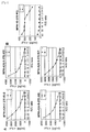
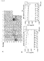
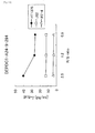
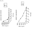
- CTLs were successfully established using MPHOSPH1-A24-9-278 (IYNEYIYDL (SEQ ID NO: 7)), MPHOSPH1-A24-10-278 (IYNEyIYDLF (SEQ ID NO: 8)), MPHOSPH1-A2-9-282 (YIYDLFVPV (SEQ ID NO: 9)), MPHOSPH1-A2-9-638 (RLAIFKDLV (set In NO: 10)), MPHOSPH1-A2-10-1714 (TMSSsKLSNV (SEQ ID NO: 11)), DEPDC1-A24-9-294 (EYYELFVNI (SEQ ID NO: 12)), DEPDC1-A02-10-644 (SLMIhTFSRC (SEQ ID NO: 240)), DEPDC1-A02-10-575 (SLLPaSSMLT (SEQ ID NO: 241)), DE
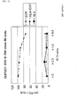
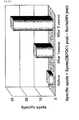
- CTLs demonstrated potent cytotoxic activity against the peptide-pulsed A24LCL and T2 cells. Furthermore, CTL clones derived from these cells also demonstrated specific cytotoxicity against HLA-A24 or HLA-A2 positive cells expressing MPHOSPH1 or DEPDC1, respectively.. However, these CTL clones did not express cytotoxic activity against cells having expression of only one of peptides, including HLA-A24, HLA-A2, MPHOSPH1 and DEPDC1.
- MPHOSPH1 and DEPDC1 as TAAs for cancer cells and that MPHOSPH1-A24-9-278 (IYNEYIYDL (SEQ ID NO: 7)), MPHOSPH1-A24-10-278 (IYNEyIYDLF (SEQ ID NO: 8)), MPHOSPH1-A2-9-282 (YIYDLFVPV (SEQ ID NO: 9)), MPHOSPH1-A2-9-638 (RLAIFKDLV (SEQ ID NO: 10)), MPHOSPH1-A2-10-1714 (TMSSsKLSNV (SEQ ID NO: 11)), DEPDC1-A24-9-294 (EYYELFVNI (SEQ ID NO: 12)), DEPDC1-A02-10-644 (SLMIhTFSRC (SEQ ID NO: 240)), DEPDC1-A02-10-575 (SLLPaSSMLT (SEQ ID NO: 241)), DEPDC1-A02-10-506
- cancers include, but are not limited to, bladder cancer, breast cancer, cervical cancer, cholangincellular carcinoma, CML, colorectal cancer, gastric cancer, NSCLC, lymphoma, osteosarcoma, prostate cancer, renal carcinoma, SCLC, soft tissue tumor.
- the present invention further provides methods of treating or preventing a disease associated with the over-expression of MPHOSPH1 and/or DEPDC1, e.g. cancers, in a subject, such methods including the steps of administering to a subject in need thereof an immunogenic peptide of less than about 40 amino acids, often less than about 20 amino acids, usually less than about 15 amino acids and having the amino acid sequence of SEQ ID NOs: 7, 8, 9, 10, 11, 12, 192, 195, 197, 209, 225, 226 228, 230, 240, 241, 243, 244, 249, 253, 254 or 255.
- the immunogenic peptide may be composed of a sequence of SEQ ID NOs: 7, 8, 9, 10, 11, 12, 192, 195, 197, 209, 225, 226 228, 230, 240, 241, 243, 244, 249, 253, 254 or 255 in which 1, 2, or several (e.g., up to 5) amino acids are substituted, deleted or added, provided the resulting variant peptide retains the immunogenic activity (i.e., the ability to induce CTLs specific to cells expressing MPHOSPH1 and/or DEPDC1, e.g. cancers).
- the number of residues to be substituted, deleted, or added is generally 5 amino acids or less, preferably 4 amino acids or less, more preferably 3 amino acids or less, even more preferably one or two amino acids.
- the cancers contemplated include, but are not limited to, bladder cancer, breast cancer, cervical cancer, cholangincellular carcinoma, CML, colorectal cancer, gastric cancer, NSCLC, lymphoma, osteosarcoma, prostate cancer, renal carcinoma, SCLC, soft tissue tumor.
- Variant peptides i.e., peptides having an amino acid sequence modified by substituting, deleting, or adding one, two or several amino acid residues to an original amino acid sequence
- Variant peptides are known to retain the original biological activity ( Mark DF et al., (1984) Proc Natl Acad Sci USA 81: 5662-6 .; Zoller MJ and Smith M, (1982) Nucleic Acids Res 10:6487-500 .; Dalbadie-McFarland G et al., (1982) Proc Natl Acad Sci USA 79: 6409-13 .).
- it is preferable that the amino acid modification results in conservation of the properties of the original amino acid side-chain (a process known as conservative amino acid substitution).
- amino acid side chains examples include hydrophobic amino acids (A, I, L, M, F, P, W, Y, V), hydrophilic amino acids (R, D, N, C, E, Q, G, H, K, S, T), and side chains having the following functional groups or characteristics in common: an aliphatic side-chain (G, A, V, L, I, P); a hydroxyl group containing side-chain (S, T, Y); a sulfur atom containing side-chain (C, M); a carboxylic acid and amide containing side-chain (D, N, E, Q); a base containing side-chain (R, K, H); and an aromatic containing side-chain (H, F, Y, W).
- A, I, L, M, F, P, W, Y, V hydrophilic amino acids
- R, D, N, C, E, Q amino acids
- G, A, V, L, I, P a hydroxyl group containing side-chain
- the immunogenic peptide is a nonapeptide (9-mer) or a decapeptide (10-mer).
- the present invention further provides a method of inducing anti-tumor immunity for a disease associated with the over-expression of MPHOSPH1 and/or DEPDC1, e.g. cancers, in a subject, such a method including the steps of administering to a subject in need thereof an immunogenic peptide of the present invention, namely one having the amino acid sequence of S SEQ ID NOs: 7, 8, 9, 10,11, 12,192,195, 197, 209, 225, 226 228, 230, 240, 241, 243, 244, 249, 253, 254 or 255 or a variant thereof (i.e., including 1, 2, or several amino acid substitutions, deletions, or additions).
- the cancers contemplated include, but are not limited to, bladder cancer, breast cancer, cervical cancer, cholangincellular carcinoma, CML, colorectal cancer, gastric cancer, NSCLC, lymphoma, osteosarcoma, prostate cancer, renal carcinoma, SCLC, soft tissue tumor.
- the subject is preferably a mammal.
- exemplary mammals include, but are not limited to, e.g., a human, non-human primate, mouse, rat, dog, cat, horse, or cow.
- the peptide can be administered to a subject via an in vivo or ex vivo protocol.
- the present invention also provides use of nonapeptide or decapeptide selected from peptides having the amino acid sequence of SEQ ID NOs: 7, 8, 9, 10, 11, 12, 192, 195, 197, 209, 225, 226 228, 230, 240, 241, 243, 244, 249, 253, 254 and 255 (and variants thereof) for manufacturing an immunogenic composition for treating or preventing a disease associated with the over-expression of MPHOSPH1 and/or DEPDC1, e.g. cancers.
- the cancers contemplated include, but are not limited to, bladder cancer, breast cancer, cervical cancer, cholangincellular carcinoma, CML, colorectal cancer, gastric cancer, NSCLC, lymphoma, osteosarcoma, prostate cancer, renal carcinoma, SCLC, soft tissue tumor.
- MPHOSPH1-A24-9-278 IYNEYIYDL (SEQ ID NO: 7)
- MPHOSPH1-A24-10-278 IYNEyIYDLF (SEQ ID NO: 8)
- MPHOSPH1-A2-9-282 YIYDLFVPV (SEQ ID NO: 9)
- MPHOSPH1-A2-9-638 RLAIFKDLV (SEQ ID NO: 10)
- MPHOSPH1-A2-10-1714 TMSSsKLSNV (SEQ ID NO: 11)
- DEPDC1-A24-9-294 EYYELFVNI (SEQ ID NO: 12)
- DEPDC1-A2-9-589 LQPHLERV (SEQ ID NO: 192)
- DEPDC1-A2-9-619 LLMRMISRM (SEQ ID NO: 195)
- DEPDC1-A2-9-290 LLTFEYYEL (SEQ ID NO: 197)
- DEPDC1-A2-9-5 I
- HLA antigens the data presented here demonstrate that the uses of A-24 type or A-2 type antigens (which are said to be highly expressed among the Japanese) are favorable for obtaining effective results.
- the uses of subtypes such as A-2402 and A-0201 are even more preferable.
- the type of HLA antigen of the patient requiring treatment is investigated in advance, which, in turn, enables the selection of appropriate peptides having high levels of binding affinity to the patient antigen, or having cytotoxic T cell (CTL) inducibility by antigen presentation.
- CTL cytotoxic T cell
- substitution, deletion, or addition of 1, 2, or several amino acids may be performed based on the amino acid sequence of the naturally occurring MPHOSPH1 and DEPDC1 partial peptide.
- the term “several” means refers to 5 or less, more preferably 3 or less.
- the regularity of the sequences of peptides displayed by binding to HLA antigens is already known ( Kubo RT, et al., (1994) J. Immunol., 152, 3913-24 .; Rammensee HG, et al., (1995) Immunogenetics.
- immunogenic peptides of the invention modifications based on such regularity can be performed on the immunogenic peptides of the invention.
- peptides possessing high HLA-24 binding affinity in which the second amino acid from the N terminus substituted with phenylalanine, tyrosine, methionine, or tryptophan may be favorably used.
- peptides whose C-terminal amino acid is substituted with phenylalanine, leucine, isoleucine, tryptophan, or methionine may also be used favorably.
- peptides possessing high HLA-A2 binding affinity having their second amino acid from the N terminus substituted with leucine or methionine, and peptides whose C-terminal amino acid is substituted with valine or leucine may be used favorably.
- 1 to 2 amino acids may be added to the N terminus and/or C terminus of the peptide.
- the peptide sequence is identical to a portion of the amino acid sequence of an endogenous or exogenous protein having a different function, side effects such as autoimmune disorders or allergic symptoms against specific substances may be induced. Therefore, it is preferable to avoid the situation wherein the immunogenic sequence matches the amino acid sequence of a known protein. This situation may be avoided by performing a homology search using available databases. If homology searches confirm that peptides in which 1, 2 or several different amino acids do not exist in nature, then the danger that modifications of the above-mentioned amino acid sequence that, for example, increase the binding affinity with HLA antigens, and/or increase the CTL inducibility can be avoided.
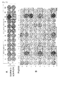
- CTL inducibility may be routinely confirmed by inducing antigen-presenting cells carrying human MHC antigens (for example, B-lymphocytes, macrophages, and dendritic cells), or more specifically dendritic cells derived from human peripheral blood mononuclear leukocytes, and, after stimulation with the peptide of interest, mixing with CD8-positive cells and measuring the cytotoxic activity against the target cells.
- human MHC antigens for example, B-lymphocytes, macrophages, and dendritic cells
- transgenic animals produced to express a human HLA antigen may be used.
- the target cells can be radiolabeled with 51 Cr and such, and cytotoxic activity can be calculated from radioactivity released from the target cells.
- it can be examined by measuring IFN-gamma produced and released by CTL in the presence of antigen-presenting cells that carry immobilized peptides, and visualizing the inhibition zone on the media using anti-IFN-gamma monoclonal antibodies.
- nonapeptides or decapeptides selected from the group of peptides having the amino acid sequences indicated by IYNEYIYDL (SEQ ID NO: 7), IYNEyIYDLF (SEQ ID NO: 8), YIYDLFVPV (SEQ ID NO: 9), RLAIFKDLV (SEQ ID NO: 10), TMSSsKLSNV (SEQ ID NO: 11), EYYELFVNI (SEQ ID NO: 12), LLQPHLERV (SEQ ID NO: 192), LLMRMISRM (SEQ ID NO: 195), LLTFEYYEL (SEQ ID NO: 197), RLCKSTIEL (SEQ ID NO: 209), CVLCCAEEV (SEQ ID NO: 225), FLMDhHQEIL (SEQ ID NO: 225), FLMDhHQEIL (SEQ ID NO: 225), FLMDhHQEIL (SEQ ID NO: 225), FLMDhHQEIL (SEQ ID NO: 225), FLMDh
- the present invention provides peptides having cytotoxic T cell inducibility, namely those having the amino acid sequence of SEQ ID NOs: 7, 8, 9, 10, 11, 12, 192, 195, 197, 209, 225, 226 228, 230, 240, 241, 243, 244, 249, 253, 254 or 255 or a variant thereof (i.e., those in which 1, 2, or several amino acids are substituted, deleted, or added).
- amino acid sequences composed of 9 or 10 amino acids indicated in SEQ ID NOs: 7, 8, 9, 10, 1 l, 12, 192, 195, 197, 209, 225, 226 228, 230, 240, 241, 243, 244, 249, 253, 254 or 255 or a variant thereof do not match an amino acid sequence associated with another endogenous protein.
- amino acid substitution to leucine or methionine at the second amino acid from the N terminus amino acid substitution to valine or leucine at the C-terminal amino acid, and amino acid addition of 1 to 2 amino acids at the N terminus and/or C terminus are examples of preferred variants.
- immunologically active fragments of the peptides may also be used in the methods of the invention. Methods for determining active fragments are well known in the art. CTL clones obtained by stimulation by these modified peptides can recognize the original peptides and cause damage for cells expressing the original peptides.
- Peptides of the present invention can be prepared using well known techniques.
- the peptides can be prepared synthetically, using either recombinant DNA technology or chemical synthesis.
- Peptides of the present invention may be synthesized individually or as longer polypeptides comprising two or more peptides.
- the peptides of the present invention are preferably isolated, i.e., substantially free of other naturally occurring host cell proteins and fragments thereof.
- the peptides of the present invention may contain modifications, such as glycosylation, side chain oxidation, or phosphorylation; so long as the modifications do not destroy the biological activity of the peptides as described herein, namely the ability to binding to an HLA antigen and induce CTL.
- modifications include incorporation of D-amino acids or other amino acid mimetics that can be used, for example, to increase the serum half life of the peptides.
- the peptides of this invention can be prepared as a combination, which includes two or more of peptides of the invention, for use as a vaccine for a disease associated with the over-expression of MPHOSPH1 and/or DEPDC1, e.g. cancers, such a vaccine inducing CTL in vivo.
- the cancers contemplated include, but are not limited to, bladder cancer, breast cancer, cervical cancer, cholangincellular carcinoma, CML, colorectal cancer, gastric cancer, NSCLC, lymphoma, osteosarcoma, prostate cancer, renal carcinoma, SCLC, soft tissue tumor.
- the peptides may be in a cocktail or may be conjugated to each other using standard techniques.
- the peptides can be expressed as a single polypeptide sequence.
- the peptides in the combination may be the same or different.
- the peptides are presented at a high density on the HLA antigens of antigen-presenting cells, which, in turn, induces CTLs that specifically react toward the complex formed between the displayed peptide and the HLA antigen.
- antigen-presenting cells having immobilized the peptides of this invention on their cell surface obtained by removing dendritic cells from the subjects, may be stimulated by the peptides of this invention. Re-administration of these cells to the respective subjects induces CTL, and, as a result, aggressiveness towards the target cells can be increased.
- the present invention provides drugs for treating and/or preventing proliferation, metastasis, and such of a disease associated with the over-expression of MPHOSPH1 and/or DEPDC1, e.g. cancers, which include one or more of peptides of this invention.
- the peptides of this invention find particular utility in the treatment of a disease associating MPHOSPH1 and/or DEPDC1, e.g. cancers.
- the cancers contemplated include, but are not limited to, bladder cancer, breast cancer, cervical cancer, cholangincellular carcinoma, CML, colorectal cancer, gastric cancer, NSCLC, lymphoma, osteosarcoma, prostate cancer, renal carcinoma, SCLC, soft tissue tumor.
- the peptides of this invention can be administered to a subject directly, as a pharmaceutical composition that has been formulated by conventional formulation methods.
- carriers, excipients, and such that are ordinarily used for drugs can be included as appropriate, without particular limitations.
- the immunogenic compositions of this invention may be used for treatment and prevention of a disease associating MPHOSPH11 and/or DEPDC, e.g. cancers.
- the cancers contemplated include, but are not limited to, bladder cancer, breast cancer, cervical cancer, cholangincellular carcinoma, CML, colorectal cancer, gastric cancer, NSCLC, lymphoma, osteosarcoma, prostate cancer, renal carcinoma, SCLC, soft tissue tumor.
- the immunogenic compositions for treatment and/or prevention of a disease associated with the over-expression of MPHOSPH1 and/or DEPDC1 can further include an adjuvant so that cellular immunity will be established effectively. Alternatively, they may be administered with other active ingredients, such as anticancer agents.
- the cancers contemplated include, but are not limited to, bladder cancer, breast cancer, cervical cancer, cholangincellular carcinoma, CML, colorectal cancer, gastric cancer, NSCLC, lymphoma, osteosarcoma, prostate cancer, renal carcinoma, SCLC, soft tissue tumor. Suitable formulations include granules.
- Suitable adjuvants are described in the literature ( Johnson AG. (1994) Clin. Microbiol. Rev., 7:277-89 .).
- Exemplary adjuvants include, but are not limited to, aluminum phosphate, aluminum hydroxide, and alum.
- liposome formulations, granular formulations in which the drug is bound to few-micrometer diameter beads, and formulations in which a lipid is bound to the peptide may be conveniently used.
- the method of administration may be oral, intradermal, subcutaneous, intravenous injection, or such, and may include systemic administration or local administration to the vicinity of the targeted tumor.
- the dose of the peptide(s) of this invention can be adjusted appropriately according to the disease to be treated, age of the patient, weight, method of administration, and such.
- the dosage is ordinarily 0.001 mg to 1000 mg, preferably 0.01 mg to 100 mg, more preferably 0.1 mg to 10 mg, preferably administered once in a few days to few months, one skilled in the art can readily select the appropriate dose and method of administration, as, the selection and optimization of these parameters is well within routine skill.
- the present invention further provides intracellular vesicles called exosomes, which present complexes formed between the peptides of this invention and HLA antigens on their surface.
- Exosomes can be prepared, for example, by using the methods described in detail in Published Japanese Translation of International Publication Nos. Hei 11-510507 and 2000-512161 , and are preferably prepared using antigen-presenting cells obtained from subjects who are targets of treatment and/or prevention.
- the exosomes of this invention can be inoculated as cancer vaccines, similarly to the peptides of this invention.
- HLA-A24 or HLA-A2 particularly HLA-A2402 or HLA-A0201, is often appropriate.
- the vaccine compositions of the present invention include a component which primes cytotoxic T lymphocytes.
- Lipids have been identified as agents capable of priming CTL in vivo against viral antigens.
- palmitic acid residues can be attached to the epsilon-and alpha-amino groups of a lysine residue and then linked to an immunogenic peptide of the invention.
- the lipidated peptide can then be administered either directly, in a micelle or particle, incorporated into a liposome, or emulsified in an adjuvant.
- a lipid priming of CTL responses E.
- coli lipoproteins such as tripalmitoyl-S-glycerylcysteinlyseryl- serine (P3CSS)
- P3CSS tripalmitoyl-S-glycerylcysteinlyseryl- serine
- the immunogenic compositions of the present invention may also include nucleic acids encoding one or more of the immunogenic peptides disclosed here. See, e.g., Wolff JA et al., (1990) Science 247:1465-8 ; U.S. Patent Nos. 5,580,859 ; 5,589,466 ; 5,804,566 ; 5,739,118 ; 5,736,524 ; 5,679,647 ; and WO 98/04720 .
- DNA-based delivery technologies include "naked DNA", facilitated (bupivicaine, polymers, peptide-mediated) delivery, cationic lipid complexes, and particle-mediated (“gene gun”) or pressure-mediated delivery (see, e.g., U.S. Patent No. 5,922,687 ).
- the immunogenic peptides of the invention can also be expressed by viral or bacterial vectors.
- suitable expression vectors include attenuated viral hosts, such as vaccinia or fowlpox. This approach involves the use of vaccinia virus, e.g., as a vector to express nucleotide sequences that encode the peptide. Upon introduction into a host, the recombinant vaccinia virus expresses the immunogenic peptide, and thereby elicits an immune response.
- Vaccinia vectors and methods useful in immunization protocols are described in, e.g., U.S. Patent No. 4,722,848 .
- Another suitable vector is BCG (Bacille Calmette Guerin).
- BCG vectors are described in Stover CK, et al., (1991) Nature 351:456-60 .
- vectors useful for therapeutic administration or immunization e.g., adeno and adeno-associated virus vectors, retroviral vectors, Salmonella typhi vectors, detoxified anthrax toxin vectors, and the like, are known in the art. See, e.g., Shata MT, et al., (2000) Mol. Med. Today 6:66-71 ; Shedlock DJ and Weiner DB., et al., (2000) J. Leukoc. Biol. 68:793-806 ; and Hipp JD, et al., (2000) In Vivo 14:571-85 .
- the present invention also provides methods of inducing antigen-presenting cells using one or more peptides of this invention.
- the antigen-presenting cells can be induced by inducing dendritic cells from the peripheral blood monocytes and then contacting (stimulating) them with one or more peptides of this invention in vitro, ex vivo or in vivo.
- peptides of the present invention are administered to the subjects, antigen-presenting cells that have the peptides of this invention immobilized to them are induced in the body of the subject.
- the cells can be administered to the subject as a vaccine.
- the ex vivo administration may include the steps of:
- the antigen-presenting cells obtained by step b can be administered to the subject as a vaccine.
- This invention also provides a method for inducing antigen-presenting cells having a high level of cytotoxic T cell inducibility, in which the method includes the step of transferring genes composed of polynucleotide(s) encoding one or more peptides of this invention to antigen-presenting cells in vitro.
- the introduced genes may be in the form of DNAs or RNAs.
- carious methods conventionally performed in this field such as lipofection, electroporation, and calcium phosphate method may be suitably used. More specifically, transfection may be performed as described in Reeves ME, et al., (1996) Cancer Res., 56:5672-7 .; Butterfield LH, et al., (1998) J.
- the present invention further provides methods for inducing CTL using one or more peptides of this invention.
- CTL are induced in the body of the subject, and the strength of the immune system targeting the cells expressing MPHOSPH1 and/or DEPDC1, e.g. cancer cells in the tumor tissues is thereby enhanced.
- the cancers contemplated include, but are not limited to, bladder cancer, breast cancer, cervical cancer, cholangincellular carcinoma, CML, colorectal cancer, gastric cancer, NSCLC, lymphoma, osteosarcoma, prostate cancer, renal carcinoma, SCLC, soft tissue tumor.
- the peptides of the present invention may be used in the context of an ex vivo therapeutic method, in which subject-derived antigen-presenting cells and CD8-positive cells or peripheral blood mononuclear leukocytes are contacted (stimulated) with one or more peptides of this invention in vitro, and, after inducing CTL, the cells are returned to the subject.
- the method may include the steps of:
- the present invention further provides methods for producing activated cytotoxic T cell using the peptides of this invention.
- the method may include the following steps of:
- the present invention also provides method for producing APC having activated T-cell inducibility using the peptides of the present invention.
- the method may include the step of contacting antigen presenting cells with the peptides to produce antigen presenting cells presenting the peptide and HLA antigen on the surface.
- activated cytotoxic T cell induces IFN-gamma producing, IFN-gamma releasing, and death of tumor cells.
- the present invention further provides isolated cytotoxic T cells induced using the peptides of this invention.
- the cytotoxic T cells induced by stimulation with an antigen-presenting cell presenting one or more peptides of this invention, are preferably derived from subjects who are the target of treatment and/or prevention, and can be administered alone or in combination with other drugs, including one or more peptides of this invention or exosomes having anti-tumor activity.
- the obtained cytotoxic T cells act specifically against target cells presenting the peptides of this invention, or preferably the same peptide(s) used for induction.
- the target cells may be cells that express MPHOSPH1 and/or DEPDC1 endogenously, or cells that are transfected with MPHOSPH11 and/or DEPDC1 genes. Cells that present the peptides of this invention on the cell surface, due to stimulation with these peptides, can also become targets of attack.
- the present invention also provides antigen-presenting cells presenting complexes formed between HLA antigens and one or more peptides of this invention.
- the antigen-presenting cells obtained through contact with the peptides of this invention or the nucleotides encoding such peptides, are preferably derived from subjects who are the target of treatment and/or prevention, and can be administered as vaccines, alone or in combination with other drugs, including the peptides, exosomes, or cytotoxic T cells of the present invention.
- the present invention also provides a composition comprising nucleic acids encoding polypeptides that are capable of forming a subunit of a T cell receptor (TCR), and methods of using the same.
- TCR subunits have the ability to form TCRs that confer specificity to T cells for tumor cells presenting MPHOSPH1 or DEPDC1.
- the nucleic acids of alpha- and beta-chain as the TCR subunits of the CTL induced with one or more peptides of this invention may be identified ( WO2007/0322 and Morgan et al., J Immunol, 171, 3288 (2003 )).
- the derivative TCRs preferably bind target cells displaying the MPHOSPH1 or DEPDC1 peptide with high avidity, and optionally mediate efficient killing of target cells presenting the MPHOSPH1 or DEPDC1 peptide in vivo and in vitro.
- the nucleic acids encoding the TCR subunits can be incorporated into suitable vectors e.g. retroviral vectors. These vectors are well known in the art.
- the nucleic acids or the vectors comprising them usefully can be transferred into a T cell, which T cell is preferably from a patient.
- the invention provides an off-the-shelf composition allowing rapid modification of a patient's own T cells (or those of another mammal) to rapidly and easily produce modified T cells having excellent cancer cell killing properties.
- the present invention provides CTLs which are prepared by transduction with the nucleic acids encoding the TCR subunits polypeptides binding with MPHOSPH1 or DEPDC1 peptide e.g. SEQ ID NOs: 7, 8, 9, 10, 11, 12, 192, 195, 197, 209, 225, 226 228, 230, 240, 241, 243, 244, 249, 253, 254 or 255 in the context of HLA-A24 or HLA-A2.
- the transduced CTLs are capable of homing to cancer cells in vivo, and expanded by well known culturing method in vitro (e.g., Kawakami et al., J Immunol., 142, 3452-3461 (1989 )).
- the T cells of the invention can be used to form an immunogenic composition useful in treating or preventing cancer in a patient in need of therapy or protection ( WO2006/031221 ).
- the term "vaccine” refers to a substance that induces anti-tumor immunity or suppresses cancers upon inoculation into animals.
- polypeptides having the amino acid sequence of SEQ ID NO: 7, 8 or 12 were suggested to be HLA-A24 restricted epitope peptides and those of SEQ ID NO: 9, 10, 11, 192, 195, 197, 209, 225, 226, 228 230, 240, 241, 243, 244, 249, 253, 254 or 255 were suggested to be HLA-A2 restricted epitope peptides that may induce potent and specific immune response against cells expressing MPHOSPH1 and/or DEPDC1, e.g.
- cancer cells expressing MPHOSPH1 and/or DEPDC1 include, but are not limited to, bladder cancer, breast cancer, cervical cancer, cholangincellular carcinoma, CML, colorectal cancer, gastric cancer, NSCLC, lymphoma, osteosarcoma, prostate cancer, renal carcinoma, SCLC, soft tissue tumor.
- the present invention also encompasses a method of inducing anti-tumor immunity using polypeptides having the amino acid sequence of SEQ ID NO: 7, 8, 9, 10, 11, 12, 192, 195, 197, 209, 225, 226, 228, 230, 240, 241, 243, 244, 249, 253, 254 or 255 or a variant thereof (i.e., including 1, 2, or several amino acid substitutions, deletions, or additions).
- anti-tumor immunity includes immune responses such as follows:
- the peptide when a certain peptide induces any one of these immune responses upon inoculation into an animal, the peptide is decided to have anti-tumor immunity inducing effect.
- the induction of the anti-tumor immunity by a peptide can be detected by observing in vivo or in vitro the response of the immune system in the host against the peptide.
- cytotoxic T lymphocytes For example, a method for detecting the induction of cytotoxic T lymphocytes is well known.
- a foreign substance that enters the living body is presented to T cells and B cells by the action of antigen-presenting cells (APCs).
- APCs antigen-presenting cells
- T cells that respond to the antigen presented by APC in antigen specific manner differentiate into cytotoxic T cells (also referred to as cytotoxic T lymphocytes or CTLs) due to stimulation by the antigen, and then proliferate; this process is referred to herein as "activation" of T cells. Therefore, CTL induction by a certain peptide can be evaluated by presenting the peptide to a T cell by APC, and detecting the induction of CTL.
- APCs have the effect of activating CD4+ T cells, CD8+ T cells, macrophages, eosinophils and NK cells. Since CD4+ T cells are also important in anti-tumor immunity, the anti-tumor immunity inducing action of the peptide can be evaluated using the activation effect of these cells as indicators.
- DC dendritic cells
- APC dendritic cells
- DC is a representative APC having the strongest CTL inducing action among APCs.
- the test polypeptide is initially contacted with DC and then this DC is contacted with T cells. Detection of T cells having cytotoxic effects against the cells of interest after the contact with DC shows that the test polypeptide has an activity of inducing the cytotoxic T cells.
- Activity of CTL against tumors can be detected, for example, using the lysis of 51 Cr-labeled tumor cells as the indicator.
- peripheral blood mononuclear cells may also be used as the APC.
- the induction of CTL is reported to be enhanced by culturing PBMC in the presence of GM-CSF and IL-4.
- CTL has been shown to be induced by culturing PBMC in the presence of keyhole limpet hemocyanin (KLH) and IL-7.
- KLH keyhole limpet hemocyanin
- test polypeptides confirmed to possess CTL inducing activity by these methods are polypeptides having DC activation effect and subsequent CTL inducing activity. Therefore, polypeptides that induce CTL against tumor cells are useful as vaccines against diseases associated with the over-expression of MPHOSPH1 and/or DEPDC1, e.g. cancers. Furthermore, APC that have acquired the ability to induce CTL against a disease associating MPHOSPH1 and/or DEPDC1, e.g. cancers, by contacting with the polypeptides are useful as vaccines against the disease.
- CTL that have acquired cytotoxicity due to presentation of the polypeptide antigens by APC can be also used as vaccines against a disease associating MPHOSPH1 and/or DEPDC1, e.g. cancers.
- a disease associating MPHOSPH1 and/or DEPDC1 and/or DEPDC1 e.g. cancers
- Such therapeutic methods for a disease associating MPHOSPH1 and/or DEPDC1, e.g. cancers, using anti-tumor immunity due to APC and CTL are referred to as cellular immunotherapy.
- the cancers contemplated include, but are not limited to, bladder cancer, breast cancer, cervical cancer, cholangincellular carcinoma, CML, colorectal cancer, gastric cancer, NSCLC, lymphoma, osteosarcoma, prostate cancer, renal carcinoma, SCLC, soft tissue tumor.
- the induction of anti-tumor immunity by a polypeptide can be further confirmed by observing the induction of antibody production against tumors. For example, when antibodies against a polypeptide are induced in a laboratory animal immunized with the polypeptide, and when growth, proliferation and/or metastasis of tumor cells is suppressed by those antibodies, the polypeptide is determined to induce anti-tumor immunity.
- Anti-tumor immunity can be induced by administering a vaccine of this invention, and the induction of anti-tumor immunity enables treatment and prevention of a disease associated with the over-expression of MPHOSPH1 and/or DEPDC1, e.g. cancers.
- Therapy against or prevention of the onset of a disease associated with the over-expression of MPHOSPH1 and/or DEPDC1, e.g. cancers may include inhibition of the growth of cells expressing MPHOSPH1 and/or DEPDC1, e.g. cancer cells, involution of these cells and suppression of occurrence of these cells, e.g. cancer cells.
- cancers decrease of the disease markers in the blood, alleviation of detectable symptoms accompanying the disease and such are also included in the therapy or prevention of the disease, e.g. cancers.
- Such therapeutic and preventive effects are preferably statistically significant, for example, observed at a significance level of 5% or less, wherein the therapeutic or preventive effect of a vaccine against a disease associating MPHOSPH1 and/or DEPDC1, e.g. cancers, is compared to a control without vaccine administration.
- Student's t-test, the Mann-Whitney U-test or ANOVA may be used for determining statistical significance.
- the present invention provides a method for treating, or preventing a disease associated with the over-expression of MPHOSPH1 and/or DEPDC1, e.g. cancers
- the therapeutic compounds or compositions may be administered prophylactically or therapeutically to subjects suffering from or at risk of (or susceptible to) developing the disease. Such subjects may be identified using standard clinical methods.
- prophylactic administration occurs prior to the manifestation of overt clinical symptoms of disease, such that a disease or disorder is prevented or alternatively delayed in its progression.
- the term “prevent” encompasses any activity which reduces the burden of mortality or morbidity from disease. Prevention can occur "t primary, secondary and tertiary prevention levels.
- the term “efficacious” refers to a treatment that leads to a decrease in size, prevalence or metastatic potential of cancer in a subject.
- “efficacious” means that the treatment retards or prevents occurrence of non cancer or alleviates a clinical symptom of cancer.
- the assessment of cancer can be made using standard clinical protocols.
- the efficaciousness of a treatment may be determined in association with any known method for diagnosing or treating cancer. For example, cancer can be diagnosed histopathologically or by identifying symptomatic anomalies.
- peptide having immunological activity, or a polynucleotide or vector encoding such a peptide
- An adjuvant refers to a compound that enhances the immune response against the peptide when administered together (or successively) with the peptide having immunological activity.
- suitable adjuvants include cholera toxin, salmonella toxin, alum and such, but are not limited thereto.
- a vaccine of this invention may be combined appropriately with a pharmaceutically acceptable carrier. Examples of such carriers are sterilized water, physiological saline, phosphate buffer, culture fluid and such.
- the vaccine may contain as necessary, stabilizers, suspensions, preservatives, surfactants and such.
- the vaccine is administered systemically or locally.
- Vaccine administration may be performed by single administration or boosted by multiple administrations.
- a disease associated with the over-expression of MPHOSPH1 and/or DEPDC1 can be treated or prevented, for example, by the ex vivo method. More specifically, PBMCs of the subject receiving treatment or prevention are collected, contacted ex vivo with a peptide of the present invention. Following the induction of APC or CTL, the cells may be administered to the subject.
- APC can be also induced by introducing a vector encoding the peptide into PBMCs ex vivo.
- APC or CTL induced in vitro can be cloned prior to administration. By cloning and growing cells having high activity of damaging target cells, cellular immunotherapy can be performed more effectively.
- APC and CTL isolated in this manner may be used for cellular immunotherapy not only against individuals from whom the cells are derived, but also against similar types of diseases in other individuals.
- A24LCL cells HLA-A24/24
- human B-lymphoblastoid cell lines T2 cell and COS7 were purchased from ATCC.
- DCs Monocyte-derived dendritic cells
- APCs antigen-presenting cells
- peripheral blood mononuclear cells isolated from a normal volunteer (HLA-A*2402 and/or HLA-A*0201) by Ficoll-Paque (Pharmacia) solution were separated by adherence to a plastic tissue culture flask (Becton Dickinson) so as to enrich them for the monocyte fraction.
- the monocyte-enriched population was cultured in the presence of 1000 U/ml of GM-CSF (Genzyme) and 1000 U/ml of IL-4 (Genzyme) in AIM-V (Invitrogen) containing 2% heat-inactivated autologous serum (AS).
- the cytokine-generated DCs were pulsed with 20 mcg/ml of the synthesized peptides in the presence of 3 mcg/ml of beta 2-microglobulin for 4 hrs at 20 degrees C in AIM-V. These peptide-pulsed DCs were then inactivated by MMC (30 mcg/ml for 30 mins) and mixed at a 1:20 ratio with autologous CD8 + T cells, obtained by positive selection with Dynabeads M-450 CD8 (Dynal) and DETACHa BEADTM (Dynal).
- CTLs were expanded in culture using the method similar to that described by Riddell SR, et al., ( Walter EA et al., (1995) N Engl J Med 333:1038-44 .; Riddel SR, et al., (1996) Nature Med. 2:216-23 .).
- a total 5x10 4 of CTLs were resuspended in 25 ml of AIM-V/5% AS with 2 kinds of human B-lymphoblastoid cell lines, inactivated by MMC, in the presence of 40 ng/ml of anti-CD3 monoclonal antibody (Pharmingen).
- 120 IU/ml of IL-2 were added to the cultures.
- the cultures were fed with fresh AIM-V/5% AS containing 30 IU/ml of IL-2 on days 5, 8 and 11.
- CTLs were cultured with 7x10 4 cells/well of 2 kinds of human B-lymphoblastoid cell lines, 30ng/ml of anti-CD3 antibody, and 125 U/ml of IL-2 in total of 150 mcl/well of AIM-V containing 5%AS. 50 mcl /well of IL-2 was added to the medium 10 days later so that IL-2 became 125 U/ml in the final concentration. CTL activity of CTLs was tested on the 14th day, and CTL clones were expanded using the same method above.
- IFN-gamma ELISPOT assay To examine the specific CTL activity, IFN-gamma ELISPOT assay and IFN-gamma ELISA were performed. Briefly, peptide-pulsed A24-LCL or T2 cell (1x10 4 /well) was prepared as a stimulator cell. Cultured Cells in 48 wells or CTL clones after limiting dilution were used as a responder cells. IFN-gamma ELISPOT assay and ELISA were performed under manufacture procedure.
- HLA-A24 B-LCLs Epstein Bar virus-transformed.
- Jiyoye, EB-3, COS7, HT1376, RT-4 and J82 were purchased from American Type Culture Collection (Rockville, MD).
- A24LCL, Jiyoye and EB-3 were maintained in RPMI1640 containing 10% fetal bovine serum (GEMINI Bio-Products) and 1% antibiotic solution (Sigma).
- COS7, HT1376, RT-4 and J82 were maintained in appropriate medium and antibiotics. Transfection of COS7 and HEK were performed using FUGENE6 (Roche).
- HEK-A2 cell HLA-A*0201 molecule expressing stable clone, was established by transfection of pcDNA6.2-HLA-A2 plasmid and isolated by limiting dilution method in the presence of 5 mcg/ml Blastcidin S.
- immunization was given using 100 ml of vaccine mixture, which contains 50 mcl (100 mcg) of HLA-A24 restricted peptide and 50 mcl of IFA per mouse.
- the vaccine was subcutaneously injected in the right flank for the first immunization on day 0 and in the left flank for the second on the day 7.
- splenocytes of the vaccinated mice without any in vitro stimulation, were used responder cells, and RLmale1 cells pulsed with or without peptides were used as the stimulator cells for IFN-gamma ELISPOT assay.
- the global gene expression profile data obtained from various cancers using cDNA-microarray revealed that MPHOSPH1 (GenBank Accession No. NM_016195; SEQ ID No.1) and DEPDC1 (GenBank Accession No. BM683578) which had two variants; DEPDC1 V1 (SEQ ID Nos.3) and DEPDC1 V2 (SEQ ID No. 5) expression was elevated.
- MPHOSPH1 expression was validly elevated in 30 out of 31 bladder cancers, 8 out of 36 breast cancers, 18 out of 18 cervical cancers, 5 out of 17 cholangincellular carcinomas, 25 out of 31 CMLs, 6 out of 11 colorectal cancers, 6 out of 14 gastric cancers, 5 out of 5 NSCLCs, 7 out of 7 lymphomas, 6 out of 10 osteosarcomas, 7 out of 22 prostate cancers, 10 out of 18 renal carcinomas and 15 out of 21soft tissue tumors as compared with corresponding normal tissue.
- DEPDC1 expression was validly elevated in 23 out of 25 bladder cancers, 6 out of 13 breast cancers, 12 out of 12 cervical cancers, 6 out of 6 cholangincellular carcinomas, 3 out of 4 CMLs, 2 out of 4 colorectal cancers, 6 out of 6 NSCLCs, 7 out of 7 lymphomas, 10 out of 14 osteosarcomas, 11 out of 24 prostate cancers, 14 out of 14 SCLCs and 22 out of 31 soft tissue tumors as compared with corresponding normal tissue (Table 1).
- CTLs for those peptides derived from MPHOSHP1 were generated according to the protocols set forth in "Materials and Methods" section above. Resulting CTLs having detectable specific CTL activity, as assessed by IFN-gamma ELISPOT assay, are shown in Figure 1A and Figure 2A .
- the cells in the well number #4 stimulated with MPHOSPH1-A24-9-278 demonstrated potent IFN-gamma production as compared with the control.
- the cells in the well number #8 stimulated with MPHOSPH1-A24-10-278 demonstrated potent IFN-gamma production as compared with the control.
- the CTL Clone having the highest specific CTL activity against COS7 was that transfected with both MPHOSPH1 and HLA-A24. However, it did not show significant specific CTL activity against COS7 transfected with neither MPHOSPH1 nor HLA-A24 ( Figure 4 ).
- CTL activity against bladder cancer cell lines endogenously expressing MPHOSPH1 The established CTL clone raised against MPHOSPH1-A24-9-278 (SEQ ID NO: 7) peptide was examined for their ability to recognize the tumor cells endogenously expressing MPHOSPH1.
- CTL activity against HT1376 cells, which endogenously express MPHOSPH1 and HLA-A24 was tested using the CTL clone raised by MPHOSPH1-A24-9-278 (SEQ ID NO: 7) as effector cells. J82 cells and RT-4 cells were used as the target cells which endogenously express MPHOSPH1 but do not express HLA-A24.
- the established CTL clone showed high IFN-gamma production against HT1376 cells that express both MPHOSPH1 and HLA-A24.
- the CTL did not show significant CTL activity against J82 and RT-4 cells which express MPHOSPH1 but not HLA-A24 ( Figure 5 ). It clearly demonstrated that MPHOSPH1-A24-9-278 (SEQ ID NO: 7) peptide was naturally processed to the tumor cell surface with HLA-A24 molecule and recognized by CTL.
- H-2Kd molecule one of the mouse MHC class I, has resemble peptide anchor motif for HLA-A24 molecule and partially cross-react HLA-A24 restricted peptide.
- the present inventors then examined whether MPHOSPH1-A24-9-278 peptide induce the CTL in vivo by vaccination with this peptide using BALB/c mice (H-2Kd). IFA-conjugated peptide was subcutaneously injected into BALB/c mice on the day 0 and 7. On day 14, splenocytes were harvested and used as the responder cells for ELISPOT assay.
- FIG. 7 Resulting CTLs having detectable specific CTL activity, as assessed by IFN-gamma ELISPOT assay, are shown in Figure 7 .
- the cells in the well number #1 and #5, stimulated with MPHOSPH1-A2-9-282 (YIYDLFVPV (SEQ ID NO: 9)) demonstrated potent IFN-gamna production as compared with the control.
- the cells in the well number #8 stimulated with MPHOSPH1-A2-9-638 RVLAIFKDLV (SEQ ID NO: 10)
- FIG. 7C the cells in the well number #4 stimulated with MPHOSPH1-A2-10-1714 (TMSSsKLSNV (SEQ ID NO: 11)) demonstrated potent IFN-gamma production as compared with the control.
- figure 8A MPHOSPH1-A2-9-282 (SEQ ID NO: 9)
- figure 8B MPHOSPH1-A2-9-638 (SEQ ID NO: 10)
- figure 8C MPHOSPH1-A2-10-1714(SEQ ID NO: 9)
- the CTL clones stimulated by the MPHOSPH1-A2-9-282 (YIYDLFVPV (SEQ ID NO: 9)) ( Figure 9A, and 9B ) demonstrated potent specific CTL activity against the peptide-pulsed target without any significant specific CTL activity against targets not pulsed with any peptide.
- CTLs for those peptides derived from DEPDC1 were generated according to the protocol described in "Materials and Methods" section above. Resulting CTLs having detectable specific CTL activity, as assessed by IFN-gamma ELISPOT assay, are shown in Figure 10 .
- Figure 10A the cells in the well number #10 stimulated with DEPDC1-A24-9-294 (EYYELFVNI (SEQ ID NO: 12)) demonstrated potent IFN-gamma production as compared with the control. Accordingly, these cells in the positive well were expanded and limiting dilution was performed.
- the established CTL clones raised against these peptides were examined for their ability to recognize the target cells endogenously expressing DEPDC1 and HLA-A*2402.
- COS7 transfected with full length DEPDC1 but not HLA-A*2402 and COS7 transfected with HLA-A*2402 but not full length DEPDC1 were prepared as controls.
- the CTL Clone demonstrated high specific CTL activity against COS7 transfected both DEPDC1 and HLA-A24. However, it did not demonstrate significant specific CTL activity against COS7 transfected neither DEPDC1 nor HLA-A24 ( Figure 12 ).
- DEPDC1-A24-9-294 EYYELFVNI (SEQ ID NO: 12) is naturally expressed to the target cell surface with HLA-A24 molecule and recognized CTL.
- the established CTL clone raised against DEPDC1-A24-9-294 peptide was examined for their ability to recognize the tumor cells endogenously expressing DEPDC1.
- CTL activity against HT1376 cells, which endogenously express DEPDC1 and HLA-A24 was tested using the CTL clone raised by DEPDC1-A24-9-294 as effector cells.
- J82 cells and RT-4 cells were used as the target cells which endogenously express DEPDC1 but do not express HLA-A24.
- the established CTL clone showed high IFN-gamma production against HT 1376 cells that express both DEPDC 1 and HLA-A24.
- H-2Kd molecule one of the mouse MHC class I, has resemble peptide anchor motif for HLA-A24 molecule and partially cross-react HLA-A24 restricted peptide.
- the present inventors then examined whether DEPDC1-A24-9-294 peptide induce the CTL in vivo by vaccination of this peptide using BALB/c mice (H-2Kd). IFA-conjugated peptide was subcutaneously injected into BALB/c mice on the day 0 and 7. On day 14, splenocytes were harvested and used as the responder cells for ELISPOT assay.
- Resulting CTLs having detectable specific CTL activity when screened by IFN-gamma ELISPOT assay are shown in Figure 15 and Table 6.
- the cells in the well number #4 and #7 stimulated with DEPDC1- A02-10-644 ((SLMIHTFSRC SEQ ID NO: 240)) showed potent IFN-gamma production compared with the control.
- the cells in the well number #2 stimulated with DEPDC1- A02-10-575 SLLPASSMLT (SEQ ID NO: 241)
- SLLPASSMLT SEQ ID NO: 241
- the cells in the well number #7 stimulated with DEPDC1- A02-10-506 showed potent IFN-gamma production compared with the control.
- the cells in the well number #1 stimulated with DEPDC1- A02-10-765 showed potent IFN-gamma production compared with the control.
- the cells in the well number #1 stimulated with DEPDC1-A02-10-395 showed potent IFN-gamma production compared with the control.
- the cells in the well number #1 and #2 stimulated with DEPDC1- A02-10-224 showed potent IFN-gamma production compared with the control.
- the cells in the well number #4 stimulated with DEPDC- A02-9-297 (ELFVNILGL (SEQ ID NO: 226)) showed potent IFN-gamma production compared with the control.
- the cells in the well number #3 and #4 stimulated with DEPDC1- A02-10-296 (YELFVNILGL (SEQ ID NO: 254)) showed potent IFN-gamma production compared with the control.
- the cells in the well number #2, #3, #5 and #7 stimulated with DEPDC1- A02-10-301 showed potent IFN-gamma production compared with the control.
- the cells in the well number #6 stimulated with DEPDC1-A02-9-598 (LLQPHLERV (SEQ ID NO: 192)) demonstrated potent IFN-gamma production as compared with the control.
- the cells in the well number #2 stimulated with DEPDC1-A02-9-290 demonstrated potent IFN-gamma production as compared with the control.
- the cells in the well number #5 stimulated with DEPDC1-A02-9-563 demonstrated potent IFN-gamma production as compared with the control.
- the cells in the well number #1 and #3, stimulated with DEPDC1-A02-9-653 demonstrated potent IFN-gamma production as compared with the control.
- the cells in the well number #1 stimulated with DEPDC1-A02-10-674 demonstrated potent IFN-ganuna production as compared with the control.
- the cells in the well number #2 and #6, stimulated with DEPDC1-A02-10-302 demonstrated potent IFN-gamma production as compared with the control.
- the established CTL lines raised against DEPDC1-A02-10-296 peptide (YELFVNILGL (SEQ ID NO: 254)) and DEPDC1-A02-9-653 (CVLCCAEEV (SEQ ID NO: 225)) were examined for their ability to recognize the target cells endogenously expressing DEPDC1 and HLA-A2.
- HEK293 cell line constitutively expressed HLA-A*0201 (HEK-A2) to efficiently determine specific CTL response.
- the CTL lines did not show significant specific CTL activity against HEK-A2 transfected Mock expressed vector and which pulsed DEPDC1-A02-9-674 peptide or DEPDC1-A02-9-462 peptide ( Figure 17 ). It clearly demonstrated that DEPDC1-A02-10-296 and DEPDC1-A02-9-653 peptide was naturally processed to the target cell surface with HLA-A2 molecule and recognized by CTL.
- cDNA microarray technologies can disclose comprehensive profiles of gene expression of malignant cells ( Lin YM, et al., Oncogene. 2002 Jun 13;21:4120-8 .; Kitahara O, et al., Cancer Res. 2001 May 1;61:3544-9 .; Suzuki C, et al., Cancer Res. 2003 Nov 1;63:7038-41 .; Ashida S, Cancer Res. 2004 Sep 1;64:5963-72 .; Ochi K, et al., Int J Oncol. 2004 Mar;24(3):647-55 .; Kaneta Y, et al., Int J Oncol. 2003 Sep;23:681-91 .; Obama K, Hepatology.
- MPHOSPH1 and DEPDC1 are over-expressed in various cancers but show minimal expression in normal tissues.
- these genes have been shown to have a significant function related to cell proliferation (See PCT/ JP2006/302684 ).
- peptides derived from MPHOSPH1 and DEPDC1 can serve as TAA epitopes, which, in turn, can be used to induce significant and specific immune responses against cancer cells.
- MPHOSPH1 and DEPDC1 are novel TAAs
- vaccines using these epitope peptides find utility as immunotherapeutics against various carcinomas or other disease expressing these molecules.
- the synthesized GMP grade peptides were purchased from Neo Multi Peptide System (MPS) (San Diego, CA). As an adjuvant, incomplete Freund's adjuvant (IFA) (MONTANIDE *ISA51) were used. 1mg of the appropriate peptide was emulsioned with 1mg of IFA.
- MPS Neo Multi Peptide System
- IFA incomplete Freund's adjuvant
- the present inventors performed immunohistochemical analysis. Tumor cells or tumor tissues from bladder cancers which was obtained from surgery or biopsy was stained by each MPHOSPH1 and DEPDC1-specific polyclonal antibody. Protocol of staining was established in Human Genome Center, Institute for Medical Science, the University of Tokyo as described previously ( Kanehira M et al. Cancer Res.;67(7):3276-3285, 2007 ., Kanehira M et al. Oncogene. 2007 Apr 23 ; [Epub ahead of print]). HLA-A*2402 expression was tested to performed at SRL (Tachikawa, Japan)
- Enrolled bladder cancer patients with HLA-A*2402, whose tumors express M phase phosphoprotein 1 (MPHOSPH1) and/or DEP domain containing 1 (DEPDC1) were immunized with HLA-A*2402-restricted epitope peptides, MPHOSPH1-9-278 (IYNEYIYDL (SEQ ID NO: 7)) and/or DEPDC1-9-294 (EYYELFVNI (SEQ ID NO: 12)).
- MPHOSPH1-9-278 IYNEYIYDL (SEQ ID NO: 7)
- DEPDC1-9-294 EYYELFVNI (SEQ ID NO: 12)
- Each peptide was combined with 1mL of incomplete Freund's adjuvant (IFA, MONTANIDE *ISA51) and was subcutaneously injected into axillary or inguinal lesion once a week. Four times injection is defined as one course, then after 1 course for immunological and clinical evaluation, blood was drawn and CT/MRI was performed
- Table 7 showed the summary of this clinical trial. There were no severe adverse effects, except Grade 2 of exanthema of Case 3. One minor response (Case 3) and one mixed response (Case 4) were obtained. The expression of MPHOSPH1 was 4 of 5 cases, whereas that of DEPDC1 was 5 of 5 cases, respectively.
- the present invention identifies new TAAs, particularly those which induce potent and specific anti-tumor immune responses. Such TAAs warrant further development aspeptide vaccines against diseases associated with MPHOSPH1 and/or DEPDC1, e.g. cancers.
Abstract
Description
- The present application claims the benefit of
U.S. Provisional Application No. 60/852,575, filed October 17, 2006 - The present invention relates to the field of biological science, more specifically to the field of cancer therapy. In particular, the present invention relates to novel peptides that serve as extremely effective cancer vaccines, and drugs for treating and preventing tumors containing such peptides.
- It has been demonstrated that CD8+ cytotoxic T lymphocytes (CTLs) recognize epitope peptides derived from tumor-associated antigens (TAAs) presented on MHC class I molecules, and lyse the tumor cells. Since the discovery of the MAGE family as the first example of TAAs, many other TAAs have been discovered using immunological approaches (Boon T. (1993) Int J Cancer 54: 177-80.; Boon T. et al., (1996) J Exp Med 183: 725-9.; van der Bruggen P et al., (1991) Science 254: 1643-7.; Brichard V et al., (1993) J Exp Med 178: 489-95.; Kawakami Y et al., (1994) J Exp Med 180: 347-52.). Some of them are now in clinical development as targets of immunotherapy. TAAs discovered so far include MAGE (van der Bruggen P et al., (1991) Science 254: 1643-7.), gp100 (Kawakami Y et al., (1994) J Exp Med 180: 347-52.), SART (Shichijo S et al., (1998) J Exp Med 187:277-88.), and NY-ESO-1 (Chen Y.T. et al., (1997) Proc. Natl. Acd. Sci. USA, 94: 1914-8.). On the other hand, certain gene products demonstrated to be somewhat specifically over-expressed in tumor cells have been shown to be recognized as targets for inducing cellular immune responses. Such gene products include p53 (Umano Y et al., (2001) Br J Cancer, 84:1052-7.), HER2/neu (Tanaka H et al., (2001) Br J Cancer, 84: 94-9.), CEA (Nukaya I et al., (1999) Int. J. Cancer 80, 92-7.) and the like.
- Despite significant progress in basic and clinical research concerning TAAs (Rosenberg SA et al., (1998) Nature Med, 4: 321-7.; Mukherji B. et al., (1995) Proc Natl Acad Sci USA, 92: 8078-82.: Hu X et al., (1996) Cancer Res, 56: 2479-83.), only a very limited number of candidate TAAs suitable for treatment of cancers are presently available. TAAs that are abundantly expressed in cancer cells, and whose expression is restricted to cancer cells, would be promising candidates as immunotherapeutic targets.
- Both HLA-A24 and HLA-A0201 are common HLA alleles in the Japanese and Caucasian populations (Date Y et al., (1996) Tissue Antigens 47: 93-101.; Kondo A et al., (1995) J Immunol 155: 4307-12.; Kubo RT et al., (1994) J Immunol 152: 3913-24.; Imanishi et al., Proceeding of the eleventh International Histocompatibility Workshop and Conference Oxford University Press, Oxford, 1065 (1992); Williams F et al., (1997) Tissue Antigen 49: 129-33.). Thus, antigenic peptides of cancers presented by these HLA alleles may find particular utility in the treatment of cancers among Japanese and Caucasian patients. Further, it is known that the induction of low-affinity CTL in vitro usually results from exposure to high concentrations of peptide, generating a high level of specific peptide/MHC complexes on antigen-presenting cells (APCs), which can effectively activate these CTL (Alexander-Miller et al., (1996) Proc Natl Acad Sci USA 93: 4102-7.).
- Recent developments in cDNA microarray technologies have enabled the construction of comprehensive profiles of gene expression of malignant cells as compared to normal cells (Okabe, H. et al., (2001) Cancer Res., 61, 2129-37.; Lin YM. et al., (2002) Oncogene, 21;4120-8.; Hasegawa S. et al., (2002) Cancer Res 62:7012-7.). This approach enables an understanding of the complex nature of cancer cells and the mechanisms of carcinogenesis and facilitates the identification of genes whose expression is deregulated in tumors (Bienz M. et al., (2000) Cell 103, 311-20.). Among the transcripts identified as up-regulated in cancers, MPHOSPH1 (M-
phase phosphoprotein 1; GenBank Accession No. NM_016195; SEQ ID Nos.1, 2), and DEPDC1 (DEP domain containing 1; GenBank Accession No. BM683578) have been recently discovered. SeeWO 2004/031413 ,WO 2006/085684 andWO 2007/013,665 , the entire contents of which are incorporated by reference herein. DEPDC1 has been described in the context of two different transcriptional variants - DEPDC1 V1 (SEQ ID Nos.3, 4) and DEPDC1 V2 (SEQ ID Nos: 5, 6). These genes have been shown to be specifically up-regulated in tumor cells of the various cancer tissues of the cases analyzed (see below); however, Northern blot analyses demonstrate that these gene products are not found in normal vital organs (seePCT/JP2006/302684 - Since cytotoxic drugs, such as M-VAC, often cause severe adverse reactions, it is clear that thoughtful selection of novel target molecules on the basis of well-characterized mechanisms of action is important in the development of effective anticancer drugs having a minimized risk of negative side effects. Toward this goal, the inventors previously performed expression profile analysis on various cancers and normal human tissue, and discovered multiple genes that are specifically over-expressed in cancer (Lin YM, et al., Oncogene. 2002 Jun 13;21:4120-8.; Kitahara O, et al., Cancer Res. 2001 May 1;61:3544-9.; Suzuki C, et al., Cancer Res. 2003 Nov 1;63:7038-41.; Ashida S, Cancer Res. 2004 Sep 1;64:5963-72.; Ochi K, et al., Int J Oncol. 2004 Mar;24(3):647-55.; Kaneta Y, et al., Int J Oncol. 2003 Sep;23:681-91.; Obama K, Hepatology. 2005 Jun;41:1339-48.; Kato T, et al., Cancer Res. 2005 Jul 1;65:5638-46.; Kitahara O, et al., Neoplasia. 2002 Jul-Aug;4:295-303.; Saito-Hisaminato A et al., DNA Res 2002, 9: 35-45.). Of these, MPHOSPH1 (in house No. C2093) and DEPDC1 (in house No. B5860N) were identified genes over-expressed in various cancers. In particular, MPHOSPH1 was identified as over-expressed in bladder cancer, breast cancer, cervical cancer, cholangincellular carcinoma, CML, colorectal cancer, gastric cancer, NSCLC, lynphoma, osteosarcoma, prostate cancer, renal carcinoma, soft tissue tumor. Similarly, DEPDC1 was identified as over-expressed in bladder cancer, breast cancer, cervical cancer, cholangincellular carcinoma, CML, NSCLC, lymphoma, osteosarcoma, prostate cancer, SCLC, soft tissue tumor
- MPHOSPH1 was previously identified as one of the proteins specifically phosphorylated at the G2/M transition and characterized as a plus-end-directed kinesin related protein (Abaza A et al., J Biol Chem 2003, 278: 27844-52.). More particularly, MPHOSPH1 has been previously documented to be a plus-end-directed molecular motor that plays a crucial role in cytokinesis, and accumulates in the midzone of the spindle during anaphase to telophase in HeLa cells (Abaza A et al., J Biol Chem 2003, 278: 27844-52; Kamimoto T et al., J Biol Chem 2001, 276: 37520-8). The MPHOSPH1 cDNA encodes a 1780-amino acid protein that is composed of three domains: an NH2-kinasin motor domain, a central coiled coil-stalk domain, and a C-globular tail domain. Together, this data suggests that MPHOSPH1 is an NH2-type kinesin-related protein.
- As for DEPDC, its function remains unclear. The DEP domain contained in this protein is also found in Dishevelled, Egl-10, and Pleckstrin. The DEP domain in Drosophila dishevelled plays an essential role in rescue planar polarity defects and induces JNK signaling; nevertheless, its function in Humans has not yet been clarified. However, as disclosed in
PCT/JP2006/302684 - As noted above, MPHOSPH1 (M-phase phosphoprotein 1), and DEPDC1 (DEP domain containing 1) have been identified as up-regulated in various cancers. More particularly, the genes were identified using gene expression profiling with a genome-wide cDNA microarray. As discussed above, expression of MPHOSPH1 and DEPDC1 has been shown to be specifically up-regulated in various tumor cells, including lung cancer and bladder cancer. As described in Table 1, MPHOSPH1 expression was shown to be validly elevated in 30 out of 31 bladder cancers, 8 out of 36 breast cancers, 18 out of 18 cervical cancers, 5 out of 17 cholangincellular carcinomas, 25 out of 31 CMLs, 6 out of 11 colorectal cancers, 6 out of 14 gastric cancers, 5 out of 5 NSCLCs, 7 out of 7 lymphomas, 6 out of 10 osteosarcomas, 7 out of 22 prostate cancers, 10 out of 18 renal carcinomas and 15 out of 21soft tissue tumors. At the same time, DEPDC1 expression was shown to be validly elevated in 23 out of 25 bladder cancers, 6 out of 13 breast cancers, 12 out of 12 cervical cancers, 6 out of 6 cholangincellular carcinomas, 3 out of 4
CMLs 2 out of 4 colorectal cancers, 6 out of 6 NSCLCs, 7 out of 7 lymphomas, 10 out of 14 osteosarcomas, 11 out of 24 prostate cancers, 14 out of 14 SCLCs and 22 out of 31 soft tissue tumors as described in Table 1. - The present invention is based, at least in part, on the identification of specific epitope peptides of the gene products of these genes (MPHOSPH1 and DEPDC1) which possess the ability to induce cytotoxic T lymphocytes (CTLs) specific to the corresponding molecules. As discussed in detail below, Peripheral Blood Mononuclear Cells (PBMC) of healthy donor were stimulated using HLA-A*2402 and HLA-A*0201 binding candidate peptides derived from MPHOSPH1 or DEPDC1. CTL clones and/or lines were then established with specific cytotoxicity against the HLA-A24 or HLA-A2 positive target cells pulsed with each of the candidate peptides. These results demonstrate that these peptides are HLA-A24 or HLA-A2 restricted epitope peptides that can induce potent and specific immune responses against cells expressing MPHOSPH1 or DEPDC1.
- Accordingly, the present invention provides methods for treating or preventing a disease associated with the over-expression of MPHOSPH1 and/or DEPDC1, e.g. cancer. Such methods involves the step of administering to a subject in need thereof a MPHOSPH1 and/or DEPDC1 polypeptide of the invention. Administration of such peptide(s) results in the induction of anti-tumor immunity. Thus, the present invention provides methods for inducing anti-tumor immunity in a subject, such methods involving the step of administering to the subject a MPHOSPH1 and/or DEPDC1 polypeptide, as well as pharmaceutical compositions for treating or preventing a disease associated with the over-expression of MPHOSPH1 and/or DEPDC1, e.g cancer, that include the MPHOSPH1 and/or DEPDC1 polypeptides. Exemplary cancers include, but are not limited to, bladder cancer, breast cancer, cervical cancer, cholangincellular carcinoma, CML, colorectal cancer, gastric cancer, NSCLC, lymphoma, osteosarcoma, prostate cancer, renal carcinoma, SCLC, and soft tissue tumor.
- That is, the present application includes following embodiments, and any combinations thereof.
- [1] An isolated peptide having cytotoxic T cell inducibility, wherein said peptide derived from amino acid sequence of SEQ ID NO: 2, 4, or 6.
- [2] An isolated peptide of less than about 15 amino acids selected from the group consisting of peptides comprising the amino acid sequences of SEQ ID NO: 7, 8 and 12, or a peptide having cytotoxic T cell inducibility, wherein said peptide comprises an amino acid sequence selected from the group consisting of SEQ ID NO: 7, 8 and 12, wherein 1, 2, or several amino acids are substituted, deleted, or added.
- [3] The peptide having cytotoxic T cell inducibility of [2] , wherein the second amino acid from the N-terminus is phenylalanine, tyrosine, methionine, or tryptophan.
- [4] The peptide having cytotoxic T cell inducibility of [2], wherein the C-terminal amino acid is phenylalanine, leucine, isoleucine, tryptophan, or methionine.
- [5] An isolated peptide of less than about 15 amino acids selected from the group consisting of peptides comprising the amino acid sequences of SEQ ID NO: 9, 10, 11, 192, 195, 197, 209, 225, 226, 228, 230, 240, 241, 243, 244, 253, 254 and 255, or a peptide having cytotoxic T cell inducibility, wherein said peptide comprises an amino acid sequence selected from the group consisting of SEQ ID NO: 9, 10, 11, 192, 195, 197,209,225,226,228,230,240,241,243,244,253,254 and 255, wherein 1, 2, or several amino acids are substituted, deleted, or added.
- [6] The peptide having cytotoxic T cell inducibility of [5], wherein the second amino acid from the N-terminus is leucine or methionine.
- [7] The peptide having cytotoxic T cell inducibility of [5], wherein the C-terminal amino acid is valine or leucine.
- [8] A vector in which the DNA encodes peptides of any one of [1] to [7].
- [9] A pharmaceutical composition for treating or preventing a disease associated with over-expression of the genes of SEQ ID NO: 1, 3 and/or 5, said composition comprising one or more peptides of any one of [1] to [7].
- [10] The pharmaceutical composition of [9], wherein the disease is cancer.
- [11] The pharmaceutical composition of [10], wherein the cancer is selected from the group consisting of bladder cancer, breast cancer, cervical cancer, cholangincellular carcinoma, CML, colorectal cancer, gastric cancer, NSCLC, lymphoma, osteosarcoma, prostate cancer, renal carcinoma, SCLC and soft tissue tumor.
- [12] An exosome that presents on its surface a complex comprising a peptide of any one of [1] to[7] and an HLA antigen.
- [13] The exosome of [12], wherein the HLA antigen is HLA-A24.
- [14] The exosome of [13], wherein the HLA antigen is HLA-A2402.
- [15] The exosome of [12], wherein the HLA antigen is HLA-A2.
- [16] The exosome of [13], wherein the HLA antigen is HLA-A0201.
- [17] A method of inducing antigen-presenting cells having a high cytotoxic T cell inducibility comprising the step of contacting an antigen-presenting cell with a peptide of any one of [1] to [7].
- [18] A method of inducing cytotoxic T cells by contacting a T cell with a peptide of any one of [1] to [7].
- [19] A method of inducing antigen-presenting cells having high cytotoxic T cell inducibility, said method comprising the step of transferring a gene comprising a polynucleotide encoding a peptide of any one of [1] to [7] to an antigen-presenting cell.
- [20] An isolated cytotoxic T cell, which is induced by contacting a T cell with a peptide of any one of
claims 1 to 7 or which is transduced with the nucleic acids encoding the TCR subunits polypeptides binding with a peptide of any oneclaims 1 to 7 in the context of HLA-A24 or HLA-A2. - [21] An antigen-presenting cell, which comprises a complex formed between an HLA antigen and a peptide of any one of [1] to [7].
- [22] The antigen-presenting cell of [21], induced by the method of [17].
- [23] A vaccine for inhibiting proliferation of cells expressing genes of SEQ ID NO: 1, 3 and/or 5, wherein the vaccine comprises a peptide of any one of [1] to [7] as the active ingredient.
- [24] The vaccine of [23], wherein the cell is a cancer cell.
- [25] The vaccine of [24], wherein the cancer is selected from the group consisting of bladder cancer, breast cancer, cervical cancer, cholangincellular carcinoma, CML, colorectal cancer, gastric cancer, NSCLC, lymphoma, osteosarcoma, prostate cancer, renal carcinoma, SCLC and soft tissue tumor.
- [26] The vaccine of [23], formulated for administration to a subject whose HLA antigen is HLA-A24 or HLA-A2.
- [27] A method of treating or preventing a disease associated with the over-expression of the genes of SEQ ID NO: 1, 3 and/or 5 in a subject comprising administering to said subject a vaccine comprising one or more peptide of any one of [1] to [7], an immunologically active fragment thereof, or a polynucleotide encoding said peptide or immunologically active fragment.
- [28] The method of [27], wherein the disease is cancer.
- [29] The method of [28], wherein the cancer is selected from the group consisting of bladder cancer, breast cancer, cervical cancer, cholangincellular carcinoma, CML, colorectal cancer, gastric cancer, NSCLC, lymphoma, osteosarcoma, prostate cancer, renal carcinoma, SCLC and soft tissue tumor.
- Alternatively, the present invention also relates to a method of inducing cytotoxic T cells comprising the step of contacting a T-cell with the antigen-presenting cell produced by the method of [19].
- These and other objects and features of the invention will become more fully apparent when the following detailed description is read in conjunction with the accompanying figures and examples. However, it is to be understood that both the foregoing summary of the invention and the following detailed description are of preferred embodiments, and not restrictive of the invention or other alternate embodiments of the invention.
-
- [
fig.1]Figure 1A depicts the results of an IFN-gamma ELISPOT assay for the screening of epitope peptides which, in turn, demonstrate that MPHOSPH1-A24-9-278 (SEQ ID NO: 7) is a potent producer of IFN-gamma. CTLs for those peptides derived from MPHOSHP1 were generated according to the protocols described in "Materials and Methods" section of the examples below. Resulting CTLs having detectable specific CTL activity are shown. In particular, the cells in thewell number # 4 stimulated with MPHOSPH1-A24-9-278 showed potent IFN-gamma production to recognize peptide pulsed target cells, as compared to the control.Figure 1B depicts the results of the IFN-gamma ELISPOT assay for the screening of CTL clones after limiting dilution (MPHOSPH1-A24-9-278 CTL clone). The cells in the positive well were expanded and limiting dilution was performed. As the depicted results demonstrate, CTL clones having higher specific CTL activities against the peptide-pulsed target as compared to the activities against target without peptide pulse were established. - [
fig.2]Figure 2A depicts the results of an IFN-gamma ELISPOT assay for the screening of epitope peptides cytotoxicity, which, in turn, demonstrate that MPHOSPH1-A24-10-278 (SEQ ID NO: 8) is a potent producer of IFN-gamna . CTLs for those peptides derived from MPHOSHP1 were generated according to the protocols described in "Materials and Methods" section of the examples below. Resulting CTLs having detectable specific CTL activity are shown. In particular, the cells in thewell number # 8 stimulated with MPHOSPH1-A24-10-278 showed potent IFN-gamma production as compared to the control.Figure 2B depicts the results of an IFN-gamma ELISPOT assay for the screening of CTL clones after limiting dilution (MPHOSPH1-A24-10-278 CTL clone). The cells in the positive well were expanded and limiting dilution was performed. As the depicted results demonstrate, CTL clones having higher specific CTL activities against the MPHOSPH1-A24-10-278-pulsed target as compared to the activities against target without peptide pulse as shown were established. - [
fig.3]Figure 3A depicts the establishment of CTL clones stimulated with MPHOSPH1-A24-9-278. (SEQ ID NO: 7). This CTL clone demonstrated high specific CTL activity against target cells (A24LCL) pulsed with MPHOSPH1-A24-9-278, but did not show significant CTL activity against the same target cells (A24LCL) pulsed with no peptides.Figure 3B depicts the establishment of CTL clones stimulated with MPHOSPH1-A24-10-278 (SEQ ID NO: 8). This CTL clone demonstrated high specific CTL activity against target cells (A24LCL) pulsed with MPHOSPH1-A24-10-278, whereas it did not show significant CTL activity against the same target cells (A24LCL) pulsed with no peptides. R means Responder: CTL clone. S means Stimulator: peptide-pulsed A24-LCL (1x10⁴/well). - [
fig.4]Figure 4 depicts the expression of MPHOSPH1-A24-9-278 (SEQ ID NO: 7) on the target cell surface with HLA-A24. Specific CTL activity against COS7 transfected both with the full length MPHOSPH1 gene and the HLA-A*2402 molecule was assayed using as effector cells the CTL clone raised by MPHOSPH1-A24-9-278. COS7 transfected with full length MPHOSPH1 but not HLA-A*2402 and COS7 transfected with HLA-A*2402 but not full length MPHOSPH1 were prepared as controls. The CTL clone demonstrated high specific CTL activity against COS7 transfected with both MPHOSPH1 and HLA-A24. However, it did not show significant specific CTL activity against COS7 transfected neither MPHOSPH1 nor HLA-A24. R means Responder: CTL clone. S means Stimulator: COS7 transfectant (1x10⁴ / well). - [
fig.5]Figure 5 depicts the CTL activity against bladder cancer cell lines endogenously expressing MPHOSPH1. The established CTL clone induced with MPHOSPH1-A24-9-278 peptide recognized tumor cells endogenously expressing MPHOSPH1. HT1376, RT-4 and J82 cells expressed MPHOSPH1 endogenously, respectively. CTL clone showed IFN-gamma production against HT1376 which have HLA-A*2402 genotype, but no showing response against RT-4 and J82, does not have HLA-A*2402 genotype. - [
fig.6]Figure 6 depicts in vivo immunogenicity analysis using MPHOSPH1-A24-9-278 peptide. IFA-conjugated peptide was injected subcutaneously into BALB/c mice ondays day 14, splenocytes of vaccinated mice were harvested and used as responder cells, and 1x10⁴ RLmale1 cells pulsed MPHOSPH1-A24-9-278 peptide were used as stimulator cells for IFN-gamma ELISPOT assay. Spot forming counts (SFC) were indicated in cases of each mice; five mice (Anil~Ani5) were vaccinated epitope peptide and three mice (nega1~nega3) were injected Mock IFA emulsion as a negative control. - [
fig.7]Figure 7 depicts the results of an IFN-gamma ELISPOT assay for the screening of epitope peptides, which, in turn, demonstrate that MPHOSPH1-A2-9-282 (SEQ ID NO: 9), MPHOSPH1-A2-9-638 (SEQ ID NO: 10) and MPHOSPH1-A2-10-1714 (SEQ ID NO: 11) possess potent IFN-gamma production activity. CTLs for those peptides derived from MPHOSHP1 were generated according to the protocols described in "Materials and Methods" section of the examples set forth below. Resulting CTLs having detectable specific CTL activity are shown. In particular,Figure 7A demonstrates that the cells in thewell number # 1 and #5, stimulated with MPHOSPH1-A2-9-282, showed potent IFN-gamma production sufficient to recognize peptide pulsed target cells, as compared to the control.Figure 7B demonstrates that the cells in thewell number # 8 stimulated with MPHOSPH1-A2-9-638 showed potent IFN-gamma production sufficient to recognize peptide pulsed target cells, as compared to the control.Figure 7C demonstrates that the cells in thewell number # 4 stimulated with MPHOSPH1-A2-10-1714 showed potent IFN-gamma production to recognize peptide pulsed target cells, as compared to the control. - [
fig.8]Figure 8 depicts the establishment for CTL lines stimulated with MPHOSPH1-A02-9-282, (SEQ ID NO: 9) MPHOSPH1-A02-9-638 (SEQ ID NO: 10) and MPHOSPH1-A02-10-1714 (SEQ ID NO: 11). The cells in the positive well were expanded, and, as the depicted results demonstrate, CTL lines having higher specific CTL activities against the MPHOSPH1-A02-9-282-pulsed target (A), MPHOSPH1-A02-9-638-pulsed target (B) or MPHOSPH1-A02-10-1714-pulsed target (C) compared to the activities against target without peptide pulse were established.R means Responder: CTL lines. S means Stimulator: peptide-pulsed T2 (1x10⁴/well). - [
fig.9]Figure 9A depicts the results of an IFN-gamma ELISPOT assay for the screening of CTL clones after limiting dilution (MPHOSPH1-A2-9-282 CTL clone). The cells in the positive well were expanded and limiting dilution was performed. As the depicted results demonstrate CTL clones having higher specific CTL activities against the MPHOSPH1-A2-9-282 (SEQ ID NO: 9) pulsed target as compared to the activities against target without peptide pulse were established.Figure 9B depicts the establishment of CTL clones stimulated with MPHOSPH1-A02-9-282. The CTL clone demonstrated high specific CTL activity against target cells (T2) pulsed with MPHOSPH1-A2-9-282, but did not possess significant CTL activity against the same target cells (T2) pulsed with no peptides. R means Responder: CTL clone. S means Stimulator: peptide-pulsed T2 (1x10⁴/well). - [
fig.10]Figure 10A depicts the results of an IFN-gamma ELISPOT assay for the screening of epitope peptides, which, in turn, demonstrate that DEPDC1-A24-9-294 (SEQ ID NO: 12) is a potent producer of IFN-gamma. CTLs for those peptides derived from DEPDC1 were generated according to the protocols described in "Materials and Methods" section of the examples set forth below. Resulting CTLs showing detectable specific CTL activity are shown. The cells in thewell number # 10 stimulated with DEPDC1-A24-9-294 showed potent IFN-gamma production to recognize peptide pulsed target cells, compared with the control.Figure 10B depicts the results of an IFN-gamma ELISPOT assay for the screening of CTL clones after limiting dilution (DEPDC1-A24-9-294 CTL clone). The cells in the positive well were expanded and limiting dilution performed. As the depicted results demonstrate, CTL clones having higher specific CTL activities against the DEPDC1-A24-9-294-pulsed target compared to the activities against target without peptide pulse were established. - [
fig.11]Figure 11 depicts the establishment for CTL clones stimulated with DEPDC1-A24-9-294 (SEQ ID NO: 12). The CTL clone showed high specific CTL activity against target cells (A24LCL) pulsed with DEPDC1-A24-9-294, whereas it did not show significant CTL activity against the same target cells (A24LCL) pulsed with no peptides. R means Responder: DEPDC-A24-9-294 CTL clone. S means Stimulator: peptide-pulsed A24-LCL (1x10⁴/well). [fig.12]Figure 12 depicts the expression of DEPDC1-A24-9-294 (SEQ ID NO: 12) on the target cell surface with HLA-A24. Specific CTL activity against COS7 transfected with both the full length DEPDC1 gene and the HLA-A*2402 molecule was assayed using as effector cells the CTL clone raised by DEPDC1-A24-9-294. COS7 transfected with full length DEPDC1 but not HLA-A*2402 and COS7 transfected HLA-A*2402 but not full length DEPDC1 were prepared as controls. The CTL clone established demonstrated high specific CTL activity against COS7 transfected with both DEPDC1 and HLA-A24. However, it did not show significant specific CTL activity against COS7 transfected with neither DEPDC1 nor HLA-A24. R means Responder: DEPA24-9-294 CTL clone. S means Stimulator: COS7 transfectant (1x10⁴/well). - [
fig.13]Figure 13 depicts the CTL activity against bladder cancer cell lines endogenously expressing DEPDC1. The established CTL clone induced with DEPDC1-A24-9-294 peptide recognized tumor cells endogenously expressing DEPDC1. HT1376, RT-4 and J82 cells expressed DEPDC1 endogenously, respectively. CTL clone showed IFN-gamma production against HT1376 which have HLA-A*2402 genotype, but no showing response against RT-4 and J82, does not have HLA-A*2402 genotype. - [
fig.14]Figure 14 depicts the in vivo immunogenicity analysis using DEPDC1-A24-9-294 peptide. IFA-conjugated peptide was injected subcutaneously into BALB/c mice ondays day 14, splenocytes of vaccinated mice were harvested and used as responder cells, and 1x10⁴ RLmale1 cells pulsed DEPDC1-A24-9-294 peptide were used as stimulator cells for IFN-gamma ELISPOT assay. Spot forming counts (SFC) were indicated in cases of each mice; five mice (Ani1~Ani5) were vaccinated epitope peptide and two mice (nega1 and nega2) were injected Mock IFA emulsion as a negative control. - [
fig.15]Figure 15 depicts potent IFN-gamma production of DEPDC1-A02-10-644, - 10-575, -10-506, -10-765, -10-395, -10-224, -9-297, -10-296 and -10-302 by IFN-gamma ELISPOT assay for the screening of epitope peptides. CTLs for those peptides derived from DEPDC1 were generated in the way described in "Materials and Methods". The cells in thewell number # 4 and #7 stimulated with DEPDC1-A02-10-644, #2 with DEPDC1-A02-10-575, #7 with DEPDC1-A02-10-506, #1 with DEPDC1-A02-10-765 and #1 with DEPDC1-A02-10-395, #1 and #2 with DEPDC1-A02-10-224, #4 with DEPDC1-A02-9-297, #3 and #4 with DEPDC1-A02-10-296 and #2, #3, #5 and #7 with DEPDC1-A02-10-302 showed potent IFN-gamma production compared with the control. - [
fig.16]Figure 16 depicts IFN-gamma production of CTL line generated with DEPDC1-A02-10-296 peptide. The established CTL lines raised by DEPDC1-A02-10-296 peptide have potent IFN-gamma production activity. It was shown IFN-gamma production against peptide-pulsed target cells, but not shown that against target cells without peptide pulse. Target cells were used T2 cells, expressed HLA-A2 molecule at cell surface. - [
fig.17]Figure 17 depicts CTL activity against targets endogenously expressing DEPDC1 and HLA-A2 molecule. It was shown in upper panel that the established CTL line generated with DEPDC1-A02-10-296 peptide possessed IFN-gamma production activity against target cells which endogenously expressed DEPDC1V2 and HLA-A2. The case of using DEPDC1-A02-10-296 peptide was shown in lower panel. The target cells expressing only DEPDC1 V2 and expressing only HLA-A2 with treatment of DEPDC1V1-9-674 or DEP-9-462 peptide pulse were prepared as the negative control. The target cells were prepared from HEK293 transfectant which stable expressed HLA-A2 or mock. - [
fig.18]Figure 18 depicts antigen expression inCase 2. InCase 2, both MPHOSPH1 and DEPDC1 were expressed strongly. Therefore, two kinds of epitope peptides derived from MPHOSPH1 and DEPDC1 have been vaccinated. - [
fig.19]Figure 19 depicts the clinical evaluation for local recurrence of bladder cancer inCase 2.Case 2 were evaluated SD in accordance with RECIST criteria. - [
fig.20]Figure 20 depicts antigen expression inCase 3. InCase 3, DEPDC1 was expressed strongly. Therefore, we have vaccinated the epitope peptide derived from DEPDC1 alone. - [
fig.21]Figure 21 depicts clinical evaluation for right lobe of metastatic lung inCase 3. The progression rate was decreased after vaccination. Especially, the size of the tumor was decreased after 3rd courses. - [
fig.22]Figure 22 depicts clinical evaluation for left lobe of metastatic lung inCase 3. The progression rate was decreased after vaccination. Especially, the size of the tumor was decreased after 3rd courses. - [
fig.23]Figure 23 depicts the Anti-tumor effect inCase 3. The progression rate of metastatic tumor was decreased after vaccination. - [
fig.24]Figure 24 depicts specific CTL response inCase 3. Specific CTL response was strongly shown after vaccination. - [
fig.25]Figure 25 depicts antigen expression inCase 4. InCase 4, MPHOSPH1 and DEPDC1 were expressed. Therefore, two kinds of epitope peptides derived from MPHOSPH1 and DEPDC1 have been vaccinated. - [
fig.26]Figure 26 depicts the clinical evaluation for local recurrence of bladder cancer inCase 4. The size of the tumor was reduced 20% in accordance with RECIST criteria after 1st course vaccination. - The words "a", "an", and "the" as used herein mean "at least one" unless otherwise specifically indicated.
- Unless otherwise defined, all technical and scientific terms used herein have the same meaning as commonly understood by one of ordinary skill in the art to which this invention belongs.
- Identification of new TAAs, particularly those that induce potent and specific anti-tumor immune responses, warrants further development of the clinical application of the peptide vaccination strategy in various types of cancer (Boon T et al., (1996) J Exp Med 183: 725-9.; van der Bruggen P et al., (1991) Science 254: 1643-7.; Brichard V et al., (1993) J Exp Med 178: 489-95.; Kawakami Y et al., (1994) J Exp Med 180: 347-52.; Shichijo S et al., (1998) J Exp Med 187:277-88.; Chen YT et al., (1997) Proc.Natl.Acd. Sci.USA, 94: 1914-8.; Harris CC, (1996) J Natl Cancer Inst 88:1442-55.; Butterfield LH et al., (1999) Cancer Res 59:3134-42.; Vissers JL et al., (1999) Cancer Res 59: 5554-9.; van der Burg SH et al., (1996) J. Immunol 156:3308-14.; Tanaka F et al., (1997) Cancer Res 57:4465-8.; Fujie T et al., (1999) Int J Cancer 80:169-72.; Kikuchi M et al., (1999) Int J Cancer 81 : 459-66.; Oiso M et al., (1999) Int J Cancer 81:387-94.). As noted above, MPHOSPH1 (M-
phase phosphoprotein 1; GenBank Accession No. NM_016195; SEQ ID Nos.1, 2) and DEPDC1 (DEP domain containing 1; GenBank Accession No. BM683578), more particularly its two variants, DEPDC1V1 (SEQ ID Nos.3,4) and DEPDC1V2 (SEQ ID No. 5, 6), were previously identified using cDNA microarray technologies as over-expressed in various cancers. MPHOSPH1 was previously identified as one of the proteins specifically phosphorylated at the G2/M transition, and characterized as a plus-end-directed kinesin related protein (Abaza A et al., J Biol Chem 2003, 278: 27844-52.). More particularly, MPHOSPH1 was previously documented to be plus-end-directed molecular motor that plays a crucial role in cytokinesis, and accumulates in the midzone of the spindle during anaphase to telophase in HeLa cells (Abaza A et al., J Biol Chem 2003, 278: 27844-52; Kamimoto T et al., J Biol Chem 2001, 276: 37520-8.).The MPHOSPH1 cDNA encodes a 1780-amino acid protein that is composed of three domains: an NH2-kinasin motor domain, a central coiled coil-stalk domain, and a C-globular tail domain. These data suggest that MPHOSPH1 is an NH2-type kinesin-related protein. - The function of DEPDC1 protein remains unclear. The DEP domain included this protein is found in Dishevelled, Egl-10, and Pleckstrin. In particular, the DEP domain in Drosophila dishevelled is essential to rescue planar polarity defects and induces JNK signaling; nevertheless, its function in Human has not yet been clarified. However, as disclosed in
PCT/JP2006/302684 exon 8 of V1 were noted, and the other remaining exons were found to be common to both variants. V2 variant has noexon 8 of the V1, but generates the same stop codon within last exon. The full-length cDNA sequences of the B5860NV1 and 85860NV2 variants consist of 5318 and 4466 nucleotides, respectively. The ORF of these variants start at within eachexon 1. Eventually, V1 and V2 transcripts encode 811 and 527 amino acids, respectively. siRNAs suppressed the growth of cancer cells. These results demonstrate that DEPDC1 plays important roles in growth of most cancer cells. - As disclosed in
PCT/JP2006/302684 - In the present invention, peptides derived from MPHOSPH1 or DEPDC1 are shown to be TAA epitopes restricted by HLA-A24 and HLA-A2, an HLA allele commonly found in the Japanese and Caucasian populations. Specifically, using their binding affinities to HLA-A24 and HLA-A2, candidates of HLA-A24 and HLA-A2 binding peptides derived from MPHOSPH1 or DEPDC1 were identified. After the in vitro stimulation of T-cells by dendritic cells (DCs) loaded with these peptides, CTLs were successfully established using MPHOSPH1-A24-9-278 (IYNEYIYDL (SEQ ID NO: 7)), MPHOSPH1-A24-10-278 (IYNEyIYDLF (SEQ ID NO: 8)), MPHOSPH1-A2-9-282 (YIYDLFVPV (SEQ ID NO: 9)), MPHOSPH1-A2-9-638 (RLAIFKDLV (set In NO: 10)), MPHOSPH1-A2-10-1714 (TMSSsKLSNV (SEQ ID NO: 11)), DEPDC1-A24-9-294 (EYYELFVNI (SEQ ID NO: 12)), DEPDC1-A02-10-644 (SLMIhTFSRC (SEQ ID NO: 240)), DEPDC1-A02-10-575 (SLLPaSSMLT (SEQ ID NO: 241)), DEPDC1-A02-10-506 (QLCRsQSLLL (SEQ ID NO: 243)), DEPDC1-A02-10-765 (KQFQkEYPLI (SEQ ID NO: 244)), DEPDC1-A02-10-395 (IMGGSCHNLI (SEQ ID NO: 249), DEPDC1-A02-10-224 (NMANtSKRGV (SEQ ID NO: 253)), DEPDC1- A02-9-297 (ELFVNILGL (SEQ ID NO: 226)), DEPDC1-A02-10-296 (YELFvNILGL (SEQ ID NO: 254)), DEPDC1-A02-10-301 (NILGILQPHL (SEQ ID NO: 255)), DEPDC1-A2-9-589 (LLQPHLERV (SEQ ID NO: 192)), DEPDC1-A2-9-619 (LLMRMISRM (SEQ ID NO: 195)), DEPDC1-A2-9-290 (LLTFEYYEL (SEQ ID NO: 197)), DEPDC1-A2-9-563 (RLCKSTIEL (SEQ ID NO: 209)), DEPDC1-A2-9-653 (CVLCCAEEV (SEQ ID NO: 225)), DEPDC1-A2-10-674 (FLMDhHQEIL (SEQ ID NO: 228)) and DEPDC1-A2-10-302 (ILVVcGYITV (SEQ ID NO: 230)). These CTLs demonstrated potent cytotoxic activity against the peptide-pulsed A24LCL and T2 cells. Furthermore, CTL clones derived from these cells also demonstrated specific cytotoxicity against HLA-A24 or HLA-A2 positive cells expressing MPHOSPH1 or DEPDC1, respectively.. However, these CTL clones did not express cytotoxic activity against cells having expression of only one of peptides, including HLA-A24, HLA-A2, MPHOSPH1 and DEPDC1. Together these results suggest the utility of MPHOSPH1 and DEPDC1 as TAAs for cancer cells and that MPHOSPH1-A24-9-278 (IYNEYIYDL (SEQ ID NO: 7)), MPHOSPH1-A24-10-278 (IYNEyIYDLF (SEQ ID NO: 8)), MPHOSPH1-A2-9-282 (YIYDLFVPV (SEQ ID NO: 9)), MPHOSPH1-A2-9-638 (RLAIFKDLV (SEQ ID NO: 10)), MPHOSPH1-A2-10-1714 (TMSSsKLSNV (SEQ ID NO: 11)), DEPDC1-A24-9-294 (EYYELFVNI (SEQ ID NO: 12)), DEPDC1-A02-10-644 (SLMIhTFSRC (SEQ ID NO: 240)), DEPDC1-A02-10-575 (SLLPaSSMLT (SEQ ID NO: 241)), DEPDC1-A02-10-506 (QLCRsQSLLL (SEQ ID NO: 243)), DEPDC1-A02-10-765 (KQFQkEYPLI (SEQ ID NO: 244)), DEPDC1- A02-10-395 (IMGGSCHNLI (SEQ ID NO: 249), DEPDC1-A02-10-224 (NMANtSKRGV (SEQ ID NO: 253)), DEPDC1- A02-9-297 (ELFVNILGL (SEQ ID NO: 226)), DEPDC1-A02-10-296 (YELFvNILGL (SEQ ID NO: 254)), DEPDC1-A02-10-301 (NILG1LQPHL (SEQ ID NO: 255)), DEPDC1-A2-9-589 (LLQPHLERV (SEQ ID NO: 192)), DEPDC1-A2-9-619 (LLMRMISRM (SEQ ID NO: 195)), DEPDC1-A2-9-290 (LLTFEYYEL (SEQ ID NO: 197)), DEPDC1-A2-9-563 (RLCKSTIEL (SEQ ID NO: 209)), DEPDC1-A2-9-653 (CVLCCAEEV (SEQ ID NO: 225)), DEPDC1-A2-10-674 (FLMDhHQEIL (SEQ ID NO: 228)) and DEPDC1-A2-10-302 (ILVVcGYITV (SEQ ID NO: 230)) are epitope peptides of each TAA restricted by HLA-A24 or HLA-A2. Since these antigens are over-expressed in most cancers and are associated with tumor cell proliferation, they find utility as immunotherapeutic targets against cancers. Exemplary cancers include, but are not limited to, bladder cancer, breast cancer, cervical cancer, cholangincellular carcinoma, CML, colorectal cancer, gastric cancer, NSCLC, lymphoma, osteosarcoma, prostate cancer, renal carcinoma, SCLC, soft tissue tumor.
- Accordingly, the present invention further provides methods of treating or preventing a disease associated with the over-expression of MPHOSPH1 and/or DEPDC1, e.g. cancers, in a subject, such methods including the steps of administering to a subject in need thereof an immunogenic peptide of less than about 40 amino acids, often less than about 20 amino acids, usually less than about 15 amino acids and having the amino acid sequence of SEQ ID NOs: 7, 8, 9, 10, 11, 12, 192, 195, 197, 209, 225, 226 228, 230, 240, 241, 243, 244, 249, 253, 254 or 255. Alternatively, the immunogenic peptide may be composed of a sequence of SEQ ID NOs: 7, 8, 9, 10, 11, 12, 192, 195, 197, 209, 225, 226 228, 230, 240, 241, 243, 244, 249, 253, 254 or 255 in which 1, 2, or several (e.g., up to 5) amino acids are substituted, deleted or added, provided the resulting variant peptide retains the immunogenic activity (i.e., the ability to induce CTLs specific to cells expressing MPHOSPH1 and/or DEPDC1, e.g. cancers). The number of residues to be substituted, deleted, or added is generally 5 amino acids or less, preferably 4 amino acids or less, more preferably 3 amino acids or less, even more preferably one or two amino acids. The cancers contemplated include, but are not limited to, bladder cancer, breast cancer, cervical cancer, cholangincellular carcinoma, CML, colorectal cancer, gastric cancer, NSCLC, lymphoma, osteosarcoma, prostate cancer, renal carcinoma, SCLC, soft tissue tumor.
- Variant peptides (i.e., peptides having an amino acid sequence modified by substituting, deleting, or adding one, two or several amino acid residues to an original amino acid sequence) are known to retain the original biological activity (Mark DF et al., (1984) Proc Natl Acad Sci USA 81: 5662-6.; Zoller MJ and Smith M, (1982) Nucleic Acids Res 10:6487-500.; Dalbadie-McFarland G et al., (1982) Proc Natl Acad Sci USA 79: 6409-13.). In the context of the present invention, it is preferable that the amino acid modification results in conservation of the properties of the original amino acid side-chain (a process known as conservative amino acid substitution). Examples of properties of amino acid side chains include hydrophobic amino acids (A, I, L, M, F, P, W, Y, V), hydrophilic amino acids (R, D, N, C, E, Q, G, H, K, S, T), and side chains having the following functional groups or characteristics in common: an aliphatic side-chain (G, A, V, L, I, P); a hydroxyl group containing side-chain (S, T, Y); a sulfur atom containing side-chain (C, M); a carboxylic acid and amide containing side-chain (D, N, E, Q); a base containing side-chain (R, K, H); and an aromatic containing side-chain (H, F, Y, W). Note, the parenthetic letters indicate the one-letter codes of amino acids.
- In preferred embodiments, the immunogenic peptide is a nonapeptide (9-mer) or a decapeptide (10-mer).
- The present invention further provides a method of inducing anti-tumor immunity for a disease associated with the over-expression of MPHOSPH1 and/or DEPDC1, e.g. cancers, in a subject, such a method including the steps of administering to a subject in need thereof an immunogenic peptide of the present invention, namely one having the amino acid sequence of S SEQ ID NOs: 7, 8, 9, 10,11, 12,192,195, 197, 209, 225, 226 228, 230, 240, 241, 243, 244, 249, 253, 254 or 255 or a variant thereof (i.e., including 1, 2, or several amino acid substitutions, deletions, or additions). The cancers contemplated include, but are not limited to, bladder cancer, breast cancer, cervical cancer, cholangincellular carcinoma, CML, colorectal cancer, gastric cancer, NSCLC, lymphoma, osteosarcoma, prostate cancer, renal carcinoma, SCLC, soft tissue tumor.
- In the context of the present invention, the subject is preferably a mammal. Exemplary mammals include, but are not limited to, e.g., a human, non-human primate, mouse, rat, dog, cat, horse, or cow.
- In the present invention, the peptide can be administered to a subject via an in vivo or ex vivo protocol. Furthermore, the present invention also provides use of nonapeptide or decapeptide selected from peptides having the amino acid sequence of SEQ ID NOs: 7, 8, 9, 10, 11, 12, 192, 195, 197, 209, 225, 226 228, 230, 240, 241, 243, 244, 249, 253, 254 and 255 (and variants thereof) for manufacturing an immunogenic composition for treating or preventing a disease associated with the over-expression of MPHOSPH1 and/or DEPDC1, e.g. cancers. The cancers contemplated include, but are not limited to, bladder cancer, breast cancer, cervical cancer, cholangincellular carcinoma, CML, colorectal cancer, gastric cancer, NSCLC, lymphoma, osteosarcoma, prostate cancer, renal carcinoma, SCLC, soft tissue tumor.
- Homology analyses of MPHOSPH1-A24-9-278 (IYNEYIYDL (SEQ ID NO: 7)), MPHOSPH1-A24-10-278 (IYNEyIYDLF (SEQ ID NO: 8)), MPHOSPH1-A2-9-282 (YIYDLFVPV (SEQ ID NO: 9)), MPHOSPH1-A2-9-638 (RLAIFKDLV (SEQ ID NO: 10)), MPHOSPH1-A2-10-1714 (TMSSsKLSNV (SEQ ID NO: 11)), DEPDC1-A24-9-294 (EYYELFVNI (SEQ ID NO: 12)), DEPDC1-A2-9-589 (LLQPHLERV (SEQ ID NO: 192)), DEPDC1-A2-9-619 (LLMRMISRM (SEQ ID NO: 195)), DEPDC1-A2-9-290 (LLTFEYYEL (SEQ ID NO: 197)), DEPDC1-A2-9-563 (RLCKSTIEL (SEQ ID NO: 209)), DEPDC1-A2-9-653 (CVLCCAEEV (SEQ ID NO: 225)), DEPDC1-A2-10-674 (FLMDhHQEIL (SEQ ID NO: 228)), DEPDC1-A2-10-302 (ILVVcGYITV (SEQ ID NO: 230)) DEPDC1-A02-10-644 (SLMIhTFSRC (SEQ ID NO: 240)), DEPDC1-A02-10-575 (SLLPaSSMLT (SEQ ID NO: 241)), DEPDC1-A02-10-506 (QLCRsQSLL(SEQ ID NO: 243)), DEPDC1-A02-10-765 (KQFQkEYPLI (SEQ ID NO: 244)), DEPDC1-A02-10-395 (IMGGSCHNLI (SEQ ID NO: 249), DEPDC1-A02-10-224 (NMANtSKRGV (SEQ ID NO: 253)), DEPDC1- A02-9-297 (ELFVNILGL (SEQ ID NO: 226)), DEPDC1-A02-10-296 (YELFvNILGL (SEQ ID NO: 254)) and DEPDC1-A02-10-301 (NILGILQPHL (SEQ ID NO: 255)) demonstrate that they do not have significant homology with the peptides derived from any known human gene products. Accordingly, the possibility of unknown or undesirable immune responses with immunotherapy against these molecules is significantly reduced.
- Regarding HLA antigens, the data presented here demonstrate that the uses of A-24 type or A-2 type antigens (which are said to be highly expressed among the Japanese) are favorable for obtaining effective results. The uses of subtypes such as A-2402 and A-0201 are even more preferable. Typically, in the clinic, the type of HLA antigen of the patient requiring treatment is investigated in advance, which, in turn, enables the selection of appropriate peptides having high levels of binding affinity to the patient antigen, or having cytotoxic T cell (CTL) inducibility by antigen presentation. Furthermore, in order to obtain peptides having high binding affinity and CTL inducibility, substitution, deletion, or addition of 1, 2, or several amino acids may be performed based on the amino acid sequence of the naturally occurring MPHOSPH1 and DEPDC1 partial peptide. Herein, the term "several" means refers to 5 or less, more preferably 3 or less. Furthermore, in addition to peptides that are naturally displayed, since the regularity of the sequences of peptides displayed by binding to HLA antigens is already known (Kubo RT, et al., (1994) J. Immunol., 152, 3913-24.; Rammensee HG, et al., (1995) Immunogenetics. 41:178-228.; Kondo A, et al., (1995) J. Immunol. 155:4307-12.), modifications based on such regularity can be performed on the immunogenic peptides of the invention. For example, peptides possessing high HLA-24 binding affinity in which the second amino acid from the N terminus substituted with phenylalanine, tyrosine, methionine, or tryptophan may be favorably used. Likewise, peptides whose C-terminal amino acid is substituted with phenylalanine, leucine, isoleucine, tryptophan, or methionine may also be used favorably. On the other hand, peptides possessing high HLA-A2 binding affinity having their second amino acid from the N terminus substituted with leucine or methionine, and peptides whose C-terminal amino acid is substituted with valine or leucine may be used favorably. Furthermore, 1 to 2 amino acids may be added to the N terminus and/or C terminus of the peptide.
- However, when the peptide sequence is identical to a portion of the amino acid sequence of an endogenous or exogenous protein having a different function, side effects such as autoimmune disorders or allergic symptoms against specific substances may be induced. Therefore, it is preferable to avoid the situation wherein the immunogenic sequence matches the amino acid sequence of a known protein. This situation may be avoided by performing a homology search using available databases. If homology searches confirm that peptides in which 1, 2 or several different amino acids do not exist in nature, then the danger that modifications of the above-mentioned amino acid sequence that, for example, increase the binding affinity with HLA antigens, and/or increase the CTL inducibility can be avoided.
- Although peptides having high binding affinity to the HLA antigens as described above are expected to be highly effective as cancer vaccines, the candidate peptides, which are selected according to the presence of high binding affinity as an indicator, must be examined for the actual presence of CTL inducibility. CTL inducibility may be routinely confirmed by inducing antigen-presenting cells carrying human MHC antigens (for example, B-lymphocytes, macrophages, and dendritic cells), or more specifically dendritic cells derived from human peripheral blood mononuclear leukocytes, and, after stimulation with the peptide of interest, mixing with CD8-positive cells and measuring the cytotoxic activity against the target cells. As the reaction system, transgenic animals produced to express a human HLA antigen (for example, those described in BenMohamed L, et al., (2000) Hum. Immunol.; 61(8):764-79 Related Articles, Books, Linkout.) may be used. For example, the target cells can be radiolabeled with 51Cr and such, and cytotoxic activity can be calculated from radioactivity released from the target cells. Alternatively, it can be examined by measuring IFN-gamma produced and released by CTL in the presence of antigen-presenting cells that carry immobilized peptides, and visualizing the inhibition zone on the media using anti-IFN-gamma monoclonal antibodies.
- As a result of examining the CTL inducibility of peptides as described above, it was discovered that those peptides having high binding affinity to an HLA antigen did not necessarily have high inducibility. However, nonapeptides or decapeptides selected from the group of peptides having the amino acid sequences indicated by IYNEYIYDL (SEQ ID NO: 7), IYNEyIYDLF (SEQ ID NO: 8), YIYDLFVPV (SEQ ID NO: 9), RLAIFKDLV (SEQ ID NO: 10), TMSSsKLSNV (SEQ ID NO: 11), EYYELFVNI (SEQ ID NO: 12), LLQPHLERV (SEQ ID NO: 192), LLMRMISRM (SEQ ID NO: 195), LLTFEYYEL (SEQ ID NO: 197), RLCKSTIEL (SEQ ID NO: 209), CVLCCAEEV (SEQ ID NO: 225), FLMDhHQEIL (SEQ ID NO: 228), ILVVcGYITV (SEQ ID NO: 230) DEPDC1-A02-10-644 (SLMIhTFSRC (SEQ ID NO: 240)), DEPDC1-A02-10-575 (SLLPaSSMLT (SEQ ID NO: 241)), DEPDC1-A02-10-506 (QLCRsQSLLL (SEQ ID NO: 243)), DEPDC1-A02-10-765 (KQFQkEYPLI (SEQ ID NO: 244)), DEPDC1- A02-10-395 (IMGGSCHNLI (SEQ ID NO: 249), DEPDC1-A02-10-224 (NMANtSKRGV (SEQ ID NO: 253)), DEPDC1- A02-9-297 (ELFVNILGL (SEQ ID NO: 226)), DEPDC1-A02-10-296 (YELFvNILGL (SEQ ID NO: 254)) and DEPDC1-A02-10-301 (NILGILQPHL (SEQ ID NO: 255)) showed particularly high CTL inducibility.
- As noted above, the present invention provides peptides having cytotoxic T cell inducibility, namely those having the amino acid sequence of SEQ ID NOs: 7, 8, 9, 10, 11, 12, 192, 195, 197, 209, 225, 226 228, 230, 240, 241, 243, 244, 249, 253, 254 or 255 or a variant thereof (i.e., those in which 1, 2, or several amino acids are substituted, deleted, or added). It is preferable that the amino acid sequences composed of 9 or 10 amino acids indicated in SEQ ID NOs: 7, 8, 9, 10, 1 l, 12, 192, 195, 197, 209, 225, 226 228, 230, 240, 241, 243, 244, 249, 253, 254 or 255 or a variant thereof do not match an amino acid sequence associated with another endogenous protein. In particular, amino acid substitution to leucine or methionine at the second amino acid from the N terminus, amino acid substitution to valine or leucine at the C-terminal amino acid, and amino acid addition of 1 to 2 amino acids at the N terminus and/or C terminus are examples of preferred variants. One of skill in the art will recognize that in addition to amino acid substitutions and additions, immunologically active fragments of the peptides may also be used in the methods of the invention. Methods for determining active fragments are well known in the art. CTL clones obtained by stimulation by these modified peptides can recognize the original peptides and cause damage for cells expressing the original peptides.
- Peptides of the present invention can be prepared using well known techniques. For example, the peptides can be prepared synthetically, using either recombinant DNA technology or chemical synthesis. Peptides of the present invention may be synthesized individually or as longer polypeptides comprising two or more peptides. The peptides of the present invention are preferably isolated, i.e., substantially free of other naturally occurring host cell proteins and fragments thereof.
- The peptides of the present invention may contain modifications, such as glycosylation, side chain oxidation, or phosphorylation; so long as the modifications do not destroy the biological activity of the peptides as described herein, namely the ability to binding to an HLA antigen and induce CTL. Other modifications include incorporation of D-amino acids or other amino acid mimetics that can be used, for example, to increase the serum half life of the peptides.
- The peptides of this invention can be prepared as a combination, which includes two or more of peptides of the invention, for use as a vaccine for a disease associated with the over-expression of MPHOSPH1 and/or DEPDC1, e.g. cancers, such a vaccine inducing CTL in vivo. The cancers contemplated include, but are not limited to, bladder cancer, breast cancer, cervical cancer, cholangincellular carcinoma, CML, colorectal cancer, gastric cancer, NSCLC, lymphoma, osteosarcoma, prostate cancer, renal carcinoma, SCLC, soft tissue tumor. The peptides may be in a cocktail or may be conjugated to each other using standard techniques. For example, the peptides can be expressed as a single polypeptide sequence. The peptides in the combination may be the same or different. By administering the peptides of this invention, the peptides are presented at a high density on the HLA antigens of antigen-presenting cells, which, in turn, induces CTLs that specifically react toward the complex formed between the displayed peptide and the HLA antigen. Alternatively, antigen-presenting cells having immobilized the peptides of this invention on their cell surface, obtained by removing dendritic cells from the subjects, may be stimulated by the peptides of this invention. Re-administration of these cells to the respective subjects induces CTL, and, as a result, aggressiveness towards the target cells can be increased.
- More specifically, the present invention provides drugs for treating and/or preventing proliferation, metastasis, and such of a disease associated with the over-expression of MPHOSPH1 and/or DEPDC1, e.g. cancers, which include one or more of peptides of this invention. The peptides of this invention find particular utility in the treatment of a disease associating MPHOSPH1 and/or DEPDC1, e.g. cancers. The cancers contemplated include, but are not limited to, bladder cancer, breast cancer, cervical cancer, cholangincellular carcinoma, CML, colorectal cancer, gastric cancer, NSCLC, lymphoma, osteosarcoma, prostate cancer, renal carcinoma, SCLC, soft tissue tumor.
- The peptides of this invention can be administered to a subject directly, as a pharmaceutical composition that has been formulated by conventional formulation methods. In such cases, in addition to the peptides of this invention, carriers, excipients, and such that are ordinarily used for drugs can be included as appropriate, without particular limitations. The immunogenic compositions of this invention may be used for treatment and prevention of a disease associating MPHOSPH11 and/or DEPDC, e.g. cancers. The cancers contemplated include, but are not limited to, bladder cancer, breast cancer, cervical cancer, cholangincellular carcinoma, CML, colorectal cancer, gastric cancer, NSCLC, lymphoma, osteosarcoma, prostate cancer, renal carcinoma, SCLC, soft tissue tumor.
- The immunogenic compositions for treatment and/or prevention of a disease associated with the over-expression of MPHOSPH1 and/or DEPDC1, e.g. cancers, which comprise as the active ingredient one or more peptides of the present invention, can further include an adjuvant so that cellular immunity will be established effectively. Alternatively, they may be administered with other active ingredients, such as anticancer agents. The cancers contemplated include, but are not limited to, bladder cancer, breast cancer, cervical cancer, cholangincellular carcinoma, CML, colorectal cancer, gastric cancer, NSCLC, lymphoma, osteosarcoma, prostate cancer, renal carcinoma, SCLC, soft tissue tumor. Suitable formulations include granules. Suitable adjuvants are described in the literature (Johnson AG. (1994) Clin. Microbiol. Rev., 7:277-89.). Exemplary adjuvants include, but are not limited to, aluminum phosphate, aluminum hydroxide, and alum. Furthermore, liposome formulations, granular formulations in which the drug is bound to few-micrometer diameter beads, and formulations in which a lipid is bound to the peptide may be conveniently used. The method of administration may be oral, intradermal, subcutaneous, intravenous injection, or such, and may include systemic administration or local administration to the vicinity of the targeted tumor. The dose of the peptide(s) of this invention can be adjusted appropriately according to the disease to be treated, age of the patient, weight, method of administration, and such. Though the dosage is ordinarily 0.001 mg to 1000 mg, preferably 0.01 mg to 100 mg, more preferably 0.1 mg to 10 mg, preferably administered once in a few days to few months, one skilled in the art can readily select the appropriate dose and method of administration, as, the selection and optimization of these parameters is well within routine skill.
- The present invention further provides intracellular vesicles called exosomes, which present complexes formed between the peptides of this invention and HLA antigens on their surface. Exosomes can be prepared, for example, by using the methods described in detail in Published Japanese Translation of International Publication Nos.
Hei 11-510507 2000-512161 - The type of HLA antigens used must match that of the subject requiring treatment and/or prevention. For example, in the Japanese population, HLA-A24 or HLA-A2, particularly HLA-A2402 or HLA-A0201, is often appropriate.
- In some embodiments, the vaccine compositions of the present invention include a component which primes cytotoxic T lymphocytes. Lipids have been identified as agents capable of priming CTL in vivo against viral antigens. For example, palmitic acid residues can be attached to the epsilon-and alpha-amino groups of a lysine residue and then linked to an immunogenic peptide of the invention. The lipidated peptide can then be administered either directly, in a micelle or particle, incorporated into a liposome, or emulsified in an adjuvant. As another example of a lipid priming of CTL responses, E. coli lipoproteins, such as tripalmitoyl-S-glycerylcysteinlyseryl- serine (P3CSS), can be used to prime CTL when covalently attached to an appropriate peptide (see, e.g., Deres K, et al., (1989) Nature 342:561-4.).
- The immunogenic compositions of the present invention may also include nucleic acids encoding one or more of the immunogenic peptides disclosed here. See, e.g., Wolff JA et al., (1990) Science 247:1465-8;
U.S. Patent Nos. 5,580,859 ;5,589,466 ;5,804,566 ;5,739,118 ;5,736,524 ;5,679,647 ; andWO 98/04720 U.S. Patent No. 5,922,687 ). - The immunogenic peptides of the invention can also be expressed by viral or bacterial vectors. Examples of suitable expression vectors include attenuated viral hosts, such as vaccinia or fowlpox. This approach involves the use of vaccinia virus, e.g., as a vector to express nucleotide sequences that encode the peptide. Upon introduction into a host, the recombinant vaccinia virus expresses the immunogenic peptide, and thereby elicits an immune response. Vaccinia vectors and methods useful in immunization protocols are described in, e.g.,
U.S. Patent No. 4,722,848 . Another suitable vector is BCG (Bacille Calmette Guerin). BCG vectors are described in Stover CK, et al., (1991) Nature 351:456-60. A wide variety of other vectors useful for therapeutic administration or immunization e.g., adeno and adeno-associated virus vectors, retroviral vectors, Salmonella typhi vectors, detoxified anthrax toxin vectors, and the like, are known in the art. See, e.g., Shata MT, et al., (2000) Mol. Med. Today 6:66-71; Shedlock DJ and Weiner DB., et al., (2000) J. Leukoc. Biol. 68:793-806; and Hipp JD, et al., (2000) In Vivo 14:571-85. - The present invention also provides methods of inducing antigen-presenting cells using one or more peptides of this invention. The antigen-presenting cells can be induced by inducing dendritic cells from the peripheral blood monocytes and then contacting (stimulating) them with one or more peptides of this invention in vitro, ex vivo or in vivo. When peptides of the present invention are administered to the subjects, antigen-presenting cells that have the peptides of this invention immobilized to them are induced in the body of the subject. Alternatively, after immobilizing the peptides of this invention to the antigen-presenting cells, the cells can be administered to the subject as a vaccine. For example, the ex vivo administration may include the steps of:
- a: collecting antigen-presenting cells from a subject, and
- b: contacting the antigen-presenting cells of step a with a peptide of the present invention.
- The antigen-presenting cells obtained by step b can be administered to the subject as a vaccine.
- This invention also provides a method for inducing antigen-presenting cells having a high level of cytotoxic T cell inducibility, in which the method includes the step of transferring genes composed of polynucleotide(s) encoding one or more peptides of this invention to antigen-presenting cells in vitro. The introduced genes may be in the form of DNAs or RNAs. For the method of introduction, without particular limitations, carious methods conventionally performed in this field, such as lipofection, electroporation, and calcium phosphate method may be suitably used. More specifically, transfection may be performed as described in Reeves ME, et al., (1996) Cancer Res., 56:5672-7.; Butterfield LH, et al., (1998) J. Immunol., 161:5607-13.; Boczkowski D, et al., (1996) J. Exp. Med., 184:465-72.; Published Japanese Translation of International Publication No.
2000-509281 - The present invention further provides methods for inducing CTL using one or more peptides of this invention. When the peptides of this invention are administered to a subject, CTL are induced in the body of the subject, and the strength of the immune system targeting the cells expressing MPHOSPH1 and/or DEPDC1, e.g. cancer cells in the tumor tissues is thereby enhanced. The cancers contemplated include, but are not limited to, bladder cancer, breast cancer, cervical cancer, cholangincellular carcinoma, CML, colorectal cancer, gastric cancer, NSCLC, lymphoma, osteosarcoma, prostate cancer, renal carcinoma, SCLC, soft tissue tumor. Alternatively, the peptides of the present invention may be used in the context of an ex vivo therapeutic method, in which subject-derived antigen-presenting cells and CD8-positive cells or peripheral blood mononuclear leukocytes are contacted (stimulated) with one or more peptides of this invention in vitro, and, after inducing CTL, the cells are returned to the subject. For example, the method may include the steps of:
- a: collecting antigen-presenting cells from a subject,
- b: contacting the antigen-presenting cells of step a with a peptide of the present invention,
- c: mixing the antigen-presenting cells of step b with CD8+ T cells and co-culturing so as to induce cytotoxic T-cells:, and
- d: collecting CD8+ T cells from the co-culture of step c.
- The present invention further provides methods for producing activated cytotoxic T cell using the peptides of this invention. For example, the method may include the following steps of:
- a: collecting T cells from a subject, and
- b: contacting T cells with following peptides.
- (1) An isolated peptide of less than about 15 amino acids selected from the group consisting of peptides having the amino acid sequences of SEQ ID NOs: 7, 8, 9, 10, 11, 12, 192, 195, 197, 209, 225, 226 228, 230, 240, 241, 243, 244, 249, 253, 254 and 255.
- (2) A peptide having cytotoxic T cell inducibility, wherein said peptide has an amino acid sequence selected from the group consisting of SEQ ID NOs: 7, 8, 9, 10, 11, 12, 192, 195, 197, 209, 225, 226 228, 230, 240, 241, 243, 244, 249, 253, 254 and 255, wherein 1, 2, or several amino acids are substituted, deleted, or added.
- The present invention also provides method for producing APC having activated T-cell inducibility using the peptides of the present invention. For instance, the method may include the step of contacting antigen presenting cells with the peptides to produce antigen presenting cells presenting the peptide and HLA antigen on the surface.
In the context of the present invention, "activated cytotoxic T cell" induces IFN-gamma producing, IFN-gamma releasing, and death of tumor cells. - The present invention further provides isolated cytotoxic T cells induced using the peptides of this invention. The cytotoxic T cells, induced by stimulation with an antigen-presenting cell presenting one or more peptides of this invention, are preferably derived from subjects who are the target of treatment and/or prevention, and can be administered alone or in combination with other drugs, including one or more peptides of this invention or exosomes having anti-tumor activity. The obtained cytotoxic T cells act specifically against target cells presenting the peptides of this invention, or preferably the same peptide(s) used for induction. The target cells may be cells that express MPHOSPH1 and/or DEPDC1 endogenously, or cells that are transfected with MPHOSPH11 and/or DEPDC1 genes. Cells that present the peptides of this invention on the cell surface, due to stimulation with these peptides, can also become targets of attack.
- The present invention also provides antigen-presenting cells presenting complexes formed between HLA antigens and one or more peptides of this invention. The antigen-presenting cells, obtained through contact with the peptides of this invention or the nucleotides encoding such peptides, are preferably derived from subjects who are the target of treatment and/or prevention, and can be administered as vaccines, alone or in combination with other drugs, including the peptides, exosomes, or cytotoxic T cells of the present invention.
- The present invention also provides a composition comprising nucleic acids encoding polypeptides that are capable of forming a subunit of a T cell receptor (TCR), and methods of using the same. The TCR subunits have the ability to form TCRs that confer specificity to T cells for tumor cells presenting MPHOSPH1 or DEPDC1. By using the known method in the art, the nucleic acids of alpha- and beta-chain as the TCR subunits of the CTL induced with one or more peptides of this invention may be identified (
WO2007/0322 - The nucleic acids encoding the TCR subunits can be incorporated into suitable vectors e.g. retroviral vectors. These vectors are well known in the art. The nucleic acids or the vectors comprising them usefully can be transferred into a T cell, which T cell is preferably from a patient. Advantageously, the invention provides an off-the-shelf composition allowing rapid modification of a patient's own T cells (or those of another mammal) to rapidly and easily produce modified T cells having excellent cancer cell killing properties.
- Also, the present invention provides CTLs which are prepared by transduction with the nucleic acids encoding the TCR subunits polypeptides binding with MPHOSPH1 or DEPDC1 peptide e.g. SEQ ID NOs: 7, 8, 9, 10, 11, 12, 192, 195, 197, 209, 225, 226 228, 230, 240, 241, 243, 244, 249, 253, 254 or 255 in the context of HLA-A24 or HLA-A2. The transduced CTLs are capable of homing to cancer cells in vivo, and expanded by well known culturing method in vitro (e.g., Kawakami et al., J Immunol., 142, 3452-3461 (1989)). The T cells of the invention can be used to form an immunogenic composition useful in treating or preventing cancer in a patient in need of therapy or protection (
WO2006/031221 ). - In the context of the present invention, the term "vaccine" (also referred to as an immunogenic composition) refers to a substance that induces anti-tumor immunity or suppresses cancers upon inoculation into animals. According to the present invention, polypeptides having the amino acid sequence of SEQ ID NO: 7, 8 or 12 were suggested to be HLA-A24 restricted epitope peptides and those of SEQ ID NO: 9, 10, 11, 192, 195, 197, 209, 225, 226, 228 230, 240, 241, 243, 244, 249, 253, 254 or 255 were suggested to be HLA-A2 restricted epitope peptides that may induce potent and specific immune response against cells expressing MPHOSPH1 and/or DEPDC1, e.g. cancer cells expressing MPHOSPH1 and/or DEPDC1. The cancers contemplated include, but are not limited to, bladder cancer, breast cancer, cervical cancer, cholangincellular carcinoma, CML, colorectal cancer, gastric cancer, NSCLC, lymphoma, osteosarcoma, prostate cancer, renal carcinoma, SCLC, soft tissue tumor. Thus, the present invention also encompasses a method of inducing anti-tumor immunity using polypeptides having the amino acid sequence of SEQ ID NO: 7, 8, 9, 10, 11, 12, 192, 195, 197, 209, 225, 226, 228, 230, 240, 241, 243, 244, 249, 253, 254 or 255 or a variant thereof (i.e., including 1, 2, or several amino acid substitutions, deletions, or additions). In general, anti-tumor immunity includes immune responses such as follows:
- an induction of cytotoxic lymphocytes against tumors containing cells expressing MPHOSPH1 and/or DEPDC1,
- an induction of antibodies that recognize tumors containing cells expressing MPHOSPH1 and/or DEPDC1, and
- an induction of anti-tumor cytokine production.
- Therefore, when a certain peptide induces any one of these immune responses upon inoculation into an animal, the peptide is decided to have anti-tumor immunity inducing effect. The induction of the anti-tumor immunity by a peptide can be detected by observing in vivo or in vitro the response of the immune system in the host against the peptide.
- For example, a method for detecting the induction of cytotoxic T lymphocytes is well known. A foreign substance that enters the living body is presented to T cells and B cells by the action of antigen-presenting cells (APCs). T cells that respond to the antigen presented by APC in antigen specific manner differentiate into cytotoxic T cells (also referred to as cytotoxic T lymphocytes or CTLs) due to stimulation by the antigen, and then proliferate; this process is referred to herein as "activation" of T cells. Therefore, CTL induction by a certain peptide can be evaluated by presenting the peptide to a T cell by APC, and detecting the induction of CTL. Furthermore, APCs have the effect of activating CD4+ T cells, CD8+ T cells, macrophages, eosinophils and NK cells. Since CD4+ T cells are also important in anti-tumor immunity, the anti-tumor immunity inducing action of the peptide can be evaluated using the activation effect of these cells as indicators.
- A method for evaluating the inducing action of CTL using dendritic cells (DCs) as APC is well known in the art. DC is a representative APC having the strongest CTL inducing action among APCs. In this method, the test polypeptide is initially contacted with DC and then this DC is contacted with T cells. Detection of T cells having cytotoxic effects against the cells of interest after the contact with DC shows that the test polypeptide has an activity of inducing the cytotoxic T cells. Activity of CTL against tumors can be detected, for example, using the lysis of 51Cr-labeled tumor cells as the indicator. Alternatively, it is well known to evaluate the degree of tumor cell damage using 3H-thymidine uptake activity or LDH (lactose dehydrogenase)-release as the indicator. Furthermore, it can be also examined by measuring IFN-gamma produced and released by CTL in the presence of antigen-presenting cells that carry immobilized peptides by visualizing using anti-IFN-gamma antibodies, such as an ELISPOT assay.
- Apart from DC, peripheral blood mononuclear cells (PBMCs) may also be used as the APC. The induction of CTL is reported to be enhanced by culturing PBMC in the presence of GM-CSF and IL-4. Similarly, CTL has been shown to be induced by culturing PBMC in the presence of keyhole limpet hemocyanin (KLH) and IL-7.
- The test polypeptides confirmed to possess CTL inducing activity by these methods are polypeptides having DC activation effect and subsequent CTL inducing activity. Therefore, polypeptides that induce CTL against tumor cells are useful as vaccines against diseases associated with the over-expression of MPHOSPH1 and/or DEPDC1, e.g. cancers. Furthermore, APC that have acquired the ability to induce CTL against a disease associating MPHOSPH1 and/or DEPDC1, e.g. cancers, by contacting with the polypeptides are useful as vaccines against the disease. Furthermore, CTL that have acquired cytotoxicity due to presentation of the polypeptide antigens by APC can be also used as vaccines against a disease associating MPHOSPH1 and/or DEPDC1, e.g. cancers. Such therapeutic methods for a disease associating MPHOSPH1 and/or DEPDC1, e.g. cancers, using anti-tumor immunity due to APC and CTL, are referred to as cellular immunotherapy. The cancers contemplated include, but are not limited to, bladder cancer, breast cancer, cervical cancer, cholangincellular carcinoma, CML, colorectal cancer, gastric cancer, NSCLC, lymphoma, osteosarcoma, prostate cancer, renal carcinoma, SCLC, soft tissue tumor.
- Generally, when using a polypeptide for cellular immunotherapy, efficiency of the CTL-induction can be increased by combining a plurality of polypeptides having different structures and contacting them with DC. Therefore, when stimulating DC with protein fragments, it is advantageous to use a mixture of multiple types of fragments.
- The induction of anti-tumor immunity by a polypeptide can be further confirmed by observing the induction of antibody production against tumors. For example, when antibodies against a polypeptide are induced in a laboratory animal immunized with the polypeptide, and when growth, proliferation and/or metastasis of tumor cells is suppressed by those antibodies, the polypeptide is determined to induce anti-tumor immunity.
- Anti-tumor immunity can be induced by administering a vaccine of this invention, and the induction of anti-tumor immunity enables treatment and prevention of a disease associated with the over-expression of MPHOSPH1 and/or DEPDC1, e.g. cancers. Therapy against or prevention of the onset of a disease associated with the over-expression of MPHOSPH1 and/or DEPDC1, e.g. cancers, may include inhibition of the growth of cells expressing MPHOSPH1 and/or DEPDC1, e.g. cancer cells, involution of these cells and suppression of occurrence of these cells, e.g. cancer cells. Decrease in mortality of individuals having a disease associating MPHOSPH1 and/or DEPDC1, e.g. cancers, decrease of the disease markers in the blood, alleviation of detectable symptoms accompanying the disease and such are also included in the therapy or prevention of the disease, e.g. cancers. Such therapeutic and preventive effects are preferably statistically significant, for example, observed at a significance level of 5% or less, wherein the therapeutic or preventive effect of a vaccine against a disease associating MPHOSPH1 and/or DEPDC1, e.g. cancers, is compared to a control without vaccine administration. For example, Student's t-test, the Mann-Whitney U-test or ANOVA may be used for determining statistical significance.
- In that the present invention provides a method for treating, or preventing a disease associated with the over-expression of MPHOSPH1 and/or DEPDC1, e.g. cancers, the therapeutic compounds or compositions may be administered prophylactically or therapeutically to subjects suffering from or at risk of (or susceptible to) developing the disease. Such subjects may be identified using standard clinical methods. In the context of the present invention, prophylactic administration occurs prior to the manifestation of overt clinical symptoms of disease, such that a disease or disorder is prevented or alternatively delayed in its progression. In the context of the field of medicine, the term "prevent" encompasses any activity which reduces the burden of mortality or morbidity from disease. Prevention can occur "t primary, secondary and tertiary prevention levels. While primary prevention avoids the development of a disease, secondary and tertiary levels of prevention encompass activities aimed at preventing the progression of a disease and the emergence of symptoms as well as reducing the negative impact of an already established disease by restoring function and reducing disease-related complications.
- In the context of cancer treatment, the term "efficacious" refers to a treatment that leads to a decrease in size, prevalence or metastatic potential of cancer in a subject. When a treatment is applied prophylactically, "efficacious" means that the treatment retards or prevents occurrence of non cancer or alleviates a clinical symptom of cancer. The assessment of cancer can be made using standard clinical protocols. Furthermore, the efficaciousness of a treatment may be determined in association with any known method for diagnosing or treating cancer. For example, cancer can be diagnosed histopathologically or by identifying symptomatic anomalies.
- The above-mentioned peptide, having immunological activity, or a polynucleotide or vector encoding such a peptide, may be combined with an adjuvant. An adjuvant refers to a compound that enhances the immune response against the peptide when administered together (or successively) with the peptide having immunological activity. Examples of suitable adjuvants include cholera toxin, salmonella toxin, alum and such, but are not limited thereto. Furthermore, a vaccine of this invention may be combined appropriately with a pharmaceutically acceptable carrier. Examples of such carriers are sterilized water, physiological saline, phosphate buffer, culture fluid and such. Furthermore, the vaccine may contain as necessary, stabilizers, suspensions, preservatives, surfactants and such. The vaccine is administered systemically or locally. Vaccine administration may be performed by single administration or boosted by multiple administrations.
- When using APC or CTL as the vaccine of this invention, a disease associated with the over-expression of MPHOSPH1 and/or DEPDC1, e.g. cancers, can be treated or prevented, for example, by the ex vivo method. More specifically, PBMCs of the subject receiving treatment or prevention are collected, contacted ex vivo with a peptide of the present invention. Following the induction of APC or CTL, the cells may be administered to the subject. APC can be also induced by introducing a vector encoding the peptide into PBMCs ex vivo. APC or CTL induced in vitro can be cloned prior to administration. By cloning and growing cells having high activity of damaging target cells, cellular immunotherapy can be performed more effectively. Furthermore, APC and CTL isolated in this manner may be used for cellular immunotherapy not only against individuals from whom the cells are derived, but also against similar types of diseases in other individuals.
- Aspects of the present invention are described in the following examples, which are presented only to illustrate the present invention and to assist one of ordinary skill in making and using the same. The examples are not intended in any way to otherwise limit the scope of the invention.
- Although methods and materials similar or equivalent to those described herein can be used in the practice or testing of the present invention, suitable methods and materials are described below.
- Hereinafter, the present invention is exemplified, but not restricted, by the following Examples. However, materials, methods and such described herein only illustrate aspects of the invention and in no way are intended to limit the scope of the present invention. As such, materials, methods and such similar or equivalent to those described therein may be used in the practice or testing of the present invention.
- A24LCL cells (HLA-A24/24), human B-lymphoblastoid cell lines, T2 cell and COS7 were purchased from ATCC.
- 9-mer and 10-mer peptides derived from MPHOSOH1 or DEPDC1 that bind to HLA-A*2402 and HLA-A*0201 molecule were predicted using binding prediction software "BIMAS" (bimas.dcrt.nih.gov/cgi-bin/molbio/ken_parker_comboform) (Parker KC, et al., (1994) J Immunol.;152(1):163-75.; Kuzushima K, et al., (2001) Blood.;98(6):1872-81.). These peptides were synthesized by Sigma (Sapporo, Japan) according to the standard solid phase synthesis method and purified by reversed phase HPLC. The purity (>90%) and the identity of the peptides were determined by analytical HPLC and mass spectrometry analysis, respectively. Peptides were dissolved in dimethylsulfoxide (DMSO) at 20 mg/ml and stored at -80 degrees C.
- Monocyte-derived dendritic cells (DCs) were used as antigen-presenting cells (APCs) to induce CTL responses against peptides presented on HLA. DCs were generated in vitro as described elsewhere (Nukaya I et al., (1999) ., Tsai V et al., (1997) J. Immunol 158:1796-802.). Briefly, peripheral blood mononuclear cells (PBMCs) isolated from a normal volunteer (HLA-A*2402 and/or HLA-A*0201) by Ficoll-Paque (Pharmacia) solution were separated by adherence to a plastic tissue culture flask (Becton Dickinson) so as to enrich them for the monocyte fraction. The monocyte-enriched population was cultured in the presence of 1000 U/ml of GM-CSF (Genzyme) and 1000 U/ml of IL-4 (Genzyme) in AIM-V (Invitrogen) containing 2% heat-inactivated autologous serum (AS). After 7 days in the culture, the cytokine-generated DCs were pulsed with 20 mcg/ml of the synthesized peptides in the presence of 3 mcg/ml of beta 2-microglobulin for 4 hrs at 20 degrees C in AIM-V. These peptide-pulsed DCs were then inactivated by MMC (30 mcg/ml for 30 mins) and mixed at a 1:20 ratio with autologous CD8+ T cells, obtained by positive selection with Dynabeads M-450 CD8 (Dynal) and DETACHa BEAD™ (Dynal). These cultures were set up in 48-well plates (Corning); each well contained 1.5x10⁴ peptide-pulsed DCs, 3x10⁵ CD8+ T cells and 10 ng/ml of IL-7 (Genzyme) in 0.5 ml of AIM-V/2% AS. Three days later, these cultures were supplemented with IL-2 (CHIRON) to a final concentration of 20 IU/ml. On
day - CTLs were expanded in culture using the method similar to that described by Riddell SR, et al., (Walter EA et al., (1995) N Engl J Med 333:1038-44.; Riddel SR, et al., (1996) Nature Med. 2:216-23.). A total 5x10⁴ of CTLs were resuspended in 25 ml of AIM-V/5% AS with 2 kinds of human B-lymphoblastoid cell lines, inactivated by MMC, in the presence of 40 ng/ml of anti-CD3 monoclonal antibody (Pharmingen). One day after initiating the cultures, 120 IU/ml of IL-2 were added to the cultures. The cultures were fed with fresh AIM-V/5% AS containing 30 IU/ml of IL-2 on
days - The dilutions were made to have 0.3, 1, and 3 CTLs/well in 96 round-bottomed micro titer plate (Nalge Nunc International). CTLs were cultured with 7x10⁴ cells/well of 2 kinds of human B-lymphoblastoid cell lines, 30ng/ml of anti-CD3 antibody, and 125 U/ml of IL-2 in total of 150 mcl/well of AIM-V containing 5%AS. 50 mcl /well of IL-2 was added to the medium 10 days later so that IL-2 became 125 U/ml in the final concentration. CTL activity of CTLs was tested on the 14th day, and CTL clones were expanded using the same method above.
- To examine the specific CTL activity, IFN-gamma ELISPOT assay and IFN-gamma ELISA were performed.
Briefly, peptide-pulsed A24-LCL or T2 cell (1x10⁴/well) was prepared as a stimulator cell. Cultured Cells in 48 wells or CTL clones after limiting dilution were used as a responder cells. IFN-gamma ELISPOT assay and ELISA were performed under manufacture procedure. - HLA-A24 B-LCLs (A24LCL), Epstein Bar virus-transformed, was established. Jiyoye, EB-3, COS7, HT1376, RT-4 and J82 were purchased from American Type Culture Collection (Rockville, MD). A24LCL, Jiyoye and EB-3 were maintained in RPMI1640 containing 10% fetal bovine serum (GEMINI Bio-Products) and 1% antibiotic solution (Sigma). COS7, HT1376, RT-4 and J82 were maintained in appropriate medium and antibiotics. Transfection of COS7 and HEK were performed using FUGENE6 (Roche). HEK-A2 cell, HLA-A*0201 molecule expressing stable clone, was established by transfection of pcDNA6.2-HLA-A2 plasmid and isolated by limiting dilution method in the presence of 5 mcg/ml Blastcidin S.
- For induction of the peptide-specific CTLs, immunization was given using 100 ml of vaccine mixture, which contains 50 mcl (100 mcg) of HLA-A24 restricted peptide and 50 mcl of IFA per mouse. The vaccine was subcutaneously injected in the right flank for the first immunization on
day 0 and in the left flank for the second on theday 7. Onday 14, splenocytes of the vaccinated mice, without any in vitro stimulation, were used responder cells, and RLmale1 cells pulsed with or without peptides were used as the stimulator cells for IFN-gamma ELISPOT assay. - The global gene expression profile data obtained from various cancers using cDNA-microarray revealed that MPHOSPH1 (GenBank Accession No. NM_016195; SEQ ID No.1) and DEPDC1 (GenBank Accession No. BM683578) which had two variants; DEPDC1 V1 (SEQ ID Nos.3) and DEPDC1 V2 (SEQ ID No. 5) expression was elevated. MPHOSPH1 expression was validly elevated in 30 out of 31 bladder cancers, 8 out of 36 breast cancers, 18 out of 18 cervical cancers, 5 out of 17 cholangincellular carcinomas, 25 out of 31 CMLs, 6 out of 11 colorectal cancers, 6 out of 14 gastric cancers, 5 out of 5 NSCLCs, 7 out of 7 lymphomas, 6 out of 10 osteosarcomas, 7 out of 22 prostate cancers, 10 out of 18 renal carcinomas and 15 out of 21soft tissue tumors as compared with corresponding normal tissue. DEPDC1 expression was validly elevated in 23 out of 25 bladder cancers, 6 out of 13 breast cancers, 12 out of 12 cervical cancers, 6 out of 6 cholangincellular carcinomas, 3 out of 4 CMLs, 2 out of 4 colorectal cancers, 6 out of 6 NSCLCs, 7 out of 7 lymphomas, 10 out of 14 osteosarcomas, 11 out of 24 prostate cancers, 14 out of 14 SCLCs and 22 out of 31 soft tissue tumors as compared with corresponding normal tissue (Table 1).
-
[Table 1] Ratio of cases observed up-regulation of MPHOSPH1 or DEPDC1 in cancerous tissue as compared with normal corresponding tissue Bladder cancer Breast cancer Cervical cancer Cholangiocellular Carcinoma CML Colorectal cancer Gastric cancer MPHOSPH1 30/31 8/36 18/18 5/17 25/31 6/11 6/14 DEPDC1 23/25 6/13 12/12 6/6 3/4 2/4 - NSCLC Lymphoma Osteosarcoma Prostate cancer Renal cancer SCLC Soft Tissue Tumor MPHOSPH1 5/5 7/7 6/10 7/22 10/18 - 15/21 DEPDC1 6/6 7/7 10/14 11/24 - 14/14 22/31 -
- Table 2 sets forth the HLA-A*2402 binding peptides for MPHOSPH1 in order of binding affinity. Table 2A sets forth 9-mer peptides derived from MPHOSPH1 and Table 2B sets forth 10-mer peptides derived from MPHOSPH1.
- Table 3 sets forth the HLA-A*0201 binding peptides for MPHOSPH1 in order of binding affinity. Table 3A sets forth 9-mer peptides derived from MPHOSPH1 and Table 3B sets forth 10-mer peptides derived from MPHOSPH1.
- Table 4 sets forth the HLA-A*2402 binding peptides for DEPDC1 V1 and V2 in order of binding affinity. Table 4A sets forth 9-mer peptides derived from
DEPDC1 V 1 and V2 and Table 4B sets forth 10-mer peptides derived fromDEPDC 1 V1. - Table 5 sets forth the HLA-A*0201 binding peptides for DEPDC1 V1 and V, Table 5A sets forth 9-mer peptides derived from DEPDC1 V1 and V2 and Table 5B sets forth 10-mer peptides derived from DEPDC1 V1 and V2.
-
[Table 2A] HLA-A*2402 binding 9-mer peptides derived from MPHOSPH1 Start Position Amino Acid Sequence SEQ ID NO. Binding Score Start Position Amino Acid Sequence SEQ ID NO. Binding Score 278 IYNEYIYDL 7 360 179 LFDSLQERL 29 24 1244 DYADLKEKL 13 316.8 268 KFSVWVSFF 30 20 1319 QYERACKDL 14 300 575 KLLDLIEDL 31 17.28 459 CYLAYDETL 15 300 1577 RFPKPELEI 32 16.5 462 AYDETLNVL 16 288 1414 KYNADRKKW 33 16.5 1054 GYKDENNRL 17 288 1230 RTQNLKADL 34 14.4 236 LYGSLTNSL 18 288 1421 KWLEEKMML 35 14.4 1446 KYAEDRERF 19 240 1617 KSNEMEEDL 36 14.4 899 NYDIAIAEL 20 220 1555 KIEDGSVVL 37 14.4 1118 CYKAKIKEL 21 220 1456 KQQNEMEIL 38 12 57 DYLQVCLRI 22 105 389 KTQNEGERL 39 12 676 KFNQIKAEL 23 92.4 1371 KWKEKCNDL 40 11.52 14 SYVFSADPI 24 75 1122 KIKELETIL 41 11.52 326 AYRLLKLGI 25 60 850 FLLTIENEL 42 11.088 255 DYEQANLNM 26 37.5 763 SSLIINNKL 43 11.088 29 NFDGIKLDL 27 28 1400 KLTNLQDEL 44 10.56 286 LFVPVSSKF 28 27.72 133 IMQPVKDLL 45 10.08 -
[Table 2B] HLA-A*2402 binding 10-mer peptides derived from MPHOSPH1 Start Position Amino Acid Sequence SEQ ID NO. Binding Score Start Position Amino Acid Sequence SEQ ID NO. Binding Score 1414 KYNAdRKKWL 46 600 1274 KLLRiKINEL 56 15.84 278 IYNEyIYDLF 8 252 1332 KIIEdMRMTL 57 14.4 1446 KYAEdRERFF 47 240 1299 RTIQqLKEQL 58 14.4 611 QYWAqREADF 48 100 1134 KVECsHSAKL 59 13.2 1740 LYTSeISSPI 49 70 859 KNEKeEKAEL 60 13.2 293 KFQKrKMLRL 50 60 586 KLINeKKEKL 61 13.2 849 AFLLtIENEL 51 55.44 943 KLMHtKIDEL 62 13.2 1667 TYSLrSQASI 52 50 838 RVLQeNNEGL 63 12 1695 DFLQhSPSIL 53 30 369 RVIRvSELSL 64 12 174 RTLNvLFDSL 54 17.28 1159 RNLKeFQEHL 65 12 870 KQIVhFQQEL 55 15.84 281 EYIYdLFVPV 66 10.8 -
[Table 3A] HLA-A*0201 binding 9-mer peptides derived from MPHOSPH1 Start Position Amino Acid Sequence SEQ ID NO. Binding Score Start Position Amino Acid Sequence SEQ ID NO. Binding Score 575 KLLDLIEDL 31 1278.29 1184 KLKEEITQL 93 24.677 282 YIYDLFVPV 9 1096.62 888 TLSKEVQQI 94 23.995 298 KMLRLSQDV 67 650.504 280 NEYIYDLFV 95 23.802 218 ALLRQIKEV 68 591.888 552 LLDEDLDKT 96 23.415 850 FLLTIENEL 42 363.588 461 LAYDETLNV 97 21.546 1108 ALSELTQGV 69 285.163 980 NLPNTQLDL 98 21.362 331 KLGIKHQSV 70 243.432 409 TLGKCINVL 99 20.145 1689 TLQKFGDFL 71 218.772 175 TLNVLFDSL 100 19.888 1251 KLTDAKKQI 72 149.711 923 KLSNEIETA 101 19.596 638 RLAIFKDLV 10 129.506 1389 KEHENNTDV 102 19.407 1467 QLTEKDSDL 73 87.586 987 DLLGNDYLV 103 19.301 1195 NLQDMKHLL 74 87.586 920 KIMKLSNEI 104 18.577 270 SVWVSFFEI 75 83.497 1703 ILQSKAKKI 105 17.736 129 FQGCIMQPV 76 74.608 512 ILNVKRATI 106 17.736 839 VLQENNEGL 77 72.959 1124 KELETILET 107 17.695 1094 TLDVQIQHV 78 63.988 453 IVNISQCYL 108 17.477 1019 AIWEECKEI 79 48.816 771 LICNETVEV 109 16.258 1696 FLQHSPSIL 80 40.289 623 TLLQEREIL 110 15.879 528 DLMEDEDLV 81 38.775 560 TLEENKAFI 111 15.057 406 SLLTLGKGI 82 38.601 1415 YNADRKKWL 112 14.465 1400 KLTNLQDEL 44 36.637 307 KGYSFIKDL 113 13.65 170 GILPRTLNV 83 35.385 133 IMQPVKDLL 45 12.852 171 ILPRTLNVL 84 34.246 1594 KMAVKHPGC 114 12.558 786 KICSERKRV 85 33.472 365 SEMSRVIRV 115 11.509 880 SLSEKKNLT 86 30.553 1191 QLTNNLQDM 116 11.426 944 LMHTKIDEL 87 29.559 871 QIVHFQQEL 117 11.162 1422 WLEEKMMLI 88 28.963 245 NISEFEESI 118 10.951 466 TLNVLKFSA 89 28.814 484 TLNSSQEKL 119 10.468 1539 KLQTEPLST 90 26.082 764 SLIINNKLI 120 10.433 132 CIMQPVKDL 91 24.997 587 LINEKKEKL 121 10.032 1260 KQVQKEVSV 92 24.681 -
[Table 3B] HLA-A*0201 binding 10-mer peptides derived from MPHOSPH1 Start Position Amino Acid Sequence SEQ ID NO. Binding Score Start Position Amino Acid Sequence SEQ ID NO. Binding Score 1274 KLLRiKINEL 56 626.279 1318 QQYErACKDL 140 28.417 551 KLLDeDLDKT 122 445.913 452 MIVNiSQCYL 141 27.464 460 YLAYdETLNV 123 319.939 923 KLSNeIETAT 142 26.082 943 KLMHtKIDEL 62 311.777 1257 KQIKqVQKEV 143 24.681 262 NMANsIKFSV 124 291.346 980 NLPNtQLDLL 144 24.075 178 VLFDsLQERL 125 269.948 985 QLDLIGNDYL 145 23.029 770 KLICnETVEV 126 243.432 1427 MMLItQAKEA 146 22.569 34 KLDLsHEFSL 127 173.463 1523 QIMDIKPKRI 147 21.762 407 LLTLgKCINV 128 118.238 1484 QLVAaLEIQL 148 21.362 1714 TMSSsKLSNV 11 115.534 466 TLNVIKFSAI 149 19.822 1353 QVLEaKLEEV 129 104.046 511 KILNvKRATI 150 18.577 880 SLSEkKNLTL 130 87.586 1340 TLEEqEQTQV 151 18.25 235 TLYGsLTNSL 131 68.36 372 RVSEISLCDL 152 17.627 1019 AIWEeCKEIV 132 65.381 1561 VVLDsCEVST 153 16.816 552 LLDEdLDKTL 133 59.558 309 YSFIkDLQWI 154 14.663 1093 VTLDvQIQHV 134 57.298 353 SIFTvKILQI 155 12.208 559 KTLEeNKAFI 135 42.314 1094 TLDVqIQHVV 156 11.407 1332 KIIEdMRMTL 57 42.151 1688 GTLQkFGDFL 157 11.242 152 GLTNsGKTYT 136 40.986 311 FIKDIQWIQV 158 10.732 830 NIAEiEDIRV 137 39.21 1079 TLIQqLKEEL 159 10.468 586 KLINeKKEKL 61 36.637 1128 TILEtQKVEC 160 10.363 182 SLQErLYTKM 138 30.553 1487 AALEiQLKAL 161 10.352 1043 QQIEkLQAEV 139 28.912 170 GILPrTLNVL 162 10.249 870 KQIVhFQQEL 55 28.807 - [Table 4A]
HLA-A*2402 binding 9-mer peptides derived from DEPDC1 Start Position Amino Acid Sequence SEQ ID NO. Binding Score Start Position Amino Acid Sequence SEQ ID NO. Binding Score 295 YYELFVNIL 163 360 505 KQLCRSQSL 167 14.4 294 EYYELFVNI 12 86.4 275 VFRTIADYF 168 14 282 YFLDLPEPL 164 43.2 36 HFKKYGNCF 169 12 118 RYPELRKNN 165 21.6 307 GYITVSDRS 170 10.5 338 SFKSTECLL 166 20 298 LFVNILGLL 171 42 -
[Table 4B] HLA-A*2402 binding 10-mer peptides derived from DEPDC1 Start Position Amino Acid Sequence SRQ ID NO. Binding Score Start Position Amino Acid Sequence SRQ ID NO. Binding Score 294 EYYEIFVNIL 172 288 275 VFRTiADYFL 182 20 281 DYFLdLPEPL 173 240 113 KTLPrRYPEL 183 15.84 118 RYPEIRKNNI 174 216 277 RTIAdYFLDL 184 14.4 770 EYPLiYQKRF 175 150 270 GFERdVFRTI 185 12.6 267 TYVGfERDVF 176 150 146 RTPKrHGLHL 186 12 523 SYINtPVAEI 177 82.5 505 KQLCrSQSLL 187 12 282 YFLDIPEPLL 178 36 340 KSTEcLLLSL 188 11.52 191 RYVIIIYLQT 179 21 295 YYELfVNILV 189 10.5 338 SFKStECLLL 180 20 129 NFSKdKDSIF 190 10 103 LFRFpATSPL 181 20 - [Table 5A]
HLA-A*0201 binding 9-mer peptides derived from DEPDC1 Start Position Amino Acid Sequence SEQ ID NO. Binding Score Start Position Amino Acid Sequence SEQ ID NO. Binding Score 674 FLMDHHQEI 191 728.022 563 RLCKSTIEL 209 21.362 589 LLQPHLERV 192 133.255 506 QLCRSQSLL 210 21.362 575 SLLPASSML 193 79.041 193 VILIYLQTI 211 20.753 246 WVLSAMKCL 194 73.172 297 ELFVNILVV 212 18.201 619 LLMRMISRM 195 71.872 235 ILQNKSDDL 213 17.795 581 SMLTGTQSL 196 57.085 616 KLQLLMRMI 214 16.797 290 LLTFEYYEL 197 54.474 623 MISRMSQNV 215 16.258 220 YIMYNMANT 198 40.111 72 TIQLLRKFL 216 16.155 283 FLDLPEPLL 199 39.307 421 CSLEGIVDV 217 15.841 787 ALFGDKPTI 200 38.601 303 LVVCGYITV 218 15.519 582 MLTGTQSLL 201 36.316 524 YINTPVAEI 219 15.177 773 LIYQKRFPT 202 32.33 194 ILIYLQTIL 220 14.89 114 TLPRRYPEL 203 32.044 239 KSDDLPHWV 221 14.333 505 KQLCRSQSL 167 28.049 576 LLPASSMLT 222 12.668 765 KQFQKEYPL 204 28.049 646 MIHTFSRCV 223 12.356 395 IMGGSCHNL 205 26.228 645 LMIHTFSRC 224 11.589 296 YELFVNILV 206 23.018 653 CVLCCAEEV 225 11.034 278 TIADYFLDL 207 22.882 297 ELFVNILGL 226 13.635 601 ALQLCCLLL 208 21.362 -
[Table 5B] HLA-A*0201 binding 10-mer peptides derived from DEPDC1 Start Position Amino Acid Sequence SEQ ID NO. Binding Score Start Position Amino Acid Sequence SEQ ID NO. Binding Score 666 LLAGrLVSFL 227 459.398 575 SLLPaSSMLT 241 27.572 674 FLMDhHQEIL 228 299-484 296 YELFvNILW 242 21.706 588 SLLQpHLERV 229 290.025 506 QLCRsQSLLL 243 21.362 302 ILVVcGYITV 230 177.358 765 KQFQkEYPLI 244 20.547 291 LTFEyYELFV 231 137.017 682 ILQVpSYLQT 245 19.003 201 ILGVpSLEEV 232 133.255 269 VGFErDVFRT 246 16.735 195 LIYLqTILGV 233 119.657 381 QLVNIRNRRV 247 13.91 688 YLQTaVEKHL 234 98.267 283 FLDLpEPLLT 248 13.712 645 LMIHtFSRCV 235 64.9 395 IMGGsCHNLI 249 12.809 581 SMLTgTQSLL 236 57.085 403 LIGLsNMHDL 250 11.485 622 RMISrMSQNV 237 50.232 773 LIYQkRFPTT 251 10.591 618 QLLMrMISRM 238 42.278 488 TLTVqDQEEL 252 10.468 654 VLCCaEEVDL 239 36.316 224 NMANtSKRGV 253 10.046 644 SLMIhTFSRC 240 34.925 296 YELFvNILGL 254 16.26 505 KQLCrSQSLL 187 28.049 301 NILGILQPHL 255 10.868 - CTLs for those peptides derived from MPHOSHP1 (SEQ ID No: 2) were generated according to the protocols set forth in "Materials and Methods" section above. Resulting CTLs having detectable specific CTL activity, as assessed by IFN-gamma ELISPOT assay, are shown in
Figure 1A andFigure 2A . InFigure 1A , the cells in thewell number # 4 stimulated with MPHOSPH1-A24-9-278 (SEQ ID NO: 7) demonstrated potent IFN-gamma production as compared with the control. InFigure 2A , the cells in thewell number # 8 stimulated with MPHOSPH1-A24-10-278 (SEQ ID NO: 8) demonstrated potent IFN-gamma production as compared with the control. Next, these cells in the positive well were expanded and limiting dilution was performed. As shown inFigure 1B (MPHOSPH1-A24-9-278 (SEQ ID NO: 7)) andFigure 2B (MPHOSPH1-A24-10-278 (SEQ ID NO: 8)), CTL clones having higher specific CTL activities against the peptide-pulsed target as compared to the activities against target without peptide pulse were established - The CTL clones stimulated by the MPHOSPHl-A24-9-278 (IYNEYIYDL (SEQ ID NO: 7)) (
Figure 3A ) and MPHOSPH1-A24-10-278 (IYNEYIYDLF (SEQ ID NO: 8)) (Figure 3B ) demonstrated potent specific CTL activity against the peptide-pulsed target without showing any significant specific CTL activity against targets not pulsed with any peptide. This suggests that the CTL clone has the peptide-specific cytotoxicity. - The established CTL clones raised against these peptides were examined for their ability to recognize the target cells endogenously expressing MPHOSPH1 and HLA-A*2402. Specific CTL activity against COS7 transfected with both the full length MPHOSPH1 gene and the HLA-A*2402 molecule, which is a specific model for the target cells endogenously express MPHOSPH1 and HLA-A*2402, was tested using as effector cells the CTL clone raised by MPHOSPH1-A24-9-278 (SEQ ID NO: 7). COS7 transfected with full length MPHOSPH1 but not HLA-A*2402 and COS7 transfected HLA-A*2402 but not full length MPHOSPH1 were prepared as controls. The CTL Clone having the highest specific CTL activity against COS7 was that transfected with both MPHOSPH1 and HLA-A24. However, it did not show significant specific CTL activity against COS7 transfected with neither MPHOSPH1 nor HLA-A24 (
Figure 4 ). - These results clearly demonstrate that MPHOSPH1-A24-9-278 (SEQ ID NO: 7) was naturally expressed to the target cell surface with HLA-A24 molecule and recognized CTL.
- CTL activity against bladder cancer cell lines endogenously expressing MPHOSPH1 The established CTL clone raised against MPHOSPH1-A24-9-278 (SEQ ID NO: 7) peptide was examined for their ability to recognize the tumor cells endogenously expressing MPHOSPH1. CTL activity against HT1376 cells, which endogenously express MPHOSPH1 and HLA-A24, was tested using the CTL clone raised by MPHOSPH1-A24-9-278 (SEQ ID NO: 7) as effector cells. J82 cells and RT-4 cells were used as the target cells which endogenously express MPHOSPH1 but do not express HLA-A24. The established CTL clone showed high IFN-gamma production against HT1376 cells that express both MPHOSPH1 and HLA-A24. On the other hand, The CTL did not show significant CTL activity against J82 and RT-4 cells which express MPHOSPH1 but not HLA-A24 (
Figure 5 ). It clearly demonstrated that MPHOSPH1-A24-9-278 (SEQ ID NO: 7) peptide was naturally processed to the tumor cell surface with HLA-A24 molecule and recognized by CTL. - It has been known that H-2Kd molecule, one of the mouse MHC class I, has resemble peptide anchor motif for HLA-A24 molecule and partially cross-react HLA-A24 restricted peptide. The present inventors then examined whether MPHOSPH1-A24-9-278 peptide induce the CTL in vivo by vaccination with this peptide using BALB/c mice (H-2Kd). IFA-conjugated peptide was subcutaneously injected into BALB/c mice on the
day day 14, splenocytes were harvested and used as the responder cells for ELISPOT assay. Splenocytes of all mice injected peptide (Ani1∼5) showed potent IFN-gamma production compared with control mice, which were injected IFA alone (nega1∼3) (Figure 6 ). This data indicated that MPHOSPH1-A24-9-278 peptide could elicit CTL response even in vivo. - Resulting CTLs having detectable specific CTL activity, as assessed by IFN-gamma ELISPOT assay, are shown in
Figure 7 . As shown inFigure 7A , the cells in thewell number # 1 and #5, stimulated with MPHOSPH1-A2-9-282 (YIYDLFVPV (SEQ ID NO: 9)) demonstrated potent IFN-gamna production as compared with the control. As shown inFigure 7B , the cells in thewell number # 8 stimulated with MPHOSPH1-A2-9-638 (RLAIFKDLV (SEQ ID NO: 10)) demonstrated potent IFN-gamma production as compared with the control. As shown inFigure 7C , the cells in thewell number # 4 stimulated with MPHOSPH1-A2-10-1714 (TMSSsKLSNV (SEQ ID NO: 11)) demonstrated potent IFN-gamma production as compared with the control. - As shown in
figure 8A (MPHOSPH1-A2-9-282 (SEQ ID NO: 9)),figure 8B (MPHOSPH1-A2-9-638 (SEQ ID NO: 10)), andfigure 8C (MPHOSPH1-A2-10-1714(SEQ ID NO: 9))., these cells in the positive well were expanded, and CTL lines having higher specific CTL activities against the peptide-pulsed target as compared to the activities against target without peptide pulse were established. - The CTL clones stimulated by the MPHOSPH1-A2-9-282 (YIYDLFVPV (SEQ ID NO: 9)) (
Figure 9A, and 9B ) demonstrated potent specific CTL activity against the peptide-pulsed target without any significant specific CTL activity against targets not pulsed with any peptide. - CTLs for those peptides derived from DEPDC1 were generated according to the protocol described in "Materials and Methods" section above. Resulting CTLs having detectable specific CTL activity, as assessed by IFN-gamma ELISPOT assay, are shown in
Figure 10 . As shown inFigure 10A , the cells in thewell number # 10 stimulated with DEPDC1-A24-9-294 (EYYELFVNI (SEQ ID NO: 12)) demonstrated potent IFN-gamma production as compared with the control. Accordingly, these cells in the positive well were expanded and limiting dilution was performed. As shown inFigure 10B (DEPDC1-A24-9-294 (SEQ ID NO: 12)), CTL clones having higher specific CTL activities against the peptide-pulsed target compared to the activities against target without peptide pulse were establishedThe CTL clones stimulated by the DEPDC1-A24-9-294 (EYYELFVNI (SEQ ID NO: 12)) (Figure 11 ) demonstrated potent specific CTL activity against the peptide-pulsed target without showing any significant specific CTL activity against targets not pulsed with any peptide. The results suggest that the CTL clone has the peptide-specific cytotoxicity. - The established CTL clones raised against these peptides were examined for their ability to recognize the target cells endogenously expressing DEPDC1 and HLA-A*2402. Specific CTL activity against COS7 transfected both with the full length DEPDC1 gene and the HLA-A*2402 molecule, which serves as a specific model for the target cells endogenously express DEPDC1 and HLA-A*2402, was tested using as effector cells the CTL clone raised by DEPDC1-A24-9-294 (EYYELFVNI (SEQ ID NO: 12)). COS7 transfected with full length DEPDC1 but not HLA-A*2402 and COS7 transfected with HLA-A*2402 but not full length DEPDC1 were prepared as controls. The CTL Clone demonstrated high specific CTL activity against COS7 transfected both DEPDC1 and HLA-A24. However, it did not demonstrate significant specific CTL activity against COS7 transfected neither DEPDC1 nor HLA-A24 (
Figure 12 ). - These results clearly demonstrate that DEPDC1-A24-9-294 (EYYELFVNI (SEQ ID NO: 12)) is naturally expressed to the target cell surface with HLA-A24 molecule and recognized CTL.
- The established CTL clone raised against DEPDC1-A24-9-294 peptide was examined for their ability to recognize the tumor cells endogenously expressing DEPDC1. CTL activity against HT1376 cells, which endogenously express DEPDC1 and HLA-A24, was tested using the CTL clone raised by DEPDC1-A24-9-294 as effector cells. J82 cells and RT-4 cells were used as the target cells which endogenously express DEPDC1 but do not express HLA-A24. The established CTL clone showed high IFN-gamma production against HT 1376 cells that express both
DEPDC 1 and HLA-A24. On the other hand, it did not show significant CTL activity against J82 and RT-4 cells which express DEPDC1 but not HLA-A24 (Figure 13 ). It clearly demonstrated that DEPDC1-A24-9-294 was naturally processed to the tumor cell surface with HLA-A24 molecule and recognized by CTL. - It has been known that H-2Kd molecule, one of the mouse MHC class I, has resemble peptide anchor motif for HLA-A24 molecule and partially cross-react HLA-A24 restricted peptide. The present inventors then examined whether DEPDC1-A24-9-294 peptide induce the CTL in vivo by vaccination of this peptide using BALB/c mice (H-2Kd). IFA-conjugated peptide was subcutaneously injected into BALB/c mice on the
day day 14, splenocytes were harvested and used as the responder cells for ELISPOT assay. Splenocytes of all mice injected peptide (Ani1∼5) showed potent IFN-garmna production compared with control mice, which were injected IFA alone (nega1, 2) (Figure 14 ). This data indicated that DEPDC1-A24-9-294 peptide could elicit CTL response even in vivo. - Resulting CTLs having detectable specific CTL activity when screened by IFN-gamma ELISPOT assay are shown in
Figure 15 and Table 6. The cells in thewell number # 4 and #7 stimulated with DEPDC1- A02-10-644 ((SLMIHTFSRC SEQ ID NO: 240)) showed potent IFN-gamma production compared with the control. The cells in thewell number # 2 stimulated with DEPDC1- A02-10-575 (SLLPASSMLT (SEQ ID NO: 241)) showed potent IFN-gamma production compared with the control. The cells in thewell number # 7 stimulated with DEPDC1- A02-10-506 (QLCRSQSLLL (SEQ ID NO: 243)) showed potent IFN-gamma production compared with the control. The cells in thewell number # 1 stimulated with DEPDC1- A02-10-765 (KQFQKEYPLI (SEQ ID NO: 244)) showed potent IFN-gamma production compared with the control. The cells in thewell number # 1 stimulated with DEPDC1-A02-10-395 (IMGGSCHNLI (SEQ ID NO: 249)) showed potent IFN-gamma production compared with the control. The cells in thewell number # 1 and #2 stimulated with DEPDC1- A02-10-224 (NMANTSKRGV (SEQ ID NO: 253)) showed potent IFN-gamma production compared with the control. The cells in thewell number # 4 stimulated with DEPDC- A02-9-297 (ELFVNILGL (SEQ ID NO: 226)) showed potent IFN-gamma production compared with the control. The cells in thewell number # 3 and #4 stimulated with DEPDC1- A02-10-296 (YELFVNILGL (SEQ ID NO: 254)) showed potent IFN-gamma production compared with the control. The cells in thewell number # 2, #3, #5 and #7 stimulated with DEPDC1- A02-10-301 (NILGLLQPHL (SEQ ID NO: 255)) showed potent IFN-gamma production compared with the control. The cells in thewell number # 6 stimulated with DEPDC1-A02-9-598 (LLQPHLERV (SEQ ID NO: 192)) demonstrated potent IFN-gamma production as compared with the control. The cells in thewell number # 6 stimulated with DEPDC1-A02-9-619 (LLMRMISRM (SEQ ID NO: 195)) demonstrated potent IFN-gamma production as compared with the control. The cells in thewell number # 2 stimulated with DEPDC1-A02-9-290 (LLTFEYYEL (SEQ ID NO: 197)) demonstrated potent IFN-gamma production as compared with the control. The cells in thewell number # 5 stimulated with DEPDC1-A02-9-563 (RLCKSTIEL (SEQ ID NO: 209)) demonstrated potent IFN-gamma production as compared with the control. The cells in thewell number # 1 and #3, stimulated with DEPDC1-A02-9-653 (CVLCCAEEV (SEQ ID NO: 225)), demonstrated potent IFN-gamma production as compared with the control. The cells in thewell number # 1 stimulated with DEPDC1-A02-10-674 (FLMDhHQEIL (SEQ ID NO: 228)) demonstrated potent IFN-ganuna production as compared with the control. Finally, the cells in thewell number # 2 and #6, stimulated with DEPDC1-A02-10-302 (ILVVcGYITV (SEQ ID NO: 230)), demonstrated potent IFN-gamma production as compared with the control. - The CTL lines stimulated by the DEPDC1-A02-10-296 (YELFVNILGL (SEQ ID NO: 254)) and DEPDC1-A02-9-653 (CVLCCAEEV (SEQ ID NO: 225)) (
Figure 16 ) showed potent specific CTL activity against the peptide-pulsed target without showing any significant specific CTL activity against targets not pulsed with any peptide. It demonstrates that the CTL clone has the peptide-specific cytotoxicity. -
[Table 6] The candidate peptides from DEPDC1 restricted with HLA-A*0201 peptide name SEQ ID No. Well No. DEPDC1-A02-9-589 192 #6 DEPDC1-A02-9-619 195 #6 DEPDC1-A02-9-290 197 #2 DEPDC1-A02-9-563 209 #5 DEPDC1-A02-9-653 225 #1 DEPDC1-A02-9-653 225 #3 DEPDC1-A02-10-674 228 #1 DEPDC1-A02-10-302 230 #2 DEPDC1-A02-10-302 230 #6 - The established CTL lines raised against DEPDC1-A02-10-296 peptide (YELFVNILGL (SEQ ID NO: 254)) and DEPDC1-A02-9-653 (CVLCCAEEV (SEQ ID NO: 225)) were examined for their ability to recognize the target cells endogenously expressing DEPDC1 and HLA-A2. At first, we established HEK293 cell line constitutively expressed HLA-A*0201 (HEK-A2) to efficiently determine specific CTL response. Specific CTL activity against HEK-A2 cells transfected full length of DEPDC1 gene, which is specific model for the target cells expressed DEPDC1 and HLA-A2, was tested using the established CTL lines raised by DEPDC1-A02-10-296 (YELFVNILGL (SEQ ID NO: 254)) or DEPDC1-A02-9-653 (CVLCCAEEV (SEQ ID NO: 225)) as effector cells. HEK-A2 transfected Mock expressed vector and HEK-A2 pulsed with no corresponding peptide derived from DEPDC1 were prepared for the negative control. The established CTL lines showed specific CTL activity against HEK-A2 transfected DEPDC1. On the other hand, the CTL lines did not show significant specific CTL activity against HEK-A2 transfected Mock expressed vector and which pulsed DEPDC1-A02-9-674 peptide or DEPDC1-A02-9-462 peptide (
Figure 17 ). It clearly demonstrated that DEPDC1-A02-10-296 and DEPDC1-A02-9-653 peptide was naturally processed to the target cell surface with HLA-A2 molecule and recognized by CTL. - The CTLs established against peptides of this invention demonstrated potent specific CTL activity. This suggests that the sequences of MPHOSPH1-A24-9-278 (SEQ ID NO: 7), MPHOSPH1-A24-10-278 (SEQ ID NO: 8), MPHOSPH1-A2-9-282 (SEQ ID NO: 9), MPHOSPH1-A2-9-638 (SEQ ID NO: 10), MPHOSPH1-A2-10-1714 (SEQ ID NO: 11), DEPDC1-A24-9-294 (SEQ ID NO: 12), DEPDC1-A2-9-589 (SEQ ID NO: 192), DEPDC1-A2-9-619 (SEQ ID NO: 195), DEPDC1-A2-9-290 (SEQ ID NO: 197), DEPDC1-A2-9-563 (SEQ ID NO: 209), DEPDC1-A2-9-653 (SEQ ID NO: 225), DEPDC1-A2-10-674 (SEQ ID NO: 228), DEPDC1-A2-10-302 (SEQ ID NO: 230) DEPDC1-A02-10-644 (SEQ ID NO: 240), DEPDC1-A02-10-575 (SEQ ID NO: 241), DEPDC1-A02-10-506 (SEQ ID NO: 243), DEPDC1-A02-10-765 (SEQ ID NO: 244), DEPDC1- A02-10-395 (SEQ ID NO: 249), DEPDC1-A02-10-224 (SEQ ID NO: 253), DEPDC1- A02-9-297 (SEQ ID NO: 226), DEPDC1-A02-10-296 (SEQ ID NO: 254) and DEPDC1-A02-10-301 (SEQ ID NO: 255) are homologous to the peptides derived from other molecules, which are known to sensitize human immune system. To exclude this possibility, homology analysis was performed with the peptide sequences as queries using BLAST algorithm (http://www.ncbi.nlm.nih.gov/blast/blast.cgi) No significant sequence homology was revealed.
- These results suggest that the sequences of MPHOSPH1-A24-9-278 (SEQ ID NO: 7), MPHOSPH1-A24-10-278 (SEQ ID NO: 8), MPHOSPH1-A2-9-282 (SEQ ID NO: 9), MPHOSPH1-A2-9-638 (SEQ ID NO: 10), MPHOSPH1-A2-10-1714 (SEQ ID NO: 11), DEPDC1-A24-9-294 (SEQ ID NO: 12), DEPDC1-A2-9-598 (SEQ ID NO: 192), DEPDC1-A2-9-619 (SEQ ID NO: 195), DEPDC1-A2-9-290 (SEQ ID NO: 197), DEPDC1-A2-9-563 (SEQ ID NO: 209), DEPDC1-A2-9-653 (SEQ ID NO: 225), DEPDC1-A2-10-674 (SEQ ID NO: 228), DEPDC1-A2-10-302 (SEQ ID NO: 230) DEPDC1-A02-10-644 (SEQ ID NO: 240), DEPDC1-A02-10-575 (SEQ ID NO: 241), DEPDC1-A02-10-506 (SEQ ID NO: 243), DEPDC1-A02-10-765 (SEQ ID NO: 244), DEPDC1-A02-10-395 (SEQ ID NO: 249), DEPDC1-A02-10-224 (SEQ ID NO: 253), DEPDC1-A02-9-297 (SEQ ID NO: 226), DEPDC1-A02-10-296 (SEQ ID NO: 254) and DEPDC1-A02-10-301 (SEQ ID NO: 255) are unique and thus possess a low risk of raising unintended immunologic response to any unrelated molecule.
- Identification of new TAAs, particularly those that induce potent and specific anti-tumor immune responses, warrant further development of the clinical application of peptide vaccination strategies in various types of cancer (Boon T. et al., (1996) J Exp Med 183: 725-9.; van der Bruggen P et al., (1991) Science 254: 1643-7.; Brichard V et al., (1993) J Exp Med 178: 489-95.; Kawakami Y et al., (1994) J Exp Med 180: 347-52.; Shichijo S et al., (1998) J Exp Med 187:277-88.; Chen YT et al., (1997) Proc.Natl.Acd. Sci. USA, 94: 1914-8.; Harris CC., (1996) J Natl Cancer Inst 88:1442-5.; Butterfield LH et al., (1999) Cancer Res 59:3134-42.; Vissers JL et al., (1999) Cancer Res 59: 5554-9.; van der Burg SH et al., (1996) J. Immunol 156:3308-14.; Tanaka F et al., (1997) Cancer Res 57:4465-8.; Fujie T et al., (1999) Int J Cancer 80:169-72.; Kikuchi M et al., (1999) Int J Cancer 81 : 459-66.; Oiso M et al., (1999) Int J Cancer 81:387-94.).
- cDNA microarray technologies can disclose comprehensive profiles of gene expression of malignant cells (Lin YM, et al., Oncogene. 2002 Jun 13;21:4120-8.; Kitahara O, et al., Cancer Res. 2001 May 1;61:3544-9.; Suzuki C, et al., Cancer Res. 2003 .; Ashida S, Cancer Res. 2004 .; Ochi K, et al., Int J Oncol. 2004 Mar;24(3):647-55.; Kaneta Y, et al., Int J Oncol. 2003 Sep;23:681-91.; Obama K, Hepatology. 2005 Jun;41:1339-48.; Kato T, et al., Cancer Res. 2005 .; Kitahara O, et al., Neoplasia. 2002 Jul-Aug;4:295-303.; Saito-Hisaminato A et al., DNA Res 2002, 9: 35-45.) and, find utility in the identification of potential TAAs. Among the transcripts that are up-regulated in various cancers, two novel human genes, termed MPHOSPH1 and DEPDC1, respectively, were identified using these technologies.
- As demonstrated above, MPHOSPH1 and DEPDC1, are over-expressed in various cancers but show minimal expression in normal tissues. In addition, these genes have been shown to have a significant function related to cell proliferation (See PCT/
JP2006/302684 - Thus, as MPHOSPH1 and DEPDC1 are novel TAAs, vaccines using these epitope peptides find utility as immunotherapeutics against various carcinomas or other disease expressing these molecules.
- The synthesized GMP grade peptides were purchased from Neo Multi Peptide System (MPS) (San Diego, CA). As an adjuvant, incomplete Freund's adjuvant (IFA) (MONTANIDE *ISA51) were used. 1mg of the appropriate peptide was emulsioned with 1mg of IFA.
- The present inventors performed immunohistochemical analysis. Tumor cells or tumor tissues from bladder cancers which was obtained from surgery or biopsy was stained by each MPHOSPH1 and DEPDC1-specific polyclonal antibody. Protocol of staining was established in Human Genome Center, Institute for Medical Science, the University of Tokyo as described previously (Kanehira M et al. Cancer Res.;67(7):3276-3285, 2007., Kanehira M et al. Oncogene. 2007 ; [Epub ahead of print]). HLA-A*2402 expression was tested to performed at SRL (Tachikawa, Japan)
-
- 1. Patients with inoperable recurrent bladder cancer with previously treated with standard chemotherapy and turned to be failure.
- 2. Patients with
performance status - 3. Patients from 20 years old to 80 years old
- 4. Patients with primary tumor or metastasis which can be recognized by image inspection (CT/MRI) before treatment, regardless of RECIST guideline
- 5. Patients with more than 4 weeks after prior treatment (surgery, chemotherapy, radiotherapy, thermotherapy, other immunotherapy etc.)
- 6. Patients expected more than 3 months prognosis
- 7. Patients with bone marrow function (WBC more than 2000, 15000 less than, plate more than 50000), liver function (GOT less than 150, GPT less than150, T-bil less than 3.0), renal function (Cr less than3.0)
- 8. Patients with HLA-A*2402
- 9. Tumor of the patients with expression of MPHOSPH1 and/or DEPDC1
- Exclusion criteria were as follows;
- 1. Patients with pregnant
- 2. Patients with breast-feeding
- 3. Patients willing to be made pregnant
- 4. Patients with uncontrollable infection
- 5. Patients with necessity of following medicine in the period of clinical trial systemic administration of steroid
systemic administration of immunosuppressant - 6. Patients who are not thought to be enrolled this trial by doctor or principal investigator
- Enrolled bladder cancer patients with HLA-A*2402, whose tumors express M phase phosphoprotein 1 (MPHOSPH1) and/or DEP domain containing 1 (DEPDC1) were immunized with HLA-A*2402-restricted epitope peptides, MPHOSPH1-9-278 (IYNEYIYDL (SEQ ID NO: 7)) and/or DEPDC1-9-294 (EYYELFVNI (SEQ ID NO: 12)). Each peptide was combined with 1mL of incomplete Freund's adjuvant (IFA, MONTANIDE *ISA51) and was subcutaneously injected into axillary or inguinal lesion once a week. Four times injection is defined as one course, then after 1 course for immunological and clinical evaluation, blood was drawn and CT/MRI was performed.
- Evaluation of adverse effect was performed along with National Cancer Institute-Common
Toxicity Criteria version 3, (NCI-CTC ver.3). - This is one of secondary endpoint in this study and we confirm whether peptide-specific CTL response occurred or not. Specific CTL response was measure as follows; Peripheral blood mononuclear cells were collected, and re-stimulated by the appropriate peptides. CTL response was tested on the 14th day by IFN-g ELISPOT assay.
- Evaluation of clinical response was performed in accordance with RECIST criteria.
- Table 7 showed the summary of this clinical trial. There were no severe adverse effects, except
Grade 2 of exanthema ofCase 3. One minor response (Case 3) and one mixed response (Case 4) were obtained. The expression of MPHOSPH1 was 4 of 5 cases, whereas that of DEPDC1 was 5 of 5 cases, respectively. - [Table 7]
The summary of this clinical trial Case Age/ Gender Vaccination Adv Effect DTH Eva. Lesion Eva. Present Status Ag expression CTL MPHOSPH1 DEPDC1 1 79/ M 1 course No No LNs, Brain PD 1.8mo, dead O No 2 72/F in 3 course No No Local Rec SD (4.5mo) 5.0mo, alive O O NT 3 49/M in 4 course exanthema No Lung Mets Minor Response .7mo, alive × O Yes 4 74/M in 2 course No No Local Rec Minor Response 1.4mo, alive O O NT 5 78/M in 2 course No No Local Rec SD 1.4mo, alive O O NT NT: not tested - In
case 2, 72 years old female with far advanced bladder cancer in standard chemotherapy failure was enrolled this clinical trial. Infigure 18 , the antigen expression of her tumor revealed both MPHOSPH1 and DEPDC1 were expressed strongly. Therefore, we have vaccinated two kinds of epitope peptides derived from MPHOSPH1 and DEPDC1.Case 2 had local recurrence of the bladder cancer. It was evaluated stable disease (SD) in accordance with RECIST criteria (Figure 19 ). - In
case Figure 20 ). Therefore, we have vaccinated the epitope peptide derived from DEPDC1 alone.Case 3 had multiple lung metastases of the bladder cancer. In right (Figure 21 ) and left (Figure 22 ) lobes of lung metastases, the progression rate was decreased after vaccination. Especially, the size of the tumor was decreased after 3rd courses.Figure 23 showed theanti-tumor effect in accordance with RECIST criteria. It was clarified that the progression rate of metastatic tumor was decreased after vaccination. It indicated that minor response was obtained by vaccination using epitope peptide derived from DEPDC1. In terms of immunological evaluation incase 3, specific CTL response was measured before and after vaccination. Specific CTL response was strongly shown after vaccination (Figure 24 ). It clearly indicated that CTL induced by epitope peptide derived from DEPDC1 may show the anti-tumor effect. - In
case 4, 74 years old male with far advanced bladder cancer in standard chemotherapy failure was enrolled this clinical trial. MPHOSPH1 and DEPDC1 were expressed from his tumor (Figure 25 ). Therefore, we have vaccinated two kinds of epitope peptides derived from MPHOSPH1 and DEPDC1.Case 4 had local recurrence of the bladder cancer. After 1 course vaccination, the size of the tumor was reduced 20% in accordance with RECIST criteria (Figure 26 ). However, new metastatic lesions in the lung were appeared. It indicated that mixed response was obtained by vaccination using two kinds of epitope peptides derived from MPHOSPH1 and DEPDC1. - Rationale of this clinical trial is described blow;
- 1. Since MPHOSPH1 and DEPDC1 are not expressed in normal tissues except testis, both antigens are highly tumor-specific.
- 2. These peptides are considered to have strong immunogenicity, since potent and specific CTLs were established by these epitope peptides.
- 3. There is 60% of Japanese population with HLA-A*2402.
- 4. These peptides are chemically stable enough to apply to the clinical trial.
- Previously reported adverse effects of vaccine clinical trial using peptides are fur-like symptom, such as fever, headache and discomfort. In rare cases, radical skin reaction with blisters, considered as transient cross reactivity at injected site, was reported. In this study, there were no severe adverse effects, except
Grade 2 of exanthema ofCase 3. This patient had clinical history to show exanthema during chemotherapy. It indicated that this adverse effect did not come from this vaccination, and therefore this protocol may be safe. - Immunological analysis was performed by specific CTL induction after vaccination. In
case 1, specific CTL response was not obtained after vaccination (data not shown). Incase 3, specific CTL response against DEPDC1 derived peptide was clearly shown after 1st and 2nd course of vaccination. Incase 3, anti-tumor effect was obtained by vaccination. It clearly demonstrated that thisDEPDC 1 derived peptide showed anti-tumor effect against bladder cancer by induction of the specific CTL. - In
case 4, after only 1st course of vaccination, anti-tumor effect was clearly obtained against the local recurrence of the bladder cancer. This evidence strongly supports that these epitope peptides show anti-tumor effect against bladder cancer. - In conclusion, it was clarified that this epitope therapy was safe, and furthermore showed strong anti-tumor effect without severe adverse effects.
- The present invention identifies new TAAs, particularly those which induce potent and specific anti-tumor immune responses. Such TAAs warrant further development aspeptide vaccines against diseases associated with MPHOSPH1 and/or DEPDC1, e.g. cancers.
- All patents, patent applications, and publications cited herein are incorporated by reference.
- While the invention has been described in detail and with reference to specific embodiments thereof, it is to be understood that the foregoing description is exemplary and explanatory in nature and is intended to illustrate the invention and its preferred embodiments. Through routine experimentation, one skilled in the art will readily recognize that various changes and modifications can be made therein without departing from the spirit and scope of the invention. Thus, the invention is intended to be defined not by the above description, but by the following claims and their equivalents.
Furthermore, the present invention relates to the following items: - [1] An isolated peptide having cytotoxic T cell inducibility, wherein said peptide derived from amino acid sequence of SEQ ID NO: 2, 4, or 6.
- [2] An isolated peptide of less than about 15 amino acids selected from the group consisting of peptides comprising the amino acid sequences of SEQ ID NO: 7, 8 and 12, or a peptide having cytotoxic T cell inducibility, wherein said peptide comprises an amino acid sequence selected from the group consisting of SEQ ID NO: 7, 8 and 12, wherein 1, 2, or several amino acids are substituted, deleted, or added.
- [3] The peptide having cytotoxic T cell inducibility of
item 2, wherein the second amino acid from the N-terminus is phenylalanine, tyrosine, methionine, or tryptophan. - [4] The peptide having cytotoxic T cell inducibility of
item 2, wherein the C-terminal amino acid is phenylalanine, leucine, isoleucine, tryptophan, or methionine. - [5] An isolated peptide of less than about 15 amino acids selected from the group consisting of peptides comprising the amino acid sequences of SEQ ID NO: 9, 10, 11, 192, 195, 197, 209, 225, 226, 228, 230, 240, 241, 243, 244, 253, 254 and 255, or a peptide having cytotoxic T cell inducibility, wherein said peptide comprises an amino acid sequence selected from the group consisting of SEQ ID NO: 9, 10, 11, 192, 195, 197, 209, 225, 226, 228, 230, 240, 241, 243, 244, 253, 254 and 255, wherein 1, 2, or several amino acids are substituted, deleted, or added.
- [6] The peptide having cytotoxic T cell inducibility of
item 5, wherein the second amino acid from the N-terminus is leucine or methionine. - [7] The peptide having cytotoxic T cell inducibility of
item 5, wherein the C-terminal amino acid is valine or leucine. - [8] A vector in which the DNA encodes peptides of any one of
items 1 to 7. - [9] A pharmaceutical composition for treating or preventing a disease associated with over-expression of the genes of SEQ ID NO: 1, 3 and/or 5, said composition comprising one or more peptides of
item - [10] The pharmaceutical composition of
item 9, wherein the disease is cancer. - [11] The pharmaceutical composition of
item 10, wherein the cancer is selected from the group consisting of bladder cancer, breast cancer, cervical cancer, cholangincellular carcinoma, CML, colorectal cancer, gastric cancer, NSCLC, lymphoma, osteosarcoma, prostate cancer, renal carcinoma, SCLC and soft tissue tumor. - [12] An exosome that presents on its surface a complex comprising a peptide of
item - [13] The exosome of
item 12, wherein the HLA antigen is HLA-A24. - [14] The exosome of item 13, wherein the HLA antigen is HLA-A2402.
- [15] The exosome of
item 12, wherein the HLA antigen is HLA-A2. - [16] The exosome of item 13, wherein the HLA antigen is HLA-A0201.
- [17] A method of inducing antigen-presenting cells having a high cytotoxic T cell inducibility comprising the step of contacting an antigen-presenting cell with a peptide of
item - [18] A method of inducing cytotoxic T cells by contacting a T cell with a peptide of
item - [19] A method of inducing antigen-presenting cells having high cytotoxic T cell inducibility, said method comprising the step of transferring a gene comprising a polynucleotide encoding a peptide of
item - [20] An isolated cytotoxic T cell, which is induced by contacting a T cell with a peptide of any one of
items 1 to 8 or which is transduced with the nucleic acids encoding the TCR subunits polypeptides binding with a peptide of any oneitems 1 to 8 in the context of HLA-A24 or HLA-A2. - [21] An antigen-presenting cell, which comprises a complex formed between an HLA antigen and a peptide of
item - [22] The antigen-presenting cell of item 21, induced by the method of item 17.
- [23] A vaccine for inhibiting proliferation of cells expressing genes of SEQ ID NO: 1, 3 and/or 5, wherein the vaccine comprises a peptide of
item - [24] The vaccine of
item 23, wherein the cell is a cancer cell. - [25] The vaccine of
item 24, wherein the cancer is selected from the group consisting of bladder cancer, breast cancer, cervical cancer, cholangincellular carcinoma, CML, colorectal cancer, gastric cancer, NSCLC, lymphoma, osteosarcoma, prostate cancer, renal carcinoma, SCLC and soft tissue tumor. - [26] The vaccine of
item 23, formulated for administration to a subject whose HLA antigen is HLA-A24 or HLA-A2. - [27] A method of treating or preventing a disease associated with the over-expression of the genes of SEQ ID NO: 1, 3 and/or 5 in a subject comprising administering to said subject a vaccine comprising one or more peptide of
item - [28] The method of item 27, wherein the disease is cancer.
- [29] The method of item 28, wherein the cancer is selected from the group consisting of bladder cancer, breast cancer, cervical cancer, cholangincellular carcinoma, CML, colorectal cancer, gastric cancer, NSCLC, lymphoma, osteosarcoma, prostate cancer, renal carcinoma, SCLC and soft tissue tumor.


Claims (21)
- A peptide of less than about 15 amino acids selected from the group consisting of peptides comprising the amino acid sequences of SEQ ID NO: 230, 192, 195, 197, 209, 225, 226, 228, 240, 241, 243, 244, 249, 253, 254 and 255 or a peptide of less than about 15 amino acids having cytotoxic T cell inducibility, wherein said peptide comprises an amino acid sequence selected from the group consisting of SEQ ID NO:230, 192, 195, 197, 209, 225, 226, 228, 240, 241, 243, 244, 249, 253, 254 and 255, wherein 1, 2, or several amino acids are substituted, deleted, or added.
- The peptide having cytotoxic T cell inducibility of claim 1, wherein the second amino acid from the N-terminus is leucine or methionine.
- The peptide having cytotoxic T cell inducibility of claim 1 or 2, wherein the C-terminal amino acid is valine or leucine.
- A peptide of less than about 15 amino acids having cytotoxic T cell inducibility, wherein said peptide derived from amino acid sequence of SEQ ID NO: 4, or 6.
- A vector in which the DNA encodes a peptide of any one of claims 1 to 4.
- A pharmaceutical composition for use in treating or preventing a disease associated with over-expression of the genes of either or both of SEQ ID NO: 3 and 5, said composition comprising at least one peptide of any one of claims 1 to 4.
- An exosome that presents on its surface a complex comprising a peptide of any one of claims 1 to 4 and an HLA antigen.
- The exosome of claim 7, wherein the HLA antigen is HLA-A2.
- The exosome of claim 8, wherein the HLA antigen is HLA-A0201.
- An in vitro method of inducing an antigen-presenting cell having a high cytotoxic T cell inducibility comprising the step of:(a) contacting an antigen-presenting cell with a peptide of any one of claims 1 to 4, or(b) transferring a gene comprising a polynucleotide encoding a peptide of any one of claims 1 to 4 to an antigen-presenting cell.
- An in vitro method of inducing a cytotoxic T cell by contacting a T cell with a peptide of any one of claims 1 to 4.
- An in vitro method of inducing a cytotoxic T cell, comprising the steps of:(i) contacting an antigen-presenting cell with a peptide of any one of claims 1 to 4, and(ii) mixing the antigen-presenting cell of step (i) with a CD8+ T cell and co-culturing.
- An isolated cytotoxic T cell, which is induced by the method of claim 11 or 12 or which is transduced with the nucleic acids encoding the TCR subunits polypeptides binding with a peptide of any one of claims 1 to 4 in the context of HLA-A2.
- An antigen-presenting cell, which comprises a complex formed between an HLA antigen and a peptide of any one of claims 1 to 4.
- The antigen-presenting cell of claim 14, induced by the method of claim 10.
- A vaccine for use in inhibiting proliferation of cells expressing the genes of either or both of SEQ ID NO: 3 and 5, wherein the vaccine comprises a peptide of any one of claims 1 to 4 as the active ingredient.
- The vaccine of claim 16, wherein the cells expressing the genes of either or both of SEQ ID NO: 3 and 5 are cancer cells.
- The vaccine of claim 16, formulated for administration to a subject whose HLA antigen is HLA-A2.
- A peptide of any one of claims 1 to 4, an immunologically active fragment thereof, or a polynucleotide encoding said peptide or immunologically active fragment for use in treating or preventing a disease associated with the over-expression of the genes of either or both of SEQ ID NO: 3 and 5.
- The pharmaceutical composition of claim 6, and the peptide or the polynucleotide of claim 19, wherein the disease associated with the over-expression of the genes of either or both of SEQ ID NO: 3 and 5 is cancer.
- The pharmaceutical composition of claim 20, the vaccine of claim 17, and the peptide or polynucleotide of claim 20, wherein the cancer is selected from the group consisting of bladder cancer, breast cancer, cervical cancer, cholangiocellular carcinoma, CML, colorectal cancer, gastric cancer, NSCLC, lymphoma, osteosarcoma, prostate cancer, renal carcinoma, SCLC and soft tissue tumor.
Priority Applications (1)
| Application Number | Priority Date | Filing Date | Title |
|---|---|---|---|
| EP20130189408 EP2687541A1 (en) | 2006-10-17 | 2007-10-16 | Peptide vaccines for cancers expressing MPHOSPH1 or DEPDC1 polypeptides |
Applications Claiming Priority (2)
| Application Number | Priority Date | Filing Date | Title |
|---|---|---|---|
| US85257506P | 2006-10-17 | 2006-10-17 | |
| EP07827901.5A EP2091965B9 (en) | 2006-10-17 | 2007-10-16 | Peptide vaccines for cancers expressing mphosph1 or depdc1 polypeptides |
Related Parent Applications (2)
| Application Number | Title | Priority Date | Filing Date |
|---|---|---|---|
| EP07827901.5 Division | 2007-10-16 | ||
| EP07827901.5A Division EP2091965B9 (en) | 2006-10-17 | 2007-10-16 | Peptide vaccines for cancers expressing mphosph1 or depdc1 polypeptides |
Related Child Applications (3)
| Application Number | Title | Priority Date | Filing Date |
|---|---|---|---|
| EP20130189408 Division-Into EP2687541A1 (en) | 2006-10-17 | 2007-10-16 | Peptide vaccines for cancers expressing MPHOSPH1 or DEPDC1 polypeptides |
| EP20130189408 Division EP2687541A1 (en) | 2006-10-17 | 2007-10-16 | Peptide vaccines for cancers expressing MPHOSPH1 or DEPDC1 polypeptides |
| EP13189408.1 Division-Into | 2013-10-18 |
Publications (3)
| Publication Number | Publication Date |
|---|---|
| EP2476699A2 true EP2476699A2 (en) | 2012-07-18 |
| EP2476699A3 EP2476699A3 (en) | 2013-11-27 |
| EP2476699B1 EP2476699B1 (en) | 2015-06-17 |
Family
ID=39313729
Family Applications (7)
| Application Number | Title | Priority Date | Filing Date |
|---|---|---|---|
| EP20130189400 Withdrawn EP2695895A1 (en) | 2006-10-17 | 2007-10-16 | Peptide vaccines for cancers expressing MPHOSPH1 or DEPDC1 polypeptides |
| EP20130189390 Withdrawn EP2687540A1 (en) | 2006-10-17 | 2007-10-16 | Peptide vaccines for cancers expressing MPHOSPH1 or DEPDC1 polypeptides |
| EP12155448.9A Active EP2476699B1 (en) | 2006-10-17 | 2007-10-16 | Peptide vaccines for cancers expressing DEPDC1 polypeptides |
| EP20130189408 Withdrawn EP2687541A1 (en) | 2006-10-17 | 2007-10-16 | Peptide vaccines for cancers expressing MPHOSPH1 or DEPDC1 polypeptides |
| EP12155444.8A Active EP2476697B1 (en) | 2006-10-17 | 2007-10-16 | Peptide vaccines for cancers expressing MPHOSPH1 polypeptides |
| EP07827901.5A Active EP2091965B9 (en) | 2006-10-17 | 2007-10-16 | Peptide vaccines for cancers expressing mphosph1 or depdc1 polypeptides |
| EP12155446.3A Active EP2476698B1 (en) | 2006-10-17 | 2007-10-16 | Peptide vaccines for cancers expressing DEPDC1 polypeptides |
Family Applications Before (2)
| Application Number | Title | Priority Date | Filing Date |
|---|---|---|---|
| EP20130189400 Withdrawn EP2695895A1 (en) | 2006-10-17 | 2007-10-16 | Peptide vaccines for cancers expressing MPHOSPH1 or DEPDC1 polypeptides |
| EP20130189390 Withdrawn EP2687540A1 (en) | 2006-10-17 | 2007-10-16 | Peptide vaccines for cancers expressing MPHOSPH1 or DEPDC1 polypeptides |
Family Applications After (4)
| Application Number | Title | Priority Date | Filing Date |
|---|---|---|---|
| EP20130189408 Withdrawn EP2687541A1 (en) | 2006-10-17 | 2007-10-16 | Peptide vaccines for cancers expressing MPHOSPH1 or DEPDC1 polypeptides |
| EP12155444.8A Active EP2476697B1 (en) | 2006-10-17 | 2007-10-16 | Peptide vaccines for cancers expressing MPHOSPH1 polypeptides |
| EP07827901.5A Active EP2091965B9 (en) | 2006-10-17 | 2007-10-16 | Peptide vaccines for cancers expressing mphosph1 or depdc1 polypeptides |
| EP12155446.3A Active EP2476698B1 (en) | 2006-10-17 | 2007-10-16 | Peptide vaccines for cancers expressing DEPDC1 polypeptides |
Country Status (18)
| Country | Link |
|---|---|
| US (5) | US8557955B2 (en) |
| EP (7) | EP2695895A1 (en) |
| JP (2) | JP5339292B2 (en) |
| KR (4) | KR101454287B1 (en) |
| CN (5) | CN105693843A (en) |
| AU (1) | AU2007311395B2 (en) |
| BR (1) | BRPI0717651A2 (en) |
| CA (2) | CA2915560A1 (en) |
| DK (1) | DK2091965T3 (en) |
| ES (4) | ES2545457T3 (en) |
| HK (4) | HK1131169A1 (en) |
| IL (4) | IL198186A (en) |
| MX (1) | MX2009004147A (en) |
| PL (1) | PL2091965T3 (en) |
| PT (1) | PT2091965E (en) |
| RU (1) | RU2469044C2 (en) |
| SG (2) | SG10201502098YA (en) |
| WO (1) | WO2008047473A1 (en) |
Families Citing this family (31)
| Publication number | Priority date | Publication date | Assignee | Title |
|---|---|---|---|---|
| US7998695B2 (en) | 2005-02-10 | 2011-08-16 | Oncotherapy Science, Inc. | Method of diagnosing bladder cancer |
| RU2483078C2 (en) * | 2007-08-20 | 2013-05-27 | Онкотерапи Сайенс, Инк. | Cdh3-peptide and drug preparation containing it |
| JP5640263B2 (en) * | 2008-08-28 | 2014-12-17 | オンコセラピー・サイエンス株式会社 | Methods for treating or preventing bladder cancer using DEPDC1 polypeptides |
| BRPI1012312A2 (en) * | 2009-03-04 | 2017-03-21 | Oncotherapy Science Inc | vangl1 peptides and vaccines including the same |
| WO2012032763A1 (en) * | 2010-09-07 | 2012-03-15 | Oncotherapy Science, Inc. | Vangl1 peptides and vaccines including the same |
| ES2647900T3 (en) * | 2011-08-12 | 2017-12-27 | Oncotherapy Science, Inc. | MPHOSPH1 peptides and vaccines that include them |
| JP2013058294A (en) * | 2011-09-09 | 2013-03-28 | Toshiba Corp | Magnetic recording medium and method for producing magnetic recording medium |
| CN104168917A (en) * | 2012-03-09 | 2014-11-26 | 肿瘤疗法科学股份有限公司 | Pharmaceutical composition containing peptide |
| JP6041297B2 (en) * | 2012-08-24 | 2016-12-07 | 国立大学法人山口大学 | Diagnostic method and diagnostic kit for canine lymphoma |
| AU2014230599B2 (en) * | 2013-03-13 | 2020-02-27 | Misako KATSURA | Microvesicle, and manufacturing method for same |
| PL3132247T3 (en) | 2014-04-16 | 2022-01-03 | Juno Therapeutics Gmbh | Methods, kits and apparatus for expanding a population of cells |
| BR112017006969A2 (en) * | 2014-10-07 | 2017-12-19 | Cytlimic Inc | hsp70-derived peptide, pharmaceutical composition for treating or preventing cancer using the same, immunity inducer and antigen presenting cell production method |
| US20180200325A1 (en) * | 2014-11-18 | 2018-07-19 | Shionogi & Co., Ltd. | Stabilized peptide composition |
| CN113388021A (en) | 2015-03-09 | 2021-09-14 | Cytlimic公司 | GPC 3-derived peptide, pharmaceutical composition using same, immune inducer, and method for producing antigen-presenting cell |
| CA2981468A1 (en) | 2015-04-07 | 2016-10-13 | Cytlimic Inc. | Medicine |
| CN112961213A (en) * | 2015-08-12 | 2021-06-15 | 肿瘤疗法科学股份有限公司 | DEPDC 1-derived peptides and vaccines containing the same |
| JP6857909B2 (en) * | 2015-10-08 | 2021-04-14 | オンコセラピー・サイエンス株式会社 | MPHOSPH1-derived peptide and vaccine containing it |
| MA45488A (en) | 2015-10-22 | 2018-08-29 | Juno Therapeutics Gmbh | CELL CULTURE PROCESSES, KITS AND APPARATUS |
| MA45489A (en) | 2015-10-22 | 2018-08-29 | Juno Therapeutics Gmbh | CELL CULTURE PROCESSES, ASSOCIATED KITS AND APPARATUS |
| JP7195141B2 (en) | 2015-10-22 | 2022-12-23 | ジュノ セラピューティクス ゲーエムベーハー | Methods, kits, agents and devices for transduction |
| BR112019006075A2 (en) | 2016-10-11 | 2019-06-18 | Cytlimic Inc | medicine |
| WO2018092754A1 (en) * | 2016-11-18 | 2018-05-24 | Oncotherapy Science, Inc. | Depdc1 epitope peptides for th1 cells and vaccines containing the same |
| TW201831503A (en) * | 2016-11-18 | 2018-09-01 | 日商腫瘤療法 科學股份有限公司 | Mphosph1 epitope peptides for th1 cells and vaccines containing the same |
| CN109923121B (en) * | 2016-11-22 | 2022-12-23 | 武汉华大吉诺因生物科技有限公司 | Polypeptide and application thereof |
| EP4029494A1 (en) | 2017-01-25 | 2022-07-20 | OSE Immunotherapeutics | Method for manufacturing a stable emulsion for peptide delivery |
| US11866465B2 (en) | 2017-04-27 | 2024-01-09 | Juno Therapeutics Gmbh | Oligomeric particle reagents and methods of use thereof |
| DK3652606T3 (en) * | 2017-07-12 | 2023-03-13 | Nouscom Ag | UNIVERSAL VACCINE BASED ON SHARED TUMOR NEOANTIGENS FOR THE PREVENTION AND TREATMENT OF CANCER WITH MICROSATELLITE INSTABILITY (MSI) |
| US11338028B2 (en) | 2017-12-13 | 2022-05-24 | Inovio Pharmaceuticals, Inc. | Cancer vaccines targeting LEMD1 and uses thereof |
| CN109932510B (en) * | 2017-12-18 | 2022-07-12 | 天津云检医学检验所有限公司 | Cervical cancer biomarker and detection kit thereof |
| CN112142824B (en) * | 2019-06-27 | 2022-03-11 | 深圳市东贵博康生化科技有限公司 | Polypeptide with cytotoxic T lymphocyte inducing ability and application thereof |
| WO2021060343A1 (en) * | 2019-09-24 | 2021-04-01 | オンコセラピー・サイエンス株式会社 | T-cell receptor expressed by human t-cells that recognize peptide derived from urlc10 or peptide derived from depdc1 |
Citations (17)
| Publication number | Priority date | Publication date | Assignee | Title |
|---|---|---|---|---|
| US4722848A (en) | 1982-12-08 | 1988-02-02 | Health Research, Incorporated | Method for immunizing animals with synthetically modified vaccinia virus |
| US5580859A (en) | 1989-03-21 | 1996-12-03 | Vical Incorporated | Delivery of exogenous DNA sequences in a mammal |
| US5679647A (en) | 1993-08-26 | 1997-10-21 | The Regents Of The University Of California | Methods and devices for immunizing a host against tumor-associated antigens through administration of naked polynucleotides which encode tumor-associated antigenic peptides |
| WO1998004720A1 (en) | 1996-07-26 | 1998-02-05 | Sloan-Kettering Institute For Cancer Research | Method and reagents for genetic immunization |
| US5736524A (en) | 1994-11-14 | 1998-04-07 | Merck & Co.,. Inc. | Polynucleotide tuberculosis vaccine |
| US5739118A (en) | 1994-04-01 | 1998-04-14 | Apollon, Inc. | Compositions and methods for delivery of genetic material |
| US5804566A (en) | 1993-08-26 | 1998-09-08 | The Regents Of The University Of California | Methods and devices for immunizing a host through administration of naked polynucleotides with encode allergenic peptides |
| US5922687A (en) | 1995-05-04 | 1999-07-13 | Board Of Trustees Of The Leland Stanford Junior University | Intracellular delivery of nucleic acids using pressure |
| JPH11510507A (en) | 1995-08-03 | 1999-09-14 | リュークスウニベルジテート トゥ ライデン | Cell-derived antigen-presenting vesicles |
| JP2000509281A (en) | 1996-04-30 | 2000-07-25 | デューク ユニバーシティ | Method for treating cancer and pathogen infection using antigen-presenting cells to which RNA has been added |
| JP2000512161A (en) | 1997-07-16 | 2000-09-19 | アンスティチュ・ナショナル・ドゥ・ラ・サンテ・エ・ドゥ・ラ・ルシェルシュ・メディカル | Intracellular vesicles called "exosomes", their use in preparation and immunostimulation |
| WO2004031413A2 (en) | 2002-09-30 | 2004-04-15 | Oncotherapy Science, Inc. | Method for diagnosing non-small cell lung cancers |
| WO2006031221A1 (en) | 2004-09-13 | 2006-03-23 | Government Of The United States Of America, Represented By The Secretary, Department Of Health And Human Services | Compositions comprising t cell receptors and methods of use thereof |
| WO2006085684A2 (en) | 2005-02-10 | 2006-08-17 | Oncotherapy Science, Inc. | Method of diagnosing bladder cancer |
| JP2006302684A (en) | 2005-04-21 | 2006-11-02 | D D K Ltd | Card connector |
| WO2007013665A2 (en) | 2005-07-27 | 2007-02-01 | Oncotherapy Science, Inc. | Method of diagnosing small cell lung cancer |
| WO2007032255A1 (en) | 2005-09-13 | 2007-03-22 | Mie University | T-cell receptor and nucleic acid encoding the receptor |
Family Cites Families (24)
| Publication number | Priority date | Publication date | Assignee | Title |
|---|---|---|---|---|
| ZA926441B (en) | 1991-08-26 | 1993-06-07 | Cytel Corp | HLA-restricted hepatitis B virus CTL epitopes. |
| CA2115839C (en) | 1991-08-26 | 2010-06-01 | Maria A. Vitiello | Hla-restricted hepatitis b virus ctl epitopes |
| AU678225B2 (en) * | 1993-03-11 | 1997-05-22 | University Of Southern California | Therapeutic strategies for immunoinfective cluster virus infections |
| US7001999B1 (en) * | 1998-04-15 | 2006-02-21 | Ludwig Institute For Cancer Research | Tumor associated nucleic acids and uses therefor |
| CZ20011144A3 (en) * | 1998-09-30 | 2002-06-12 | Corixa Corporation | Preparations and methods for WT1 specific immunotherapy |
| US20040241726A1 (en) * | 1999-01-06 | 2004-12-02 | Chondrogene Limited | Method for the detection of allergies related gene transcripts in blood |
| US20050123938A1 (en) | 1999-01-06 | 2005-06-09 | Chondrogene Limited | Method for the detection of osteoarthritis related gene transcripts in blood |
| US6291663B1 (en) * | 1999-03-03 | 2001-09-18 | Board Of Trustees Of The University Of Arkansas | TADG-12: a novel transmembrane serine protease overexpressed in a ovarian carcinoma |
| US7030215B2 (en) * | 1999-03-24 | 2006-04-18 | Sangamo Biosciences, Inc. | Position dependent recognition of GNN nucleotide triplets by zinc fingers |
| EP1259812A2 (en) * | 1999-05-28 | 2002-11-27 | Ludwig Institute For Cancer Research | Breast, gastric and prostate cancer associated antigens and uses therefor |
| US20030166526A1 (en) | 2000-08-22 | 2003-09-04 | Challita-Eid Pia M. | Nucleic acid and corresponding protein named 158P1H4 useful in the treatment and detection of bladder and other cancers |
| AU2001296235A1 (en) | 2000-10-12 | 2002-04-22 | Hyseq, Inc. | Novel nucleic acids and polypeptides |
| ATE536187T1 (en) * | 2000-10-19 | 2011-12-15 | Epimmune Inc | HLA CLASS I AND CLASS II BINDING PEPTIDES AND USES THEREOF |
| US7919467B2 (en) | 2000-12-04 | 2011-04-05 | Immunotope, Inc. | Cytotoxic T-lymphocyte-inducing immunogens for prevention, treatment, and diagnosis of cancer |
| AU2002226030A1 (en) * | 2000-12-04 | 2002-06-18 | Argonex Pharmaceuticals | Cytotoxic t-lymphocyte-inducing immunogens for prevention, treatment, and diagnosis of cancer |
| US20040142325A1 (en) * | 2001-09-14 | 2004-07-22 | Liat Mintz | Methods and systems for annotating biomolecular sequences |
| WO2003064622A2 (en) * | 2002-01-31 | 2003-08-07 | Wyeth | Aggrecanase molecules |
| WO2003083074A2 (en) | 2002-03-28 | 2003-10-09 | Idec Pharmaceuticals Corporation | Novel gene targets and ligands that bind thereto for treatment and diagnosis of colon carcinomas |
| US20060024692A1 (en) * | 2002-09-30 | 2006-02-02 | Oncotherapy Science, Inc. | Method for diagnosing non-small cell lung cancers |
| WO2005007090A2 (en) | 2003-07-03 | 2005-01-27 | President And Fellows Of Harvard College | Inhibitors of the map kinase pathway |
| AU2006304605A1 (en) * | 2005-10-17 | 2007-04-26 | Institute For Systems Biology | Tissue-and serum-derived glycoproteins and methods of their use |
| TWM336439U (en) * | 2008-02-14 | 2008-07-11 | Optivision Technology Inc | Diffuser capable of light condensing |
| JP5640263B2 (en) * | 2008-08-28 | 2014-12-17 | オンコセラピー・サイエンス株式会社 | Methods for treating or preventing bladder cancer using DEPDC1 polypeptides |
| ES2647900T3 (en) | 2011-08-12 | 2017-12-27 | Oncotherapy Science, Inc. | MPHOSPH1 peptides and vaccines that include them |
-
2007
- 2007-10-16 EP EP20130189400 patent/EP2695895A1/en not_active Withdrawn
- 2007-10-16 US US12/445,729 patent/US8557955B2/en active Active
- 2007-10-16 ES ES12155448.9T patent/ES2545457T3/en active Active
- 2007-10-16 CN CN201610069574.3A patent/CN105693843A/en active Pending
- 2007-10-16 PL PL07827901T patent/PL2091965T3/en unknown
- 2007-10-16 JP JP2009516776A patent/JP5339292B2/en active Active
- 2007-10-16 CA CA2915560A patent/CA2915560A1/en not_active Abandoned
- 2007-10-16 ES ES12155446.3T patent/ES2545818T3/en active Active
- 2007-10-16 CN CN2007800467286A patent/CN101568550B/en active Active
- 2007-10-16 CN CN201210305732.2A patent/CN102850435B/en active Active
- 2007-10-16 DK DK07827901.5T patent/DK2091965T3/en active
- 2007-10-16 CN CN201210305211.7A patent/CN102850434B/en active Active
- 2007-10-16 EP EP20130189390 patent/EP2687540A1/en not_active Withdrawn
- 2007-10-16 KR KR1020097010056A patent/KR101454287B1/en active IP Right Grant
- 2007-10-16 EP EP12155448.9A patent/EP2476699B1/en active Active
- 2007-10-16 SG SG10201502098YA patent/SG10201502098YA/en unknown
- 2007-10-16 PT PT78279015T patent/PT2091965E/en unknown
- 2007-10-16 CA CA2667030A patent/CA2667030C/en not_active Expired - Fee Related
- 2007-10-16 AU AU2007311395A patent/AU2007311395B2/en not_active Ceased
- 2007-10-16 BR BRPI0717651-1A2A patent/BRPI0717651A2/en not_active IP Right Cessation
- 2007-10-16 KR KR1020147011020A patent/KR101493773B1/en active IP Right Grant
- 2007-10-16 CN CN201210305207.0A patent/CN102850433B/en active Active
- 2007-10-16 EP EP20130189408 patent/EP2687541A1/en not_active Withdrawn
- 2007-10-16 RU RU2009118432/10A patent/RU2469044C2/en not_active IP Right Cessation
- 2007-10-16 KR KR1020147022451A patent/KR101543622B1/en active IP Right Grant
- 2007-10-16 EP EP12155444.8A patent/EP2476697B1/en active Active
- 2007-10-16 EP EP07827901.5A patent/EP2091965B9/en active Active
- 2007-10-16 MX MX2009004147A patent/MX2009004147A/en active IP Right Grant
- 2007-10-16 KR KR1020157000123A patent/KR101527473B1/en active IP Right Grant
- 2007-10-16 SG SG2011070455A patent/SG175566A1/en unknown
- 2007-10-16 ES ES07827901T patent/ES2415958T3/en active Active
- 2007-10-16 WO PCT/JP2007/001122 patent/WO2008047473A1/en active Application Filing
- 2007-10-16 ES ES12155444.8T patent/ES2545817T3/en active Active
- 2007-10-16 EP EP12155446.3A patent/EP2476698B1/en active Active
-
2009
- 2009-04-16 IL IL198186A patent/IL198186A/en not_active IP Right Cessation
- 2009-11-23 HK HK09110942.9A patent/HK1131169A1/en not_active IP Right Cessation
- 2009-11-23 HK HK12111314.2A patent/HK1171024A1/en not_active IP Right Cessation
- 2009-11-23 HK HK12111316.0A patent/HK1171026A1/en not_active IP Right Cessation
- 2009-11-23 HK HK12111315.1A patent/HK1171025A1/en not_active IP Right Cessation
-
2011
- 2011-12-01 IL IL216713A patent/IL216713A0/en unknown
- 2011-12-01 IL IL216712A patent/IL216712A/en not_active IP Right Cessation
-
2012
- 2012-06-27 US US13/535,303 patent/US8653234B2/en active Active
- 2012-06-27 US US13/535,297 patent/US9017688B2/en active Active
- 2012-07-30 US US13/562,250 patent/US8552146B2/en active Active
- 2012-11-08 IL IL222925A patent/IL222925A0/en unknown
-
2013
- 2013-01-18 JP JP2013006870A patent/JP2013116115A/en active Pending
-
2015
- 2015-03-24 US US14/667,560 patent/US9545437B2/en active Active
Patent Citations (18)
| Publication number | Priority date | Publication date | Assignee | Title |
|---|---|---|---|---|
| US4722848A (en) | 1982-12-08 | 1988-02-02 | Health Research, Incorporated | Method for immunizing animals with synthetically modified vaccinia virus |
| US5580859A (en) | 1989-03-21 | 1996-12-03 | Vical Incorporated | Delivery of exogenous DNA sequences in a mammal |
| US5589466A (en) | 1989-03-21 | 1996-12-31 | Vical Incorporated | Induction of a protective immune response in a mammal by injecting a DNA sequence |
| US5679647A (en) | 1993-08-26 | 1997-10-21 | The Regents Of The University Of California | Methods and devices for immunizing a host against tumor-associated antigens through administration of naked polynucleotides which encode tumor-associated antigenic peptides |
| US5804566A (en) | 1993-08-26 | 1998-09-08 | The Regents Of The University Of California | Methods and devices for immunizing a host through administration of naked polynucleotides with encode allergenic peptides |
| US5739118A (en) | 1994-04-01 | 1998-04-14 | Apollon, Inc. | Compositions and methods for delivery of genetic material |
| US5736524A (en) | 1994-11-14 | 1998-04-07 | Merck & Co.,. Inc. | Polynucleotide tuberculosis vaccine |
| US5922687A (en) | 1995-05-04 | 1999-07-13 | Board Of Trustees Of The Leland Stanford Junior University | Intracellular delivery of nucleic acids using pressure |
| JPH11510507A (en) | 1995-08-03 | 1999-09-14 | リュークスウニベルジテート トゥ ライデン | Cell-derived antigen-presenting vesicles |
| JP2000509281A (en) | 1996-04-30 | 2000-07-25 | デューク ユニバーシティ | Method for treating cancer and pathogen infection using antigen-presenting cells to which RNA has been added |
| WO1998004720A1 (en) | 1996-07-26 | 1998-02-05 | Sloan-Kettering Institute For Cancer Research | Method and reagents for genetic immunization |
| JP2000512161A (en) | 1997-07-16 | 2000-09-19 | アンスティチュ・ナショナル・ドゥ・ラ・サンテ・エ・ドゥ・ラ・ルシェルシュ・メディカル | Intracellular vesicles called "exosomes", their use in preparation and immunostimulation |
| WO2004031413A2 (en) | 2002-09-30 | 2004-04-15 | Oncotherapy Science, Inc. | Method for diagnosing non-small cell lung cancers |
| WO2006031221A1 (en) | 2004-09-13 | 2006-03-23 | Government Of The United States Of America, Represented By The Secretary, Department Of Health And Human Services | Compositions comprising t cell receptors and methods of use thereof |
| WO2006085684A2 (en) | 2005-02-10 | 2006-08-17 | Oncotherapy Science, Inc. | Method of diagnosing bladder cancer |
| JP2006302684A (en) | 2005-04-21 | 2006-11-02 | D D K Ltd | Card connector |
| WO2007013665A2 (en) | 2005-07-27 | 2007-02-01 | Oncotherapy Science, Inc. | Method of diagnosing small cell lung cancer |
| WO2007032255A1 (en) | 2005-09-13 | 2007-03-22 | Mie University | T-cell receptor and nucleic acid encoding the receptor |
Non-Patent Citations (74)
| Title |
|---|
| ABAZA A ET AL., J BIOL CHEM, vol. 278, 2003, pages 27844 - 52 |
| ALEXANDER-MILLER ET AL., PROC NATL ACAD SCI USA, vol. 93, 1996, pages 4102 - 7 |
| ASHIDA S, CANCER RES., vol. 64, 1 September 2004 (2004-09-01), pages 5963 - 72 |
| BENMOHAMED L ET AL., HUM. IMMUNOL., vol. 61, no. 8, 2000, pages 764 - 79 |
| BIENZ M. ET AL., CELL, vol. 103, 2000, pages 311 - 20 |
| BOCZKOWSKI D ET AL., J. EXP. MED., vol. 184, 1996, pages 465 - 72 |
| BOON T ET AL., J EXP MED, vol. 183, 1996, pages 725 - 9 |
| BOON T. ET AL., J EXP MED, vol. 183, 1996, pages 725 - 9 |
| BOON T., INT J CANCER, vol. 54, 1993, pages 177 - 80 |
| BRICHARD V ET AL., J EXP MED, vol. 178, 1993, pages 489 - 95 |
| BUTTERFIELD LH ET AL., CANCER RES, vol. 59, 1999, pages 3134 - 42 |
| BUTTERFIELD LH ET AL., J. IMMUNOL., vol. 161, 1998, pages 5607 - 13 |
| CHEN Y.T. ET AL., PROC. NATL. ACD. SCI. USA, vol. 94, 1997, pages 1914 - 8 |
| CHEN YT ET AL., PROC.NATL.ACD. SCI. USA, vol. 94, 1997, pages 1914 - 8 |
| CHEN YT ET AL., PROC.NATL.ACD. SCI.USA, vol. 94, 1997, pages 1914 - 8 |
| DALBADIE-MCFARLAND G ET AL., PROC NATL ACAD SCI USA, vol. 79, 1982, pages 6409 - 13 |
| DATE Y ET AL., TISSUE ANTIGENS, vol. 47, 1996, pages 93 - 101 |
| DERES K ET AL., NATURE, vol. 342, 1989, pages 561 - 4 |
| FUJIE T ET AL., INT J CANCER, vol. 80, 1999, pages 169 - 72 |
| HARRIS CC, J NATL CANCER INST, vol. 88, 1996, pages 1442 - 55 |
| HARRIS CC., J NATL CANCER INST, vol. 88, 1996, pages 1442 - 5 |
| HASEGAWA S. ET AL., CANCER RES, vol. 62, 2002, pages 7012 - 7 |
| HIPP JD ET AL., IN VIVO, vol. 14, 2000, pages 571 - 85 |
| HU X ET AL., CANCER RES, vol. 56, 1996, pages 2479 - 83 |
| IMANISHI ET AL.: "Proceeding of the eleventh International Histocompatibility Workshop and Conference", 1992, OXFORD UNIVERSITY PRESS, pages: 1065 |
| JOHNSON AG., CLIN. MICROBIOL. REV., vol. 7, 1994, pages 277 - 89 |
| KAMIMOTO T ET AL., J BIOL CHEM, vol. 276, 2001, pages 37520 - 8 |
| KANEHIRA M ET AL., CANCER RES., vol. 67, no. 7, 2007, pages 3276 - 3285 |
| KANEHIRA M ET AL., ONCOGENE, 23 April 2007 (2007-04-23) |
| KANETA Y ET AL., INT J ONCOL., vol. 23, September 2003 (2003-09-01), pages 681 - 91 |
| KATO T ET AL., CANCER RES., vol. 65, 1 July 2005 (2005-07-01), pages 5638 - 46 |
| KAWAKAMI ET AL., J IMMUNOL., vol. 142, 1989, pages 3452 - 3461 |
| KAWAKAMI Y ET AL., J EXP MED, vol. 180, 1994, pages 347 - 52 |
| KIKUCHI M ET AL., INT J CANCER, vol. 81, 1999, pages 459 - 66 |
| KITAHARA O ET AL., CANCER RES., vol. 61, 1 May 2001 (2001-05-01), pages 3544 - 9 |
| KITAHARA O ET AL., NEOPLASIA, vol. 4, July 2002 (2002-07-01), pages 295 - 303 |
| KITAHARA O ET AL., NEOPLASIA., vol. 4, July 2002 (2002-07-01), pages 295 - 303 |
| KONDO A ET AL., J IMMUNOL, vol. 155, 1995, pages 4307 - 12 |
| KONDO A ET AL., J. IMMUNOL., vol. 155, 1995, pages 4307 - 12 |
| KUBO RT ET AL., J IMMUNOL, vol. 152, 1994, pages 3913 - 24 |
| KUBO RT ET AL., J. IMMUNOL., vol. 152, 1994, pages 3913 - 24 |
| KUZUSHIMA K ET AL., BLOOD, vol. 98, no. 6, 2001, pages 1872 - 81 |
| LIN YM ET AL., ONCOGENE, vol. 21, 13 June 2002 (2002-06-13), pages 4120 - 8 |
| LIN YM ET AL., ONCOGENE, vol. 21, 2002, pages 4120 - 8 |
| MARK DF ET AL., PROC NATL ACAD SCI USA, vol. 81, 1984, pages 5662 - 6 |
| MORGAN ET AL., J IMMUNOL, vol. 171, 2003, pages 3288 |
| MUKHERJI B. ET AL., PROC NATL ACAD SCI USA, vol. 92, 1995, pages 8078 - 82 |
| NUKAYA I ET AL., INT. J. CANCER, vol. 80, 1999, pages 92 - 7 |
| OBAMA K, HEPATOLOGY, vol. 41, June 2005 (2005-06-01), pages 1339 - 48 |
| OCHI K ET AL., INT J ONCOL., vol. 24, no. 3, March 2004 (2004-03-01), pages 647 - 55 |
| OISO M ET AL., INT J CANCER, vol. 81, 1999, pages 387 - 94 |
| OKABE, H. ET AL., CANCER RES., vol. 61, 2001, pages 2129 - 37 |
| PARKER KC ET AL., J INMUNOL., vol. 152, no. 1, 1994, pages 163 - 75 |
| RAMMENSEE HG ET AL., IMMUNOGENETICS, vol. 41, 1995, pages 178 - 228 |
| REEVES ME ET AL., CANCER RES., vol. 56, 1996, pages 5672 - 7 |
| RIDDEL SR ET AL., NATURE MED., vol. 2, 1996, pages 216 - 23 |
| ROSENBERG SA ET AL., NATURE MED, vol. 4, 1998, pages 321 - 7 |
| SAITO-HISAMINATO A ET AL., DNA RES, vol. 9, 2002, pages 35 - 45 |
| SHATA MT ET AL., MOL. MED. TODAY, vol. 6, 2000, pages 66 - 71 |
| SHEDLOCK DJ; WEINER DB. ET AL., J. LEUKOC. BIOL., vol. 68, 2000, pages 793 - 806 |
| SHICHIJO S ET AL., J EXP MED, vol. 187, 1998, pages 277 - 88 |
| STOVER CK ET AL., NATURE, vol. 351, 1991, pages 456 - 60 |
| SUZUKI C ET AL., CANCER RES., vol. 63, 1 November 2003 (2003-11-01), pages 7038 - 41 |
| TANAKA F ET AL., CANCER RES, vol. 57, 1997, pages 4465 - 8 |
| TANAKA H ET AL., BR J CANCER, vol. 84, 2001, pages 94 - 9 |
| TSAI V ET AL., J. IMMUNOL, vol. 158, 1997, pages 1796 - 802 |
| UMANO Y ET AL., BR J CANCER, vol. 84, 2001, pages 1052 - 7 |
| VAN DER BRUGGEN P ET AL., SCIENCE, vol. 254, 1991, pages 1643 - 7 |
| VAN DER BURG SH ET AL., J. IMMUNOL, vol. 156, 1996, pages 3308 - 14 |
| VISSERS JL ET AL., CANCER RES, vol. 59, 1999, pages 5554 - 9 |
| WALTER EA ET AL., N ENGL J MED, vol. 333, 1995, pages 1038 - 44 |
| WILLIAMS F ET AL., TISSUE ANTIGEN, vol. 49, 1997, pages 129 - 33 |
| WOLFF JA ET AL., SCIENCE, vol. 247, 1990, pages 1465 - 8 |
| ZOLLER MJ; SMITH M, NUCLEIC ACIDS RES, vol. 10, 1982, pages 6487 - 500 |
Also Published As
Similar Documents
| Publication | Publication Date | Title |
|---|---|---|
| US9545437B2 (en) | Peptide vaccines for cancers expressing MPHOSPH1 or DEPDC1 polypeptides | |
| EP2476693B1 (en) | Peptide vaccines for cancers expressing HIG2 tumor-associated antigens | |
| AU2013200566B2 (en) | Peptide vaccines for cancers expressing MPHOSPH1 or DEPDC1 polypeptides |
Legal Events
| Date | Code | Title | Description |
|---|---|---|---|
| PUAI | Public reference made under article 153(3) epc to a published international application that has entered the european phase |
Free format text: ORIGINAL CODE: 0009012 |
|
| AC | Divisional application: reference to earlier application |
Ref document number: 2091965 Country of ref document: EP Kind code of ref document: P |
|
| AK | Designated contracting states |
Kind code of ref document: A2 Designated state(s): AT BE BG CH CY CZ DE DK EE ES FI FR GB GR HU IE IS IT LI LT LU LV MC MT NL PL PT RO SE SI SK TR |
|
| REG | Reference to a national code |
Ref country code: HK Ref legal event code: DE Ref document number: 1171026 Country of ref document: HK |
|
| REG | Reference to a national code |
Ref country code: DE Ref legal event code: R079 Ref document number: 602007041846 Country of ref document: DE Free format text: PREVIOUS MAIN CLASS: C07K0014435000 Ipc: C07K0014470000 |
|
| PUAL | Search report despatched |
Free format text: ORIGINAL CODE: 0009013 |
|
| AK | Designated contracting states |
Kind code of ref document: A3 Designated state(s): AT BE BG CH CY CZ DE DK EE ES FI FR GB GR HU IE IS IT LI LT LU LV MC MT NL PL PT RO SE SI SK TR |
|
| RIC1 | Information provided on ipc code assigned before grant |
Ipc: A61K 38/00 20060101ALI20131018BHEP Ipc: C12N 15/09 20060101ALI20131018BHEP Ipc: A61K 39/00 20060101ALI20131018BHEP Ipc: A61P 35/00 20060101ALI20131018BHEP Ipc: C07K 14/47 20060101AFI20131018BHEP Ipc: C07K 7/06 20060101ALI20131018BHEP |
|
| 17P | Request for examination filed |
Effective date: 20140521 |
|
| RBV | Designated contracting states (corrected) |
Designated state(s): AT BE BG CH CY CZ DE DK EE ES FI FR GB GR HU IE IS IT LI LT LU LV MC MT NL PL PT RO SE SI SK TR |
|
| 17Q | First examination report despatched |
Effective date: 20140704 |
|
| GRAP | Despatch of communication of intention to grant a patent |
Free format text: ORIGINAL CODE: EPIDOSNIGR1 |
|
| INTG | Intention to grant announced |
Effective date: 20150105 |
|
| GRAS | Grant fee paid |
Free format text: ORIGINAL CODE: EPIDOSNIGR3 |
|
| GRAA | (expected) grant |
Free format text: ORIGINAL CODE: 0009210 |
|
| AC | Divisional application: reference to earlier application |
Ref document number: 2091965 Country of ref document: EP Kind code of ref document: P |
|
| AK | Designated contracting states |
Kind code of ref document: B1 Designated state(s): AT BE BG CH CY CZ DE DK EE ES FI FR GB GR HU IE IS IT LI LT LU LV MC MT NL PL PT RO SE SI SK TR |
|
| REG | Reference to a national code |
Ref country code: GB Ref legal event code: FG4D |
|
| REG | Reference to a national code |
Ref country code: CH Ref legal event code: EP |
|
| REG | Reference to a national code |
Ref country code: AT Ref legal event code: REF Ref document number: 731884 Country of ref document: AT Kind code of ref document: T Effective date: 20150715 |
|
| REG | Reference to a national code |
Ref country code: IE Ref legal event code: FG4D |
|
| REG | Reference to a national code |
Ref country code: DE Ref legal event code: R096 Ref document number: 602007041846 Country of ref document: DE |
|
| REG | Reference to a national code |
Ref country code: NL Ref legal event code: T3 |
|
| REG | Reference to a national code |
Ref country code: SE Ref legal event code: TRGR |
|
| REG | Reference to a national code |
Ref country code: ES Ref legal event code: FG2A Ref document number: 2545457 Country of ref document: ES Kind code of ref document: T3 Effective date: 20150911 |
|
| REG | Reference to a national code |
Ref country code: FR Ref legal event code: PLFP Year of fee payment: 9 |
|
| PG25 | Lapsed in a contracting state [announced via postgrant information from national office to epo] |
Ref country code: LT Free format text: LAPSE BECAUSE OF FAILURE TO SUBMIT A TRANSLATION OF THE DESCRIPTION OR TO PAY THE FEE WITHIN THE PRESCRIBED TIME-LIMIT Effective date: 20150617 Ref country code: FI Free format text: LAPSE BECAUSE OF FAILURE TO SUBMIT A TRANSLATION OF THE DESCRIPTION OR TO PAY THE FEE WITHIN THE PRESCRIBED TIME-LIMIT Effective date: 20150617 |
|
| REG | Reference to a national code |
Ref country code: HK Ref legal event code: GR Ref document number: 1171026 Country of ref document: HK |
|
| REG | Reference to a national code |
Ref country code: LT Ref legal event code: MG4D |
|
| PG25 | Lapsed in a contracting state [announced via postgrant information from national office to epo] |
Ref country code: LV Free format text: LAPSE BECAUSE OF FAILURE TO SUBMIT A TRANSLATION OF THE DESCRIPTION OR TO PAY THE FEE WITHIN THE PRESCRIBED TIME-LIMIT Effective date: 20150617 Ref country code: GR Free format text: LAPSE BECAUSE OF FAILURE TO SUBMIT A TRANSLATION OF THE DESCRIPTION OR TO PAY THE FEE WITHIN THE PRESCRIBED TIME-LIMIT Effective date: 20150918 |
|
| PG25 | Lapsed in a contracting state [announced via postgrant information from national office to epo] |
Ref country code: EE Free format text: LAPSE BECAUSE OF FAILURE TO SUBMIT A TRANSLATION OF THE DESCRIPTION OR TO PAY THE FEE WITHIN THE PRESCRIBED TIME-LIMIT Effective date: 20150617 |
|
| REG | Reference to a national code |
Ref country code: AT Ref legal event code: UEP Ref document number: 731884 Country of ref document: AT Kind code of ref document: T Effective date: 20150617 |
|
| PG25 | Lapsed in a contracting state [announced via postgrant information from national office to epo] |
Ref country code: IS Free format text: LAPSE BECAUSE OF FAILURE TO SUBMIT A TRANSLATION OF THE DESCRIPTION OR TO PAY THE FEE WITHIN THE PRESCRIBED TIME-LIMIT Effective date: 20151017 Ref country code: PT Free format text: LAPSE BECAUSE OF FAILURE TO SUBMIT A TRANSLATION OF THE DESCRIPTION OR TO PAY THE FEE WITHIN THE PRESCRIBED TIME-LIMIT Effective date: 20151019 Ref country code: SK Free format text: LAPSE BECAUSE OF FAILURE TO SUBMIT A TRANSLATION OF THE DESCRIPTION OR TO PAY THE FEE WITHIN THE PRESCRIBED TIME-LIMIT Effective date: 20150617 Ref country code: PL Free format text: LAPSE BECAUSE OF FAILURE TO SUBMIT A TRANSLATION OF THE DESCRIPTION OR TO PAY THE FEE WITHIN THE PRESCRIBED TIME-LIMIT Effective date: 20150617 Ref country code: CZ Free format text: LAPSE BECAUSE OF FAILURE TO SUBMIT A TRANSLATION OF THE DESCRIPTION OR TO PAY THE FEE WITHIN THE PRESCRIBED TIME-LIMIT Effective date: 20150617 Ref country code: RO Free format text: LAPSE BECAUSE OF NON-PAYMENT OF DUE FEES Effective date: 20150617 |
|
| REG | Reference to a national code |
Ref country code: DE Ref legal event code: R097 Ref document number: 602007041846 Country of ref document: DE |
|
| PLBE | No opposition filed within time limit |
Free format text: ORIGINAL CODE: 0009261 |
|
| STAA | Information on the status of an ep patent application or granted ep patent |
Free format text: STATUS: NO OPPOSITION FILED WITHIN TIME LIMIT |
|
| PG25 | Lapsed in a contracting state [announced via postgrant information from national office to epo] |
Ref country code: DK Free format text: LAPSE BECAUSE OF FAILURE TO SUBMIT A TRANSLATION OF THE DESCRIPTION OR TO PAY THE FEE WITHIN THE PRESCRIBED TIME-LIMIT Effective date: 20150617 |
|
| 26N | No opposition filed |
Effective date: 20160318 |
|
| PG25 | Lapsed in a contracting state [announced via postgrant information from national office to epo] |
Ref country code: LU Free format text: LAPSE BECAUSE OF FAILURE TO SUBMIT A TRANSLATION OF THE DESCRIPTION OR TO PAY THE FEE WITHIN THE PRESCRIBED TIME-LIMIT Effective date: 20151016 |
|
| PG25 | Lapsed in a contracting state [announced via postgrant information from national office to epo] |
Ref country code: MC Free format text: LAPSE BECAUSE OF FAILURE TO SUBMIT A TRANSLATION OF THE DESCRIPTION OR TO PAY THE FEE WITHIN THE PRESCRIBED TIME-LIMIT Effective date: 20150617 |
|
| REG | Reference to a national code |
Ref country code: IE Ref legal event code: MM4A |
|
| PG25 | Lapsed in a contracting state [announced via postgrant information from national office to epo] |
Ref country code: SI Free format text: LAPSE BECAUSE OF FAILURE TO SUBMIT A TRANSLATION OF THE DESCRIPTION OR TO PAY THE FEE WITHIN THE PRESCRIBED TIME-LIMIT Effective date: 20150617 |
|
| REG | Reference to a national code |
Ref country code: FR Ref legal event code: PLFP Year of fee payment: 10 |
|
| PG25 | Lapsed in a contracting state [announced via postgrant information from national office to epo] |
Ref country code: IE Free format text: LAPSE BECAUSE OF NON-PAYMENT OF DUE FEES Effective date: 20151016 |
|
| PGFP | Annual fee paid to national office [announced via postgrant information from national office to epo] |
Ref country code: CH Payment date: 20161020 Year of fee payment: 10 Ref country code: NL Payment date: 20161019 Year of fee payment: 10 |
|
| PGFP | Annual fee paid to national office [announced via postgrant information from national office to epo] |
Ref country code: AT Payment date: 20161020 Year of fee payment: 10 Ref country code: ES Payment date: 20161011 Year of fee payment: 10 Ref country code: IT Payment date: 20161024 Year of fee payment: 10 Ref country code: BE Payment date: 20161019 Year of fee payment: 10 Ref country code: SE Payment date: 20161019 Year of fee payment: 10 Ref country code: BG Payment date: 20161024 Year of fee payment: 10 |
|
| PG25 | Lapsed in a contracting state [announced via postgrant information from national office to epo] |
Ref country code: HU Free format text: LAPSE BECAUSE OF FAILURE TO SUBMIT A TRANSLATION OF THE DESCRIPTION OR TO PAY THE FEE WITHIN THE PRESCRIBED TIME-LIMIT; INVALID AB INITIO Effective date: 20071016 |
|
| PG25 | Lapsed in a contracting state [announced via postgrant information from national office to epo] |
Ref country code: CY Free format text: LAPSE BECAUSE OF FAILURE TO SUBMIT A TRANSLATION OF THE DESCRIPTION OR TO PAY THE FEE WITHIN THE PRESCRIBED TIME-LIMIT Effective date: 20150617 |
|
| PG25 | Lapsed in a contracting state [announced via postgrant information from national office to epo] |
Ref country code: TR Free format text: LAPSE BECAUSE OF FAILURE TO SUBMIT A TRANSLATION OF THE DESCRIPTION OR TO PAY THE FEE WITHIN THE PRESCRIBED TIME-LIMIT Effective date: 20150617 Ref country code: MT Free format text: LAPSE BECAUSE OF FAILURE TO SUBMIT A TRANSLATION OF THE DESCRIPTION OR TO PAY THE FEE WITHIN THE PRESCRIBED TIME-LIMIT Effective date: 20150617 |
|
| REG | Reference to a national code |
Ref country code: FR Ref legal event code: PLFP Year of fee payment: 11 |
|
| REG | Reference to a national code |
Ref country code: SE Ref legal event code: EUG |
|
| REG | Reference to a national code |
Ref country code: CH Ref legal event code: PL |
|
| REG | Reference to a national code |
Ref country code: NL Ref legal event code: MM Effective date: 20171101 |
|
| REG | Reference to a national code |
Ref country code: AT Ref legal event code: MM01 Ref document number: 731884 Country of ref document: AT Kind code of ref document: T Effective date: 20171016 |
|
| PG25 | Lapsed in a contracting state [announced via postgrant information from national office to epo] |
Ref country code: NL Free format text: LAPSE BECAUSE OF NON-PAYMENT OF DUE FEES Effective date: 20171101 Ref country code: CH Free format text: LAPSE BECAUSE OF NON-PAYMENT OF DUE FEES Effective date: 20171031 Ref country code: LI Free format text: LAPSE BECAUSE OF NON-PAYMENT OF DUE FEES Effective date: 20171031 |
|
| REG | Reference to a national code |
Ref country code: BE Ref legal event code: FP Effective date: 20150817 Ref country code: BE Ref legal event code: MM Effective date: 20171031 |
|
| PG25 | Lapsed in a contracting state [announced via postgrant information from national office to epo] |
Ref country code: AT Free format text: LAPSE BECAUSE OF NON-PAYMENT OF DUE FEES Effective date: 20171016 Ref country code: BE Free format text: LAPSE BECAUSE OF NON-PAYMENT OF DUE FEES Effective date: 20171031 Ref country code: SE Free format text: LAPSE BECAUSE OF NON-PAYMENT OF DUE FEES Effective date: 20171017 |
|
| REG | Reference to a national code |
Ref country code: FR Ref legal event code: PLFP Year of fee payment: 12 |
|
| PG25 | Lapsed in a contracting state [announced via postgrant information from national office to epo] |
Ref country code: IT Free format text: LAPSE BECAUSE OF NON-PAYMENT OF DUE FEES Effective date: 20171016 |
|
| REG | Reference to a national code |
Ref country code: ES Ref legal event code: FD2A Effective date: 20181221 |
|
| PG25 | Lapsed in a contracting state [announced via postgrant information from national office to epo] |
Ref country code: BG Free format text: LAPSE BECAUSE OF NON-PAYMENT OF DUE FEES Effective date: 20180506 |
|
| PG25 | Lapsed in a contracting state [announced via postgrant information from national office to epo] |
Ref country code: ES Free format text: LAPSE BECAUSE OF NON-PAYMENT OF DUE FEES Effective date: 20171017 |
|
| P01 | Opt-out of the competence of the unified patent court (upc) registered |
Effective date: 20230519 |
|
| PGFP | Annual fee paid to national office [announced via postgrant information from national office to epo] |
Ref country code: GB Payment date: 20231020 Year of fee payment: 17 |
|
| PGFP | Annual fee paid to national office [announced via postgrant information from national office to epo] |
Ref country code: FR Payment date: 20231024 Year of fee payment: 17 Ref country code: DE Payment date: 20231020 Year of fee payment: 17 |