EP2319857A2 - Pon polypeptides, polynucleotides encoding same and compositions and methods utilizing same - Google Patents
Pon polypeptides, polynucleotides encoding same and compositions and methods utilizing same Download PDFInfo
- Publication number
- EP2319857A2 EP2319857A2 EP10183160A EP10183160A EP2319857A2 EP 2319857 A2 EP2319857 A2 EP 2319857A2 EP 10183160 A EP10183160 A EP 10183160A EP 10183160 A EP10183160 A EP 10183160A EP 2319857 A2 EP2319857 A2 EP 2319857A2
- Authority
- EP
- European Patent Office
- Prior art keywords
- pon1
- pon
- activity
- enzyme
- protein
- Prior art date
- Legal status (The legal status is an assumption and is not a legal conclusion. Google has not performed a legal analysis and makes no representation as to the accuracy of the status listed.)
- Withdrawn
Links
- 0 C*1=C*(C=C)=N1 Chemical compound C*1=C*(C=C)=N1 0.000 description 1
Images


Classifications
-
- C—CHEMISTRY; METALLURGY
- C12—BIOCHEMISTRY; BEER; SPIRITS; WINE; VINEGAR; MICROBIOLOGY; ENZYMOLOGY; MUTATION OR GENETIC ENGINEERING
- C12N—MICROORGANISMS OR ENZYMES; COMPOSITIONS THEREOF; PROPAGATING, PRESERVING, OR MAINTAINING MICROORGANISMS; MUTATION OR GENETIC ENGINEERING; CULTURE MEDIA
- C12N9/00—Enzymes; Proenzymes; Compositions thereof; Processes for preparing, activating, inhibiting, separating or purifying enzymes
- C12N9/14—Hydrolases (3)
- C12N9/16—Hydrolases (3) acting on ester bonds (3.1)
- C12N9/18—Carboxylic ester hydrolases (3.1.1)
-
- A—HUMAN NECESSITIES
- A61—MEDICAL OR VETERINARY SCIENCE; HYGIENE
- A61P—SPECIFIC THERAPEUTIC ACTIVITY OF CHEMICAL COMPOUNDS OR MEDICINAL PREPARATIONS
- A61P25/00—Drugs for disorders of the nervous system
- A61P25/14—Drugs for disorders of the nervous system for treating abnormal movements, e.g. chorea, dyskinesia
- A61P25/16—Anti-Parkinson drugs
-
- A—HUMAN NECESSITIES
- A61—MEDICAL OR VETERINARY SCIENCE; HYGIENE
- A61P—SPECIFIC THERAPEUTIC ACTIVITY OF CHEMICAL COMPOUNDS OR MEDICINAL PREPARATIONS
- A61P25/00—Drugs for disorders of the nervous system
- A61P25/24—Antidepressants
-
- A—HUMAN NECESSITIES
- A61—MEDICAL OR VETERINARY SCIENCE; HYGIENE
- A61P—SPECIFIC THERAPEUTIC ACTIVITY OF CHEMICAL COMPOUNDS OR MEDICINAL PREPARATIONS
- A61P25/00—Drugs for disorders of the nervous system
- A61P25/28—Drugs for disorders of the nervous system for treating neurodegenerative disorders of the central nervous system, e.g. nootropic agents, cognition enhancers, drugs for treating Alzheimer's disease or other forms of dementia
-
- A—HUMAN NECESSITIES
- A61—MEDICAL OR VETERINARY SCIENCE; HYGIENE
- A61P—SPECIFIC THERAPEUTIC ACTIVITY OF CHEMICAL COMPOUNDS OR MEDICINAL PREPARATIONS
- A61P3/00—Drugs for disorders of the metabolism
- A61P3/06—Antihyperlipidemics
-
- A—HUMAN NECESSITIES
- A61—MEDICAL OR VETERINARY SCIENCE; HYGIENE
- A61P—SPECIFIC THERAPEUTIC ACTIVITY OF CHEMICAL COMPOUNDS OR MEDICINAL PREPARATIONS
- A61P9/00—Drugs for disorders of the cardiovascular system
- A61P9/10—Drugs for disorders of the cardiovascular system for treating ischaemic or atherosclerotic diseases, e.g. antianginal drugs, coronary vasodilators, drugs for myocardial infarction, retinopathy, cerebrovascula insufficiency, renal arteriosclerosis
-
- C—CHEMISTRY; METALLURGY
- C12—BIOCHEMISTRY; BEER; SPIRITS; WINE; VINEGAR; MICROBIOLOGY; ENZYMOLOGY; MUTATION OR GENETIC ENGINEERING
- C12N—MICROORGANISMS OR ENZYMES; COMPOSITIONS THEREOF; PROPAGATING, PRESERVING, OR MAINTAINING MICROORGANISMS; MUTATION OR GENETIC ENGINEERING; CULTURE MEDIA
- C12N9/00—Enzymes; Proenzymes; Compositions thereof; Processes for preparing, activating, inhibiting, separating or purifying enzymes
- C12N9/14—Hydrolases (3)
- C12N9/16—Hydrolases (3) acting on ester bonds (3.1)
-
- A—HUMAN NECESSITIES
- A61—MEDICAL OR VETERINARY SCIENCE; HYGIENE
- A61K—PREPARATIONS FOR MEDICAL, DENTAL OR TOILETRY PURPOSES
- A61K38/00—Medicinal preparations containing peptides
-
- Y—GENERAL TAGGING OF NEW TECHNOLOGICAL DEVELOPMENTS; GENERAL TAGGING OF CROSS-SECTIONAL TECHNOLOGIES SPANNING OVER SEVERAL SECTIONS OF THE IPC; TECHNICAL SUBJECTS COVERED BY FORMER USPC CROSS-REFERENCE ART COLLECTIONS [XRACs] AND DIGESTS
- Y02—TECHNOLOGIES OR APPLICATIONS FOR MITIGATION OR ADAPTATION AGAINST CLIMATE CHANGE
- Y02A—TECHNOLOGIES FOR ADAPTATION TO CLIMATE CHANGE
- Y02A50/00—TECHNOLOGIES FOR ADAPTATION TO CLIMATE CHANGE in human health protection, e.g. against extreme weather
- Y02A50/30—Against vector-borne diseases, e.g. mosquito-borne, fly-borne, tick-borne or waterborne diseases whose impact is exacerbated by climate change
Definitions
- the present invention relates to novel PON polypeptides and polynucleotides encoding same and to compositions and methods utilizing same. More particularly, the present invention relates to compositions including mutated PON polynucleotides or polypeptides and to methods of utilizing same for detoxification and decontamination, and for treating PON-associated diseases such as atherosclerosis.
- Serum paraoxonases are calcium dependent phosphotriesterases which are essential to the detoxification process of organophosphates (OPs) such as the insecticide paraoxon and the nerve agents sarin and soman (Davies et al., 1996). Approximately 16 % of the population is deficient in this enzyme and at high risk for damage from exposure to these and other OP agents, which are effected by the enzyme. PONls also catalyze the hydrolysis of a broad range of carboxy-esters including lactones and thiolactones (Billecke et al., 2000; Jakubowski, 2000).
- PON1 resides within the cholesterol-carrying particles HDL ("good cholesterol") and exhibits a multitude of activities related to the metabolism of drugs, lipids and other molecules associated with atheroscleorotic vascular and cardiac diseases (Ahmed et al., 2001; Billecke et al., 2000; Rodrigo et al., 2001).
- the levels of PON1 in the blood and its catalytic proficiency appear to have a major impact on susceptibility to athreosclerosis, cardiac and vascular diseases, cholesterol reducing drugs and various toxins and pollutants including insecticides (Smolen et al. , 1991). It was also shown that mice lacking the PON1 gene are susceptible to atherosclerosis and organophosphate toxicity much more than PON1-carrying mice (Shih et al. , 1998).
- PON1 Despite its physiological and therapeutic importance, the structure and mechanism of action of PON1 have yet to be elucidated. PON1 appears to exhibit a curiously broad range of hydrolytic activities - catalyzing both phosphotriesters and carboxy-esters, as well as thiolactones (Billeclke et al. , 2000). In addition, PON1 has been implicated in the reduction of lipid peroxides suggesting that it may also function as a peroxireductase (Aviram et al. , 1998). Whether the latter is related to PON1's hydrolytic activities or not, is yet to be determined.
- PON3 i.e., a variant of PON1 which is also found in HDL; (Ahmed et al. , 2001)]
- PAF platelet activating factor
- PON1 could not be functionally expressed in E. coli, presumably due to the absence of glycosylation in prokaryotes and the aggregation of the over-expressed protein into inclusion bodies (Brushia et al. , 2001; Josse et al. , 2002). PON1s also posses a disulphide bond, the formation of which may be hindered by the reducing environment present in E. coli' s cytoplasm. Indeed, attempts to express hPON1 in E. coli under a broad range of conditions failed to yield soluble and active protein.
- GFP fusion protein As a folding reporter is based on the correlation between folding of the target protein and the fluorescence of the E. coli cells expressing the GFP fusion (Waldo et al. , 1999; Yang et al. , 2003).
- Another approach is based on fusion of chloramphenicol acyltransferase (CAT) to the target protein allowing soluble mutants to be selected by growths at high levels on chloramphenicol (Maxwell et al. , 1999).
- CAT chloramphenicol acyltransferase
- the present inventors While reducing the present invention to practice, the present inventors employed a directed evolution approach in order to engineer highly expressed recombinant variants of PON exhibiting activity spectra comparable to that of respective wild-type PONs purified from sera. Using the same approach, the present inventors also generated PON variants with specialized catalytic activities.
- an isolated polynucleotide comprising a nucleic acid sequence encoding a mutated PON1 enzyme, the mutated PON1 enzyme being characterized by: (i) a substrate specificity which is substantially identical to that of a respective wild-type PON; and (ii) no substantial formation of aggregates when expressed in bacteria.
- an isolated polypeptide comprising an amino acid sequence of a mutated PON1 enzyme, the mutated PON1 enzyme being characterized by: (i) a substrate specificity which is substantially identical to that of a respective wild-type PON; and (ii) no substantial formation of aggregates when expressed in bacteria.
- an isolated polynucleotide comprising a nucleic acid sequence encoding a mutated PON1 enzyme, wherein an amino acid sequence of a hydrophobic region of the mutated PON1 enzyme includes at least one amino acid substitution with respect to an amino acid sequence of a respective hydrophobic region of a human PON1, the mutated PON1 enzyme being characterized by: (i) a substrate specificity which is substantially identical to that of a respective wild-type PON; and (ii) no substantial formation of aggregates when expressed in bacteria.
- an isolated polypeptide comprising an amino acid sequence of a mutated PON1 enzyme, wherein an amino acid sequence of a hydrophobic region of the mutated PON1 enzyme includes at least one amino acid substitution with respect to an amino acid sequence of a respective hydrophobic region of a human PON1, the mutated PON1 enzyme being characterized by: (i) a substrate specificity which is substantially identical to that of a respective wild-type PON; and (ii) no substantial formation of aggregates when expressed in bacteria.
- the bacteria is E. coli.
- the E. coli is selected from the group of strains consisting of BL21, BL21 (DE3), Origami B (DE3) and RIL (DE3).
- the isolated polypeptide further comprising a tag, the tag being fused in-frame to the mutated PON1 enzyme.
- the tag is selected from the group consisting of thioredoxin, NUS, GST and MBP.
- the isolated polypeptide is as set forth in SEQ ID NO: 57, 58, 59, 60, 61 or 56.
- amino acid sequence of the mutated PON1 enzyme is of an origin selected from the group consisting of a mouse, a human, a rat, a rabbit and a combination thereof.
- the mutated PON1 enzyme further includes a lysine at a position equivalent to amino acid coordinate 192 of rabbit PON1.
- the hydrophobic region of the human PON1 is set forth by amino acid coordinates 126-142 of SEQ ID NO: 36 and/or amino acid coordinates 301-343 of SEQ ID NO: 36.
- the at least one amino acid substitution is of amino acid I126, M130, K137, L142, A301, A320 M341 or V343 of the human PON1.
- the mutated PON1 enzyme is as set forth in SEQ ID NO: 57, 58, 59, 60, 61 or 56.
- an isolated polypeptide as set forth in SEQ ID NO: 49, 50, 51, 52, 53, 54, 56, 57, 58, 59, 60,61,63,65,67,69,71,73,75 or 77.
- an isolated polynucleotide as set forth in SEQ ID NO: 25, 26, 27, 28, 29, 43, 44, 45, 46, 47, 48, 55, 62, 64, 66, 68, 70, 72, 74 or 76.
- an isolated polypeptide comprising an amino acid sequence as set forth in SEQ ID NO: 49, 50, 51, 52, 53, 54, 56, 57, 58, 59, 60, 61, 63, 65, 67, 69, 71, 73, 75 or 77.
- an isolated polynucleotide comprising a nucleic acid sequence as set forth in SEQ ID NO: 25, 26, 27, 28, 29, 43, 44, 45, 46, 47, 48, 55, 62, 64, 66, 68, 70, 72, 74 or 76.
- an isolated polynucleotide comprising a nucleic acid sequence encoding a mutated PON enzyme exhibiting an increased substrate specificity to at least one known PON substrate as compared to a respective wild-type PON.
- the at least one known PON substrate is selected from the group consisting of an ester, a phosphotriesters, such as paraoxon, sarin and soman; a lactone, such as, dihydrocuomarin and ⁇ -butyrolactone; thiolactones, such as, ⁇ -butyrothiolactone and homocysteine thiolactone.
- ester is selected from the group consisting of naphtyl, benzyl acetate and lipids.
- the phosphotriester is selected from the group consisting of paraoxon, sarin and soman,
- the lactone is selected from the group consisting of dihydrocuomarin and ⁇ -butyrolactone.
- the thiolactone is selected from the group consisting of ⁇ -butyrothiolactone and homocysteine thiolactone.
- an isolated polypeptide comprising an amino acid sequence of a mutated PON enzyme exhibiting an increased substrate specificity to at least one known PON substrate as compared to a respective wild-type PON.
- the at least one known PON substrate is selected from the group consisting of esters, phosphotriesters, lactones and thiolactones .
- the mutated PON enzyme includes at least one amino acid substitution which is equivalent to an amino acid coordinate located in an active site of human PON1.
- the active site of human PON1 is distant by no more than 15 ⁇ from a Calcium ion of the active site.
- the Calcium ion is Ca401.
- the increased substrate specificity is at least 3 fold higher K cat /K M ratio for phosphotriester hysrolysis.
- the increased substrate specificity is at least 3 fold higher K cat /K M ratio for ester hydrolysis.
- the increased substrate specificity is at least 3 fold higher K cat /K M ratio for lactone hysrolysis.
- the increased substrate specificity is at least 3 fold higher K cat /K M ratio for thiolactone hydrolysis.
- the increased substrate specificity is at least 3 fold higher K cat /K M ratio for lipid hydrolysis.
- the increased substrate specificity is at least 3 fold higher K cat /K M ratio for lipid peroxide hydrolysis.
- an isolated polynucleotide comprising a nucleic acid sequence encoding a mutated PON enzyme exhibiting a modified substrate range as compared to a corresponding wild-type PON.
- the PON enzyme is selected from the group consisting of PON1, PON2 and PON3.
- the narrower susbstrate range is manifested in an increased phosphotriesterase activity and a decreased esterase activity.
- the narrower susbstrate range is manifested in an increased thiolactonase activity and a decreased phosphotriesterase activity.
- the PON1 enzyme includes amino acid substitutions equivalent to amino acid coordinates selected from the group consisting of 69, 74, 75, 76, 78, 190, 192, 193, 196, 222, 240, 291, 292, 293, 332, and 346 of human PON1.
- the PON1 enzyme includes amino acid substitutions equivalent to amino acid coordinates S193, N287, G19 and V346 of human PON1.
- the PON1 enzyme includes amino acid substitutions equivalent to amino acid coordinates L69 and E218 of human PON1.
- nucleic acid construct comprising an isolated polynucleotide of the present invention.
- a host cell comprising the nucleic acid construct of the present invention.
- a method of identifying proteins amenable to an in-vitro evolution process comprising identifying proteins which exhibit at least two distinct catalytic activities and/or structural plasticity, the proteins being amenable to the in-vitro evolution process.
- the method further comprising subjecting the proteins to the in-vitro evolution process, to thereby test amenability of the proteins to the in-vitro evolution process.
- a method of generating proteins with desired traits comprising: (a) identifying proteins exhibiting at least two distinct catalytic activities and/or structural plasticity, the proteins being amenable to the in-vitro evolution process; and (b) subjecting the proteins to the in-vitro evolution process, thereby generating proteins with desired traits.
- the proteins are selected from the group consisting of E. coli thioesterase/protease I and human carboxylesterase 1.
- identifying the proteins having the at least two distinct catalytic activities is effected by: (a) database annotations; (b) screening for the catalytic activities; and (c) screening for substrate binding.
- the identifying the proteins having the structural plasticity is effected by: (a) a biophysical method; and (b) database annotations.
- biophysical method is selected from the group consisting of NMR, X-ray crystallography and circular-dischroism.
- composition-of-matter comprising a crystalline form of PON1 at a resolution higher than or equal to 2.2 ⁇ .
- a method of identifying a putative small molecule inhibitor of PON1 comprising: (a) constructing a model using a set of atomic structural coordinates defining a three-dimensional atomic structure of a crystallized PON1; and (b) computationally screening a plurality of compounds for a compound capable of specifically binding an active site of the model, thereby identifying the small molecule inhibitor of PON1.
- a computing platform for generating a three-dimensional model of PON1, the computing platform comprising: (a) a data-storage device storing the set of atomic structural coordinates listed in Figure 26 ; and (b) a processing unit being for generating the three-dimensional model from the data stored in the data-storage device.
- a method of treating or preventing a PON1-related disease or condition in a subject is effected by administering to the subject a therapeutically effective amount of a mutated PON1 enzyme which: (i) has substrate specificity which is substantially identical to that of human PON1; and (ii) does not substantially form aggregates when expressed in bacteria.
- the PON1-related disease or condition is selected from the group consisting of hyperlipidemia, atherosclerosis, neurological disease, cancer and organophosphate poisoning.
- the neurological disease is selected from the group consisting of Alzheimer's disease, neurofibromatosis, Huntington's disease, depression, amyotrophic lateral sclerosis, multiple sclerosis, stroke, Parkinson's disease, or multi-infarct dementia.
- a method of treating or preventing a PON-related disease or condition in a subject is effected by administering to the subject a therapeutically effective amount of a mutated PON enzyme exhibiting an increased substrate specificity to at least one known PON substrate as compared to a respective wild-type PON.
- a method of treating or preventing a PON-related disease or condition in a subject is effected by administering to the subject a therapeutically effective amount of a mutated PON enzyme exhibiting a modified substrate range as compared to a corresponding wild-type PON.
- composition comprising a therapeutically effective amount of the isolated polypeptide of the present invention.
- composition comprising a therapeutically effective amount of the isolated polynucleotide of the present invention.
- the present invention successfully addresses the shortcomings of the presently known configurations by providing novel PON polypeptides and polynucleotides encoding same and compositions and methods utilizing same.
- the present invention is of PON polypeptides, polynucleotides encoding same and compositions and methods utilizing same. Specifically, the present invention can be used for decontamination and detoxification of toxic agents and for treatment of PON-associated diseases and conditions, such as atherosclerosis.
- Directed evolution is a powerful tool that enables generation of new protein variants.
- directed evolution generates new variation in one of two ways.
- the first is by inducing mutations in the gene of interest, either by using mutator strains, or by error-prone PCR (in which the fidelity of Taq polymerase is decreased rather than optimised).
- the second involves shuffling different regions of members of the same gene family to produce genes in which the inherent variation of the family is recombined to give novel gene products.
- directed evolution has a specific goal, to produce variants which exhibit enahnced functions, lack of unwanted functions or new functions altogether.
- in vitro evolution process refers to the manipulation of genes and selection or screening of a desired activity.
- methods which can be uilized to effect in vitro evolution, are known in the art.
- One approach of executing the in-vitro evolution process is provided in Example 2 of the Examples section.
- a protein which is amenable to an in vitro evolution process is a protein, which would exhibit the desired trait selected for (e.g., catalytic activity, thermostrability), at early generations of the directed evolution process; preferably, following one cycle of evolution (one generation).
- desired trait selected for e.g., catalytic activity, thermostrability
- the method is effected by identifying proteins which exhibit at least two distinct catalytic activities and/or structural plasticity. As is described in detail hereinbelow, the present inventors have uncovered that proteins exhibiting such traits are highly amenable to the in-vitro evolution process.
- the phrase "at least two distinct catalytic activities” refers to the ability of a protein to catalyze the turn over of at least two substrates, which differ in the chemical group reacting with the active site of the enzyme, or in groups adjacent thereto (e.g., steric effect).
- the ability of an enzyme to hydrolyze carboxy esters and phosphotriesters or, the ability of an enzyme to hydrolyze esters of different carboxylic acids, or esters having different alkoxy or phenoxy leaving groups.
- biochemical methods are known in the art which can be efficiently used to screen for enzyme catalysis. These methods employ fluoregenic and chromogenic substrates for detecting hydrolytic activity such as, for example, esterase, lipase, amidase, epoxi hydrolase and phosphatase activities. More recently product sensitive sensors have been used for screening catalysis. Examples of such sensors include monoclonal anti product antibodies and metal chelators [see Lorenz (2002) Curr. Opin. Biotechnol. 13:572-7 ; Wahler (2001) Curr. Opin. Biotechnol. 12:535-44 ].
- high throughput screening methods can be used to identify substrate binding and thus be used to identify enzymes which exhibit at least two distinct catalytic activities.
- Information regarding the catalytic activitiy of a protein can also be retrieved from publicly available or proprietary databases.
- databases include, but are not limited to, the Gene Ontology Consortium (http://www.geneontology.org/GO.annotation.html), the GeneCards database of the Weizmann institute of Science (http://www.rzpd.de/cards/index.html), GenBank (www.ncbi.nlm.nih.gov/GenBank/), Swiss-Prot (www.expasy.ch/sprot/sprot-top.html), GDB (www.gdb.org/), PIR (www.mips.biochem.mpg.de/proj/prostseqdb/), YDB (www.mips.biochem.mpg.
- structural plasticity refers to proteins which have at least two distinct structural isomers (e.g., independent of ligand binding).
- proteins which exhibit structural plasticity include, but are not limited to, the prion protein PrPc which interconverts between an all ⁇ -helix or all ⁇ -sheet conformation [ Derreumaux (2001) Biophys J. 81:1657-65 ].
- NMR analysis has shown that a considerable number of distinct conformers can prevail in a single protein ( ⁇ 60) and the structures of some of these conformers can vary dramatically [ Choy and Forman-Kay (2001) Journal of Molecular Biology 308(5): 1011-1032 ].
- Single molecule techniques offer unique opportunities to monitor the activity and conformation of protein molecules. Such measurements have already shown that protein molecules exist in various states or conformations that exhibit different levels of activity [ Xue and Yeung (1995) Nature 373(6516): 681-3 ; Dyck and Craig (2002) Luminescence 17: 15-18 ].
- spectroscopic techniques such as fluorescence studies [e.g., FRET, ChaKraborty, Ittah (2001) Biochemistry 40:7228-7238 ] and scattering techniques such as quasi-elastic and dynamic light scattering, and X-ray and neutron small-angle scattering (for further information see Protein crystallography, DOE Genomes to Life, US http://www.doegenomestolife.org/technology/crystallography.html).
- Structural data can also be retrieved from protein structure databases.
- databases include, but are not limited to, the Protein Data Bank (PDB at the Brookhaven National Laboratory) and the Database of Molecular Movement, (http://molmovdb.mbb.yale.edu/molmovdb/). It will be appreciated that in such a case (i.e., retrieval from PDB), further computer modeling should be employed to uncover the distinct conformations of the protein (see further hereinbelow).
- protein structural information can be bioinformatically obtained, by using, for example, structural homology algorithms.
- the combinatorial extension method finds structural alignments by selecting from sequences from the PDB.
- the software calculates structural alignments for two chains either from the PDB or uploaded by the user. Structural neighbors are then identified (http://cl.sdsc.edu/ce.html).
- evolvable proteins can be effected on the basis of a single parameter or several parameters considered individually or in combination.
- proteins which are amenable to an in-vitro evolution.
- examples include, but are not limited to the Escherichia coli thioesterase/protease I [ Tyukhtenko, S.I. et al. Sequential structural changes of Escherichia coli thioesterase/protease I in the serial formation of Michaelis and tetrahedral complexes with diethyl p-nitrophenyl phosphate. Biochemistry 42, 8289-8297 (2003 ); Huang, Y.T., Liaw, Y.C., Gorbatyuk, V.Y. & Huang, T.H.
- PONs PONs
- the PONs are catalytically diverse, as they catalyze the hydrolysis of a very broad range of substrates including, but not limited to, carboxy esters, lactones, phosphotriesters [or organophosphates (OPs)], platelet activating factor (PAF), homocysteine thiolactone (L-Hyc; a known factor for atherosclerotic vascular diseases), as well as the reduction and hydrolysis of lipid peroxides (for further details see the preceding Background section).
- PON1 structural plasticity in the PON family (see Example 6 of the Examples section). Thus, PONs were deemed highly suitable for directed evolution and were used herein as starting points for the evolution of bacteria expressible hydrolases.
- an isolated polynucleotide including a nucleic acid sequence, which encodes a mutated PON1 enzyme.
- an isolated polynucleotide refers to a single or a double stranded nucleic acid sequence which is isolated and provided in the form of an RNA sequence, a complementary polynucleotide sequence (cDNA), a genomic polynucleotide sequence and/or a composite polynucleotide sequences (e.g., a combination of the above).
- complementary polynucleotide sequence refers to a sequence, which results from reverse transcription of messenger RNA using a reverse transcriptase or any other RNA dependent DNA polymerase. Such a sequence can be subsequently amplified in vivo or in vitro using a DNA dependent DNA polymerase.
- genomic polynucleotide sequence refers to a sequence derived (isolated) from a chromosome and thus it represents a contiguous portion of a chromosome.
- composite polynucleotide sequence refers to a sequence, which is at least partially complementary and at least partially genomic.
- a composite sequence can include some exonal sequences required to encode the polypeptide of the present invention, as well as some intronic sequences interposing therebetween.
- the intronic sequences can be of any source, including of other genes, and typically will include conserved splicing signal sequences. Such intronic sequences may further include cis acting expression regulatory elements.
- the "mutated PON1 enzyme" of this aspect of the present invention refers to a protein which differs from a respective wild-type PON (i.e., the starting point PON) by at least one mutation (e.g., insertion, deletion, substitution).
- the mutated PON1 enzyme of this aspect of the present invention is characterized by:
- the nucleic acid sequence which encodes the mutated PON1 enzyme is of a mammalian origin, such as a mouse origin, a human origin, a rat origin, a rabbit origin or a combination thereof (e.g., a result of gene shuffling).
- the mutated PON1 enzyme of this aspect of the present invention is characterized by at least one amino acid substitution with respect to an amino acid sequence of a respective hydrophobic region of the wild type PON1.
- amino acid substitutions may be of amino acids I126, M130, K137, L142, A301, A320 M341 or V343 of human PON1.
- the mutated PON1 enzyme of this aspect of the present invention preferably includes a lysine at a position equivalent to amino acid coordinate 192 of rabbit PON1 (GenBank Accession No. AF220943, SEQ ID NO: 37).
- an equivalent amino acid refers to an amino acid which is homologous (i.e., corresponding in position in either primary or tertiary structure) and/or analogous to a specific residue or portion thereof in a given PON sequence.
- the isolated polynucleotide encoding the mutated PON1 of this aspect of the present invention may also include an additional nucleic acid sequence, which encodes a tag fused in-frame to the nucleic acid sequence which encodes the mutated PON1 enzyme of this aspect of the present invention.
- a tag may promote receptor solubility (as exemplified in Examples one of the Examples section) and/or may simplify the purification process of the recombinant proteins, as is further described hereinbelow.
- the isolated polynucleotide encoding the mutated PON1 of this aspect of the present invention is as set forth in SEQ ID NO: 25, 26, 27, 28, 29 or 55.
- amino acid sequence of the mutated PON1 enzyme is set forth in SEQ ID NO: 57, 58, 59, 60, 61 or 56.
- Example 2 of the Examples section the above-described soluble PON1 polynucleotide was used as a starting point for the evolution of substrate specialized PON hydrolases.
- an isolated polynucleotide including a nucleic acid sequence encoding a mutated PON enzyme which exhibits a modified substrate range as compared to a respective wild-type PON.
- PON enzyme refers to a nucleic acid or amino acid sequence of a known PON family member (e.g., PON1, PON2 or PON3) and variants thereof.
- PON family members include, but are not limited to, Human PON1 (GenBank Accession Nos. NM_00046, S64696, S64615, M63012, U55877, I42585), Rabbit PON1 (GenBank Accession Nos. AF220943, S64616), Mouse PON1 (GenBank Accession Nos. NM_011134, U32684, L40488), Rat PON1 (GenBank Accession Nos. XM_342639, U94856), C.
- modified substrate range refers to an increase or a decrease in the catalytic activity of the respective wild-type PON to at least one known natural or synthetic substrate thereof. In certain cases, a decrease in activity towards one substrate, can be accompanied by an increase in activity towards another substrate.
- the modified substrate range of the mutated PON enzyme can be manifested by an increased phosphotriesterase activity and a decreased esterate activity.
- a mutated PON enzyme is expected to find valuable utility in the decontamination of chemical warfare and industrial OP-based insecticides.
- Other examples include increased thiolactonase activity (e.g., towards Homocysteince thiolactone) or lipase-like activity (e.g., towards oxidized lipids and lipid peroxides).
- Table 13 lists a set of 16 residues which define the perimeter of the active site of the PON family of genes, and thus govern substrates selectivity. Each of these residues can be modified to obtain susbtrate specialized PON hydrolazes as described in this Table.
- the mutated PON enzyme of thus aspect of the present invention may include amino acid substitutions which are equivalent to amino acid coordinates S193, N287, G19 and V346 of human PON1, which results in highly expressible, highly active phosphotriesterase.
- the mutated PON enzyme of thus aspect of the present invention may include amino acid substitutions which are equivalent to amino acid coordinates I291 and T332 of human PON1, which result in highly expressible, highly active lactonase which may be used for the hydrolysis of Homocysteine thiolactone (HcyT).
- HcyT Homocysteine thiolactone
- the mutated PON enzyme of this aspect of the present invention may include amino acid substitutions which are equivalent to amino acid coordinates I74, I291, F292, F293 of human PON1, which result in highly expressible, highly active esterase including hydrolysis of hydrophobic esters (see Table 11, below).
- the present inventors also generated a mutated PON enzyme which exhibits increased susbstrate specificity to at least one known PON susbtrate as compared to a respective (i.e., of the same origin) wild-type PON (e.g., see Example 5 of the Examples section which follows).
- the phrase "increased substrate specificity" refers to an increase in the K cat /K M ratio of the mutated PON enzyme for a specific substrate as compared to the respective wild-type PON. PON mutants with increased or diminished K cat /K M ratios are described in the Examples section which follows.
- Examples of known synthetic and naturally occurring PON substrates include, but are not limited to, esters, such as phenyl, naphtyl and benzyl acetate, and lipids; phosphotriesters, such as DEPCyc, paraoxon, sarin and soman; lactones, such as, ⁇ -angelicolactone, dihydrocuomarin and ⁇ -butyrolactone; thiolactones, such as, ⁇ -butyothiolactone and homocysteine thiolactone [ Draganov, D.I. & La Du, B.N. Pharmacogenetics of paraoxonases: a brief review. Nau Schm Arch Pharmacol (2003 ); and references therein].
- esters such as phenyl, naphtyl and benzyl acetate, and lipids
- phosphotriesters such as DEPCyc, paraoxon, sarin and soman
- lactones such as, ⁇ -angelic
- Mutated PON enzymes of this aspect of the present invention preferably include at least one amino acid substituion in an amino acid equivalent to amino acid coordinate that is located in the active site of human PON1, which is preferably defined by a distant not exceeding 15 ⁇ from a Calcium ion, preferably Ca401). Examples of such susbstitutions are illustrated in Table 13 hereinbelow.
- the mutated PON enzyme exhibits at least 3 folds higher M cat /K M ratio for phosphotriester hydrolysis, ester hydrolysis, carboxy-ester, lactone hydrolysis, thiolactone hydrolysis, lipid hydrolysis and/or lipid peroxide hydrolysis as compared to the respective wild-type PON.
- the isolated polynucleotide encoding the mutated PON enzyme is as set forth in SEQ ID NO: 62, 64, 66, 68, 70, 72, 74 or 76.
- amino acid sequence of this mutated PON enzyme is as set forth in SEQ ID NO: 63, 65, 67, 69, 71, 73, 75 or 77.
- the present invention encompasses nucleic acid sequences described hereinabove; fragments thereof, sequences hybridizable therewith, sequences homologous thereto, sequences encoding similar polypeptides with different codon usage, altered sequences characterized by mutations, such as deletion, insertion or substitution of one or more nucleotides, either naturally occurring or man induced, either randomly or in a targeted fashion.
- polypeptide sequences of the present invention encode previously unidentified polypeptides
- the present invention also encompasses novel PON polypeptides or portions thereof, which are encoded by the isolated polynucleotide and respective nucleic acid fragments thereof described hereinabove.
- the present invention also encompasses polypeptides encoded by the novel PON nucleotide sequences of the present invention.
- the amino acid sequences of these novel polypeptides are set forth in SEQ ID NO: 49, 50, 51, 52, 53, 54, 56, 57, 58, 59, 60, 61, 63, 65, 67, 69, 71, 73, 75 or 77.
- the present invention also encompasses functional homologues of these polypeptides, such homologues can be at least 50 %, at least 55 %, at least 60%, at least 65 %, at least 70 %, at least 75 %, at least 80 %, at least 85 %, at least 95 % or more say 100 % homologous to SEQ ID NO: : 49, 50, 51, 52, 53, 54, 56, 57, 58, 59, 60, 61, 63, 65, 67, 69, 71, 73, 75 or 77.
- the present invention also encompasses fragments of the above described polypeptides and polypeptides having mutations, such as deletions, insertions or substitutions of one or more amino acids, either naturally occurring or man induced, either randomly or in a targeted fashion.
- Recombinant techniques are preferably used to generate the polypeptides of the present invention. Such recombinant techniques are described by Bitter et al., (1987) Methods in Enzymol. 153:516-544 , Studier et al. (1990) Methods in Enzymol. 185:60-89 , Brisson et al. (1984) Nature 310:511-514 , Takamatsu et al. (1987) EMBO J. 3:1671-1680 and Brogli et al., (1984) Science 224:838-843 , Gurley et al. (1986) Mol. Cell. Biol. 6:559-565 and Weissbach & Weissbach, 1988, Methods for Plant Molecular Biology, Academic Press, NY, Section VIII, pp 421-463 .
- a polynucleotide encoding a polypeptide of the present invention (e.g., SEQ ID NO: 62, 64, 66, 68, 70, 72, 74 or 76) is ligated into a nucleic acid expression construct, which includes the polynucleotide sequence under the transcriptional control of a promoter sequence suitable for directing constitutive or inducible transcription in the host cells, as further described hereinbelow.
- the expression construct of the present invention can also include sequences (i.e., tags) engineered to enhance stability, production, purification, yield or toxicity of the expressed polypeptide.
- sequences i.e., tags
- Such a fusion protein can be designed so that the fusion protein can be readily isolated by affinity chromatography; e.g., by immobilization on a column specific for the heterologous protein.
- the peptide can be released from the chromatographic column by treatment with an appropriate enzyme or agent that disrupts the cleavage site [e.g., see Booth et al. (1988) Immunol. Lett. 19:65-70 ; and Gardella et al., (1990) J. Biol. Chem. 265:15854-15859 ].
- prokaryotic or eukaryotic cells can be used as host-expression systems to express the peptide coding sequence.
- These include, but are not limited to, microorganisms, such as bacteria transformed with a recombinant bacteriophage DNA, plasmid DNA or cosmid DNA expression vector containing the peptide coding sequence; yeast transformed with recombinant yeast expression vectors containing the polypeptide coding sequence; plant cell systems infected with recombinant virus expression vectors (e.g., cauliflower mosaic virus, CaMV; tobacco mosaic virus, TMV) or transformed with recombinant plasmid expression vectors, such as Ti plasmid, containing the polypeptide coding sequence.
- Mammalian expression systems can also be used to express the peptides of the present invention.
- Bacterial systems are preferably used to produce recombinant polypeptides, according to the present invention, thereby enabling a high production volume at low cost.
- transformed cells are cultured under effective conditions, which allow for the expression of high amounts of recombinant polypeptides.
- Effective culture conditions include, but are not limited to, effective media, bioreactor, temperature, pH and oxygen conditions that permit protein production.
- An effective medium refers to any medium in which a cell is cultured to produce the recombinant peptides of the present invention.
- Such a medium typically includes an aqueous solution having assimilable carbon, nitrogen and phosphate sources, and appropriate salts, minerals, metals and other nutrients, such as vitamins.
- Cells of the present invention can be cultured in conventional fermentation bioreactors, shake flasks, test tubes, microtiter dishes, and petri plates. Culturing can be carried out at a temperature, pH and oxygen content appropriate for a recombinant cell. Such culturing conditions are within the expertise of one of ordinary skill in the art (see Example 1 of the Examples section).
- resultant proteins of the present invention may either remain within the recombinant cell; be secreted into the fermentation medium; be secreted into a space between two cellular membranes, such as the periplasmic space in E. coli ; or be retained on the outer surface of a cell or viral membrane.
- recovery of the recombinant protein refers to collecting the whole fermentation medium containing the protein and need not imply additional steps of separation or purification.
- Proteins of the present invention can be purified using a variety of standard protein purification techniques, such as, but not limited to, affinity chromatography, ion exchange chromatography, filtration, electrophoresis, hydrophobic interaction chromatography, gel filtration chromatography, reverse phase chromatography, concanavalin A chromatography, chromatofocusing and differential solubilization.
- inhibitor molecules e.g., small molecules
- PONs such as PON1.
- These inhibitors may be complete inhibitors, or partial inhibitors which can modify substrate specificity, as described above.
- small molecule inhibitors may shift the substrate specificity of PON1 towards the thiolactonase active configuration of the protein, thereby serving as valuable therapeutics in the treatment of atherosclerosis.
- the present invention also provides a method of identifying a putative inhibitor of PON1.
- an inhibitor of PON1 refers to an inhibitor or a partial inhibitor of PON1 (e.g., modifies the substrate range of PON1), preferably human PON1. It will be appreciated that due to structural conservation shared between PON1 and other members of this protein family (i.e., PON2 and PON3), compounds of the present invention may serve as inhibitors of these proteins as well.
- the method of this aspect of the present invention is effected by constructing a model using a set of atomic structural coordinates defining a three-dimensional atomic structure of a crystallized PON1 and computationally screening a plurality of compounds for a compound capable of specifically binding the active site of the model, to thereby identify the PON1 inhibitor.
- obtaining the set of atomic coordinates which define the three dimensional structure of an enzyme can be effected using various approaches which are well known in the art.
- an enzyme e.g., PON1
- X-ray crystallography which can be effected as described in Examples 6 of the Examples section, which follows.
- Structural data obtained is preferably recorded on a computer readable medium so as to enable data manipulation and construction of computational models.
- computer readable medium refers to any medium which can be read and accessed directly by a computer. Such media include, but are not limited to, magnetic storage media, such as floppy discs, hard disc storage medium, and magnetic tape; optical storage media such as optical discs or CD-ROM; electrical storage media such as RAM and ROM; and hybrids of these categories such as magnetic/optical storage media. Selection and use of appropriate storage media is well within the capabilities of one of ordinary skill in the art.
- recorded refers to a process of storing information on computer readable medium.
- a number of data storage devices can be used for creating a computer readable medium having recorded thereon the structural data of the present invention.
- the choice of the data storage structure is typically based on the means chosen to access the stored information.
- a variety of data processor programs and formats can be used to store the data information of the present invention on computer readable medium.
- the sequence information can be represented in a word processing text file, formatted in commercially-available software such as WordPerfect and MICROSOFT Word, or represented in the form of an ASCII file, stored in a database application, such as DB2, Sybase, Oracle, or the like.
- the coordinate data used to define the structure of PON1 or a portion thereof is derived from the set of coordinates set forth in the Figure 26 .
- structure models of the present invention are preferably generated by a computing platform, which generates a graphic output of the models via a display generating device such as screen or printer.
- the computing platform generates graphic representations of atomic structure models via a processing unit which processes structure coordinate data stored in a retrievable format in the data storage device (such as described above, see Figure 24).
- Suitable software applications which may be used by the processing unit to process structure coordinate data so as to provide a graphic output of three-dimensional structure models generated therewith via display include RIBBONS ( Carson, M., 1997. Methods in Enzymology 277, 25 ), O ( Jones, TA. et al., 1991. Acta Crystallogr A47, 110 ), DINO ( DINO: Visualizing Structural Biology (2001) http://www.dino3d.org ); and QUANTA, CHARMM, INSIGHT, SYBYL, MACROMODE, ICM, MOLMOL, RASMOL and GRASP (reviewed in Kraulis, J., 1991. Appl Crystallogr. 24, 946 ).
- RDD is a potent means of identifying enzyme inhibitors which, for example, has notably been used to identify HIV protease ( Lam et al., 1994. Science 263, 380 ; Wlodawer et al., 1993. Ann Rev Biochem. 62, 543 ; Appelt, 1993. Perspectives in Drug Discovery and Design 1, 23 ; Erickson, 1993. Perspectives in Drug Discovery and Design 1, 109 ), and bcr-abl tyrosine kinase inhibitors ( Mauro MJ. et al., 2002. J Clin Oncol. 20, 325-34 ) used to provide the first effective pharmacological cures for human acquired immunodeficiency syndrome (AIDS) caused by human immunodeficiency virus (HIV)), and a human cancer (chronic myeloid leukemia), respectively.
- AIDS acquired immunodeficiency syndrome
- HAV human immunodeficiency virus
- chronic myeloid leukemia chronic myeloid leukemia
- One approach to identify a putative inhibitor via rational drug design is by screening a chemical structure database ("3D database"), using software employing "scanner” type algorithms.
- Such software applications utilize atomic coordinates defining the three-dimensional structure of a binding pocket of a molecule and of a chemical structure stored in the database to computationally model the "docking" of the screened chemical structure with the binding pocket so as to qualify the binding of the binding pocket with the chemical structure. Iterating this process with each of a plurality of chemical structures stored in the database therefore enables computational screening of such a plurality to identify a chemical structure potentially having a desired binding interaction with the binding pocket, and hence the putative inhibitor.
- Examples of suitable chemical structure databases for identifying the inhibitor molecules of the present invention include ISIS (MDL Information Systems, San Leandro, http://www.molinfo.com), MACCS-3D ( Martin, Y. C., 1992. J. Med. Chem. 35, 2145-2154 ), The Cambridge Structural Database (CSD; http://www.ccdc.cam.ac.uk/prods/csd/csd.html), Fine Chemical Database (reviewed in Rusinko A., 1993. Chem Des Auto. News 8, 44-47 ), and the NCBI's Molecular Modeling DataBase: MMDB; http://www.ncbi.nlm.nih.gov/Structure /MMDB/mmdb.shtml.
- identifying the inhibitor molecule can be effected using de novo rational drug design, or via modification of a known chemical structure.
- software comprising "builder" type algorithms utilizes a set of atomic coordinates defining a three-dimensional structure of the binding pocket and the three-dimensional structures of basic chemical building blocks to computationally assemble a putative inhibitor.
- Such an approach may be employed to structurally refine a putative inhibitor identified, for example, via chemical database screening as described above.
- Criteria employed by software programs used in rational drug design to qualify the binding of screened chemical structures with binding pockets include gap space, hydrogen bonding, electrostatic interactions, van der Waals forces, hydrophilicity/hydrophobicity, etc.
- Gap space refers to unoccupied space between the van der Waals surface of a screened molecule positioned within a binding pocket and the surface of the binding pocket defined by amino acid residues in the binding pocket. Gap space may be identified, for example, using an algorithm based on a series of cubic grids surrounding the docked molecule.
- Contact area between compounds may be directly calculated from the coordinates of the compounds in docked conformation using the MS program ( Connolly ML., 1983. Science 221, 709-713 ).
- Suitable software employing "scanner” type algorithms include, for example, docking software such as GRAM, DOCK, or AUTODOCK (reviewed in Dunbrack et al., 1997. Folding and Design 2, 27 ), AFFINITY software of the INSIGHTII package (Molecular Simulations Inc., 1996, San Diego, Calif.), GRID ( Goodford PJ., 1985. "A Computational Procedure for Determining Energetically Favorable Binding Sites on Biologically Important Macromolecules", J. Med. Chem. 28, 849-857 ; GRID is available from Oxford University, Oxford, UK), and MCSS ( Miranker A. and Karplus M., 1991. "Functionality Maps of Binding Sites: A Multiple Copy Simultaneous Search Method", Proteins: Structure Function and Genetics 11, 29-34 ; MCSS is available from Molecular Simulations, Burlington, Mass.).
- docking software such as GRAM, DOCK, or AUTODOCK (reviewed in Dunbrack
- the AUTODOCK program ( Goodsell DS. and Olson AJ., 1990. Proteins: Struct Funct Genet. 8, 195-202 ; available from Scripps Research Institute, La Jolla, Ca.) helps in docking screened molecules to binding pockets in a flexible manner using a Monte Carlo simulated annealing approach. The procedure enables a search without bias introduced by the researcher which can influence orientation and conformation of a screened molecule in the targeted binding pocket.
- the DOCK program ( Kuntz ID. et al., 1982. J Mol Biol. 161, 269-288 ; available from University of California, San Francisco), is based on a description of the negative image of a space-filling representation of the binding pocket, and includes a force field for energy evaluation, limited conformational flexibility and consideration of hydrophobicity in the energy evaluation.
- Modeling or docking may be followed by energy minimization with standard molecular mechanics force fields or dynamics with programs such as CHARMM ( Brooks BR. et al., 1983. J Comp Chem. 4, 187-217 ) or AMBER ( Weiner SJ. et al., 1984. J Am Chem Soc. 106, 765-784 ).
- CHARMM Brooks BR. et al., 1983. J Comp Chem. 4, 187-217
- AMBER Weiner SJ. et al., 1984. J Am Chem Soc. 106, 765-784 ).
- minimization of energy means achieving an atomic geometry of a chemical structure via systematic alteration such that any further minor perturbation of the atomic geometry would cause the total energy of the system, as measured by a molecular mechanics force-field, to increase.
- Minimization and molecular mechanics force fields are well understood in computational chemistry (for example, refer to Burkert U. and Allinger NL., "Molecular Mechanics", ACS Monograph 177, pp. 59-78, American Chemical Society, Washington, D.C. (1982 )).
- the CAVEAT program suggests possible binding molecules based on desired bond vectors.
- the HOOK program proposes docking sites by using multiple copies of functional groups in simultaneous searches.
- LUDI is a program based on fragments rather than on descriptors which proposes somewhat larger fragments as possible matches with a binding pocket and scores its hits based on geometric criteria taken from the Cambridge Structural Database (CSD), the Protein Data Bank (PDB) and on criteria based on binding data.
- CSD Cambridge Structural Database
- PDB Protein Data Bank
- LUDI may be advantageously employed to calculate the inhibition constant of a docked chemical structure. Inhibition constants (Ki values) of compounds in the final docking positions can be evaluated using LUDI software.
- docking of an intermediate chemical structure or of the putative inhibitor with the binding pocket may be visualized via structural models, such as three-dimensional models thereof displayed on a computer screen, so as to advantageously allow user intervention during the rational drug design to optimize a chemical structure.
- polynucleotides, polypeptides and/or compounds of the present invention can be used to treat a PON-associated disease or condition in a subject in need thereof.
- PON-associated disease or condition refers to a disease or a condition which may be treated by upregulating at least one PON activity in a subject (e.g., phosphotriesterase, phospholipase A2 like activities).
- diseases or conditions include, but are not limited to, hyperlipidemia, atherosclerosis (see U.S. Pat. No. 6, 521,226 ), neurological diseases (e.g., Alzheimer's disease, neurofibromatosis, Huntington's disease, depression, amyotrophic lateral sclerosis, multiple sclerosis, stroke, Parkinson's disease, or multi-infarct dementia, see U.S. Pat. No.
- treating refers to preventing, curing, reversing, attenuating, alleviating, minimizing, suppressing or halting the deleterious effects of a PON associated condition or disease.
- the phrase "subject in need thereof' refers to an organism, which may benefit from upregulation in PON activity.
- the method is effected by administering to the subject a therapeutically effective amount of the agent of the present invention.
- the agent can be provided to the subject per se, or as part of a pharmaceutical composition where it is mixed with a pharmaceutically acceptable carrier.
- a "pharmaceutical composition” refers to a preparation of one or more of the active ingredients described herein with other chemical components such as physiologically suitable carriers and excipients.
- the purpose of a pharmaceutical composition is to facilitate administration of a compound to an organism.
- active ingredient refers to the compound preparation, which is accountable for the biological effect.
- physiologically acceptable carrier and “pharmaceutically acceptable carrier” which may be interchangeably used refer to a carrier or a diluent that does not cause significant irritation to an organism and does not abrogate the biological activity and properties of the administered compound.
- An adjuvant is included under these phrases.
- One of the ingredients included in the pharmaceutically acceptable carrier can be for example polyethylene glycol (PEG), a biocompatible polymer with a wide range of solubility in both organic and aqueous media (Mutter et al. (1979).
- the physiologically acceptable carrier may be HDL particles, HDL-like particles, and/or reconstituted HDL particles, to which PON enzymes are linked (see U.S. Pat. Nos. 5,128,318 , 5,652,339 and 6,514,523 ).
- excipient refers to an inert substance added to a pharmaceutical composition to further facilitate administration of an active ingredient.
- excipients include calcium carbonate, calcium phosphate, various sugars and types of starch, cellulose derivatives, gelatin, vegetable oils and polyethylene glycols.
- Suitable routes of administration may, for example, include oral, rectal, transmucosal, especially transnasal, intestinal or parenteral delivery, including intramuscular, subcutaneous and intramedullary injections as well as intrathecal, direct intraventricular, intravenous, intraperitoneal, intranasal, or intraocular injections.
- the preferred route of administration is presently oral.
- compositions of the present invention may be manufactured by processes well known in the art, e.g., by means of conventional mixing, dissolving, granulating, dragee-making, levigating, emulsifying, encapsulating, entrapping or lyophilizing processes.
- compositions for use in accordance with the present invention may be formulated in conventional manner using one or more physiologically acceptable carriers comprising excipients and auxiliaries, which facilitate processing of the active ingredients into preparations which, can be used pharmaceutically. Proper formulation is dependent upon the route of administration chosen.
- the active ingredients of the invention may be formulated in aqueous solutions, preferably in physiologically compatible buffers such as Hank's solution, Ringer's solution, or physiological salt buffer.
- physiologically compatible buffers such as Hank's solution, Ringer's solution, or physiological salt buffer.
- penetrants appropriate to the barrier to be permeated are used in the formulation. Such penetrants are generally known in the art.
- the compounds can be formulated readily by combining the active compounds with pharmaceutically acceptable carriers well known in the art.
- Such carriers enable the compounds of the invention to be formulated as tablets, pills, dragees, capsules, liquids, gels, syrups, slurries, suspensions, and the like, for oral ingestion by a patient.
- Pharmacological preparations for oral use can be made using a solid excipient, optionally grinding the resulting mixture, and processing the mixture of granules, after adding suitable auxiliaries if desired, to obtain tablets or dragee cores.
- Suitable excipients are, in particular, fillers such as sugars, including lactose, sucrose, mannitol, or sorbitol; cellulose preparations such as, for example, maize starch, wheat starch, rice starch, potato starch, gelatin, gum tragacanth, methyl cellulose, hydroxypropylmethylcellulose, sodium carbomethylcellulose; and/or physiologically acceptable polymers such as polyvinylpyrrolidone (PVP).
- disintegrating agents may be added, such as cross-linked polyvinyl pyrrolidone, agar, or alginic acid or a salt thereof such as sodium alginate.
- Dragee cores are provided with suitable coatings.
- suitable coatings For this purpose, concentrated sugar solutions may be used which may optionally contain gum arabic, talc, polyvinyl pyrrolidone, carbopol gel, polyethylene glycol, titanium dioxide, lacquer solutions and suitable organic solvents or solvent mixtures.
- Dyestuffs or pigments may be added to the tablets or dragee coatings for identification or to characterize different combinations of active compound doses.
- compositions which can be used orally, include push-fit capsules made of gelatin as well as soft, sealed capsules made of gelatin and a plasticizer, such as glycerol or sorbitol.
- the push-fit capsules may contain the active ingredients in admixture with filler such as lactose, binders such as starches, lubricants such as talc or magnesium stearate and, optionally, stabilizers.
- the active ingredients may be dissolved or suspended in suitable liquids, such as fatty oils, liquid paraffin, or liquid polyethylene glycols.
- stabilizers may be added. All formulations for oral administration should be in dosages suitable for the chosen route of administration.
- compositions may take the form of tablets or lozenges formulated in conventional manner.
- the active ingredients for use according to the present invention are conveniently delivered in the form of an aerosol spray presentation from a pressurized pack or a nebulizer with the use of a suitable propellant, e.g., dichlorodifluoromethane, trichlorofluoromethane, dichlorotetrafluoroethane or carbon dioxide.
- a suitable propellant e.g., dichlorodifluoromethane, trichlorofluoromethane, dichlorotetrafluoroethane or carbon dioxide.
- the dosage unit may be determined by providing a valve to deliver a metered amount.
- Capsules and cartridges of, e.g., gelatin for use in a dispenser may be formulated containing a powder mix of the compound and a suitable powder base such as lactose or starch.
- compositions described herein may be formulated for parenteral administration, e.g., by bolus injection or continuous infusion.
- Formulations for injection may be presented in unit dosage form, e.g., in ampoules or in multidose containers with optionally, an added preservative.
- the compositions may be suspensions, solutions or emulsions in oily or aqueous vehicles, and may contain formulatory agents such as suspending, stabilizing and/or dispersing agents.
- compositions for parenteral administration include aqueous solutions of the active preparation in water-soluble form. Additionally, suspensions of the active ingredients may be prepared as appropriate oily or water based injection suspensions. Suitable lipophilic solvents or vehicles include fatty oils such as sesame oil, or synthetic fatty acids esters such as ethyl oleate, triglycerides or liposomes. Aqueous injection suspensions may contain substances, which increase the viscosity of the suspension, such as sodium carboxymethyl cellulose, sorbitol or dextran. Optionally, the suspension may also contain suitable stabilizers or agents which increase the solubility of the active ingredients to allow for the preparation of highly concentrated solutions.
- the active ingredient may be in powder form for constitution with a suitable vehicle, e.g., sterile, pyrogen-free water based solution, before use.
- a suitable vehicle e.g., sterile, pyrogen-free water based solution
- the preparation of the present invention may also be formulated in rectal compositions such as suppositories or retention enemas, using, e.g., conventional suppository bases such as cocoa butter or other glycerides.
- compositions suitable for use in context of the present invention include compositions wherein the active ingredients are contained in an amount effective to achieve the intended purpose. More specifically, a therapeutically effective amount means an amount of active ingredients effective to prevent, alleviate or ameliorate symptoms of disease or prolong the survival of the subject being treated.
- the therapeutically effective amount or dose can be estimated initially from in vitro assays.
- a dose can be formulated in animal models (e.g., obese models such as disclosed by Bayli's J Pharmacol Exp Ther. 2003 ; and models for atherosclerosis such as described by Brousseau J Lipid Res. 1999 40(3):365-75 ) and such information can be used to more accurately determine useful doses in humans.
- Toxicity and therapeutic efficacy of the active ingredients described herein can be determined by standard pharmaceutical procedures in vitro, in cell cultures or experimental animals.
- the data obtained from these in vitro and cell culture assays and animal studies can be used in formulating a range of dosage for use in human.
- the dosage may vary depending upon the dosage form employed and the route of administration utilized.
- the exact formulation, route of administration and dosage can be chosen by the individual physician in view of the patient's condition. [See e.g., Fingl, et al., (1975) "The Pharmacological Basis of Therapeutics", Ch. 1 p.1 ].
- dosing can be effected over a short period of time (i.e., several days to several weeks) or until cure is effected or diminution of the disease state is achieved.
- compositions to be administered will, of course, be dependent on the subject being treated, the severity of the affliction, the manner of administration, the judgment of the prescribing physician, etc.
- compositions including the preparation of the present invention formulated in a compatible pharmaceutical carrier may also be prepared, placed in an appropriate container, and labeled for treatment of an indicated condition.
- compositions of the present invention may, if desired, be presented in a pack or dispenser device, such as an FDA approved kit, which may contain one or more unit dosage forms containing the active ingredient.
- the pack may, for example, comprise metal or plastic foil, such as a blister pack.
- the pack or dispenser device may be accompanied by instructions for administration.
- the pack or dispenser may also be accommodated by a notice associated with the container in a form prescribed by a governmental agency regulating the manufacture, use or sale of pharmaceuticals, which notice is reflective of approval by the agency of the form of the compositions or human or veterinary administration.
- Such notice for example, may be of labeling approved by the U.S. Food and Drug Administration for prescription drugs or of an approved product insert.
- polypeptides of the present invention can also be expressed from a nucleic acid construct administered to the subject employing any suitable mode of administration, described hereinabove (i.e., in-vivo gene therapy).
- the nucleic acid construct is introduced into a suitable cell via an appropriate gene delivery vehicle/method (transfection, transduction, homologous recombination, etc.) and an expression system as needed and then the modified cells are expanded in culture and returned to the individual (i.e., ex-vivo gene therapy).
- the nucleic acid construct of the present invention includes at least one cis acting regulatory element, such as a promoter.
- the promoter utilized by the nucleic acid construct of the present invention is active in the specific cell population transformed.
- cell type-specific and/or tissue-specific promoters include promoters such as albumin that is liver specific [ Pinkert et al., (1987) Genes Dev. 1:268-277 ], lymphoid specific promoters [ Calame et al., (1988) Adv. Immunol. 43:235-275 ]; in particular promoters of T-cell receptors [ Winoto et al., (1989) EMBO J. 8:729-733 ] and immunoglobulins; [ Banerji et al.
- the nucleic acid construct of the present invention can further include an enhancer, which can be adjacent or distant to the promoter sequence and can function in up regulating the transcription therefrom.
- the nucleic acid construct of the present invention preferably further includes an appropriate selectable marker and/or an origin of replication.
- the nucleic acid construct utilized is a shuttle vector, which can propagate both in E. coli (wherein the construct comprises an appropriate selectable marker and origin of replication) and be compatible for propagation in cells, or integration in a gene and a tissue of choice.
- the construct according to the present invention can be, for example, a plasmid, a bacmid, a phagemid, a cosmid, a phage, a virus or an artificial chromosome.
- suitable constructs include, but are not limited to, pcDNA3, pcDNA3.1 (+/-), pGL3, PzeoSV2 (+/-), pDisplay, pEF/myc/cyto, pCMV/myc/cyto each of which is commercially available from Invitrogen Co. (www.invitrogen.com).
- retroviral vector and packaging systems are those sold by Clontech, San Diego, Calif., including Retro-X vectors pLNCX and pLXSN, which permit cloning into multiple cloning sites and the trasgene is transcribed from CMV promoter.
- Vectors derived from Mo-MuLV are also included such as pBabe, where the transgene will be transcribed from the 5'LTR promoter.
- nucleic acid transfer techniques include transfection with viral or non-viral constructs, such as adenovirus, lentivirus, Herpes simplex I virus, or adeno-associated virus (AAV) and lipid-based systems.
- viral or non-viral constructs such as adenovirus, lentivirus, Herpes simplex I virus, or adeno-associated virus (AAV) and lipid-based systems.
- Useful lipids for lipid-mediated transfer of the gene are, for example, DOTMA, DOPE, and DC-Chol [ Tonkinson et al., Cancer Investigation, 14(1): 54-65 (1996 )].
- the most preferred constructs for use in gene therapy are viruses, most preferably adenoviruses, AAV, lentiviruses, or retroviruses.
- a viral construct such as a retroviral construct includes at least one transcriptional promoter/enhancer or locus-defining element(s), or other elements that control gene expression by other means such as alternate splicing, nuclear RNA export, or post-translational modification of messenger.
- Such vector constructs also include a packaging signal, long terminal repeats (LTRs) or portions thereof, and positive and negative strand primer binding sites appropriate to the virus used, unless it is already present in the viral construct.
- LTRs long terminal repeats
- such a construct typically includes a signal sequence for secretion of the peptide from a host cell in which it is placed.
- the signal sequence for this purpose is a mammalian signal sequence or the signal sequence of the polypeptide variants of the present invention.
- the construct may also include a signal that directs polyadenylation, as well as one or more restriction sites and a translation termination sequence.
- a signal that directs polyadenylation will typically include a 5' LTR, a tRNA binding site, a packaging signal, an origin of second-strand DNA synthesis, and a 3' LTR or a portion thereof.
- Other vectors can be used that are non-viral, such as cationic lipids, polylysine, and dendrimers.
- agents of the present invention may also be used to coat the surface of medical devices, such as for the treatment of artery narrowing caused by fatty deposits (plaque) on the walls of the arteries.
- a modality of treatment combines mechanical and pharmacological solutions.
- coating is desired whenever the activity of the device is hampered by the accumulation of biomaterial therein or bio-incompatability, leading to foci formation and inflammation (e.g., in-stent restenosis).
- a medical device refers to a device having surfaces which contact human or animal bodily tissue and/or fluids in the course of its operation.
- biomedical devices include, but are not limited to, extracorporeal devices for use in surgery such as blood oxygenators, blood pumps, blood sensors, tubing used to carry blood and the like which contact blood which is then returned to the subject; endoprostheses which are implanted in blood contact in a human or animal body such as vascular grafts, stents, pacemaker leads, heart valves, and the like that are implanted in blood vessels or in the heart; and devices for temporary intravascular use such as catheters, guide wires, and the like which are placed into the blood vessels or the heart for purposes of monitoring or repair.
- medical devices which can be used in accordance with the present invention, include, but are not limited to, vascular grafts, aortic grafts, arterial, venous, or vascular tubing, vascular stents, dialysis membranes, tubing or connectors, blood oxygenator tubing or membranes, ultrafiltration membranes, intra-aortic balloons, blood bags, catheters, sutures, soft or hard tissue prostheses, synthetic prostheses, prosthetic heart valves, tissue adhesives, cardiac pacemaker leads, artificial organs, endotracheal tubes, lenses for the eye such as contact or intraocular lenses, blood handling equipment, apheresis equipment, diagnostic and monitoring catheters and sensors, biosensors, dental devices, drug delivery systems, or bodily implants of any kind
- a number of approaches are known in the art for attaching proteins to the surface of a biomedical device. These approaches typically include the use of coupling agents such as glutaraldehyde, cyanogens bromide, p-benzoquinone, succinic anhydrides, carbodiimides, diisocyanates, ethyl chloroformate, dipyridyl disulphide, epichlorohydrin, azides, among others, which serve as attachment vehicles for coupling of biomolecules to substrate surfaces.
- coupling agents such as glutaraldehyde, cyanogens bromide, p-benzoquinone, succinic anhydrides, carbodiimides, diisocyanates, ethyl chloroformate, dipyridyl disulphide, epichlorohydrin, azides, among others, which serve as attachment vehicles for coupling of biomolecules to substrate surfaces.
- coupling agents such as glutaraldehyde, cyanogens bromide, p-benz
- Agents of the present invention may be immobilized to the surface of the medical device or released therefrom, either constitutively or upon induction.
- treatment with the agents of the present invention can be combined with pre-established clinical modalities.
- treatment of organophosphate poisoning using the agents of the present invention can be combined with the administration of carbamates, antimuscarnics, reactivators and anticonvulsants.
- the agents of the present invention can be used for decontamination and detoxification of PON-susceptible toxic agents.
- toxic agents e.g., organophosphates, pesticides and volatile organic compounds
- Such treatments may be often optimized by UV-ozonation [ Kearney (1986) J. Agric. Food Chem. 34:702-6 ].
- Trangenic organisms e.g., bacteria
- PONs of the present invention can be grown on OP toxins, for bioremediation of contaminated regions [ Walker (2002) Biotechnol. Bioeng. 78:715-21 ; Shimazu (2001) Biotechnol. Bioeng. 76:318-24 ; Munnecke (1974) Appl. Microbiol. 28: 212-17 ].
- These PONs are preferably cell-surface expressed to avoid substrate diffusion through the cell wall or membrane [ Hong (1998) Bioremediation J. 2:145-57 ; Richins (1997) Nat. Biotechnol. 15:984-87 ; Kaneva (1998) Biotechnol. Prog. 14:275-78 ; Kim (2002) Biotechnol.
- Toxic vapors may be hydrolyzed using solid lyophilized PONs in gas phase bioreactors [ Russell (1994) Chemtech 24:23-31 ; Komives (1994) Biotechnol. Bioeng. 43:946-59 ; Lejeune (1998) Nature 395:27-28 ; Lejeune (1999) Biotechnol. Bioeng. 62:659-65 ; Cheng (1998) Ann. NY Acad. Sci. 864:253-58 ].
- Immobolized agents of the present invention are also of valuable use in detexification and as protective barriers [see Russell (2003) Annu. Rev. Biomed. 5:1-27 and references therein].
- such agents can be adsorped in protective clothing, however, measures are taken to employ effective immobilization methods which allow for the preparation of immobilized enzymes which retain most activity, high operational stability in its working environment and high storage stability.
- Methods of immobilization include the attachment of the enzyme on glass-beads via an azide-coupling method, adsorption onto trityl agarose, covalent attachment using glutaraldehyde onto nylon supports, immobilization of bacterially expressed enzymes onto cotton.
- bioplastics e.g., polyurethanes and foams thereof, sol-gel, silicone
- enzyme-polymer composites for use as reactive monoliths, foams, fibers, wipes, and coatings.
- bioplastics e.g., polyurethanes and foams thereof, sol-gel, silicone
- enzyme-polymer composites for use as reactive monoliths, foams, fibers, wipes, and coatings.
- These polymers involve the incorporation (usually covalent) of the enzyme directly into the polymer.
- the enzyme may participate in the reaction and via the reactive functionalities on the enzyme surface, form multiple covalent attachments with the polymer. This ensures retention and stability of the polymeric material.
- the agents of the present invention may also be incorporated in cleaning solutions (such as for cleaning pesticide-treated foods). These enzyme-based detergents also known as 'green chemicals' may find a wide range of applications in laundry, dishwashing, textile and other such industries.
- Cleaning compositions of the present invention include the active agents of the present invention as well as cleaning adjunct materials, preferably compatible with the enzyme action.
- cleaning adjunct materials refers to a liquid, solid or gaseous material selected for the particular type of cleaning composition desired and the form of the product (e.g., liquid; granule; powder; bar; paste; spray; tablet; gel; foam composition), which materials are also preferably compatible with the hydrolytic enzyme used in the composition.
- Granular compositions can also be in "compact” form and the liquid compositions can also be in a "concentrated” form.
- cleaning adjunct materials are readily made by considering the surface, item or fabric to be cleaned, and the desired form of the composition for the cleaning conditions during use (e.g., through the wash detergent use).
- compatible means the cleaning composition materials do not reduce the hydrolytic activity of the enzyme to such an extent that the enzyme is not effective as desired during normal use situations.
- cleaning adjunct materials include, but are not limited to, surfactants, builders, bleaches, bleach activators, bleach catalysts, other enzymes, enzyme stabilizing systems, chelants, optical brighteners, soil release polymers, dye transfer agents, dispersants, suds suppressors, dyes, perfumes, colorants, filler salts, hydrotropes, photoactivators, fluorescers, fabric conditioners, hydrolyzable surfactants, perservatives, anti-oxidants, anti-shrinkage agents, anti-wrinkle agents, germicides, fungicides, color speckles, silvercare, anti-tarnish and/or anti-corrosion agents, alkalinity sources, solubilizing agents, carriers, processing aids, pigments and pH control agents as described in U.S.
- cleaning adjunct materials are not compatible with the enzyme(s) in the cleaning compositions, then suitable methods of keeping the cleaning adjunct materials and the enzyme(s) separate (not in contact with each other) until combination of the two components is appropriate can be used. Suitable methods can be any method known in the art, such as gelcaps, encapulation, tablets, physical separation, etc.
- compositions useful for cleaning a variety of surfaces in need of toxic agent removal include detergent compositions for cleaning hard surfaces, unlimited in form (e.g., liquid and granular); detergent compositions for cleaning fabrics, unlimited in form (e.g., granular, liquid and bar formulations); and dishwashing compositions (unlimited in form and including both granular and liquid automatic dishwashing).
- the cleaning compositions comprise from about 0.0001 %, preferably from about 0.001%, more preferably from about 0.01% by weight of the cleaning compositions of one or more enzymes of the present invention, to about 10%, preferably to about 1%, more preferably to about 0.1%.
- the enzyme of the present invention is present in the compositions in an amount sufficient to provide a ratio of mg of active enzyme per 100 grams of composition to ppm theoretical Available O 2 from any peroxyacid in the wash liquor, referred to herein as the Enzyme to Bleach ratio (E/B ratio), ranging from about 1:1 to about 20:1.
- E/B ratio Enzyme to Bleach ratio
- the cleaning compositions may include from about 1 % to about 99.9% by weight of the composition of the cleaning adjunct materials.
- pET43-hPON1bc (Table 1, below) was used as a back primer annealing downstream to the ATG initiation codon and appending an SpeI restriction.
- pMAL EcoRI site
- pMAL-hPON1-fo forward primer
- mice PON1 (mPON1, GenBank Accession No. NM_011134) and Rat PON1 (RatPON1, GenBank Accession No. XM_342639) were directly amplified by PCR from liver cDNA (CLONTECH) with a back primer pET32-mPON1-bc and pET32-RatPON1(+8)-bc (appending the first 8 amino acids from the mPON1 gene sequence, see Table 1, below), respectively, to introduce an Nco I restriction site at the ATG initiation codon, and forward primers pET32-mPON1-fo and pET32-RatPON1-fo (Table 1, below) to introduce a NotI restriction site downstream to a stop codon.
- the gene for Rabbit PON1 was cloned from rabbit cDNA.
- the total mRNA of a fresh liver of a Rabbit (New-Zealand) was extracted using an RNeasy kit (Beit Haemek) and used as a template for the RT reaction with hexamer random primer and oligo dT (Superscript II, Stratagene).
- the rabbit PON1 gene was amplified from this cDNA with a back primer (pET32-RabPON1-bc, Table 1) introducing an NcoI site and a forward primer (pET32-RabPON1-fo, Table 1) with a NotI site downstream to the stop codon.
- the resulting DNA fragments were digested and cloned into pET32b(+) vector (containing Trx and an His tag as a fusion protein) to generate the pET32b-hPON1, pET32b-mPON1, pET32b-RatPON1 and pET32b-RabPON1 constructs.
- E.coli strains- BL21, BL21 (DE3), Origami B (DE3) (Novagen), RIL (DE3) (Stratagene), were used at different growth temperatures (i.e., 20-37 °C) and under different induction conditions (i.e., 0-1 mM IPTG).
- a 50 ⁇ l digestion contained ⁇ 10 ⁇ g of DNA and 0.1 unit of DnaseI in 0.1 M Tris-HCl (pH 7.5) containing 10mM manganese chloride. Following 3.5, 5 and 7 minutes at 20 °C, aliquots of 17 ⁇ l were removed into 5 ⁇ l of 0.25 M EDTA and immediately heated to 90 °C for 10 minutes. The digest was separated on 2 % agarose gel and fragments from 50-125 bp were extracted and purified using QIAEX II gel extraction kit (QIAGEN).
- Fragments were reassembled in a 50 ⁇ l reaction containing ⁇ 0.5 ⁇ g of DNA, dNTPs (0.2 mM each) and 2.5 units of ExTaq polymerase (Takara). Cycling was according to the following protocol (Abecassis et al., 2000): 94°C, 2 min; 35 cycles of (94 °C, 30 seconds; 65 °C, 90 seconds; 62 °C, 90 seconds; 59°C, 90 seconds; 56 °C, 90 seconds; 53 °C, 90 seconds; 50 °C, 90 seconds; 47 °C, 90 seconds; 44 °C, 90 seconds; 41 °C, 90 seconds; 72 °C, 2 minutes); 72 °C, 7 minutes; and 15 °C for 5-10 hours.
- the plates were screened for esterase activity by covering them with a layer soft agar (0.5 %) in activity buffer (50 mM Tris-HCl pH 8, 1 mM CaCl 2 ) supplemented with 0.3 mM 2-naphtylacetate and 1.3 mg/ml of Fast Red (Sigma) (Khalameyzer et al., 1999; Ziomek et al., 1990). Colonies that turned bright red were picked from the replica plate into 500 ⁇ l of LB media with Amp (100 ⁇ g/ml), Kanamycin (15 ⁇ g/ml) and 1mM CaCl 2 , in a 96 deep-well plate, and grown O/N in a shaking incubator at 30°C.
- the present inventors used a directed evolution and screening approaches to generate soluble PON1 which retains catalytic function with both carboxy-esters and paraoxon.
- DNA Shuffling DNA shuffling of families of homologues genes has proven a powerful technique for the directed evolution of enzymes with specific functions (Abecassis et al., 2000; Crameri et al., 1998; Joern et al., 2002). This technique generates genetic diversity by recombination, thereby combining mutations from preselected functional genes. Such libraries are usually generated by random fragmentation of a pool of related genes, followed by reassembly of the fragments in a self-priming polymerase reaction (Stemmer, 1994).
- G1 library a gene library that was used for screening for functional PON1 expression.
- the library was generated using the procedure of Stemmer et al (Stemmer, 1994) with minor modifications (Abecassis et al., 2000) and analyzed by DNA sequencing.
- the G1 library was recloned to an expression vector and transformed to Origami B (DE3) cells. Screening was first performed on agar colonies for esterase activity using 2-naphtylacetate and an azo dye (e.g., Fast Red) that reacts with the released naphtol to generate a red insoluble product (Khalameyzer et al., 1999; Ziomek et al., 1990) ( Figure 6 ). Positive clones were picked from a replica plate and grown for expression. Cells were lysed and then assayed spectrophotometrically for the hydrolysis of paraoxon as well as 2-naphtylacetate.
- an azo dye e.g., Fast Red
- the ratio of esterase activity relative to the paraoxonase activity was used as a marker for selecting clones that maintained the catalytic properties and specificities of wild-type PON1 but had increased amounts of soluble, active protein.
- the twenty best clones were sub-cloned and assayed to verify their activity. Enzymatic activity and SDS-PAGE analysis both indicated an increase in solubility of the PON1 with the best clones from the first generation (G1A5, G1C4) exhibiting a 3-6 fold increase in soluble protein ( Figures 2a-b ).
- Plasmid DNA isolated from the 20 best clones was used as template for PCR amplification of the active PON1 genes and DNA re-shuffling.
- the resulting gene-library (G2) was cloned and screened as above.
- a third round of shuffling yielded the G3 library but its screening indicated no further increase in expression.
- the G2 library was further cloned into a truncated pET32 vector in which the fusion protein (thioredoxin) and the linker peptide carrying the His and S-tags were all deleted, leaving the intact, 355 amino acids, PON1 protein without any additions (rPON1; Figure 7c ).
- Screening of this library revealed similar activity levels as the library of the thioredoxin-fused PON1 from the second round of evolution, allowing the identification of at least 20 clones of highly soluble rPON1.
- Two of these clones (G3H8 and G3C9; Figures 2a-b and 3 ) were expressed and purified to homogeneity with high yield ( ⁇ 12 mg per litre culture).
- Origam B (DE3) cells were transformed with plasmid DNA isolated from selected variants. Single colonies were used to inoculate 5 ml of LB media supplemented with Amp (100 ⁇ g/ml), Kanamycin (15 ⁇ g/ml) and Calcium chloride (1 mM) and the resulting cultures were grown O/N at 30 °C. Cells were harvested by centrifugation and resuspended in 60 ml of activity buffer supplemented with 1 ⁇ l of Pepstatin A, 0.1 mM DTT and 0.03 % of n-dodecyl- ⁇ -D-maltopyranoside (C 12 -maltoside).
- the cells were disrupted by sonication and the suspension was gently shaken at room temperature for 2 hours. Cell debris was removed by centrifugation and ammonium sulfate was added to 50 % saturation (w/v). The resulting precipitate was dissolved in activity buffer and dialysed against the same buffer. Ni-NTA resin (5ml; Qiagen) was added to the lysate and the mixture gently shaken at 4 °C for three hours and then loaded on a column. The resin was rinsed with activity buffer containing increasing concentrations of imidazole (10-25 mM) and the enzyme eluted at 125 mM of imidazole.
- Fractions with the highest paraoxonase activity were pooled together, chromatographed on a High Trap Q column with a linear gradient of 26-33 % of buffer B (20 mM Tris Hcl pH8, 1mM CaCl 2 , 0.03% maltoside and 1M NaCl) in buffer A (i.e., same as B without NaCl). Fractions with the highest paraoxonase activity were analyzed for purity on 12 % SDS gel, pooled, dialysed against buffer A and concentrated.
- rPON1 was eluted at ⁇ 99 kDa (calibrated by molecular weight markers). The paraoxonase active fractions were analyzed for purity on 12 % SDS gel, pooled and concentrated. All various purified PON1 variants were stored for over two months at 4 °C with no apparent loss in activity.
- Enzyme kinetics were measured with 0.005-1.4 ⁇ M purified PON1 and 0.01- 4 ⁇ M PON3.
- Activity buffer at pH 8 was used with paraoxon, DEPCyC, phenyl acetate, 2Naphtyl acetate (2NA) and dihydrocoumarin; as substrates, and Hepes buffer at pH 7.3 with p-nitrophenyl acetate and L-homocysteine thiolactone (L-Hcy).
- a range a substrate concentration was applied from 0.3xK M up to (2-3)xK M .
- position 192 is a lysine as in the rabbit wild-type sequence. This position is either an arginine or an Glutamine in the human R/Q PON1 isoenzymes of which the R form exhibits ⁇ 8-fold higher catalytic activity [ Li (2000) Pharmacogenetics 10: 767-779 ]. Lysine is probably equivalent to arginine hence the resemblence of the rate parameters of the new variants to the R form of the human PON1.
- the convergence of the newly-evolved PON1s towards the RabPON1 sequence may be driven by the higher stability of the wild-type RabPON1 protein (certainly with respect to hPON1) and its higher affinity to calcium.
- PON1s bind two calcium ions. One calcium ion is bound with higher affinity and is required for maintaining the structure; the second binds with lower affinity and is thought to be involved in catalysis (Kuo and La Du, 1998).
- the concentration of calcium ions in E.coli is rather low (0.3 ⁇ M) and does not exceed 0.7 ⁇ M even in the presence of high calcium concentrations in the medium (Jones et al., 1999). In the absence of calcium, hPON1 irreversibly aggregates (Kuo and La Du, 1995). The higher affinity of RabPON1 and the soluble variants that evolved from it, may have allowed them to bind calcium at sub-micromolar concentrations and stabilize the structure to prevent aggregation in the E.coli cytoplasm.
- the kinetic parameters of the various newly evolved PON1 variants are similar to those measured with hPON1 purified from sera, although some differences were notable [e.g., the K M of the directly-evolved PON1s (0.085 - 0.12mM) was significantly lower than that of hPON1 (0.54 mM)]. It should be noted, however, that the sequences of the directly-evolved PON1s appeared to be most similar to RabPON1 and not hPON1 ( Figure 5 ).
- RabPON1 The kinetic parameters for RabPON1 are not reported in the literature, but comparison of the human and rabbit PON1s indicated similar substrate specificities and a very close specific activity towards phenyl acetate (Josse et al., 1999; Kuo and La Du, 1995). Such similarity is also observed between the directly-evolved PON1 derivatives from the various generations and of different constructs (trx-rPON1 vs. rPON1, for example) and hPON1 purified from sera.
- Enzyme kinetics for L-HcyT hydrlysis - Rates for L-HcyT hydrlysis were measured with 1.4 ⁇ M purified PONlin a Hepes buffer at pH 7.3 supplemented with 1mM of CaCl 2 .
- the competitive inhibitor 2-hydroxyquinoline was used at concentrations between 0-0.25mM and used to inhibit Phenyl acetate, Paraoxon and L-HcyT hydrlysis at fixed substrates concentration (0.2mM for Phenyl acetate, 0.25mM for Paraoxon and 1.5mM for L-HcyT).
- PON3 Relative to PON1, little is known about PON3. Because of low amounts in the serum (i.e., ⁇ 50-fold lower than PON1), PON3 was only recently purified and characterized [ Draganov (2000) J. Biol. Chem. 275:33435-33442 ; Reddy (2001) Arterioscler. Thromb. Vasc. Biol. 21:542-547 ] and it was not expressed in heterologous expression systems. The specific kinetic parameters of serum PON3, have not been determined, though relative activities of RabPON3 are available [Draganov (2000) supra]. For these reasons, recopmbinant PON# of various organisms was expressed in a bacterial expression system.
- the gene for MoPON3 was amplified from mouse liver cDNA (Clontech) using back primer (pET32-mPON3-bc, Table 5, below) and a forward primer (pET32-mPON3-fo, Table 5, below).
- RabPON3 GenBank Accession No. AF220944
- PON3 variants Wild-type PON3s exhibit 100- to 1,000-fold lower rates of ester and phosphotriester hydrolysis than PON1, although lactonase activities of the two enzymes are similar [Draganov (2000) supra, Reddy (2001) supra]. Their sequences exhibit 65 % similarity at the amino acid level. In order to determine how far the two enzymes had diverged and whether, under directed selection pressure, they might re-converge to exhibit, similar, if not identical, phenotypes, PON3 genes were subjected to directed evolution through DNA shuffling. Following three rounds of shuffling and screening of wild-type PON3 genes, a number of clones were obtained with increased rates of both ester and phosphotriester hydrolysis.
- Sequence analysis of the selected rPON3 variants indicates that the parental genes are evenly represented in variants of the first round of evolution ( Figure 11b ). Alignment of variants from the second and third rounds of evolution revealed that, although the sequence similarity between the selected clones is very low, the RabPON3 gene is mildly over-represented relative to the HuPON3 and MoPON3 genes. These subtle patterns of convergence in the sequences of the selected PON3 are in contrast to the clear convergence of the PON1 newly evolved variants (see Figure 5 and Figure 11 a ).
- rPON1 variants presented in Example 3 above were used as the starting point for evolution of PON1 variants with improved paraoxonase or esterase activity.
- Mutagenesis - Error-prone PCR libraries were derived from highly soluble rePON1 variant G3C9 gene using wobble-base PCR essentially as described [ Zaccolo (1996) J. Mol. Biol. 255:589-603 ].
- the reG3C9 gene was PCR-amplified from a plasmid template using standard conditions, except that the nucleotide analog 6-(2-deoxy- d -ribofuranosyl)-3,4-dihydro-8H-pyrimidino-[4,5-c][1,2]oxazin-7-one-triphosphate (dPTP) was added at either 1/8 (25 ⁇ M) or 1/16 (12.5 ⁇ M) the concentration of the other four dNTPs (200 ⁇ M). In a separate reaction, 8-oxo-2'-deoxoguanosine (8-oxo-dGTP) was added at 200 ⁇ M.
- dPTP nucleotide analog 6-(2-deoxy- d -ribofuranosyl)-3,4-dihydro-8H-pyrimidino-[4,5-c][1,2]oxazin-7-one-triphosphate
- PCRs were performed for either 5 or 5, 10 and 15 cycles with dPTP and 8-oxo-dGTP, respectively, resulting in 5 different libraries.
- the PCR-amplified libraries were recloned into pET32-tr and plasmids comprising libraries were prepared as described above.
- the average number of synonymous mutations per gene was 5-10 for the dPTP and 3-8 for the 8-oxo-dGTP libraries, respectively.
- Gene libraries of the highly soluble rPON1 variant, G3C9 were prepared by random mutagenesis using the wobble-base PCR method with either dPTP or 8-oxo-dGTP nucleoside analogs [ Zaccolo (1996) J. Mol. Biol. 255:589-603 ]. The number of mutations per gene was adjusted by varying the concentration of dNTP analogs and the number of PCR cycles.
- dPTP libraries Five libraries were generated: two dPTP libraries with 98 % transitions, an average of 5-14 mutations per gene, and 20 % to 6 % residual activity in the pools of genes, respectively.; and three 8-oxo-dGTP libraries with an average of 85 % transversions, 3-10 mutations per gene, and 13 % to 7 % residual activity.
- the libraries were directly screened on agar plates, negating the use of replication, for the hydrolysis of a newly synthesized fluorogenic OP substrate, DEPCyC [see Figure 25 ].
- DEPCyC is a close homologue of the insecticide, coumaphos, previously used for the directed evolution of a bacterial phosphotriesterase [ Harcourt (2002) Lett. Appl. Microbiol. 34:263-268 ; Yang (2003) Protein Eng. 16:135-145 ].
- the substrate exhibits higher fluorescence at a more convenient wavelength (408nm).
- Screening of ⁇ 10 3 to 10 4 colonies from each library with 2NA yielded variants with mildly improved activity against OP substrates.
- the best variants were re-shuffled and screened directly with DEPCyC, yielding a highly improved variant, reG3C9.49 that carried four mutations: G19R, S193P, N287D and V346A.
- the V346A mutation appeared in other improved variant, suggesting its primary role in the enhancement of DEPCyC-hydrolyzing activity.
- the first generation 8-oxo-dGTP libraries were also screened directly with DEPCyC to yield another highly improved variant, reG3C9.10, which contained two mutations, L69V and E218D.
- the two clones were purified and analyzed as described in Figures 12a-b and in Table 9, below.
- the mutations affected the specific activity toward DEPCyC and phenylacetate, whereas the expression level of the two selected variants was not altered. Both variants exhibited a ⁇ 40 fold increase in catalytic proficiency (K cat / K M ) toward DEPCyC compared with wild-type PON1 and its recombinant variant G3C, and a simultaneous 50-fold reduction in catalytic proficiency toward phenylacetate. The change in catalytic efficiencies, resulted primarily from changes in Kcat values for both substrates. Both variants showed a similarly reduced activity with other esters (e.g., 2NA) and mildly improved activity with another OP substrate (paraxon).
- esters e.g., 2NA
- the SeMet labeled rePON1-G2E6 was obtained as follows: pET 32b plasmid containing rePON1-G2E6 (see Example 1) was freshly transformed to B834(DE3) cells and plated on an LB agar plates supplemented with Ampicilin.
- Colonies from three agar plates were scraped and rinsed (by resuspention and centrifugation) with M9 salt solution supplemented with 2mM MgSO 4 , 0.4% Glucose, 25 ⁇ g/ml FeSO 4 , 40 ⁇ g/ml of each of the 20 natural amino acid except L-methionine, 40 ⁇ g/ml of Seleno-L-Methionine, 1 ⁇ g/ml of vitamins (Riboflavin, Niacinamide, Pyridoxine monohydrochloride and Thiamine) and 100 ⁇ g/ml of Ampicilin.
- 1 L of M9 minimal media with the above supplements was inoculated with 5ml of rinsed E.
- Crystallization, data collection and structure refinement -Crystallization The rePON1 crystals were grown using the crystallization Douglas Instruments robot IMPAX 1-5 microbatch method. Native crystals grew in 3 days from an optimization grid varying the protein concentration from 1.1 to 4.5 mg/ml and the mother liquor (20% PEG 3350, 0.2M NaH 2 PO 4 ) from 50 to 62.5 % concentration. The drops consisted of 0.3 ⁇ L protein and 0.3 ⁇ L mother liquor. The crystals were cryoprotected with a gradient of 5 %-15 % glycerol.
- SeMet protein crystals were grown in an optimization grid, varying the protein concentration from 1.1 to 5.5 mg/ml and the mother liquor (0.17M NH 4 Ac, 0.085M citrate buffer pH 5.6, 25.5% PEG 4000, 15% glycerol) from 25 to 45 % concentration.
- MR Molecular replacement
- the isomorphous difference phasing power was very low (0.22 overall) due to the lack of isomorphism between the native and SeMet data sets, however, the anomalous phasing power for the SeMet SAD data set was good to at least 4 ⁇ (0.74 overall).
- Phases were improved by applying solvent-flipping density modification using SOLOMON [Abrahams (1996)] as directed by SHARP using a 63% solvent content giving an overall FOM of 0.88.
- An automated tracing program ARP/wARP [Perrakis (1999)] using native amplitudes to 2.2 ⁇ , coupled with experimental phase-restraints resulted in an automatic tracing of ca. 95% of the chain.
- Manual model completion was performed using program O [Jones (1999)] iterated with refinement using REFMAC [Murshudov (1999)]. The refinement and model statistics are listed in Table 10, below.
- rPON1 variants exhibiting enzymatic properties essentially identical to those of wild-type (wt) PON1, and similar biological activities in inhibiting LDL oxidation and mediating macrophage cholesterol efflux were used for crystallization.
- Rabbit and human PON1s are also highly homologous in sequence (86%) and function. Sequence variations between rPON1-G2E6 and rabbit and human PON1 are in regions that do not affect their active sites and overall structures (see Figures 13-14a-b .).
- Table 14 lists amino acid changes in G2E6 relative to Rabbit PON1.
- Table 14 Group Mutations a, b Origin c,d Location and nature e I T126, L130, S138, V143, G301, V320, L341, I343 wild type Human, Mouse, Rat, PON1c hydrophobic core; conserved residues among all soluble PON variants. II M12, K93, E94, A96, S98, E101, I103, N105, L107, I109, I121, E149, V261, S263, D265, F293, A296, E297, E313, D354. wild type Human, Mouse, Rat, PON1c protein surface; non-conserved between different soluble PON1 variants.
- the refined 2.2 ⁇ crystal structure of rPON1 contains one molecule per asymmetric unit. It was solved by single isomorphous replacement anomalous scattering (SIRAS) from data collected on crystals of the native protein and the selenomethionine (SeMet) protein at 2.6 ⁇ resolution (Table 10, below). The structure shows all residues except N-terminal residues 1-15 and a surface loop (72-79). Two calcium atoms, a phosphate ion, and 115 water molecules are also seen.
- SIRAS single isomorphous replacement anomalous scattering
- rePON1-G2E6 was expressed and purified in fusion with thioredoxin (via a linker containing a 6His tag) [ A. Aharoni et al., Proc. Natl. Acad. Sci. US A 101, 482 (2004 )].
- the Trx tag was cleaved, leaving rePONl's intact sequence plus 4 amino acids from the linker peptide.
- the purified protein was crystallized using the Douglas Instruments robot IMPAX 1-5 microbatch method.
- the SeMet rePON1-G2E6 was expressed, purified and crystallized as above, but from a different mother liquor. Details for the native and SeMet protein are provided in the Supplementary experimental section.
- the overall architecture of PON1 - PON1 adopts the fold of a 6-bladed ⁇ -propeller, with each blade containing 4 strands ( Figures 15a-b ).
- the ⁇ velcro' closure characteristic of this fold is between the fourth strand of blade 6 (6D) coming from the N-terminus, and the third strand of blade 6 (6C) that comprises the C-terminus of the protein.
- This consensus interaction [ Z. Jawad, M. Paoli, Structure 10, 447 (2002 )] is complemented by a disulphide bridge between Cys42 (strand 6D) and Cys353 (strand 6C).
- This covalent closure of the N- and C-termini is rarely seen in ⁇ - propellers with more than four blades, but is conserved throughout the PON family (see below).
- Ca-2 is most probably a 'structural calcium' whose dissociation leads to irreversible denaturation [ C. L. Kuo, B. N. La Du, Drug Metab Dispos 26, 653 (1998 )]. It appears to be ligated to three protein residues (the carboxylate oxygens of Asp54 and Asp169 and the backbone carbonyl of Ile117) and three water molecules ( Figure 16 ).
- Ca-1 is assigned as the 'catalytic calcium' [Kuo (1998) Supra]. It appears to interact with five protein residues (the side-chain oxygens of Asn224, Asn270, Asn168, Asp269 and Glu53), 2.2-2.5 ⁇ away.
- PON1's structure resembles Loligo Volgaris DFPase [Scharff (2001) Supra]. Both are 6-bladed propellers with two calcium atoms in their central tunnel. They also share functional homology, since both exhibit phosphotriesterase activity, although PON1 is primarily an esterase or lactonase. However, there is no clear sequence homology between them (BLAST E-score >>3.6) although more sensitive algorithms indicate weak but significant similarity [ A. Fokine et al., Acta Crystallogr D Biol Crystallogr 59, 2083 (2003 )]. This is not surprising: low sequence homology is a distinct characteristic of ⁇ -propellers [ H.
- DFPase exhibits no sequence homology to other 6-blade ⁇ -propellers [ E. I. Scharff J. Koepke, G. Fritzsch, C. Lucke, H. Ruterjans, Structure 9, 493 (2001 )]. Closer inspection reveals that PON1 and DFPase differ significantly in their overall architecture, active-site structure and mechanism. Most distinctly, PON1 possesses a unique addition in the form of an active-site canopy defined by helices H2 and H3 and the loops connecting them to the ⁇ -propeller scaffold.
- This addition provides PON with an uncharacteristically closed active site as ⁇ -propellers, including DFPase, generally exhibit uncovered active sites defined only by loops that connect the ⁇ -strands. This addition seems to play a critical role in PON1's function, both in defining the active- site architecture and sequestering it from solvent, and in anchoring PON1 to the HDL particle. It is notable that the LDL receptor which, like PON1, is involved in prevention of atherosclerosis and in cholesterol efflux or homeostasis, contains a 6-bladed ⁇ -propeller domain [ G. Rudenko et al., Science 298, 2353 (2002 ); H. Jeon et al., Nature Struct Biology 8, 499 (2001 )].
- PON1 forms dimers and higher oligomers [ D. Josse et al., J Biol Chem 277, 33386 (2002 )], but there is only one molecule per asymmetric unit, and very few contacts between symmetry-related molecules. It could be that crystallization favours a monomeric form. But it seems more likely that oligomerization of PON1 is a consequence of its anchoring to detergent micelles in a mode similar to its anchoring to HDL.
- PON1 expressed in animal cells is glycosylated [ D. I. Draganov, B. N. La Du, Nau Schm Arch Pharmacol (2003 )]. Glycosylation is not essential for the hydrolytic activities of PONs [ A. Aharoni et al., Proc. Natl. Acad. Sci. US A 101, 482 (2004 ); D. Josse et al., Biochemistry 38, 2816 (1999 )] but may be important in increasing their solubility and stability, or in preventing non-specific binding to cell membranes, as proposed for other HDL-associated enzymes [ A. Jonas, Biochim Biophys Acta-Mol Cell Biol Lipids 1529, 245 (2000 )].
- N X(S/T) sites There are four potential N -glycosylation sites on PON1 ( N X(S/T) sites). Two (Asn227 and Asn270) are in the central tunnel of the propeller, and are largely inaccessible to solvent. Asn253 and Asn324 are located on surface loops, and are most probably, as previously proposed [ D. Josse et al., Biochemistry 38, 2816 (1999 )], PON1's glycosylation sites.
- Directed evolution of an array of specialized variants reveals the location and structure of PON1's active site - Site-directed mutagenesis is routinely used to identify residues in active site residues.
- This approach suffers, however, from a well-recognized drawback. Loss of activity does not necessarily indicate direct involvement of a particular amino acid in the protein's function since mutations often disrupt the overall structure. Indeed, whereas certain residues identified by site-directed mutagenesis as being essential for PON1's activity [ D. Josse et al., Biochemistry 38, 2816 (1999 ); D. Josse et al., J Appl Toxicol 21, S7 (2001 ); D. Josse, W. H. Xie, P. Masson, L.
- d Libraries were initially screened with 2NA, and positive clones picked from replica plates and grown in 96-well plates as described [ A. Aharoni et al., Proc. Natl. Acad. Sci. US A 101, 482 (2004 )].
- the crude cell lysates and purified proteins were assayed for hydrolysis of ⁇ -butyrothiolactone using DTNB (5,5-dithio-bis-2-nitrobenzoic acid) for detection of product (absorbance at 412nm).
- the side-chains of Leu69 and Va1346, and C ⁇ and the side-chain methylene of Ser193, are part of the active-site walls, as are the side-chain methylenes of Lys192.
- this position is normally arginine, but a commonly observed polymorphism to glutamine (192Q) results in ⁇ 10-fold decrease in paraoxonase activity and higher susceptibility to atherosclerosis [ D. I. Draganov, B. N. La Du, Nau Schm Arch Pharmacol (2003 )].
- the human 192Q variant may indeed exhibit significantly reduced activities with PON1's physiological substrates, resulting in increased susceptibility to atherosclerosis.
- residues lining the cavity which did not mutate during directed evolution, include the side chains of Met196, Phe222, and Leu240, and Glu53 (the carboxylate of which interacts with Ca-1 at the bottom of the cavity).
- the perimeter is defined by several residues that are ⁇ 15-20 ⁇ from the deepest point of the cavity, including Tyr190, His 197, Phe293 and Tyr294.
- the top of the active site is partly covered, e.g., by the phenyl ring of Phe347 and the backbones of Lys70 and Tyr71. The latter form the end of a loop that is disordered and hence not seen in the structure. This loop may be part of an active-site lid, since mutations in residue 74 were also observed that led to changes in substrate selectivity (e.g., variant 4HY, Table 11).
- the catalytic mechanism of PON1 - At the very bottom of the active-site cavity just described, lies the upper calcium (Ca-1), and a phosphate ion which was present in the mother liquor ( Figure 18b ).
- One of the phosphate's oxygens is only 2.2 ⁇ from Ca-1.
- This phosphate ion may be bound in a mode similar to the intermediates in the hydrolytic reactions catalyzed by PON.
- One of its negatively-charged oxygens, that closest to Ca-1 may mimic the oxyanionic moiety of these intermediates stabilized by the positively-charged calcium. This type of 'oxyanion hole' is seen in secreted phospholipases A2 (PLA2s) [ K.
- the attacking hydroxide is generated by a His-Asp dyad, in which the imidazole acts as a base to deprotonate a water molecule, and the Asp carboxylate increases the imidazole's basicity via a proton-shuttle mechanism.
- the closest His nitrogen in PLA2 is 6.3 ⁇ from the catalytic calcium, and two water molecules are involved: one attacking the substrate (after deprotonation), and another 'catalytic water' that mediates between the attacking water and the His base [Sekar (1997) Supra].
- the DFPase active site also contains a His-Glu dyad with a His nitrogen 7.2 ⁇ from the catalytic calcium [Scharff (2001) Supra].
- PON1 adopted a similar mechanism - namely a His-Glu/Asp dyad acting as base on a two-water-molecule cascade.
- PON1's active site contains a His-Glu/Asp dyad (Asp183, His184), with the Asp183 carboxylate oxygen apparently H-bonded to one of His184's nitrogens.
- His184's nitrogens are 10.8 ⁇ and 12 ⁇ from Ca-1, and the His184Asn mutant of human PON1 is active [Josse (1999) Supra].
- Another such dyad in PON1's active site is His285-Asp269.
- Asp269 ligates Ca-1, and His285 is ⁇ 8 ⁇ from Ca-1 and ⁇ 5 ⁇ from the nearest phosphate oxygen.
- a His-His dyad was identified near both Ca-1 and the phosphate ion ( Figure 20-21 ). It is suggested that His115 (the closer nitrogen of which is only 4.1 ⁇ from Ca-1) acts as a general-base to deprotonate a single water molecule and generate the attacking hydroxide, while His134 acts in a proton shuttle mechanism to increase His115's basicity. His134 appears to make a second H-bond to Ser139-O ⁇ that may stabilize the dyad and further increase its basicity.
- His115 adopts distorted dihedral angles - a phenomenon observed in catalytic residues of many enzymes.
- His115Gln mutation that resulted in a dramatic decrease ( ⁇ 2x10 4 fold) in activity
- His134Gln that resulted in a milder, yet significant decrease (6-150 fold; Table 12, below).
- the 3D-structure does provide a hint regarding the origins of PON1's remarkably wide substrate range.
- the transition states, intermediates and leaving groups of the various substrates are obviously very different.
- hydrophobicity is common to all of PON1's effective substrates.
- the hydrophobicity and depth of PON1's active site explain this preference, and account for the fact that PON1's substrates, whether poor or effective, generally exhibit similar K M values (0.1-0.5mM) but dramatically different k cat values.
- Homocysteine thiolactone is perhaps the only poor PON substrate with a notably high K M ( ⁇ 20mM) (8, 13). It is also the only charged substrate.
- PON1's multi-specificity is driven primarily by non-specific hydrophobic forces, as observed with other enzymes that possess deep hydrophobic active sites [e.g., acetylcholinesterase; H. M. Greenblatt, H. Dvir, 1. Silman, J. L. Sussman, J Mol Neurosci 20, 369 (2003 )].
- the mutations that led to up to 4,600-fold changes in PON1's substrate selectivity also affect the k cat values, whereas K M values remain essentially unchanged. Yet the structure makes it clear that the active-site chemistry of these mutants is identical to the wt.
- the results presented herein thus indicate that, poor as well as effective substrates, bind at the active site with similar affinity; yet the mode of binding differs, as the poor substrates are inadequately positioned relative to Ca-1 and to the catalytic base.
- the mutations reshape the active site walls and perimeter, thereby improving the positioning of certain substrates (and of their respective catalytic intermediates and transition states) and worsening that of others. As discussed below, reshaping of the active site walls is also the driving force behind the divergence of various PON sub-families.
- PON1 Structural and evolutionary analysis of the PON family members -
- the 3D structure of PON1 provides key insights as to how the substrate and reaction selectivity of different PONs is determined, and how they diverged towards different activities. Further insights are provided into the possible effects of various SNPs on PON's activity and stability.
- PON catalytic site The key elements of the PON catalytic site are highly conserved; these include the residues that ligate Ca-1, neighboring residues that are in H-bond contact with the latter, and the catalytic histidines (see Figure 22 ).
- the different PON sub-families therefore diverged while maintaining their overall active-site structure and catalytic machinery. However, their substrate and reaction selectivities changed dramatically.
- the present results show how PONs readily adopt new selectivities.
- PON1 variants evolved in the laboratory show patterns reminiscent of PON2 or PON3 (e.g., variants 1HT and 2AC, with high lactonase and esterase activity and low phosphotriesterase activity).
- Residues 74-79 belong to the selectivity-determining residues which differ between the PON subfamilies ( Figure 22 ) but are conserved within them. These residues are part of a mobile loop, which also contains residue 74, is not seen in the structure but is part of the active site.
- Lipase-like activity refers to esters of long-chain carboxylic acids (Table 11).
- Variants exhibiting patterns of activities that have not yet been identified in natural PONs carry mutations at the same positions, but to amino acids other than those observed in wt PONs (e.g., 4PC, with higher phosphotriesterase activity than wt PON1 and dramatically lower esterase activity; Table 13, above).
- changes in the selectivity-determining residues led to divergence of the individual PON sub-families, each of which is highly conserved with respect to these residues. It may be that each of the sub-families evolved, and is evolutionarily preserved, for a different substrate of key physiological importance, yet the identities of these substrates remain obscure.
- PONs may be linked to non-hydrolytic roles of PONs, in particular in relation to atherosclerosis, and to their localization (e.g., PON1 and PON3 are exclusively localized in the liver and HDL, whereas PON2 is found in many tissues).
- Residue 55 falls into this category (Leu/Ile in all PONs, Leu in PON1s) except for the human polymorphism of Met55. Residue 55's conservation is clear, given its neighboring residues (Glu53 and Asp54 that ligate Ca-1 and Ca-2), and its role in packing the propeller's central tunnel at the interface between blade 1 and 6.
- a mutation of Leu to Met may significantly affect PON1's stability and account for the lower enzymatic activity [ I. Leviev, S. Deakin, R.
- Cys284 (strand 5B) is another example. It is in a highly conserved stretch (283-287) that includes active-site His285. It is packed against four highly-conserved residues from the adjacent strands: Leu267 and Va1268 (strand 5A) and Leu305 and the methylenes of Glu303's side-chain (strand 5C). Mutation of Cys284 is, therefore, likely to destabilize the core structure, thus indirectly affecting function. Although mutation of Cys284 has no significant effect on PON1's hydrolytic activity [ R. C.
- MEC-6 maintains the key structural elements of PON (most notably, the Cys42-Cys353 disulphide bridge, and two of the three residues that ligate the structural calcium).
- the hydrolytic site however, including the residues that ligate Ca-1 and the His-dyad, were mutated away.
- PON1s structure suggests a mode of anchoring of PON1 to HDL - PON1 and PON3 are synthesized in the liver, and secreted into the blood, where they specifically associate with HDL.
- HDL mediates reverse transport of cholesterol from peripheral cells and limits LDL oxidation by the activity of HDL-associated enzymes such as platelet-activating factor actetylhydrolase (PAF-AH), PON1 and PON3 [ S. Lund-Katz, L. J. Liu, S. T. Thuahnai, M. C. Phillips, Frontiers in Bioscience 8, D1044 (2003 ); M. Navab et al., Curr Opin Lipidology 9, 449 (1998 )].
- PAF-AH platelet-activating factor actetylhydrolase
- HDL is a particle of ⁇ 10 nm diameter, composed primarily of membrane components (phospholipids, cholesterol and cholesterol esters), and apolipoprotein A-I (apoA-I), the amphipathic helices of which are thought to wrap around the particle's membrane-like bilayer in a belt-wise manner [ J. P. Segrest, S. C. Harvey, V. Zannis, Trends Cardiovasc Med 10, 246 (2000 )].
- LCAT lecithin:cholesterol acyl transferase
- PON1 is the first HDL-associated protein the 3D-structure of which becomes known.
- PON1 retains its hydrophobic N-terminus, which resembles a signal peptide, and is thought to be involved in anchoring of PON1 to HDL [ R. C. Sorenson et al., Arterioscler Thromb Vasc Biol 19, 2214 (1999 )]. Most of the N-terminus is disordered and invisible in the crystal structure, yet the hydrophilic part of it that extends beyond the signal peptide (residues 19-28) adopts a helical structure (H1). The entire sequence of the N-terminus is compatible with a trans-membrane helix, yet following a secondary structure prediction, only residues 7-18 were found to be part of H1 ( Figure 23 ).
- Helix H2 adjacent to H1 shows a clear amphipathic nature. Its hydrophobic face points unexpectedly to the solvent, as do several residues from the two loops that connect H2 to the propeller scaffold. Helices H1 and H2 form, therefore, two adjacent hydrophobic patches that clearly provide a potential membrane-binding surface ( Figure 23 ).
- the interface with HDL was further defined by a characteristic 'aromatic belt' rich in Trp and Tyr side chains, and a Lys side chain on H1 ( 38 ). Notably, the glycosylation sites point away from the interface ( Figure 23 ).
- the postulated mode of HDL-anchoring may support the notion of interfacial activation, whereby HDL-anchoring modifies PON1's active site.
- This mode is present in both lipases [ K. Sekar et al., Biochemistry 36, 3104 (1997 )] and LCAT [ A. Jonas, Biochim Biophys Acta-Mol Cell Biol Lipids 1529, 245 (2000 )] for both of which interfacial activation leads to dramatically higher activity towards lipid substrates.
- the structure of PON1 presented here should direct further research aimed at elucidating PON1's mode of binding to HDL and its effect on the enzyme's activities, as well as the precise physiological roles of these activities.
Abstract
Description
- The present invention relates to novel PON polypeptides and polynucleotides encoding same and to compositions and methods utilizing same. More particularly, the present invention relates to compositions including mutated PON polynucleotides or polypeptides and to methods of utilizing same for detoxification and decontamination, and for treating PON-associated diseases such as atherosclerosis.
- Serum paraoxonases (PONls) are calcium dependent phosphotriesterases which are essential to the detoxification process of organophosphates (OPs) such as the insecticide paraoxon and the nerve agents sarin and soman (Davies et al., 1996). Approximately 16 % of the population is deficient in this enzyme and at high risk for damage from exposure to these and other OP agents, which are effected by the enzyme. PONls also catalyze the hydrolysis of a broad range of carboxy-esters including lactones and thiolactones (Billecke et al., 2000; Jakubowski, 2000). PON1 resides within the cholesterol-carrying particles HDL ("good cholesterol") and exhibits a multitude of activities related to the metabolism of drugs, lipids and other molecules associated with atheroscleorotic vascular and cardiac diseases (Ahmed et al., 2001; Billecke et al., 2000; Rodrigo et al., 2001). The levels of PON1 in the blood and its catalytic proficiency appear to have a major impact on susceptibility to athreosclerosis, cardiac and vascular diseases, cholesterol reducing drugs and various toxins and pollutants including insecticides (Smolen et al. , 1991). It was also shown that mice lacking the PON1 gene are susceptible to atherosclerosis and organophosphate toxicity much more than PON1-carrying mice (Shih et al. , 1998).
- Despite its physiological and therapeutic importance, the structure and mechanism of action of PON1 have yet to be elucidated. PON1 appears to exhibit a curiously broad range of hydrolytic activities - catalyzing both phosphotriesters and carboxy-esters, as well as thiolactones (Billeclke et al. , 2000). In addition, PON1 has been implicated in the reduction of lipid peroxides suggesting that it may also function as a peroxireductase (Aviram et al. , 1998). Whether the latter is related to PON1's hydrolytic activities or not, is yet to be determined.
- Almost all the research on PON1 has been performed on protein samples purified from sera. However, the yields of sera-purified PON1 are low, and the intimate association of PON1 with HDL can result in contamination by other HDL-associated enzymes including PON3 (Ahmed et al. , 2001). The newly-reported activities of PON1 are orders of magnitude lower than with its well-characterized substrates - For example, hydrolysis of homocysteine thiolactone by such purified enzymes is 2,800-25000 times lower than with phenylacetate depending on the enzyme preparation (Billeclke et al. , 2000; Jakubowski, 2000). Such low activities may result from miniscule amounts of a contaminating enzyme, such as PON3 [i.e., a variant of PON1 which is also found in HDL; (Ahmed et al. , 2001)], or other serum enzymes. The difficulties in characterizing PON1 are highlighted by the recent discussion regarding its hydrolytic activity with PAF [i.e., platelet activating factor; (Rodrigo et al. , 2001)]. Whilst one set of experiments suggests that PAF hydrolysis is mediated by PON1 and is not due to contamination of the purified PON1 with PAFAH (PAF acetyl hydrolase), a more recent publication argues that this activity is due to very low PAFAH contaminations (Marathe et al. , 2002).
- Amongst the issues yet to be clarified is the role of PON1 glycosylation in the enzyme's activity. Josse et al mutated two putative N-linked glycosylation sites (N252 and N323) of hPON1 expressed in human embryonic kidney cell line with no effect on its esterolytic activity (Josse et al. , 1999). In contrast, enzymatic deglycosilation of hPON1 expressed in baculovirus abolished the enzyme's arylesterase activity, suggesting that glycosylation is essential to the enzyme's activity, and that sites other than N252 and N323 may be involved (Brushia et al. , 2001). It was also shown that PON1 could not be functionally expressed in E. coli, presumably due to the absence of glycosylation in prokaryotes and the aggregation of the over-expressed protein into inclusion bodies (Brushia et al. , 2001; Josse et al. , 2002). PON1s also posses a disulphide bond, the formation of which may be hindered by the reducing environment present in E. coli's cytoplasm. Indeed, attempts to express hPON1 in E. coli under a broad range of conditions failed to yield soluble and active protein.
- The availability of bacterially over-expressed PON1 that is soluble and catalytically active is of prime clinical value. Furthermore, the expression and purification of PON1 from E. coli can shed light on the different activities attributed to the enzyme, making it possible to explore its less pronounced activities while unambiguously ruling out contamination by other mammalian enzymes. The mechanism of PON1 activity could be investigated using biophysical methods which require high amounts of purified protein. Finally, functional expression in E. coli is the key for future attempts to engineer or directly evolve PON1 to have improved catalytic efficiencies towards therapeutic targets such as highly toxic nerve agents, or cardiac and vascular diseases related substrates such as lipid peroxides and homocysteine thiolactone.
- Low solubility of proteins expressed in host systems is a major obstacle in the structural and functional characterization of numerous proteins (Waldo, 2003). Several methods have been developed to screen for mutant proteins with increased solubility. One approach using GFP fusion protein as a folding reporter is based on the correlation between folding of the target protein and the fluorescence of the E. coli cells expressing the GFP fusion (Waldo et al. , 1999; Yang et al. , 2003). Another approach is based on fusion of chloramphenicol acyltransferase (CAT) to the target protein allowing soluble mutants to be selected by growths at high levels on chloramphenicol (Maxwell et al. , 1999). The main drawback of these approaches is, that selection pressure for solubility can only generate soluble mutant proteins with significant structural alterations and no function. In contrast, a direct screening for function ensures that soluble protein variants retain function [for the evolution of a soluble galactose oxidase in E. coli by a functional screen see Sun et al. (2001)].
- While reducing the present invention to practice, the present inventors employed a directed evolution approach in order to engineer highly expressed recombinant variants of PON exhibiting activity spectra comparable to that of respective wild-type PONs purified from sera. Using the same approach, the present inventors also generated PON variants with specialized catalytic activities.
- As is further described in the Examples section which follows, the availability of recombinant PON variants exhibiting kinetic parameters similar to those reported for PONs purified from sera enabled elucidation of the three dimensional (3D) structure of PON1, shedding light on its unique active site and catalytic mechanism. These findings allow, for the first time, to generate and use PON enzymes with improved catalytic efficiencies towards therapeutic targets such as highly toxic nerve agents, or cardiac and vascular diseases related substrates such as lipid peroxides and homocysteine thiolactone.
- According to one aspect of the present invention there is provided an isolated polynucleotide comprising a nucleic acid sequence encoding a mutated PON1 enzyme, the mutated PON1 enzyme being characterized by: (i) a substrate specificity which is substantially identical to that of a respective wild-type PON; and (ii) no substantial formation of aggregates when expressed in bacteria.
- According to another aspect of the present invention there is provided an isolated polypeptide comprising an amino acid sequence of a mutated PON1 enzyme, the mutated PON1 enzyme being characterized by: (i) a substrate specificity which is substantially identical to that of a respective wild-type PON; and (ii) no substantial formation of aggregates when expressed in bacteria.
- According to yet another aspect of the present invention there is provided an isolated polynucleotide comprising a nucleic acid sequence encoding a mutated PON1 enzyme, wherein an amino acid sequence of a hydrophobic region of the mutated PON1 enzyme includes at least one amino acid substitution with respect to an amino acid sequence of a respective hydrophobic region of a human PON1, the mutated PON1 enzyme being characterized by: (i) a substrate specificity which is substantially identical to that of a respective wild-type PON; and (ii) no substantial formation of aggregates when expressed in bacteria.
- According to still another aspect of the present invention there is provided an isolated polypeptide comprising an amino acid sequence of a mutated PON1 enzyme, wherein an amino acid sequence of a hydrophobic region of the mutated PON1 enzyme includes at least one amino acid substitution with respect to an amino acid sequence of a respective hydrophobic region of a human PON1, the mutated PON1 enzyme being characterized by: (i) a substrate specificity which is substantially identical to that of a respective wild-type PON; and (ii) no substantial formation of aggregates when expressed in bacteria.
- According to still further features in the described preferred embodiments the bacteria is E. coli.
- According to still further features in the described preferred embodiments the E. coli is selected from the group of strains consisting of BL21, BL21 (DE3), Origami B (DE3) and RIL (DE3).
- According to still further features in the described preferred embodiments the isolated polypeptide further comprising a tag, the tag being fused in-frame to the mutated PON1 enzyme.
- According to still further features in the described preferred embodiments the tag is selected from the group consisting of thioredoxin, NUS, GST and MBP.
- According to still further features in the described preferred embodiments the isolated polypeptide is as set forth in SEQ ID NO: 57, 58, 59, 60, 61 or 56.
- According to still further features in the described preferred embodiments the amino acid sequence of the mutated PON1 enzyme is of an origin selected from the group consisting of a mouse, a human, a rat, a rabbit and a combination thereof.
- According to still further features in the described preferred embodiments the mutated PON1 enzyme further includes a lysine at a position equivalent to
amino acid coordinate 192 of rabbit PON1. - According to still further features in the described preferred embodiments the hydrophobic region of the human PON1 is set forth by amino acid coordinates 126-142 of SEQ ID NO: 36 and/or amino acid coordinates 301-343 of SEQ ID NO: 36.
- According to still further features in the described preferred embodiments the at least one amino acid substitution is of amino acid I126, M130, K137, L142, A301, A320 M341 or V343 of the human PON1.
- According to still further features in the described preferred embodiments the mutated PON1 enzyme is as set forth in SEQ ID NO: 57, 58, 59, 60, 61 or 56.
- According to an additional aspect of the present invention there is provided an isolated polypeptide as set forth in SEQ ID NO: 49, 50, 51, 52, 53, 54, 56, 57, 58, 59, 60,61,63,65,67,69,71,73,75 or 77.
- According to yet an additional aspect of the present invention there is provided an isolated polynucleotide as set forth in SEQ ID NO: 25, 26, 27, 28, 29, 43, 44, 45, 46, 47, 48, 55, 62, 64, 66, 68, 70, 72, 74 or 76.
- According to still an additional aspect of the present invention there is provided an isolated polypeptide comprising an amino acid sequence as set forth in SEQ ID NO: 49, 50, 51, 52, 53, 54, 56, 57, 58, 59, 60, 61, 63, 65, 67, 69, 71, 73, 75 or 77.
- According to a further aspect of the present invention there is provided an isolated polynucleotide comprising a nucleic acid sequence as set forth in SEQ ID NO: 25, 26, 27, 28, 29, 43, 44, 45, 46, 47, 48, 55, 62, 64, 66, 68, 70, 72, 74 or 76.
- According to yet a further aspect of the present invention there is provided an isolated polynucleotide comprising a nucleic acid sequence encoding a mutated PON enzyme exhibiting an increased substrate specificity to at least one known PON substrate as compared to a respective wild-type PON.
- According to still further features in the described preferred embodiments the at least one known PON substrate is selected from the group consisting of an ester, a phosphotriesters, such as paraoxon, sarin and soman; a lactone, such as, dihydrocuomarin and γ-butyrolactone; thiolactones, such as, γ-butyrothiolactone and homocysteine thiolactone.
- According to still further features in the described preferred embodiments the ester is selected from the group consisting of naphtyl, benzyl acetate and lipids.
- According to still further features in the described preferred embodiments the phosphotriester is selected from the group consisting of paraoxon, sarin and soman,
- According to still further features in the described preferred embodiments the lactone is selected from the group consisting of dihydrocuomarin and γ-butyrolactone.
- According to still further features in the described preferred embodiments the thiolactone is selected from the group consisting of γ-butyrothiolactone and homocysteine thiolactone.
- According to still a further aspect of the present invention there is provided an isolated polypeptide comprising an amino acid sequence of a mutated PON enzyme exhibiting an increased substrate specificity to at least one known PON substrate as compared to a respective wild-type PON.
- According to still further features in the described preferred embodiments the at least one known PON substrate is selected from the group consisting of esters, phosphotriesters, lactones and thiolactones .
- According to still further features in the described preferred embodiments the mutated PON enzyme includes at least one amino acid substitution which is equivalent to an amino acid coordinate located in an active site of human PON1.
- According to still further features in the described preferred embodiments the active site of human PON1 is distant by no more than 15 Å from a Calcium ion of the active site.
- According to still further features in the described preferred embodiments the Calcium ion is Ca401.
- According to still further features in the described preferred embodiments the increased substrate specificity is at least 3 fold higher Kcat/KM ratio for phosphotriester hysrolysis.
- According to still further features in the described preferred embodiments the increased substrate specificity is at least 3 fold higher Kcat/KM ratio for ester hydrolysis.
- According to still further features in the described preferred embodiments the increased substrate specificity is at least 3 fold higher Kcat/KM ratio for lactone hysrolysis.
- According to still further features in the described preferred embodiments the increased substrate specificity is at least 3 fold higher Kcat/KM ratio for thiolactone hydrolysis.
- According to still further features in the described preferred embodiments the increased substrate specificity is at least 3 fold higher Kcat/KM ratio for lipid hydrolysis.
- According to still further features in the described preferred embodiments the increased substrate specificity is at least 3 fold higher Kcat/KM ratio for lipid peroxide hydrolysis.
- According to still a further aspect of the present invention there is provided an isolated polynucleotide comprising a nucleic acid sequence encoding a mutated PON enzyme exhibiting a modified substrate range as compared to a corresponding wild-type PON.
- According to still further features in the described preferred embodiments the PON enzyme is selected from the group consisting of PON1, PON2 and PON3.
- According to still further features in the described preferred embodiments the narrower susbstrate range is manifested in an increased phosphotriesterase activity and a decreased esterase activity.
- According to still further features in the described preferred embodiments the narrower susbstrate range is manifested in an increased thiolactonase activity and a decreased phosphotriesterase activity.
- According to still further features in the described preferred embodiments the PON1 enzyme includes amino acid substitutions equivalent to amino acid coordinates selected from the group consisting of 69, 74, 75, 76, 78, 190, 192, 193, 196, 222, 240, 291, 292, 293, 332, and 346 of human PON1.
- According to still further features in the described preferred embodiments the PON1 enzyme includes amino acid substitutions equivalent to amino acid coordinates S193, N287, G19 and V346 of human PON1.
- According to still further features in the described preferred embodiments the PON1 enzyme includes amino acid substitutions equivalent to amino acid coordinates L69 and E218 of human PON1.
- According to still a further aspect of the present invention there is provided a nucleic acid construct comprising an isolated polynucleotide of the present invention.
- According to still further features in the described preferred embodiments a host cell comprising the nucleic acid construct of the present invention.
- According to still a further aspect of the present invention there is provided a method of identifying proteins amenable to an in-vitro evolution process, the method comprising identifying proteins which exhibit at least two distinct catalytic activities and/or structural plasticity, the proteins being amenable to the in-vitro evolution process.
- According to still further features in the described preferred embodiments the method further comprising subjecting the proteins to the in-vitro evolution process, to thereby test amenability of the proteins to the in-vitro evolution process.
- According to still a further aspect of the present invention there is provided a method of generating proteins with desired traits, the method comprising: (a) identifying proteins exhibiting at least two distinct catalytic activities and/or structural plasticity, the proteins being amenable to the in-vitro evolution process; and (b) subjecting the proteins to the in-vitro evolution process, thereby generating proteins with desired traits.
- According to still further features in the described preferred embodiments the proteins are selected from the group consisting of E. coli thioesterase/protease I and
human carboxylesterase 1. - According to still further features in the described preferred embodiments identifying the proteins having the at least two distinct catalytic activities is effected by: (a) database annotations; (b) screening for the catalytic activities; and (c) screening for substrate binding.
- According to still further features in the described preferred embodiments the identifying the proteins having the structural plasticity is effected by: (a) a biophysical method; and (b) database annotations.
- According to still further features in the described preferred embodiments the biophysical method is selected from the group consisting of NMR, X-ray crystallography and circular-dischroism.
- According to still a further aspect of the present invention there is provided a composition-of-matter comprising a crystalline form of PON1 at a resolution higher than or equal to 2.2 Å.
- According to still a further aspect of the present invention there is provided a method of identifying a putative small molecule inhibitor of PON1, the method comprising: (a) constructing a model using a set of atomic structural coordinates defining a three-dimensional atomic structure of a crystallized PON1; and (b) computationally screening a plurality of compounds for a compound capable of specifically binding an active site of the model, thereby identifying the small molecule inhibitor of PON1.
- According to still a further aspect of the present invention there is provided a computing platform for generating a three-dimensional model of PON1, the computing platform comprising: (a) a data-storage device storing the set of atomic structural coordinates listed in
Figure 26 ; and (b) a processing unit being for generating the three-dimensional model from the data stored in the data-storage device. - According to still a further aspect of the present invention there is provided a method of treating or preventing a PON1-related disease or condition in a subject, the method is effected by administering to the subject a therapeutically effective amount of a mutated PON1 enzyme which: (i) has substrate specificity which is substantially identical to that of human PON1; and (ii) does not substantially form aggregates when expressed in bacteria.
- According to still further features in the described preferred embodiments the PON1-related disease or condition is selected from the group consisting of hyperlipidemia, atherosclerosis, neurological disease, cancer and organophosphate poisoning.
- According to still further features in the described preferred embodiments the neurological disease is selected from the group consisting of Alzheimer's disease, neurofibromatosis, Huntington's disease, depression, amyotrophic lateral sclerosis, multiple sclerosis, stroke, Parkinson's disease, or multi-infarct dementia.
- According to still a further aspect of the present invention there is provided a method of treating or preventing a PON-related disease or condition in a subject, the method is effected by administering to the subject a therapeutically effective amount of a mutated PON enzyme exhibiting an increased substrate specificity to at least one known PON substrate as compared to a respective wild-type PON.
- According to still a further aspect of the present invention there is provided a method of treating or preventing a PON-related disease or condition in a subject, the method is effected by administering to the subject a therapeutically effective amount of a mutated PON enzyme exhibiting a modified substrate range as compared to a corresponding wild-type PON.
- According to still a further aspect of the present invention there is provided a pharmaceutical composition comprising a therapeutically effective amount of the isolated polypeptide of the present invention.
- According to still a further aspect of the present invention there is provided a pharmaceutical composition comprising a therapeutically effective amount of the isolated polynucleotide of the present invention.
- According to still a further aspect of the present invention there is provided the compound 7-O-Diethylphosphoryl-(3-cyano-7-hydroxycuomarin).
- The present invention successfully addresses the shortcomings of the presently known configurations by providing novel PON polypeptides and polynucleotides encoding same and compositions and methods utilizing same.
- Unless otherwise defined, all technical and scientific terms used herein have the same meaning as commonly understood by one of ordinary skill in the art to which this invention belongs. Although methods and materials similar or equivalent to those described herein can be used in the practice or testing of the present invention, suitable methods and materials are described below. In case of conflict, the patent specification, including definitions, will control. In addition, the materials, methods, and examples are illustrative only and not intended to be limiting.
- The invention is herein described, by way of example only, with reference to the accompanying drawings. With specific reference now to the drawings in detail, it is stressed that the particulars shown are by way of example and for purposes of illustrative discussion of the preferred embodiments of the present invention only, and are presented in the cause of providing what is believed to be the most useful and readily understood description of the principles and conceptual aspects of the invention. In this regard, no attempt is made to show structural details of the invention in more detail than is necessary for a fundamental understanding of the invention, the description taken with the drawings making apparent to those skilled in the art how the several forms of the invention may be embodied in practice.
- In the drawings:
-
FIG. 1 is a schematic representation of the DNA sequence of five unselected clones from the family shuffling of the PON1 wild-type genes (i.e., G1 library). Different colors are assigned to each parental gene as follows: red, hPON1; yellow, mPON1; blue, RatPON1; green, RabPON1. Complete identity regions between the library clones and each of the four parental genes were followed to the point where a nucleotide, or a stretch of nucleotides, from another parental gene could be identified, allowing the identification of crossover points between parental genes. -
FIG. 2a is a histogram depicting paraoxonase activity of PON1 variants in crude E.coli lysates calculated per mg of E.coli cells. Activity is shown for the wild-type hPON1 and RabPON1, G1A5 and G1C4 variants from first round of evolution, G2D6 and G2E6 of the second round and G3C9 from the third round of evolution (i.e., without protein tags) -
FIG. 2b is a photograph depicting SDS-PAGE (12%) analysis of the crude cell lysates ofFigure 2a .Lane 1 shows a crude lysate expression of the RabPON1 in pET32b, Lanes 2-5 show crude lysate expression of variants G1A5 G1C4 from first round of evolution and G2D6, G2E6 from second round of evolution (all in pET32b), respectively.Lane 6 shows crude lysate expression of G3C9 in modified pET32 with no protein tags. -
FIG. 3 is a photograph depicting SDS-PAGE (12%) analysis of purified rPON1 variants G3C9 and G3H8 from the third round of evolution. The protein is expressed with no tags in a modified pET32b vector.Lanes lanes -
FIGs. 4a-b are graphs depicting Michaelis-Menten analysis for the paraoxon (Figure 4a ) and phenyl acetate hydrolysis (Figure 4b ) by variants G2E6 (open symbols) and G3C9 (closed symbols) from the second and third round of evolution, respectively. The rPON1 concentrations for the two variants are the same and are 0.078 µMin (Figure 4a ) and 0.0052 µMin (Figure 4b ). The kinetic parameters derived from the fit of the data to Michaelis-Menten model are listed in Table 2 below. -
FIG. 5 is a sequence alignment of PON1 variants from different rounds of evolution aligned against the sequence of RabPON1. Mutations in the sequence relative to the RabPON1 are colored according to their parental origin: red, hPON1; yellow, mPON1; blue, RatPONI; pink, for mutations that occurred during the shuffling and amplification processes. Conserved mutations in the directly-evolved variants are marked with an arrow. -
FIG. 6 is a flow chart depicting the steps of directed evolution and screening for soluble PON variants with desired spectrum of activities. -
FIGs. 7a-c are schematic illustrations depicting the structure of thioredoxin fused rPON1 (trx-rPONI,Figure 7a ), thioredoxin fused to N-terminally deleted rPON1 (trx-rPON1-20,Figure 7b ) and rPON1 (Figure 7c ). -
FIGs. 8a-b are steady-state plots for L-homocysteine thiolactone (L-Hcy) hydrolysis by trx-RabPONI (Figure 8a ) and rePON1 (G3H8, expressed without fusion proteins,Figure 8b ). Diamonds denote a 1.42 µM concentration of trx-RabPON1; Squares denote an 0.6 µM concentration of trx-RabPON1; Circles denote the absence of enzyme. The concentration of rePON1 (G3H8) inFigure 8b is 1.2 µM. Data inFigure 8b was fitted to Michaelis Menten equation and the KM is estimated to be ∼19mM. -
FIGs. 9a-b are plots depicting competitive inhibition of the hydrolysis of paraoxon (Figure 9a ) by 2-hydroxyquinoline and of the hydrolysis of L-Hcy (Figure 9b ) by rePON1 (G3H8, expressed without fusion proteins). Substrate concentrations were 0.25 mM for paraoxon and 1.5 mM for L-Hcy. Data was fitted to the competitive inhibition model V o=V max[S]0/([S]0+KM(1+[I]/Ki). Enzyme concentrations for paraoxon and L-Hcy hydrolysis were 0.625µM and 1.9 µM respectively. -
FIG. 10 is a histogram depicting improvement in catalytic specificity (K cat/K M) toward various substrates of variants of trx-rPON3 from different rounds of evolution as compared to wild-type trx-RabPON3. G1A7 and G1B11 are first generation variants; G2C2 is a second-generation variant; and G3A5, G3G3 and G3H9 are third generation variants. The kinetic parameters are presented in Table 7-8, below. -
FIG. 11a-b are graphs depicting nucleic acid sequence alignments of selected clones from three rounds of evolution with their parental genes.Figure 11 a shows selected clones from directed evolution of PON1 pairwise aligned to the HuPON1, RabPON1, MoPON1 and Rat PON1 genes [First generation clones 1-3 are G1C4, G1A5 and G1A9 (SEQ ID NO: 78) respectively; second-generation clones 3-5 are G2D6, G2E6 and G2D4 (SEQ ID NO: 79) respectively; third-generation clones 7-9 are G3C9, G3H8 and G3H10 (SEQ ID NO: 80)].Figure 11b shows selected clones from directed evolution of PON3 pairwise aligned to the HuPON3, RabPON3 and MoPON1 genes [First generation clones 1-4 are G1A7, G1B11, G1E10 (SEQ ID NO: 81) and G1G7 (SEQ ID NO: 82), respectively; second-generation clones 5-8 are G2E11 (SEQ ID NO: 83), G2A7 (SEQ ID NO: 84), G2C2 and G2F8 (SEQ ID NO: 85), respectively; third-generation clones 9-12 are G3C6 (SEQ ID NO: 86), G3G5 (SEQ ID NO: 87), G3H9 and G3A5, respectively]. -
FIGs. 12a-b are Michealis-Menten plots depicting hydrolysis of DEPCyC (Figure 12a ) and phenylacetate (Figure 12b ) by rPON1 variant G3C9 and its variant G3C9.49 that displays greatly enhanced OP-hydrolyzing activity. Enzyme concentratios are 7.5·10-8 M and 5.6·10-9 M inFigure 12a and 3.4·10-7 M inFigure 12b for G3C9 and G3C9.49, respectively. The kinetic parameters derived from the fits are listed in Table 9, below. -
FIG. 13 is a multiple sequence alignment of human (H), rat, mouse (M) and rabbit (Rab) PON1, and rPON1 variants G2E6 and G3C9. -
FIGs. 14a-b are schematic illustrations depicting two surface representations of the side of the β-propeller - one rotated 180° relative to the other. The amino acid variations between rPON1 variant G2E6 and wt RabPON1 are marked: In red are amino acids that originate from Human, Mouse and Rat PON1 and are conserved among all the E. coli expressed rPON1 variants (Table 14, below, group I). In blue - amino acids that originate from Human, Mouse and Rat PON1 and are not conserved among soluble rPON1 variants (Table 14, below, group II). In green - amino acids that were mutated during the DNA shuffling process (Table 14, below, group III). -
FIGs. 15a-b are schematic representations depicting the overall structure of PON1.Figure 15a is a view of the 6-bladed β-propeller from the top. The top of the propeller is, by convention, the face carrying the loops connecting the outer β-strand of each blade (strand D) with the inner strand (A) of the next blade [E. I. Scharff, J. Koepke, G. Fritzsch, C. Lucke, H. Ruterjans,Structure 9, 493 (2001)]. Shown are the N- and C-termini, and the two calcium atoms in the central tunnel of the propeller.Figure 15b is a side view of the propeller. Shown are the three helices at the top of the propeller (H1-H3) and the calcium atoms (Ca-1, in green, Ca-2 in red). Figures were generated using PyMol (http://pymol.sourceforge.net/). -
FIG. 16 is a view of the binding site for the inner calcium (Ca-2) in crystallized PON1. -
FIG. 17 is a view of the binding site for the upper calcium (Ca-1) in crystallized PON1. -
FIGs. 18a-b are schematic illustrations showing PON1's active site viewed from the top of the propeller.Figure 18a shows the central tunnel of the propeller with the two calcium atoms, and the side-chains of the residues found to be mutated in the newly-evolved PON1 variants for esterase and lactonase (in orange) or for phosphotriesterase activity (in yellow), includingposition 192 of the Q/R human polymorphism.Figure 18b is a surface view of the active site. Lys70, Tyr71and Phe347 are shown as sticks to permit a better view of the active site. At the deepest point of the cavity lies the upper calcium atom (Ca-1, in green) to which a phosphate ion (PO4) is bound. -
FIG. 19 is a graph depicting pH-rate profiles of PON1. kcat and KM values were determined for rPONl-G2E6 with 2-naphthyl acetate (2NA) and paraoxon [A. Aharoni et al., Proc. Natl. Acad. )] at pH 5.6-9.5. kcat/KM values for each pH value ((kcat/KM)H) were fitted to a 'bell-shaped' model using the equation: (kcat/KM)H = (kcat/KM)max/[(10-pH/10-pKa1)+(10-pKa2/10-pH)+1]; where (kcat/KM)max is the pH-independent (or plateau value) of kcat/KM, and pKa 1 and pKa 2, are the pKa values for the acidic and basic groups, respectively. The fit gave the following values for paraoxon: (kcat/KM)max = 7016 M-1s-1, pka 1 = 7.06 and pKa 2 = 9.78; for 2NA: (kcat/KM)max = 1.67x10⁵ M-1s-1, pKa 1 = 7.15 and pKa 2 = 9.03. Kinetic parameters were obtained from 3-5 independent measurements averaged with standard deviations of 2-23%. Buffers used: MES (pH 5.6-6.5) and bis-tris propane (pH 6.5-9.4) at 0.1M, plus 1mM CaCl2; the ionic strength was adjusted to 0.2M with NaCl. -
FIGs. 20a-b are proposed catalytic site and mechanism of action of PON1 enzyme.Figure 20a is a schematic illustration of PON1's catalytic site: the upper calcium atom (Ca-1), the phosphate ion found at the bottom of the active site, and the postulated His-dyad.Figure 20b is a schematic representation of the proposed mechanism of action of PON1 on ester substrates such as phenyl and 2-naphthylacetate. The first step involves deprotonation of a water molecule by the His-dyad to generate an hydroxide anion which attacks the ester carbonyl, producing an oxyanionic, tetrahedral intermediate. This intermediate breaks down (second step) to an acetate ion and either phenol or 2-naphthol. -
FIG. 21 is an Fo-Fc OMIT map of the phosphate ion at 4ó. Crystallization mother liquor of the native rPON1 contained 0.2M NaH2PO4. The electron density map of the SeMet protein, which was crystallized with no phosphate, showed no peak at this location. The presence of the phosphate was verified by atomic absorption measurements after dialysis against20mM Tris pH 8 buffer that did not contain phosphate ions, using the ICP instrument, Spectroflame Modula E (Specto, Kleve, Germany). The Instrument was first calibrated against various pre-mix metal ions standards (Merck) at concentration of 1ppm and the allowed maximal variation was ±5%. The amount of phosphate found was 0.72mole per lmole of rPON1-G2E6. -
FIG. 22 is a sequence alignment of representative members of the PON family. Shown are human PON1, PON2 and PON3 (with an 'H' prefix), C. elegans PON1 (CPON1), and rPON1 variant G2E6, the structure of which was solved. -
FIGs. 23a-b show a model of anchoring of PON1 to the surface of HDL.Figure 23a is a tertiary structure cartoon of rPON1 showing its exposed hydrophobic surfaces. N-terminal residues 7-18 missing in the crystal structure and predicted to be helical were modeled as part of H1. Indicated in green are all the hydrophobic residues (L,F,P,I,Y,W,V) appearing with accessible surface area greater than 20Å2 as calculated by the program AREAIMOL in the CCP4 package [B. Lee, F. M. Richards, )].Figure 23b shows the hydrophobic residues proposed to be involved in HDL-anchoring with their side-chains in yellow. The line - defined by the side chains of Tyr185,Phe 186, Tyr190, Trp194, Trp202 (helix H2 and the adjacent loops) and Lys21 (helix H1) - models the putative interface between HDL's hydrophobic interior and the exterior aqueous phase. The hydrophobic side chains of Leu and Phe residues of H1 are found primarily within the apolar region [J. A. Killian, G. von Heijne, )]. The active site and the selectivity-determining residues (Table 13, below) are marked in blue, and the proposed glycosylation sites (Asn253 and Asn324) in red. - FIG. 24 is a box diagram showing a
computing platform 10 which can be used for practicing the present invention and which comprises computer readable medium, e.g., adata storage device 12, storing thereindata 14 which is retrievable and processable bydata processing unit 16 and the data or processed data can be displayed via a display such as adisplay screen 18 and/or aprinter 20. -
FIG. 25 is a schematic illustration depicting the structure of DEPCyC. -
FIG. 26 present the coordinates of the three-dimensional model of PON1. - The present invention is of PON polypeptides, polynucleotides encoding same and compositions and methods utilizing same. Specifically, the present invention can be used for decontamination and detoxification of toxic agents and for treatment of PON-associated diseases and conditions, such as atherosclerosis.
- The principles and operation of the present invention may be better understood with reference to the drawings and accompanying descriptions.
- Before explaining at least one embodiment of the invention in detail, it is to be understood that the invention is not limited in its application to the details set forth in the following description or exemplified by the Examples. The invention is capable of other embodiments or of being practiced or carried out in various ways. Also, it is to be understood that the phraseology and terminology employed herein is for the purpose of description and should not be regarded as limiting.
- Directed evolution is a powerful tool that enables generation of new protein variants.
- Rather than rely upon natural variation, directed evolution generates new variation in one of two ways. The first is by inducing mutations in the gene of interest, either by using mutator strains, or by error-prone PCR (in which the fidelity of Taq polymerase is decreased rather than optimised). The second involves shuffling different regions of members of the same gene family to produce genes in which the inherent variation of the family is recombined to give novel gene products.
- Once a population has been produced, screening and selection can isolate those members which most closely match the desired attributes. The whole process can then be repeated using these members as the starting point for the generation of new variation. [Tao (2002) Curr. Opin. Chem. Biol. 6(6):858-64].
- Unlike natural evolution, directed evolution has a specific goal, to produce variants which exhibit enahnced functions, lack of unwanted functions or new functions altogether.
- One severe drawback of directed evolution is the inability at present to select targets which are more "responsive" to directed evolution.
- To date, there is no approach which enables to predict the evolutionary response of a starting point protein and thus in many cases, it is difficult if not impossible to predict the outcome of a directed evolution study.
- While reducing the present invention to practice the present inventors designed an approach for the identification of genes that are highly amenable to directed evolution processes. As is further illustrated hereinbelow, the present inventors showed that structural promiscuity and structural plasticity can be used as probes for the evolvability of proteins. Using this approach, the present inventors uncovered one such starting point, the enzyme family of serum paraoxonases, which meets these criteria and serves as starting point for the directed evolution of a variety of hydrolases with pre-determined specificities.
- Thus, according to one aspect of the present invention there is provided a method of identifying proteins amenable to an in-vitro evolution process.
- As used herein the phrase "in vitro evolution process" (also referred to as "a directed evolution process") refers to the manipulation of genes and selection or screening of a desired activity. A number of methods, which can be uilized to effect in vitro evolution, are known in the art. One approach of executing the in-vitro evolution process is provided in Example 2 of the Examples section.
- A protein which is amenable to an in vitro evolution process is a protein, which would exhibit the desired trait selected for (e.g., catalytic activity, thermostrability), at early generations of the directed evolution process; preferably, following one cycle of evolution (one generation).
- The method, according to this aspect of the present invention is effected by identifying proteins which exhibit at least two distinct catalytic activities and/or structural plasticity. As is described in detail hereinbelow, the present inventors have uncovered that proteins exhibiting such traits are highly amenable to the in-vitro evolution process.
- As used herein the phrase "at least two distinct catalytic activities" refers to the ability of a protein to catalyze the turn over of at least two substrates, which differ in the chemical group reacting with the active site of the enzyme, or in groups adjacent thereto (e.g., steric effect). For example, the ability of an enzyme to hydrolyze carboxy esters and phosphotriesters, or, the ability of an enzyme to hydrolyze esters of different carboxylic acids, or esters having different alkoxy or phenoxy leaving groups.
- A number of biochemical methods are known in the art which can be efficiently used to screen for enzyme catalysis. These methods employ fluoregenic and chromogenic substrates for detecting hydrolytic activity such as, for example, esterase, lipase, amidase, epoxi hydrolase and phosphatase activities. More recently product sensitive sensors have been used for screening catalysis. Examples of such sensors include monoclonal anti product antibodies and metal chelators [see Lorenz (2002) Curr. Opin. Biotechnol. 13:572-7; Wahler (2001) Curr. Opin. Biotechnol. 12:535-44].
- Alternatively, high throughput screening methods can be used to identify substrate binding and thus be used to identify enzymes which exhibit at least two distinct catalytic activities.
- Information regarding the catalytic activitiy of a protein (i.e., annotative information) can also be retrieved from publicly available or proprietary databases. Examples of such databases include, but are not limited to, the Gene Ontology Consortium (http://www.geneontology.org/GO.annotation.html), the GeneCards database of the Weizmann institute of Science (http://www.rzpd.de/cards/index.html), GenBank (www.ncbi.nlm.nih.gov/GenBank/), Swiss-Prot (www.expasy.ch/sprot/sprot-top.html), GDB (www.gdb.org/), PIR (www.mips.biochem.mpg.de/proj/prostseqdb/), YDB (www.mips.biochem.mpg. de/proj/yeast/), MIPS (www.mips.biochem.mpg.de/proj/human), HGI (www.tigr.org/tdb/hgi/), Celera Assembled Human Genome (www.celera.com/products/human_ann.cfm and LifeSeq Gold (https://lifeseqgold.incyte.com) and specialized annotated databases of metabolic pathways (http://www.genome.ad.jp/kegg/metabolism.html).
- As used herein the phrase "structural plasticity" refers to proteins which have at least two distinct structural isomers (e.g., independent of ligand binding). Examples of proteins which exhibit structural plasticity include, but are not limited to, the prion protein PrPc which interconverts between an all α-helix or all β-sheet conformation [Derreumaux (2001) Biophys J. 81:1657-65].
- A number of biophysical methods for determining the structural plasticity of proteins are well known in the art [Bhattacharjya, Xu (2001) Protein Science 10(5): 934-942; Eliezer, Kutluay(2001) Journal of Molecular Biology 307(4): 1061-1073; Hitchens, Mannervik (2001) Biochemistry 40(39): 11660-11669; and Spoerner, Hermann (2001) Proc Natl Acad Sci U S A 98(9): 4944-9]. Examples of such methods include, but are not limited to, NMR, X-ray crystallography and circular-dischroism. For example, NMR analysis has shown that a considerable number of distinct conformers can prevail in a single protein (≤60) and the structures of some of these conformers can vary dramatically [Choy and Forman-Kay (2001) Journal of Molecular Biology 308(5): 1011-1032]. Single molecule techniques offer unique opportunities to monitor the activity and conformation of protein molecules. Such measurements have already shown that protein molecules exist in various states or conformations that exhibit different levels of activity [Xue and Yeung (1995) Nature 373(6516): 681-3; Dyck and Craig (2002) Luminescence 17: 15-18].
- Other tools which provide dynamic structural information include spectroscopic techniques such as fluorescence studies [e.g., FRET, ChaKraborty, Ittah (2001) Biochemistry 40:7228-7238] and scattering techniques such as quasi-elastic and dynamic light scattering, and X-ray and neutron small-angle scattering (for further information see Protein crystallography, DOE Genomes to Life, US http://www.doegenomestolife.org/technology/crystallography.html).
- Structural data can also be retrieved from protein structure databases. Examples of such databases include, but are not limited to, the Protein Data Bank (PDB at the Brookhaven National Laboratory) and the Database of Molecular Movement, (http://molmovdb.mbb.yale.edu/molmovdb/). It will be appreciated that in such a case (i.e., retrieval from PDB), further computer modeling should be employed to uncover the distinct conformations of the protein (see further hereinbelow).
- Alternatively, protein structural information can be bioinformatically obtained, by using, for example, structural homology algorithms. The combinatorial extension method (CE) finds structural alignments by selecting from sequences from the PDB. The software calculates structural alignments for two chains either from the PDB or uploaded by the user. Structural neighbors are then identified (http://cl.sdsc.edu/ce.html).
- In any case, once such catalytic data and/or structural data are obtained for a given protein, the applicability of this protein to directed evolution can be qualified.
- The following describes in detail parameters, which can be used by the present invention to qualify proteins for directed evolution.
- Position of structural variance within the protein - Proteins having flexible active sites are considered evolvable, since such proteins would in all likelihood facilitate binding and/or catalysis of multiple ligands or substrates [Huang, Y.T., Liaw, Y.C., Gorbatyuk, V.Y. & Huang, T.H. Backbone dynamics of Escherichia coli thioesterase/protease I: Evidence of a flexible active-site environment for a serine protease. Journal of ); and Bencharit, S., Morton, C.L., Xue, Y., Potter, P.M. & Redinbo, M.R. Structural basis of heroin and cocaine metabolism by a promiscuous human drug-processing enzyme. )].
- Functional diversity and structural plasticity - Proteins which exhibit both functional diversity (i.e., more than two distinct binding or catalytic activities) and structural plasticity are considered the most amenable to directed evolution, since these properties may be interlinked [James LC, Roversi P, Tawfik DS (2003) Antibody multispecificity mediated by conformational diversity. Science 299: 1362-1367].
- Functional promiscuity or diversity - Proteins which exhibit functional promiscuity, or diversity or a broad substrate range, which terms are by enlarge synonymous [James LC, Tawfik DS (2003) Conformational diversity and protein evolution - a 60-year-old hypothesis revisited. Trends Biochem Sci 28: 361-368]. Namely, proteins that exhibit more than two distinct binding or catalytic activities, are considered the most amenable to directed evolution.
- Selection of evolvable proteins can be effected on the basis of a single parameter or several parameters considered individually or in combination.
- Proteins, which are considered amenable to an in-vitro evolution process are tested in the laboratory (see Example 2 of the Examples section).
- Using the above-described methodology the present inventors were able to identify proteins, which are amenable to an in-vitro evolution. Examples include, but are not limited to the Escherichia coli thioesterase/protease I [Tyukhtenko, S.I. et al. Sequential structural changes of Escherichia coli thioesterase/protease I in the serial formation of Michaelis and tetrahedral complexes with diethyl p-nitrophenyl phosphate. ); Huang, Y.T., Liaw, Y.C., Gorbatyuk, V.Y. & Huang, T.H. Backbone dynamics of Escherichia coli thioesterase/protease I: Evidence of a flexible active-site environment for a serine protease. Journal of )], and the human carboxylesterase 1 [hCE1, Bencharit, S., Morton, C.L., Xue, Y., Potter, P.M. & Redinbo, M.R. Structural basis of heroin and cocaine metabolism by a promiscuous human drug-processing enzyme. )].
- Additionally, using the above-described methodology and existing literature, the present inventors identified the PON family (PONs) of enzymes as being highly evolvable. The PONs are catalytically diverse, as they catalyze the hydrolysis of a very broad range of substrates including, but not limited to, carboxy esters, lactones, phosphotriesters [or organophosphates (OPs)], platelet activating factor (PAF), homocysteine thiolactone (L-Hyc; a known factor for atherosclerotic vascular diseases), as well as the reduction and hydrolysis of lipid peroxides (for further details see the preceding Background section). In addition, structural analysis of PON1 identified structural plasticity in the PON family (see Example 6 of the Examples section). Thus, PONs were deemed highly suitable for directed evolution and were used herein as starting points for the evolution of bacteria expressible hydrolases.
- As is illustrated in Examples 2-5 of the Examples section which follows, directed evolution studies employing PONs as a starting point yielded PONs with modified substrate range (see Tables 7-9 and 11, below), thus conclusively proving that this family of enzymes is highly amenbale to directed evolution.
- Thus, according to another aspect of the present invention there is provided an isolated polynucleotide including a nucleic acid sequence, which encodes a mutated PON1 enzyme.
- As used herein the phrase "an isolated polynucleotide" refers to a single or a double stranded nucleic acid sequence which is isolated and provided in the form of an RNA sequence, a complementary polynucleotide sequence (cDNA), a genomic polynucleotide sequence and/or a composite polynucleotide sequences (e.g., a combination of the above).
- As used herein the phrase "complementary polynucleotide sequence" refers to a sequence, which results from reverse transcription of messenger RNA using a reverse transcriptase or any other RNA dependent DNA polymerase. Such a sequence can be subsequently amplified in vivo or in vitro using a DNA dependent DNA polymerase.
- As used herein the phrase "genomic polynucleotide sequence" refers to a sequence derived (isolated) from a chromosome and thus it represents a contiguous portion of a chromosome.
- As used herein the phrase "composite polynucleotide sequence" refers to a sequence, which is at least partially complementary and at least partially genomic. A composite sequence can include some exonal sequences required to encode the polypeptide of the present invention, as well as some intronic sequences interposing therebetween. The intronic sequences can be of any source, including of other genes, and typically will include conserved splicing signal sequences. Such intronic sequences may further include cis acting expression regulatory elements.
- The "mutated PON1 enzyme" of this aspect of the present invention refers to a protein which differs from a respective wild-type PON (i.e., the starting point PON) by at least one mutation (e.g., insertion, deletion, substitution).
- The mutated PON1 enzyme of this aspect of the present invention is characterized by:
- (i) a substrate specificity which is substantially identical to that of a respective wild-type PON (see preceding Background section, Examples 2-4 of the examples section which follows and
U.S. Pat. No. 6,573,370 ); and - (ii) no substantial formation of aggregates (i.e., non-recoverable aggregates) when expressed in bacteria such as E.coli [e.g., BL21, BL21 (DE3), Origami B (DE3), available from Novagen (www.calbiochem.com ) and RIL (DE3) available from Stratagene, (www.stratagene.com). Essentially, at least 2 %, at least 5 %, at least 10 %, at least 20 %, at least 30 %, at least 40 %, at least 50 %, at least 60 %, at least 70 %, at least 80 %, at least 90 %, at least 95 % or more, say 100 %, of bacterially expressed protein remains soluble (i.e., does not precipitate into inclusion bodies).
- According to a preferred embodiment of this aspect of the present invention the nucleic acid sequence which encodes the mutated PON1 enzyme is of a mammalian origin, such as a mouse origin, a human origin, a rat origin, a rabbit origin or a combination thereof (e.g., a result of gene shuffling).
- The mutated PON1 enzyme of this aspect of the present invention is characterized by at least one amino acid substitution with respect to an amino acid sequence of a respective hydrophobic region of the wild type PON1. For example, in the case of human PON1, such a substitution would be in a region encompassed by amino acid coordinates 126-142 and/or 301-343 of SEQ ID NO: 36). These amino acid substitutions may be of amino acids I126, M130, K137, L142, A301, A320 M341 or V343 of human PON1.
- Additionally or alternatively, the mutated PON1 enzyme of this aspect of the present invention preferably includes a lysine at a position equivalent to amino acid coordinate 192 of rabbit PON1 (GenBank Accession No. AF220943, SEQ ID NO: 37).
- As used herein, an equivalent amino acid refers to an amino acid which is homologous (i.e., corresponding in position in either primary or tertiary structure) and/or analogous to a specific residue or portion thereof in a given PON sequence.
- In addition to the mutations described above, the isolated polynucleotide encoding the mutated PON1 of this aspect of the present invention may also include an additional nucleic acid sequence, which encodes a tag fused in-frame to the nucleic acid sequence which encodes the mutated PON1 enzyme of this aspect of the present invention. Such a tag may promote receptor solubility (as exemplified in Examples one of the Examples section) and/or may simplify the purification process of the recombinant proteins, as is further described hereinbelow.
- According to preferred embodiments of this aspect of the present invention the isolated polynucleotide encoding the mutated PON1 of this aspect of the present invention is as set forth in SEQ ID NO: 25, 26, 27, 28, 29 or 55.
- According to other preferred embodiments of this aspect of the present invention the amino acid sequence of the mutated PON1 enzyme is set forth in SEQ ID NO: 57, 58, 59, 60, 61 or 56.
- As is illustrated in Example 2 of the Examples section, the above-described soluble PON1 polynucleotide was used as a starting point for the evolution of substrate specialized PON hydrolases.
- Thus, according to yet another aspect of the present invention, there is provided an isolated polynucleotide including a nucleic acid sequence encoding a mutated PON enzyme which exhibits a modified substrate range as compared to a respective wild-type PON.
- As used herein the phrase "PON enzyme" refers to a nucleic acid or amino acid sequence of a known PON family member (e.g., PON1, PON2 or PON3) and variants thereof. Examples of PON family members include, but are not limited to, Human PON1 (GenBank Accession Nos. NM_00046, S64696, S64615, M63012, U55877, I42585), Rabbit PON1 (GenBank Accession Nos. AF220943, S64616), Mouse PON1 (GenBank Accession Nos. NM_011134, U32684, L40488), Rat PON1 (GenBank Accession Nos. XM_342639, U94856), C. elegans PON1 (GenBank Accession No. AF003141), Dog PON2 (GenBank Accession No. L48515), Human PON2 (GenBank Accession Nos. L48513, AF001602, AR022313, NM_00035), Mouse PON2 (GenBank Accession Nos. L48514, NM_008896), Chicken PON2 (GenBank Accession Nos. L47573), Turkey PON2 (GenBank Accession No. L47572) rabbit PON3 (GenBank Accession No. AF220944), Human PON3 (GenBank Accession No. NM_000940, AC005021), Mouse PON3 (GenBank Accession No. L76193),
- As used herein the phrase "modified substrate range" refers to an increase or a decrease in the catalytic activity of the respective wild-type PON to at least one known natural or synthetic substrate thereof. In certain cases, a decrease in activity towards one substrate, can be accompanied by an increase in activity towards another substrate.
- For example, the modified substrate range of the mutated PON enzyme can be manifested by an increased phosphotriesterase activity and a decreased esterate activity. Such a mutated PON enzyme is expected to find valuable utility in the decontamination of chemical warfare and industrial OP-based insecticides. Other examples include increased thiolactonase activity (e.g., towards Homocysteince thiolactone) or lipase-like activity (e.g., towards oxidized lipids and lipid peroxides).
- Table 13 below lists a set of 16 residues which define the perimeter of the active site of the PON family of genes, and thus govern substrates selectivity. Each of these residues can be modified to obtain susbtrate specialized PON hydrolazes as described in this Table.
- For example, the mutated PON enzyme of thus aspect of the present invention may include amino acid substitutions which are equivalent to amino acid coordinates S193, N287, G19 and V346 of human PON1, which results in highly expressible, highly active phosphotriesterase.
- Alternatively, the mutated PON enzyme of thus aspect of the present invention may include amino acid substitutions which are equivalent to amino acid coordinates I291 and T332 of human PON1, which result in highly expressible, highly active lactonase which may be used for the hydrolysis of Homocysteine thiolactone (HcyT).
- Yet alternatively, the mutated PON enzyme of this aspect of the present invention may include amino acid substitutions which are equivalent to amino acid coordinates I74, I291, F292, F293 of human PON1, which result in highly expressible, highly active esterase including hydrolysis of hydrophobic esters (see Table 11, below).
- Using this structural data, the present inventors also generated a mutated PON enzyme which exhibits increased susbstrate specificity to at least one known PON susbtrate as compared to a respective (i.e., of the same origin) wild-type PON (e.g., see Example 5 of the Examples section which follows).
- As used herein the phrase "increased substrate specificity" refers to an increase in the Kcat/KM ratio of the mutated PON enzyme for a specific substrate as compared to the respective wild-type PON. PON mutants with increased or diminished Kcat/KM ratios are described in the Examples section which follows.
- Examples of known synthetic and naturally occurring PON substrates include, but are not limited to, esters, such as phenyl, naphtyl and benzyl acetate, and lipids; phosphotriesters, such as DEPCyc, paraoxon, sarin and soman; lactones, such as, α-angelicolactone, dihydrocuomarin and γ-butyrolactone; thiolactones, such as, γ-butyothiolactone and homocysteine thiolactone [Draganov, D.I. & La Du, B.N. Pharmacogenetics of paraoxonases: a brief review. Nau Schm Arch Pharmacol (2003); and references therein].
- Mutated PON enzymes of this aspect of the present invention preferably include at least one amino acid substituion in an amino acid equivalent to amino acid coordinate that is located in the active site of human PON1, which is preferably defined by a distant not exceeding 15 Å from a Calcium ion, preferably Ca401). Examples of such susbstitutions are illustrated in Table 13 hereinbelow.
- According to one preferred embodiment of this aspect of the present invention the mutated PON enzyme exhibits at least 3 folds higher Mcat/KM ratio for phosphotriester hydrolysis, ester hydrolysis, carboxy-ester, lactone hydrolysis, thiolactone hydrolysis, lipid hydrolysis and/or lipid peroxide hydrolysis as compared to the respective wild-type PON.
- According to other preferred embodiments of this aspect of the present invention, the isolated polynucleotide encoding the mutated PON enzyme is as set forth in SEQ ID NO: 62, 64, 66, 68, 70, 72, 74 or 76.
- According to yet other preferred embodiments of this aspect of the present invention the amino acid sequence of this mutated PON enzyme is as set forth in SEQ ID NO: 63, 65, 67, 69, 71, 73, 75 or 77.
- Thus, the present invention encompasses nucleic acid sequences described hereinabove; fragments thereof, sequences hybridizable therewith, sequences homologous thereto, sequences encoding similar polypeptides with different codon usage, altered sequences characterized by mutations, such as deletion, insertion or substitution of one or more nucleotides, either naturally occurring or man induced, either randomly or in a targeted fashion.
- Since the polynucleotide sequences of the present invention encode previously unidentified polypeptides, the present invention also encompasses novel PON polypeptides or portions thereof, which are encoded by the isolated polynucleotide and respective nucleic acid fragments thereof described hereinabove.
- Thus, the present invention also encompasses polypeptides encoded by the novel PON nucleotide sequences of the present invention. The amino acid sequences of these novel polypeptides are set forth in SEQ ID NO: 49, 50, 51, 52, 53, 54, 56, 57, 58, 59, 60, 61, 63, 65, 67, 69, 71, 73, 75 or 77. The present invention also encompasses functional homologues of these polypeptides, such homologues can be at least 50 %, at least 55 %, at least 60%, at least 65 %, at least 70 %, at least 75 %, at least 80 %, at least 85 %, at least 95 % or more say 100 % homologous to SEQ ID NO: : 49, 50, 51, 52, 53, 54, 56, 57, 58, 59, 60, 61, 63, 65, 67, 69, 71, 73, 75 or 77. Finally, the present invention also encompasses fragments of the above described polypeptides and polypeptides having mutations, such as deletions, insertions or substitutions of one or more amino acids, either naturally occurring or man induced, either randomly or in a targeted fashion.
- Recombinant techniques are preferably used to generate the polypeptides of the present invention. Such recombinant techniques are described by Bitter et al., (1987) Methods in Enzymol. 153:516-544, Studier et al. (1990) Methods in Enzymol. 185:60-89, Brisson et al. (1984) Nature 310:511-514, Takamatsu et al. (1987) EMBO J. 6:307-311, Coruzzi et al. (1984) EMBO J. 3:1671-1680 and Brogli et al., (1984) Science 224:838-843, Gurley et al. (1986) Mol. Cell. Biol. 6:559-565 and Weissbach & Weissbach, 1988, Methods for Plant Molecular Biology, Academic Press, NY, Section VIII, pp 421-463.
- To produce a polypeptide of the present invention using recombinant technology, a polynucleotide encoding a polypeptide of the present invention (e.g., SEQ ID NO: 62, 64, 66, 68, 70, 72, 74 or 76) is ligated into a nucleic acid expression construct, which includes the polynucleotide sequence under the transcriptional control of a promoter sequence suitable for directing constitutive or inducible transcription in the host cells, as further described hereinbelow.
- Other than containing the necessary elements for the transcription and translation of the inserted coding sequence, the expression construct of the present invention can also include sequences (i.e., tags) engineered to enhance stability, production, purification, yield or toxicity of the expressed polypeptide. Such a fusion protein can be designed so that the fusion protein can be readily isolated by affinity chromatography; e.g., by immobilization on a column specific for the heterologous protein. Where a cleavage site is engineered between the peptide moiety and the heterologous protein, the peptide can be released from the chromatographic column by treatment with an appropriate enzyme or agent that disrupts the cleavage site [e.g., see Booth et al. (1988) Immunol. Lett. 19:65-70; and Gardella et al., (1990) J. Biol. Chem. 265:15854-15859].
- A variety of prokaryotic or eukaryotic cells can be used as host-expression systems to express the peptide coding sequence. These include, but are not limited to, microorganisms, such as bacteria transformed with a recombinant bacteriophage DNA, plasmid DNA or cosmid DNA expression vector containing the peptide coding sequence; yeast transformed with recombinant yeast expression vectors containing the polypeptide coding sequence; plant cell systems infected with recombinant virus expression vectors (e.g., cauliflower mosaic virus, CaMV; tobacco mosaic virus, TMV) or transformed with recombinant plasmid expression vectors, such as Ti plasmid, containing the polypeptide coding sequence. Mammalian expression systems can also be used to express the peptides of the present invention. Bacterial systems are preferably used to produce recombinant polypeptides, according to the present invention, thereby enabling a high production volume at low cost.
- Other expression systems such as insects and mammalian host cell systems, which are well known in the art can also be used by the present invention.
- In any case, transformed cells are cultured under effective conditions, which allow for the expression of high amounts of recombinant polypeptides. Effective culture conditions include, but are not limited to, effective media, bioreactor, temperature, pH and oxygen conditions that permit protein production. An effective medium refers to any medium in which a cell is cultured to produce the recombinant peptides of the present invention. Such a medium typically includes an aqueous solution having assimilable carbon, nitrogen and phosphate sources, and appropriate salts, minerals, metals and other nutrients, such as vitamins. Cells of the present invention can be cultured in conventional fermentation bioreactors, shake flasks, test tubes, microtiter dishes, and petri plates. Culturing can be carried out at a temperature, pH and oxygen content appropriate for a recombinant cell. Such culturing conditions are within the expertise of one of ordinary skill in the art (see Example 1 of the Examples section).
- Depending on the vector and host system used for production, resultant proteins of the present invention may either remain within the recombinant cell; be secreted into the fermentation medium; be secreted into a space between two cellular membranes, such as the periplasmic space in E. coli; or be retained on the outer surface of a cell or viral membrane.
- Following a certain time in culture, recovery of the recombinant protein is effected. The phrase "recovering the recombinant protein" refers to collecting the whole fermentation medium containing the protein and need not imply additional steps of separation or purification. Proteins of the present invention can be purified using a variety of standard protein purification techniques, such as, but not limited to, affinity chromatography, ion exchange chromatography, filtration, electrophoresis, hydrophobic interaction chromatography, gel filtration chromatography, reverse phase chromatography, concanavalin A chromatography, chromatofocusing and differential solubilization.
- The availability of large amounts of recombinant PON1 allowed the present inventors to elucidate the three dimensional (3D) structure of the protein, using x-ray crystallography. As is exemplified in Example 6 of the Examples section which follows, the 3D structure of PON1 provides key insights as to how the substrate and reaction selectivity of different PONs is determined. As mentioned hereinabove, a set of 16 residues which constitute the walls and perimeter of the PON active site and governs substrate selectivity was defined (see Table 13 below).
- Using this structural information it is possible to computationally identify inhibitor molecules (e.g., small molecules) of PONs, such as PON1. These inhibitors may be complete inhibitors, or partial inhibitors which can modify substrate specificity, as described above. For example, such small molecule inhibitors may shift the substrate specificity of PON1 towards the thiolactonase active configuration of the protein, thereby serving as valuable therapeutics in the treatment of atherosclerosis.
- Thus, the present invention also provides a method of identifying a putative inhibitor of PON1.
- As used herein the phrase "an inhibitor of PON1" refers to an inhibitor or a partial inhibitor of PON1 (e.g., modifies the substrate range of PON1), preferably human PON1. It will be appreciated that due to structural conservation shared between PON1 and other members of this protein family (i.e., PON2 and PON3), compounds of the present invention may serve as inhibitors of these proteins as well.
- The method of this aspect of the present invention is effected by constructing a model using a set of atomic structural coordinates defining a three-dimensional atomic structure of a crystallized PON1 and computationally screening a plurality of compounds for a compound capable of specifically binding the active site of the model, to thereby identify the PON1 inhibitor.
- Typically, obtaining the set of atomic coordinates which define the three dimensional structure of an enzyme (e.g., PON1) can be effected using various approaches which are well known in the art. Preferably used, is X-ray crystallography which can be effected as described in Examples 6 of the Examples section, which follows.
- Structural data obtained is preferably recorded on a computer readable medium so as to enable data manipulation and construction of computational models. As used herein, "computer readable medium" refers to any medium which can be read and accessed directly by a computer. Such media include, but are not limited to, magnetic storage media, such as floppy discs, hard disc storage medium, and magnetic tape; optical storage media such as optical discs or CD-ROM; electrical storage media such as RAM and ROM; and hybrids of these categories such as magnetic/optical storage media. Selection and use of appropriate storage media is well within the capabilities of one of ordinary skill in the art.
- As used herein, "recorded" refers to a process of storing information on computer readable medium.
- It will be appreciated that a number of data storage devices can be used for creating a computer readable medium having recorded thereon the structural data of the present invention. The choice of the data storage structure is typically based on the means chosen to access the stored information. In addition, a variety of data processor programs and formats can be used to store the data information of the present invention on computer readable medium. The sequence information can be represented in a word processing text file, formatted in commercially-available software such as WordPerfect and MICROSOFT Word, or represented in the form of an ASCII file, stored in a database application, such as DB2, Sybase, Oracle, or the like.
- According to preferred embodiments of this aspect of the present invention, the coordinate data used to define the structure of PON1 or a portion thereof is derived from the set of coordinates set forth in the
Figure 26 . - It will be appreciated that structure models of the present invention are preferably generated by a computing platform, which generates a graphic output of the models via a display generating device such as screen or printer. The computing platform generates graphic representations of atomic structure models via a processing unit which processes structure coordinate data stored in a retrievable format in the data storage device (such as described above, see Figure 24).
- Suitable software applications, well known to those of skill in the art, which may be used by the processing unit to process structure coordinate data so as to provide a graphic output of three-dimensional structure models generated therewith via display include RIBBONS (Carson, M., 1997. Methods in ), O (Jones, TA. et al., 1991. Acta Crystallogr A47, 110), DINO (DINO: Visualizing Structural Biology (2001) http://www.dino3d.org); and QUANTA, CHARMM, INSIGHT, SYBYL, MACROMODE, ICM, MOLMOL, RASMOL and GRASP (reviewed in Kraulis, J., 1991. Appl Crystallogr. 24, 946).
- As mentioned hereinabove, once a structural model of PON1 is obtained compounds which specifically bind the active site of the model are identifiable. This is preferably effected using Rational Drug Design (RDD).
- RDD is a potent means of identifying enzyme inhibitors which, for example, has notably been used to identify HIV protease (Lam et al., 1994. ; Wlodawer et al., 1993. Ann Rev Biochem. 62, 543; Appelt, 1993. Perspectives in Drug Discovery and ; Erickson, 1993. Perspectives in Drug Discovery and ), and bcr-abl tyrosine kinase inhibitors (Mauro MJ. et al., 2002. J Clin Oncol. 20, 325-34) used to provide the first effective pharmacological cures for human acquired immunodeficiency syndrome (AIDS) caused by human immunodeficiency virus (HIV)), and a human cancer (chronic myeloid leukemia), respectively.
- One approach to identify a putative inhibitor via rational drug design is by screening a chemical structure database ("3D database"), using software employing "scanner" type algorithms. Such software applications utilize atomic coordinates defining the three-dimensional structure of a binding pocket of a molecule and of a chemical structure stored in the database to computationally model the "docking" of the screened chemical structure with the binding pocket so as to qualify the binding of the binding pocket with the chemical structure. Iterating this process with each of a plurality of chemical structures stored in the database therefore enables computational screening of such a plurality to identify a chemical structure potentially having a desired binding interaction with the binding pocket, and hence the putative inhibitor.
- Examples of suitable chemical structure databases for identifying the inhibitor molecules of the present invention include ISIS (MDL Information Systems, San Leandro, http://www.molinfo.com), MACCS-3D (Martin, Y. C., 1992. J. Med. Chem. 35, 2145-2154), The Cambridge Structural Database (CSD; http://www.ccdc.cam.ac.uk/prods/csd/csd.html), Fine Chemical Database (reviewed in Rusinko A., 1993. Chem Des Auto. ), and the NCBI's Molecular Modeling DataBase: MMDB; http://www.ncbi.nlm.nih.gov/Structure /MMDB/mmdb.shtml.
- Other libraries of chemicals are commercially available from, for example, Merck, GlaxoWelcome, Bristol Meyers Squib, Monsanto/Searle, Eli Lilly, Novartis and Pharmacia UpJohn.
- Alternatively, identifying the inhibitor molecule can be effected using de novo rational drug design, or via modification of a known chemical structure. In such case, software comprising "builder" type algorithms utilizes a set of atomic coordinates defining a three-dimensional structure of the binding pocket and the three-dimensional structures of basic chemical building blocks to computationally assemble a putative inhibitor. Such an approach may be employed to structurally refine a putative inhibitor identified, for example, via chemical database screening as described above.
- Ample guidance for performing rational drug design by utilizing software employing such "scanner" and "builder" type algorithms is available in the literature (see, for example, Halperin I. et al., 2002. ; Gohlke H. and Klebe G., 2001. Curr Opin Struct Biol. 11, 231-5; Zeng J., 2000. Comb Chem High Throughput Screen. 3, 355-62; and RACHEL: Theory of drug design, http://www.newdrugdesign.com/Rachel_Theory.htm#Software). Additional guidance is provided hereinbelow and in the Examples section which follows.
- Criteria employed by software programs used in rational drug design to qualify the binding of screened chemical structures with binding pockets include gap space, hydrogen bonding, electrostatic interactions, van der Waals forces, hydrophilicity/hydrophobicity, etc. Generally, the greater the contact area between the screened molecule and the binding region of the PON1, the lower the steric hindrance, the lower the "gap space", the greater the number of hydrogen bonds, and the greater the sum total of the van der Waals forces between the screened molecule and the substrate docking site of the PON1 protein, the greater will be the capacity of the screened molecule to bind with the active site of PON1.
- The "gap space" refers to unoccupied space between the van der Waals surface of a screened molecule positioned within a binding pocket and the surface of the binding pocket defined by amino acid residues in the binding pocket. Gap space may be identified, for example, using an algorithm based on a series of cubic grids surrounding the docked molecule.
- Contact area between compounds may be directly calculated from the coordinates of the compounds in docked conformation using the MS program (Connolly ML., 1983. ).
- Suitable software employing "scanner" type algorithms include, for example, docking software such as GRAM, DOCK, or AUTODOCK (reviewed in Dunbrack et al., 1997. Folding and ), AFFINITY software of the INSIGHTII package (Molecular Simulations Inc., 1996, San Diego, Calif.), GRID (Goodford PJ., 1985. "A Computational Procedure for Determining Energetically Favorable Binding Sites on Biologically Important Macromolecules", J. Med. Chem. 28, 849-857; GRID is available from Oxford University, Oxford, UK), and MCSS (Miranker A. and Karplus M., 1991. "Functionality Maps of Binding Sites: A Multiple Copy Simultaneous Search Method", Proteins: Structure Function and ; MCSS is available from Molecular Simulations, Burlington, Mass.).
- The AUTODOCK program (Goodsell DS. and Olson AJ., 1990. Proteins: Struct Funct Genet. 8, 195-202; available from Scripps Research Institute, La Jolla, Ca.) helps in docking screened molecules to binding pockets in a flexible manner using a Monte Carlo simulated annealing approach. The procedure enables a search without bias introduced by the researcher which can influence orientation and conformation of a screened molecule in the targeted binding pocket.
- The DOCK program (Kuntz ID. et al., 1982. J Mol Biol. 161, 269-288; available from University of California, San Francisco), is based on a description of the negative image of a space-filling representation of the binding pocket, and includes a force field for energy evaluation, limited conformational flexibility and consideration of hydrophobicity in the energy evaluation.
- Modeling or docking may be followed by energy minimization with standard molecular mechanics force fields or dynamics with programs such as CHARMM (Brooks BR. et al., 1983. J Comp Chem. 4, 187-217) or AMBER (Weiner SJ. et al., 1984. J Am Chem Soc. 106, 765-784).
- As used herein, "minimization of energy" means achieving an atomic geometry of a chemical structure via systematic alteration such that any further minor perturbation of the atomic geometry would cause the total energy of the system, as measured by a molecular mechanics force-field, to increase. Minimization and molecular mechanics force fields are well understood in computational chemistry (for example, refer to Burkert U. and Allinger NL., "Molecular Mechanics", )).
- Programs employing "builder" type algorithms include LEGEND (Nishibata Y. and Itai A., 1991. ; available from Molecular Simulations, Burlington, Mass.), LEAPFROG (Tripos Associates, St. Louis, Mo.), CAVEAT (Bartlett, PA. et al., 1989. Special Pub Royal Chem Soc. 78, 182-196; available from University of California, Berkeley), HOOK (Molecular Simulations, Burlington, Mass.), and LUDI (Bohm HJ., 1992. J. Comp ; available from Biosym Technologies, San Diego, Calif. See Examples section which follows).
- The CAVEAT program suggests possible binding molecules based on desired bond vectors. The HOOK program proposes docking sites by using multiple copies of functional groups in simultaneous searches. LUDI is a program based on fragments rather than on descriptors which proposes somewhat larger fragments as possible matches with a binding pocket and scores its hits based on geometric criteria taken from the Cambridge Structural Database (CSD), the Protein Data Bank (PDB) and on criteria based on binding data. LUDI may be advantageously employed to calculate the inhibition constant of a docked chemical structure. Inhibition constants (Ki values) of compounds in the final docking positions can be evaluated using LUDI software.
- During or following rational drug design, docking of an intermediate chemical structure or of the putative inhibitor with the binding pocket may be visualized via structural models, such as three-dimensional models thereof displayed on a computer screen, so as to advantageously allow user intervention during the rational drug design to optimize a chemical structure.
- Software programs useful for displaying such three-dimensional structural models, include RIBBONS (Carson, M., 1997. Methods in ), O (Jones, TA. et al., 1991. Acta Crystallogr. A47, 110), DINO (DINO: Visualizing Structural Biology (2001) http://www.dino3d.org); and QUANTA, INSIGHT, SYBYL, MACROMODE, ICM, MOLMOL, RASMOL and GRASP (reviewed in Kraulis, J., 1991. Appl Crystallogr. 24, 946).
- Other molecular modeling techniques may also be employed in accordance with this invention (for example, refer to: Cohen NC. et al, 1990. "Molecular Modeling Software and Methods for Medicinal Chemistry", J. Med. Chem. 33, :883-894; Navia M. A. and Murcko M. A., 1992. "The Use of Structural Information in Drug Design", Current Opinions in ). For example, where the structures of test compounds are known, a model of the test compound may be superimposed over the model of the structure of the invention. Numerous methods and techniques which are well known in the art can be used for performing this step (for example, refer to: Farmer P. S., "Drug Design", Ariens EJ. (ed.), Vol. 10, pp 119-143 (Academic Press, New York, 1980);
U.S. Pat. No. 5,331,573 ;U.S. Pat. No. 5,500,807 ; Verlinde C., 1994. ; and Kuntz ID., 1992. ). - The polynucleotides, polypeptides and/or compounds of the present invention, collectively termed agents, can be used to treat a PON-associated disease or condition in a subject in need thereof.
- As used herein the phrase "PON-associated disease or condition" refers to a disease or a condition which may be treated by upregulating at least one PON activity in a subject (e.g., phosphotriesterase, phospholipase A2 like activities). Examples of such diseases or conditions include, but are not limited to, hyperlipidemia, atherosclerosis (see
U.S. Pat. No. 6, 521,226 ), neurological diseases (e.g., Alzheimer's disease, neurofibromatosis, Huntington's disease, depression, amyotrophic lateral sclerosis, multiple sclerosis, stroke, Parkinson's disease, or multi-infarct dementia, seeU.S. Pat. No. 6,573,049 ), cancer (e.g., caused by carcinogenic lactones, see alsoU.S. Pat. No. 6,242,186 ), oxidative stress, sepsis (seeU.S. Pat. No. 6,573,370 ), restenoses and intoxication by agents (i.e., endogenous or exogenous agents) which may be hydrolyzed and thereby inactivated by PONs, such as organophosphate poisoning (seeU.S. Pat. Appl. Pub. No. 20020151068 ). - As used herein the term "treating" refers to preventing, curing, reversing, attenuating, alleviating, minimizing, suppressing or halting the deleterious effects of a PON associated condition or disease.
- As used herein the phrase "subject in need thereof' refers to an organism, which may benefit from upregulation in PON activity.
- The method is effected by administering to the subject a therapeutically effective amount of the agent of the present invention.
- The agent can be provided to the subject per se, or as part of a pharmaceutical composition where it is mixed with a pharmaceutically acceptable carrier.
- As used herein a "pharmaceutical composition" refers to a preparation of one or more of the active ingredients described herein with other chemical components such as physiologically suitable carriers and excipients. The purpose of a pharmaceutical composition is to facilitate administration of a compound to an organism.
- Herein the term "active ingredient" refers to the compound preparation, which is accountable for the biological effect.
- Hereinafter, the phrases "physiologically acceptable carrier" and "pharmaceutically acceptable carrier" which may be interchangeably used refer to a carrier or a diluent that does not cause significant irritation to an organism and does not abrogate the biological activity and properties of the administered compound. An adjuvant is included under these phrases. One of the ingredients included in the pharmaceutically acceptable carrier can be for example polyethylene glycol (PEG), a biocompatible polymer with a wide range of solubility in both organic and aqueous media (Mutter et al. (1979). Alternatively, the physiologically acceptable carrier may be HDL particles, HDL-like particles, and/or reconstituted HDL particles, to which PON enzymes are linked (see
U.S. Pat. Nos. 5,128,318 ,5,652,339 and6,514,523 ). - Herein the term "excipient" refers to an inert substance added to a pharmaceutical composition to further facilitate administration of an active ingredient. Examples, without limitation, of excipients include calcium carbonate, calcium phosphate, various sugars and types of starch, cellulose derivatives, gelatin, vegetable oils and polyethylene glycols.
- Techniques for formulation and administration of drugs may be found in "Remington's Pharmaceutical Sciences," Mack Publishing Co., Easton, PA, latest edition, which is incorporated herein by reference.
- Suitable routes of administration may, for example, include oral, rectal, transmucosal, especially transnasal, intestinal or parenteral delivery, including intramuscular, subcutaneous and intramedullary injections as well as intrathecal, direct intraventricular, intravenous, intraperitoneal, intranasal, or intraocular injections. The preferred route of administration is presently oral.
- Pharmaceutical compositions of the present invention may be manufactured by processes well known in the art, e.g., by means of conventional mixing, dissolving, granulating, dragee-making, levigating, emulsifying, encapsulating, entrapping or lyophilizing processes.
- Pharmaceutical compositions for use in accordance with the present invention may be formulated in conventional manner using one or more physiologically acceptable carriers comprising excipients and auxiliaries, which facilitate processing of the active ingredients into preparations which, can be used pharmaceutically. Proper formulation is dependent upon the route of administration chosen.
- For injection, the active ingredients of the invention may be formulated in aqueous solutions, preferably in physiologically compatible buffers such as Hank's solution, Ringer's solution, or physiological salt buffer. For transmucosal administration, penetrants appropriate to the barrier to be permeated are used in the formulation. Such penetrants are generally known in the art.
- For oral administration, the compounds can be formulated readily by combining the active compounds with pharmaceutically acceptable carriers well known in the art. Such carriers enable the compounds of the invention to be formulated as tablets, pills, dragees, capsules, liquids, gels, syrups, slurries, suspensions, and the like, for oral ingestion by a patient. Pharmacological preparations for oral use can be made using a solid excipient, optionally grinding the resulting mixture, and processing the mixture of granules, after adding suitable auxiliaries if desired, to obtain tablets or dragee cores. Suitable excipients are, in particular, fillers such as sugars, including lactose, sucrose, mannitol, or sorbitol; cellulose preparations such as, for example, maize starch, wheat starch, rice starch, potato starch, gelatin, gum tragacanth, methyl cellulose, hydroxypropylmethylcellulose, sodium carbomethylcellulose; and/or physiologically acceptable polymers such as polyvinylpyrrolidone (PVP). If desired, disintegrating agents may be added, such as cross-linked polyvinyl pyrrolidone, agar, or alginic acid or a salt thereof such as sodium alginate.
- Dragee cores are provided with suitable coatings. For this purpose, concentrated sugar solutions may be used which may optionally contain gum arabic, talc, polyvinyl pyrrolidone, carbopol gel, polyethylene glycol, titanium dioxide, lacquer solutions and suitable organic solvents or solvent mixtures. Dyestuffs or pigments may be added to the tablets or dragee coatings for identification or to characterize different combinations of active compound doses.
- Pharmaceutical compositions, which can be used orally, include push-fit capsules made of gelatin as well as soft, sealed capsules made of gelatin and a plasticizer, such as glycerol or sorbitol. The push-fit capsules may contain the active ingredients in admixture with filler such as lactose, binders such as starches, lubricants such as talc or magnesium stearate and, optionally, stabilizers. In soft capsules, the active ingredients may be dissolved or suspended in suitable liquids, such as fatty oils, liquid paraffin, or liquid polyethylene glycols. In addition, stabilizers may be added. All formulations for oral administration should be in dosages suitable for the chosen route of administration.
- For buccal administration, the compositions may take the form of tablets or lozenges formulated in conventional manner.
- For administration by nasal inhalation, the active ingredients for use according to the present invention are conveniently delivered in the form of an aerosol spray presentation from a pressurized pack or a nebulizer with the use of a suitable propellant, e.g., dichlorodifluoromethane, trichlorofluoromethane, dichlorotetrafluoroethane or carbon dioxide. In the case of a pressurized aerosol, the dosage unit may be determined by providing a valve to deliver a metered amount. Capsules and cartridges of, e.g., gelatin for use in a dispenser may be formulated containing a powder mix of the compound and a suitable powder base such as lactose or starch.
- The preparations described herein may be formulated for parenteral administration, e.g., by bolus injection or continuous infusion. Formulations for injection may be presented in unit dosage form, e.g., in ampoules or in multidose containers with optionally, an added preservative. The compositions may be suspensions, solutions or emulsions in oily or aqueous vehicles, and may contain formulatory agents such as suspending, stabilizing and/or dispersing agents.
- Pharmaceutical compositions for parenteral administration include aqueous solutions of the active preparation in water-soluble form. Additionally, suspensions of the active ingredients may be prepared as appropriate oily or water based injection suspensions. Suitable lipophilic solvents or vehicles include fatty oils such as sesame oil, or synthetic fatty acids esters such as ethyl oleate, triglycerides or liposomes. Aqueous injection suspensions may contain substances, which increase the viscosity of the suspension, such as sodium carboxymethyl cellulose, sorbitol or dextran. Optionally, the suspension may also contain suitable stabilizers or agents which increase the solubility of the active ingredients to allow for the preparation of highly concentrated solutions.
- Alternatively, the active ingredient may be in powder form for constitution with a suitable vehicle, e.g., sterile, pyrogen-free water based solution, before use.
- The preparation of the present invention may also be formulated in rectal compositions such as suppositories or retention enemas, using, e.g., conventional suppository bases such as cocoa butter or other glycerides.
- Pharmaceutical compositions suitable for use in context of the present invention include compositions wherein the active ingredients are contained in an amount effective to achieve the intended purpose. More specifically, a therapeutically effective amount means an amount of active ingredients effective to prevent, alleviate or ameliorate symptoms of disease or prolong the survival of the subject being treated.
- Determination of a therapeutically effective amount is well within the capability of those skilled in the art.
- For any preparation used in the methods of the invention, the therapeutically effective amount or dose can be estimated initially from in vitro assays. For example, a dose can be formulated in animal models (e.g., obese models such as disclosed by Bayli's J Pharmacol Exp Ther. 2003; and models for atherosclerosis such as described by Brousseau J Lipid Res. 1999 40(3):365-75) and such information can be used to more accurately determine useful doses in humans.
- Toxicity and therapeutic efficacy of the active ingredients described herein can be determined by standard pharmaceutical procedures in vitro, in cell cultures or experimental animals. The data obtained from these in vitro and cell culture assays and animal studies can be used in formulating a range of dosage for use in human. The dosage may vary depending upon the dosage form employed and the route of administration utilized. The exact formulation, route of administration and dosage can be chosen by the individual physician in view of the patient's condition. [See e.g., Fingl, et al., (1975) "The Pharmacological Basis of Therapeutics", Ch. 1 p.1].
- Depending on the severity and responsiveness of the condition to be treated, dosing can be effected over a short period of time (i.e., several days to several weeks) or until cure is effected or diminution of the disease state is achieved.
- The amount of a composition to be administered will, of course, be dependent on the subject being treated, the severity of the affliction, the manner of administration, the judgment of the prescribing physician, etc.
- Compositions including the preparation of the present invention formulated in a compatible pharmaceutical carrier may also be prepared, placed in an appropriate container, and labeled for treatment of an indicated condition.
- Compositions of the present invention may, if desired, be presented in a pack or dispenser device, such as an FDA approved kit, which may contain one or more unit dosage forms containing the active ingredient. The pack may, for example, comprise metal or plastic foil, such as a blister pack. The pack or dispenser device may be accompanied by instructions for administration. The pack or dispenser may also be accommodated by a notice associated with the container in a form prescribed by a governmental agency regulating the manufacture, use or sale of pharmaceuticals, which notice is reflective of approval by the agency of the form of the compositions or human or veterinary administration. Such notice, for example, may be of labeling approved by the U.S. Food and Drug Administration for prescription drugs or of an approved product insert.
- It will be appreciated that the polypeptides of the present invention can also be expressed from a nucleic acid construct administered to the subject employing any suitable mode of administration, described hereinabove (i.e., in-vivo gene therapy). Alternatively, the nucleic acid construct is introduced into a suitable cell via an appropriate gene delivery vehicle/method (transfection, transduction, homologous recombination, etc.) and an expression system as needed and then the modified cells are expanded in culture and returned to the individual (i.e., ex-vivo gene therapy).
- As mentioned hereinabove, to enable cellular expression of the polynucleotides of the present invention, the nucleic acid construct of the present invention includes at least one cis acting regulatory element, such as a promoter.
- Preferably, the promoter utilized by the nucleic acid construct of the present invention is active in the specific cell population transformed. Examples of cell type-specific and/or tissue-specific promoters include promoters such as albumin that is liver specific [Pinkert et al., (1987) Genes Dev. 1:268-277], lymphoid specific promoters [Calame et al., (1988) Adv. Immunol. 43:235-275]; in particular promoters of T-cell receptors [Winoto et al., (1989) EMBO J. 8:729-733] and immunoglobulins; [Banerji et al. (1983) Cell 33729-740], neuron-specific promoters such as the neurofilament promoter [Byme et al. (1989) Proc. Natl. Acad. Sci. USA 86:5473-5477], pancreas-specific promoters [Edlunch et al. (1985) Science 230:912-916] or mammary gland-specific promoters such as the milk whey promoter (
U.S. Pat. No. 4,873,316 and European Application Publication No.264,166 - The nucleic acid construct of the present invention preferably further includes an appropriate selectable marker and/or an origin of replication. Preferably, the nucleic acid construct utilized is a shuttle vector, which can propagate both in E. coli (wherein the construct comprises an appropriate selectable marker and origin of replication) and be compatible for propagation in cells, or integration in a gene and a tissue of choice. The construct according to the present invention can be, for example, a plasmid, a bacmid, a phagemid, a cosmid, a phage, a virus or an artificial chromosome.
- Examples of suitable constructs include, but are not limited to, pcDNA3, pcDNA3.1 (+/-), pGL3, PzeoSV2 (+/-), pDisplay, pEF/myc/cyto, pCMV/myc/cyto each of which is commercially available from Invitrogen Co. (www.invitrogen.com). Examples of retroviral vector and packaging systems are those sold by Clontech, San Diego, Calif., including Retro-X vectors pLNCX and pLXSN, which permit cloning into multiple cloning sites and the trasgene is transcribed from CMV promoter. Vectors derived from Mo-MuLV are also included such as pBabe, where the transgene will be transcribed from the 5'LTR promoter.
- Currently preferred in vivo nucleic acid transfer techniques include transfection with viral or non-viral constructs, such as adenovirus, lentivirus, Herpes simplex I virus, or adeno-associated virus (AAV) and lipid-based systems. Useful lipids for lipid-mediated transfer of the gene are, for example, DOTMA, DOPE, and DC-Chol [Tonkinson et al., Cancer Investigation, 14(1): 54-65 (1996)]. The most preferred constructs for use in gene therapy are viruses, most preferably adenoviruses, AAV, lentiviruses, or retroviruses. A viral construct such as a retroviral construct includes at least one transcriptional promoter/enhancer or locus-defining element(s), or other elements that control gene expression by other means such as alternate splicing, nuclear RNA export, or post-translational modification of messenger. Such vector constructs also include a packaging signal, long terminal repeats (LTRs) or portions thereof, and positive and negative strand primer binding sites appropriate to the virus used, unless it is already present in the viral construct. In addition, such a construct typically includes a signal sequence for secretion of the peptide from a host cell in which it is placed. Preferably the signal sequence for this purpose is a mammalian signal sequence or the signal sequence of the polypeptide variants of the present invention. Optionally, the construct may also include a signal that directs polyadenylation, as well as one or more restriction sites and a translation termination sequence. By way of example, such constructs will typically include a 5' LTR, a tRNA binding site, a packaging signal, an origin of second-strand DNA synthesis, and a 3' LTR or a portion thereof. Other vectors can be used that are non-viral, such as cationic lipids, polylysine, and dendrimers.
- Due to their hydrolytic activities (e.g., lacton and lipid hydrolysis), agents of the present invention may also be used to coat the surface of medical devices, such as for the treatment of artery narrowing caused by fatty deposits (plaque) on the walls of the arteries. Such a modality of treatment combines mechanical and pharmacological solutions. Also such coating is desired whenever the activity of the device is hampered by the accumulation of biomaterial therein or bio-incompatability, leading to foci formation and inflammation (e.g., in-stent restenosis).
- As used herein the phrase "a medical device" refers to a device having surfaces which contact human or animal bodily tissue and/or fluids in the course of its operation. Examples of biomedical devices include, but are not limited to, extracorporeal devices for use in surgery such as blood oxygenators, blood pumps, blood sensors, tubing used to carry blood and the like which contact blood which is then returned to the subject; endoprostheses which are implanted in blood contact in a human or animal body such as vascular grafts, stents, pacemaker leads, heart valves, and the like that are implanted in blood vessels or in the heart; and devices for temporary intravascular use such as catheters, guide wires, and the like which are placed into the blood vessels or the heart for purposes of monitoring or repair.
- Specific examples of medical devices which can be used in accordance with the present invention, include, but are not limited to, vascular grafts, aortic grafts, arterial, venous, or vascular tubing, vascular stents, dialysis membranes, tubing or connectors, blood oxygenator tubing or membranes, ultrafiltration membranes, intra-aortic balloons, blood bags, catheters, sutures, soft or hard tissue prostheses, synthetic prostheses, prosthetic heart valves, tissue adhesives, cardiac pacemaker leads, artificial organs, endotracheal tubes, lenses for the eye such as contact or intraocular lenses, blood handling equipment, apheresis equipment, diagnostic and monitoring catheters and sensors, biosensors, dental devices, drug delivery systems, or bodily implants of any kind
- A number of approaches are known in the art for attaching proteins to the surface of a biomedical device. These approaches typically include the use of coupling agents such as glutaraldehyde, cyanogens bromide, p-benzoquinone, succinic anhydrides, carbodiimides, diisocyanates, ethyl chloroformate, dipyridyl disulphide, epichlorohydrin, azides, among others, which serve as attachment vehicles for coupling of biomolecules to substrate surfaces. For example, covalent attachment of biomolecules using water soluble carbodiimides is described by Hoffman et al., "Covalent Binding of Biomolecules to Radiation-Grafted Hydrogels on Inert Polymer Surfaces," Trans. Am. Soc. Artif. Intern. Organs, 18, 10-18 (1972); and Ito et al., "Materials for Enhancing Cell Adhesion by Immobilization of Cell-Adhesive Peptide," J. Biomed. Mat. Res., 25, 1325-1337 (1991).
U.S. Pat. No. 6,617,142 discloses methods for forming a coating of immobilized biomolecule on a surface of a medical device to impart improved biocompatibility for contacting tissue and bodily fluids. - Agents of the present invention may be immobilized to the surface of the medical device or released therefrom, either constitutively or upon induction.
- To improve therapeutic efficacy, treatment with the agents of the present invention can be combined with pre-established clinical modalities. Thus, for example, treatment of organophosphate poisoning using the agents of the present invention can be combined with the administration of carbamates, antimuscarnics, reactivators and anticonvulsants.
- As mentioned hereinabove, the agents of the present invention can be used for decontamination and detoxification of PON-susceptible toxic agents. Thus, for example, stockpiles of toxic agents (e.g., organophosphates, pesticides and volatile organic compounds) can be hydrolyzed using the agents of the present invention [DeFrank (1991) Applications of Enzyme Biotechnology ed. JW Kelly, TO Baldwin p:165-80, Mazur (1946) J. Biol. Chem. 164:271-89]. Such treatments may be often optimized by UV-ozonation [Kearney (1986) J. Agric. Food Chem. 34:702-6]. Trangenic organisms (e.g., bacteria) expressing PONs of the present invention can be grown on OP toxins, for bioremediation of contaminated regions [Walker (2002) Biotechnol. Bioeng. 78:715-21; Shimazu (2001) Biotechnol. Bioeng. 76:318-24; Munnecke (1974) Appl. Microbiol. 28: 212-17]. These PONs are preferably cell-surface expressed to avoid substrate diffusion through the cell wall or membrane [Hong (1998) Bioremediation J. 2:145-57; Richins (1997) Nat. Biotechnol. 15:984-87; Kaneva (1998) Biotechnol. Prog. 14:275-78; Kim (2002) Biotechnol. Prog. 18:429-36; Zhang (1999) Biotechnol. Bioeng. 64:221-31; Kim (1997) Biotech. Lett. 19:1067-71 Mulchandani (1999) Biotechnol. Bioeng. 63:216-23; Wang (2002) Appl. Environ. Microbiol. 68:1684-89]. To improve water solubility of purified enzymes, microemulsions to solubilize and concentrate the hydrophobic compounds. PONs can also be incorporated in fire fighting foams, which can effectively decontaminate large surface without solvents and toxic solutions. Toxic vapors may be hydrolyzed using solid lyophilized PONs in gas phase bioreactors [Russell (1994) Chemtech 24:23-31; Komives (1994) Biotechnol. Bioeng. 43:946-59; Lejeune (1998) Nature 395:27-28; Lejeune (1999) Biotechnol. Bioeng. 62:659-65; Cheng (1998) Ann. NY Acad. Sci. 864:253-58].
- Immobolized agents of the present invention are also of valuable use in detexification and as protective barriers [see Russell (2003) Annu. Rev. Biomed. 5:1-27 and references therein]. For example such agents can be adsorped in protective clothing, however, measures are taken to employ effective immobilization methods which allow for the preparation of immobilized enzymes which retain most activity, high operational stability in its working environment and high storage stability. Methods of immobilization include the attachment of the enzyme on glass-beads via an azide-coupling method, adsorption onto trityl agarose, covalent attachment using glutaraldehyde onto nylon supports, immobilization of bacterially expressed enzymes onto cotton. Recent advances in materials synthesis using enzymes allows the preparation of a variety of bioplastics (e.g., polyurethanes and foams thereof, sol-gel, silicone) and enzyme-polymer composites for use as reactive monoliths, foams, fibers, wipes, and coatings. These polymers involve the incorporation (usually covalent) of the enzyme directly into the polymer. The enzyme may participate in the reaction and via the reactive functionalities on the enzyme surface, form multiple covalent attachments with the polymer. This ensures retention and stability of the polymeric material.
- The agents of the present invention may also be incorporated in cleaning solutions (such as for cleaning pesticide-treated foods). These enzyme-based detergents also known as 'green chemicals' may find a wide range of applications in laundry, dishwashing, textile and other such industries. Cleaning compositions of the present invention include the active agents of the present invention as well as cleaning adjunct materials, preferably compatible with the enzyme action. The term "cleaning adjunct materials", as used herein, refers to a liquid, solid or gaseous material selected for the particular type of cleaning composition desired and the form of the product (e.g., liquid; granule; powder; bar; paste; spray; tablet; gel; foam composition), which materials are also preferably compatible with the hydrolytic enzyme used in the composition. Granular compositions can also be in "compact" form and the liquid compositions can also be in a "concentrated" form.
- The specific selection of cleaning adjunct materials are readily made by considering the surface, item or fabric to be cleaned, and the desired form of the composition for the cleaning conditions during use (e.g., through the wash detergent use). The term "compatible", as used herein, means the cleaning composition materials do not reduce the hydrolytic activity of the enzyme to such an extent that the enzyme is not effective as desired during normal use situations. Examples of suitable cleaning adjunct materials include, but are not limited to, surfactants, builders, bleaches, bleach activators, bleach catalysts, other enzymes, enzyme stabilizing systems, chelants, optical brighteners, soil release polymers, dye transfer agents, dispersants, suds suppressors, dyes, perfumes, colorants, filler salts, hydrotropes, photoactivators, fluorescers, fabric conditioners, hydrolyzable surfactants, perservatives, anti-oxidants, anti-shrinkage agents, anti-wrinkle agents, germicides, fungicides, color speckles, silvercare, anti-tarnish and/or anti-corrosion agents, alkalinity sources, solubilizing agents, carriers, processing aids, pigments and pH control agents as described in
U.S. Pat. Nos. 5,705,464 ,5,710,115 ,5,698,504 ,5,695,679 ,5,686,014 ,5,646,101 and6,610,642 . Specific cleaning composition materials are exemplified in detail hereinafter. - If the cleaning adjunct materials are not compatible with the enzyme(s) in the cleaning compositions, then suitable methods of keeping the cleaning adjunct materials and the enzyme(s) separate (not in contact with each other) until combination of the two components is appropriate can be used. Suitable methods can be any method known in the art, such as gelcaps, encapulation, tablets, physical separation, etc.
- Preferably an effective amount of one or more enzymes described above are included in compositions useful for cleaning a variety of surfaces in need of toxic agent removal. Such cleaning compositions include detergent compositions for cleaning hard surfaces, unlimited in form (e.g., liquid and granular); detergent compositions for cleaning fabrics, unlimited in form (e.g., granular, liquid and bar formulations); and dishwashing compositions (unlimited in form and including both granular and liquid automatic dishwashing).
- As used herein, "effective amount of enzyme" refers to the quantity of enzymes described hereinbefore necessary to achieve the enzymatic activity necessary in the specific cleaning composition. Such effective amounts are readily ascertained by one of ordinary skill in the art and is based on many factors, such as the particular enzyme used, the cleaning application, the specific composition of the cleaning composition, and whether a liquid or dry (e.g., granular, bar) composition is required, and the like. Preferably the cleaning compositions comprise from about 0.0001 %, preferably from about 0.001%, more preferably from about 0.01% by weight of the cleaning compositions of one or more enzymes of the present invention, to about 10%, preferably to about 1%, more preferably to about 0.1%. Also preferably the enzyme of the present invention is present in the compositions in an amount sufficient to provide a ratio of mg of active enzyme per 100 grams of composition to ppm theoretical Available O2 from any peroxyacid in the wash liquor, referred to herein as the Enzyme to Bleach ratio (E/B ratio), ranging from about 1:1 to about 20:1. Several examples of various cleaning compositions wherein the enzymes of the present invention may be employed are discussed in further detail below. Also, the cleaning compositions may include from about 1 % to about 99.9% by weight of the composition of the cleaning adjunct materials.
- Additional objects, advantages, and novel features of the present invention will become apparent to one ordinarily skilled in the art upon examination of the following examples, which are not intended to be limiting. Additionally, each of the various embodiments and aspects of the present invention as delineated hereinabove and as claimed in the claims section below finds experimental support in the following examples.
- Reference is now made to the following examples, which together with the above descriptions, illustrate the invention in a non limiting fashion.
- Generally, the nomenclature used herein and the laboratory procedures utilized in the present invention include molecular, biochemical, microbiological and recombinant DNA techniques. Such techniques are thoroughly explained in the literature. See, for example, "Molecular Cloning: A laboratory Manual" Sambrook et al., (1989); "Current Protocols in Molecular Biology" Volumes I-III Ausubel, R. M., ed. (1994); Ausubel et al., "Current Protocols in Molecular Biology", John Wiley and Sons, Baltimore, Maryland (1989); Perbal, "A Practical Guide to Molecular Cloning", John Wiley & Sons, New York (1988); Watson et al., "Recombinant DNA", Scientific American Books, New York; Birren et al. (eds) "Genome Analysis: A Laboratory Manual Series", Vols. 1-4, Cold Spring Harbor Laboratory Press, New York (1998); methodologies as set forth in
U.S. Pat. Nos. 4,666,828 ;4,683,202 ;4,801,531 ;5,192,659 and5,272,057 ; "Cell Biology: A Laboratory Handbook", Volumes I-III Cellis, J. E., ed. (1994); "Current Protocols in Immunology" Volumes I-III Coligan J. E., ed. (1994); Stites et al. (eds), "Basic and Clinical Immunology" (8th Edition), Appleton & Lange, Norwalk, CT (1994); Mishell and Shiigi (eds), "Selected Methods in Cellular Immunology", W. H. Freeman and Co., New York (1980); available immunoassays are extensively described in the patent and scientific literature, see, for example,U.S. Pat. Nos. 3,791,932 ;3,839,153 ;3,850,752 ;3,850,578 ;3,853,987 ;3,867,517 ;3,879,262 ;3,901,654 ;3,935,074 ;3,984,533 ;3,996,345 ;4,034,074 ;4,098,876 ;4,879,219 ;5,011,771 and5,281,521 ; "Oligonucleotide Synthesis" Gait, M. J., ed. (1984); "Nucleic Acid Hybridization" Hames, B. D., and Higgins S. J., eds. (1985); "Transcription and Translation" Hames, B. D., and Higgins S. J., Eds. (1984); "Animal Cell Culture" Freshney, R. I., ed. (1986); "Immobilized Cells and Enzymes" IRL Press, (1986); "A Practical Guide to Molecular Cloning" Perbal, B., (1984) and "Methods in Enzymology" Vol. 1-317, Academic Press; "PCR Protocols: A Guide To Methods And Applications", Academic Press, San Diego, CA (1990); Marshak et al., "Strategies for Protein Purification and Characterization - A Laboratory Course Manual" CSHL Press (1996); all of which are incorporated by reference as if fully set forth herein. Other general references are provided throughout this document. The procedures therein are believed to be well known in the art and are provided for the convenience of the reader. All the information contained therein is incorporated herein by reference. - Cloning of PON1 genes - The plasmid pGex-6p-2 containing the hPON1 gene ((Reddy et al., 2001) kindly provided by Srinivasa T. Reddy, UCLA) was used as a template for PCR amplification with a back primer (pET20-hPON1-bc; Table 1, below) that introduces an NcoI restriction site at the ATG initiation codon, and a forward primer (pET20-hPON1-fo, Table 1, below) annealing downstream to the NotI site. The resulting fragment was digested and cloned into pET20b and pET32b (Novagen,) using the NcoI and NotI sites. For cloning into pET43b, pET43-hPON1bc (Table 1, below) was used as a back primer annealing downstream to the ATG initiation codon and appending an SpeI restriction. For cloning to pMAL (NEB), a back primer appending an EcoRI site (pMAL-hPONI-bc, Table 1, below), and a forward primer (pMAL-hPON1-fo, Table 1, below) annealing to the hPON1 gene upstream to the stop codon and appending an additional stop codon and a pstI site, were used.
- The genes for mouse PON1 (mPON1, GenBank Accession No. NM_011134) and Rat PON1 (RatPON1, GenBank Accession No. XM_342639) were directly amplified by PCR from liver cDNA (CLONTECH) with a back primer pET32-mPON1-bc and pET32-RatPON1(+8)-bc (appending the first 8 amino acids from the mPON1 gene sequence, see Table 1, below), respectively, to introduce an NcoI restriction site at the ATG initiation codon, and forward primers pET32-mPON1-fo and pET32-RatPON1-fo (Table 1, below) to introduce a NotI restriction site downstream to a stop codon. The gene for Rabbit PON1 (RabPON1, GenBank Accession No. AF220943) was cloned from rabbit cDNA. The total mRNA of a fresh liver of a Rabbit (New-Zealand) was extracted using an RNeasy kit (Beit Haemek) and used as a template for the RT reaction with hexamer random primer and oligo dT (Superscript II, Stratagene). The rabbit PON1 gene was amplified from this cDNA with a back primer (pET32-RabPON1-bc, Table 1) introducing an NcoI site and a forward primer (pET32-RabPON1-fo, Table 1) with a NotI site downstream to the stop codon. The resulting DNA fragments were digested and cloned into pET32b(+) vector (containing Trx and an His tag as a fusion protein) to generate the pET32b-hPON1, pET32b-mPON1, pET32b-RatPON1 and pET32b-RabPON1 constructs.
Table 1 pET20-hPON1-bc SEQ ID NO: 1 5 CGACGAAACCATGGCGAAGCTGATTGCG 3pET20-hPON1-fo SEQ ID NO: 2 5 CCGGGAGCTGCATGTGTCAGAGG 3pET43-hPON1-bc SEQ ID NO: 3 
PMAL-hPON1-bc SEQ ID NO: 4 5'CTGTCAAGGAATTCTTGGCGAAGCTGATTGCG3' PMAL-hPON1-fo SEQ ID NO: 5 
pET32-mPON1-bc SEQ ID NO: 6 
pET32-RatPON1 (+8)-bc SEQ ID NO: 7 
pET32-mPON1-fo SEQ ID NO: 8 
pET32-RatPON1-fo SEQ ID NO: 9 
Hexamer-primer SEQ ID NO: 10 5'NNNNNN3' Oligo dT SEQ ID NO: 11 5'T(20)VN 3' pET32-RabPON1-bc SEQ ID NO: 12 5"CGACAAGGCCATGGCTAAACTGACAGCGCTC3' pET32-RabPON1-fo SEQ ID NO: 13 
pET32-Seq-bc SEQ ID NO: 14 5'CGCGGTTCTGGTATGAAAGAAACCGC3' T7-term-Fo SEQ ID NO:15 5'CCCGTTTAGAGGCCCCAAGGGG3' Nes-shuf-fo SEQ ID NO:16 5'- GGCAGCCAACTCAGCTTCC -3' Nes-shuf-bc SEQ ID NO:17 5'- CGAACGCCAGCACATGG -3' - Initial attempts to express recombinant PON1 were effected in E.coli using the human gene (GenBank Accession No. NM_000446). The gene was sub-cloned into different vectors to express the protein under different promoters (i.e., T7 or tac) and fused to different proteins [bacterial signal peptide, thioredoxin (trx), Nus (Novagen), glutathione S-transferase (GST) or maltose binding protein (MBP)]. Different E.coli strains- BL21, BL21 (DE3), Origami B (DE3) (Novagen), RIL (DE3) (Stratagene), were used at different growth temperatures (i.e., 20-37 °C) and under different induction conditions (i.e., 0-1 mM IPTG).
- Paraoxonase activity in the crude cell lysates served as a test for the amount of active hPON1. Although, under many conditions, large amounts of full size protein could be detected in the form of inclusion bodies, none of the above-described constructs and conditions produced detectable amounts of active hPON1. However, expression of the rabbit PON1 fused to thioredoxin (Trx-rPON1;
Figure 2 ) and under the T7 promoter (pET32 vector) in Origami B (DE3) cells resulted in very low paraoxonase activity (Figure 2a ). As is shown inFigure 2b , thioredoxin was the only tag capable to solubilize rabbit PON1 when fused thereto. The ability of thioredoxin to partially solubilize rabbit PON1 was is in accordance with a recently published assessment of fusion proteins (Hammarstrom et al., 2002). - Once conditions, under which high amounts of PON1 were expressed to form inclusion bodies in equilibrium with low amount of soluble and active PON1, were at hand (see Example 1, above), these served as a starting point for the directed evolution of highly soluble recombinant PON1 mutants.
- Family DNA shuffling - The PON1 genes, hPON1, mPON1, RatPON1 and RabPON1 were shuffled using established protocols (Abecassis et al., 2000; Crameri et al., 1998; Stemmer, 1994). A forward primer (T7-term-Fo, Table 1, above) and a reverse primer (pET32-Seq-bc, Table 1, above) were used to individually amplify the various PON1 genes from the respective pET32b(+) plasmid, using ExTaq (Takara). Equal amounts of the four DNA fragments were purified, mixed together and subjected to DNase I digestion (Bovine pancrease, Sigma). A 50 µl digestion contained ∼10 µg of DNA and 0.1 unit of DnaseI in 0.1 M Tris-HCl (pH 7.5) containing 10mM manganese chloride. Following 3.5, 5 and 7 minutes at 20 °C, aliquots of 17 µl were removed into 5 µl of 0.25 M EDTA and immediately heated to 90 °C for 10 minutes. The digest was separated on 2 % agarose gel and fragments from 50-125 bp were extracted and purified using QIAEX II gel extraction kit (QIAGEN). Fragments were reassembled in a 50 µl reaction containing ∼ 0.5 µg of DNA, dNTPs (0.2 mM each) and 2.5 units of ExTaq polymerase (Takara). Cycling was according to the following protocol (Abecassis et al., 2000): 94°C, 2 min; 35 cycles of (94 °C, 30 seconds; 65 °C, 90 seconds; 62 °C, 90 seconds; 59°C, 90 seconds; 56 °C, 90 seconds; 53 °C, 90 seconds; 50 °C, 90 seconds; 47 °C, 90 seconds; 44 °C, 90 seconds; 41 °C, 90 seconds; 72 °C, 2 minutes); 72 °C, 7 minutes; and 15 °C for 5-10 hours.
- To amplify the full-length genes, 0.01 µl of the reassembly reaction mixture was used as a template for a nested PCR reaction using forward and reverse primers (Nes-shuf-fo and Nes-shuf-bc respectively, Table 1, above). The assembled and PCR-amplified library was cloned to pET32b(+) vector using the NcoI/NotI sites and transformed to DH5α cells. >5x10⁴ individual colonies were combined, grown and the plasmid DNA comprising this first generation (G1) library extracted with a maxiprep (Nucleobond AX).
- Screening Procedures (esterase activity assay and paraoxonase activity assay) - The G1 library, abotained as described hereinabove, was transformed to origami B (DE3) cells and plated on LB agar plates supplemented with Amp (100 µg/ml), Kanamycin (15 µg/ml) and 1 mM CaCl2. Colonies were grown overnight (O/N) at 37 °C, replicated with velvet cloth and incubated at 37 °C for another 9 hours. The plates were screened for esterase activity by covering them with a layer soft agar (0.5 %) in activity buffer (50 mM Tris-
HCl pH - The present inventors used a directed evolution and screening approaches to generate soluble PON1 which retains catalytic function with both carboxy-esters and paraoxon.
- DNA Shuffling - DNA shuffling of families of homologues genes has proven a powerful technique for the directed evolution of enzymes with specific functions (Abecassis et al., 2000; Crameri et al., 1998; Joern et al., 2002). This technique generates genetic diversity by recombination, thereby combining mutations from preselected functional genes. Such libraries are usually generated by random fragmentation of a pool of related genes, followed by reassembly of the fragments in a self-priming polymerase reaction (Stemmer, 1994).
- For these reasons, four different PON1 genes (human, mouse, rat and rabbit) were cloned and shuffled to generate a gene library (G1 library) that was used for screening for functional PON1 expression. The library was generated using the procedure of Stemmer et al (Stemmer, 1994) with minor modifications (Abecassis et al., 2000) and analyzed by DNA sequencing.
- Ten randomly chosen clones were analyzed and found to contain full-length open reading frames with no deletions insertions or stop codons. The distribution of the individual parental genes in the G1 library was close to even (mouse: 36.2%, rat: 18%, human: 19.1%, rabbit: 26.7%). In average, a crossover every 78.3 bp (13.6 crossovers per gene), was identified (see
Figure 1 ). The high number of crossovers and nearly even parental gene distribution in the library allowed for efficient screen of the sequence space of the four parental genes (Crameri et al., 1998). - Directed evolution of soluble and active PON1 variants - The G1 library was recloned to an expression vector and transformed to Origami B (DE3) cells. Screening was first performed on agar colonies for esterase activity using 2-naphtylacetate and an azo dye (e.g., Fast Red) that reacts with the released naphtol to generate a red insoluble product (Khalameyzer et al., 1999; Ziomek et al., 1990) (
Figure 6 ). Positive clones were picked from a replica plate and grown for expression. Cells were lysed and then assayed spectrophotometrically for the hydrolysis of paraoxon as well as 2-naphtylacetate. Colony screening of ∼104 different colonies generated ∼500 colonies in the first screen. Out of these 500 colonies, about 50 clones with significantly improved paraoxonase and esterase activity (relative to wild-type RabPON1) were observed in the second screen. - The ratio of esterase activity relative to the paraoxonase activity (i.e., 588 in the wild-type hPON1 purified from serum and ∼ 250 for all the various directly-evolved PON1 variants; Table 2, below) was used as a marker for selecting clones that maintained the catalytic properties and specificities of wild-type PON1 but had increased amounts of soluble, active protein. The twenty best clones were sub-cloned and assayed to verify their activity. Enzymatic activity and SDS-PAGE analysis both indicated an increase in solubility of the PON1 with the best clones from the first generation (G1A5, G1C4) exhibiting a 3-6 fold increase in soluble protein (
Figures 2a-b ). Plasmid DNA isolated from the 20 best clones was used as template for PCR amplification of the active PON1 genes and DNA re-shuffling. The resulting gene-library (G2) was cloned and screened as above. - Screening of the second-round library (G2) generated at least 20 new clones with an increase in enzymatic activity of up to 3 fold relative to the best clone from the G1 library (G1A5). The best clone from the G2 library (G2D6) exhibited over 16-fold increase in activity relative to RabPON1. Analysis of the crude lysates by SDS-PAGE indicated that the improved activity resulted from improved solubility of the PON1 variants and not a change in specific activity (
Figures 2a-b , Table 2, below). The amount of soluble active PON1 protein purified of the G2D6 and G2E6 clones exceeded 14 mg per liter of cell culture (Table 2, below).Table 2 Clone/ SEQ ID NO kcat (PheAc) sec-1 KM(PheAc) mM kcat(Paraoxon) sec-1 KM(Paraoxon) mM Ratio of kcat/ KM PheAc/ Paraoxon Yield of purified enzyme (mg/L culture) G1A5/25 833 0.39 1.16 0.085 157.9 2.2 G1C4/26 552 0.54 0.54 0.12 227.1 2.7 G1C4-20 238 0.87 0.25 0.27 297.3 5 G2D6/27 562 0.32 0.98 0.10 179.2 14.4 G2E6/28 965 0.43 0.87 0.089 229.7 20 G3H8/29 1018 0.32 1.2 0.088 233.9 11.8 G3C9/55 789 0.33 1.1 0.094 204.3 12.6 HPON1a (w.t.)/24 1236 0.42 3 0.54 535 0 - A third round of shuffling yielded the G3 library but its screening indicated no further increase in expression. The G2 library was further cloned into a truncated pET32 vector in which the fusion protein (thioredoxin) and the linker peptide carrying the His and S-tags were all deleted, leaving the intact, 355 amino acids, PON1 protein without any additions (rPON1;
Figure 7c ). Screening of this library revealed similar activity levels as the library of the thioredoxin-fused PON1 from the second round of evolution, allowing the identification of at least 20 clones of highly soluble rPON1. Two of these clones (G3H8 and G3C9;Figures 2a-b and3 ) were expressed and purified to homogeneity with high yield (∼12 mg per litre culture). - Sequence analysis of the directly evolved PON1 variants generated as described above was effected as is described herein below.
- Expression and purification of the directly-evolved PON1 variants. Origam B (DE3) cells were transformed with plasmid DNA isolated from selected variants. Single colonies were used to inoculate 5 ml of LB media supplemented with Amp (100 µg/ml), Kanamycin (15 µg/ml) and Calcium chloride (1 mM) and the resulting cultures were grown O/N at 30 °C. Cells were harvested by centrifugation and resuspended in 60 ml of activity buffer supplemented with 1 µl of Pepstatin A, 0.1 mM DTT and 0.03 % of n-dodecyl-β-D-maltopyranoside (C12-maltoside). The cells were disrupted by sonication and the suspension was gently shaken at room temperature for 2 hours. Cell debris was removed by centrifugation and ammonium sulfate was added to 50 % saturation (w/v). The resulting precipitate was dissolved in activity buffer and dialysed against the same buffer. Ni-NTA resin (5ml; Qiagen) was added to the lysate and the mixture gently shaken at 4 °C for three hours and then loaded on a column. The resin was rinsed with activity buffer containing increasing concentrations of imidazole (10-25 mM) and the enzyme eluted at 125 mM of imidazole. Fractions with the highest paraoxonase activity were pooled together, chromatographed on a High Trap Q column with a linear gradient of 26-33 % of buffer B (20 mM Tris Hcl pH8, 1mM CaCl2, 0.03% maltoside and 1M NaCl) in buffer A (i.e., same as B without NaCl). Fractions with the highest paraoxonase activity were analyzed for purity on 12 % SDS gel, pooled, dialysed against buffer A and concentrated.
- For purification of rPON1 (G3H8 and G3C9; with no affinity tags) from the third round of evolution, dialysed cell lysates after ammonium sulfate precipitation were applied to a Hitrap Q column (Pharmacia) and the protein was eluted as above. Active fractions were pooled dialysed and loaded to
ceramic Hydroxyapetite type 1 column (Biorad) using Biologic low-pressure chromatography system (Biorad). The protein was eluted with 0-200 mM potassium phosphate gradient in buffer A. Active fractions were pooled, dialysed against buffer A supplemented with 0.2 M NaCl, concentrated and loaded to a size exclusion column (Hiload superdex 200 26/60, pharmacia). rPON1 was eluted at ∼99 kDa (calibrated by molecular weight markers). The paraoxonase active fractions were analyzed for purity on 12 % SDS gel, pooled and concentrated. All various purified PON1 variants were stored for over two months at 4 °C with no apparent loss in activity. - Enzyme kinetics - Enzyme kinetic rates were measured with 0.005-1.4 µM purified PON1 and 0.01- 4 µM PON3. Activity buffer at
pH 8 was used with paraoxon, DEPCyC, phenyl acetate, 2Naphtyl acetate (2NA) and dihydrocoumarin; as substrates, and Hepes buffer at pH 7.3 with p-nitrophenyl acetate and L-homocysteine thiolactone (L-Hcy). In general, a range a substrate concentration was applied from 0.3xKM up to (2-3)xKM. Product formation was monitored in 200µl reaction volumes using 96-well microtitre plates, at 405nm for p-nitrophenol (ε = 9100 M-1), 408 nm for CyC (ε = 21500 M-1), 270 nm for phenol (ε = 430 M-1) and the hydrolysis product of dihydrocoumarin (ε = 430 M-1), and 320 nm for 2-naphthol (ε = 860 M-1). L-Hyc formation was monitored by detecting the free thiol group with Ellman's reagent (5,5-dithio-bis-2-nitrobenzoic acid) at 412 nm (ε = 7000 M-1 for 5-thio-2-nitrobenzoic acid). Kinetic parameters were determined by fitting the data directly to the Michaelis-Menten model: Vo= kcat[E]0[S]0/([S]0+KM) using Kaleidograph. Inhibition constants were determined by fitting the data to a competitive inhibition model: vo=Vmax[S]0/([S]0+KM(1+[I]/Ki). - Sequence analysis - The sequences of the selected clones revealed that, as early as the first round of evolution, convergence to the RabPON1 gene has occurred with relatively small contributions from the three other parental genes (
Figure 5 ). This is not surprising given that the wild-type RabPON1 was the only wild-type PON1 that expressed soluble in E.coli at a measurable amount. - In all selected variants,
position 192 is a lysine as in the rabbit wild-type sequence. This position is either an arginine or an Glutamine in the human R/Q PON1 isoenzymes of which the R form exhibits ∼ 8-fold higher catalytic activity [Li (2000) Pharmacogenetics 10: 767-779]. Lysine is probably equivalent to arginine hence the resemblence of the rate parameters of the new variants to the R form of the human PON1. - Most of the mutations in the different clones analyzed (with respect to RabPON1) did not seem essential to solubility as they did not appear in all the highly-soluble variants.
- Eight conserved mutations were identified, in all the selected variants located in only two regions, residues 126-142, and 301-343. The mutations in the first region are: I126T, M130L, K137S, L142V, and in the second region: A301G, A320V, M341L, V3431. Most of these mutations were fixed already at the first round of evolution (I126T, M130L, K137S, L142V, A320V, M341L, V343I;
Figure 5 ). Interestingly, none of the 8 conserved mutations included a drastic change. One mutation involved a change from a hydrophobic to a polar residue (I126T) and another the reverse change (A320V). But most of the mutations were in fact, from one hydrophobic amino acid into another (M130L, L142V, M341L, V343I). - The convergence of the newly-evolved PON1s towards the RabPON1 sequence may be driven by the higher stability of the wild-type RabPON1 protein (certainly with respect to hPON1) and its higher affinity to calcium. PON1s bind two calcium ions. One calcium ion is bound with higher affinity and is required for maintaining the structure; the second binds with lower affinity and is thought to be involved in catalysis (Kuo and La Du, 1998). The affinity for the first calcium is ∼7 fold higher in RabPON1 relative to hPON1 (Kd= 0.05µM and 0.36µM, repectively). The concentration of calcium ions in E.coli is rather low (0.3 µM) and does not exceed 0.7 µM even in the presence of high calcium concentrations in the medium (Jones et al., 1999). In the absence of calcium, hPON1 irreversibly aggregates (Kuo and La Du, 1995). The higher affinity of RabPON1 and the soluble variants that evolved from it, may have allowed them to bind calcium at sub-micromolar concentrations and stabilize the structure to prevent aggregation in the E.coli cytoplasm.
- Kinetic analysis - Selected variants of the trx-rPON1 (
Figure 7a ) from the first and second round of directed evolution (G1A5, G1C4, G2D6 and G2E6) and variants of rPON1 (Figure 7c ) from the third round of evolution (G3C9, G3H8), were purified to homogeneity as determined by SDS PAGE (Figure 3 ). - Kinetic analysis of different trx-rPON1 variants from the first and second round of evolution, with both phenyl acetate and paraoxon hydrolysis, indicated that the kinetic parameters were not significantly changed between one round to another. Variants isolated from the third round of evolution in which the enzyme was expressed without a fusion protein or any other tag (rPON1) gave similar kinetic parameters to those observed with the trx-rPON1 variants from the first and second rounds of evolution (Table 2, above,
Figure 4 ). It appears therefore, that the thioredoxin and tags to which the enzyme was fused in the first and second rounds did not alter its catalytic properties. Further, the kinetic parameters of the various newly evolved PON1 variants are similar to those measured with hPON1 purified from sera, although some differences were notable [e.g., the KM of the directly-evolved PON1s (0.085 - 0.12mM) was significantly lower than that of hPON1 (0.54 mM)]. It should be noted, however, that the sequences of the directly-evolved PON1s appeared to be most similar to RabPON1 and not hPON1 (Figure 5 ). - The kinetic parameters for RabPON1 are not reported in the literature, but comparison of the human and rabbit PON1s indicated similar substrate specificities and a very close specific activity towards phenyl acetate (Josse et al., 1999; Kuo and La Du, 1995). Such similarity is also observed between the directly-evolved PON1 derivatives from the various generations and of different constructs (trx-rPON1 vs. rPON1, for example) and hPON1 purified from sera.
- These results indicate that, following the selection pressure for a simultaneous increase in both paraoxonase and esterase activity, the evolutionary process was directed primarily towards increased solubility, and the specific activity and other catalytic properties of PON1 were not significantly altered. In each of the three rounds of the evolutionary process, variants have been identified in which the ratio of paraoxonase vs. esterase activity was significantly different than wild-type (data not shown).
- The two mutated regions in the directly-evolved PON1s (amino acid 126-142, and 301-343;
Figure 5 ) are presumably involved in the hydrophobic packing of PON1 and its correct folding in E.coli. A recent paper described the oligomeric state of detergent-solubilized hPON1 purified from sera (Josse et al., 2002). The work highlighted the role of detergent in determining the oligomeric state of hPON1 and suggested that, in addition to the hydrophobic N-terminal leader sequence (Sorenson et al., 1999), exposed hydrophobic surfaces of hPON1 are involved in oligomerisation and aggeregation of hPON1, and its incorporation to HDL or to nonionic detergent micelles. These presumed hydrophobic surfaces may also lead to the aggregation of wild-type PON1s when expressed in E.coli. Mutations in these solvent-exposed hydrophobic surfaces may prevent misfolding and formation of high-order aggregates. These mutations, being quite subtle in their nature (see above), have probably induced minor changes in other structures and functional features of PON1 as manifested in the very similar kinetic parameters relative to wild-type PON1. In addition, analysis by gel filtration of the oligomerization state of rPON1 (G3H8, G3C9) in the presence of C12-maltoside (0.6 mM) reveled that the protein is mostly present as a dimer, although trimers and higher aggregates could also be observed. This result is not dissimilar to the equilibrium between monomers and dimers observed with hPON1 solubilized by nonionic detergents including C12-maltoside (Josse et al., 2002). - All wild-type PON1 posses a conserved, hydrophobic, 20 amino acid leader sequence at their N-termini. This sequence, which is not excised and is part of the mature protein, is directly involved in the binding of PON1s to HDL phospholipids (Sorenson et al., 1999). A dramatic decrease was observed upon deletion of the leader sequence of hPON1 (56 fold drop in kcat /KM; (Sorenson et al., 1999)). Therefore a newly-evolved clone (G1C4) without its N-terminal leader sequence was constructed (G1C4-20 N-ter amino acids;
Figure 7b ). The expression levels and solubility of this clone were not decreased, and the kinetic analysis indicated only a 4-fold decrease in its catalytic activity (in terms of kcat /KM) for both paraoxon and phenyl acetate hydrolysis (Table 2, above). The different effect of the leader sequence on C4 vs. hPON1 could be due to differences between hPON1 and RabPON1 (no data is available regarding the effect of the leader sequence on wild-type RabPON1) and to the increased solubility of the newly-evolved PON1s. In any case, the present results suggest that the leader sequence is not directly involved in forming the active site of PON1s, nor in determining the protein's conformation. Rather, deletion of the leader sequence in PON1 affects catalysis indirectly, probably via a change in its oligomeric state or solubilization. - It is generally assumed that two out of the three cysteine residues of PON1s (
Cys 42 and Cys 353) form a disulphide bond, and that formation of this bond is crucial for the correct folding and activity of rPON1 (Kuo and La Du, 1995). The formation of disulfide bonds is often hindered by the reducing environments of E.coli cytoplasm. Therefore the Origami B (DE3) bacterial strain was used in which the reduction by thioredoxin and glutathione is impaired and disulfide bond formation is therefore facilitated (Bessette et al., 1999). - The results presented herein also indicate that glycosilation of PON1 is not contributing or affecting its enzymatic function. This conclusion stems from the fact that the newly-evolved PON1s that were expressed in E.coli where no glycosylation occurs, posses kinetic parameters that are very close to wild-type PON1s (see above and Table 2). This is in agreement with the findings that mutagenesis at two putative glycosylation sites of hPON1 (N252 and N323) had no effect on its catalytic function (Josse et al., 1999).
- Enzyme kinetics for L-HcyT hydrlysis - Rates for L-HcyT hydrlysis were measured with 1.4 µM purified PONlin a Hepes buffer at pH 7.3 supplemented with 1mM of CaCl2. The L-HcyT concentration used was between 0.1-10mM the L-Hyc formation was monitored by detecting the free thiol group with Ellman's reagent (5,5-dithio-bis-2-nitrobenzoic acid) at 412 nm (ε = 7000 M-1 for 5-thio-2-nitrobenzoic acid). The competitive inhibitor 2-hydroxyquinoline was used at concentrations between 0-0.25mM and used to inhibit Phenyl acetate, Paraoxon and L-HcyT hydrlysis at fixed substrates concentration (0.2mM for Phenyl acetate, 0.25mM for Paraoxon and 1.5mM for L-HcyT). Kinetic parameters were determined by fitting the data directly to the Michaelis-Menten model: V o = kcat[E]0[S]0/([S]0+KM) using Kaleidograph. Inhibition constants were determined by fitting the data to a competitive inhibition model: V o=V max[S]0/([S]0+KM(1+[I]/Ki).
- Recently it has been reported that PON1 can hydrolyze L-HcyT, which is a major risk factor in atherosclerosis [Jakubowski (2000) J. Biol. Chem. 275:3957-3962; Davies (1996) Nat. Genet. 14:334-336]. For this reason, the ability of trx-rRabPON1 and the newly evolved variants to hydrolyze L-HcyT was addressed (i.e., G1A5 and G3H8]. As is shown in
Figures 8a-b and in Table 3, below, the KM measured for L-HcyT hydrolysis (∼ 19 mM) was in good agreement with the KM of serum HuPON1 [23 mM, Jakubowski (2000) Supra]. These results establish the ability of the newly evolved variants to catalyze the hydrolysis of multiple substrates.Table 3 Varianta / SEQ ID NO: 2-Naphthyl acetate Dihydrocoumarin L-HcyT kcat /KM (M-1 sec-1) kcat (see-1) KM (mM) kcat/KM (M-1 sec-1) kcat (sec-1) KM (mM) kcat/KM (M-1 sec-1) G1A5a/25 14 0.08 1.7*105 295 0.12 2.5*106 12.2 G3H8b/29 20 0.11 1.8*105 234 0.076 3.1*106 11.6 a G1A5 was expressed fused to thioredoxin through S- and His-peptide linkers.
bG3H8 was expressed as the unmodified 355 aa PON1 protein without the fusion tags or protein. - To test whether all these activities are mediated by a single catalytic site, the competitive inhibitor, 2-hydroxyquinoline, was used on clone G3H8 [Billecke (2000) Drug Metab. Dispos. 28:1335-1342]. Despite the vast differences in K cat and KM values, the inhibition constants for phenyl-acetate, paraxon and L-HcyT hydrolysis were found to be 3-11 µM (see Table 4, below and
Figures 9a-b ). These Values are in accordance with an inhibition constant of ∼ 5 µM which was measured for hydrolysis of phenylacetate by serum huPONI [Billecke (2000) Supra]. The close similarity of the inhibition constants serves as a strong indication that all three substrates are hydrolyzed at the same active site.Table 4 Substrate kcat /KM (M1 sec-1) Inhibition constant (Ki) µM Phenyl Acetate 3.2*106 11±1.0(∼5)a Paraoxon 1.4*104 3±0.06 L-Homocysteinthiolactone 11.6 4.2±0.2 aKi for 2-hydroxyquinoline inhibition of phenyl acetate hydrolysis by HuPON1 purified from serum11. - Relative to PON1, little is known about PON3. Because of low amounts in the serum (i.e., ∼ 50-fold lower than PON1), PON3 was only recently purified and characterized [Draganov (2000) J. Biol. Chem. 275:33435-33442; Reddy (2001) Arterioscler. Thromb. Vasc. Biol. 21:542-547] and it was not expressed in heterologous expression systems. The specific kinetic parameters of serum PON3, have not been determined, though relative activities of RabPON3 are available [Draganov (2000) supra]. For these reasons, recopmbinant PON# of various organisms was expressed in a bacterial expression system.
- Cloning of PON3 genes - The plasmid pGex-6p-2 containing the HuPON3 (GenBank Accession No. NM_000940) gene (Reddy et al., 2001) was provided by Srinivasa T. (Reddy, UCLA). This gene was used as a template for PCR amplification with a back primer (pET32-hPON3-bc, Table 5, below) and a forward primer (pGex-seq-fo, Table 5, below). The gene for MoPON3 was amplified from mouse liver cDNA (Clontech) using back primer (pET32-mPON3-bc, Table 5, below) and a forward primer (pET32-mPON3-fo, Table 5, below). RabPON3 (GenBank Accession No. AF220944) was amplified from fresh liver of New Zealand rabbits using back primer (pET32-RabPON3-bc, Table 5, below) and a forward primer (pET32-RabPON3-fo, Table 5, below). All genes were cloned into PET32b(+) (Novagen). Primers used for amplifications are listed in Table 5 below.
Table 5 Primer SEQ ID NO: Sequence pGex-Seq- fo 18 5CCGGGAGCTGCATGTGTCAGAGG 3pET32-hPON3- bc 19 5CGACAAGGCCATGGGGAAGCTCGTGGC3 pET32-mPON3- fo 20 
pET32-mPON3- bc 21 SCGACAAGGCCATGGGGCACCTCGTGGC3 pET32-RabPON3- fo 22 
pET32-RabPON3- fo 23 5CGACAAGGCCATGGCGAAGCTCCTGCTGC3 - Construction of PON3 libraries by DNA shuffling - see Example 2, above.
- Screening procedures - see Example 2, above.
- Characterization of the evolved PON3 variants including purification and Kinetic analysis- see Example 3, above.
- Recombinant expression and activities of PON3 - The present inventors were able to express reasonable levels (4-6 mg/lit) of soluble and active PON3 genes when fused to thioredoxin. Serum-purified and E. coli expressed RabPON3s (SEQ ID NO: 41) show the same pattern of specificity with the lactone substrate, dihydrocoumarin, and the ester substrates phenyl and naphtyl acetate (see Table 6, below).
Table 6 Substrate Trx-reRabPON3 kcat /KM (M1sec-1) Ratio to PA for trx-rRabPON3 Ratio to Phenyl acetate for RabPON3 (Draganov et al) 14 Phenyl Acetate 278.8 1 1 2-Naphthyl acetate 3128 11.2 4.6 Dihydrocoumarin 67901 243.5 220 PNPA a331 1.2 - Paraoxon 0.73 0.0026 - L-Hcyb 0.09 0.0003 - aPNPA-p-nitrophenyl acetate.
bL-HcyT- L-Homocysteinethiolactone. - Interestingly, very weak paraoxonase activity was observed with both mouse and rabbit PON3 (SEQ ID NOs: 42 and 41, respectively). Activity was very low (i.e., < 0.1 % of that of PON1) and exhibited a prolonged lag (∼ 30 min) in onset (i.e., hysteresis). These findings indicate that the enzymatic properties of RabPON3 are not significantly altered on fusion to thioredoxin and expression in E. coli, and the detailed kinetic parameters obtained herein (see Table 7 below) are in general relevant to wild-type PON3s.
- Directed evolution of PON3 variants - Wild-type PON3s exhibit 100- to 1,000-fold lower rates of ester and phosphotriester hydrolysis than PON1, although lactonase activities of the two enzymes are similar [Draganov (2000) supra, Reddy (2001) supra]. Their sequences exhibit 65 % similarity at the amino acid level. In order to determine how far the two enzymes had diverged and whether, under directed selection pressure, they might re-converge to exhibit, similar, if not identical, phenotypes, PON3 genes were subjected to directed evolution through DNA shuffling. Following three rounds of shuffling and screening of wild-type PON3 genes, a number of clones were obtained with increased rates of both ester and phosphotriester hydrolysis. Comparison of selected variants from three rounds of evolution (see Table 7, below) indicated that the evolutionary process was directed primarily toward an increase in the catalytic efficiency of PON3.
Table 7 Paraxon hydrolysis 2NA hydrolysis Variant* /SEQ ID NO: Kcat S-1 KM, mM Kcat/KM § , M1S-1 M Kcat S-1 KM, mM Kcat /KM § , M 1S-1 RabPON3/41,30 0.001 1.3 0.73 (1) 0.66 0.211 3.1·103 (1) MoPON3/42,32 0.0008 1.4 0.58 2.12 0.5 4.2·103 G1A7/43,49 0.007 2.5 2.9 (4) 1.9 0.16 1.2·104 (3.7) G1B11/44,50 0.009 1.1 7.6 (10.4) 2 0.18 1.1·104 (3.5) G2C2/45/.51 0.036 0.8 45 (62) 18.8 0.26 7.2·104 (23) G3A5/46,52 0.04 0.51 78.3 (107) ND ND 3.5.104 (11) G3G3/47,53 0.11 0.75 156.6 (215) 26.25 0.65 4.0·104 (13) G3H9/48,54 0.14 0.8 175(240) ND ND 3.5-104 (11) * All variants described above are trx-rPON3 variants, expressed fused to thioredoxin by S and 6xHis tags. ND - not determined.
§ Noted in parenthesis is the fold improvement relative to trx-RabPON3. - As seen in Table 7, above, following the first round of evolution, a mild improvement in catalytic efficiency was observed (i.e., variants G1A7 and G1B11). Following the second round of evolution, one clone was isolated (G2C2) that exhibited 62- and 23-fold higher activity for paraxon and 2NA hydrolysis, respectively as compared to wild type RabPON3. Following the third round of evolution, three clones were isolated (i.e., G3A5, G3G3 and G3H9), which exhibited an overall improvement of up to 240-fold in paraoxonase activity. Thus, although wild-type PON3s exhibit almost no paraoxonase activity, shuffled variants of the same genes exhibit paraoxonase activity which is only 60 fold lower than that of PON1. However, no further improvement in 2NA activity was observed with these clones (see
Figure 10 and Table 7 above). A 4- to 7-fold fold increase in the expression level of PON3s was also observed following three rounds of evolution (see Table 8, below). Like PON1, the newly evolved PON3 could be expressed and purified, by adding a C-terminal 6xHis tag (rPON3 variant G3H9), without the thioredoxin fusion protein (and its catalytic parameters were similar to those of trx-rPON3.Table 8 Clonea,b / SEQ ID SEQ PNPAc kcat /KM (M-1 sec-1) Dihydrocoumarinc kcat /KM (M-1sec-1) PAc kcat /KM (M1sec-1) L-Hcyc kcat /KM (M-1sec-1) Yieldc (mg/L) RabPON3/ 30,41 3.3*102 6.8*104 2.8*102 0.09 5.84 MPON3/32 ,42 1.3*102 7.1*104 3.5*102 0.16 3.45 G1A7 /43,49 4.2*103 (12.6) 2.5*105 (3.6) 3.4*102 (1.2) 0.15 (1.7) 28.8 (4.9) G1B11/44, 50 1.1*103 (3.5) 1.3*105 (1.9) 1.3*102 (0.5) 0.08 (0.9) 20.8 (3.6) G2C2 /45.51 4.2*104 (126) 6.9*105 (10.1) 1.1*103 (3.9) 0.54 (6.0) 28.2 (4.8) G3A5 /46,52 3.8*104 (116) 4.0*105 (5.8) 2.1*103 (7.5) 0.86 (9.5) 20.3 (3.5) G3G3 /47,53 1.0*104 (30.7) 2.0*105 (2.9) 5.5*102 (2.0) 0.48 (5.3) 21.5 (3.7) G3H9 /48,54 7.5*104 (227) 1.3*106 (19.2) 3.6*103 (12.9) 1.3 (14.4) 37.8 (6.5) a All PON3 proteins were expressed fused to thioredoxin through S- and His- linker peptides.
b G1, G2 and G3 designate clones isolated after the first, second and third round of evolution, respectively.
c The numbers in parentheses show the fold improvement relative to trx-RabPON3. - Kinetic parameters for other ester and lactone substrates (Table 8, above) revealed that the improvements in the catalytic efficiency can be divided into two groups. For the first group, which included 2NA, phenylacetate, dihydrocoumarin and L-HcyT, the increase in Kcat /KM values for the third-generation variants was 2- to 20-fold relative to the wild-type, trx-rRabPON3. The second group included, paraxon and p-nitrophenylacetate, for which the third-generation variants exhibited Kcat /KM values up to 240-fold higher than wild-type PON3 (see
Figure 10 ). Thus, the activity toward p-nitrophenylacetate co-evolved with that of paraxon, whereas all the other substrates showed a much milder improvement. It is appreciated that this does not indicate the existence of two catalytic sites or subsites, rather the common feature of p-nitrophenylacetate and paraxon is the p-nitrophenol-leaving group. The co-evolution of the two substrates suggests that hydrolysis of both carboxy- and phospho-esters occur by means of the same mechanism and at the same site. - Sequence analysis of the selected rPON3 variants indicates that the parental genes are evenly represented in variants of the first round of evolution (
Figure 11b ). Alignment of variants from the second and third rounds of evolution revealed that, although the sequence similarity between the selected clones is very low, the RabPON3 gene is mildly over-represented relative to the HuPON3 and MoPON3 genes. These subtle patterns of convergence in the sequences of the selected PON3 are in contrast to the clear convergence of the PON1 newly evolved variants (seeFigure 5 andFigure 11 a ). - rPON1 variants presented in Example 3 above, were used as the starting point for evolution of PON1 variants with improved paraoxonase or esterase activity.
- Mutagenesis - Error-prone PCR libraries were derived from highly soluble rePON1 variant G3C9 gene using wobble-base PCR essentially as described [Zaccolo (1996) J. Mol. Biol. 255:589-603]. Briefly, the reG3C9 gene was PCR-amplified from a plasmid template using standard conditions, except that the nucleotide analog 6-(2-deoxy-d-ribofuranosyl)-3,4-dihydro-8H-pyrimidino-[4,5-c][1,2]oxazin-7-one-triphosphate (dPTP) was added at either 1/8 (25 µM) or 1/16 (12.5 µM) the concentration of the other four dNTPs (200 µM). In a separate reaction, 8-oxo-2'-deoxoguanosine (8-oxo-dGTP) was added at 200µM. PCRs were performed for either 5 or 5, 10 and 15 cycles with dPTP and 8-oxo-dGTP, respectively, resulting in 5 different libraries. The PCR-amplified libraries were recloned into pET32-tr and plasmids comprising libraries were prepared as described above. The average number of synonymous mutations per gene was 5-10 for the dPTP and 3-8 for the 8-oxo-dGTP libraries, respectively.
- Synthesis of 7-O-Diethylphosphoryl-(3-cyano-7-hydroxycuomarin) (DEPCyC) - Triethylamine (0.6ml, 4.3 mmol) was added to a suspension of 3-cyano-7-hydroxycoumarin (Indofine, NJ; 562mg, 3 mmol) in dichloromethane (50ml) containing diethylphosphorochloridate (0.61ml, 4.2 mmol). The mixture was stirred for 3 h at room temperature, by which time the insoluble 3-cyano-7-hydroxycoumarin had almost completely disappeared. TLC on silica (solvent: 5% methanol in dichloromethane) indicated the disappearance of the fluorescent starting material (Rf<0.1) and a non-fluorescent product with Rf≈0.7. The reaction mixture was diluted with dichloromethane (100ml) and extracted twice with 0.5N HCI, once with 0.1M NaHCO3 and finally with brine acidified with HCl. The reaction mixture was dried over Na2SO4, the organic solvent evaporated, and the product purified by chromatography on silica using the same solvent system as for TLC. Recrystallization in dichloromethane-ether gave a white crystalline solid (650 mg; 68% yield). 1H NMR (CDCl3): 8.22(s, 1H), 7.57(d, J=8Hz, 1H), 7.27(m, 1H), 7.25(d, J=5.5Hz), 4.28(m, 4H), 1.36(m, 6H).
- Gene libraries of the highly soluble rPON1 variant, G3C9, were prepared by random mutagenesis using the wobble-base PCR method with either dPTP or 8-oxo-dGTP nucleoside analogs [Zaccolo (1996) J. Mol. Biol. 255:589-603]. The number of mutations per gene was adjusted by varying the concentration of dNTP analogs and the number of PCR cycles. Five libraries were generated: two dPTP libraries with 98 % transitions, an average of 5-14 mutations per gene, and 20 % to 6 % residual activity in the pools of genes, respectively.; and three 8-oxo-dGTP libraries with an average of 85 % transversions, 3-10 mutations per gene, and 13 % to 7 % residual activity. In addition to the plate screen for 2NA hydrolysis described above, the libraries were directly screened on agar plates, negating the use of replication, for the hydrolysis of a newly synthesized fluorogenic OP substrate, DEPCyC [see
Figure 25 ]. DEPCyC is a close homologue of the insecticide, coumaphos, previously used for the directed evolution of a bacterial phosphotriesterase [Harcourt (2002) Lett. Appl. Microbiol. 34:263-268; Yang (2003) Protein Eng. 16:135-145]. The substrate exhibits higher fluorescence at a more convenient wavelength (408nm). Screening of ∼ 103 to 104 colonies from each library with 2NA yielded variants with mildly improved activity against OP substrates. The best variants were re-shuffled and screened directly with DEPCyC, yielding a highly improved variant, reG3C9.49 that carried four mutations: G19R, S193P, N287D and V346A. The V346A mutation appeared in other improved variant, suggesting its primary role in the enhancement of DEPCyC-hydrolyzing activity. - The first generation 8-oxo-dGTP libraries were also screened directly with DEPCyC to yield another highly improved variant, reG3C9.10, which contained two mutations, L69V and E218D. The two clones were purified and analyzed as described in
Figures 12a-b and in Table 9, below.Table 9 DEPCyC hydrolysis§ Phenylacetate hydrolysis Selectivity factor Kcat /KM (DEPCyC)/ Kcat /KM (PA) Variant*/SEQ ID NO: Kcat, S-1 KM , mM Kcat /KM, M 1S-1 Kcat, S-1 KM , mM Kcat /KM, M-1S-1 G3C9/55,56 0.8 0.07 1.1·104 789 0.33 2.4·106 0.0046 G3C9.10/62,63 25.7 0.05 4.8·105 39.6 0.76 5.2·104 9.2 G3C9.49/64,65 28.1 0.08 3.6·105 12.4 0.3 4.1.104 8.8 * All rPON1 variants above include the unmodified 355-aa PON1 protein without any additions. § see Figure 12 . - The mutations affected the specific activity toward DEPCyC and phenylacetate, whereas the expression level of the two selected variants was not altered. Both variants exhibited a ∼ 40 fold increase in catalytic proficiency (Kcat /KM ) toward DEPCyC compared with wild-type PON1 and its recombinant variant G3C, and a simultaneous 50-fold reduction in catalytic proficiency toward phenylacetate. The change in catalytic efficiencies, resulted primarily from changes in Kcat values for both substrates. Both variants showed a similarly reduced activity with other esters (e.g., 2NA) and mildly improved activity with another OP substrate (paraxon).
- Thus, in contrast to wild type PON1 which is essentially an esterase with very weak phosphotriesterase activity, the directly evolved variants were converted into a phosphotriesterase with weak esterase activity. Noteworthy are the subtle nature of mutations, which led to the shift in specificity and the fact that mutations at two different locations (i.e., V346A inrG3C9.49 vs. L69V in E218D in rG3C9.10) produce the same shift.
- Previous attempts to determine the structure of PON1 relied on limited amounts of serum-purified proteins and led to the crystallization of a protein that co-purified with it [A. Fokine et al., Acta Crystallogr )]. Human PON1 is rather unstable, and tends to aggregate in the absence of detergents [D. Josse et al., )]. For these reasons, soluble recombinant PON1 variants generated according to the teachings of the present invention (see Examples 1-3 above) were used for performing detailed evolutionary, mechanistic and structural studies of PON enzymes.
- Expression and purification of native rePON1-G2E6 and its SeMet derivative - rePON1-G2E6 was expressed in fusion with thioredoxin (Trx) and purified essentially as described above. During the course of the purification and crystallization attempts it was noticed that during storage, the linker between the Trx and rePONl was spontaneously cleaved and the crystals were comprised of intact rePON1. The origins of this cleavage are still under investigation. It was observed in all rePONl variants, and could even be mediated by PON1 itself. Subsequent crystallizations were set up with the cleaved and purified rePON1 as described below. Following purification of the Trx fusion by Ni-NTA and ion exchange, the protein was incubated at 25 °C for ten days. The cleavage was monitored by 10 % SDS gel and mass spectrometry. The cleaved protein was concentrated and applied to
HiLoad 26/60/ Superdex 200 (preperative grade, Pharmacia). Fractions from the main peak were analyzed by 10% SDS gel, and enzymatic activity, pooled, and concentrated to 10mg/ml. Sodium azide was added to a concentration of 0.02 %. Mass spectrometry indicated a mass of 40223±201 Da. N-terminal Edman sequencing gave the following sequence: H2N-DDDKAM. Both data are consistent with cleavage of thelinker 5 amino acids (expected mass spec 40108Da) before the methionine residue that comprised PON1's first amino acid. - The SeMet labeled rePON1-G2E6 was obtained as follows: pET 32b plasmid containing rePON1-G2E6 (see Example 1) was freshly transformed to B834(DE3) cells and plated on an LB agar plates supplemented with Ampicilin. Colonies from three agar plates were scraped and rinsed (by resuspention and centrifugation) with M9 salt solution supplemented with 2mM MgSO4, 0.4% Glucose, 25 µg/ml FeSO4, 40 µg/ml of each of the 20 natural amino acid except L-methionine, 40µg/ml of Seleno-L-Methionine, 1µg/ml of vitamins (Riboflavin, Niacinamide, Pyridoxine monohydrochloride and Thiamine) and 100µg/ml of Ampicilin. Typically, 1 L of M9 minimal media with the above supplements was inoculated with 5ml of rinsed E. coli colonies and grown at 30 °C to OD600nm of 0.7. Cultures were then transferred to 20 °C and IPTG added to 0.5mM. Growth was continued for another 36 hours at 20 °C after which, cells were harvested, lysed and purified as above. Mass spectrometry indicated the incorporation of six SeMets per rePONl including the Trx tag. Purification, cleavage of the Trx fusion, and isolation of rePONl were performed as above.
- Crystallization, data collection and structure refinement -Crystallization: The rePON1 crystals were grown using the crystallization Douglas Instruments robot IMPAX 1-5 microbatch method. Native crystals grew in 3 days from an optimization grid varying the protein concentration from 1.1 to 4.5 mg/ml and the mother liquor (20% PEG 3350, 0.2M NaH2PO4) from 50 to 62.5 % concentration. The drops consisted of 0.3µL protein and 0.3µL mother liquor. The crystals were cryoprotected with a gradient of 5 %-15 % glycerol. The SeMet protein crystals were grown in an optimization grid, varying the protein concentration from 1.1 to 5.5 mg/ml and the mother liquor (0.17M NH4Ac, 0.085M citrate buffer pH 5.6, 25.5
% PEG 4000, 15% glycerol) from 25 to 45 % concentration. - Data collection: 3 X-ray datasets were collected from the SeMet protein crystal at 100K at Se peak wavelength (0.9794 Å) in order to increase the redundancy and accuracy of the Se anomalous signal while monitoring the extent of radiation damage. A data set of the native crystal was collected at a wavelength of 0.9796 Å. Data were collected on beamline ID14-4 at the European Synchrotron Radiation Facility (ESRF) and processed with XDS [Kabsch (1993)]. Data collection statistics are given in Table 10, below.
- Molecular replacement (MR): Although PON1 bears no sequence similarity to any other protein sequence, it was suggested that it may have a 6-beta propeller conformation. As a consequence, we attempted to solve its structure by MR using the structure of 3-carboxy-cis, cis-muconate lactonizing enzyme (PDB-code 1jof) as a search template. A weak but still significant MR peak was found by the maximum likelihood program PHASER [Storoni (2004)]. Although this did not result in structure solution, it did help in selecting the correct space group (
P4 3212). - Structure solution and refinement: 3 Se sites were located on the basis of the anomalous difference using SHELXD after local scaling using XPREP [Uson (1999)]. SHELXE confirmed the correct space group and solvent content. Good experimental SIRAS (single isomorphous replacement anomalous scattering) phases were obtained using the program SHARP [Fortelle (1997)] while refining 3 Se sites against the 2.2Å native and 2.6Å Se SAD data, resulting in an overall figure of merit (FOM) of 0.11/0.06 for the acentric/centric reflections respectively. The isomorphous difference phasing power was very low (0.22 overall) due to the lack of isomorphism between the native and SeMet data sets, however, the anomalous phasing power for the SeMet SAD data set was good to at least 4Å (0.74 overall). Phases were improved by applying solvent-flipping density modification using SOLOMON [Abrahams (1996)] as directed by SHARP using a 63% solvent content giving an overall FOM of 0.88. An automated tracing program ARP/wARP [Perrakis (1999)], using native amplitudes to 2.2Å, coupled with experimental phase-restraints resulted in an automatic tracing of ca. 95% of the chain. Manual model completion was performed using program O [Jones (1999)] iterated with refinement using REFMAC [Murshudov (1999)]. The refinement and model statistics are listed in Table 10, below.
- rPON1 variants exhibiting enzymatic properties essentially identical to those of wild-type (wt) PON1, and similar biological activities in inhibiting LDL oxidation and mediating macrophage cholesterol efflux were used for crystallization.
- An interesting correlation was observed between solubility and degree of evolution towards bacterial expression, and the tendency to crystallize. Variants from the 1st round of evolution (e.g., variants G1A5 and G1C4, see Example 1-4, above) aggregated, and none crystallized. The 2nd-generation variants (obtained by shuffling of the 1st generation variants and screening for highest expression levels) did not aggregate, and at least one (G2E6) gave stable and well diffracting crystals. rPON1-G2E6 exhibits 91% homology to wt rabbit PON1 with the vast majority of variations deriving from human, mouse, or rat wt PON1. Rabbit and human PON1s are also highly homologous in sequence (86%) and function. Sequence variations between rPON1-G2E6 and rabbit and human PON1 are in regions that do not affect their active sites and overall structures (see
Figures 13-14a-b .). - Table 14, below, lists amino acid changes in G2E6 relative to Rabbit PON1.
Table 14 Group Mutations a, b Origin c,d Location and naturee I T126, L130, S138, V143, G301, V320, L341, I343 wild type Human, Mouse, Rat, PON1c hydrophobic core; conserved residues among all soluble PON variants. II M12, K93, E94, A96, S98, E101, I103, N105, L107, I109, I121, E149, V261, S263, D265, F293, A296, E297, E313, D354. wild type Human, Mouse, Rat, PON1c protein surface; non-conserved between different soluble PON1 variants. III R19, D123 ,R260 Mutationd protein surface I T126, L130, S138, V143, G301, V320, L341, I343 wild type Human, Mouse, Rat, PON1c hydrophobic core; conserved residues among all soluble PON variants. II M12, K93, E94, A96, S98, E101, I103, N105, L107, I109, I121, E149; V261, S263, D265, F293, A296, E297, E313, D354. wild type Human, Mouse, Rat, PON1c protein surface; non-conserved between different soluble PON1 variants. III R19, D123 ,R260 Mutationd protein surface a The rePON1-G2E6 sequence was aligned to the Rabbit PON1 protein sequence using ClustaWI. Amino acid identities are to rePON1-G2E6 sequence.
b positions highlighted in red were also found to be mutated in the directed evolution of PON1 for catalytic specialization (see Table 2 and article text
a The rePON1-G2E6 sequence was aligned to the Rabbit PON1 protein sequence using ClustaWI. Amino acid identities are to rePON1-G2E6 sequence.
b positions highlighted in red were also found to be mutated in the directed evolution of PON1 for catalytic specialization (see Table 2 and article text for additional information).
c Positions identical to Human, Mouse and Rat PON1 sequence.
d Mutations that occurred during the shuffling process.
e The precise locations of these amino acids on PON1's structure are shown inFigure 14a-b . for additional information).
c Positions identical to Human, Mouse and Rat PON1 sequence.
d Mutations that occurred during the shuffling process.
e The precise locations of these amino acids on PON1's structure are shown inFigure 14a-b . - The refined 2.2 Å crystal structure of rPON1 (R-factor 18.5%; R-free 21.7%) contains one molecule per asymmetric unit. It was solved by single isomorphous replacement anomalous scattering (SIRAS) from data collected on crystals of the native protein and the selenomethionine (SeMet) protein at 2.6Å resolution (Table 10, below). The structure shows all residues except N-terminal residues 1-15 and a surface loop (72-79). Two calcium atoms, a phosphate ion, and 115 water molecules are also seen.
Table 10 Part A: Data collection Native a SeMet protein b Wavelength (A) 0.9796 0.9794 Unit cell (A) 98.44, 139.17 98.49, 139.56 Space group P4 3212 P4 3212Resolution range (Å) 20-2.2 30-2.6 Number of unique reflections 35,312 39,473 Completeness (%) c 99.7 (97.9) 97.8 (97.7) I/σ(I) c 12.7(2.7) 13.7 (4.2) Rsym(I)(%) c 8.6(66.1) 10.4(51.0) Part B: Refinement and model statisticsd Resolution range (Å) 20-2.2 Number of reflections 33,505 R-factor: work, free (%) 18.5, 21.7 Average B-factors (Å2) 347.6 RMSD from ideal values: Bond length (Å) 0.028 Bond angle (°) 2.02 Dihedral angles (°) 28.7 Improper torsion angles (°) 2.06 Estimated coordinate error: Low resolution cutoff (Å) 5.0 ESD from Luzzati plot (Å) 0.32 ESD from SIGMAA (Å) 0.34 Ramachandran outliers (%) e 3.9 a The structure was determined for rePON1 variant G2E6 (sequence is given in Figure 13 ). rePON1-G2E6 was expressed and purified in fusion with thioredoxin (via a linker containing a 6His tag) [A. Aharoni et al., Proc. Natl. Acad. )]. The Trx tag was cleaved, leaving rePONl's intact sequence plus 4 amino acids from the linker peptide. The purified protein was crystallized using the Douglas Instruments robot IMPAX 1-5 microbatch method.
b The SeMet rePON1-G2E6 was expressed, purified and crystallized as above, but from a different mother liquor. Details for the native and SeMet protein are provided in the Supplementary experimental section.
c Data for the outer shell given in parentheses. - The overall architecture of PON1 - PON1 adopts the fold of a 6-bladed β-propeller, with each blade containing 4 strands (
Figures 15a-b ). The `velcro' closure characteristic of this fold is between the fourth strand of blade 6 (6D) coming from the N-terminus, and the third strand of blade 6 (6C) that comprises the C-terminus of the protein. This consensus interaction [Z. Jawad, M. Paoli, )] is complemented by a disulphide bridge between Cys42 (strand 6D) and Cys353 (strand 6C). This covalent closure of the N- and C-termini is rarely seen in β-propellers with more than four blades, but is conserved throughout the PON family (see below). - Two calcium ions, 7.4 Å apart, were seen in the central tunnel of the propeller: one at the top (Ca-1) and one in the central section (Ca-2). Ca-2 is most probably a 'structural calcium' whose dissociation leads to irreversible denaturation [C. L. Kuo, B. N. La Du, )]. It appears to be ligated to three protein residues (the carboxylate oxygens of Asp54 and Asp169 and the backbone carbonyl of Ile117) and three water molecules (
Figure 16 ). - Ca-1 is assigned as the 'catalytic calcium' [Kuo (1998) Supra]. It appears to interact with five protein residues (the side-chain oxygens of Asn224, Asn270, Asn168, Asp269 and Glu53), 2.2-2.5 Å away.
- Two other potential ligands are a water molecule (2.5Å away in the direction of Ca-2), and one of the oxygens of a phosphate ion (see
Figure 17 ). PON1's two calcium ions exhibit markedly different affinities [C. L. Kuo, B. N. La Du, )]. Ca-1's ligation is more extensive than Ca-2's. However, two of Ca-1's ligating residues (Asn224, Asp269) exhibited distorted dihedral angles. This, and the higher solvent accessibility of Ca-1, indicate that Ca-2 is the higher affinity calcium. - As is shown in
Figures 15a-b , PON1's structure resembles Loligo Volgaris DFPase [Scharff (2001) Supra]. Both are 6-bladed propellers with two calcium atoms in their central tunnel. They also share functional homology, since both exhibit phosphotriesterase activity, although PON1 is primarily an esterase or lactonase. However, there is no clear sequence homology between them (BLAST E-score >>3.6) although more sensitive algorithms indicate weak but significant similarity [A. Fokine et al., Acta Crystallogr )]. This is not surprising: low sequence homology is a distinct characteristic of β-propellers [H. Jakubowski, )]. DFPase, for example, exhibits no sequence homology to other 6-blade β -propellers [E. I. Scharff J. Koepke, G. Fritzsch, C. Lucke, H. Ruterjans, )]. Closer inspection reveals that PON1 and DFPase differ significantly in their overall architecture, active-site structure and mechanism. Most distinctly, PON1 possesses a unique addition in the form of an active-site canopy defined by helices H2 and H3 and the loops connecting them to the β-propeller scaffold. This addition provides PON with an uncharacteristically closed active site as β-propellers, including DFPase, generally exhibit uncovered active sites defined only by loops that connect the β-strands. This addition seems to play a critical role in PON1's function, both in defining the active- site architecture and sequestering it from solvent, and in anchoring PON1 to the HDL particle. It is notable that the LDL receptor which, like PON1, is involved in prevention of atherosclerosis and in cholesterol efflux or homeostasis, contains a 6-bladed β -propeller domain [G. Rudenko et al., ); H. Jeon et al., )]. - Detergent-solubilized PON1 forms dimers and higher oligomers [D. Josse et al., )], but there is only one molecule per asymmetric unit, and very few contacts between symmetry-related molecules. It could be that crystallization favours a monomeric form. But it seems more likely that oligomerization of PON1 is a consequence of its anchoring to detergent micelles in a mode similar to its anchoring to HDL.
- PON1 expressed in animal cells is glycosylated [D. I. Draganov, B. N. La Du, Nau Schm Arch Pharmacol (2003)]. Glycosylation is not essential for the hydrolytic activities of PONs [A. Aharoni et al., Proc. Natl. Acad. ); D. Josse et al., )] but may be important in increasing their solubility and stability, or in preventing non-specific binding to cell membranes, as proposed for other HDL-associated enzymes [A. Jonas, Biochim Biophys Acta-Mol )]. There are four potential N-glycosylation sites on PON1 (NX(S/T) sites). Two (Asn227 and Asn270) are in the central tunnel of the propeller, and are largely inaccessible to solvent. Asn253 and Asn324 are located on surface loops, and are most probably, as previously proposed [D. Josse et al., )], PON1's glycosylation sites.
- Directed evolution of an array of specialized variants reveals the location and structure of PON1's active site - Site-directed mutagenesis is routinely used to identify residues in active site residues. This approach suffers, however, from a well-recognized drawback. Loss of activity does not necessarily indicate direct involvement of a particular amino acid in the protein's function since mutations often disrupt the overall structure. Indeed, whereas certain residues identified by site-directed mutagenesis as being essential for PON1's activity [D. Josse et al., ); D. Josse et al., ); D. Josse, W. H. Xie, P. Masson, L. M. Schopfer, O. Lockridge, Chem. Biol. Interact. 120, 79 (1999)] are related to its active site (e.g., Glu53 that ligates Ca-1, His115 and His134), others are not (e.g., Glu54 that ligates the structural calcium (Ca-2) and Trp281 that is far from the active site). In contrast, mutations identified following directed evolution towards a modified function are inevitably relevant to activity, and involve residues located within, or in the vicinity of, the active site. Indeed, the amino acids identified by the directed evolution process described above led to the unambiguous identification of PON1's active site, and provided key insights as to how the substrate selectivity of the PON family members evolved in nature.
- As is mentioned herein above (see Example 5), the newly-evolved PON1 variants clearly define a set of amino acids the alteration of which dramatically shifts PON1's reactivity and substrate selectivity (see Table 11, below).
Table 11 Varianta Phospho-triesterase activityb,c Lactonase activityd Esterase activitye short chain ester long chain esters rPON1 (wt-like activity) 3.5*103 1.4*102 3.0*104 1.7*102 Directly-evolved variants with `specialized'substrate selectivities ID/ SEQ ID NO: Mutation 7PC/ 66.67 V346A 1.3*104
(3.7) b5.0*101
(0.36)1.4*103
(0.05)1.4*101
(0.08)4PC/ 68,69 L69V, S193P, V346A 5.7*104
(16.3)0.9
(0.006)4.4*102
(0.015)n.d 1HT/ 70.71 I291L, T332A, G339E 6.0*102
(0.17)3.0*103
(25.9)8.6*103
(0.3)n.d. 2AC/ 72,73 F292S, V346M V30A, E249K 1.1*102
(0.03)6.4
(0.04)6.0*105
(20)7.0*102
(4.1)7HY/ 74,75 F292V, Y293D, I109M 4.1*101
(0.01)4.1
(0.03)1.2*105
(4.0)8.0*103
(47)4HY /76.77 I74L, F292L K84Q.I343M 5.3*101
(0.015)5.9
(0.04)5.2*104
(1.7)6.5*103
(38)a rPON1 variant G3C9 was used as the starting point for directed evolution. It exhibits a sequence almost identical to those of wt rabbit PON1 and rPON1 variant G2E6, whose 3D-structure was determined (91% homology; Figure 13 ), and the same enzymatic parameters.
b All activities were determined at 0.1 mM substrate, and are expressed as µmoles of product released per minute per mg enzyme. In parentheses are the activities of the new variants relative to wt PON1's activity for the same substrate.
c Phosphotriesterase activity was screened, and subsequently quantified, with the fluorogenic OP substrate, 7-O-diethylphosphoryl-3-cyano-7-hydroxycoumarin as described [A. Aharoni et al., Proc. Natl. Acad. )].
d Libraries were initially screened with 2NA, and positive clones picked from replica plates and grown in 96-well plates as described [A. Aharoni et al., Proc. Natl. Acad. )]. The crude cell lysates and purified proteins were assayed for hydrolysis of γ-butyrothiolactone using DTNB (5,5-dithio-bis-2-nitrobenzoic acid) for detection of product (absorbance at 412nm).
e Esterase activity was screened with the fluorogenic substrate, 7-acetoxycoumarin (for short-chain esterase activity) or with 2-naphthyl octanoate (for long-chain esterase activity) using fast red for the detection of 2-naphthol [A. Aharoni et al., Proc. Natl. Acad. )]. Colonies exhibiting the highest activity were grown in 96-well plates, and the crude cell lysate assayed spectrophotometrically at 365nm, for hydrolysis of 7-acetoxycoumarin, and at 320nm with 2-naphthyl laurate. The activity of the variants described above was determined with the same substrates and assays.
f Mutations are given in relation to the sequence of the wt-like variant G3C9. In bold are positions found to be mutated in all the highest-activity variants for a given substrate. Typically, the same mutations could be individually identified in the sequence of selected variants from the 1st and 2nd rounds of evolution, and appear together in the 3rd generation variants. Mutations that appear in only one selected variant, but not in others selected for the same substrate, and/or do not appear in the 1st and 2nd round of evolution, are noted in regular print. - These shifts involve not only an increase of 16-46-fold in activity towards the substrate for which each particular variant was evolved, but also a drastic decrease in activity on substrates which had not been selected for (6-167-fold). Overall, shifts in substrate selectivity of up to 4,600-fold were observed (e.g., variant 7HY that exhibits, relative to wt PON1, 46-fold higher long-chain esterase activity and 100-fold lower phosphotriesterase activity). Some of the new variants, which were all derived from PON1, represent substrate and reaction selectivities that are closer to PON2 or PON3. For example, variants 2AC and 1HT exhibit ∼20-fold higher esterase and lactonase activity relative to PON1 and 5-30-fold weaker phosphotriesterase activity.
- The positions identified by directed evolution all appear in the same region at the top of the β-propeller and thereby clearly mark the entrance to and walls of PON1's active site (
Figures 18a-b ). The Ca and phenyl side-chain of Phe292, the entire side-chain of Ile291, and the carbonyl oxygens of both these residues, all line the cavity. At the bottom of the cavity lies the methyl side chain of Thr332. The phenyl ring of Phe293 is part of the active-site perimeter. All these residues seem to affect the esterase and lactonase activities of PON1. Another subset of residues was identified in the variants specialized for OP hydrolysis. - The side-chains of Leu69 and Va1346, and Cα and the side-chain methylene of Ser193, are part of the active-site walls, as are the side-chain methylenes of Lys192. In human PON1, this position is normally arginine, but a commonly observed polymorphism to glutamine (192Q) results in ∼10-fold decrease in paraoxonase activity and higher susceptibility to atherosclerosis [D. I. Draganov, B. N. La Du, Nau Schm Arch Pharmacol (2003)]. Given the drastic effects that changes in other active-site residues have on PON1's substrate selectivity, the human 192Q variant may indeed exhibit significantly reduced activities with PON1's physiological substrates, resulting in increased susceptibility to atherosclerosis.
- Other residues lining the cavity, which did not mutate during directed evolution, include the side chains of Met196, Phe222, and Leu240, and Glu53 (the carboxylate of which interacts with Ca-1 at the bottom of the cavity). The perimeter is defined by several residues that are ∼15-20Å from the deepest point of the cavity, including Tyr190, His 197, Phe293 and Tyr294. The top of the active site is partly covered, e.g., by the phenyl ring of Phe347 and the backbones of Lys70 and Tyr71. The latter form the end of a loop that is disordered and hence not seen in the structure. This loop may be part of an active-site lid, since mutations in
residue 74 were also observed that led to changes in substrate selectivity (e.g., variant 4HY, Table 11). - The catalytic mechanism of PON1 - At the very bottom of the active-site cavity just described, lies the upper calcium (Ca-1), and a phosphate ion which was present in the mother liquor (
Figure 18b ). One of the phosphate's oxygens is only 2.2Å from Ca-1. This phosphate ion may be bound in a mode similar to the intermediates in the hydrolytic reactions catalyzed by PON. One of its negatively-charged oxygens, that closest to Ca-1, may mimic the oxyanionic moiety of these intermediates stabilized by the positively-charged calcium. This type of 'oxyanion hole' is seen in secreted phospholipases A2 (PLA2s) [K. Sekar et al., )] and has also been suggested for DFPase. Two other phosphate oxygens may be mimicking the attacking hydroxyl ion and the oxygen of the alkoxy or phenoxy leaving groups of ester and lactone substrates. - To help elucidate PON1's mechanism, the pH-rate profile thereof was determined with two typical substrates: an ester (2-naphthyl acetate, 2NA,
Figure 19 ) and a phosphotriester (paraoxon). Both profiles exhibit a bell-shaped curve. The minor basic shoulder fits a pKa of 9.8 (paraoxon) or 9.0 (2NA), probably reflecting the deprotonation of a basic side-chain that affects the active site but is not directly involved in catalysis. The fully pronounced acidic shoulder, with a pKa of ∼7.1, may be ascribed to a His imidazole involved in a base-catalyzed, rate-determining step. In hydrolytic enzymes, His often serves as a base, deprotonating a water molecule and generating the attacking hydroxide ion that produces hydrolysis. In secreted PLA2, the attacking hydroxide is generated by a His-Asp dyad, in which the imidazole acts as a base to deprotonate a water molecule, and the Asp carboxylate increases the imidazole's basicity via a proton-shuttle mechanism. The closest His nitrogen in PLA2 is 6.3Å from the catalytic calcium, and two water molecules are involved: one attacking the substrate (after deprotonation), and another 'catalytic water' that mediates between the attacking water and the His base [Sekar (1997) Supra]. The DFPase active site also contains a His-Glu dyad with a His nitrogen 7.2Å from the catalytic calcium [Scharff (2001) Supra]. - Interestingly PON1 adopted a similar mechanism - namely a His-Glu/Asp dyad acting as base on a two-water-molecule cascade. PON1's active site contains a His-Glu/Asp dyad (Asp183, His184), with the Asp183 carboxylate oxygen apparently H-bonded to one of His184's nitrogens. However, His184's nitrogens are 10.8Å and 12Å from Ca-1, and the His184Asn mutant of human PON1 is active [Josse (1999) Supra]. Another such dyad in PON1's active site is His285-Asp269. Yet Asp269 ligates Ca-1, and His285 is ∼8 Å from Ca-1 and ∼5 Å from the nearest phosphate oxygen. A His-His dyad was identified near both Ca-1 and the phosphate ion (
Figure 20-21 ). It is suggested that His115 (the closer nitrogen of which is only 4.1Å from Ca-1) acts as a general-base to deprotonate a single water molecule and generate the attacking hydroxide, while His134 acts in a proton shuttle mechanism to increase His115's basicity. His134 appears to make a second H-bond to Ser139-Oγ that may stabilize the dyad and further increase its basicity. Interestingly, His115 adopts distorted dihedral angles - a phenomenon observed in catalytic residues of many enzymes. In strong support of the proposed mechanism are the His115Gln mutation, that resulted in a dramatic decrease (∼2x10⁴ fold) in activity, and His134Gln, that resulted in a milder, yet significant decrease (6-150 fold; Table 12, below).Table 12 Variant Phenyl acetatea
(Units)
(fold decrease)Paraoxona
(Units)
(fold decrease)WT 1.5*106
(1)3.8*103
(1)H134Q 9.5*103
(154)6.1*102
(6.2)H115Q 42
(35,100)0.22
(17,270)C283A 8.7*105
(1.7)1.9*103
(2)C283Sb - - a Activities for phenyl acetate and paraoxon hydrolyses were determined at 0.2mM and 0.1mM, respectively, and are given in units (µmoles product/minute/mg enzyme). The decrease in activity of the mutants relative to 'wt' rPON1-G2E6 are given in parenthesis.
b The C283S mutant did not express in E. coli, neither as soluble protein nor in inclusion bodies. It is presumably misfolded and proteolitically digested. - It will be appreciated that, the mechanism proposed above has not been observed before, although its key elements are seen in DFPase and PLA2. Catalysis of both C-O and P-O hydrolyses at one site is unusual but not unprecedented [ Bencharit, C. L. Morton, Y. Xue, P. M. Potter, M. R. Redinbo, ); C. B. Millard, O. Lockridge, C. A. Broomfield, )]. The structure, the directed evolution results, the pH-rate profiles, and previous biochemical data (see Draganov and Examples 1-5) show that both these activities take place at the same site. At this stage, however, a possibility remains that certain PON1 activities (e.g., as homocysteine thiolactonase) make use of a different subset of residues of this site, including His285, whose side chain also points towards both the center of the cavity and the phosphate ion. In addition, nucleophilic catalysis by His115 cannot yet be ruled out, although there is currently no evidence to support catalysis via an acyl- or phosphoryl-enzyme intermediate.
- The 3D-structure does provide a hint regarding the origins of PON1's remarkably wide substrate range. The transition states, intermediates and leaving groups of the various substrates (phenoxy, alkoxy, etc.) are obviously very different. Yet hydrophobicity is common to all of PON1's effective substrates. The hydrophobicity and depth of PON1's active site explain this preference, and account for the fact that PON1's substrates, whether poor or effective, generally exhibit similar KM values (0.1-0.5mM) but dramatically different kcat values. Homocysteine thiolactone is perhaps the only poor PON substrate with a notably high KM (∼20mM) (8, 13). It is also the only charged substrate. Thus, PON1's multi-specificity is driven primarily by non-specific hydrophobic forces, as observed with other enzymes that possess deep hydrophobic active sites [e.g., acetylcholinesterase; H. M. Greenblatt, H. Dvir, 1. Silman, J. L. Sussman, )]. The mutations that led to up to 4,600-fold changes in PON1's substrate selectivity also affect the kcat values, whereas KM values remain essentially unchanged. Yet the structure makes it clear that the active-site chemistry of these mutants is identical to the wt. The results presented herein thus indicate that, poor as well as effective substrates, bind at the active site with similar affinity; yet the mode of binding differs, as the poor substrates are inadequately positioned relative to Ca-1 and to the catalytic base. The mutations reshape the active site walls and perimeter, thereby improving the positioning of certain substrates (and of their respective catalytic intermediates and transition states) and worsening that of others. As discussed below, reshaping of the active site walls is also the driving force behind the divergence of various PON sub-families.
- Structural and evolutionary analysis of the PON family members - The 3D structure of PON1 provides key insights as to how the substrate and reaction selectivity of different PONs is determined, and how they diverged towards different activities. Further insights are provided into the possible effects of various SNPs on PON's activity and stability.
- The key elements of the PON catalytic site are highly conserved; these include the residues that ligate Ca-1, neighboring residues that are in H-bond contact with the latter, and the catalytic histidines (see
Figure 22 ). The different PON sub-families therefore diverged while maintaining their overall active-site structure and catalytic machinery. However, their substrate and reaction selectivities changed dramatically. The present results show how PONs readily adopt new selectivities. PON1 variants evolved in the laboratory show patterns reminiscent of PON2 or PON3 (e.g., variants 1HT and 2AC, with high lactonase and esterase activity and low phosphotriesterase activity). These results define a set of sixteen residues that comprise the walls and perimeter of the PON active site, and thereby govern substrate selectivity (Table 13, below).Table 13 Sub-family PON1 PON2 PON3 Newly-evolved PON1s Position Residue Selectivity 69 L L L V/I PTE a 74 b I L M L/M lactonase/ esterase 75° K/M K/H P 76 b S S N/A 78 b N/D A A 190 Y F/I L/V/F 192 K/R K/M S/A/V human R/Q SNP 193 S Y/F F/L P PTE a 196 M M/T M 222 F S S 240 L I V 291 I L/V L L lactonase 292 F F/Y L LN/S esterase 293 F/Y V/Y/I N/I D lipase-like c 332 T S S/T A lactonase 346 V LN IN A PTE a a PTE = phosphotriesterase. Note that the PTE activity of these mutants is much higher than that of wt PON1 which is the best PTE amongst all PONs (Table 11).
b Residues 74-79 belong to the selectivity-determining residues which differ between the PON subfamilies (Figure 22 ) but are conserved within them. These residues are part of a mobile loop, which also containsresidue 74, is not seen in the structure but is part of the active site.
c Lipase-like activity refers to esters of long-chain carboxylic acids (Table 11). - Variants exhibiting patterns of activities that have not yet been identified in natural PONs, carry mutations at the same positions, but to amino acids other than those observed in wt PONs (e.g., 4PC, with higher phosphotriesterase activity than wt PON1 and dramatically lower esterase activity; Table 13, above). At some stage in evolution, changes in the selectivity-determining residues (Table 13, above) led to divergence of the individual PON sub-families, each of which is highly conserved with respect to these residues. It may be that each of the sub-families evolved, and is evolutionarily preserved, for a different substrate of key physiological importance, yet the identities of these substrates remain obscure.
- There are also residues outside the active site that vary from one subfamily to another, yet are conserved within each family. Most seem to be related to the β-propeller scaffold, but some may be linked to function. A clear example is Asn253, which is one of the two presumed glycosylation sites of PON1, and thus is within an NX(S/T) consensus sequence. This site is abolished in PON2 and PON3 due to a change in the third position, from Thr to Asn or Asp (
Figure 22 ). Other residues, or clusters of residues, appear to be specific for each sub-family (e.g., in the region of 20-50). These may be linked to non-hydrolytic roles of PONs, in particular in relation to atherosclerosis, and to their localization (e.g., PON1 and PON3 are exclusively localized in the liver and HDL, whereas PON2 is found in many tissues). - As is the case for the catalytic machinery, the residues maintaining the hydrophobic core of the β-propeller, its central tunnel, the two calciums, and the 'velcro' closure, are also highly conserved (
Figure 22 ).Residue 55 falls into this category (Leu/Ile in all PONs, Leu in PON1s) except for the human polymorphism of Met55.Residue 55's conservation is clear, given its neighboring residues (Glu53 and Asp54 that ligate Ca-1 and Ca-2), and its role in packing the propeller's central tunnel at the interface betweenblade strand 5B) is another example. It is in a highly conserved stretch (283-287) that includes active-site His285. It is packed against four highly-conserved residues from the adjacent strands: Leu267 and Va1268 (strand 5A) and Leu305 and the methylenes of Glu303's side-chain (strand 5C). Mutation of Cys284 is, therefore, likely to destabilize the core structure, thus indirectly affecting function. Although mutation of Cys284 has no significant effect on PON1's hydrolytic activity [R. C. Sorenson et al., Proc Natl )], Cys284 mutants of rPON1 were found to be poorly expressed and relatively unstable (Table 12, above). As the 3D structure reveals that Cys284 has no solvent accessibility, this residue is less likely to serve in alternative functions of PON1 related to atherosclerosis [M. Aviram et al., Arterioscler )]. - Some remote PON family members, found in bacteria and fungi, exhibit functional and, presumably, structural resemblance to mammalian PONs [M. Kobayashi, M. Shinohara, C. Sakoh, M. Kataoka, S. Shimizu, Proc Natl )]. Nature has also recruited the PON scaffold for completely different tasks. An interesting example is the C. elegans MEC-6 protein, shown to be part of the degenerin channel that mediates mechanotransduction [D. S. Chelur et al., ).]. The present analysis, based on the PON1 structure, suggests that MEC-6 maintains the key structural elements of PON (most notably, the Cys42-Cys353 disulphide bridge, and two of the three residues that ligate the structural calcium). The hydrolytic site, however, including the residues that ligate Ca-1 and the His-dyad, were mutated away.
- PON1s structure suggests a mode of anchoring of PON1 to HDL - PON1 and PON3 are synthesized in the liver, and secreted into the blood, where they specifically associate with HDL. HDL mediates reverse transport of cholesterol from peripheral cells and limits LDL oxidation by the activity of HDL-associated enzymes such as platelet-activating factor actetylhydrolase (PAF-AH), PON1 and PON3 [S. Lund-Katz, L. J. Liu, S. T. Thuahnai, M. C. Phillips, Frontiers in ); M. Navab et al., )]. HDL is a particle of ∼10 nm diameter, composed primarily of membrane components (phospholipids, cholesterol and cholesterol esters), and apolipoprotein A-I (apoA-I), the amphipathic helices of which are thought to wrap around the particle's membrane-like bilayer in a belt-wise manner [J. P. Segrest, S. C. Harvey, V. Zannis, )]. Several other proteins are associated with HDL, including lecithin:cholesterol acyl transferase (LCAT) (24), but their mode of binding to HDL is still under investigation. PON1 is the first HDL-associated protein the 3D-structure of which becomes known.
- PON1 retains its hydrophobic N-terminus, which resembles a signal peptide, and is thought to be involved in anchoring of PON1 to HDL [R. C. Sorenson et al., Arterioscler )]. Most of the N-terminus is disordered and invisible in the crystal structure, yet the hydrophilic part of it that extends beyond the signal peptide (residues 19-28) adopts a helical structure (H1). The entire sequence of the N-terminus is compatible with a trans-membrane helix, yet following a secondary structure prediction, only residues 7-18 were found to be part of H1 (
Figure 23 ). Helix H2, adjacent to H1, shows a clear amphipathic nature. Its hydrophobic face points unexpectedly to the solvent, as do several residues from the two loops that connect H2 to the propeller scaffold. Helices H1 and H2 form, therefore, two adjacent hydrophobic patches that clearly provide a potential membrane-binding surface (Figure 23 ). The interface with HDL was further defined by a characteristic 'aromatic belt' rich in Trp and Tyr side chains, and a Lys side chain on H1 (38). Notably, the glycosylation sites point away from the interface (Figure 23 ). - It has also been suggested that PON1 interacts specifically with apoA-I, and that these interactions modulate its activity [M. N. Oda, J. K. Bielicki, T. Berger, T. M. Forte, )]. This hypothesis is supported by the striking proximity of PON1's HDL-anchoring region to its active site. In fact, the proposed HDL-anchoring region is part of an active-site lid, and several of the selectivity-determining residues are on amphipathic helix H2 and on the loops linked to H 1 (
Figure 23 ). If the postulated mode of HDL-anchoring is correct, it may support the notion of interfacial activation, whereby HDL-anchoring modifies PON1's active site. This mode is present in both lipases [K. Sekar et al., )] and LCAT [A. Jonas, Biochim Biophys Acta-Mol )] for both of which interfacial activation leads to dramatically higher activity towards lipid substrates. The structure of PON1 presented here should direct further research aimed at elucidating PON1's mode of binding to HDL and its effect on the enzyme's activities, as well as the precise physiological roles of these activities. - It is appreciated that certain features of the invention, which are, for clarity, described in the context of separate embodiments, may also be provided in combination in a single embodiment. Conversely, various features of the invention, which are, for brevity, described in the context of a single embodiment, may also be provided separately or in any suitable subcombination.
- Although the invention has been described in conjunction with specific embodiments thereof, it is evident that many alternatives, modifications and variations will be apparent to those skilled in the art. Accordingly, it is intended to embrace all such alternatives, modifications and variations that fall within the spirit and broad scope of the appended claims. All publications, patents and patent applications mentioned in this specification are herein incorporated in their entirety by reference into the specification, to the same extent as if each individual publication, patent or patent application was specifically and individually indicated to be incorporated herein by reference. In addition, citation or identification of any reference in this application shall not be construed as an admission that such reference is available as prior art to the present invention.
-
- Abecassis, V., Pompon, D. and Truan, G. (2000) High efficiency family shuffling based on multi-step PCR and in vivo DNA recombination in yeast: statistical and functional analysis of a combinatorial library between human cytochrome P450 1A1 and 1A2. Nucleic Acids Res, 28, e88.
- Ahmed, Z., Ravandi, A., Maguire, G.F., Emili, A., Draganov, D., La Du, B.N., Kuksis, A. and Connelly, P.W. (2001) Apolipoprotein A-I promotes the formation of phosphatidylcholine core aldehydes that are hydrolyzed by paraoxonase (PON-1) during high density lipoprotein oxidation with a peroxynitrite donor. Journal of Biological Chemistry, 276, 24473-24481.
- Aviram, M., Rosenblat, M., Bisgaier, C.L., Newton, R.S., Primo-Parmo, S.L. and La Du, B.N. (1998) Paraoxonase inhibits high-density lipoprotein oxidation and preserves its functions. A possible peroxidative role for paraoxonase. J Clin Invest, 101, 1581-1590.
- Bessette, P., Aslund, F., Beckwith, J. and Georgiou, G. (1999) Efficient folding of proteins with multiple disulfide bonds in the Escherichia coli cytoplasm. Proc Natl Acad Sci U S A, 96, 13703-13708.
- Billecke, S., Draganov, D., Counsell, R., Stetson, P., Watson, C., Hsu, C. and La Du, B.N. (2000) Further characterization of human serum paraoxonase (PON1) lactonase activity. Faseb Journal, 14, 74.
- Billeclke, S., Draganov, D., Counsell, R., Stetson, P., Watson, C. and La Du, B.N. (2000) Human serum paraoxonase (PON1) isozymes Q and R hydrolyze lactones and cyclic carbonate esters. Drug Metabolism and Disposition, 28, 1335-1342.
- Brushia, R.J., Forte, T.M., Oda, M.N., La Du, B. and Bielicki, J.K. (2001) Baculovirus-mediated expression and purification of human serum paraoxonase IA. J Lipid Res, 42, 951-958.
- Crameri, A., Raillard, S.A., Bermudez, E. and Stemmer, W.P. (1998) DNA shuffling of a family of genes from diverse species accelerates directed evolution. Nature, 391, 288-291.
- Davies, H.G., Richter, R.J., Keifer, M., Broomfield, C.A., Sowalla, J. and Furlong, C.E. (1996) The effect of the human serum paraoxonase polymorphism is reversed with diazoxon, soman and sarin. Nat Genet, 14, 334-336.
- Georgiou, G. (2000) Analysis of large libraries of protein mutants using flow cytometry. Adv Protein Chem, 55, 293-315.
- Griffiths, A.D. and Tawfik, D.S. (2000) Man-made enzymes--from design to in vitro compartmentalisation. Curr Opin Biotechnol, 11, 338-353.
- Hammarstrom, M., Hellgren, N., van Den Berg, S., Berglund, H. and Hard, T. (2002) Rapid screening for improved solubility of small human proteins produced as fusion proteins in Escherichia coli. Protein Sci, 11, 313-321.
- Jakubowski, H. (2000) Calcium-dependent human serum homocysteine thiolactone hydrolase. A protective mechanism against protein N-homocysteinylation. J Biol Chem, 275, 3957-3962.
- Joem, J.M., Meinhold, P. and Arnold, F.H. (2002) Analysis of shuffled gene libraries. J Mol Biol, 316, 643-656.
- Jones, H.E., Holland, I.B., Baker, H.L. and Campbell, A.K. (1999) Slow changes in cytosolic free Ca2+ in Escherichia coli highlight two putative influx mechanisms in response to changes in extracellular calcium. Cell Calcium, 25, 265-274.
- Josse, D., Ebel, C., Stroebel, D., Fontaine, A., Borges, F., Echalier, A., Baud, D., Renault, F., le Maire, M., Chabrieres, E. and Masson, P. (2002) Oligomeric states of the detergent-solubilized human serum paraoxonase (PONl). Journal of Biological Chemistry, 277, 33386-33397.
- Josse, D., Xie, W.H., Renault, F., Rochu, D., Schopfer, L.M., Masson, P. and Lockridge, O. (1999) Identification of residues essential for human paraoxonase (PON1) arylesterase/organophosphatase activities. Biochemistry, 38, 2816-2825.
- Khalameyzer, V., Fischer, I., Bomscheuer, U. and Altenbuchner, J. (1999) Screening, nucleotide sequence, and biochemical characterization of an esterase from Pseudomonas fluorescens with high activity towards lactones. Appl Environ Microbiol, 65, 477-482.
- Kuo, C.L. and La Du, B.N. (1995) Comparison of purified human and rabbit serum paraoxonases. Drug Metab Dispos, 23, 935-944.
- Kuo, C.L. and La Du, B.N. (1998) Calcium binding by human and rabbit serum paraoxonases. Structural stability and enzymatic activity. Drug Metab Dispos, 26, 653-660.
- Mackness, M.I., Mackness, B., Durrington, P.N., Connelly, P.W. and Hegele, R.A. (1996) Paraoxonase: biochemistry, genetics and relationship to plasma lipoproteins. Curr Opin Lipidol, 7, 69-76.
- Marathe, G., Zimmerman, G. and McIntyre, T. (2002) PAF acetylhydrolase, and not paraoxonase-1, is the oxidized phospholipid hydrolase of high density lipoprotein particles. J Biol Chem, In Press.
- Maxwell, K.L., Mittermaier, A.K., Forman-Kay, J.D. and Davidson, A.R. (1999) A simple in vivo assay for increased protein solubility. Protein Sci, 8, 1908-1911.
- Petrounia, I.P. and Arnold, F.H. (2000) Designed evolution of enzymatic properties. Curr Opin Biotechnol, 11, 325-330.
- Reddy, S.T., Wadleigh, D.J., Grijalva, V., Ng, C., Hama, S., Gangopadhyay, A., Shih, D.M., Lusis, A.J., Navab, M. and Fogelman, A.M. (2001) Human paraoxonase-3 is an HDL-associated enzyme with biological activity similar to paraoxonase-1 protein but is not regulated by oxidized lipids. Arteriosclerosis Thrombosis and Vascular Biology, 21, 542-547.
- Rodrigo, L., Mackness, B., Durrington, P.N., Hernandez, A. and Mackness, M.I. (2001) Hydrolysis of platelet-activating factor by human serum paraoxonase. Biochemical Journal, 354, 1-7.
- Shih, D.M., Gu, L.J., Xia, Y.R., Navab, M., Li, W.F., Hama, S., Castellani, L.W., Furlong, C.E., Costa, L.G., Fogelman, A.M. and Lusis, A.J. (1998) Mice lacking serum paraoxonase are susceptible to organophosphate toxicity and atherosclerosis. Nature, 394, 284-287.
- Smolen, A., Eckerson, H., Gan, K., Hailat, N. and La Du, B. (1991) Characteristics of the genetically determined allozymic forms of human serum paraoxonase/arylesterase. Drug Metab Dispos, 19, 107-112.
- Sorenson, R.C., Bisgaier, C.L., Aviram, M., Hsu, C., Billecke, S. and La Du, B.N. (1999) Human serum paraoxonase/arylesterase's retained hydrophobic N-terminal leader sequence associates with HDLs by binding phospholipids - Apolipoprotein A-I stabilizes activity. Arteriosclerosis Thrombosis and Vascular Biology, 19, 2214-2225.
- Stemmer, W.P. (1994) DNA shuffling by random fragmentation and reassembly: in vitro recombination for molecular evolution. Proc Natl Acad Sci U S A, 91, 10747-10751.
- Sun, L., Petrounia, I.P., Yagasaki, M., Bandara, G. and Arnold, F.H. (2001) Expression and stabilization of galactose oxidase in Escherichia coli by directed evolution. Protein Eng, 14, 699-704.
- Waldo, G.S. (2003) Genetic screens and directed evolution for protein solubility. Curr Opin Chem biol, 7, 33-38.
- Waldo, G.S., Standish, B.M., Berendzen, J. and Terwilliger, T.C. (1999) Rapid protein-folding assay using green fluorescent protein. Nat Biotechnol, 17, 691-695.
- Yang, J.K., Park, M.S., Waldo, G.S. and Suh, S.W. (2003) Directed evolution approach to a structural genomics project: Rv2002 from Mycobacteriumtuberculosis. Proc Natl Acad Sci U S A, 100, 455-460.
- Ziomek, C.A., Lepire, M.L. and Torres, I. (1990) A highly fluorescent simultaneous azo dye technique for demonstration of nonspecific alkaline phosphatase activity. J Histochem Cytochem, 38, 437-442.
- Kabsch, W. J. Appl. Cryst. 26, 795-800, (1993).
- Storoni, L.C., McCoy, A.J. and Read, R., Acta Crystallog. D (in press).
- Uson, I. And Sheldrick, G.M., Curr. Opin. Struct. Biol., 9,643-648 (1999).
- Fortelle, E. and Bricogne, G., Methods Enzymol., 276, 472-494 (1997).
- Abrahams, J.P. and Leslie, A.G., Acta Crystallog. D., 52, 30-42 (1996).
- Perrakis, A., Morris, R. and Lamzin, V.S., Nature Struct. Biol., 6, 458-463 ( 1999).
- Jones, T.A., Zou, J.-Y., Cowan, S.W. and Kjeldgaard, M., Acta Crystallogr. A., 47, 110-119 (1991).
- Murshudov, G.N., Vagin, A.A., Lebedev, A., Wilson, K.S. and Dodson, E.J., Acta Crystallogr. D., 55, 247-255 (1999).


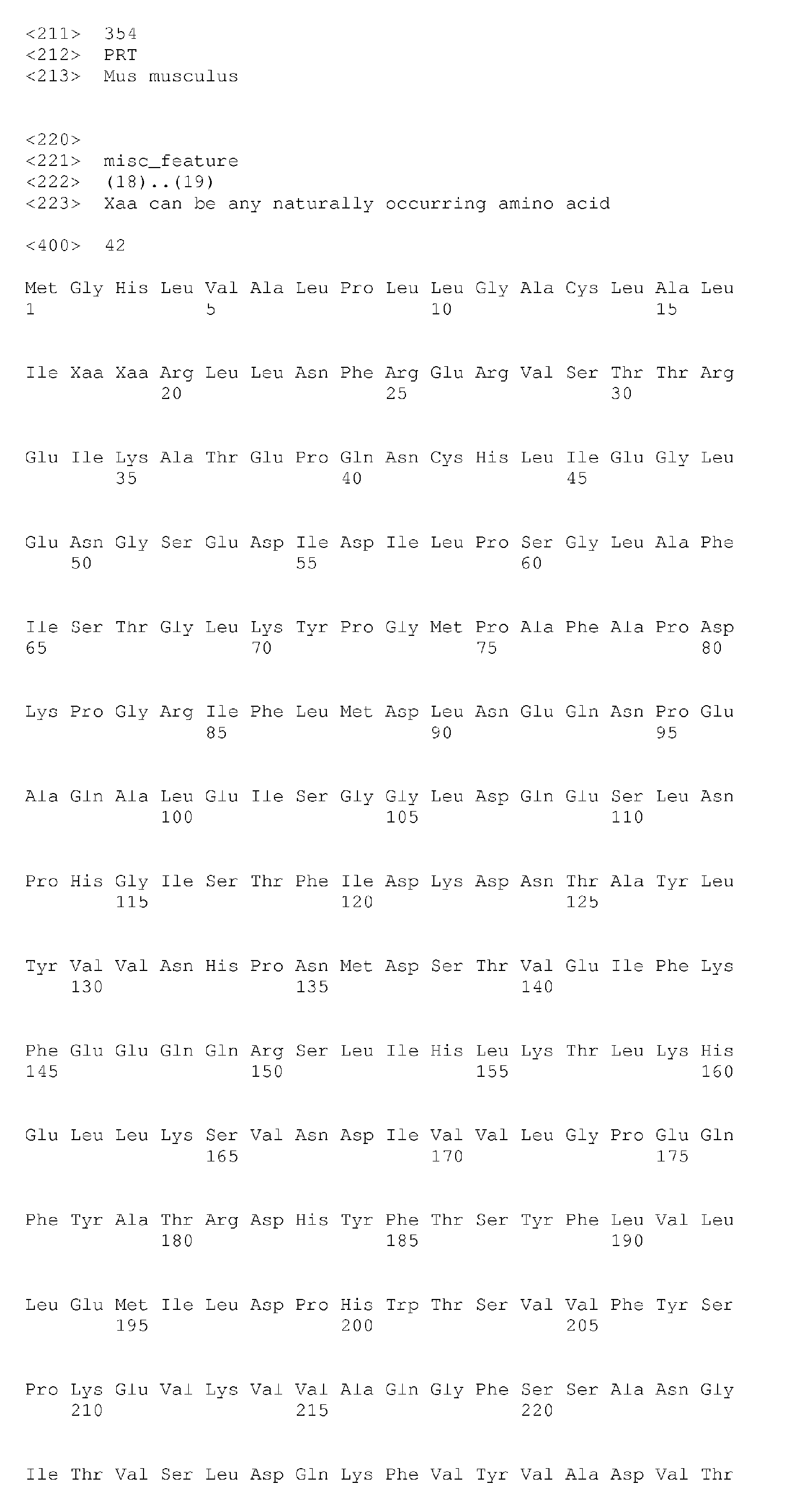


Claims (15)
- An isolated polynucleotide comprising a nucleic acid sequence encoding a mutated PON enzyme exhibiting an increased substrate specificity to at least one known PON substrate as compared to a respective wild-type PON.
- An isolated polypeptide comprising an amino acid sequence of a mutated PON enzyme exhibiting an increased substrate specificity to at least one known PON substrate as compared to a respective wild-type PON.
- The isolated polynucleotide of claim 1 or polypeptide of claim 2, wherein said PON enzyme is selected from the group consisting of PON1, PON2 and PON3.
- The isolated polynucleotide of claim 1 or polypeptide of claim 2, wherein said at least one known PON substrate comprises a phosphotriester.
- The isolated polynucleotide or polypeptide of claim 4, wherein said phosphotriester is selected from the group consisting of paraoxon, sarin and soman,
- The isolated polynucleotide of claim 1 or polypeptide of claim 2, wherein said increased substrate specificity is at least 3 fold higher Kcat/KM ratio for phospho-triester hysrolysis.
- The isolated polynucleotide of claim 1 or polypeptide of claim 2, wherein said increased substrate specificity is at least 3 fold higher Kcat/KM ratio for ester hydrolysis.
- An isolated polynucleotide comprising a nucleic acid sequence encoding a mutated PON enzyme exhibiting a modified substrate range as compared to a corresponding wild-type PON.
- An isolated polypeptide comprising an amino acid sequence of a mutated PON enzyme exhibiting a modified substrate range as compared to a corresponding wild-type PON.
- The isolated polynucleotide of claim 8 or polypeptide of claim 9, wherein said modified substrate range is manifested in an increased phosphotriesterase activity and a decreased esterase activity.
- The isolated polypeptide of claim 2-7, 9-10, comprising a mutation at an amino acid position selected from the group consisting of 69, 74, 75, 76, 78, 190, 192, 193, 196, 222, 240, 291, 292, 293, 332 and 346 of human PON1.
- An isolated polypeptide comprising an amino acid sequence as set forth in SEQ ID NO: 69, 71, 73, 75 or 77.
- A nucleic acid construct comprising the isolated polynucleotide of claim 1 or 3-7.
- A pharmaceutical composition comprising as an active ingredient the isolated polypeptide of claim 2 or 9 and a pharmaceutically acceptable carrier.
- The isolated polypeptide of claim 2 or 9 for use in treating organophosphate poisoning.
Applications Claiming Priority (3)
| Application Number | Priority Date | Filing Date | Title |
|---|---|---|---|
| US45126703P | 2003-03-04 | 2003-03-04 | |
| US51292503P | 2003-10-22 | 2003-10-22 | |
| EP04717208A EP1599598A4 (en) | 2003-03-04 | 2004-03-04 | Pon polypeptides, polynucleotides encoding same and compositions and methods utilizing same |
Related Parent Applications (2)
| Application Number | Title | Priority Date | Filing Date |
|---|---|---|---|
| EP04717208.5 Division | 2004-03-04 | ||
| WOPCT/IL2004/000216 Previously-Filed-Application | 2004-03-04 |
Publications (2)
| Publication Number | Publication Date |
|---|---|
| EP2319857A2 true EP2319857A2 (en) | 2011-05-11 |
| EP2319857A3 EP2319857A3 (en) | 2012-06-27 |
Family
ID=32965540
Family Applications (2)
| Application Number | Title | Priority Date | Filing Date |
|---|---|---|---|
| EP04717208A Withdrawn EP1599598A4 (en) | 2003-03-04 | 2004-03-04 | Pon polypeptides, polynucleotides encoding same and compositions and methods utilizing same |
| EP10183160A Withdrawn EP2319857A3 (en) | 2003-03-04 | 2004-03-04 | Pon polypeptides, polynucleotides encoding same and compositions and methods utilizing same |
Family Applications Before (1)
| Application Number | Title | Priority Date | Filing Date |
|---|---|---|---|
| EP04717208A Withdrawn EP1599598A4 (en) | 2003-03-04 | 2004-03-04 | Pon polypeptides, polynucleotides encoding same and compositions and methods utilizing same |
Country Status (3)
| Country | Link |
|---|---|
| US (3) | US7786071B2 (en) |
| EP (2) | EP1599598A4 (en) |
| WO (1) | WO2004078991A2 (en) |
Families Citing this family (10)
| Publication number | Priority date | Publication date | Assignee | Title |
|---|---|---|---|---|
| EP1599598A4 (en) | 2003-03-04 | 2007-08-08 | Yeda Res & Dev | Pon polypeptides, polynucleotides encoding same and compositions and methods utilizing same |
| US20090208992A1 (en) * | 2008-02-20 | 2009-08-20 | The Cleveland Clinic Foundation | Paraoxonase 1 enzymatic activity, a risk indicator for major adverse cardiovascular events |
| EP2268305A4 (en) * | 2008-03-14 | 2011-08-03 | Cleveland Clinic Foundation | Oxidized paraoxonase 1 and paraoxonase 1/hdl particle number ratio as risk markers for cardiovascular disease |
| EP2478093A2 (en) | 2009-09-17 | 2012-07-25 | Yeda Research and Development Co. Ltd. | Isolated pon1 polypeptides, polynucleotides encoding same and uses thereof in treating or preventing organophosphate exposure associated damage |
| US8735124B2 (en) * | 2009-09-17 | 2014-05-27 | Yeda Research And Development Co. Ltd. | Isolated PON1 polypeptides, polynucleotides encoding same and uses thereof in treating or preventing organophosphate exposure associated damage |
| WO2013040501A1 (en) | 2011-09-16 | 2013-03-21 | Pharmathene, Inc. | Compositions and combinations of organophosphorus bioscavengers and hyaluronan-degrading enzymes, and uses thereof |
| ES2753391T3 (en) | 2014-10-14 | 2020-04-08 | Halozyme Inc | Adenosine deaminase 2 (ADA2) compositions, variants thereof and methods of use thereof |
| WO2016092555A2 (en) | 2014-12-11 | 2016-06-16 | Yeda Research And Development Co. Ltd. | Isolated phosphotriesterase polypeptides, polynucleotides encoding same and uses thereof in treating or preventing organophosphate exposure associated damage |
| CN105218583B (en) * | 2015-09-10 | 2017-03-29 | 上海交通大学 | A kind of ultra-high throughput screening method of phosphoric triesterase |
| US20190359956A1 (en) | 2016-11-10 | 2019-11-28 | Yeda Research And Development Co. Ltd. | Phosphotriesterases for treating or preventing organophosphate exposure associated damage |
Citations (41)
| Publication number | Priority date | Publication date | Assignee | Title |
|---|---|---|---|---|
| US3791932A (en) | 1971-02-10 | 1974-02-12 | Akzona Inc | Process for the demonstration and determination of reaction components having specific binding affinity for each other |
| US3839153A (en) | 1970-12-28 | 1974-10-01 | Akzona Inc | Process for the detection and determination of specific binding proteins and their corresponding bindable substances |
| US3850578A (en) | 1973-03-12 | 1974-11-26 | H Mcconnell | Process for assaying for biologically active molecules |
| US3850752A (en) | 1970-11-10 | 1974-11-26 | Akzona Inc | Process for the demonstration and determination of low molecular compounds and of proteins capable of binding these compounds specifically |
| US3853987A (en) | 1971-09-01 | 1974-12-10 | W Dreyer | Immunological reagent and radioimmuno assay |
| US3867517A (en) | 1971-12-21 | 1975-02-18 | Abbott Lab | Direct radioimmunoassay for antigens and their antibodies |
| US3879262A (en) | 1972-05-11 | 1975-04-22 | Akzona Inc | Detection and determination of haptens |
| US3901654A (en) | 1971-06-21 | 1975-08-26 | Biological Developments | Receptor assays of biologically active compounds employing biologically specific receptors |
| US3935074A (en) | 1973-12-17 | 1976-01-27 | Syva Company | Antibody steric hindrance immunoassay with two antibodies |
| US3984533A (en) | 1975-11-13 | 1976-10-05 | General Electric Company | Electrophoretic method of detecting antigen-antibody reaction |
| US3996345A (en) | 1974-08-12 | 1976-12-07 | Syva Company | Fluorescence quenching with immunological pairs in immunoassays |
| US4034074A (en) | 1974-09-19 | 1977-07-05 | The Board Of Trustees Of Leland Stanford Junior University | Universal reagent 2-site immunoradiometric assay using labelled anti (IgG) |
| US4098876A (en) | 1976-10-26 | 1978-07-04 | Corning Glass Works | Reverse sandwich immunoassay |
| US4666828A (en) | 1984-08-15 | 1987-05-19 | The General Hospital Corporation | Test for Huntington's disease |
| US4683202A (en) | 1985-03-28 | 1987-07-28 | Cetus Corporation | Process for amplifying nucleic acid sequences |
| EP0264166A1 (en) | 1986-04-09 | 1988-04-20 | Genzyme Corporation | Transgenic animals secreting desired proteins into milk |
| US4801531A (en) | 1985-04-17 | 1989-01-31 | Biotechnology Research Partners, Ltd. | Apo AI/CIII genomic polymorphisms predictive of atherosclerosis |
| US4873316A (en) | 1987-06-23 | 1989-10-10 | Biogen, Inc. | Isolation of exogenous recombinant proteins from the milk of transgenic mammals |
| US4879219A (en) | 1980-09-19 | 1989-11-07 | General Hospital Corporation | Immunoassay utilizing monoclonal high affinity IgM antibodies |
| US5011771A (en) | 1984-04-12 | 1991-04-30 | The General Hospital Corporation | Multiepitopic immunometric assay |
| US5128318A (en) | 1987-05-20 | 1992-07-07 | The Rogosin Institute | Reconstituted HDL particles and uses thereof |
| US5192659A (en) | 1989-08-25 | 1993-03-09 | Genetype Ag | Intron sequence analysis method for detection of adjacent and remote locus alleles as haplotypes |
| US5272057A (en) | 1988-10-14 | 1993-12-21 | Georgetown University | Method of detecting a predisposition to cancer by the use of restriction fragment length polymorphism of the gene for human poly (ADP-ribose) polymerase |
| US5281521A (en) | 1992-07-20 | 1994-01-25 | The Trustees Of The University Of Pennsylvania | Modified avidin-biotin technique |
| US5331573A (en) | 1990-12-14 | 1994-07-19 | Balaji Vitukudi N | Method of design of compounds that mimic conformational features of selected peptides |
| US5500807A (en) | 1989-01-06 | 1996-03-19 | The Regents Of The University Of California | Selection method for pharmacologically active compounds |
| US5646101A (en) | 1993-01-18 | 1997-07-08 | The Procter & Gamble Company | Machine dishwashing detergents containing an oxygen bleach and an anti-tarnishing mixture of a paraffin oil and sequestrant |
| US5652339A (en) | 1993-12-31 | 1997-07-29 | Rotkreuzstiftung Zentrallaboratorium | Method of producing reconstituted lipoproteins |
| US5686014A (en) | 1994-04-07 | 1997-11-11 | The Procter & Gamble Company | Bleach compositions comprising manganese-containing bleach catalysts |
| US5695679A (en) | 1994-07-07 | 1997-12-09 | The Procter & Gamble Company | Detergent compositions containing an organic silver coating agent to minimize silver training in ADW washing methods |
| US5698504A (en) | 1993-07-01 | 1997-12-16 | The Procter & Gamble Company | Machine dishwashing composition containing oxygen bleach and paraffin oil and benzotriazole compound silver tarnishing inhibitors |
| US5705464A (en) | 1995-06-16 | 1998-01-06 | The Procter & Gamble Company | Automatic dishwashing compositions comprising cobalt catalysts |
| US5710115A (en) | 1994-12-09 | 1998-01-20 | The Procter & Gamble Company | Automatic dishwashing composition containing particles of diacyl peroxides |
| US6242186B1 (en) | 1999-06-01 | 2001-06-05 | Oy Jurilab Ltd. | Method for detecting a risk of cancer and coronary heart disease and kit therefor |
| US20020151068A1 (en) | 2001-01-03 | 2002-10-17 | Board Of Regents, The University Of Texas System | Compositions and methods for the diagnosis and treatment of organophosphate toxicity |
| US6514523B1 (en) | 2000-02-14 | 2003-02-04 | Ottawa Heart Institute Research Corporation | Carrier particles for drug delivery and process for preparation |
| US6521226B1 (en) | 1998-11-25 | 2003-02-18 | Bayer Corporation | Method of using PON-1 to decrease atheroma formation |
| US6573370B1 (en) | 2000-05-19 | 2003-06-03 | Regents Of The University Of Michigan | PON3 and uses thereof |
| US6573049B1 (en) | 1999-07-26 | 2003-06-03 | Nuvelo, Inc. | Genotyping of the paraoxonase 1 gene for prognosing, diagnosing, and treating a disease |
| US6610642B2 (en) | 2000-04-20 | 2003-08-26 | The Procter And Gamble Company | Cleaning compositions containing multiply-substituted protease variants |
| US6617142B2 (en) | 1996-04-25 | 2003-09-09 | Medtronic, Inc. | Method for attachment of biomolecules to medical device surfaces |
Family Cites Families (2)
| Publication number | Priority date | Publication date | Assignee | Title |
|---|---|---|---|---|
| US6037173A (en) * | 1997-04-11 | 2000-03-14 | Millennium Pharmaceuticals, Inc. | Isolated nucleic acid encoding TRBP |
| EP1599598A4 (en) | 2003-03-04 | 2007-08-08 | Yeda Res & Dev | Pon polypeptides, polynucleotides encoding same and compositions and methods utilizing same |
-
2004
- 2004-03-04 EP EP04717208A patent/EP1599598A4/en not_active Withdrawn
- 2004-03-04 EP EP10183160A patent/EP2319857A3/en not_active Withdrawn
- 2004-03-04 WO PCT/IL2004/000216 patent/WO2004078991A2/en active Application Filing
- 2004-03-04 US US10/547,771 patent/US7786071B2/en not_active Expired - Fee Related
-
2010
- 2010-03-15 US US12/723,725 patent/US20110171197A1/en not_active Abandoned
-
2013
- 2013-10-21 US US14/058,334 patent/US9023631B2/en not_active Expired - Fee Related
Patent Citations (42)
| Publication number | Priority date | Publication date | Assignee | Title |
|---|---|---|---|---|
| US3850752A (en) | 1970-11-10 | 1974-11-26 | Akzona Inc | Process for the demonstration and determination of low molecular compounds and of proteins capable of binding these compounds specifically |
| US3839153A (en) | 1970-12-28 | 1974-10-01 | Akzona Inc | Process for the detection and determination of specific binding proteins and their corresponding bindable substances |
| US3791932A (en) | 1971-02-10 | 1974-02-12 | Akzona Inc | Process for the demonstration and determination of reaction components having specific binding affinity for each other |
| US3901654A (en) | 1971-06-21 | 1975-08-26 | Biological Developments | Receptor assays of biologically active compounds employing biologically specific receptors |
| US3853987A (en) | 1971-09-01 | 1974-12-10 | W Dreyer | Immunological reagent and radioimmuno assay |
| US3867517A (en) | 1971-12-21 | 1975-02-18 | Abbott Lab | Direct radioimmunoassay for antigens and their antibodies |
| US3879262A (en) | 1972-05-11 | 1975-04-22 | Akzona Inc | Detection and determination of haptens |
| US3850578A (en) | 1973-03-12 | 1974-11-26 | H Mcconnell | Process for assaying for biologically active molecules |
| US3935074A (en) | 1973-12-17 | 1976-01-27 | Syva Company | Antibody steric hindrance immunoassay with two antibodies |
| US3996345A (en) | 1974-08-12 | 1976-12-07 | Syva Company | Fluorescence quenching with immunological pairs in immunoassays |
| US4034074A (en) | 1974-09-19 | 1977-07-05 | The Board Of Trustees Of Leland Stanford Junior University | Universal reagent 2-site immunoradiometric assay using labelled anti (IgG) |
| US3984533A (en) | 1975-11-13 | 1976-10-05 | General Electric Company | Electrophoretic method of detecting antigen-antibody reaction |
| US4098876A (en) | 1976-10-26 | 1978-07-04 | Corning Glass Works | Reverse sandwich immunoassay |
| US4879219A (en) | 1980-09-19 | 1989-11-07 | General Hospital Corporation | Immunoassay utilizing monoclonal high affinity IgM antibodies |
| US5011771A (en) | 1984-04-12 | 1991-04-30 | The General Hospital Corporation | Multiepitopic immunometric assay |
| US4666828A (en) | 1984-08-15 | 1987-05-19 | The General Hospital Corporation | Test for Huntington's disease |
| US4683202A (en) | 1985-03-28 | 1987-07-28 | Cetus Corporation | Process for amplifying nucleic acid sequences |
| US4683202B1 (en) | 1985-03-28 | 1990-11-27 | Cetus Corp | |
| US4801531A (en) | 1985-04-17 | 1989-01-31 | Biotechnology Research Partners, Ltd. | Apo AI/CIII genomic polymorphisms predictive of atherosclerosis |
| EP0264166A1 (en) | 1986-04-09 | 1988-04-20 | Genzyme Corporation | Transgenic animals secreting desired proteins into milk |
| US5128318A (en) | 1987-05-20 | 1992-07-07 | The Rogosin Institute | Reconstituted HDL particles and uses thereof |
| US4873316A (en) | 1987-06-23 | 1989-10-10 | Biogen, Inc. | Isolation of exogenous recombinant proteins from the milk of transgenic mammals |
| US5272057A (en) | 1988-10-14 | 1993-12-21 | Georgetown University | Method of detecting a predisposition to cancer by the use of restriction fragment length polymorphism of the gene for human poly (ADP-ribose) polymerase |
| US5500807A (en) | 1989-01-06 | 1996-03-19 | The Regents Of The University Of California | Selection method for pharmacologically active compounds |
| US5192659A (en) | 1989-08-25 | 1993-03-09 | Genetype Ag | Intron sequence analysis method for detection of adjacent and remote locus alleles as haplotypes |
| US5331573A (en) | 1990-12-14 | 1994-07-19 | Balaji Vitukudi N | Method of design of compounds that mimic conformational features of selected peptides |
| US5281521A (en) | 1992-07-20 | 1994-01-25 | The Trustees Of The University Of Pennsylvania | Modified avidin-biotin technique |
| US5646101A (en) | 1993-01-18 | 1997-07-08 | The Procter & Gamble Company | Machine dishwashing detergents containing an oxygen bleach and an anti-tarnishing mixture of a paraffin oil and sequestrant |
| US5698504A (en) | 1993-07-01 | 1997-12-16 | The Procter & Gamble Company | Machine dishwashing composition containing oxygen bleach and paraffin oil and benzotriazole compound silver tarnishing inhibitors |
| US5652339A (en) | 1993-12-31 | 1997-07-29 | Rotkreuzstiftung Zentrallaboratorium | Method of producing reconstituted lipoproteins |
| US5686014A (en) | 1994-04-07 | 1997-11-11 | The Procter & Gamble Company | Bleach compositions comprising manganese-containing bleach catalysts |
| US5695679A (en) | 1994-07-07 | 1997-12-09 | The Procter & Gamble Company | Detergent compositions containing an organic silver coating agent to minimize silver training in ADW washing methods |
| US5710115A (en) | 1994-12-09 | 1998-01-20 | The Procter & Gamble Company | Automatic dishwashing composition containing particles of diacyl peroxides |
| US5705464A (en) | 1995-06-16 | 1998-01-06 | The Procter & Gamble Company | Automatic dishwashing compositions comprising cobalt catalysts |
| US6617142B2 (en) | 1996-04-25 | 2003-09-09 | Medtronic, Inc. | Method for attachment of biomolecules to medical device surfaces |
| US6521226B1 (en) | 1998-11-25 | 2003-02-18 | Bayer Corporation | Method of using PON-1 to decrease atheroma formation |
| US6242186B1 (en) | 1999-06-01 | 2001-06-05 | Oy Jurilab Ltd. | Method for detecting a risk of cancer and coronary heart disease and kit therefor |
| US6573049B1 (en) | 1999-07-26 | 2003-06-03 | Nuvelo, Inc. | Genotyping of the paraoxonase 1 gene for prognosing, diagnosing, and treating a disease |
| US6514523B1 (en) | 2000-02-14 | 2003-02-04 | Ottawa Heart Institute Research Corporation | Carrier particles for drug delivery and process for preparation |
| US6610642B2 (en) | 2000-04-20 | 2003-08-26 | The Procter And Gamble Company | Cleaning compositions containing multiply-substituted protease variants |
| US6573370B1 (en) | 2000-05-19 | 2003-06-03 | Regents Of The University Of Michigan | PON3 and uses thereof |
| US20020151068A1 (en) | 2001-01-03 | 2002-10-17 | Board Of Regents, The University Of Texas System | Compositions and methods for the diagnosis and treatment of organophosphate toxicity |
Non-Patent Citations (195)
| Title |
|---|
| "Animal Cell Culture", 1986 |
| "Basic and Clinical Immunology", 1994, APPLETON & LANGE |
| "Cell Biology: A Laboratory Handbook", vol. I-III, 1994 |
| "Current Protocols in Immunology", vol. I-III, 1994 |
| "Current Protocols in Molecular Biology", vol. I-III, 1994 |
| "Genome Analysis: A Laboratory Manual Series", vol. 1-4, 1998, COLD SPRING HARBOR LABORATORY PRESS |
| "Immobilized Cells and Enzymes", 1986, IRL PRESS |
| "Methods in Enzymology", vol. 1-317, ACADEMIC PRESS |
| "Nucleic Acid Hybridization", 1985 |
| "Oligonucleotide Synthesis", 1984 |
| "PCR Protocols: A Guide To Methods And Applications", 1990, ACADEMIC PRESS |
| "Remington's Pharmaceutical Sciences", MACK PUBLISHING CO. |
| "Selected Methods in Cellular Immunology", 1980, W. H. FREEMAN AND CO. |
| "Transcription and Translation", 1984 |
| A. AHARONI ET AL., PROC. NATL. ACAD. SCI. US A, vol. 101, 2004, pages 482 |
| A. FOKINE ET AL., ACTA CRYSTALLOGR D BIOL CRYSTALLOGR, vol. 59, 2003, pages 2083 |
| A. JONAS, BIOCHIM BIOPHYS ACTA-MOL CELL BIOL LIPIDS, vol. 1529, 2000, pages 245 |
| ABECASSIS, V.; POMPON, D.; TRUAN, G.: "High efficiency family shuffling based on multi-step PCR and in vivo DNA recombination in yeast: statistical and functional analysis of a combinatorial library between human cytochrome P450 1A1 and 1A2", NUCLEIC ACIDS RES, vol. 28, 2000, pages E88 |
| ABRAHAMS, J.P.; LESLIE, A.G., ACTA CRYSTALLOG. D., vol. 52, 1996, pages 30 - 42 |
| AHMED, Z.; RAVANDI, A.; MAGUIRE, G.F.; EMILI, A.; DRAGANOV, D.; LA DU, B.N.; KUKSIS, A.; CONNELLY, P.W.: "Apolipoprotein A-I promotes the formation of phosphatidylcholine core aldehydes that are hydrolyzed by paraoxonase (PON-1) during high density lipoprotein oxidation with a peroxynitrite donor", JOURNAL OF BIOLOGICAL CHEMISTRY, vol. 276, 2001, pages 24473 - 24481 |
| APPELT, PERSPECTIVES IN DRUG DISCOVERY AND DESIGN, vol. 1, 1993, pages 23 |
| AUSUBEL ET AL.: "Current Protocols in Molecular Biology", 1989, JOHN WILEY AND SONS |
| AVIRAM, M.; ROSENBLAT, M.; BISGAIER, C.L.; NEWTON, R.S.; PRIMO-PARMO, S.L.; LA DU, B.N.: "Paraoxonase inhibits high-density lipoprotein oxidation and preserves its functions. A possible peroxidative role for paraoxonase", J CLIN INVEST, vol. 101, 1998, pages 1581 - 1590 |
| B. LEE; F. M. RICHARDS, J MOL BIOL, vol. 55, 1971, pages 379 |
| BANERJI ET AL., CELL, 1983, pages 33729 - 740 |
| BARTLETT, PA. ET AL., SPECIAL PUB ROYAL CHEM SOC., vol. 78, 1989, pages 182 - 196 |
| BAYLI'S J PHARMACOL EXP THER., 2003 |
| BENCHARIT, S.; MORTON, C.L.; XUE, Y.; POTTER, P.M.; REDINBO, M.R.: "Structural basis of heroin and cocaine metabolism by a promiscuous human drug-processing enzyme", NATURE STRUCTURAL BIOLOGY, vol. 10, 2003, pages 349 - 356 |
| BENCHARIT; C. L. MORTON; Y. XUE; P. M. POTTER; M. R. REDINBO, NATURE STRUCT BIOL, vol. 10, 2003, pages 349 |
| BESSETTE, P.; ASLUND, F.; BECKWITH, J.; GEORGIOU, G.: "Efficient folding of proteins with multiple disulfide bonds in the Escherichia coli cytoplasm", PROC NATL ACAD SCI USA, vol. 96, 1999, pages 13703 - 13708 |
| BHATTACHARJYA, XU, PROTEIN SCIENCE, vol. 10, no. 5, 2001, pages 934 - 942 |
| BILLECKE, DRUG METAB. DISPOS., vol. 28, 2000, pages 1335 - 1342 |
| BILLECKE, S.; DRAGANOV, D.; COUNSELL, R.; STETSON, P.; WATSON, C.; HSU, C.; LA DU, B.N.: "Further characterization of human serum paraoxonase (PON1) lactonase activity", FASEB JOURNAL, vol. 14, 2000, pages 74 |
| BILLECLKE, S.; DRAGANOV, D.; COUNSELL, R.; STETSON, P.; WATSON, C.; LA DU, B.N.: "Human serum paraoxonase (PON1) isozymes Q and R hydrolyze lactones and cyclic carbonate esters", DRUG METABOLISM AND DISPOSITION, vol. 28, 2000, pages 1335 - 1342 |
| BITTER ET AL., METHODS IN ENZYMOL., vol. 153, 1987, pages 516 - 544 |
| BOHM HJ., J. COMP AID MOLEC DESIGN, vol. 6, 1992, pages 61 - 78 |
| BOOTH ET AL., IMMUNOL. LETT., vol. 19, 1988, pages 65 - 70 |
| BRISSON ET AL., NATURE, vol. 310, 1984, pages 511 - 514 |
| BROGLI ET AL., SCIENCE, vol. 224, 1984, pages 838 - 843 |
| BROOKS BR. ET AL., J COMP CHEM., vol. 4, 1983, pages 187 - 217 |
| BROUSSEAU, J LIPID RES., vol. 40, no. 3, 1999, pages 365 - 75 |
| BRUSHIA, R.J.; FORTE, T.M.; ODA, M.N.; LA DU, B.; BIELICKI, J.K.: "Baculovirus-mediated expression and purification of human serum paraoxonase IA", J LIPID RES, vol. 42, 2001, pages 951 - 958 |
| BURKERT U.; ALLINGER NL.: "Molecular Mechanics", vol. 177, 1982, AMERICAN CHEMICAL SOCIETY, pages: 59 - 78 |
| BYRNE ET AL., PROC. NATL. ACAD. SCI. USA, vol. 86, 1989, pages 5473 - 5477 |
| C. B. MILLARD; 0. LOCKRIDGE; C. A. BROOMFIELD, BIOCHEMISTRY, vol. 37, 1998, pages 237 |
| C. L. KUO; B. N. LA DU, DRUG METAB DISPOS, vol. 23, 1995, pages 935 |
| C. L. KUO; B. N. LA DU, DRUG METAB DISPOS, vol. 26, 1998, pages 653 |
| CALAME ET AL., ADV. IMMUNOL., vol. 43, 1988, pages 235 - 275 |
| CARSON, M., METHODS IN ENZYMOLOGY, vol. 277, 1997, pages 25 |
| CHENG, ANN. NY ACAD. SCI., vol. 864, 1998, pages 253 - 58 |
| CHOY; FORMAN-KAY, JOURNAL OF MOLECULAR BIOLOGY, vol. 308, no. 5, 2001, pages 1011 - 1032 |
| COHEN NC. E: "Molecular Modeling Software and Methods for Medicinal Chemistry", J. MED. CHEM., vol. 33, 1990, pages 883 - 894 |
| CONNOLLY ML., SCIENCE, vol. 221, 1983, pages 709 - 713 |
| CORUZZI ET AL., EMBO J., vol. 3, 1984, pages 1671 - 1680 |
| CRAMERI, A.; RAILLARD, S.A.; BERMUDEZ, E.; STEMMER, W.P.: "DNA shuffling of a family of genes from diverse species accelerates directed evolution", NATURE, vol. 391, 1998, pages 288 - 291 |
| D. JOSSE ET AL., BIOCHEMISTRY, vol. 38, 1999, pages 2816 |
| D. JOSSE ET AL., J APPL TOXICOL, vol. 21, 2001, pages S7 |
| D. JOSSE ET AL., J BIOL CHEM, vol. 277, 2002, pages 33386 |
| D. JOSSE; W. H. XIE; P. MASSON; L. M. SCHOPFER; 0. LOCKRIDGE, CHEM. BIOL. INTERACT., vol. 120, 1999, pages 79 |
| D. S. CHELUR ET AL., NATURE, vol. 420, 2002, pages 669 |
| DAVIES, H.G.; RICHTER, R.J.; KEIFER, M.; BROOMFIELD, C.A.; SOWALLA, J.; FURLONG, C.E.: "The effect of the human serum paraoxonase polymorphism is reversed with diazoxon, soman and sarin", NLAT GENET, vol. 14, 1996, pages 334 - 336 |
| DAVIES, NAT. GENET., vol. 14, 1996, pages 334 - 336 |
| DEFRANK: "Applications of Enzyme Biotechnology", 1991, pages: 165 - 80 |
| DERREUMAUX, BIOPHYS J., vol. 81, 2001, pages 1657 - 65 |
| DRAGANOV, B.; N. LA DU, NAU SCHM ARCH PHARMACOL, 2003 |
| DRAGANOV, J. BIOL. CHEM., vol. 275, 2000, pages 33435 - 33442 |
| DUNBRACK ET AL., FOLDING AND DESIGN, vol. 2, 1997, pages 27 |
| DYCK; CRAIG, LUMINESCENCE, vol. 17, 2002, pages 15 - 18 |
| E. 1. SCHARFF; J. KOEPKE; G. FRITZSCH; C. LUCKE; H. RUTERJANS, STRUCTURE, vol. 9, 2001, pages 493 |
| EDLUNCH ET AL., SCIENCE, vol. 230, 1985, pages 912 - 916 |
| ELIEZER; KUTLUAY, JOURNAL OF MOLECULAR BIOLOGY, vol. 307, no. 4, 2001, pages 1061 - 1073 |
| ERICKSON, PERSPECTIVES IN DRUG DISCOVERY AND DESIGN, vol. 1, 1993, pages 109 |
| FARMER P. S.: "Drug Design", vol. 10, 1980, ACADEMIC PRESS, pages: 119 - 143 |
| FINGL ET AL.: "The Pharmacological Basis of Therapeutics", 1975, pages: L |
| FORTELLE, E.; BRICOGNE, G., METHODS ENZYMOL., vol. 276, 1997, pages 472 - 494 |
| FRET; CHAKRABORTY; ITTAH, BIOCHEMISTRY, vol. 40, 2001, pages 7228 - 7238 |
| G. RUDENKO ET AL., SCIENCE, vol. 298, 2002, pages 2353 |
| GARDELLA ET AL., J. BIOL. CHEM., vol. 265, 1990, pages 15854 - 15859 |
| GEORGIOU, U.: "Analysis ot large libraries ot protein mutants using now cytometry", ADV PROTEIN CHEM, vol. 55, 2000, pages 293 - 315 |
| GOHLKE H.; KLEBE G., CURR OPIN STRUCT BIOL., vol. 11, 2001, pages 231 - 5 |
| GOODFORD PJ.: "A Computational Procedure for Determining Energetically Favorable Binding Sites on Biologically Important Macromolecules", J. MED. CHEM., vol. 28, 1985, pages 849 - 857 |
| GOODSELL DS.; OLSON AJ., PROTEINS: STRUCT FUNCT GENET., vol. 8, 1990, pages 195 - 202 |
| GRIFFITHS, A.D.; TAWFIK, D.S.: "Man-made enzymes--from design to in vitro compartmentalisation", CURR OPIN BIOTECHNOL, vol. 11, 2000, pages 338 - 353 |
| GURLEY ET AL., MOL. CELL. BIOL., vol. 6, 1986, pages 559 - 565 |
| H. JAKUBOWSKI, J BIOL CHEM, vol. 275, 2000, pages 3957 |
| H. JEON ET AL., NATURE STRUCT BIOLOGY, vol. 8, 2001, pages 499 |
| H. M. GREENBLATT; DVIR; 1. SILMAN; J. L. SUSSMAN, J MOL NEUROSCI, vol. 20, 2003, pages 369 |
| HALPERIN 1. ET AL., PROTEINS, vol. 47, 2002, pages 409 - 43 |
| HAMMARSTROM, M.; HELLGREN, N.; VAN DEN BERG, S.; BERGLUND, H.; HARD, T.: "Rapid screening for improved solubility of small human proteins produced as fusion proteins in Escherichia coli", PROTEIN SCI, vol. 11, 2002, pages 313 - 321 |
| HARCOURT, LETT. APPL. MICROBIOL., vol. 34, 2002, pages 263 - 268 |
| HITCHENS; MANNERVIK, BIOCHEMISTRY, vol. 40, no. 39, 2001, pages 11660 - 11669 |
| HOFFMAN ET AL.: "Covalent Binding of Biomolecules to Radiation-Grafted Hydrogels on Inert Polymer Surfaces", TRANS. AM. SOC. ARTIF. INTERN. ORGANS, vol. 18, 1972, pages 10 - 18 |
| HONG, BIOREMEDIATION J., vol. 2, 1998, pages 145 - 57 |
| HUANG, Y.T.; LIAW, Y.C.; GORBATYUK, V.Y.; HUANG, T.H.: "Backbone dynamics of Escherichia coli thioesterase/protease I: Evidence of a flexible active-site environment for a serine protease", JOURNAL OF MOLECULAR BIOLOGY, vol. 307, 2001, pages 1075 - 1090 |
| ITO ET AL.: "Materials for Enhancing Cell Adhesion by Immobilization of Cell-Adhesive Peptide", J. BIOMED. MAT. RES., vol. 25, 1991, pages 1325 - 1337 |
| J. A. KILLIAN; G. VON HEIJNE, TRENDS BIOCHEM SCI, vol. 25, 2000, pages 429 |
| J. P. SEGREST; S. C. HARVEY; V. ZANNIS, TRENDS CARDIOVASC MED, vol. 10, 2000, pages 246 |
| JAKUBOWSKI, H.: "Calcium-dependent human serum homocysteine thiolactone hydrolase. A protective mechanism against protein N-homocysteinylation", J BIOL CHEM, vol. 275, 2000, pages 3957 - 3962 |
| JAKUBOWSKI, J. BIOL. CHEM., vol. 275, 2000, pages 3957 - 3962 |
| JAMES LC; ROVERSI P; TAWFIK DS: "Antibody multispecificity mediated by conformational diversity", SCIENCE, vol. 299, 2003, pages 1362 - 1367 |
| JAMES LC; TAWFIK DS: "Conformational diversity and protein evolution - a 60-year-old hypothesis revisited", TRENDS BIOCHEM SCI, vol. 28, 2003, pages 361 - 368 |
| JOEM, J.M.; MEINHOLD, P.; ARNOLD, F.H.: "Analysis of shuffled gene libraries", J MOL BIOL, vol. 316, 2002, pages 643 - 656 |
| JONES, H.E.; HOLLAND, LB.; BAKER, H.L.; CAMPBELL, A.K.: "Slow changes in cytosolic free Ca2+ in Escherichia coli highlight two putative influx mechanisms in response to changes in extracellular calcium", CELL CALCIUM, vol. 25, 1999, pages 265 - 274 |
| JONES, T.A.; ZOU, J.-Y.; COWAN, S.W.; KJELDGAARD, M., ACTA CRYSTALLOGR. A., vol. 47, 1991, pages 110 - 119 |
| JONES, TA. ET AL., ACTA CRYSTALLOGR, vol. A47, 1991, pages 110 |
| JONES, TA. ET AL., ACTA CRYSTALLOGR., vol. A47, 1991, pages 110 |
| JOSSE, D.; EBEL, C.; STROEBEL, D.; FONTAINE, A.; BORGES, F.; ECHALIER, A.; BAUD, D.; RENAULT, F.; LE MAIRE, M.; CHABRIERES, E.: "Oligomeric states of the detergent-solubilized human serum paraoxonase (PON1)", JOURNAL OFBIOLOGICAL CHEMISTRY, vol. 277, 2002, pages 33386 - 33397 |
| JOSSE, D.; XIE, W.H.; RENAULT, F.; ROCHU, D.; SCHOPFER, L.M.; MASSON, P.; LOCKRIDGE, 0.: "Identification of residues essential for human paraoxonase (PON1) arylesterase/organophosphatase activities", BIOCHEMISTRY, vol. 38, 1999, pages 2816 - 2825 |
| K. SEKAR ET AL., BIOCHEMISTRY, vol. 36, 1997, pages 3104 |
| KABSCH, W. J., APPL. CRYST., vol. 26, 1993, pages 795 - 800 |
| KANEVA, BIOTECHNOL. PROG., vol. 14, 1998, pages 275 - 78 |
| KEAMEY, J. AGRIC. FOOD CHEM., vol. 34, 1986, pages 702 - 6 |
| KHALAMEYZER, V.; FISCHER, I.; BOMSCHEUER, U.; ALTENBUCHNER, J.: "Screening, nucleotide sequence, and biochemical characterization of an esterase from Pseudomonas fluorescens with high activity towards lactones", APPL ENVIRON MICROBIOL, vol. 65, 1999, pages 477 - 482 |
| KIM, BIOTECH. LETT., vol. 19, 1997, pages 1067 - 71 |
| KIM, BIOTECHNOL. PROG., vol. 18, 2002, pages 429 - 36 |
| KOMIVES, BIOTECHNOL. BIOENG, vol. 43, 1994, pages 946 - 59 |
| KRAULIS, J., APPL CRYSTALLOGR., vol. 24, 1991, pages 946 |
| KUNTZ ID. ET AL., J MOL BIOL., vol. 161, 1982, pages 269 - 288 |
| KUNTZ ID., SCIENCE, vol. 257, 1992, pages 1078 - 108 |
| KUO, C.L.; LA DU, B.N.: "Calcium binding by human and rabbit serum paraoxonases. Structural stability and enzymatic activity", DRUG METAB DISPOS, vol. 26, 1998, pages 653 - 660 |
| KUO, C.L.; LA DU, B.N.: "Comparison of purified human and rabbit serum paraoxonases", DRUG METAB DISPOS, vol. 23, 1995, pages 935 - 944 |
| LAM ET AL., SCIENCE, vol. 263, 1994, pages 380 |
| LEJEUNE, BIOTECHNOL. BIOENG., vol. 62, 1999, pages 659 - 65 |
| LEJEUNE, NATURE, vol. 395, 1998, pages 27 - 28 |
| LEVIEV; S. DEAKIN; R. W. JAMES, J LIPID RES, vol. 42, 2001, pages 528 |
| LI, PHARMACOGENETICS, vol. 10, 2000, pages 767 - 779 |
| LORENZ, CURR. OPIN. BIOTECHNOL., vol. 13, 2002, pages 572 - 7 |
| M. AVIRAM ET AL., ARTERIOSCLER THROMB VASC BIOL, vol. 18, 1998, pages 1617 |
| M. KOBAYASHI; M. SHINOHARA; C. SAKOH; M. KATAOKA; S. SHIMIZU, PROC NATL ACAD SCI U S A, vol. 95, 1998, pages 12787 |
| M. N. ODA; J. K. BIELICKI; T. BERGER; T. M. FORTE, BIOCHEMISTRY, vol. 40, 2001, pages 1710 |
| M. NAVAB ET AL., CURR OPIN LIPIDOLOGY, vol. 9, 1998, pages 449 |
| MACKNESS, 1V1.1.; MACKNESS, D.; DUMNGTON, P.N.; COMENY, R.VV.; NEGERE, R.A.: "Paraoxonase: biochemistry, genetics and relationship to plasma lipoproteins", CURR OPIN LIPIDOL, vol. 7, 1996, pages 69 - 76 |
| MARATHE, G.; ZIMMERMAN, G.; MCINTYRE, T.: "PAF acetylhydrolase, and not paraoxonase-1, is the oxidized phospholipid hydrolase of high density lipoprotein particles", JBIOL CHEM, 2002 |
| MARSHAK ET AL.: "Strategies for Protein Purification and Characterization - A Laboratory Course Manual", 1996, CSHL PRESS |
| MARTIN, Y. C., J. MED. CHEM., vol. 35, 1992, pages 2145 - 2154 |
| MAURO MJ. ET AL., J CLIN ONCOL., vol. 20, 2002, pages 325 - 34 |
| MAXWELL, K.L.; MITTERMAIER, A.K.; FORMAN-KAY, J.D.; DAVIDSON, A.R.: "A simple in vivo assay for increased protein solubility", PROTEIN SCI, vol. 8, 1999, pages 1908 - 1911 |
| MAZUR, J. BIOL. CHEM., vol. 164, 1946, pages 271 - 89 |
| MIRANKER A.; KARPLUS M.: "Functionality Maps of Binding Sites: A Multiple Copy Simultaneous Search Method", PROTEINS: STRUCTURE FUNCTION AND GENETICS, vol. 11, 1991, pages 29 - 34 |
| MULCHANDANI, BIOTECHNOL. BIOENG., vol. 63, 1999, pages 216 - 23 |
| MUNNECKE, APPL. MICROBIOL., vol. 28, 1974, pages 212 - 17 |
| MURSHUDOV, G.N.; VAGIN, A.A.; LEBEDEV, A.; WILSON, K.S.; DODSON, E.J., ACTA CRYSTALLOGR. D., vol. 55, 1999, pages 247 - 255 |
| NAU SCHM ARCH PHARMACOL, 2003 |
| NAVIA M. A.; MURCKO M. A.: "The Use of Structural Information in Drug Design", CURRENT OPINIONS IN STRUCTURAL BIOLOGY, vol. 2, 1992, pages 202 - 210 |
| NISHIBATA Y.; ITAI A., TETRAHEDRON, vol. 47, 1991, pages 8985 |
| PERBAL, B.: "A Practical Guide to Molecular Cloning", 1984 |
| PERBAL: "A Practical Guide to Molecular Cloning", 1988, JOHN WILEY & SONS |
| PERRAKIS, A.; MORRIS, R.; LAMZIN, V.S., NATURE STRUCT. BIOL., vol. 6, 1999, pages 458 - 463 |
| PETROUNIA, I.P.; ARNOLD, F.H.: "Designed evolution of enzymatic properties", CURR OPIN BIOTECHNOL, vol. 11, 2000, pages 325 - 330 |
| PINKERT ET AL., GENES DEV., vol. 1, 1987, pages 268 - 277 |
| R. C. SORENSON ET AL., ARTERIOSCLER THROMB VASE BIOL, vol. 19, 1999, pages 2214 |
| R. C. SORENSON ET AL., PROC NATL ACAD SCI U S A, vol. 92, 1995, pages 7187 |
| REDDY, ARTERIOSCLER. THROMB. VASC. BIOL., vol. 21, 2001, pages 542 - 547 |
| REDDY, S.T.; WADLEIGH, D.J.; GRIJALVA, V.; NG, C.; HAMA, S.; GANGOPADHYAY, A.; SHIH, D.M.; LUSIS, A.J.; NAVAB, M.; FOGELMAN, A.M.: "Human paraoxonase-3 is an HDL-associated enzyme with biological activity similar to paraoxonase-1 protein but is not regulated by oxidized lipids", ARTERIOSCLEROSIS THROMBOSIS AND VASCULAR BIOLOGY, vol. 21, 2001, pages 542 - 547 |
| RICHINS, NAT. BIOTECHNOL., vol. 15, 1997, pages 984 - 87 |
| RODRIGO, L.; MACKNESS, B.; DURRINGTON, P.N.; HERNANDEZ, A.; MACKNESS, M.I.: "Hydrolysis of platelet-activating factor by human serum paraoxonase", BIOCHEMICAL JOURNAL, vol. 354, 2001, pages 1 - 7 |
| RUSINKO A., CHEM DES AUTO. NEWS, vol. 8, 1993, pages 44 - 47 |
| RUSSELL, ANNU. REV. BIOMED., vol. 5, 2003, pages 1 - 27 |
| RUSSELL, CHEMTECH, vol. 24, 1994, pages 23 - 31 |
| S. LUND-KATZ; L. J. LIU; S. T. THUAHNAI; M. C. PHILLIPS, FRONTIERS IN BIOSCIENCE, vol. 8, 2003, pages D1044 |
| SAMBROOK ET AL.: "Molecular Cloning: A laboratory Manual", 1989 |
| SCHARFF J. KOEPKE; G. FRITZSCH; C. LUCKE; H. RUTERJANS, STRUCTURE, vol. 9, 2001, pages 493 |
| SHIH, D.M.; GU, L.J.; XIA, Y.R.; NAVAB, M.; LI, W.F.; HAMA, S.; CASTELLANI, L.W.; FURLONG, C.E.; COSTA, L.G.; FOGELMAN, A.M.: "Mice lacking serum paraoxonase are susceptible to organophosphate toxicity and atherosclerosis", NATURE, vol. 394, 1998, pages 284 - 287 |
| SHIMAZU, BIOTECHNOL. BIOENG., vol. 76, 2001, pages 318 - 24 |
| SMOLEN, A.; ECKERSON, H.; GAN, K.; HAILAT, N.; LA DU, B.: "Characteristics of the genetically determined allozymic forms of human serum paraoxonase/arylesterase", DRUG METAB DISPOS, vol. 19, 1991, pages 107 - 112 |
| SORENSON, R.C.; BISGAIER, C.L.; AVIRAM, M.; HSU, C.; BILLECKE, S.; LA DU, B.N.: "Human serum paraoxonase/arylesterase's retained hydrophobic N-terminal leader sequence associates with HDLs by binding phospholipids - Apolipoprotein A-I stabilizes activity", ARTERIOSCLEROSIS THROMBOSIS AND VASCULAR BIOLOGY, vol. 19, 1999, pages 2214 - 2225 |
| SPOERNER; HERMANN, PROC NATL ACAD SCI U S A, vol. 98, no. 9, 2001, pages 4944 - 9 |
| STEMMER, W.P.: "DNA shuffling by random fragmentation and reassembly: in vitro recombination for molecular evolution", PROC NATL ACAD SCI USA, vol. 91, 1994, pages 10747 - 10751 |
| STORONI, L.C.; MCCOY, A.J.; READ, R., ACTA CRYSTALLOG. D |
| STUDIER ET AL., METHODS IN ENZYMOL., vol. 185, 1990, pages 60 - 89 |
| SUN, L.; PETROUNIA, I.P.; YAGASAKI, M.; BANDARA, G.; ARNOLD, F.H.: "Expression and stabilization of galactose oxidase in Escherichia coli by directed evolution", PROTEIN ENG, vol. 14, 2001, pages 699 - 704 |
| TAKAMATSU ET AL., EMBO J., vol. 6, 1987, pages 307 - 311 |
| TAO, CURR. OPIN. CHEM. BIOL., vol. 6, no. 6, 2002, pages 858 - 64 |
| TONKINSON ET AL., CANCER INVESTIGATION, vol. 14, no. 1, 1996, pages 54 - 65 |
| TYUKHTENKO, S.I. ET AL.: "Sequential structural changes of Escherichia coli thioesterase/protease I in the serial formation of Michaelis and tetrahedral complexes with diethyl p-nitrophenyl phosphate", BIOCHEMISTRY, vol. 42, 2003, pages 8289 - 8297 |
| USON, 1.; SHELDRICK, G.M., CURR. OPIN. STRUCT. BIOL., vol. 9, 1999, pages 643 - 648 |
| VERLINDE C., STRUCTURE, vol. 2, 1994, pages 577 - 587 |
| WAHLER, CURR. OPIN. BIOTECHNOL., vol. 12, 2001, pages 535 - 44 |
| WALDO, G.S.: "Genetic screens and directed evolution for protein solubility", CURR OPIN CHEM BIOL, vol. 7, 2003, pages 33 - 38 |
| WALDO, G.S.; STANDISH, B.M.; BERENDZEN, J.; TERWILLIGER, T.C.: "Rapid protein-folding assay using green fluorescent protein", NAT BIOTECHNOL, vol. 17, 1999, pages 691 - 695 |
| WALKER, BIOTECHNOL. BIOENG., vol. 78, 2002, pages 715 - 21 |
| WANG, APPL. ENVIRON. MICROBIOL., vol. 68, 2002, pages 1684 - 89 |
| WATSON ET AL.: "Recombinant DNA", SCIENTIFIC AMERICAN BOOKS |
| WEINER SJ. ET AL., J AM CHEM SOC., vol. 106, 1984, pages 765 - 784 |
| WEISSBACH; WEISSBACH: "Methods for Plant Molecular Biology", 1988, ACADEMIC PRESS, pages: 421 - 463 |
| WINOTO ET AL., EMBO J., vol. 8, 1989, pages 729 - 733 |
| WLODAWER ET AL., ANN REV BIOCHEM., vol. 62, 1993, pages 543 |
| XUE; YEUNG, NATURE, vol. 373, no. 6516, 1995, pages 681 - 3 |
| YANG, J.K.; PARK, M.S.; WALDO, G.S.; SUH, S.W.: "Directed evolution approach to a structural genomics project: Rv2002 from Mycobacteriumtuberculosis", PROC NATL ACAD SCI USA, vol. 100, 2003, pages 455 - 460 |
| YANG, PROTEIN ENG., vol. 16, 2003, pages 135 - 145 |
| Z. JAWAD; M. PAOLI, STRUCTURE, vol. 10, 2002, pages 447 |
| ZACCOLO, J. MOL. BIOL., vol. 255, 1996, pages 589 - 603 |
| ZENG J., COMB CHEM HIGH THROUGHPUT SCREEN., vol. 3, 2000, pages 355 - 62 |
| ZHANG, BIOTECHNOL. BIOENG., vol. 64, 1999, pages 221 - 31 |
| ZIOMEK, C.A.; LEPIRE, M.L.; TORRES, 1.: "A highly fluorescent simultaneous azo dye technique for demonstration of nonspecific alkaline phosphatase activity", JHISTOCHEM CYTOCHEM, vol. 38, 1990, pages 437 - 442 |
Also Published As
| Publication number | Publication date |
|---|---|
| US20140079682A1 (en) | 2014-03-20 |
| US7786071B2 (en) | 2010-08-31 |
| WO2004078991A2 (en) | 2004-09-16 |
| US20110171197A1 (en) | 2011-07-14 |
| US20060205933A1 (en) | 2006-09-14 |
| EP1599598A4 (en) | 2007-08-08 |
| EP2319857A3 (en) | 2012-06-27 |
| US9023631B2 (en) | 2015-05-05 |
| EP1599598A2 (en) | 2005-11-30 |
| WO2004078991A3 (en) | 2006-01-05 |
Similar Documents
| Publication | Publication Date | Title |
|---|---|---|
| US9023631B2 (en) | PON polypeptides, polynucleotides encoding same and compositions and methods utilizing same | |
| Rydel et al. | The crystal structure, mutagenesis, and activity studies reveal that patatin is a lipid acyl hydrolase with a Ser-Asp catalytic dyad | |
| Goldsmith et al. | Evolved stereoselective hydrolases for broad-spectrum G-type nerve agent detoxification | |
| Faustinella et al. | Structural and functional roles of highly conserved serines in human lipoprotein lipase. Evidence that serine 132 is essential for enzyme catalysis | |
| US9234191B2 (en) | Cytotoxic ribonuclease variants | |
| EP0970194B1 (en) | Method of cell surface activation and inhibition | |
| CN102066413A (en) | Tissue protective peptides and peptide analogs for preventing and treating diseases and disorders associated with tissue damage | |
| Aminetzach et al. | Convergent evolution of novel protein function in shrew and lizard venom | |
| Peisach et al. | X-ray crystal structure of the hypothetical phosphotyrosine phosphatase MDP-1 of the haloacid dehalogenase superfamily | |
| Faustinella et al. | Functional topology of a surface loop shielding the catalytic center in lipoprotein lipase | |
| Karlberg et al. | Structure of human argininosuccinate synthetase | |
| Charlab et al. | The invertebrate growth factor/CECR1 subfamily of adenosine deaminase proteins | |
| US20070166813A1 (en) | Gaucher disease drugs and methods of identifying same | |
| Eckenroth et al. | Structural and biochemical studies reveal differences in the catalytic mechanisms of mammalian and Drosophila melanogaster thioredoxin reductases | |
| Grzechowiak et al. | Crystal structures of plant inorganic pyrophosphatase, an enzyme with a moonlighting autoproteolytic activity | |
| IL211357A (en) | Mutated pon polypeptides, polynucleotides encoding same and compositions and methods utilizing same | |
| Borders et al. | Characterization of recombinant Saccharomyces cerevisiae manganese-containing superoxide dismutase and its H30A and K170R mutants expressed in Escherichia coli | |
| Bramkamp et al. | FITC binding site and p-nitrophenyl phosphatase activity of the Kdp-ATPase of Escherichia coli | |
| Zhu et al. | High-level expression of recombinant human paraoxonase 1 Q in silkworm larvae (Bombyx mori) | |
| US20030119162A1 (en) | Structural basis of quorum sensing signal generation and methods and therapeutic agents derived therefrom | |
| Gaur et al. | Role of aspartic acid 121 in human pancreatic ribonuclease catalysis | |
| Singh et al. | A chimeric cysteine protease of Plasmodium berghei engineered to resemble the Plasmodium falciparum protease falcipain-2 | |
| US9617525B2 (en) | Phosphotriesterase enzymes, methods and compositions related thereto | |
| US20040181038A1 (en) | Novel fabh enzyme, compositions capable of binding to said enzyme and methods of use thereof | |
| Reyes | Engineering PON1 for Improved Stereoselectivity |
Legal Events
| Date | Code | Title | Description |
|---|---|---|---|
| PUAI | Public reference made under article 153(3) epc to a published international application that has entered the european phase |
Free format text: ORIGINAL CODE: 0009012 |
|
| 17P | Request for examination filed |
Effective date: 20100930 |
|
| AC | Divisional application: reference to earlier application |
Ref document number: 1599598 Country of ref document: EP Kind code of ref document: P |
|
| AK | Designated contracting states |
Kind code of ref document: A2 Designated state(s): AT BE BG CH CY CZ DE DK EE ES FI FR GB GR HU IE IT LI LU MC NL PL PT RO SE SI SK TR |
|
| PUAL | Search report despatched |
Free format text: ORIGINAL CODE: 0009013 |
|
| AK | Designated contracting states |
Kind code of ref document: A3 Designated state(s): AT BE BG CH CY CZ DE DK EE ES FI FR GB GR HU IE IT LI LU MC NL PL PT RO SE SI SK TR |
|
| RIC1 | Information provided on ipc code assigned before grant |
Ipc: C12N 9/16 20060101AFI20120521BHEP |
|
| 17Q | First examination report despatched |
Effective date: 20130826 |
|
| STAA | Information on the status of an ep patent application or granted ep patent |
Free format text: STATUS: THE APPLICATION IS DEEMED TO BE WITHDRAWN |
|
| 18D | Application deemed to be withdrawn |
Effective date: 20160524 |