EP1757937A2 - Antagonists of HMG1 for treating inflammatory conditions - Google Patents
Antagonists of HMG1 for treating inflammatory conditions Download PDFInfo
- Publication number
- EP1757937A2 EP1757937A2 EP06075291A EP06075291A EP1757937A2 EP 1757937 A2 EP1757937 A2 EP 1757937A2 EP 06075291 A EP06075291 A EP 06075291A EP 06075291 A EP06075291 A EP 06075291A EP 1757937 A2 EP1757937 A2 EP 1757937A2
- Authority
- EP
- European Patent Office
- Prior art keywords
- hmg1
- sepsis
- antagonist
- condition
- sample
- Prior art date
- Legal status (The legal status is an assumption and is not a legal conclusion. Google has not performed a legal analysis and makes no representation as to the accuracy of the status listed.)
- Granted
Links
Images


Classifications
-
- C—CHEMISTRY; METALLURGY
- C07—ORGANIC CHEMISTRY
- C07K—PEPTIDES
- C07K16/00—Immunoglobulins [IGs], e.g. monoclonal or polyclonal antibodies
- C07K16/18—Immunoglobulins [IGs], e.g. monoclonal or polyclonal antibodies against material from animals or humans
-
- A—HUMAN NECESSITIES
- A61—MEDICAL OR VETERINARY SCIENCE; HYGIENE
- A61K—PREPARATIONS FOR MEDICAL, DENTAL OR TOILETRY PURPOSES
- A61K39/00—Medicinal preparations containing antigens or antibodies
- A61K39/395—Antibodies; Immunoglobulins; Immune serum, e.g. antilymphocytic serum
-
- A—HUMAN NECESSITIES
- A61—MEDICAL OR VETERINARY SCIENCE; HYGIENE
- A61K—PREPARATIONS FOR MEDICAL, DENTAL OR TOILETRY PURPOSES
- A61K39/00—Medicinal preparations containing antigens or antibodies
- A61K39/395—Antibodies; Immunoglobulins; Immune serum, e.g. antilymphocytic serum
- A61K39/39533—Antibodies; Immunoglobulins; Immune serum, e.g. antilymphocytic serum against materials from animals
-
- A—HUMAN NECESSITIES
- A61—MEDICAL OR VETERINARY SCIENCE; HYGIENE
- A61P—SPECIFIC THERAPEUTIC ACTIVITY OF CHEMICAL COMPOUNDS OR MEDICINAL PREPARATIONS
- A61P1/00—Drugs for disorders of the alimentary tract or the digestive system
-
- A—HUMAN NECESSITIES
- A61—MEDICAL OR VETERINARY SCIENCE; HYGIENE
- A61P—SPECIFIC THERAPEUTIC ACTIVITY OF CHEMICAL COMPOUNDS OR MEDICINAL PREPARATIONS
- A61P1/00—Drugs for disorders of the alimentary tract or the digestive system
- A61P1/18—Drugs for disorders of the alimentary tract or the digestive system for pancreatic disorders, e.g. pancreatic enzymes
-
- A—HUMAN NECESSITIES
- A61—MEDICAL OR VETERINARY SCIENCE; HYGIENE
- A61P—SPECIFIC THERAPEUTIC ACTIVITY OF CHEMICAL COMPOUNDS OR MEDICINAL PREPARATIONS
- A61P11/00—Drugs for disorders of the respiratory system
-
- A—HUMAN NECESSITIES
- A61—MEDICAL OR VETERINARY SCIENCE; HYGIENE
- A61P—SPECIFIC THERAPEUTIC ACTIVITY OF CHEMICAL COMPOUNDS OR MEDICINAL PREPARATIONS
- A61P11/00—Drugs for disorders of the respiratory system
- A61P11/06—Antiasthmatics
-
- A—HUMAN NECESSITIES
- A61—MEDICAL OR VETERINARY SCIENCE; HYGIENE
- A61P—SPECIFIC THERAPEUTIC ACTIVITY OF CHEMICAL COMPOUNDS OR MEDICINAL PREPARATIONS
- A61P19/00—Drugs for skeletal disorders
- A61P19/02—Drugs for skeletal disorders for joint disorders, e.g. arthritis, arthrosis
-
- A—HUMAN NECESSITIES
- A61—MEDICAL OR VETERINARY SCIENCE; HYGIENE
- A61P—SPECIFIC THERAPEUTIC ACTIVITY OF CHEMICAL COMPOUNDS OR MEDICINAL PREPARATIONS
- A61P25/00—Drugs for disorders of the nervous system
- A61P25/02—Drugs for disorders of the nervous system for peripheral neuropathies
-
- A—HUMAN NECESSITIES
- A61—MEDICAL OR VETERINARY SCIENCE; HYGIENE
- A61P—SPECIFIC THERAPEUTIC ACTIVITY OF CHEMICAL COMPOUNDS OR MEDICINAL PREPARATIONS
- A61P29/00—Non-central analgesic, antipyretic or antiinflammatory agents, e.g. antirheumatic agents; Non-steroidal antiinflammatory drugs [NSAID]
-
- A—HUMAN NECESSITIES
- A61—MEDICAL OR VETERINARY SCIENCE; HYGIENE
- A61P—SPECIFIC THERAPEUTIC ACTIVITY OF CHEMICAL COMPOUNDS OR MEDICINAL PREPARATIONS
- A61P3/00—Drugs for disorders of the metabolism
- A61P3/04—Anorexiants; Antiobesity agents
-
- A—HUMAN NECESSITIES
- A61—MEDICAL OR VETERINARY SCIENCE; HYGIENE
- A61P—SPECIFIC THERAPEUTIC ACTIVITY OF CHEMICAL COMPOUNDS OR MEDICINAL PREPARATIONS
- A61P31/00—Antiinfectives, i.e. antibiotics, antiseptics, chemotherapeutics
- A61P31/04—Antibacterial agents
-
- A—HUMAN NECESSITIES
- A61—MEDICAL OR VETERINARY SCIENCE; HYGIENE
- A61P—SPECIFIC THERAPEUTIC ACTIVITY OF CHEMICAL COMPOUNDS OR MEDICINAL PREPARATIONS
- A61P37/00—Drugs for immunological or allergic disorders
-
- A—HUMAN NECESSITIES
- A61—MEDICAL OR VETERINARY SCIENCE; HYGIENE
- A61P—SPECIFIC THERAPEUTIC ACTIVITY OF CHEMICAL COMPOUNDS OR MEDICINAL PREPARATIONS
- A61P37/00—Drugs for immunological or allergic disorders
- A61P37/02—Immunomodulators
- A61P37/06—Immunosuppressants, e.g. drugs for graft rejection
-
- A—HUMAN NECESSITIES
- A61—MEDICAL OR VETERINARY SCIENCE; HYGIENE
- A61P—SPECIFIC THERAPEUTIC ACTIVITY OF CHEMICAL COMPOUNDS OR MEDICINAL PREPARATIONS
- A61P43/00—Drugs for specific purposes, not provided for in groups A61P1/00-A61P41/00
-
- C—CHEMISTRY; METALLURGY
- C07—ORGANIC CHEMISTRY
- C07K—PEPTIDES
- C07K16/00—Immunoglobulins [IGs], e.g. monoclonal or polyclonal antibodies
- C07K16/18—Immunoglobulins [IGs], e.g. monoclonal or polyclonal antibodies against material from animals or humans
- C07K16/24—Immunoglobulins [IGs], e.g. monoclonal or polyclonal antibodies against material from animals or humans against cytokines, lymphokines or interferons
- C07K16/241—Tumor Necrosis Factors
-
- C—CHEMISTRY; METALLURGY
- C07—ORGANIC CHEMISTRY
- C07K—PEPTIDES
- C07K16/00—Immunoglobulins [IGs], e.g. monoclonal or polyclonal antibodies
- C07K16/18—Immunoglobulins [IGs], e.g. monoclonal or polyclonal antibodies against material from animals or humans
- C07K16/28—Immunoglobulins [IGs], e.g. monoclonal or polyclonal antibodies against material from animals or humans against receptors, cell surface antigens or cell surface determinants
- C07K16/2866—Immunoglobulins [IGs], e.g. monoclonal or polyclonal antibodies against material from animals or humans against receptors, cell surface antigens or cell surface determinants against receptors for cytokines, lymphokines, interferons
-
- G—PHYSICS
- G01—MEASURING; TESTING
- G01N—INVESTIGATING OR ANALYSING MATERIALS BY DETERMINING THEIR CHEMICAL OR PHYSICAL PROPERTIES
- G01N33/00—Investigating or analysing materials by specific methods not covered by groups G01N1/00 - G01N31/00
- G01N33/48—Biological material, e.g. blood, urine; Haemocytometers
- G01N33/50—Chemical analysis of biological material, e.g. blood, urine; Testing involving biospecific ligand binding methods; Immunological testing
- G01N33/68—Chemical analysis of biological material, e.g. blood, urine; Testing involving biospecific ligand binding methods; Immunological testing involving proteins, peptides or amino acids
- G01N33/6875—Nucleoproteins
-
- A—HUMAN NECESSITIES
- A61—MEDICAL OR VETERINARY SCIENCE; HYGIENE
- A61K—PREPARATIONS FOR MEDICAL, DENTAL OR TOILETRY PURPOSES
- A61K39/00—Medicinal preparations containing antigens or antibodies
- A61K2039/505—Medicinal preparations containing antigens or antibodies comprising antibodies
-
- C—CHEMISTRY; METALLURGY
- C07—ORGANIC CHEMISTRY
- C07K—PEPTIDES
- C07K2317/00—Immunoglobulins specific features
- C07K2317/70—Immunoglobulins specific features characterized by effect upon binding to a cell or to an antigen
-
- C—CHEMISTRY; METALLURGY
- C07—ORGANIC CHEMISTRY
- C07K—PEPTIDES
- C07K2317/00—Immunoglobulins specific features
- C07K2317/70—Immunoglobulins specific features characterized by effect upon binding to a cell or to an antigen
- C07K2317/76—Antagonist effect on antigen, e.g. neutralization or inhibition of binding
-
- G—PHYSICS
- G01—MEASURING; TESTING
- G01N—INVESTIGATING OR ANALYSING MATERIALS BY DETERMINING THEIR CHEMICAL OR PHYSICAL PROPERTIES
- G01N2800/00—Detection or diagnosis of diseases
- G01N2800/06—Gastro-intestinal diseases
- G01N2800/065—Bowel diseases, e.g. Crohn, ulcerative colitis, IBS
-
- G—PHYSICS
- G01—MEASURING; TESTING
- G01N—INVESTIGATING OR ANALYSING MATERIALS BY DETERMINING THEIR CHEMICAL OR PHYSICAL PROPERTIES
- G01N2800/00—Detection or diagnosis of diseases
- G01N2800/06—Gastro-intestinal diseases
- G01N2800/067—Pancreatitis or colitis
-
- G—PHYSICS
- G01—MEASURING; TESTING
- G01N—INVESTIGATING OR ANALYSING MATERIALS BY DETERMINING THEIR CHEMICAL OR PHYSICAL PROPERTIES
- G01N2800/00—Detection or diagnosis of diseases
- G01N2800/12—Pulmonary diseases
- G01N2800/125—Adult respiratory distress syndrome
-
- G—PHYSICS
- G01—MEASURING; TESTING
- G01N—INVESTIGATING OR ANALYSING MATERIALS BY DETERMINING THEIR CHEMICAL OR PHYSICAL PROPERTIES
- G01N2800/00—Detection or diagnosis of diseases
- G01N2800/24—Immunology or allergic disorders
- G01N2800/245—Transplantation related diseases, e.g. graft versus host disease
-
- G—PHYSICS
- G01—MEASURING; TESTING
- G01N—INVESTIGATING OR ANALYSING MATERIALS BY DETERMINING THEIR CHEMICAL OR PHYSICAL PROPERTIES
- G01N2800/00—Detection or diagnosis of diseases
- G01N2800/26—Infectious diseases, e.g. generalised sepsis
-
- Y—GENERAL TAGGING OF NEW TECHNOLOGICAL DEVELOPMENTS; GENERAL TAGGING OF CROSS-SECTIONAL TECHNOLOGIES SPANNING OVER SEVERAL SECTIONS OF THE IPC; TECHNICAL SUBJECTS COVERED BY FORMER USPC CROSS-REFERENCE ART COLLECTIONS [XRACs] AND DIGESTS
- Y02—TECHNOLOGIES OR APPLICATIONS FOR MITIGATION OR ADAPTATION AGAINST CLIMATE CHANGE
- Y02A—TECHNOLOGIES FOR ADAPTATION TO CLIMATE CHANGE
- Y02A50/00—TECHNOLOGIES FOR ADAPTATION TO CLIMATE CHANGE in human health protection, e.g. against extreme weather
- Y02A50/30—Against vector-borne diseases, e.g. mosquito-borne, fly-borne, tick-borne or waterborne diseases whose impact is exacerbated by climate change
Definitions
- the present invention provides a pharmaceutical composition and method for treating diseases characterized by activation of an inflammatory cytokine cascade, particularly sepsis, including septic shock and ARDS (acute respiratory distress syndrome), comprising administering an effective amount of an antagonist to the high mobility group 1 protein (HMG1).
- HMG1 high mobility group 1 protein
- the present invention further provides a diagnostic method for monitoring the severity of sepsis and related conditions, comprising measuring the serum concentration of HMG1 in a patient exhibiting symptoms of a disease characterized by activation of inflammatory cytokine cascade.
- the present invention provides a pharmaceutical composition and method for effecting weight loss or treating obesity, comprising administering an effective amount of an HMG1 protein or a therapeutically active fragment of the gene product of an HMG1 gene.
- Sepsis is an often fatal clinical syndrome that develops after infection or injury. Sepsis is the most frequent cause of mortality in hospitalized patients.
- Experimental models of gram negative sepsis based on administration of bacterial endotoxin (lipopolysaccharide, LPS) have led to an improved understanding of the pathogenic mechanisms of lethal sepsis and conditions related to sepsis by virtue of the activation of a common underlying inflammatory cytokine cascade.
- This cascade of host-response mediators includes TNF, IL-1, PAF and other macrophage-derived factors that have been widely studied as acute, early mediators of eventual lethality in severe endotoxemia (Zhang and Tracey, In The Cytokine Handbook, 3rd ed. Ed. Thompson (Academic Press Limited, USA). 515-547, 1998).
- HMG1 is a 30 kDa chromosomal nucleoprotein belonging to the burgeoning high mobility group (HMG) of non-histone chromatin-associated proteins.
- HMG high mobility group
- the HMG proteins recognize unique DNA structures and have been implicated in diverse cellular functions, including determination of nucleosome structure and stability, as well as in transcription and/or replication.
- the HMG proteins were first characterized by Johns and Goodwin as chromatin components with a high electrophoretic mobility in polyacrylamide gels (see in The HMG Chromosomal Proteins, E.W. Johns, Academic Press, London, 1982).
- HMG proteins are highly conserved across species, ubiquitously distributed and highly abundant, and are extractable from chromatin in 0.35 M NaCl and are soluble in 5% perchloric or trichloroacetic acid. Generally, HMG proteins are thought to bend DNA and facilitate binding of various transcription factors to their cognate sequences, including for instance, progesterone receptor, estrogen receptor, HOX proteins, and Oct1, Oct2 and Oct6.
- HMG1 box or HMG1 domain cDNAs coding for HMG1 have been cloned from human, rat, trout, hamster, pig and calf cells, and HMG1 is believed to be abundant in all vertebrate cell nuclei. The protein is highly conserved with interspecies sequence identities in the 80% range.
- HMG1 In chromatin, HMG1 binds to linker DNA between nucleosomes and to a variety of non- ⁇ -DNA structures such as palindromes, cruciforms and stem-loop structures, as well as cisplatin-modified DNA. DNA binding by HMG1 is generally believed to be sequence insensitive. HMG1 is most frequently prepared from washed nuclei or chromatin, but the protein has also been detected in the cytoplasm. (Reviewed in Landsman and Bustin, BioEssays 15:539-546, 1993; Baxevanis and Landsman, Nucleic Acids Research 23:514-523, 1995). To date, no link has been established between the HMG proteins and any clinical condition or disease.
- HMG1 has been alternatively identified as a heparin-binding protein abundantly expressed in developing brain and dubbed "amphoterin” for its highly dipolar sequence, comprising two internal repeats of a positively charged domain of about 80 amino acids (the HMG1 box) and an acidic C-terminal domain containing a stretch of approximately 30 continuous glutamic or aspartic acid residues.
- Amphoterin/HMG1 has been localized to the outer surface of the plasma membranes of epithelial, and especially neuronal cells, where it has been specifically localized to the filipodia of neural cells.
- amphoterin/HMG1 is required for process (neurite) extension and amphoterin/HMG1 also may be involved in neuron-glia interactions (Merenmies et al., J. Biol. Chem. 266:16722-16729,1991; Merenmies et al., J. Biol. Chem. 266:16722-16729, 1991; Milev et al., J. Biol. Chem . 273:6998-7005, 1998; and Salmivirta et al., Exp. Cell Res. 200:444-451, 1992).
- Amphoterin/HMG1 can be released from murine erythroleukemia cells after stimulation with the chemical inducer hexamethylenebisacetamide (Melloni et al., Biochem. Biophys. Res. Commun. 210:82-89, 1995).
- the HMG1 gene product has been shown to interact with plasminogen and tissue-type plasminogen activator (t-PA) and effectively enhance plasmin generation at the cell surface, a system that is known to play a role in extracellular proteolysis during cell invasion and tissue remodeling.
- Amphoterin/HMG1 has also been shown to interact with the receptor of advanced glycosylation end products (RAGE) (Mohan et al., Biochem. Biophys. Res. Commun. 182:689-696, 1992; Yamawaki et al., J. Neurosci. Res. 44:586-593, 1996; Salmivirta et al., Exp. Cell Res.
- RAGE advanced glycosylation end products
- the present invention provides a pharmaceutical composition for treating conditions (diseases) mediated by the inflammatory cytokine cascade, comprising an effective amount of an antagonist or inhibitor of HMG1.
- the HMG1 antagonist is selected from the group consisting of antibodies that bind to an HMG1 protein, HMG1 gene antisense sequences and HMG1 receptor antagonists.
- the present invention provides a method for treating a condition mediated by the inflammatory cytokine cascade, comprising administering an effective amount of an HMG1 antagonist.
- the inventive method further comprises administering a second agent in combination with the HMG1 antagonist, wherein the second agent is an antagonist of an early sepsis mediator, such as TNF, IL-1 ⁇ , IL-1 ⁇ , MIF or IL-6.
- the second agent is an antibody to TNF or an IL-1 receptor antagonist (IL-1ra).
- the present invention further provides a diagnostic and prognostic method for monitoring the severity and predicting the likely clinical course of sepsis and related conditions for a patient exhibiting shock-like symptoms or at risk to exhibit symptoms associated with conditions mediated by the inflammatory cascade.
- the inventive diagnostic and prognostic method comprises measuring the concentration of HMG1 in a sample, preferably a serum sample, and comparing that concentration to a standard for HMG1 representative of a normal concentration range of HMG1 in a like sample, whereby higher levels of HMG1 are indicative of poor prognosis or the likelihood of toxic reactions.
- the diagnostic method may also be applied to other tissue or fluid compartments such as cerebrospinal fluid or urine.
- the present invention provides a pharmaceutical composition and method for effecting weight loss or treating obesity, comprising administering an effective amount of HMG 1 or a therapeutically active fragment thereof.
- the present invention is based upon the discovery and isolation of a highly inducible 30 kDa protein that is released by, and accumulates in media conditioned by, cultured murine macrophage-like cells (RAW 264.7) following stimulation with LPS, TNF, or IL-1.
- a partial amino acid sequence of this isolated polypeptide was identical to the sequence of the HMG1 protein, also known as amphoterin, a protein not before linked to the pathogenesis of any disease. This information was used to clone a cDNA encoding HMG1, which sequence was expressed to provide recombinant protein, which protein was used to generate specific anti-HMG1 antibodies.
- Therapeutic and diagnostic efficacy was determined in a series of predictive in vitro and in vivo experiments. The experiments are detailed in the Examples section. For example, following administration of endotoxin (LD 100 ) to mice, serum HMG1 levels increased later (at 16 h) than well-known "early" mediators of sepsis (such as TNF and IL-1) and plateau levels of HMG1 were maintained for 16 to 32 hours. Patients with lethal sepsis had high serum HMG1 levels, which were not detected in normal healthy volunteers. Moreover, acute experimental administration of rHMG1 to test animals, whether alone or in combination with sub-lethal amounts of LPS, caused marked pathological responses and even death.
- endotoxin LD 100
- serum HMG1 levels increased later (at 16 h) than well-known "early" mediators of sepsis (such as TNF and IL-1) and plateau levels of HMG1 were maintained for 16 to 32 hours.
- Patients with lethal sepsis had high serum HMG1 levels, which
- HMG1 is a mediator of endotoxemia and particularly a late mediator, as opposed to known "early" mediators such as TNF and IL-1.
- TNF and IL-1 mediators of endotoxemia
- serum HMG1 is a marker for the severity or potential lethality of sepsis and related conditions.
- HMG1 is inducible by TNF and IL-1 ⁇ , and dose-dependently stimulates TNF release from huPBMCs.
- TNF is a marker of macrophage activation, so it is likely (without limitation as to implied mechanisms or being bound by theory) that HMG1 promotes downstream re-activation of cytokine cascades which, in turn, mediates late pathogenesis and lethality in sepsis and related conditions involving activation of pro-inflammatory cytokine responses.
- HMG1 likely occupies a central role in mediating the inflammatory response to infection and injury, and antagonists of HMG1 will be of therapeutic benefit in sepsis and related conditions of inflammatory cascade activation.
- the appearance of HMG 1 in the inflammatory cytokine cascade is suitable to propagate later phases of the host response and contribute to toxicity and lethality.
- the predictive data provided herein support the therapeutic efficacy of HMG1 antagonists and provide evidence in support of the aforementioned theory regarding mechanism of action.
- the in vivo treatment data showed the efficacy of HMG1 antagonists in general, and anti-HMG1 antibodies in particular, for treating conditions mediated by the inflammatory cytokine cascade in general and particularly sepsis conditions, including, for example, septic shock, sepsis syndrome or other "sepsis-like" conditions mediated by inflammatory cytokines.
- the independent pathogenicity and toxicity/lethality of HMG1 shows that HMG1 antagonists are particularly effective when co-administered with antagonists of "early" inflammatory mediators such as TNF, MIF, IL-1 and IL-6.
- HMG1 is a cytokine mediator of inflammatory reactions because: 1) HMG1 is released from macrophages and pituicytes following stimulation with bacterial toxins or with pro-inflammatory cytokines (TNF or IL-1 ⁇ ); 2) HMG1 accumulates in serum of animals exposed to LPS and in patients with sepsis; and 3) HMG1-specific antibodies protect against mortality in a predictive lethal endotoxemia animal model of clinical sepsis and related conditions.
- TNF or IL-1 ⁇ pro-inflammatory cytokines
- inventive pharmaceutical composition or inventive pharmaceutical combination can be administered to a patient either by itself (complex or combination) or in pharmaceutical compositions where it is mixed with suitable carriers and excipients.
- inventive pharmaceutical composition or inventive pharmaceutical combination can be administered parenterally, such as by intravenous injection or infusion, intraperitoneal injection, subcutaneous injection, or intramuscular injection.
- inventive pharmaceutical composition or inventive pharmaceutical combination can be administered orally or rectally through appropriate formulation with carriers and excipients to form tablets, pills, capsules, liquids, gels, syrups, slurries, suspensions and the like.
- inventive pharmaceutical composition or inventive pharmaceutical combination can be administered topically, such as by skin patch, to achieve consistent systemic levels of active agent.
- inventive pharmaceutical composition or inventive pharmaceutical combination can be formulated into topical creams, skin or mucosal patches, liquids or gels suitable for topical application to skin or mucosal membrane surfaces.
- inventive pharmaceutical composition or inventive pharmaceutical combination can be administered by inhaler to the respiratory tract for local or systemic treatment.
- the dosage of the inventive pharmaceutical composition or inventive pharmaceutical combination of the present invention can be determined by those skilled in the art from this disclosure.
- the pharmaceutical composition or inventive pharmaceutical combination will contain an effective dosage (depending upon the route of administration and pharmacokinetics of the active agent) of the inventive pharmaceutical composition or inventive pharmaceutical combination and suitable pharmaceutical carriers and excipients, which are suitable for the particular route of administration of the formulation (i.e., oral, parenteral, topical or by inhalation).
- the active agent is mixed into the pharmaceutical formulation by means of mixing, dissolving, granulating, dragee-making, emulsifying, encapsulating, entrapping or lyophilizing processes.
- the pharmaceutical formulations for parenteral administration include aqueous solutions of the active agent or combination in water-soluble form.
- suspensions of the active agent may be prepared as oily injection suspensions.
- Suitable lipophilic solvents or vehicles include fatty oils such as sesame oil, or synthetic fatty acid esters, such as ethyl oleate or triglycerides, or liposomes.
- Aqueous injection suspensions may contain substances that increase the viscosity of the suspension, such as sodium carboxymethyl cellulose, sorbitol, or dextran.
- the suspension may optionally contain stabilizers or agents to increase the solubility of the active agent or combination to allow for more concentrated solutions.
- compositions for oral administration can be obtained by combining the active agent with solid excipients, such as sugars (e.g., lactose, sucrose, mannitol or sorbitol), cellulose preparations (e.g., starch, methyl cellulose, hydroxypropylmethyl cellulose, and sodium carboxymethyl cellulose), gelaten, gums, or polyvinylpyrrolidone.
- solid excipients such as sugars (e.g., lactose, sucrose, mannitol or sorbitol), cellulose preparations (e.g., starch, methyl cellulose, hydroxypropylmethyl cellulose, and sodium carboxymethyl cellulose), gelaten, gums, or polyvinylpyrrolidone.
- a disintegrating agent may be added, and a stabilizer may be added.
- the present invention provides antisense oligomers having a sequence effective to inhibit or block the expression of the HMG1 gene or mRNA sequence.
- Antisense technology which uses specific- oligonucleotides to inhibit expression of target gene products, is developing as a therapeutic modality for human disease.
- Several selection criteria are available to contribute to the optimization of antisense oligonucleotide antagonists. For example, it is advisable to choose sequences with 50% or more GC content.
- Preferred sequences span the AUG initiation codon of the target protein, but sites in the coding region and 5' UTR may perform equally well. Such sequences are generally about 18-30 nucleotides long and chosen to overlap the ATG initiation codon from the HMG1 cDNA sequence to inhibit protein expression.
- oligomers are often found to inhibit the target to a greater extent, indicating that a preferred length is about 25 mer for the first oligonucleotides chosen as antisense reagents.
- three oligonucleotide sequences are chosen with regard to these criteria, and compared for antagonist activity to control oligonucleotide sequences, such as "reverse" oligonucleotides or those in which about every fourth base of the antisense sequence is randomized.
- a preferred sequence for making antisense oligomer sequences to HMG1 is a 25 mer sequence chosen to overlap the ATG initiation codon (underlined) from the HMG1 cDNA sequence: GAGGAAAAATAACTAAAC ATG GGCAAAGGAGATCCTAAGAAG [SEQ ID NO. 5] and such preferred antisense sequences are used to construct antisense oligonucleotide agents (and suitable controls) for an in vitro comparison as antagonists of HMG1. These in vitro data are predictive of human clinical utility using antisense agents of comparable design.
- HMGI-directed Antibodies are predictive of human clinical utility using antisense agents of comparable design.
- the antibodies disclosed herein may be polyclonal or monoclonal; may be from any of a number of human, non-human eukaryotic, cellular, fungal or bacterial sources.
- the antibodies may be encoded by genomic or vector-borne coding sequences, and may be elicited against native or recombinant HMG1 or fragments thereof with or without the use of adjuvants.
- the methods for making antibodies are methods and procedures well-known in the art for generating and producing antibodies. Generally, neutralizing antibodies against HMG1 (i.e., those that inhibit biological activities of HMG1 particularly with regard to its pro-inflammatory cytokine-like role) are preferred for therapeutic applications while non-neutralizing antibodies may be as suitable for diagnostic applications. Examples of such useful antibodies include but are not limited to polyclonal, monoclonal, chimeric, single-chain, and various human or humanized types of antibodies, as well as various fragments thereof such as Fab fragments and fragments produced from specialized expression systems.
- the diagnostic assay provided here uses anti-HMG1 antibodies that can be either polycolonal or monoclonal or both.
- the diagnostic procedure can utilize standard antibody-based techniques for measuring concentrations of the gene product of HMG1 genes in a biological fluid.
- Preferred standard diagnostic procedures are ELISA assays and Western techniques.
- Example 1 Identification of HMG1 as a "late" mediator of endotoxemia
- This example provides the results of an experiment to identify and isolate later released macrophage-derived factors that play a role in sepsis and in related conditions typified by inflammatory cytokine activity.
- the experiment reported in this example examined murine macrophage RAW 264.7 cell-conditioned media after stimulation of the cultures with TNF.
- Murine macrophage RAW 264.7 cells were obtained from American Type Culture Collections (ATCC, Rockville, MD, USA), and proliferated in culture under DMEM supplemented with 10% fetal bovine serum and 1% glutamine.
- OPTI-MEM I medium When confluency reached 70-80%, the medium was replaced by serum-free OPTI-MEM I medium and cultures were stimulated with pro-inflammatory cytokines (e.g., TNF ⁇ or IL-1) or bacterial endotoxin (LPS).
- pro-inflammatory cytokines e.g., TNF ⁇ or IL-1
- LPS bacterial endotoxin
- the proteins released from the above stimulated macrophage cultures were surveyed. Specifically, at different time points, cells and cell-conditioned media were separately collected by centrifugation (3000 rpm, 10 minutes). Proteins in the conditioned medium were concentrated by ultrafiltration over Amicon membranes with Mr cutoff of 10 kDa (Amicon Inc., Beverly, MA, USA), subsequently fractionated by SDS-PAGE, and stained with Coomassie blue (1.25% Coomassie Blue R250 in 30% methanol/10% acetic acid).
- protein(s) of interest i.e., those that preferentially accumulated in conditioned media of stimulated cultures
- N-terminal sequencing analysis (Commonwealth Biotechnologies, Inc., Richmond, VA, USA).
- HMG1 as a "late-appearing" product of LPS-stimulated macrophage cultures, and therefore as a candidate pro-inflammatory mediator. This activity was confirmed by administration of recombinantly produced HMG1 and/or of anti-HMG1 antibodies in cellular and animal model systems that are predictive of human clinical conditions.
- This example shows which cell sources are capable of releasing HMG1 in response to TNF, IL-1 and/or LPS.
- Cells studied include GH 3 pituicytes, murine macrophage RAW 264.7 cells, human primary peripheral blood mononuclear cells (huPBMCs), human primary T cells, rat adrenal PC-12 cells, and rat primary kidney cells (Table 1).
- the rat pituitary GH 3 cell line was obtained from American Type Culture Collection (ATCC, Rockville, MD, USA), and cultured in DEME supplemented with 10% fetal bovine serum and 1% glutamine.
- Human PBMCs and T cells were freshly isolated from whole blood of healthy donors and cultured in RPMI 1640 supplemented with 10% human serum as previously described (Zhang et al., J Exp. Med. 185:1759-1768, 1997). When confluency reached 70-80%, the medium was replaced by serum-free OPTI-MEM I medium and cultures stimulated with proinflammatory cytokines (e.g., TNF ⁇ or IL-1) or bacterial endotoxin (LPS).
- proinflammatory cytokines e.g., TNF ⁇ or IL-1
- LPS bacterial endotoxin
- HMG1 did not significantly accumulate in the medium of these cultures after stimulation with either TNF, IL-1 ⁇ , or LPS (Table 1).
- Table 1 Induced release of HMG1 from various types of cells.
- Cell Type Stimulus TNF IL-1 ⁇ LPS Murine RAW 264.7 cells Yes Yes Yes Human PBMCs Yes Yes Yes Human primary T cells No No No Rat adrenal PC-12 cells No No No Rat pituitary GH 3 cells Yes Yes No Rat primary kidney cells No No No No Note: PBMCs, peripheral blood mononuclear cells.
- HMG1 The release of HMG1 was not due to cell death, because cell viability was unaffected by TNF, IL-1 ⁇ , or LPS, as judged by trypan blue exclusion (90-92 ⁇ 5% viable for control vs. 88-95 ⁇ 4% in the presence of 100 ng/ml TNF, IL-1 ⁇ or LPS).
- the amount of HMG1 released by pituicytes and macrophages inversely correlated with the intracellular concentration of HMG1, as determined by Western blotting analysis, indicating that the released material is, in part, derived from preformed cell-associated HMG1 protein.
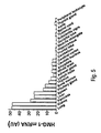
- HMG1-specific probe Potential sources of circulating HMG1 in vivo were assessed by hybridization of an HMG1-specific probe to mRNA prepared from various normal human tissues (blot substrate available from commercial sources), with the results summarized in Figure 5.
- mRNA prepared from various normal human tissues (blot substrate available from commercial sources), with the results summarized in Figure 5.
- macrophage-rich tissues lung, liver, kidney, pancreas and spleen
- this study shows the practicality and utility of assaying for HMG1-specific nucleic acid sequences in tissue samples.
- Example 3 Recombinant HMG1 Administration, in vitro and in vivo
- HMG1 open reading frame was amplified by PCR and subcloned into an expression vector (pCAL-n). Briefly, the 648-bp open reading frame of HMG1 cDNA was PCR amplified (94 °C 1', 56 °C 2', 72 °C 45", 30 cycles) from 5 ng Rat Brain Quick-Clone cDNA (Catalog # 7150-1, Clontech, Palo Alto, CA, USA) using primers containing the following sequences, 5'- CCC GC G GAT CC A TCG AGG GAA GGA TGG GCA AAG GAG ATC CTA -3' [SEQ ID NO.
- recombinant HMG1 To express recombinant HMG1, positive clones were cultured at 37 °C with vigorous shaking (250 rpm) until OD 600 reached 0.6, when IPTG (1 mM) was added. Twelve hours after IPTG induction, bacterial cells were harvested by centrifugation (6500 rpm, 15 minutes), and lysed by freeze-thaw cycles. The water-soluble fraction was collected after centrifugation (30 minutes, 12,000 rpm), and recombinant HMG1 was purified on a calmodulin-binding resin column as instructed by the manufacturer (Stratagene).
- Bacterial endotoxin was removed from the recombinant HMG1 by using Detoxi-Gel endotoxin-removing gel (Pierce, Rockford, IL USA, Cat. #20344), and residual LPS content was determined by the Limulus Amebocyte Lysate Test (LAL test, Cat. # 50-648U, QCL-1000 Chromogenic LAL, Bio-Whittaker, Inc., Walkersvi11e, MD, USA).
- Purified recombinant HMG1 was added to cultures of human peripheral blood mononuclear cells (HuPBMCs), and supernatants assayed for TNF by ELISA four hours after stimulation.
- the LPS-neutralizing agent polymyxin B (10 ⁇ g/ml) was added concurrently with recombinant HMG 1 to eliminate the effect of any contaminating LPS on TNF release. Additionally, recombinantly derived HMG1 was administered to test animals, with or without the additional endotoxemic challenge of exogenous LPS, to study the pathogenic potential of high levels of HMG1 in vivo (see Figures 2B and 2C). In some experiments, serum samples were secured from HMG1-treated animals to be assayed for TNF as detailed herein (see Figure 1B).
- the above procedure provides recombinant HMG1 as a fusion peptide comprising a 3.0 kDa calmodulin-binding domain and a thrombin cleavage site as an amino terminal extension in register with the HMG1 peptide sequence.
- the fusion tag was removed from an aliquot of the recombinant protein and the bioactivity of the full fusion protein was compared to the cleaved HMG1 peptide; no significant difference in bioactivity was noted and additional experiments (especially those requiring administration of recombinantly produced HMG 1 to animals) typically were conducted with the (uncleaved) fusion protein.
- polyclonal antibodies against an oligopeptide corresponding to the N-terminal amino acid sequence of HMG1, or against purified recombinant HMG1, were generated in rabbits according to standard procedures well known in the art. Briefly, eight copies of an oligopeptide with the sequence GKGDPKKPRGKMSSC [SEQ ID NO. 4] were anchored to radially branching lysine dendrites (small immunogenically inert core). These large macromolecules were injected three times both subcutaneously and intradermally (0.5 -1.0 mg per injection) into rabbits at week 1, 2, and 4 after pre-bleed at Day 0.
- HMG1-specific antibodies were used to measure by Western blotting analysis the inducible release of HMG1 from RAW 264.7 cells after treatment with TNF or LPS ( Figure 1). Briefly, proteins were fractionated by SDS-PAGE on a 4-20% gradient gel, transferred to a PVDF membrane, and blotted with rabbit antiserum raised against either the N-terminal synthetic HMG1 antigen or against recombinant HMG1. The signal was detected using a ECL kit as instructed by the manufacturer (Amersham Life Science Inc., Arlington Heights, IL, USA), and levels of HMG1 were determined by measuring optical intensity of bands on Western blots digitized for analysis using NIH 1.59 image software, with reference to a standard curve of purified recombinant HMG1.
- HMG 1 protein was detected in RAW 264.7 cells-conditioned medium in the absence of TNF or LPS treatment, but HMG 1 accumulated in conditioned medium to high levels after such stimulation, reaching a plateau at 8-28 hours after stimulation (Figure 1A).
- the data presented in Examples 1, 3 and in Figure 1A show that the release of HMG1 from macrophages is stimulus-specific and time- and dose-dependent, with maximal accumulation observed within 8 hours after stimulation with TNF at concentrations as low as 5 ng/ml. It is well appreciated that sepsis, septic shock and related conditions may occur in humans in response to stimuli that differ qualitatively or quantitatively from the single large, lethal LPS bolus used in this predictive model.
- HMG1 antagonists are perhaps more therapeutically attractive than TNF antagonists in view of the later appearance of HMG1 versus TNF in the response to endotoxin.
- This example illustrates an in vivo experiment in rodents measuring serum HMG1 levels after administration of a sublethal dose of LPS (LD 50 ).
- Mice or rats were treated with LPS, and sera were collected at different time points, and assayed for levels of HMG1 by Western blotting analysis.
- the serum concentrations of HMG1 were estimated by measuring the optical band intensity with reference to a standard curve of purified HMG1. Serum levels increased significantly by 16 hours after LPS, and remained high for at least 32 hours ( Figure 1B), and were not detectable in vehicle-treated control animals.
- This example provides the results of a predictive in vivo assay to measure therapeutic activity of antagonists of HMG1 in relation to treatment of sepsis and related conditions of cytokine-mediated toxicity.
- the HMG1 antagonist was an anti-HMG1 antibody preparation. Controls treated with pre-immune serum developed lethargy, piloerection, diarrhea, and succumbed to death within 48 hours. These clinical signs of endotoxemia were significantly prevented by administration of anti-HMG 1 antibodies.
- mice Male Balb/C mice (6-7 weeks, 20-23 grams) were randomly grouped (10 animals per group) and pre-treated either with control (pre-immune) or anti-HMG1 serum (as made in Example 4) 30 minutes before administration (intraperitoneally) of a lethal dose of LPS (50 mg/kg in 1 x PBS). Other experimental groups received additional doses of anti-HMG1 serum at +12 or, +12, and +36 hours after LPS administration. Animals were observed for appearance and survival for at least two weeks.
- Polyclonal antibodies against recombinant HMG1 were generated in rabbits, and antiserum was assayed for specificity and titer by ELISA and Western blotting procedures.
- the polyclonal antiserum immunospecifically recognized (bound to) recombinant HMG1 in Western blot analysis, for instance, and discriminated rHMG1 from other proteins in both crude bacterial lysates and as a purified protein that had been diluted into mouse serum.
- chemiluminescence-amplified detection methods in Western blotting analysis polyclonal anti-HMG1 antiserum at dilutions up to 1:1000 was useful to detect as little as 50 pg rHMG1 protein.
- FIG. 2B illustrates that rHMG1 causes dose-dependent lethality in endotoxic mice.
- Male Balb/C mice (20-23 grams) were randomized in groups of ten to receive LPS (3.15 mg/kg; a non-lethal dose) alone or in combination with purified recombinant HMG1 protein.
- LPS 3.15 mg/kg; a non-lethal dose
- HMG1 administered at the indicated doses 2, 16, 28 and 40 hours after LPS challenge significantly increased the lethality of the underlying endotoxemia.
- Figure 2C illustrates the independent lethal toxicity of HMG1 as a function of dose.
- Purified rHMG1 was administered to male Balb/C mice (five mice per treatment group) as a single i.p. bolus at the indicated dosage. Mice were observed for at least 48 hours, and 60% of mice treated with rHMG1 at a dose of 500 ⁇ g/mouse died within 24 hours of rHMG1 challenge, indicating a single dose LD 50 of less than 500 ⁇ g/mouse.
- anti-HMG1 antibodies The protection conferred by anti-HMG1 antibodies was specific, because administration of pre-immune serum, which showed no immunospecific reactivity to HMG1 on Western blots, did not spare subjects from LPS-mediated mortality ( Figure 2A). Moreover, HMG1-specific antibodies did not cross-react with other macrophage-derived cytokines (e.g. IL-1 and TNF), eliminating the possibility that antibodies conferred protection by binding and thereby neutralizing these mediators. Protection against sepsis, sepsis associated pathogenesis and sepsis-related diseases involving activation of pro-inflammatory cytokine cascades may be improved by combination therapy targeted against more than one component of the cytokine cascade.
- macrophage-derived cytokines e.g. IL-1 and TNF
- Antagonists of HMG1 in this regard can be combined with specific antagonists of TNF, IL-1, MIF and other inflammatory mediators, or with more broadly active antagonists of inflammatory responses that inhibit multiple components of the inflammatory cascade (e.g., aspirin, NSAIDS, anti-inflammatory steroids, etc.), to provide even more effective therapeutic modalities. Protection against LPS toxicity was antibody dose-related, and more frequent dosing with higher amounts of antibody reduced mortality by up to 70% (Figure 2A). Mice were observed for at least 2 weeks in all experiments, and no late mortality occurred, indicating that anti-HMG1 antibody treatment confers lasting protection against LPS lethality, and does not merely delay the time of death.
- This example provides data that establish an association between HMG1 and human sepsis, and thereby support an indication for using HMG1 antagonists generally and anti-HMG1 antibodies in particular in human sepsis and related conditions of cytokine toxicity.
- Serum HMG1 levels in normal healthy individuals and critically ill patients were measured using the polyclonal antibodies generated as in Example 4 in a Western blot format with reference to a standard curve of rHMG1.
- HMG1 was not detectable in normal controls, but accumulated to high levels in critically ill patients with sepsis (Table 2).
- Table 2 Serum appearance of HMG1 in sepsis patients.
- Normal Healthy 2 34 ⁇ d.1. Normal Healthy 3 35 ⁇ d.1. Normal Healthy 4 36 ⁇ d.1. Normal Healthy 5 61 ⁇ d.1. Normal Healthy 6 31 ⁇ d.1. Nonnal Healthy 7 55 10 Sepsis, anastomotic leak Recovered 8 70 7-20 Sepsis, colonic perforation Recovered 9 44 10-60 Sepsis, MOF, spinal reconstruction Died 10 60 >120 Sepsis, MOF, perforated gastric ulcer Died 11 47 >120 Sepsis, MOF, pneumonia Died Note: ⁇ d.l. - below detection limit; MOF - Multiple Organ Failure.
- HMG1 sequences protein or nucleic acid
- Example 8 HMG1 induces pro-inflammatory mediators and weight loss
- HMG1 is a late released mediator element of the inflammatory cytokine cascade.
- HMG1 induced serum TNF release in vivo
- purified recombinant HMG1 was administered intraperitoneally to Balb/C mice, and blood samples were collected to be assayed for TNF by the L929 assay.
- TNF was not detectable in serum of control animals, but was significantly increased two hours after administration of recombinant HMG1 protein.
- Serum HMG1 levels in hypophysectomized versus control rats also were measured by quantitation of Western blot intensities as described above. There were significantly higher HMG1 levels within 12 hours after endotoxic challenge (LPS at 1.0 mg/kg) in hypophysectomized rats (approximately 75 ng/ml) as compared to controls (approximately 25 ng/ml). These results indicate that pituicytes are not the major source of serum HMG1 levels and that macrophages may play a quantitatively more important role.
Abstract
Description
- The present invention provides a pharmaceutical composition and method for treating diseases characterized by activation of an inflammatory cytokine cascade, particularly sepsis, including septic shock and ARDS (acute respiratory distress syndrome), comprising administering an effective amount of an antagonist to the
high mobility group 1 protein (HMG1). The present invention further provides a diagnostic method for monitoring the severity of sepsis and related conditions, comprising measuring the serum concentration of HMG1 in a patient exhibiting symptoms of a disease characterized by activation of inflammatory cytokine cascade. Lastly, the present invention provides a pharmaceutical composition and method for effecting weight loss or treating obesity, comprising administering an effective amount of an HMG1 protein or a therapeutically active fragment of the gene product of an HMG1 gene. - Sepsis is an often fatal clinical syndrome that develops after infection or injury. Sepsis is the most frequent cause of mortality in hospitalized patients. Experimental models of gram negative sepsis based on administration of bacterial endotoxin (lipopolysaccharide, LPS) have led to an improved understanding of the pathogenic mechanisms of lethal sepsis and conditions related to sepsis by virtue of the activation of a common underlying inflammatory cytokine cascade. This cascade of host-response mediators includes TNF, IL-1, PAF and other macrophage-derived factors that have been widely studied as acute, early mediators of eventual lethality in severe endotoxemia (Zhang and Tracey, In The Cytokine Handbook, 3rd ed. Ed. Thompson (Academic Press Limited, USA). 515-547, 1998).
- Unfortunately, therapeutic approaches based on inhibiting these individual "early" mediators of endotoxemia have met with only limited success in large prospective clinical trials against sepsis in human patients. It is possible to infer from these disappointing results that later-appearing factors in the host response might critically determine pathogenesis and/or lethality in sepsis and related disorders. Accordingly, there is a need to discover such putative "late" mediators necessary and/or sufficient for part or all of the extensive multisystem pathogenesis, or for the lethality, of severe endotoxemia, particularly as endotoxemia is representative of clinical sepsis and related clinical disorders.
- HMG1 is a 30 kDa chromosomal nucleoprotein belonging to the burgeoning high mobility group (HMG) of non-histone chromatin-associated proteins. As a group, the HMG proteins recognize unique DNA structures and have been implicated in diverse cellular functions, including determination of nucleosome structure and stability, as well as in transcription and/or replication. The HMG proteins were first characterized by Johns and Goodwin as chromatin components with a high electrophoretic mobility in polyacrylamide gels (see in The HMG Chromosomal Proteins, E.W. Johns, Academic Press, London, 1982). Higher eukaryotes exhibit three families of HMG proteins: the HMG-1/-2 family, the HMG-14/-17 family and the HMG-I/-Y family. Although the families are distinguishable by size and DNA-binding properties, they are similar in their physical properties. HMG proteins are highly conserved across species, ubiquitously distributed and highly abundant, and are extractable from chromatin in 0.35 M NaCl and are soluble in 5% perchloric or trichloroacetic acid. Generally, HMG proteins are thought to bend DNA and facilitate binding of various transcription factors to their cognate sequences, including for instance, progesterone receptor, estrogen receptor, HOX proteins, and Oct1, Oct2 and Oct6. Recently, it has become apparent that a large, highly diverse group of proteins including several transcription factors and other DNA-interacting proteins, contain one or more regions similar to HMG1, and this feature has come to be known as the HMG1 box or HMG1 domain. cDNAs coding for HMG1 have been cloned from human, rat, trout, hamster, pig and calf cells, and HMG1 is believed to be abundant in all vertebrate cell nuclei. The protein is highly conserved with interspecies sequence identities in the 80% range. In chromatin, HMG1 binds to linker DNA between nucleosomes and to a variety of non-β-DNA structures such as palindromes, cruciforms and stem-loop structures, as well as cisplatin-modified DNA. DNA binding by HMG1 is generally believed to be sequence insensitive. HMG1 is most frequently prepared from washed nuclei or chromatin, but the protein has also been detected in the cytoplasm. (Reviewed in Landsman and Bustin, BioEssays 15:539-546, 1993; Baxevanis and Landsman, Nucleic Acids Research 23:514-523, 1995). To date, no link has been established between the HMG proteins and any clinical condition or disease.
- HMG1 has been alternatively identified as a heparin-binding protein abundantly expressed in developing brain and dubbed "amphoterin" for its highly dipolar sequence, comprising two internal repeats of a positively charged domain of about 80 amino acids (the HMG1 box) and an acidic C-terminal domain containing a stretch of approximately 30 continuous glutamic or aspartic acid residues. Amphoterin/HMG1 has been localized to the outer surface of the plasma membranes of epithelial, and especially neuronal cells, where it has been specifically localized to the filipodia of neural cells. Inhibition studies have suggested that amphoterin/HMG1 is required for process (neurite) extension and amphoterin/HMG1 also may be involved in neuron-glia interactions (Merenmies et al., J. Biol. Chem. 266:16722-16729,1991; Merenmies et al., J. Biol. Chem. 266:16722-16729, 1991; Milev et al., J. Biol. Chem. 273:6998-7005, 1998; and Salmivirta et al., Exp. Cell Res. 200:444-451, 1992). Amphoterin/HMG1 can be released from murine erythroleukemia cells after stimulation with the chemical inducer hexamethylenebisacetamide (Melloni et al., Biochem. Biophys. Res. Commun. 210:82-89, 1995). Previous study suggested that the gene product of the HMG1 gene functions as a differentiation enhancing factor by stimulating α-PKC (Melloni et al., Biochem. Biophys. Res. Cornrnun. 210:82-89, 1995; and Melloni et al., FEBS Lett. 368:466-470, 1995).
- The HMG1 gene product has been shown to interact with plasminogen and tissue-type plasminogen activator (t-PA) and effectively enhance plasmin generation at the cell surface, a system that is known to play a role in extracellular proteolysis during cell invasion and tissue remodeling. Amphoterin/HMG1 has also been shown to interact with the receptor of advanced glycosylation end products (RAGE) (Mohan et al., Biochem. Biophys. Res. Commun. 182:689-696, 1992; Yamawaki et al., J. Neurosci. Res. 44:586-593, 1996; Salmivirta et al., Exp. Cell Res. 200:444-451, 1992; and Vassalli et al., J. Clin. Invest. 88:1067-1072, 1991), (Redlitz and Plow, Baillieres Clin. Haematol. 8:313-327, 1995; and Parkkinen et al., J. Biol. Chem. 266:16730-16735, 1991).
- There is a longstanding need in the art to discover improved agents that can prevent the cytokine-mediated inflammatory cascade and have therapeutic activity in a large variety of cytokine-mediated inflammatory diseases. The present invention was made during the course of investigative research to identify agents that mediate toxicity, pathogenesis and/or lethality in sepsis and other disorders related by a common activation of the inflammatory cytokine cascade.
- Diseases and conditions mediated by the inflammatory cytokine cascade are numerous. Such conditions include the following grouped in disease categories:
- Systemic Inflammatory Response Syndrome, which includes:
- Sepsis syndrome
- Gram positive sepsis
- Gram negative sepsis
- Culture negative sepsis
- Fungal sepsis
- Neutropenic fever
- Urosepsis
- Meningococcemia
- Trauma hemorrhage
- Hums
- Ionizing radiation exposure
- Acute pancreatitis
- Adult respiratory distress syndrome (ARDS)
- Reperfusion Injury, which includes
- Post-pump syndrome
- Ischemia-reperfusion injury
- Cardiovascular Disease, which includes
- Cardiac stun syndrome
- Myocardial infarction
- Congestive heart failure
- Infectious Disease, which includes
- HIV infection/HIV neuropathy
- Meningitis
- Hepatitis
- Septic arthritis
- Peritonitis
- Pneumonia Epiglottitis
- E. coli 0157:H7
- Hemolytic uremic syndromc/thrombolytic thrombocytopcnic purpura
- Malaria
- Dengue hemorrhagic fever
- Leishmaniasis
- Leprosy
- Toxic shock syndrome
- Streptococcal myositis
- Gas gangrene
- Mycobacterium tuberculosis
- Mycobaclerium aviun intracellulare
- Pneumocystis carinii pneumonia
- Pelvic inflammatory disease
- Orchitis/epidydimitis
- Legionella
- Lyme disease
- Influenza A
- Epstein-Barr Virus
- Viral associated hemiaphagocytic syndrome
- Viral encephalitis/aseptic meningitis
- Obstetrics/Gynecology, including:
- Premature labor
- Miscarriage
- Infertility
- Inflammatory Disease/Autoimmunity, which includes:
- Rheumatoid arthritis/seronegative arthropathies
- Osteoarthritis
- Inflammatory bowel disease
- Systemic lupus erythematosis
- Iridoeyelitis/uveitistoptic neuritis
- Idiopathic pulmonary fibrosis
- Systemic vasculitis/Wegener's gramilomatosis
- Sarcoidosis
- Orchitis/vasectomy reversal procedures
- Allergic/Atopic Diseases, which includes:
- Asthma
- Allergic rhinitis
- Eczema
- Allergic contact dermatitis
- Allergic conjunctivitis
- Hypersensitivity pneumonitis
- Malignancy, which includes:
- ALL
- AML
- CML
- CLL
- Hodgkin's disease, non-Hodgkin's lymphoma
- Kaposi's sarcoma
- Colorectal carcinoma
- Nasopharyngeal carcinoma
- Malignant histiocytosis
- Paraneoplastic syndrome/hypercalcemia of malignancy
- Transplants, including:
- Organ transplant rejection
- Graft-versus-host disease
- Cachexia
- Congenital, which includes:
- Cystic fibrosis
- Familial hematophagocytic lymphohistiocytosis
- Sickle cell anemia
- Dermatologic, which includes:
- Psoriasis
- Alopecia
- Neurologic, which includes:
- Multiple sclerosis
- Migraine headache
- Renal, which includes:
- Nephrotic syndrome
- Hemodialysis
- Uremia
- Toxicity, which includes:
- OKT3 therapy
- Anti-CD3 therapy
- Cytokine therapy
- Chemotherapy
- Radiation therapy
- Chronic salicylate intoxication
- Metabolic/ldiopathic, which includes:
- Vlilson's disease
- Hemachromatosis
- Alpha-1 antitrypsin deficiency
- Diabetes
- Hashimoto's thyroiditis
- Osteoporosis
- Hypothalamic-pitultary-adrenal axis evaluation
- Primary biliary cirrhosis
- The present invention provides a pharmaceutical composition for treating conditions (diseases) mediated by the inflammatory cytokine cascade, comprising an effective amount of an antagonist or inhibitor of HMG1. Preferably, the HMG1 antagonist is selected from the group consisting of antibodies that bind to an HMG1 protein, HMG1 gene antisense sequences and HMG1 receptor antagonists. The present invention provides a method for treating a condition mediated by the inflammatory cytokine cascade, comprising administering an effective amount of an HMG1 antagonist. In another embodiment, the inventive method further comprises administering a second agent in combination with the HMG1 antagonist, wherein the second agent is an antagonist of an early sepsis mediator, such as TNF, IL-1α, IL-1β, MIF or IL-6. Most preferably, the second agent is an antibody to TNF or an IL-1 receptor antagonist (IL-1ra).
- The present invention further provides a diagnostic and prognostic method for monitoring the severity and predicting the likely clinical course of sepsis and related conditions for a patient exhibiting shock-like symptoms or at risk to exhibit symptoms associated with conditions mediated by the inflammatory cascade. The inventive diagnostic and prognostic method comprises measuring the concentration of HMG1 in a sample, preferably a serum sample, and comparing that concentration to a standard for HMG1 representative of a normal concentration range of HMG1 in a like sample, whereby higher levels of HMG1 are indicative of poor prognosis or the likelihood of toxic reactions. The diagnostic method may also be applied to other tissue or fluid compartments such as cerebrospinal fluid or urine. Lastly, the present invention provides a pharmaceutical composition and method for effecting weight loss or treating obesity, comprising administering an effective amount of
HMG 1 or a therapeutically active fragment thereof. -
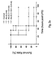
- Figure 1 shows two graphs that profile the induction of HMG1 release by LPS in vitro (Figure 1A) and in vivo (Figure 1B). Specifically, Figure 1A shows the accumulation of HMG1 in culture supernatants of macrophage RAW 264.7 cells after stimulation with LPS (100 ng/ml). The inset is a Western blot (using antibodies raised against recombinant HMG1) showing induction of HMG1 release from RAW 264.7 cells after induction with TNF. Figure 1B shows accumulation of HMG1 in serum of LPS-treated mice. Serum from Balb/C mice was collected at various time points after LPS administration, and assayed for HMG1 by Western blotting using antibodies raised against recombinant HMG1.
- Figure 2 illustrates that HMG1 is a mediator of pathogenesis and lethality in endotoxemia. Figure 2A shows the protective effect of anti-HMG1 antibodies against LPS lethality, tested in mice. Administration of anti-HMG1 antiserum in the indicated amounts at - 0.5 (if one dose), -0.5 and 12 (if two doses), or -0.5, 12 and 36 (if three doses) hours relative to LPS challenge (at time 0) was protective against LPS-induced lethality, and repeated dosing schedules provided better protection. Figure 2B illustrates that rHMG1 caused dose-dependent lethality in endotoxic mice. Male Ba1b/C mice (20-23 grams) were randomized in groups of ten to receive LPS (3.15 mg/kg; a non-lethal dose) alone or in combination with purified recombinant HMG1 protein. Administration of HMG1 at the indicated doses 2, 16, 28 and 40 hours after LPS challenge significantly increased the lethality of the underlying endotoxemia. Figure 2C illustrates independent lethal toxicity of HMG1 as a function of dose. Purified rHMG1 was administered to male Ba1b/C mice (five mice per treatment group) as a single i.p. bolus at the indicated dosage. Mice were observed for at least 48 hours, and 60% of mice treated with rHMG1 at a dose of 500 µg/mouse died within 24 hours of rHMG1 challenge, indicating a single dose LD50 of less than 500 µg/mouse.
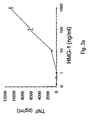
- Figure 3 shows that HMG1 induced TNF release both in vitro (Figure 3A) and in vivo (Figure 3B). Specifically, Figure 3A shows that HMG1 induces TNF release from huPPBMCs in dose-dependent fashion. Freshly isolated huPBMC cultures were stimulated with purified recombinant HMG1 protein at the indicated doses, and culture media were sampled four hours later to be assayed for TNF according to known immunologic methods (ELISA). Figure 3A shows the mean ± S.E.M. of the induced TNF response in two experiments (in triplicate). Figure 3B shows that administration of HMG1 induced accumulation of TNF in serum of treated mice. Balb/C mice (20-23 g) were treated intraperitoneally with purified recombinant HMG1 at the indicated doses and blood samples were taken two hours later for assay of TNF by an L929 bioassay and (TNF levels expressed as mean ± S.E.M., N=3).
- Figure 4 shows that HMG1 caused body weight loss in mice. Purified HMG1 was administered intraperitoneally to mice at 100 µg/mouse/day for three days, and body weight was monitored. Figure 4 shows the mean ± S.E.M. of net body weight change of three mice per group.
- Figure 5 shows the tissue distribution of HMG1 mRNA. Human RNA master blots containing poly(A)+ RNA of various tissues (Clontech, Palo Alto, CA, USA) were hybridized with a 0.6 kb digoxigenin-11-dUTP-labeled HMG1 cDNA probe synthesized by PCR using recombinant plasmid containing the HMG1 cDNA insert, all in accordance with methods well-known in the art. Briefly, hybridization was performed in a hybridization buffer (5X SSC/2% blocking reagent/0.1% SDS/50% formamide, Boehringer Mannheim, Indianapolis. IN) with a probe concentration of 10 ng/ml for 16 hours at 65 °C. After hybridization, the filter was subjected to two washes of0.5X SSC/0.1% SDS for 5 minutes, and two washes of 0.2X SSC/0.1% SDS for 10 minutes at room temperature. Signal was detected using anti-digoxigenin antibodies conjugated to phosphotase and detection reagents 4-nitrobluetetrazolium chloride (NBT) and 5-cromo-4-chloro-3-indolyl-phosphate (BCIP) (Boehringer-Mannheim) according to standard methods. The blots were scanned with a silver image scanner (Silverscanner II, Lacie Limited, Beaverton, OR), and relative optical density (in arbitrary units, AU) was quantified using NIH 1.59 image software. Note that highest levels were observed in macrophage-rich tissues.
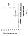
- Figure 6 shows, in comparison to a group of normal control subjects, increased human serum HMG1 levels as detected in hospitalized human subjects with sepsis, wherein the septic patients have been further categorized as to whether the patient died or survived.
- The present invention is based upon the discovery and isolation of a highly inducible 30 kDa protein that is released by, and accumulates in media conditioned by, cultured murine macrophage-like cells (RAW 264.7) following stimulation with LPS, TNF, or IL-1. A partial amino acid sequence of this isolated polypeptide was identical to the sequence of the HMG1 protein, also known as amphoterin, a protein not before linked to the pathogenesis of any disease. This information was used to clone a cDNA encoding HMG1, which sequence was expressed to provide recombinant protein, which protein was used to generate specific anti-HMG1 antibodies.
- Therapeutic and diagnostic efficacy was determined in a series of predictive in vitro and in vivo experiments. The experiments are detailed in the Examples section. For example, following administration of endotoxin (LD100) to mice, serum HMG1 levels increased later (at 16 h) than well-known "early" mediators of sepsis (such as TNF and IL-1) and plateau levels of HMG1 were maintained for 16 to 32 hours. Patients with lethal sepsis had high serum HMG1 levels, which were not detected in normal healthy volunteers. Moreover, acute experimental administration of rHMG1 to test animals, whether alone or in combination with sub-lethal amounts of LPS, caused marked pathological responses and even death. More distributed dosing schedules of lower amounts of rHMG1 led to significant weight loss in treated animals. These results give evidence that HMG1 is a mediator of endotoxemia and particularly a late mediator, as opposed to known "early" mediators such as TNF and IL-1. These data further show the importance of serum HMG1 as a marker for the severity or potential lethality of sepsis and related conditions.
- In addition, treatment with anti-HMG1 antibodies provided full protection from LD100 doses of LPS in mice. HMG1 is inducible by TNF and IL-1β, and dose-dependently stimulates TNF release from huPBMCs. TNF is a marker of macrophage activation, so it is likely (without limitation as to implied mechanisms or being bound by theory) that HMG1 promotes downstream re-activation of cytokine cascades which, in turn, mediates late pathogenesis and lethality in sepsis and related conditions involving activation of pro-inflammatory cytokine responses. Thus, HMG1 likely occupies a central role in mediating the inflammatory response to infection and injury, and antagonists of HMG1 will be of therapeutic benefit in sepsis and related conditions of inflammatory cascade activation. The appearance of
HMG 1 in the inflammatory cytokine cascade is suitable to propagate later phases of the host response and contribute to toxicity and lethality. The predictive data provided herein support the therapeutic efficacy of HMG1 antagonists and provide evidence in support of the aforementioned theory regarding mechanism of action. The in vivo treatment data showed the efficacy of HMG1 antagonists in general, and anti-HMG1 antibodies in particular, for treating conditions mediated by the inflammatory cytokine cascade in general and particularly sepsis conditions, including, for example, septic shock, sepsis syndrome or other "sepsis-like" conditions mediated by inflammatory cytokines. Further, the independent pathogenicity and toxicity/lethality of HMG1 shows that HMG1 antagonists are particularly effective when co-administered with antagonists of "early" inflammatory mediators such as TNF, MIF, IL-1 and IL-6. - In summary, HMG1 is a cytokine mediator of inflammatory reactions because: 1) HMG1 is released from macrophages and pituicytes following stimulation with bacterial toxins or with pro-inflammatory cytokines (TNF or IL-1β); 2) HMG1 accumulates in serum of animals exposed to LPS and in patients with sepsis; and 3) HMG1-specific antibodies protect against mortality in a predictive lethal endotoxemia animal model of clinical sepsis and related conditions.
- The inventive pharmaceutical composition or inventive pharmaceutical combination can be administered to a patient either by itself (complex or combination) or in pharmaceutical compositions where it is mixed with suitable carriers and excipients. The inventive pharmaceutical composition or inventive pharmaceutical combination can be administered parenterally, such as by intravenous injection or infusion, intraperitoneal injection, subcutaneous injection, or intramuscular injection. The inventive pharmaceutical composition or inventive pharmaceutical combination can be administered orally or rectally through appropriate formulation with carriers and excipients to form tablets, pills, capsules, liquids, gels, syrups, slurries, suspensions and the like. The inventive pharmaceutical composition or inventive pharmaceutical combination can be administered topically, such as by skin patch, to achieve consistent systemic levels of active agent. The inventive pharmaceutical composition or inventive pharmaceutical combination can be formulated into topical creams, skin or mucosal patches, liquids or gels suitable for topical application to skin or mucosal membrane surfaces. The inventive pharmaceutical composition or inventive pharmaceutical combination can be administered by inhaler to the respiratory tract for local or systemic treatment.
- The dosage of the inventive pharmaceutical composition or inventive pharmaceutical combination of the present invention can be determined by those skilled in the art from this disclosure. The pharmaceutical composition or inventive pharmaceutical combination will contain an effective dosage (depending upon the route of administration and pharmacokinetics of the active agent) of the inventive pharmaceutical composition or inventive pharmaceutical combination and suitable pharmaceutical carriers and excipients, which are suitable for the particular route of administration of the formulation (i.e., oral, parenteral, topical or by inhalation). The active agent is mixed into the pharmaceutical formulation by means of mixing, dissolving, granulating, dragee-making, emulsifying, encapsulating, entrapping or lyophilizing processes. The pharmaceutical formulations for parenteral administration include aqueous solutions of the active agent or combination in water-soluble form. Additionally, suspensions of the active agent may be prepared as oily injection suspensions. Suitable lipophilic solvents or vehicles include fatty oils such as sesame oil, or synthetic fatty acid esters, such as ethyl oleate or triglycerides, or liposomes. Aqueous injection suspensions may contain substances that increase the viscosity of the suspension, such as sodium carboxymethyl cellulose, sorbitol, or dextran. The suspension may optionally contain stabilizers or agents to increase the solubility of the active agent or combination to allow for more concentrated solutions.
- Pharmaceutical formulations for oral administration can be obtained by combining the active agent with solid excipients, such as sugars (e.g., lactose, sucrose, mannitol or sorbitol), cellulose preparations (e.g., starch, methyl cellulose, hydroxypropylmethyl cellulose, and sodium carboxymethyl cellulose), gelaten, gums, or polyvinylpyrrolidone. In addition, a disintegrating agent may be added, and a stabilizer may be added.
- The present invention provides antisense oligomers having a sequence effective to inhibit or block the expression of the HMG1 gene or mRNA sequence. Antisense technology, which uses specific- oligonucleotides to inhibit expression of target gene products, is developing as a therapeutic modality for human disease. Several selection criteria are available to contribute to the optimization of antisense oligonucleotide antagonists. For example, it is advisable to choose sequences with 50% or more GC content. Preferred sequences span the AUG initiation codon of the target protein, but sites in the coding region and 5' UTR may perform equally well. Such sequences are generally about 18-30 nucleotides long and chosen to overlap the ATG initiation codon from the HMG1 cDNA sequence to inhibit protein expression. Longer oligomers are often found to inhibit the target to a greater extent, indicating that a preferred length is about 25 mer for the first oligonucleotides chosen as antisense reagents. Typically, three oligonucleotide sequences are chosen with regard to these criteria, and compared for antagonist activity to control oligonucleotide sequences, such as "reverse" oligonucleotides or those in which about every fourth base of the antisense sequence is randomized. Therefore, a preferred sequence for making antisense oligomer sequences to HMG1 is a 25 mer sequence chosen to overlap the ATG initiation codon (underlined) from the HMG1 cDNA sequence:
GAGGAAAAATAACTAAACATGGGCAAAGGAGATCCTAAGAAG [SEQ ID NO. 5]
and such preferred antisense sequences are used to construct antisense oligonucleotide agents (and suitable controls) for an in vitro comparison as antagonists of HMG1. These in vitro data are predictive of human clinical utility using antisense agents of comparable design. HMGI-directed Antibodies - The antibodies disclosed herein may be polyclonal or monoclonal; may be from any of a number of human, non-human eukaryotic, cellular, fungal or bacterial sources. The antibodies may be encoded by genomic or vector-borne coding sequences, and may be elicited against native or recombinant HMG1 or fragments thereof with or without the use of adjuvants. The methods for making antibodies are methods and procedures well-known in the art for generating and producing antibodies. Generally, neutralizing antibodies against HMG1 (i.e., those that inhibit biological activities of HMG1 particularly with regard to its pro-inflammatory cytokine-like role) are preferred for therapeutic applications while non-neutralizing antibodies may be as suitable for diagnostic applications. Examples of such useful antibodies include but are not limited to polyclonal, monoclonal, chimeric, single-chain, and various human or humanized types of antibodies, as well as various fragments thereof such as Fab fragments and fragments produced from specialized expression systems.
- The diagnostic assay provided here uses anti-HMG1 antibodies that can be either polycolonal or monoclonal or both. The diagnostic procedure can utilize standard antibody-based techniques for measuring concentrations of the gene product of HMG1 genes in a biological fluid. Preferred standard diagnostic procedures are ELISA assays and Western techniques.
- This example provides the results of an experiment to identify and isolate later released macrophage-derived factors that play a role in sepsis and in related conditions typified by inflammatory cytokine activity. The experiment reported in this example examined murine macrophage RAW 264.7 cell-conditioned media after stimulation of the cultures with TNF. Murine macrophage RAW 264.7 cells were obtained from American Type Culture Collections (ATCC, Rockville, MD, USA), and proliferated in culture under DMEM supplemented with 10% fetal bovine serum and 1% glutamine. When confluency reached 70-80%, the medium was replaced by serum-free OPTI-MEM I medium and cultures were stimulated with pro-inflammatory cytokines (e.g., TNFα or IL-1) or bacterial endotoxin (LPS).
- The proteins released from the above stimulated macrophage cultures were surveyed. Specifically, at different time points, cells and cell-conditioned media were separately collected by centrifugation (3000 rpm, 10 minutes). Proteins in the conditioned medium were concentrated by ultrafiltration over Amicon membranes with Mr cutoff of 10 kDa (Amicon Inc., Beverly, MA, USA), subsequently fractionated by SDS-PAGE, and stained with Coomassie blue (1.25% Coomassie Blue R250 in 30% methanol/10% acetic acid). After destaining with 30% methanol/7% acetic acid, protein(s) of interest (i.e., those that preferentially accumulated in conditioned media of stimulated cultures) was isolated by excision from the SDS-PAGE gel, and subjected to N-terminal sequencing analysis (Commonwealth Biotechnologies, Inc., Richmond, VA, USA).
- Comparison of SDS-PAGE gel analysis of profiles of proteins accumulated in control (without TNFα stimulation) versus TNF-stimulated RAW 264.7 cells revealed a strongly inducible 30 kDa protein whose concentration in the cell-conditioned medium was significantly increased after stimulation for 16 hours. Amino acid sequence analysis of this isolated protein revealed its N-terminal sequence as Gly-Lys-Gly-Asp-Pro-Lys-Lys-Pro-Arg-Gly-Lys-Met-Ser-Ser [SEQ ID NO. 1]. A review of relevant gene databases found a 100% identity to the N-terminal amino acid sequence of HMG1.
- These data identified HMG1 as a "late-appearing" product of LPS-stimulated macrophage cultures, and therefore as a candidate pro-inflammatory mediator. This activity was confirmed by administration of recombinantly produced HMG1 and/or of anti-HMG1 antibodies in cellular and animal model systems that are predictive of human clinical conditions.
- This example shows which cell sources are capable of releasing HMG1 in response to TNF, IL-1 and/or LPS. Cells studied include GH3 pituicytes, murine macrophage RAW 264.7 cells, human primary peripheral blood mononuclear cells (huPBMCs), human primary T cells, rat adrenal PC-12 cells, and rat primary kidney cells (Table 1). The rat pituitary GH3 cell line was obtained from American Type Culture Collection (ATCC, Rockville, MD, USA), and cultured in DEME supplemented with 10% fetal bovine serum and 1% glutamine. Human PBMCs and T cells were freshly isolated from whole blood of healthy donors and cultured in RPMI 1640 supplemented with 10% human serum as previously described (Zhang et al., J Exp. Med. 185:1759-1768, 1997). When confluency reached 70-80%, the medium was replaced by serum-free OPTI-MEM I medium and cultures stimulated with proinflammatory cytokines (e.g., TNFα or IL-1) or bacterial endotoxin (LPS).
- Although human T cell, rat adrenal (PC-12) cells, and rat primary kidney cells contained cell-associated HMG1 as demonstrated by Western blotting analysis of whole cell lysates using HMG1-specific antibodies (see example 4 below), HMG1 did not significantly accumulate in the medium of these cultures after stimulation with either TNF, IL-1β, or LPS (Table 1).
Table 1. Induced release of HMG1 from various types of cells. Cell Type Stimulus TNF IL-1β LPS Murine RAW 264.7 cells Yes Yes Yes Human PBMCs Yes Yes Yes Human primary T cells No No No Rat adrenal PC-12 cells No No No Rat pituitary GH3 cells Yes Yes No Rat primary kidney cells No No No Note: PBMCs, peripheral blood mononuclear cells. - Potential sources of circulating HMG1 in vivo were assessed by hybridization of an HMG1-specific probe to mRNA prepared from various normal human tissues (blot substrate available from commercial sources), with the results summarized in Figure 5. Several macrophage-rich tissues (lung, liver, kidney, pancreas and spleen) exhibited the most
abundant HMG 1 mRNA expression; less was observed in pituitary, bone marrow, thymus, lymph node and adrenal gland. In addition to providing information as to the relative tissue distribution of-HMG1 expression, this study shows the practicality and utility of assaying for HMG1-specific nucleic acid sequences in tissue samples. - This example details procedures to produce HMG1 by well-known recombinant DNA technologies. The HMG1 open reading frame was amplified by PCR and subcloned into an expression vector (pCAL-n). Briefly, the 648-bp open reading frame of HMG1 cDNA was PCR amplified (94 °C 1', 56 °C 2', 72 °C 45", 30 cycles) from 5 ng Rat Brain Quick-Clone cDNA (Catalog # 7150-1, Clontech, Palo Alto, CA, USA) using primers containing the following sequences, 5'- CCC GCG GAT CCA TCG AGG GAA GGA TGG GCA AAG GAG ATC CTA -3' [SEQ ID NO. 2], and 5'- CCC GCA AGC TTA TTC ATC ATC ATC ATC TTC T -3' [SEQ ID NO. 3]. The 680 bp PCR product (4 µg) was digested with Bam HI and Hind III, and cloned into the Bam HI/Hind III cloning sites of the pCAL-n vector (Stratagene, La Jolla, CA, USA). The recombinant plasmid was transformed into E. coli BL21(DE3)pLysS (Novagen, Madison, WI, USA), and positive clones were screened and confirmed by DNA sequencing on both strands using a Tag DyeDeoxy terminator cycle sequencing kit on the ABI 373A automated fluorescent sequencer (Applied Biosystems, Foster City, CA, USA).
- To express recombinant HMG1, positive clones were cultured at 37 °C with vigorous shaking (250 rpm) until OD600 reached 0.6, when IPTG (1 mM) was added. Twelve hours after IPTG induction, bacterial cells were harvested by centrifugation (6500 rpm, 15 minutes), and lysed by freeze-thaw cycles. The water-soluble fraction was collected after centrifugation (30 minutes, 12,000 rpm), and recombinant HMG1 was purified on a calmodulin-binding resin column as instructed by the manufacturer (Stratagene). Bacterial endotoxin was removed from the recombinant HMG1 by using Detoxi-Gel endotoxin-removing gel (Pierce, Rockford, IL USA, Cat. #20344), and residual LPS content was determined by the Limulus Amebocyte Lysate Test (LAL test, Cat. # 50-648U, QCL-1000 Chromogenic LAL, Bio-Whittaker, Inc., Walkersvi11e, MD, USA). Purified recombinant HMG1 was added to cultures of human peripheral blood mononuclear cells (HuPBMCs), and supernatants assayed for TNF by ELISA four hours after stimulation. The LPS-neutralizing agent polymyxin B (10 µg/ml) was added concurrently with
recombinant HMG 1 to eliminate the effect of any contaminating LPS on TNF release. Additionally, recombinantly derived HMG1 was administered to test animals, with or without the additional endotoxemic challenge of exogenous LPS, to study the pathogenic potential of high levels of HMG1 in vivo (see Figures 2B and 2C). In some experiments, serum samples were secured from HMG1-treated animals to be assayed for TNF as detailed herein (see Figure 1B). - The above procedure provides recombinant HMG1 as a fusion peptide comprising a 3.0 kDa calmodulin-binding domain and a thrombin cleavage site as an amino terminal extension in register with the HMG1 peptide sequence. In some experiments, the fusion tag was removed from an aliquot of the recombinant protein and the bioactivity of the full fusion protein was compared to the cleaved HMG1 peptide; no significant difference in bioactivity was noted and additional experiments (especially those requiring administration of recombinantly produced
HMG 1 to animals) typically were conducted with the (uncleaved) fusion protein. - As demonstrated in Figures 3A and 3B, in vitro or in vivo administration of recombinantly derived HMG1 induced a brisk TNF response, confirming the identification of HMG1 as a late-appearing LPS-induced macrophage-derived endogenous mediator with pro-inflammatory activity.
- This example provides the results of experiments to generate and use polyclonal antibodies against
HMG 1. Briefly, polyclonal antibodies against an oligopeptide corresponding to the N-terminal amino acid sequence of HMG1, or against purified recombinant HMG1, were generated in rabbits according to standard procedures well known in the art. Briefly, eight copies of an oligopeptide with the sequence GKGDPKKPRGKMSSC [SEQ ID NO. 4] were anchored to radially branching lysine dendrites (small immunogenically inert core). These large macromolecules were injected three times both subcutaneously and intradermally (0.5 -1.0 mg per injection) into rabbits atweek Day 0. Two weeks after the last immunization, rabbits were bled and boosted intramuscularly with 1.0 mg of antigen, followed by a second bleeding two weeks later. Alternatively, to produce polyclonal antibodies against recombinant HMG1, rabbits were immunized with recombinant HMG1 fusion peptide (100 µg per injection) following a similar protocol. Monoclonal antibodies reactive against HMG1 (i.e., that bind, and in some cases, neutralize or antagonize the biological activity of HMG1) are conveniently prepared according to methods well known in the art using the HMG1 antigens described herein or other HMG1 peptide fragments as immunogens. Such monoclonal antibodies, and/or the hybridomas that produce them, are useful to produce various "humanized" antibodies reactive against HMG1 (all according to methods known in the art), which humanized antibodies are useful as taught herein. - HMG1-specific antibodies were used to measure by Western blotting analysis the inducible release of HMG1 from RAW 264.7 cells after treatment with TNF or LPS (Figure 1). Briefly, proteins were fractionated by SDS-PAGE on a 4-20% gradient gel, transferred to a PVDF membrane, and blotted with rabbit antiserum raised against either the N-terminal synthetic HMG1 antigen or against recombinant HMG1. The signal was detected using a ECL kit as instructed by the manufacturer (Amersham Life Science Inc., Arlington Heights, IL, USA), and levels of HMG1 were determined by measuring optical intensity of bands on Western blots digitized for analysis using NIH 1.59 image software, with reference to a standard curve of purified recombinant HMG1.
- No HMG1 protein was detected in RAW 264.7 cells-conditioned medium in the absence of TNF or LPS treatment, but
HMG 1 accumulated in conditioned medium to high levels after such stimulation, reaching a plateau at 8-28 hours after stimulation (Figure 1A). In summary, the data presented in Examples 1, 3 and in Figure 1A show that the release of HMG1 from macrophages is stimulus-specific and time- and dose-dependent, with maximal accumulation observed within 8 hours after stimulation with TNF at concentrations as low as 5 ng/ml. It is well appreciated that sepsis, septic shock and related conditions may occur in humans in response to stimuli that differ qualitatively or quantitatively from the single large, lethal LPS bolus used in this predictive model. Nevertheless, experimental endotoxemia has been a valuable and predictive model system by which to identify critical components of the inflammatory cytokine cascade and by which to identify specific antagonists with predicted clinical utility. In this regard, HMG1 antagonists are perhaps more therapeutically attractive than TNF antagonists in view of the later appearance of HMG1 versus TNF in the response to endotoxin. - This example illustrates an in vivo experiment in rodents measuring serum HMG1 levels after administration of a sublethal dose of LPS (LD50). Mice or rats were treated with LPS, and sera were collected at different time points, and assayed for levels of HMG1 by Western blotting analysis. The serum concentrations of HMG1 were estimated by measuring the optical band intensity with reference to a standard curve of purified HMG1. Serum levels increased significantly by 16 hours after LPS, and remained high for at least 32 hours (Figure 1B), and were not detectable in vehicle-treated control animals. These data show that HMG1 represents a particularly attractive target for diagnosis of, and pharmaceutical intervention against, sepsis and related disorders of cytokine toxicity because
HMG 1 is a late-appearing mediator in the inflammatory cytokine cascade. - This example provides the results of a predictive in vivo assay to measure therapeutic activity of antagonists of HMG1 in relation to treatment of sepsis and related conditions of cytokine-mediated toxicity. In this example, the HMG1 antagonist was an anti-HMG1 antibody preparation. Controls treated with pre-immune serum developed lethargy, piloerection, diarrhea, and succumbed to death within 48 hours. These clinical signs of endotoxemia were significantly prevented by administration of anti-HMG 1 antibodies. Male Balb/C mice (6-7 weeks, 20-23 grams) were randomly grouped (10 animals per group) and pre-treated either with control (pre-immune) or anti-HMG1 serum (as made in Example 4) 30 minutes before administration (intraperitoneally) of a lethal dose of LPS (50 mg/kg in 1 x PBS). Other experimental groups received additional doses of anti-HMG1 serum at +12 or, +12, and +36 hours after LPS administration. Animals were observed for appearance and survival for at least two weeks.
- Polyclonal antibodies against recombinant HMG1 were generated in rabbits, and antiserum was assayed for specificity and titer by ELISA and Western blotting procedures. The polyclonal antiserum immunospecifically recognized (bound to) recombinant HMG1 in Western blot analysis, for instance, and discriminated rHMG1 from other proteins in both crude bacterial lysates and as a purified protein that had been diluted into mouse serum. Using chemiluminescence-amplified detection methods in Western blotting analysis, polyclonal anti-HMG1 antiserum at dilutions up to 1:1000 was useful to detect as little as 50 pg rHMG1 protein. Administration of anti-HMG1 antiserum in the indicated (Figure 2A) amounts at -0.5 (if one dose), -0.5 and 12 (if two doses), or -0.5, 12 and 36 (if three doses) hours relative to LPS challenge (at time 0) was protective against LPS-induced lethality, and repeated dosing schedules provided better protection.
- Figure 2B illustrates that rHMG1 causes dose-dependent lethality in endotoxic mice. Male Balb/C mice (20-23 grams) were randomized in groups of ten to receive LPS (3.15 mg/kg; a non-lethal dose) alone or in combination with purified recombinant HMG1 protein. Administration of HMG1 at the indicated doses 2, 16, 28 and 40 hours after LPS challenge significantly increased the lethality of the underlying endotoxemia.
- Figure 2C illustrates the independent lethal toxicity of HMG1 as a function of dose. Purified rHMG1 was administered to male Balb/C mice (five mice per treatment group) as a single i.p. bolus at the indicated dosage. Mice were observed for at least 48 hours, and 60% of mice treated with rHMG1 at a dose of 500 µg/mouse died within 24 hours of rHMG1 challenge, indicating a single dose LD50 of less than 500 µg/mouse.
- The protection conferred by anti-HMG1 antibodies was specific, because administration of pre-immune serum, which showed no immunospecific reactivity to HMG1 on Western blots, did not spare subjects from LPS-mediated mortality (Figure 2A). Moreover, HMG1-specific antibodies did not cross-react with other macrophage-derived cytokines (e.g. IL-1 and TNF), eliminating the possibility that antibodies conferred protection by binding and thereby neutralizing these mediators. Protection against sepsis, sepsis associated pathogenesis and sepsis-related diseases involving activation of pro-inflammatory cytokine cascades may be improved by combination therapy targeted against more than one component of the cytokine cascade. Antagonists of HMG1 in this regard can be combined with specific antagonists of TNF, IL-1, MIF and other inflammatory mediators, or with more broadly active antagonists of inflammatory responses that inhibit multiple components of the inflammatory cascade (e.g., aspirin, NSAIDS, anti-inflammatory steroids, etc.), to provide even more effective therapeutic modalities. Protection against LPS toxicity was antibody dose-related, and more frequent dosing with higher amounts of antibody reduced mortality by up to 70% (Figure 2A). Mice were observed for at least 2 weeks in all experiments, and no late mortality occurred, indicating that anti-HMG1 antibody treatment confers lasting protection against LPS lethality, and does not merely delay the time of death.
- This example provides data that establish an association between HMG1 and human sepsis, and thereby support an indication for using HMG1 antagonists generally and anti-HMG1 antibodies in particular in human sepsis and related conditions of cytokine toxicity. Serum HMG1 levels in normal healthy individuals and critically ill patients were measured using the polyclonal antibodies generated as in Example 4 in a Western blot format with reference to a standard curve of rHMG1. HMG1 was not detectable in normal controls, but accumulated to high levels in critically ill patients with sepsis (Table 2).
Table 2. Serum appearance of HMG1 in sepsis patients. Patient (#) Age (year) HMG1 (ng/ml) Diagnosis Outcome 1 27 <d.1. Normal Healthy 2 34 <d.1. Normal Healthy 3 35 <d.1. Normal Healthy 4 36 <d.1. Normal Healthy 5 61 <d.1. Normal Healthy 6 31 <d.1. Nonnal Healthy 7 55 10 Sepsis, anastomotic leak Recovered 8 70 7-20 Sepsis, colonic perforation Recovered 9 44 10-60 Sepsis, MOF, spinal reconstruction Died 10 60 >120 Sepsis, MOF, perforated gastric ulcer Died 11 47 >120 Sepsis, MOF, pneumonia Died Note: <d.l. - below detection limit; MOF - Multiple Organ Failure. - These data show that elevated serum HMG1 levels are observed in patients with sepsis, and the highest levels of serum HMG1 are observed in lethal cases (Table 2). These data further indicate the therapeutic importance of HMG1 antagonists in sepsis and also provide evidence for the diagnostic utility of an assay for sepsis and severity (i.e., potential lethality) of sepsis by measuring serum concentrations of HMG1. This diagnostic assay is also useful for diagnosing the severity of allied conditions involving activation of the inflammatory cytokine cascade.
- Additional subjects were screened for serum HMG1 levels in association with lethal versus non-lethal sepsis, with results (cumulative with Table 2) as described in Figure 6. The data summarized in Figure 6 represent serum samples obtained from eight healthy subjects and twenty-five septic patients infected with Gram positive [Bacillus fragilis (1 patient), Enterococcus facecalis (1 patient), Streptococcus pneumonia (4 patients), Listeria monocytogenes (1 patient), or Staphylococcus aureus (2 patients)], Gram negative [Eschericlzia coli (7 patients), Klebsiella pneumonia (1 patient), Acinetobacter calcoaceticus (1 patient), Pseudomonas aeruginosa (1 patient), Fusobacterium nucleatum (1 patient), Citrobacter freundii (1 patient)], or unidentified pathogens (5 patients). Serum was fractionated by SDS-PAGE gel electrophoresis, and HMG1 levels were determined by Western blotting analysis with reference to standard curves of purified rHMG1 diluted in normal human serum. The detection limit by Western blotting analysis is 50 pg. Note that HMG1 is not detectable in normal controls, but significantly increased in septic patients. The average level of HMG1 in serum of non-surviving septic patients (N = 13 patients, mean HMG1 level = 83.7 ± 22.3 ng/ml) is significantly higher than in survivors (N = 12, mean HMG1 level = 25.2 ± 15.1 ng/ml, P < 0.05). These data provide direct evidence of the utility of screening tissue (including, without limitation blood or serum) samples for HMG1 sequences (protein or nucleic acid) as a diagnostic and prognostic indicator of the presence of sepsis and related disorders of cytokine activation and of the severity and likely clinical course of such diseases and conditions.
- The present results provide evidence that HMG1 is a late released mediator element of the inflammatory cytokine cascade. Addition of recombinant HMG1 to primary human peripheral blood mononuclear cells led to the dose-dependent induction of TNF within four hours after stimulation (Figure 3A). This stimulation by recombinant HMG1 of TNF release by HuPBMCs was not due to LPS contamination because: (i) purified recombinant HMG1 was not contaminated by LPS as judged by an LAL endotoxin assay; ii) addition of the LPS-neutralizing agent polymyxin B did not affect HMG1-induced TNF release; and iii) proteolytic cleavage of recombinant HMG1 preparations with trypsin completely abolished the TNF release activity for the PBMC cultures. HMG1 stimulation also induced macrophages to release nitric oxide (NO).
- To confirm that HMG1 induced serum TNF release in vivo, purified recombinant HMG1 was administered intraperitoneally to Balb/C mice, and blood samples were collected to be assayed for TNF by the L929 assay. As shown in Figure 3B, TNF was not detectable in serum of control animals, but was significantly increased two hours after administration of recombinant HMG1 protein.
- Repetitive administration of recombinant gene product of the HMG1 gene (100 µg/mouse/day) caused significant body weight loss (Figure 4) in mice. Without limitation as to mechanism and without being bound by theory, these data are consistent with the hypothesis that HMG1 acts as a feed-forward stimulator of the pro-inflammatory cascade under both in vitro and in vivo conditions. These in vivo data in a predictive model of weight loss also provide predictive evidence that a pharmaceutical formulation comprising HMG1, or a therapeutically active fragment thereof, is an effective weight loss therapy.
- Serum HMG1 levels in hypophysectomized versus control rats also were measured by quantitation of Western blot intensities as described above. There were significantly higher HMG1 levels within 12 hours after endotoxic challenge (LPS at 1.0 mg/kg) in hypophysectomized rats (approximately 75 ng/ml) as compared to controls (approximately 25 ng/ml). These results indicate that pituicytes are not the major source of serum HMG1 levels and that macrophages may play a quantitatively more important role.


Claims (16)
- A method for diagnosing a condition in a patient characterized by activation of the inflammatory cytokine cascade, the method comprising measuring the concentration of HMG1 in a sample from said patient, and comparing that concentration to a standard for HMG1 representative of a normal concentration range of HMG1 in a like sample, whereby a higher level of HMG1 in the sample from the patient indicates a diagnosis of a condition characterized by activation of the inflammatory cytokine cascade.
- A prognostic method for monitoring the severity and predicting the likely clinical course in a patient of a condition characterized by activation of the inflammatory cytokine cascade, the method comprising measuring the concentration of HMG1 in a sample from said patient, and comparing that concentration to a standard for HMG1 representative of a normal range of HMG1 in a like sample, whereby a higher level of HMG1 in the sample from the patient indicates a poor prognosis or the likelihood of toxic reactions.
- The method of Claim 1 or Claim 2, wherein the condition is sepsis.
- The method of Claim 1 or Claim 2, wherein the condition is septic arthritis or rheumatoid arthritis.
- The method of Claim 1 or Claim 2, wherein the condition is systemic lupus erythematosus.
- The method of Claim 1 or Claim 2, wherein the condition is trauma hemorrhage.
- The method of Claim 1 or Claim 2, wherein the condition is malaria.
- The method of Claim 1 or Claim 2, wherein the sample from said patient is a serum sample.
- A pharmaceutical composition for treating conditions (diseases) characterized by activation of the inflammatory cytokine cascade, comprising an effective amount of an antagonist or inhibitor of HMG1.
- A pharmaceutical composition for treating sepsis and related conditions, comprising an effective amount of an HMG1 antagonist.
- The pharmaceutical composition of claims 9 or 10 wherein the HMG1 antagonist is selected from the group consisting of antibodies that bind to HMG1, HMG1 antisense sequences and HMG1 receptor antagonists.
- A method for treating a condition characterized by activation of the inflammatory cytokine cascade, comprising administering an effective amount of an HMG1 antagonist.
- A method for treating sepsis and related conditions involving activation of the inflammatory cytokine cascade syndrome, comprising administering an effective amount of an HMG1 antagonist.
- The method of claims 12 or 13 further comprising administering a second agent in combination with the HMG1 antagonist, wherein the second agent is an antagonist of an early sepsis mediator.
- The methods of claim 14 wherein the second agent is an antagonist of a cytokine selected from the group consisting of TNF, IL-1α, IL-1β, MIF and IL-6.
- The method of claim 15 wherein the second agent is an antibody to TNF or MIF or an IL-1 receptor antagonist (IL-1ra). 17. A pharmaceutical composition for effecting weight loss or treating obesity, comprising an effective amount of HMG1 or a therapeutically active fragment thereof. 18. A method for effecting weight loss or traiting obesity, comprising administering an effective amount of HMG1 or therapeutically active fragment thereof.
Applications Claiming Priority (2)
| Application Number | Priority Date | Filing Date | Title |
|---|---|---|---|
| US09/248,574 US6303321B1 (en) | 1999-02-11 | 1999-02-11 | Methods for diagnosing sepsis |
| EP00915762.9A EP1165110B2 (en) | 1999-02-11 | 2000-02-11 | Antagonists of hmg1 for treating inflammatory conditions |
Related Parent Applications (3)
| Application Number | Title | Priority Date | Filing Date |
|---|---|---|---|
| EP00915762.9A Division-Into EP1165110B2 (en) | 1999-02-11 | 2000-02-11 | Antagonists of hmg1 for treating inflammatory conditions |
| EP00915762.9A Division EP1165110B2 (en) | 1999-02-11 | 2000-02-11 | Antagonists of hmg1 for treating inflammatory conditions |
| EP00915762.9 Division | 2000-02-11 |
Publications (3)
| Publication Number | Publication Date |
|---|---|
| EP1757937A2 true EP1757937A2 (en) | 2007-02-28 |
| EP1757937A3 EP1757937A3 (en) | 2007-04-11 |
| EP1757937B1 EP1757937B1 (en) | 2013-05-15 |
Family
ID=22939703
Family Applications (4)
| Application Number | Title | Priority Date | Filing Date |
|---|---|---|---|
| EP00915762.9A Expired - Lifetime EP1165110B2 (en) | 1999-02-11 | 2000-02-11 | Antagonists of hmg1 for treating inflammatory conditions |
| EP20100075469 Ceased EP2362221A1 (en) | 1999-02-11 | 2000-02-11 | Antagonists of HMG1 for treating inflamatory conditions |
| EP05077852A Withdrawn EP1707214A1 (en) | 1999-02-11 | 2000-02-11 | Antagonists of HMG1 for treating inflammatory conditions and the use of HMG1 for effecting weightloss or treating obesity |
| EP06075291.2A Expired - Lifetime EP1757937B1 (en) | 1999-02-11 | 2000-02-11 | Use of HMG1 in the diagnosis of inflammatory conditions |
Family Applications Before (3)
| Application Number | Title | Priority Date | Filing Date |
|---|---|---|---|
| EP00915762.9A Expired - Lifetime EP1165110B2 (en) | 1999-02-11 | 2000-02-11 | Antagonists of hmg1 for treating inflammatory conditions |
| EP20100075469 Ceased EP2362221A1 (en) | 1999-02-11 | 2000-02-11 | Antagonists of HMG1 for treating inflamatory conditions |
| EP05077852A Withdrawn EP1707214A1 (en) | 1999-02-11 | 2000-02-11 | Antagonists of HMG1 for treating inflammatory conditions and the use of HMG1 for effecting weightloss or treating obesity |
Country Status (11)
| Country | Link |
|---|---|
| US (12) | US6303321B1 (en) |
| EP (4) | EP1165110B2 (en) |
| JP (3) | JP4812166B2 (en) |
| AT (1) | ATE327764T1 (en) |
| AU (1) | AU782984B2 (en) |
| CA (1) | CA2359926C (en) |
| DE (1) | DE60028358T3 (en) |
| DK (1) | DK1165110T3 (en) |
| ES (1) | ES2269118T5 (en) |
| PT (1) | PT1165110E (en) |
| WO (1) | WO2000047104A2 (en) |
Cited By (4)
| Publication number | Priority date | Publication date | Assignee | Title |
|---|---|---|---|---|
| US7632500B2 (en) | 2003-09-11 | 2009-12-15 | Cornerstone Therapeutics, Inc. | Monoclonal antibodies against HMGB1 |
| US7749959B2 (en) | 2001-05-15 | 2010-07-06 | The Feinstein Institute For Medical Research | Use of HMGB fragments as anti-inflammatory agents |
| US8053206B2 (en) | 1999-02-11 | 2011-11-08 | The Feinstein Institute For Medical Research | Antagonists of HMG1 for treating inflammatory conditions |
| US8188041B2 (en) | 2003-06-06 | 2012-05-29 | The Feinstein Institute For Medical Research | Inhibitors of the interaction between HMGB polypeptides and toll-like receptor 2 as anti-inflammatory agents |
Families Citing this family (123)
| Publication number | Priority date | Publication date | Assignee | Title |
|---|---|---|---|---|
| US6387639B1 (en) * | 1998-11-10 | 2002-05-14 | Sloan-Kettering Institute For Cancer Research | Ma family polypeptides and anti-Ma antibodies |
| US7151082B2 (en) * | 1999-02-11 | 2006-12-19 | The Feinstein Institute For Medical Research | Antagonists of HMG1 for treating inflammatory conditions |
| US20060154319A1 (en) * | 1999-04-29 | 2006-07-13 | Flodgaard Hans J | Inhibition of bradykinin release |
| US6610713B2 (en) * | 2000-05-23 | 2003-08-26 | North Shore - Long Island Jewish Research Institute | Inhibition of inflammatory cytokine production by cholinergic agonists and vagus nerve stimulation |
| US8914114B2 (en) | 2000-05-23 | 2014-12-16 | The Feinstein Institute For Medical Research | Inhibition of inflammatory cytokine production by cholinergic agonists and vagus nerve stimulation |
| ITMI20010562A1 (en) * | 2001-03-16 | 2002-09-16 | Marco E Bianchi | HMG1 PROTEIN INHIBITORS OR ANTAGONISTS FOR THE TREATMENT OF VASCULAR DISORDERS |
| PL367132A1 (en) * | 2001-05-15 | 2005-02-21 | North Shore-Long Island Jewish Research Institute | Use of hmg fragments as anti-inflammatory agents |
| US7220723B2 (en) * | 2001-05-15 | 2007-05-22 | The Feinstein Institute For Medical Research | Inhibitors of the interaction between HMGB polypeptides and toll-like receptor 2 as anti-inflammatory agents |
| US7026121B1 (en) | 2001-06-08 | 2006-04-11 | Expression Diagnostics, Inc. | Methods and compositions for diagnosing and monitoring transplant rejection |
| US6905827B2 (en) | 2001-06-08 | 2005-06-14 | Expression Diagnostics, Inc. | Methods and compositions for diagnosing or monitoring auto immune and chronic inflammatory diseases |
| US7235358B2 (en) | 2001-06-08 | 2007-06-26 | Expression Diagnostics, Inc. | Methods and compositions for diagnosing and monitoring transplant rejection |
| JP5055598B2 (en) * | 2001-07-13 | 2012-10-24 | 株式会社シノテスト | Method and reagent for immunological measurement of human HMG-1 using an antibody that specifically binds to human HMG-1 |
| ITMI20011986A1 (en) * | 2001-09-25 | 2003-03-25 | San Raffaele Centro Fond | METHOD AND COMPOSITION FOR THE ACTIVATION OF CELLS PRESENTING THE ANTIGEN |
| US7465555B2 (en) * | 2002-04-02 | 2008-12-16 | Becton, Dickinson And Company | Early detection of sepsis |
| ATE424416T1 (en) | 2002-07-03 | 2009-03-15 | San Raffaele Centro Fond | THE USE OF HMGB1 TO TREAT TISSUE INJURY AND/OR TO SUPPORT TISSUE RECOVERY |
| WO2004005539A1 (en) * | 2002-07-05 | 2004-01-15 | The University Of British Columbia | Diagnosis of sepsis using mitochondrial nucleic acid assays |
| GB0226251D0 (en) * | 2002-11-11 | 2002-12-18 | San Raffaele Centro Fond | Acetylated protein |
| MXPA05005072A (en) * | 2002-11-12 | 2005-07-22 | Becton Dickinson Co | Diagnosis of sepsis or sirs using biomarker profiles. |
| EP1565570A4 (en) * | 2002-11-12 | 2005-12-28 | Becton Dickinson Co | Diagnosis of sepsis or sirs using biomarker profiles |
| AU2003291482A1 (en) * | 2002-11-12 | 2004-06-03 | Becton, Dickinson And Company | Diagnosis of sepsis or sirs using biomarker profiles |
| US20040141948A1 (en) * | 2002-11-20 | 2004-07-22 | Critical Therapeutics, Inc. | Use of HMGB fragments as anti-inflammatory agents |
| EP1569684A4 (en) * | 2002-11-20 | 2006-08-02 | Critical Therapeutics Inc | Use of hmgb fragments as anti-inflammatory agents |
| US20040156851A1 (en) * | 2002-11-20 | 2004-08-12 | Critical Therapeutics, Inc. | HMGB1 combination therapies |
| EP1567544A4 (en) * | 2002-11-20 | 2009-07-22 | Long Island Jewish Res Inst | Use of hmgb polypeptides for increasing immune responses |
| CN1735414A (en) | 2002-12-06 | 2006-02-15 | 北岸长岛犹太人研究学院 | Inhibition of inflamation using alpha 7 receptor-binding cholinergic agonists |
| US7700307B2 (en) | 2003-03-08 | 2010-04-20 | Auvation Limited | Mitochondrial stress-70 protein markers for colorectal cancer |
| US7892745B2 (en) | 2003-04-24 | 2011-02-22 | Xdx, Inc. | Methods and compositions for diagnosing and monitoring transplant rejection |
| US20090069227A9 (en) * | 2003-04-29 | 2009-03-12 | Capogrossi Maurizio C | Use of HMGB1 to promote stem cell migration and/or proliferation |
| FR2855832B1 (en) * | 2003-06-03 | 2007-09-14 | Biomerieux Sa | DIAGNOSTIC AND / OR PROGNOSTIC METHOD OF SEPTIC SYNDROME |
| EP1668034A1 (en) * | 2003-09-19 | 2006-06-14 | Leukotech A/S | Pro-inflammatory and anti-inflammatory antibodies against the heparin-binding protein (hbp) |
| US20050148029A1 (en) * | 2003-09-29 | 2005-07-07 | Biosite, Inc. | Methods and compositions for determining treatment regimens in systemic inflammatory response syndromes |
| JP2007518062A (en) * | 2003-09-29 | 2007-07-05 | バイオサイト インコーポレイテッド | Method for diagnosing sepsis and composition for diagnosing |
| US20050196817A1 (en) * | 2004-01-20 | 2005-09-08 | Molecular Staging Inc. | Biomarkers for sepsis |
| US10912712B2 (en) | 2004-03-25 | 2021-02-09 | The Feinstein Institutes For Medical Research | Treatment of bleeding by non-invasive stimulation |
| EP1734941A2 (en) | 2004-03-25 | 2006-12-27 | The Feinstein Institute for Medical Research | Neural tourniquet |
| US20080305120A1 (en) * | 2004-06-17 | 2008-12-11 | Medimmune, Inc. | Immunogenic Compositions Comprising Hmgb 1 Polypeptides |
| EP1814576A2 (en) * | 2004-07-20 | 2007-08-08 | Critical Therapeutics, Inc. | Combination therapies of hmgb and complement inhibitors against inflammation |
| AU2005279308B2 (en) * | 2004-09-03 | 2012-05-03 | Creabilis Therapeutics S.R.L. | Protease resistant human and non-human HMGB1 Box-A mutants and their therapeutic/diagnostic use |
| US7645575B2 (en) * | 2004-09-08 | 2010-01-12 | Xdx, Inc. | Genes useful for diagnosing and monitoring inflammation related disorders |
| AU2005333602B2 (en) | 2004-10-22 | 2012-04-12 | Medimmune, Llc | High affinity antibodies against HMGB1 and methods of use thereof |
| US8129130B2 (en) | 2004-10-22 | 2012-03-06 | The Feinstein Institute For Medical Research | High affinity antibodies against HMGB1 and methods of use thereof |
| US20080050832A1 (en) * | 2004-12-23 | 2008-02-28 | Buechler Kenneth F | Methods and compositions for diagnosis and/or prognosis in systemic inflammatory response syndromes |
| EP1833559B1 (en) | 2004-12-27 | 2010-11-24 | The Feinstein Institute for Medical Research | Treating inflammatory disorders by electrical vagus nerve stimulation |
| US11207518B2 (en) | 2004-12-27 | 2021-12-28 | The Feinstein Institutes For Medical Research | Treating inflammatory disorders by stimulation of the cholinergic anti-inflammatory pathway |
| US20060157647A1 (en) * | 2005-01-18 | 2006-07-20 | Becton, Dickinson And Company | Multidimensional liquid chromatography/spectrometry |
| ITRM20050032A1 (en) * | 2005-01-21 | 2006-07-22 | Marco E Bianchi | INHIBITORS AND ANTAGONISTS OF HMGB1 ABLE TO INHIBIT PATHOGENESIS AND PROGRESSION OF ATEROSCLEROTIC DISEASE. |
| FR2881437B1 (en) | 2005-01-31 | 2010-11-19 | Biomerieux Sa | METHOD FOR THE DIAGNOSIS / PROGNOSIS OF A SEPTIC SYNDROME |
| JP2008538007A (en) | 2005-04-15 | 2008-10-02 | ベクトン,ディッキンソン アンド カンパニー | Diagnosis of sepsis |
| US20100040607A1 (en) * | 2005-05-13 | 2010-02-18 | Tracey Kevin J | Combination Therapy with Inhibitors of HMGB and Caspase for the Treatment of Inflammatory Diseases |
| EP1899376A2 (en) | 2005-06-16 | 2008-03-19 | The Feinstein Institute for Medical Research | Antibodies against hmgb1 and fragments thereof |
| KR20080025174A (en) | 2005-06-23 | 2008-03-19 | 메디뮨 인코포레이티드 | Antibody formulations having optimized aggregation and fragmentation profiles |
| WO2007011606A2 (en) * | 2005-07-18 | 2007-01-25 | Critical Therapeutics, Inc. | USE OF HMGBl ANTAGONISTS FOR THE TREATMENT OF INFLAMMATORY SKIN CONDITIONS |
| WO2007031100A1 (en) * | 2005-09-14 | 2007-03-22 | Ostini, Marco | Active immunotherapy of life-threatening systemic inflammation |
| JP3876325B1 (en) * | 2005-10-24 | 2007-01-31 | 国立大学法人 岡山大学 | Cerebral infarction inhibitor |
| US20100172909A1 (en) * | 2005-10-24 | 2010-07-08 | Masahiro Nishibori | Cerebral edema suppressant |
| WO2007076200A2 (en) * | 2005-11-28 | 2007-07-05 | Medimmune, Inc. | Antagonists of hmgb1 and/or rage and methods of use thereof |
| US7829097B2 (en) | 2006-02-06 | 2010-11-09 | University Of Pittsburgh - Of The Commonwealth System Of Higher Education | Use of HMGB1 for protection against ischemia reperfusion injury |
| US8570881B2 (en) * | 2006-03-28 | 2013-10-29 | Advanced Micro Devices, Inc. | Transmitter voltage and receiver time margining |
| US7986727B2 (en) * | 2006-03-28 | 2011-07-26 | Globalfoundries Inc. | In-band method to configure equalization levels |
| JP3882090B1 (en) | 2006-05-19 | 2007-02-14 | 国立大学法人 岡山大学 | Cerebral vasospasm inhibitor |
| US7993832B2 (en) | 2006-08-14 | 2011-08-09 | Xdx, Inc. | Methods and compositions for diagnosing and monitoring the status of transplant rejection and immune disorders |
| CN101516975B (en) | 2006-09-20 | 2012-06-27 | 旭化成电子材料株式会社 | Polyolefin microporous membrane and separator for nonaqueous electrolyte battery |
| WO2008140484A2 (en) | 2006-11-09 | 2008-11-20 | Xdx, Inc. | Methods for diagnosing and monitoring the status of systemic lupus erythematosus |
| US8470325B2 (en) * | 2007-02-15 | 2013-06-25 | Kagoshima University | Method of treating amykloidosis comprising administering an anti-HMGB-1 antibody |
| TW200900077A (en) | 2007-02-15 | 2009-01-01 | Univ Kyushu Nat Univ Corp | Therapeutic agent for interstitial pulmonary disease comprising anti-HMGB-1 antibody |
| WO2008099917A1 (en) * | 2007-02-15 | 2008-08-21 | Fukuoka University | Agent for suppressing rejection in organ transplantation comprising anti-hmgb-1 antibody |
| US8221995B2 (en) * | 2007-03-23 | 2012-07-17 | Seok-Won Lee | Methods and compositions for diagnosis and/or prognosis in systemic inflammatory response syndromes |
| US20090004755A1 (en) * | 2007-03-23 | 2009-01-01 | Biosite, Incorporated | Methods and compositions for diagnosis and/or prognosis in systemic inflammatory response syndromes |
| US20100255513A1 (en) * | 2007-03-30 | 2010-10-07 | Denson Lee A | Serological markers of inflammatory bowel disease phenotype and disease progression |
| GB0711327D0 (en) | 2007-06-12 | 2007-07-25 | Hansa Medical Ab | Diagnostic method |
| US8470966B2 (en) | 2007-08-10 | 2013-06-25 | Protelica, Inc. | Universal fibronectin type III binding-domain libraries |
| WO2009029614A1 (en) | 2007-08-27 | 2009-03-05 | The Feinstein Institute For Medical Research | Devices and methods for inhibiting granulocyte activation by neural stimulation |
| US20090208513A1 (en) * | 2007-09-07 | 2009-08-20 | University Of Pittsburgh - Of The Commonwealth System Of Higher Education | Diagnostic And Therapeutic Uses Of Augmenter Of Liver Regeneration In Inflammatory Conditions |
| JP5224325B2 (en) * | 2007-10-15 | 2013-07-03 | 国立大学法人 岡山大学 | B cell malignant lymphoma treatment |
| US9662490B2 (en) | 2008-03-31 | 2017-05-30 | The Feinstein Institute For Medical Research | Methods and systems for reducing inflammation by neuromodulation and administration of an anti-inflammatory drug |
| US9211409B2 (en) | 2008-03-31 | 2015-12-15 | The Feinstein Institute For Medical Research | Methods and systems for reducing inflammation by neuromodulation of T-cell activity |
| US8669113B2 (en) | 2008-04-03 | 2014-03-11 | Becton, Dickinson And Company | Advanced detection of sepsis |
| US8412338B2 (en) | 2008-11-18 | 2013-04-02 | Setpoint Medical Corporation | Devices and methods for optimizing electrode placement for anti-inflamatory stimulation |
| KR20110127637A (en) * | 2008-12-22 | 2011-11-25 | 칠드런스리서치인스티튜트 | Methods for detection of sepsis |
| US9211410B2 (en) | 2009-05-01 | 2015-12-15 | Setpoint Medical Corporation | Extremely low duty-cycle activation of the cholinergic anti-inflammatory pathway to treat chronic inflammation |
| US8996116B2 (en) | 2009-10-30 | 2015-03-31 | Setpoint Medical Corporation | Modulation of the cholinergic anti-inflammatory pathway to treat pain or addiction |
| AU2010258792B2 (en) | 2009-06-09 | 2015-07-02 | Setpoint Medical Corporation | Nerve cuff with pocket for leadless stimulator |
| WO2014169145A1 (en) | 2013-04-10 | 2014-10-16 | Setpoint Medical Corporation | Closed-loop vagus nerve stimulation |
| US9833621B2 (en) | 2011-09-23 | 2017-12-05 | Setpoint Medical Corporation | Modulation of sirtuins by vagus nerve stimulation |
| US20110123483A1 (en) * | 2009-11-23 | 2011-05-26 | Deutsches Krebsforschungszentrum | Hmgb1 for cancer treatment |
| EP3636314B1 (en) | 2009-12-23 | 2021-09-08 | Setpoint Medical Corporation | Neural stimulation devices and systems for treatment of chronic inflammation |
| DK2365332T3 (en) * | 2010-03-10 | 2013-08-26 | Pasteur Institut | HMGB1 and anti-HMGB1 antibodies in HIV-infected patients, especially with neurological disorders |
| WO2011123396A1 (en) | 2010-03-29 | 2011-10-06 | University Of Southern California | Compositions and methods for the removal of biofilms |
| WO2011127176A2 (en) * | 2010-04-06 | 2011-10-13 | The Children's Research Institute | Methods for treating or screening for compounds for the treatment of sepsis |
| WO2011129382A1 (en) | 2010-04-16 | 2011-10-20 | Abbott Japan Co. Ltd. | Methods and reagents for diagnosing rheumatoid arthritis |
| IT1406051B1 (en) * | 2010-08-05 | 2014-02-06 | D M G Italia S R L | USE OF HMGB1 AS A BIOLOGICAL MARKER OF HUMAN INTESTINAL INFLAMMATION, A NON-INVASIVE METHOD FOR ITS DETECTION IN FECAL SAMPLES AND RELATIVE KIT. |
| EP2613797B1 (en) * | 2010-09-09 | 2015-11-04 | University Of Southern California | Compositions and methods for the removal of biofilms |
| GB201016161D0 (en) | 2010-09-24 | 2010-11-10 | Hansa Medical Ab | Diagnostic method |
| GB201102108D0 (en) | 2011-02-07 | 2011-03-23 | Hansa Medical Ab | Diagnostic method |
| EP2707094B1 (en) | 2011-05-09 | 2016-02-03 | Setpoint Medical Corporation | Single-pulse activation of the cholinergic anti-inflammatory pathway to treat chronic inflammation |
| WO2012170742A2 (en) | 2011-06-07 | 2012-12-13 | University Of Hawaii | Treatment and prevention of cancer with hmgb1 antagonists |
| US9244074B2 (en) | 2011-06-07 | 2016-01-26 | University Of Hawaii | Biomarker of asbestos exposure and mesothelioma |
| EP2764093B1 (en) * | 2011-10-05 | 2018-04-18 | The Rockefeller University | Dimeric bacteriophage lysins |
| US20130171659A1 (en) * | 2011-12-28 | 2013-07-04 | Abbott Japan Co., Ltd. | Methods of prognosis and diagnosis of rheumatoid arthritis |
| US9572983B2 (en) | 2012-03-26 | 2017-02-21 | Setpoint Medical Corporation | Devices and methods for modulation of bone erosion |
| WO2014110225A1 (en) * | 2013-01-09 | 2014-07-17 | The Feinstein Institute For Medical Research | Hmgb1-binding beads and uses thereof |
| JP6247646B2 (en) | 2013-01-28 | 2017-12-13 | 株式会社イーベック | Humanized anti-HMGB1 antibody or antigen-binding fragment thereof |
| US11274144B2 (en) | 2013-06-13 | 2022-03-15 | Research Institute At Nationwide Children's Hospital | Compositions and methods for the removal of biofilms |
| US9745366B2 (en) | 2013-09-23 | 2017-08-29 | University Of Southern California | Compositions and methods for the prevention of microbial infections |
| US10233234B2 (en) | 2014-01-13 | 2019-03-19 | Trellis Bioscience, Llc | Binding moieties for biofilm remediation |
| US11248040B2 (en) | 2013-09-26 | 2022-02-15 | Trellis Bioscience, Llc | Binding moieties for biofilm remediation |
| US9840555B2 (en) | 2013-11-04 | 2017-12-12 | Li-Te Chin | Method for producing human monoclonal antibodies that binds to at least one part of HMGB1 |
| US11311725B2 (en) | 2014-10-24 | 2022-04-26 | Setpoint Medical Corporation | Systems and methods for stimulating and/or monitoring loci in the brain to treat inflammation and to enhance vagus nerve stimulation |
| US11406833B2 (en) | 2015-02-03 | 2022-08-09 | Setpoint Medical Corporation | Apparatus and method for reminding, prompting, or alerting a patient with an implanted stimulator |
| CA2993009A1 (en) | 2015-07-31 | 2017-02-09 | Research Institute At Nationwide Children's Hospital | Peptides and antibodies for the removal of biofilms |
| WO2017098051A2 (en) | 2015-12-11 | 2017-06-15 | Ruprecht-Karls-Universität Heidelberg | Combined preparations of pkm2 modulators and hmgb1 |
| US10596367B2 (en) | 2016-01-13 | 2020-03-24 | Setpoint Medical Corporation | Systems and methods for establishing a nerve block |
| US11471681B2 (en) | 2016-01-20 | 2022-10-18 | Setpoint Medical Corporation | Batteryless implantable microstimulators |
| WO2017127756A1 (en) | 2016-01-20 | 2017-07-27 | Setpoint Medical Corporation | Control of vagal stimulation |
| US10314501B2 (en) | 2016-01-20 | 2019-06-11 | Setpoint Medical Corporation | Implantable microstimulators and inductive charging systems |
| US10583304B2 (en) | 2016-01-25 | 2020-03-10 | Setpoint Medical Corporation | Implantable neurostimulator having power control and thermal regulation and methods of use |
| AU2018236271B2 (en) | 2017-03-15 | 2023-12-21 | Research Institute At Nationwide Children's Hospital | Composition and methods for disruption of bacterial biofilms without accompanying inflammation |
| EP3668402A4 (en) | 2017-08-14 | 2021-05-19 | Setpoint Medical Corporation | Vagus nerve stimulation pre-screening test |
| US11260229B2 (en) | 2018-09-25 | 2022-03-01 | The Feinstein Institutes For Medical Research | Methods and apparatuses for reducing bleeding via coordinated trigeminal and vagal nerve stimulation |
| JP7313659B2 (en) * | 2019-03-12 | 2023-07-25 | 株式会社シノテスト | Method and reagent for measuring HMGB1 in sample |
| US11524048B2 (en) | 2019-05-09 | 2022-12-13 | The Feinstein Institutes For Medical Research | HMGB1 antagonist treatment of severe sepsis |
| JP2023526080A (en) | 2020-05-21 | 2023-06-20 | ザ・フェインステイン・インスティチュート・フォー・メディカル・リサーチ | Systems and methods for vagus nerve stimulation |
| JP7207772B2 (en) | 2021-04-23 | 2023-01-18 | 株式会社タイセン工業 | Temporary bridge unit |
Family Cites Families (87)
| Publication number | Priority date | Publication date | Assignee | Title |
|---|---|---|---|---|
| US4678772A (en) * | 1983-02-28 | 1987-07-07 | Yissum Research Development Company Of The Hebrew University Of Jerusalem | Compositions containing glycyrrhizin |
| JPS6032714A (en) * | 1983-08-01 | 1985-02-19 | Teijin Ltd | Stabilized powdery pharmaceutical composition for application to nasal mucous membrane |
| US5585344A (en) | 1984-03-19 | 1996-12-17 | The Rockefeller University | Liver-derived receptors for advanced glycosylation endproducts and uses thereof |
| JPS62166897A (en) * | 1986-01-20 | 1987-07-23 | Toyo Soda Mfg Co Ltd | Monoclonal antibody against intranuclear nonhistone protein |
| JPS63135351A (en) * | 1986-11-28 | 1988-06-07 | Sanwa Kagaku Kenkyusho Co Ltd | Glycyrrhetic acid derivative, production thereof and antiulcer agent containing said compound as active component |
| GB8823869D0 (en) | 1988-10-12 | 1988-11-16 | Medical Res Council | Production of antibodies |
| US5530101A (en) | 1988-12-28 | 1996-06-25 | Protein Design Labs, Inc. | Humanized immunoglobulins |
| US5605690A (en) | 1989-09-05 | 1997-02-25 | Immunex Corporation | Methods of lowering active TNF-α levels in mammals using tumor necrosis factor receptor |
| US5859205A (en) | 1989-12-21 | 1999-01-12 | Celltech Limited | Humanised antibodies |
| US5545806A (en) | 1990-08-29 | 1996-08-13 | Genpharm International, Inc. | Ransgenic non-human animals for producing heterologous antibodies |
| JP2998287B2 (en) * | 1991-03-13 | 2000-01-11 | 千寿製薬株式会社 | Glycyrrhetinic acid derivatives |
| US5656272A (en) | 1991-03-18 | 1997-08-12 | New York University Medical Center | Methods of treating TNF-α-mediated Crohn's disease using chimeric anti-TNF antibodies |
| PT1696031E (en) | 1991-12-02 | 2010-06-25 | Medical Res Council | Production of anti-self antibodies from antibody segment repertoires and displayed on phage |
| IT1253431B (en) | 1991-12-02 | 1995-08-08 | Valetudo S R L | PHARMACEUTICAL PREPARATIONS FOR TOPICAL USE FOR THE TREATMENT OF PSORIASIS AND ATOPIC DERMATITIS |
| IT1254321B (en) * | 1992-04-10 | 1995-09-14 | Kemiprogress S R L | PHARMACEUTICAL COMPOSITION FOR THE TREATMENT AND PREVENTION OF CUTANEOUS INFLAMMATIONS AND ORAL MUCOSA. |
| GB9217316D0 (en) * | 1992-08-14 | 1992-09-30 | Ludwig Inst Cancer Res | Schwann cell mitogenic factor,its preparation and use |
| EP0727487A1 (en) † | 1995-02-17 | 1996-08-21 | K.U. Leuven Research & Development | Multiple-tumor aberrant growth genes |
| JP3472048B2 (en) | 1995-10-09 | 2003-12-02 | 鐘淵化学工業株式会社 | Diagnostics for autoimmune diseases |
| DE19548122A1 (en) | 1995-12-21 | 1997-06-26 | Joern Prof Dr Bullerdiek | Nucleic acid sequences of high mobility group protein genes and uses thereof |
| US6323329B1 (en) * | 1995-12-21 | 2001-11-27 | Jorn Bullerdiek | Nucleic acid sequences of genes encoding high mobility group proteins |
| US5864018A (en) | 1996-04-16 | 1999-01-26 | Schering Aktiengesellschaft | Antibodies to advanced glycosylation end-product receptor polypeptides and uses therefor |
| US6171779B1 (en) | 1996-07-12 | 2001-01-09 | University Of Medicine & Dentistry Of New Jersey | HMGI proteins in cancer |
| US6720472B2 (en) | 1996-07-12 | 2004-04-13 | University Of Medicine And Dentistry Of New Jersey | HMGI proteins in cancer and obesity |
| WO1998002744A1 (en) * | 1996-07-17 | 1998-01-22 | Kaneka Corporation | Diagnostic drugs for autoimmune diseases |
| US7258857B2 (en) | 1996-11-22 | 2007-08-21 | The Trustees Of Columbia University In The City Of New York | Rage-related methods for treating inflammation |
| US20030032090A1 (en) | 1997-05-07 | 2003-02-13 | Schering Corporation, A New Jersey Corporation | Human receptor proteins; related reagents and methods |
| AU740333B2 (en) | 1997-05-07 | 2001-11-01 | Schering Corporation | Human receptor proteins; related reagents and methods |
| IT1291366B1 (en) * | 1997-05-14 | 1999-01-07 | Angelini Ricerche Spa | ANTIVIRAL PHARMACEUTICAL COMPOSITION INCLUDING GLYCYRHIZIC ACID AND AT LEAST ONE PROTEIN WITH ANTIVIRAL ACTIVITY |
| US20030027260A1 (en) | 1997-10-17 | 2003-02-06 | Genentech, Inc. | Human Toll homologues |
| CA2305385C (en) | 1997-10-17 | 2013-05-21 | Genentech, Inc. | Human toll homologues |
| ES2137125B1 (en) | 1997-11-18 | 2000-08-16 | Vinyals S A Lab Dr | THE USE OF THE ZINC SALT OF THE GLICIRRETIC ACID IN PREPARATIONS AGAINST ACNE AND THE COMPOSITIONS CONTAINING SUCH SALT. |
| US6783961B1 (en) | 1999-02-26 | 2004-08-31 | Genset S.A. | Expressed sequence tags and encoded human proteins |
| IT1299583B1 (en) | 1998-05-19 | 2000-03-16 | Vander Way Limited | USE OF HMG-I PROTEIN FOR THE PREPARATION OF MEDICATIONS WITH CYTOTOXIC ACTIVITY |
| ATE378418T1 (en) | 1998-10-06 | 2007-11-15 | Univ Columbia | EXTRACELLULAR NEW RAGE-BINDING PROTEIN (EN-RAGE) AND USES THEREOF |
| US6303321B1 (en) | 1999-02-11 | 2001-10-16 | North Shore-Long Island Jewish Research Institute | Methods for diagnosing sepsis |
| US7151082B2 (en) * | 1999-02-11 | 2006-12-19 | The Feinstein Institute For Medical Research | Antagonists of HMG1 for treating inflammatory conditions |
| US6177077B1 (en) | 1999-02-24 | 2001-01-23 | Edward L. Tobinick | TNT inhibitors for the treatment of neurological disorders |
| AU3395900A (en) | 1999-03-12 | 2000-10-04 | Human Genome Sciences, Inc. | Human lung cancer associated gene sequences and polypeptides |
| TWI221082B (en) | 1999-04-14 | 2004-09-21 | Sumitomo Chemical Co | Pesticidal compositions |
| AU5320700A (en) | 1999-06-04 | 2000-12-28 | Millennium Pharmaceuticals, Inc. | Novel toll molecules and uses therefor |
| US6794132B2 (en) | 1999-10-02 | 2004-09-21 | Biosite, Inc. | Human antibodies |
| GB9927332D0 (en) | 1999-11-18 | 2000-01-12 | Leiv Eiriksson Nyfotek As | Novel antibody and uses thereof |
| US6677321B1 (en) | 1999-12-09 | 2004-01-13 | Bruce Levin | Methods and compositions for treatment of inflammatory disease |
| GB0001704D0 (en) | 2000-01-25 | 2000-03-15 | Glaxo Group Ltd | Protein |
| US6436703B1 (en) | 2000-03-31 | 2002-08-20 | Hyseq, Inc. | Nucleic acids and polypeptides |
| CA2404511A1 (en) | 2000-03-31 | 2001-10-04 | Mochida Pharmaceutical Co., Ltd. | Tlr/cd14 binding inhibitor |
| JP2001322276A (en) * | 2000-05-15 | 2001-11-20 | Fuji Xerox Co Ltd | Ink jet recording head, ink jet recorder and method of making the head |
| EP1433792A3 (en) | 2000-05-25 | 2004-11-17 | Schering Corporation | Human receptor proteins, related reagents and methods |
| US7423177B2 (en) | 2001-03-05 | 2008-09-09 | Transtech Pharma, Inc. | Carboxamide derivatives as therapeutic agents |
| US7361678B2 (en) | 2002-03-05 | 2008-04-22 | Transtech Pharma, Inc. | Azole derivatives and fused bicyclic azole derivatives as therapeutic agents |
| GB0015325D0 (en) | 2000-06-22 | 2000-08-16 | Danionics As | Electrochemical cells |
| NZ528076A (en) | 2001-03-02 | 2005-09-30 | Medimmune Inc | Methods of preventing or treating inflammatory or autoimmune disorders by administering integrin alphav beta3 antagonists with an immunomodulatory agents, anti-inflammatory agents, TNF-alpha antagonists or CD2 binding molecules |
| CN1235583C (en) | 2001-03-05 | 2006-01-11 | 特兰斯泰克制药公司 | Benzimidazole derivatives as therapeutic agents |
| EP1379230A4 (en) | 2001-03-15 | 2009-01-21 | Univ Pittsburgh | Method of using pyruvate and/or its derivatives for the treatment of cytokine-mediated inflammatory conditions |
| ITMI20010562A1 (en) | 2001-03-16 | 2002-09-16 | Marco E Bianchi | HMG1 PROTEIN INHIBITORS OR ANTAGONISTS FOR THE TREATMENT OF VASCULAR DISORDERS |
| WO2002090520A2 (en) | 2001-05-09 | 2002-11-14 | Yale University | Toll/interleukin-1 receptor adaptor protein (tirap) |
| MXPA03009995A (en) | 2001-05-09 | 2004-06-30 | Univ Michigan | Use of compositions for treating rosacea. |
| PL367132A1 (en) | 2001-05-15 | 2005-02-21 | North Shore-Long Island Jewish Research Institute | Use of hmg fragments as anti-inflammatory agents |
| US7220723B2 (en) | 2001-05-15 | 2007-05-22 | The Feinstein Institute For Medical Research | Inhibitors of the interaction between HMGB polypeptides and toll-like receptor 2 as anti-inflammatory agents |
| US7304034B2 (en) | 2001-05-15 | 2007-12-04 | The Feinstein Institute For Medical Research | Use of HMGB fragments as anti-inflammatory agents |
| JP4823465B2 (en) | 2001-07-13 | 2011-11-24 | 株式会社シノテスト | Antibody specifically binding to human HMG-1 and method and reagent for immunoassay of human HMG-1 using this antibody |
| US20030032674A1 (en) | 2001-08-13 | 2003-02-13 | Hwang Daniel H. | Use of unsaturated fatty acids to treat severe inflammatory diseases |
| BR0212366A (en) | 2001-09-07 | 2004-07-27 | Univ Boston | Method and composition for treating disorders associated with the immune complex |
| JP2003088388A (en) | 2001-09-14 | 2003-03-25 | Herikkusu Kenkyusho:Kk | NEW FULL-LENGTH cDNA |
| EP1293566A1 (en) | 2001-09-17 | 2003-03-19 | Societe Des Produits Nestle S.A. | A soluble toll-like receptor |
| WO2003057715A2 (en) | 2001-12-28 | 2003-07-17 | The Regents Of The University Of California | Novel ligand involved in the transmigration of leukocytes . |
| ATE424416T1 (en) | 2002-07-03 | 2009-03-15 | San Raffaele Centro Fond | THE USE OF HMGB1 TO TREAT TISSUE INJURY AND/OR TO SUPPORT TISSUE RECOVERY |
| GB0226251D0 (en) | 2002-11-11 | 2002-12-18 | San Raffaele Centro Fond | Acetylated protein |
| EP1567544A4 (en) | 2002-11-20 | 2009-07-22 | Long Island Jewish Res Inst | Use of hmgb polypeptides for increasing immune responses |
| EP1569684A4 (en) | 2002-11-20 | 2006-08-02 | Critical Therapeutics Inc | Use of hmgb fragments as anti-inflammatory agents |
| US20040141948A1 (en) | 2002-11-20 | 2004-07-22 | Critical Therapeutics, Inc. | Use of HMGB fragments as anti-inflammatory agents |
| US7696169B2 (en) | 2003-06-06 | 2010-04-13 | The Feinstein Institute For Medical Research | Inhibitors of the interaction between HMGB polypeptides and toll-like receptor 2 as anti-inflammatory agents |
| CN1878793A (en) | 2003-09-11 | 2006-12-13 | 鉴定医疗有限公司 | Monoclonal antibodies against HMGB1 |
| US7807696B2 (en) | 2003-10-07 | 2010-10-05 | The Feinstein Institute For Medical Research | Isoxazole and isothiazole compounds useful in the treatment of inflammation |
| US20080305120A1 (en) | 2004-06-17 | 2008-12-11 | Medimmune, Inc. | Immunogenic Compositions Comprising Hmgb 1 Polypeptides |
| US7470521B2 (en) | 2004-07-20 | 2008-12-30 | Critical Therapeutics, Inc. | RAGE protein derivatives |
| US20090062187A1 (en) | 2004-07-20 | 2009-03-05 | Marco Bianchi | Use of Hmgb1 for Wound Healing |
| KR101323411B1 (en) | 2004-08-03 | 2013-10-30 | 트랜스테크 파르마, 인크. | RAGE Fusion Proteins and Methods of Use |
| AU2005279308B2 (en) | 2004-09-03 | 2012-05-03 | Creabilis Therapeutics S.R.L. | Protease resistant human and non-human HMGB1 Box-A mutants and their therapeutic/diagnostic use |
| US8129130B2 (en) | 2004-10-22 | 2012-03-06 | The Feinstein Institute For Medical Research | High affinity antibodies against HMGB1 and methods of use thereof |
| AU2005333602B2 (en) | 2004-10-22 | 2012-04-12 | Medimmune, Llc | High affinity antibodies against HMGB1 and methods of use thereof |
| WO2007011606A2 (en) | 2005-07-18 | 2007-01-25 | Critical Therapeutics, Inc. | USE OF HMGBl ANTAGONISTS FOR THE TREATMENT OF INFLAMMATORY SKIN CONDITIONS |
| EP1957534A1 (en) | 2005-11-09 | 2008-08-20 | Pharmexa A/S | Therapeutic vaccines targeting hmgb1 |
| WO2007076200A2 (en) | 2005-11-28 | 2007-07-05 | Medimmune, Inc. | Antagonists of hmgb1 and/or rage and methods of use thereof |
| MY188368A (en) | 2006-09-08 | 2021-12-06 | Abbott Lab | Interleukin-13 binding proteins |
| WO2008076758A2 (en) * | 2006-12-13 | 2008-06-26 | William Marsh Rice University | Nerolidol, terpene, and terpene derivative synthesis |
| US20100249038A1 (en) | 2007-06-12 | 2010-09-30 | Board Of Regents, University Of Texas System | Antagonists of the receptor for advanced glycation end-products (rage) |
-
1999
- 1999-02-11 US US09/248,574 patent/US6303321B1/en not_active Expired - Lifetime
-
2000
- 2000-02-11 EP EP00915762.9A patent/EP1165110B2/en not_active Expired - Lifetime
- 2000-02-11 AT AT00915762T patent/ATE327764T1/en active
- 2000-02-11 WO PCT/US2000/003583 patent/WO2000047104A2/en active IP Right Grant
- 2000-02-11 PT PT00915762T patent/PT1165110E/en unknown
- 2000-02-11 EP EP20100075469 patent/EP2362221A1/en not_active Ceased
- 2000-02-11 DK DK00915762T patent/DK1165110T3/en active
- 2000-02-11 EP EP05077852A patent/EP1707214A1/en not_active Withdrawn
- 2000-02-11 EP EP06075291.2A patent/EP1757937B1/en not_active Expired - Lifetime
- 2000-02-11 AU AU36983/00A patent/AU782984B2/en not_active Expired
- 2000-02-11 JP JP2000598059A patent/JP4812166B2/en not_active Expired - Lifetime
- 2000-02-11 DE DE60028358.5T patent/DE60028358T3/en not_active Expired - Lifetime
- 2000-02-11 CA CA2359926A patent/CA2359926C/en not_active Expired - Lifetime
- 2000-02-11 ES ES00915762.9T patent/ES2269118T5/en not_active Expired - Lifetime
- 2000-02-14 US US09/503,632 patent/US6468533B1/en not_active Expired - Lifetime
-
2001
- 2001-08-15 US US09/929,962 patent/US6448223B1/en not_active Expired - Lifetime
-
2002
- 2002-07-31 US US10/210,747 patent/US7097838B2/en not_active Expired - Lifetime
- 2002-09-11 US US10/242,056 patent/US7060504B2/en not_active Expired - Fee Related
-
2005
- 2005-12-01 US US11/292,320 patent/US7537908B2/en not_active Expired - Fee Related
-
2006
- 2006-06-29 US US11/477,835 patent/US7572446B2/en not_active Expired - Fee Related
-
2009
- 2009-04-13 US US12/386,053 patent/US8053206B2/en not_active Expired - Fee Related
- 2009-07-01 US US12/496,333 patent/US8138141B2/en not_active Expired - Fee Related
-
2011
- 2011-03-16 JP JP2011058031A patent/JP5745299B2/en not_active Expired - Lifetime
-
2012
- 2012-02-22 US US13/402,057 patent/US8822169B2/en not_active Expired - Fee Related
-
2013
- 2013-11-22 JP JP2013241528A patent/JP2014031386A/en active Pending
-
2014
- 2014-08-29 US US14/473,333 patent/US20150322140A1/en not_active Abandoned
-
2016
- 2016-01-21 US US15/002,688 patent/US20170129946A1/en not_active Abandoned
Non-Patent Citations (2)
| Title |
|---|
| E.W. JOHNS: "The HMG Chromosomal Proteins", 1982, ACADEMIC PRESS |
| ZHANG; TRACEY: "The Cytokine Handbook", 1998, ACADEMIC PRESS LIMITED, pages: 515 - 547 |
Cited By (9)
| Publication number | Priority date | Publication date | Assignee | Title |
|---|---|---|---|---|
| US8053206B2 (en) | 1999-02-11 | 2011-11-08 | The Feinstein Institute For Medical Research | Antagonists of HMG1 for treating inflammatory conditions |
| US8138141B2 (en) | 1999-02-11 | 2012-03-20 | The Feinstein Institute For Medical Research | HMG1 antibody for treating inflammatory conditions |
| US8822169B2 (en) | 1999-02-11 | 2014-09-02 | The Feinstein Institute For Medical Research | HMG1 antibody for treating inflammatory conditions |
| US7749959B2 (en) | 2001-05-15 | 2010-07-06 | The Feinstein Institute For Medical Research | Use of HMGB fragments as anti-inflammatory agents |
| US7897569B2 (en) | 2001-05-15 | 2011-03-01 | The Feinstein Institute For Medical Research | Use of HMGB fragments as anti-inflammatory agents |
| US8501173B2 (en) | 2001-05-15 | 2013-08-06 | The General Hospital Corporation | Antibodies to high mobility group-1(HMGB1) B-box polypeptides |
| US8188041B2 (en) | 2003-06-06 | 2012-05-29 | The Feinstein Institute For Medical Research | Inhibitors of the interaction between HMGB polypeptides and toll-like receptor 2 as anti-inflammatory agents |
| US7632500B2 (en) | 2003-09-11 | 2009-12-15 | Cornerstone Therapeutics, Inc. | Monoclonal antibodies against HMGB1 |
| US8846047B2 (en) | 2003-09-11 | 2014-09-30 | The Feinstein Institute For Medical Research | Monoclonal antibodies against HMGB1 |
Also Published As
Similar Documents
| Publication | Publication Date | Title |
|---|---|---|
| EP1757937B1 (en) | Use of HMG1 in the diagnosis of inflammatory conditions | |
| US7192917B2 (en) | Antagonists of HMG1 for treating inflammatory conditions | |
| KR20120042959A (en) | AMYLOID β(1-42) OLIGOMERS, DERIVATIVES THEREOF, ANTIBODIES FOR THE SAME, METHOD FOR PRODUCTION AND USE THEREOF | |
| JP2006523085A (en) | Acetylated protein | |
| AU2005244514B2 (en) | Antagonists of HMG1 for treating inflammatory conditions |
Legal Events
| Date | Code | Title | Description |
|---|---|---|---|
| PUAI | Public reference made under article 153(3) epc to a published international application that has entered the european phase |
Free format text: ORIGINAL CODE: 0009012 |
|
| 17P | Request for examination filed |
Effective date: 20060223 |
|
| AC | Divisional application: reference to earlier application |
Ref document number: 1165110 Country of ref document: EP Kind code of ref document: P |
|
| AK | Designated contracting states |
Kind code of ref document: A2 Designated state(s): AT BE CH CY DE DK ES FI FR GB GR IE IT LI LU MC NL PT SE |
|
| PUAL | Search report despatched |
Free format text: ORIGINAL CODE: 0009013 |
|
| RTI1 | Title (correction) |
Free format text: USE OF HMG1 IN THE DIAGNOSIS OF INFLAMMATORY CONDITIONS |
|
| AK | Designated contracting states |
Kind code of ref document: A3 Designated state(s): AT BE CH CY DE DK ES FI FR GB GR IE IT LI LU MC NL PT SE |
|
| 17Q | First examination report despatched |
Effective date: 20070831 |
|
| AKX | Designation fees paid |
Designated state(s): AT BE CH CY DE DK ES FI FR GB GR IE IT LI LU MC NL PT SE |
|
| RIN1 | Information on inventor provided before grant (corrected) |
Inventor name: WANG, HAICHO Inventor name: TRACEY, KEVIN J. |
|
| GRAP | Despatch of communication of intention to grant a patent |
Free format text: ORIGINAL CODE: EPIDOSNIGR1 |
|
| GRAC | Information related to communication of intention to grant a patent modified |
Free format text: ORIGINAL CODE: EPIDOSCIGR1 |
|
| GRAS | Grant fee paid |
Free format text: ORIGINAL CODE: EPIDOSNIGR3 |
|
| GRAA | (expected) grant |
Free format text: ORIGINAL CODE: 0009210 |
|
| AC | Divisional application: reference to earlier application |
Ref document number: 1165110 Country of ref document: EP Kind code of ref document: P |
|
| AK | Designated contracting states |
Kind code of ref document: B1 Designated state(s): AT BE CH CY DE DK ES FI FR GB GR IE IT LI LU MC NL PT SE |
|
| REG | Reference to a national code |
Ref country code: GB Ref legal event code: FG4D Ref country code: CH Ref legal event code: EP |
|
| REG | Reference to a national code |
Ref country code: CH Ref legal event code: NV Representative=s name: ISLER AND PEDRAZZINI AG, CH |
|
| REG | Reference to a national code |
Ref country code: AT Ref legal event code: REF Ref document number: 612403 Country of ref document: AT Kind code of ref document: T Effective date: 20130615 |
|
| REG | Reference to a national code |
Ref country code: IE Ref legal event code: FG4D |
|
| REG | Reference to a national code |
Ref country code: DE Ref legal event code: R096 Ref document number: 60048017 Country of ref document: DE Effective date: 20130704 |
|
| REG | Reference to a national code |
Ref country code: AT Ref legal event code: MK05 Ref document number: 612403 Country of ref document: AT Kind code of ref document: T Effective date: 20130515 |
|
| REG | Reference to a national code |
Ref country code: NL Ref legal event code: VDEP Effective date: 20130515 |
|
| PG25 | Lapsed in a contracting state [announced via postgrant information from national office to epo] |
Ref country code: SE Free format text: LAPSE BECAUSE OF FAILURE TO SUBMIT A TRANSLATION OF THE DESCRIPTION OR TO PAY THE FEE WITHIN THE PRESCRIBED TIME-LIMIT Effective date: 20130515 Ref country code: GR Free format text: LAPSE BECAUSE OF FAILURE TO SUBMIT A TRANSLATION OF THE DESCRIPTION OR TO PAY THE FEE WITHIN THE PRESCRIBED TIME-LIMIT Effective date: 20130816 Ref country code: ES Free format text: LAPSE BECAUSE OF FAILURE TO SUBMIT A TRANSLATION OF THE DESCRIPTION OR TO PAY THE FEE WITHIN THE PRESCRIBED TIME-LIMIT Effective date: 20130826 Ref country code: FI Free format text: LAPSE BECAUSE OF FAILURE TO SUBMIT A TRANSLATION OF THE DESCRIPTION OR TO PAY THE FEE WITHIN THE PRESCRIBED TIME-LIMIT Effective date: 20130515 Ref country code: AT Free format text: LAPSE BECAUSE OF FAILURE TO SUBMIT A TRANSLATION OF THE DESCRIPTION OR TO PAY THE FEE WITHIN THE PRESCRIBED TIME-LIMIT Effective date: 20130515 Ref country code: PT Free format text: LAPSE BECAUSE OF FAILURE TO SUBMIT A TRANSLATION OF THE DESCRIPTION OR TO PAY THE FEE WITHIN THE PRESCRIBED TIME-LIMIT Effective date: 20130916 |
|
| PG25 | Lapsed in a contracting state [announced via postgrant information from national office to epo] |
Ref country code: DK Free format text: LAPSE BECAUSE OF FAILURE TO SUBMIT A TRANSLATION OF THE DESCRIPTION OR TO PAY THE FEE WITHIN THE PRESCRIBED TIME-LIMIT Effective date: 20130515 Ref country code: BE Free format text: LAPSE BECAUSE OF FAILURE TO SUBMIT A TRANSLATION OF THE DESCRIPTION OR TO PAY THE FEE WITHIN THE PRESCRIBED TIME-LIMIT Effective date: 20130515 |
|
| PG25 | Lapsed in a contracting state [announced via postgrant information from national office to epo] |
Ref country code: NL Free format text: LAPSE BECAUSE OF FAILURE TO SUBMIT A TRANSLATION OF THE DESCRIPTION OR TO PAY THE FEE WITHIN THE PRESCRIBED TIME-LIMIT Effective date: 20130515 Ref country code: IT Free format text: LAPSE BECAUSE OF FAILURE TO SUBMIT A TRANSLATION OF THE DESCRIPTION OR TO PAY THE FEE WITHIN THE PRESCRIBED TIME-LIMIT Effective date: 20130515 |
|
| PLBE | No opposition filed within time limit |
Free format text: ORIGINAL CODE: 0009261 |
|
| STAA | Information on the status of an ep patent application or granted ep patent |
Free format text: STATUS: NO OPPOSITION FILED WITHIN TIME LIMIT |
|
| 26N | No opposition filed |
Effective date: 20140218 |
|
| REG | Reference to a national code |
Ref country code: DE Ref legal event code: R097 Ref document number: 60048017 Country of ref document: DE Effective date: 20140218 |
|
| PG25 | Lapsed in a contracting state [announced via postgrant information from national office to epo] |
Ref country code: LU Free format text: LAPSE BECAUSE OF FAILURE TO SUBMIT A TRANSLATION OF THE DESCRIPTION OR TO PAY THE FEE WITHIN THE PRESCRIBED TIME-LIMIT Effective date: 20140211 Ref country code: MC Free format text: LAPSE BECAUSE OF FAILURE TO SUBMIT A TRANSLATION OF THE DESCRIPTION OR TO PAY THE FEE WITHIN THE PRESCRIBED TIME-LIMIT Effective date: 20130515 |
|
| REG | Reference to a national code |
Ref country code: FR Ref legal event code: PLFP Year of fee payment: 17 |
|
| PGFP | Annual fee paid to national office [announced via postgrant information from national office to epo] |
Ref country code: IE Payment date: 20160226 Year of fee payment: 17 Ref country code: CH Payment date: 20160226 Year of fee payment: 17 Ref country code: DE Payment date: 20160226 Year of fee payment: 17 |
|
| PGFP | Annual fee paid to national office [announced via postgrant information from national office to epo] |
Ref country code: FR Payment date: 20160217 Year of fee payment: 17 Ref country code: GB Payment date: 20160226 Year of fee payment: 17 |
|
| PG25 | Lapsed in a contracting state [announced via postgrant information from national office to epo] |
Ref country code: CY Free format text: LAPSE BECAUSE OF FAILURE TO SUBMIT A TRANSLATION OF THE DESCRIPTION OR TO PAY THE FEE WITHIN THE PRESCRIBED TIME-LIMIT Effective date: 20130515 |
|
| REG | Reference to a national code |
Ref country code: DE Ref legal event code: R119 Ref document number: 60048017 Country of ref document: DE |
|
| REG | Reference to a national code |
Ref country code: CH Ref legal event code: PL |
|
| GBPC | Gb: european patent ceased through non-payment of renewal fee |
Effective date: 20170211 |
|
| PG25 | Lapsed in a contracting state [announced via postgrant information from national office to epo] |
Ref country code: CH Free format text: LAPSE BECAUSE OF NON-PAYMENT OF DUE FEES Effective date: 20170228 Ref country code: LI Free format text: LAPSE BECAUSE OF NON-PAYMENT OF DUE FEES Effective date: 20170228 |
|
| REG | Reference to a national code |
Ref country code: FR Ref legal event code: ST Effective date: 20171031 |
|
| PG25 | Lapsed in a contracting state [announced via postgrant information from national office to epo] |
Ref country code: DE Free format text: LAPSE BECAUSE OF NON-PAYMENT OF DUE FEES Effective date: 20170901 Ref country code: FR Free format text: LAPSE BECAUSE OF NON-PAYMENT OF DUE FEES Effective date: 20170228 |
|
| PG25 | Lapsed in a contracting state [announced via postgrant information from national office to epo] |
Ref country code: GB Free format text: LAPSE BECAUSE OF NON-PAYMENT OF DUE FEES Effective date: 20170211 |
|
| REG | Reference to a national code |
Ref country code: IE Ref legal event code: MM4A |
|
| PG25 | Lapsed in a contracting state [announced via postgrant information from national office to epo] |
Ref country code: IE Free format text: LAPSE BECAUSE OF NON-PAYMENT OF DUE FEES Effective date: 20170211 |