EP1623738A1 - Volumetric imaging in a radiation therapy device - Google Patents
Volumetric imaging in a radiation therapy device Download PDFInfo
- Publication number
- EP1623738A1 EP1623738A1 EP04018757A EP04018757A EP1623738A1 EP 1623738 A1 EP1623738 A1 EP 1623738A1 EP 04018757 A EP04018757 A EP 04018757A EP 04018757 A EP04018757 A EP 04018757A EP 1623738 A1 EP1623738 A1 EP 1623738A1
- Authority
- EP
- European Patent Office
- Prior art keywords
- couch
- patient
- computer
- image
- image data
- Prior art date
- Legal status (The legal status is an assumption and is not a legal conclusion. Google has not performed a legal analysis and makes no representation as to the accuracy of the status listed.)
- Granted
Links
Images
Classifications
-
- A—HUMAN NECESSITIES
- A61—MEDICAL OR VETERINARY SCIENCE; HYGIENE
- A61B—DIAGNOSIS; SURGERY; IDENTIFICATION
- A61B6/00—Apparatus for radiation diagnosis, e.g. combined with radiation therapy equipment
- A61B6/40—Apparatus for radiation diagnosis, e.g. combined with radiation therapy equipment with arrangements for generating radiation specially adapted for radiation diagnosis
- A61B6/4007—Apparatus for radiation diagnosis, e.g. combined with radiation therapy equipment with arrangements for generating radiation specially adapted for radiation diagnosis characterised by using a plurality of source units
- A61B6/4014—Apparatus for radiation diagnosis, e.g. combined with radiation therapy equipment with arrangements for generating radiation specially adapted for radiation diagnosis characterised by using a plurality of source units arranged in multiple source-detector units
-
- A—HUMAN NECESSITIES
- A61—MEDICAL OR VETERINARY SCIENCE; HYGIENE
- A61B—DIAGNOSIS; SURGERY; IDENTIFICATION
- A61B6/00—Apparatus for radiation diagnosis, e.g. combined with radiation therapy equipment
- A61B6/02—Devices for diagnosis sequentially in different planes; Stereoscopic radiation diagnosis
- A61B6/03—Computerised tomographs
- A61B6/032—Transmission computed tomography [CT]
-
- A—HUMAN NECESSITIES
- A61—MEDICAL OR VETERINARY SCIENCE; HYGIENE
- A61B—DIAGNOSIS; SURGERY; IDENTIFICATION
- A61B6/00—Apparatus for radiation diagnosis, e.g. combined with radiation therapy equipment
- A61B6/04—Positioning of patients; Tiltable beds or the like
- A61B6/0487—Motor-assisted positioning
-
- A—HUMAN NECESSITIES
- A61—MEDICAL OR VETERINARY SCIENCE; HYGIENE
- A61B—DIAGNOSIS; SURGERY; IDENTIFICATION
- A61B6/00—Apparatus for radiation diagnosis, e.g. combined with radiation therapy equipment
- A61B6/58—Testing, adjusting or calibrating apparatus or devices for radiation diagnosis
- A61B6/582—Calibration
- A61B6/583—Calibration using calibration phantoms
- A61B6/584—Calibration using calibration phantoms determining position of components of the apparatus or device using images of the phantom
-
- A—HUMAN NECESSITIES
- A61—MEDICAL OR VETERINARY SCIENCE; HYGIENE
- A61N—ELECTROTHERAPY; MAGNETOTHERAPY; RADIATION THERAPY; ULTRASOUND THERAPY
- A61N5/00—Radiation therapy
- A61N5/10—X-ray therapy; Gamma-ray therapy; Particle-irradiation therapy
- A61N5/1048—Monitoring, verifying, controlling systems and methods
- A61N5/1049—Monitoring, verifying, controlling systems and methods for verifying the position of the patient with respect to the radiation beam
- A61N2005/1061—Monitoring, verifying, controlling systems and methods for verifying the position of the patient with respect to the radiation beam using an x-ray imaging system having a separate imaging source
Landscapes
- Health & Medical Sciences (AREA)
- Life Sciences & Earth Sciences (AREA)
- Engineering & Computer Science (AREA)
- Medical Informatics (AREA)
- Biomedical Technology (AREA)
- Animal Behavior & Ethology (AREA)
- Veterinary Medicine (AREA)
- Nuclear Medicine, Radiotherapy & Molecular Imaging (AREA)
- Public Health (AREA)
- Pathology (AREA)
- Radiology & Medical Imaging (AREA)
- General Health & Medical Sciences (AREA)
- Heart & Thoracic Surgery (AREA)
- Molecular Biology (AREA)
- Surgery (AREA)
- Physics & Mathematics (AREA)
- Biophysics (AREA)
- Optics & Photonics (AREA)
- High Energy & Nuclear Physics (AREA)
- Pulmonology (AREA)
- Theoretical Computer Science (AREA)
- Radiation-Therapy Devices (AREA)
- Apparatus For Radiation Diagnosis (AREA)
Abstract
Description
Die Erfindung betrifft die volumetrische Bildgebung an einem Strahlentherapiegerät. Strahlentherapiegeräte, die im Weiteren auch LINACs (Linear Accelerators) genannt werden, dienen dem Grundsatz nach dazu, einem Patienten an einem vorbestimmten Körperpunkt eine Strahlendosis zu verabreichen, um beispielsweise einen Tumor zu behandeln. Es kommt bei diesen Anwendungen immer wieder vor, dass man kurz vor oder während der Behandlung nochmals Durchleuchtungsaufnahmen des Patientenkörpers machen möchte, um entweder dessen Positionierung zu überprüfen oder Veränderungen im Behandlungszielbereich festzustellen. Um dies zu bewerkstelligen, wird meist mit Röntgenbildgebung gearbeitet, und hier zeigt beispielsweise die DE 100 51 370 A1 eine Strahlentherapie- bzw. Radiochirurgieanordnung mit zwei Röntgenquellen, die oberhalb der Patientenliege angeordnet sind und nach unten auf einen Bilddetektor an der Patientenliege strahlen. Durch einen Vergleich der erhaltenen zweidimensionalen Röntgenbilder mit virtuellen, digital rekonstruierten Röntgenbildern aus vorab akquirierten CT-Aufnahmen kann die Verschiebung des Patienten aus der Sollposition heraus bestimmt und eine Neupositionierung vorgenommen werden.The invention relates to volumetric imaging on a radiotherapy device. Radiation therapy devices, which are also referred to as LINACs (linear accelerators), serve in principle to administer a radiation dose to a patient at a predetermined body point in order, for example, to treat a tumor. It often happens in these applications that shortly before or during treatment it is advisable to take further fluoroscopic images of the patient's body in order to either check its positioning or to detect changes in the treatment target area. In order to accomplish this, X-ray imaging is usually used, and here, for example, DE 100 51 370 A1 shows a radiotherapy or radiosurgery arrangement with two x-ray sources, which are arranged above the patient couch and radiate downward onto an image detector on the patient couch. By comparing the obtained two-dimensional X-ray images with virtual, digitally reconstructed X-ray images from previously acquired CT images, the displacement of the patient from the desired position can be determined and a repositioning carried out.
Die Verwendung solcher zweidimensionaler Röntgenbilder gestattet es schon, Abweichungen der Patientenposition in Hinsicht auf die translatorischen und rotatorischen Freiheitsgrade festzustellen und zu korrigieren.The use of such two-dimensional X-ray images already makes it possible to detect and correct deviations of the patient's position with respect to the translational and rotational degrees of freedom.
Wenn zusätzlich Verformungen innerhalb des Bestrahlungsbereichs aktuell festgestellt werden sollen, muss aber ein Volumendatensatz erstellt werden. Hierzu wird als mögliche Lösung in der WO 01/60236 ein System aus Linearbeschleuniger und Röntgenanlage vorgeschlagen, bei dem Röntgenquelle und Bildaufnehmer quer waagrecht zum LINAC-Strahlengang an der Gantry angeordnet sind. Die Röntgenanlage und die LINAC-Gantry bilden hier eine Einheit.If, in addition, deformations within the irradiation area are currently to be determined, a volume data set must be created. For this purpose, a system of linear accelerator and X-ray system is proposed as a possible solution in WO 01/60236, are arranged at the x-ray source and imager transversely horizontally to the LINAC beam path to the gantry. The X-ray system and the LINAC gantry form a unit here.
Es ist grundsätzlich von Nachteil, zusätzliche Apparate und damit mehr Gewicht an der LINAC-Gantry anzubringen. Die LINAC-Gantry dreht sich in vorbestimmter Weise und in vorbestimmte Stellungen, und zusätzliches Gewicht erschwert diese Drehung und macht die Einhaltung bestimmter Vorgaben im Drehvorgang schwieriger und die Bestrahlung damit ungenauer. Ein weiterer Nachteil solcher Systeme liegt darin, dass sie in nur im Ganzen als komplette Einheit bereitgestellt werden, so dass die Auf- und Umrüstung anderer Systeme nicht möglich ist, ohne vollständig auf ein solches System umzusteigen.It is generally disadvantageous to attach additional devices and therefore more weight to the LINAC gantry. The LINAC gantry rotates in a predetermined manner and in predetermined positions, and additional weight complicates this rotation and makes compliance with certain specifications in the turning process more difficult and the irradiation more inaccurate. Another disadvantage of such systems is that they are provided as a whole as a whole, so that the upgrading and conversion of other systems is not possible without completely switching to such a system.
Es ist die Aufgabe der vorliegenden Erfindung, eine volumetrische Bildgebung an einem Strahlentherapiegerät zu ermöglichen, welche die oben genannten Nachteile des Standes der Technik überwindet. Insbesondere soll eine volumetrische Bildgebung ermöglicht werden, welche die LINAC-Bewegung nicht beeinträchtigt, und bevorzugt soll es ermöglicht werden, bestehende Systeme ohne schwerwiegende Umrüstungen für die volumetrische Bildgebung verwendbar zu machen.It is the object of the present invention to enable volumetric imaging on a radiotherapy device which overcomes the above-mentioned disadvantages of the prior art. In particular, it is intended to enable volumetric imaging that does not interfere with the LINAC movement, and preferably, to allow existing systems to be used for volumetric imaging without severe retooling.
Diese Aufgabe wird erfindungsgemäß durch ein Verfahren zur volumetrischen Bildgebung an einem Strahlentherapiegerät gemäß dem Anspruch 1 gelöst. Die Erfindung offenbart ferner ein Verfahren zur Patientenpositionierung an einem Strahlentherapiegerät, bei dem das vorgenannte Bildgebungsverfahren eingesetzt werden kann, sowie eine Vorrichtung zur volumetrischen Bildgebung an einem Strahlentherapiegerät gemäß dem Anspruch 9. Weiterhin umfasst die vorliegende Erfindung ein Programm, das, wenn es auf einem Computer läuft oder in einem Computer geladen ist, den Computer veranlasst, ein Verfahren durchzuführen, wie es oben beschrieben wurde, sowie ein Computerprogramm-Speichermedium, das ein solches Programm aufweist.This object is achieved by a method for volumetric imaging on a radiotherapy device according to claim 1. The invention further discloses a method for patient positioning on a radiotherapy device, wherein the aforementioned imaging method can be used, and a device for volumetric imaging on a radiotherapy device according to claim 9. Furthermore, the present invention comprises a program that, when on a computer is running or loaded into a computer, causing the computer to perform a method as described above, and a computer program storage medium having such a program.
Gemäß einem ersten Aspekt der vorliegenden Erfindung umfasst diese ein Verfahren zur volumetrischen Bildgebung an einem Strahlentherapiegerät, bei dem
- ein Körper, von dem ein volumetrischer Bilddatensatz erstellt werden soll, auf der Couch des Strahlentherapiegeräts positioniert wird;
- die Couch oder die Liegefläche der Couch oder der Körper selbst um eine raumfeste Achse gedreht wird;
- während der Drehung mehrere Röntgenbilder des Körpers oder eines Teils davon mittels mindestens einem vom Bestrahlungsgerät separaten Strahlungsquellen/Bildaufnehmersystem erstellt und gespeichert werden, dessen Strahlungsverlauf im wesentlichen nicht parallel zu der Achse liegt;
- die jeweilige Drehstellung der Couch, der Liegefläche der Couch, oder des Köpers selbst bei der Bilderstellung erfasst und dem Bild zugeordnet wird; und bei dem
- mittels eines Computersystems durch Bildverarbeitung und -zuordnung aus den Röntgenbildern ein volumetrischer Bilddatensatz des Körpers rekonstruiert wird.
- a body from which a volumetric image data set is to be created is positioned on the couch of the radiotherapy device;
- the couch or the couch surface of the couch or the body itself is rotated about a solid axis;
- during the rotation, a plurality of X-ray images of the body or a part thereof are created and stored by means of at least one radiation source / image recording system separate from the irradiation device, whose radiation profile is substantially not parallel to the axis;
- the respective rotational position of the couch, the lying surface of the couch, or the body itself captured during the image creation and the image is assigned; and at the
- a volumetric image data set of the body is reconstructed by means of a computer system by image processing and assignment from the x-ray images.
Anders ausgedrückt wird gemäß der vorliegenden Erfindung nicht mehr eine gesamte Röntgenanlage gedreht, um einen Volumendatensatz zu erhalten, sondern die hierzu erforderliche Drehbewegung wird durch eine Drehung des Patienten erzeugt. Dadurch wird vermieden, dass die gesamte Gantry-Einheit umgebaut oder ersetzt werden muss, um volumetrische Bilddaten zu erhalten, es genügt, die Patientenliege oder Couch bzw. deren Auflage drehbar zu gestalten, was sich als einfacher herausstellen wird, da solche Patientenliegen ohnehin meist Bewegungseinrichtungen aufweisen. Die LINAC-Gantry erhält erfindungsgemäß kein zusätzliches Gewicht und kann damit schon grundsätzlich genauer arbeiten und einfacher gedreht werden. Röntgenanlagen, die separat vorhanden sind, können oftmals ohne Modifikation weiterverwendet werden.In other words, according to the present invention, no longer an entire X-ray machine is rotated to obtain a volume data set, but the rotational movement required for this purpose is generated by a rotation of the patient. This avoids that the entire gantry unit must be rebuilt or replaced in order to obtain volumetric image data, it is sufficient to make the patient bed or couch or its support rotatable, which will turn out to be easier, since such patient couches usually movement facilities anyway exhibit. According to the invention, the LINAC gantry does not receive any additional weight and thus can in principle work more accurately and be turned more easily. X-ray systems, which are available separately, can often be reused without modification.
Bei einer bevorzugten Ausführungsform des erfindungsgemäßen Verfahrens verläuft die raumfeste Achse durch das Isozentrum des Strahlentherapiegeräts, insbesondere senkrecht zur Drehachse des Strahlentherapiegeräts und bevorzugt vertikal. Die Drehung der Couch oder der Liegefläche der Couch kann eine kontinuierliche Drehung insbesondere mit konstanter Geschwindigkeit sein. Die Couch wird bevorzugt um einen Winkel von mindestens 180 Grad gedreht um eine optimale Bildanzahl zu erhalten. Es hängt von den Systemparametern ab, in wie weit der Drehwinkel über 180 Grad hinaus gesteigert werden soll. Natürlich kommen bei der Verwendung geeigneter Bildverarbeitungsprogramme auch Drehwinkelbereiche in Frage, die geringer und möglicherweise weitaus geringer sind als 180 Grad.In a preferred embodiment of the method according to the invention, the spatially fixed axis extends through the isocenter of the radiotherapy device, in particular perpendicular to the axis of rotation of the radiotherapy device and preferably vertically. The rotation of the couch or the couch surface of the couch can be a continuous rotation, in particular at a constant speed. The couch is preferably rotated through an angle of at least 180 degrees to obtain an optimal number of images. It depends on the system parameters how far the rotation angle should be increased beyond 180 degrees. Of course, with the use of suitable image processing programs, rotational angle ranges which are smaller and possibly far less than 180 degrees are also possible.
Gemäß einer vorteilhaften Ausführungsvariante der Erfindung werden die Röntgenbilder durch zwei Strahlungsquellen / Bildaufnehmersysteme, erstellt deren Strahlengänge sich am Körper oder am Körperteil (welches das zu bestrahlende Zielgebiet enthält) kreuzen, wobei die Strahlengänge in einem Winkel zueinander stehen, der vorzugsweise etwa 90 Grad beträgt. Der volumetrische Bilddatensatz, der gemäß dem erfindungsgemäßen Verfahren rekonstruiert wird, kann ein rekonstruierter CT-Datensatz sein.According to an advantageous embodiment variant of the invention, the X-ray images are generated by two radiation sources / image sensor systems whose beam paths intersect the body or the body part (which contains the target area to be irradiated), the beam paths being at an angle to one another, which is preferably approximately 90 degrees. The volumetric image data set, which is reconstructed according to the method of the invention, may be a reconstructed CT data set.
Es besteht ferner die Möglichkeit, die Drehstellung der Couch oder der Liegefläche der Couch und/oder die Position des Körpers oder des Patienten mittels eines computergestützten medizinischen Navigations- und Trackingsystems zu erfassen und zu verfolgen.There is also the possibility of detecting and tracking the rotational position of the couch or the lying surface of the couch and / or the position of the body or the patient by means of a computer-aided medical navigation and tracking system.
Gemäß einem weiteren Aspekt betrifft die vorliegende Erfindung ferner ein Verfahren zur Patientenpositionierung an einem Strahlentherapiegerät, bei dem
- - ein Patient auf einer Couch eines Strahlentherapiegeräts positioniert und mittels eines computergestützten medizinischen Navigations- und Trackingsystems die Position des Patienten erfasst und verfolgt wird;
- - mittels eines vorgenannten Verfahrens ein volumetrischer Bilddatensatz eines Bestrahlungszielgebiets erstellt, und die aktuelle Raumposition des rekonstruierten Bilddatensatzes festgestellt wird;
- - mittels des Navigationssystems eine Sollposition für das Bestrahlungszielgebiet im Bereich des Einstrahlungspunktes des Strahlentherapiegeräts unter Verwendung vorab akquirierter Patienten-Volumenbilddaten, insbesondere CT-oder MR- Volumenbilddaten, festgelegt wird;
- - die Abweichung zwischen der Sollposition und der aktuellen Raumposition ermittelt wird; und
- - der Patient bzw. das Bestrahlungszielgebiet durch eine Verlagerung der Couch mittels einer der Abweichung entsprechenden Positionskorrektur in die Sollposition gebracht wird.
- - Position a patient on a couch of a radiotherapy device and using a computer-aided medical navigation and tracking system, the position of the patient is detected and tracked;
- - By means of an aforementioned method, a volumetric image data set of an irradiation target area is created, and the current spatial position of the reconstructed image data set is determined;
- a target position for the irradiation target area in the region of the irradiation point of the radiotherapy device is determined by means of the navigation system using previously acquired patient volume image data, in particular CT or MR volumetric image data;
- - the deviation between the nominal position and the current spatial position is determined; and
- - The patient or the irradiation target area is brought by a displacement of the couch by means of a deviation corresponding position correction in the target position.
Somit lässt sich die volumetrische Bildgebung gemäß der vorliegenden Erfindung auch dazu nutzen, eine genaue Patientenpositionierung während der Bestrahlung sicherzustellen. Natürlich lassen sich die bei der volumetrischen Bildgebung erfassten Daten auch noch anderweitig nutzen; sie enthalten nämlich aktuelle Daten über Verformungen im Bestrahlungszielgebiet, die ebenfalls während der Bestrahlung Berücksichtigung finden können.Thus, volumetric imaging according to the present invention can also be used to assure accurate patient positioning during irradiation. Of course, those in volumetric imaging use collected data also otherwise; they contain current data on deformations in the target area, which can also be taken into account during the irradiation.
Gemäß noch einem weiteren Aspekt betrifft die vorliegende Erfindung deshalb ferner ein Verfahren zur Bildunterstützung der Arbeit mit einem Strahlentherapiegerät, bei dem
- - ein Patient auf einer Couch eines Strahlentherapiegeräts positioniert und mittels eines computergestützten medizinischen Navigations- und Trackingsystems die Position des Patienten erfasst und verfolgt wird;
- - mittels eines vorgenannten Verfahrens ein volumetrischer Bilddatensatz eines Bestrahlungszielgebiets erstellt, und die aktuelle Kontur des Bestrahlungsziels oder interner Organe ermittelt wird.
- - die ermittelte Kontur mit der Kontur aus einem Bestrahlunsplan verglichen wird;
- - die Behandlungsparameter des Strahlentherapiegeräts an die neu ermittelte Kontur des Zielgebiets angepasst wird.
- - Position a patient on a couch of a radiotherapy device and using a computer-aided medical navigation and tracking system, the position of the patient is detected and tracked;
- - Created by means of an aforementioned method, a volumetric image data set of an irradiation target area, and the current contour of the irradiation target or internal organs is determined.
- - The determined contour is compared with the contour of a irradiation plan;
- - The treatment parameters of the radiotherapy device is adapted to the newly determined contour of the target area.
Anstatt den Patienten zu verschieben, kann beispielsweise auch der Kollimator des Strahlentherapiegeräts so angepasst werden, dass das Zielgebiet richtig getroffen wird. Die Informationen über das Zielvolumen können nicht nur für eine Verschiebung verwendet werden, sondern auch, um den Plan anzupassen oder neuzurechnen. Wenn sich zum Beispiel die Prostata vergrößert hat, wir das Bestrahlungsfeld auch vergrößert.Instead of moving the patient, for example, the collimator of the radiotherapy device can be adjusted so that the target area is hit correctly. The information about the target volume can be used not only for a shift, but also to adjust or recalculate the plan. For example, if the prostate enlarges, we also increase the field of radiation.
Ferner umfasst die Erfindung noch eine Vorrichtung zur volumetrischen Bildgebung an einem Strahlentherapiegerät, mit einem Strahlentherapiegerät, dem eine Couch zugeordnet ist, auf der ein Körper, von dem ein volumetrischer Bilddatensatz erstellt werden soll, positioniert werden kann, und mit mindestens einem vom Bestrahlungsgerät separaten Strahlungsquellen/Bildaufnehmersystem, mittels dem während der Drehung der Couch mehrere Röntgenbilder des Körpers oder eines Teils davon erstellt werden können, wobei der Strahlungsverlauf des Strahlungsquellen/Bildaufnehmersystems im wesentlichen nicht auf der Achse liegt, wobei die Vorrichtung aufweist:
- eine Drehvorrichtung, mittels der die Couch oder die Liegefläche der Couch oder der Patient selbst um eine raumfeste Achse drehbar angeordnet ist;
- eine Trackingvorrichtung, welche die jeweilige Drehstellung der Couch oder der Liegefläche der Couch bei der Bilderstellung erfassen und dem Bild zuordnen kann, und
- ein Computersystem, mit dem die Drehstellung und die Bildzuordnung gespeichert werden und welches durch Bildverarbeitung und -zuordnung aus den Röntgenbildern einen volumetrischen Bilddatensatz des Körpers rekonstruiert.
- a turning device, by means of which the couch or the lying surface of the couch or the patient himself is rotatably arranged about a spatially fixed axis;
- a tracking device, which can detect the respective rotational position of the couch or the couch surface of the couch in the image position and assign the image, and
- a computer system with which the rotational position and the image assignment are stored and which reconstructs a volumetric image data set of the body by image processing and assignment from the X-ray images.
Die Vorrichtung kann die oben für das Verfahren beschriebenen Merkmale vorrichtungsgemäß umsetzen, und sie erzielt dem gemäß dieselben Vorteile.The device can implement the features described above for the method according to the device, and it achieves the same advantages accordingly.
Die Erfindung wird durch die anliegenden unabhängigen Patentansprüche definiert. Die abhängigen Patentansprüche definieren vorteilhafte Ausführungsformen der Erfindung.The invention is defined by the appended independent claims. The dependent claims define advantageous embodiments of the invention.
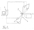
Die Erfindung, die insofern alle hierin beschriebenen Merkmale einzeln oder in jedweder Kombination umfassen kann, wird im Weiteren anhand einer Ausführungsform näher erläutert, wobei auf die einzige beiliegende Figur 1 Bezug genommen wird, welche schematisch eine Vorrichtung zeigt, mittels welcher die Erfindung umgesetzt werden kann.The invention, which may include all features described herein individually or in any combination, will be further elucidated hereinbelow with reference to an embodiment thereof, reference being made to the sole attached Figure 1, which schematically shows a device by means of which the invention can be implemented ,
In der Figur 1 ist eine Couch 3 schematisch dargestellt, die einen Couch-Grundkörper aufweist, auf dem eine Liegefläche für den Patienten angeordnet ist. Die Couch 3 steht vor einer LINAC-Gantry, die der besseren Übersichtlichkeit wegen nicht dargestellt ist. Es handelt sich um eine herkömmliche LINAC-Gantry ohne Röntgenbilderstellungs-Zusatzeinheiten.1 shows a
Das Isozentrum der LINAC-Bestrahlung, also der Einstrahlungspunkt, der sich ergibt, wenn die LINAC-Gantry beim Bestrahlen gedreht wird, ist in der Zeichnung mit dem Bezugszeichen 5 angegeben. Durch das Isozentrum 5 verläuft die raumfeste, vertikale Achse 4, welche die Achse ist, um die sich zur Ausführung des erfindungsgemäßen Verfahrens die Couch 3 drehen kann. Dies kann beispielsweise dadurch bewerkstelligt werden, dass die Couch 3 auf einem Drehteller am Boden angeordnet wird. Anzumerken ist noch, dass die Achse 4 nicht unbedingt eine Isozentrums-Achse sein muss; es genügt wenn sie raumfest und ihr Verlauf koordinatenmäßig bekannt ist.The isocenter of the LINAC irradiation, that is to say the irradiation point which results when the LINAC gantry is rotated during the irradiation, is indicated in the drawing by the
Zwei Röntgenröhren, jeweils mit dem Bezugszeichen 1 bezeichnet, sind bei der dargestellten Ausführungsform unterhalb der Gantry und des Patiententisches, im vorliegenden Fall im Boden montiert. Oberhalb der Couch 3, bevorzugt an der Decke befestigt, befinden sich zwei Röntgendetektoren, die jeweils das Bezugszeichen 2 tragen. Diese können insbesondere aus amorphen Silizium aufgebaut sein.Two x-ray tubes, each denoted by the reference numeral 1, are mounted in the illustrated embodiment below the gantry and the patient table, in the present case in the ground. Above the
Ein Computersystem 6 ist mit den Röntgenröhren 1 und den Röntgendetektoren 2 verbunden. Es dient dazu, die Röntgenbilder zu akquirieren und aus den Bildinformationen der Röntgenbilder einen Volumendatensatz zu rekonstruieren, beispielsweise einen rekonstruierten CT-Datensatz.A computer system 6 is connected to the X-ray tubes 1 and the
Des Weiteren können noch einige Einrichtungen zur Verfügung gestellt werden, die in der Figur 1 nicht gezeigt sind. Beispielsweise ist es möglich, ein Navigations- oder Trackingsystem bereitzustellen, das zu seinen herkömmlichen Funktionen im Rahmen der vorliegenden Erfindung noch die Aufgabe übernehmen kann, den Drehwinkel der Couch zu messen. Natürlich kann der Drehwinkel auch direkt, beispielsweise an einem Drehteller durch bekannte Winkel-Messvorrichtungen ermittelt werden. Wenn aber, wie dies oft der Fall ist, ohnehin ein Navigationssystem bzw. ein Trackingsystem verwendet wird, um die Bestrahlung zu planen und zu unterstützen, kann dieses auch verwendet werden, um den Drehwinkel für die Couch 3 zu bestimmen.Furthermore, some devices can still be made available which are not shown in FIG. For example, it is possible to provide a navigation or tracking system that can still take over the task of measuring the angle of rotation of the couch to its conventional functions in the context of the present invention. Of course, the rotation angle can also be determined directly, for example on a turntable by known angle measuring devices. If, however, as is often the case, a navigation system or a tracking system is used anyway to plan and support the irradiation, this can also be used to determine the angle of rotation for the
Ferner ist es möglich, ein Kalibrierphantom mit röntgensichtbaren Markern zur Bestimmung der räumlichen Lage des Röntgensystems bereitzustellen, das vorteilhafterweise folgende Funktionen erfüllen kann: Es dient zur Definition eines räumlichen Koordinatensystems mit Ursprung im Isozentrum, wobei die vertikale (Z-)Achse des Koordinatensystems der Rotationsachse entspricht, die Y-Achse, die Null-Grad-Richtung der Drehvorrichtung definiert und senkrecht zur Z-Achse steht, und wobei die X-Achse senkrecht zur Y- und zur Z-Achse steht und ein rechtshändiges Koordinatensystem definiert (LINAC-Koordinatensystem). Es dient weiterhin dazu, die räumliche Lage der Röntgenquellen und der Detektoren bezüglich des LINAC-Koordinatensystems zu definieren, und außerdem dient es noch dazu, die Parameter der projektiven Abbildungen von Punkten aus dem LINAC-Koordinatensystem auf Punkte in den aufgenommen Röntgenbildern zu bestimmen.Furthermore, it is possible to provide a calibration phantom with x-ray-visible markers for determining the spatial position of the x-ray system, which can advantageously fulfill the following functions: It serves to define a spatial coordinate system originating in the isocenter, wherein the vertical (Z) axis of the coordinate system of the axis of rotation corresponds, the Y-axis, the zero-degree direction of the rotary device defines and is perpendicular to the Z-axis, and wherein the X-axis is perpendicular to the Y and Z axis and a right-handed coordinate system defined (LINAC coordinate system). It also serves to define the spatial location of the x-ray sources and detectors relative to the LINAC coordinate system, and also serves to determine the parameters of the projective mappings of points from the LINAC coordinate system to points in the captured x-ray images.
Etwas allgemeiner ausgedrückt betrifft die vorliegende Erfindung in einer bevorzugten Ausführungsform ein System zur Rekonstruktion von 3D-Datensätzen aus Röntgenbildern. Dabei werden Röntgenbilder aus zwei fest installierten Röntgenquellen 1 geschossen und aus Röntgendetektoren 2 ausgelesen. Der Patient befindet sich auf einer um eine feste Achse 4 drehbaren Couch 3. Zur Rekonstruktion von 3D-Datensätzen werden vom Patienten Röntgenbilder erstellt, wobei die Couch 3 von Bild zu Bild jeweils weiter gedreht wird. Dabei steht die Drehachse 4 der Couch und damit des Patienten nicht orthogonal zur Richtung von einer Röntgenquelle 1 zum Röntgendetektor 2. Die Röntgenquellen bzw. Röntgenröhren 1 und die Röntgendetektoren 2 sind so angebracht, dass sich die Strahlen ungefähr in einem Winkel von 90 Grad schneiden. Die beiden Kombinationen aus Röntgenquelle 1 und Röntgendetektor 2 sind zueinander kalibriert. Auf diese Weise können beide Röntgenquellen 1 genutzt werden, um Bilder zu erzeugen, die zur Rekonstruktion des CT-Datensatzes verwendet werden. Dies beschleunigt einerseits die Zeit zur Aufnahme der Röntgenbilder, andererseits wird der Winkelbereich, in dem Röntgenbilder vom Patienten aufgenommen werden können, um 90 Grad vergrößert. Der Drehwinkel der. Couch wird mit Hilfe eines geeigneten Systems gemessen, welches beispielsweise eine Infrarot-Trackingsystem zum Verfolgen von passiven Markern sein kann. Dazu wird ein geeigneter Referenzstern mit Infrarotmarkern an der Couch befestigt.More generally, in a preferred embodiment, the present invention relates to a system for reconstructing 3D data sets from X-ray images. X-ray images are shot from two permanently installed X-ray sources 1 and read out of
Aus den aufgenommenen Röntgenbildern wird mit Hilfe geeigneter, computergestützter Verfahren ein 3D-Datensatz rekonstruiert. Hierbei ist der zu rekonstruierende Bereich vorzugsweise so groß zu wählen, dass die Rückprojektion des CT-Datensatzes auf einen der beiden Röntgendetektoren 2 aus jedem möglichen Couchwinkel die gesamte Fläche des Detektors 2 abdeckt.From the recorded X-ray images, a 3D data record is reconstructed with the aid of suitable computer-assisted methods. In this case, the region to be reconstructed is preferably to be selected so large that the back projection of the CT data set onto one of the two
Im Weiteren wird nun ein etwas detaillierterer Verfahrensablauf bei der praktischen Umsetzung der vorliegenden Erfindung erläutert. Am Anfang steht die Kalibrierung, wobei das Kalibrierphantom in Isozentrumsposition 5 auf der Couch 3 bzw. auf der Liegefläche der Couch 3 platziert wird. Nunmehr werden mit Unterstützung des Computersystems 6 zwei Röntgenbilder aus den beiden Röntgenquellen 1 aufgenommen, und im Computersystem 6 werden die Projektionen aller röntgensichtbaren Marker in beiden Röntgenbildern mit Hilfe einer Bildverarbeitungssoftware automatisch detektiert. Zum Abschluss der Kalibrierung werden aus der Lage des Phantoms und der Projektion der röntgensichtbaren Marker mittels des Computersystems die benötigten Parameter errechnet, also LINAC-Koordinatensystem, räumliche Lage der Röntgenquellen und Detektoren 1, 2 und Parameter der projektiven Abbildungen.In the following, a somewhat more detailed procedure in the practical implementation of the present invention will be explained. At the beginning is the calibration, wherein the calibration phantom is placed in the
Beim der eigentlichen Erstellung und Akquirierung der notwendigen Bildinformationen werden die folgenden Schritte durchgeführt. Der Patient wird auf der Couch 3 positioniert, und zwar so, dass der interessante Bereich sich um das Isozentrum herum befindet. Dieser Bildbereich wird der Bereich sein, in dem das Bestrahlungsziel liegt, also beispielsweise ein Tumor.When actually creating and acquiring the necessary image information, the following steps are performed. The patient is positioned on the
Die Couch 3 wird dann auf einen Startwinkel gedreht und der Akquisitionszyklus beginnt. Im Akquisitionszyklus findet eine konstante Rotation der Couch 3 auf ihrer Drehvorrichtung statt, und zwar um die Achse 4. Dabei werden Röntgenbilder aufgenommen, und zwar in einer solchen Weise, dass bei der Aufnahme jeweils ein bestimmter Drehwinkel einem bestimmten Zeitpunkt während einer kontinuierlichem Aufnahme oder einem einzelnen Röntgenbild zugeordnet werden kann. Die Bilddaten und die Rotationsparameter werden im Computersystem 6 gespeichert, und dieser Vorgang wird solange wiederholt, bis der Endwinkel erreicht ist.The
Die weitere Arbeit wird dann von dem Computersystem 6 erledigt, das aus den aufgenommenen Röntgenbildern mit Hilfe geeigneter computergestützter Verfahren (Bildverarbeitung) einen CT-Datensatz aus den Cone-Beam-Röntgenbildern rekonstruiert.The further work is then done by the computer system 6, which reconstructs a CT data set from the cone-beam X-ray images from the recorded X-ray images with the aid of suitable computer-assisted methods (image processing).
Damit kann mit nur wenigen Modifikationen an Strahlentherapie-Vorrichtungen in einfacher Weise ein aktueller volumetrischer Bilddatensatz erhalten werden, der gemäß einer weiteren Ausführungsform auch noch dazu verwendet werden kann, den Patienten exakt zu positionieren.Thus, with only a few modifications to radiation therapy devices, a current volumetric image data set can be obtained in a simple manner, which, according to a further embodiment, can also be used to precisely position the patient.
Claims (15)
Priority Applications (4)
| Application Number | Priority Date | Filing Date | Title |
|---|---|---|---|
| DE502004004956T DE502004004956D1 (en) | 2004-08-06 | 2004-08-06 | Volumetric imaging on a radiotherapy device |
| EP05024826A EP1623739B1 (en) | 2004-08-06 | 2004-08-06 | Volumetric imaging in a radiation therapy device |
| EP04018757A EP1623738B1 (en) | 2004-08-06 | 2004-08-06 | Volumetric imaging in a radiation therapy device |
| US11/198,887 US7324626B2 (en) | 2004-08-06 | 2005-08-05 | Volumetric imaging on a radiotherapy apparatus |
Applications Claiming Priority (1)
| Application Number | Priority Date | Filing Date | Title |
|---|---|---|---|
| EP04018757A EP1623738B1 (en) | 2004-08-06 | 2004-08-06 | Volumetric imaging in a radiation therapy device |
Related Child Applications (1)
| Application Number | Title | Priority Date | Filing Date |
|---|---|---|---|
| EP05024826A Division EP1623739B1 (en) | 2004-08-06 | 2004-08-06 | Volumetric imaging in a radiation therapy device |
Publications (2)
| Publication Number | Publication Date |
|---|---|
| EP1623738A1 true EP1623738A1 (en) | 2006-02-08 |
| EP1623738B1 EP1623738B1 (en) | 2007-09-12 |
Family
ID=34926096
Family Applications (2)
| Application Number | Title | Priority Date | Filing Date |
|---|---|---|---|
| EP05024826A Active EP1623739B1 (en) | 2004-08-06 | 2004-08-06 | Volumetric imaging in a radiation therapy device |
| EP04018757A Expired - Fee Related EP1623738B1 (en) | 2004-08-06 | 2004-08-06 | Volumetric imaging in a radiation therapy device |
Family Applications Before (1)
| Application Number | Title | Priority Date | Filing Date |
|---|---|---|---|
| EP05024826A Active EP1623739B1 (en) | 2004-08-06 | 2004-08-06 | Volumetric imaging in a radiation therapy device |
Country Status (3)
| Country | Link |
|---|---|
| US (1) | US7324626B2 (en) |
| EP (2) | EP1623739B1 (en) |
| DE (1) | DE502004004956D1 (en) |
Families Citing this family (100)
| Publication number | Priority date | Publication date | Assignee | Title |
|---|---|---|---|---|
| CA2974143C (en) | 2004-02-20 | 2020-11-10 | University Of Florida Research Foundation, Inc. | System for delivering conformal radiation therapy while simultaneously imaging soft tissue |
| JP4896526B2 (en) * | 2006-01-11 | 2012-03-14 | 株式会社東芝 | Magnetic resonance imaging system |
| IL191676A (en) * | 2007-05-24 | 2013-05-30 | Cure Ltd P | Apparatus for teletherapy positioning and validation |
| US7847275B2 (en) * | 2007-05-24 | 2010-12-07 | Pcure Ltd. | Method and apparatus for teletherapy positioning and validation |
| US9579525B2 (en) | 2008-05-22 | 2017-02-28 | Vladimir Balakin | Multi-axis charged particle cancer therapy method and apparatus |
| US8373145B2 (en) * | 2008-05-22 | 2013-02-12 | Vladimir Balakin | Charged particle cancer therapy system magnet control method and apparatus |
| AU2009249863B2 (en) * | 2008-05-22 | 2013-12-12 | Vladimir Yegorovich Balakin | Multi-field charged particle cancer therapy method and apparatus |
| US10684380B2 (en) | 2008-05-22 | 2020-06-16 | W. Davis Lee | Multiple scintillation detector array imaging apparatus and method of use thereof |
| US8901509B2 (en) * | 2008-05-22 | 2014-12-02 | Vladimir Yegorovich Balakin | Multi-axis charged particle cancer therapy method and apparatus |
| US9095040B2 (en) | 2008-05-22 | 2015-07-28 | Vladimir Balakin | Charged particle beam acceleration and extraction method and apparatus used in conjunction with a charged particle cancer therapy system |
| US9044600B2 (en) * | 2008-05-22 | 2015-06-02 | Vladimir Balakin | Proton tomography apparatus and method of operation therefor |
| US9168392B1 (en) | 2008-05-22 | 2015-10-27 | Vladimir Balakin | Charged particle cancer therapy system X-ray apparatus and method of use thereof |
| US9974978B2 (en) | 2008-05-22 | 2018-05-22 | W. Davis Lee | Scintillation array apparatus and method of use thereof |
| US8129699B2 (en) | 2008-05-22 | 2012-03-06 | Vladimir Balakin | Multi-field charged particle cancer therapy method and apparatus coordinated with patient respiration |
| WO2009142545A2 (en) | 2008-05-22 | 2009-11-26 | Vladimir Yegorovich Balakin | Charged particle cancer therapy patient positioning method and apparatus |
| US8089054B2 (en) | 2008-05-22 | 2012-01-03 | Vladimir Balakin | Charged particle beam acceleration and extraction method and apparatus used in conjunction with a charged particle cancer therapy system |
| US10092776B2 (en) | 2008-05-22 | 2018-10-09 | Susan L. Michaud | Integrated translation/rotation charged particle imaging/treatment apparatus and method of use thereof |
| US10070831B2 (en) | 2008-05-22 | 2018-09-11 | James P. Bennett | Integrated cancer therapy—imaging apparatus and method of use thereof |
| US9177751B2 (en) | 2008-05-22 | 2015-11-03 | Vladimir Balakin | Carbon ion beam injector apparatus and method of use thereof |
| US9910166B2 (en) | 2008-05-22 | 2018-03-06 | Stephen L. Spotts | Redundant charged particle state determination apparatus and method of use thereof |
| US9737733B2 (en) | 2008-05-22 | 2017-08-22 | W. Davis Lee | Charged particle state determination apparatus and method of use thereof |
| US20090314960A1 (en) * | 2008-05-22 | 2009-12-24 | Vladimir Balakin | Patient positioning method and apparatus used in conjunction with a charged particle cancer therapy system |
| US8374314B2 (en) | 2008-05-22 | 2013-02-12 | Vladimir Balakin | Synchronized X-ray / breathing method and apparatus used in conjunction with a charged particle cancer therapy system |
| US8309941B2 (en) | 2008-05-22 | 2012-11-13 | Vladimir Balakin | Charged particle cancer therapy and patient breath monitoring method and apparatus |
| US8378321B2 (en) * | 2008-05-22 | 2013-02-19 | Vladimir Balakin | Charged particle cancer therapy and patient positioning method and apparatus |
| US7939809B2 (en) * | 2008-05-22 | 2011-05-10 | Vladimir Balakin | Charged particle beam extraction method and apparatus used in conjunction with a charged particle cancer therapy system |
| US9056199B2 (en) | 2008-05-22 | 2015-06-16 | Vladimir Balakin | Charged particle treatment, rapid patient positioning apparatus and method of use thereof |
| NZ589387A (en) | 2008-05-22 | 2012-11-30 | Vladimir Yegorovich Balakin | Charged particle beam extraction method and apparatus used in conjunction with a charged particle cancer therapy system |
| US8718231B2 (en) | 2008-05-22 | 2014-05-06 | Vladimir Balakin | X-ray tomography method and apparatus used in conjunction with a charged particle cancer therapy system |
| US8710462B2 (en) * | 2008-05-22 | 2014-04-29 | Vladimir Balakin | Charged particle cancer therapy beam path control method and apparatus |
| US8907309B2 (en) | 2009-04-17 | 2014-12-09 | Stephen L. Spotts | Treatment delivery control system and method of operation thereof |
| US8642978B2 (en) * | 2008-05-22 | 2014-02-04 | Vladimir Balakin | Charged particle cancer therapy dose distribution method and apparatus |
| US8637833B2 (en) | 2008-05-22 | 2014-01-28 | Vladimir Balakin | Synchrotron power supply apparatus and method of use thereof |
| US8896239B2 (en) * | 2008-05-22 | 2014-11-25 | Vladimir Yegorovich Balakin | Charged particle beam injection method and apparatus used in conjunction with a charged particle cancer therapy system |
| US9616252B2 (en) | 2008-05-22 | 2017-04-11 | Vladimir Balakin | Multi-field cancer therapy apparatus and method of use thereof |
| JP5450602B2 (en) * | 2008-05-22 | 2014-03-26 | エゴロヴィチ バラキン、ウラジミール | Tumor treatment device for treating tumor using charged particles accelerated by synchrotron |
| US8624528B2 (en) * | 2008-05-22 | 2014-01-07 | Vladimir Balakin | Method and apparatus coordinating synchrotron acceleration periods with patient respiration periods |
| US8975600B2 (en) | 2008-05-22 | 2015-03-10 | Vladimir Balakin | Treatment delivery control system and method of operation thereof |
| US9744380B2 (en) | 2008-05-22 | 2017-08-29 | Susan L. Michaud | Patient specific beam control assembly of a cancer therapy apparatus and method of use thereof |
| US8519365B2 (en) * | 2008-05-22 | 2013-08-27 | Vladimir Balakin | Charged particle cancer therapy imaging method and apparatus |
| US9981147B2 (en) | 2008-05-22 | 2018-05-29 | W. Davis Lee | Ion beam extraction apparatus and method of use thereof |
| US8368038B2 (en) | 2008-05-22 | 2013-02-05 | Vladimir Balakin | Method and apparatus for intensity control of a charged particle beam extracted from a synchrotron |
| US8188688B2 (en) | 2008-05-22 | 2012-05-29 | Vladimir Balakin | Magnetic field control method and apparatus used in conjunction with a charged particle cancer therapy system |
| US10548551B2 (en) | 2008-05-22 | 2020-02-04 | W. Davis Lee | Depth resolved scintillation detector array imaging apparatus and method of use thereof |
| US9937362B2 (en) | 2008-05-22 | 2018-04-10 | W. Davis Lee | Dynamic energy control of a charged particle imaging/treatment apparatus and method of use thereof |
| US9155911B1 (en) | 2008-05-22 | 2015-10-13 | Vladimir Balakin | Ion source method and apparatus used in conjunction with a charged particle cancer therapy system |
| US8487278B2 (en) * | 2008-05-22 | 2013-07-16 | Vladimir Yegorovich Balakin | X-ray method and apparatus used in conjunction with a charged particle cancer therapy system |
| US9782140B2 (en) | 2008-05-22 | 2017-10-10 | Susan L. Michaud | Hybrid charged particle / X-ray-imaging / treatment apparatus and method of use thereof |
| US10143854B2 (en) | 2008-05-22 | 2018-12-04 | Susan L. Michaud | Dual rotation charged particle imaging / treatment apparatus and method of use thereof |
| US8598543B2 (en) * | 2008-05-22 | 2013-12-03 | Vladimir Balakin | Multi-axis/multi-field charged particle cancer therapy method and apparatus |
| US9737734B2 (en) | 2008-05-22 | 2017-08-22 | Susan L. Michaud | Charged particle translation slide control apparatus and method of use thereof |
| US9498649B2 (en) | 2008-05-22 | 2016-11-22 | Vladimir Balakin | Charged particle cancer therapy patient constraint apparatus and method of use thereof |
| US8178859B2 (en) | 2008-05-22 | 2012-05-15 | Vladimir Balakin | Proton beam positioning verification method and apparatus used in conjunction with a charged particle cancer therapy system |
| WO2009142544A2 (en) * | 2008-05-22 | 2009-11-26 | Vladimir Yegorovich Balakin | Charged particle cancer therapy beam path control method and apparatus |
| US10029122B2 (en) | 2008-05-22 | 2018-07-24 | Susan L. Michaud | Charged particle—patient motion control system apparatus and method of use thereof |
| US8373146B2 (en) * | 2008-05-22 | 2013-02-12 | Vladimir Balakin | RF accelerator method and apparatus used in conjunction with a charged particle cancer therapy system |
| US8373143B2 (en) * | 2008-05-22 | 2013-02-12 | Vladimir Balakin | Patient immobilization and repositioning method and apparatus used in conjunction with charged particle cancer therapy |
| US8569717B2 (en) * | 2008-05-22 | 2013-10-29 | Vladimir Balakin | Intensity modulated three-dimensional radiation scanning method and apparatus |
| US9737272B2 (en) | 2008-05-22 | 2017-08-22 | W. Davis Lee | Charged particle cancer therapy beam state determination apparatus and method of use thereof |
| US8436327B2 (en) * | 2008-05-22 | 2013-05-07 | Vladimir Balakin | Multi-field charged particle cancer therapy method and apparatus |
| US8969834B2 (en) | 2008-05-22 | 2015-03-03 | Vladimir Balakin | Charged particle therapy patient constraint apparatus and method of use thereof |
| US9682254B2 (en) | 2008-05-22 | 2017-06-20 | Vladimir Balakin | Cancer surface searing apparatus and method of use thereof |
| US9855444B2 (en) | 2008-05-22 | 2018-01-02 | Scott Penfold | X-ray detector for proton transit detection apparatus and method of use thereof |
| US8627822B2 (en) * | 2008-07-14 | 2014-01-14 | Vladimir Balakin | Semi-vertical positioning method and apparatus used in conjunction with a charged particle cancer therapy system |
| US8625739B2 (en) | 2008-07-14 | 2014-01-07 | Vladimir Balakin | Charged particle cancer therapy x-ray method and apparatus |
| MX2011009222A (en) | 2009-03-04 | 2011-11-02 | Protom Aozt | Multi-field charged particle cancer therapy method and apparatus. |
| US10589128B2 (en) | 2010-04-16 | 2020-03-17 | Susan L. Michaud | Treatment beam path verification in a cancer therapy apparatus and method of use thereof |
| US10376717B2 (en) | 2010-04-16 | 2019-08-13 | James P. Bennett | Intervening object compensating automated radiation treatment plan development apparatus and method of use thereof |
| US10556126B2 (en) | 2010-04-16 | 2020-02-11 | Mark R. Amato | Automated radiation treatment plan development apparatus and method of use thereof |
| US10518109B2 (en) | 2010-04-16 | 2019-12-31 | Jillian Reno | Transformable charged particle beam path cancer therapy apparatus and method of use thereof |
| US11648420B2 (en) | 2010-04-16 | 2023-05-16 | Vladimir Balakin | Imaging assisted integrated tomography—cancer treatment apparatus and method of use thereof |
| US10086214B2 (en) | 2010-04-16 | 2018-10-02 | Vladimir Balakin | Integrated tomography—cancer treatment apparatus and method of use thereof |
| US10555710B2 (en) | 2010-04-16 | 2020-02-11 | James P. Bennett | Simultaneous multi-axes imaging apparatus and method of use thereof |
| US9737731B2 (en) | 2010-04-16 | 2017-08-22 | Vladimir Balakin | Synchrotron energy control apparatus and method of use thereof |
| US10349906B2 (en) | 2010-04-16 | 2019-07-16 | James P. Bennett | Multiplexed proton tomography imaging apparatus and method of use thereof |
| US10188877B2 (en) | 2010-04-16 | 2019-01-29 | W. Davis Lee | Fiducial marker/cancer imaging and treatment apparatus and method of use thereof |
| US10751551B2 (en) | 2010-04-16 | 2020-08-25 | James P. Bennett | Integrated imaging-cancer treatment apparatus and method of use thereof |
| US10625097B2 (en) | 2010-04-16 | 2020-04-21 | Jillian Reno | Semi-automated cancer therapy treatment apparatus and method of use thereof |
| US10179250B2 (en) | 2010-04-16 | 2019-01-15 | Nick Ruebel | Auto-updated and implemented radiation treatment plan apparatus and method of use thereof |
| US10638988B2 (en) | 2010-04-16 | 2020-05-05 | Scott Penfold | Simultaneous/single patient position X-ray and proton imaging apparatus and method of use thereof |
| US8755489B2 (en) | 2010-11-11 | 2014-06-17 | P-Cure, Ltd. | Teletherapy location and dose distribution control system and method |
| US8963112B1 (en) | 2011-05-25 | 2015-02-24 | Vladimir Balakin | Charged particle cancer therapy patient positioning method and apparatus |
| US9510771B1 (en) | 2011-10-28 | 2016-12-06 | Nuvasive, Inc. | Systems and methods for performing spine surgery |
| US10561861B2 (en) | 2012-05-02 | 2020-02-18 | Viewray Technologies, Inc. | Videographic display of real-time medical treatment |
| US20150243025A1 (en) * | 2012-09-28 | 2015-08-27 | Brainlab Ag | Isocentric patient rotation for detection of the position of a moving object |
| AU2013334064A1 (en) | 2012-10-26 | 2015-05-14 | Viewray Technologies, Inc. | Assessment and improvement of treatment using imaging of physiological responses to radiation therapy |
| US8933651B2 (en) | 2012-11-16 | 2015-01-13 | Vladimir Balakin | Charged particle accelerator magnet apparatus and method of use thereof |
| US9446263B2 (en) | 2013-03-15 | 2016-09-20 | Viewray Technologies, Inc. | Systems and methods for linear accelerator radiotherapy with magnetic resonance imaging |
| US9848922B2 (en) | 2013-10-09 | 2017-12-26 | Nuvasive, Inc. | Systems and methods for performing spine surgery |
| AU2016361432A1 (en) | 2015-11-24 | 2018-06-07 | Viewray Technologies, Inc. | Radiation beam collimating systems and methods |
| EP3423153B1 (en) | 2016-03-02 | 2021-05-19 | ViewRay Technologies, Inc. | Particle therapy with magnetic resonance imaging |
| US9907981B2 (en) | 2016-03-07 | 2018-03-06 | Susan L. Michaud | Charged particle translation slide control apparatus and method of use thereof |
| US10037863B2 (en) | 2016-05-27 | 2018-07-31 | Mark R. Amato | Continuous ion beam kinetic energy dissipater apparatus and method of use thereof |
| AU2017281519A1 (en) | 2016-06-22 | 2019-01-24 | Viewray Technologies, Inc. | Magnetic resonance imaging at low field strength |
| EP3554635B1 (en) | 2016-12-13 | 2021-01-20 | ViewRay Technologies, Inc. | Radiation therapy systems |
| CN116036499A (en) | 2017-12-06 | 2023-05-02 | 优瑞技术公司 | Optimization of multi-modality radiation therapy |
| WO2019140637A1 (en) * | 2018-01-19 | 2019-07-25 | 深圳市奥沃医学新技术发展有限公司 | Positioning method and apparatus, and radiotherapy system |
| US11209509B2 (en) | 2018-05-16 | 2021-12-28 | Viewray Technologies, Inc. | Resistive electromagnet systems and methods |
| JP7078955B2 (en) * | 2018-07-26 | 2022-06-01 | 東芝エネルギーシステムズ株式会社 | Treatment system, calibration method, and program |
| EP3896653A1 (en) * | 2020-04-15 | 2021-10-20 | Stryker European Operations Limited | Technique for determining a position of one or more imaged markers in an image coordinate system |
Citations (6)
| Publication number | Priority date | Publication date | Assignee | Title |
|---|---|---|---|---|
| EP0562585A2 (en) * | 1992-03-24 | 1993-09-29 | Jun Ikebe | System for stereotactic radiotherapy with a computerized tomographic scanning system |
| US5615430A (en) * | 1994-08-22 | 1997-04-01 | Kabushiki Kaisha Toshiba | Medical bed system |
| US6269143B1 (en) * | 1998-08-31 | 2001-07-31 | Shimadzu Corporation | Radiotherapy planning system |
| WO2001060236A2 (en) * | 2000-02-18 | 2001-08-23 | William Beaumont Hospital | Cone-beam computerized tomography with a flat-panel imager |
| DE10051370A1 (en) * | 2000-10-17 | 2002-05-02 | Brainlab Ag | Method and appliance for exact positioning of patient for radiation therapy and radio surgery with which only one camera is used to determine and compensate for positional error |
| EP1389479A1 (en) * | 2002-08-14 | 2004-02-18 | Minoru Uematsu | Composite system for radiation therapy |
Family Cites Families (2)
| Publication number | Priority date | Publication date | Assignee | Title |
|---|---|---|---|---|
| US5014290A (en) * | 1988-10-28 | 1991-05-07 | Moore Robert M | Method and apparatus for generating radiation blockers |
| US6148058A (en) * | 1998-10-23 | 2000-11-14 | Analogic Corporation | System and method for real time measurement of detector offset in rotating-patient CT scanner |
-
2004
- 2004-08-06 DE DE502004004956T patent/DE502004004956D1/en active Active
- 2004-08-06 EP EP05024826A patent/EP1623739B1/en active Active
- 2004-08-06 EP EP04018757A patent/EP1623738B1/en not_active Expired - Fee Related
-
2005
- 2005-08-05 US US11/198,887 patent/US7324626B2/en not_active Expired - Fee Related
Patent Citations (6)
| Publication number | Priority date | Publication date | Assignee | Title |
|---|---|---|---|---|
| EP0562585A2 (en) * | 1992-03-24 | 1993-09-29 | Jun Ikebe | System for stereotactic radiotherapy with a computerized tomographic scanning system |
| US5615430A (en) * | 1994-08-22 | 1997-04-01 | Kabushiki Kaisha Toshiba | Medical bed system |
| US6269143B1 (en) * | 1998-08-31 | 2001-07-31 | Shimadzu Corporation | Radiotherapy planning system |
| WO2001060236A2 (en) * | 2000-02-18 | 2001-08-23 | William Beaumont Hospital | Cone-beam computerized tomography with a flat-panel imager |
| DE10051370A1 (en) * | 2000-10-17 | 2002-05-02 | Brainlab Ag | Method and appliance for exact positioning of patient for radiation therapy and radio surgery with which only one camera is used to determine and compensate for positional error |
| EP1389479A1 (en) * | 2002-08-14 | 2004-02-18 | Minoru Uematsu | Composite system for radiation therapy |
Also Published As
| Publication number | Publication date |
|---|---|
| EP1623739B1 (en) | 2006-10-25 |
| US7324626B2 (en) | 2008-01-29 |
| DE502004004956D1 (en) | 2007-10-25 |
| EP1623738B1 (en) | 2007-09-12 |
| EP1623739A2 (en) | 2006-02-08 |
| EP1623739A3 (en) | 2006-04-26 |
| US20060050848A1 (en) | 2006-03-09 |
Similar Documents
| Publication | Publication Date | Title |
|---|---|---|
| EP1623739B1 (en) | Volumetric imaging in a radiation therapy device | |
| EP2307096B1 (en) | Apparatus and method for evaluating an activity distribution, and irradiation system | |
| DE69931164T2 (en) | SYSTEM FOR ADJUSTING THE RADIATION RADIATION FOR RADIOTHERAPY | |
| DE10051370A1 (en) | Method and appliance for exact positioning of patient for radiation therapy and radio surgery with which only one camera is used to determine and compensate for positional error | |
| DE112013001546T5 (en) | Control device for radiotherapy and control program for radiotherapy | |
| DE102014207906A1 (en) | Image guided radiotherapy | |
| DE102012201798A1 (en) | Method for planning X-ray imaging by using X-ray imaging device for imaging examination zone of e.g. heart of human patient for treatment of heart disease, involves determining irradiated region of object based on attitude parameters | |
| EP2110161A1 (en) | Device for carrying out irradiation and method for monitoring same | |
| DE102009021740A1 (en) | Radiotherapy device with an imaging unit | |
| DE102005044407A1 (en) | Artifact reduced radiological three dimensional imaging method, involves combining two three dimensional image data sets to artifact reduced three dimensional image data set for producing artifact reduced radiological data sets | |
| DE102010020781A1 (en) | Determination and verification of the coordinate transformation between an X-ray system and a surgical navigation system | |
| DE102011083854B4 (en) | Time-resolved tomosynthesis imaging | |
| DE102014210458B4 (en) | Determining a position of a target region of a patient to be irradiated in an irradiation device | |
| WO2013041720A1 (en) | System and method for positioning by means of nuclear imaging | |
| DE102011080371B4 (en) | Radiotherapy with overlay-free 3D-CT imaging | |
| EP1388322B1 (en) | System for patient positioning in radiationtherapy / radiosurgery based on magnetic tracking of an implant | |
| EP2926734B1 (en) | Method for setting up a patient irradiation device | |
| DE102011080607A1 (en) | X-ray apparatus has x-ray source pivotable around axis and detector co-operating with the x-ray source, where collimator is arranged between x-ray source and detector | |
| DE10139934A1 (en) | Chemotherapy device with integral radiographic imaging device for preparation of 3-D data of the examination area so that radiation treatment is accurately targeted and its effect is maximized while side effects are minimized | |
| DE102008013613B4 (en) | Method and control device for position optimization of a number of examination objects, tomography system and computer program product | |
| DE102011005723B4 (en) | Control device and method for moving a freely positionable medical X-ray imaging device, medical device, computer program and data carrier | |
| DE102011083414B4 (en) | Dose normalization in radiotherapy with adaptation of isolines or isosurfaces | |
| DE102004040629B4 (en) | Adjusting a relative position of an irradiation device and a patient | |
| DE102011081422B4 (en) | Image-based radiotherapy | |
| DE102022203903B3 (en) | Method for providing a radiation plan, device for determining and device for applying the radiation plan |
Legal Events
| Date | Code | Title | Description |
|---|---|---|---|
| GRAP | Despatch of communication of intention to grant a patent |
Free format text: ORIGINAL CODE: EPIDOSNIGR1 |
|
| GRAS | Grant fee paid |
Free format text: ORIGINAL CODE: EPIDOSNIGR3 |
|
| PUAI | Public reference made under article 153(3) epc to a published international application that has entered the european phase |
Free format text: ORIGINAL CODE: 0009012 |
|
| 17P | Request for examination filed |
Effective date: 20040806 |
|
| AK | Designated contracting states |
Kind code of ref document: A1 Designated state(s): AT BE BG CH CY CZ DE DK EE ES FI FR GB GR HU IE IT LI LU MC NL PL PT RO SE SI SK TR |
|
| AX | Request for extension of the european patent |
Extension state: AL HR LT LV MK |
|
| AKX | Designation fees paid |
Designated state(s): DE FR GB IT |
|
| GRAA | (expected) grant |
Free format text: ORIGINAL CODE: 0009210 |
|
| AK | Designated contracting states |
Kind code of ref document: B1 Designated state(s): DE FR GB IT |
|
| REG | Reference to a national code |
Ref country code: GB Ref legal event code: FG4D Free format text: NOT ENGLISH |
|
| GBT | Gb: translation of ep patent filed (gb section 77(6)(a)/1977) |
Effective date: 20070912 |
|
| REF | Corresponds to: |
Ref document number: 502004004956 Country of ref document: DE Date of ref document: 20071025 Kind code of ref document: P |
|
| ET | Fr: translation filed | ||
| PLBE | No opposition filed within time limit |
Free format text: ORIGINAL CODE: 0009261 |
|
| STAA | Information on the status of an ep patent application or granted ep patent |
Free format text: STATUS: NO OPPOSITION FILED WITHIN TIME LIMIT |
|
| 26N | No opposition filed |
Effective date: 20080613 |
|
| GBPC | Gb: european patent ceased through non-payment of renewal fee |
Effective date: 20080806 |
|
| PG25 | Lapsed in a contracting state [announced via postgrant information from national office to epo] |
Ref country code: IT Free format text: LAPSE BECAUSE OF NON-PAYMENT OF DUE FEES Effective date: 20080806 |
|
| PG25 | Lapsed in a contracting state [announced via postgrant information from national office to epo] |
Ref country code: GB Free format text: LAPSE BECAUSE OF NON-PAYMENT OF DUE FEES Effective date: 20080806 |
|
| REG | Reference to a national code |
Ref country code: DE Ref legal event code: R082 Ref document number: 502004004956 Country of ref document: DE Representative=s name: SCHWABE SANDMAIR MARX, DE |
|
| REG | Reference to a national code |
Ref country code: DE Ref legal event code: R082 Ref document number: 502004004956 Country of ref document: DE Representative=s name: SCHWABE SANDMAIR MARX, DE Effective date: 20131104 Ref country code: DE Ref legal event code: R081 Ref document number: 502004004956 Country of ref document: DE Owner name: BRAINLAB AG, DE Free format text: FORMER OWNER: BRAINLAB AG, 85622 FELDKIRCHEN, DE Effective date: 20131104 Ref country code: DE Ref legal event code: R082 Ref document number: 502004004956 Country of ref document: DE Representative=s name: SCHWABE SANDMAIR MARX PATENTANWAELTE RECHTSANW, DE Effective date: 20131104 |
|
| REG | Reference to a national code |
Ref country code: FR Ref legal event code: CA Effective date: 20131122 Ref country code: FR Ref legal event code: CD Owner name: BRAINLAB AG Effective date: 20131122 |
|
| REG | Reference to a national code |
Ref country code: FR Ref legal event code: PLFP Year of fee payment: 13 |
|
| REG | Reference to a national code |
Ref country code: DE Ref legal event code: R082 Ref document number: 502004004956 Country of ref document: DE Representative=s name: SSM SANDMAIR PATENTANWAELTE RECHTSANWALT PARTN, DE Ref country code: DE Ref legal event code: R082 Ref document number: 502004004956 Country of ref document: DE Representative=s name: SCHWABE SANDMAIR MARX PATENTANWAELTE RECHTSANW, DE Ref country code: DE Ref legal event code: R081 Ref document number: 502004004956 Country of ref document: DE Owner name: BRAINLAB AG, DE Free format text: FORMER OWNER: BRAINLAB AG, 85622 FELDKIRCHEN, DE |
|
| REG | Reference to a national code |
Ref country code: FR Ref legal event code: CA Effective date: 20170706 |
|
| REG | Reference to a national code |
Ref country code: FR Ref legal event code: PLFP Year of fee payment: 14 |
|
| REG | Reference to a national code |
Ref country code: FR Ref legal event code: PLFP Year of fee payment: 15 |
|
| PGFP | Annual fee paid to national office [announced via postgrant information from national office to epo] |
Ref country code: FR Payment date: 20180827 Year of fee payment: 15 Ref country code: DE Payment date: 20180823 Year of fee payment: 15 |
|
| REG | Reference to a national code |
Ref country code: DE Ref legal event code: R119 Ref document number: 502004004956 Country of ref document: DE |
|
| PG25 | Lapsed in a contracting state [announced via postgrant information from national office to epo] |
Ref country code: FR Free format text: LAPSE BECAUSE OF NON-PAYMENT OF DUE FEES Effective date: 20190831 Ref country code: DE Free format text: LAPSE BECAUSE OF NON-PAYMENT OF DUE FEES Effective date: 20200303 |