1. INTRODUCTION
-
The present invention relates to a novel in vivo method
for the presentation and direct transfer of DNA encoding a
therapeutic protein of interest into mammalian repair cells.
The method involves implanting a matrix containing DNA of
interest (referred to herein as a "gene activated matrix")
into a fresh wound site. Repair cells, which normally
originate in viable tissue surrounding the wound, proliferate
and migrate into the gene activated matrix, wherein they
encounter, take up and express the DNA. Transfected repair
cells, therefore act, as in situ bioreactors (localized
within the wound site) which produce agents (DNA-encoded
RNAs, proteins, etc.) that heal the wound.
-
The invention further relates to pharmaceutical
compositions that may be used in the practice of the
invention to transfer the DNA of interest. Such compositions
include any suitable matrix in combination with the DNA of
interest.
2. BACKGROUND OF INVENTION
2.1 WOUND HEALING
-
Currently available wound healing therapies involve the
administration of therapeutic proteins. Such therapeutic
proteins may include regulatory factors involved in the
normal healing process such as systemic hormones, cytokines,
growth factors and other proteins that regulate proliferation
and differentiation of cells. Growth factors, cytokines and
hormones reported to have such wound healing capacity
include, for example, the transforming growth factor-β
superfamily (TGF-β) of proteins (Cox, D.A., 1995, Cell
Biology International, 19:357-371) acidic fibroblast growth
factor (FGF) (Slavin, J., 1995, Cell Biology International,
19:431-444), macrophage-colony stimulating factor (M-CSF) and
calcium regulatory agents such as parathyroid hormone (PTH).
-
A number of problems are associated with the use of
therapeutic proteins, i.e. cytokines, in wound healing
therapies. First, the purification and/or recombinant
production of therapeutic proteins is often an expensive and
time-consuming process. Despite best efforts, however,
purified protein preparations are often unstable making
storage and use cumbersome, and protein instability can lead
to unexpected inflammatory reactions (to protein breakdown
products) that are toxic to the host.
-
Second, systemic delivery of therapeutic proteins, i.e.
cytokines, can be associated with serious unwanted side
effects in unwounded tissue. Due to inefficient delivery to
specific cells and tissues in the body, administration of
high doses of protein are required to ensure that sufficient
amounts of the protein reach the appropriate tissue target.
Because of the short half life in the body due to proteolytic
degradation, the proteins must also be administered
repeatedly which may give rise to an immune reaction to the
therapeutic proteins. The circulation of high doses of
therapeutic proteins is often toxic due to pleiotropic
effects of the administered protein, and may give rise to
serious side effects.
-
Third, exogenous delivery of recombinant proteins is
inefficient. Attempts have been made to limit the
administration of high levels of protein through
immobilization of therapeutic protein at the target site.
However, this therapeutic approach complicates the
readministration of the protein for repeated dosing.
-
Fourth, for a variety of proteins such as membrane
receptors, transcription factors and intracellular binding
proteins, biological activity is dependant on correct
expression and localization in the cell. For many proteins,
correct cellular localization occurs as the protein is post-translationally
modified inside the cells. Therefore, such
proteins cannot be administered exogenously in such a way as
to be taken up and properly localized inside the cell.
-
As these problems attest, current recombinant protein
therapies for wound healing are flawed, because they do not
present a rational method for delivery of exogenous proteins.
These proteins, i.e. cytokines, are normally produced at
their site of action in physiological amounts and efficiently
delivered to cell surface signaling receptors.
2.2 GENE THERAPY
-
Gene therapy was originally conceived of as a specific
gene replacement therapy for correction of heritable defects
to deliver functionally active therapeutic genes into
targeted cells. Initial efforts toward somatic gene therapy
have relied on indirect means of introducing genes into
tissues, called ex vivo gene therapy, e.g., target cells are
removed from the body, transfected or infected with vectors
carrying recombinant genes, and re-implanted into the body
("autologous cell transfer"). A variety of transfection
techniques are currently available and used to transfer DNA
in vitro into cells; including calcium phosphate-DNA
precipitation, DEAE-Dextran transfection, electroporation,
liposome mediated DNA transfer or transduction with
recombinant viral vectors. Such ex vivo treatment protocols
have been proposed to transfer DNA into a variety of
different cell types including epithelial cells (U.S. Patent
4,868,116; Morgan and Mulligan WO87/00201; Morgan et al.,
1987, Science 237:1476-1479; Morgan and Mulligan, U.S. Patent
No. 4,980,286), endothelial cells (WO89/05345), hepatocytes
(WO89/07136; Wolff et al., 1987, Proc. Natl. Acad. Sci. USA
84:3344-3348; Ledley et al., 1987 Proc. Natl. Acad. Sci.
84:5335-5339; Wilson and Mulligan, WO89/07136; Wilson et al.,
1990, Proc. Natl. Acad. Sci. 87:8437-8441) fibroblasts
(Palmer et al., 1987, Proc. Natl. Acad. Sci. USA 84:1055-1059;
Anson et al., 1987, Mol. Biol. Med. 4:11-20; Rosenberg
et al., 1988, Science 242:1575-1578; Naughton & Naughton,
U.S. Patent 4,963,489), lymphocytes (Anderson et al., U.S.
Patent No. 5,399,346; Blaese, R.M. et al., 1995, Science
270:475-480) and hematopoietic stem cells (Lim, B. et al.
1989, Proc. Natl. Acad. Sci. USA 86:8892-8896; Anderson et
al., U.S. Patent No. 5,399,346).
-
Direct in vivo gene transfer has recently been attempted
with formulations of DNA trapped in liposomes (Ledley et al.,
1987, J. Pediatrics 110:1); or in proteoliposomes that
contain viral envelope receptor proteins (Nicolau et al.,
1983, Proc. Natl. Acad. Sci. U.S.A. 80:1068); and DNA coupled
to a polylysine-glycoprotein carrier complex. In addition,
"gene guns" have been used for gene delivery into cells
(Australian Patent No. 9068389). It has even been speculated
that naked DNA, or DNA associated with liposomes, can be
formulated in liquid carrier solutions for injection into
interstitial spaces for transfer of DNA into cells (Felgner,
WO90/11092).
-
Perhaps one of the greatest problems associated with
currently devised gene therapies, whether ex vivo or in vivo,
is the inability to transfer DNA efficiently into a targeted
cell population and to achieve high level expression of the
gene product in vivo. Viral vectors are regarded as the
most efficient system, and recombinant replication-defective
viral vectors have been used to transduce (i.e., infect)
cells both ex vivo and in vivo. Such vectors have included
retroviral, adenovirus and adeno-associated and herpes viral
vectors. While highly efficient at gene transfer, the major
disadvantages associated with the use of viral vectors
include the inability of many viral vectors to infect non-dividing
cells; problems associated with insertional
mutagenesis; inflammatory reactions to the virus and
potential helper virus production, and/or production and
transmission of harmful virus to other human patients.
-
In addition to the low efficiency of most cell types to
take up and express foreign DNA, many targeted cell
populations are found in such low numbers in the body that
the efficiency of presentation of DNA to the specific
targeted cell types is even further diminished. At present,
no protocol or method, currently exists to increase the
efficiency with which DNA is targeted to the targeted cell
population.
3. SUMMARY OF THE INVENTION
-
The present invention relates to a novel method for
specific targeting and transfer of DNA into mammalian repair
cells involved in wound healing in order to express
therapeutic products at the wound site. The method of the
invention involves administering a gene activated matrix into
a fresh wound site in the body. In this setting, repair
cells are localized to the wound site, where they become
transfected and eventually produce DNA-encoded agents (RNAs,
proteins, etc.) that enhance wound healing.
-
The invention is based, in part, on the discovery that
repair cells, active in the wound healing process,
proliferate and migrate from surrounding tissue into the area
of the wound and infiltrate the gene activated matrix. The
matrix acts as a scaffolding that promotes cell ingrowth,
and, in turn, gene transfer, through the local accumulation
of repair cells near the DNA. While in the matrix, repair
cells are surprisingly efficient at taking up the DNA and
expressing it as translational products, i.e., proteins, or
transcriptional products, i.e., antisense and ribozymes. The
transfected repair cells then serve as local bioreactors
amplifying the production of the gene product in vivo.
-
While any number of DNA sequences can be used in the
method, preferred DNA sequences are those that encode
translational products (i.e. proteins) or transcriptional
products (i.e. antisense or ribozymes) that (a) promote
tissue repair; or (b) are capable of disrupting a disease
process (thereby allowing normal tissue healing to take
place).
-
The invention overcomes the shortcomings of procedures
currently used for wound healing involving the administration
of therapeutic proteins. First, DNA, which is both stable
and non-toxic, can be safely administered in high doses in
vivo. Second, repeated administration, while possible, is
not required. The cells which take up and express the DNA
provide a supply of gene product at the site of the wound.
Third, the invention could be practiced in a way that
addresses the temporal requirements of dosing. For example,
the DNA can be presented in vectors that integrate into the
genome of the targeted cell. In this case, all daughter
cells will contain and express the transferred DNA thereby
acting as a continuous source for the therapeutic agent. In
contrast, non-integrating systems may be utilized wherein the
DNA does not integrate into the genome and the gene is not
passed on to daughter cells. In such an instance, when the
wound healing process is completed and the gene product is no
longer needed, the gene product will not be expressed.
-
The invention is demonstrated by way of examples, which
show that genes can be reproducibly transferred and expressed
in a variety of wounded soft and hard tissues in vivo.
Specifically, it is shown that the method of the invention
overcomes the problems associated with currently available
gene therapy protocols. The method of the invention provides
gene transfer to a suitable number of repair cells to achieve
functional effects, i.e., in the absence of any further
targeting or cellular identification by the practitioner. In
vivo methods of gene therapy require some form of targeting
which very often does not work. In the method of the
invention, targeting is not a problem. By analogy, the DNA
acts much like "bait" in a "trap": the DNA is encountered by
unwitting repair cells that have proliferated and then
migrated into the gene activated matrix. These cells, in
turn, are surprisingly capable of taking up DNA and
expressing it as a therapeutic agent.
-
In one embodiment of the invention, the method of the
invention may be used as a drug delivery system through
transfer of DNA into mammalian repair cells for the purpose
of stimulating soft and hard tissue repair and tissue
regeneration. The repair cells will be those cells that
normally arrive at the area of the wound to be treated.
Accordingly, there is no difficulty associated with the
obtaining of suitable target cells to which the present
therapeutic compositions should be applied. All that is
required is the implantation of a gene activated matrix at
the wound site. The nature of this biological environment is
such that the appropriate repair cells will actively take up
and express the "bait" DNA in the absence of any further
targeting or cellular identification by the practitioner.
-
In another embodiment, the method of the invention,
using both biological and synthetic matrices, may be used to
transfer DNA into mammalian repair cells to stimulate
skeletal regeneration. In a further embodiment, the method
of the invention, using both biological and synthetic
matrices, may be used to transfer DNA into mammalian cells to
stimulate ligament and tendon repair. The method of the
invention may further be employed, using both biological and
synthetic matrices to transfer DNA into mammalian repair
cells to stimulate skeletal muscle repair and/or blood vessel
repair.
-
The DNA to be used in the practice of the invention may
include any DNA encoding translational products (i.e.
proteins) or transcriptional products (i.e. antisense or
ribozymes) that promote tissue repair or are capable of
disrupting a disease process. For example, the DNA may
comprise genes encoding therapeutically useful proteins such
as growth factors, cytokines, hormones, etc. Additionally,
the DNA may encode anti-sense or ribozyme molecules that may
inhibit the translation of mRNAs encoding proteins that
inhibit wound healing or which induce inflammation.
-
The DNA encoding the therapeutic product of interest is
associated with, or impregnated within, a matrix to form a
gene activated matrix. Once prepared, the gene activated
matrix is placed within the mammal at the site of a wound.
-
The invention is demonstrated by way of examples,
wherein the efficient in vivo transfer and expression of
genes into tissue undergoing repair and regeneration is
demonstrated.
3.1 DEFINITIONS
-
As used herein, the following terms will have the
meanings indicated below.
-
A gene activated matrix (GAM) is defined herein as any
matrix material containing DNA encoding a therapeutic agent
of interest. For example, gene activated matrices are placed
within wound sites in the body of a mammalian host to enhance
wound healing.
-
A repair cell is defined herein as any cell which is
stimulated to migrate and proliferate in response to tissue
injury. Repair cells are a component of the wound healing
response. Such cells include fibroblasts, capillary
endothelial cells, capillary pericytes, mononuclear
inflammatory cells, segmented inflammatory cells and
granulation tissue cells.
-
A wound site is defined as any location in the host that
arises from traumatic tissue injury, or alternatively, from
tissue damage either induced by, or resulting from, surgical
procedures.
4. DESCRIPTION OF THE DRAWINGS
-
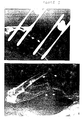
- FIG. 1A. Femoral Osteotomy Model of Fibrous Nonunion.
A 5mm osteotomy was created surgically in the femurs of adult
retired male breeder Sprague-Dawley rats. Gaps shown here
are representative of the entire control group, with
mammalian hosts receiving either an osteotomy alone (n=3), an
osteotomy plus a collagen sponge (n=10) or and osteotomy plus
a collagen sponge containing a control (marker gene) plasmid
DNA (n=23). A plain x-ray film showing a control rat femur
immediately after surgery. The gap was stabilized by an
external fixator consisting of a plate and 4 pins. The skin
incision was closed by metal clips.
- FIG. 1B A plain x-ray film showing a control rat femur
osteotomy 9 weeks after surgery. Rounded surgical margins
(arrows) are due to a reactive bone formation and are
consistent with the classical radiographic appearance of
nonunion fracture.
- FIG. 1C. Histology section of gap tissue 3 weeks post-surgery
showing proliferating repair fibroblasts and
capillaries embedded in an edematous extracellular matrix.
Also present is a focal inflammatory infiltrate consisting of
lymphocytes and macrophages.
- FIG. 1D. Histology section from a 9 week control gap
showing dense fibrous tissue. 1cm=20 µm (C and D).
- FIG. 2. Schematic diagram of the pGAM1 construct
encoding mouse BMP-4. The position of the CMV promoter, BMP-4
coding sequence, HA epitope, and bovine growth hormone
polyadenylation signal are shown.
- FIG. 3A. BMP-4 expression by repair fibroblasts.
Plasmid-encoded BMP-4 expression was detected in Bouins-fixed,
demineralized, paraffin-embedded tissue sections using
the anti-HA antibody and immunoperoxide method 4 weeks post-implantation
of a gene activated matrix containing pGAM1
plasmid DNA. Arrows point to examples of positive (red-brown)
staining of fibroblast cytoplasm (micrograph on upper
left). These cells were identified as fibroblasts based on
spindled morphology, growth in fascicle, and positive
immunostaining for type I procollagen (not shown). Serial
sections incubated with rabbit pre-immune serum or without
the first antibody were negative. Negative results were also
obtained with sham-operated controls (collagen sponge alone)
incubated with the anti-HA.11 antibody (micrograph on upper
right). False positive staining of macrophages, osteoclasts,
and osteoblasts was consistently observed in control sections
incubated with the HA.11 antibody. An island of newly formed
bone 3 weeks following pGAM1 transfer is shown in the
micrograph at bottom, left. New bone is associated with
formation of granulation tissue. High power view of newly
formed bone is shown in the micrograph at bottom, right.
Arrows point to presumptive osteoblasts on the surface of new
bone trabeculae. Gap tissues were stained using Hematoxylin
and eosin (upper micrograph or with Gomori trichrome method
(collagen-rich tissues appear green, lower micrographs).
1cm = 20 mm (upper micrographs).
- FIG. 3B. Plain film radiographs of the animal (23 weeks
post-operatively). In the plain film radiograph (left), the
arrow indicates the approximate position of the osteotomy
gap, which is filled with radio-dense tissue. Note that the
external fixator has been removed. As indicated by the
variegated pattern, bone remodeling is taking place.
Arrowheads point to defects in bone adjacent to the gap (a
consequence of pin placement). The two distal pin sites were
completely healed at this time (not shown). The whole mount
photograph (right) presents a Gomori trichrome-stained tissue
section from the gap of the animal shown (following
sacrifice). Arrow points to the gap, which is now surfaced
by well-integrated cortical bone. Circular defects in marrow
space (either space of the gap) result from placement of
innermost fixator pins. Tissue disruption at bottom of
micrograph is an artifact of specimen handling.
- FIG. 4A. Schematic diagram of the pGAM2 construct
encoding human PTH1-34. The position of an upstream long
terminal repeat that drives PTH1-34 expression (arrow), the
PTH1-34 coding sequence, the SV40 promoter that drives neo
expression (arrow), the neo coding sequence, pBR sequences,
and the downstream long terminal repeat are shown.
- FIG. 4B. PTH1-34 gene transfer and expression drives
new bone formation in vivo. Plain film radiograph showing
new bone bridging of a 5 mm osteotomy gap 9 weeks post-implantation
in an animal that received a gene activated
matrix containing pGAM2 plasmid DNA. Arrows point to radio-dense
tissue in the gap. Results shown here are
representative of experiments with one additional animal.
- FIG. 5. New bone formation in vivo via a two-plasmid
GAM. (top) Plain film radiograph showing new bone bridging
of a 5mm gap 4 weeks post-implantation in an animal that
received a gene activated matrix containing pGAM1 plus pGAM2
plasmid DNA. Arrows point to radio-dense tissue in the gap
(confirmed histologically to be bone). (bottom) Plain film
radiograph of the gap shown in photo at top following removal
(5 weeks earlier; total of 17 weeks post-surgery) of the
external fixator. Arrows indicate location of the gap, which
is filled with radio-dense tissue except for a strip of
undermineralized tissue near the proximal surgical margin.
As indicated by the variegated pattern, an extensive
remodeling response is taking place. Results shown here are
representative of experiments with one additional animal.
- FIG. 6. Adenovirus-mediated Gene Transfer into Bone
Repair/Regeneration Cells in vivo. The UltraFiber™ implant
was soaked for 6 min. in a solution of the AdCMVlacZ virus
(1010-1011 plaque forming units or PFU/ml) and then implanted
into the osteotomy site. The defect was allowed to heal for
3 weeks, during which time the progress of the wound healing
response was monitored by weekly radiographic examination.
By three weeks, it was estimated that 40% of the defect was
filled with callus tissue. The mammalian host was sacrificed
and tissues were fixed in Bouins fixation and then
demineralized for 7 days using standard formic acid
solutions. Photomicrographs were taken from transverse
sections of new bone (callus) that formed in the osteotomy
site 3 weeks after surgery. Panel at top left: Note the
positive (red) β-gal cytoplasmic staining of callus tissue
cells from the UltraFiber™ adenovirus implant. This result
indicates that cell surface receptors that mediate infection,
and thus viral transduction, are expressed by (at least one
population) callus cells during the fracture healing process.
Panel at top left: serial section negative control stained
with the vehicle of the β-gal antibody plus cocktail of nonspecific
rabbit LgG antibodies. Panel at bottom: note the
positive (red) β-gal nuclear staining of chondrocytes in the
osteotomy site filled with UltraFiber™ and AdRSVntlacZ. This
result demonstrates the exquisite specificity of the anti-β-gal
antibody, and conclusively demonstrates expression of the
marker gene product in the osteotomy gap.
- FIG. 7. pGAM2 plasmid gene transfer to repair
fibroblasts results in new bone growth in the rat osteotomy
model. Plain film radiograph showing new bone bridging of a
5 mm gap 6 weeks post-implantation in an animal that received
a gene activated matrix containing pGAM1 plus pGAM2 plasmid
DNA. Arrows point to radio-dense tissue in the gap
(confirmed histologically to be bone).
-
5. DETAILED DESCRIPTION OF THE INVENTION
-
The present invention relates to an in vivo method for
presentation and transfer of DNA into mammalian repair cells
for the purpose of expressing therapeutic agents. The method
of the invention involves implanting or placing gene
activated matrices into a fresh wound site.
-
Wound healing is usually a coordinated, stereotyped
sequence of events that includes (a) tissue disruption and
loss of normal tissue architecture; (b) cell necrosis and
hemorrhage; hemostasis (clot formation); (c) infiltration of
segmented and mononuclear inflammatory cells, with vascular
congestion and tissue edema; (d) dissolution of the clot as
well as damaged cells and tissues by mononuclear cells
(macrophages) (e) formation of granulation tissue
(fibroplasia and angiogenesis). This sequence of cellular
events has been observed in wounds from all tissues and
organs generated in a large number of mammalian species
(Gailet et al., 1994, Curr. Opin. Cell. Biol. 6:717-725).
Therefore, the cellular sequence described above is a
universal aspect of the repair of all mammalian tissues.
-
The invention is based on the discovery that repair
cells involved in the wound healing process will naturally
proliferate and migrate to the site of tissue injury and
infiltrate the gene activated matrix. Surprisingly, these
repair cells, which are normally difficult to efficiently
transfect, either in vitro or in vivo, are extremely
efficient at taking up and expressing DNA when activated to
proliferate by the wound healing process.
-
Taking advantage of this feature, the methods of the
present invention are designed to efficiently transfer, one
or more DNA molecules encoding therapeutic agents to the
proliferating repair cells. The methods involve the
administration of a gene activated matrix containing DNA
encoding translational products (i.e. therapeutic proteins)
or transcriptional products (i.e. antisense or ribozymes)
within a mammalian host at the site of a wound. The wound
may arise from traumatic tissue injury, or alternatively,
from tissue damage either induced by, or resulting from,
surgical procedures.
-
As the proliferating repair cells migrate into and
contact a gene activated matrix, they take up and express the
DNA of interest thereby amplifying the amount of the
therapeutic agent, protein or RNA. The transfected repair
cells thereby serve as local bioreactors producing
therapeutic agents that influence the local repair
environment. For example, growth factors or cytokines
produced by the transfected repair cells, will bind and
stimulate targeted effector cells that express cognate cell
surface receptors, thereby stimulating and amplifying the
cascade of physiological events normally associated with the
wound healing process.
-
Alternatively, the repair cells may take up and express
DNA encoding proteins that inhibit the activity of
antagonists of the wound healing process. The DNA may also
encode antisense or ribozyme RNA molecules that may be used
to inhibit translation of mRNAs encoding inflammatory
proteins or other factors that inhibit wound healing or cause
excessive fibrosis.
-
The gene activated matrix of the invention can be
transferred to the patient using a variety of techniques.
For example, when stimulating wound healing and regeneration,
the matrices are transferred directly to the site of the
wound, i.e., the fractured bone, injured connective tissue,
etc. For use in skin repair, the matrices will be topically
administered. For use in organ regeneration, the matrices
will be surgically placed in a wound made in the organ.
-
Since the method of the invention is based on the
natural migration and proliferation of repair cells into a
wound site, and infiltration into the gene activated matrix
located at the wound site, followed by the uptake of DNA, it
is understood that the matrices must be transferred into a
site in the body where the wound healing process has been
induced.
-
One particularly important feature of the present
invention is that the repair process may be engineered to
result in either the formation of scar tissue and/or tissue
regeneration. For example, the overexpression of the
therapeutic proteins at the site of the wound, may result in
regeneration of the injured tissue without the formation of
scar tissue. In many instances, for example, such as bone
repair, such regeneration is desirable because scar tissue is
not optimally designed to support normal mechanical function.
Alternatively, around a suture it may be desirable to form
scar tissue to hold inherently weak tissue together.
Therefore, the methods of invention may be used to stimulate
wound healing either with, or without, the formation of scar
tissue depending on the type and level of therapeutic protein
expressed.
-
Direct plasmid DNA transfer from a matrix to a mammalian
repair cell, through stimulation of the wound healing
process, offers a number of advantages. First, the ease of
producing and purifying DNA constructs compares favorably
with traditional protein production method cost. Second,
matrices can act as structural scaffolds that, in and of
themselves, promote cell ingrowth and proliferation. Thus,
they facilitate the targeting of repair cells for gene
transfer. Third, direct gene transfer may be an advantageous
method of drug delivery for molecules that normally undergo
complex biosynthetic processing or for receptors which must
be properly positioned in the cellular membrane. These types
of molecules would fail to work if exogenously delivered to
cells.
-
The present invention also relates to pharmaceutical
compositions comprising matrices containing DNA for use in
wound healing. The compositions of the invention are
generally comprised of a biocompatible, or bone compatible,
matrix material containing DNA encoding a therapeutic protein
of interest.
-
The invention overcomes shortcomings specifically
associated with current recombinant protein therapies for
wound healing applications. First, direct gene transfer is a
rational strategy that allows transfected cells to (a) make
physiological amounts of therapeutic protein, modified in a
tissue- and context-specific manner, and (b) deliver this
protein to the appropriate cell surface signaling receptor
under the appropriate circumstances. For reasons described
above, exogenous delivery of such molecules is expected to be
associated with significant dosing and delivery problems.
Second, repeated administration, while possible, is not
required with gene activated matrix technology: cell uptake
of DNA can be controlled precisely with well-established
sustained release delivery technologies, or, alternatively,
integration of transfected DNA can be associated with long
term recombinant protein expression.
-
The method of the invention can be universally applied
to wounds that involve many different cells, tissues and
organs; the repair cells of granulation tissue (Gailet et
al., 1994, Curr. Opin. Cell. Biol. 6:717-725) are "targeted"
where the method of the invention is used. The invention is
demonstrated herein in three animal models (dog, rat and
rabbit) and five tissues (bone, tendon, ligament, blood
vessel and skeletal muscle), using three marker genes (β-galactosidase,
luciferase and alkaline phosphatase), three
promoter systems (CMV, RSV, LTR and SV40), two types of
matrices (biological and synthetic). In all instances,
repair cells that migrated into the gene activated matrix
were successfully transfected. In particular, a functional
outcome (bone growth) has been demonstrated following gene
transfer to repair fibroblasts of a plasmid construct
encoding either BMP-4, which acts as a signal transducing
switch for osteoblast differentiation and growth (Wozney,
1992, Mol. Reprod. Dev. 32:160-167; Reddi, 1994, Curr. Opin.
Genet. Deve. 4:737-744) or PTH1-34, which recruits
osteoprogenitor cells (Orloff, et al, 1992, Endocrinology
131:1603-1611; Dempster et al., 1995 Endocrin Rev. 4:247-250).
5.1 THE GENE ACTIVATED MATRIX
-
Any biocompatible matrix material containing DNA
encoding a therapeutic agent of interest, such as a
translational product, i.e. therapeutic proteins, or
transcriptional products, i.e. antisense or ribozymes, can be
formulated and used in accordance with the invention.
-
The gene activated matrices of the invention may be
derived from any biocompatible material. Such materials may
include, but are not limited to, biodegradable or non-biodegradable
materials formulated into scaffolds that
support cell attachment and growth, powders or gels.
Matrices may be derived from synthetic polymers or naturally
occurring proteins such as collagen, other extracellular
matrix proteins, or other structural macromolecules.
-
The DNA incorporated into the matrix may encode any of a
variety of therapeutic proteins depending on the envisioned
therapeutic use. Such proteins may include growth factors,
cytokines, hormones or any other proteins capable of
regulating the growth, differentiation or physiological
function of cells. The DNA may also encode antisense or
ribozyme molecules which inhibit the translation of proteins
that inhibit wound repair and/or induce inflammation.
-
The transferred DNA need not be integrated into the
genome of the target cell; indeed, the use of non-integrating
DNA in the gene activated matrix is the preferred embodiment
of the present invention. In this way, when the wound
healing process is completed and the gene product is no
longer needed, the gene product will not be expressed.
-
Therapeutic kits containing a biocompatible matrix and
DNA form another aspect of the invention. In some instances
the kits will contain preformed gene activated matrices
thereby allowing the physician, to directly administer the
matrix within the body. Alternatively, the kits may contain
the components necessary for formation of a gene activated
matrix. In such cases the physician may combine the
components to form the gene activated matrices which may then
be used therapeutically by placement within the body. In an
embodiment of the invention the matrices may be used to coat
surgical devices such as suture materials or implants. In
yet another embodiment of the invention, gene activated
matrices may include ready to use sponges, tubes, band-aids,
lyophilized components, gels, patches or powders and telfa
pads.
5.1.1 THE MATRIX MATERIALS
-
In one aspect of the invention, compositions are
prepared in which the DNA encoding the therapeutic agent of
interest is associated with or impregnated within a matrix to
form a gene activated matrix. The matrix compositions
function (i) to facilitate ingrowth of repair cells
(targeting); and (ii) to harbor DNA (delivery). Once the
gene activated matrix is prepared it is stored for future use
or placed immediately at the site of the wound.
-
The type of matrix that may be used in the compositions,
devices and methods of the invention is virtually limitless
and may include both biological and synthetic matrices. The
matrix will have all the features commonly associated with
being "biocompatible", in that it is in a form that does not
produce an adverse, allergic or other untoward reaction when
administered to a mammalian host. Such matrices may be
formed from both natural or synthetic materials. The
matrices may be non-biodegradable in instances where it is
desirable to leave permanent structures in the body; or
biodegradable where the expression of the therapeutic protein
is required only for a short duration of time. The matrices
may take the form of sponges, implants, tubes, telfa pads,
band-aids, bandages, pads, lyophilized components, gels,
patches, powders or nanoparticles. In addition, matrices can
be designed to allow for sustained release of the DNA over
prolonged periods of time.
-
The choice of matrix material will differ according to
the particular circumstances and the site of the wound that
is to be treated. Matrices such as those described in U.S.
Patent 5,270,300, incorporated herein by reference, may be
employed. Physical and chemical characteristics, such as,
e.g., biocompatibility, biodegradability, strength, rigidity,
interface properties and even cosmetic appearance may be
considered in choosing a matrix, as is well known to those of
skill in the art. Appropriate matrices will both deliver the
DNA molecule and also act as an in situ scaffolding through
which mammalian repair cells may migrate.
-
Where the matrices are to be maintained for extended
periods of time, non-biodegradable matrices may be employed,
such as sintered hydroxyapatite, bioglass, aluminates, other
bioceramic materials and metal materials, particularly
titanium. A suitable ceramic delivery system is that
described in U.S. Patent 4,596,574, incorporated herein by
reference. The bioceramics may be altered in composition,
such as in calcium-aluminate-phosphate; and they may be
processed to modify particular physical and chemical
characteristics, such as pore size, particle size, particle
shape, and biodegradability. Polymeric matrices may also be
employed, including acrylic ester polymers and lactic acid
polymers, as disclosed in U.S. Patents 4,521,909, and
4,563,489, respectively, each incorporated herein by
reference. Particular examples of useful polymers are those
of orthoesters, anhydrides, propylene-cofumarates, or a
polymer of one or more γ-hydroxy carboxylic acid monomers,
e.g., γ-hydroxy auric acid (glycolic acid) and/or γ-hydroxy
propionic acid (lactic acid).
-
A particularly important aspect of the present invention
is its use in connection with orthopaedic implants and
interfaces and artificial joints, including implants
themselves and functional parts of an implant, such as, e.g.,
surgical screws, pins, and the like. In preferred
embodiments, it is contemplated that the metal surface or
surfaces of an implant or a portion thereof, such as a
titanium surface, will be coated with a material that has an
affinity for nucleic acids, most preferably, with hydroxyl
apatite, and then the coated-metal will be further coated
with the gene or nucleic acid that one wishes to transfer.
The available chemical groups of the absorptive material,
such as hydroxyl apatite, may be readily manipulated to
control its affinity for nucleic acids, as is known to those
of skill in the art.
-
In preferred embodiments, it is contemplated that a
biodegradable matrix will likely be most useful. A
biodegradable matrix is generally defined as one that is
capable of being reabsorbed into the body. Potential
biodegradable matrices for use in connection with the
compositions, devices and methods of this invention include,
for example, biodegradable and chemically defined calcium
sulfate, tricalciumphosphate, hydroxyapatite, polyactic acid,
polyanhidrides, matrices of purified proteins, and semipurified
extracellular matrix compositions.
-
Other biocompatible biodegradable polymers that may be
used are well known in the art and include, by way of example
and not limitation, polyesters such as polyglycolides,
polylactides and polylactic polyglycolic acid copolymers
("PLGA")(Langer and Folkman, 1976, Nature 263:797-800);
polyethers such as polycaprolactone ("PCL"); polyanhydrides;
polyalkyl cyanoacrylates such as n-butyl cyanoacrylate and
isopropyl cyanoacrylate; polyacrylamides; poly(orthoesters);
polyphosphazenes; polypeptides; polyurethanes; and mixtures
of such polymers.
-
It is to be understood that virtually any polymer that
is now known or that will be later developed suitable for the
sustained or controlled release of nucleic acids may be
employed in the present invention.
-
In preferred embodiments, the biocompatible
biodegradable polymer is a copolymer of glycolic acid and
lactic acid ("PLGA") having a proportion between the lactic
acid/glycolic acid units ranging from about 100/0 to about
25/75. The average molecular weight ("MW") of the polymer
will typically range from about 6,000 to 700,000 and
preferably from about 30,000 to 120,000, as determined by
gel-permeation chromatography using commercially available
polystyrene of standard molecular weight, and have an
intrinsic viscosity ranging from 0.5 to 10.5.
-
The length of the period of continuous sustained or
controlled release of nucleic acids from the matrix according
to the invention will depend in large part on the MW of the
polymer and the composition ratio of lactic acid/glycolic
acid. Generally, a higher ratio of lactic acid/glycolic
acid, such as for example 75/25, will provide for a longer
period of controlled of sustained release of the nucleic
acids, whereas a lower ratio of lactic acid/glycolic acid
will provide for more rapid release of the nucleic acids.
Preferably, the lactic acid/glycolic acid ratio is 50/50.
-
The length of period of sustained or controlled release
is also dependent on the MW of the polymer. Generally, a
higher MW polymer will provide for a longer period of
controlled or sustained release. In the case of preparing,
for example, matrices providing controlled or sustained
release for about three months, when the composition ratio of
lactic acid/glycolic acid is 100/0, the preferable average MW
of polymer ranges from about 7,000 to 25,000; when 90/10,
from about 6,000 to 30,000; and when 80/20, from about 12,000
to 30,000.
-
Another type of biomaterial that may be used is small
intestinal submucosa (SIS). The SIS graft material may be
prepared from a segment of jejunum of adult pigs. Isolation
of tissue samples may be carried out using routine tissue
culture techniques such as those described in Badybak et al.,
1989, J. Surg. Res. 47:74-80. SIS material is prepared by
removal of mesenteric tissue, inversion of the segment,
followed by removal of the mucosa and superficial submucosa
by a mechanical abrasion technique. After returning the
segment to its original orientation, the serosa and muscle
layers are rinsed and stored for further use.
-
Another particular example of a suitable material is
fibrous collagen, which may be lyophilized following
extraction and partial purification from tissue and then
sterilized. Matrices may also be prepared from tendon or
dermal collagen, as may be obtained from a variety of
commercial sources, such as, e.g., Sigma and Collagen
Corporation. Collagen matrices may also be prepared as
described in U.S. Patents 4,394,370 and 4,975,527, each
incorporated herein by reference.
-
In addition, lattices made of collagen and
glycosaminoglycan (GAG) such as that described in Yannas &
Burke, U.S. Patent 4,505,266, may be used in the practice of
the invention. The collagen/GAG matrix may effectively serve
as a support or "scaffolding" structure into which repair
cells may migrate. Collagen matrix, such as those disclosed
in Bell, U.S. Patent No. 4,485,097, may also be used as a
matrix material.
-
The various collagenous materials may also be in the
form of mineralized collagen. For example, the fibrous
collagen implant material termed UltraFiber™, as may be
obtained from Norian Corp., (1025 Terra Bella Ave., Mountain
View, CA, 94043) may be used for formation of matrices. U.S.
Patent 5,231,169, incorporated herein by reference, describes
the preparation of mineralized collagen through the formation
of calcium phosphate mineral under mild agitation in situ in
the presence of dispersed collagen fibrils. Such a
formulation may be employed in the context of delivering a
nucleic acid segment to a bone tissue site. Mineralized
collagen may be employed, for example, as part of gene
activated matrix therapeutic kit for fracture repair.
-
At least 20 different forms of collagen have been
identified and each of these collagens may be used in the
practice of the invention. For example, collagen may be
purified from hyaline cartilage, as isolated from
diarthrodial joints or growth plates. Type II collagen
purified from hyaline cartilage is commercially available and
may be purchased from, e.g., Sigma Chemical Company,
St. Louis. Type I collagen from rat tail tendon may be
purchased from, e.g., Collagen Corporation. Any form of
recombinant collagen may also be employed, as may be obtained
from a collagen-expressing recombinant host cell, including
bacterial yeast, mammalian, and insect cells. When using
collagen as a matrix material it may be advantageous to
remove what is referred to as the "telopeptide" which is
located at the end of the collagen molecule and known to
induce an inflammatory response.
-
The collagen used in the invention may, if desired be
supplemented with additional minerals, such as calcium, e.g.,
in the form of calcium phosphate. Both native and
recombinant type collagen may be supplemented by admixing,
absorbing, or otherwise associating with, additional minerals
in this manner.
5.1.2 THE DNA
-
The present methods and compositions may employ a
variety of different types of DNA molecules. The DNA
molecules may include genomic, cDNAs, single stranded DNA,
double stranded DNA, triple stranded DNA, oligonucleotides
and Z-DNA.
-
The DNA molecules may code for a variety of factors that
promote wound healing including extracellular, cell surface,
and intracellular RNAs and proteins. Examples of
extracellular proteins include growth factors, cytokines
therapeutic proteins, hormones and peptide fragments of
hormones, inhibitors of cytokines, peptide growth and
differentiation factors, interleukins, chemokines,
interferons, colony stimulating factors and angiogenic
factors. Examples of such proteins include, but are not
limited to, the superfamily of TGF-β molecules, including the
five TGF-β isoforms and bone morphogenetic proteins (BMP),
latent TGF-β binding proteins, LTBP; keratinocyte growth
factor (KGF); hepatocyte growth factor (HGF); platelet
derived growth factor (PDGF); insulin-like growth factor
(IGF); the basic fibroblast growth factors (FGF-1, FGF-2
etc.), vascular endothelial growth factor (VEGF); Factor VIII
and Factor IX; erythropoietin (EPO); tissue plasminogen
activator (TPA); activins and inhibins. Hormones which may
be used in the practice of the invention include growth
hormone (GH) and parathyroid hormone (PTH). Examples of
extracellular proteins also include the extracellular matrix
proteins such as collagen, laminin, and fibronectin.
Examples of cell surface proteins include the family of cell
adhesion molecules (e.g., the integrins, selectins, Ig family
members such as N-CAM and L1, and cadherins); cytokine
signaling receptors such as the type I and type II TGF-β
receptors and the FGF receptor; and non-signaling co-receptors
such as betaglycan and syndecan. Examples of
intracellular RNAs and proteins include the family of signal
transducing kinases, cytoskeletal proteins such as talin and
vinculin, cytokine binding proteins such as the family of
latent TGF-β binding proteins, and nuclear trans acting
proteins such as transcription factors and enhancing factors.
-
The DNA molecules may also code for proteins that block
pathological processes, thereby allowing the natural wound
healing process to occur unimpeded. Examples of blocking
factors include ribozymes that destroy RNA function and DNAs
that, for example, code for tissue inhibitors of enzymes that
destroy tissue integrity, e.g., inhibitors of
metalloproteinases associated with arthritis.
-
One may obtain the DNA segment encoding the protein of
interest using a variety of molecular biological techniques,
generally known to those skilled in the art. For example,
cDNA or genomic libraries may be screened using primers or
probes with sequences based on the known nucleotide
sequences. Polymerase chain reaction (PCR) may also be used
to generate the DNA fragment encoding the protein of
interest. Alternatively, the DNA fragment may be obtained
from a commercial source.
-
Genes with sequences that vary from those described in
the literature are also encompassed by the invention, so long
as the altered or modified gene still encodes a protein that
functions to stimulate wound healing in any direct or
indirect manner. These sequences include those caused by
point mutations, those due to the degeneracies of the genetic
code or naturally occurring allelic variants, and further
modifications that have been introduced by genetic
engineering, i.e., by the hand of man.
-
Techniques for introducing changes in nucleotide
sequences that are designed to alter the functional
properties of the encoded proteins or polypeptides are well
known in the art. Such modifications include the deletion,
insertion or substitution of bases which result in changes in
the amino acid sequence. Changes may be made to increase the
activity of an encoded protein, to increase its biological
stability or half-life, to change its glycosylation pattern,
confer temperature sensitivity or to alter the expression
pattern of the protein and the like. All such modifications
to the nucleotide sequences are encompassed by this
invention.
-
The DNA encoding the translational or transcriptional
products of interest may be recombinantly engineered into
variety of vector systems that provide for replication of the
DNA in large scale for the preparation of gene activated
matrices. These vectors can be designed to contain the
necessary elements for directing the transcription and/or
translation of the DNA sequence taken up by the repair cells
at the wound in vivo.
-
Vectors that may be used include, but are not limited to
those derived from recombinant bacteriophage DNA, plasmid DNA
or cosmid DNA. For example, plasmid vectors such as pBR322,
pUC 19/18, pUC 118, 119 and the M13 mp series of vectors may
be used. Bacteriophage vectors may include λgt10, λgt11,
λgt18-23, λZAP/R and the EMBL series of bacteriophage
vectors. Cosmid vectors that may be utilized include, but
are not limited to, pJB8, pCV 103, pCV 107, pCV 108, pTM,
pMCS, pNNL, pHSG274, COS202, COS203, pWE15, pWE16 and the
charomid 9 series of vectors. Vectors that allow for the in
vitro transcription of RNA, such as SP6 vectors, may also be
used to produce large quantities of RNA that may be
incorporated into matrices. Alternatively, recombinant virus
vectors including, but not limited to those derived from
viruses such as herpes virus, retroviruses, vaccinia viruses,
adenoviruses, adeno-associated viruses or bovine papilloma
virus may be engineered. While integrating vectors may be
used, non-integrating systems, which do not transmit the gene
product to daughter cells for many generations are preferred
for wound healing. In this way, the gene product is
expressed during the wound healing process, and as the gene
is diluted out in progeny generations, the amount of
expressed gene product is diminished.
-
Methods which are well known to those skilled in the art
can be used to construct expression vectors containing the
protein coding sequence operatively associated with
appropriate transcriptional/translational control signals.
These methods include in vitro recombinant DNA techniques,
and synthetic techniques. See, for example, the techniques
described in Sambrook, et al., 1992, Molecular Cloning, A
Laboratory Manual, Cold Spring Harbor Laboratory, N.Y. and
Ausubel et al., 1989, Current Protocols in Molecular Biology,
Greene Publishing Associates & Wiley Interscience, N.Y.
-
The genes encoding the proteins of interest may be
operatively associated with a variety of different
promoter/enhancer elements. The expression elements of these
vectors may vary in their strength and specificities.
Depending on the host/vector system utilized, any one of a
number of suitable transcription and translation elements may
be used. The promoter may be in the form of the promoter
which is naturally associated with the gene of interest.
Alternatively, the DNA may be positioned under the control of
a recombinant or heterologous promoter, i.e., a promoter that
is not normally associated with that gene. For example,
tissue specific promoter/enhancer elements may be used to
regulate the expression of the transferred DNA in specific
cell types. Examples of transcriptional control regions that
exhibit tissue specificity which have been described and
could be used, include but are not limited to: elastase I
gene control region which is active in pancreatic acinar
cells (Swift et al., 1984, Cell 38:639-646; Ornitz et al.,
1986, Cold Spring Harbor Symp. Quant. Biol. 50:399-409;
MacDonald, 1987, Hepatology 7:42S-51S); insulin gene control
region which is active in pancreatic beta cells (Hanahan,
1985, Nature 315:115-122); immunoglobulin gene control region
which is active in lymphoid cells (Grosschedl et al., 1984,
Cell 38:647-658; Adams et al., 1985, Nature 318:533-538;
Alexander et al., 1987, Mol. Cell. Biol. 7:1436-1444):
albumin gene control region which is active in liver (Pinkert
et al., 1987, Genes and Devel. 1:268-276) alpha-fetoprotein
gene control region which is active in liver (Krumlauf et
al., 1985, Mol. Cell. Biol. 5:1639-1648; Hammer et al., 1987,
Science 235:53-58); alpha-1-antitrypsin gene control region
which is active in liver (Kelsey et al., 1987, Genes and
Devel. 1:161-171); beta-globin gene control region which is
active in myeloid cells (Magram et al., 1985, Nature 315:338-340;
Kollias et al., 1986, Cell 46:89-94); myelin basic
protein gene control region which is active in
oligodendrocyte cells in the brain (Readhead et al., 1987,
Cell 48:703-712); myosin light chain-2 gene control region
which is active in skeletal muscle (Shani, 1985, Nature
314:283-286); and gonadotropic releasing hormone gene control
region which is active in the hypothalamus (Mason et al.,
1986, Science 234:1372-1378). Promoters isolated from the
genome of viruses that grow in mammalian cells, (e.g., RSV,
vaccinia virus 7.5K, SV40, HSV, adenoviruses MLP, MMTV LTR
and CMV promoters) may be used, as well as promoters produced
by recombinant DNA or synthetic techniques.
-
In some instances, the promoter elements may be
constitutive or inducible promoters and can be used under the
appropriate conditions to direct high level or regulated
expression of the gene of interest. Expression of genes
under the control of constitutive promoters does not require
the presence of a specific substrate to induce gene
expression and will occur under all conditions of cell
growth. In contrast, expression of genes controlled by
inducible promoters is responsive to the presence or absence
of an inducing agent.
-
Specific initiation signals are also required for
sufficient translation of inserted protein coding sequences.
These signals include the ATG initiation codon and adjacent
sequences. In cases where the entire coding sequence,
including the initiation codon and adjacent sequences are
inserted into the appropriate expression vectors, no
additional translational control signals may be needed.
However, in cases where only a portion of the coding sequence
is inserted, exogenous translational control signals,
including the ATG initiation codon must be provided.
Furthermore, the initiation codon must be in phase with the
reading frame of the protein coding sequences to ensure
translation of the entire insert. These exogenous
translational control signals and initiation codons can be of
a variety of origins, both natural and synthetic. The
efficiency and control of expression may be enhanced by the
inclusion of transcription attenuation sequences, enhancer
elements, etc.
-
In addition to DNA sequences encoding therapeutic
proteins of interest, the scope of the present invention
includes the use of ribozymes or antisense DNA molecules that
may be transferred into the mammalian repair cells. Such
ribozymes and antisense molecules may be used to inhibit the
translation of RNA encoding proteins of genes that inhibit a
disease process or the wound healing process thereby allowing
tissue repair to take place.
-
The expression of antisense RNA molecules will act to
directly block the translation of mRNA by binding to targeted
mRNA and preventing protein translation. The expression of
ribozymes, which are enzymatic RNA molecules capable of
catalyzing the specific cleavage of RNA may also be used to
block protein translation. The mechanism of ribozyme action
involves sequence specific hybridization of the ribozyme
molecule to complementary target RNA, followed by a
endonucleolytic cleavage. Within the scope of the invention
are engineered hammerhead motif ribozyme molecules that
specifically and efficiently catalyze endonucleolytic
cleavage of RNA sequences. RNA molecules may be generated by
transcription of DNA sequences encoding the RNA molecule.
-
It is also within the scope of the invention that
multiple genes, combined on a single genetic construct under
control of one or more promoters, or prepared as separate
constructs of the same or different types may be used. Thus,
an almost endless combination of different genes and genetic
constructs may be employed. Certain gene combinations may be
designed to, or their use may otherwise result in, achieving
synergistic effects on cell stimulation and regeneration, any
and all such combinations are intended to fall within the
scope of the present invention. Indeed, many synergistic
effects have been described in the scientific literature, so
that one of ordinary sill in the art would readily be able to
identify likely synergistic gene combinations, or even gene-protein
combinations.
5.1.3 PREPARATION OF THE GENE ACTIVATED MATRICES
-
In preferred embodiments, matrix or implant material is
contacted with the DNA encoding a therapeutic product of
interest by soaking the matrix material in a recombinant DNA
stock solution. The amount of DNA, and the amount of contact
time required for incorporation of the DNA into the matrix,
will depend on the type of matrix used and can be readily
determined by one of ordinary skill in the art without undue
experimentation. Alternatively, the DNA may be encapsulated
within a matrix of synthetic polymers, such as, for example,
block copolymers of polyactic-polyglycolic acid (See Langer
and Folkman, 1976 Nature, 263:797-800 which is incorporated
herein by reference). Again, these parameters can be readily
determined by one of ordinary skill in the art without undue
experimentation. For example, the amount of DNA construct
that is applied to the matrix will be determined considering
various biological and medical factors. One would take into
consideration the particular gene, the matrix, the site of
the wound, the mammalian host's age, sex and diet and any
further clinical factors that may effect wound healing such
as the serum levels of various factors and hormones.
-
In additional embodiments of the invention compositions
of both biological and synthetic matrices and DNA may be
lyophilized together to form a dry pharmaceutical powder.
The gene activated matrix may be,rehydrated prior to
implantation in the body, or alternatively, the gene
activated matrix may become naturally rehydrated when placed
in the body.
-
In some instances medical devices such as implants,
sutures, wound dressings, etc. may be coated with the nucleic
acid compositions of the invention using conventional coating
techniques as are well known in the art. Such methods
include, by way of example and not limitation, dipping the
device in the nucleic acid composition, brushing the device
with the nucleic acid composition and/or spraying the device
with the aerosol nucleic acid compositions of the invention.
The devide is then dried, either at room temperature or with
the aid of a drying oven, optionally at reduced pressure. A
preferred method for coating sutures is provided in the
examples.
-
For sutures coated with a polymeric matrix containing
plasmid DNA, applicants have discovred that applying a
coating composition containing a total of about .01 to 10mg
plasmid DNA and preferably about 1 to 5 mg plasmid DNA, to a
70 cm length of suture using about 5 to 100, preferably about
5 to 50, and more preferably about 15 to 30 coating
applications yields a therapeutically effective and uniform
coating.
-
In a particularly preferred embodiment, the invention
provides coated sutures, especially sutures coated with a
polymeric matrix containing nucleic acids encoding
therapeutic proteins that stimulate wound healing in vivo.
-
Sutures which may be coated in accordance with the
methods and compositions of the present invention include any
suture of natural or synthetic origin. Typical suture
materials include, by way of example and not limitation,
silk; cotton; linen; polyolefins such as polyethylene and
polypropylene; polyesters such as polyethylene terephthalate;
homopolymers and copolymers of hydroxycarboxylic acid esters;
collagen (plain or chromicized); catgut (plain or
chromicized); and suture-substitutes such as cyanoacrylates.
The sutures may take any convenient form such as braids or
twists, and may have a wide range of sizes as are commonly
employed in the art.
-
The advantages of coated sutures, especially sutures
coated with a polymeric matrix containing nucleic acids
encoding therapeutic proteins that stimulate wound healing
cover virtually every field of surgical use in humans and
animals.
5.2. USES OF THE GENE ACTIVATED MATRIX
-
The invention is applicable to a wide variety of wound
healing situations in human medicine. These include, but are
not limited to, bone repair, tendon repair, ligament, repair,
blood vessel repair, skeletal muscle repair, and skin repair.
For example, using the gene activated matrix technology,
cytokine growth factors produced by transfected repair cells
will influence other cells in the wound, through binding of
cell surface signaling receptors, thereby stimulating and
amplifying the cascade of physiological events normally
associated with the process of wound healing. The end result
is the augmentation of tissue repair and regeneration.
-
The method of the invention also is useful when the
clinical goal is to block a disease process, thereby allowing
natural tissue healing to take place, or when the goal is to
replace a genetically defective protein function.
-
Wounds may arise from traumatic injury, or
alternatively, from tissue damage either induced by, or
resulting from, a surgical procedure. The gene activated
matrix of the invention can be transferred to the patient
using various techniques. For example, matrices can be
transferred directly to the site of the wound by the hand of
the physician, either as a therapeutic implant or as a coated
device (e.g., suture, stent, coated implant, etc.). Matrices
can be topically administered, either as placed surgically in
a normal tissue site in order to treat diseased tissue some
distance away.
-
The process of wound healing is a coordinated sequence
of events which includes, hemorrhage, clot formation,
dissolution of the clot with concurrent removal of damaged
tissue, and deposition of granulation tissue as initial
repair material. The granulation tissue is a mixture of
fibroblasts and capillary blood vessels. The wound healing
process involves diverse cell populations including
endothelial cells, stem cells, macrophages and fibroblasts.
The regulatory factors involved in wound repair are known to
include systemic hormones, cytokines, growth factors,
extracellular matrix proteins and other proteins that
regulate growth and differentiation.
-
The DNA transfer methods and matrix compositions of the
present invention will have a wide range of applications as a
drug delivery method for stimulating tissue repair and
regeneration in a variety of different types of tissues.
These include but are not limited to bone repair, skin
repair, connective tissue repair, organ regeneration, or
regulation of vasculogenesis and/or angiogenesis. The use of
gene activated matrices may also be used to treat patients
with impaired healing capacity resulting from, for example,
the effects of aging or diabetes. The matrices may also be
used for treatment of wounds that heal slowly due to natural
reasons, e.g., in the elderly, and those who do not respond
to existing therapies, such as in those individuals with
chronic skin wounds.
-
One important feature of the present invention is that
the formation of scar tissue at the site of the wound may be
regulated by the selective use of gene activated matrices.
The formation of scar tissue may be regulated by controlling
the levels of therapeutic protein expressed. In instances,
such as the treatment of burns or connective tissue damage it
is especially desirable to inhibit the formation of scar
tissue.
-
The methods of the present invention include the
grafting or transplantation of the matrices containing the
DNA of interest into the host. Procedures for transplanting
the matrices may include surgical placement, or injection, of
the matrices into the host. In instances where the matrices
are to be injected, the matrices are drawn up into a syringe
and injected into a patient at the site of the wound.
Multiple injections may be made in the area of the wound.
Alternatively, the matrices may be surgically placed at the
site of the wound. The amount of matrices needed to achieve
the purpose of the present invention i.e. stimulation of
wound repair and regeneration, is variable depending on the
size, age and weight of the host.
-
It is an essential feature of the invention that
whenever a gene activated matrix is transferred to the host,
whether by injection or surgery, that the local tissue damage
be sufficient enough to induce the wound healing process.
This is a necessary prerequisite for induction of migration
and proliferation of the targeted mammalian repair cells to
the site of the gene activated matrix.
-
Specific embodiments are described in the sections that
follow.
5.3. BONE REGENERATION
-
Bone has a substantial capacity to regenerate following
fracture. The complex but ordered fracture repair sequence
includes hemostasis, clot dissolution, granulation tissue
ingrowth, formation of a callus, and remodeling of the callus
to an optimized structure (A.W. Ham., 1930, J. Bone Joint
Surg. 12, 827-844). Cells participating in this process
include platelets, inflammatory cells, fibroblasts,
endothelial cells, pericytes, osteoclasts, and osteogenic
progenitors. Recently, several peptide growth and
differentiation factors have been identified that appear to
control cellular events associated with bone formation and
repair (Erlebacher, A., et al., 1995, Cell 80, 371-378).
Bone morphogenetic proteins (BMPs), for example, are soluble
extracellular factors that control osteogenic cell fate: BMP
genes are normally expressed by cultured fetal osteoblasts
(Harris, S.E., et al., 1994, J. Bone Min. Res. 9, 389-394)
and by osteoblasts during mouse embryo skeletogenesis (Lyons,
K.M., et al., 1989, Genes Dev. 3, 1657-1668; Lyons, K.M., et
al., 1990, Development 190, 833-844; Jones, M.C., et al.,
1991, Development 111, 531-542), recombinant BMP proteins
initiate cartilage and bone progenitor cell differentiation
(Yamaguchi, A., et al., 1991, J. Cell Biol. 113, 681-687;
Ahrens, M., et al., 1993, J. Bone Min. Res. 12, 871-880;
Gitelman, S.E., et al., 1994, J. Cell Biol. 126, 1595-1609;
Rosen, V., et al., 1994, J. Cell Biol. 127, 1755-1766),
delivery of recombinant BMPs induce a bone formation sequence
similar to endochondral bone formation (Wozney, J.M., 1992,
Mol. Reprod. Dev. 32, 160-167; Reddi, A.H., 1994, Curr. Opin.
Genet. Dev. 4, 737-744); and BMP-4 gene expression is
unregulated early in the process of fracture repair (Nakase,
T., et al., 1994, J. Bone Min. Res. 9, 651-659). Osteogenic
protein-1, a member of a family of molecules related to the
BMPs (Ozkaynak, E., et al., 1990, EMBO J. 9, 2085-2093), is
capable of similar effects in vitro and in vivo (Sampath,
T.K., et al., 1992, J. Biol. Chem. 267, 20352-20362; Cook,
S.D., et al., (1994) J. Bone Joint Surg. 76-A, 827-838).
TGF-β has also been shown to stimulate cartilage and bone
formation in vivo (Centrella, M., et al., 1994, Endocrine
Rev. 15, 27-38; Sumner, D.R., et al., 1995, J. Bone Joint
Surge 77A, 1135-1147). Finally, parathyroid hormone (PTH) is
an 84 amino acid hormone that raises the plasma and
extracellular fluid Ca+2 concentration. In skeletal tissues,
intermittent administration of a PTH fragment-possessing the
structural requirements for biological activity (aa 1-34)
produces a true anabolic effect: numerous in vivo and in
vitro studies provide strong evidence that PTH1-34
administration in animals (including rats) results in
uncoupled, high-quality bone formation due to a combined
inhibitory effect on osteoclasts and stimulatory effect on
osteogenic cells (Dempster, D.W., et al., 1993, Endocrine
Rev. 14, 690-709). The PTH1-34 peptide is known to interact
synergistically with BMP-4, which up-regulates the expression
of functional cell surface PTH receptors in differentiating
osteoblasts in vitro (Ahrens, M., et al., 1993, J. Bone Min.
Res. 12, 871-880).
-
As recombinant proteins, peptide growth and
differentiation factors such as BMP and TGF-β1 represent
promising therapeutic alternatives for fracture repair
(Wozney, J.M., 1992, Mol. Reprod. Dev. 32, 160-167; Reddi,
A.H., 1994, Curr. Opin. Genet. Dev. 4, 737-744; Centrella,
M., et al., 1994, Endocrine Rev. 15, 27-38; Sumner, D.R., et
al., 1995 J. Bone Joint Surg. 77-A, 1135-1147). However,
relatively large doses (microgram amounts) are required to
stimulate significant new bone formation in animals, raising
the concern that future human therapies may be expensive and
may possess an increased risk of toxicity.
-
In an embodiment of the invention, gene activated
matrices are surgically implanted into a 5 mm osteomy site in
the rat, a model of a complex, non-healing fracture in
humans. The present inventors have found that gene transfer
to repair cells in the osteotomy gap could be readily
achieved.
-
Defects in the process of bone repair and regeneration
are associated with significant complications in clinical
orthopaedic practice, for example, fibrous non-union
following bone fracture, implant interface failures and large
allograft failures. Many complex fractures are currently
treated using autografts but this technique is not effective
and is associated with complications.
-
Naturally, any new technique designed to stimulate bone
repair would be a valuable tool in treating bone fractures.
A significant portion of fractured bones are still treated by
casting, allowing natural mechanisms to effect wound repair.
Although there have been advances in fracture treatment in
recent years, including improved devices, the development of
new processes to stimulate, or complement, the wound repair
mechanisms would represent significant progress in this area.
-
The present invention may be used to transfer a bone
growth gene to promote fracture repair. Other important
aspects of this technology include the use of gene transfer
to treat patents with "weak bones", such as in diseases like
osteoporosis; to improve poor healing which may arise for
unknown reasons, e.g., fibrous non-union; to promote implant
integration and the function of artificial joints; to
stimulate healing of other skeletal tissues such as Achilles
tendon; and as an adjuvant to repair large defects.
-
Bone tissue is known to have the capacity for repair and
regeneration and there is a certain understanding of the
cellular and molecular basis of these processes. The
initiation of new bone formation involves the commitment,
clonal expansion, and differentiation of repair cells. Once
initiated, bone formation is promoted by a variety of
polypeptide growth factors. Newly formed bone is then
maintained by a series of local and systemic growth and
differentiation factors.
-
Several bone morphogenetic protein genes have now been
cloned (Wozney et al., 1988; Rosen et al. 1989, Connect.
Tissue Res., 20:313:319; summarized in Alper, 1994) and this
work has established BMPs as members of the transforming
growth factor-β (TGF-β) superfamily based on DNA sequence
homologies. The cloning of distinct BMP genes has led to the
designation of individual BMP genes and proteins as BMP-1
through at least BMP-8. BMPs 2-8 are generally thought to be
osteogenic while BMP-1 may be a more generalized morphogen;
Shimell et al., 1991, Cell, 67:469-481). BMP-3 is also
called osteogen (Luyten et al., 1989, J. Biol. Chem.,
264:13377-13380) and BMP-7 is also called OP-1 (Ozkaynak et
al., 1990, EMBO J., 9:2085-2093). TGFs and BMPs each act on
cells via complex, tissue-specific interactions with families
of cell surface receptors (Roberts & Sporn, 1989, M.B. Sporn
and A.B. Roberts, Eds., Springer-Verlag, Heidelberg, 95 (Part
1); Aralkar et al., 1991).
-
Transforming growth factors (TGFs) have also been shown
to have a central role in regulating tissue healing by
affecting cell proliferation, gene expression, and matrix
protein synthesis (Roberts & Sporn, 1989, M.B. Sporn and A.B.
Roberts, Eds., Springer-Verlag, Heidelberg, 95 (Part 1)).
For example, TGF-β1 and TGF-β2 can initiate both
chondrogenesis and osteogenesis (Joyce et al., 1990, J. Cell
Biol., 110:195-2007; Izumi et al., 1992, J. Bone Min. Res.,
7:115-11; Jingushi et al., 1992, J. Orthop. Res., 8:364-371).
-
Other growth factors/hormones besides TGF and BMP can be
used in the practice of the invention to influence new bone
formation following fracture. For example, fibroblast growth
factor injected into a rat fracture site (Jingushi et al.,
1990) at multiple high doses (1.0. mg/50 ml) resulted in a
significant increase in cartilage tissue in the fracture gap,
while lower doses had no effect.
-
Calcium regulating hormones such as parathyroid hormone
(PTH) may also be used in one aspect of the invention. PTH
is an 84 amino acid calcium-regulated hormone whose principal
function is to raise Ca+2 concentration in plasma and
extracellular fluid. Intact PTH was also shown to stimulate
bone reabsorption in organ culture over 30 years ago, and the
hormone is known to increase the number and activity of
osteoclasts. Studies with the native hormone and with
synthetic peptides have demonstrated that the amino terminus
of the molecule (aa-1-34) contains the structural
requirements for biological activity (Tregear et al., 1973;
Hermann-Erlee et al., 1976, Endocrine Research
Communications, 3:21-35; Riond, 1993, Clin. Sci., 85-:223-228).
-
In an embodiment of the invention the gene activated
matrices are surgically implanted into the site of the bone
fracture. Such surgical procedures may include direct
injection of a GAM preparation into the fracture site, the
surgical repair of a complex fracture, or arthroscopic
surgery. In instances where the gene activated matrices are
being used to repair fractured bone, the mammalian repair
cells will naturally migrate and proliferate at the site of
bone damage.
-
The present inventors have surprisingly found that gene
transfer into repair cells in the regenerating tissue in the
osteotomy gap could be readily achieved. Currently, the
preferred methods for achieving gene transfer generally
involve using a fibrous collagen implant material soaked in a
solution of DNA shortly before being placed in the site in
which one desires to promote bone growth or using a
preparation of plasmid DNA encapsulated in a synthetic matrix
such as a block copolymer of PLGA. As the studies presented
herein show, the implant material facilitates the targeted
uptake of exogenous plasmid constructs by cells in the
osteotomy gap, which clearly participate in bone
regeneration/repair. The transgenes, following cellular
uptake, direct the expression of recombinant polypeptides, as
evidenced by the in vivo expression of functional marker gene
products.
-
Further studies are presented herein demonstrating that
the transfer of an osteotropic gene results in cellular
expression of a recombinant osteotropic molecule, which
expression is directly associated with stimulation of new
bone formation. Specifically, a gene transfer vector coding
for BMP-4 and a gene transfer vector encoding a fragment of
human PTH1-34, alone and in combination, will stimulate new
bone formation. After considering a relatively large number
of candidate genes, a gene transfer vector coding for a
fragment of human parathyroid hormone, hPTH1-34, will
stimulate new bone formulation in Sprague-Dawley rats,
indicating that the human peptide can efficiently bind the
PTH/PTHrP receptor on the rat osteoblast cell surface.
5.4. SOFT TISSUES
-
The present invention may also be used to stimulate the
growth or regeneration of soft tissues such as ligament,
tendon, cartilage and skin. Skeletal connective tissue
damage due to traumatic injury may be treated using matrices
containing genes encoding a variety of growth factors.
Connective tissue normally consists of cells and
extracellular matrix organized in a characteristic tissue
architecture. Tissue wounding can disrupt this architecture
and stimulate a wound healing response. The methods of the
present invention are particularly well suited for
stimulation of growth and regeneration of connective tissue
as it is important that the injured tissue regenerate without
the formation of scar tissue as scar tissue can interfere the
normal mechanical function of connective tissue.
-
Various growth factors may be used to promote soft
tissue repair. These include, but are not limited to,
members of the TGF-β superfamily (e.g., TGF-β itself), which
stimulates expression of genes coding for extracellular
matrix proteins, and other cytokines such as EGF and PDGF.
Examples of other genes that may be used include (a)
cytokines such as the peptide growth and differentiation
factors, interleukines, chemokines, interferons, colony
stimulating factors; (b) angiogenic factors such as FGF and
VEGF; (c) extracellular matrix proteins such as collagen,
laminin, and fibronectin; (d) the family of cell adhesion
molecules (e.g., the integrins, selectins, Ig family members
such as N-CAM and L1, and cadherins); (e) cell surface
cytokine signaling receptors such as the type I and type II
TGF-β receptors and the FGF receptors; (f) non-signaling co-receptors
such as betaglycan and syndecan; (g) the family of
signal transducing kinases; (h) cytoskeletal proteins such as
talin and vinculin; (i) cytokine binding proteins such as the
family of latent TGF-β binding proteins; and (j) nuclear
trans acting proteins such as transcription factors.
-
Once formed, such matrices, may then be placed in the
host mammal in the area of the connective tissue wound. The
gene activated matrices may be injected directly into the
area of connective tissue injury. Alternatively, surgical
techniques, such as arthroscopic surgery, may be used to
deliver the matrices to the area of the connective tissue
wound.
5.5. ORGAN REGENERATION
-
The present invention may also be used to stimulate the
repair and regeneration of organ tissue. Organ damage due to
traumatic injury, or surgery, may be treated using the
methods of the present invention. In the case of liver, the
liver may be damaged due to excessive alcohol consumption or
due to infection with various types of infectious agents such
as the hepatitis family of viruses. The kidney may likewise
fail to function normally as a result of damage resulting
from kidney disease. Mucous membranes of the esophagus,
stomach or duodenum may contain ulcerations caused by acid
and pepsin in gastric juices. The ulcerations may also arise
from colonization of gastric mucosal cells with Helicobacter
pylori bacteria. These organs and diseases serve only as
examples, indeed the methods of the invention may be used to
treat diseases, or to stimulate organ regeneration in any
organ of the body.
-
Matrices containing DNA encoding cytokines which
stimulate proliferation and differentiation of cells, and/or
regulate tissue morphogenesis, may be transplanted to the
appropriate organ site. Such factors may include but are not
limited to, the transforming growth factor family of
proteins, platelet derived growth factor (PDGF), insulin like
growth factor (IGF) and fibroblast growth factory (FGF). In
some instances it may be useful to express growth factors
and/or cytokines that stimulate the proliferation of cell
types specific for a given organ, i.e., hepatocytes, kidney
or cardiac cells, etc. For example, hepatocyte growth factor
may be expressed to stimulate the wound healing process in
the liver. For treatment of ulcers, resulting from
Helicobacter infection, the gene activated matrices may
contain DNA encoding anti-microbial proteins.
-
The gene activated matrices of the invention can be
surgically implanted into the organ that is to be treated.
Alternatively, laproscopic surgical procedures may be
utilized to transfer the gene activated matrices into the
body. In cases where the treatment is in response to tissue
injury, the natural wound healing process will stimulate the
migration and proliferation of the repair cells to the
transplanted matrices. Alternatively, where the gene
activated matrices are transferred to organs which have not
been injured, for example, where, matrices are implanted to
express therapeutic proteins not involved in wound healing,
the wound healing process can be stimulated by induction of
tissue injury.
5.6. REGULATION OF ANGIOGENESIS
-
The present invention may also be used to regulate the
formation and spreading of blood vessels, or vasculogenesis
and angiogenesis, respectively. Both these physiological
processes play an important role in wound healing and organ
regeneration.
-
Initially, at the site of a wound, granulation tissue
which is a mixture of collagen, matrix and blood vessels, is
deposited and provides wound strength during tissue repair.
The formation of new blood vessels involves the
proliferation, migration and infiltration of vascular
endothelial cells, and is known to be regulated by a variety
of polypeptide growth factors. Several polypeptides with
endothelial cell growth promoting activity have been
identified, including acidic and basic fibroblastic growth
factors (FGF), vascular endothelial growth factor (VEGF), and
placental derived growth factor (PDGF).
-
To stimulate the formation and spreading of blood
vessels, DNA encoding such growth factors may be incorporated
into matrices and these matrices may be implanted into the
host. In some instances, it may be necessary to induce the
wound healing process through tissue injury.
-
It may be desirable to inhibit the proliferation of
blood vessel formation, such as in angiogenesis associated
with the growth of solid tumors which rely on vascularization
for growth. Tumor angiogenesis may be inhibited through the
transfer of DNA's encoding negative inhibitors of
angiogenesis, such as thrombospondin or angiostatin. In
specific embodiments of the invention, DNA encoding, for
example, thrombospondin or angiostatin, may be incorporated
into a matrix followed by the implanting of the matrix into a
patient at the site of the tumor.
5.7. REPAIR OF THE SKIN
-
The present invention may also be used to stimulate the
growth and repair of skin tissue. In wounds which involve
injury to areas of the skin, and particularly in the case of
massive burns, it is important that the skin grow very
rapidly in order to prevent infections, reduce fluid loss,
and reduce the area of potential scarring. Skin damage
resulting from burns, punctures, cuts and/or abrasions may be
treated using the gene activated matrices of the present
invention. Skin disorders such as psoriasis, atopic
dermatitis or skin damage arising from fungal, bacterial and
viral infections or treatment of skin cancers such as
melanoma, may also be treated using the methods of the
invention.
-
Matrices containing DNA encoding cytokines which
stimulate proliferation and differentiation of cells of the
skin, including central basal stem cells, keratinocytes,
melanoytes, Langerhans cells and Merkel cells may be used to
treat skin injuries and disorders. The gene activated
matrices serve two functions, the protection of the wound
from infection and dehydration and supplying the DNA for
uptake by repair cells. The gene activated matrices of the
invention may include dermal patches, cadaver skin, band-aids,
gauze pads, collagen lattices such as those disclosed
in U.S. Patent 4,505,266 or U.S. Patent 4,485,097, topical
creams or gels. Prior to the application of the matrices to
the wound site, damaged skin or devitalized tissue may be
removed. The DNA to be incorporated into the matrices may
encode a variety of different growth factors including
keratinocyte-growth-factor (KGF) or epidermal growth factor
(EGF). DNA encoding IL-1 which has been shown to be a potent
inducer of epithelial cell migration and proliferation as
part of the healing process may also be incorporated into the
matrices of the invention.
6. EXAMPLE: IMPLANT MATERIAL FOR USE
IN BONE GENE TRANSFER
-
Various implant materials may be used for transferring
genes into the site of bone repair and/or regeneration in
vivo. These materials are soaked in a solution containing
the DNA or gene that is to be transferred to the bone
regrowth site. Alternatively, DNA may be incorporated into
the matrix as a preferred method of making.
-
One particular example of a suitable material is fibrous
collagen, which may be lyophilized following extraction and
partial purification from tissue and then sterilized.
Another particularly preferred collagen is type II collagen,
with the most particularly preferred collagen being either
recombinant type II collagen, or mineralized type II
collagen. Prior to placement in osteotomy sites, implant
materials are soaked in solutions of DNA (or virus) under
sterile conditions. The soaking may be for any appropriate
and convenient period, e.g., from 6 minutes to over-night.
The DNA (e.g., plasmid) solution will be a sterile aqueous
solution, such as sterile water or an acceptable buffer, with
the concentration generally being about 0.5 - 1.0 mg/ml.
Currently preferred plasmids are those such as pGL2
(Promega), pSV40β-gal, pAd.CMVlacZ, and pcDNA3.
7. EXAMPLE: IN VIVO PROTEIN DETECTION
FOLLOWING TRANSGENE EXPRESSION
7.1. β-GALACTOSIDASE TRANSGENE
-
Bacterial β-galactosidase can be detected
immunohistochemically. Osteotomy tissue specimens were fixed
in Bouins fixative, demineralized, and then split in half
along the longitudinal plane. One-half of each specimen was
embedded in paraffin for subsequent immunohistochemical
identification of the bacterial β-galactosidase protein.
-
For immunohistochemistry, cross-Sections (2-3 mm thick)
were transferred to poly-L-Lysine coated microscope slides
and fixed in acetone at 0°C for at least 20 min. Sections
were rehydrated in PBS. Endogenous peroxidase activity was
quenched by immersion of tissue sections in 0.1% hydrogen
peroxide (in 95% methanol) at room temperature for 10 min,
and quenched sections were washed 3x in PBS. In some cases,
sectioned calvariae were demineralized by immersion in 4%
EDTA, 5% polyvinyl pyrrolidone, and 7% sucrose, pH 7.4, for
24 h at 4°C. Demineralized sections were washed 3x before
application for antibodies. Primary antibodies were used
without dilution in the form of hybridoma supernatant.
Purified antibodies were applied to tissue sections at a
concentration of 5 mg/ml. Primary antibodies were detected
with biotinylated rabbit antimouse IgG and peroxidase
conjugated streptavidin (Zymed Histostain-SPkit). After
peroxidase staining, sections were counterstained with
hematoxylin.
-
Bacterial β-gal was also detected by substrate
utilization assays using commercially available kits (e.g.,
Promega) according to the manufacturers' instructions.
7.2. LUCIFERASE TRANSGENE
-
Luciferase was detected by substrate utilization assays
using commercially available kits (e.g., Promega) according
to the manufacturers' instructions.
7.3. PTH TRANSGENES
-
Recombinant PTH, such as hPTH1-34 peptide, was assayed
in homogenates of osteotomy gap tissue, for example, using
two commercially available radioimmunoassay kits according to
the manufacturer's protocols (Nichols Institute Diagnostics,
San Juan Capistrano, CA).
-
One kit is the Intact PTH-Parathyroid Hormone 100T Kit.
This radioimmunoassay utilizes an antibody to the carboxy
terminus of the intact hormone, and this is used to measure
endogenous levels of hormone in gap osteotomy tissue. This
assay may be used to establish a baseline value PTH
expression in the rat osteotomy model.
-
The second kit is a two=site immunoradiometric kit for
the measurement of rat PTH. This kit uses affinity purified
antibodies specific for the amino terminus of the intact rat
hormone (PTH1-34) and thus will measure endogenous PTH
production as well as the recombinant protein. Previous
studies have shown that these antibodies cross-react with
human PTH and this are able to recognize recombinant
molecules in vivo.
-
Values obtained with kit #1 (antibody to the carboxy
terminus) were subtracted from values obtained with kit #2
(antibody to the amino terminus) to obtain an accurate and
sensitive measurements. The level of recombinant peptide was
thus correlated with the degree of new bone formation.
7.4. BMP TRANSGENE
-
BMP proteins, such as the murine BMP-4 transgene peptide
product, were detected immunohistochemically using a specific
antibody that recognizes the HA epitope (Majmudar et al.,
1991, J. Bone and Min. Res. 6:869-881), such as the
monoclonal antibody available from Boehringer-Mannheim.
Antibodies to BMP proteins themselves may also be used. Such
antibodies, along with various immunoassay methods, are
described in U.S. Patent 4,857,456, incorporated herein by
reference.
-
Osteotomy tissue specimens were fixed in Bouins
fixative, demineralized, and then split in half along the
longitudinal plane. One-half of each specimen was embedded
in paraffin for subsequent immunohistochemical identification
of the recombinant murine BMP-4 molecule.
8. EXAMPLE: TRANSFER OF AN OSTEOTROPIC GENE
STIMULATES BONE REGENERATION/REPAIR IN VIVO
-
The following experiment was designed to investigate
whether gene transfer could be employed to create transfected
cells that constitutively express recombinant hPTH1-34 in
vivo, and whether this transgene can stimulate bone
formation. The rate of new bone formation was analyzed as
follows. At necropsy the osteotomy site was carefully
dissected for histomorphometric analysis. The A-P and M-L
dimensions of the callus tissue are measured using calipers.
Specimens were then immersion fixed in Bouins fixative,
washed in ethanol, and demineralized in buffered formic acid.
Plastic embedding of decalcified material was used because of
the superior dimensional stability of methacrylate during
sample preparation and sectioning.
-
Tissue blocks were dehydrated in increasing alcohol
concentrations and embedded. 5 mm thick sections were cut in
the coronal plane using a Reichert Polycot microtome.
Sections were prepared from midway through the width of the
marrow cavity to guard against a sampling bias. Sections for
light microscopy were stained using a modified Goldner's
trichrome stain, to differentiate bone, osteoid, cartilage,
and fibrous tissue. Sections were cover-slipped using
Eukitt's mounting medium (Calibrated Instruments, Ardsley,
NY). Histomorphometric analyses were performed under
brightfield using a Nikon Optiphot Research microscope.
Standard point count stereology techniques using a 10 mm x
10 mm eyepiece grid reticular.
-
Total callus area was measured at 125x magnification as
an index of the overall intensity of the healing reaction.
Area fractions of bone, cartilage, and fibrous tissue were
measured at 250x magnification to examine the relative
contribution of each tissue to callus formation. Since the
dimensions of the osteotomy gap reflect the baseline (time
0), a measurement of bone area at subsequent time intervals
was used to indicate the rate of bone infill. Statistical
significance was assessed using analysis of variance, with
post-hoc comparisons between groups conducted using Tukey's
studentized range test.
-
In the 5 mm rat osteotomy model described above, it was
found that PTH transgene expression can stimulate bone
regeneration/repair in live animals. This is a particularly
important finding as it is known that hPTH1-34 is a more
powerful anabolic agent when given intermittently as opposed
to continuously, and it is the continuous-type delivery that
results from the gene transfer methods used here.
9. EXAMPLE: DIRECT GENE TRANSFER INTO
REGENERATING BONE IN VIVO
-
Gene activated matrices containing mammalian
expression plasmid DNA were implanted into large segmental
gaps created in the adult male femur. Implantation of gene-activated
matrices containing beta-galactosidase or
luciferase plasmids led to DNA uptake and functional enzyme
expression by repair cells growing into the gap.
Additionally, implantation of a gene activated matrix
containing either a bone morphogenetic protein-4 plasmid or a
plasmid coding for a fragment of parathyroid hormone (amino
acids 1-34) resulting in a biological response of new bone
filling the gap. Finally, implantation of a two-plasmid
gene-activated matrix encoding bone morphogenetic protein-4
and the parathyroid hormone fragment, which have been shown
to act synergistically in vitro, caused new bone to form
faster than with either factor alone. These studies
demonstrate that for the first time that repair cells in bone
can be genetically manipulated in vivo. While serving as a
useful tool to study the biology of repair fibroblasts and
the wound healing response, the gene activated matrix of the
present invention also has wide therapeutic utility.
9.1. MATERIALS AND METHODS
9.1.1. MAMMALIAN HOST MODEL
-
To create a 5 mm osteotomy, four 1.2 mm diameter
pins were screwed into the femoral diaphysis of normal adult
Sprague-Dawley rats under general anesthesia and with
constant irrigation. A surgical template guided parallel pin
placement, which was confirmed by fluorography (pins were set
3.5 mm from the edge of the fixator place and 2.5 mm apart).
An external fixator place (30 x 10 x 5 mm) was then secured
on the pins. External fixator plates were fabricated with
aluminum alloy on a CNC mill to ensure high tolerances.
Prefabricated fasteners with associated lockwashers and
threaded pins were made of stainless steel. All fixator
parts were sterilized with ethylene oxide gas prior to
surgery. 5 mm segmental defects were created at mid-shaft
with a Hall Micro 100 oscillating saw (Zimmer Inc., Warsaw,
IN). Collagen sponges were placed and held in the osteotomy
gap until surrounded by clotted blood; preliminary studies
showed that this maneuver fixed the sponge with the osteotomy
site. The skin incision was closed with staples. The
fixator provided the necessary stability so that the
mammalian host's ambulation was unlimited for a several week
period.
9.1.2. IMMUNOHISTOCHEMISTRY
-
Tissues were prepared for light microscopy and
immunohistochemistry was performed as described (Wong et al.,
1992, J. Biol. Chem. 267: 5592-5598). Histology sections
were incubated with a commercially available anti-β-gal
antibody (1:200 dilution, 5 Prime ―>3 Prime) and with a
commercially available anti-HA.11 polyclonal antibody (1:500
dilution, BAbCO).
9.1.3. LUCIFERASE AND β-gal ENZYME ASSAYS
-
Luciferase and β-gal activity was determined using the
Luciferase Assay System (Promega) and β-galactosidase Enzyme
Assay System (Promega) according to protocols supplied by the
manufacturer.
9.1.4. pGAM1 EXPRESSION PLASMID
-
To assemble pGAM1, mRNA was prepared from day 13.5 p.c.
CD-1 mouse embryos using kit reagents and protocols (Poly AT
Tract mRNA Isolation System I, Promega). An aliquot of mRNA
was used to generate cDNA using commercial reagents (Reverse
Transcriptase System, Promega). A full length mouse BMP-4
cDNA coding sequence was generated by the polymerase chain
reaction (PCR) using the following conditions: 94°C, 4 min.,
1 cycle; 94°C, 1 min., 65°C, 1 min., 72°C, 1 min., 30 cycles;
72°C, 8 min., 1 cycle. The sequence of the PCR primers was
based on the known mouse BMP-4 sequence (GenBank): upstream
primer-5' CCATGATTCCTGGTAACCGAATGCTG 3'; downstream primer-5'
CTCAGCGGCATCCGCACCCCTC 3'. A single PCR product of the
expected size (1.3kb) was purified by agarose gel
electrophoresis and cloned into the TA cloning vector
(Invitrogen). The 5' end of the BMP-4 insert was further
modified (PCR) by addition of a 27 nucleotide sequence that
codes for the HA epitope, and the BMP-4 insert was cloned
into the pcDNA3 expression vector (InVitrogen). Plasmid DNA
was prepared and sequenced (both strands) to ensure the
orientation and integrity of the BMP-4 insert.
-
The pGAM1 plasmid was expressed using an in vitro
transcription and translation kit (TNT T7 Coupled
Reticulocyte Lysate System, Promega) according to protocols
supplied by the manufacturer. Protein radiolabeling,
immunoprecipitation, sample preparation and SDS-PAGE,
autoradiography, transient transfection, and Western analysis
were performed as described (Yin et al., 1995, J. Biol. Chem.
270:10147-10160).
9.1.5. pGAM2 EXPRESSION PLASMID
-
Human parathyroid hormone cDNA fragments encoding amino
acids preprol-34 were generated by PCR. The sequence of the
PCR primers was based on known human PTH sequence (GenBank):
upstream primer-5' GCGGATCCGCGATGATACCTGCAAAAGACATG 3';
downstream primer-5' GCGGATCCGCGTCAAAAATTGTGCACATCC 3'. This
primer pair created BamHI sites at both ends of the PCR
fragment. The fragment was digested with BamHI and ligated
into a BamHI cloning site in the PLJ retrovirus vector
(Wilson et al., 1992, Endocrinol. 130: 2947-2954). A clone
with the insert in the coding orientation (pGAM2) eventually
was isolated and characterized by DNA sequence analysis.
-
To generate retroviral stocks, the ϕ CRIP packaging cell
line (Wilson, J.M., et al., 1992, Endocrinology 130:2947-2954)
was transfected with 10µg of recombinant vector DNA
using the calcium phosphate method. After an overnight
incubation, culture medium (Dulbecco's Modified Eagle's
Medium, supplemented with 10% fetal bovine serum, penicillin
(100 units/ml), and streptomycin (100 mg/ml) (all reagents
from Gibco-BRL Life Technologies, Inc.) containing
retrovirion particles was harvested and applied to cultured
Rat-1 cells. Independent clones of successfully transduced
Rat-1 cells were obtained by standard infection and selection
procedures. Briefly, cultured Rat-1 cells were grown to
confluence, split 1:10, and selected in G418 (1 mg/ml. Gibco-BRL
Life Technologies, Inc.). In some instances, antibioticresistant
colonies were pooled into a single culture. In
other instances, single colonies of resistant cells were
maintained. Similar methods were used to generated clones of
Rat-1 cells transduced with the BAGT retrovirus, which
encodes the bacterial b-gal enzyme.
-
The hPTH1-34 concentration in cell culture media was
estimated using a commercial radioimmunoassay kit (INS-PTH,
Nichols) and according to the manufacturer's protocol. The
biological activity of the peptide encoded by pGAM2 was
evaluated as described (McCauley, et al., 1994, Mol. Cell.
Endocrinol. 101: 331-336).
9.1.6. PREPARATION OF GENE ACTIVATED COLLAGEN SPONGES
-
For each osteotomy gap, lyophilized bovine tracheal
collagen (10 mg, Sigma), was thoroughly wetted in a sterile
solution of 0.5-1.0 mg plasmid DNA and allowed to incubate
for 1-16 hours at 4°C prior to implantation.
9.1.7. RADIOGRAPHY
-
Weekly plain film radiographs (posterior-anterior view)
were obtained while mammalian hosts were awake using a
portable X-ray unit (GE, model 100). The exposure was 1/10
sec at 57 kV and 15 ma.
9.2. RESULTS
9.2.1. OSTEOTOMY MODEL
-
Our model system employed a 5 mm mid-shaft osteotomy in
the adult rat femur. The osteotomy gap was stabilized by a
four-pin external fixator. Whereas osteotomy repair in the
rat is completed by 9 weeks post-surgery, the manner of
repair depends on the size of the gap: a 2 mm gap heals by
bony union, but a 5 mm gap heals by fibrous nonunion
(Rouleau, J.P., et al., Trans. Ortho. Res. Soc. 20:).
Controlled mammalian hosts maintained for up to 13 weeks
post-surgery confirmed the observation that 5 mm gaps
typically heal by fibrous nonunion. Weekly plain film
radiography and histology (Fig. 1A-D) demonstrated that bone
did not form in mammalian hosts that received either a 5 mm
osteotomy alone (n=3), a 5 mm osteotomy plus a collagen
sponge (n=10), or a 5 mm osteotomy plus a collagen sponge
containing marker gene naked plasmid DNA (n=23). All 36
control gaps healed by deposition of fibrous tissue. Control
femurs exhibited focal periosteal new bone formation (a
complication of pin placement). A focal, transient
inflammatory response (lymphocytes and macrophages) in gap
tissues was also observed post-surgery.
9.2.2. MARKER GENE STUDIES
-
In a preliminary feasibility study, lacZ and β-gal
expression plasmid DNA were successfully transferred in vivo.
The goal was to standardize the gene activated matrix
preparation protocol and post-operative time course. A GAM
encoding luciferase was placed in the osteotomy gap of one
rat and a gene activated matrix encoding β-gal was placed in
the gap of a second animal. Three weeks later, gap
homogenates (consisting a granulation tissue) were prepared
after careful dissection of surrounding bone, cartilage, and
skeletal muscle. Aliquots of each homogenate were evaluated
for enzyme expression by substrate utilization assay. The
expected enzyme activity was detected in each homogenate
sample. Positive results were obtained in other experiments
in which conditions varied (e.g., DNA dose, time to assay
protein expression).
9.2.3. BMP-4 GENE TRANSFER
-
Having demonstrated that gap cells express functional
enzymes following uptake of plasmid DNA from a matrix, we
asked whether gene transfer could be used to modulate bone
regeneration. We chose to overexpress BMP-4, an
osteoinductive factor that normally is expressed by
progenitor cells during fracture repair. A full length mouse
BMP-4 CDNA was generated by PCR and subcloned into the pcDNA3
(Invitrogen) eukaryotic expression vector (Fig. 2). To
specifically detect recombinant proteins, the 3' end of the
BMP-4 coding sequence was modified by addition of a
hemagglutinin (HA) epitope. Recombinant BMP-4 was expressed
from this construct (pGAMI) using an in vitro transcription
and translation protocol. Immunoprecipitation studies
established the ability of the HA epitope to be recognized by
an anti-HA polyclonal antibody. Biosynthesis of recombinant
BMP-4 was evaluated following transient transfection of
cultured 293T cells with PGAMI plasmid DNA. As demonstrated
by immunoprecipitation, BMP-4 molecules were assembled into
homodimers, secreted, and processed as expected. Taken
together these results established that the HA-epitope was
recognized by the anti-HA polyclonal antibody.
-
Collagen sponges containing pGAMI DNA were placed in the
gap of nine adult rats maintained for 4-24 weeks. In one
mammalian host sacrificed 4 weeks post surgery,
immunohistochemical studies using the anti-HA antibody
demonstrated PGAMI expression by repair fibroblasts within
the gap. This was significant, given that we did not observe
false positive staining in a survey of gap tissue from
thirteen control mammalian hosts. Microscopic foci of new
bone, originating from both surgical margins, were also
observed in the 4 week specimens. Consistent with a classic
description of bone formation by autoinduction (Urist, 1965,
Science 150:893-899), these foci consisted of bony plates
surfaced by large cuboidal osteoblasts and supported by a
cellular connective tissue composed of pleomorphic spindled
fibroblasts and capillary vessels. In seven mammalian hosts
sacrificed 5-12 weeks post-surgery, the amount of
radiographic new bone steadily increased (Fig. 3A), even
though BMP-4 encoded by the transgene was not detectable by
immunohistochemistry. Bridging, defined as new bone
extending from the surgical margins across the osteotomy gap,
typically was observed by 9 weeks. A ninth mammalian host
survived without complication for 24 weeks post-surgery.
Sufficient new bone formed by 18 weeks to allow removal of
the external fixator, and the mammalian host ambulated well
for an additional 6 weeks (Fig. 3A). At sacrifice, the gap
was filled with new bone undergoing active remodeling, with
the exception of a thin strip of radiolucent tissue near the
distal margin of the gap. Given that the mammalian host had
successfully ambulated without fixation, this strip was
assumed to be partially mineralized. Consistent with this
hypothesis biomechanical testing (Frankenburg et al., 1994,
Trans. Ortho. Res. Soc. 19:513), which demonstrated that the
healed gap had essentially the same mechanical strength as
the unoperated femur from the same mammalian host (6.3%
difference, maximum torque test). The radiographic
appearance of the contralateral (unoperated) femur was
unchanged in all nine cases, implying that the effects of
gene transfer and BMP-4 overexpression were limited to the
osteotomy gap.
9.2.4. TRANSFER AND EXPRESSION OF A
PLASMID COCKTAIL (BMP-4 + PTH1-34)
-
Bone regeneration normally is governed by multiple
factors acting in a regulated sequence, and we wondered,
therefore, if the expression of several anabolic factors
would stimulate bone formation more powerfully than a single
factor alone. To evaluate this hypothesis, we chose to
deliver a two-plasmid GAM encoding BMP-4 plus a peptide
fragment of parathyroid hormone (PTH). PTH is an 84 amino
acid hormone that raises the plasma and extracellular fluid
Ca+2 concentration. In skeletal tissues, the intermittent
administration of a PTH fragment possessing the structural
requirements for biological activity (aa 1-34) produces a
true anabolic effect: numerous in vivo and in vitro studies
provide strong evidence that PTH1-34 administration in
mammalian hosts (including rats) results in uncoupled, high-quality
bone formation due to a combined inhibitory effect on
osteoclasts and stimulatory effect on osteogenic cells
(Dempster et al., 1993, Endocrin Rev. 14:690-709). The PTH1-34
peptide is known to interact synergistically with BMP-4,
which up-regulates the expression of functional cell surface
PTH receptors in differentiating osteoblasts (Ahrens et al.,
1993, J. Bone Min. Res. 12:871-880).
-
A cDNA fragment encoding human PTH1-34 was generated by
PCR. To establish its biological activity, the fragment was
subcloned into the PLJ retroviral vector (Wilson et al.,
1992, Endocrin, 130:2947-2954), generating the pGAM2
expression plasmid (Fig. 4A). A stock of replication-defective,
recombinant retrovirus was prepared and applied to
Rat-I cells in culture. Independent clones of transduced
Rat-I cells were obtained, and stable integration and
expression of retroviral DNA was demonstrated by Southern and
Northern analyses. Radioimmunoassay was used to establish
the concentration of human PTH 1 -34 in conditioned media of
individual clones. ROS 17/2.8 cells possess PTH cell surface
receptors, which belong to the G protein-coupled receptor
superfamily (Dempster et al., 1993, Endocrin. Rev. 14:690-709).
Incubation of ROS 17/2.8 cells with aliquots of
conditioned media from a stably transduced cell line
(secreting >2 pg/ml via radioimmunoassay) resulted in a 2.7-fold
increase in CAMP response versus the control, a result
that established that the secreted PTH1-34 peptide was
biologically active.
-
GAMs containing pGAM2 plasmid DNA alone stimulated bone
GAMs containing the BMP-4 and PTH1-34 expression plasmid DNAs
together were then implanted in the osteotomy gap of an
additional three mammalian hosts. Bridging was observed by 4
weeks in all three mammalian hosts (one mammalian host was
sacrificed at this time for histology), and sufficient new
bone had formed by 12 weeks post-implantation in the
remaining mammalian hosts to allow removal of the external
fixator (Fig. 5). Both mammalian hosts are ambulating well
at the time of publication 15 and 26 weeks post-implantation,
respectively. Based on plain-film radiography, the effects
of gene transfer and overexpression again appeared to be
limited to the osteotomy gap.
-
Subsequent to studies using a collagen sponge, it has
also been shown that plasmid DNA could be delivered to cells
in a sustained manner following encapsulation within a
preparation of block co-polymers of polylactic-polyglycolic
particles. The results demonstrate that cultured cells can
be transfected by plasmid DNA released from polylactic-polyglycolic
particles. Results also indicated that repair
fibroblasts (rat osteotomy model) in vivo will take up and
express plasmid DNA released from block co-polymers of
polylactic-polyglycolic particles. Figure 7 demonstrates
that repair fibroblasts (rat osteotomy model) in vivo will
take up and express pGAM2 plasmid DNA following release from
polylactic-polyglycolic particles. As shown in Figure 7,
expression of plasmid-encoded PTH1-34 is associated with
significant new bone formation in the osteotomy gap.
-
Taken together, these studies show that the gene
activated matrix technology does not depend on a collagenous
matrix for success. Therefore, the technology is broad
enough that it can be combined with both biological and
synthetic matrices.
10. EXAMPLE: TRANSFER OF GENES TO REGENERATING TENDON
AND TO REGENERATING CRUCIATE LIGAMENT IN VIVO
-
There is a clinical need to stimulate scar formation
during the repair of Achilles' tendon and ligaments (shoulder
and knee) in order to enhance the mechanical competence of
the injured tissue. A model system has been developed in
which segmental defects in the Achilles' tendon is created
and a novel biomaterial, small intestinal submucosa or SIS,
is used as a tendon implant/molecular delivery agent. In the
present example, the ability to deliver and express marker
gene constructs into regenerating tendon tissue using the SIS
graft is demonstrated.
10.1. MATERIALS AND METHODS
-
Segmental defects in Achilles tendon have been created
and a preparation of SIS has been used as a tendon
implant/molecular delivery system. Plasmid (pSVogal,
Promega) stock solutions were prepared according to standard
protocols (Sambrook et al., 1989, Molecular Cloning, A
Laboratory Manual Cold Spring Harbor Laboratory Press). SIS
graft material was prepared from a segment of jejunum of
adult pigs (Badylak et al., 1989, J. Surg. Res. 47:74-80).
At harvest, mesenteric tissues were removed, the segment was
inverted, and the mucosa and superficial submucosa were
removed by a mechanical abrasion technique. After returning
the segment to its original orientation, the serosa and
muscle layers were rinsed, sterilized by treatment with
dilute peracetic acid, and stored at 4°C until use.
-
Mongrel dogs (all studies) were anesthetized, intubated,
placed in right-lateral recumbency upon a heating pad, and
maintained with inhalant anesthesia. A lateral incision from
the musculotendinous junction to the plantar fascia was used
to expose the Achilles, tendon. A double thickness sheet of
SIS was wrapped around a central portion of the tendon, both
ends were sutured, a 1.5 cm segment of the tendon was removed
through a lateral opening in the graft material, and the
graft and surgical site were closed. The leg was immobilized
for 6 weeks and then used freely for 6 weeks. Graft tissues
were harvested at time points indicated below, fixed in
Bouins solution, and embedded in paraffin. Tissue sections
(8 µm) were cut and used for immunohistochemistry.
10.2. RESULTS
-
In an initial study, SIS material alone (SIS-alone
graft) engrafted and promoted the regeneration of Achilles,
tendon following the creation of a segmental defect in
mongrel dogs as long as 6 months post surgery. The
remodeling process involved the rapid formation of
granulation tissue and eventual degradation of the graft.
Scar tissue did not form, and evidence of immune-mediated
rejection was not observed.
-
In a second study, SIS was soaked in a plasmid DNA
solution (SIS+plasmid graft) and subsequently implanted as an
Achilles' tendon graft (n=2 dogs) or a cruciate ligament
graft (n=2 dogs) in normal mongrel dogs. A pSVβgal plasmid
that employs simian virus 40 regulatory sequences to drive
β-galactosidase (β-gal) activity was detectable by immunohistochemistry
using a specific antibody in 4/4 mammalian
hosts. As a negative control, β-gal activity was not
detected in the unoperated Achilles, tendon and cruciate
ligament of these mammalian hosts. It appeared, therefore,
that SIS facilitated the uptake and subsequent expression of
plasmid DNA by neotendon cells in both tendon and ligament.
-
A third study was designed to evaluate the time course
of β-gal transgene expression. SIS+plasmid grafts were
implanted for 3, 6, 9, and 12 weeks (n=2 dogs per time point)
and transgene expression was assayed by immunohistochemistry.
Cross-sections (8µm) of Bouins fixed, paraffin embedded
tissue were cut and mounted on Probeon Plus slides (Fisher).
Immunohisto-chemistry was performed according to the protocol
provided with the Histostain-SP kit (Zymed). In brief,
slides were incubated with a well characterized anti-β-galactosidase
antibody (12:00 dilution, 5 Prime->3 Prime),
washed in PBS, incubated with a biotinylated second antibody,
washed, stained with the enzyme conjugate plus a substrate-chromogen
mixture, and then counterstained with hematoxylin
and eosin.
-
Bacterial β-gal activity was detected in tendons that
received the SIS+plasmid graft (8/8 mammalian hosts).
Although not rigorously quantitative, transgene expression
appeared to peak at 9-12 weeks. Bacterial β-gal gene
expression was not detected in 35 mammalian hosts that
received SIS-alone grafts.
11. EXAMPLE: ADENOVIRAL GENE TRANSFER INTO
REGENERATING BONE IN VIVO
-
An alternative method to achieve in vivo gene transfer
into regenerating tissue is to utilize an adenovirus-mediated
transfer event. Successful adenoviral gene transfer of a
marker gene construct into bone repair cells in the rat
osteotomy model has been achieved.
11.1. MATERIALS AND METHODS
-
Adenoviral vector pAd. CMVlacZ, is an example of a
replication-defective adenoviral vector which can replicate
in permissive cells (Stratford-Perricaudet et al., 1992, J.
Clin. Invest. 90:626-630). In this particular vector the
early enhancer/promoter of the cytomegalovirus (CMV) is used
to drive transcription of lacZ with an SV40 polyadenylation
sequence cloned downstream from the reporter gene (Davidson
et al., 1993, Nature Genetics 3:219-223).
-
pAd.RSV4 has essentially the same backbone as
pAdCMVlacZ, however the CMV promoter and the single BglII
cloning site has been replaced in a cassette-like fashion
with a BglII fragment that consists of an RSV promoter, a
multiple cloning site, and a poly(A+) site. The greater
flexibility of this vector is contemplated to be useful in
subcloning osteotropic genes, such as the hPTH1-34 cDNA
fragment, for use in further studies.
-
An Ultra Fiber ™ implant was soaked for 6 minutes in a
solution of AdCMV lacZ virus (1010-1011 plaque forming units or
PFU/ml) and then implanted into the osteotomy site. The
defect was allowed to heal for 3 weeks, during which time the
progress of the wound healing response was monitored by
weekly radiographic examination. By three weeks, it was
estimated that 40% of the defect was filled with callus
tissue. The mammalian host was sacrificed and tissues were
fixed in Bouins fixation and then demineralized for 7 days
using standard formic acid solutions.
11.2. RESULTS
-
The results obtained conclusively demonstrated
expression of the marker gene product in chondrocyte-like
cells of the osteotomy gap (Fig. 6). The nuclear-targeted
signal has also been observed in pre-osteoblasts.
12. EXAMPLE: TRANSFER OF GENES TO SKELETAL MUSCLE
-
There is a clinical need to stimulate scar formation
during the repair of soft tissues besides Achilles' tendon
and ligaments (shoulder and knee) in order to enhance the
mechanical competence of the injured tissue. A model system
has been developed in which incisions in adult rat skeletal
muscle are made and a suture preparation coated with a
preparation of sustained release PLGA particles and plasmid
DNA is used as a skeletal muscle/gene delivery device. To
demonstrate the feasibility of the coating compositions and
methods of the invention, a surgical suture was coated with
marker DNA (encoding human placental alkaline phosphatase)
and used to suture rat muscle tissue. The experiment
demonstrates successful transfer and expression of DNA in the
tissue repaired with the coated suture.
12.1 MATERIALS AND METHODS
12.1.1 PREPARATION OF DNA-PLGA COATING COMPOSITION
-
To 1.5 mL of a PLGA/chloroform solution (3% (w/v) 50/50
polylactic polyglycolic acid PLGA co-polymer, ave. MW 90,000,
inherent viscosity 1.07) was added 0.2 mL of a solution
containing marker DNA encoding human placental alkaline
phosphatase (1 mg DNA, 0.5 mM Tris-EDTA, 0.5 mM EDTA, pH
7.3). The solution was emulsified by vortexing for 2 minutes
followed by sonicating for 30 seconds at about 0°C using a
microtip probe-type sonicator at 55 Watts output. This
process yielded an emulsion that looked very milky.
12.1.2 COATING A SURGICAL SUTURE
-
A hole was pierced in a piece of Teflon-coated foil
(Norton Performance Plastic Corp., Akron, OH) using a 22-gauge
needle. On the hole was placed a drop (about 60 µL) of
the DNA-PLGA emulsion. A 70 cm length of 3-0 chromic suture
(Ethicon) was drawn through the hole to coat the suture. As
the suture passed through the hole it became coated with a
thin (ca. 30 µm-thick), uniform coating of the coating
composition. The suture was allowed to air dry for about 3
minutes, and the coating process repeated 15 times, allowing
each coat to air dry. The coated suture was examined by
electron microscopy (150X) and the suture was found to be
coated with a uniform coating of DNA-PLGA. Furthermore, the
coating remained intact even after passing the suture through
tissue multiple times.
12.1.3 REPAIRING SKELETAL MUSCLE WITH THE COATED SUTURE
-
The suture prepared above was sewn into the skeletal
muscle tissue of two normal adult rats with satisfactory
surgical results. The suture exhibited good tie-down
properties. One week later, muscle plus suture was
dissected, snap frozen in liquid nitrogen and ground into a
powder. The powder was incubated in 200 µL lysis buffer,
exposed to three freeze-thaw cycles and clarified. The clear
liquid was assayed for alkaline phosphatase activity using
standard methods after incubation at 65°C.
12.2 RESULTS
-
The results indicated that rat skeletal muscle sewn with
coated sutures and retrieved after one week exhibited
alkaline phosphatase activity, signifying that the marker
alkaline phosphatase gene was expressed in the muscle tissue.
Control retrievals showed no significant alkaline phosphatase
activity. These data demonstrate that emulsions can be used
to effectively coat sutures and deliver genes to
proliferating repair cells in vivo.
13. EXAMPLE: TRANSFER OF GENES TO BLOOD VESSEL
-
There is a clinical need to prevent excessive fibrosis
(restenosis), as, for example, may occur during blood vessel
repair following angioplasty. This might be accomplished,
for example, by delivery of genes that code for lysyl oxidase
inhibitors, or by transfer of genes that code for certain
TGF-βs. There is, in addition, a clinical need to regulate
angiogenesis, as, for example, in vascular insufficiency
disorders, where the goal would be to stimulate new vessel
formation in order to prevent tissue hypoxia and cell death.
A model system has been developed in which repair cells in
large blood vessels in rabbit are transfected with a
preparation of sustained release PLGA particles and plasmid
DNA. Repair cells are present because these rabbit blood
vessels harbor a foam cell lesion that mimics clinical
atherosclerosis in humans. The present example demonstrates
the ability to deliver and express marker gene constructs
into large blood vessel repair cells.
13.1. MATERIALS AND METHODS
-
New Zealand white rabbits of either sex, weighing 3.1 to
3.5kg, were used for this study. Rabbits were anesthetized
using Ketamine (35/mg/Kg) and Xylazine (5mg/kg) given
intramuscularly, and maintenance anesthesia was achieved with
intravenous ketamine (8mg/kg) administered via a marginal
vein. Approximately 2cm Segments of both iliac arteries
between the descending aortic bifurcation and inguinal
ligament were isolated, tied off proximally, and all small
branches of this arterial segments were ligated. Local
thrombus were prevented by the ear-marginal vein
administration of heparin (100 mg). Via an iliac
arteriotomy, a balloon angioplasty catheter (2.0 mm balloon)
was introduced into iliac arteric segments and balloon was
dilated for 1-minute at 8 atm pressure.
-
Following balloon dilatation, the angioplasty catheter
was removed, 20 mg of heparin was injected intra-arterially
to prevent distal thrombosis. Both ends of iliac artery were
tightened with 10.0 silk, the 5 mg/ml DNA-Nanoparticle
suspension was infused in each iliac artery over 3 minutes at
0.5 atm. The wound was sutured. Rabbits were sacrificed 2
weeks after the balloon angioplasty and nanoparticle
delivery. Through a vertical lower abdominal incision, both
iliac arteries were isolated. A 2 cm segment of iliac artery
was excised bilaterally. Carotid arteries from rabbit was
taken as a control sample. The tissue was preserved in
liquid nitrogen for alkaline phosphatase assay.
13.2. RESULTS
-
The results of the phosphatase expression assays
indicated that a nanoparticle plus DNA formulation was
capable of delivering nucleic acids to repair cells in the
iliac arterics of adult rabbits injured with a ballon
catheter. Both the right and left iliac arterics were
positive for phosphatase activity after exposure to
nanoparticle plus DNA formulations. No phosphatase activity
was detected in the control aorta. These positive results
indicate upon exposure to a gene activated matrix repair
cells in large blood vessels can take up and express nucleic
acid molecules.
-
The present invention is not to be limited in scope by
the exemplified embodiments which are intended as
illustrations of single aspects of the invention, and any
clones, DNA or amino acid sequences which are functionally
equivalent are within the scope of the invention. Indeed,
various modifications of the invention in addition to those
skilled in the art from the foregoing description and
accompanying drawings. Such modifications are intended to
fall within the scope of the appended claims.
-
It is also to be understood that all base pair sizes
given for nucleotides are approximate and are used for
purposes of description.