EP1491135A2 - Method and apparatus for monitoring blood constituents - Google Patents
Method and apparatus for monitoring blood constituents Download PDFInfo
- Publication number
- EP1491135A2 EP1491135A2 EP20040075850 EP04075850A EP1491135A2 EP 1491135 A2 EP1491135 A2 EP 1491135A2 EP 20040075850 EP20040075850 EP 20040075850 EP 04075850 A EP04075850 A EP 04075850A EP 1491135 A2 EP1491135 A2 EP 1491135A2
- Authority
- EP
- European Patent Office
- Prior art keywords
- radiation
- blood
- wavelength
- biologic constituent
- wavelengths
- Prior art date
- Legal status (The legal status is an assumption and is not a legal conclusion. Google has not performed a legal analysis and makes no representation as to the accuracy of the status listed.)
- Withdrawn
Links
Images


Classifications
-
- A—HUMAN NECESSITIES
- A61—MEDICAL OR VETERINARY SCIENCE; HYGIENE
- A61B—DIAGNOSIS; SURGERY; IDENTIFICATION
- A61B5/00—Measuring for diagnostic purposes; Identification of persons
- A61B5/145—Measuring characteristics of blood in vivo, e.g. gas concentration, pH value; Measuring characteristics of body fluids or tissues, e.g. interstitial fluid, cerebral tissue
- A61B5/1455—Measuring characteristics of blood in vivo, e.g. gas concentration, pH value; Measuring characteristics of body fluids or tissues, e.g. interstitial fluid, cerebral tissue using optical sensors, e.g. spectral photometrical oximeters
-
- G—PHYSICS
- G01—MEASURING; TESTING
- G01N—INVESTIGATING OR ANALYSING MATERIALS BY DETERMINING THEIR CHEMICAL OR PHYSICAL PROPERTIES
- G01N21/00—Investigating or analysing materials by the use of optical means, i.e. using sub-millimetre waves, infrared, visible or ultraviolet light
- G01N21/17—Systems in which incident light is modified in accordance with the properties of the material investigated
- G01N21/25—Colour; Spectral properties, i.e. comparison of effect of material on the light at two or more different wavelengths or wavelength bands
- G01N21/31—Investigating relative effect of material at wavelengths characteristic of specific elements or molecules, e.g. atomic absorption spectrometry
- G01N21/314—Investigating relative effect of material at wavelengths characteristic of specific elements or molecules, e.g. atomic absorption spectrometry with comparison of measurements at specific and non-specific wavelengths
- G01N21/3151—Investigating relative effect of material at wavelengths characteristic of specific elements or molecules, e.g. atomic absorption spectrometry with comparison of measurements at specific and non-specific wavelengths using two sources of radiation of different wavelengths
-
- A—HUMAN NECESSITIES
- A61—MEDICAL OR VETERINARY SCIENCE; HYGIENE
- A61B—DIAGNOSIS; SURGERY; IDENTIFICATION
- A61B5/00—Measuring for diagnostic purposes; Identification of persons
- A61B5/145—Measuring characteristics of blood in vivo, e.g. gas concentration, pH value; Measuring characteristics of body fluids or tissues, e.g. interstitial fluid, cerebral tissue
- A61B5/14535—Measuring characteristics of blood in vivo, e.g. gas concentration, pH value; Measuring characteristics of body fluids or tissues, e.g. interstitial fluid, cerebral tissue for measuring haematocrit
-
- A—HUMAN NECESSITIES
- A61—MEDICAL OR VETERINARY SCIENCE; HYGIENE
- A61B—DIAGNOSIS; SURGERY; IDENTIFICATION
- A61B5/00—Measuring for diagnostic purposes; Identification of persons
- A61B5/145—Measuring characteristics of blood in vivo, e.g. gas concentration, pH value; Measuring characteristics of body fluids or tissues, e.g. interstitial fluid, cerebral tissue
- A61B5/14546—Measuring characteristics of blood in vivo, e.g. gas concentration, pH value; Measuring characteristics of body fluids or tissues, e.g. interstitial fluid, cerebral tissue for measuring analytes not otherwise provided for, e.g. ions, cytochromes
-
- A—HUMAN NECESSITIES
- A61—MEDICAL OR VETERINARY SCIENCE; HYGIENE
- A61B—DIAGNOSIS; SURGERY; IDENTIFICATION
- A61B5/00—Measuring for diagnostic purposes; Identification of persons
- A61B5/145—Measuring characteristics of blood in vivo, e.g. gas concentration, pH value; Measuring characteristics of body fluids or tissues, e.g. interstitial fluid, cerebral tissue
- A61B5/1455—Measuring characteristics of blood in vivo, e.g. gas concentration, pH value; Measuring characteristics of body fluids or tissues, e.g. interstitial fluid, cerebral tissue using optical sensors, e.g. spectral photometrical oximeters
- A61B5/14551—Measuring characteristics of blood in vivo, e.g. gas concentration, pH value; Measuring characteristics of body fluids or tissues, e.g. interstitial fluid, cerebral tissue using optical sensors, e.g. spectral photometrical oximeters for measuring blood gases
- A61B5/14552—Details of sensors specially adapted therefor
-
- A—HUMAN NECESSITIES
- A61—MEDICAL OR VETERINARY SCIENCE; HYGIENE
- A61B—DIAGNOSIS; SURGERY; IDENTIFICATION
- A61B5/00—Measuring for diagnostic purposes; Identification of persons
- A61B5/68—Arrangements of detecting, measuring or recording means, e.g. sensors, in relation to patient
- A61B5/6801—Arrangements of detecting, measuring or recording means, e.g. sensors, in relation to patient specially adapted to be attached to or worn on the body surface
- A61B5/6813—Specially adapted to be attached to a specific body part
- A61B5/6825—Hand
- A61B5/6826—Finger
-
- A—HUMAN NECESSITIES
- A61—MEDICAL OR VETERINARY SCIENCE; HYGIENE
- A61B—DIAGNOSIS; SURGERY; IDENTIFICATION
- A61B5/00—Measuring for diagnostic purposes; Identification of persons
- A61B5/68—Arrangements of detecting, measuring or recording means, e.g. sensors, in relation to patient
- A61B5/6801—Arrangements of detecting, measuring or recording means, e.g. sensors, in relation to patient specially adapted to be attached to or worn on the body surface
- A61B5/683—Means for maintaining contact with the body
- A61B5/6838—Clamps or clips
-
- A—HUMAN NECESSITIES
- A61—MEDICAL OR VETERINARY SCIENCE; HYGIENE
- A61B—DIAGNOSIS; SURGERY; IDENTIFICATION
- A61B5/00—Measuring for diagnostic purposes; Identification of persons
- A61B5/68—Arrangements of detecting, measuring or recording means, e.g. sensors, in relation to patient
- A61B5/6801—Arrangements of detecting, measuring or recording means, e.g. sensors, in relation to patient specially adapted to be attached to or worn on the body surface
- A61B5/6843—Monitoring or controlling sensor contact pressure
-
- G—PHYSICS
- G01—MEASURING; TESTING
- G01N—INVESTIGATING OR ANALYSING MATERIALS BY DETERMINING THEIR CHEMICAL OR PHYSICAL PROPERTIES
- G01N33/00—Investigating or analysing materials by specific methods not covered by groups G01N1/00 - G01N31/00
- G01N33/48—Biological material, e.g. blood, urine; Haemocytometers
-
- G—PHYSICS
- G01—MEASURING; TESTING
- G01N—INVESTIGATING OR ANALYSING MATERIALS BY DETERMINING THEIR CHEMICAL OR PHYSICAL PROPERTIES
- G01N21/00—Investigating or analysing materials by the use of optical means, i.e. using sub-millimetre waves, infrared, visible or ultraviolet light
- G01N21/17—Systems in which incident light is modified in accordance with the properties of the material investigated
- G01N21/25—Colour; Spectral properties, i.e. comparison of effect of material on the light at two or more different wavelengths or wavelength bands
- G01N21/31—Investigating relative effect of material at wavelengths characteristic of specific elements or molecules, e.g. atomic absorption spectrometry
- G01N21/314—Investigating relative effect of material at wavelengths characteristic of specific elements or molecules, e.g. atomic absorption spectrometry with comparison of measurements at specific and non-specific wavelengths
- G01N2021/3144—Investigating relative effect of material at wavelengths characteristic of specific elements or molecules, e.g. atomic absorption spectrometry with comparison of measurements at specific and non-specific wavelengths for oxymetry
-
- G—PHYSICS
- G01—MEASURING; TESTING
- G01N—INVESTIGATING OR ANALYSING MATERIALS BY DETERMINING THEIR CHEMICAL OR PHYSICAL PROPERTIES
- G01N21/00—Investigating or analysing materials by the use of optical means, i.e. using sub-millimetre waves, infrared, visible or ultraviolet light
- G01N21/17—Systems in which incident light is modified in accordance with the properties of the material investigated
- G01N21/25—Colour; Spectral properties, i.e. comparison of effect of material on the light at two or more different wavelengths or wavelength bands
- G01N21/31—Investigating relative effect of material at wavelengths characteristic of specific elements or molecules, e.g. atomic absorption spectrometry
- G01N21/314—Investigating relative effect of material at wavelengths characteristic of specific elements or molecules, e.g. atomic absorption spectrometry with comparison of measurements at specific and non-specific wavelengths
- G01N2021/3148—Investigating relative effect of material at wavelengths characteristic of specific elements or molecules, e.g. atomic absorption spectrometry with comparison of measurements at specific and non-specific wavelengths using three or more wavelengths
-
- G—PHYSICS
- G01—MEASURING; TESTING
- G01N—INVESTIGATING OR ANALYSING MATERIALS BY DETERMINING THEIR CHEMICAL OR PHYSICAL PROPERTIES
- G01N21/00—Investigating or analysing materials by the use of optical means, i.e. using sub-millimetre waves, infrared, visible or ultraviolet light
- G01N21/17—Systems in which incident light is modified in accordance with the properties of the material investigated
- G01N21/25—Colour; Spectral properties, i.e. comparison of effect of material on the light at two or more different wavelengths or wavelength bands
- G01N21/31—Investigating relative effect of material at wavelengths characteristic of specific elements or molecules, e.g. atomic absorption spectrometry
- G01N21/314—Investigating relative effect of material at wavelengths characteristic of specific elements or molecules, e.g. atomic absorption spectrometry with comparison of measurements at specific and non-specific wavelengths
- G01N2021/3181—Investigating relative effect of material at wavelengths characteristic of specific elements or molecules, e.g. atomic absorption spectrometry with comparison of measurements at specific and non-specific wavelengths using LEDs
-
- G—PHYSICS
- G01—MEASURING; TESTING
- G01N—INVESTIGATING OR ANALYSING MATERIALS BY DETERMINING THEIR CHEMICAL OR PHYSICAL PROPERTIES
- G01N2201/00—Features of devices classified in G01N21/00
- G01N2201/06—Illumination; Optics
- G01N2201/062—LED's
- G01N2201/0625—Modulated LED
-
- G—PHYSICS
- G01—MEASURING; TESTING
- G01N—INVESTIGATING OR ANALYSING MATERIALS BY DETERMINING THEIR CHEMICAL OR PHYSICAL PROPERTIES
- G01N2201/00—Features of devices classified in G01N21/00
- G01N2201/06—Illumination; Optics
- G01N2201/069—Supply of sources
- G01N2201/0691—Modulated (not pulsed supply)
Definitions
- This invention relates to systems and methods for noninvasively measuring one or more biologic constituent values. More particularly, the present invention relates to noninvasive spectrophotometric systems and methods for quantitatively and continuously monitoring the hematocrit and other blood parameters of a subject.
- Hematocrit is the volume, expressed as a percentage, of the patient's blood which is occupied by red corpuscles (commonly referred to as red blood cells).
- Human blood consists principally of liquid plasma (which is comprised of over 90% water with more than 100 other constituents such as proteins, lipids, salts, etc.) and three different corpuscles.
- the three corpuscles found in blood are red corpuscles, white corpuscles, and platelets.
- red corpuscles The chief function of red corpuscles is to carry oxygen from the lungs to the body tissues and carbon dioxide from the tissues to the lungs. This critical life supporting function is made possible by hemoglobin which is the principal active constituent of red corpuscles. In the lungs, hemoglobin rapidly absorbs oxygen to form oxyhemoglobin which gives it a bright scarlet color. As the red corpuscles travel to the tissues, the oxyhemoglobin releases oxygen, i.e., is reduced, and the hemoglobin turns a dark red color.
- Red corpuscles greatly outnumber other corpuscles being about 700 times greater than the number of white corpuscles in a healthy human subject.
- the centrifuge process provides a hematocrit value which includes all corpuscles, not just red corpuscles. Nevertheless, the vastly greater numbers of red corpuscles in a healthy subject allows the hematocrit value obtained by the centrifuge process to be clinically usable in such healthy subjects. Nevertheless, in subjects with low hematocrit or dramatically high white corpuscle content, it may be desirable to diminish the effect of the non-red corpuscles when obtaining an hematocrit value.
- the previously available techniques all require that a sample of blood be withdrawn from the patient for in vitro analysis. Any invasion of the subject to obtain blood is accompanied by the problems of inconvenience, stress, and discomfort imposed upon the subject and also the risks which are always present when the body is invaded. Drawing blood also creates certain contamination risks to the paramedical professional. Moreover, even in a setting where obtaining a blood sample does not impose any additional problems, e.g., during surgery, the previously available techniques require a delay between the time that the sample is drawn and the hematocrit value is obtained. Still further, none of the previously available techniques allow continuous monitoring of a subject's hematocrit, as might be desirable during some surgical procedures or intensive care treatment, but require the periodic withdrawal and processing of blood samples.
- the present invention is directed to apparatus and methods for determining biologic constituent values, such as the hematocrit value, transcutaneously and noninvasively. This is achieved by passing at least two wavelengths of light onto or through body tissues such as the finger, earlobe, or scalp, etc. and then compensating for the effects body tissue and fluid effects.
- biologic constituent includes proteins, red cells, metabolites, drugs, cytochromes, hormones, etc.
- the wavelengths of light are selected to be near or at the isobestic points of reduced hemoglobin and oxyhemoglobin to eliminate the effects of variable blood oxygenation.
- the extinction coefficient, ⁇ is the same for both reduced and oxygenated hemoglobin.
- the amount of light absorption is independent of the amount of oxygenated or reduced hemoglobin in the red cells.
- Means are provided for delivering and detecting those wavelengths of light and for analyzing the light intensities.
- the sensing and radiation emitting elements are preferably spatially arranged to allow ease of use and to be accessible to a patient's exterior body parts.
- the configuration of the sensing and emitting elements is important to give optimum repeatability of the signals and data derived therefrom.
- Memory and calculation means are included which are capable of storing, manipulating, and displaying the detected signals in a variety of ways.
- the continuous pulse wave contour, the pulse rate value, the hematocrit value and the continuous analog hematocrit curve in real time, the hematocrit-independent oxygen saturation value, and the oxygen content value of the blood all as digital values or as continuous analog curves in real time are capable of being displayed.
- An important advantage of monitoring and analyzing each individual pulsatile signal is that averaging algorithms may be performed for identifying and rejecting erroneous data. In addition, such techniques also improve repeatability.
- Another significant advantage of the present invention is the capability of monitoring multiple wavelengths (including nonisobestic wavelengths) for the simultaneous real time computation and display of the hematocrit-independent oxygen saturation value.
- Techniques in prior art oximetry have all suffered inaccuracies due to hematocrit sensitivities.
- the present invention is directed to apparatus and methods for determining biologic constituent values, such as the hematocrit value, transcutaneously and noninvasively. This is achieved by passing at least two wavelengths of light onto or through body tissues such as the finger, earlobe, or scalp, etc., and then compensating for the effects of body tissue and fluid by modifying the Beer-Lambert Law.
- the principles within the scope of the present invention may also be utilized to provide a hematocrit-independent oxygen saturation and oxygen content measurements as well as noninvasive measurement of blood constituents such as glucose, cholesterol, bilirubin, creatinine, etc.
- body part is intended to include skin, earlobe, finger, lip, forehead, etc. Because the principles within the scope of the present invention can be adapted by those skilled in the art for in vitro measurement of hematocrit and other blood constituents, the term “body part” is also intended to include various in vitro blood containers such as tubes and cuvettes.
- spectrophotometric methods have been described in the prior art which monitor various metabolites in body fluids. Radiation, typically in the visible or near infrared region, is directed onto an exterior body part for transcutaneous penetration of the radiation. The radiation is then monitored reflectively or transmissively by a photodetector or similar sensor. Radiation spectra are chosen at wavelengths where the metabolite or compound sought for either absorbs highly or poorly. Some examples of such spectrophotometric methods are described in U.S. Patent No. 4,653,498 for pulse oximetry, U.S. Patent No. 4,655,225 for blood glucose monitoring, and more recently U.S. Patent No. 4,805,623 for monitoring various blood metabolites (glucose, cholesterol, etc.).
- the Beer-Lambert Law (1) permits in vitro solute concentration determinations.
- quantitative measurements have not been possible in the body since the scattering of the incident photons passing into and through the integument and subdermal regions is extensive and highly variable. This scattering spoils the Beer-Lambert Law by adding a variable loss of radiation to the measurement and also extends the path length of the incident photons by an unknown amount as well.
- V' 805 /V' 1310 provides useful information regarding relative changes in hematocrit, it should be recognized that the simple V' 805 /V' 1310 ratio is not sufficiently accurate for hematocrit value determination unless the ⁇ i X' i term is known or eliminated.
- the ⁇ i805 X' i term can be neglected since ⁇ i805 is extremely small, whereas the ⁇ i1310 X' i term is about 25%-50% of the ⁇ b1310 value of blood itself and cannot, therefore, be neglected without affecting accuracy.
- equation (9) describes the theory of the noninvasive hematocrit device
- the four assumptions (A-D) are important to the repeatability and accurate functioning of the hematocrit device.
- ⁇ the extinction coefficient
- k the absorption coefficient
- s the diffusion or scattering term
- the ratio ⁇ ⁇ 1 / ⁇ ⁇ 2 is a function of hematocrit. From Figure 4, a look up table or polynomial curve fit equation may be obtained and utilized in the final displayed hematocrit results. Knowing the actual hematocrit value, it is straightforward to see ( Figure 2) that a wavelength at 660 nanometers can be selected to obtain an ⁇ ratio wherein the hematocrit-independent oxygen saturation value is derived.
- Equation (17) shows both the hematocrit and oxygen saturation dependence on each other.
- Figure 11 graphically demonstrates the need for a hematocrit-independent blood saturation device. As either the hematocrit value or percent oxygen saturation decreases, the percent saturation error becomes unacceptable for clinical usage. For example, it is not uncommon to see patients with a low hematocrit (about 20%) who have respiratory embarrassment (low oxygen saturation) as well. Hence, the clinician simply requires more accurate oxygen saturation values.
- Valsalva's maneuver is an attempt to forcibly exhale with the glottis, nose, and mouth closed. This maneuver increases intrathoracic pressure, slows the pulse, decreases return of blood to the heart, and increases venous pressure.
- Obtaining intensity measurements before and during Valsalva's, maneuver provide sufficiently different intensities to utilize equation (19). Even a deep breath can be enough to obtain sufficiently different intensities.
- ⁇ 2 the reference wavelength
- ⁇ 2 the reference wavelength
- the blood absorption coefficients depend on hematocrit and water
- the blood absorption coefficient only depends on hematocrit. Therefore, utilizing in combination, wavelengths of 660, 805, and 1550 will also give a technique to determine hematocrit ( ⁇ 805 / ⁇ 1550 ) and oxygen saturation ( ⁇ 660 / ⁇ 805 ).
- LEDs such as, respectively, MLED76-Motorola, HLP30RGB-Hitachi, and ETX1550-EPITAXX (or NDL5300-NEC), with the benefits of low cost and low optical power (reducing any question of possible eye damage).
- the manufacturing of a multi-chip LED emitter becomes reasonable, cost-wise, and provides increased accuracy since the LED sources have practically no separation distances and appear as a single point source.
- This invention may be applied to the determination of other components (included, but not limited to, glucose, or cholesterol) in any range of the electromagnetic spectrum in which spectrophotometric techniques can be utilized.
- An earlobe clip assembly 10 as in Figures 6, 6A, 15, and 16 (with or without the stepper motor 9 shown in Figure 6A) a finger clip assembly 6 used on a finger 7 as shown in Figures 1, 1A, and 1B are two currently preferred embodiments for practicing the present invention.
- the photodiodes 3 and emitters 1 and 2 in each are placed in accordance with appropriate alignment.
- An earlobe or fingertip housing can be provided with discreet emitters and two photodiode chips (of different sensitivity ranges, 600-1000 nm and 1000-1700 nm ranges) placed on one substrate, such as a TO-5 can (Hamamatsu K1713-03).
- a single substrate multi-wavelength emitter and a multi-wavelength detector assembled in one small physical housing for each, make alignment and detection sensitivity more repeatable, and hence more accurate.
- the preferred emitter chips would have wavelengths, for hematocrit-only measurements, at 805 nm, 950 nm, and 1310 nm (or 805 nm, 950 nm, and 1550 nm) .
- an emitter having a wavelength of 970 nanometers, rather than 950 nm, would provide more accurate information
- 970 nm emitters are not presently available commercially. These wavelengths are currently preferred because of the different curvature and baseline offset of the ⁇ versus Hematocrit at those wavelengths. See Figure 3.
- the hematocrit information will exist in the ratio ⁇ ⁇ 1 / ⁇ ⁇ 2 . See Figure 4.
- the emitter chip wavelengths would be 660 nm, 805 nm, 950 nm, and 1310 nm (or 1550 nm) (the 660 nm is MLED76, Motorola or TOLD 9200, Toshiba).
- the photodetector single substrate could house at least two chips, such as a Hamamatsu K1713-03.
- the sensor technology in the reflectance mode must conform to two embodiment parameters. See Figure 1B.
- the diameter and thickness of the aperture 8 in which figure 7 is received in combination with the sensor-emitter separation distance are important to provide a detection region within the subdermis 12 at points a and b of Figure 1B, where the radiation impinges on blood-tissue without the multiple scattering effects of the epithelial layer, R t .
- the determination of optimum sensor 3 separation and aperture 8 sizes is done empirically from numerous fingers 7 with varying callous and fingernails 13. Minimum sensor separation and aperture diameters can be established wherein R t , of equation (14) is eliminated.
- Figures 7, 8A-8C, 9A-9D, and 10A-10B detail the electronics of one circuit suitable for use within the scope of the present invention.
- the memory and computation means ( Figures 8A-8C) are connected via a "bus" structure between PROMS (U110,U111), microprocessor MC68HC000 (U106), static RAMS (U112,U113), and isolation buffers to the low-level analog circuitry ( Figure 7).
- a crystal controlled oscillator circuit (U101A,B) is divided by 2 to provides a symmetric master clock to the microprocessor; this clock is further subdivided and used to provide clocking for the analog-to-digital converter (U208) and timer (U109).
- Strobe lines are generated through a decoder arrangement to drive each of the subsystems of the device and also control the isolation bus buffers (U201,U202).
- Timer outputs are fed back into the microprocessor and encoded (U104) to produce interrupts at specific intervals for system functions.
- One timer is shared by subsystems which control the liquid crystal display means, the keyboard entry means, the audible indicator, and the cycling background system self-test.
- Another timer is dedicated exclusively to provide a high priority interrupt to the microprocessor; this interrupt drives software which controls the basic sensor sampling mechanism.
- An expansion connector (J101) is included to allow extended testing of the device or connection to external data-logging equipment such as a printer or computer interface.
- the local bus isolates the sensitive analog circuitry from the main digital circuitry. This prevents spurious crosstalk from digital signals into the analog circuitry and thereby reduces superimposed noise on the measured signals. It is on this local bus that the Digital-to-Analog Converters (DAC) and Analog-to-Digital Convertors (ADC) transmit and receive digital information while processing the low-level analog signals.
- DAC Digital-to-Analog Converters
- ADC Analog-to-Digital Convertors
- the Low Level Sensor electronic section combines subsystems to both measure and modulate the current produced from each optical sensor. Since the pulsatile component of the optical energy transmitted through or reflected off of tissue comprises only a small part of the overall optical energy incident on the sensor, means are provided to "null out” in a carefully controlled and accurately known way the non-pulsatile component of the light-produced current in the sensing detector. The remaining signal can then be dc-amplified and filtered in a straightforward manner and presented to the ADC (U208) for conversion into a digital value representative of the relative AC pulsatile component.
- ADC U208
- the DC component can easily be calculated as a function of the sensing means' sensitivities and the electronic stages' gains.
- the functions determining these AC and DC values can (if necessary) be trimmed in software by calibration constants which are stored in EEPROM (U307) and retrieved each time the unit is powered on.
- the current which modulates the optical sources is also controlled (U203) and precisely adjusted (U306) to optimize signal reception and detection.
- the modulation current can be adjusted on a pulse-by-pulse basis to minimize noise-induced inaccuracies.
- background noise such as 60 Hz
- Interrupt-driven software algorithms acquire the sensor data, provide a real-time pulse wave contour, and determine pulse boundaries. Completed buffers (i.e. one entire pulse per buffer) of sensor data are then passed to the foreground software processes for computation. This involves the determination of the background-compensated AC pulsatile and DC static values of intensities for each wavelength. Through averaging and selective elimination of abnormal values, results are then calculated using equation (9) and displayed on the LCD. The modulating and nulling currents are (if necessary) also adjusted to utilize the electronic hardware efficiently and optimally.
- determining blood hematocrit values may be adapted for determining non-hematocrit biologic constituent values such as glucose, cholesterol, etc.
- non-hematocrit biologic constituent values such as glucose, cholesterol, etc.
- tissue hematocrit value in contrast with the blood hematocrit value, reflects the amount of red blood cells in a given volume of tissue (blood, interstitial fluids, fat, hair follicles, etc.).
- the present invention is capable of determining actual intravascular blood hematocrit and hemoglobin values.
- the present invention provides a system and method for noninvasively and quantitatively determining a subject's hematocrit or other blood constituent value.
- the present invention determines the hematocrit noninvasively by utilizing electromagnetic radiation as the transcutaneous information carrier.
- the present invention may be used on various body parts to provide accurate quantitative hematocrit values.
- the present invention also provides a system and method which can provide immediate and continuous hematocrit information for a subject.
- the present invention further provides a system and method for noninvasively determining a subjects's blood oxygen saturation (S a O 2 ) independent of the subject's hematocrit.
- the present invention provides a system and method for noninvasively determining a subject's hematocrit and/or blood oxygen saturation even under conditions of low blood perfusion.
Abstract
Description
- This invention relates to systems and methods for noninvasively measuring one or more biologic constituent values. More particularly, the present invention relates to noninvasive spectrophotometric systems and methods for quantitatively and continuously monitoring the hematocrit and other blood parameters of a subject.
- Modern medical practice utilizes a number of procedures and indicators to assess a patient's condition. One of these indicators is the patient's hematocrit. Hematocrit (often abbreviated as Hct) is the volume, expressed as a percentage, of the patient's blood which is occupied by red corpuscles (commonly referred to as red blood cells).
- Human blood consists principally of liquid plasma (which is comprised of over 90% water with more than 100 other constituents such as proteins, lipids, salts, etc.) and three different corpuscles. The three corpuscles found in blood are red corpuscles, white corpuscles, and platelets.
- The chief function of red corpuscles is to carry oxygen from the lungs to the body tissues and carbon dioxide from the tissues to the lungs. This critical life supporting function is made possible by hemoglobin which is the principal active constituent of red corpuscles. In the lungs, hemoglobin rapidly absorbs oxygen to form oxyhemoglobin which gives it a bright scarlet color. As the red corpuscles travel to the tissues, the oxyhemoglobin releases oxygen, i.e., is reduced, and the hemoglobin turns a dark red color.
- The oxygen transportation functions of the body rely essentially entirely on the presence of hemoglobin in the red corpuscles. Red corpuscles greatly outnumber other corpuscles being about 700 times greater than the number of white corpuscles in a healthy human subject.
- Medical professionals routinely desire to know the hematocrit of a patient. In order to determine hematocrit using any of the techniques available to date, it is necessary to draw a sample of blood by puncturing a vein or invading a capillary. Then, using a widely accepted technique, the sample of blood is subjected to a high speed centrifuge treatment for several minutes (e.g., 7 or more minutes). The centrifuging process, if properly carried out, separates the corpuscles into a packed mass. The volume occupied by the packed corpuscles, expressed as a percentage of the total volume of the plasma/corpuscle combination, is taken as the hematocrit.
- It will be appreciated that the centrifuge process provides a hematocrit value which includes all corpuscles, not just red corpuscles. Nevertheless, the vastly greater numbers of red corpuscles in a healthy subject allows the hematocrit value obtained by the centrifuge process to be clinically usable in such healthy subjects. Nevertheless, in subjects with low hematocrit or dramatically high white corpuscle content, it may be desirable to diminish the effect of the non-red corpuscles when obtaining an hematocrit value.
- There have been various techniques and devices introduced which have automated and increased the precision of obtaining a hematocrit value. Nevertheless, all the previously available techniques have one or more drawbacks.
- Specifically, the previously available techniques all require that a sample of blood be withdrawn from the patient for in vitro analysis. Any invasion of the subject to obtain blood is accompanied by the problems of inconvenience, stress, and discomfort imposed upon the subject and also the risks which are always present when the body is invaded. Drawing blood also creates certain contamination risks to the paramedical professional. Moreover, even in a setting where obtaining a blood sample does not impose any additional problems, e.g., during surgery, the previously available techniques require a delay between the time that the sample is drawn and the hematocrit value is obtained. Still further, none of the previously available techniques allow continuous monitoring of a subject's hematocrit, as might be desirable during some surgical procedures or intensive care treatment, but require the periodic withdrawal and processing of blood samples.
- In view of the drawbacks inherent in the available art dealing with invasive hematocrit determinations, it would be an advance in the art to noninvasively and quantitatively determine a subject's hematocrit value. It would also be an advance in the art to provide a system and method for noninvasive hematocrit monitoring which can be applied to a plurality of body parts and which utilizes electromagnetic emissions as an hematocrit information carrier. It would be another advance in the art to provide a system and method which can provide both immediate and continuous hematocrit information for a subject. It would be yet another advance to provide repeatable and reliable systems for noninvasive monitoring of a subject's hematocrit. It would be still another advance in the art to noninvasively and accurately determine a subjects blood oxygen saturation while accounting for the patient's low or varying hematocrit and/or under conditions of low perfusion.
- The present invention is directed to apparatus and methods for determining biologic constituent values, such as the hematocrit value, transcutaneously and noninvasively. This is achieved by passing at least two wavelengths of light onto or through body tissues such as the finger, earlobe, or scalp, etc. and then compensating for the effects body tissue and fluid effects. As used herein, the term biologic constituent includes proteins, red cells, metabolites, drugs, cytochromes, hormones, etc.
- In one embodiment within the scope of the present invention, the wavelengths of light are selected to be near or at the isobestic points of reduced hemoglobin and oxyhemoglobin to eliminate the effects of variable blood oxygenation. At an isobestic wavelength, the extinction coefficient, ε, is the same for both reduced and oxygenated hemoglobin. Thus, at isobestic wavelengths, the amount of light absorption is independent of the amount of oxygenated or reduced hemoglobin in the red cells.
- Means are provided for delivering and detecting those wavelengths of light and for analyzing the light intensities. The sensing and radiation emitting elements are preferably spatially arranged to allow ease of use and to be accessible to a patient's exterior body parts. The configuration of the sensing and emitting elements is important to give optimum repeatability of the signals and data derived therefrom.
- Memory and calculation means are included which are capable of storing, manipulating, and displaying the detected signals in a variety of ways. For instance, the continuous pulse wave contour, the pulse rate value, the hematocrit value and the continuous analog hematocrit curve in real time, the hematocrit-independent oxygen saturation value, and the oxygen content value of the blood, all as digital values or as continuous analog curves in real time are capable of being displayed.
- An important advantage of monitoring and analyzing each individual pulsatile signal is that averaging algorithms may be performed for identifying and rejecting erroneous data. In addition, such techniques also improve repeatability.
- Another significant advantage of the present invention is the capability of monitoring multiple wavelengths (including nonisobestic wavelengths) for the simultaneous real time computation and display of the hematocrit-independent oxygen saturation value. Techniques in prior art oximetry have all suffered inaccuracies due to hematocrit sensitivities.
- Rather than apply AC-DC cancellation techniques only, it is also within the scope of the present invention to detect and analyze multiple wavelengths using a logarithmic DC analysis technique. In this embodiment, a pulse wave is not required. Hence, this embodiment may be utilized in states of low blood pressure or low blood flow.
-
- Figure 1 is a perspective view of a first presently preferred embodiment of the present invention.
- Figure 1A is an enlarged cross sectional view of the body part (finger) and system components represented in Figure 1 used in a transmission mode.
- Figure 1B is an enlarged cross sectional view of the body part (finger) and associated system components represented in Figure 1 used in a reflective mode.
- Figure 2 is a chart showing the optical absorption coefficients of oxyhemoglobin (HbO2), reduced hemoglobin (Hb), and water (H2O) versus wavelength.
- Figure 3 is a chart showing the relationship between the extinction coefficient of light at three different wavelengths versus hematocrit for whole blood.
- Figure 4 is a chart showing the relationship between the ratio of the extinction coefficients of two rays having differing wavelengths versus hematocrit.
- Figures 5A-5D provide a flow chart showing the steps carried out during one presently preferred method of the present invention using the pulsatile component of the subject's blood flow to provide accurate hematocrit and blood oxygen saturation values.
- Figure 6 is a perspective view of a second presently preferred system of the present invention which is applied to the ear and includes structures to squeeze out the blood to blanch the ear tissues.
- Figure 6A is an enlarged cross sectional view of the ear and system components represented in Figure 6.
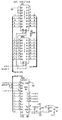
- Figure 7 provides a detailed schematic diagram of the low level sensor circuitry included in the presently preferred system of the present invention.
- Figures 8A-8C provide a detailed schematic diagram digital section circuitry included in the presently preferred system of the present invention.
- Figures 9A-9D provide a detailed schematic diagram of the analog section circuitry included in the presently preferred system of the present invention.
- Figures 10A-10C provide a detailed schematic diagram of the power supply and input/output (I/O) section included in the presently preferred system of the present invention.
- Figure 11 is a graph showing variation in oxygen saturation as a function of hematocrit.
- Figure 12 is a graph of εb805/εb970 versus Hematocrit.
- Figures 13A-13B are graphs of ε versus Hematocrit at two non-preferred wavelengths and ε1/ε2 versus Hematocrit at those non-preferred wavelengths.
- Figures 14A-14B are graphs of ε versus Hematocrit at two non-preferred wavelengths and ε1/ε2 versus Hematocrit at those non-preferred wavelengths.
- Figure 15 illustrate vertical emitter alignment and the resulting non-identical ΔXb regions.
- Figure 16 illustrates horizontal emitter alignment.
- The present invention is directed to apparatus and methods for determining biologic constituent values, such as the hematocrit value, transcutaneously and noninvasively. This is achieved by passing at least two wavelengths of light onto or through body tissues such as the finger, earlobe, or scalp, etc., and then compensating for the effects of body tissue and fluid by modifying the Beer-Lambert Law. The principles within the scope of the present invention may also be utilized to provide a hematocrit-independent oxygen saturation and oxygen content measurements as well as noninvasive measurement of blood constituents such as glucose, cholesterol, bilirubin, creatinine, etc.
- Although the present invention will describe in great detail the transillumination of various body parts, it will be appreciated that reflectance spectrophotometry may alternatively be employed where transillumination is difficult to accomplish. As used herein, the term "body part" is intended to include skin, earlobe, finger, lip, forehead, etc. Because the principles within the scope of the present invention can be adapted by those skilled in the art for in vitro measurement of hematocrit and other blood constituents, the term "body part" is also intended to include various in vitro blood containers such as tubes and cuvettes.
- spectrophotometric methods have been described in the prior art which monitor various metabolites in body fluids. Radiation, typically in the visible or near infrared region, is directed onto an exterior body part for transcutaneous penetration of the radiation. The radiation is then monitored reflectively or transmissively by a photodetector or similar sensor. Radiation spectra are chosen at wavelengths where the metabolite or compound sought for either absorbs highly or poorly. Some examples of such spectrophotometric methods are described in U.S. Patent No. 4,653,498 for pulse oximetry, U.S. Patent No. 4,655,225 for blood glucose monitoring, and more recently U.S. Patent No. 4,805,623 for monitoring various blood metabolites (glucose, cholesterol, etc.).
- A theoretical basis for the spectrophotometric techniques is the Beer-Lambert Law:
I = I0 e -εXd (1)
Equation (1) may also be written:
- The Beer-Lambert Law (1) permits in vitro solute concentration determinations. However, quantitative measurements have not been possible in the body since the scattering of the incident photons passing into and through the integument and subdermal regions is extensive and highly variable. This scattering spoils the Beer-Lambert Law by adding a variable loss of radiation to the measurement and also extends the path length of the incident photons by an unknown amount as well.
- Even though optical pulse rate monitors, plethysmographs, and pulse oximeters are known, their development has been accelerated by techniques which allow for cancellation of the optical scattering effects to a large extent. This development began with U.S. Patent No. 2,706,927 and was further refined by Yoshiya, et. al. (Med. and Biol. Eng. and Computing, 1980 Vol. 18, Pages. 27-32), Koneshi in U.S. Patent No. 3,998,550, and Hamaguri in U.S. No. Patent 4,266,554, which utilized a technique of analyzing the resultant opto-electronic signal by dividing it into its AC and DC components. The AC and DC components are manipulated with logarithmic amplifiers in such a way as to eliminate the above-mentioned transdermal optical effects (the variable amount of radiation loss due to scattering in the tissue and the unknown and variable amounts of optical path length increase).
- Until now, the AC-DC cancellation techniques have not been successfully adapted for the measurement of hematocrit or hematocrit-independent blood oxygen saturation.
- It is assumed that incident radiation passing onto or into a living tissue will pass through a combination of blood, tissue, and interstitial fluid compartments. The light attenuated by such a living tissue can be expressed by the modified Beer-Lambert equation:


- As the blood layer pulsates, the concentration terms change. The term d can be fixed by the geometric configuration of the device. Taking the partial derivatives of equation (2) with respect to time and dividing by equation (2) gives:


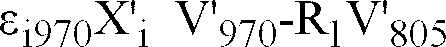
- Figures 3 and 12 suggest that a linear combination of V'λ at λ=805 nm and λ= 970nm will have a near constant value .for. a range of Hct values. Since the extinction coefficients εi805 and εi970 are well known, or can be empirically determined, a precise proportionality constant R1 can be found to produce


- It should be noticed that the following assumptions and requirements are essential in hematocrit determinations (but in the case of pulse oximetry these requirements may not be of the same degree of significance).
- A. Even though wavelengths λ=805 nm and λ=1310 nm are near isobestic, the actual function of ε versus Hematocrit at each given wavelength must hold hematocrit information that is different in curvature, or offset, or linearity, or sign from the other. See Figure 3. If the functions ελ versus hematocrit are not sufficiently different, then the ratio εbλ1/εbλ2 will not hold hematocrit information. See Figures 13A and 13B and Figures 14A and 14B. Even though the foregoing discussion refers to the isobestic wavelengths of λ=805 nm and λ=1310 nm, it will be appreciated that other isobestic wavelengths, such as λ=570 nm, λ=589 nm, and λ=1550 nm, may also be utilized.
- B. Further, the wavelengths should be selected close enough to one another such that the optical path lengths, d, are approximately the same. Longer wavelengths are preferred since they exhibit less sensitivity to scattering, s:

- C. The geometric or spatial relationship of the emitters and sensors is important. For instance, if vertically aligned emitters are used in an earlobe-measuring device, then the top-most emitter may illuminate a different amount of blood filled tissue than the lower emitter. If only one sensor is used, then there will be a disparity between X'b at each wavelength. See Figure 15, wherein Xb1 > Xb2 > Xb3. Furthermore, the sensor-emitter spatial separation distance is very important because the pressure applied to the tissue between the sensor and emitters affects the arteriolar and capillary vessel compliance. This changes the X' as the pressure (or distance) changes. This change in X' then modulates the V'λ function. Therefore, the sensor-emitter separation distance must be such that the pressure applied to the earlobe, fingertip, or other body member, does not affect the V'λ function. This sensor separation distance is empirically determined and should generate less than 40 mm Hg applied transmural pressure.
A horizontal alignment (Figure 15) of the emitters with respect to the single sensor can be arranged so that the emitters and sensors illuminate and detect identical regions of ∂Xλ1 and ∂Xλ2. It is important to note that the term d, the sensor-emitter separation, will be different between λ1 and λ2 by the cosine of the angle between the sensor and emitter. Therefore, if any misalignment from normal occurs, the term d will not cancel to obtain equation (9).
The preferred arrangement is wherein all the emitters (660, 805, 950, and 1310 nm) are located on the same substrate. This is preferred because the emitters will then illuminate essentially the same Xb region. - D. In the case of reflectance spectrophotometry, an aperture for the sensor and each emitter is required. See Figure 1B. Also, a sensor-emitter separation is required so that the reflectance of the first layer of tissue, Rt, (a non-blood layer of epithelium) does not further exaggerate a multiple scattering effect, i.e. the total reflectance, R, measured would contain spurious information of the epithelial layers' reflectance as well, where:

- The reflectance equations describing Rt or Rb must now sum all of the backscattered light that the sensor detects, i.e.,:

- While equation (9) describes the theory of the noninvasive hematocrit device, the four assumptions (A-D) are important to the repeatability and accurate functioning of the hematocrit device.
- Assuming items A through D are dealt with appropriately, then (9) becomes:


- From the foregoing, ε, the extinction coefficient, is not a simple function of the absorption coefficient, k, normally determined in pure solutions. Rather, it contains a diffusion or scattering term, s, which must be accounted for in a non-pure solution media such as whole blood and tissue.
- Finally, substituting (14) and (15) into (13):

- Therefore, the ratio ελ1/ελ2 is a function of hematocrit. From Figure 4, a look up table or polynomial curve fit equation may be obtained and utilized in the final displayed hematocrit results. Knowing the actual hematocrit value, it is straightforward to see (Figure 2) that a wavelength at 660 nanometers can be selected to obtain an ε ratio wherein the hematocrit-independent oxygen saturation value is derived. For example, equation (16) would become:

- Figure 11 graphically demonstrates the need for a hematocrit-independent blood saturation device. As either the hematocrit value or percent oxygen saturation decreases, the percent saturation error becomes unacceptable for clinical usage. For example, it is not uncommon to see patients with a low hematocrit (about 20%) who have respiratory embarrassment (low oxygen saturation) as well. Hence, the clinician simply requires more accurate oxygen saturation values.
- Knowing the hematocrit and oxygen saturation values, the computation of the Oxygen Content is trivial and may be displayed directly (a value heretofore unavailable to the clinician as a continuous, real-time, noninvasive result):

- Referring to the equations (16) and (9) a decision must be made by the computer as to the suitability of utilizing the Taylor expansion approximation to the logarithm. This algorithm is maintained in the software as a qualifying decision for the averaging and readout algorithms. The Taylor approximation is only valid for small ∂I/∂t values.
- It is interesting to see the similarities between this AC pulsatile derivation and an analogous DC technique. By taking the logarithm of two intensity ratios, values of εb and εi can be obtained from the modified Beer-Lambert equation (equation (2a)). These same extinction coefficients can be manipulated by the identical proportionality constants R1 and R2 found previously to exactly eliminate εi1310Xi and yield


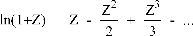
- It should also be noted that the two derivations (AC and DC) fold into one another through the power series expansion of the ln(1+Z) function:

- - One currently preferred method of obtaining the two intensities is to have the patient perform Valsalva's maneuver. Valsalva's maneuver is an attempt to forcibly exhale with the glottis, nose, and mouth closed. This maneuver increases intrathoracic pressure, slows the pulse, decreases return of blood to the heart, and increases venous pressure. Obtaining intensity measurements before and during Valsalva's, maneuver provide sufficiently different intensities to utilize equation (19). Even a deep breath can be enough to obtain sufficiently different intensities.
- Another technique to simulate pulsatile blood flow and to eliminate the skin's optical scattering effects, while at the same time preserving the blood-borne hematocrit and oxygen saturation information, is described below. By utilizing a
stepper motor 9 in theearlobe clip assembly 10 on anearlobe 11 of a patient such as that illustrated in Figures 6, 6A, 15, and 16, one can produce a variation of Xb sufficient to utilizeequation 19. Thestepper motor 9 could even produce a bloodless (Xb=0) state, if required. However,equation 19 shows that only a difference between Xb1 and Xb2 is needed. - The major advantage of this technique is that under clinical conditions of poor blood flow, poor blood pressure, or peripheral vascular disease, where pulse wave forms are of poor quality for the (∂I/∂t)/I technique, this DC stepper motor technique could be utilized.
- The above techniques describe conditions and equations wherein isobestic wavelengths are chosen such that the hematocrit value obtained has no interference from oxygen saturation, hence an independently determined hematocrit value.
- One, however, may choose λ2 (the reference wavelength) in equation (13) at 1550 nm as well. In the
radiation region 900 to 2000 nm the blood absorption coefficients depend on hematocrit and water, whereas at 805 nm the blood absorption coefficient only depends on hematocrit. Therefore, utilizing in combination, wavelengths of 660, 805, and 1550 will also give a technique to determine hematocrit (ε805/ε1550) and oxygen saturation (ε660/ε805). - These 3 wavelengths are particularly important since 660, 805, and 1550 nm (or 1310 nm) are readily available LEDs, such as, respectively, MLED76-Motorola, HLP30RGB-Hitachi, and ETX1550-EPITAXX (or NDL5300-NEC), with the benefits of low cost and low optical power (reducing any question of possible eye damage).
- The manufacturing of a multi-chip LED emitter becomes reasonable, cost-wise, and provides increased accuracy since the LED sources have practically no separation distances and appear as a single point source.
- This invention may be applied to the determination of other components (included, but not limited to, glucose, or cholesterol) in any range of the electromagnetic spectrum in which spectrophotometric techniques can be utilized.
- An
earlobe clip assembly 10 as in Figures 6, 6A, 15, and 16 (with or without thestepper motor 9 shown in Figure 6A) afinger clip assembly 6 used on afinger 7 as shown in Figures 1, 1A, and 1B are two currently preferred embodiments for practicing the present invention. Thephotodiodes 3 andemitters - Consider first the sensor technology in the transmissive mode of operation. An earlobe or fingertip housing can be provided with discreet emitters and two photodiode chips (of different sensitivity ranges, 600-1000 nm and 1000-1700 nm ranges) placed on one substrate, such as a TO-5 can (Hamamatsu K1713-03). The emitters likewise can be two or more emitter chips (i.e., λ = 805, 1310, 660, and 950 nm) placed on a common substrate and illuminated through a TO-39 can.
- Finally, a single substrate multi-wavelength emitter and a multi-wavelength detector, assembled in one small physical housing for each, make alignment and detection sensitivity more repeatable, and hence more accurate.
- The preferred emitter chips would have wavelengths, for hematocrit-only measurements, at 805 nm, 950 nm, and 1310 nm (or 805 nm, 950 nm, and 1550 nm) . Although in theory, an emitter having a wavelength of 970 nanometers, rather than 950 nm, would provide more accurate information, 970 nm emitters are not presently available commercially. These wavelengths are currently preferred because of the different curvature and baseline offset of the ε versus Hematocrit at those wavelengths. See Figure 3. Hence, the hematocrit information will exist in the ratio ελ1/ελ2. See Figure 4.
- Furthermore, the choice of 805 nm and 1310 nm (or 1550 nm) rather than 570 nm and 805 nm is because there is no water absorption in the 570 nm (or 589 nm) and 805 nm isobestic wavelengths. However, there is tremendous water absorption at 1310 nm and 1550 nm. Hence, the ratio of 570 nm to 805 nm, as a reference, would not yield hematocrit information because there would be no offset due to water in the plasma. See Figures 13A and 13B and Figures 14A and 14B.
- If hematocrit-independent oxygen saturation is desired then the emitter chip wavelengths would be 660 nm, 805 nm, 950 nm, and 1310 nm (or 1550 nm) (the 660 nm is MLED76, Motorola or TOLD 9200, Toshiba). Likewise, the photodetector single substrate could house at least two chips, such as a Hamamatsu K1713-03.
- It will be appreciated that those skilled in the art would be able to add other chips to the single substrate at wavelengths sensitive to other metabolites (glucose, cholesterol, etc.). The above mentioned emitter and detector connections can be seen in the analog schematic diagram illustrated in Figures 7 and 9B-9D.
- The sensor technology in the reflectance mode must conform to two embodiment parameters. See Figure 1B. The diameter and thickness of the
aperture 8 in which figure 7 is received in combination with the sensor-emitter separation distance are important to provide a detection region within thesubdermis 12 at points a and b of Figure 1B, where the radiation impinges on blood-tissue without the multiple scattering effects of the epithelial layer, Rt. The determination ofoptimum sensor 3 separation andaperture 8 sizes is done empirically fromnumerous fingers 7 with varying callous andfingernails 13. Minimum sensor separation and aperture diameters can be established wherein Rt, of equation (14) is eliminated. - Figures 7, 8A-8C, 9A-9D, and 10A-10B detail the electronics of one circuit suitable for use within the scope of the present invention. The memory and computation means (Figures 8A-8C) are connected via a "bus" structure between PROMS (U110,U111), microprocessor MC68HC000 (U106), static RAMS (U112,U113), and isolation buffers to the low-level analog circuitry (Figure 7). A crystal controlled oscillator circuit (U101A,B) is divided by 2 to provides a symmetric master clock to the microprocessor; this clock is further subdivided and used to provide clocking for the analog-to-digital converter (U208) and timer (U109). Strobe lines are generated through a decoder arrangement to drive each of the subsystems of the device and also control the isolation bus buffers (U201,U202).
- Timer outputs are fed back into the microprocessor and encoded (U104) to produce interrupts at specific intervals for system functions. One timer is shared by subsystems which control the liquid crystal display means, the keyboard entry means, the audible indicator, and the cycling background system self-test. Another timer is dedicated exclusively to provide a high priority interrupt to the microprocessor; this interrupt drives software which controls the basic sensor sampling mechanism. An expansion connector (J101) is included to allow extended testing of the device or connection to external data-logging equipment such as a printer or computer interface.
- The local bus isolates the sensitive analog circuitry from the main digital circuitry. This prevents spurious crosstalk from digital signals into the analog circuitry and thereby reduces superimposed noise on the measured signals. It is on this local bus that the Digital-to-Analog Converters (DAC) and Analog-to-Digital Convertors (ADC) transmit and receive digital information while processing the low-level analog signals.
- The Low Level Sensor electronic section, Figure 7, combines subsystems to both measure and modulate the current produced from each optical sensor. Since the pulsatile component of the optical energy transmitted through or reflected off of tissue comprises only a small part of the overall optical energy incident on the sensor, means are provided to "null out" in a carefully controlled and accurately known way the non-pulsatile component of the light-produced current in the sensing detector. The remaining signal can then be dc-amplified and filtered in a straightforward manner and presented to the ADC (U208) for conversion into a digital value representative of the relative AC pulsatile component. Furthermore, because the relationship between the nulling current and the average value of this AC component is known, the DC component can easily be calculated as a function of the sensing means' sensitivities and the electronic stages' gains. The functions determining these AC and DC values can (if necessary) be trimmed in software by calibration constants which are stored in EEPROM (U307) and retrieved each time the unit is powered on.
- The current which modulates the optical sources (LEDs or Laser Diodes) is also controlled (U203) and precisely adjusted (U306) to optimize signal reception and detection. Through software control, the modulation current can be adjusted on a pulse-by-pulse basis to minimize noise-induced inaccuracies. Furthermore, by sampling the sensors with the modulation sources disabled appropriately, background noise (such as 60 Hz) can be rejected digitally as common-mode noise. Thus, by controlling the optical source energy and modulating the nulling current in the photosensor circuitry, it is possible to effectively cancel the effects of ambient radiation levels and accurately measure both the static (DC) and time-varying (AC) components of transmitted or reflected light.
- Interrupt-driven software algorithms acquire the sensor data, provide a real-time pulse wave contour, and determine pulse boundaries. Completed buffers (i.e. one entire pulse per buffer) of sensor data are then passed to the foreground software processes for computation. This involves the determination of the background-compensated AC pulsatile and DC static values of intensities for each wavelength. Through averaging and selective elimination of abnormal values, results are then calculated using equation (9) and displayed on the LCD. The modulating and nulling currents are (if necessary) also adjusted to utilize the electronic hardware efficiently and optimally.
- Although the foregoing discussion has related to noninvasive analysis of blood hematocrit information, it will be appreciated that the above-mentioned emitters, sensors, and circuitry may be adapted for invasive in vitro analysis of blood hematocrit values. The principles within the scope of the present invention which compensate for spatial, geometric, and tissue variations may be used to compensate for similar variations in an in vitro blood container. Such a device would allow hematocrit values to be determined rapidly and accurately.
- Those skilled in the art will also appreciate that the methods within the scope of the present invention for determining blood hematocrit values may be adapted for determining non-hematocrit biologic constituent values such as glucose, cholesterol, etc. To determine biologic constituent information, the effects of competing blood, tissue, and interstitial fluid constituents must be eliminated. It is believed that these effects may be eliminated by appropriate modification of the differential ratiometric techniques described above.
- It is important to recognize that the present invention is not directed to determining the tissue hematocrit value. The tissue hematocrit value, in contrast with the blood hematocrit value, reflects the amount of red blood cells in a given volume of tissue (blood, interstitial fluids, fat, hair follicles, etc.). The present invention is capable of determining actual intravascular blood hematocrit and hemoglobin values.
- From the foregoing, it will be appreciated that the present invention provides a system and method for noninvasively and quantitatively determining a subject's hematocrit or other blood constituent value. The present invention determines the hematocrit noninvasively by utilizing electromagnetic radiation as the transcutaneous information carrier. Importantly, the present invention may be used on various body parts to provide accurate quantitative hematocrit values.
- It will also be appreciated that the present invention also provides a system and method which can provide immediate and continuous hematocrit information for a subject. The present invention further provides a system and method for noninvasively determining a subjects's blood oxygen saturation (SaO2) independent of the subject's hematocrit. In addition, the present invention provides a system and method for noninvasively determining a subject's hematocrit and/or blood oxygen saturation even under conditions of low blood perfusion.
- The present invention may be embodied in other specific forms without departing from its spirit or essential characteristics. The described embodiments are to be considered in all respects only as illustrative and not restrictive. The scope of the invention is, therefore, indicated by the appended claims rather than by the foregoing description. All changes which come within the meaning and range of equivalency of the claims are to be embraced within their scope.
- The present application is a divisional application based on an earlier European application 93910585.4. The following numbered clauses, which correspond to the claims of that earlier application as filed, form part of the present disclosure, but not the present claims.


Claims (41)
- A method for determining a desired biologic constituent of the blood of a patient, the blood flowing in a body part (7) of the patient or in an extracorporeal passageway in communication with the circulatory system of the patient so as to be subjectable to transcutaneous examination in the extracorporeal passageway, the body part or extracorporeal passageway defining a blood conduit, the method comprising the steps of:selecting a first radiation wavelength having a first extinction characteristic as a function of the desired biologic constituent;selecting a second radiation wavelength which exhibits a greater absorption coefficient to water than said first radiation wavelength, said second radiation having a second extinction characteristic as a function of the desired biologic constituent different from the first extinction characteristic in at least one of curvature, offset, linearity and sign;directing (1)(2) said first and second radiation wavelengths into the blood conduit;detecting (3) the amount of first radiation after passing through the blood conduit;detecting (3) the amount of second radiation after passing through the blood conduit; andcomparing variations over time in the detected amounts of first and second radiations to determine the desired biologic constituent value by a differential ratiometric technique, whereby said desired biologic constituent value is determined quantitatively.
- A method as claimed in claim 1 wherein the first and second wavelengths are selected at the isobestic points of reduced haemoglobin and oxyhaemoglobin.
- A method as claimed in claim 1 or 2 wherein said first and second extinction characteristics comprise first and second curvatures.
- A method as claimed in claim 1 or 2 wherein said first and second extinction characteristics comprise first and second offsets.
- A method as claimed in claim 1 or 2 wherein said first and second extinction characteristics comprise first and second linearity characteristics.
- A method as claimed in claim 1 or 2 wherein said first sign is different from said second sign.
- A method as claimed in any preceding claim, further comprising the steps of:selecting a third radiation wavelength which will be extinguished by a competing biologic constituent in the blood in a manner characteristic of said competing biologic constituent and having an extinction characteristic with regard to constituents other than said competing biologic constituent which is substantially different from one of said first and second extinction characteristics;directing radiation having the third wavelength into the blood conduit;determining the amount of radiation having the third wavelength which is extinguished by the blood conduit;operating on the amount of detected radiation having the first, second and third wavelengths such that spatial, geometric and/or tissue variations are eliminated in each radiation wavelength; andcompensating for the effect of the second biologic constituent by operating on the amount of detected radiation having first, second and third wavelengths.
- A method as claimed in any preceding claim wherein said comparison step comprises mathematically manipulating the detected amount of first and second radiation wavelengths with a polynomial function to determine the desired biologic constituent value.
- A method as claimed in any preceding claim wherein said comparison step comprises forming the ratio of the ΔI/I ratios for each of the first and second radiation wavelengths, where ΔI represents a time derivative of a pulsatile transmitted intensity component, and I represents a steady state transmitted intensity.
- A method as claimed in any of claims 1 to 9 wherein the desired biologic constituent is hematocrit.
- A method as claimed in claim 10 further comprising:selecting a further radiation wavelength;directing said further radiation into the blood conduit;detecting the amount of said further radiation after passing through the blood conduit; andcomparing variations in the detected amounts of said further radiation and said first radiation to compute a hematocrit-independent oxygen saturation value in addition to the computed hematocrit.
- A method as claimed in claim 11 wherein said further radiation wavelength is one which is not isobestic for oxyhaemoglobin and reduced haemoglobin.
- A method as claimed in any of claims 1 to 9 wherein the desired biologic constituent comprises glucose.
- A method as claimed in any of claims 1 to 9 wherein the desired biologic constituent comprises cholesterol.
- A method as claimed in any of claims 1 to 9 wherein the desired biologic constituent comprises bilirubin.
- A method as claimed in any of claims 1 to 9 wherein the desired biologic constituent comprises creatinine.
- A method as claimed in any of claims 1 to 9 wherein the desired biologic constituent comprises haemoglobin.
- A system for determining a desired biological constituent value of the blood of a patient, the blood flowing in a body part of the patient or in an extracorporeal passageway in communication with the circulatory system of the patient so as to be subjectable to transcutaneous examination in the body part or to non-invasive examination in the extracorporeal passageway, the body part (7) or the extracorporeal passageway defining a blood conduit, the system comprising:blood conduit receiving means (6) for receiving a blood conduit containing the flowing blood of the patient;a first emitter (1) positioned on said conduit receiving means for emitting a first radiation wavelength, said first radiation having at least a first extinction characteristic with regard to the desired biologic constituent;a second emitter (2) positioned on said conduit receiving means for emitting a second radiation wavelength which exhibits a greater absorption coefficient to water than said first radiation wavelength, said second radiation having at least a second extinction characteristic different from the first extinction characteristic in at least one of curvature, offset, linearity or sign;directing means for directing said first and second radiation wavelengths into the blood conduit;first detecting means (3) for detecting the amount of first radiation after passing through the blood conduit;second detecting means (3) for detecting the amount of second radiation after passing through the blood conduit; andmeans for comparing variations over time in the detected amounts of first and second radiations to determine the desired biologic constituent value by a differential ratiometric technique, whereby said desired biologic constituent value is determined quantitatively.
- A system as claimed in claim 18, wherein said first and second wavelengths are at or near the isobestic points of reduced haemoglobin and oxyhaemoglobin.
- A system as claimed in claim 18 or 19 wherein said first and second extinction characteristics comprise first and second curvatures.
- A system as claimed in claim 18 or 19 wherein said first and second extinction characteristics comprise first and second offsets.
- A system as claimed in claim 18 or 19 wherein said first and second extinction characteristics comprise first and second linearity characteristics.
- A system as claimed in claim 18 or 19 wherein said first sign is different from said second sign.
- A system as claimed in any of claims 18 to 23 wherein said first and second photodetecting means are formed by a single photodetector.
- A system as claimed in any of claims 18 to 24, the system further comprising:a third emitter positioned on said conduit receiving means for emitting a third radiation wavelength which will be extinguished by a competing biologic constituent in the blood in a manner characteristic of said competing biologic constituent and having an extinction characteristic with regard to constituents other than said competing biologic constituent which is substantially different from one of said first and second extinction characteristics;second directing means for directing radiation having the third wavelength into the blood conduit;detecting means for detecting the amount of radiation having the third wavelength which is extinguished by the blood in the blood conduit;means for operating on the amount of detected radiation having the first, second and third wavelengths such that spatial, geometric and/or tissue variations are eliminated in each radiation wavelength; andmeans for compensating for the effect of the second biologic constituent by operating on the amount of detected radiation having first, second and third wavelengths.
- A system as claimed in any of claims 18 to 25 wherein said comparing means is arranged for mathematically manipulating the detected amount of first and second radiation wavelengths with a polynomial function to determine the desired biologic constituent value.
- A system as claimed in any of claims 18 to 26, wherein said comparing means comprises means for forming the ratio of the ΔI/I ratios for each of the first and second radiation wavelengths, where ΔI represents a time derivative of a pulsatile transmitted intensity component, and I represents a steady state transmitted intensity.
- A system as claimed in any of claims 18 to 27 wherein the desired biologic constituent is hematocrit.
- A system as claimed in claim 28 further comprising:a further emitter positioned on said conduit receiving means for emitting a further radiation wavelength;directing means for directing said further radiation wavelength into the blood conduit;further detecting means for detecting the amount of said further radiation after passing through the blood conduit; andmeans for comparing variations in the detected amounts of said further radiation and said first radiation to compute a hematocrit-independent oxygen saturation value in addition to the computed hematocrit.
- A method as claimed in claim 29 wherein said further radiation wavelength is one which is not isobestic for oxyhaemoglobin and reduced haemoglobin.
- A system as claimed in any of claims 18 to 27 wherein the desired biologic constituent comprises glucose.
- A system as claimed in any of claims 18 to 27 wherein the desired biologic constituent comprises cholesterol.
- A system as claimed in any of claims 18 to 27 wherein the desired biologic constituent comprises bilirubin.
- A system as claimed in any of claims 18 to 27 wherein the desired biologic constituent comprises creatinine.
- A system as claimed in any of claims 18 to 27 wherein the desired biologic constituent comprises haemoglobin.
- A method for determining oxygen saturation of the blood of a patient, the blood flowing in a body part (7) of the patient or in an extracorporeal passageway in communication with the circulatory system of the patient so as to be subjectable to transcutaneous examination in the extracorporeal passageway, the body part or extracorporeal passageway defining a blood conduit, the method comprising the steps of:selecting a first radiation wavelength having a first extinction characteristic as a function of the desired biologic constituent;selecting a second radiation wavelength which exhibits a greater absorption coefficient to water than said first radiation wavelength, said second radiation having a second extinction characteristic as a function of the desired biologic constituent different from the first extinction characteristic in at least one of curvature, offset, linearity and sign;selecting a further radiation wavelength;directing (1)(2) said first and second and further radiation wavelengths into the blood conduit;detecting (3) the respective amounts of said first, second and further radiation after passing through the blood conduit;detecting the amount of said further radiation after passing through the blood conduit;comparing variations over time in the detected amounts of first and second radiations by a differential ratiometric technique and comparing variations in the detected amounts of said further radiation and said first radiation thereby to compute a hematocrit-independent oxygen saturation value.
- A method as claimed in claim 36 wherein the first and second wavelengths are selected at the isobestic points of reduced haemoglobin and oxyhaemoglobin.
- A method as claimed in claim 36 or 37 wherein said further radiation wavelength is one which is not isobestic for oxyhaemoglobin and reduced haemoglobin.
- A system for determining oxygen saturation of the blood of a patient, the blood flowing in a body part of the patient or in an extracorporeal passageway in communication with the circulatory system of the patient so as to be subjectable to transcutaneous examination in the body part or to non-invasive examination in the extracorporeal passageway, the body part (7) or the extracorporeal passageway defining a blood conduit, the system comprising:blood conduit receiving means (6) for receiving a blood conduit containing the flowing blood of the patient;a first emitter (1) positioned on said conduit receiving means for emitting a first radiation wavelength, said first radiation having at least a first extinction characteristic with regard to the desired biologic constituent;a second emitter (2) positioned on said conduit receiving means for emitting a second radiation wavelength which exhibits a greater absorption coefficient to water than said first radiation wavelength, said second radiation having at least a second extinction characteristic different from the first extinction characteristic in at least one of curvature, offset, linearity or sign;a further emitter positioned on said conduit receiving means for emitting a further radiation wavelength;directing means for directing said first, second and further radiation wavelengths into the blood conduit;detecting means (3) for detecting the respective amounts of said first, second and further radiation after passing through the blood conduit;means for comparing variations over time in the detected amounts of first and second radiations by a differential ratiometric technique and comparing variations in the detected amounts of said further radiation and said first radiation thereby to compute a hematocrit-independent oxygen saturation value.
- A system as claimed in claim 39, wherein said first and second wavelengths are at or near the isobestic points of reduced haemoglobin and oxyhaemoglobin.
- A method as claimed in claim 39 or 40 wherein said further radiation wavelength is one which is not isobestic for oxyhaemoglobin and reduced haemoglobin.
Applications Claiming Priority (2)
| Application Number | Priority Date | Filing Date | Title |
|---|---|---|---|
| EP93910585A EP0693900B1 (en) | 1993-04-12 | 1993-04-12 | System and method for noninvasive hematocrit monitoring |
| PCT/US1993/003427 WO1994023643A1 (en) | 1993-04-12 | 1993-04-12 | System and method for noninvasive hematocrit monitoring |
Related Parent Applications (2)
| Application Number | Title | Priority Date | Filing Date |
|---|---|---|---|
| EP93910585A Division EP0693900B1 (en) | 1993-04-12 | 1993-04-12 | System and method for noninvasive hematocrit monitoring |
| EP93910585.4 Division | 1994-10-27 |
Publications (2)
| Publication Number | Publication Date |
|---|---|
| EP1491135A2 true EP1491135A2 (en) | 2004-12-29 |
| EP1491135A3 EP1491135A3 (en) | 2005-09-07 |
Family
ID=22236501
Family Applications (2)
| Application Number | Title | Priority Date | Filing Date |
|---|---|---|---|
| EP04075850A Withdrawn EP1491135A3 (en) | 1993-04-12 | 1993-04-12 | Method and apparatus for monitoring blood constituents |
| EP93910585A Expired - Lifetime EP0693900B1 (en) | 1993-04-12 | 1993-04-12 | System and method for noninvasive hematocrit monitoring |
Family Applications After (1)
| Application Number | Title | Priority Date | Filing Date |
|---|---|---|---|
| EP93910585A Expired - Lifetime EP0693900B1 (en) | 1993-04-12 | 1993-04-12 | System and method for noninvasive hematocrit monitoring |
Country Status (8)
| Country | Link |
|---|---|
| EP (2) | EP1491135A3 (en) |
| JP (1) | JP3625475B2 (en) |
| KR (1) | KR100294294B1 (en) |
| AT (1) | ATE261698T1 (en) |
| AU (1) | AU4102293A (en) |
| CA (1) | CA2160252C (en) |
| DE (1) | DE69333456T2 (en) |
| WO (1) | WO1994023643A1 (en) |
Cited By (41)
| Publication number | Priority date | Publication date | Assignee | Title |
|---|---|---|---|---|
| US7657292B2 (en) | 2001-03-16 | 2010-02-02 | Nellcor Puritan Bennett Llc | Method for evaluating extracellular water concentration in tissue |
| US7706852B2 (en) | 2006-01-30 | 2010-04-27 | Nellcor Puritan Bennett Llc | System and method for detection of unstable oxygen saturation |
| US7725146B2 (en) | 2005-09-29 | 2010-05-25 | Nellcor Puritan Bennett Llc | System and method for pre-processing waveforms |
| US8007441B2 (en) | 2004-03-08 | 2011-08-30 | Nellcor Puritan Bennett Llc | Pulse oximeter with alternate heart-rate determination |
| US8068891B2 (en) | 2006-09-29 | 2011-11-29 | Nellcor Puritan Bennett Llc | Symmetric LED array for pulse oximetry |
| US8095192B2 (en) | 2003-01-10 | 2012-01-10 | Nellcor Puritan Bennett Llc | Signal quality metrics design for qualifying data for a physiological monitor |
| US8116852B2 (en) | 2006-09-29 | 2012-02-14 | Nellcor Puritan Bennett Llc | System and method for detection of skin wounds and compartment syndromes |
| US8135448B2 (en) | 2001-03-16 | 2012-03-13 | Nellcor Puritan Bennett Llc | Systems and methods to assess one or more body fluid metrics |
| US8140272B2 (en) | 2008-03-27 | 2012-03-20 | Nellcor Puritan Bennett Llc | System and method for unmixing spectroscopic observations with nonnegative matrix factorization |
| US8175667B2 (en) | 2006-09-29 | 2012-05-08 | Nellcor Puritan Bennett Llc | Symmetric LED array for pulse oximetry |
| US8175665B2 (en) | 2007-03-09 | 2012-05-08 | Nellcor Puritan Bennett Llc | Method and apparatus for spectroscopic tissue analyte measurement |
| US8204567B2 (en) | 2007-12-13 | 2012-06-19 | Nellcor Puritan Bennett Llc | Signal demodulation |
| US8229529B2 (en) | 2001-03-16 | 2012-07-24 | Nellcor Puritan Bennett Llc | Device and method for monitoring body fluid and electrolyte disorders |
| US8238994B2 (en) | 2005-10-28 | 2012-08-07 | Nellcor Puritan Bennett Llc | Adjusting parameters used in pulse oximetry analysis |
| US8315684B2 (en) | 2004-02-25 | 2012-11-20 | Covidien Lp | Oximeter ambient light cancellation |
| US8346327B2 (en) | 2007-03-09 | 2013-01-01 | Covidien Lp | Method for identification of sensor site by local skin spectrum data |
| US8364224B2 (en) | 2008-03-31 | 2013-01-29 | Covidien Lp | System and method for facilitating sensor and monitor communication |
| US8376955B2 (en) | 2009-09-29 | 2013-02-19 | Covidien Lp | Spectroscopic method and system for assessing tissue temperature |
| US8423109B2 (en) | 2005-03-03 | 2013-04-16 | Covidien Lp | Method for enhancing pulse oximery calculations in the presence of correlated artifacts |
| US8420405B2 (en) | 2006-09-25 | 2013-04-16 | Covidien Lp | Carbon dioxide detector having borosilicate substrate |
| US8431087B2 (en) | 2006-09-25 | 2013-04-30 | Covidien Lp | Carbon dioxide detector having borosilicate substrate |
| US8431088B2 (en) | 2006-09-25 | 2013-04-30 | Covidien Lp | Carbon dioxide detector having borosilicate substrate |
| US8442608B2 (en) | 2007-12-28 | 2013-05-14 | Covidien Lp | System and method for estimating physiological parameters by deconvolving artifacts |
| US8449834B2 (en) | 2006-09-25 | 2013-05-28 | Covidien Lp | Carbon dioxide detector having borosilicate substrate |
| US8457722B2 (en) | 2001-03-16 | 2013-06-04 | Covidien Lp | Device and method for monitoring body fluid and electrolyte disorders |
| US8509869B2 (en) | 2009-05-15 | 2013-08-13 | Covidien Lp | Method and apparatus for detecting and analyzing variations in a physiologic parameter |
| US8515512B2 (en) | 2006-09-26 | 2013-08-20 | Covidien Lp | Opaque, electrically nonconductive region on a medical sensor |
| US8521246B2 (en) | 2010-07-29 | 2013-08-27 | Covidien Lp | Cable cross talk suppression |
| US8532729B2 (en) | 2011-03-31 | 2013-09-10 | Covidien Lp | Moldable ear sensor |
| US8577435B2 (en) | 2011-03-31 | 2013-11-05 | Covidien Lp | Flexible bandage ear sensor |
| US8690864B2 (en) | 2007-03-09 | 2014-04-08 | Covidien Lp | System and method for controlling tissue treatment |
| US8768426B2 (en) | 2011-03-31 | 2014-07-01 | Covidien Lp | Y-shaped ear sensor with strain relief |
| US8968193B2 (en) | 2008-09-30 | 2015-03-03 | Covidien Lp | System and method for enabling a research mode on physiological monitors |
| US8983800B2 (en) | 2003-01-13 | 2015-03-17 | Covidien Lp | Selection of preset filter parameters based on signal quality |
| US8991034B2 (en) | 2005-08-08 | 2015-03-31 | Covidien Lp | Methods of manufacturing a compliant diaphragm medical sensor |
| CN106999111A (en) * | 2014-10-01 | 2017-08-01 | 纽洛斯公司 | System and method for detecting invisible human emotion |
| WO2017152577A1 (en) * | 2016-03-09 | 2017-09-14 | 潘卫江 | Multi-section finger sleeve-type probe |
| US9895068B2 (en) | 2008-06-30 | 2018-02-20 | Covidien Lp | Pulse oximeter with wait-time indication |
| US10076276B2 (en) | 2008-02-19 | 2018-09-18 | Covidien Lp | Methods and systems for alerting practitioners to physiological conditions |
| DE102017110216A1 (en) * | 2017-05-11 | 2018-11-15 | Osram Opto Semiconductors Gmbh | Optoelectronic sensor module and method for producing an optoelectronic sensor module |
| US11857323B2 (en) | 2017-10-24 | 2024-01-02 | Nuralogix Corporation | System and method for camera-based stress determination |
Families Citing this family (60)
| Publication number | Priority date | Publication date | Assignee | Title |
|---|---|---|---|---|
| US6580086B1 (en) * | 1999-08-26 | 2003-06-17 | Masimo Corporation | Shielded optical probe and method |
| US6390977B1 (en) | 1995-06-07 | 2002-05-21 | Alliance Pharmaceutical Corp. | System and methods for measuring oxygenation parameters |
| CA2221968C (en) * | 1995-06-09 | 2007-08-21 | Cybro Medical Ltd. | Sensor, method and device for optical blood oximetry |
| US6018673A (en) | 1996-10-10 | 2000-01-25 | Nellcor Puritan Bennett Incorporated | Motion compatible sensor for non-invasive optical blood analysis |
| JP4214324B2 (en) * | 1997-08-20 | 2009-01-28 | アークレイ株式会社 | Biological tissue measurement device |
| US6743172B1 (en) | 1998-01-14 | 2004-06-01 | Alliance Pharmaceutical Corp. | System and method for displaying medical process diagrams |
| US6860266B2 (en) | 2000-11-03 | 2005-03-01 | Dartmouth-Hitchcock Clinic | Physiological object displays |
| US6675031B1 (en) | 1999-04-14 | 2004-01-06 | Mallinckrodt Inc. | Method and circuit for indicating quality and accuracy of physiological measurements |
| DE10051943B4 (en) | 2000-10-19 | 2015-01-15 | Fresenius Medical Care Deutschland Gmbh | Method and device for pulse wave transit time determination and extracorporeal blood treatment device with such a device |
| US6748254B2 (en) | 2001-10-12 | 2004-06-08 | Nellcor Puritan Bennett Incorporated | Stacked adhesive optical sensor |
| US20040267166A1 (en) * | 2002-05-01 | 2004-12-30 | Kiyoko Ooshima | Biological information detecting contact |
| US20030212316A1 (en) * | 2002-05-10 | 2003-11-13 | Leiden Jeffrey M. | Method and apparatus for determining blood parameters and vital signs of a patient |
| KR100493157B1 (en) * | 2002-08-02 | 2005-06-03 | 삼성전자주식회사 | Probe using in measuring organism signal and system for measuring organism signal comprising the same |
| US20040242977A1 (en) * | 2003-06-02 | 2004-12-02 | Dosmann Andrew J. | Non-invasive methods of detecting analyte concentrations using hyperosmotic fluids |
| US7865223B1 (en) * | 2005-03-14 | 2011-01-04 | Peter Bernreuter | In vivo blood spectrometry |
| US7657295B2 (en) | 2005-08-08 | 2010-02-02 | Nellcor Puritan Bennett Llc | Medical sensor and technique for using the same |
| US7590439B2 (en) | 2005-08-08 | 2009-09-15 | Nellcor Puritan Bennett Llc | Bi-stable medical sensor and technique for using the same |
| US7869850B2 (en) | 2005-09-29 | 2011-01-11 | Nellcor Puritan Bennett Llc | Medical sensor for reducing motion artifacts and technique for using the same |
| US7904130B2 (en) | 2005-09-29 | 2011-03-08 | Nellcor Puritan Bennett Llc | Medical sensor and technique for using the same |
| US7555327B2 (en) | 2005-09-30 | 2009-06-30 | Nellcor Puritan Bennett Llc | Folding medical sensor and technique for using the same |
| US7881762B2 (en) | 2005-09-30 | 2011-02-01 | Nellcor Puritan Bennett Llc | Clip-style medical sensor and technique for using the same |
| US7486979B2 (en) | 2005-09-30 | 2009-02-03 | Nellcor Puritan Bennett Llc | Optically aligned pulse oximetry sensor and technique for using the same |
| US20070106134A1 (en) | 2005-11-10 | 2007-05-10 | O'neil Michael P | Medical sensor and technique for using the same |
| US8073518B2 (en) | 2006-05-02 | 2011-12-06 | Nellcor Puritan Bennett Llc | Clip-style medical sensor and technique for using the same |
| US8145288B2 (en) | 2006-08-22 | 2012-03-27 | Nellcor Puritan Bennett Llc | Medical sensor for reducing signal artifacts and technique for using the same |
| US8219170B2 (en) | 2006-09-20 | 2012-07-10 | Nellcor Puritan Bennett Llc | System and method for practicing spectrophotometry using light emitting nanostructure devices |
| US8396524B2 (en) | 2006-09-27 | 2013-03-12 | Covidien Lp | Medical sensor and technique for using the same |
| US7574245B2 (en) | 2006-09-27 | 2009-08-11 | Nellcor Puritan Bennett Llc | Flexible medical sensor enclosure |
| US7796403B2 (en) | 2006-09-28 | 2010-09-14 | Nellcor Puritan Bennett Llc | Means for mechanical registration and mechanical-electrical coupling of a faraday shield to a photodetector and an electrical circuit |
| US7684842B2 (en) | 2006-09-29 | 2010-03-23 | Nellcor Puritan Bennett Llc | System and method for preventing sensor misuse |
| US8265724B2 (en) | 2007-03-09 | 2012-09-11 | Nellcor Puritan Bennett Llc | Cancellation of light shunting |
| US8280469B2 (en) | 2007-03-09 | 2012-10-02 | Nellcor Puritan Bennett Llc | Method for detection of aberrant tissue spectra |
| US8437822B2 (en) | 2008-03-28 | 2013-05-07 | Covidien Lp | System and method for estimating blood analyte concentration |
| US8112375B2 (en) | 2008-03-31 | 2012-02-07 | Nellcor Puritan Bennett Llc | Wavelength selection and outlier detection in reduced rank linear models |
| DE202008005840U1 (en) | 2008-04-05 | 2009-08-13 | Bluepoint Medical Gmbh & Co. Kg | measuring device |
| DE102008017403A1 (en) | 2008-04-05 | 2009-10-08 | Bluepoint Medical Gmbh & Co. Kg | Device for measuring physiological parameters i.e. oxygen saturation, in blood of patient in hospital, has transmission and receiving units with cap elements that are sterilized by superheated steam and optically permeable in relevant area |
| US8071935B2 (en) | 2008-06-30 | 2011-12-06 | Nellcor Puritan Bennett Llc | Optical detector with an overmolded faraday shield |
| US7880884B2 (en) | 2008-06-30 | 2011-02-01 | Nellcor Puritan Bennett Llc | System and method for coating and shielding electronic sensor components |
| US8577431B2 (en) | 2008-07-03 | 2013-11-05 | Cercacor Laboratories, Inc. | Noise shielding for a noninvasive device |
| US20100030040A1 (en) | 2008-08-04 | 2010-02-04 | Masimo Laboratories, Inc. | Multi-stream data collection system for noninvasive measurement of blood constituents |
| KR101009284B1 (en) | 2008-09-02 | 2011-01-18 | 주식회사 셀텍 | Portble Multi Medical Diagnosis system |
| DE202008015271U1 (en) | 2008-11-18 | 2010-04-08 | Bluepoint Medical Gmbh & Co. Kg | measuring device |
| JP4502060B2 (en) * | 2008-12-01 | 2010-07-14 | ソニー株式会社 | Blood information acquisition device |
| US8494786B2 (en) | 2009-07-30 | 2013-07-23 | Covidien Lp | Exponential sampling of red and infrared signals |
| US8515511B2 (en) | 2009-09-29 | 2013-08-20 | Covidien Lp | Sensor with an optical coupling material to improve plethysmographic measurements and method of using the same |
| US8401608B2 (en) | 2009-09-30 | 2013-03-19 | Covidien Lp | Method of analyzing photon density waves in a medical monitor |
| EP2836122A4 (en) * | 2012-04-09 | 2016-03-16 | Mec Dynamics Corp | Measurement of total hemoglobin in whole blood |
| CN103432664B (en) * | 2013-06-17 | 2015-09-23 | 浙江大学 | Based on the intelligent oxygen-therapy instrument that wireless finger clip type blood oxygen saturation detects |
| US10809246B2 (en) * | 2014-05-23 | 2020-10-20 | Nova Biomedical Corporation | Hemolysis detection method and system |
| US9459201B2 (en) | 2014-09-29 | 2016-10-04 | Zyomed Corp. | Systems and methods for noninvasive blood glucose and other analyte detection and measurement using collision computing |
| DE102014117879A1 (en) * | 2014-12-04 | 2016-06-09 | Osram Opto Semiconductors Gmbh | A pulse oximetry device and method of operating a pulse oximetry device |
| US10448871B2 (en) | 2015-07-02 | 2019-10-22 | Masimo Corporation | Advanced pulse oximetry sensor |
| FR3046048B1 (en) * | 2015-12-23 | 2020-03-27 | Bioserenity | DEVICE AND METHOD FOR MEASURING THE CONCENTRATION OF A BLOOD COMPOUND |
| US9554738B1 (en) | 2016-03-30 | 2017-01-31 | Zyomed Corp. | Spectroscopic tomography systems and methods for noninvasive detection and measurement of analytes using collision computing |
| CN106691410B (en) * | 2016-12-13 | 2024-01-16 | 佛山科学技术学院 | Pulse and red blood cell concentration monitor and method |
| KR102227430B1 (en) * | 2018-11-26 | 2021-03-15 | 주식회사 알파도 | a multi portgable NIR spectroscope apparatus for analyzing the ingredient |
| JP7298244B2 (en) * | 2019-03-28 | 2023-06-27 | 株式会社ジェイ・エム・エス | Sensor clip and liquid component measuring device |
| CN111544009B (en) * | 2020-05-16 | 2022-04-12 | 深圳诺康医疗科技股份有限公司 | Blood oxygen detection device and control method thereof |
| US11504014B2 (en) | 2020-06-01 | 2022-11-22 | Arc Devices Limited | Apparatus and methods for measuring blood pressure and other vital signs via a finger |
| JP2023540990A (en) * | 2020-09-11 | 2023-09-27 | アトナープ株式会社 | measurement system |
Citations (3)
| Publication number | Priority date | Publication date | Assignee | Title |
|---|---|---|---|---|
| US4086915A (en) * | 1975-04-30 | 1978-05-02 | Harvey I. Kofsky | Ear oximetry process and apparatus |
| US4167331A (en) * | 1976-12-20 | 1979-09-11 | Hewlett-Packard Company | Multi-wavelength incremental absorbence oximeter |
| EP0341059A2 (en) * | 1988-05-05 | 1989-11-08 | Sentinel Monitoring, Inc. | Pulse oximetry |
Family Cites Families (10)
| Publication number | Priority date | Publication date | Assignee | Title |
|---|---|---|---|---|
| US3638604A (en) * | 1969-02-10 | 1972-02-01 | Agfa Gevaert Ag | Apparatus for coating strip-form substrates |
| US4651741A (en) * | 1985-05-30 | 1987-03-24 | Baxter Travenol Laboratories, Inc. | Method and apparatus for determining oxygen saturation in vivo |
| DE3726524A1 (en) * | 1987-08-10 | 1989-02-23 | Fresenius Ag | HAEMOGLOBIN DETECTOR |
| US4805623A (en) * | 1987-09-04 | 1989-02-21 | Vander Corporation | Spectrophotometric method for quantitatively determining the concentration of a dilute component in a light- or other radiation-scattering environment |
| US5101825A (en) * | 1988-10-28 | 1992-04-07 | Blackbox, Inc. | Method for noninvasive intermittent and/or continuous hemoglobin, arterial oxygen content, and hematocrit determination |
| US5048524A (en) * | 1989-03-03 | 1991-09-17 | Camino Laboratories, Inc. | Blood parameter measurement |
| US5054487A (en) * | 1990-02-02 | 1991-10-08 | Boston Advanced Technologies, Inc. | Laser systems for material analysis based on reflectance ratio detection |
| US5066859A (en) * | 1990-05-18 | 1991-11-19 | Karkar Maurice N | Hematocrit and oxygen saturation blood analyzer |
| US5277181A (en) * | 1991-12-12 | 1994-01-11 | Vivascan Corporation | Noninvasive measurement of hematocrit and hemoglobin content by differential optical analysis |
| WO1993013706A2 (en) * | 1992-01-17 | 1993-07-22 | The Government Of The United States Of America, As Represented By The Secretary Of The Department Of Health And Human Services | Optical method for monitoring arterial blood hematocrit |
-
1993
- 1993-04-12 AT AT93910585T patent/ATE261698T1/en not_active IP Right Cessation
- 1993-04-12 WO PCT/US1993/003427 patent/WO1994023643A1/en active IP Right Grant
- 1993-04-12 JP JP52306994A patent/JP3625475B2/en not_active Expired - Lifetime
- 1993-04-12 AU AU41022/93A patent/AU4102293A/en not_active Abandoned
- 1993-04-12 DE DE69333456T patent/DE69333456T2/en not_active Expired - Lifetime
- 1993-04-12 EP EP04075850A patent/EP1491135A3/en not_active Withdrawn
- 1993-04-12 KR KR1019950704502A patent/KR100294294B1/en not_active IP Right Cessation
- 1993-04-12 EP EP93910585A patent/EP0693900B1/en not_active Expired - Lifetime
- 1993-04-12 CA CA002160252A patent/CA2160252C/en not_active Expired - Lifetime
Patent Citations (3)
| Publication number | Priority date | Publication date | Assignee | Title |
|---|---|---|---|---|
| US4086915A (en) * | 1975-04-30 | 1978-05-02 | Harvey I. Kofsky | Ear oximetry process and apparatus |
| US4167331A (en) * | 1976-12-20 | 1979-09-11 | Hewlett-Packard Company | Multi-wavelength incremental absorbence oximeter |
| EP0341059A2 (en) * | 1988-05-05 | 1989-11-08 | Sentinel Monitoring, Inc. | Pulse oximetry |
Cited By (49)
| Publication number | Priority date | Publication date | Assignee | Title |
|---|---|---|---|---|
| US8457722B2 (en) | 2001-03-16 | 2013-06-04 | Covidien Lp | Device and method for monitoring body fluid and electrolyte disorders |
| US8509866B2 (en) | 2001-03-16 | 2013-08-13 | Covidien Lp | Device and method for monitoring body fluid and electrolyte disorders |
| US7657292B2 (en) | 2001-03-16 | 2010-02-02 | Nellcor Puritan Bennett Llc | Method for evaluating extracellular water concentration in tissue |
| US8229529B2 (en) | 2001-03-16 | 2012-07-24 | Nellcor Puritan Bennett Llc | Device and method for monitoring body fluid and electrolyte disorders |
| US8135448B2 (en) | 2001-03-16 | 2012-03-13 | Nellcor Puritan Bennett Llc | Systems and methods to assess one or more body fluid metrics |
| US8095192B2 (en) | 2003-01-10 | 2012-01-10 | Nellcor Puritan Bennett Llc | Signal quality metrics design for qualifying data for a physiological monitor |
| US8983800B2 (en) | 2003-01-13 | 2015-03-17 | Covidien Lp | Selection of preset filter parameters based on signal quality |
| US8315684B2 (en) | 2004-02-25 | 2012-11-20 | Covidien Lp | Oximeter ambient light cancellation |
| US8874181B2 (en) | 2004-02-25 | 2014-10-28 | Covidien Lp | Oximeter ambient light cancellation |
| US8007441B2 (en) | 2004-03-08 | 2011-08-30 | Nellcor Puritan Bennett Llc | Pulse oximeter with alternate heart-rate determination |
| US9351674B2 (en) | 2005-03-03 | 2016-05-31 | Covidien Lp | Method for enhancing pulse oximetry calculations in the presence of correlated artifacts |
| US8818475B2 (en) | 2005-03-03 | 2014-08-26 | Covidien Lp | Method for enhancing pulse oximetry calculations in the presence of correlated artifacts |
| US8423109B2 (en) | 2005-03-03 | 2013-04-16 | Covidien Lp | Method for enhancing pulse oximery calculations in the presence of correlated artifacts |
| US8991034B2 (en) | 2005-08-08 | 2015-03-31 | Covidien Lp | Methods of manufacturing a compliant diaphragm medical sensor |
| US7725146B2 (en) | 2005-09-29 | 2010-05-25 | Nellcor Puritan Bennett Llc | System and method for pre-processing waveforms |
| US8238994B2 (en) | 2005-10-28 | 2012-08-07 | Nellcor Puritan Bennett Llc | Adjusting parameters used in pulse oximetry analysis |
| US7706852B2 (en) | 2006-01-30 | 2010-04-27 | Nellcor Puritan Bennett Llc | System and method for detection of unstable oxygen saturation |
| US8420405B2 (en) | 2006-09-25 | 2013-04-16 | Covidien Lp | Carbon dioxide detector having borosilicate substrate |
| US8431088B2 (en) | 2006-09-25 | 2013-04-30 | Covidien Lp | Carbon dioxide detector having borosilicate substrate |
| US8449834B2 (en) | 2006-09-25 | 2013-05-28 | Covidien Lp | Carbon dioxide detector having borosilicate substrate |
| US8431087B2 (en) | 2006-09-25 | 2013-04-30 | Covidien Lp | Carbon dioxide detector having borosilicate substrate |
| US8515512B2 (en) | 2006-09-26 | 2013-08-20 | Covidien Lp | Opaque, electrically nonconductive region on a medical sensor |
| US8116852B2 (en) | 2006-09-29 | 2012-02-14 | Nellcor Puritan Bennett Llc | System and method for detection of skin wounds and compartment syndromes |
| US8068891B2 (en) | 2006-09-29 | 2011-11-29 | Nellcor Puritan Bennett Llc | Symmetric LED array for pulse oximetry |
| US8175667B2 (en) | 2006-09-29 | 2012-05-08 | Nellcor Puritan Bennett Llc | Symmetric LED array for pulse oximetry |
| US8690864B2 (en) | 2007-03-09 | 2014-04-08 | Covidien Lp | System and method for controlling tissue treatment |
| US8175665B2 (en) | 2007-03-09 | 2012-05-08 | Nellcor Puritan Bennett Llc | Method and apparatus for spectroscopic tissue analyte measurement |
| US8346327B2 (en) | 2007-03-09 | 2013-01-01 | Covidien Lp | Method for identification of sensor site by local skin spectrum data |
| US8204567B2 (en) | 2007-12-13 | 2012-06-19 | Nellcor Puritan Bennett Llc | Signal demodulation |
| US8442608B2 (en) | 2007-12-28 | 2013-05-14 | Covidien Lp | System and method for estimating physiological parameters by deconvolving artifacts |
| US11298076B2 (en) | 2008-02-19 | 2022-04-12 | Covidien Lp | Methods and systems for alerting practitioners to physiological conditions |
| US10076276B2 (en) | 2008-02-19 | 2018-09-18 | Covidien Lp | Methods and systems for alerting practitioners to physiological conditions |
| US8140272B2 (en) | 2008-03-27 | 2012-03-20 | Nellcor Puritan Bennett Llc | System and method for unmixing spectroscopic observations with nonnegative matrix factorization |
| US8364224B2 (en) | 2008-03-31 | 2013-01-29 | Covidien Lp | System and method for facilitating sensor and monitor communication |
| US9895068B2 (en) | 2008-06-30 | 2018-02-20 | Covidien Lp | Pulse oximeter with wait-time indication |
| US8968193B2 (en) | 2008-09-30 | 2015-03-03 | Covidien Lp | System and method for enabling a research mode on physiological monitors |
| US8509869B2 (en) | 2009-05-15 | 2013-08-13 | Covidien Lp | Method and apparatus for detecting and analyzing variations in a physiologic parameter |
| US8376955B2 (en) | 2009-09-29 | 2013-02-19 | Covidien Lp | Spectroscopic method and system for assessing tissue temperature |
| US8521246B2 (en) | 2010-07-29 | 2013-08-27 | Covidien Lp | Cable cross talk suppression |
| US8768426B2 (en) | 2011-03-31 | 2014-07-01 | Covidien Lp | Y-shaped ear sensor with strain relief |
| US8577435B2 (en) | 2011-03-31 | 2013-11-05 | Covidien Lp | Flexible bandage ear sensor |
| US8532729B2 (en) | 2011-03-31 | 2013-09-10 | Covidien Lp | Moldable ear sensor |
| CN106999111A (en) * | 2014-10-01 | 2017-08-01 | 纽洛斯公司 | System and method for detecting invisible human emotion |
| WO2017152577A1 (en) * | 2016-03-09 | 2017-09-14 | 潘卫江 | Multi-section finger sleeve-type probe |
| US10881351B2 (en) | 2016-03-09 | 2021-01-05 | Weijiang Pan | Multi-section finger sleeve-type probe |
| DE102017110216A1 (en) * | 2017-05-11 | 2018-11-15 | Osram Opto Semiconductors Gmbh | Optoelectronic sensor module and method for producing an optoelectronic sensor module |
| US11596333B2 (en) | 2017-05-11 | 2023-03-07 | Osram Oled Gmbh | Optoelectronic sensor module and method for producing an optoelectronic sensor module |
| DE102017110216B4 (en) | 2017-05-11 | 2023-03-09 | OSRAM Opto Semiconductors Gesellschaft mit beschränkter Haftung | Optoelectronic sensor module and method for producing an optoelectronic sensor module |
| US11857323B2 (en) | 2017-10-24 | 2024-01-02 | Nuralogix Corporation | System and method for camera-based stress determination |
Also Published As
| Publication number | Publication date |
|---|---|
| EP0693900A1 (en) | 1996-01-31 |
| DE69333456D1 (en) | 2004-04-22 |
| EP0693900B1 (en) | 2004-03-17 |
| AU4102293A (en) | 1994-11-08 |
| JPH10501141A (en) | 1998-02-03 |
| WO1994023643A1 (en) | 1994-10-27 |
| EP0693900A4 (en) | 1997-05-07 |
| KR960702273A (en) | 1996-04-27 |
| ATE261698T1 (en) | 2004-04-15 |
| DE69333456T2 (en) | 2005-01-27 |
| CA2160252C (en) | 2004-06-22 |
| KR100294294B1 (en) | 2001-09-17 |
| JP3625475B2 (en) | 2005-03-02 |
| EP1491135A3 (en) | 2005-09-07 |
| CA2160252A1 (en) | 1994-10-27 |
Similar Documents
| Publication | Publication Date | Title |
|---|---|---|
| US6681128B2 (en) | System for noninvasive hematocrit monitoring | |
| US5803908A (en) | System for noninvasive hematocrit monitoring | |
| EP0693900B1 (en) | System and method for noninvasive hematocrit monitoring | |
| US6266546B1 (en) | System for noninvasive hematocrit monitoring | |
| US8078250B2 (en) | Method for spectrophotometric blood oxygenation monitoring | |
| US5137023A (en) | Method and apparatus for monitoring blood analytes noninvasively by pulsatile photoplethysmography | |
| US6873865B2 (en) | Method and apparatus for non-invasive blood constituent monitoring | |
| EP0522674B1 (en) | Oximeter for reliable clinical determination of blood oxygen saturation in a fetus | |
| US6456862B2 (en) | Method for non-invasive spectrophotometric blood oxygenation monitoring | |
| US6931268B1 (en) | Active pulse blood constituent monitoring | |
| US7239905B2 (en) | Active pulse blood constituent monitoring | |
| US6064898A (en) | Non-invasive blood component analyzer | |
| US6594513B1 (en) | Method and apparatus for determining oxygen saturation of blood in body organs | |
| EP2129288B1 (en) | Device and method for monitoring blood parameters | |
| CA2449621C (en) | System and method for noninvasive hematocrit monitoring | |
| Yoon et al. | Robust design of finger probe in non-invasive total haemoglobin monitor | |
| ES2218519T3 (en) | SYSTEM AND METHOD FOR NON-AGGRESSIVE MEASUREMENT OF THE HEMATOCRITE. | |
| Domanski et al. | Fiber-optic absorptive oximeter | |
| Kraitl et al. | Optical sensor technology for a noninvasive medical blood diagnosis |
Legal Events
| Date | Code | Title | Description |
|---|---|---|---|
| PUAI | Public reference made under article 153(3) epc to a published international application that has entered the european phase |
Free format text: ORIGINAL CODE: 0009012 |
|
| AC | Divisional application: reference to earlier application |
Ref document number: 0693900 Country of ref document: EP Kind code of ref document: P |
|
| AK | Designated contracting states |
Kind code of ref document: A2 Designated state(s): AT BE CH DE ES FR GB IT LI NL SE |
|
| PUAL | Search report despatched |
Free format text: ORIGINAL CODE: 0009013 |
|
| AK | Designated contracting states |
Kind code of ref document: A3 Designated state(s): AT BE CH DE ES FR GB IT LI NL SE |
|
| 17P | Request for examination filed |
Effective date: 20060306 |
|
| AKX | Designation fees paid |
Designated state(s): AT BE CH DE ES FR GB IT LI NL SE |
|
| 17Q | First examination report despatched |
Effective date: 20071001 |
|
| STAA | Information on the status of an ep patent application or granted ep patent |
Free format text: STATUS: THE APPLICATION IS DEEMED TO BE WITHDRAWN |
|
| 18D | Application deemed to be withdrawn |
Effective date: 20091201 |