EP1411348B1 - Implement and device for analysis - Google Patents
Implement and device for analysis Download PDFInfo
- Publication number
- EP1411348B1 EP1411348B1 EP02746114.4A EP02746114A EP1411348B1 EP 1411348 B1 EP1411348 B1 EP 1411348B1 EP 02746114 A EP02746114 A EP 02746114A EP 1411348 B1 EP1411348 B1 EP 1411348B1
- Authority
- EP
- European Patent Office
- Prior art keywords
- substrate
- measurement
- electrodes
- blocks
- division
- Prior art date
- Legal status (The legal status is an assumption and is not a legal conclusion. Google has not performed a legal analysis and makes no representation as to the accuracy of the status listed.)
- Expired - Lifetime
Links
Images


Classifications
-
- G—PHYSICS
- G01—MEASURING; TESTING
- G01N—INVESTIGATING OR ANALYSING MATERIALS BY DETERMINING THEIR CHEMICAL OR PHYSICAL PROPERTIES
- G01N27/00—Investigating or analysing materials by the use of electric, electrochemical, or magnetic means
- G01N27/26—Investigating or analysing materials by the use of electric, electrochemical, or magnetic means by investigating electrochemical variables; by using electrolysis or electrophoresis
- G01N27/28—Electrolytic cell components
- G01N27/30—Electrodes, e.g. test electrodes; Half-cells
- G01N27/327—Biochemical electrodes, e.g. electrical or mechanical details for in vitro measurements
- G01N27/3271—Amperometric enzyme electrodes for analytes in body fluids, e.g. glucose in blood
- G01N27/3272—Test elements therefor, i.e. disposable laminated substrates with electrodes, reagent and channels
Definitions
- the present invention relates to a technology for measurement of at least one of the followings: a concentration level of a specific component in a specimen liquid (e.g. a glucose concentration in a body fluid such as blood), and a physical quantity correlating to an amount of coexisting component in the specimen liquid.
- a concentration level of a specific component in a specimen liquid e.g. a glucose concentration in a body fluid such as blood
- a physical quantity correlating to an amount of coexisting component in the specimen liquid e.g. a glucose concentration in a body fluid such as blood
- Blood contains solid component (blood cells), and it is known that the amount of blood cells influences the measurement.
- some methods of measurement include steps of measuring the percentage of blood-cell component (hematocrit value) in the blood, and correcting the calculated blood sugar level in accordance with the hematocrit value.
- the hematocrit value can be measured, for example, by using methods disclosed in the Japanese Patent Laid-Open No. 11-118794 and the Japanese Patent Laid-Open No. 2002-55069 . In each method, the hematocrit value is measured by correlation to the level of electric conductivity (resistance).
- a hematocrit value measuring element 9 includes a substrate 90 formed with a pair of electrodes 91, 92 spaced from each other on the same plane, and a layer of reagent 93 covering a pair of ends of the electrodes 91, 92.
- electric lines of force between electrodes 95, 96 should desirably be uniform. In order to provide uniform electric lines of force, it is necessary that the measuring electrodes 95, 96 are opposed to each other, and that an area of the opposed surface (A2) in the electrodes 95, 96 must be greater than or equal to an area of the section (A1) of a body of an object liquid 97.
- the electrodes 91, 92 in the hematocrit measuring element 9 are spaced in the same plane, and therefore are not opposed to each other. This leads to an electric conductivity measurement based on a localized carrier movement due to non-uniform electric lines of force. As a result, it is impossible to accurately measure the electric conductivity, nor the hematocrit value derived therefrom.
- the hematocrit value measurement is made using the hematocrit value measuring element 9, which is a separate implement from the biosensor implement which is for the measurement of blood sugar level.
- This poses another problem of increased cost in measurement incurred by the needs for two analyzing implements for a single measurement of the blood sugar level.
- Still another problem is inconvenience, i.e. the hematocrit value measurement using the hematocrit value measuring element 9 and the blood sugar level measurement using the biosensor must be made separately by replacing the hematocrit value measuring element 9 with the biosensor.
- a blood tester uses a blood sampling probe having a double-wall construction provided by an inner tube and an outer tube, and each of the inner tube and the outer tube has a tip serving as an electrode for measurement of the electric conductivity of blood.
- This method is primarily for a large apparatus such as one having a sampling probe, and is not a technique applicable to a combination of a handy-type blood sugar level tester and a biosensor.
- a first aspect of the present invention provides an analyzing implement including a capillary for movement of a specimen liquid comprising a substrate, a cover covering the substrate, and a spacer between the substrate and the cover, wherein the capillary is formed by the substrate, the cover and the spacer, wherein the spacer includes two blocks opposed to each other, characterized in that inner side surfaces of the two blocks forming the capillary are electrically conductive for providing a first and a second opposed electrodes, and wherein the two blocks are formed entirely of an electrically conductive material.
- the capillary has an inner surface provided with a first and a second opposed electrodes opposed to each other.
- the implement according to the present aspect includes, for example, a substrate, a cover covering the substrate, and a spacer between the substrate and the cover.
- the capillary is formed by the substrate, the cover and the spacer. Surfaces of the spacer forming the capillary are electrically conductive and provide the first and the second opposed electrodes.
- Each block may be formed of an electrically insulating resin containing an electrically conductive polymer or electrically conductive filler, or of a metal.
- each block may include an electrically insulating part and an electrically conductive part, and the electrically conductive parts of the pair of spacers serve as the first and the second electrodes.
- the spacer may be a single member having a slit. In this case, the first and the second electrodes are provided in mutually opposing surfaces in the slit.
- Each block has, for example, a rectangular cross section defined by an upper surface, a lower surface and two side surfaces, with the upper surfaces and the lower surfaces being adhesive.
- the cover is bonded to the substrate via the two blocks.
- the spacer may serve as an adhesive layer for the cover to be bonded to the substrate.
- the first and the second opposed electrodes are for measurement of e.g. a dielectric constant, a resistance value or a physical quantity correlating to electrical conductivity of the specimen liquid.
- the analyzing implement according to the present aspect preferably further includes a first and a second measurement electrodes for measurement of a physical quantity correlating to concentration of a specific component of the specimen liquid.
- the analyzing implement includes, for example, a first through a fourth leads formed on the substrate.
- the spacer is bonded to the substrate via an insulating layer, for example.
- the first and the second measurement electrodes are electrically connected to the first and the second leads respectively and the first and the second opposed electrodes are electrically connected to the third and the fourth leads respectively, via through holes formed in the insulating layer.
- the first and the second electrodes are formed on the substrate, and are electrically connected to the first and the second leads.
- the first and the second opposed electrodes can also be used in the measurement of a responding current value correlating to concentration of a specific component in the specimen liquid.
- the first and the second leads are formed on the substrate, wherein the spacer is bonded to the substrate via an insulating layer, the first and the second opposed electrodes are electrically connected to the first and the second leads respectively, via through holes formed in the insulating layer.
- an analyzing device which uses an analyzing implement, for measurement of concentration of a specific component in the specimen liquid.
- the analyzing implement includes: a capillary for movement of the specimen liquid; a first and a second opposed electrodes opposed to each other in an inner surface of the capillary for measurement of a first physical quantity correlating to dielectric constant, resistance value or electric conductivity of the specimen liquid; and a first and a second electrodes for measurement of a second physical quantity correlating to concentration of a specific component in the specimen liquid.
- the analyzing device includes: DC voltage applying means for applying a DC voltage between the first and the second electrodes; AC voltage applying means for applying an AC voltage between the first and the second opposed electrodes; first measuring means for measuring the first physical quantity; second measuring means for measuring the second physical quantity; and calculating means for calculating a concentration level of a specific component in the specimen liquid based on the first physical quantity measured by the first measuring means.
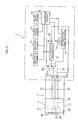
- a biosensor 1 shown in Fig. 1 and Fig. 2 is for a measurement of a blood glucose level, and is used as attached to an analyzing device 5 to be detailed later with reference to Fig. 4 .
- the biosensor 1 includes a substrate 20, a cover 40 laminated thereon, and a spacer 30 in between. These members 20, 30 and 40 form a capillary 10.
- the capillary 10 introduces the blood from an open end 11, allows the blood to move and stay within, under the principle of capillarity.
- the capillary 10 has another open end 12 in order to release gas from within the capillary 10 when introducing the blood into the capillary 10.
- the spacer 30 includes a pair of blocks 31, 32 opposed to each other. These blocks 31, 32 serve as electrodes for applying an AC voltage to the blood when measuring a resistance value of the blood.
- the blocks 31, 32 have upper surfaces 31a, 32a, lower surfaces 31b, 32b, and two side surfaces 31c, 32c and 31d, 32d respectively. Each block has a rectangular section, and the whole block is electrically conductive. Therefore, the electrodes for applying AC voltage to the blood are opposed to each other.
- the blocks 31, 32 can be formed of an electrically conductive polymer, a resin containing an electrically conductive filler, or a metal.
- the upper surfaces 31a, 32a and the lower surfaces 31b, 32b of the blocks 31, 32 are adhesive.
- the adhesiveness of the upper surfaces 31a, 32a and the lower surfaces 31b, 32b is used to bond the cover 40 onto the substrate 20.
- the substrate 20 is rectangular.
- the substrate 20 has an upper surface 20a formed with a first through fourth leads 21-24, each extending longitudinally of the substrate 20.
- the upper surface 20a of the substrate 20 is provided with a first and a second electrodes 25, 26 and a reactor 27.
- the reactor 27 is solid for example, and bridges the first and the second electrodes 25, 26.
- the reactor 27 includes, for example, an oxidation-reduction enzyme and an electron transfer material.
- the oxidation-reduction enzyme is provided by glucose oxidase or glucose dehydrogenase for example, whereas the electron transfer material is provided by potassium ferricyanide for example.
- the first and the second leads 21, 22 are for application of a voltage between the first and the second electrodes 25, 26.
- the first lead 21 is formed integrally with the first electrode 25, and the second lead 22 is formed integrally with the second electrode 26.
- the third and the fourth leads 23, 24 are for application of a voltage between the two blocks 31, 32.
- the third lead 23 is electrically connected to the block 31, and the fourth lead 24 is electrically connected to the block 32.
- the blocks 31, 32 are bonded to the substrate 20, via insulating layers 28, 29 respectively.
- the insulating layer 28 covers the first through the fourth leads 21-24 whereas the insulating layer 29 covers the first, the third and the fourth leads 21, 22, 23.
- the insulating layers 28, 29 are formed with through holes 28a, 29a.
- the blocks 31, 32 are connected to the third lead 23 or the fourth lead 24 via the through holes 28a, 29a. It should be noted however, that electrical connection between the blocks 31, 32 and the relevant third lead 23 or fourth lead 24 may be established any other way, without being limited by the use of the through holes 28a, 29a.
- capillarity allows the blood to come in from the open end 11 of the capillary 10 and move toward the open end 12 of the capillary 10. During this movement, the blood solves the reactor 27. The introduction of the blood continues until the blood reaches the open end 12, i.e. until the blood has filled the capillary 10. As a result, a liquid-phase reaction system is established inside the capillary 10.
- an analyzing device 5 includes a DC power source 50, an electric current value measuring division 51, a detecting division 52, a resistance value measuring division 53, an AC power source 54, a storing division 55, a controlling division 56, a calculating division 57 and a display division 58.
- the DC power source 50 applies a DC voltage between the first and the second electrodes 25, 26 via ends 21a, 22a of the first and the second leads 21, 22.
- the DC power source 50 is electrically connected with the ends 21a, 22a when the biosensor 1 is attached to the analyzing device 5.
- the DC power source 50 can be provided by a dry battery or a rechargeable battery.
- the electric current value measuring division 51 measures a value of responding current when the DC voltage is applied between the first and the second electrodes 25, 26.
- the detecting division 52 detects, after the biosensor 1 is attached to the analyzing device 5, if the blood has been supplied inside the capillary 10.
- the detecting division 52 checks whether or not an electric current value detected by the electric current value measuring division 51 exceeds a predetermined threshold value, thereby determining whether or not the blood has been introduced.
- the resistance value measuring division 53 measures a resistance value of the blood (the liquid-phase reaction system) held in the capillary 10. Resistance values of blood correlate with the amount of ionized component, i. e. percentage of blood cells (hematocrit value) in the blood. Therefore, by measuring a resistance value of the blood, a hematocrit value can be calculated and a level of influence from the hematocrit value can be estimated.
- the resistance value measuring division 53 includes a measuring circuit 53a shown in Fig. 5 .
- the measuring circuit 53a constitutes a Wheatstone bridge circuit together with the blocks 31, 32 and other members.
- the measuring circuit 53a includes three resisters R1-R3 and a balance detector D.
- the resistors R1, R2 are variable resistors.
- Blood held in the capillary 10 is represented by a resistor R4 in the figure.
- the measuring circuit 53a controls resistance values of the variable resistors R1, R2 so that there is no current flowing through the balance detector D, and a measurement is made for a blood resistance value on the basis of the controlled resistance values of the R1, R2 and a known value of the resistor R3.
- the blood resistance value r4 is given by the following formula, where resistance values of the resistors R1 through R4 are represented by r1 through r4 respectively.
- r ⁇ 4 r ⁇ 1 x r ⁇ 3 / r ⁇ 2
- circuits different from the measuring circuit 53a in Fig. 5 may be constructed for measurement of blood dielectric constant or electric conductivity, for calculation of hematocrit values or estimation of the level of influence from the hematocrit values.
- the AC power source 54 applies an AC voltage between the blocks 31, 32 and to the measuring circuit 53a.
- the application of the AC voltage between the two blocks 31, 32 is made via the ends 23a, 24a of the third and the fourth leads 23, 24.
- the storing division 55 stores a control program for controlling the DC power source 50, the AC power source 54 and so on, calibration curve data used in calculating blood glucose levels, a calculation program for calculating the glucose levels, a resistance calculation program for calculating the resistance value r4, correction data for the calculated value to be corrected on the basis of the given resistance value r4, and a correction program for correcting the calculated value.
- the calibration curve data or the correction data is stored in the form of lookup table which indicates a relationship between, for example, the value of responding current and the glucose level or between the resistance value r4 and the corrected value.
- the storing division 55 is provided by a ROM or a RAM.
- the controlling division 56 is provided by a CPU for example, and controls voltage application from the DC power source 50 and the AC power source 54, as well as operation of the divisions 52, 53 and 58.
- the calculating division 57 calculates a blood glucose level based on the responding current value measured by the electric current value measuring division 51, and corrects the calculated value, on the basis of the resistance value measured by the resistance value measuring division 53.
- the display division 58 displays the glucose level and other information, and is provided by a liquid crystal display for example.
- the biosensor 1 When measuring the blood sugar level, first, the biosensor 1 is attached to an analyzing device 5 (S1), and blood is introduced into the capillary 10 (S2).
- the controlling division 56 applies a DC voltage between the first and the second electrodes 25, 26 (S3).
- the electric current value measuring division 51 measures a value of responding current (S4).
- the measurement of the responding current value is made per 0.05-0.2 seconds for example.
- the detecting division 52 monitors the responding current values measured at the electric current value measuring division 51, and checks if the responding current value is not smaller than a threshold value (S5).
- the detecting division 52 determines that the blood has been introduced into the capillary 10. On the other hand, if the detecting division 52 sees that the responding current value is not greater than the threshold value (S5: NO), the detecting division 52 continues the check in Step S5. However, if the detecting division 52 continues to see the responding current value being smaller than the threshold value after a lapse of a predetermined duration of time (S5: NO), then the program may execute an error processing routine.
- a liquid-phase reaction system is established as described earlier.
- the glucose is oxidized while the electron transfer material is reduced.
- the electron transfer material is then oxidized when a DC voltage is applied, and a quantity of electrons released per a unit of time can be measured as a responding current value.
- the controlling division 56 checks if a predetermined amount of time (e.g. 5-30 seconds) has passed since the time when the responding current exceeded the threshold value (S6). This check in Step S6 is repeated until the controlling division 56 sees that the predetermined amount of time has passed (S6: YES).
- a predetermined amount of time e.g. 5-30 seconds
- the electric current value measuring division 51 measures the responding current value (S7), and the measured responding current value is converted into a voltage value (S8).
- the controlling division 56 applies an AC voltage between the blocks 31 and 32 (S9) while allowing the resistance value measuring division 53 to measure a blood resistance value (S10).
- the measurement of the resistance value is made after the detecting division 52 has detected introduction of the blood into the capillary 10.
- the calculating division 57 calculates a glucose concentration level (S11) with reference to the calibration curve data stored in the storing division 55 and based on the voltage value given by the conversion in Step S8. Then, the calculating division 57 corrects the calculated value, based on the resistance value measured by the resistance value measuring division 53. The correction of the calculated value is made by referring to the correction data stored in the storing division 55. The corrected value is displayed in the display division 58.
- S11 glucose concentration level
- the display division may display, in addition to the corrected value, a hematocrit value which can be calculated from the pre-correction calculation or from the resistance value. Alternatively, both of the pre-correction value and the hematocrit value may be displayed.
- a DC voltage can be applied to the biosensor 1 or between the first and the second electrodes 25, 26, whereas an AC voltage can be applied between the blocks 31, 32. Therefore, it is possible to measure a responding current value which is necessary for calculating the glucose level and a resistance value which is necessary for correcting the calculated value, with a single biosensor 1.
- This provides a cost advantage in measurement, to the extent that there is no need for separate preparation of an analyzing implement for measuring glucose level and an analyzing implement for measuring hematocrit value (resistance value). Further, this provides an improved operability to the extent that there is no need for replacing an analyzing implement necessary for glucose level measurement with an analyzing implement necessary for hematocrit value measurement in order to allow measurement of a hematocrit value.
- the biosensor 1 uses the blocks 31, 32 as electrodes for measuring a resistance value.
- the object of measurement i.e. the liquid-phase reaction system (the capillary 10) has an area of the section which is generally equal to an area of the opposed surface in the blocks (electrodes) 31, 32, making possible to make uniform distribution of the electric lines of force. This makes possible to measure a value of resistance in the liquid-phase reaction system (blood) at a high accuracy, leading to accurate measurement of a blood sugar level regardless of the hematocrit value.
- the first and the second leads 21, 22 used for the application of DC voltage to the first and the second electrode 25, 26 are also used for the application of AC voltage to the blocks 31, 32.
- an analyzing device 5' shown in Fig. 8 includes a switching division 59 for selection whether the first and the second leads 21, 22 are electrically connected to the DC power source 50 or to the AC power source 54.
- the switching division 59 has a switch 59a, so that the controlling division 56 controls the switch 59a thereby selecting different states of voltage application to the liquid-phase system.
- the switch 59a When measuring a responding current value, the switch 59a establishes connection with the electric current value measuring division 51, based on the control by the controlling division 56. This allows the DC power source 50 to apply a DC voltage to the liquid-phase system in the capillary 10, and a responding current value is measured by the electric current value measuring division 51.
- the switch 59a when measuring a resistance value, the switch 59a establishes connection with the resistance value measuring division 53, based on the control by the controlling division 56. This allows the AC power source 54 to apply an AC voltage to the liquid-phase system in the capillary 10, and a resistance value is measured by the resistance value measuring division 51.
- the blocks may be used for both measurements; the measurement of a resistance value of the specimen liquid and the measurement of a responding current value.
- portions representing the first and the second electrodes 25, 26 in the biosensor are covered by an electrically insulating layer.
- the present invention can also provide an analyzing implement only for a measurement of a resistance value of a specimen liquid (e.g. hematocrit value of blood).
- a specimen liquid e.g. hematocrit value of blood.
Description
- The present invention relates to a technology for measurement of at least one of the followings: a concentration level of a specific component in a specimen liquid (e.g. a glucose concentration in a body fluid such as blood), and a physical quantity correlating to an amount of coexisting component in the specimen liquid.
- For a purpose of measuring blood sugar levels privately at home and elsewhere, palm-size, portable blood sugar level testers are used widely. When measuring the blood sugar level with these handy-type blood sugar level testers, a biosensor which serves as an enzyme reaction field must be attached to the level tester, and then the blood must be supplied to the biosensor.
- Blood contains solid component (blood cells), and it is known that the amount of blood cells influences the measurement. In order to overcome this, some methods of measurement include steps of measuring the percentage of blood-cell component (hematocrit value) in the blood, and correcting the calculated blood sugar level in accordance with the hematocrit value.
- The hematocrit value can be measured, for example, by using methods disclosed in the Japanese Patent Laid-Open No.
11-118794 2002-55069 - The method disclosed in the Japanese Patent Laid-Open No.
11-118794 Fig. 9A and Fig. 9B , a hematocritvalue measuring element 9 includes asubstrate 90 formed with a pair ofelectrodes reagent 93 covering a pair of ends of theelectrodes - In a liquid phase such as blood, ions in the liquid serve as a carrier in electric charge transfer, and therefore in order to measure the level of electric conductivity of a liquid phase at a high accuracy, appropriate movement of the carrier is necessary between the electrodes. For this purpose, as shown in a conceptual drawing in
Fig. 10 , electric lines of force betweenelectrodes electrodes electrodes object liquid 97. - However, as clearly shown in
Fig. 9A and Fig. 9B , theelectrodes hematocrit measuring element 9 are spaced in the same plane, and therefore are not opposed to each other. This leads to an electric conductivity measurement based on a localized carrier movement due to non-uniform electric lines of force. As a result, it is impossible to accurately measure the electric conductivity, nor the hematocrit value derived therefrom. - In the above method, the hematocrit value measurement is made using the hematocrit
value measuring element 9, which is a separate implement from the biosensor implement which is for the measurement of blood sugar level. This poses another problem of increased cost in measurement incurred by the needs for two analyzing implements for a single measurement of the blood sugar level. Still another problem is inconvenience, i.e. the hematocrit value measurement using the hematocritvalue measuring element 9 and the blood sugar level measurement using the biosensor must be made separately by replacing the hematocritvalue measuring element 9 with the biosensor. - On the other hand, in the method disclosed in the Japanese Patent Laid-Open No.
2002-55069 - This method is primarily for a large apparatus such as one having a sampling probe, and is not a technique applicable to a combination of a handy-type blood sugar level tester and a biosensor.
- It is an object of the present invention to provide a simple method of reducing the influence of the hematocrit value, for accurate measurement of the concentration level.
- A first aspect of the present invention provides an analyzing implement including a capillary for movement of a specimen liquid comprising a substrate, a cover covering the substrate, and a spacer between the substrate and the cover, wherein the capillary is formed by the substrate, the cover and the spacer, wherein the spacer includes two blocks opposed to each other, characterized in that inner side surfaces of the two blocks forming the capillary are electrically conductive for providing a first and a second opposed electrodes, and wherein the two blocks are formed entirely of an electrically conductive material. The capillary has an inner surface provided with a first and a second opposed electrodes opposed to each other.
- The implement according to the present aspect includes, for example, a substrate, a cover covering the substrate, and a spacer between the substrate and the cover. In this case, the capillary is formed by the substrate, the cover and the spacer. Surfaces of the spacer forming the capillary are electrically conductive and provide the first and the second opposed electrodes.
- Each block may be formed of an electrically insulating resin containing an electrically conductive polymer or electrically conductive filler, or of a metal.
- Alternatively however, each block may include an electrically insulating part and an electrically conductive part, and the electrically conductive parts of the pair of spacers serve as the first and the second electrodes. The spacer may be a single member having a slit. In this case, the first and the second electrodes are provided in mutually opposing surfaces in the slit.
- Each block has, for example, a rectangular cross section defined by an upper surface, a lower surface and two side surfaces, with the upper surfaces and the lower surfaces being adhesive. In this case, the cover is bonded to the substrate via the two blocks. In other words, the spacer may serve as an adhesive layer for the cover to be bonded to the substrate.
- The first and the second opposed electrodes are for measurement of e.g. a dielectric constant, a resistance value or a physical quantity correlating to electrical conductivity of the specimen liquid.
- The analyzing implement according to the present aspect preferably further includes a first and a second measurement electrodes for measurement of a physical quantity correlating to concentration of a specific component of the specimen liquid.
- The analyzing implement according to the present aspect includes, for example, a first through a fourth leads formed on the substrate. Wherein The spacer is bonded to the substrate via an insulating layer, for example. The first and the second measurement electrodes are electrically connected to the first and the second leads respectively and the first and the second opposed electrodes are electrically connected to the third and the fourth leads respectively, via through holes formed in the insulating layer. On the other hand, the first and the second electrodes are formed on the substrate, and are electrically connected to the first and the second leads. This construction allows individual measurements, i.e. the measurement of the physical quantity correlating to e.g. dielectric coefficient using the first and the second opposed electrodes and the measurement of the physical quantity correlating to concentration of a specific component using the first and the second electrodes.
- The first and the second opposed electrodes can also be used in the measurement of a responding current value correlating to concentration of a specific component in the specimen liquid. In this case, the first and the second leads are formed on the substrate, wherein the spacer is bonded to the substrate via an insulating layer, the first and the second opposed electrodes are electrically connected to the first and the second leads respectively, via through holes formed in the insulating layer. Also disclosed is an analyzing device which uses an analyzing implement, for measurement of concentration of a specific component in the specimen liquid. The analyzing implement includes: a capillary for movement of the specimen liquid; a first and a second opposed electrodes opposed to each other in an inner surface of the capillary for measurement of a first physical quantity correlating to dielectric constant, resistance value or electric conductivity of the specimen liquid; and a first and a second electrodes for measurement of a second physical quantity correlating to concentration of a specific component in the specimen liquid. The analyzing device includes: DC voltage applying means for applying a DC voltage between the first and the second electrodes; AC voltage applying means for applying an AC voltage between the first and the second opposed electrodes; first measuring means for measuring the first physical quantity; second measuring means for measuring the second physical quantity; and calculating means for calculating a concentration level of a specific component in the specimen liquid based on the first physical quantity measured by the first measuring means.
-
-
Fig. 1 is an overall perspective view showing a biosensor according to a first embodiment of the present invention. -
Fig. 2 is an exploded perspective view of the biosensor inFig. 1 . -
Fig. 3 is a sectional view taken in lines III-III inFig. 1 . -
Fig. 4 is a plan view of the biosensor as attached to an analyzing device according to the first embodiment, and a block diagram of the analyzing device. -
Fig. 5 is a circuit diagram for describing a resistance value measuring circuit. -
Fig. 6 is a flowchart for describing a concentration level measuring operation. -
Fig. 7 is an exploded perspective view of a biosensor according to a third embodiment of the present invention. -
Fig. 8 is a plan view of the biosensor as attached to an analyzing device according to the third embodiment, and a block diagram of the analyzing device. -
Fig. 9A is an overall perspective view showing a conventional hematocrit measuring element, andFig. 9B is a sectional view taken in lines XB-XB inFig. 9A . -
Fig. 10 is a perspective view for describing an electric conductivity measuring system ideal for the liquid phase. - Now, a first mode of embodiment of the present invention will be described with reference to
Fig. 1 through Fig. 6 . - A
biosensor 1 shown inFig. 1 andFig. 2 is for a measurement of a blood glucose level, and is used as attached to ananalyzing device 5 to be detailed later with reference toFig. 4 . Thebiosensor 1 includes asubstrate 20, acover 40 laminated thereon, and aspacer 30 in between. Thesemembers open end 11, allows the blood to move and stay within, under the principle of capillarity. The capillary 10 has anotheropen end 12 in order to release gas from within the capillary 10 when introducing the blood into the capillary 10. - The
spacer 30 includes a pair ofblocks blocks blocks upper surfaces lower surfaces side surfaces blocks - The
upper surfaces lower surfaces blocks upper surfaces lower surfaces cover 40 onto thesubstrate 20. - The
substrate 20 is rectangular. Thesubstrate 20 has anupper surface 20a formed with a first through fourth leads 21-24, each extending longitudinally of thesubstrate 20. Further, theupper surface 20a of thesubstrate 20 is provided with a first and asecond electrodes reactor 27. Thereactor 27 is solid for example, and bridges the first and thesecond electrodes reactor 27 includes, for example, an oxidation-reduction enzyme and an electron transfer material. The oxidation-reduction enzyme is provided by glucose oxidase or glucose dehydrogenase for example, whereas the electron transfer material is provided by potassium ferricyanide for example. - The first and the second leads 21, 22 are for application of a voltage between the first and the
second electrodes first lead 21 is formed integrally with thefirst electrode 25, and thesecond lead 22 is formed integrally with thesecond electrode 26. - The third and the fourth leads 23, 24 are for application of a voltage between the two
blocks third lead 23 is electrically connected to theblock 31, and thefourth lead 24 is electrically connected to theblock 32. - The
blocks substrate 20, via insulatinglayers layer 28 covers the first through the fourth leads 21-24 whereas the insulatinglayer 29 covers the first, the third and the fourth leads 21, 22, 23. - The insulating layers 28, 29 are formed with through
holes blocks third lead 23 or thefourth lead 24 via the throughholes blocks third lead 23 orfourth lead 24 may be established any other way, without being limited by the use of the throughholes - According to the
biosensor 1, capillarity allows the blood to come in from theopen end 11 of the capillary 10 and move toward theopen end 12 of the capillary 10. During this movement, the blood solves thereactor 27. The introduction of the blood continues until the blood reaches theopen end 12, i.e. until the blood has filled the capillary 10. As a result, a liquid-phase reaction system is established inside the capillary 10. - As shown in
Fig. 4 , an analyzingdevice 5 includes aDC power source 50, an electric currentvalue measuring division 51, a detectingdivision 52, a resistancevalue measuring division 53, anAC power source 54, a storingdivision 55, a controllingdivision 56, a calculatingdivision 57 and adisplay division 58. - The
DC power source 50 applies a DC voltage between the first and thesecond electrodes ends DC power source 50 is electrically connected with theends biosensor 1 is attached to theanalyzing device 5. TheDC power source 50 can be provided by a dry battery or a rechargeable battery. - The electric current
value measuring division 51 measures a value of responding current when the DC voltage is applied between the first and thesecond electrodes - The detecting
division 52 detects, after thebiosensor 1 is attached to theanalyzing device 5, if the blood has been supplied inside the capillary 10. The detectingdivision 52 checks whether or not an electric current value detected by the electric currentvalue measuring division 51 exceeds a predetermined threshold value, thereby determining whether or not the blood has been introduced. - The resistance
value measuring division 53 measures a resistance value of the blood (the liquid-phase reaction system) held in the capillary 10. Resistance values of blood correlate with the amount of ionized component, i. e. percentage of blood cells (hematocrit value) in the blood. Therefore, by measuring a resistance value of the blood, a hematocrit value can be calculated and a level of influence from the hematocrit value can be estimated. The resistancevalue measuring division 53 includes a measuringcircuit 53a shown inFig. 5 . The measuringcircuit 53a constitutes a Wheatstone bridge circuit together with theblocks circuit 53a includes three resisters R1-R3 and a balance detector D. The resistors R1, R2 are variable resistors. Blood held in the capillary 10 is represented by a resistor R4 in the figure. The measuringcircuit 53a controls resistance values of the variable resistors R1, R2 so that there is no current flowing through the balance detector D, and a measurement is made for a blood resistance value on the basis of the controlled resistance values of the R1, R2 and a known value of the resistor R3. Specifically, the blood resistance value r4 is given by the following formula, where resistance values of the resistors R1 through R4 are represented by r1 through r4 respectively.
- Alternatively, circuits different from the measuring
circuit 53a inFig. 5 may be constructed for measurement of blood dielectric constant or electric conductivity, for calculation of hematocrit values or estimation of the level of influence from the hematocrit values. - The
AC power source 54 applies an AC voltage between theblocks circuit 53a. The application of the AC voltage between the twoblocks ends - The storing
division 55 stores a control program for controlling theDC power source 50, theAC power source 54 and so on, calibration curve data used in calculating blood glucose levels, a calculation program for calculating the glucose levels, a resistance calculation program for calculating the resistance value r4, correction data for the calculated value to be corrected on the basis of the given resistance value r4, and a correction program for correcting the calculated value. The calibration curve data or the correction data is stored in the form of lookup table which indicates a relationship between, for example, the value of responding current and the glucose level or between the resistance value r4 and the corrected value. The storingdivision 55 is provided by a ROM or a RAM. - The controlling
division 56 is provided by a CPU for example, and controls voltage application from theDC power source 50 and theAC power source 54, as well as operation of thedivisions - The calculating
division 57 calculates a blood glucose level based on the responding current value measured by the electric currentvalue measuring division 51, and corrects the calculated value, on the basis of the resistance value measured by the resistancevalue measuring division 53. - The
display division 58 displays the glucose level and other information, and is provided by a liquid crystal display for example. - Hereinafter, description will cover how a blood sugar level is measured by the
biosensor 1 and theanalyzing device 5, with reference toFig. 1 through Fig. 5 as well as a flowchart inFig. 6 . - When measuring the blood sugar level, first, the
biosensor 1 is attached to an analyzing device 5 (S1), and blood is introduced into the capillary 10 (S2). - Meanwhile, the controlling
division 56 applies a DC voltage between the first and thesecond electrodes 25, 26 (S3). At this time, the electric currentvalue measuring division 51 measures a value of responding current (S4). The measurement of the responding current value is made per 0.05-0.2 seconds for example. The detectingdivision 52 monitors the responding current values measured at the electric currentvalue measuring division 51, and checks if the responding current value is not smaller than a threshold value (S5). - When the detecting
division 52 sees that the responding current value is not smaller than the threshold value (S5: YES), the detectingdivision 52 determines that the blood has been introduced into the capillary 10. On the other hand, if the detectingdivision 52 sees that the responding current value is not greater than the threshold value (S5: NO), the detectingdivision 52 continues the check in Step S5. However, if the detectingdivision 52 continues to see the responding current value being smaller than the threshold value after a lapse of a predetermined duration of time (S5: NO), then the program may execute an error processing routine. - When blood is introduced into the capillary 10, a liquid-phase reaction system is established as described earlier. In this liquid-phase reaction system, the glucose is oxidized while the electron transfer material is reduced. The electron transfer material is then oxidized when a DC voltage is applied, and a quantity of electrons released per a unit of time can be measured as a responding current value.
- When the detecting
division 52 has determined that the responding current value is not smaller than the threshold value (S5: YES), then the controllingdivision 56 checks if a predetermined amount of time (e.g. 5-30 seconds) has passed since the time when the responding current exceeded the threshold value (S6). This check in Step S6 is repeated until the controllingdivision 56 sees that the predetermined amount of time has passed (S6: YES). - When the controlling
division 56 sees that the predetermined amount of time has passed (S6: YES), the electric currentvalue measuring division 51 measures the responding current value (S7), and the measured responding current value is converted into a voltage value (S8). - Meanwhile, in parallel to the application of the DC voltage, the controlling
division 56 applies an AC voltage between theblocks 31 and 32 (S9) while allowing the resistancevalue measuring division 53 to measure a blood resistance value (S10). The measurement of the resistance value is made after the detectingdivision 52 has detected introduction of the blood into the capillary 10. - The calculating
division 57 calculates a glucose concentration level (S11) with reference to the calibration curve data stored in thestoring division 55 and based on the voltage value given by the conversion in Step S8. Then, the calculatingdivision 57 corrects the calculated value, based on the resistance value measured by the resistancevalue measuring division 53. The correction of the calculated value is made by referring to the correction data stored in thestoring division 55. The corrected value is displayed in thedisplay division 58. - The display division may display, in addition to the corrected value, a hematocrit value which can be calculated from the pre-correction calculation or from the resistance value. Alternatively, both of the pre-correction value and the hematocrit value may be displayed.
- According to the present embodiment, a DC voltage can be applied to the
biosensor 1 or between the first and thesecond electrodes blocks single biosensor 1. This provides a cost advantage in measurement, to the extent that there is no need for separate preparation of an analyzing implement for measuring glucose level and an analyzing implement for measuring hematocrit value (resistance value). Further, this provides an improved operability to the extent that there is no need for replacing an analyzing implement necessary for glucose level measurement with an analyzing implement necessary for hematocrit value measurement in order to allow measurement of a hematocrit value. - The
biosensor 1 uses theblocks - According to the
biosensor 1, application of a DC voltage between the first and thesecond electrodes blocks biosensor 1, the amount of time necessary for measurement is not long even if a measurement is made for a resistance value in addition to a responding current. - Next, a second embodiment of the present invention will be described with reference to
Fig. 7 andFig. 8 . It should be noted that inFig. 7 andFig. 8 , elements identical with those used in the biosensor or the analyzing device according to the first embodiment are given the same alpha-numeric codes, and their description will not be repeated hereinafter. - In a
biosensor 1" shown inFig. 7 , the first and the second leads 21, 22 used for the application of DC voltage to the first and thesecond electrode blocks - Correspondingly, an analyzing device 5' shown in
Fig. 8 includes aswitching division 59 for selection whether the first and the second leads 21, 22 are electrically connected to theDC power source 50 or to theAC power source 54. The switchingdivision 59 has aswitch 59a, so that the controllingdivision 56 controls theswitch 59a thereby selecting different states of voltage application to the liquid-phase system. - When measuring a responding current value, the
switch 59a establishes connection with the electric currentvalue measuring division 51, based on the control by the controllingdivision 56. This allows theDC power source 50 to apply a DC voltage to the liquid-phase system in the capillary 10, and a responding current value is measured by the electric currentvalue measuring division 51. On the other hand, when measuring a resistance value, theswitch 59a establishes connection with the resistancevalue measuring division 53, based on the control by the controllingdivision 56. This allows theAC power source 54 to apply an AC voltage to the liquid-phase system in the capillary 10, and a resistance value is measured by the resistancevalue measuring division 51. - According to the present invention, the blocks (opposed electrodes) may be used for both measurements; the measurement of a resistance value of the specimen liquid and the measurement of a responding current value. In this case, portions representing the first and the
second electrodes - The present invention can also provide an analyzing implement only for a measurement of a resistance value of a specimen liquid (e.g. hematocrit value of blood). Such an implement is simply the biosensor in
Fig. 7 less the reaction division.
Claims (9)
- An analyzing implement including a capillary (10) for movement of a specimen liquid, comprising a substrate (20), a cover (40) covering the substrate (20), and a spacer (30) between the substrate (20) and the cover (40),
wherein the capillary (10) is formed by the substrate (20), the cover (40) and the spacer (30),
wherein the spacer (30) includes two blocks (31, 32) opposed to each other,
characterized in that inner side surfaces (31c, 32c) of the two blocks (31, 32) forming the capillary (10) are conductive for providing first and second opposed electrodes, and
wherein the two blocks (31, 32) are formed entirely of an electrically conductive material. - The analyzing implement according to Claim 1, wherein the two blocks (31, 32) are formed of an electrically insulating resin containing an electrically conductive polymer or an electrically conductive filler.
- The analyzing implement according to Claim 1, wherein each of the two blocks (31, 32) has a rectangular cross section defined by an upper surface (31a, 32a), a lower surface (31b, 32b) and two side surfaces (31c, 31d, 32c),
the upper surfaces (31a, 32a) and the lower surfaces (31b, 32b) being adhesive, the cover (40) being bonded to the substrate (20) via the two blocks (31, 32). - The analyzing implement according to Claim 1, wherein the first and the second opposed electrodes (31, 32) are for measurement of a dielectric constant, a resistance value or a physical quantity correlating to electrical conductivity of the specimen liquid.
- The analyzing implement according to Claim 4, further comprising a first and a second measurement electrodes (25, 26) for measurement of a physical quantity correlating to concentration of a specific component of the specimen liquid.
- The analyzing implement according to Claim 5, further comprising a first lead (21) electrically connected to the first opposed electrode (31) and to the first measurement electrode (25), and a second lead (22) connected to the second opposed electrode (32) and to the second measurement electrode (26).
- The analyzing implement according to Claim 4, comprising a first through a fourth leads (21-24) formed on the substrate (20),
wherein the spacer (30) being bonded to the substrate (20) via an insulating layer (28, 29),
the first and the second measurement electrodes (25, 26) being formed on the substrate (20) and electrically connected to the first and the second leads (21, 22) respectively,
the first and the second opposed electrodes (31, 32) being electrically connected to the third and the fourth leads (23, 24) respectively, via through holes (28a, 29a) formed in the insulating layer (28, 29). - The analyzing implement according to Claim 1, wherein the first and the second opposed electrodes (31, 32) serve in measurement of a responding current value correlating to concentration of a specific component in the specimen liquid.
- The analyzing implement according to Claim 8, comprising a first and a second leads (21, 22) formed on the substrate (20),
wherein the spacer (30) being bonded to the substrate (20) via an insulating layer (28, 29),
the first and the second opposed electrodes (31, 32) being electrically connected to the first and the second leads (21, 22) respectively, via through holes (28a, 29a) formed in the insulating layer (28, 29).
Applications Claiming Priority (3)
| Application Number | Priority Date | Filing Date | Title |
|---|---|---|---|
| JP2001218021 | 2001-07-18 | ||
| JP2001218021 | 2001-07-18 | ||
| PCT/JP2002/007239 WO2003008956A1 (en) | 2001-07-18 | 2002-07-16 | Implement and device for analysis |
Publications (3)
| Publication Number | Publication Date |
|---|---|
| EP1411348A1 EP1411348A1 (en) | 2004-04-21 |
| EP1411348A4 EP1411348A4 (en) | 2005-04-20 |
| EP1411348B1 true EP1411348B1 (en) | 2015-11-11 |
Family
ID=19052258
Family Applications (1)
| Application Number | Title | Priority Date | Filing Date |
|---|---|---|---|
| EP02746114.4A Expired - Lifetime EP1411348B1 (en) | 2001-07-18 | 2002-07-16 | Implement and device for analysis |
Country Status (5)
| Country | Link |
|---|---|
| US (1) | US7351375B2 (en) |
| EP (1) | EP1411348B1 (en) |
| JP (1) | JP4214275B2 (en) |
| CN (1) | CN100401045C (en) |
| WO (1) | WO2003008956A1 (en) |
Cited By (1)
| Publication number | Priority date | Publication date | Assignee | Title |
|---|---|---|---|---|
| US9410917B2 (en) | 2004-02-06 | 2016-08-09 | Ascensia Diabetes Care Holdings Ag | Method of using a biosensor |
Families Citing this family (73)
| Publication number | Priority date | Publication date | Assignee | Title |
|---|---|---|---|---|
| US6391005B1 (en) | 1998-03-30 | 2002-05-21 | Agilent Technologies, Inc. | Apparatus and method for penetration with shaft having a sensor for sensing penetration depth |
| US8641644B2 (en) | 2000-11-21 | 2014-02-04 | Sanofi-Aventis Deutschland Gmbh | Blood testing apparatus having a rotatable cartridge with multiple lancing elements and testing means |
| US9427532B2 (en) | 2001-06-12 | 2016-08-30 | Sanofi-Aventis Deutschland Gmbh | Tissue penetration device |
| US7041068B2 (en) | 2001-06-12 | 2006-05-09 | Pelikan Technologies, Inc. | Sampling module device and method |
| US7033371B2 (en) | 2001-06-12 | 2006-04-25 | Pelikan Technologies, Inc. | Electric lancet actuator |
| US7344507B2 (en) | 2002-04-19 | 2008-03-18 | Pelikan Technologies, Inc. | Method and apparatus for lancet actuation |
| US9795747B2 (en) | 2010-06-02 | 2017-10-24 | Sanofi-Aventis Deutschland Gmbh | Methods and apparatus for lancet actuation |
| CA2448902C (en) | 2001-06-12 | 2010-09-07 | Pelikan Technologies, Inc. | Self optimizing lancing device with adaptation means to temporal variations in cutaneous properties |
| US7981056B2 (en) | 2002-04-19 | 2011-07-19 | Pelikan Technologies, Inc. | Methods and apparatus for lancet actuation |
| US9226699B2 (en) | 2002-04-19 | 2016-01-05 | Sanofi-Aventis Deutschland Gmbh | Body fluid sampling module with a continuous compression tissue interface surface |
| US8337419B2 (en) | 2002-04-19 | 2012-12-25 | Sanofi-Aventis Deutschland Gmbh | Tissue penetration device |
| US7749174B2 (en) | 2001-06-12 | 2010-07-06 | Pelikan Technologies, Inc. | Method and apparatus for lancet launching device intergrated onto a blood-sampling cartridge |
| US7229458B2 (en) | 2002-04-19 | 2007-06-12 | Pelikan Technologies, Inc. | Method and apparatus for penetrating tissue |
| US7547287B2 (en) | 2002-04-19 | 2009-06-16 | Pelikan Technologies, Inc. | Method and apparatus for penetrating tissue |
| US7976476B2 (en) | 2002-04-19 | 2011-07-12 | Pelikan Technologies, Inc. | Device and method for variable speed lancet |
| US7297122B2 (en) | 2002-04-19 | 2007-11-20 | Pelikan Technologies, Inc. | Method and apparatus for penetrating tissue |
| US8579831B2 (en) | 2002-04-19 | 2013-11-12 | Sanofi-Aventis Deutschland Gmbh | Method and apparatus for penetrating tissue |
| US9314194B2 (en) | 2002-04-19 | 2016-04-19 | Sanofi-Aventis Deutschland Gmbh | Tissue penetration device |
| US9795334B2 (en) | 2002-04-19 | 2017-10-24 | Sanofi-Aventis Deutschland Gmbh | Method and apparatus for penetrating tissue |
| US8221334B2 (en) | 2002-04-19 | 2012-07-17 | Sanofi-Aventis Deutschland Gmbh | Method and apparatus for penetrating tissue |
| US8784335B2 (en) | 2002-04-19 | 2014-07-22 | Sanofi-Aventis Deutschland Gmbh | Body fluid sampling device with a capacitive sensor |
| US7331931B2 (en) | 2002-04-19 | 2008-02-19 | Pelikan Technologies, Inc. | Method and apparatus for penetrating tissue |
| US8372016B2 (en) | 2002-04-19 | 2013-02-12 | Sanofi-Aventis Deutschland Gmbh | Method and apparatus for body fluid sampling and analyte sensing |
| US7491178B2 (en) | 2002-04-19 | 2009-02-17 | Pelikan Technologies, Inc. | Method and apparatus for penetrating tissue |
| US7226461B2 (en) | 2002-04-19 | 2007-06-05 | Pelikan Technologies, Inc. | Method and apparatus for a multi-use body fluid sampling device with sterility barrier release |
| US7892183B2 (en) | 2002-04-19 | 2011-02-22 | Pelikan Technologies, Inc. | Method and apparatus for body fluid sampling and analyte sensing |
| US9248267B2 (en) | 2002-04-19 | 2016-02-02 | Sanofi-Aventis Deustchland Gmbh | Tissue penetration device |
| US7901362B2 (en) | 2002-04-19 | 2011-03-08 | Pelikan Technologies, Inc. | Method and apparatus for penetrating tissue |
| US7674232B2 (en) | 2002-04-19 | 2010-03-09 | Pelikan Technologies, Inc. | Method and apparatus for penetrating tissue |
| US8360992B2 (en) | 2002-04-19 | 2013-01-29 | Sanofi-Aventis Deutschland Gmbh | Method and apparatus for penetrating tissue |
| US8267870B2 (en) | 2002-04-19 | 2012-09-18 | Sanofi-Aventis Deutschland Gmbh | Method and apparatus for body fluid sampling with hybrid actuation |
| US7232451B2 (en) | 2002-04-19 | 2007-06-19 | Pelikan Technologies, Inc. | Method and apparatus for penetrating tissue |
| US7909778B2 (en) | 2002-04-19 | 2011-03-22 | Pelikan Technologies, Inc. | Method and apparatus for penetrating tissue |
| US8702624B2 (en) | 2006-09-29 | 2014-04-22 | Sanofi-Aventis Deutschland Gmbh | Analyte measurement device with a single shot actuator |
| US8574895B2 (en) | 2002-12-30 | 2013-11-05 | Sanofi-Aventis Deutschland Gmbh | Method and apparatus using optical techniques to measure analyte levels |
| ES2347248T3 (en) | 2003-05-30 | 2010-10-27 | Pelikan Technologies Inc. | PROCEDURE AND APPLIANCE FOR FLUID INJECTION. |
| WO2004107964A2 (en) | 2003-06-06 | 2004-12-16 | Pelikan Technologies, Inc. | Blood harvesting device with electronic control |
| WO2006001797A1 (en) | 2004-06-14 | 2006-01-05 | Pelikan Technologies, Inc. | Low pain penetrating |
| US7723099B2 (en) | 2003-09-10 | 2010-05-25 | Abbott Point Of Care Inc. | Immunoassay device with immuno-reference electrode |
| WO2005033659A2 (en) | 2003-09-29 | 2005-04-14 | Pelikan Technologies, Inc. | Method and apparatus for an improved sample capture device |
| US9351680B2 (en) | 2003-10-14 | 2016-05-31 | Sanofi-Aventis Deutschland Gmbh | Method and apparatus for a variable user interface |
| CN100472210C (en) * | 2003-12-04 | 2009-03-25 | 松下电器产业株式会社 | Blood component measuring method, sensor used therefor, and measuring instrument |
| EP1707953B1 (en) | 2003-12-04 | 2015-07-01 | Panasonic Healthcare Holdings Co., Ltd. | Method of measuring hematocrit (Hct) |
| US7822454B1 (en) | 2005-01-03 | 2010-10-26 | Pelikan Technologies, Inc. | Fluid sampling device with improved analyte detecting member configuration |
| EP1706026B1 (en) | 2003-12-31 | 2017-03-01 | Sanofi-Aventis Deutschland GmbH | Method and apparatus for improving fluidic flow and sample capture |
| JP4717637B2 (en) * | 2004-01-07 | 2011-07-06 | アークレイ株式会社 | Analytical tool and analytical method with improved arrangement of reagent part |
| CA2559297C (en) * | 2004-04-19 | 2012-05-22 | Matsushita Electric Industrial Co., Ltd. | Method for measuring blood components and biosensor and measuring instrument for use therein |
| BRPI0510779A (en) * | 2004-05-14 | 2007-11-20 | Bayer Healthcare Llc | methods for performing hematocrit adjustment in assays and devices for same |
| US8828203B2 (en) | 2004-05-20 | 2014-09-09 | Sanofi-Aventis Deutschland Gmbh | Printable hydrogels for biosensors |
| US9775553B2 (en) | 2004-06-03 | 2017-10-03 | Sanofi-Aventis Deutschland Gmbh | Method and apparatus for a fluid sampling device |
| US9820684B2 (en) | 2004-06-03 | 2017-11-21 | Sanofi-Aventis Deutschland Gmbh | Method and apparatus for a fluid sampling device |
| AU2005295106B2 (en) | 2004-10-12 | 2012-03-15 | Bayer Healthcare Llc | Concentration determination in a diffusion barrier layer |
| US8652831B2 (en) | 2004-12-30 | 2014-02-18 | Sanofi-Aventis Deutschland Gmbh | Method and apparatus for analyte measurement test time |
| JP2006223501A (en) * | 2005-02-17 | 2006-08-31 | Japan Health Science Foundation | Biological fluid osmotic pressure sensor |
| US7323887B2 (en) * | 2005-04-01 | 2008-01-29 | Rosemount Analytical Inc. | Conductivity sensor and manufacturing method therefor |
| CN103558284B (en) | 2005-07-20 | 2017-04-12 | 安晟信医疗科技控股公司 | Gated amperometry |
| CN101273266B (en) | 2005-09-30 | 2012-08-22 | 拜尔健康护理有限责任公司 | Gated voltammetry |
| JP4814952B2 (en) * | 2006-10-19 | 2011-11-16 | パナソニック株式会社 | Method for measuring hematocrit value of blood sample, method for measuring concentration of analyte in blood sample, sensor chip and sensor unit |
| US8691072B2 (en) * | 2006-10-19 | 2014-04-08 | Panasonic Corporation | Method for measuring hematocrit value of blood sample, method for measuring concentration of analyte in blood sample, sensor chip and sensor unit |
| ES2825036T3 (en) | 2006-10-24 | 2021-05-14 | Ascensia Diabetes Care Holdings Ag | Transient decay amperometry |
| CN101755043B (en) | 2007-07-23 | 2013-06-19 | 埃葛梅崔克斯股份有限公司 | Electrochemical test strip |
| WO2009076302A1 (en) | 2007-12-10 | 2009-06-18 | Bayer Healthcare Llc | Control markers for auto-detection of control solution and methods of use |
| WO2009126900A1 (en) | 2008-04-11 | 2009-10-15 | Pelikan Technologies, Inc. | Method and apparatus for analyte detecting device |
| US8178313B2 (en) * | 2008-06-24 | 2012-05-15 | Lifescan, Inc. | Method for determining an analyte in a bodily fluid |
| US9375169B2 (en) | 2009-01-30 | 2016-06-28 | Sanofi-Aventis Deutschland Gmbh | Cam drive for managing disposable penetrating member actions with a single motor and motor and control system |
| IT1394915B1 (en) | 2009-07-21 | 2012-07-20 | Pietro Fiorentini Spa | DEVICE FOR THE MEASUREMENT OF ELECTRICAL FLUID AND METHOD PROPERTIES TO MEASURE THESE ELECTRICAL PROPERTIES |
| FI123318B (en) * | 2009-09-29 | 2013-02-28 | Outotec Oyj | Three-dimensional imaging of a mass flow |
| FR2950972A1 (en) * | 2009-10-02 | 2011-04-08 | Commissariat Energie Atomique | METHOD AND CELL FOR MEASURING THE GLOBAL ION CONCENTRATION OF A BODY FLUID |
| US8965476B2 (en) | 2010-04-16 | 2015-02-24 | Sanofi-Aventis Deutschland Gmbh | Tissue penetration device |
| US20130084590A1 (en) * | 2011-09-30 | 2013-04-04 | Lifescan Scotland Ltd. | Analytical test strip with bodily fluid phase-shift measurement electrodes |
| JP6246211B2 (en) * | 2012-09-07 | 2017-12-13 | シラグ・ゲーエムベーハー・インターナショナルCilag GMBH International | Electrochemical sensors and methods for their manufacture |
| DE102013227125B4 (en) | 2013-12-23 | 2016-04-07 | Senslab-Gesellschaft Zur Entwicklung Und Herstellung Bioelektrochemischer Sensoren Mbh | A method for determining a hematocrit-dependent measurement signal in the determination of an analyte from whole blood using single-use enzymatic-voltammetric sensors |
| KR102016393B1 (en) * | 2017-10-26 | 2019-09-10 | (주)오상헬스케어 | Method and apparatus of measuring concentration of sample |
Family Cites Families (15)
| Publication number | Priority date | Publication date | Assignee | Title |
|---|---|---|---|---|
| JP2665806B2 (en) | 1989-09-13 | 1997-10-22 | 株式会社豊田中央研究所 | Hematocrit measuring device |
| US5264103A (en) | 1991-10-18 | 1993-11-23 | Matsushita Electric Industrial Co., Ltd. | Biosensor and a method for measuring a concentration of a substrate in a sample |
| US5580435A (en) * | 1994-06-10 | 1996-12-03 | The Board Of Trustees Of The Leland Stanford Junior University | System for detecting components of a sample in electrophoretic separation |
| US5494562A (en) * | 1994-06-27 | 1996-02-27 | Ciba Corning Diagnostics Corp. | Electrochemical sensors |
| JPH10170471A (en) * | 1996-12-06 | 1998-06-26 | Casio Comput Co Ltd | Biosensor |
| JPH10311814A (en) | 1997-05-09 | 1998-11-24 | Daikin Ind Ltd | Method for correcting output of differential sensor and concentration measuring device |
| JP3810534B2 (en) | 1997-10-17 | 2006-08-16 | 松下電器産業株式会社 | Hematocrit value measuring element and method for measuring hematocrit value |
| BR9814386B1 (en) | 1997-12-22 | 2009-08-11 | apparatus and methods for determining the concentration of a medically significant component of a biological fluid. | |
| JP3874321B2 (en) * | 1998-06-11 | 2007-01-31 | 松下電器産業株式会社 | Biosensor |
| JP2000258382A (en) * | 1999-03-05 | 2000-09-22 | Arkray Inc | Specimen small-quantity-type bio sensor |
| KR100340174B1 (en) * | 1999-04-06 | 2002-06-12 | 이동준 | Electrochemical Biosensor Test Strip, Fabrication Method Thereof and Electrochemical Biosensor |
| WO2001013127A1 (en) * | 1999-08-11 | 2001-02-22 | Asahi Kasei Kabushiki Kaisha | Analyzing cartridge and liquid feed control device |
| WO2001036954A1 (en) * | 1999-11-15 | 2001-05-25 | Arkray, Inc. | Biosensor |
| US6676815B1 (en) * | 1999-12-30 | 2004-01-13 | Roche Diagnostics Corporation | Cell for electrochemical analysis of a sample |
| JP4580074B2 (en) | 2000-08-09 | 2010-11-10 | 株式会社堀場製作所 | Blood measuring device, whole blood immunoassay device, and sampling probe used in these devices |
-
2002
- 2002-07-16 US US10/483,442 patent/US7351375B2/en active Active
- 2002-07-16 JP JP2003514249A patent/JP4214275B2/en not_active Expired - Lifetime
- 2002-07-16 WO PCT/JP2002/007239 patent/WO2003008956A1/en active Application Filing
- 2002-07-16 EP EP02746114.4A patent/EP1411348B1/en not_active Expired - Lifetime
- 2002-07-16 CN CNB028143442A patent/CN100401045C/en not_active Expired - Lifetime
Cited By (1)
| Publication number | Priority date | Publication date | Assignee | Title |
|---|---|---|---|---|
| US9410917B2 (en) | 2004-02-06 | 2016-08-09 | Ascensia Diabetes Care Holdings Ag | Method of using a biosensor |
Also Published As
| Publication number | Publication date |
|---|---|
| WO2003008956A1 (en) | 2003-01-30 |
| JP4214275B2 (en) | 2009-01-28 |
| US7351375B2 (en) | 2008-04-01 |
| US20040173458A1 (en) | 2004-09-09 |
| JPWO2003008956A1 (en) | 2004-11-11 |
| CN1531649A (en) | 2004-09-22 |
| EP1411348A1 (en) | 2004-04-21 |
| CN100401045C (en) | 2008-07-09 |
| EP1411348A4 (en) | 2005-04-20 |
Similar Documents
| Publication | Publication Date | Title |
|---|---|---|
| EP1411348B1 (en) | Implement and device for analysis | |
| EP1447660B1 (en) | Specific component concentration measuring method and concentration measuring instrument | |
| AU2010202056B2 (en) | Method and apparatus for assay of electrochemical properties | |
| US7491310B2 (en) | Concentration measuring method and concentration measuring apparatus | |
| EP2149792B1 (en) | Sample analyzing method | |
| EP2341341B1 (en) | Electrochemical test strip, electrochemical test system, and measurement method using the same | |
| EP2539711B1 (en) | Capacitance detection in electrochemical assay | |
| CA2521370A1 (en) | Biosensor system | |
| KR102286694B1 (en) | Methods for correcting uncompensated resistances in conductive elements of biosensors, as well as devices and systems | |
| US20210199615A1 (en) | Electrochemical-based analytical test strip with electrode voltage sensing connections and hand-held test meter for use therewith | |
| JPWO2009057793A1 (en) | Analysis tool, analyzer, sample shortage detection method and sample analysis method | |
| EP2679992B1 (en) | Biological sample measuring system | |
| US20200033287A1 (en) | Method of operation of a meter | |
| AU2012204094B2 (en) | Method and apparatus for assay of electrochemical properties | |
| MXPA06008846A (en) | Electrochemical biosensor | |
| AU2013204819A1 (en) | Method and apparatus for assay of electrochemical properties |
Legal Events
| Date | Code | Title | Description |
|---|---|---|---|
| PUAI | Public reference made under article 153(3) epc to a published international application that has entered the european phase |
Free format text: ORIGINAL CODE: 0009012 |
|
| 17P | Request for examination filed |
Effective date: 20040106 |
|
| AK | Designated contracting states |
Kind code of ref document: A1 Designated state(s): AT BE BG CH CY CZ DE DK EE ES FI FR GB GR IE IT LI LU MC NL PT SE SK TR |
|
| AX | Request for extension of the european patent |
Extension state: AL LT LV MK RO SI |
|
| A4 | Supplementary search report drawn up and despatched |
Effective date: 20050303 |
|
| RIC1 | Information provided on ipc code assigned before grant |
Ipc: 7C 12Q 1/00 B Ipc: 7G 01N 33/487 B Ipc: 7G 01N 27/06 A |
|
| 17Q | First examination report despatched |
Effective date: 20090126 |
|
| GRAP | Despatch of communication of intention to grant a patent |
Free format text: ORIGINAL CODE: EPIDOSNIGR1 |
|
| GRAJ | Information related to disapproval of communication of intention to grant by the applicant or resumption of examination proceedings by the epo deleted |
Free format text: ORIGINAL CODE: EPIDOSDIGR1 |
|
| GRAP | Despatch of communication of intention to grant a patent |
Free format text: ORIGINAL CODE: EPIDOSNIGR1 |
|
| RIC1 | Information provided on ipc code assigned before grant |
Ipc: G01N 27/327 20060101ALI20150617BHEP Ipc: C12Q 1/00 20060101ALI20150617BHEP Ipc: G01N 33/487 20060101ALI20150617BHEP Ipc: G01N 27/06 20060101AFI20150617BHEP |
|
| INTG | Intention to grant announced |
Effective date: 20150702 |
|
| RIN1 | Information on inventor provided before grant (corrected) |
Inventor name: KADO, KOTARO Inventor name: KATSUKI, KOJI Inventor name: NODA, NORIMASA |
|
| INTG | Intention to grant announced |
Effective date: 20150715 |
|
| RAP1 | Party data changed (applicant data changed or rights of an application transferred) |
Owner name: ARKRAY, INC. |
|
| GRAS | Grant fee paid |
Free format text: ORIGINAL CODE: EPIDOSNIGR3 |
|
| GRAA | (expected) grant |
Free format text: ORIGINAL CODE: 0009210 |
|
| AK | Designated contracting states |
Kind code of ref document: B1 Designated state(s): AT BE BG CH CY CZ DE DK EE ES FI FR GB GR IE IT LI LU MC NL PT SE SK TR |
|
| REG | Reference to a national code |
Ref country code: GB Ref legal event code: FG4D |
|
| REG | Reference to a national code |
Ref country code: CH Ref legal event code: EP |
|
| REG | Reference to a national code |
Ref country code: IE Ref legal event code: FG4D |
|
| REG | Reference to a national code |
Ref country code: AT Ref legal event code: REF Ref document number: 760705 Country of ref document: AT Kind code of ref document: T Effective date: 20151215 |
|
| REG | Reference to a national code |
Ref country code: DE Ref legal event code: R096 Ref document number: 60247617 Country of ref document: DE |
|
| REG | Reference to a national code |
Ref country code: NL Ref legal event code: MP Effective date: 20160211 |
|
| REG | Reference to a national code |
Ref country code: AT Ref legal event code: MK05 Ref document number: 760705 Country of ref document: AT Kind code of ref document: T Effective date: 20151111 |
|
| PG25 | Lapsed in a contracting state [announced via postgrant information from national office to epo] |
Ref country code: ES Free format text: LAPSE BECAUSE OF FAILURE TO SUBMIT A TRANSLATION OF THE DESCRIPTION OR TO PAY THE FEE WITHIN THE PRESCRIBED TIME-LIMIT Effective date: 20151111 Ref country code: NL Free format text: LAPSE BECAUSE OF FAILURE TO SUBMIT A TRANSLATION OF THE DESCRIPTION OR TO PAY THE FEE WITHIN THE PRESCRIBED TIME-LIMIT Effective date: 20151111 |
|
| PG25 | Lapsed in a contracting state [announced via postgrant information from national office to epo] |
Ref country code: AT Free format text: LAPSE BECAUSE OF FAILURE TO SUBMIT A TRANSLATION OF THE DESCRIPTION OR TO PAY THE FEE WITHIN THE PRESCRIBED TIME-LIMIT Effective date: 20151111 Ref country code: GR Free format text: LAPSE BECAUSE OF FAILURE TO SUBMIT A TRANSLATION OF THE DESCRIPTION OR TO PAY THE FEE WITHIN THE PRESCRIBED TIME-LIMIT Effective date: 20160212 Ref country code: PT Free format text: LAPSE BECAUSE OF FAILURE TO SUBMIT A TRANSLATION OF THE DESCRIPTION OR TO PAY THE FEE WITHIN THE PRESCRIBED TIME-LIMIT Effective date: 20160311 Ref country code: SE Free format text: LAPSE BECAUSE OF FAILURE TO SUBMIT A TRANSLATION OF THE DESCRIPTION OR TO PAY THE FEE WITHIN THE PRESCRIBED TIME-LIMIT Effective date: 20151111 Ref country code: FI Free format text: LAPSE BECAUSE OF FAILURE TO SUBMIT A TRANSLATION OF THE DESCRIPTION OR TO PAY THE FEE WITHIN THE PRESCRIBED TIME-LIMIT Effective date: 20151111 |
|
| REG | Reference to a national code |
Ref country code: FR Ref legal event code: PLFP Year of fee payment: 15 |
|
| PG25 | Lapsed in a contracting state [announced via postgrant information from national office to epo] |
Ref country code: CZ Free format text: LAPSE BECAUSE OF FAILURE TO SUBMIT A TRANSLATION OF THE DESCRIPTION OR TO PAY THE FEE WITHIN THE PRESCRIBED TIME-LIMIT Effective date: 20151111 |
|
| REG | Reference to a national code |
Ref country code: DE Ref legal event code: R097 Ref document number: 60247617 Country of ref document: DE |
|
| PG25 | Lapsed in a contracting state [announced via postgrant information from national office to epo] |
Ref country code: DK Free format text: LAPSE BECAUSE OF FAILURE TO SUBMIT A TRANSLATION OF THE DESCRIPTION OR TO PAY THE FEE WITHIN THE PRESCRIBED TIME-LIMIT Effective date: 20151111 Ref country code: EE Free format text: LAPSE BECAUSE OF FAILURE TO SUBMIT A TRANSLATION OF THE DESCRIPTION OR TO PAY THE FEE WITHIN THE PRESCRIBED TIME-LIMIT Effective date: 20151111 Ref country code: SK Free format text: LAPSE BECAUSE OF FAILURE TO SUBMIT A TRANSLATION OF THE DESCRIPTION OR TO PAY THE FEE WITHIN THE PRESCRIBED TIME-LIMIT Effective date: 20151111 |
|
| PLBE | No opposition filed within time limit |
Free format text: ORIGINAL CODE: 0009261 |
|
| STAA | Information on the status of an ep patent application or granted ep patent |
Free format text: STATUS: NO OPPOSITION FILED WITHIN TIME LIMIT |
|
| 26N | No opposition filed |
Effective date: 20160812 |
|
| PG25 | Lapsed in a contracting state [announced via postgrant information from national office to epo] |
Ref country code: BE Free format text: LAPSE BECAUSE OF FAILURE TO SUBMIT A TRANSLATION OF THE DESCRIPTION OR TO PAY THE FEE WITHIN THE PRESCRIBED TIME-LIMIT Effective date: 20151111 |
|
| REG | Reference to a national code |
Ref country code: CH Ref legal event code: PL |
|
| PG25 | Lapsed in a contracting state [announced via postgrant information from national office to epo] |
Ref country code: MC Free format text: LAPSE BECAUSE OF FAILURE TO SUBMIT A TRANSLATION OF THE DESCRIPTION OR TO PAY THE FEE WITHIN THE PRESCRIBED TIME-LIMIT Effective date: 20151111 |
|
| PG25 | Lapsed in a contracting state [announced via postgrant information from national office to epo] |
Ref country code: LI Free format text: LAPSE BECAUSE OF NON-PAYMENT OF DUE FEES Effective date: 20160731 Ref country code: CH Free format text: LAPSE BECAUSE OF NON-PAYMENT OF DUE FEES Effective date: 20160731 |
|
| REG | Reference to a national code |
Ref country code: IE Ref legal event code: MM4A |
|
| REG | Reference to a national code |
Ref country code: FR Ref legal event code: PLFP Year of fee payment: 16 |
|
| PG25 | Lapsed in a contracting state [announced via postgrant information from national office to epo] |
Ref country code: IE Free format text: LAPSE BECAUSE OF NON-PAYMENT OF DUE FEES Effective date: 20160716 |
|
| PG25 | Lapsed in a contracting state [announced via postgrant information from national office to epo] |
Ref country code: LU Free format text: LAPSE BECAUSE OF NON-PAYMENT OF DUE FEES Effective date: 20160716 |
|
| PG25 | Lapsed in a contracting state [announced via postgrant information from national office to epo] |
Ref country code: CY Free format text: LAPSE BECAUSE OF FAILURE TO SUBMIT A TRANSLATION OF THE DESCRIPTION OR TO PAY THE FEE WITHIN THE PRESCRIBED TIME-LIMIT Effective date: 20151111 |
|
| PG25 | Lapsed in a contracting state [announced via postgrant information from national office to epo] |
Ref country code: TR Free format text: LAPSE BECAUSE OF FAILURE TO SUBMIT A TRANSLATION OF THE DESCRIPTION OR TO PAY THE FEE WITHIN THE PRESCRIBED TIME-LIMIT Effective date: 20151111 |
|
| REG | Reference to a national code |
Ref country code: FR Ref legal event code: PLFP Year of fee payment: 17 |
|
| PG25 | Lapsed in a contracting state [announced via postgrant information from national office to epo] |
Ref country code: BG Free format text: LAPSE BECAUSE OF FAILURE TO SUBMIT A TRANSLATION OF THE DESCRIPTION OR TO PAY THE FEE WITHIN THE PRESCRIBED TIME-LIMIT Effective date: 20151111 |
|
| PGFP | Annual fee paid to national office [announced via postgrant information from national office to epo] |
Ref country code: IT Payment date: 20210727 Year of fee payment: 20 Ref country code: FR Payment date: 20210727 Year of fee payment: 20 |
|
| PGFP | Annual fee paid to national office [announced via postgrant information from national office to epo] |
Ref country code: GB Payment date: 20210721 Year of fee payment: 20 Ref country code: DE Payment date: 20210721 Year of fee payment: 20 |
|
| REG | Reference to a national code |
Ref country code: DE Ref legal event code: R071 Ref document number: 60247617 Country of ref document: DE |
|
| REG | Reference to a national code |
Ref country code: GB Ref legal event code: PE20 Expiry date: 20220715 |
|
| PG25 | Lapsed in a contracting state [announced via postgrant information from national office to epo] |
Ref country code: GB Free format text: LAPSE BECAUSE OF EXPIRATION OF PROTECTION Effective date: 20220715 |