EP1196637B1 - Peptide acceptor ligation methods - Google Patents
Peptide acceptor ligation methods Download PDFInfo
- Publication number
- EP1196637B1 EP1196637B1 EP00947524A EP00947524A EP1196637B1 EP 1196637 B1 EP1196637 B1 EP 1196637B1 EP 00947524 A EP00947524 A EP 00947524A EP 00947524 A EP00947524 A EP 00947524A EP 1196637 B1 EP1196637 B1 EP 1196637B1
- Authority
- EP
- European Patent Office
- Prior art keywords
- rna
- linker
- peptide acceptor
- sequence
- molecule
- Prior art date
- Legal status (The legal status is an assumption and is not a legal conclusion. Google has not performed a legal analysis and makes no representation as to the accuracy of the status listed.)
- Expired - Lifetime
Links
Images


Classifications
-
- C—CHEMISTRY; METALLURGY
- C12—BIOCHEMISTRY; BEER; SPIRITS; WINE; VINEGAR; MICROBIOLOGY; ENZYMOLOGY; MUTATION OR GENETIC ENGINEERING
- C12P—FERMENTATION OR ENZYME-USING PROCESSES TO SYNTHESISE A DESIRED CHEMICAL COMPOUND OR COMPOSITION OR TO SEPARATE OPTICAL ISOMERS FROM A RACEMIC MIXTURE
- C12P19/00—Preparation of compounds containing saccharide radicals
- C12P19/26—Preparation of nitrogen-containing carbohydrates
- C12P19/28—N-glycosides
- C12P19/30—Nucleotides
- C12P19/34—Polynucleotides, e.g. nucleic acids, oligoribonucleotides
-
- C—CHEMISTRY; METALLURGY
- C12—BIOCHEMISTRY; BEER; SPIRITS; WINE; VINEGAR; MICROBIOLOGY; ENZYMOLOGY; MUTATION OR GENETIC ENGINEERING
- C12N—MICROORGANISMS OR ENZYMES; COMPOSITIONS THEREOF; PROPAGATING, PRESERVING, OR MAINTAINING MICROORGANISMS; MUTATION OR GENETIC ENGINEERING; CULTURE MEDIA
- C12N15/00—Mutation or genetic engineering; DNA or RNA concerning genetic engineering, vectors, e.g. plasmids, or their isolation, preparation or purification; Use of hosts therefor
- C12N15/09—Recombinant DNA-technology
- C12N15/11—DNA or RNA fragments; Modified forms thereof; Non-coding nucleic acids having a biological activity
-
- C—CHEMISTRY; METALLURGY
- C07—ORGANIC CHEMISTRY
- C07K—PEPTIDES
- C07K1/00—General methods for the preparation of peptides, i.e. processes for the organic chemical preparation of peptides or proteins of any length
- C07K1/107—General methods for the preparation of peptides, i.e. processes for the organic chemical preparation of peptides or proteins of any length by chemical modification of precursor peptides
- C07K1/1072—General methods for the preparation of peptides, i.e. processes for the organic chemical preparation of peptides or proteins of any length by chemical modification of precursor peptides by covalent attachment of residues or functional groups
- C07K1/1077—General methods for the preparation of peptides, i.e. processes for the organic chemical preparation of peptides or proteins of any length by chemical modification of precursor peptides by covalent attachment of residues or functional groups by covalent attachment of residues other than amino acids or peptide residues, e.g. sugars, polyols, fatty acids
-
- C—CHEMISTRY; METALLURGY
- C07—ORGANIC CHEMISTRY
- C07K—PEPTIDES
- C07K17/00—Carrier-bound or immobilised peptides; Preparation thereof
- C07K17/02—Peptides being immobilised on, or in, an organic carrier
-
- C—CHEMISTRY; METALLURGY
- C12—BIOCHEMISTRY; BEER; SPIRITS; WINE; VINEGAR; MICROBIOLOGY; ENZYMOLOGY; MUTATION OR GENETIC ENGINEERING
- C12N—MICROORGANISMS OR ENZYMES; COMPOSITIONS THEREOF; PROPAGATING, PRESERVING, OR MAINTAINING MICROORGANISMS; MUTATION OR GENETIC ENGINEERING; CULTURE MEDIA
- C12N15/00—Mutation or genetic engineering; DNA or RNA concerning genetic engineering, vectors, e.g. plasmids, or their isolation, preparation or purification; Use of hosts therefor
- C12N15/09—Recombinant DNA-technology
- C12N15/10—Processes for the isolation, preparation or purification of DNA or RNA
- C12N15/1003—Extracting or separating nucleic acids from biological samples, e.g. pure separation or isolation methods; Conditions, buffers or apparatuses therefor
- C12N15/1006—Extracting or separating nucleic acids from biological samples, e.g. pure separation or isolation methods; Conditions, buffers or apparatuses therefor by means of a solid support carrier, e.g. particles, polymers
-
- C—CHEMISTRY; METALLURGY
- C12—BIOCHEMISTRY; BEER; SPIRITS; WINE; VINEGAR; MICROBIOLOGY; ENZYMOLOGY; MUTATION OR GENETIC ENGINEERING
- C12N—MICROORGANISMS OR ENZYMES; COMPOSITIONS THEREOF; PROPAGATING, PRESERVING, OR MAINTAINING MICROORGANISMS; MUTATION OR GENETIC ENGINEERING; CULTURE MEDIA
- C12N15/00—Mutation or genetic engineering; DNA or RNA concerning genetic engineering, vectors, e.g. plasmids, or their isolation, preparation or purification; Use of hosts therefor
- C12N15/09—Recombinant DNA-technology
- C12N15/10—Processes for the isolation, preparation or purification of DNA or RNA
- C12N15/1034—Isolating an individual clone by screening libraries
- C12N15/1062—Isolating an individual clone by screening libraries mRNA-Display, e.g. polypeptide and encoding template are connected covalently
-
- C—CHEMISTRY; METALLURGY
- C12—BIOCHEMISTRY; BEER; SPIRITS; WINE; VINEGAR; MICROBIOLOGY; ENZYMOLOGY; MUTATION OR GENETIC ENGINEERING
- C12N—MICROORGANISMS OR ENZYMES; COMPOSITIONS THEREOF; PROPAGATING, PRESERVING, OR MAINTAINING MICROORGANISMS; MUTATION OR GENETIC ENGINEERING; CULTURE MEDIA
- C12N15/00—Mutation or genetic engineering; DNA or RNA concerning genetic engineering, vectors, e.g. plasmids, or their isolation, preparation or purification; Use of hosts therefor
- C12N15/09—Recombinant DNA-technology
- C12N15/10—Processes for the isolation, preparation or purification of DNA or RNA
- C12N15/1034—Isolating an individual clone by screening libraries
- C12N15/1075—Isolating an individual clone by screening libraries by coupling phenotype to genotype, not provided for in other groups of this subclass
Definitions
- the present invention relates to ligation methods, in particular, for joining peptide acceptors to nucleic acids.
- RNA-protein fusion is created by attaching a peptide acceptor to the 3' end of an RNA molecule, followed by in vitro or in situ translation of the RNA.
- the product is a peptide attached to the 3' end of the RNA encoding it.
- the generation of these RNA-protein fusions facilitates the isolation of proteins with desired properties from large pools of partially or completely random amino acid sequences, and solves the problem of recovering and amplifying protein sequence information by covalently attaching the RNA coding sequence to its corresponding protein molecule.
- the present invention features methods for the attachment of a peptide acceptor to an RNA molecule. These methods facilitate the production of RNA-protein fusions which can be used, for example, for the isolation of proteins or nucleic acids with desired properties from large pools of partially or completely random amino acid or nucleic acid sequences.
- the methods of the invention may be carried out by a variety of strategies for affixing a peptide acceptor to a nucleic acid molecule. These various approaches differ from one another in the types of bonds formed by the attachment of the peptide acceptor to the nucleic acid, and in the reagents used to achieve the attachment.
- the invention features a method for affixing a peptide acceptor to an RNA molecule comprising providing an RNA molecule, providing a peptide acceptor covalently bonded to a nucleic acid linker molecule, and hybridizing the RNA molecule to the nucleic acid linker molecule under conditions which allow covalent bond formation to occur between the peptide acceptor and the RNA molecule, wherein the 3' sequence of the RNA molecule forms a hairpin structure or the 5' sequence of the linker molecule forms a hairpin structure.
- the peptide acceptor is bonded to the RNA molecule using T4 DNA ligase.
- the RNA molecule may include a translation initiation sequence and a start codon operably linked to a candidate protein coding sequence.
- one preferred peptide acceptor is puromycin, a nucleoside analog which adds to the C-terminus of a growing peptide chain and terminates translation.
- the peptide acceptor includes puromycin attached to a linker, for example, a nucleotide linker. This linker facilitates the alignment of the peptide acceptor to the RNA molecule for attachment.
- the linker region of the peptide acceptor includes non-nucleotide moieties, for example, PEG.
- acceptors include tRNA-like structures at the 3' end of the RNA, as well as other compounds that act in a manner similar to puromycin.
- Such compounds include, without limitation, any compound which possesses an amino acid linked to an adenine or an adenine-like compound, such as the amino acid nucleotides, phenylalanyl-adenosine (A-Phe), tyrosyl adenosine (A-Tyr), and alanyl adenosine (A-Ala), as well as amide-linked structures, such as phenylalanyl 3' deoxy 3' amino adenosine, alanyl 3' deoxy 3' amino adenosine, and tyrosyl 3' deoxy 3' amino adenosine; in any of these compounds, any of the naturally-occurring L-amino acids or their analogs may be utilized.
- a DNA sequence is included between the end of the message and the peptide acceptor. This sequence is designed to cause the ribosome to pause at the end of the open reading frame, providing additional time for the peptide acceptor (for example, puromycin) to accept the nascent peptide chain before hydrolysis of the peptidyl-tRNA linkage. During in vitro translation the ribosome may also pause at the site of chemical ligation, especially at a psoralen crosslinking site or at a PNA clamp.
- the peptide acceptor for example, puromycin
- the linker may contain triethylene glycol spacers.
- the linker may also contain 2'-OMe-RNA phosphoramidites. In some cases where hybridization is a prerequisite for chemical or enzymatic ligation, a sufficient portion of the linker next to the ligation site must be comprised of nucleic acids.
- RNA or linker used according to the invention may contain a sequence (e.g., a poly(A) sequence) for use in purification, for example, affinity purification of the RNA or an RNA-protein fusion molecule formed from such an RNA or linker.
- a sequence e.g., a poly(A) sequence
- affinity purification of the RNA or an RNA-protein fusion molecule formed from such an RNA or linker for example, affinity purification of the RNA or an RNA-protein fusion molecule formed from such an RNA or linker.
- RNA-protein fusion molecule may be in vitro or in situ translated to produce an RNA-protein fusion molecule.
- the RNA-protein fusion molecule is then incubated in the presence of high salt and/or incubated at low temperature (e.g., overnight at -20°C) as described by Szostak et al. (09/247,190).
- the RNA-protein fusion molecule may be also purified, for example, using standard poly(A) purification techniques.
- protein any two or more naturally occurring or modified amino acids joined by one or more peptide bonds.
- Protein and “peptide” are used interchangeably.
- RNA is meant a sequence of two or more covalently bonded, naturally occurring or modified ribonucleotides.
- a modified RNA included within this term is phosphorothioate RNA.
- start codon is meant three bases which signal the beginning of a protein coding sequence.
- stop codon is meant three bases which signal the termination of a protein coding sequence.
- start codons are AUG (or ATG) and stop codons are UAA (or TAA), UAG (or TAG), or UGA (or TGA); however, any other base triplets capable of being utilized as start or stop codons may be substituted.
- covalently bonded is meant joined together either directly through a covalent bond or indirectly through another covalently bonded sequence (for example, DNA corresponding to a pause site).
- non-covalently bonded is meant joined together by means other than a covalent bond.
- hairpin structure is meant a double-stranded region formed by a single nucleic acid strand.
- hairpin structures are at least 8 base pairs in length, and more preferably, between 8 and 15 base pairs in length.
- chemically ligating is meant the joining together of two molecules without the use of an enzyme. Chemical ligation can result in non-covalent as well as covalent bonds.
- Peptide acceptors may also possess a nucleophile, which may be, without limitation, an amino group, a hydroxyl group, or a sulfhydryl group.
- peptide acceptors may be composed of nucleotide mimetics, amino acid mimetics, or mimetics of the combined nucleotide-amino acid structure.
- linker or “linker molecule” is meant a sequence that includes deoxyribonucleotides, ribonucleotides, or analogs thereof.
- RNA molecule may be functionalized through IO 4 - oxidation or amination, or a peptide acceptor may be functionalized by attaching an amine, hydrazine, (thio)hydrazide, or (thio)semicarbazone group.
- high salt having a concentration of a monovalent cation of at least 200 mM, and, preferably, at least 500 mM or even 1 M, and/or a concentration of a divalent or higher valence cation of at least 25 mM, preferably, at least 50 mM, and, most preferably, at least 100 mM.
- affinity purification sequence is meant a nucleotide sequence that is utilized in the purification of a nucleic acid or a nucleic acid-protein fusion molecule.
- an affinity purification sequence may be a poly(A) sequence, such as A 8-20 , which can be used for purification of nucleic acid or fusion molecules on oligo-dT cellulose.
- An affinity purification sequence may also be a polypeptide sequence that is used to purify a nucleic acid-protein fusion molecule.
- Other exemplary purification techniques are described by Szostak et al. U.S.S.N. 09/247,190.
- Example 1 Enzymatic Ligation Methods Involving T4 DNA Ligase on a Hairpin Template
- T4 DNA ligase is used to attach a peptide acceptor to an RNA molecule using a hairpin-containing template, for example, as shown in Figure 1.
- the ligation reaction is carried out in a self-templating manner and does not utilize a splint oligo.
- Either the RNA 3' sequence or the 5' end of the linker region of the peptide acceptor is designed to form a hairpin structure.
- RNA molecule Another enzymatic method for the attachment of a peptide acceptor to an RNA molecule involves the modification of the 3' end of the RNA followed by ligation using terminal deoxynucleotidyl transferase, for example, as shown in Figure 2.
- the TdT reaction is carried out as generally described in Sambrook, Fritsch, & Maniatis ( supra ). This enzyme extends the 3' end of nucleic acids with deoxy- (or dideoxy-) nucleotide triphosphates.
- (d/dd)NTPs may be chemically modified on their base moieties to carry the desired linker structures (as described, for example, in Meyer, Methods in Molecular Biology, Agrawal, ed., vol 26, Totowa: Humana Press, 1994, pages 73-91; Kumar et al., Anal. Biochem. 169:376, 1988; Riley et al., DNA 5:333, 1986; and Schmitz et al., Anal. Biochem. 192:222, 1991).
- the linker molecule must initiate with a (d/dd) NTP.
- the remainder of the linker composition may vary.
- a special feature of this terminal transferase ligation method is that the DNA linker region of the peptide acceptor can be as short as one (d/dd)NTP unit.
- a peptide acceptor can also be attached to an RNA molecule by functionalization of the 3' end of the RNA via IO 4 - oxidation, followed by chemical ligation of the peptide acceptor to the RNA, as shown in Figure 3A.
- the IO 4 - oxidation is carried out as described below and according to the methods of Agrawal ( Methods in Molecular Biology, Agrawal, ed., vol 26, Totowa: Humana Press, 1994, pages 93-120), Proudnikov and Mirzabekov, (Nucleic Acids Res. 24:4535, 1996), Gosh et al., (Anal. Biochem. 178:43, 1989), Bauman et al., (J. Histochem.
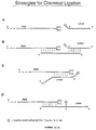
- the above-described coupling reactions can be executed using a number of different strategies, as shown in Figures 4A-4D.
- the ligation can be carried out in an external template-independent manner as shown in Figure 4A.
- This strategy requires a large excess of modified peptide acceptor (for example, 100 to 1000-fold excess) to achieve successful ligation to the RNA molecule.
- external template oligos may be used for substrate alignment as shown in Figure 4B.
- such oligos which are complementary in sequence to the RNA and peptide acceptor linker sequences, are at least approximately ten nucleotides in length.
- this oligo will include at least approximately ten nucleotides which are complementary to the RNA molecule and approximately ten nucleotides which are complementary to the peptide acceptor linker.
- the reactive sites may be brought into close proximity by direct hybridization of linker and RNA domains as shown in Figures 4C-4D.
- the extent of hybridizing sequence is at least ten to fifteen nucleotides.
- one type of exemplary construct includes a peptide acceptor attached to a linker carrying a modification at its 5' end as shown in Figures 4A-4C.
- An alternative construct may comprise an internal functional group flanked by a hybridization domain on one side and a puromycin-linker portion on the other ( Figure 4D).
- a carbohydrazide phosphoramidite was generated by combining a lactone with hydrazine and methanol and placing the reactants under reflux for 2 hours to produce a carbohydrazide moiety.
- the yield for this step of the synthesis was 97%.
- the resulting product was then reacted with the salt of a dimethoxytrityl group and pyridine/toluene at room temperature for 2.5 hours to yield a protected carbohydrazide moiety.
- the product yield for this step was 88%.
- the reactivity of the peptide acceptor toward the IO 4 - -oxidized RNA may be further enhanced through introduction of multiple copies of reactive groups.
- RNA consisting of a transcript encoding a flag epitope and a strep tag, of the sequence: 5' G GGA CAA UUA CUA UUU ACA AUU ACA AUG GAC UAC AAG GAC GAU GAC GAU AAG GGC GGC UGG UCC CAC CCC CAG UUC GAG AAG GCA UCC GCU (SEQ ID NO:1) was combined with 20 ⁇ l of 500 mM NaOAc (pH 5.2), 10 ⁇ l of 5 mM NaIO 4 , and brought up to a final volume of 100 ⁇ l with water. The reaction mixture was incubated for 15 minutes at room temperature.
- a peptide acceptor can also be affixed to an RNA molecule by attaching a functional group to the 3' end of the RNA followed by chemical ligation, as shown in Figure 6.
- reductive amination and related reactions are used to attach functional groups to the 3' terminus of an RNA.
- These newly introduced groups then react further with suitably modified linkers on the peptide acceptor, leading to covalent bond formation between the RNA and peptide acceptor.
- exemplary reactive groups include thiols (for disulfide formation or reactions with thiolphilic reagents such as pyridyl disulfides; Figure 6, Reaction A) or maleimides (Figure 6, Reaction B).
- N-hydroxysuccinimide-esters can be generated on 5' amino-modified linkers of the peptide acceptor by reaction with disuccinimidyl glutarate (DSG) or related reagents as described in Cox et al. (J. Immunol. 145:1719, 1990) and http://www. piercenet.com/Products/linksearch.cfm (Pierce, Rockford, IL; Figure 6, Reaction C). This modified linker can then be reacted with the amino functional group of the modified RNA.
- DSG disuccinimidyl glutarate
- This modified linker can then be reacted with the amino functional group of the modified RNA.
- This type of ligation reaction may be carried out in either an external template-independent or -dependent manner as described above, using the same general approaches.
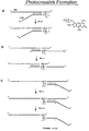
- a peptide acceptor can also be attached to an RNA molecule using photochemical methods as shown in Figures 7A-7H.
- Peptide acceptor linker molecules that carry psoralen groups allow the introduction of crosslinks to complementary RNA strands upon irradiation with long wave UV-light. This technique may be carried out generally as described in Pieles and Englisch, (Nucleic Acids Res. 17:285, 1989); and Godard et al. (Nucleic Acids Res. 22:4789, 1994).
- Attachment of the psoralen moiety to the 5' terminus of the linker region of the peptide acceptor can be accomplished using commercially available psoralen amidites, 2'-OMe-RNA phosphoramidites, psoralen C6 phosphoramidites, and triethylene glycol (TEG) phosphoramidites (Glen Research, Sterling, VA) on a standard DNA synthesizer.
- psoralen amidites 2'-OMe-RNA phosphoramidites
- psoralen C6 phosphoramidites psoralen C6 phosphoramidites
- TAG triethylene glycol
- RNA consisting of an RNA transcript encoding a flag epitope, a strep tag, and a photochemical target site of the sequence: 5' G GGA CAA UUA CUA UUU ACA AUU ACA AUG GAC UAC AAG GAC GAU GAC GAU AAG GGC GGC UGG UCC GAG CCC CAG UUC GAG AAG AAC GGC UAU A (SEQ ID NO:3), 1.2 nmole of Photolinker 30/10 consisting of the sequence: 5' Pso TAG CCG TTC T AAA AAA AAA AAA AAA AAA AAA AAA AAA AAA AAA AAA AAA CC Pu (SEQ ID NO:4) (where Pso is a psoralen C2 amidite [Glen Research], and Pu is Puromycin-CPG [Glen Research]), synthesized according to standard manufacturer protocols, or 30/15 consisting of the sequence: 5' Pso TAG CCG TTC TTC TCG AAA
- the reaction mixture was then slowly cooled to room temperature.
- the reaction mixture was next irradiated for 15 minutes at 0°C with ⁇ greater than 310 nm using a 450 W immersion lamp (medium pressure; ACE Glass, cat. no. 7825-34), equipped with a Pyrex absorption sleeve (ACE Glass, cat. no. 7835-44) in a Quartz immersion well (ACE Glass, cat. no. 7854-25), with the sample in a microcentrifuge tube strapped to the immersion well, and cooled in ice-water.
- the sample was then precipitated with 40 ⁇ l of 3 M NaOAc and 1000 ⁇ l of ethanol, and resuspended in 75 ⁇ l of water.
- various mRNAs and puromycin linkers were synthesized such that the peptide acceptor was positioned at the 3' end of the linker.
- the linkers were annealed to the target mRNAs and evaluated for their efficiency in forming mRNA-protein fusions molecules through in vitro translation techniques. The effect of the linker length and composition on fusion molecule yield was also determined.
- FIG. 7D illustrates the design of the mRNAs used in the crosslinking reactions.
- Each of the RNA molecules contained a portion of the tobacco mosaic virus 5'-UTR (TMV) with good initiation codon context (Gallie et al., Nucleic Acids Res., 16:883-93, 1988; and Kozak, Microbiol. Rev., 47:1-45, 1983), and a translation initiation codon (AUG).
- TMV tobacco mosaic virus 5'-UTR
- UAG translation initiation codon
- All mRNAs also carried a 3'-terminal ten nucleotide linker hybridization sequence 5'-GCAUCCGCUAUU coding for the amino acid sequence ASA, and ending with the dinucleotide sequence 5'-UA for psoralen photocrosslinking (const.), as described by Sinden and Hagerman (Biochemistry, 23:6299-6303, 198) and Gamper et al., (Photochem. Photobiol., 40:29-34, 1984).
- mRNA 1 contained a Flag epitope, DYKDDDDK (Hopp et al., Biotechnology, 6:1205-1210, 1988) followed by a Strep-Tag II sequence, WSHPQFEK (Schmidt et al., J. Mol. Biol., 255:753-66, 1996).
- Other mRNA molecules contained a 4-4-20 scFv, anti-fluorescein single chain antibody template, as described by Bedzyk et al. (J. Biol. Chem., 265:18615-20, 1990) and Mallender et al. (J. Biol. Chem., 271:5338-46, 1996).
- mRNAs 3 and 4 contained a downstream stop codon (UAA) to induce mRNA release from the ribosome after translation of residual un-crosslinked template (vide infra).
- UAA downstream stop codon
- a poly-A tail was also attached to mRNA 4 for purification by oligo-dT.
- RNAs used in these studies were prepared by T7 RNA polymerase run off-transcription (Megashortscript Transcription Kit, Ambion, TX) of PCR DNA templates, according to the methods of Milligan et al. (Nucleic Acids Res., 15:8783-8798, 1987). After transcription, all RNAs were purified by electrophoresis on 6% TBE-urea polyacrylamide gels (Novex, CA). The product bands were visualized by UV-shadowing, excised, crushed, and soaked overnight in 0.3 M NaOAc. Following ethanol precipitation, the RNAs were resuspended and stored in H 2 O. Radiolabeled RNA was synthesized according to the same procedure by including [ ⁇ - 32 P] UTP (Amersham, IL) in the transcription buffer.
- TAG Flexible triethyleneglycol phosphate
- polynucleotide sequences of various lengths were used to tether 5'-dCdC-puromycin to the 3'-end (see the table in Figure 7E).
- the linker hybridization sequence was prepared from 2'-OMe-RNA phosphoramidites to enhance the pairing stability of the stem structure (Inoue et al., Nucleic Acids Res., 15:6131-6148, 1987; and Majlessi et al., Nucleic Acids Res., 26:2224-2229, 1998).
- the product mixture of the photocrosslinking reaction between radiolabeled mRNA 1 and linker B was analyzed on a denaturing 6% TBE-Urea gel (Novex) and visualized on a phosporimaging system (Molecular Dynamics, CA) ( Figure 7E).
- These photocrosslink product mixtures generally contained ⁇ 20% unreacted and >80% photocrosslinked mRNA, and were used directly for in vitro translation and subsequent mRNA-protein fusion formation without further purification.
- the relative size difference between mRNA and crosslinked mRNA became too small to be separated on a gel. In these cases, the crude photo-crosslinking reaction mixtures were directly added to the lysate for fusion formation, without further purification.
- mRNA-protein fusion formation was promoted by the addition of KCl and MgCl 2 to the final concentrations of 590 mM and 50 mM, respectively, in a 500 ⁇ l volume, according to the methods of Roberts & Szostak and Szostak et al. ( supra ). Incubation was continued for another 60 minutes at 20°C. Varying concentrations of KCl and MgCl 2 were also tested to explore salt dependence on fusion formation.
- the in vitro translation products were isolated by diluting the lysate into 10 ml of binding buffer (100 mM Tris HCl, pH 8.0, 10 mM EDTA, I M NaCl, 0.25% v/v Triton X-100) and by adding to the mixture 10 mg of oligo-dT cellulose type 7 (Pharmacia, NJ). The samples were rotated for 60 minutes at 4°C. The solid support was then washed with 5 ml of ice-cold binding buffer, followed by elution with 100 ⁇ l aliquots of deionized H 2 O. The amount of mRNA-protein fusion isolated was determined by scintillation counting of the incorporated [ 35 S] methionine.
- the product was analyzed by electrophoresis on 4-12% NuPage gels using MES running buffer (Novex). The gels were dried after extensive washing to remove excess [ 35 S] methionine, and bands were visualized on a phosphorimager system (Molecular Dynamics).
- mRNA-scFv fusion molecule prepared from mRNA 4 of Figure 7E, was purified by binding of the A 18 linker regions to oligo-dT cellulose, followed by washing with binding buffer, according to the methods of Roberts & Szostak and Szostak et al. ( supra ) ( Figure 7E, lane 5).
- the fusion product could be isolated with a 1.3% yield based on the amount of photo-crosslinked input mRNA.
- Physical properties (gel mobility, binding to oligo-dT cellulose, selective peptide binding to affinity reagents) of the fusions prepared from photo-crosslinked mRNA were found to be identical to those of the fusion product obtained from enzymatically ligated mRNA templates.
- fusions prepared from the mRNA template 1 crosslinked to linker B of Figure 7E, encoding the Flag and Strep-Tag II epitope were tested for protein binding.
- a solution of 10 ⁇ l of 35 S-labeled mRNA-peptide fusion (prepared from mRNA 1 with linker B) was added to 20 ⁇ l of Anti-Flag M2 Affinity Gel (Sigma, MO) in 300 ⁇ l of buffer containing 50 mM Tris HCl, pH 7.4, 1% NP 40, 150 mM NaCl, 1 mM EDTA, 1 mM Na 3 VO 4 , and 1 mM NaF.
- a second precipitation experiment was carried out by adding the same fusion product to 20 ⁇ l of StrepTactin Sepharose (Genosys, TX) in 300 ⁇ l of buffer containing 100 mM Tris-HCl, pH 7.1, 1 mM EDTA, and 0.5 mg/ml yeast tRNA. Both precipitation mixtures were processed in parallel under identical conditions: The mixtures were rotated for 1 hour at 4°C and then transferred onto an Ultrafree-MC filter unit (0.45 ⁇ m; Millipore, MA). The buffer was removed by centrifugation, and the residue washed with 5 x 300 ⁇ l of ice-cold buffer.
- mRNA 3 was photo-crosslinked to linkers A-F of Figure 7E and each template was subsequently tested for fusion formation (Figure 7G). After incubation for 30 minutes at 30°C, the samples were divided into aliquots, to which varying amounts of KCl and MgCl 2 were added. The samples were incubated for another 60 minutes at 20°C; then, the fusion product was analyzed by gel-electrophoresis. The highest mRNA-protein fusion yields were obtained with the long linkers A and B (40 and 35 nucleotides, respectively) under high salt conditions.
- the psoralen moiety may also be incorporated at an internal position of the linker region of the peptide acceptor, as shown in Figure 7B, and as described in Pieles et al. (Nucleic Acids Res. 17:8967, 1989), or by incorporation of a branched phosphoramidite (Clontech, Palo Alto, CA) within the linker sequence, followed by addition of a psoralen phosphoramidite (Glen Research, Sterling, VA).
- the psoralen moiety may be crosslinked to the RNA through a branched linker, as described in U.S.S.N. 09/453,190.
- a linker with the sequence 5' cgt agg cga gaa agt gat X AAA AAA AAA AAA AAA AAA AAA AAA AAA CC Pu (where Pu is Puromycin-CPG [Glen Research]; C and A are standard 3'-amidites [Glen Research]; a, t, c, and g are 5'-phosphoramidites [Glen Research]; and X is an asymmetric branching amidite [Clontech]) has been synthesized according to standard manufacturer protocols, followed by selective deprotection of the branching point X (according to the instructions of Clontech) and subsequent coupling of a psoralen C6 amidite (Glen Research).
- This linker was then photocrosslinked to an RNA with the target sequence 5' ...GCA UCC GCU CUU UCA CUA UA using the photocrosslinking techniques described above.
- This RNA-linker construct was then successfully used for the synthesis of RNA-protein fusions.
- a linker containing a psoralen attached to the 3' end of the target hybridization domain may also be constructed (Figure 7C).
- the 5' end of the psoralen-containing linker is extended by another linker, that after reversal of strand orientation, terminates with a 3' puromycin.
- a subsequent digestion of the RNA target domain with RNase H is optional and will increase the flexibility of the linker construct.
- this approach allows the attachment of the linker at internal positions of much longer RNA molecules, and the untranslated region downstream from the linker site is then clipped off before translation and RNA-protein fusion formation.
- One advantage of the photochemical methods for attaching a peptide acceptor to an RNA is that these methods do not require chemical modification of the RNA prior to ligation. This makes the process very robust and selective, and allows the use of an RNA from a crude T7 transcription reaction as the substrate for the chemical ligation.
- an affinity-based RNA purification step may be combined with a photochemical ligation procedure described above ( Figure 7H).
- a suitable linker molecule is modified at its puromycin terminus with a photocleavable biotin moiety (e.g., EZ-Link TM NHS-PC-LC-Biotin, Pierce, Rockford, IL).
- the target RNA obtained, for example, from a crude transcription reaction
- the target RNA is then hybridized to a defined amount of linker, and the resulting duplex is captured on a solid support, such as streptavidin (or a related) resin.
- the excess RNA, as well as the components of the transcription reaction are then removed by extensive washing. Irradiation with long-wave UV light simultaneously leads to photocrosslink formation and product release from the resin.
- the ligated RNA can then be directly fed into a translation and fusion formation reaction, without further purification.
- RNA merely has to be estimated to exceed the amount of linker used.
- its amount is automatically reduced to a quantity not more than the amount of linker used. This, in turn, allows one to proceed to the next step without further quantization of the RNA (or ligated RNA) by, for example, taking A 260 UV readings, and the RNA amounts do not have to be adjusted otherwise.
- the nucleic-acid protein fusion molecule preparation process is therefore more suitable for automation.
- the mixture is incubated at room temperature for 2 hours, and results in a quantitative yield.
- the mixture is then precipitated twice with ethanol, with an expected recovery of >90% of the PC-biotinylated linker, and resuspended in 200 ⁇ l H 2 O.
- RNA is next transcribed using, for example, the T7 Megashortscript Kit (Ambion). Transcription is carried out using 10 pmole of a DNA template containing the sequence GCA UCC GCU AUU UAA A n at the 3'-terminus for a 250 ⁇ l reaction. This transcription reaction should yield approximately 2-5 nmol of crude RNA product. No purification (phenol extraction, NAP-5 column, or RNeasy column) is required prior to proceeding to the next step.
- the biotinylated linker (10 ⁇ l of 50 pmol/ ⁇ l biotinylated linker; 500 pmol total) is annealed to 62.5 to 250 ⁇ l of the transcription mixture (containing an estimated minimum of 500 pmole of RNA) using, for example, a PCR machine (heating for 30 seconds to 80°C, then cooling to 4°C at 0.3°C/sec) with 15 ⁇ l of 5 M NaCl (to a final concentration of 0.25 M for a 300 ⁇ l reaction) and H 2 O, to a final reaction volume of 300 ⁇ l.
- RNA of the reaction mixture is next immobilized onto 100 ⁇ l of Neutravidin beads (Pierce, Rockford, IL) by gently rocking the reaction mixture at 4°C for 30 minutes. The beads are then washed and resuspended in 300 ⁇ l H 2 O.
- Neutravidin beads Pieris, Rockford, IL
- the beads are next spun down and washed 3 times with 100 ⁇ l of buffer (25 mM Tris pH 7.0 and 0.25 M NaCl). Then the beads are UV irradiated for 15 minutes at room temperature (using a hand-held UV lamp UVGL-25; a microcentrifuge tube containing the beads is put directly on the lamp) to chemically ligate the linker to the RNA and photo-release the ligated molecule from the beads. It is expected that 250 pmol of ligated RNA is photo-released. Seventy-five ⁇ l of H 2 O is added to the tubes, the tubes are vortexed for 30 seconds, and the beads are spun down. The 75 ⁇ l supernatant, containing the ligated RNA/linker, is used for translation and formation of nucleic acid-protein fusion molecules.
- buffer 25 mM Tris pH 7.0 and 0.25 M NaCl
- the nucleic acid-protein fusion molecules are formed by combining the 75 ⁇ l of supernatant from the previous step, and combining it with 225 ⁇ l of the buffer components and lysate of the Rabbit Reticulocyte Lysate Kit (Ambion), and incubating the mixture for 30 minutes at 30°C.
- KCl and MgCl 2 are next added to final concentrations of 500 mM and 50 mM, respectively, and the reaction continues to incubate for 60 minutes at room temperature to produce the nucleic acid-protein fusion molecules.
- PNAs peptide nucleic acids
- triple-helix-forming constructs comprising two PNA molecules binding to RNA, thereby forming a clamp, can provide an efficient means for strong binding of RNA, since such constructs can be extremely resistant to thermal denaturation and conditions used for in vitro translation (Figure 8A) (Hanvey et al., Science, 258:1481, 1992).
- nucleic acid linker-PNA constructs may be accomplished by solid phase synthesis starting with puromycin-CPG, as shown in Figure 8B and as described in Uhlmann et al. (Angew. Chem. Int. Ed. Engl. 35:2633, 1996). After assembly of the desired nucleic acid portion (or PEG-spacer, if so desired) using standard automated synthesis, the solid phase synthesis is continued by attaching the PNA domain with the appropriate reagents (for example, as described in Uhlman et al. ( supra )). Alternatively, the PNA can be pre-synthesized as a separate moiety (PE Biosystems, Foster City, CA), followed by chemical coupling to the desired linker portion.
- RNA hybridization strength and consequently ligation efficiency are known to be increased by the use of 2-OMe RNA or propyne-modified nucleobases, rather than DNA (as described, for example, in Inoue et al., Nucleic Acids Res. 15:6131, 1987; Kibler-Herzog et al., Nucleic Acids Res. 19:2979, 1991; and Wagner et al., Science 260:1510, 1993).
- the linkers may also be optimized for their effectiveness in the RNA-protein fusion reaction. This will generally involve varying the length of the linker, but may also involve the use of different building blocks for RNA-protein joining.
- the deoxynucleotides of the linker may be replaced with PEG-spacers or 2-OMe-RNA units (both from Glen Research, Sterling, VA).
Abstract
Description
- In general, the present invention relates to ligation methods, in particular, for joining peptide acceptors to nucleic acids.
- Methods currently exist for the preparation of RNA-protein fusions An RNA-protein fusion is created by attaching a peptide acceptor to the 3' end of an RNA molecule, followed by in vitro or in situ translation of the RNA. The product is a peptide attached to the 3' end of the RNA encoding it. The generation of these RNA-protein fusions facilitates the isolation of proteins with desired properties from large pools of partially or completely random amino acid sequences, and solves the problem of recovering and amplifying protein sequence information by covalently attaching the RNA coding sequence to its corresponding protein molecule.
- The present invention features methods for the attachment of a peptide acceptor to an RNA molecule. These methods facilitate the production of RNA-protein fusions which can be used, for example, for the isolation of proteins or nucleic acids with desired properties from large pools of partially or completely random amino acid or nucleic acid sequences. The methods of the invention may be carried out by a variety of strategies for affixing a peptide acceptor to a nucleic acid molecule. These various approaches differ from one another in the types of bonds formed by the attachment of the peptide acceptor to the nucleic acid, and in the reagents used to achieve the attachment.
- Accordingly, the invention features a method for affixing a peptide acceptor to an RNA molecule comprising providing an RNA molecule, providing a peptide acceptor covalently bonded to a nucleic acid linker molecule, and hybridizing the RNA molecule to the nucleic acid linker molecule under conditions which allow covalent bond formation to occur between the peptide acceptor and the RNA molecule, wherein the 3' sequence of the RNA molecule forms a hairpin structure or the 5' sequence of the linker molecule forms a hairpin structure.
- In one embodiment of the invention, the peptide acceptor is bonded to the RNA molecule using T4 DNA ligase.
- The RNA molecule may include a translation initiation sequence and a start codon operably linked to a candidate protein coding sequence. In addition, one preferred peptide acceptor is puromycin, a nucleoside analog which adds to the C-terminus of a growing peptide chain and terminates translation. In one embodiment, the peptide acceptor includes puromycin attached to a linker, for example, a nucleotide linker. This linker facilitates the alignment of the peptide acceptor to the RNA molecule for attachment. In a further embodiment, the linker region of the peptide acceptor includes non-nucleotide moieties, for example, PEG. Other possible choices for acceptors include tRNA-like structures at the 3' end of the RNA, as well as other compounds that act in a manner similar to puromycin. Such compounds include, without limitation, any compound which possesses an amino acid linked to an adenine or an adenine-like compound, such as the amino acid nucleotides, phenylalanyl-adenosine (A-Phe), tyrosyl adenosine (A-Tyr), and alanyl adenosine (A-Ala), as well as amide-linked structures, such as phenylalanyl 3' deoxy 3' amino adenosine, alanyl 3' deoxy 3' amino adenosine, and tyrosyl 3' deoxy 3' amino adenosine; in any of these compounds, any of the naturally-occurring L-amino acids or their analogs may be utilized. In addition, a combined tRNA-like 3' structure-puromycin conjugate may also be used in the invention.
- In one preferred design of the invention, a DNA sequence is included between the end of the message and the peptide acceptor. This sequence is designed to cause the ribosome to pause at the end of the open reading frame, providing additional time for the peptide acceptor (for example, puromycin) to accept the nascent peptide chain before hydrolysis of the peptidyl-tRNA linkage. During in vitro translation the ribosome may also pause at the site of chemical ligation, especially at a psoralen crosslinking site or at a PNA clamp.
- In another preferred design of the invention, predominantly non-nucleotide linker moieties may be used in place of the nucleotide linkers attached to the peptide acceptor. This design facilitates the ligation of a peptide acceptor to an RNA molecule. For example, the linker may contain triethylene glycol spacers. The linker may also contain 2'-OMe-RNA phosphoramidites. In some cases where hybridization is a prerequisite for chemical or enzymatic ligation, a sufficient portion of the linker next to the ligation site must be comprised of nucleic acids.
Furthermore, the RNA or linker used according to the invention may contain a sequence (e.g., a poly(A) sequence) for use in purification, for example, affinity purification of the RNA or an RNA-protein fusion molecule formed from such an RNA or linker. - The RNA molecule affixed to a peptide acceptor may be in vitro or in situ translated to produce an RNA-protein fusion molecule. The RNA-protein fusion molecule is then incubated in the presence of high salt and/or incubated at low temperature (e.g., overnight at -20°C) as described by Szostak et al. (09/247,190). The RNA-protein fusion molecule may be also purified, for example, using standard poly(A) purification techniques.
- As used herein, by a "protein" is meant any two or more naturally occurring or modified amino acids joined by one or more peptide bonds. "Protein" and "peptide" are used interchangeably.
- By an "RNA" is meant a sequence of two or more covalently bonded, naturally occurring or modified ribonucleotides. One example of a modified RNA included within this term is phosphorothioate RNA.
- By a "translation initiation sequence" is meant any sequence that is capable of providing a functional ribosome entry site. In bacterial systems, this region is sometimes referred to as a Shine-Dalgarno sequence.
- By a "start codon" is meant three bases which signal the beginning of a protein coding sequence. By a "stop codon" is meant three bases which signal the termination of a protein coding sequence. Generally, start codons are AUG (or ATG) and stop codons are UAA (or TAA), UAG (or TAG), or UGA (or TGA); however, any other base triplets capable of being utilized as start or stop codons may be substituted.
- By "covalently bonded" is meant joined together either directly through a covalent bond or indirectly through another covalently bonded sequence (for example, DNA corresponding to a pause site).
- By "non-covalently bonded" is meant joined together by means other than a covalent bond.
- By a "hairpin structure" is meant a double-stranded region formed by a single nucleic acid strand. Preferably, such hairpin structures are at least 8 base pairs in length, and more preferably, between 8 and 15 base pairs in length.
- By "chemically ligating" is meant the joining together of two molecules without the use of an enzyme. Chemical ligation can result in non-covalent as well as covalent bonds.
- By a "peptide acceptor" is meant any molecule capable of being added to the C-terminus of a growing protein chain by the catalytic activity of the ribosomal peptidyl transferase function. Typically, such molecules contain (i) a nucleotide or nucleotide-like moiety, for example adenosine or an adenosine analog (di-methylation at the N-6 amino position is acceptable), (ii) an amino acid or amino acid-like moiety, such as any of the 20 D- or L-amino acids or any amino acid analog thereof including O-methyl tyrosine or any of the analogs described by Ellman et al. (Meth. Enzymol. 202:301, 1991), and (iii) a linkage between the two (for example, an ester, amide, or ketone linkage at the 3' position or, less preferably, the 2' position). Preferably, this linkage does not significantly perturb the pucker of the ring from the natural ribonucleotide conformation. Peptide acceptors may also possess a nucleophile, which may be, without limitation, an amino group, a hydroxyl group, or a sulfhydryl group. In addition, peptide acceptors may be composed of nucleotide mimetics, amino acid mimetics, or mimetics of the combined nucleotide-amino acid structure.
- By a "linker" or "linker molecule" is meant a sequence that includes deoxyribonucleotides, ribonucleotides, or analogs thereof.
- By "functionalize" is meant to modify in a manner that results in the attachment of a functional group or moiety. For example, an RNA molecule may be functionalized through IO4 - oxidation or amination, or a peptide acceptor may be functionalized by attaching an amine, hydrazine, (thio)hydrazide, or (thio)semicarbazone group.
- By an "external template" is meant a nucleic acid sequence which is added to a ligation reaction mixture, but which is not a part of the final product of the ligation reaction.
- By "high salt" is meant having a concentration of a monovalent cation of at least 200 mM, and, preferably, at least 500 mM or even 1 M, and/or a concentration of a divalent or higher valence cation of at least 25 mM, preferably, at least 50 mM, and, most preferably, at least 100 mM.
- By "affinity purification sequence" is meant a nucleotide sequence that is utilized in the purification of a nucleic acid or a nucleic acid-protein fusion molecule. For example, an affinity purification sequence may be a poly(A) sequence, such as A8-20, which can be used for purification of nucleic acid or fusion molecules on oligo-dT cellulose. An affinity purification sequence may also be a polypeptide sequence that is used to purify a nucleic acid-protein fusion molecule. Other exemplary purification techniques are described by Szostak et al. U.S.S.N. 09/247,190.
- The present invention provides a number of advantages. For example, the methods described herein facilitate the efficient ligation of peptide acceptors to RNA molecules, in some aspects, without the need for an external template to bring the RNA and peptide acceptor together. The invention also reduces the cost associated with the generation of an RNA-protein fusion.
- Other features and advantages of the invention will be apparent from the following detailed description, and from the claims.
- The drawings will first briefly be described.
-
- FIGURE 1 is a schematic representation of exemplary steps involved in the ligation of a peptide acceptor linker to an RNA using T4 DNA ligase on a hairpin template. Either the RNA 3' sequence or the peptide acceptor linker is designed to form a hairpin structure, and the peptide acceptor linker hybridizes to the RNA, thus bringing the RNA and peptide acceptor into close proximity to each other.
- FIGURE 2 is a schematic representation of exemplary steps involved in the ligation of a peptide acceptor linker to an RNA using terminal deoxynucleotidyl transferase.
- FIGURE 3A is a schematic representation of exemplary steps involved in the 3' modification of an RNA through hydrazide and semicarbazone formation.
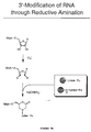
- FIGURE 3B is a schematic representation of exemplary steps involved in the 3' modification of an RNA through reductive amination.
- FIGURES 4A-4D are a series of diagrams showing exemplary strategies for the chemical ligation of a functionalized puromycin linker to a functionalized RNA. One strategy is template-independent (Figure 4A). Other strategies involve using external oligo templates to align the RNA and puromycin linker. In the strategies involving external oligo templates, the oligo template may hybridize to both the RNA and puromycin linker (Figure 4B), or be attached to the puromycin linker and hybridize to the RNA (Figure 4D). In addition, the functional group may be at the 5' end of (Figures 4B and 4C) or internal to (Figure 4D) the puromycin linker.
- FIGURE 5 is a schematic representation of exemplary steps involved in the synthesis of a fully protected carbohydrazide phosphoramidite for use in a modified puromycin linker.
- FIGURE 6 is a schematic representation of exemplary steps involved in the ligation of a peptide acceptor linker to an RNA by attaching a functional group to the 3' end of the RNA followed by chemical ligation. The functional group can then react with a suitably modified linker molecule to covalently bond the RNA and puromycin linker through a thiol (A), maleimide (B), or amine group (C).
- FIGURES 7A-7C are a series of schematic representations of exemplary steps involved in the ligation of a peptide acceptor to an RNA using photocrosslinking. In this general method, a psoralen moiety attached to the puromycin linker crosslinks the puromycin linker to the RNA upon exposure to UV irradiation. The psoralen moiety can be at the 5' end of (Figure 7A), internal to (Figure 7B), or at the 3' end of (Figure 7C) the puromycin linker.
- FIGURE 7D is a schematic representation of mRNAs used for photo-crosslink formation.
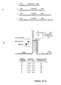
- FIGURE 7E is a schematic representation of a duplex formed between the constant 3'-sequence of the mRNA (const. region of the mRNAs of Figure 7D) and the photolinker showing the putative psoralen intercalation site. Also shown is the structure of psoralen, which was attached through a C6 alkyl chain to the 5'-phosphate of the linker. The variables x and y determine the number of dA-nucleotides and triethyleneglycol-units (TEG) in the linker, respectively. The autoradiograph on the left depicts the gel-electrophoretic analysis of the photo-crosslinking reaction between
mRNA 1 and linker B. - FIGURE 7F is an image of a gel illustrating in vitro translation and fusion formation. Protein synthesis from
mRNA template 2 andtemplate 3 is shown inlanes lanes 2 and 4).Template 3 carries a stop codon following the linker crosslink-site.Lane 5 shows the fusion product after purification on oligo-dT cellulose. A schematic representation of fusion formation using an mRNA molecule containing a stop codon is depicted above the image of the gel. - FIGURE 7G is an image of a gel illustrating the dependence of mRNA-protein fusion molecule yield on linker composition and on salt concentration.
- FIGURE 7H is a schematic representation of the steps involved in the ligation of mRNA to photocleavable biotin-based hybridized linkers and their subsequent fusion formation.
- FIGURE 8A is a schematic representation of the attachment of a puromycin linker to an RNA through (PNA)2-RNA triplex formation. The puromycin linker is attached to two PNA molecules which bind to RNA forming a triple helix through strong non-covalent bonds.
- FIGURE 8B is a schematic representation of steps involved in the synthesis of PNA-linker conjugates. The linker is attached to a solid support and modified to attach to the PNA. The PNA is then coupled to the desired linker, and the PNA linker molecule is deprotected and separated from the solid support.
- Described herein are various methods of attaching a peptide acceptor to an RNA molecule. The RNA may be generated by any standard approach, including normal cellular synthesis, recombinant techniques, and chemical synthesis, and includes, without limitation, cellular RNA, mRNA libraries, and random synthetic RNA libraries. The peptide acceptor (for example, puromycin) is typically bonded to a DNA or RNA linker. Such peptide acceptor molecules may be generated by any standard technique, for example, the techniques described in Roberts and Szostak (Proc. Natl. Acad. Sci. USA 94:12297, 1997), Szostak et al. (WO 98/31700), and Szostak et al., U.S.S.N. 09/247,190. Techniques for carrying out each method of the invention are now described in detail, using particular examples. These examples are provided for the purpose of illustrating the invention, and should not be construed as limiting.
- In one particular approach according to the invention, T4 DNA ligase is used to attach a peptide acceptor to an RNA molecule using a hairpin-containing template, for example, as shown in Figure 1. The ligation reaction is carried out in a self-templating manner and does not utilize a splint oligo. Either the RNA 3' sequence or the 5' end of the linker region of the peptide acceptor is designed to form a hairpin structure. Close positioning of the 5' end of the linker region of the peptide acceptor and the 3' end of the RNA in the context of a hairpin structure facilitates enzymatic ligation with T4 DNA ligase, as described, for example, in Sambrook, Fritsch, & Maniatis, Molecular Cloning, Cold Spring Harbor, New York, Cold Spring Harbor Laboratory Press, 1989. The ratio of the reactants is approximately 1:1, although a slight excess (1:1.2) of either reactant is acceptable. Optimal ligation conditions may vary slightly from reaction to reaction and may be determined experimentally using techniques known to those skilled in the art.
- Another enzymatic method for the attachment of a peptide acceptor to an RNA molecule involves the modification of the 3' end of the RNA followed by ligation using terminal deoxynucleotidyl transferase, for example, as shown in Figure 2. The TdT reaction is carried out as generally described in Sambrook, Fritsch, & Maniatis (supra). This enzyme extends the 3' end of nucleic acids with deoxy- (or dideoxy-) nucleotide triphosphates. These (d/dd)NTPs may be chemically modified on their base moieties to carry the desired linker structures (as described, for example, in Meyer, Methods in Molecular Biology, Agrawal, ed., vol 26, Totowa: Humana Press, 1994, pages 73-91; Kumar et al., Anal. Biochem. 169:376, 1988; Riley et al., DNA 5:333, 1986; and Schmitz et al., Anal. Biochem. 192:222, 1991). As shown in Figure 2, the linker molecule must initiate with a (d/dd) NTP. The remainder of the linker composition, however, may vary. A special feature of this terminal transferase ligation method is that the DNA linker region of the peptide acceptor can be as short as one (d/dd)NTP unit.
- A peptide acceptor can also be attached to an RNA molecule by functionalization of the 3' end of the RNA via IO4 - oxidation, followed by chemical ligation of the peptide acceptor to the RNA, as shown in Figure 3A. The IO4 - oxidation is carried out as described below and according to the methods of Agrawal (Methods in Molecular Biology, Agrawal, ed., vol 26, Totowa: Humana Press, 1994, pages 93-120), Proudnikov and Mirzabekov, (Nucleic Acids Res. 24:4535, 1996), Gosh et al., (Anal. Biochem. 178:43, 1989), Bauman et al., (J. Histochem. Cytochem. 29:227, 1981), and Wu et al., (Nucleic Acids Res. 24:3472, 1996). Since the IO4 - (oxidation step strictly requires a 1,2-diol for reaction, only the terminal nucleotide is modified, leaving internal residues of the RNA molecule unaffected. The resulting dialdehyde can be subjected to a further reaction with various nucleophiles such as amines, hydrazines, carbo(thio)hydrazides, or (thio)semicarbazides, yielding Schiff base-like structures. These reactions are carried out as described, for example, in Agrawal (supra), Proudnikov and Mirzabekov (supra), Gosh et al. (supra), Bauman et al. (supra), and Wu et al. (supra). While the (thio)hydrazides and (thio)semicarbazones obtained after reaction with carbo(thio)hydrazides and (thio)semicarbazides, respectively, are fairly stable, the initial adducts containing amines or hydrazines usually require a subsequent reduction step, such as reductive amination, as shown in Figure 3B and as described in Agrawal (supra), Proudnikov and Mirzabekov (supra), Gosh et al. (supra), Bauman et al. (supra), and Wu et al. (supra), to render the newly formed bonds stable toward hydrolysis.
- The above-described coupling reactions can be executed using a number of different strategies, as shown in Figures 4A-4D. For example, the ligation can be carried out in an external template-independent manner as shown in Figure 4A. This strategy requires a large excess of modified peptide acceptor (for example, 100 to 1000-fold excess) to achieve successful ligation to the RNA molecule. To increase the efficiency of the RNA ligation, external template oligos may be used for substrate alignment as shown in Figure 4B. Preferably, such oligos, which are complementary in sequence to the RNA and peptide acceptor linker sequences, are at least approximately ten nucleotides in length. Typically, this oligo will include at least approximately ten nucleotides which are complementary to the RNA molecule and approximately ten nucleotides which are complementary to the peptide acceptor linker. Alternatively, the reactive sites may be brought into close proximity by direct hybridization of linker and RNA domains as shown in Figures 4C-4D. Here, the extent of hybridizing sequence is at least ten to fifteen nucleotides.
- A number of different constructs for the attachment of peptide acceptors to RNA via crosslink formation may be utilized. For example, one type of exemplary construct includes a peptide acceptor attached to a linker carrying a modification at its 5' end as shown in Figures 4A-4C. An alternative construct may comprise an internal functional group flanked by a hybridization domain on one side and a puromycin-linker portion on the other (Figure 4D).
- The synthesis of these modified linkers involves standard automated DNA synthesis using commercially available phosphoramidites (Glen Research, Sterling, VA) for assembling the main body of nucleotides or spacer moieties. The 3' puromycin may be introduced through the use of puromycin-CPG (Glen Research, Sterling, VA) as a solid support for synthesis. The attachment of the reactive functional groups may be achieved using commercially available reagents such as amino terminus-modifiers (Glen Research, Sterling, VA) or uni-link amino modifiers (Clontech, Palo Alto, CA). Other functional groups may be incorporated utilizing appropriate phosphoramidites. One exemplary method to generate a carbohydrazide phosphoramidite is described in Figure 5. A carbohydrazide phosphoramidite was generated by combining a lactone with hydrazine and methanol and placing the reactants under reflux for 2 hours to produce a carbohydrazide moiety. The yield for this step of the synthesis was 97%. The resulting product was then reacted with the salt of a dimethoxytrityl group and pyridine/toluene at room temperature for 2.5 hours to yield a protected carbohydrazide moiety. The product yield for this step was 88%. This product was further reacted with a phosphoramidite moiety in the presence of tetrahydrofuran and diisopropylamine at room temperature for 30 minutes, which yielded (72%) a reaction product of a fully protected carbohydrazide phosphoramidite.
- In addition, the reactivity of the peptide acceptor toward the IO4 --oxidized RNA may be further enhanced through introduction of multiple copies of reactive groups.
- One exemplary ligation reaction was carried out as follows. One nmole of RNA, consisting of a transcript encoding a flag epitope and a strep tag, of the sequence: 5' G GGA CAA UUA CUA UUU ACA AUU ACA AUG GAC UAC AAG GAC GAU GAC GAU AAG GGC GGC UGG UCC CAC CCC CAG UUC GAG AAG GCA UCC GCU (SEQ ID NO:1) was combined with 20 µl of 500 mM NaOAc (pH 5.2), 10µl of 5 mM NaIO4, and brought up to a final volume of 100 µl with water. The reaction mixture was incubated for 15 minutes at room temperature. Next, 10 µl of 10 mM Na2SO3 was added, and the reaction mixture was incubated again for 15 minutes at room temperature. Forty µl of 1 M phosphate buffer (pH 8.0), 1.5 nmole of the peptide acceptor linker Uni-Al/8, having the sequence: 5' X CGC GGA TGC AAA AAA AAA AAA AAA AAA AAA AAA AAA CC Pu (SEQ ID NO:2) (where X is a Uni-link amino modifier [Clontech], and Pu is Puromycin-CPG [Glen Research]), and 20 µl of NaCNBH, were added to the reaction mixture. The mixture was then incubated for 18 hours at room temperature, precipitated, purified on a 6% TBE-Urea gel, and crush-soaked overnight to obtain the RNA-protein fusion molecule. This reaction yielded 230 pmole of product.
- A peptide acceptor can also be affixed to an RNA molecule by attaching a functional group to the 3' end of the RNA followed by chemical ligation, as shown in Figure 6. In a variation of the process described above, reductive amination and related reactions are used to attach functional groups to the 3' terminus of an RNA. These newly introduced groups then react further with suitably modified linkers on the peptide acceptor, leading to covalent bond formation between the RNA and peptide acceptor. Exemplary reactive groups include thiols (for disulfide formation or reactions with thiolphilic reagents such as pyridyl disulfides; Figure 6, Reaction A) or maleimides (Figure 6, Reaction B).
- Other possible reactive groups for the functionalization of the 3' end of an RNA followed by attachment of a peptide acceptor are amines. For example, N-hydroxysuccinimide-esters (NHS-esters) can be generated on 5' amino-modified linkers of the peptide acceptor by reaction with disuccinimidyl glutarate (DSG) or related reagents as described in Cox et al. (J. Immunol. 145:1719, 1990) and http://www. piercenet.com/Products/linksearch.cfm (Pierce, Rockford, IL; Figure 6, Reaction C). This modified linker can then be reacted with the amino functional group of the modified RNA.
- This type of ligation reaction may be carried out in either an external template-independent or -dependent manner as described above, using the same general approaches.
- A peptide acceptor can also be attached to an RNA molecule using photochemical methods as shown in Figures 7A-7H. Peptide acceptor linker molecules that carry psoralen groups allow the introduction of crosslinks to complementary RNA strands upon irradiation with long wave UV-light. This technique may be carried out generally as described in Pieles and Englisch, (Nucleic Acids Res. 17:285, 1989); and Godard et al. (Nucleic Acids Res. 22:4789, 1994). Attachment of the psoralen moiety to the 5' terminus of the linker region of the peptide acceptor can be accomplished using commercially available psoralen amidites, 2'-OMe-RNA phosphoramidites, psoralen C6 phosphoramidites, and triethylene glycol (TEG) phosphoramidites (Glen Research, Sterling, VA) on a standard DNA synthesizer.
- In one exemplary approach, this method was carried out as follows. One nmole of RNA consisting of an RNA transcript encoding a flag epitope, a strep tag, and a photochemical target site of the sequence: 5' G GGA CAA UUA CUA UUU ACA AUU ACA AUG GAC UAC AAG GAC GAU GAC GAU AAG GGC GGC UGG UCC GAG CCC CAG UUC GAG AAG AAC GGC UAU A (SEQ ID NO:3), 1.2 nmole of
Photolinker 30/10 consisting of the sequence: 5' Pso TAG CCG TTC T AAA AAA AAA AAA AAA AAA AAA AAA AAA CC Pu (SEQ ID NO:4) (where Pso is a psoralen C2 amidite [Glen Research], and Pu is Puromycin-CPG [Glen Research]), synthesized according to standard manufacturer protocols, or 30/15 consisting of the sequence: 5' Pso TAG CCG TTC TTC TCG AAA AAA AAA AAA AAA AAA AAA AAA AAA CC Pu (SEQ ID NO:5) (where Pso is a psoralen C2 amidite, and Pu is Puromycin-CPG), 10x buffer (250 mM Tris pH 7.0; 1 M NaCl), and water (bringing the final volume to 360 µl) were combined and heated to 80°C for 2 minutes. The reaction mixture was then slowly cooled to room temperature. The reaction mixture was next irradiated for 15 minutes at 0°C with λ greater than 310 nm using a 450 W immersion lamp (medium pressure; ACE Glass, cat. no. 7825-34), equipped with a Pyrex absorption sleeve (ACE Glass, cat. no. 7835-44) in a Quartz immersion well (ACE Glass, cat. no. 7854-25), with the sample in a microcentrifuge tube strapped to the immersion well, and cooled in ice-water. The sample was then precipitated with 40 µl of 3 M NaOAc and 1000 µl of ethanol, and resuspended in 75 µl of water. Next, 75 µl of 2x loading buffer (Novex) was added to the sample, and the sample was purified on a precast 6% TBE-Urea gel (Novex). The product was recovered using a crush and soak method (0.3 M NaOAc, overnight at room temperature) followed by ethanol precipitation. This photocrosslinking method yielded 272 pmole of RNA-protein fusionproduct using Photolinker 10/30, and 227 pmole using Photolinker 15/30. - For this photocrosslinking method of chemical ligation, various parameters of the reaction were evaluated. First, the salt dependence of the photocrosslink formation was tested. A set of crosslinking experiments with buffers containing 100-1000 mM NaCl were performed. No difference in ligation efficiencies were observed between the various reactions. In addition, a change of the RNA target sequence to: 5'...GAC UAC AAG GAC GAG GCA UCC GCU CUU UCA CUA UA (SEQ ID NO:6) (with the underlined sequence being a target for the psoralen linker) gave significantly reduced product yields (15 to 20% reduced), indicating that the RNA target sequence was important. Next it was determined that the product yield could be increased by the repeated replacement of psoralen linkers that had been inactivated during the course of the reaction. This experiment was carried out as follows. The RNA, linker, and 10x buffer were combined and heated to 80°C for 2 minutes. The reaction mixture was then slowly cooled to room temperature and irradiated as described above. Then an additional 1 nmole of linker and 1 µl of buffer were added. The linker annealed to the RNA and was then irradiated. This process was repeated, by adding 2 nmole of linker and 2 µl of buffer and irradiating. This procedure allowed an increased product yield from 20% to greater than 40% for certain sequences.
- The performance of the ligation products generated by photochemical crosslinking methods was also evaluated. In experiments with linkers of different lengths (psoralen plus 15 base pairs of target hybridization domain plus dAnCCPu [where n=7, 12, 17, or 22]) the following observations were made. Long linkers gave the highest RNA-protein fusion yields under high salt conditions (500 mM KCl plus 50 mM MgCl2). In buffers with reduced salt (250 mM KCl plus 10 mM MgCl2, or 250 mM KCl), the short linkers produced higher yields than the longer ones, but the overall yields were generally lower. In general, yields seem to be comparable to those obtained with enzymatically ligated RNA templates.
- In other exemplary methods, various mRNAs and puromycin linkers were synthesized such that the peptide acceptor was positioned at the 3' end of the linker. The linkers were annealed to the target mRNAs and evaluated for their efficiency in forming mRNA-protein fusions molecules through in vitro translation techniques. The effect of the linker length and composition on fusion molecule yield was also determined.
- Figure 7D illustrates the design of the mRNAs used in the crosslinking reactions. Each of the RNA molecules contained a portion of the tobacco mosaic virus 5'-UTR (TMV) with good initiation codon context (Gallie et al., Nucleic Acids Res., 16:883-93, 1988; and Kozak, Microbiol. Rev., 47:1-45, 1983), and a translation initiation codon (AUG). All mRNAs also carried a 3'-terminal ten nucleotide linker hybridization sequence 5'-GCAUCCGCUAUU coding for the amino acid sequence ASA, and ending with the dinucleotide sequence 5'-UA for psoralen photocrosslinking (const.), as described by Sinden and Hagerman (Biochemistry, 23:6299-6303, 198) and Gamper et al., (Photochem. Photobiol., 40:29-34, 1984). In addition one mRNA (mRNA 1) contained a Flag epitope, DYKDDDDK (Hopp et al., Biotechnology, 6:1205-1210, 1988) followed by a Strep-Tag II sequence, WSHPQFEK (Schmidt et al., J. Mol. Biol., 255:753-66, 1996). Other mRNA molecules (mRNAs 2, 3, and 4) contained a 4-4-20 scFv, anti-fluorescein single chain antibody template, as described by Bedzyk et al. (J. Biol. Chem., 265:18615-20, 1990) and Mallender et al. (J. Biol. Chem., 271:5338-46, 1996). In addition,
mRNAs mRNA 4 for purification by oligo-dT. - The mRNAs used in these studies (Figure 7D) were prepared by T7 RNA polymerase run off-transcription (Megashortscript Transcription Kit, Ambion, TX) of PCR DNA templates, according to the methods of Milligan et al. (Nucleic Acids Res., 15:8783-8798, 1987). After transcription, all RNAs were purified by electrophoresis on 6% TBE-urea polyacrylamide gels (Novex, CA). The product bands were visualized by UV-shadowing, excised, crushed, and soaked overnight in 0.3 M NaOAc. Following ethanol precipitation, the RNAs were resuspended and stored in H2O. Radiolabeled RNA was synthesized according to the same procedure by including [α-32P] UTP (Amersham, IL) in the transcription buffer.
- The puromycin-linkers used in these studies (Figure 7E) were prepared using an Expedite Synthesizer Model 8909 (PerSeptive Biosystems, MA), using conventional solid-support phosphoramidite chemistry. Puromycin-CPG, DNA phosphoramidites, 2'-OMe-RNA phosphoramidites, psoralen C6 phosphoramidite, and triethylene glycol (TEG) phosphoramidites (spacer 9) were used according to the recommended protocols (Glen Research). A psoralen moiety was attached through a C6 alkyl chain to the 5'-phosphate of the linker. Flexible triethyleneglycol phosphate (TEG) spacers and polynucleotide sequences of various lengths were used to tether 5'-dCdC-puromycin to the 3'-end (see the table in Figure 7E). The linker hybridization sequence was prepared from 2'-OMe-RNA phosphoramidites to enhance the pairing stability of the stem structure (Inoue et al., Nucleic Acids Res., 15:6131-6148, 1987; and Majlessi et al., Nucleic Acids Res., 26:2224-2229, 1998). Following deprotection in concentrated ammonium hydroxide for 8 hours at 55°C, the linkers were purified by reversed phase HPLC on a C18 Spheri-5 column (Perkin Elmer, CA) with 50 mM triethylammonium acetate in 5% v/v acetonitrile as buffer A, and 50 mM triethylammonium acetate in 70% v/v acetonitrile as buffer B and with a flow rate of 1.5 ml/min. A linear gradient of 15 - 60% buffer B over 45 minutes was used for elution. After drying, the linkers were resuspended and stored in H2O.
- The linkers (5 µM) were annealed to the target mRNAs (2.5 µM) in 25 mM Tris HCl buffer, pH 7, and 100 mM NaCl by heating to 85°C for 30 seconds, followed by cooling to 4°C over a period of 5 minutes. The reaction mixture was irradiated for 15 minutes at room temperature in borosilicate glass vials (Kimble/Kontes, NJ), using a handheld multiwavelength UV lamp model UVGL-25 (UVP, CA) set to a long wave (wavelength λ > 300 nm). The product mixture of the photocrosslinking reaction between
radiolabeled mRNA 1 and linker B was analyzed on adenaturing 6% TBE-Urea gel (Novex) and visualized on a phosporimaging system (Molecular Dynamics, CA) (Figure 7E). These photocrosslink product mixtures generally contained <20% unreacted and >80% photocrosslinked mRNA, and were used directly for in vitro translation and subsequent mRNA-protein fusion formation without further purification. For thelonger mRNA substrates - Translation and fusion formation of the mRNA fusion molecules were first tested using
mRNA 2 in the following experiments. In vitro translation reactions were performed using rabbit reticulocyte lysates (Ambion) for 30 minutes at 30°C. The reactions contained 100 pmole photo-crosslinked mRNA (see above), 10 mM creatine phosphate, 150 mM KOAc, 0.5 mM MgCl2, 0.1 mM of each amino acid except methionine, 150 µCi of [35S] methionine (Amersham), and 67% v/v of lysate in a total volume of 300 µl. mRNA-protein fusion formation was promoted by the addition of KCl and MgCl2 to the final concentrations of 590 mM and 50 mM, respectively, in a 500 µl volume, according to the methods of Roberts & Szostak and Szostak et al. (supra). Incubation was continued for another 60 minutes at 20°C. Varying concentrations of KCl and MgCl2 were also tested to explore salt dependence on fusion formation. - The in vitro translation products were isolated by diluting the lysate into 10 ml of binding buffer (100 mM Tris HCl, pH 8.0, 10 mM EDTA, I M NaCl, 0.25% v/v Triton X-100) and by adding to the
mixture 10 mg of oligo-dT cellulose type 7 (Pharmacia, NJ). The samples were rotated for 60 minutes at 4°C. The solid support was then washed with 5 ml of ice-cold binding buffer, followed by elution with 100 µl aliquots of deionized H2O. The amount of mRNA-protein fusion isolated was determined by scintillation counting of the incorporated [35S] methionine. The product was analyzed by electrophoresis on 4-12% NuPage gels using MES running buffer (Novex). The gels were dried after extensive washing to remove excess [35S] methionine, and bands were visualized on a phosphorimager system (Molecular Dynamics). - Gel analysis showed two bands that corresponded to the peptidyl-tRNA and the free peptide (Figure 7F, lane 1). When translation was carried out with photo-crosslinked
mRNA 2, a third and slower migrating band appeared, thus indicating successful mRNA-protein fusion formation (Figure 7F, lane 2). Increased yields of free protein and fusion (approximately 20% more) were obtained withmRNA 3, which was identical in coding sequence tomRNA 2, but carried a stop-codon downstream of the photo-crosslink site (Figure 7F,lanes 3 and 4). Relative band intensities indicated that 30% of the total amount of synthesized protein was converted into mRNA-protein fusion (Figure 7F, lane 4). - An mRNA-scFv fusion molecule, prepared from
mRNA 4 of Figure 7E, was purified by binding of the A18 linker regions to oligo-dT cellulose, followed by washing with binding buffer, according to the methods of Roberts & Szostak and Szostak et al. (supra) (Figure 7E, lane 5). The fusion product could be isolated with a 1.3% yield based on the amount of photo-crosslinked input mRNA. Physical properties (gel mobility, binding to oligo-dT cellulose, selective peptide binding to affinity reagents) of the fusions prepared from photo-crosslinked mRNA were found to be identical to those of the fusion product obtained from enzymatically ligated mRNA templates. - In order to confirm the composition of the peptide portion of the fusion molecules, fusions prepared from the
mRNA template 1 crosslinked to linker B of Figure 7E, encoding the Flag and Strep-Tag II epitope, were tested for protein binding. A solution of 10 µl of 35S-labeled mRNA-peptide fusion (prepared frommRNA 1 with linker B) was added to 20 µl of Anti-Flag M2 Affinity Gel (Sigma, MO) in 300 µl of buffer containing 50 mM Tris HCl, pH 7.4, 1% NP 40, 150 mM NaCl, 1 mM EDTA, 1 mM Na3VO4, and 1 mM NaF. A second precipitation experiment was carried out by adding the same fusion product to 20 µl of StrepTactin Sepharose (Genosys, TX) in 300 µl of buffer containing 100 mM Tris-HCl, pH 7.1, 1 mM EDTA, and 0.5 mg/ml yeast tRNA. Both precipitation mixtures were processed in parallel under identical conditions: The mixtures were rotated for 1 hour at 4°C and then transferred onto an Ultrafree-MC filter unit (0.45 µm; Millipore, MA). The buffer was removed by centrifugation, and the residue washed with 5 x 300 µl of ice-cold buffer. The residues were analyzed by scintillation counting, and fusion binding was determined to be 54% and 62% for the Anti-Flag M2 matrix and the StrepTactin matrix, respectively. A control reaction with protein-A agarose (Sigma) showed no detectable binding to the matrix. - To test the effect of linker length and composition on fusion formation in the presence of various salt concentrations,
mRNA 3 was photo-crosslinked to linkers A-F of Figure 7E and each template was subsequently tested for fusion formation (Figure 7G). After incubation for 30 minutes at 30°C, the samples were divided into aliquots, to which varying amounts of KCl and MgCl2 were added. The samples were incubated for another 60 minutes at 20°C; then, the fusion product was analyzed by gel-electrophoresis. The highest mRNA-protein fusion yields were obtained with the long linkers A and B (40 and 35 nucleotides, respectively) under high salt conditions. A lower salt concentration resulted in a significant drop in fusion formation with the linkers A and B. On the other hand, fusion yields for the shorter linkers C to F were less salt dependent. In addition, the fusion molecule yield generally increased with the number of flexible TEG spacers. Analysis of the crude mRNA-protein fusion molecule lysates revealed that up to 45% of the total protein was present as mRNA-protein fusion. Because linker F lacked the oligo-dA track needed for oligo-dT purification,mRNA 4 was prepared with an A18 stretch at its 3'-end, which allowed fusion purification on oligo-dT cellulose. - In an alternative to the above techniques which involve attachment of psoralen to the linker 5' end, the psoralen moiety may also be incorporated at an internal position of the linker region of the peptide acceptor, as shown in Figure 7B, and as described in Pieles et al. (Nucleic Acids Res. 17:8967, 1989), or by incorporation of a branched phosphoramidite (Clontech, Palo Alto, CA) within the linker sequence, followed by addition of a psoralen phosphoramidite (Glen Research, Sterling, VA). For example, the psoralen moiety may be crosslinked to the RNA through a branched linker, as described in U.S.S.N. 09/453,190.
- In one exemplary approach, a linker with the sequence 5' cgt agg cga gaa agt gat X AAA AAA AAA AAA AAA AAA AAA AAA AAA CC Pu (where Pu is Puromycin-CPG [Glen Research]; C and A are standard 3'-amidites [Glen Research]; a, t, c, and g are 5'-phosphoramidites [Glen Research]; and X is an asymmetric branching amidite [Clontech]) has been synthesized according to standard manufacturer protocols, followed by selective deprotection of the branching point X (according to the instructions of Clontech) and subsequent coupling of a psoralen C6 amidite (Glen Research). This linker was then photocrosslinked to an RNA with the target sequence 5' ...GCA UCC GCU CUU UCA CUA UA using the photocrosslinking techniques described above. This RNA-linker construct was then successfully used for the synthesis of RNA-protein fusions.
- In yet another alternative, a linker containing a psoralen attached to the 3' end of the target hybridization domain may also be constructed (Figure 7C). In this method, the 5' end of the psoralen-containing linker is extended by another linker, that after reversal of strand orientation, terminates with a 3' puromycin. A subsequent digestion of the RNA target domain with RNase H is optional and will increase the flexibility of the linker construct. In addition, this approach allows the attachment of the linker at internal positions of much longer RNA molecules, and the untranslated region downstream from the linker site is then clipped off before translation and RNA-protein fusion formation.
- In one exemplary approach to this method, a linker with the sequence 5' Pso atg cga gaa agt gat aaa aaa aaa aaa CC Pu (where Pu is Puromycin-CPG [Glen Research]; C is a standard 3'-amidite [Glen Research]; a, t, c, and g are 5'-phosphoramidites [Glen Research]; and Pso is a psoralen C6 phosphoramidite [Gen Research]) was constructed according to standard manufacturer protocols. The linker was then photocrosslinked to an RNA with the target sequence 5'...GUA UAC GCU CUU UCA CUA using the photocrosslinking techniques described above. This RNA-linker construct (with and without prior treatment with RNase H) was then successfully used for the synthesis of RNA-protein fusions.
- One advantage of the photochemical methods for attaching a peptide acceptor to an RNA is that these methods do not require chemical modification of the RNA prior to ligation. This makes the process very robust and selective, and allows the use of an RNA from a crude T7 transcription reaction as the substrate for the chemical ligation.
- If desired, an affinity-based RNA purification step may be combined with a photochemical ligation procedure described above (Figure 7H). A suitable linker molecule is modified at its puromycin terminus with a photocleavable biotin moiety (e.g., EZ-Link™ NHS-PC-LC-Biotin, Pierce, Rockford, IL). The target RNA (obtained, for example, from a crude transcription reaction) is then hybridized to a defined amount of linker, and the resulting duplex is captured on a solid support, such as streptavidin (or a related) resin. The excess RNA, as well as the components of the transcription reaction are then removed by extensive washing. Irradiation with long-wave UV light simultaneously leads to photocrosslink formation and product release from the resin. The ligated RNA can then be directly fed into a translation and fusion formation reaction, without further purification.
- This method is advantageous over previous RNA purification schemes. For example, after transcription, the amount of RNA merely has to be estimated to exceed the amount of linker used. When subjected to the described procedure, its amount is automatically reduced to a quantity not more than the amount of linker used. This, in turn, allows one to proceed to the next step without further quantization of the RNA (or ligated RNA) by, for example, taking A260 UV readings, and the RNA amounts do not have to be adjusted otherwise. The nucleic-acid protein fusion molecule preparation process is therefore more suitable for automation.
- In one exemplary technique, this biotin-based RNA purification and ligation protocol may be carried out as follows. In this photocleavable biotin-based RNA purification and ligation procedure, the linker is first biotinylated. The linker C6-psoralen-2-OMe[U AGC GGA UGC] dA18 TEG2 dCdC-puromycin is biotinylated by combining 100 µl of 100 µM linker (10 nmol total), 50 µl of 1 µmole EZ-Link™ PC-LC-Biotin in DMSO, 20 µl of 10x PBS, pH 7.4, and 30 µl H2O. The mixture is incubated at room temperature for 2 hours, and results in a quantitative yield. The mixture is then precipitated twice with ethanol, with an expected recovery of >90% of the PC-biotinylated linker, and resuspended in 200 µl H2O.
- RNA is next transcribed using, for example, the T7 Megashortscript Kit (Ambion). Transcription is carried out using 10 pmole of a DNA template containing the sequence GCA UCC GCU AUU UAA An at the 3'-terminus for a 250 µl reaction. This transcription reaction should yield approximately 2-5 nmol of crude RNA product. No purification (phenol extraction, NAP-5 column, or RNeasy column) is required prior to proceeding to the next step.
- In the next step of this purification and ligation technique, the biotinylated linker (10 µl of 50 pmol/µl biotinylated linker; 500 pmol total) is annealed to 62.5 to 250 µl of the transcription mixture (containing an estimated minimum of 500 pmole of RNA) using, for example, a PCR machine (heating for 30 seconds to 80°C, then cooling to 4°C at 0.3°C/sec) with 15 µl of 5 M NaCl (to a final concentration of 0.25 M for a 300 µl reaction) and H2O, to a final reaction volume of 300 µl. The RNA of the reaction mixture is next immobilized onto 100 µl of Neutravidin beads (Pierce, Rockford, IL) by gently rocking the reaction mixture at 4°C for 30 minutes. The beads are then washed and resuspended in 300 µl H2O.
- The beads are next spun down and washed 3 times with 100 µl of buffer (25 mM Tris pH 7.0 and 0.25 M NaCl). Then the beads are UV irradiated for 15 minutes at room temperature (using a hand-held UV lamp UVGL-25; a microcentrifuge tube containing the beads is put directly on the lamp) to chemically ligate the linker to the RNA and photo-release the ligated molecule from the beads. It is expected that 250 pmol of ligated RNA is photo-released. Seventy-five µl of H2O is added to the tubes, the tubes are vortexed for 30 seconds, and the beads are spun down. The 75 µl supernatant, containing the ligated RNA/linker, is used for translation and formation of nucleic acid-protein fusion molecules.
- The nucleic acid-protein fusion molecules are formed by combining the 75 µl of supernatant from the previous step, and combining it with 225 µl of the buffer components and lysate of the Rabbit Reticulocyte Lysate Kit (Ambion), and incubating the mixture for 30 minutes at 30°C. KCl and MgCl2 are next added to final concentrations of 500 mM and 50 mM, respectively, and the reaction continues to incubate for 60 minutes at room temperature to produce the nucleic acid-protein fusion molecules.
- As an alternative to covalent bond formation for the attachment of a peptide acceptor to an RNA molecule, methods relying solely on strong complex formation are also possible. One method involves the use of peptide nucleic acids (PNAs) for RNA recognition and binding, as shown in Figure 8A. PNAs are DNA mimics comprising a backbone composed of achiral and uncharged N-(2-aminoethyl)glycine units (Knudsen and Nielsen, Nucleic Acids Res. 24:494,1996). PNAs have been shown to hybridize with sequence specificity and high affinity to complementary single-stranded DNA and to RNA. In particular, triple-helix-forming constructs comprising two PNA molecules binding to RNA, thereby forming a clamp, can provide an efficient means for strong binding of RNA, since such constructs can be extremely resistant to thermal denaturation and conditions used for in vitro translation (Figure 8A) (Hanvey et al., Science, 258:1481, 1992).
- The use of pseudoisocytosine bases further enhances stability at neutral and basic pH (Egholm et al,, Nucleic Acids Res. 23:217, 1995). It has been demonstrated that such PNA-clamps remain associated with mRNA under in vitro translation conditions and cannot be displaced by the ribosome (Knudsen and Nielsen, Nucleic Acids Res. 24:494, 1996). This property maximizes the stability of the corresponding RNA-protein fusion constructs.
- The preparation of nucleic acid linker-PNA constructs may be accomplished by solid phase synthesis starting with puromycin-CPG, as shown in Figure 8B and as described in Uhlmann et al. (Angew. Chem. Int. Ed. Engl. 35:2633, 1996). After assembly of the desired nucleic acid portion (or PEG-spacer, if so desired) using standard automated synthesis, the solid phase synthesis is continued by attaching the PNA domain with the appropriate reagents (for example, as described in Uhlman et al. (supra)). Alternatively, the PNA can be pre-synthesized as a separate moiety (PE Biosystems, Foster City, CA), followed by chemical coupling to the desired linker portion.
- In one particular example, a puromycin-DNA linker may be modified with a 5' terminal amino group, which can be further converted into a chemically activated ester (e.g., an NHS-ester through reaction with disuccinimidyl glutarate or related reagents; this technique is described, for example, in Cox et al. (J. Immunol. 145:1719, 1990); and http://www.piercenet.com/Products/ linksearch.cfm; Pierce, Rockford, IL). Subsequent reaction with the PNA moiety (having either an unprotected amino-terminus or a carboxy-terminal lysine) covalently links the domains. This process may be carried out in a homogenous solution containing the final DNA-linker product, or with the DNA bearing protecting groups and remaining attached to the solid resin.
- For all of the strategies described above, it is preferable to optimize the linker construct. Factors to be considered, for example, are the potential inclusion of template/target recognition elements and the steric accessibility of attached functional groups. Particularly when template or target recognition through nucleic acid hybridization is involved, factors including the target sequence and the chemical nature of the linker are preferably optimized. For example, RNA hybridization strength and consequently ligation efficiency are known to be increased by the use of 2-OMe RNA or propyne-modified nucleobases, rather than DNA (as described, for example, in Inoue et al., Nucleic Acids Res. 15:6131, 1987; Kibler-Herzog et al., Nucleic Acids Res. 19:2979, 1991; and Wagner et al., Science 260:1510, 1993).
- The linkers may also be optimized for their effectiveness in the RNA-protein fusion reaction. This will generally involve varying the length of the linker, but may also involve the use of different building blocks for RNA-protein joining. In one particular example, the deoxynucleotides of the linker may be replaced with PEG-spacers or 2-OMe-RNA units (both from Glen Research, Sterling, VA).
Claims (8)
- A method of affixing a peptide acceptor to an RNA molecule, said method comprising:(a) providing an RNA molecule;(b) providing a peptide acceptor covalently bonded to a nucleic acid linker molecule; and(c) hybridizing said RNA molecule to said nucleic acid linker molecule under conditions which allow covalent bond formation to occur between said peptide acceptor and said RNA molecule; wherein the 3' sequence of the RNA molecule forms a hairpin structure or the 5' sequence of said linker molecule forms a hairpin structure.
- The method of claim 1, wherein said covalent bond formation is catalyzed by T4 DNA ligase.
- The method of any one of claims 1 or 2 wherein said peptide acceptor is covalently bonded to a non-nucleotide linker molecule.
- The method of any one of claims 1-3 wherein said linker comprises triethylene glycol spacers.
- The method of any one of claims 1-4 wherein said linker comprises 2'-OMe-RNA phosphoramidites.
- The method of any one of claims 1-5 wherein said RNA or said linker contains an affinity purification sequence and said method further comprises purifying said RNA.
- The method of claim 6 wherein said affinity purification sequence comprises a poly (A) sequence.
- The method of any one of claims 1-7 wherein said peptide acceptor is puromycin.
Priority Applications (2)
| Application Number | Priority Date | Filing Date | Title |
|---|---|---|---|
| DK07005917.5T DK1870417T3 (en) | 1999-07-27 | 2000-07-19 | Peptide ligation methods |
| EP07005917A EP1870417B9 (en) | 1999-07-27 | 2000-07-19 | Peptide acceptor ligation methods |
Applications Claiming Priority (3)
| Application Number | Priority Date | Filing Date | Title |
|---|---|---|---|
| US14583499P | 1999-07-27 | 1999-07-27 | |
| US145834P | 1999-07-27 | ||
| PCT/US2000/019653 WO2001007657A1 (en) | 1999-07-27 | 2000-07-19 | Peptide acceptor ligation methods |
Related Child Applications (1)
| Application Number | Title | Priority Date | Filing Date |
|---|---|---|---|
| EP07005917A Division EP1870417B9 (en) | 1999-07-27 | 2000-07-19 | Peptide acceptor ligation methods |
Publications (3)
| Publication Number | Publication Date |
|---|---|
| EP1196637A1 EP1196637A1 (en) | 2002-04-17 |
| EP1196637A4 EP1196637A4 (en) | 2003-11-12 |
| EP1196637B1 true EP1196637B1 (en) | 2007-04-25 |
Family
ID=22514769
Family Applications (2)
| Application Number | Title | Priority Date | Filing Date |
|---|---|---|---|
| EP00947524A Expired - Lifetime EP1196637B1 (en) | 1999-07-27 | 2000-07-19 | Peptide acceptor ligation methods |
| EP07005917A Expired - Lifetime EP1870417B9 (en) | 1999-07-27 | 2000-07-19 | Peptide acceptor ligation methods |
Family Applications After (1)
| Application Number | Title | Priority Date | Filing Date |
|---|---|---|---|
| EP07005917A Expired - Lifetime EP1870417B9 (en) | 1999-07-27 | 2000-07-19 | Peptide acceptor ligation methods |
Country Status (16)
| Country | Link |
|---|---|
| US (3) | US6429300B1 (en) |
| EP (2) | EP1196637B1 (en) |
| JP (2) | JP4766808B2 (en) |
| KR (1) | KR20020033743A (en) |
| AT (2) | ATE360706T1 (en) |
| AU (2) | AU781783B2 (en) |
| CA (2) | CA2377468C (en) |
| CY (1) | CY1113185T1 (en) |
| DE (1) | DE60034566T2 (en) |
| DK (2) | DK1196637T3 (en) |
| ES (2) | ES2383332T3 (en) |
| HK (2) | HK1045334A1 (en) |
| IL (1) | IL147037A0 (en) |
| NO (1) | NO20020348L (en) |
| PT (2) | PT1870417E (en) |
| WO (1) | WO2001007657A1 (en) |
Families Citing this family (102)
| Publication number | Priority date | Publication date | Assignee | Title |
|---|---|---|---|---|
| ATE360706T1 (en) * | 1999-07-27 | 2007-05-15 | Adnexus Therapeutics Inc | PEPTIDE ACCEPTOR LIGATION PROCEDURE |
| AU6769700A (en) | 2000-01-11 | 2001-07-24 | Nanogen, Inc. | Biomolecules having multiple attachment moieties for binding to a substrate surface |
| AU2001262152A1 (en) * | 2000-03-31 | 2001-10-08 | Memorec Stoffel Gmbh | Method for extracting nucleic acids |
| US6962781B1 (en) * | 2000-05-19 | 2005-11-08 | Proteonova, Inc. | In vitro evolution of nucleic acids and encoded polypeptide |
| US20040229271A1 (en) * | 2000-05-19 | 2004-11-18 | Williams Richard B. | Compositions and methods for the identification and selection of nucleic acids and polypeptides |
| WO2003044194A1 (en) * | 2001-11-16 | 2003-05-30 | Proteonova, Inc. | Selection and evolution of nucleic acids and polypeptides |
| US7410761B2 (en) * | 2000-05-19 | 2008-08-12 | Proteonova, Inc. | System for rapid identification and selection of nucleic acids and polypeptides, and method thereof |
| US7129229B2 (en) | 2000-08-11 | 2006-10-31 | Nanogen Recognomics Gmbh | Hydrazide building blocks and hydrazide modified biomolecules |
| WO2002014558A2 (en) * | 2000-08-11 | 2002-02-21 | Nanogen Recognomics Gmbh | New hydrazide building blocks and hydrazide modified biomolecules |
| JP4963142B2 (en) * | 2000-12-14 | 2012-06-27 | 学校法人慶應義塾 | Genotype-phenotype mapping molecule and its constituents, and method for producing and using mapping molecule |
| US20060234231A1 (en) * | 2001-06-20 | 2006-10-19 | Nuevolution A/S | Microarrays displaying encoded molecules |
| US7727713B2 (en) | 2001-06-20 | 2010-06-01 | Nuevolution A/S | Templated molecules and methods for using such molecules |
| US6893822B2 (en) | 2001-07-19 | 2005-05-17 | Nanogen Recognomics Gmbh | Enzymatic modification of a nucleic acid-synthetic binding unit conjugate |
| CA2456105A1 (en) * | 2001-07-31 | 2003-02-13 | Phylos, Inc. | Modular assembly of nucleic acid-protein fusion multimers |
| DE10147074A1 (en) * | 2001-09-25 | 2003-05-08 | Beru Ag | Method for operating a multi-stage electric heater consisting of several heating elements |
| US7125669B2 (en) | 2001-11-27 | 2006-10-24 | Compound Therapeutics, Inc. | Solid-phase immobilization of proteins and peptides |
| DE20200926U1 (en) * | 2002-01-23 | 2002-04-18 | Hegenscheidt Mfd Gmbh & Co Kg | Deep rolling device of a deep rolling machine for crankshafts |
| AU2003218630A1 (en) * | 2002-03-15 | 2003-09-29 | Nuevolution A/S | A building block capable of functional entity transfer to nucleophil |
| AU2003218629A1 (en) * | 2002-03-15 | 2003-09-29 | Nuevolution A/S | A building block forming a c-c or a c-hetero atom bond uponreaction |
| US20050221318A1 (en) * | 2002-03-15 | 2005-10-06 | Gouliaev Alex H | Building block forming a c-c bond upon reaction |
| CN101006177B (en) * | 2002-03-15 | 2011-10-12 | 纽韦卢森公司 | An improved method for synthesising templated molecules |
| WO2004013070A2 (en) * | 2002-08-01 | 2004-02-12 | Nuevolution A/S | Multi-step synthesis of templated molecules |
| US20040058886A1 (en) * | 2002-08-08 | 2004-03-25 | Dharmacon, Inc. | Short interfering RNAs having a hairpin structure containing a non-nucleotide loop |
| CN102838654B (en) | 2002-10-30 | 2016-05-18 | 纽韦卢森公司 | The method of synthetic difunctional compound |
| AU2003291964A1 (en) | 2002-12-19 | 2004-07-14 | Nuevolution A/S | Quasirandom structure and function guided synthesis methods |
| EP1597394A2 (en) * | 2003-02-21 | 2005-11-23 | Nuevolution A/S | A method for obtaining structural information about an encoded molecule |
| US20070026397A1 (en) * | 2003-02-21 | 2007-02-01 | Nuevolution A/S | Method for producing second-generation library |
| EP1608748B1 (en) * | 2003-03-20 | 2009-03-04 | Nuevolution A/S | Ligational encoding of small molecules |
| WO2004110964A2 (en) * | 2003-06-16 | 2004-12-23 | Nuevolution A/S | Encoded molecules by translation (emt) |
| US20050095627A1 (en) * | 2003-09-03 | 2005-05-05 | The Salk Institute For Biological Studies | Multiple antigen detection assays and reagents |
| DK1670939T3 (en) * | 2003-09-18 | 2010-03-01 | Nuevolution As | Method for obtaining structural information on a coded molecule and method for selecting compounds |
| EP1692313A2 (en) * | 2003-11-20 | 2006-08-23 | Sanofi Pasteur Inc. | Methods for purifying pertussis toxin and peptides useful therefor |
| AU2004296376B2 (en) | 2003-12-05 | 2010-03-04 | Bristol-Myers Squibb Company | Inhibitors of type 2 vascular endothelial growth factor receptors |
| CA2548465A1 (en) * | 2003-12-12 | 2005-08-11 | Proteonova, Inc. | System and methods for nucleic acid and polypeptide selection |
| US20090239211A1 (en) * | 2004-02-17 | 2009-09-24 | Nuevolution A/S | Method For Enrichment Involving Elimination By Mismatch Hybridisation |
| DK1730277T3 (en) * | 2004-03-22 | 2010-03-01 | Nuevolution As | Coding by ligation using building block oligonucleotides |
| CA2467321A1 (en) * | 2004-05-14 | 2005-11-14 | Paul J. Santerre | Polymeric coupling agents and pharmaceutically-active polymers made therefrom |
| EP1816192B1 (en) * | 2004-10-15 | 2012-12-12 | National Institute of Advanced Industrial Science and Technology | LINKER FOR CONSTRUCTING mRNA-PUROMYCIN-PROTEIN CONJUGATE |
| US10206998B2 (en) | 2005-01-12 | 2019-02-19 | Proteonova, Inc. | Modular targeted therapeutic agents and methods of making same |
| US20080199499A1 (en) | 2005-01-12 | 2008-08-21 | Proteonova, Inc. | Method for Making Targeted Therapeutic Agents |
| WO2007059312A2 (en) | 2005-11-16 | 2007-05-24 | Ambrx, Inc. | Methods and compositions comprising non-natural amino acids |
| EP2336315B1 (en) | 2005-12-01 | 2017-07-19 | Nuevolution A/S | Enzymatic encoding methods for efficient synthesis of large libraries |
| US7749957B2 (en) | 2006-04-06 | 2010-07-06 | E.I. Du Pont De Nemours And Company | Clay-binding peptides and methods of use |
| US8470332B2 (en) | 2006-11-22 | 2013-06-25 | Bristol-Myers Squibb Company | Targeted therapeutics based on engineered proteins for tyrosine kinases receptors, including IGF-IR |
| WO2008100961A2 (en) | 2007-02-12 | 2008-08-21 | Proteonova, Inc. | GENERATION OF LIBRARY OF SOLUBLE RANDOM POLYPEPTIDES LINKED TO mRNA |
| US7951559B2 (en) * | 2007-07-25 | 2011-05-31 | E.I. Du Pont De Nemours And Company | Recombinant peptide production using a cross-linkable solubility tag |
| US7829311B2 (en) | 2007-07-25 | 2010-11-09 | E.I. Du Pont De Nemours And Company | Ketosteroid isomerase inclusion body tag engineered to be acid-resistant by replacing aspartates with glutamate |
| US7678883B2 (en) | 2007-07-25 | 2010-03-16 | E.I. Du Pont De Nemours And Company | Solubility tags for the expression and purification of bioactive peptides |
| US7794963B2 (en) | 2007-11-02 | 2010-09-14 | E.I. Du Pont De Nemours And Company | Use of tetracysteine tags in fluorescence-activated cell sorting analysis of prokaryotic cells producing peptides or proteins |
| US8883146B2 (en) | 2007-11-30 | 2014-11-11 | Abbvie Inc. | Protein formulations and methods of making same |
| MX354100B (en) | 2007-11-30 | 2018-02-13 | Abbvie Biotechnology Ltd Star | Protein formulations and methods of making same. |
| MX2010008874A (en) | 2008-02-14 | 2010-09-22 | Bristol Myers Squibb Co | Targeted therapeutics based on engineered proteins that bind egfr. |
| JP6192891B2 (en) | 2008-05-02 | 2017-09-06 | ノバルティス アーゲー | Improved fibronectin-based binding molecules and their use |
| EP2283132B1 (en) * | 2008-05-02 | 2016-10-26 | Epicentre Technologies Corporation | Selective 5' ligation tagging of rna |
| EP2291399B1 (en) | 2008-05-22 | 2014-06-25 | Bristol-Myers Squibb Company | Multivalent fibronectin based scaffold domain proteins |
| EP2316030B1 (en) | 2008-07-25 | 2019-08-21 | Wagner, Richard W. | Protein screeing methods |
| AU2009298499A1 (en) * | 2008-09-30 | 2010-04-08 | Abbvie Inc. | Improved method of RNA display |
| TWI496582B (en) | 2008-11-24 | 2015-08-21 | 必治妥美雅史谷比公司 | Bispecific egfr/igfir binding molecules |
| US8287845B2 (en) * | 2008-12-18 | 2012-10-16 | E I Du Pont De Nemours And Company | Hair-binding peptides |
| US20100158822A1 (en) | 2008-12-18 | 2010-06-24 | E .I. Du Pont De Nemours And Company | Peptides that bind to silica-coated particles |
| US20100158837A1 (en) | 2008-12-18 | 2010-06-24 | E. I. Du Pont De Nemours And Company | Iron oxide-binding peptides |
| US20100158846A1 (en) * | 2008-12-18 | 2010-06-24 | E. I. Du Pont De Nemours And Company | Hair-binding peptides |
| US8481678B2 (en) | 2009-03-30 | 2013-07-09 | E I Du Pont De Nemours And Company | Peptide-based tooth whitening reagents |
| BRPI1014677A2 (en) * | 2009-03-30 | 2016-04-12 | Du Pont | peptide-based systems for dispensing cosmetic agents |
| JP5733784B2 (en) * | 2009-09-24 | 2015-06-10 | 国立大学法人埼玉大学 | Efficient synthesis of cDNA / mRNA-protein conjugates |
| WO2011051327A2 (en) | 2009-10-30 | 2011-05-05 | Novartis Ag | Small antibody-like single chain proteins |
| WO2011051466A1 (en) | 2009-11-02 | 2011-05-05 | Novartis Ag | Anti-idiotypic fibronectin-based binding molecules and uses thereof |
| WO2011092233A1 (en) | 2010-01-29 | 2011-08-04 | Novartis Ag | Yeast mating to produce high-affinity combinations of fibronectin-based binders |
| MX2012009318A (en) | 2010-02-10 | 2012-09-07 | Novartis Ag | Methods and compounds for muscle growth. |
| HUE030724T2 (en) * | 2010-04-09 | 2017-06-28 | Univ Utrecht Holding Bv | Recombinant multimeric influenza proteins |
| WO2011127933A1 (en) | 2010-04-16 | 2011-10-20 | Nuevolution A/S | Bi-functional complexes and methods for making and using such complexes |
| TW201138808A (en) | 2010-05-03 | 2011-11-16 | Bristol Myers Squibb Co | Serum albumin binding molecules |
| WO2011150133A2 (en) | 2010-05-26 | 2011-12-01 | Bristol-Myers Squibb Company | Fibronectin based scaffold proteins having improved stability |
| MX345300B (en) | 2010-07-30 | 2017-01-24 | Novartis Ag * | Fibronectin cradle molecules and libraries thereof. |
| DE102010056289A1 (en) | 2010-12-24 | 2012-06-28 | Geneart Ag | Process for the preparation of reading frame correct fragment libraries |
| ES2926988T3 (en) | 2011-03-15 | 2022-10-31 | X Body Inc | Antibody screening methods |
| CN103732738A (en) | 2011-04-28 | 2014-04-16 | 小利兰斯坦福大学托管委员会 | Identification of polynucleotides associated with a sample |
| CA2843955A1 (en) | 2011-08-01 | 2013-02-07 | The General Hospital Corporation | Protein and peptide libraries |
| KR102132997B1 (en) | 2011-10-11 | 2020-07-14 | 메디뮨 엘엘씨 | Cd40l-specific tn3-derived scaffolds and methods of use thereof |
| US9416179B2 (en) | 2011-12-05 | 2016-08-16 | X-Body, Inc. | PDGF receptor beta binding polypeptides |
| EP2975121A4 (en) | 2013-03-13 | 2016-07-20 | Univ Tokyo | Nucleic acid linker |
| AU2014302811B2 (en) | 2013-06-28 | 2020-01-23 | X-Body, Inc. | Target antigen discovery, phenotypic screens and use thereof for identification of target cell specific target epitopes |
| CN103382579B (en) * | 2013-07-06 | 2015-10-21 | 中国科学院成都生物研究所 | A kind of method of in-vitro screening polypeptide |
| AU2014321251B2 (en) | 2013-09-23 | 2020-04-16 | X-Body, Inc. | Methods and compositions for generation of binding agents against cell surface antigens |
| US20150184214A1 (en) * | 2013-12-31 | 2015-07-02 | Roche Nimblegen, Inc. | Enrichment of full length oligonucleotides via transcription/translation mediated purification |
| CA2937524A1 (en) | 2014-02-05 | 2015-08-13 | Molecular Templates, Inc. | Methods of screening, selecting, and identifying cytotoxic recombinant polypeptides based on an interim diminution of ribotoxicity |
| IL275497B (en) | 2014-03-20 | 2022-09-01 | Bristol Myers Squibb Co | Serum albumin-binding fibronectin type iii domains |
| JP6478392B2 (en) * | 2014-12-26 | 2019-03-06 | 国立研究開発法人産業技術総合研究所 | Nucleic acid linker |
| AU2016228806B2 (en) | 2015-03-12 | 2020-10-22 | Medimmune, Llc | Method of purifying albumin-fusion proteins |
| CN108290941A (en) | 2015-09-23 | 2018-07-17 | 百时美施贵宝公司 | The seralbumin associativity fibronectin type III domain of fast dissociation rate |
| CA3022863A1 (en) | 2016-05-02 | 2017-11-09 | Encodia, Inc. | Macromolecule analysis employing nucleic acid encoding of molecular recognition events |
| WO2017218858A1 (en) * | 2016-06-15 | 2017-12-21 | Genecapture, Inc. | Compositions, methods and devices comprising stem-loop captor molecules |
| MA45491A (en) | 2016-06-27 | 2019-05-01 | Juno Therapeutics Inc | CMH-E RESTRICTED EPITOPES, BINDING MOLECULES AND RELATED METHODS AND USES |
| EP3475446A1 (en) | 2016-06-27 | 2019-05-01 | Juno Therapeutics, Inc. | Method of identifying peptide epitopes, molecules that bind such epitopes and related uses |
| CN110475785B (en) | 2017-03-21 | 2023-06-13 | 富士胶片株式会社 | Peptide compound, method for producing same, composition for screening, and method for selecting peptide compound |
| AU2018358057B2 (en) * | 2017-10-31 | 2023-03-02 | Encodia, Inc. | Kits for analysis using nucleic acid encoding and/or label |
| SG11202003664TA (en) | 2017-11-30 | 2020-05-28 | Arrakis Therapeutics Inc | Nucleic acid-binding photoprobes and uses thereof |
| JP2021512852A (en) | 2018-02-02 | 2021-05-20 | リップル セラピューティクス コーポレーション | Glass preparation containing steroid dimer and its use |
| CN111684073B (en) * | 2018-02-09 | 2024-01-05 | 住友化学株式会社 | Method for producing nucleic acid molecule |
| JP6817493B2 (en) * | 2018-03-30 | 2021-01-20 | 住友化学株式会社 | Method for producing single-strand RNA |
| WO2019236644A1 (en) * | 2018-06-05 | 2019-12-12 | Arrakis Therapeutics, Inc. | Encoded libraries and methods of use for screening nucleic acid targets |
| MX2022013665A (en) | 2020-05-01 | 2022-11-30 | Ripple Therapeutics Corp | Heterodimer compositions and methods for the treatment of ocular disorders. |
Family Cites Families (16)
| Publication number | Priority date | Publication date | Assignee | Title |
|---|---|---|---|---|
| US5391723A (en) * | 1989-05-31 | 1995-02-21 | Neorx Corporation | Oligonucleotide conjugates |
| US5843701A (en) * | 1990-08-02 | 1998-12-01 | Nexstar Pharmaceticals, Inc. | Systematic polypeptide evolution by reverse translation |
| WO1995011922A1 (en) * | 1993-10-29 | 1995-05-04 | Affymax Technologies N.V. | In vitro peptide and antibody display libraries |
| DE19646372C1 (en) * | 1995-11-11 | 1997-06-19 | Evotec Biosystems Gmbh | Conjugates of polypeptide and encoding nucleic acid |
| US6143495A (en) * | 1995-11-21 | 2000-11-07 | Yale University | Unimolecular segment amplification and sequencing |
| US6117635A (en) * | 1996-07-16 | 2000-09-12 | Intergen Company | Nucleic acid amplification oligonucleotides with molecular energy transfer labels and methods based thereon |
| US5842701A (en) * | 1996-10-08 | 1998-12-01 | Smith International, Inc. | Dual functioning seal for rock bits |
| EP0932693A1 (en) | 1996-10-10 | 1999-08-04 | Henrik Garoff | Alphavirus-retrovirus vectors |
| JP3683282B2 (en) * | 1996-10-17 | 2005-08-17 | 三菱化学株式会社 | Genotype-phenotype mapping molecule and use thereof |
| ES2373110T3 (en) | 1997-01-21 | 2012-01-31 | The General Hospital Corporation | SELECTION OF PROTEINS USING ARN-PROTEIN FUSIONS. |
| US6261804B1 (en) | 1997-01-21 | 2001-07-17 | The General Hospital Corporation | Selection of proteins using RNA-protein fusions |
| US5935791A (en) * | 1997-09-23 | 1999-08-10 | Becton, Dickinson And Company | Detection of nucleic acids by fluorescence quenching |
| ATE236998T1 (en) * | 1997-12-22 | 2003-04-15 | Genset Sa | PROSTATE CANCER GENE |
| US5985575A (en) * | 1998-05-20 | 1999-11-16 | Wisconsin Alumni Research Foundation | Tethered function assay for protein function |
| ATE360706T1 (en) * | 1999-07-27 | 2007-05-15 | Adnexus Therapeutics Inc | PEPTIDE ACCEPTOR LIGATION PROCEDURE |
| JP4803933B2 (en) * | 1999-08-27 | 2011-10-26 | ブリストル−マイヤーズ スクウィブ カンパニー | Encoding method and selection method of in vitro translated protein |
-
2000
- 2000-07-19 AT AT00947524T patent/ATE360706T1/en active
- 2000-07-19 AU AU61112/00A patent/AU781783B2/en not_active Expired
- 2000-07-19 US US09/619,103 patent/US6429300B1/en not_active Expired - Lifetime
- 2000-07-19 EP EP00947524A patent/EP1196637B1/en not_active Expired - Lifetime
- 2000-07-19 DK DK00947524T patent/DK1196637T3/en active
- 2000-07-19 WO PCT/US2000/019653 patent/WO2001007657A1/en active IP Right Grant
- 2000-07-19 EP EP07005917A patent/EP1870417B9/en not_active Expired - Lifetime
- 2000-07-19 PT PT07005917T patent/PT1870417E/en unknown
- 2000-07-19 CA CA2377468A patent/CA2377468C/en not_active Expired - Lifetime
- 2000-07-19 ES ES07005917T patent/ES2383332T3/en not_active Expired - Lifetime
- 2000-07-19 ES ES00947524T patent/ES2284508T3/en not_active Expired - Lifetime
- 2000-07-19 PT PT00947524T patent/PT1196637E/en unknown
- 2000-07-19 CA CA2693274A patent/CA2693274C/en not_active Expired - Lifetime
- 2000-07-19 DE DE60034566T patent/DE60034566T2/en not_active Expired - Lifetime
- 2000-07-19 AT AT07005917T patent/ATE551352T1/en active
- 2000-07-19 DK DK07005917.5T patent/DK1870417T3/en active
- 2000-07-19 KR KR1020027001058A patent/KR20020033743A/en not_active Application Discontinuation
- 2000-07-19 IL IL14703700A patent/IL147037A0/en unknown
- 2000-07-19 JP JP2001512922A patent/JP4766808B2/en not_active Expired - Lifetime
-
2002
- 2002-01-23 NO NO20020348A patent/NO20020348L/en not_active Application Discontinuation
- 2002-07-30 US US10/208,357 patent/US7078197B2/en not_active Expired - Lifetime
- 2002-09-13 HK HK02106728.4A patent/HK1045334A1/en unknown
-
2005
- 2005-09-15 AU AU2005211548A patent/AU2005211548B2/en not_active Expired
-
2006
- 2006-07-10 US US11/483,495 patent/US7790421B2/en not_active Expired - Fee Related
-
2008
- 2008-06-26 HK HK08107091.5A patent/HK1112007A1/en not_active IP Right Cessation
-
2010
- 2010-08-24 JP JP2010186725A patent/JP5512463B2/en not_active Expired - Lifetime
-
2012
- 2012-06-08 CY CY20121100522T patent/CY1113185T1/en unknown
Non-Patent Citations (1)
| Title |
|---|
| None * |
Also Published As
Similar Documents
| Publication | Publication Date | Title |
|---|---|---|
| EP1196637B1 (en) | Peptide acceptor ligation methods | |
| AU775997B2 (en) | DNA-protein fusions and uses thereof | |
| US6312927B1 (en) | Methods for producing nucleic acids lacking 3'-untranslated regions and optimizing cellular RNA-protein fusion formation | |
| JP6057185B2 (en) | Nucleic acid linker | |
| JP2008253176A (en) | Linker for obtaining highly affinitive molecule | |
| AU2008200974B2 (en) | Peptide acceptor ligation methods | |
| WO2003062417A1 (en) | Rna-dna ligation product and utilization thereof | |
| JP4745577B2 (en) | Catalytic RNAs with aminoacylation activity | |
| JPWO2005024018A1 (en) | Nucleic acid construct and method for producing the same | |
| JP2002291491A (en) | Rna-dna conjugate |
Legal Events
| Date | Code | Title | Description |
|---|---|---|---|
| PUAI | Public reference made under article 153(3) epc to a published international application that has entered the european phase |
Free format text: ORIGINAL CODE: 0009012 |
|
| 17P | Request for examination filed |
Effective date: 20020122 |
|
| AK | Designated contracting states |
Kind code of ref document: A1 Designated state(s): AT BE CH CY DE DK ES FI FR GB GR IE IT LI LU MC NL PT SE |
|
| AX | Request for extension of the european patent |
Free format text: AL;LT;LV;MK;RO;SI |
|
| RIC1 | Information provided on ipc code assigned before grant |
Ipc: 7C 12P 21/06 B Ipc: 7C 12Q 1/68 A Ipc: 7C 12N 15/10 B Ipc: 7C 12N 15/11 B Ipc: 7C 12P 19/34 B |
|
| A4 | Supplementary search report drawn up and despatched |
Effective date: 20030925 |
|
| RAP1 | Party data changed (applicant data changed or rights of an application transferred) |
Owner name: COMPOUND THERAPEUTICS, INC. |
|
| 17Q | First examination report despatched |
Effective date: 20050523 |
|
| RAP1 | Party data changed (applicant data changed or rights of an application transferred) |
Owner name: ADNEXUS THERAPEUTICS, INC. |
|
| GRAP | Despatch of communication of intention to grant a patent |
Free format text: ORIGINAL CODE: EPIDOSNIGR1 |
|
| GRAS | Grant fee paid |
Free format text: ORIGINAL CODE: EPIDOSNIGR3 |
|
| GRAA | (expected) grant |
Free format text: ORIGINAL CODE: 0009210 |
|
| AK | Designated contracting states |
Kind code of ref document: B1 Designated state(s): AT BE CH CY DE DK ES FI FR GB GR IE IT LI LU MC NL PT SE |
|
| REG | Reference to a national code |
Ref country code: GB Ref legal event code: FG4D |
|
| REG | Reference to a national code |
Ref country code: IE Ref legal event code: FG4D |
|
| REG | Reference to a national code |
Ref country code: CH Ref legal event code: EP |
|
| REF | Corresponds to: |
Ref document number: 60034566 Country of ref document: DE Date of ref document: 20070606 Kind code of ref document: P |
|
| REG | Reference to a national code |
Ref country code: SE Ref legal event code: TRGR |
|
| REG | Reference to a national code |
Ref country code: CH Ref legal event code: NV Representative=s name: SERVOPATENT GMBH |
|
| REG | Reference to a national code |
Ref country code: PT Ref legal event code: SC4A Free format text: AVAILABILITY OF NATIONAL TRANSLATION Effective date: 20070625 |
|
| REG | Reference to a national code |
Ref country code: DK Ref legal event code: T3 |
|
| ET | Fr: translation filed | ||
| REG | Reference to a national code |
Ref country code: ES Ref legal event code: FG2A Ref document number: 2284508 Country of ref document: ES Kind code of ref document: T3 |
|
| REG | Reference to a national code |
Ref country code: CH Ref legal event code: PFA Owner name: ADNEXUS THERAPEUTICS, INC. Free format text: ADNEXUS THERAPEUTICS, INC.#100 BEAVER STREET#WALTHAM MA 02453 (US) -TRANSFER TO- ADNEXUS THERAPEUTICS, INC.#100 BEAVER STREET#WALTHAM MA 02453 (US) |
|
| PLBE | No opposition filed within time limit |
Free format text: ORIGINAL CODE: 0009261 |
|
| STAA | Information on the status of an ep patent application or granted ep patent |
Free format text: STATUS: NO OPPOSITION FILED WITHIN TIME LIMIT |
|
| 26N | No opposition filed |
Effective date: 20080128 |
|
| PG25 | Lapsed in a contracting state [announced via postgrant information from national office to epo] |
Ref country code: GR Free format text: LAPSE BECAUSE OF FAILURE TO SUBMIT A TRANSLATION OF THE DESCRIPTION OR TO PAY THE FEE WITHIN THE PRESCRIBED TIME-LIMIT Effective date: 20070726 |
|
| REG | Reference to a national code |
Ref country code: CH Ref legal event code: PUE Owner name: BRISTOL-MYERS SQUIBB COMPANY Free format text: ADNEXUS, A BRISTOL-MYERS SQUIBB R&D COMPANY#100 BEAVER STREET#WALTHAM, MA 02453 (US) -TRANSFER TO- BRISTOL-MYERS SQUIBB COMPANY#RT. 206 & PROVINCE LINE ROAD#PRINCETON, NJ 08543-4000 (US) Ref country code: CH Ref legal event code: PFA Owner name: ADNEXUS, A BRISTOL-MYERS SQUIBB R&D COMPANY Free format text: ADNEXUS, A BMS R&D COMPANY#100 BEAVER STREET#WALTHAM, MA 02453 (US) -TRANSFER TO- ADNEXUS, A BRISTOL-MYERS SQUIBB R&D COMPANY#100 BEAVER STREET#WALTHAM, MA 02453 (US) Ref country code: CH Ref legal event code: PUE Owner name: ADNEXUS, A BMS R&D COMPANY Free format text: ADNEXUS THERAPEUTICS, INC.#100 BEAVER STREET#WALTHAM MA 02453 (US) -TRANSFER TO- ADNEXUS, A BMS R&D COMPANY#100 BEAVER STREET#WALTHAM, MA 02453 (US) |
|
| REG | Reference to a national code |
Ref country code: PT Ref legal event code: PC4A Owner name: ADNEXUS, A BMS R&D COMPANY, US Effective date: 20090128 |
|
| REG | Reference to a national code |
Ref country code: HK Ref legal event code: WD Ref document number: 1045334 Country of ref document: HK |
|
| REG | Reference to a national code |
Ref country code: PT Ref legal event code: PC4A Owner name: ADNEXUS, A BMS R&D COMPANY, US Effective date: 20090128 |
|
| REG | Reference to a national code |
Ref country code: PT Ref legal event code: PC4A Owner name: BRISTOL-MYERS SQUIBB COMPANY, US Effective date: 20090128 |
|
| PG25 | Lapsed in a contracting state [announced via postgrant information from national office to epo] |
Ref country code: CY Free format text: LAPSE BECAUSE OF FAILURE TO SUBMIT A TRANSLATION OF THE DESCRIPTION OR TO PAY THE FEE WITHIN THE PRESCRIBED TIME-LIMIT Effective date: 20070425 |
|
| REG | Reference to a national code |
Ref country code: GB Ref legal event code: 732E Free format text: REGISTERED BETWEEN 20100107 AND 20100113 |
|
| REG | Reference to a national code |
Ref country code: FR Ref legal event code: CD Ref country code: FR Ref legal event code: TP |
|
| BECA | Be: change of holder's address |
Owner name: BRISTOL-MYERS SQUIBB CY206 & PROVINCE LINE ROAD, U Effective date: 20100707 |
|
| BECH | Be: change of holder |
Owner name: BRISTOL-MYERS SQUIBB CY Effective date: 20100707 |
|
| BECN | Be: change of holder's name |
Owner name: BRISTOL-MYERS SQUIBB CY Effective date: 20100707 |
|
| REG | Reference to a national code |
Ref country code: NL Ref legal event code: SD Effective date: 20100813 Ref country code: NL Ref legal event code: TD Effective date: 20100813 |
|
| REG | Reference to a national code |
Ref country code: FR Ref legal event code: PLFP Year of fee payment: 17 |
|
| REG | Reference to a national code |
Ref country code: FR Ref legal event code: PLFP Year of fee payment: 18 |
|
| REG | Reference to a national code |
Ref country code: FR Ref legal event code: PLFP Year of fee payment: 19 |
|
| PGFP | Annual fee paid to national office [announced via postgrant information from national office to epo] |
Ref country code: MC Payment date: 20190626 Year of fee payment: 20 |
|
| PGFP | Annual fee paid to national office [announced via postgrant information from national office to epo] |
Ref country code: NL Payment date: 20190712 Year of fee payment: 20 Ref country code: FR Payment date: 20190619 Year of fee payment: 20 Ref country code: BE Payment date: 20190617 Year of fee payment: 20 Ref country code: LU Payment date: 20190710 Year of fee payment: 20 |
|
| PGFP | Annual fee paid to national office [announced via postgrant information from national office to epo] |
Ref country code: DE Payment date: 20190710 Year of fee payment: 20 Ref country code: FI Payment date: 20190709 Year of fee payment: 20 Ref country code: SE Payment date: 20190710 Year of fee payment: 20 Ref country code: IE Payment date: 20190710 Year of fee payment: 20 Ref country code: ES Payment date: 20190801 Year of fee payment: 20 Ref country code: IT Payment date: 20190719 Year of fee payment: 20 Ref country code: PT Payment date: 20190719 Year of fee payment: 20 Ref country code: DK Payment date: 20190710 Year of fee payment: 20 |
|
| PGFP | Annual fee paid to national office [announced via postgrant information from national office to epo] |
Ref country code: GB Payment date: 20190717 Year of fee payment: 20 Ref country code: AT Payment date: 20190625 Year of fee payment: 20 |
|
| PGFP | Annual fee paid to national office [announced via postgrant information from national office to epo] |
Ref country code: CH Payment date: 20190718 Year of fee payment: 20 |
|
| REG | Reference to a national code |
Ref country code: DE Ref legal event code: R071 Ref document number: 60034566 Country of ref document: DE |
|
| REG | Reference to a national code |
Ref country code: DK Ref legal event code: EUP Expiry date: 20200719 |
|
| REG | Reference to a national code |
Ref country code: NL Ref legal event code: MK Effective date: 20200718 |
|
| REG | Reference to a national code |
Ref country code: CH Ref legal event code: PL |
|
| REG | Reference to a national code |
Ref country code: FI Ref legal event code: MAE |
|
| REG | Reference to a national code |
Ref country code: GB Ref legal event code: PE20 Expiry date: 20200718 |
|
| REG | Reference to a national code |
Ref country code: BE Ref legal event code: MK Effective date: 20200719 |
|
| REG | Reference to a national code |
Ref country code: SE Ref legal event code: EUG |
|
| REG | Reference to a national code |
Ref country code: IE Ref legal event code: MK9A |
|
| REG | Reference to a national code |
Ref country code: AT Ref legal event code: MK07 Ref document number: 360706 Country of ref document: AT Kind code of ref document: T Effective date: 20200719 |
|
| REG | Reference to a national code |
Ref country code: SE Ref legal event code: EUG |
|
| REG | Reference to a national code |
Ref country code: ES Ref legal event code: FD2A Effective date: 20201026 |
|
| PG25 | Lapsed in a contracting state [announced via postgrant information from national office to epo] |
Ref country code: PT Free format text: LAPSE BECAUSE OF EXPIRATION OF PROTECTION Effective date: 20200729 Ref country code: IE Free format text: LAPSE BECAUSE OF EXPIRATION OF PROTECTION Effective date: 20200719 Ref country code: GB Free format text: LAPSE BECAUSE OF EXPIRATION OF PROTECTION Effective date: 20200718 |
|
| PG25 | Lapsed in a contracting state [announced via postgrant information from national office to epo] |
Ref country code: ES Free format text: LAPSE BECAUSE OF EXPIRATION OF PROTECTION Effective date: 20200720 |