EP1142608A2 - System and method for prevention of recurrent vasovagal syncope using cardiac pacing - Google Patents
System and method for prevention of recurrent vasovagal syncope using cardiac pacing Download PDFInfo
- Publication number
- EP1142608A2 EP1142608A2 EP01300732A EP01300732A EP1142608A2 EP 1142608 A2 EP1142608 A2 EP 1142608A2 EP 01300732 A EP01300732 A EP 01300732A EP 01300732 A EP01300732 A EP 01300732A EP 1142608 A2 EP1142608 A2 EP 1142608A2
- Authority
- EP
- European Patent Office
- Prior art keywords
- contractility
- heart
- vasovagal syncope
- syncope
- vasovagal
- Prior art date
- Legal status (The legal status is an assumption and is not a legal conclusion. Google has not performed a legal analysis and makes no representation as to the accuracy of the status listed.)
- Withdrawn
Links
Images


Classifications
-
- A—HUMAN NECESSITIES
- A61—MEDICAL OR VETERINARY SCIENCE; HYGIENE
- A61N—ELECTROTHERAPY; MAGNETOTHERAPY; RADIATION THERAPY; ULTRASOUND THERAPY
- A61N1/00—Electrotherapy; Circuits therefor
- A61N1/18—Applying electric currents by contact electrodes
- A61N1/32—Applying electric currents by contact electrodes alternating or intermittent currents
- A61N1/36—Applying electric currents by contact electrodes alternating or intermittent currents for stimulation
- A61N1/362—Heart stimulators
- A61N1/365—Heart stimulators controlled by a physiological parameter, e.g. heart potential
- A61N1/368—Heart stimulators controlled by a physiological parameter, e.g. heart potential comprising more than one electrode co-operating with different heart regions
Definitions
- the invention generally relates to cardiac pacing techniques and in particular to techniques for preventing vasovagal syncope using cardiac pacing.
- Syncope is a sudden loss of strength or consciousness caused by reduced cerebral circulation, itself typically the result of vasodilation.
- Vasovagal syncope is a type of syncope referred to as a neurocardiogenic syncope wherein the syncope is triggered by an interaction between the heart and nerve tissue connected to the heart.
- Neurocardiogenic syncope may also be referred to as neuromediated syncope, neurally mediated syncope, neurocardiogenic syncope, cardioneurogenic syncope, vasodepressor syncope, malignant vasovagal syndrome, neurally mediated hypotension/bradycardia and cardiovascular neurogenic syncope.
- the interaction occurs between the heart and the vagus nerve.
- vasovagal syncope is initially triggered by a sudden reduction in peripheral vascular resistance, perhaps resulting from stress, pooling of blood in the extremities, or other factors.
- peripheral vascular resistance the pressure of blood entering the heart drops and the amount of blood filling the ventricles prior to ventricular contractions therefore also drops.
- the ventricles With less blood in the ventricles, the ventricles thereby contract much more quickly and vigorously than would otherwise occur in an effort to maintain a constant stroke volume.
- the more vigorous ventricular contractions have the effect of stimulating ventricular mechanoreceptors, also known as C fibers, that normally only respond to ventricular expansion or stretching, rather than contraction.
- the activation of the ventricular mechanoreceptors results in a surge in neural traffic to the brainstem, particularly to the nucleus tractus solitaries, via the vasovagal nerve.
- the neurological system properly interprets the increase of activity of the mechanoreceptors as being in response to a drop in peripheral vascular resistance and compensates by increasing the heart rate and constricting the blood vessels.
- the surge in neural traffic is falsely perceived by the neurological system as being representative of hypertension.
- the brainstem triggers an increase in peripheral vasodilation and a reduction in heart rate.
- the vasodilation and the drop in heart rate cause a still further reduction in blood pressure, i.e. hypotension.
- the actions taken by the brainstem exacerbate the problem.
- vasovagal syncope may be found in S. Serge Harold and Jacques Mugica, Recent Advances in Cardiac Pacing, Futura Publishing Company, 92-95, 1997.
- vasovagal syncope loss of consciousness can be particularly dangerous for the patient if occurring while the patient is driving a motor vehicle, climbing a flight of stairs or engaged in any other activity wherein the loss of consciousness could result in injury or death. Accordingly, it is highly desirable to provide techniques for preventing vasovagal syncope or other forms of neurocardiological syncope.
- One possible technique for preventing vasovagal syncope is to employ a pacemaker, or other implantable cardiac pacing device, to pace the heart to prevent the reduction in blood pressure associated with vasovagal syncope from occurring. Indeed, the American College of Cardiology-American Heart Association suggested in 1991 that vasovagal syncope in patients should be used as a Class 2 indication for pacing therapy.
- bradycardia-triggered vasovagal syncope prevention techniques employed by some conventional cardiac pacemakers often fail to prevent the syncope.
- conventional cardiac pacemakers are often unable to distinguish physiologic bradycardia such as that associated with sleep from bradycardia associated with vasovagal syncope or other neurocardiogenic syncope.
- a cardiac pacing device is provided with a vasovagal syncope prediction and prevention unit for predicting the onset of vasovagal syncope and for administering pacing therapy to prevent the syncope from occurring. Prediction of the onset of vasovagal syncope is based upon an analysis of the contractility of the heart muscle.
- prediction of the onset of vasovagal syncope is achieved by sensing the contractility of the heart muscle and determining whether the sensed contractility exceeds an average contractility by a predetermined threshold amount. If so, the heart is paced at a vasovagal syncope prevention rate which may be, for example, 20 to 40 beats per minute faster than a previous heart rate.
- the contractility of the heart is determined, for example, by measuring the electrical impedance change or pressure change in the ventricle, or by measuring the movement of heart tissue in the wall of the heart using an accelerometer. The contractility measured is averaged over a period of time to determine the average contractility.
- vasovagal syncope is predicted based upon changes in heart muscle contractility and syncope is substantially prevented from occurring by immediately administering vasovagal syncope preventative pacing measures. Because prediction of imminent vasovagal syncope is based upon a measurement of the contractility of the heart tissue, rather than upon detection of an episode of bradycardia, vasovagal syncope is thereby detected even in circumstances wherein hypotension occurs before bradycardia or wherein bradycardia does not occur at all. Also, physiologic episodes of bradycardia, such as those associated with sleep, do not erroneously trigger the vasovagal syncope pacing therapy.
- preventative pacing may be administered before the blood pressure has dropped significantly, as could otherwise occur if relying solely on bradycardia detection.
- Two or more independent contractility sensors may be provided to help prevent a false positive detection of vasovagal syncope.
- the step of detecting the contractility of the heart is performed to detect contractility of the left ventricle.
- the step of detecting the contractility of the heart includes the step of detecting the electrical impedance of blood within one of the ventricles of the heart.
- the step of detecting the contractility of the heart includes the step of detecting the movement of muscle tissue in the wall of one of the ventricles of the heart using an accelerometer.
- the step of determining whether there is a significant risk of the onset of vasovagal syncope includes the steps of:
- the implantable medical device is a cardiac pacing device and wherein the method further includes the step of: responsive to a determination that there is a significant risk of the onset of vasovagal syncope, pacing the heart to reduce the likelihood of the vasovagal syncope and preferably the step of pacing the heart to reduce the likelihood of the vasovagal syncope includes the steps of:
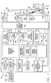
- a simplified block diagram is shown of a dual-chamber implantable stimulation device 10 which is capable of treating both fast and slow arrhythmias with stimulation therapy, including cardioversion, defibrillation, and pacing stimulation. While a dual-chamber device is shown, this is for illustration purposes only, and one of skill in the art could readily eliminate or disable the appropriate circuitry to provide a single-chamber stimulation device capable of treating one chamber with cardioversion, defibrillation and pacing stimulation.
- the stimulation device 10 is shown in electrical communication with a patient's heart 12 by way of an implantable atrial lead 20 having an atrial tip electrode 22 and an atrial ring electrode 24 which typically is implanted in the patient's atrial appendage.
- the stimulation device 10 is also shown in electrical communication with the patient's heart 12 by way of an implantable ventricular lead 30 having, in this embodiment, a ventricular tip electrode 32, a ventricular ring electrode 34, a right ventricular (RV) coil electrode 36, and an SVC coil electrode 38.
- the ventricular lead 30 is transvenously inserted into the heart 12 so as to place the RV coil electrode 36 in the right ventricular apex, and the SVC coil electrode 38 in the superior vena cava.
- the ventricular lead 30 is capable of receiving cardiac signals, and delivering stimulation in the form of pacing and shock therapy to the right ventricle.
- additional stimulation leads may be used in order to efficiently and effectively provide pacing stimulation to the left side of the heart or atrial cardioversion and/or defibrillation.
- a lead designed for placement in the coronary sinus region could be implanted to deliver left atrial pacing, atrial shocking therapy, and/or for left ventricular pacing stimulation.
- U.S. Patent Application No. 09/196,898 "A Self-Anchoring Coronary Sinus Lead” (Pianca et. al), and U.S. Patent No. 5,466,254, "Coronary Sinus Lead with Atrial Sensing Capability” (Helland), which patents are hereby incorporated herein by reference.
- the housing 40 (shown schematically) for the stimulation device 10 includes a connector (not shown) having an atrial pin terminal 42 and an atrial ring terminal 44, which are adapted for connection to the atrial tip electrode 22 and the atrial ring electrode 24, respectively.
- the housing 40 further includes a ventricular pin terminal 52, a ventricular ring terminal 54, a ventricular shocking terminal 56, and an SVC shocking terminal 58, which are adapted for connection to the ventricular tip electrode 32, the ventricular ring electrode 34, the RV coil electrode 36, and the SVC coil electrode 38, respectively.
- the housing 40 (often referred to as the "can", “case” or “case electrode”) may be programmably selected to act as the return electrode, or anode, alone or in combination with one of the coil electrodes, 36 and 38. For convenience, the names of the electrodes are shown next to the terminals.
- the microcontroller 60 includes a microprocessor, or equivalent control circuitry, designed specifically for controlling the delivery of stimulation therapy and may further include RAM or ROM memory, logic and timing circuitry, state machine circuitry, and I/O circuitry.
- the microcontroller 60 includes the ability to process or monitor input signals (data) as controlled by a program code stored in a designated block of memory. The details of the design and operation of the microcontroller 60 are not critical to the present invention. Rather, any suitable microcontroller 60 may be used that carries out the functions described herein.
- an atrial pulse generator 70 and a ventricular pulse generator 72 generate pacing stimulation pulses for delivery by the atrial lead 20 and the ventricular lead 30, respectively, via a switch bank 74.
- the pulse generators, 70 and 72 are controlled by the microcontroller 60 via appropriate control signals, 76 and 78, respectively, to trigger or inhibit the stimulation pulses.
- the microcontroller 60 further includes timing circuitry that controls the operation of the stimulation device timing of such stimulation pulses , that is well known in the art.
- the switch bank 74 includes a plurality of switches for switchably connecting the desired electrodes to the appropriate I/O circuits, thereby providing complete electrode programmability. Accordingly, the switch bank 74, in response to a control signal 80 from the microcontroller 60, determines the polarity of the stimulation pulses (e.g., unipolar or bipolar) by selectively closing the appropriate combination of switches (not shown) as is known in the art.
- An atrial sense amplifier 82 and a ventricular sense amplifier 84 are also coupled to the atrial and ventricular leads 20 and 30, respectively, through the switch bank 74 for detecting the presence of cardiac activity.
- the switch bank 74 determines the "sensing polarity" of the cardiac signal by selectively closing the appropriate switches, as is also known in the art. In this way, the clinician may program the sensing polarity independent of the stimulation polarity.
- Each sense amplifier, 82 and 84 preferably employs a low power, precision amplifier with programmable gain and/or automatic gain control, bandpass filtering, and a threshold detection circuit, known in the art, to selectively sense the cardiac signal of interest.
- the automatic gain control enables the device 10 to deal effectively with the difficult problem of sensing the low frequency, low amplitude signal characteristics of ventricular fibrillation.
- the outputs of the atrial and ventricular sense amplifiers, 82 and 84, are connected to the microcontroller 60 which, in turn, inhibit the atrial and ventricular pulse generators, 70 and 72, respectively, in a demand fashion whenever cardiac activity is sensed in the respective chambers.
- the present device utilizes the atrial and ventricular sense amplifiers, 82 and 84, to sense cardiac signals to determine whether a rhythm is physiologic or pathologic.
- sensing is reserved for the noting of an electrical depolarization
- detection is the processing of these sensed depolarization signals and noting the presence of an arrhythmia.
- the timing intervals between sensed events are then classified by the microcontroller 60 by comparing them to a predefined rate zone limit (i.e., bradycardia, normal, low rate VT, high rate VT, and fibrillation rate zones) and various other characteristics (e.g., sudden onset, stability, physiologic sensors, and morphology, etc.) in order to determine the type of remedial therapy that is needed (e.g., bradycardia pacing, anti-tachycardia pacing, cardioversion shocks or defibrillation shocks, also known as "tiered therapy").
- a predefined rate zone limit i.e., bradycardia, normal, low rate VT, high rate VT, and fibrillation rate zones
- various other characteristics e.g., sudden onset, stability, physiologic sensors, and morphology, etc.
- remedial therapy e.g., bradycardia pacing, anti-tachycardia pacing, cardioversion shock
- Cardiac signals are also applied to the inputs of an analog to digital (A/D) data acquisition system 90.
- the data acquisition system 90 is configured to acquire intracardiac electrogram signals, convert the raw analog data into a digital signal, and store the digital signals for later processing and/or telemetric transmission to an external device 102.
- the data acquisition system 90 is coupled to the atrial and ventricular leads, 20 and 30, through the switch bank 74 to sample cardiac signals across any pair of desired electrodes.
- the microcontroller 60 is further coupled to a memory 94 by a suitable data/address bus 96, wherein the programmable operating parameters used by the microcontroller 60 are stored and modified, as required, in order to customize the operation of the stimulation device 10 to suit the needs of a particular patient.
- Such operating parameters define, for example, pacing pulse amplitude, pulse duration, electrode polarity, rate, sensitivity, automatic features, arrhythmia detection criteria, and the amplitude, waveshape and vector of each shocking pulse to be delivered to the patient's heart 28 within each respective tier of therapy.
- the operating parameters of the implantable device 10 may be non-invasively programmed into the memory 94 through a telemetry circuit 100 in telemetric communication with an external device 102, such as a programmer, transtelephonic transceiver, or a diagnostic system analyzer.
- the telemetry circuit 100 is activated by the microcontroller by a control signal 106.
- the telemetry circuit 100 advantageously allows intracardiac electrograms and status information relating to the operation of the device 10 (as contained in the microcontroller 60 or memory 94) to be sent to the external device 102 through the established communication link 104.
- the stimulation device 10 further includes a physiologic sensor 110.
- a physiologic sensor 110 Such sensors are commonly called “rate-responsive" sensors.
- the physiological sensor 110 is used to detect the exercise state of the patient, to which the microcontroller 60 responds by adjusting the rate and AV Delay at which the atrial and ventricular pulse generators, 70 and 72, generate stimulation pulses.
- the type of sensor used is not critical to the present invention and is shown only for completeness.
- the stimulation device additionally includes a battery 114 which provides operating power to all of the circuits shown in Fig. 1.
- the battery must be capable of operating at low current drains for long periods of time , and then be capable of providing high-current pulses (for capacitor charging) when the patient requires a shock pulse.
- the battery 114 must also have a predictable discharge characteristic so that elective replacement time can be detected. Accordingly, the device employs lithium/silver vanadium oxide batteries, as is true for most (if not all) such devices to date.
- the microcontroller 60 further controls a shocking circuit 130 by way of a control signal 132.
- the shocking circuit 130 generates shocking pulses of low (up to 0.5 Joules), moderate (0.5 - 10 Joules), or high energy (11 to 40 Joules), as controlled by the microcontroller 60.
- Such shocking pulses are applied to the patient's heart through at least two shocking electrodes, and as shown in this embodiment, using the RV and SVC coil electrodes, 36 and 38, respectively.
- the housing 40 may act as an active electrode in combination with the RV electrode 36 alone, or as part of a split electrical vector using the SVC coil electrode 38 (i.e., using the RV electrode as common).
- Cardioversion shocks are generally considered to be of low to moderate energy level (so as to minimize pain felt by the patient), and/or synchronized with an R-wave and/or pertaining to the treatment of tachycardia.
- Defibrillation shocks are generally of moderate to high energy level (i.e., corresponding to thresholds in the range of 5-40 Joules), delivered asychronously (since R-waves may be too disorganized), and pertaining exclusively to the treatment of fibrillation.
- the microcontroller 60 is capable of controlling the synchronous or asynchronous delivery of the shocking pulses.
- ventricular lead 30 includes at least one contractility sensor 121, e.g., an accelerometer, a pressure sensor, and/or an impedance sensor (hereinafter referred to as the "contractility sensor(s) 121").
- Device 10 is configured operate to sense the contractility of the heart using the contractility sensor(s) 121 on ventricular lead 20 and, in response to a sudden increase in the contractility of the heart, to immediately pace the heart at an elevated rate to prevent vasovagal syncope from occurring, or to delay the progression of vasovagal syncope.
- device 10 includes signal conditioning circuitry 120 or other input circuit which receives analog signals via one or more terminals 123 and 124 from the contractility sensor(s) and converts the analog signals into digital signals from processing by controller 60.
- Controller 60 includes software for analyzing the signals received from the conditioning circuitry to determine the contractility of the heart. More specifically, controller 60 analyzes the signals to first estimate the amount of blood within the right ventricle. Based upon the estimate of the amount of blood in the right ventricle, controller 60 further then estimates the contractility of the muscles of the right ventricle.
- the controller also determines the average contractility and stores it in memory 94 or within a separate contractility storage register (not shown). Controller 60 then compares the sensed contractility of the heart with the average contractility of the heart and controls atrial and ventricular pulse generators 70 and 72 to administer pacing pulses to the heart at an accelerated rate in an effort to compensate for the reduction in blood pressure otherwise occurring prior to vasovagal syncope.
- controller 60 may determine contractility based on pressure, acceleration or pressure signals, or a combination thereof, received from the contractility sensor(s).
- One technique for estimating contractility based upon electrical impedance detected within the heart is described within,U.S. Patent No. 5,800,467 to Park et al., entitled “Cardio-Synchronous Impedance Measurement System for an Implantable Stimulation Device", which is incorporated by reference herein. Briefly, blood is an electrical conductor and, hence, a decrease in the amount of blood in the ventricle results in an increase in electrical impedance. Impedance therefore, as measured within the ventricle of the heart, provides an indication of the amount of blood in the ventricle.
- Controller 60 monitors the impedance of the heart and detects any sharp increase in the impedance from one cycle to the next.
- the increase is impedance is therefore interpreted as a decrease in the amount of blood filling the ventricle during the cardiac cycle.
- a sudden drop in ventricular filling causes the ventricle to contract more vigorously, i.e. the contractility of the ventricle increases.
- the sharp increase in contractility causes mechano-receptors within the ventricle to transmit signals to the vagus nerve which are forwarded to the brainstem.
- the resulting surge in signals is misinterpreted as being representative of hypertension and the brainstem dilates blood vessels and decreases the heart rate in an attempt to compensate for the hypertension.
- One technique for estimating contractility based upon signals from an accelerometer within the right ventricle is as follows.
- the accelerometer generates signals representative of the speed at which the ventricular wall moves during a contraction.
- the increase in contractility caused by a sudden drop in ventricular filling results in the ventricular walls moving more forcefully.
- controller 60 is able to estimate the contractility of the ventricle.
- controller 60 tracks the average and current contractility of the heart.
- the average contractility stored within memory 94, is determined, for example, by averaging the measured contractility over relatively long periods of time such as minutes, hours, days, weeks, months or years.
- Controller 60 periodically compares the measured contractility with the average contractility and detects any sharp increase in contractility from one cardiac cycle to the next. As noted above, a sharp increase in contractility from one period to the next is indicative of the onset of vasovagal syncope, at least within patients subject to recurrent vasovagal syncope syndrome. If a sharp increase in contractility is detected, the controller promptly controls atrial and ventricular pulse generators 70 and 72 to increases the heart rate to compensate for the drop in blood pressure that initially triggered the increase in contractility.
- a flow chart is shown describing an overview of the operation of the novel features of the present invention.
- the various algorithmic steps are summarized in individual “blocks”. Such blocks describe specific actions or decisions made or carried out as the algorithm proceeds.
- the flow charts presented herein provide the basis for a "control program” that may be used by such a microcontroller (or equivalent) to effectuate the desired control of the stimulation device.
- a control program that may be used by such a microcontroller (or equivalent) to effectuate the desired control of the stimulation device.
- Those skilled in the art may readily write such a control program based on the flow charts and other descriptions presented herein.
- FIG. 2 illustrates a pacing regime which may be employed by controller 60 of FIG. 1 for prevention of vasovagal syncope.
- the pacing rate is immediately increased from an initial pacing rate, PR1, (which may be a programmed base rate such as 60 bpm) to a syncope prevention rate, PR2 (which may be a programmable value, for example, 20, 30 or 40 bpm above the initial pacing rate, PR1).
- PR1 initial pacing rate
- PR2 syncope prevention rate
- the increase from initial pacing rate, PR1, to syncope prevention rate, PR2 could be a linear increase performed during time T1 which may be, for example, in the range of three to five seconds.
- the heart is then continuously paced at syncope prevention rate, PR2, for time T2 which may be, for example, in the range of 30 seconds to 3 minutes. Then, the pacing rate is reduced from syncope prevention rate, PR2, to initial pacing rate, PR1, during T3 wherein T3 is, for example, in the range of 10 to 30 seconds.
- each individual time value T1, T2 and T3 is not separately programmable. Rather, the overall time period T4 is programmed, and the pacemaker automatically calculates the subportions T1, T2 and T3 in accordance with prestored timing ratios. For ease of implementation T1, T2, T3 and T4 could be replaced with the number of the corresponding beats.
- the drop in blood pressure that would otherwise occur during the onset of vasovagal syncope syndrome is at least partially compensated.
- the increase in the heart rate may not be sufficient to prevent the blood pressure from dropping to the point where the patient feels lightheaded or dizzy, it is typically sufficient to prevent the blood pressure from dropping to the point wherein the patient becomes unconscious.
- the loss of consciousness is delayed by at least some amount permitting the patient to take whatever action is necessary to ensure his or her safety. For example, if the patient is driving a vehicle, the patient, upon becoming lightheaded and dizzy, will preferably pull the vehicle to the side of the road. Thus, even if the patient loses consciousness, the delay in loss of consciousness may be sufficient to save the patient from injury.
- the increase in pacing rate based upon a contractility measurement By triggering the increase in pacing rate based upon a contractility measurement, the increase is triggered much more promptly than typically occurs within pacemakers configured to detect the onset of vasovagal syncope based upon bradycardia. As noted above, by the time a pacemaker has detected bradycardia, the blood patient of the patient may already have dropped to the point where a subsequent increase in the heart rate is ineffective for preventing immediate loss of consciousness. By triggering the increase in heart rate based upon a sharp change in contractility, the increase is typically triggered within one or two beats subsequent to the initial drop in blood pressure which marks the onset of vasovagal syncope. By significantly increasing the heart rate before the blood pressure has a chance to drop significantly, the pacemaker thereby either prevents any drop in blood pressure or, at the very least, defers the drop in blood pressure thereby delaying or preventing unconsciousness.
- step 200 device 10 inputs the electrical impedance signals, pressure, and/or accelerometer signals.
- step 202 device 10 determines the contractility and, at step 204, compares the detected contractility with the average contractility. If, at step 205, the contractility increases to the point where the detected contractility minus the average contractility exceeds a predetermined threshold, then step 206 is performed wherein the device 10 immediately increases the pacing rate to the vasovagal syncope preventative pacing mode using the pacing rate defined in FIG. 2. Otherwise, processing returns to step 200 wherein device 10 continues to monitor the input impedance, pressure and accelerometer signals.
- Vasovagal syncope preventative pacing mode performed at step 206 is administered, as described in connection with FIG. 2, for example, for about 30 seconds to 3 minutes. Then, the heart rate is reduced to a base pacing rate or pacing is eliminated completely. Ultimately, processing returns to step 100 to monitor impedance, accelerometer signals and/or pressure.
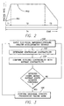
- FIG. 3 illustrates, at a high level, steps performed to predict and prevent vasovagal syncope using device 10.
- device 10 additionally performs numerous other functions simultaneously, such as monitoring the heart to detect heart arrhythmias, such as atrial tachycardia. If a significant heart arrhythmia is detected while the pacemaker is administering the vasovagal syncope preventative pacing mode, the preventative pacing is terminated and the pacemaker administers whatever therapy is appropriate for the arrhythmia.
- the therapy may include the administration of defibrillation pulses or the like.
- FIGS. 4 and 5 illustrate a sequence of steps performed to provide vasovagal preventative pacing within device 10 if the device is configured to pace at a sensor indicated rate (SIR).
- SIR pacing is described in further detail in U.S. Patent No. 5,476,483 to Bornzin et al., entitled “System and Method for Modulating the Base Rate During Sleep for a Rate-Responsive Cardiac Pacemaker", which is incorporated by reference herein.
- Steps 300, 302, 304, 314, 320, 322 and 312 are steps performed by device 10 in response to the initial detection of a sharp increase in contractility wherein the vasovagal preventative pacing mode is initiated.
- the patient is paced at either the vasovagal preventative pacing rate (VPPR) illustrated in FIG. 2 or a predetermined SIR, whichever is greater.
- VPPR vasovagal preventative pacing rate
- vasovagal preventative pacing mode will be reinitiated so long as the contractility remains far above the average contractility level (step 304), i.e., the detected contractility minus the average contractility exceeds the predetermined threshold.
- VPPR pacing is suspended only as long as needed to detect that the contractility is still high and, during a subsequent execution of the steps of FIG. 4, VPPR pacing is again triggered.
- the pacemaker takes the patient out of the vasovagal preventative pacing mode. This is provided by steps 300, 302, 304, 306, 316, 308, 310 and 312 and also by steps 300, 302, 304, 306, 316, 308, 318 and 312.
Abstract
A pacemaker or other cardiac stimulation device (10) is configured
to predict the onset of vasovagal syncope and administers pacing therapy
to prevent the syncope from occurring. Prediction of the onset of
vasovagal syncope is based upon an analysis of the contractility of the
heart muscle. In an example described herein, the contractility of the
heart muscle is detected and compared with the average contractility. If
the current contractility exceeds the average contractility by a
predetermined threshold, a high risk of vasovagal syncope is thereby
detected and the heart is paced at a vasovagal syncope prevention rate
which may be, for example, 20 to 40 beats per minute faster than a
previous heart rate. The contractility of the heart is determined, for
example, by measuring the impedance of the heart tissue or by
measuring the movement of heart tissue in the wall of the heart or by
pressure using an accelerometer. The contractility is averaged over a
period of time to determine the average contractility.
Description
- The invention generally relates to cardiac pacing techniques and in particular to techniques for preventing vasovagal syncope using cardiac pacing.
- Syncope is a sudden loss of strength or consciousness caused by reduced cerebral circulation, itself typically the result of vasodilation. Vasovagal syncope is a type of syncope referred to as a neurocardiogenic syncope wherein the syncope is triggered by an interaction between the heart and nerve tissue connected to the heart. Neurocardiogenic syncope may also be referred to as neuromediated syncope, neurally mediated syncope, neurocardiogenic syncope, cardioneurogenic syncope, vasodepressor syncope, malignant vasovagal syndrome, neurally mediated hypotension/bradycardia and cardiovascular neurogenic syncope. For vasovagal syncope, the interaction occurs between the heart and the vagus nerve.
- Evidence suggests that vasovagal syncope is initially triggered by a sudden reduction in peripheral vascular resistance, perhaps resulting from stress, pooling of blood in the extremities, or other factors. As a result of the reduction in peripheral vascular resistance, the pressure of blood entering the heart drops and the amount of blood filling the ventricles prior to ventricular contractions therefore also drops. With less blood in the ventricles, the ventricles thereby contract much more quickly and vigorously than would otherwise occur in an effort to maintain a constant stroke volume. The more vigorous ventricular contractions have the effect of stimulating ventricular mechanoreceptors, also known as C fibers, that normally only respond to ventricular expansion or stretching, rather than contraction. The activation of the ventricular mechanoreceptors results in a surge in neural traffic to the brainstem, particularly to the nucleus tractus solitaries, via the vasovagal nerve.
- For most people, the neurological system properly interprets the increase of activity of the mechanoreceptors as being in response to a drop in peripheral vascular resistance and compensates by increasing the heart rate and constricting the blood vessels. However, in certain patients, as a result of a neurological condition within the vagus nerve or for some other reason, the surge in neural traffic is falsely perceived by the neurological system as being representative of hypertension. In response thereto, the brainstem triggers an increase in peripheral vasodilation and a reduction in heart rate. The vasodilation and the drop in heart rate, in turn, cause a still further reduction in blood pressure, i.e. hypotension. In other words, the actions taken by the brainstem exacerbate the problem. If the degree of hypotension is sufficiently severe, cerebral hypoperfusion occurs wherein brain cells do not receive enough oxygen and, consequently, the victim loses consciousness. Accordingly, within these patients, any sudden drop in peripheral vascular resistance can trigger vasovagal syncope and the patients are the to suffer from recurrent vasovagal syncope. Further information regarding vasovagal syncope may be found in S. Serge Harold and Jacques Mugica, Recent Advances in Cardiac Pacing, Futura Publishing Company, 92-95, 1997.
- As can be appreciated, loss of consciousness can be particularly dangerous for the patient if occurring while the patient is driving a motor vehicle, climbing a flight of stairs or engaged in any other activity wherein the loss of consciousness could result in injury or death. Accordingly, it is highly desirable to provide techniques for preventing vasovagal syncope or other forms of neurocardiological syncope. One possible technique for preventing vasovagal syncope is to employ a pacemaker, or other implantable cardiac pacing device, to pace the heart to prevent the reduction in blood pressure associated with vasovagal syncope from occurring. Indeed, the American College of Cardiology-American Heart Association suggested in 1991 that vasovagal syncope in patients should be used as a Class 2 indication for pacing therapy. However, conventional cardiac pacemakers have had only limited success in preventing recurrent vasovagal syncope. (See David G. Benditt et al., Cardiac Pacing for Prevention of Recurrent Vasovagal Syncope, Ann Intern Med. 1995; 122; 204-209.) Insofar as vasovagal syncope is concerned, conventional pacemakers only respond to the reduction in heart rate associated therewith. The reduction in heart rate, also referred to as bradycardia, is detected by the pacemaker, which increases the heart rate to compensate Depending upon the programming of the pacemaker, it may take five to eight seconds or more before the pacemaker begins increasing the heart rate in response to detection of bradycardia. For many patients subject to recurrent vasovagal syncope, a significant drop in blood pressure occurs before there is any bradycardia. Hence, by the time the pacemaker begins to increase the heart rate, the blood pressure will already have dropped to the point where the increase in heart rate is ineffective. In this regard, the drop in blood pressure results in significantly less blood filling the ventricles, such that there is simply not enough incoming blood to pump. Hence, overall blood pressure is not significantly increased merely by pumping the heart faster, and the aforementioned cerebral hypoperfusion still occurs resulting in unconsciousness. Moreover, for at least some patients subject to recurrent vasovagal syncope, there is no significant drop in heart rate, only a drop in blood pressure. Hence, pacemakers programmed to prevent vasovagal syncope based solely upon detection of bradycardia will have little or no effect.
- Thus, bradycardia-triggered vasovagal syncope prevention techniques employed by some conventional cardiac pacemakers often fail to prevent the syncope. Moreover, conventional cardiac pacemakers are often unable to distinguish physiologic bradycardia such as that associated with sleep from bradycardia associated with vasovagal syncope or other neurocardiogenic syncope.
- Accordingly, it would be highly desirable to provide improved cardiac pacing devices capable of predicting the onset of vasovagal syncope or other types of neurocardiogenic syncope, and for reliably administering pacing therapy to prevent the syncope from occurring. It is to these ends that the invention is primarily directed.
- In accordance with one aspect of the invention, a cardiac pacing device is provided with a vasovagal syncope prediction and prevention unit for predicting the onset of vasovagal syncope and for administering pacing therapy to prevent the syncope from occurring. Prediction of the onset of vasovagal syncope is based upon an analysis of the contractility of the heart muscle.
- In an exemplary embodiment, prediction of the onset of vasovagal syncope is achieved by sensing the contractility of the heart muscle and determining whether the sensed contractility exceeds an average contractility by a predetermined threshold amount. If so, the heart is paced at a vasovagal syncope prevention rate which may be, for example, 20 to 40 beats per minute faster than a previous heart rate. The contractility of the heart is determined, for example, by measuring the electrical impedance change or pressure change in the ventricle, or by measuring the movement of heart tissue in the wall of the heart using an accelerometer. The contractility measured is averaged over a period of time to determine the average contractility.
- In this manner, the onset of vasovagal syncope is predicted based upon changes in heart muscle contractility and syncope is substantially prevented from occurring by immediately administering vasovagal syncope preventative pacing measures. Because prediction of imminent vasovagal syncope is based upon a measurement of the contractility of the heart tissue, rather than upon detection of an episode of bradycardia, vasovagal syncope is thereby detected even in circumstances wherein hypotension occurs before bradycardia or wherein bradycardia does not occur at all. Also, physiologic episodes of bradycardia, such as those associated with sleep, do not erroneously trigger the vasovagal syncope pacing therapy. Moreover, the increase in contractility associated with vasovagal syncope can be detected quickly whereas reliable bradycardia detection may require analysis of several heart beats. Hence, preventative pacing may be administered before the blood pressure has dropped significantly, as could otherwise occur if relying solely on bradycardia detection. Two or more independent contractility sensors may be provided to help prevent a false positive detection of vasovagal syncope.
- Method embodiments of the invention are also provided as set out in the appended claims.
- In a preferred aspect of the method, the step of detecting the contractility of the heart is performed to detect contractility of the left ventricle.
- Preferably the step of detecting the contractility of the heart includes the step of detecting the electrical impedance of blood within one of the ventricles of the heart.
- Preferably the step of detecting the contractility of the heart includes the step of detecting the movement of muscle tissue in the wall of one of the ventricles of the heart using an accelerometer.
- Preferably the step of determining whether there is a significant risk of the onset of vasovagal syncope includes the steps of:
- determining an average contractility of the heart for a plurality of cardiac cycles;
- comparing the contractility of the heart detected during a current cardiac cycle with an average contractility of the heart a plurality of cardiac cycles; and
- concluding that there is a significant risk of the onset of vasovagal syncope if the contractility of the heart detected during the current cardiac cycle exceeds the average contractility of the heart for a plurality of cardiac cycles by a predetermined threshold, and preferably the step of determining the average contractility of the heart for a plurality of cardiac cycles includes the step of iteratively updating the average contractility using each new value of the detected contractility.
-
- Preferably the implantable medical device is a cardiac pacing device and wherein the method further includes the step of:
responsive to a determination that there is a significant risk of the onset of vasovagal syncope, pacing the heart to reduce the likelihood of the vasovagal syncope and preferably the step of pacing the heart to reduce the likelihood of the vasovagal syncope includes the steps of: - incrementally increasing a pacing rate from a preexisting pacing rate to a predetermined vasovagal syncope prevention rate during a first predetermined period of time;
- maintaining the pacing rate at the predetermined vasovagal syncope prevention rate during a second predetermined period of time; and
- incrementally decreasing the pacing rate from the vasovagal prevention rate to the preexisting pacing rate during a third predetermined period of time, and preferably the first, second and third predetermined period of times are in the range of 3 to 5 seconds, 30 to 180 seconds, and 10 to 30 seconds, respectively, and/or the vasovagal syncope prevention rate is in the range of 20 to 40 beats per minute above the preexisting pacing rate, and or if the preexisting pacing rate exceeds the vasovagal syncope prevention rate, the pacemaker continues to pace at the preexisting rate, and/or the preexisting pacing rate is a predetermined base pacing rate, and/or the preexisting pacing rate is a sensor responsive pacing rate.
-
- Embodiments of the invention will now be described, by way of example, with reference to the drawings of which:
- Fig. 1 is a functional block diagram of a dual-chamber implantable stimulation device illustrating the basic elements of a stimulation device which can provide cardioversion, defibrillation and pacing stimulation and can predict and prevent vasovagal syncope in accordance with an exemplary embodiment of the invention;
- FIG. 2 is a graph illustrating a vasovagal syncope preventative pacing regime;
- FIG. 3 is a flow chart illustrating steps performed by the pacemaker components of FIG. 2; and
- FIGS. 4-5 illustrate a detailed flow chart describing the steps performed by a pacemaker configured to pace the heart using a sensor indicated rate (SIR) and also configured to perform vasovagal syncope preventative pacing.
-
- In the description of the invention that follows, like numerals or reference designators will be used to refer to like parts or elements throughout.
- In Fig. 1, a simplified block diagram is shown of a dual-chamber
implantable stimulation device 10 which is capable of treating both fast and slow arrhythmias with stimulation therapy, including cardioversion, defibrillation, and pacing stimulation. While a dual-chamber device is shown, this is for illustration purposes only, and one of skill in the art could readily eliminate or disable the appropriate circuitry to provide a single-chamber stimulation device capable of treating one chamber with cardioversion, defibrillation and pacing stimulation. - To provide atrial chamber pacing stimulation and sensing, the
stimulation device 10 is shown in electrical communication with a patient'sheart 12 by way of an implantableatrial lead 20 having anatrial tip electrode 22 and anatrial ring electrode 24 which typically is implanted in the patient's atrial appendage. - The
stimulation device 10 is also shown in electrical communication with the patient'sheart 12 by way of animplantable ventricular lead 30 having, in this embodiment, aventricular tip electrode 32, aventricular ring electrode 34, a right ventricular (RV)coil electrode 36, and anSVC coil electrode 38. Typically, theventricular lead 30 is transvenously inserted into theheart 12 so as to place theRV coil electrode 36 in the right ventricular apex, and theSVC coil electrode 38 in the superior vena cava. Accordingly, theventricular lead 30 is capable of receiving cardiac signals, and delivering stimulation in the form of pacing and shock therapy to the right ventricle. - While only two leads are shown in FIG. 1, it is to be understood that additional stimulation leads (with one or more pacing, sensing and/or shocking electrodes) may be used in order to efficiently and effectively provide pacing stimulation to the left side of the heart or atrial cardioversion and/or defibrillation. For example, a lead designed for placement in the coronary sinus region could be implanted to deliver left atrial pacing, atrial shocking therapy, and/or for left ventricular pacing stimulation. For a complete description of a coronary sinus lead, see U.S. Patent Application No. 09/196,898, "A Self-Anchoring Coronary Sinus Lead" (Pianca et. al), and U.S. Patent No. 5,466,254, "Coronary Sinus Lead with Atrial Sensing Capability" (Helland), which patents are hereby incorporated herein by reference.
- The housing 40 (shown schematically) for the
stimulation device 10 includes a connector (not shown) having anatrial pin terminal 42 and anatrial ring terminal 44, which are adapted for connection to theatrial tip electrode 22 and theatrial ring electrode 24, respectively. Thehousing 40 further includes aventricular pin terminal 52, aventricular ring terminal 54, a ventricularshocking terminal 56, and an SVCshocking terminal 58, which are adapted for connection to theventricular tip electrode 32, theventricular ring electrode 34, theRV coil electrode 36, and theSVC coil electrode 38, respectively. The housing 40 (often referred to as the "can", "case" or "case electrode") may be programmably selected to act as the return electrode, or anode, alone or in combination with one of the coil electrodes, 36 and 38. For convenience, the names of the electrodes are shown next to the terminals. - At the core of the
stimulation device 10 is aprogrammable microcontroller 60 which controls the various modes of stimulation therapy. As is well known in the art, themicrocontroller 60 includes a microprocessor, or equivalent control circuitry, designed specifically for controlling the delivery of stimulation therapy and may further include RAM or ROM memory, logic and timing circuitry, state machine circuitry, and I/O circuitry. Typically, themicrocontroller 60 includes the ability to process or monitor input signals (data) as controlled by a program code stored in a designated block of memory. The details of the design and operation of themicrocontroller 60 are not critical to the present invention. Rather, anysuitable microcontroller 60 may be used that carries out the functions described herein. The use of microprocessor-based control circuits for performing timing and data analysis functions is well known in the art. As shown in Fig. 1, anatrial pulse generator 70 and aventricular pulse generator 72 generate pacing stimulation pulses for delivery by theatrial lead 20 and theventricular lead 30, respectively, via aswitch bank 74. The pulse generators, 70 and 72, are controlled by themicrocontroller 60 via appropriate control signals, 76 and 78, respectively, to trigger or inhibit the stimulation pulses. Themicrocontroller 60 further includes timing circuitry that controls the operation of the stimulation device timing of such stimulation pulses , that is well known in the art. - The
switch bank 74 includes a plurality of switches for switchably connecting the desired electrodes to the appropriate I/O circuits, thereby providing complete electrode programmability. Accordingly, theswitch bank 74, in response to acontrol signal 80 from themicrocontroller 60, determines the polarity of the stimulation pulses (e.g., unipolar or bipolar) by selectively closing the appropriate combination of switches (not shown) as is known in the art. Anatrial sense amplifier 82 and aventricular sense amplifier 84 are also coupled to the atrial and ventricular leads 20 and 30, respectively, through theswitch bank 74 for detecting the presence of cardiac activity. Theswitch bank 74 determines the "sensing polarity" of the cardiac signal by selectively closing the appropriate switches, as is also known in the art. In this way, the clinician may program the sensing polarity independent of the stimulation polarity. - Each sense amplifier, 82 and 84, preferably employs a low power, precision amplifier with programmable gain and/or automatic gain control, bandpass filtering, and a threshold detection circuit, known in the art, to selectively sense the cardiac signal of interest. The automatic gain control enables the
device 10 to deal effectively with the difficult problem of sensing the low frequency, low amplitude signal characteristics of ventricular fibrillation. - The outputs of the atrial and ventricular sense amplifiers, 82 and 84, are connected to the
microcontroller 60 which, in turn, inhibit the atrial and ventricular pulse generators, 70 and 72, respectively, in a demand fashion whenever cardiac activity is sensed in the respective chambers. - For arrhythmia detection, the present device utilizes the atrial and ventricular sense amplifiers, 82 and 84, to sense cardiac signals to determine whether a rhythm is physiologic or pathologic. As used herein "sensing" is reserved for the noting of an electrical depolarization, and "detection" is the processing of these sensed depolarization signals and noting the presence of an arrhythmia. The timing intervals between sensed events (e.g., the P-P and R-R intervals) are then classified by the
microcontroller 60 by comparing them to a predefined rate zone limit (i.e., bradycardia, normal, low rate VT, high rate VT, and fibrillation rate zones) and various other characteristics (e.g., sudden onset, stability, physiologic sensors, and morphology, etc.) in order to determine the type of remedial therapy that is needed (e.g., bradycardia pacing, anti-tachycardia pacing, cardioversion shocks or defibrillation shocks, also known as "tiered therapy"). - Cardiac signals are also applied to the inputs of an analog to digital (A/D)
data acquisition system 90. Thedata acquisition system 90 is configured to acquire intracardiac electrogram signals, convert the raw analog data into a digital signal, and store the digital signals for later processing and/or telemetric transmission to anexternal device 102. Thedata acquisition system 90 is coupled to the atrial and ventricular leads, 20 and 30, through theswitch bank 74 to sample cardiac signals across any pair of desired electrodes. - The
microcontroller 60 is further coupled to amemory 94 by a suitable data/address bus 96, wherein the programmable operating parameters used by themicrocontroller 60 are stored and modified, as required, in order to customize the operation of thestimulation device 10 to suit the needs of a particular patient. Such operating parameters define, for example, pacing pulse amplitude, pulse duration, electrode polarity, rate, sensitivity, automatic features, arrhythmia detection criteria, and the amplitude, waveshape and vector of each shocking pulse to be delivered to the patient's heart 28 within each respective tier of therapy. - Advantageously, the operating parameters of the
implantable device 10 may be non-invasively programmed into thememory 94 through atelemetry circuit 100 in telemetric communication with anexternal device 102, such as a programmer, transtelephonic transceiver, or a diagnostic system analyzer. Thetelemetry circuit 100 is activated by the microcontroller by acontrol signal 106. Thetelemetry circuit 100 advantageously allows intracardiac electrograms and status information relating to the operation of the device 10 (as contained in themicrocontroller 60 or memory 94) to be sent to theexternal device 102 through the establishedcommunication link 104. - In the preferred embodiment, the
stimulation device 10 further includes aphysiologic sensor 110. Such sensors are commonly called "rate-responsive" sensors. Thephysiological sensor 110 is used to detect the exercise state of the patient, to which themicrocontroller 60 responds by adjusting the rate and AV Delay at which the atrial and ventricular pulse generators, 70 and 72, generate stimulation pulses. The type of sensor used is not critical to the present invention and is shown only for completeness. - The stimulation device additionally includes a
battery 114 which provides operating power to all of the circuits shown in Fig. 1. For thestimulation device 10, which employs shocking therapy, the battery must be capable of operating at low current drains for long periods of time , and then be capable of providing high-current pulses (for capacitor charging) when the patient requires a shock pulse. Thebattery 114 must also have a predictable discharge characteristic so that elective replacement time can be detected. Accordingly, the device employs lithium/silver vanadium oxide batteries, as is true for most (if not all) such devices to date. - It is the primary function of the invention to function as an implantable cardioverter/defibrillator (ICD) device. That is, it must detect the occurrence of an arrhythmia, and automatically apply an appropriate electrical shock therapy to the heart aimed at terminating the detected arrhythmia. To this end, the
microcontroller 60 further controls ashocking circuit 130 by way of acontrol signal 132. Theshocking circuit 130 generates shocking pulses of low (up to 0.5 Joules), moderate (0.5 - 10 Joules), or high energy (11 to 40 Joules), as controlled by themicrocontroller 60. Such shocking pulses are applied to the patient's heart through at least two shocking electrodes, and as shown in this embodiment, using the RV and SVC coil electrodes, 36 and 38, respectively. In alternative embodiments, thehousing 40 may act as an active electrode in combination with theRV electrode 36 alone, or as part of a split electrical vector using the SVC coil electrode 38 (i.e., using the RV electrode as common). - Cardioversion shocks are generally considered to be of low to moderate energy level (so as to minimize pain felt by the patient), and/or synchronized with an R-wave and/or pertaining to the treatment of tachycardia. Defibrillation shocks are generally of moderate to high energy level (i.e., corresponding to thresholds in the range of 5-40 Joules), delivered asychronously (since R-waves may be too disorganized), and pertaining exclusively to the treatment of fibrillation. Accordingly, the
microcontroller 60 is capable of controlling the synchronous or asynchronous delivery of the shocking pulses. - In addition to the aforementioned components,
ventricular lead 30 includes at least onecontractility sensor 121, e.g., an accelerometer, a pressure sensor, and/or an impedance sensor (hereinafter referred to as the "contractility sensor(s) 121").Device 10 is configured operate to sense the contractility of the heart using the contractility sensor(s) 121 onventricular lead 20 and, in response to a sudden increase in the contractility of the heart, to immediately pace the heart at an elevated rate to prevent vasovagal syncope from occurring, or to delay the progression of vasovagal syncope. To this end,device 10 includessignal conditioning circuitry 120 or other input circuit which receives analog signals via one ormore terminals controller 60.Controller 60 includes software for analyzing the signals received from the conditioning circuitry to determine the contractility of the heart. More specifically,controller 60 analyzes the signals to first estimate the amount of blood within the right ventricle. Based upon the estimate of the amount of blood in the right ventricle,controller 60 further then estimates the contractility of the muscles of the right ventricle. - The controller also determines the average contractility and stores it in
memory 94 or within a separate contractility storage register (not shown).Controller 60 then compares the sensed contractility of the heart with the average contractility of the heart and controls atrial andventricular pulse generators - As noted,
controller 60 may determine contractility based on pressure, acceleration or pressure signals, or a combination thereof, received from the contractility sensor(s). One technique for estimating contractility based upon electrical impedance detected within the heart is described within,U.S. Patent No. 5,800,467 to Park et al., entitled "Cardio-Synchronous Impedance Measurement System for an Implantable Stimulation Device", which is incorporated by reference herein. Briefly, blood is an electrical conductor and, hence, a decrease in the amount of blood in the ventricle results in an increase in electrical impedance. Impedance therefore, as measured within the ventricle of the heart, provides an indication of the amount of blood in the ventricle.Controller 60 monitors the impedance of the heart and detects any sharp increase in the impedance from one cycle to the next. The increase is impedance is therefore interpreted as a decrease in the amount of blood filling the ventricle during the cardiac cycle. As noted above, a sudden drop in ventricular filling causes the ventricle to contract more vigorously, i.e. the contractility of the ventricle increases. The sharp increase in contractility causes mechano-receptors within the ventricle to transmit signals to the vagus nerve which are forwarded to the brainstem. For patients with recurrent vasovagal syncope, the resulting surge in signals is misinterpreted as being representative of hypertension and the brainstem dilates blood vessels and decreases the heart rate in an attempt to compensate for the hypertension. This results in a significant drop in blood pressure causing hypoperfusion of the cerebrum and possibly loss of consciousness. Hence, a sharp increase in impedance from one period of ventricular contraction to the next is a good indicator of the potential onset of vasovagal syncope, at least within patients who suffer from recurrent vasovagal syncope. - One technique for estimating contractility based upon signals from an accelerometer within the right ventricle is as follows. The accelerometer generates signals representative of the speed at which the ventricular wall moves during a contraction. The increase in contractility caused by a sudden drop in ventricular filling results in the ventricular walls moving more forcefully. Hence, by monitoring the signals received from the accelerometer,
controller 60 is able to estimate the contractility of the ventricle. - One specific technique for measuring heart contractility using an accelerometer is described within U.S. Patent No. 5,480,412 to Mouchawar et al., entitled "System and Method for Deriving Hemodynamic Signals from a Cardiac Wall Motion Sensor", which is incorporated by reference herein. One specific technique for measuring heart contractility using an impedance sensor is described within U.S. Patent No. 5,139,020 to Koestner, et al., entitled "Method and Apparatus for Controlling the Hemodynamic State of a Patient Based on Systolic Time Interval Measurements Detecting Using Doppler Ultrasound Techniques", which is incorporated by reference herein.
- Thus,
controller 60 tracks the average and current contractility of the heart. The average contractility, stored withinmemory 94, is determined, for example, by averaging the measured contractility over relatively long periods of time such as minutes, hours, days, weeks, months or years.Controller 60 periodically compares the measured contractility with the average contractility and detects any sharp increase in contractility from one cardiac cycle to the next. As noted above, a sharp increase in contractility from one period to the next is indicative of the onset of vasovagal syncope, at least within patients subject to recurrent vasovagal syncope syndrome. If a sharp increase in contractility is detected, the controller promptly controls atrial andventricular pulse generators - In Fig. 2, a flow chart is shown describing an overview of the operation of the novel features of the present invention. In this flow chart, and the other flow charts described herein, the various algorithmic steps are summarized in individual "blocks". Such blocks describe specific actions or decisions made or carried out as the algorithm proceeds. Where a microcontroller (or equivalent) is employed, the flow charts presented herein provide the basis for a "control program" that may be used by such a microcontroller (or equivalent) to effectuate the desired control of the stimulation device. Those skilled in the art may readily write such a control program based on the flow charts and other descriptions presented herein.
- More specifically, FIG. 2 illustrates a pacing regime which may be employed by
controller 60 of FIG. 1 for prevention of vasovagal syncope. Upon detection of the sharp increase in contractility from one cardiac cycle to the next, the pacing rate is immediately increased from an initial pacing rate, PR1, (which may be a programmed base rate such as 60 bpm) to a syncope prevention rate, PR2 (which may be a programmable value, for example, 20, 30 or 40 bpm above the initial pacing rate, PR1). The increase from initial pacing rate, PR1, to syncope prevention rate, PR2, could be a linear increase performed during time T1 which may be, for example, in the range of three to five seconds. The heart is then continuously paced at syncope prevention rate, PR2, for time T2 which may be, for example, in the range of 30 seconds to 3 minutes. Then, the pacing rate is reduced from syncope prevention rate, PR2, to initial pacing rate, PR1, during T3 wherein T3 is, for example, in the range of 10 to 30 seconds. In the alternative, each individual time value T1, T2 and T3 is not separately programmable. Rather, the overall time period T4 is programmed, and the pacemaker automatically calculates the subportions T1, T2 and T3 in accordance with prestored timing ratios. For ease of implementation T1, T2, T3 and T4 could be replaced with the number of the corresponding beats. - By increasing the heart rate to syncope prevention rate, PR2, the drop in blood pressure that would otherwise occur during the onset of vasovagal syncope syndrome is at least partially compensated. Although the increase in the heart rate may not be sufficient to prevent the blood pressure from dropping to the point where the patient feels lightheaded or dizzy, it is typically sufficient to prevent the blood pressure from dropping to the point wherein the patient becomes unconscious. Even if the patient ultimately becomes unconsciousness, the loss of consciousness is delayed by at least some amount permitting the patient to take whatever action is necessary to ensure his or her safety. For example, if the patient is driving a vehicle, the patient, upon becoming lightheaded and dizzy, will preferably pull the vehicle to the side of the road. Thus, even if the patient loses consciousness, the delay in loss of consciousness may be sufficient to save the patient from injury.
- By triggering the increase in pacing rate based upon a contractility measurement, the increase is triggered much more promptly than typically occurs within pacemakers configured to detect the onset of vasovagal syncope based upon bradycardia. As noted above, by the time a pacemaker has detected bradycardia, the blood patient of the patient may already have dropped to the point where a subsequent increase in the heart rate is ineffective for preventing immediate loss of consciousness. By triggering the increase in heart rate based upon a sharp change in contractility, the increase is typically triggered within one or two beats subsequent to the initial drop in blood pressure which marks the onset of vasovagal syncope. By significantly increasing the heart rate before the blood pressure has a chance to drop significantly, the pacemaker thereby either prevents any drop in blood pressure or, at the very least, defers the drop in blood pressure thereby delaying or preventing unconsciousness.
- The steps performed by the components of FIG. 1 for vasovagal detection and prevention are summarized in FIG. 3. Briefly, at
step 200,device 10 inputs the electrical impedance signals, pressure, and/or accelerometer signals. Atstep 202,device 10 determines the contractility and, atstep 204, compares the detected contractility with the average contractility. If, at step 205, the contractility increases to the point where the detected contractility minus the average contractility exceeds a predetermined threshold, then step 206 is performed wherein thedevice 10 immediately increases the pacing rate to the vasovagal syncope preventative pacing mode using the pacing rate defined in FIG. 2. Otherwise, processing returns to step 200 whereindevice 10 continues to monitor the input impedance, pressure and accelerometer signals. Vasovagal syncope preventative pacing mode performed atstep 206 is administered, as described in connection with FIG. 2, for example, for about 30 seconds to 3 minutes. Then, the heart rate is reduced to a base pacing rate or pacing is eliminated completely. Ultimately, processing returns to step 100 to monitor impedance, accelerometer signals and/or pressure. - Thus, FIG. 3 illustrates, at a high level, steps performed to predict and prevent vasovagal
syncope using device 10. As described above,device 10 additionally performs numerous other functions simultaneously, such as monitoring the heart to detect heart arrhythmias, such as atrial tachycardia. If a significant heart arrhythmia is detected while the pacemaker is administering the vasovagal syncope preventative pacing mode, the preventative pacing is terminated and the pacemaker administers whatever therapy is appropriate for the arrhythmia. The therapy may include the administration of defibrillation pulses or the like. - FIGS. 4 and 5 illustrate a sequence of steps performed to provide vasovagal preventative pacing within
device 10 if the device is configured to pace at a sensor indicated rate (SIR). SIR pacing is described in further detail in U.S. Patent No. 5,476,483 to Bornzin et al., entitled "System and Method for Modulating the Base Rate During Sleep for a Rate-Responsive Cardiac Pacemaker", which is incorporated by reference herein. - The sequence of steps illustrated in FIGS. 4 and 5 are generally in accordance with the techniques described above. Hence, the sequence of steps of FIGS. 4 and 5 will not be described in detail but merely summarized herein. Steps 300, 302, 304, 314, 320, 322 and 312 are steps performed by
device 10 in response to the initial detection of a sharp increase in contractility wherein the vasovagal preventative pacing mode is initiated. Here, the patient is paced at either the vasovagal preventative pacing rate (VPPR) illustrated in FIG. 2 or a predetermined SIR, whichever is greater. If a further increase in contractility is detected while the patient is already within the vasovagal preventative pacing mode, the pacemaker continues to pace at either SIR or VPPR, whichever is greater. This is illustrated by steps 300, 302, 304, 314, 316, 322 and 312. Note that the vasovagal preventative pacing mode will be reinitiated so long as the contractility remains far above the average contractility level (step 304), i.e., the detected contractility minus the average contractility exceeds the predetermined threshold. In this regard, if the period for vasovagal preventative pacing has completed (step 216), VPPR pacing is suspended only as long as needed to detect that the contractility is still high and, during a subsequent execution of the steps of FIG. 4, VPPR pacing is again triggered. - Once contractility has dropped to an acceptable level, the pacemaker takes the patient out of the vasovagal preventative pacing mode. This is provided by steps 300, 302, 304, 306, 316, 308, 310 and 312 and also by steps 300, 302, 304, 306, 316, 308, 318 and 312.
- What have been described are techniques for predicting and preventing vasovagal syncope within patients subject to recurrent vasovagal syncope using an implanted cardiac pacing device. Principles of the invention are applicable to predicting and preventing other types of syncope as well such as other types of neurocardiogenic syncope. Other physiological sensor that reflect the contractility change of the heart can be employed, such as ventricular gradient (also known as paced depolarization interval (PDI); pre-ejection period (PEP), etc.). The techniques are also applicable for use with other implantable medical devices, such as ICDs or the like. Thus, the specific examples herein are intended merely to be illustrative of the invention and should not be construed as limiting the scope of the invention.
Claims (10)
- A system in an implantable device for detecting the possible onset of vasovagal syncope within a patient, the system comprising:an input circuit for receiving a signal representative of the contractility of the heart of the patient; anda controller for determining, based on the detected contractility, whether there is a significant risk of the onset of vasovagal syncope.
- The system of Claim 1, wherein the input circuit receives a signal representative of at least one of the contractility of the ventricles of the heart of the patient, electrical blood impedance, heart wall acceleration or blood pressure within the left ventricle.
- The system of Claim 1, wherein the controller determines an average contractility value of the heart, stores the average contractility value, compares a current value for the contractility of the heart to the average contractility of the heart stored in the memory register, and determines whether there is a significant risk of the onset of vasovagal syncope based upon the comparison of the current value of the contractility to the average value.
- The system of Claim 1, further including a pulse generator and wherein the controller, upon detecting a significant risk of the onset of vasovagal syncope, controls the pulse generator to generate pacing pulses for applying to the heart so as to reduce the likelihood of the vasovagal syncope.
- The system of Claim 4, wherein the controller:incrementally increases a pacing rate from a preexisting pacing rate to a predetermined vasovagal syncope prevention rate during a first predetermined on number of beats;maintains the pacing rate at the predetermined vasovagal syncope prevention rate during a second predetermined on number of beats; andincrementally decreases the pacing rate from the vasovagal prevention rate to the preexisting pacing rate during a third predetermined on number of beats.
- The system of Claim 5, wherein the first, second and third predetermined period of times are in the range of 3 to 5 seconds, 30 to 180 seconds, and 10 to 30 seconds, respectively.
- The system of Claim 5, wherein the vasovagal syncope prevention rate is in the range of 20 to 40 beats per minute above the preexisting pacing rate.
- The system of Claim 5, wherein the preexisting pacing rate is a predetermined base pacing rate.
- The system of Claim 5, wherein the preexisting pacing rate is a sensor responsive pacing rate.
- A method for use in an implantable device for detecting the possible onset of vasovagal syncope within a patient comprising the steps of:detecting the contractility of the heart; anddetermining, based on the detected contractility, whether there is a significant risk of the onset of vasovagal syncope.
Applications Claiming Priority (2)
| Application Number | Priority Date | Filing Date | Title |
|---|---|---|---|
| US54383200A | 2000-04-05 | 2000-04-05 | |
| US543832 | 2000-04-05 |
Publications (1)
| Publication Number | Publication Date |
|---|---|
| EP1142608A2 true EP1142608A2 (en) | 2001-10-10 |
Family
ID=24169710
Family Applications (1)
| Application Number | Title | Priority Date | Filing Date |
|---|---|---|---|
| EP01300732A Withdrawn EP1142608A2 (en) | 2000-04-05 | 2001-01-29 | System and method for prevention of recurrent vasovagal syncope using cardiac pacing |
Country Status (2)
| Country | Link |
|---|---|
| US (1) | US6788970B1 (en) |
| EP (1) | EP1142608A2 (en) |
Cited By (3)
| Publication number | Priority date | Publication date | Assignee | Title |
|---|---|---|---|---|
| EP1426078A1 (en) * | 2002-12-04 | 2004-06-09 | Terumo Kabushiki Kaisha | Heart treatment equipment for preventing fatal arrhythmia |
| US6788970B1 (en) | 2000-04-05 | 2004-09-07 | Pacesetter, Inc. | System and method for treating vasovagal syncope using cardiac pacing |
| EP2422843B1 (en) * | 2010-08-30 | 2015-03-04 | BIOTRONIK SE & Co. KG | Implantable electronic therapy device |
Families Citing this family (41)
| Publication number | Priority date | Publication date | Assignee | Title |
|---|---|---|---|---|
| US7370086B2 (en) * | 2000-03-24 | 2008-05-06 | Eliza Corporation | Web-based speech recognition with scripting and semantic objects |
| US7657312B2 (en) | 2003-11-03 | 2010-02-02 | Cardiac Pacemakers, Inc. | Multi-site ventricular pacing therapy with parasympathetic stimulation |
| FR2862543B1 (en) * | 2003-11-21 | 2010-09-24 | Ela Medical Sa | ACTIVE IMPLANTABLE MEDICAL DEVICE COMPRISING A FUNCTION FOR MONITORING SYMPATHIC-VAGAL ACTIVITY BY ANALYZING ENDOCARDIAL ACCELERATION |
| US8126560B2 (en) | 2003-12-24 | 2012-02-28 | Cardiac Pacemakers, Inc. | Stimulation lead for stimulating the baroreceptors in the pulmonary artery |
| US7460906B2 (en) | 2003-12-24 | 2008-12-02 | Cardiac Pacemakers, Inc. | Baroreflex stimulation to treat acute myocardial infarction |
| US7647114B2 (en) | 2003-12-24 | 2010-01-12 | Cardiac Pacemakers, Inc. | Baroreflex modulation based on monitored cardiovascular parameter |
| US9020595B2 (en) * | 2003-12-24 | 2015-04-28 | Cardiac Pacemakers, Inc. | Baroreflex activation therapy with conditional shut off |
| US20050149129A1 (en) * | 2003-12-24 | 2005-07-07 | Imad Libbus | Baropacing and cardiac pacing to control output |
| US7509166B2 (en) | 2003-12-24 | 2009-03-24 | Cardiac Pacemakers, Inc. | Automatic baroreflex modulation responsive to adverse event |
| US8024050B2 (en) | 2003-12-24 | 2011-09-20 | Cardiac Pacemakers, Inc. | Lead for stimulating the baroreceptors in the pulmonary artery |
| US7706884B2 (en) | 2003-12-24 | 2010-04-27 | Cardiac Pacemakers, Inc. | Baroreflex stimulation synchronized to circadian rhythm |
| DE102004034337A1 (en) * | 2004-04-14 | 2005-11-03 | Biotronik Gmbh & Co. Kg | Electrotherapy device |
| FR2879432B1 (en) * | 2004-12-21 | 2007-02-16 | Ela Medical Soc Par Actions Si | APPARATUS FOR THE NON-INVASIVE DIAGNOSIS OF VASOVAGAL SYNCOPE CONDITIONS IN PATIENTS |
| US7499748B2 (en) | 2005-04-11 | 2009-03-03 | Cardiac Pacemakers, Inc. | Transvascular neural stimulation device |
| US7617003B2 (en) | 2005-05-16 | 2009-11-10 | Cardiac Pacemakers, Inc. | System for selective activation of a nerve trunk using a transvascular reshaping lead |
| US7922669B2 (en) | 2005-06-08 | 2011-04-12 | Cardiac Pacemakers, Inc. | Ischemia detection using a heart sound sensor |
| US8660647B2 (en) * | 2005-07-28 | 2014-02-25 | Cyberonics, Inc. | Stimulating cranial nerve to treat pulmonary disorder |
| US7856273B2 (en) | 2005-07-28 | 2010-12-21 | Cyberonics, Inc. | Autonomic nerve stimulation to treat a gastrointestinal disorder |
| US7706874B2 (en) | 2005-07-28 | 2010-04-27 | Cyberonics, Inc. | Stimulating cranial nerve to treat disorders associated with the thyroid gland |
| US7616990B2 (en) | 2005-10-24 | 2009-11-10 | Cardiac Pacemakers, Inc. | Implantable and rechargeable neural stimulator |
| US7657310B2 (en) | 2006-01-26 | 2010-02-02 | Cyberonics, Inc. | Treatment of reproductive endocrine disorders by vagus nerve stimulation |
| US8050759B2 (en) * | 2006-01-31 | 2011-11-01 | Medtronic, Inc. | Subcutaneous ICD with separate cardiac rhythm sensor |
| US7780606B2 (en) | 2006-03-29 | 2010-08-24 | Cardiac Pacemakers, Inc. | Hemodynamic stability assessment based on heart sounds |
| US7706875B2 (en) * | 2007-01-25 | 2010-04-27 | Cyberonics, Inc. | Modulation of drug effects by vagus nerve stimulation |
| US20080288013A1 (en) * | 2007-04-11 | 2008-11-20 | Pacesetter, Inc. | Pulmonary pressure monitoring |
| US7962214B2 (en) | 2007-04-26 | 2011-06-14 | Cyberonics, Inc. | Non-surgical device and methods for trans-esophageal vagus nerve stimulation |
| US7904175B2 (en) | 2007-04-26 | 2011-03-08 | Cyberonics, Inc. | Trans-esophageal vagus nerve stimulation |
| US7769436B1 (en) | 2007-06-04 | 2010-08-03 | Pacesetter, Inc. | System and method for adaptively adjusting cardiac ischemia detection thresholds and other detection thresholds used by an implantable medical device |
| US8070686B2 (en) * | 2007-07-02 | 2011-12-06 | Cardiac Pacemakers, Inc. | Monitoring lung fluid status using the cardiac component of a thoracic impedance-indicating signal |
| US8688201B2 (en) * | 2008-04-24 | 2014-04-01 | Medtronic, Inc. | System and method to monitor ejection time and QT interval to alert patients before syncopal events |
| US20090275998A1 (en) * | 2008-04-30 | 2009-11-05 | Medtronic, Inc. | Extra-cardiac implantable device with fusion pacing capability |
| US20090275999A1 (en) * | 2008-04-30 | 2009-11-05 | Burnes John E | Extra-cardiac implantable device with fusion pacing capability |
| US8241221B2 (en) * | 2009-02-05 | 2012-08-14 | Pacesetter, Inc. | Systems and methods for use with an implantable medical device for detecting stroke based on electrocardiac signals |
| JP5416274B2 (en) | 2009-05-27 | 2014-02-12 | カーディアック ペースメイカーズ, インコーポレイテッド | Adaptive event memory in an implantable device |
| US8467864B2 (en) * | 2010-03-11 | 2013-06-18 | Pacesetter, Inc. | Systems and methods for use by an implantable medical device for detecting and discriminating stroke and cardiac ischemia using electrocardiac signals and hemodynamic parameters |
| US8843197B2 (en) * | 2011-03-16 | 2014-09-23 | Pacesetter, Inc. | Method and system to correct contractility based on non-heart failure factors |
| US8989852B2 (en) | 2011-08-10 | 2015-03-24 | Pacesetter, Inc. | Systems and methods for use by implantable medical devices for detecting and discriminating stroke and cardiac ischemia using electrocardiac signals |
| US8768461B2 (en) | 2011-09-06 | 2014-07-01 | Pacesetter, Inc. | Systems and methods for controlling paired pacing interpulse intervals to reduce contractility disequilibrium using an implantable medical device |
| US9101275B2 (en) | 2013-03-14 | 2015-08-11 | Cardiac Pacemakers, Inc. | Determining systolic time intervals using heart sounds |
| US11717186B2 (en) | 2019-08-27 | 2023-08-08 | Medtronic, Inc. | Body stability measurement |
| US11602313B2 (en) | 2020-07-28 | 2023-03-14 | Medtronic, Inc. | Determining a fall risk responsive to detecting body position movements |
Citations (5)
| Publication number | Priority date | Publication date | Assignee | Title |
|---|---|---|---|---|
| US196898A (en) | 1877-11-06 | Improvement in napkin rings and holders | ||
| US5139020A (en) | 1991-03-08 | 1992-08-18 | Telectronics Pacing Systems, Inc. | Method and apparatus for controlling the hemodynamic state of a patient based on systolic time interval measurements detecting using doppler ultrasound techniques |
| US5466254A (en) | 1993-09-22 | 1995-11-14 | Pacesetter, Inc. | Coronary sinus lead with atrial sensing capability |
| US5480412A (en) | 1993-11-16 | 1996-01-02 | Pacesetter, Inc. | System and method for deriving hemodynamic signals from a cardiac wall motion sensor |
| US5800467A (en) | 1995-12-15 | 1998-09-01 | Pacesetter, Inc. | Cardio-synchronous impedance measurement system for an implantable stimulation device |
Family Cites Families (15)
| Publication number | Priority date | Publication date | Assignee | Title |
|---|---|---|---|---|
| US4485813A (en) | 1981-11-19 | 1984-12-04 | Medtronic, Inc. | Implantable dynamic pressure transducer system |
| US4535774A (en) | 1983-06-30 | 1985-08-20 | Medtronic, Inc. | Stroke volume controlled pacer |
| US4527568A (en) | 1983-12-27 | 1985-07-09 | Vitafin N.V. | Dual chamber pacer with alternative rate adaptive means and method |
| US4719921A (en) | 1985-08-28 | 1988-01-19 | Raul Chirife | Cardiac pacemaker adaptive to physiological requirements |
| US4759366A (en) | 1986-03-19 | 1988-07-26 | Telectronics N.V. | Rate responsive pacing using the ventricular gradient |
| US4733667A (en) | 1986-08-11 | 1988-03-29 | Cardiac Pacemakers, Inc. | Closed loop control of cardiac stimulator utilizing rate of change of impedance |
| DE3732640C1 (en) | 1987-09-28 | 1989-05-18 | Alt Eckhard | Medical device for determining physiological functional parameters |
| US5476483A (en) | 1994-06-10 | 1995-12-19 | Pacesetter, Inc. | System and method for modulating the base rate during sleep for a rate-responsive cardiac pacemaker |
| US6208900B1 (en) | 1996-03-28 | 2001-03-27 | Medtronic, Inc. | Method and apparatus for rate-responsive cardiac pacing using header mounted pressure wave transducer |
| SE9603573D0 (en) | 1996-09-30 | 1996-09-30 | Pacesetter Ab | Implantable medecal device |
| US5919210A (en) * | 1997-04-10 | 1999-07-06 | Pharmatarget, Inc. | Device and method for detection and treatment of syncope |
| US6049735A (en) * | 1999-03-01 | 2000-04-11 | Cardiac Pacemakers Inc. | Cardiac rhythm management device with detection and therapy for sudden syncopal events |
| EP1142608A2 (en) | 2000-04-05 | 2001-10-10 | Pacesetter, Inc. | System and method for prevention of recurrent vasovagal syncope using cardiac pacing |
| US6625492B2 (en) | 2000-05-15 | 2003-09-23 | Pacesetter, Inc. | Implantable cardiac stimulation device with detection and therapy for patients with vasovagal syncope |
| US6647295B2 (en) | 2000-05-15 | 2003-11-11 | Pacesetter, Inc. | Implantable cardiac stimulation device with detection and therapy for patients with vasovagal syncope |
-
2001
- 2001-01-29 EP EP01300732A patent/EP1142608A2/en not_active Withdrawn
-
2002
- 2002-04-24 US US10/132,044 patent/US6788970B1/en not_active Expired - Fee Related
Patent Citations (5)
| Publication number | Priority date | Publication date | Assignee | Title |
|---|---|---|---|---|
| US196898A (en) | 1877-11-06 | Improvement in napkin rings and holders | ||
| US5139020A (en) | 1991-03-08 | 1992-08-18 | Telectronics Pacing Systems, Inc. | Method and apparatus for controlling the hemodynamic state of a patient based on systolic time interval measurements detecting using doppler ultrasound techniques |
| US5466254A (en) | 1993-09-22 | 1995-11-14 | Pacesetter, Inc. | Coronary sinus lead with atrial sensing capability |
| US5480412A (en) | 1993-11-16 | 1996-01-02 | Pacesetter, Inc. | System and method for deriving hemodynamic signals from a cardiac wall motion sensor |
| US5800467A (en) | 1995-12-15 | 1998-09-01 | Pacesetter, Inc. | Cardio-synchronous impedance measurement system for an implantable stimulation device |
Cited By (4)
| Publication number | Priority date | Publication date | Assignee | Title |
|---|---|---|---|---|
| US6788970B1 (en) | 2000-04-05 | 2004-09-07 | Pacesetter, Inc. | System and method for treating vasovagal syncope using cardiac pacing |
| EP1426078A1 (en) * | 2002-12-04 | 2004-06-09 | Terumo Kabushiki Kaisha | Heart treatment equipment for preventing fatal arrhythmia |
| US7142917B2 (en) | 2002-12-04 | 2006-11-28 | Terumo Kabushiki Kaisha | Heart treatment equipment and method for preventing fatal arrhythmia |
| EP2422843B1 (en) * | 2010-08-30 | 2015-03-04 | BIOTRONIK SE & Co. KG | Implantable electronic therapy device |
Also Published As
| Publication number | Publication date |
|---|---|
| US6788970B1 (en) | 2004-09-07 |
Similar Documents
| Publication | Publication Date | Title |
|---|---|---|
| US6788970B1 (en) | System and method for treating vasovagal syncope using cardiac pacing | |
| US6625492B2 (en) | Implantable cardiac stimulation device with detection and therapy for patients with vasovagal syncope | |
| US6647295B2 (en) | Implantable cardiac stimulation device with detection and therapy for patients with vasovagal syncope | |
| US7529580B2 (en) | Detection of renal failure by cardiac implantable medical device | |
| US7207947B2 (en) | System and method for detecting circadian states using an implantable medical device | |
| US6934585B1 (en) | System and method for far-field R-wave detection | |
| EP1291036B1 (en) | Pacing mode to reduce effects of orthostatic hypotension and syncope | |
| US7437192B2 (en) | System and method for detecting heart failure and pulmonary edema based on ventricular end-diastolic pressure using an implantable medical device | |
| US8504144B2 (en) | Systems and methods for employing multiple filters to detect T-wave oversensing and to improve tachyarrhythmia detection within an implantable medical device | |
| US7062327B2 (en) | Method and apparatus for providing atrial autocapture in a dynamic atrial overdrive pacing system for use in an implantable cardiac stimulation device | |
| US6904313B1 (en) | Methods and systems for monitoring heart rate recovery | |
| US7136705B1 (en) | Method and apparatus for monitoring sensor performance during rate-responsive cardiac stimulation | |
| US8260404B1 (en) | Systems and methods for employing multiple filters to detect T-wave oversensing and to improve tachyarrhythmia detection within an implantable medical device | |
| US6775571B1 (en) | Dynamic control of overdrive pacing based on degree of randomness within heart rate | |
| US20240115870A1 (en) | Systems and methods for suppressing and treating atrial fibrillation and atrial tachycardia | |
| US6766194B1 (en) | Dynamic control of overdrive pacing based on degree of randomness within heart rate | |
| US7400920B1 (en) | Detection of renal failure by cardiac implantable medical device | |
| US6694188B1 (en) | Dynamic control of overdrive pacing based on degree of randomness within heart rate | |
| US7181277B1 (en) | Methods and systems for reducing the likelihood of arrhythmia onset | |
| US6636766B2 (en) | System and method for preventing sudden heart rate drop using implantable cardiac pacing device | |
| US8818510B2 (en) | Systems and methods for paired/coupled pacing | |
| US7672716B1 (en) | QT-based system and method for detecting and distinguishing dilated cardiomyopathy and heart failure using an implantable medical device | |
| US8706224B1 (en) | Systems and methods for paired/coupled pacing and dynamic overdrive/underdrive pacing | |
| US8155731B1 (en) | Systems and methods for detecting and compensating for changes in posture during ischemia detection using an implantable medical device | |
| US8989852B2 (en) | Systems and methods for use by implantable medical devices for detecting and discriminating stroke and cardiac ischemia using electrocardiac signals |
Legal Events
| Date | Code | Title | Description |
|---|---|---|---|
| PUAI | Public reference made under article 153(3) epc to a published international application that has entered the european phase |
Free format text: ORIGINAL CODE: 0009012 |
|
| AK | Designated contracting states |
Kind code of ref document: A2 Designated state(s): AT BE CH CY DE DK ES FI FR GB GR IE IT LI LU MC NL PT SE TR |
|
| AX | Request for extension of the european patent |
Free format text: AL;LT;LV;MK;RO;SI |
|
| STAA | Information on the status of an ep patent application or granted ep patent |
Free format text: STATUS: THE APPLICATION HAS BEEN WITHDRAWN |
|
| 18W | Application withdrawn |
Withdrawal date: 20021004 |