EP1018987B1 - Neocartilage and methods of use - Google Patents
Neocartilage and methods of use Download PDFInfo
- Publication number
- EP1018987B1 EP1018987B1 EP98917986.6A EP98917986A EP1018987B1 EP 1018987 B1 EP1018987 B1 EP 1018987B1 EP 98917986 A EP98917986 A EP 98917986A EP 1018987 B1 EP1018987 B1 EP 1018987B1
- Authority
- EP
- European Patent Office
- Prior art keywords
- neocartilage
- chondrocytes
- vivo
- composition
- serum
- Prior art date
- Legal status (The legal status is an assumption and is not a legal conclusion. Google has not performed a legal analysis and makes no representation as to the accuracy of the status listed.)
- Expired - Lifetime
Links
Images


Classifications
-
- A—HUMAN NECESSITIES
- A61—MEDICAL OR VETERINARY SCIENCE; HYGIENE
- A61L—METHODS OR APPARATUS FOR STERILISING MATERIALS OR OBJECTS IN GENERAL; DISINFECTION, STERILISATION OR DEODORISATION OF AIR; CHEMICAL ASPECTS OF BANDAGES, DRESSINGS, ABSORBENT PADS OR SURGICAL ARTICLES; MATERIALS FOR BANDAGES, DRESSINGS, ABSORBENT PADS OR SURGICAL ARTICLES
- A61L27/00—Materials for grafts or prostheses or for coating grafts or prostheses
- A61L27/36—Materials for grafts or prostheses or for coating grafts or prostheses containing ingredients of undetermined constitution or reaction products thereof, e.g. transplant tissue, natural bone, extracellular matrix
- A61L27/38—Materials for grafts or prostheses or for coating grafts or prostheses containing ingredients of undetermined constitution or reaction products thereof, e.g. transplant tissue, natural bone, extracellular matrix containing added animal cells
- A61L27/3804—Materials for grafts or prostheses or for coating grafts or prostheses containing ingredients of undetermined constitution or reaction products thereof, e.g. transplant tissue, natural bone, extracellular matrix containing added animal cells characterised by specific cells or progenitors thereof, e.g. fibroblasts, connective tissue cells, kidney cells
- A61L27/3817—Cartilage-forming cells, e.g. pre-chondrocytes
-
- A—HUMAN NECESSITIES
- A61—MEDICAL OR VETERINARY SCIENCE; HYGIENE
- A61K—PREPARATIONS FOR MEDICAL, DENTAL OR TOILETRY PURPOSES
- A61K35/00—Medicinal preparations containing materials or reaction products thereof with undetermined constitution
- A61K35/12—Materials from mammals; Compositions comprising non-specified tissues or cells; Compositions comprising non-embryonic stem cells; Genetically modified cells
- A61K35/32—Bones; Osteocytes; Osteoblasts; Tendons; Tenocytes; Teeth; Odontoblasts; Cartilage; Chondrocytes; Synovial membrane
-
- A—HUMAN NECESSITIES
- A61—MEDICAL OR VETERINARY SCIENCE; HYGIENE
- A61L—METHODS OR APPARATUS FOR STERILISING MATERIALS OR OBJECTS IN GENERAL; DISINFECTION, STERILISATION OR DEODORISATION OF AIR; CHEMICAL ASPECTS OF BANDAGES, DRESSINGS, ABSORBENT PADS OR SURGICAL ARTICLES; MATERIALS FOR BANDAGES, DRESSINGS, ABSORBENT PADS OR SURGICAL ARTICLES
- A61L27/00—Materials for grafts or prostheses or for coating grafts or prostheses
- A61L27/36—Materials for grafts or prostheses or for coating grafts or prostheses containing ingredients of undetermined constitution or reaction products thereof, e.g. transplant tissue, natural bone, extracellular matrix
- A61L27/3604—Materials for grafts or prostheses or for coating grafts or prostheses containing ingredients of undetermined constitution or reaction products thereof, e.g. transplant tissue, natural bone, extracellular matrix characterised by the human or animal origin of the biological material, e.g. hair, fascia, fish scales, silk, shellac, pericardium, pleura, renal tissue, amniotic membrane, parenchymal tissue, fetal tissue, muscle tissue, fat tissue, enamel
- A61L27/3612—Cartilage, synovial fluid
-
- A—HUMAN NECESSITIES
- A61—MEDICAL OR VETERINARY SCIENCE; HYGIENE
- A61L—METHODS OR APPARATUS FOR STERILISING MATERIALS OR OBJECTS IN GENERAL; DISINFECTION, STERILISATION OR DEODORISATION OF AIR; CHEMICAL ASPECTS OF BANDAGES, DRESSINGS, ABSORBENT PADS OR SURGICAL ARTICLES; MATERIALS FOR BANDAGES, DRESSINGS, ABSORBENT PADS OR SURGICAL ARTICLES
- A61L27/00—Materials for grafts or prostheses or for coating grafts or prostheses
- A61L27/36—Materials for grafts or prostheses or for coating grafts or prostheses containing ingredients of undetermined constitution or reaction products thereof, e.g. transplant tissue, natural bone, extracellular matrix
- A61L27/3604—Materials for grafts or prostheses or for coating grafts or prostheses containing ingredients of undetermined constitution or reaction products thereof, e.g. transplant tissue, natural bone, extracellular matrix characterised by the human or animal origin of the biological material, e.g. hair, fascia, fish scales, silk, shellac, pericardium, pleura, renal tissue, amniotic membrane, parenchymal tissue, fetal tissue, muscle tissue, fat tissue, enamel
- A61L27/3633—Extracellular matrix [ECM]
-
- A—HUMAN NECESSITIES
- A61—MEDICAL OR VETERINARY SCIENCE; HYGIENE
- A61L—METHODS OR APPARATUS FOR STERILISING MATERIALS OR OBJECTS IN GENERAL; DISINFECTION, STERILISATION OR DEODORISATION OF AIR; CHEMICAL ASPECTS OF BANDAGES, DRESSINGS, ABSORBENT PADS OR SURGICAL ARTICLES; MATERIALS FOR BANDAGES, DRESSINGS, ABSORBENT PADS OR SURGICAL ARTICLES
- A61L27/00—Materials for grafts or prostheses or for coating grafts or prostheses
- A61L27/36—Materials for grafts or prostheses or for coating grafts or prostheses containing ingredients of undetermined constitution or reaction products thereof, e.g. transplant tissue, natural bone, extracellular matrix
- A61L27/3641—Materials for grafts or prostheses or for coating grafts or prostheses containing ingredients of undetermined constitution or reaction products thereof, e.g. transplant tissue, natural bone, extracellular matrix characterised by the site of application in the body
- A61L27/3645—Connective tissue
- A61L27/3654—Cartilage, e.g. meniscus
-
- A—HUMAN NECESSITIES
- A61—MEDICAL OR VETERINARY SCIENCE; HYGIENE
- A61L—METHODS OR APPARATUS FOR STERILISING MATERIALS OR OBJECTS IN GENERAL; DISINFECTION, STERILISATION OR DEODORISATION OF AIR; CHEMICAL ASPECTS OF BANDAGES, DRESSINGS, ABSORBENT PADS OR SURGICAL ARTICLES; MATERIALS FOR BANDAGES, DRESSINGS, ABSORBENT PADS OR SURGICAL ARTICLES
- A61L27/00—Materials for grafts or prostheses or for coating grafts or prostheses
- A61L27/36—Materials for grafts or prostheses or for coating grafts or prostheses containing ingredients of undetermined constitution or reaction products thereof, e.g. transplant tissue, natural bone, extracellular matrix
- A61L27/3683—Materials for grafts or prostheses or for coating grafts or prostheses containing ingredients of undetermined constitution or reaction products thereof, e.g. transplant tissue, natural bone, extracellular matrix subjected to a specific treatment prior to implantation, e.g. decellularising, demineralising, grinding, cellular disruption/non-collagenous protein removal, anti-calcification, crosslinking, supercritical fluid extraction, enzyme treatment
- A61L27/3695—Materials for grafts or prostheses or for coating grafts or prostheses containing ingredients of undetermined constitution or reaction products thereof, e.g. transplant tissue, natural bone, extracellular matrix subjected to a specific treatment prior to implantation, e.g. decellularising, demineralising, grinding, cellular disruption/non-collagenous protein removal, anti-calcification, crosslinking, supercritical fluid extraction, enzyme treatment characterised by the function or physical properties of the final product, where no specific conditions are defined to achieve this
-
- A—HUMAN NECESSITIES
- A61—MEDICAL OR VETERINARY SCIENCE; HYGIENE
- A61L—METHODS OR APPARATUS FOR STERILISING MATERIALS OR OBJECTS IN GENERAL; DISINFECTION, STERILISATION OR DEODORISATION OF AIR; CHEMICAL ASPECTS OF BANDAGES, DRESSINGS, ABSORBENT PADS OR SURGICAL ARTICLES; MATERIALS FOR BANDAGES, DRESSINGS, ABSORBENT PADS OR SURGICAL ARTICLES
- A61L27/00—Materials for grafts or prostheses or for coating grafts or prostheses
- A61L27/36—Materials for grafts or prostheses or for coating grafts or prostheses containing ingredients of undetermined constitution or reaction products thereof, e.g. transplant tissue, natural bone, extracellular matrix
- A61L27/38—Materials for grafts or prostheses or for coating grafts or prostheses containing ingredients of undetermined constitution or reaction products thereof, e.g. transplant tissue, natural bone, extracellular matrix containing added animal cells
- A61L27/3839—Materials for grafts or prostheses or for coating grafts or prostheses containing ingredients of undetermined constitution or reaction products thereof, e.g. transplant tissue, natural bone, extracellular matrix containing added animal cells characterised by the site of application in the body
- A61L27/3843—Connective tissue
- A61L27/3852—Cartilage, e.g. meniscus
-
- A—HUMAN NECESSITIES
- A61—MEDICAL OR VETERINARY SCIENCE; HYGIENE
- A61L—METHODS OR APPARATUS FOR STERILISING MATERIALS OR OBJECTS IN GENERAL; DISINFECTION, STERILISATION OR DEODORISATION OF AIR; CHEMICAL ASPECTS OF BANDAGES, DRESSINGS, ABSORBENT PADS OR SURGICAL ARTICLES; MATERIALS FOR BANDAGES, DRESSINGS, ABSORBENT PADS OR SURGICAL ARTICLES
- A61L27/00—Materials for grafts or prostheses or for coating grafts or prostheses
- A61L27/36—Materials for grafts or prostheses or for coating grafts or prostheses containing ingredients of undetermined constitution or reaction products thereof, e.g. transplant tissue, natural bone, extracellular matrix
- A61L27/38—Materials for grafts or prostheses or for coating grafts or prostheses containing ingredients of undetermined constitution or reaction products thereof, e.g. transplant tissue, natural bone, extracellular matrix containing added animal cells
- A61L27/3895—Materials for grafts or prostheses or for coating grafts or prostheses containing ingredients of undetermined constitution or reaction products thereof, e.g. transplant tissue, natural bone, extracellular matrix containing added animal cells using specific culture conditions, e.g. stimulating differentiation of stem cells, pulsatile flow conditions
-
- A—HUMAN NECESSITIES
- A61—MEDICAL OR VETERINARY SCIENCE; HYGIENE
- A61P—SPECIFIC THERAPEUTIC ACTIVITY OF CHEMICAL COMPOUNDS OR MEDICINAL PREPARATIONS
- A61P19/00—Drugs for skeletal disorders
-
- C—CHEMISTRY; METALLURGY
- C12—BIOCHEMISTRY; BEER; SPIRITS; WINE; VINEGAR; MICROBIOLOGY; ENZYMOLOGY; MUTATION OR GENETIC ENGINEERING
- C12N—MICROORGANISMS OR ENZYMES; COMPOSITIONS THEREOF; PROPAGATING, PRESERVING, OR MAINTAINING MICROORGANISMS; MUTATION OR GENETIC ENGINEERING; CULTURE MEDIA
- C12N5/00—Undifferentiated human, animal or plant cells, e.g. cell lines; Tissues; Cultivation or maintenance thereof; Culture media therefor
- C12N5/06—Animal cells or tissues; Human cells or tissues
- C12N5/0602—Vertebrate cells
- C12N5/0652—Cells of skeletal and connective tissues; Mesenchyme
- C12N5/0655—Chondrocytes; Cartilage
-
- A—HUMAN NECESSITIES
- A61—MEDICAL OR VETERINARY SCIENCE; HYGIENE
- A61K—PREPARATIONS FOR MEDICAL, DENTAL OR TOILETRY PURPOSES
- A61K35/00—Medicinal preparations containing materials or reaction products thereof with undetermined constitution
- A61K35/12—Materials from mammals; Compositions comprising non-specified tissues or cells; Compositions comprising non-embryonic stem cells; Genetically modified cells
-
- A—HUMAN NECESSITIES
- A61—MEDICAL OR VETERINARY SCIENCE; HYGIENE
- A61L—METHODS OR APPARATUS FOR STERILISING MATERIALS OR OBJECTS IN GENERAL; DISINFECTION, STERILISATION OR DEODORISATION OF AIR; CHEMICAL ASPECTS OF BANDAGES, DRESSINGS, ABSORBENT PADS OR SURGICAL ARTICLES; MATERIALS FOR BANDAGES, DRESSINGS, ABSORBENT PADS OR SURGICAL ARTICLES
- A61L2430/00—Materials or treatment for tissue regeneration
- A61L2430/06—Materials or treatment for tissue regeneration for cartilage reconstruction, e.g. meniscus
-
- C—CHEMISTRY; METALLURGY
- C12—BIOCHEMISTRY; BEER; SPIRITS; WINE; VINEGAR; MICROBIOLOGY; ENZYMOLOGY; MUTATION OR GENETIC ENGINEERING
- C12N—MICROORGANISMS OR ENZYMES; COMPOSITIONS THEREOF; PROPAGATING, PRESERVING, OR MAINTAINING MICROORGANISMS; MUTATION OR GENETIC ENGINEERING; CULTURE MEDIA
- C12N2500/00—Specific components of cell culture medium
- C12N2500/05—Inorganic components
- C12N2500/10—Metals; Metal chelators
- C12N2500/20—Transition metals
- C12N2500/24—Iron; Fe chelators; Transferrin
- C12N2500/25—Insulin-transferrin; Insulin-transferrin-selenium
-
- C—CHEMISTRY; METALLURGY
- C12—BIOCHEMISTRY; BEER; SPIRITS; WINE; VINEGAR; MICROBIOLOGY; ENZYMOLOGY; MUTATION OR GENETIC ENGINEERING
- C12N—MICROORGANISMS OR ENZYMES; COMPOSITIONS THEREOF; PROPAGATING, PRESERVING, OR MAINTAINING MICROORGANISMS; MUTATION OR GENETIC ENGINEERING; CULTURE MEDIA
- C12N2500/00—Specific components of cell culture medium
- C12N2500/30—Organic components
- C12N2500/38—Vitamins
-
- C—CHEMISTRY; METALLURGY
- C12—BIOCHEMISTRY; BEER; SPIRITS; WINE; VINEGAR; MICROBIOLOGY; ENZYMOLOGY; MUTATION OR GENETIC ENGINEERING
- C12N—MICROORGANISMS OR ENZYMES; COMPOSITIONS THEREOF; PROPAGATING, PRESERVING, OR MAINTAINING MICROORGANISMS; MUTATION OR GENETIC ENGINEERING; CULTURE MEDIA
- C12N2500/00—Specific components of cell culture medium
- C12N2500/90—Serum-free medium, which may still contain naturally-sourced components
-
- C—CHEMISTRY; METALLURGY
- C12—BIOCHEMISTRY; BEER; SPIRITS; WINE; VINEGAR; MICROBIOLOGY; ENZYMOLOGY; MUTATION OR GENETIC ENGINEERING
- C12N—MICROORGANISMS OR ENZYMES; COMPOSITIONS THEREOF; PROPAGATING, PRESERVING, OR MAINTAINING MICROORGANISMS; MUTATION OR GENETIC ENGINEERING; CULTURE MEDIA
- C12N2501/00—Active agents used in cell culture processes, e.g. differentation
- C12N2501/01—Modulators of cAMP or cGMP, e.g. non-hydrolysable analogs, phosphodiesterase inhibitors, cholera toxin
-
- C—CHEMISTRY; METALLURGY
- C12—BIOCHEMISTRY; BEER; SPIRITS; WINE; VINEGAR; MICROBIOLOGY; ENZYMOLOGY; MUTATION OR GENETIC ENGINEERING
- C12N—MICROORGANISMS OR ENZYMES; COMPOSITIONS THEREOF; PROPAGATING, PRESERVING, OR MAINTAINING MICROORGANISMS; MUTATION OR GENETIC ENGINEERING; CULTURE MEDIA
- C12N2501/00—Active agents used in cell culture processes, e.g. differentation
- C12N2501/20—Cytokines; Chemokines
- C12N2501/23—Interleukins [IL]
-
- C—CHEMISTRY; METALLURGY
- C12—BIOCHEMISTRY; BEER; SPIRITS; WINE; VINEGAR; MICROBIOLOGY; ENZYMOLOGY; MUTATION OR GENETIC ENGINEERING
- C12N—MICROORGANISMS OR ENZYMES; COMPOSITIONS THEREOF; PROPAGATING, PRESERVING, OR MAINTAINING MICROORGANISMS; MUTATION OR GENETIC ENGINEERING; CULTURE MEDIA
- C12N2501/00—Active agents used in cell culture processes, e.g. differentation
- C12N2501/20—Cytokines; Chemokines
- C12N2501/25—Tumour necrosing factors [TNF]
-
- C—CHEMISTRY; METALLURGY
- C12—BIOCHEMISTRY; BEER; SPIRITS; WINE; VINEGAR; MICROBIOLOGY; ENZYMOLOGY; MUTATION OR GENETIC ENGINEERING
- C12N—MICROORGANISMS OR ENZYMES; COMPOSITIONS THEREOF; PROPAGATING, PRESERVING, OR MAINTAINING MICROORGANISMS; MUTATION OR GENETIC ENGINEERING; CULTURE MEDIA
- C12N2501/00—Active agents used in cell culture processes, e.g. differentation
- C12N2501/70—Enzymes
-
- C—CHEMISTRY; METALLURGY
- C12—BIOCHEMISTRY; BEER; SPIRITS; WINE; VINEGAR; MICROBIOLOGY; ENZYMOLOGY; MUTATION OR GENETIC ENGINEERING
- C12N—MICROORGANISMS OR ENZYMES; COMPOSITIONS THEREOF; PROPAGATING, PRESERVING, OR MAINTAINING MICROORGANISMS; MUTATION OR GENETIC ENGINEERING; CULTURE MEDIA
- C12N2501/00—Active agents used in cell culture processes, e.g. differentation
- C12N2501/90—Polysaccharides
- C12N2501/91—Heparin
-
- C—CHEMISTRY; METALLURGY
- C12—BIOCHEMISTRY; BEER; SPIRITS; WINE; VINEGAR; MICROBIOLOGY; ENZYMOLOGY; MUTATION OR GENETIC ENGINEERING
- C12N—MICROORGANISMS OR ENZYMES; COMPOSITIONS THEREOF; PROPAGATING, PRESERVING, OR MAINTAINING MICROORGANISMS; MUTATION OR GENETIC ENGINEERING; CULTURE MEDIA
- C12N2501/00—Active agents used in cell culture processes, e.g. differentation
- C12N2501/998—Proteins not provided for elsewhere
-
- C—CHEMISTRY; METALLURGY
- C12—BIOCHEMISTRY; BEER; SPIRITS; WINE; VINEGAR; MICROBIOLOGY; ENZYMOLOGY; MUTATION OR GENETIC ENGINEERING
- C12N—MICROORGANISMS OR ENZYMES; COMPOSITIONS THEREOF; PROPAGATING, PRESERVING, OR MAINTAINING MICROORGANISMS; MUTATION OR GENETIC ENGINEERING; CULTURE MEDIA
- C12N2502/00—Coculture with; Conditioned medium produced by
- C12N2502/13—Coculture with; Conditioned medium produced by connective tissue cells; generic mesenchyme cells, e.g. so-called "embryonic fibroblasts"
- C12N2502/1317—Chondrocytes
-
- C—CHEMISTRY; METALLURGY
- C12—BIOCHEMISTRY; BEER; SPIRITS; WINE; VINEGAR; MICROBIOLOGY; ENZYMOLOGY; MUTATION OR GENETIC ENGINEERING
- C12N—MICROORGANISMS OR ENZYMES; COMPOSITIONS THEREOF; PROPAGATING, PRESERVING, OR MAINTAINING MICROORGANISMS; MUTATION OR GENETIC ENGINEERING; CULTURE MEDIA
- C12N2503/00—Use of cells in diagnostics
-
- C—CHEMISTRY; METALLURGY
- C12—BIOCHEMISTRY; BEER; SPIRITS; WINE; VINEGAR; MICROBIOLOGY; ENZYMOLOGY; MUTATION OR GENETIC ENGINEERING
- C12N—MICROORGANISMS OR ENZYMES; COMPOSITIONS THEREOF; PROPAGATING, PRESERVING, OR MAINTAINING MICROORGANISMS; MUTATION OR GENETIC ENGINEERING; CULTURE MEDIA
- C12N2533/00—Supports or coatings for cell culture, characterised by material
- C12N2533/90—Substrates of biological origin, e.g. extracellular matrix, decellularised tissue
Definitions
- the neocartilage of the invention may be used as a model for studying articular cartilage disease and articular cartilage response to natural and synthetic compounds in vitro.
- Natural and synthetic compounds of interest such as enzymes, cytokines, growth factors, anti-invasion factors, dedifferentiation factors and pharmacologic agents are generally known in the art.
- Neocartilage formation was not recapitulated in these experiments using articular cartilage obtained from post-adolescent subjects. TABLE I.
- Fibrocartilage contamination a problem typically encountered when articular chondrocytes are cultivated using traditional cell culture methods (i.e., media containing 10% serum), could not be identified in pepsinized extracts of neocartilage material by Western analysis (chemiluminescence detection) or by transmission electron microscopy (TEM) following tissue fixation.
- the dominant collagen identified by TEM consisted of 20 nm fibrils, while beaded filaments, indicative of type VI collagen, were localized to the lacunae ( FIG. 6 ).
- Type I collagen fibers typically display a fibril diameter of 100 nm.
- Western analysis confirmed the presence of type II collagen as the dominant (90%) isotype, and further identified minor cartilage-specific collagens such as collagen types IX and XI ( FIG. 7 ).
- Histological evaluation i.e., safranin-O and pentachrome staining
- Histological evaluation i.e., safranin-O and pentachrome staining
- Histological evaluation i.e., safranin-O and pentachrome staining
- TEM studies also demonstrated a significant alteration in the morphological appearance of the chondrocytes to more of a fibroblast/ macrophage lineage (i.e., numerous microvillus projections were identified on the surface of flattened, spindle shaped cells), whereas chondrocytes in untreated controls maintained their rounded phenotype (not shown).
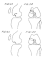
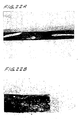
- FIG. 22A and B illustrate the morphologic appearance of neocartilage/LamboneTM composites following pentachrome staining. Magnification: 22A, 100x; 22B 200x.
Description
- This invention relates generally to novel neocartilage compositions useful as implants and as replacement tissue for damaged or defective cartilage, as model systems for studying articular cartilage disease and articular cartilage response to natural and synthetic compounds and for the isolation of cartilage derived substances useful in the biotechnology industry.
- More specifically, the invention concerns an ex vivo neocartilage composition, particularly human neocartilage, having multiple layers of chondrocytes surrounded by a substantially continuous insoluble glycosaminoglycan and collagen-enriched hyaline extracellular matrix. The neocartilage of the invention may have membrane phospholipids enriched in the anti-inflammatory n-9 fatty acids, particularly 20:3n-9 eicosatrienoic or Mead acid. Also provided are methods of producing neocartilage in vitro by growing chondrocytes in substantially serum-free growth media.
- Unlike most tissues, adult articular cartilage does not self-repair. Normal articular cartilage is hyaline cartilage comprising a distinctive combination of cartilage-specific collagens (types II, VI, IX, and XI) and aggregating proteoglycans (aggrecan) which give it the unique ability to withstand compressive forces.
- Chondrocytes are the cartilage-specific cells which give rise to normal articular cartilage tissue growth in vivo. Adult chondrocytes, however, have generally lost their potential to reproduce and generate new cartilage in vivo, although they are responsible for maintaining tissue homeostasis.
- Attempts to grow human articular cartilage using traditional cell culture methods such as growing chondrocytes on tissue-culture plastic surfaces using serum-containing growth media have proved unsuccessful. Although serum (the non-red blood cell portion of blood, clotted and spun down) is known to be a potent mitogen to chondrocytes, their culture in serum-containing growth media has been reported to result in dedifferentiation of the chondrocyte phenotype.
- It is also well known in the art that growing chondrocytes in monolayers on plastic culture vessels for prolonged periods leads to loss of their spherical shape and the acquisition of an elongated fibroblastic morphology. Reginato, et al., Arthritis & Rheumatism 37: 1338-1359 (1994). Biochemical changes associated with this morphological change include loss of the articular cartilage phenotype, e.g., loss of rounded cell shape, an arrest of cartilage-specific collagen and proteoglycan synthesis, the initiation of collagen type I and III synthesis, and an increase in small non-aggregating proteoglycan synthesis. Reginato, et al., supra.
- Adult human chondrocytes grown directly on tissue-culture plastic in growth media containing serum, attach to the plastic substrate and fail to deposit an insoluble matrix enriched in glycosaminoglycan. Glycosaminoglycan is the proteoglycan component essential to the physiological function of articular cartilage and is the hallmark of hyaline tissue. The extracellular matrix initially produced by methods using serum-containing growth media is not enriched in glycosaminoglycan and resorption of the matrix material occurs as the cell culture ages.
- Attempts to overcome chondrocyte dedifferentiation in vitro have included culturing chondrocytes at high densities and growing them in suspension culture or on substrata that prevent cellular spreading and attachment to the tissue-culture plastic. Reginato et al. described a method of growing human fetal chondrocytes cultured on polyHEMA-coated plastic dishes in a serum-supplemented DMEM growth media. Arthritis & Rheumatism 37: 1338-1359 (1994). This method was successful at maintaining the cartilage-specific phenotype but produced only nodules resembling articular cartilage, not a continuous layer of articular cartilage tissue.
- Kuettner described in
U.S. Pat. No. 4,356,262 , a method of producing bovine cartilaginous tissue from which an anti-invasion factor may be recovered. This method provided culturing a monolayer of chondrocytes at high densities in a suspension of serum-containing growth media in a roller bottle. This method produced nodules of tissue having an extracellular matrix, but not a continuous layer of articular cartilage tissue. - Another method that prevents chondrocyte attachment to tissue culture plastic is described in
Kandel, U.S. Pat. No. 5,326,357 . Kandel described methods of reconstituting bovine cartilage tissue in vitro by seeding chondrocytes on a porous tissue culture insert substrate which had been coated with type I collagen to facilitate chondrocyte attachment and growth in a serum-containing growth media. The tissue culture insert is used to separate the chondrocytes from the tissue culture plastic. This method produced a continuous cartilaginous tissue having zones of elongated and spherical chondrocytes which resemble native bovine cartilage. - Without a readily available replacement tissue, recent methods of articular cartilage repair have focused on biological resurfacing of cartilage defects with either a prosthetic device or with live chondrocytes. Methods of in vivo articular cartilage repair include transplanting chondrocytes as injectable cells or as a composition of cells embedded in a three-dimensional scaffold. These methods, like in vitro neocartilage production, have been less than completely successful. One such repair method is autogenous chondrocyte transplantation.
Vacanti et al., WO 90/12603 - Allograft transplant methods, which require a single surgery, use implants made of donor chondrocytes seeded and grown on a natural or synthetic three dimensional scaffold (
Vacanti, et al., U.S. Pat. No. 5, 041, 138 ;Gendler, EP 0739631 A2 ). In these methods, the natural or synthetic three-dimensional scaffold is provided to give the cell culture structure and to mimic the natural extracellular matrix while the cartilage tissue is produced in vivo. - It has recently been shown, however, that neither the autogenous nor the allogenic transplant method results in consistent growth of articular cartilage in vivo, but rather results in chondrocyte dedifferentiation and formation of fibrocartilage. Because of the reduced aggrecan content of fibrocartilage, it cannot withstand the same biomechanical stresses as articular cartilage. Fibrocartilage degenerates with use, and its formation following joint repair may promote joint dysfunction and permanent disability.
- In addition to the clinical need for readily available replacement tissue, healthy articular cartilage is needed for use in model systems for studying articular cartilage disease and to evaluate chondrocyte responses to growth factors, cytokines and pharmaceutical compositions.
- Osteoarthritis, the most common form of arthritic disease, affects almost 16 million people in the United States alone. Osteoarthritis is characterized by the appearance of focal lesions at the cartilage surface. With advancing age and disease progression, these changes are accompanied by a marked reduction in proteoglycan content, extensive destruction of the collagen framework, a marked increase in tissue hydration, and subsequent joint dysfunction.
- Osteoarthritis appears to develop within the articular cartilage of weight-bearing joints, particularly joints of the knee, hip, hand, and foot. Under normal physiological conditions, cartilage homeostasis is maintained by the resident chondrocytes. This highly specialized cell functions to synthesize, assemble, and remodel all components of cartilage extracellular matrix, including aggregating proteoglycan as well as collagens type II, VI, IX, and XI. Despite intensive research efforts to ascertain the biological basis of osteoarthritis, its development and progression remain poorly understood.
- Recent studies attempting to characterize collagenolytic activity in human osteoarthritis indicate a clear need for a reliable alternative to animal models for elucidating early biological events of disease progression. Most animal tissues do not express the complexity of enzymes that have been implicated in human disease. Thus, animal models are inadequate for evaluating the efficacy of potential disease modifying agents in human osteoarthritis.
- A shortage of normal articular cartilage for studying articular cartilage disease and articular cartilage response to natural and synthetic compounds exists because the only source of healthy articular cartilage currently available is from deceased adult donors which may show degenerative changes.
- Adkisson et al., in the Journal of the Federation of American Societies for Experimental Biology, 1991, reported their findings that normal, young cartilages, in distinction from all other tissues examined, have unusually high levels of n-9 eicosatrienoic (20:3 cis-delta 5,8,11) acid and low levels of n-6 polyunsaturated fatty acids (n-6 PUFA). This pattern is identical to that found in tissues of animals subjected to prolonged depletion of nutritionally essential n-6 polyunsaturated fatty acids (EFA). This apparent deficiency is consistently observed in cartilage of all species so far studied (young chicken, fetal calf, newborn pig, rabbit, and human), even though levels of n-6 PUFA in blood and all other tissues is normal. The n-9 20:3 acid was found to be particularly abundant in phosphatidylethanolamine, phosphatidylinositol, and the free fatty acid fractions from the young cartilage. Several factors appeared to contribute to the reduction in n-6 PUFA and the appearance of high levels of the n-9 20:3 acid in cartilage, namely: 1) limited access to nutritional sources of EFA due to the impermeability and avascularity of cartilage, 2) rapid metabolism of n-6 PUFA to prostanoids by chondrocytes, and 3) a unique fatty acid metabolism by cartilage. Evidence is presented that each of these factors contributes. Previously, EFA deficiency has been shown to greatly suppress the inflammatory response of leukocytes and rejection of tissues transplanted into allogeneic recipients. Because eicosanoids, which are derived from EFA, have been implicated in the inflammatory responses associated with arthritic disease, reduction of n-6 PUFA and accumulation of the n-9 20:3 acid in cartilage was considered by Adkisson et al. to be potentially important for maintaining normal cartilage structure.
- Xu et al., in LIPIDS, 1994, , evaluated the effects of dietary lipids on the fatty acid composition of hyaline cartilage, epiphyseal chondrocytes (EC) and matrix vesicles (MV) in chicks. A basal semipurified diet was fed to chicks containing one of the following lipid sources at 70 g/kg: soybean oil, butter and corn oil, margarine and corn oil or menhaden oil and corn oil (MEC). Articular and epiphyseal growth cartilage were isolated from the proximal tibiotarsus; EC and MV were subsequently released by trypsin (EC 3.4.21.4) and collagenase (EC 3.4.24.3) digestion followed by ultracentrifugation. The fatty acid composition of polar lipids in chick epiphyseal cartilage at three and six weeks, as well as articular cartilage, EC and MV at eight weeks of age revealed the presence of high levels of saturated and monounsaturated fatty acids (up to 85.5%) but low levels of n-6 polyunsaturated fatty acids (PUFA) (2.6-10.2%). Mead acid (20:3n-9,>3%) was also present in cartilage, EC and MV lipids, and was unaffected by the dietary lipid treatments. Total n-3 PUFA concentrations were the highest in cartilage, EC and MV of chicks consuming MEC. Feeding MEC lowered the levels of 20:4n-6 in cartilage, but increased 20:5n-3 levels. The data was found by Xu et al. to be consistent with those reported previously which showed that cartilage tissues are low in n-6 PUFA and that they contain 20:3n-9.
-
WO 94/09118 -
EP 0739631 provides a method for producing a biological material comprising reconstituted cartilage tissue, wherein the method comprises isolating chondrocytes from cartilage tissue, contacting a substrate comprised of a continuous, flexible sheet of demineralized natural bone, having a thickness less than about 1.5m, with said chondrocytes, and culturing the chondrocytes on the substrate in growth media to produce a biological material characterized by a continuous layer of cartilage tissue.EP0739631 also discloses a biological material made by this method. - Chang et al., in the Journal of Biological Chemistry, 1994, volume 259, no. 45, pages 28227 to 28234, reported that partially purified extracts from newborn calf articular cartilage were found to induce cartilage and bone when subcutaneously implanted in rats. This activity showed characteristics of bone morphogenetic proteins (BMPs). Degenerate oligonucleotide primer sets derived from the highly conserved carboxyl-terminal region of the BMP family were designed and used in reverse transcription-polymerase chain reactions with poly(A)+ RNA from articular cartilage as template to determine which BMPs are produced by chondrocytes. Two novel members of the transforming growth factor-beta (TGF-beta) superfamily were identified and designated cartilage-derived morphogenetic protein-1 (CDMP-1) and -2 (CDMP-2). Their carboxyl-terminal TGF-beta domains are 82% identical, thus defining a novel subfamily most closely related to BMP-5, BMP-6, and osteogenic protein-1. Northern analyses showed that both genes are predominantly expressed in cartilaginous tissues. In situ hybridization and immunostaining of sections from human embryos showed that CDMP-1 was predominantly found at the stage of precartilaginous mesenchymal condensation and throughout the cartilaginous cores of the developing long bones, whereas CDMP-2 expression was restricted to the hypertrophic chondrocytes of ossifying long bone centers. Neither gene was detectable in the axial skeleton during human embryonic development. The cartilage-specific localization pattern of these novel TGF-beta superfamily members, which contrasts with the more ubiquitous presence of other BMP family members, was considered by Chang et al. to suggest a potential role for these proteins in chondrocyte differentiation and growth of long bones.
-
WO 98/04681 - Ismaiel et al., in the Journal of Pharmacy and Pharmacology, 1991, volume 43, reported on experimentation whey they found that human cartilage biopsies incubated for 2 days in-vitro with 15% synovial fluid from rheumatoid arthritis patients contained less glycosaminoglycans (GAG) than control biopsies. Recombinant human (rHu-) interleukin-1α (IL-1α) and IL-1β at 10 or 100 ng mL-1 had no effect on human cartilage GAG levels. Similarly, GAG loss from human cartilage biopsies into medium over 5 days was significantly increased by synovial fluid but unaffected by 100 ng mL-1 IL-1α or IL-1β compared with controls. However, when rat femoral head cartilage samples were incubated with 100 ng mL-1 rHu-IL-1α or IL-1β for 5 days there was a significant increase in GAG loss from the cartilage into medium, whilst human synovial fluid significantly decreased the loss of GAG from rat cartilage into medium, compared with controls. Ismaiel et al. concluded that the results demonstrate that human and rat cartilage differ from each other in their responses to degrading stimuli and suggest that animal cartilage may have limited application for the screening of drugs intended for the treatment of human arthritides.
- Among the several objects of the invention, therefore, may be noted:
- the provision of novel neocartilage compositions and uses thereof;
- the provision of methods of producing such novel neocartilage compositions
- According to a first aspect of the present invention, there is provided an ex vivo neocartilage composition comprising multiple layers of chondrocytes surrounded by a continuous insoluble glycosaminoglycan and collagen-enriched hyaline extracellular matrix.
- The neocartilage of the invention is useful, for example, as replacement tissue for damaged or defective cartilage and as a model system for studying articular cartilage disease and response to natural and synthetic compounds. Illustratively, surgical implants, or allografts, of the neocartilage were created in vitro and surgically attached to natural cartilage in animal models to repair surgically created defects.
- Briefly, therefore, the present invention is directed to neocartilage characterized by one or more of the following attributes: containing membrane phospholipids enriched in Mead acid, containing membrane phospholipids depleted in linoleic or arachidonic acid, being substantially free of endothelial, bone and/or synovial cells, having a S-GAG content of at least about 400 mg/mg of OH-proline, being substantially free of type I, III and X collagen, containing a matrix substantially free of biglycan, being enriched in high molecular weight aggrecan, being produced in vitro using serum-free growth medium, being essentially free of non-cartilage material, and being characterized by having multiple layers of cells surrounded by a substantially continuous insoluble glycosaminoglycan and collagen-enriched hyaline extracellular matrix.
- According to a second aspect of the present invention, there is provided an ex vivo osteochondral composite comprising the neocartilage composition of the above first aspect of the present invention, wherein the continuous hyaline extracellular matrix is affixed to a noncartilaginous biocompatible material.
- The present invention is further directed to a method for producing the ex vivo neocartilage composition of the invention, the method comprising: isolating a plurality of chondrocytes in vitro; adhering said chondrocytes under conditions sufficient to form an in vitro culture of chondrocytes; and growing the chondrocytes in a serum-free growth medium to produce neocartilage, wherein the neocartilage composition comprises multiple layers of chondrocytes surrounded by a continuous insoluble glycosaminoglycan and collagen-enriched hyaline extracellular matrix.
- According to a further aspect of the present invention, the neocartilage compositions of the above first aspect of the osteochondral composite of the above second aspect are used in the manufacture of a preparation for the treatment of damaged, defective or diseased cartilage.
- Although not in accordance with the invention as claimed, the neocartilage composition as described herein may be employed in a method of screening a pharmaceutical for its capacity to modulate arthritic disease . In this method, the neocartilage is co-cultured with the pharmaceutical in an amount and under conditions effective for determining whether characteristic indications of arthritic modulation are observed in the neocartilage.
- Other objects and features will be in part apparent and in part pointed out hereinafter.
-
-
FIG. 1 , in two parts,FIGS.1A and 1B , shows gross morphology of human neocartilage produced in vitro. Fetal chondrocytes grown under serum-free conditions today 120 produce hyaline tissue that is roughly 1.5-2 mm thick. Wet weights of 300-400 mg were obtained from material grown in 12 well dishes.FIGS.1A and 1B , respectively, represent lateral and birds-eye views of human neocartilage atday 90 of culture. The structural characteristics of this hyaline cartilage mimics that of native articular cartilage. -
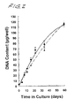
FIG. 2 is a graphical representation which shows growth curves for fetal chondrocytes grown in the presence (solid line) and absence (dashed line) of serum. DNA content was measured over a 60-day time course using Hoechst 33528 fluorescent dye, following papain digestion. Herring sperm DNA was used as standard. Chondrocytes grown under the specified serum-free conditions go through the same number of doublings as those cells grown in the presence of serum. -
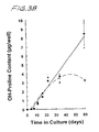
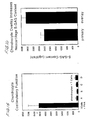
FIG. 3 , in two parts,FIGS.3A and3B , is a graphical representation which shows that serum deprivation promotes neocartilage formation in vitro: Glycosaminoglycan and collagen deposition by fetal human chondrocytes. Chondrocytes were isolated from the upper region of the proximal tibia and distal femur of human fetal knees (18-21 wks gestation) by sequential enzymatic digestion. Cell suspensions were then filtered, counted, and seeded at high density (24 well clusters) in media containing 10% serum to allow adherence. Upon reaching confluence (day 7), the cultures were subdivided into two groups such that half of the dishes remained serum-free (dashed line), while the remaining dishes were maintained in serum-containing media (solid line). Growth media were supplemented with 50 µg/ml ascorbate at every media change, usually every 72 h. Sulfated glycosaminoglycan (S-GAG) (FIG.3A ), and hydroxyproline (OH-proline) (FIG.3B ), content of the newly synthesized, insoluble hyaline matrix were measured as described hereinbelow. Chondrocytes grown under serum-free conditions produced at least 10-fold greater amounts of proteoglycan (sulfated glycosaminoglycan) and more than 2-fold greater levels of collagen (hydroxyproline) as compared to parallel cultures that were maintained in 10% FBS. -
FIG. 4 , in two parts,FIGS.4A and 4B shows the morphologic appearance of human neocartilage (day 56) following serum repletion (10% FBS).Day 28 neocartilage was treated with 10% FBS until termination on day 56 (FIG.4B ), at which point neocartilages were fixed and stained with safranin-O to visualize aggrecan.FIG.4A ,day 56 control neocartilage (serum-free). Notice the marked decrease in metachromatic staining which occurred in response to repletion with 10% FBS (FIG.4B ). Also notice the flattened and elongated phenotype of chondrocytes present at the surface of the neocartilage. These changes suggest that in the absence of mechanical stimulation, serum promotes autolytic resorption of hyaline cartilage matrix, the mechanism of which requires further study. -
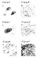
FIG. 5 , in six parts,FIGS.5A - 5F shows the morphologic appearance of native and neocartilage matrix following formalin fixation and paraffin embedding. Tissue sections were cut and stained with either safranin-O (FIGS.5A, 5C and 5E ) or pentachrome (FIGS.5B, 5D, and 5F ) to visualize specific extracellular matrix components.
Safranin-O stains red and identifies S-GAG, whereas pentachrome stains yellow for collagen, green for proteoglycan, and black for elastin. Because the tissues are enriched in collagen and proteoglycan, an aqua blue color is achieved upon pentachrome staining.-
FIGS.5A and 5B show longitudinal sections of fetal proximal tibia used to create the neocartilage grafts inFIGS.5C - 5F . -
FIGS.5C and 5D represent neocartilage grown under serum-free conditions and harvested atday 90. -
FIGS.5E and 5F demonstrate the inhibitory effect of serum on neocartilage formation (day 0-30). (10% FBS).
Serum supplementation caused the cell layers to ball up and slough away from the tissue culture surface afterday 30. Note thatFIGS.5C and 5D show only a small fraction of the full thickness ofday 90 neocartilage. Chondrocytes are seen localized to individual lacunae, forming hyaline tissue that is virtually indistinguishable in appearance from the native starting material and which spans 15-20 cell layers deep.Magnification x 200 forFIGS.5A, 5B, 5E and 5F , and x 400 forFIGS.5C and 5D . -
-
FIG. 6 , in six parts,FIGS.6A - 6F shows the ultrastructural characterization of neocartilage matrix by transmission electron microscopy (TEM). Representative cultures fromFIG. 5 were fixed with glutaraldehyde, post fixed with osmium-tetroxide, and stained en-bloc with tannic acid and uranyl acetate. Ultra-thin sections were counter-stained routinely with uranyl acetate and lead citrate. High power magnification x 61,900 (B,D,F) of the ECM of native (FIGS.6A and 6B ) and neocartilage tissue grown in either the presence (FIGS.6E and 6F ), or absence (FIGS.6C and 6D ) of 10% FBS.
Note thatFIG.6E represents the entire thickness of the culture material and that these chondrocytes display cell/cell contact. Type II collagen (20 nm diameter fibrils) comprised the dominant structural protein in each of these tissues, confirming that the engineered tissue is hyaline in nature. Low power magnification x 3,365 (A,C,E). A lack of matrix proteoglycan probably contributes to stacking of the collagen fibrils observed inFIG.6F . -
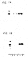
FIG. 7 shows SDS-PAGE and Western analysis of matrix associated collagen obtained by limited pepsinization and neutral salt precipitation.- S1 and S2 are duplicate samples of neocartilage tissue (day 90), while FC and Sk represent native fetal cartilage and skin, respectively.
- Antibodies to collagen types-II, IX and I were obtained from Oncogene Sciences.
The analysis shows the presence of type II collagen and the absence of type I collagen. Only the positive control (fetal skin) recognized monoclonal antibody directed to collagen type I. -
FIG. 8 , in two parts,FIGS.8A and 8B , shows cytokine-induced resorption of fetal articular cartilage. It is a demonstration of the relative appearance of treated and untreated neocartilage cultures showing that neocartilage exposed to cytokine is markedly reduced in size and lifts away from the plastic surface.- Tissue was propagated as described above (day 28) and stimulated with increasing concentrations of the indicated cytokine or growth factor (
FIG.8B ) for an additional 36 days. - Cytokines were added to fresh ascorbate containing media every 48 hours.
- Spent media was collected and frozen at each feeding for analysis of S-GAG, hydroxyproline content, and matrix metalloproteinase (MMP) synthesis.
- Likewise, remnant neocartilage was frozen for chemical analysis of ECM components.
Note that in each case, the IL-1 and TNF-treated samples retracted considerably upon removal from the plastic surface (FIG.8A ), implying that increased synthesis and activation of the MMPs reduced the structural integrity of the tissue. - Tissue was propagated as described above (day 28) and stimulated with increasing concentrations of the indicated cytokine or growth factor (
-
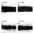
FIG. 9 , in four parts,FIGS. 9A -9D , shows a comparison of the extracellular matrix of control (FIGS. 9A and 9B ) and activated (FIGS. 9C and 9D ) neocartilage following stimulation with interleukin-1. It is seen that chronic IL-1 stimulation alters cartilage staining for proteoglycan and collagen.-
Day 60 neocartilages were treated with 1 ng/ml rhIL-1β for 30 days. Fresh media and cytokine were added every 48 hours. - Neocartilages were harvested (day 90) and stained with safranin-O and pentachrome as before. Staining intensity is markedly reduced in stimulated (
FIGS. 9C, 9D) versus untreated controls (FIGS. 9A , 9B). - Chondrocyte activation with IL-1 caused a significant reduction in the thickness of the synthesized matrix. Altered pentachrome staining following IL-1 stimulation suggests that MMP-mediated cleavage of collagen directly affects the binding characteristics of pentachrome dye to collagen. Magnification x100.
-
-
FIG. 10 , in two parts,FIGS.10A and 10B, represent SDS-substrate gel (zymogram) analysis of cytokine-mediated production of matrix metalloproteinases (MMPs) in fetal articular chondrocytes (neocartilage disks). Negative staining indicates the molecular weight (MW) in kDa of the active proteases.- Spent media were collected 72 h post stimulation with the indicated cytokine (5 ng/ml) and concentrated 16-fold by dialysis/lyophilization.
- Unreduced samples were separated on 10% SDS substrate gels containing either gelatin
(FIG.10A) or casein(FIG.10B) 0.1%. - MMPs were renatured following SDS removal by extensive washing in 2.5% Triton X-100 and lytic bands visualized via negative staining with Coomassie blue, following incubation in a calcium and zinc containing buffer for 4 hr at 37°C.
- Latent enzyme activity is shown in lanes 1-4, while the corresponding active enzymes were prepared by preincubation with 1 mM 4-aminophenyl mercuric acetate (APMA lanes 5-8).
-
FIG. 11 , in two parts,FIG.11A and FIG.11B , is an immunoblot demonstrating cytokine-induced production of collagenase-1 (FIG.11A ) and collagenase-3 (FIG.11B ) in culture media.- Cell layers were stimulated chronically with 5 ng/ml rhIL-β and the spent media collected every 72 h.
- Twenty µl fractions were reduced and immunoblotted with monospecific polyclonal antisera to collagenase-1 (
FIG.11A ) and collagenase-3 (FIG.11B ). - Positive controls (purified recombinant protein) for each blot are shown in
lane 1 and ranged in size from 50-54 kDa. - Media control,
Lane 2; unstimulated control, lanes 3-4; 3-day stimulation,lane 5; 6-day stimulation,lane 6.
-
FIG. 12 , in four parts,FIGS.12A, 12B, 12C, and 12D , shows a surgical model for articular repair. Arthrotomy was performed by a medial parapatellar incision and lateral patella dislocation. A full-thickness cartilage defect, measuring 3 mm from proximal to distal traversing 5 mm from the medial edge of the medial femoral condyle, was created using a No. 15 scalpel. The defect was curetted down to, while not violating, the subchondral bony plate. The medial side of the neocartilage graft was sutured to the periosteum of the medial femur, while the lateral aspect of the graft was sutured to host cartilage at the lateral edge of the defect using 7-0 Proline sutures. After applying tissue transglutaminase (Sigma Chemical Co.) under the graft via syringe (Jurgensen et sal., J Bone Joint Surg, 79-A:185-193, 1997), the graft-defect interface was stabilized for five minutes via finger pressure. The patella was then reduced, and the arthrotomy closed in layers with interrupted 5-0 Ethibond suture. The skin incision was closed with interrupted subcuticular 5-0 Proline sutures. Medial and anterior aspects of the operated knee are depicted inFIGS.12A, 12C , and inFIGS. 12B, 12D , respectively. -
FIG. 13 , in two parts,FIGS.13A and 13B show the gross appearance of rabbit knees at harvest following surgical implantation of rabbit neocartilage. Three by 5 mm (3 x 5) experimental defects were created in the medial femoral condyles of 30 wk New Zealand White rabbits. Gross inspection six weeks post-operatively revealed that the neocartilage allograft was in place (FIG.13B ), and has taken on the appearance of the surrounding native cartilage. The unfilled defect of the contralateral knee remained empty (FIG.13A ). -
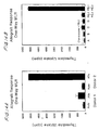
FIG. 14 , in two parts,FIG.14A and FIG.14B , shows the results of two separate one-way mixed leukocyte assays used to measure alloreactivity via lymphocyte proliferation. The experiment inFIG. 14B was set up to investigate alloreactivity of neocartilage, following enzymatic dissociation with collagenase and hyaluronidase. Legend: A = chondrocytes isolated fromday 40 neocartilage (fetal donor); B = chondrocytes isolated fromday 30 culture (20 year male donor); C = chondrocytes isolated fromday 30 culture (27 year male); D = chondrocytes isolated fromday 30 culture (36 year female).- In
FIG.14A ,1 x 105 irradiated peripheral blood leukocytes (PBL, American Red Cross, St. Louis, Missouri) from two unrelated donors were mixed with non-irradiated PBL from either the first (auto) or second donor (allo). Tritiated thymidine was added onday 6. Cultures were harvested and the amount of radiolabel incorporated into newly synthesized DNA counted. - In
FIG.14B ,1 x 105 irradiated chondrocytes from four different donors of 20-36 yrs age were incubated with 1 x 10⁵ non-irradiated PBL and their proliferation measured onday 7. Chondrocytes failed to stimulate proliferation of allogeneic PBL obtained fromdonor 2. Positive (allo) and negative (auto) controls were run on the same plate and are included for comparison. Legend: PBL1=peripheral blood leukocytes fromunrelated donor 1; PBL2=peripheral blood leukocytes fromdonor 2.
- In
-
FIG. 15 shows the co-stimulatory function of human neocartilage chondrocytes. 1 x 10⁵ T-cells, semi-purified from donor 2 (FIG.14A ) using affinity columns from R & D Systems, were incubated with irradiated chondrocytes used inFIG.14 . Incubations were carried out at 37°C for three days. Again, chondrocytes failed to stimulate a proliferative response above background. -
FIG. 16 is a bar chart which shows that overlaying neocartilage with secondary passage of chondrocytes increases allograft thickness and rigidity.Day 10 human neocartilage (12 well dishes) was overlayed with 5 x 10⁵ chondrocytes obtained via secondary passage. These cultures were harvested atday 28 and compared to control cultures that were initially seeded with 1 x 10⁶ cells/well. -
FIG. 17 , in two parts,FIGS.17A and 17B , is a characterization of neocartilage proteoglycans on 1.2% agarose gels. Human neocartilage (day 35) was labeled for 72 hr with carrier-free sodium sulfate (10µCi/ml). Matrix proteoglycans were then guanidine extracted, ethanol precipitated, and extensively dialyzed prior to fractionation on 1.2% agarose gels.FIG.17A , toluidine blue stained proteoglycan.FIG.17B , autoradiograph showing localization of incorporated label in six replicates of neocartilage. Trace amounts of decorin were identified in six replicates of neocartilage, whereas biglycan, present in the 12- and 43-year subjects, was not synthesized. Lanes: 1 (chondroitin-4-sulfate standard); 2 (12-yr female); 3(43-yr male); 4-9 (replicates of human neocartilage). Note that native decorin can only be viewed in the neocartilage following metabolic labeling. -
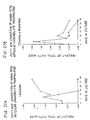
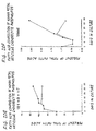
FIG. 18 , in two parts,FIGS. 18A and 18B shows the effect of heparin on neocartilage formation.Day 10 neocartilage was treated with increasing amounts of heparin for 18 days. Fresh media, containing heparin and ascorbate were added every 72-96 hrs. Media were collected and frozen for future analysis. Cultures were harvested onday 28 and the S-GAG content (FIG. 18A ) and OH-proline content (FIG. 18B ) of the resultant neocartilage assayed as described hereinbelow. -
FIG. 19 shows cartilage-derived morphogenetic proteins (CDMP's) in human neocartilage matrix. Neocartilage formed by 90-day cultures of chondrocytes from donors of various ages were extracted in a 1.2 M guanidine-HCl buffer and passed over a heparin affinity column. Proteins were eluted with 1 M NaCl and concentrated using a Centricon filter with a 10K cutoff. The proteins were electrophoresed on a 12% SDS gel under reducing conditions, and transferred to Immobilon (Millipore) membranes. Immunoblots were probed with anti-CDMP antibody (N442) provided by the NIH. The secondary antibody was detected using chemiluminescence. Lanes: 1 (fetal; 2 (1-day neonate); 3 (8-month infant); 4 (12-yr adolescent); 5 (48-year adult). Immunoreactivity was detected in 56 Kd pre-forms, 34 Kd dimers and 14-17 Kd mature proteins. -
FIG. 20 , in six parts,FIGS. 20A - 20F , shows the fatty acid composition of human neocartilage phospholipids versus time. Chondrocytes were grown in either the presence (open circles) or absence (filled circles) of 10% serum as described herein and the fatty acid composition of the membrane phospholipids isolated via silicic acid chromatography determined via capillary gas chromatography. The upper panels (FIGS.20A, 20B and20C ), correspond to the n-6 polyunsaturated fatty acid precursors of eicosanoid synthesis, while the lower panels (FIGS.20D ,20E and 20F ) designate the n-9 fatty acids which are abundant in rapidly growing hyaline cartilage. Notice that Mead acid (20:3 n-9 eicosatrienoic acid) accumulates under serum-free conditions to a level that was two-fold greater than that identified in the native tissue at time zero. Additionally, serum supplementation caused a three-fold accumulation in arachidonate (20:4 n-6). The Mead-to-arachidonate ratio of serum vs. serum-free cultures mirrored that identified in adult and fetal tissue, respectively (Adkisson et al., 1991, supra). Samples were run in duplicate. -
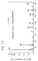
FIG. 21 is a mass chormatogram showing the dominant polyunsaturated fatty acid identified in neocartilage phospholipids (i.e. 20:3 n-9 eicosatrienoic acid) atday 28 of culture. The fragmentation pattern matches that of authentic 20:3 n-9 eicosatrienoic or Mead Acid. -
FIG. 22 , in two parts,FIGS. 22A and 22B , shows the morphologic appearance of neocartilage/demineralized bone (Lambone) composites following pentachrome staining. Magnification: 22A, 100x; 22B, 200x. - In accordance with the present invention, applicant has produced neocartilage tissue which has a morphological appearance largely indistinguishable from healthy native articular cartilage. The cartilage produced is strong, yet malleable. It is readily removable from culture vessels and can be grown with or without the aid of a three-dimensional scaffold. Moreover, this novel cartilage has a membrane phospholipid fatty acid profile which is conducive to resistance to transplant rejection and inflammation. This newly developed biological material also serves as a ready resource for obtaining purified compositions important for applications in biotechnology, including cartilage-specific macromolecules such as high molecular weight aggrecan and collagen types II, VI, IX and XI.
- The biological material hereby provided comprises neocartilage having multiple layers of cells surrounded by a substantially continuous insoluble glycosaminoglycan and collagen enriched hyaline extracellular matrix. Furthermore, the membrance phospholipids of the neocartilage are unexpectedly and advantageously enriched in 20:3 n-9 eicosatriene (Mead) fatty acids and depleted in linoleic and arachidonic fatty acids. The Mead acid content is preferably at least about 2% and most preferably, between about 2.5% and 10% of the total fatty acid content. The linoleic acid and arachidonic acid content is preferably less than about 0.5% and most preferably at least about 0.2% of the total fatty acid content of the membrane phospholipids. This neocartilage is further characterized by its high S-GAG and hydroxyproline content, enrichment in high molecular weight aggrecan, and the relative absence of endothelial, bone and synovial cells, as well as being substantially free of biglycan. Preferably, the neocartilage has a S-GAG content of at least about 400 mg/mg of OH-proline, and more preferably, from about 500 mg to about 2500 mg/mg of OH-proline. Preferably, the high molecular weight aggrecan contains at least about 80%, and more preferably, it contains about 90%, of the total cartilage proteglycan content. The chondrocytes of the neocartilage of the present invention are substantially spherical throughout the composition and maintain their articular cartilage phenotype. The neocartilage composition of the present invention is a substantially continuous layer of tissue at least two cell layers thick. After 14 days of growth, the neocartilage had grown to between 10 and 15 cell layers thick. The neocartilage can be grown for at least 120 days to a thickness of at least 2mm and to a weight of between 300 and 400 mg when 3.8 cm2 dishes are used (15-20 cell layers thick).
- The neocartilage can be grown to substantially greater thicknesses when grown under conditions that mimic the biomechanical forces to which articular cartilage is naturally subjected in vivo such as in a compression chamber. It has been found that after 30 days of growth, and as described herein, the glycosaminoglycan content of the neocartilage is approximately 600-1,500 mg S-GAG/mg OH-proline. This is 10 times greater than that of chondrocytes grown in the presence of 10% serum.
- The neocartilage may comprise avian or mammalian chondrocytes, preferably human chondrocytes. Additionally, mammalian chondrocytes may be derived from transgenic animals which have been genetically engineered to prevent immune-mediated xenograft rejection (Sandrin, MS et al., Nature Med 1:1261-1267, 1995; Sandrin, MS et al., Xenotransplantation 3:134-140, 1996; and Osman, N et al., Proc Nat Acad Sci 94:14677-14682, 1997). Thus, the neocartilage may be mammalian neocartilage, including human and porcine, or avian neocartilage.
- The use of organs/tissues from animal donors (xenotransplantation) is a potential solution to the chronic shortage of allogeneic organs. Porcine tissues are thought to be most suitable for human use due to similarities in size, anatomy and organ physiology between pigs and humans. Recent insights into the mechanisms underlying vascular rejection, endothelial cell activation, and cellular responses to xenogeneic tissue have led to the development of novel strategies designed to inhibit immune-mediated xenograft rejection (Dorling, A., Expert Opinion on Therapeutic Patents, 7:1307-1319, 1997).
- The neocartilage of the present invention does not require the inclusion of biosynthetic polymer scaffolds in the composition. However, such scaffolds can also be used, if desired. In one alternative embodiment, neocartilage can be grown on demineralized bone allograft forming a composite which is particularly amenable to surgical implementation.
- Because the neocartilage of the present invention can be produced free of non-cartilage material, its use as an implant or as replacement tissue provides enhanced biocompatibility. For example scaffold-less neocartilage readily integrates into the surrounding tissue whereas cartilage constructs containing artificial polymer scaffolds are likely to take longer to integrate because the cells must first break down the artificial scaffolds.
- In another aspect of the invention, the neocartilage can be used as a replacement tissue for the repair of damaged or defective articular cartilage.
- The replacement tissue can be mammalian or avian replacement tissue, most preferably human replacement tissue. Furthermore, mammalian replacement tissue can be produced using chondrocytes from transgenic animals which have been genetically engineered to prevent immune-mediated xenograft rejection.
- The replacement tissue can be implanted using procedures well known in the art, such as using traditional surgical means or by implanting orthoscopically.
- Surgical implants comprising neocartilage can be surgically implanted and attached to natural cartilage in vivo by sutures or a combination of sutures and biocompatible biological glues, such as tissue trans-glutaminase (Jurgensen et al., J. Bone J. Surg. 79A:185-193.)
- The neocartilage replacement tissue can also be attached to natural cartilage in vivo by sutureless attachment such as chemical tissue welding.
- The neocartilage can be grown to various size specifications to facilitate implantation.
- The neocartilage of the invention may be used as a model for studying articular cartilage disease and articular cartilage response to natural and synthetic compounds in vitro. Natural and synthetic compounds of interest such as enzymes, cytokines, growth factors, anti-invasion factors, dedifferentiation factors and pharmacologic agents are generally known in the art.
- In particular, the neocartilage may be used in the testing of pharmacologic agents useful in the treatment of diseases of the joint, for example, osteoarthritis and joint inflammation. Arthritis is marked by an increase in the synthesis and release of a variety of cartilage-derived metalloproteinases and mediators of inflammation. Because these enzymes are directly responsible for tissue destruction in arthritis, the matrix metalloproteinases (MMPs) offer excellent drug targets for the development of novel disease-modifying agents. In this aspect, pharmaceuticals are screened for their capacity to modulate arthritic disease. The neocartilage is co-cultured with a candidate pharmaceutical and observed to determine whether characteristics indicative of arthritic modulation are observed. The amounts and conditions employed are largely dependent on the particular pharmaceutical tested and employ methods well known in the art.
- In a further aspect, the present invention provides a method for producing the ex vivo neocartilage composition of the invention the method comprising: isolating a plurality of chondrocytes in vitro; adhering said chondrocytes under conditions sufficient to form an in vitro culture of chondrocytes; and growing the chondrocytes in a serum-free growth medium to produce neocartilage, wherein the neocartilage composition comprises multiple layers of chondrocytes surrounded by a continuous insoluble glycosaminoglycan and collagen-enriched hyaline extracellular matrix.
- In a preferred method, chondrocytes isolated from immature non-fetal donors such as neonatal infant, or pre-adolescent chondrocytes are isolated and grown in a substantially serum-free growth media to produce neocartilage.
- The chondrocytes used in this method can be avian or mammalian, preferably human chondrocytes. Further, in contrast to other methods of producing neocartilage known in the art, such as seeding cells on three dimensional scaffold material or on material that prevents cellular spreading, further exogenous material is not required to produce three dimensional neocartilage. Unlike methods known in the art, the method of the present invention provides for seeding chondrocytes in direct contact with an appropriate tissue-culture vessel, most preferably uncoated tissue-culture plastic. Although scaffold material is unnecessary, it can be used.
- In a preferred embodiment of the invention, a cell culture is produced by isolating immature chondrocytes, e.g., fetal, neonatal, and pre-adolescent chondrocytes from donor articular cartilage.
- Chondrocytes may be isolated by methods known in the art such as by sequential enzyme digestion techniques.
- The isolated chondrocytes may then be seeded directly on a tissue culture vessel in a basal media comprising an effective amount of serum such as Dulbecco's modified Eagle's medium (DMEM) to allow adherence of the chondrocytes directly to the culture vessel and to promote mitogenesis.
- The effective amount of serum added is between 2 and 15% fetal bovine serum, preferably 10%.
- The culture medium may also comprise ascorbate, exogenous autocrine growth factors or conditioned growth media as described below.
- The cell culture may be grown under suitable culture conditions known in the art such as growing the cell culture at 37 degrees C in a humidified atmosphere with the addition of 2-10% CO2, preferably 5%.
- The growth medium containing serum is replaced with growth medium containing half as much serum, preferably 5% of the total growth medium on between
day 1 andday 10, preferably onday 7. - On between
day 5 andday 14, preferablyday 10, the serum containing growth medium is replaced with substantially serum-free growth medium to produce a substantially serum-free cell culture. - The preferred substantially serum-free growth medium is HL-1, a serum-free medium containing insulin-transferrin-selenium-complex as its only source of protein. HL-1 is a registered trademark of Hycor Biomedical, Inc., and also is available from Biowhittaker, Walkersville, Maryland. Other suitable serum-free growth media will be readily apparent to those skilled in the art.
- The substantially serum-free growth medium is preferably partially changed periodically throughout the growth period. Following 10 more days in the substantially serum-free medium, the neocartilage of the invention is between 10 and 15 cell layers thick and can be removed from the cell culture with forceps as a rigid disk of neocartilage.
- The neocartilage produced by the substantially serum-free cell culture can be grown for at least 300 days. Even after 120 days in culture, the chondrocytes of the neocartilage do not dedifferentiate and fail to synthesize collagen types I, III, and X. The method of the present invention can produce neocartilage of various sizes by using various sized culture vessels.
- In order to illustrate the invention in further detail, the following specific laboratory examples were carried out. Although specific examples are thus illustrated herein, it will be appreciated that the invention is not limited to these specific examples or the details therein.
- The sources of various materials used in the specific laboratory examples are as follows:
- Materials - Dulbecco's modified Eagle's medium (DMEM) with added L-glutamine, sodium pyruvate, and glucose (4.5 g/liter), fetal bovine serum (FBS) and antibiotic (100x) (penicillin G, sodium (10,000 units) and streptomycin sulfate (25 mg/ml of normal saline)) were obtained from Life Technologies, Inc. (Grand Island, NY).
- Pronase-E (Type XIV, from Streptomyces griseus), hyaluronidase (type III), N-tris[hydroxymethyl]methyl-2-aminoethanesulfonic acid (TES), and MILLEX-GS syringe sterilization filters were obtained from Sigma Chemical Company (St. Louis, MO).
- Collagenase (CLS II) was purchased from Worthington Biochemicals (Freehold, NJ). Tissue-culture dishes (12 and 24 well cluster) and bottle top sterilization filter units (type CA) were obtained from Costar Corporation (Cambridge, MA).
- Bovine serum albumin (fraction V, fatty acid-free) was from Calbiochem (San Diego, CA). HL-1 growth medium, was obtained from Hycor Biomedical (distributed currently by BioWhittaker).
- Reparation and Culture of Immature and Adult Human Chondrocytes - Chondrocytes were isolated within 8-24 hours of death from the upper region of the tibial plateau and femoral condyle of intact fetal bones (18-21 weeks gestation, Advanced Bioscience Resources, Inc., Alameda, CA). Hyaline cartilage from infant knees (
s 8 months) was removed from the metaphysis of both the proximal tibia and distal femur using rib cutters and transferred to sterile serum-free DMEM, containing antibiotics (2x) and 1% bovine serum albumin, for chondrocyte isolation as described below. Additional samples of more mature cartilage (12-58 years) were processed similarly. All of these cartilage tissues were provided by Mid-America Transplant Association (St. Louis, MO). All tissues were transported on wet ice prior to use. - Skin from fetal limbs was removed and stored at -20°C for preparation of collagen standards (i.e., types I and III) via limited pepsinization. Skeletal muscle and other connective tissue were dissected under aseptic conditions to expose the articulating surface of the tibia and femoral condyle. The cruciate ligaments, menisci, and synovial capsule were removed.
- Articular cartilage was separated from its underlying epiphysis (i.e., more vascularized region), transferred to the cold synthetic cartilage lymph (SCL) described by Majeska and Wuthier, Biochim Biophys Acta, 391:51-60 (1975), and washed extensively (4x) to remove contaminating synovial fluid. Any remaining connective tissue was removed from the cartilage by incubation with 1x trypsin-EDTA (Sigma Chemical Co.) for 30 minutes at 37°C. The enzyme solution was replenished with fresh serum-free DMEM and residual trypsin removed by four additional washes using a vortex mixer.
- At this stage of preparation, the cartilage was glistening white in color and showed no evidence of fibrous tissue contamination. Cartilage was then diced into 1 mm cubes, washed and transferred to 50 ml sterile conical tubes (approximately 2 g tissue/tube) containing 7 ml of pronase-E solution (2 mg/ml Sigma Type XIV from S. griseus) in HL-1 for 30 minutes digestion at 37°C in an environmental incubation shaker set at 200 rpm (New Brunswick Scientific). The enzyme solution was removed and 4 ml of HL-1, containing 1 mg/ml BSA, antibiotics and ascorbate (50 µg/ml) was added. To this solution, 2 ml of stock collagenase (Worthington CLS-II 1,000 units/ml in HL-1) and 1 ml of hyaluronidase (Sigma Type III, 5 mg/ml in HL-1) was added for overnight digestion at 37°C with mechanical agitation. The following morning cartilage remnants were diluted with 10 ml of fresh medium and gently vortexed (3 x 1 minutes) to release chondrocytes from the remaining extracellular matrix.
- Chondrocytes were then separated from tissue debris by gravity filtration through a sterile Falcon cell strainer unit (70 µ m) and sedimented at 600 x g for 10 minutes in a clinical centrifuge. Cell viability was greater than 95%. Next, chondrocytes were diluted in DMEM + 10% FBS for plating in either 12 or 24 well plastic culture dishes at a density of 1-2
x 105 cells/cm2 in 1 ml of growth medium and grown at 37°C in a 95% air, 5% CO2 atmosphere. - Chondrocytes were initially seeded in FBS to promote adherence and to stimulate cell division. Ascorbate (50 µg/ml, Sigma Chemical, St. Louis, MO) was added fresh at plating and at each feeding, generally every 72-96 hours.
- On
day 7, neocartilage cultures were weaned of serum (5%) and remained 100% serum-free (i.e., in HL-1) fromday 10 onward. HL-1 is a chemically defined serum-free medium containing insulin-transferrin-selenium-complex as its only source of protein. However, other serum-free media high in arginine and supplemented with insulin-transferrin-selenium may be substituted for HL-1. Where indicated, conditioned medium from fetal culture was added to 20% at all media changes to enhance neocartilage formation from infant chondrocytes. - Under the described serum-free conditions, immature chondrocytes displayed tremendous proliferative capacity, as well as the ability to synthesize and deposit an insoluble hyaline cartilage matrix. A gross representation of
day 90 neocartilage tissue is shown inFIG. 1 . The neocartilage is rigid, yet malleable and was easily removed from culture vessels afterday 20 using forceps. This material was strong enough to hold suture material afterday 30 of culture and was amenable to surgical implantation atday 40. - Neocartilage cultures were maintained under said conditions and harvested for biochemical and histological analyses between day 1-60 and again at
days - Neocartilage formation was assessed by colorimetric analyses of sulfated glycosaminoglycan (S-GAG) and hydroxyproline (OH-proline), general measures of proteoglycan and collagen synthesis, respectively. Neocartilage disks were lyophilized and digested 18 hrs at 56°C in 500 µl of 0.1 M sodium acetate buffer (pH 5.6), containing 5mM Na4EDTA, 5mM L-cysteine, and 125 µg/ml papain. The digests were cooled to room temperature for determination of S-GAG, OH-proline, and DNA content.
- Sulfated-GAG and the OH-proline content of papain digested material were determined at the indicated times via microplate colorimetric procedures adapted from Farndale et al., Biochim Biophys Acta 883: 173-177 (1986), and Stegemann and Stalder, Clin Chim Acta 18: 267-273 (1967), respectively. Chondroitin-6-sulfate (Calbiochem) and cis-4-hydroxy-L-proline (Sigma) were used as standard.
- DNA content was estimated by fluorometric analysis using a CytoFluor microplate reader. Bisbenzamide (Hoechst 33258, Sigma Chemical Co., St. Louis, MO) was dissolved at 1 mg/ml in water and a working stock diluted further to 0.1 µg/ml in 10 mM tris-HCl, pH 7.4, containing 0.1 M NaCl and 10 mM EDTA.
- Ten, 20- and 50-fold dilutions of each sample were prepared and 10 µl aliquots assayed by mixing with 100 µl of dye solution. Fluorescence was read (excitation wavelength = 355 nm; emission wavelength = 460 nm) and DNA content determined from a standard curve that was constructed using herring sperm DNA (Gibco BRL).
- Fetal chondrocytes displayed a rapid proliferative phase of cell growth (
FIG. 2 ) and matrix deposition when grown under serum-free conditions, generating tissue that was 1.5-2.0 mm thick and weighing 300-400 mg (3.8 cm2 dishes) atday 120. In contrast, supplementation with 10% serum reduced total S-GAG and hydroxyproline content by 80-90% at day 30 (FIG. 3 ). Furthermore, the structural integrity of these samples (+ 10% serum) was very poor. These samples could not be takenpast day 30 of culture without shrinking and balling up.Day 28 neocartilage with 10% serum for an additional 28-day period causes >90% loss in matrix aggrecan (FIG. 4 ). This effect was titratable, thereby suggesting that either specific components of serum degrade cartilage matrix directly or, alternatively, that serum induces synthesis and/or activation of chondrocyte-derived matrix metalloproteinases. - The potential for neocartilage formation from preadolescent and adult articular chondrocytes is depicted in Table I and appears to correlate with skeletal development. Neocartilage formation was not recapitulated in these experiments using articular cartilage obtained from post-adolescent subjects.
TABLE I. Effect of Donor Age on Neocartilage Potential Donor Age Time in Culture (days) S-GAG (µg) Fetal # 130 3,583 ± 106 Fetal # 260 9,126 ± 30 Fetal # 3120 7,750 ± 125 Infant (1d) 25 6,853 ± 79 Infant (8 mo) 300 9,664 ± 313 12 y 30 1,102 ± 117 20 y 30 107 ± 15 27 y 30 46 ± 8 36 y 30 53 ± 13 43 y 30 31 ± 2 High density cultures were established in 12 well clusters as described, and neocartilage formation was allowed to proceed until time of harvest. Triplicate samples were assayed for total S-GAG using dimethyl methylene blue. - Characterization of the morphological appearance and biochemical composition of the tissue revealed an ultrastructural organization that was hyaline in nature and nearly indistinguishable form native articular cartilage. Chondrocytes were constrained to individual lacunae and were encased in an extensive extracellular matrix which stained metachromatically for aggregating proteoglycan (safranin-O) and collagen (pentachrome) (
FIG. 5 ). Moreover, the pentachrome technique failed to identify elastic fibers previously shown to be present in cultures of bovine articular chondrocytes maintained in the presence of serum [Lee et al., Dev. Biol. 163: 241-252 (1994)]. - Fibrocartilage contamination, a problem typically encountered when articular chondrocytes are cultivated using traditional cell culture methods (i.e., media containing 10% serum), could not be identified in pepsinized extracts of neocartilage material by Western analysis (chemiluminescence detection) or by transmission electron microscopy (TEM) following tissue fixation. The dominant collagen identified by TEM consisted of 20 nm fibrils, while beaded filaments, indicative of type VI collagen, were localized to the lacunae (
FIG. 6 ). Type I collagen fibers typically display a fibril diameter of 100 nm. Western analysis confirmed the presence of type II collagen as the dominant (90%) isotype, and further identified minor cartilage-specific collagens such as collagen types IX and XI (FIG. 7 ). - Cross reactivity with antibodies to collagen types III and X were also negative in neocartilage, but were reactive in collagen preparations obtained from adult articular cartilage. Thus, the composition of the collagen contained within the neocartilage tissue produced by the method of the invention is indistinguishable in appearance from the starting material from which the cells originated; however, it differs significantly from that of adult and osteoarthritic articular cartilage in that collagen types I, III and X were not detected.
- Collagens of the neocartilage matrix can be isolated via pepsinization and neutral salt precipitation (Miller, EJ and Rhodes RK; 1982, Methods of Enzymology 82:33-64) for use in the production of cartilage biomatrices. The culture conditions described herein are ideal for producing the normal complement and ratio of cartilage collagens, which include but are not limited to types II, VI, IX, and XI.
- Model systems for studying articular cartilage disease and articular cartilage response to natural and synthetic compounds. Neocartilage cultures were established in 24 or 48 well plastic dishes (Corning) as described above (Example 1) and grown to
day 30. The hyaline cartilage matrix was then stimulated to undergo autolytic degradation following 30 day activation with increasing concentrations of a variety of cytokine and/or inflammatory agents, including IL-1, Il-6, IL-17, TNF-α, LPS, and phorbol ester. Culture media were collected every 48 hours and subsequently changed by the addition of fresh medium ± inflammatory stimuli. - Cultures were terminated and the biochemical composition of the neocartilage matrix that remained was analyzed as described previously. Parallel samples were processed for light and transmission electron microscopy for histological evaluation of extracellular matrix components.
- Conditioned media were thawed and identification of chondrocyte-derived metalloproteinases assessed by zymogen substrate gel electrophoresis, as well as Western analysis following SDS-PAGE. Further studies examined the effect of activating agents on synthesis of S-GAG and collagen via radiolabeled incorporation.
- Resorption of neocartilage material was readily apparent by gross examination between day 20-25 of stimulation (
FIG. 8 ). Resorption was characterized by a reduction in the diameter of the neocartilage material and its retraction upon removal from the culture vessel, indicating that the structural integrity of the tissue was significantly compromised. - Histological evaluation (i.e., safranin-O and pentachrome staining) of treated and control material revealed a marked reduction in the S-GAG and collagen content of treated samples (
FIG. 9 ). TEM studies also demonstrated a significant alteration in the morphological appearance of the chondrocytes to more of a fibroblast/ macrophage lineage (i.e., numerous microvillus projections were identified on the surface of flattened, spindle shaped cells), whereas chondrocytes in untreated controls maintained their rounded phenotype (not shown). - The OH-proline and S-GAG content of treated neocartilage disks were reduced by 20-85% depending on the dose and identity of the activating agent (Table II). Both zymogram and Western analysis identified collagenolytic and casienase activities that were upregulated in the presence of activating agent alone (
FIG. 10 ).TABLE II. Hydroxyproline content of tissue matrix following chronic cytokine treatment Condition OH-proline content of tissue (pmoles)* Control 1710±251 + 50 mU plasminogen‡ 1648± 72 IL-1β 0.1 (ng/ml) 572±105 1.0 541± 16 5.0 501± 48 + 50 mU plasminogen 460± 16 TNF-α 1.0 403 5.0 355 *Samples were lyophilized and digested with papain prior to hydrolysis for determination of OH-proline content via HPLC analysis. ‡Human plasminogen was added to enhance the activation of latent MMPs via the plasminogen activator/plasroin cascade. - Phorbol ester specifically induced synthesis of gelatinase B, whereas the cytokine treatments resulted in the production of numerous bands (9-11) on substrate gels, three of which were identified as
collagenase FIG. 11 ). Each of these enzymes are implicated in arthritic disease. - This system is unique in that the anabolic and catabolic properties of normal human chondrocyte metabolism can be examined, under defined serum-free conditions. It is clear that the described model of neocartilage formation will facilitate the development and evaluation of pharmacological agents to protect the cartilage matrix from arthritic destruction.
- Insofar as this example discloses steps of a surgical nature, it does not form part of the claimed invention, as defined by the appended claims.
- Neocartilage allografts were produced from 10-day neonates as described in Example 1. Extreme care was taken to eliminate the use of vascularized epiphyseal cartilage during tissue preparation.
- Neocartilage implants were grown to
day 30, fixed and stained for histological evaluation, and further extracted for analysis of cartilage specific macromolecules. This procedure yielded hyaline tissue that was enriched in both cartilage specific collagens and glycosaminoglycans (Table III.)TABLE III. Composition of Rabbit Neocartilage Component Relative Abundance Collagen type I - type II +++ type IX + type XI + Aggrecan 14,365 ± 410 µg High density cultures were established in 12 well clusters as described, and neocartilage formation was allowed to proceed until harvest on day 40. Triplicate samples were assayed for total S-GAG using dimethyl methylene blue. Isotyping of collagen was performed via electrophoresis on 6.5% SDS-polyacrylamide gels under reduced conditions. - Eighteen (18) skeletally mature (30 wk) male New Zealand White rabbits were divided into three groups to assess the healing potential of transplanted neocartilage at 1, 3, 6 and 12 weeks post-operatively. Rectangular defects of approximately 3 mm in width, spanning the girth of the medial femoral condyle (5mm), were created surgically in both knees (
FIG. 12 ). Violation of subchondral bone was avoided during this procedure. - Sterile neocartilage implants (day 35-45 in vitro) were subsequently cut to size and sutured into the experimental defect (right side) using 7-0 vicryl suture, anchoring neocartilage to the medial and lateral perichondria, following addition of tissue transglutaminase as described in Jurgensen et al., J. Bone Joint Surg. 79-A, 185-193 (1997). Unfilled defects (left side) were allowed to heal intrinsically and served as the contralateral sham control. Arthrotomies were repaired using 4-0 vicryl suture and animals were permitted free cage activity. Oral analgesic was provided for 24 hours post-operatively as needed for pain.
- The animals showed excellent tolerance of the surgical procedures, displaying normal ambulation and increased appetite within 24 hours post-operatively. Gross examination of the experimental defects harvested at six weeks revealed good adherence of grafts to surrounding tissue, whereas unfilled defects remained unfilled (
FIG.13 ). - Immunoloaical Assessment of Allograft Rejection - A one-way mixed leukocyte reaction (MLR) was set up in which chondrocytes, acting as the stimulator population, were first gamma-irradiated in order to block their proliferative potential. Allogeneic leukocytes, obtained from either peripheral blood or buffy coats (American Red Cross) were then passed over a Ficoll gradient. This population of mononuclear leukocytes was counted and subsequently co-cultured with chondrocytes, having first been isolated from day 30-45 neocartilage allografts. Adult cells were also isolated from
day 30 cultures which were grown under conditions identical to the neocartilage. - Proliferation of non-irradiated leukocytes was determined on
day 7, following a 24 hr pulse with tritiated thymidine (Amersham, 1 µCi/ml) (Abbas, AK, et al. 1991 In Cellular and Molecular Immunology, pp. 320-322). Culture plates were frozen, the cells lysed in water, and the nuclear DNA aspirated and bound to glass filtermats using an automated cell harvester. Filtermats were then dried and counted in a Wallac MicroBeta scintillation counter. Chondrocytes isolated from day 30-45 neocartilage failed to generate an MLR response that would indicate a cross match (FIG. 14B ), while the positive controls showed a 19-fold increase in proliferation. - These data support the concept that chondrocytes are immunologically privileged. Although chondrocytes are reported to present MHC class II antigens on their cell surface [Elves, J. Bone Joint Surg., 56B:178-185 (1974); Jahn et al., Arth Rheum 30:64-74 (1987); and Jobanputra et al., Clin. Exp. Immunol. 90:336-344 (1992)], proliferation of the responder leukocyte population was not detected in three separate assays involving at least twelve (12) different neocartilage samples. From an immunological perspective, these experiments indicate that the present approach to cartilage repair (e.g., allograft transplantation) is feasible.
- Additional studies examining the co-stimulatory function of chondrocytes in a T-cell based assay (Abbas, AK, et al. 1991, In Cellular and Molecular Immunology, pp. 320-322) were also negative, indicating that co-stimulatory molecules, probably B7.1 (CD80) and B7.2 (CD86) are not normally expressed by chondrocytes (
FIG. 15 ). Thus, applicant has discovered appropriate culture conditions which permit transplantation of neocartilage into allogeneic recipients. - Overlaying of chondrocytes to increase the thickness and structural integrity of neocartilage - Rabbit and human chondrocytes were isolated, seeded into 12-well dishes and grown to
day 10 as described above. During the initial plating of these cultures, 1.4 x 10⁷ cells were reserved for plating in 100 mm dishes. Cells were subsequently released from the culture surface onday 10 using the collagenase/hyaluronidase procedure described in Example 1. - Chondrocytes were filtered, counted and resuspended in a 1:1 mixture of DMEM + 10% FBS/Hl-1, containing ascorbate, at a density of 0.5 x 10⁶/
ml. Day 10 cultures (12-well plates) were overlayed with 1 ml of the cell suspension to increase the thickness and structural integrity of the neocartilage grafts. The cultures were maintained in the 1:1 mix of DMEM/HL-1 from day 13-17, after which they were grown in HL-1 media until harvest atday 40. Total S-GAG in the neocartilage matrix were quantified as described above.FIG. 16 shows that the overlaying procedure increased total S-GAG content by approximately 50%, and as a result increased the rigidity of the neocartilage matrix. - Isolation and Characterization of Neocartilage Proteoglycan - Neocartilage cultures were established and grown to day 35 as described in Example 1. Newly synthesized proteoglycans were metabolically labeled for 72 hrs using carrier-free [35S]-sodium sulfate (55 µCi/ml, Amersham, supra). Following three washes with PBS to remove unincorporated label, the proteoglycans of the neocartilage matrix were extracted in 4M guanidine and precipitated in ethanol. Unlabeled native proteoglycans were also extracted from the articular cartilage of a 12-yr female and 43-yr male for comparison. Twenty (20) µg of S-GAG were loaded and separated on 1.2% agarose gels according to the method of Bjornsson, Anal. Biochem., 210: 292-298 (1993), (
FIG. 17 ). - Greater than 95% of the labeled material was identified as aggrecan, as judged by molecular weight. However, it was noted that this material was of higher molecular weight than the aggrecan found in pre-teen and adult tissue, indicating that the aggrecan monomers were more highly glycosylated. A minor band with electrophoretic mobility identical to that of native decorin was also observed in the neocartilage matrix. Biglycan, present as a minor component of native articular cartilage, was not identified via radiolabel incorporation in 6 replicates of neocartilage. Only 12-15% of the total radiolabeled S-GAG could be recovered in culture supernatant, generating the same profile as that shown here.
- Thus, the composition of neocartilage proteoglycan differs from that of mature articular cartilage in two distinct ways. First, biglycan was not identified as a component of the neocartilage matrix; and second, the innate composition of neocartilage proteoglycan appears to be more highly glycosylated, resulting in a high molecular weight aggrecan. It is believed that this aggrecan may play a role in tissue growth and repair by binding specific growth factors with greater avidity than adult aggrecan.
- Purification of this material for testing as a wound healing agent is relatively easy and inexpensive. High molecular weight aggrecan bands identified on 1.2% agarose gels were removed and eluted to purity using a BioRad electroelution device. Preliminary studies demonstrate that a 200 mg neocartilage disk yielded 3-4 mg of high molecular weight aggrecan.
- Role of heparin binding proteins in neocartilage formation: Identification of two new members of the TGF-β Superfamily - Fetal chondrocytes were grown as described above and subdivided into three groups on
day 10 to investigate the effect of heparin on neocartilage formation. In low doses, heparin is known to increase the activity of certain growth factors, for example the fibroblast growth factor (FGF) family of related proteins, by acting as a co-factor, whereas greater amounts of heparin will strip away certain growth factors from the extracellular matrix and thereby impede tissue repair. - Sterile heparin was added to the media at a final concentration of 0.2, 2.0, and 20 µg for the duration of the culture period (i.e. from day 10-28). Media were exchanged every 3-4 days and recovered for electrophoretic analysis of secreted proteins. Cell layers were removed and the S-GAG (
FIG. 18A ), hydroxy proline (FIG. 18B ), and DNA content quantitated as before (FIG. 18 ). The highest concentration of heparin caused a severe defect (60% inhibition in S-GAG and 33% in OH-proline) in matrix deposition. This effect was also evident in the 2 µg/ml treatment group, but was much less severe. - When these heparin binding proteins are then extracted from the neocartilage matrix using procedures adapted from Chang et al., approximately 10 proteins showing a molecular mass of less than 60 kDa are visible on a 4-20% gradient Tris/Tricine polyacrylamide gel (
FIG. 19 ). Western blotting using antibodies from the NIH have shown that cartilage derived morphogenetic protein-1 and -2 (CDMP-1 and -2) are present, as was connective tissue derived growth Factor (CTGF) (not shown). - Serum-free culture depletes neocartilages of precursor fatty acids for eicosanoid synthesis - Adkisson et al., FASEB J 5: 344-353 (1991), discovered high concentrations of 20:3 n-9 eicosatrienoic acid and low concentrations of n-6 polyunsaturated fatty acids (PUFA) in normal, rapidly growing cartilages of several vertebrates, a biochemical hallmark of essential fatty acid deficiency. Because the n-6 fatty acids, in particular arachidonic acid (20:4 n-6), are key mediators of inflammation and transplant rejection (Schreiner et al., Science 240: 1032-1033 (1988), the fatty acid composition of neocartilage grafts were characterized during a 28-day time course in vitro.
- Total cellular lipids and the phospholipid fraction were isolated as described in Adkisson et al., supra. Pentafluorobenzyl ester derivatives of the free fatty acids obtained from the phospholipid fraction were prepared for microanalysis by capillary gas chromatography using a SP-2380 fused silica column (Supelco Inc., Bellefonte, PA). The gas chromatograph (HP model 5890) was held at 160°C for 2 min., temperature programmed at 10°C/min to 200°C, and maintained at this temperature for an additional 10 min. Both the injector and the electrochemical detector temperatures were set at 225°C. Derivatized fatty acids were identified by co-migration with authentic standards, while unusual fatty acids were characterized by their fragmentation pattern using mass spectrometry analysis of their picolinyl ester derivative as described previously (Adkisson et al., 1991). Percent composition of individual fatty acids was determined by integration using Hewlett-Packard model 3393A integrator.
-
FIG. 20 illustrates that essential fatty acids, a component of the culture media (i.e., 5-10% FBS from day 1-10), are rapidly incorporated into the phospholipid stores of isolated chondrocytes. Moreover, it is clear that once the cultures are switched to and maintained in serum-free HL-1 media (closed circles), the native complement of fatty acids is restored, such that low levels of the n-6 PUFAs are detected. The n-6 fatty acids, shown in the upper three panels, continue to accumulate in chondrocyte phospholipids when the neocartilage cultures are maintained in serum-containing media (open circles). -
FIG. 21 is a mass chromatogram of the dominant polyunsaturated fatty acid identified in neocartilage phospholipids (i.e. 20:3 n-9 eicosatrienoic acid) atday 28 of culture. The fragmentation pattern matches that of authentic 20:3 n-9 eicosatrienoic or Mead Acid. - It is unclear whether enrichment in n-9 fatty acids reduces antigen expression of MHC class II molecules or B7 co-stimulatory molecules which are required to establish immunogeneity in allograft rejection. The literature does support the fact that a dearth of n-6 PUFA modulates the infiltration of inflammatory cells by inhibiting leukotriene formation (Cleland et al., 1984, Lipids 29:151-155, and Schreiner et al., 1988, supra). This in turn protects organs from possible transplant rejection.
- In summary, the novel serum-free culture system used to produce neocartilage tissue may facilitate allogeneic transplantation of the neocartilage by regulating chondrocyte immunogenicity, as well as the elaboration of inflammatory eicosanoids/cytokines.
-
Day 60 neocartilage cultures were enzymatically dispersed as described in Example 1 one million chondrocytes were then seeded onto samples of demineralized allograft bone (Lambone™, 100-300 µm thick) which were first trimmed to accomodate in vitro culture in 12 well clusters. Lambone™, obtained from Pacific Coast Tissue Bank, Los Angeles, CA, was washed extensively in culture media to remove residual ethylene oxide gas used during tissue processing. - Neocartilage formation on demineralized allograft bone was allowed to proceed to
day 28 as described herein. Neocartilage/Lambone composites were then rinsed and fixed overnight in 10% neutral buffered formalin for morphological examination via pentachrome staining. -
FIG. 22A and B illustrate the morphologic appearance of neocartilage/Lambone™ composites following pentachrome staining. Magnification: 22A, 100x; 22B 200x. - In view of the above, it will be seen that the several objects of the invention are achieved.
- As various changes could be made in the above compositions and methods by the person skilled in the art after reading the present disclosure without departing from the invention as defined by the appended claims, it is intended that all matter contained in the above description shall be interpreted as illustrative of the claimed invention and not in a limiting sense.
Claims (49)
- An ex vivo neocartilage composition comprising multiple layers of chondrocytes surrounded by a continuous insoluble glycosaminoglycan and collagen-enriched hyaline extracellular matrix.
- An ex vivo neocartilage composition in accordance with Claim 1, wherein the chondrocytes comprise Mead acid-enriched membrane phospholipids; wherein the Mead acid content of the Mead acid-enriched membrane phospholipids is at least 2% of the total fatty acid content of the membrane phospholipids.
- An ex vivo neocartilage composition in accordance with Claim 2, wherein the Mead acid content of the Mead acid-enriched membrane phospholipids is at least 2.5% up to 10% of the total fatty acid content of the membrane phospholipids.
- An ex vivo neocartilage composition in accordance with Claim 1, wherein the chondrocytes comprise linoleic acid-depleted membrane phospholipids, wherein the linoleic acid content of the linoleic acid-depleted membrane phospholipids is less than 0.5% of the total fatty acid content of the membrane phospholipids.
- An ex vivo neocartilage composition in accordance with Claim 4, wherein the linoleic acid content of the linoleic acid-depleted membrane phospholipids is less than 0.2% of the total fatty acid content of the membrane phospholipids.
- An ex vivo neocartilage composition in accordance with Claim 1, wherein the chondrocytes comprise arachidonic acid-depleted membrane phospholipids, wherein the arachidonic acid content of the arachidonic acid-depleted membrane phospholipids is less than 0.5% of the total fatty acid content of the membrane phospholipids..
- An ex vivo neocartilage composition in accordance with Claim 6, wherein the arachidonic acid content of the arachidonic acid-depleted membrane phospholipids is less than 0.2% of the total fatty acid content of the membrane phospholipids.
- An ex vivo neocartilage composition in accordance with any one of Claims 1 to 7, wherein the continuous hyaline extracellular matrix is enriched in high molecular weight aggrecan, wherein the matrix has a high molecular weight aggrecan content of at least 400 mg/mg of OH proline.
- An ex vivo neocartilage composition in accordance with Claim 8, wherein the high molecular weight aggrecan comprises at least 80% of total proteoglycan content of the neocartilage.
- An ex vivo neocartilage composition in accordance with Claim 8 or 9, wherein the high molecular weight aggrecan comprises at least 90% of total proteoglycan content of the neocartilage.
- An ex vivo neocartilage composition in accordance with any one of Claims 1 to 10, wherein the neocartilage is free of endothelial, bone and synovial cells.
- An ex vivo neocartilage composition in accordance with any one of Claims 1 to 11, wherein the continuous hyaline extracellular matrix is substantially free of type I collagen, type III collagen, type X collagen, and biglycan.
- An ex vivo neocartilage composition in accordance with any one of Claims 1 to 12, wherein the chondrocytes are mammalian or avian chondrocytes.
- An ex vivo neocartilage composition in accordance with Claim 13, wherein the mammalian chondrocytes are transgenic mammalian chondrocytes.
- An ex vivo neocartilage composition in accordance with Claim 1 or Claim 13, wherein the chondrocytes are human chondrocytes.
- An ex vivo neocartilage composition in accordance with any one of Claims 1 to 15, wherein the chondrocytes are immature human chondrocytes selected from the group consisting of neonatal infant chondrocytes, and preadolescent chondrocytes.
- An ex vivo neocartilage composition in accordance with any one of Claims 1 to 16, comprising multiple layers of chondrocytes surrounded by a continuous extracellular matrix enriched in glycosaminoglycan, collagen and high molecular weight aggrecan, wherein: (a) the chondrocytes comprise membrane phospholipids enriched in Mead acid and depleted in linoleic acid and arachidonic acid, (b) the extracellular matrix is substantially free of biglycan, and (c) the composition is substantially free of endothelial, bone and synovial cells.
- An ex vivo osteochondral composite comprising the neocartilage composition of any one of Claims 1 to 17, wherein the continuous hyaline extracellular matrix is affixed to a noncartilaginous biocompatible material.
- An ex vivo osteochondral composite in accordance with Claim 18, wherein the noncartilaginous biocompatible material is demineralized bone.
- A method for producing the ex vivo neocartilage composition of Claim 1, the method comprising:isolating a plurality of chondrocytes in vitro;adhering said chondrocytes under conditions sufficient to form an in vitro culture of chondrocytes; andgrowing the chondrocytes in a serum-free growth medium to produce neocartilage, wherein the neocartilage composition comprises multiple layers of chondrocytes surrounded by a continuous insoluble glycosaminoglycan and collagen-enriched hyaline extracellular matrix.
- A method in accordance with Claim 20, wherein the serum-free growth medium comprises ascorbate, insulin, transferrin and selenium.
- Use of the ex vivo neocartilage composition or ostochondral composite of any one of Claims 1 to 19 for the manufacture of a preparation for treatment of damaged, or defective or diseased cartilage.
- The method for growing neocartilage as set forth in Claim 20 or 21, wherein said adhering means comprise growing said chondrocytes in a growth media containing an amount of serum effective to allow adherence of said chondrocytes.
- The method of Claim 23, wherein the method further comprises adding to said substantially serum-free cell culture an amount of conditioned growth media effective to enhance neocartilage formation.
- The method of Claim 23 or 24, wherein the method further comprises adding ascorbate to said cell culture.
- The method of any one of Claims 20, 21 or 23 to 25, wherein said chondrocytes are immature chondrocytes.
- The method of any one of Claims 20, 21 or 23 to 26, wherein said chondrocytes are human chondrocytes.
- The method of Claim 24, wherein said conditioned growth media is between 5 percent and 30 percent of said serum-free growth media.
- The method of Claim 24 or 28, wherein said conditioned growth media is 20 percent of said serum-free growth media.
- The method of any one of Claims 20, 21 or 23 to 29, wherein said serum-free growth media comprises growth factors for the proliferation of said chondrocytes.
- The method of Claim 24, wherein said conditioned growth media comprises growth factors for the proliferation of said chondrocytes.
- The method of Claim 31, wherein said growth factors are autocrine factors.
- The method of Claim 24, wherein said conditioned growth media contains factors for the differentiation of said chondrocytes
- The method of Claim 24, wherein said conditioned growth media contains factors for the anti-invasion of non-chondrocytic cells.
- A method of Claim 20, 21 or 23 to 34, wherein said serum-free growth media is HL-1.
- A method of Claim 24, wherein said conditioned growth media comprises cartilage-derived morphogenic protein-1.
- A method of Claim 24 wherein said conditioned growth media comprises cartilage-derived morphogenic protein-2.
- A method of Claim 23, wherein said growth media comprises between 2 and 15 percent fetal bovine serum.
- A method of Claim 38, wherein said growth media comprises 10 percent fetal bovine serum.
- A method of Claim 20, 21 or 23 to 39, wherein the method further comprises growing said cell culture at 37 degrees Celsius in a humidified atmosphere with the addition of 2 to 10 percent CO2.
- A method of Claim 40, wherein said cell culture is grown at 37°C in a humidified atmosphere with the addition of 5 percent CO2.
- A method of Claim 20, 21 or 23 to 41, wherein said neocartilage is enriched in Mead acid.
- A method of Claim 20, 21 or 23 to 42, wherein said neocartilage is free of endothelial, bone and synovial cells.
- A method of Claim 20, 21 or 23 to 43, wherein said neocartilage is free of type I, III and X collagen.
- A method of Claim 20, 21 or 23 to 44, wherein said neocartilage is enriched in high molecular weight aggrecan.
- A method of Claim 20, 21 or 23 to 45, wherein said neocartilage is free of biglycan.
- A method of Claim 20, 21 or 23 to 46, wherein said neocartilage comprises multiple layers of cells surrounded by a continuous insoluble glycosaminoglycan and collagen enriched hyaline extra cellular matrix containing no biglycan.
- A method of Claim 20, 21 or 23 to 47, wherein said neocartilage comprises multiple layers of cells surrounded by a continuous insoluble glycosaminoglycan and collagen enriched hyaline extra cellular matrix depleted in both linoleic acid and arachidonic acid.
- A method of Claim 20, 21 or 23 to 48, wherein said neocartilage is characterized as follows:(a) containing membrane phospholipids enriched in Mead acid and depleted in linoleic and arachidonic acid; (b) being enriched in high molecular weight aggrecan; (c) being free of endothelial, bone and synovial cells; (d) being free of biglycan.
Applications Claiming Priority (3)
| Application Number | Priority Date | Filing Date | Title |
|---|---|---|---|
| US4336997P | 1997-04-04 | 1997-04-04 | |
| US43369P | 1997-04-04 | ||
| PCT/US1998/006586 WO1998044874A1 (en) | 1997-04-04 | 1998-04-03 | Neocartilage and methods of use |
Publications (3)
| Publication Number | Publication Date |
|---|---|
| EP1018987A1 EP1018987A1 (en) | 2000-07-19 |
| EP1018987A4 EP1018987A4 (en) | 2004-12-15 |
| EP1018987B1 true EP1018987B1 (en) | 2014-10-29 |
Family
ID=21926820
Family Applications (1)
| Application Number | Title | Priority Date | Filing Date |
|---|---|---|---|
| EP98917986.6A Expired - Lifetime EP1018987B1 (en) | 1997-04-04 | 1998-04-03 | Neocartilage and methods of use |
Country Status (6)
| Country | Link |
|---|---|
| US (3) | US6235316B1 (en) |
| EP (1) | EP1018987B1 (en) |
| JP (1) | JP4394169B2 (en) |
| AU (1) | AU742613B2 (en) |
| CA (1) | CA2285382C (en) |
| WO (1) | WO1998044874A1 (en) |
Cited By (1)
| Publication number | Priority date | Publication date | Assignee | Title |
|---|---|---|---|---|
| US11696929B2 (en) | 2017-09-20 | 2023-07-11 | The Regents Of The University Of California | Methods and systems for conserving highly expanded cells |
Families Citing this family (60)
| Publication number | Priority date | Publication date | Assignee | Title |
|---|---|---|---|---|
| US6235316B1 (en) * | 1997-04-04 | 2001-05-22 | Barnes-Jewish Hospital | Neocartilage and methods of use |
| AU7984498A (en) * | 1997-06-25 | 1999-01-04 | Government Of The United States Of America, As Represented By The Secretary Of The Department Of Health And Human Services, The | Serum-free cell growth medium |
| US6528052B1 (en) * | 2000-09-29 | 2003-03-04 | The Board Of Trustees Of The Leland Stanford Junior University | Method for in vivo ex vivo and in vitro repair and regeneration of cartilage and collagen and bone remodeling |
| EP1099443A1 (en) | 1999-11-11 | 2001-05-16 | Sulzer Orthopedics Ltd. | Transplant/implant device and method for its production |
| AU2001278700A1 (en) | 2000-08-09 | 2002-02-18 | Hiroko Yanaga | Method of culturing human chondrocytes |
| ITRM20010088A1 (en) * | 2001-02-21 | 2002-08-21 | Idi Irccs | PEPTIDE ABLE TO INHIBIT THE ACTIVITY OF THE GROWTH FACTOR DERIVED FROM THE PLATES (PDGF-BB) AND OF THE GROWTH FACTOR DERIVED FROM THE FI |
| JP4500898B2 (en) | 2001-03-23 | 2010-07-14 | 高木産業株式会社 | Compositions and methods for producing biological tissues and tissue constructs |
| US20040151705A1 (en) * | 2002-03-22 | 2004-08-05 | Shuichi Mizuno | Neo-cartilage constructs and a method for preparation thereof |
| WO2003061653A1 (en) * | 2002-01-22 | 2003-07-31 | Pfizer Limited | 3-(imidazolyl)-2-aminopropanoic acids for use as tafi-a inhibitors for the treatment of thrombotic diseases |
| US7537780B2 (en) | 2002-03-22 | 2009-05-26 | Histogenics Corporation | Method for preparing and implanting a cartilage construct to treat cartilage lesions |
| US7468192B2 (en) | 2002-03-22 | 2008-12-23 | Histogenics Corporation | Method for repair of cartilage lesions |
| US7622562B2 (en) | 2002-06-26 | 2009-11-24 | Zimmer Orthobiologics, Inc. | Rapid isolation of osteoinductive protein mixtures from mammalian bone tissue |
| US20040153145A1 (en) * | 2002-11-26 | 2004-08-05 | Clemson University | Fixation method for bioprostheses |
| US7189259B2 (en) * | 2002-11-26 | 2007-03-13 | Clemson University | Tissue material and process for bioprosthesis |
| EP1613364B1 (en) * | 2003-02-26 | 2013-01-16 | Zimmer Orthobiologics, Inc. | Preparation for repairing cartilage tissue, especially articular cartilage defects |
| US7642092B2 (en) * | 2003-03-03 | 2010-01-05 | Technion Research & Development Foundation Ltd. | Cultured cartilage/bone cells/tissue, method of generating same and uses thereof |
| US7067123B2 (en) | 2003-04-29 | 2006-06-27 | Musculoskeletal Transplant Foundation | Glue for cartilage repair |
| US7488348B2 (en) * | 2003-05-16 | 2009-02-10 | Musculoskeletal Transplant Foundation | Cartilage allograft plug |
| US7901457B2 (en) | 2003-05-16 | 2011-03-08 | Musculoskeletal Transplant Foundation | Cartilage allograft plug |
| WO2004104183A2 (en) * | 2003-05-23 | 2004-12-02 | Haeuselmann Hans Joerg | Method for the in vitro production of cartilage-like tissue |
| US20050124038A1 (en) * | 2003-07-14 | 2005-06-09 | Pharmacia Corporation | Transfection of cartilage and matrices thereof in vitro |
| ES2404682T3 (en) * | 2003-12-11 | 2013-05-28 | Isto Technologies Inc. | Particle Cartilage System |
| EP1753860B1 (en) * | 2004-02-20 | 2012-04-11 | Isto Technologies Inc. | Intervertebral disc repair and methods therefor |
| US20060275273A1 (en) * | 2004-02-20 | 2006-12-07 | Seyedin Mitchell S | Intervertebral Disc Repair, Methods and Devices Therefor |
| US8697139B2 (en) | 2004-09-21 | 2014-04-15 | Frank M. Phillips | Method of intervertebral disc treatment using articular chondrocyte cells |
| US8017394B2 (en) * | 2004-10-01 | 2011-09-13 | Isto Technologies, Inc. | Method for chondrocyte expansion with phenotype retention |
| US7273756B2 (en) * | 2004-10-01 | 2007-09-25 | Isto Technologies, Inc. | Method for chondrocyte expansion with phenotype retention |
| US7837740B2 (en) | 2007-01-24 | 2010-11-23 | Musculoskeletal Transplant Foundation | Two piece cancellous construct for cartilage repair |
| US20060111778A1 (en) * | 2004-10-29 | 2006-05-25 | Michalow Alexander E | Methods of promoting healing of cartilage defects and method of causing stem cells to differentiate by the articular chondrocyte pathway |
| US7815926B2 (en) * | 2005-07-11 | 2010-10-19 | Musculoskeletal Transplant Foundation | Implant for articular cartilage repair |
| WO2007025290A2 (en) * | 2005-08-26 | 2007-03-01 | Isto Technologies, Inc. | Implants and methods for repair, replacement and treatment of joint disease |
| EP1926459B1 (en) | 2005-09-19 | 2015-01-07 | Histogenics Corporation | Cell-support matrix having narrowly defined uniformly vertically and non-randomly organized porosity and pore density and a method for preparation thereof |
| EP1764117A1 (en) | 2005-09-20 | 2007-03-21 | Zimmer GmbH | Implant for the repair of a cartilage defect and method for manufacturing the implant |
| US20100209397A1 (en) * | 2005-11-10 | 2010-08-19 | Carticure Ltd. | Method for non-autologous cartilage regeneration |
| US7416889B2 (en) * | 2006-04-27 | 2008-08-26 | Rhode Island Hospital | Methods and compositions for repairing cartilage |
| EP1894581A1 (en) * | 2006-08-31 | 2008-03-05 | CellCoTec B.V. | Repair of cartilage tissue using a matrix gel containing chondrocytes |
| US8142805B1 (en) | 2006-11-06 | 2012-03-27 | Clemson University Research Foundation | Implantable devices including fixed tissues |
| US8163549B2 (en) | 2006-12-20 | 2012-04-24 | Zimmer Orthobiologics, Inc. | Method of obtaining viable small tissue particles and use for tissue repair |
| US8435551B2 (en) | 2007-03-06 | 2013-05-07 | Musculoskeletal Transplant Foundation | Cancellous construct with support ring for repair of osteochondral defects |
| US8423148B1 (en) * | 2007-03-19 | 2013-04-16 | Michael Lytinas | Method for treating cartilage defects |
| CA2684040C (en) | 2007-04-12 | 2016-12-06 | Isto Technologies, Inc. | Method of forming an implant using a mold that mimics the shape of the tissue defect site and implant formed therefrom |
| US8685107B2 (en) | 2007-07-03 | 2014-04-01 | Histogenics Corporation | Double-structured tissue implant and a method for preparation and use thereof |
| US20090054984A1 (en) | 2007-08-20 | 2009-02-26 | Histogenics Corporation | Method For Use Of A Double-Structured Tissue Implant For Treatment Of Tissue Defects |
| EP2182887B1 (en) | 2007-08-20 | 2016-12-14 | Histogenics Corporation | A method for improvement of differentiation of mesenchymal stem cells using a double-structured tissue implant |
| WO2009111069A1 (en) | 2008-03-05 | 2009-09-11 | Musculoskeletal Transplant Foundation | Cancellous constructs, cartilage particles and combinations of cancellous constructs and cartilage particles |
| US9192695B2 (en) | 2008-11-20 | 2015-11-24 | Allosource | Allografts combined with tissue derived stem cells for bone healing |
| US20140286911A1 (en) | 2013-03-15 | 2014-09-25 | Allosource | Cell repopulated collagen matrix for soft tissue repair and regeneration |
| US9352003B1 (en) | 2010-05-14 | 2016-05-31 | Musculoskeletal Transplant Foundation | Tissue-derived tissuegenic implants, and methods of fabricating and using same |
| US8883210B1 (en) | 2010-05-14 | 2014-11-11 | Musculoskeletal Transplant Foundation | Tissue-derived tissuegenic implants, and methods of fabricating and using same |
| US10130736B1 (en) | 2010-05-14 | 2018-11-20 | Musculoskeletal Transplant Foundation | Tissue-derived tissuegenic implants, and methods of fabricating and using same |
| US8834928B1 (en) | 2011-05-16 | 2014-09-16 | Musculoskeletal Transplant Foundation | Tissue-derived tissugenic implants, and methods of fabricating and using same |
| WO2014011889A1 (en) | 2012-07-11 | 2014-01-16 | Osiris Therapeutics, Inc. | Porated cartilage products |
| EP2919794B1 (en) | 2012-11-15 | 2021-01-20 | AlloSource | Minced cartilage systems and methods |
| US20140178343A1 (en) | 2012-12-21 | 2014-06-26 | Jian Q. Yao | Supports and methods for promoting integration of cartilage tissue explants |
| EP2958523B1 (en) | 2013-02-22 | 2020-04-22 | AlloSource | Cartilage mosaic compositions and methods |
| US9168140B2 (en) | 2013-03-15 | 2015-10-27 | Allosource | Perforated osteochondral allograft compositions |
| US10729814B2 (en) | 2014-08-01 | 2020-08-04 | Isto Technologies, Inc. | Neocartilage compositions and methods for modifying proteoglycan content |
| US10077420B2 (en) | 2014-12-02 | 2018-09-18 | Histogenics Corporation | Cell and tissue culture container |
| WO2016187413A1 (en) | 2015-05-21 | 2016-11-24 | Musculoskeletal Transplant Foundation | Modified demineralized cortical bone fibers |
| JP7113436B2 (en) * | 2020-02-17 | 2022-08-05 | 義昭 工藤 | Method for producing chondroitin sulfate-type proteoglycan and hyaluronic acid |
Citations (1)
| Publication number | Priority date | Publication date | Assignee | Title |
|---|---|---|---|---|
| US6645764B1 (en) * | 1997-04-04 | 2003-11-11 | Barnes-Jewish Hospital | Neocartilage and methods of use |
Family Cites Families (19)
| Publication number | Priority date | Publication date | Assignee | Title |
|---|---|---|---|---|
| IL68218A (en) | 1983-03-23 | 1985-12-31 | Univ Ramot | Compositions for cartilage repair comprising embryonal chondrocytes |
| US4681763A (en) * | 1985-06-11 | 1987-07-21 | University Of Medicine And Dentistry Of New Jersey | Composition for stimulating bone growth |
| US5902741A (en) | 1986-04-18 | 1999-05-11 | Advanced Tissue Sciences, Inc. | Three-dimensional cartilage cultures |
| US4877864A (en) | 1987-03-26 | 1989-10-31 | Genetics Institute, Inc. | Osteoinductive factors |
| US5736372A (en) | 1986-11-20 | 1998-04-07 | Massachusetts Institute Of Technology | Biodegradable synthetic polymeric fibrous matrix containing chondrocyte for in vivo production of a cartilaginous structure |
| CA1340581C (en) | 1986-11-20 | 1999-06-08 | Joseph P. Vacanti | Chimeric neomorphogenesis of organs by controlled cellular implantation using artificial matrices |
| US5041138A (en) | 1986-11-20 | 1991-08-20 | Massachusetts Institute Of Technology | Neomorphogenesis of cartilage in vivo from cell culture |
| US4846835A (en) | 1987-06-15 | 1989-07-11 | Grande Daniel A | Technique for healing lesions in cartilage |
| US5688678A (en) | 1990-05-16 | 1997-11-18 | Genetics Institute, Inc. | DNA encoding and methods for producing BMP-8 proteins |
| US5326357A (en) | 1992-03-18 | 1994-07-05 | Mount Sinai Hospital Corporation | Reconstituted cartridge tissue |
| WO1994009118A1 (en) * | 1992-10-13 | 1994-04-28 | The General Hospital Corporation | Immortalized human chondrocytes |
| US5709854A (en) | 1993-04-30 | 1998-01-20 | Massachusetts Institute Of Technology | Tissue formation by injecting a cell-polymeric solution that gels in vivo |
| US5368051A (en) | 1993-06-30 | 1994-11-29 | Dunn; Allan R. | Method of regenerating articular cartilage |
| US5585237A (en) | 1993-10-25 | 1996-12-17 | Creative Biomolecules, Inc. | Methods and compositions for high protein production from recombinant DNA |
| PT733109E (en) | 1993-12-07 | 2006-07-31 | Genetics Inst Llc | MORPHOGENETIC PROTEINS OF 0SS0S PMO-12 AND PMO-13 AND THEIR COMPOSITIONS FOR TENDAO INDUCTION |
| US5723331A (en) | 1994-05-05 | 1998-03-03 | Genzyme Corporation | Methods and compositions for the repair of articular cartilage defects in mammals |
| US5906827A (en) * | 1994-06-03 | 1999-05-25 | Creative Biomolecules, Inc. | Matrix for the manufacture of autogenous replacement body parts |
| US5904716A (en) * | 1995-04-26 | 1999-05-18 | Gendler; El | Method for reconstituting cartilage tissue using demineralized bone and product thereof |
| JP3638614B2 (en) * | 1996-07-25 | 2005-04-13 | ジェンザイム・コーポレーション | Chondrocyte medium composition and culture method |
-
1998
- 1998-04-03 US US09/054,913 patent/US6235316B1/en not_active Expired - Lifetime
- 1998-04-03 CA CA002285382A patent/CA2285382C/en not_active Expired - Lifetime
- 1998-04-03 EP EP98917986.6A patent/EP1018987B1/en not_active Expired - Lifetime
- 1998-04-03 AU AU71003/98A patent/AU742613B2/en not_active Expired
- 1998-04-03 JP JP54291298A patent/JP4394169B2/en not_active Expired - Fee Related
- 1998-04-03 WO PCT/US1998/006586 patent/WO1998044874A1/en active IP Right Grant
-
1999
- 1999-12-21 US US09/467,520 patent/US6645764B1/en not_active Expired - Lifetime
-
2003
- 2003-04-14 US US10/413,000 patent/US7087227B2/en not_active Expired - Fee Related
Patent Citations (1)
| Publication number | Priority date | Publication date | Assignee | Title |
|---|---|---|---|---|
| US6645764B1 (en) * | 1997-04-04 | 2003-11-11 | Barnes-Jewish Hospital | Neocartilage and methods of use |
Non-Patent Citations (2)
| Title |
|---|
| H. DAVIS ADKISSON ET AL.: "The Potential of Human Allogeneic Juvenile Chondrocytes for Restoration of Articular Cartilage", AMERICAN JOURNAL OF SPORTS MEDICINE, vol. 38, 27 April 2010 (2010-04-27), SAGE Publications Thousand Oaks CA 91320 USA, pages 1324 - 1333, Retrieved from the Internet <URL:DOI: 10.1177/0363546510361950> * |
| R.F. LOESER ETAL.: "Human chondrocyte expression of growth-arrest-specific gene 6 and the tyrosine kinase receptor AXL.", ARTHRITIS AND RHEUMATISM, vol. 40, no. 8, 1 October 1996 (1996-10-01), Atlanta GA USA, pages 1455 - 1465 * |
Cited By (1)
| Publication number | Priority date | Publication date | Assignee | Title |
|---|---|---|---|---|
| US11696929B2 (en) | 2017-09-20 | 2023-07-11 | The Regents Of The University Of California | Methods and systems for conserving highly expanded cells |
Also Published As
| Publication number | Publication date |
|---|---|
| AU742613B2 (en) | 2002-01-10 |
| WO1998044874A1 (en) | 1998-10-15 |
| JP4394169B2 (en) | 2010-01-06 |
| US6645764B1 (en) | 2003-11-11 |
| AU7100398A (en) | 1998-10-30 |
| JP2001519700A (en) | 2001-10-23 |
| EP1018987A4 (en) | 2004-12-15 |
| WO1998044874A9 (en) | 1999-04-01 |
| EP1018987A1 (en) | 2000-07-19 |
| US6235316B1 (en) | 2001-05-22 |
| US20030224518A1 (en) | 2003-12-04 |
| CA2285382C (en) | 2008-03-25 |
| CA2285382A1 (en) | 1998-10-15 |
| US7087227B2 (en) | 2006-08-08 |
Similar Documents
| Publication | Publication Date | Title |
|---|---|---|
| EP1018987B1 (en) | Neocartilage and methods of use | |
| Wakitani et al. | Mesenchymal cell-based repair of large, full-thickness defects of articular cartilage. | |
| KR100944204B1 (en) | Stratified cartilage tissue and methods to engineer same | |
| Grande et al. | Healing of experimentally produced lesions in articular cartilage following chondrocyte transplantation | |
| Harris et al. | Mesenchymal stem cells used for rabbit tendon repair can form ectopic bone and express alkaline phosphatase activity in constructs | |
| US4846835A (en) | Technique for healing lesions in cartilage | |
| JP2858782B2 (en) | Production of articular cartilage and bone regenerating composition, skeletal tissue transplant, and method of regenerating skeletal tissue | |
| US20040077079A1 (en) | Improved in vitro method of culturing mammalian cells for autologous cell implantation/transplantation methods | |
| JP4500898B2 (en) | Compositions and methods for producing biological tissues and tissue constructs | |
| Kayakabe et al. | Transplantation of autologous rabbit BM-derived mesenchymal stromal cells embedded in hyaluronic acid gel sponge into osteochondral defects of the knee | |
| JP2000508922A (en) | Skin regeneration using mesenchymal stem cells | |
| AU6566000A (en) | In vitro repair of bone and/or cartilage defects | |
| US20100209397A1 (en) | Method for non-autologous cartilage regeneration | |
| Takahashi et al. | Autogenous callo-osseous grafts for the repair of osteochondral defects | |
| JP2002525043A (en) | Improved submucosal tissue graft composition | |
| Bruns et al. | The rib perichondrium: an anatomical study in sheep of a tissue used as transplant in the treatment of hyaline-cartilage defects | |
| Young | Stem cells and tissue engineering | |
| Szeparowicz et al. | Is the repair of articular cartilage lesion by costal chondrocyte transplantation donor age-dependent? An experimental study in rabbits. | |
| Patti et al. | Human chondrocyte cell lines from articular cartilage of metatarsal phalangeal joints | |
| US7101545B1 (en) | Method for using human intervertebral disc cells | |
| RU2244521C2 (en) | Methods, instruments and materials for transplanting cartilage tissue cells | |
| AU2002227888A1 (en) | An improved in vitro method of culturing mammalian cells for autologous cell implantation/transplantation methods | |
| Juncosa-Melvin | The use of functional tissue engineering and mesenchymal stem cell seeded constructs for patellar tendon repair | |
| Fornasari | Livia Roseti1, Alessandra Bassi1, Brunella Grigolo2 and | |
| Zhang | Tissue-Engineering eines steochondralen Konstruktes zum Ersatz des proximalen Interphalangealgelenkes |
Legal Events
| Date | Code | Title | Description |
|---|---|---|---|
| PUAI | Public reference made under article 153(3) epc to a published international application that has entered the european phase |
Free format text: ORIGINAL CODE: 0009012 |
|
| 17P | Request for examination filed |
Effective date: 19991019 |
|
| AK | Designated contracting states |
Kind code of ref document: A1 Designated state(s): AT BE CH CY DE DK ES FI FR GB GR IE IT LI LU MC NL PT SE |
|
| RIC1 | Information provided on ipc code assigned before grant |
Ipc: 7A 61L 27/00 B Ipc: 7A 61K 35/32 B Ipc: 7C 12N 5/06 B Ipc: 7A 61F 2/08 A |
|
| A4 | Supplementary search report drawn up and despatched |
Effective date: 20041029 |
|
| 17Q | First examination report despatched |
Effective date: 20051020 |
|
| REG | Reference to a national code |
Ref country code: DE Ref legal event code: R079 Ref document number: 69843312 Country of ref document: DE Free format text: PREVIOUS MAIN CLASS: A61F0002080000 Ipc: A61L0027360000 |
|
| RIC1 | Information provided on ipc code assigned before grant |
Ipc: A61K 35/32 20060101ALI20130228BHEP Ipc: A61L 27/00 20060101ALI20130228BHEP Ipc: A61L 27/36 20060101AFI20130228BHEP Ipc: A61F 2/08 20060101ALI20130228BHEP Ipc: A61L 27/38 20060101ALI20130228BHEP |
|
| GRAP | Despatch of communication of intention to grant a patent |
Free format text: ORIGINAL CODE: EPIDOSNIGR1 |
|
| GRAS | Grant fee paid |
Free format text: ORIGINAL CODE: EPIDOSNIGR3 |
|
| INTG | Intention to grant announced |
Effective date: 20140819 |
|
| GRAA | (expected) grant |
Free format text: ORIGINAL CODE: 0009210 |
|
| AK | Designated contracting states |
Kind code of ref document: B1 Designated state(s): AT BE CH CY DE DK ES FI FR GB GR IE IT LI LU MC NL PT SE |
|
| REG | Reference to a national code |
Ref country code: GB Ref legal event code: FG4D |
|
| REG | Reference to a national code |
Ref country code: CH Ref legal event code: EP |
|
| REG | Reference to a national code |
Ref country code: AT Ref legal event code: REF Ref document number: 693245 Country of ref document: AT Kind code of ref document: T Effective date: 20141115 |
|
| REG | Reference to a national code |
Ref country code: IE Ref legal event code: FG4D |
|
| REG | Reference to a national code |
Ref country code: DE Ref legal event code: R096 Ref document number: 69843312 Country of ref document: DE Effective date: 20141204 |
|
| REG | Reference to a national code |
Ref country code: AT Ref legal event code: MK05 Ref document number: 693245 Country of ref document: AT Kind code of ref document: T Effective date: 20141029 |
|
| REG | Reference to a national code |
Ref country code: NL Ref legal event code: VDEP Effective date: 20141029 |
|
| PG25 | Lapsed in a contracting state [announced via postgrant information from national office to epo] |
Ref country code: NL Free format text: LAPSE BECAUSE OF FAILURE TO SUBMIT A TRANSLATION OF THE DESCRIPTION OR TO PAY THE FEE WITHIN THE PRESCRIBED TIME-LIMIT Effective date: 20141029 Ref country code: FI Free format text: LAPSE BECAUSE OF FAILURE TO SUBMIT A TRANSLATION OF THE DESCRIPTION OR TO PAY THE FEE WITHIN THE PRESCRIBED TIME-LIMIT Effective date: 20141029 Ref country code: PT Free format text: LAPSE BECAUSE OF FAILURE TO SUBMIT A TRANSLATION OF THE DESCRIPTION OR TO PAY THE FEE WITHIN THE PRESCRIBED TIME-LIMIT Effective date: 20150302 Ref country code: ES Free format text: LAPSE BECAUSE OF FAILURE TO SUBMIT A TRANSLATION OF THE DESCRIPTION OR TO PAY THE FEE WITHIN THE PRESCRIBED TIME-LIMIT Effective date: 20141029 |
|
| PG25 | Lapsed in a contracting state [announced via postgrant information from national office to epo] |
Ref country code: AT Free format text: LAPSE BECAUSE OF FAILURE TO SUBMIT A TRANSLATION OF THE DESCRIPTION OR TO PAY THE FEE WITHIN THE PRESCRIBED TIME-LIMIT Effective date: 20141029 Ref country code: GR Free format text: LAPSE BECAUSE OF FAILURE TO SUBMIT A TRANSLATION OF THE DESCRIPTION OR TO PAY THE FEE WITHIN THE PRESCRIBED TIME-LIMIT Effective date: 20150130 Ref country code: CY Free format text: LAPSE BECAUSE OF FAILURE TO SUBMIT A TRANSLATION OF THE DESCRIPTION OR TO PAY THE FEE WITHIN THE PRESCRIBED TIME-LIMIT Effective date: 20141029 Ref country code: SE Free format text: LAPSE BECAUSE OF FAILURE TO SUBMIT A TRANSLATION OF THE DESCRIPTION OR TO PAY THE FEE WITHIN THE PRESCRIBED TIME-LIMIT Effective date: 20141029 |
|
| REG | Reference to a national code |
Ref country code: DE Ref legal event code: R097 Ref document number: 69843312 Country of ref document: DE |
|
| PG25 | Lapsed in a contracting state [announced via postgrant information from national office to epo] |
Ref country code: DK Free format text: LAPSE BECAUSE OF FAILURE TO SUBMIT A TRANSLATION OF THE DESCRIPTION OR TO PAY THE FEE WITHIN THE PRESCRIBED TIME-LIMIT Effective date: 20141029 |
|
| PG25 | Lapsed in a contracting state [announced via postgrant information from national office to epo] |
Ref country code: IT Free format text: LAPSE BECAUSE OF FAILURE TO SUBMIT A TRANSLATION OF THE DESCRIPTION OR TO PAY THE FEE WITHIN THE PRESCRIBED TIME-LIMIT Effective date: 20141029 |
|
| PLBE | No opposition filed within time limit |
Free format text: ORIGINAL CODE: 0009261 |
|
| STAA | Information on the status of an ep patent application or granted ep patent |
Free format text: STATUS: NO OPPOSITION FILED WITHIN TIME LIMIT |
|
| 26N | No opposition filed |
Effective date: 20150730 |
|
| REG | Reference to a national code |
Ref country code: DE Ref legal event code: R119 Ref document number: 69843312 Country of ref document: DE |
|
| PG25 | Lapsed in a contracting state [announced via postgrant information from national office to epo] |
Ref country code: LU Free format text: LAPSE BECAUSE OF FAILURE TO SUBMIT A TRANSLATION OF THE DESCRIPTION OR TO PAY THE FEE WITHIN THE PRESCRIBED TIME-LIMIT Effective date: 20150403 Ref country code: MC Free format text: LAPSE BECAUSE OF FAILURE TO SUBMIT A TRANSLATION OF THE DESCRIPTION OR TO PAY THE FEE WITHIN THE PRESCRIBED TIME-LIMIT Effective date: 20141029 |
|
| REG | Reference to a national code |
Ref country code: CH Ref legal event code: PL |
|
| GBPC | Gb: european patent ceased through non-payment of renewal fee |
Effective date: 20150403 |
|
| REG | Reference to a national code |
Ref country code: IE Ref legal event code: MM4A |
|
| PG25 | Lapsed in a contracting state [announced via postgrant information from national office to epo] |
Ref country code: CH Free format text: LAPSE BECAUSE OF NON-PAYMENT OF DUE FEES Effective date: 20150430 Ref country code: GB Free format text: LAPSE BECAUSE OF NON-PAYMENT OF DUE FEES Effective date: 20150403 Ref country code: LI Free format text: LAPSE BECAUSE OF NON-PAYMENT OF DUE FEES Effective date: 20150430 Ref country code: DE Free format text: LAPSE BECAUSE OF NON-PAYMENT OF DUE FEES Effective date: 20151103 |
|
| REG | Reference to a national code |
Ref country code: FR Ref legal event code: ST Effective date: 20151231 |
|
| PG25 | Lapsed in a contracting state [announced via postgrant information from national office to epo] |
Ref country code: FR Free format text: LAPSE BECAUSE OF NON-PAYMENT OF DUE FEES Effective date: 20150430 |
|
| PG25 | Lapsed in a contracting state [announced via postgrant information from national office to epo] |
Ref country code: IE Free format text: LAPSE BECAUSE OF NON-PAYMENT OF DUE FEES Effective date: 20150403 |
|
| PG25 | Lapsed in a contracting state [announced via postgrant information from national office to epo] |
Ref country code: BE Free format text: LAPSE BECAUSE OF FAILURE TO SUBMIT A TRANSLATION OF THE DESCRIPTION OR TO PAY THE FEE WITHIN THE PRESCRIBED TIME-LIMIT Effective date: 20141029 |