EP0947583A2 - Expressionsaktivierende Nukleinsäurefragmente des Melanom-inhibierenden Proteins (MIA) - Google Patents
Expressionsaktivierende Nukleinsäurefragmente des Melanom-inhibierenden Proteins (MIA) Download PDFInfo
- Publication number
- EP0947583A2 EP0947583A2 EP99101554A EP99101554A EP0947583A2 EP 0947583 A2 EP0947583 A2 EP 0947583A2 EP 99101554 A EP99101554 A EP 99101554A EP 99101554 A EP99101554 A EP 99101554A EP 0947583 A2 EP0947583 A2 EP 0947583A2
- Authority
- EP
- European Patent Office
- Prior art keywords
- mia
- protein
- dna
- seq
- cells
- Prior art date
- Legal status (The legal status is an assumption and is not a legal conclusion. Google has not performed a legal analysis and makes no representation as to the accuracy of the status listed.)
- Withdrawn
Links
Images


Classifications
-
- C—CHEMISTRY; METALLURGY
- C07—ORGANIC CHEMISTRY
- C07K—PEPTIDES
- C07K14/00—Peptides having more than 20 amino acids; Gastrins; Somatostatins; Melanotropins; Derivatives thereof
- C07K14/435—Peptides having more than 20 amino acids; Gastrins; Somatostatins; Melanotropins; Derivatives thereof from animals; from humans
- C07K14/46—Peptides having more than 20 amino acids; Gastrins; Somatostatins; Melanotropins; Derivatives thereof from animals; from humans from vertebrates
- C07K14/47—Peptides having more than 20 amino acids; Gastrins; Somatostatins; Melanotropins; Derivatives thereof from animals; from humans from vertebrates from mammals
- C07K14/4701—Peptides having more than 20 amino acids; Gastrins; Somatostatins; Melanotropins; Derivatives thereof from animals; from humans from vertebrates from mammals not used
- C07K14/4702—Regulators; Modulating activity
- C07K14/4703—Inhibitors; Suppressors
-
- A—HUMAN NECESSITIES
- A61—MEDICAL OR VETERINARY SCIENCE; HYGIENE
- A61P—SPECIFIC THERAPEUTIC ACTIVITY OF CHEMICAL COMPOUNDS OR MEDICINAL PREPARATIONS
- A61P35/00—Antineoplastic agents
-
- A—HUMAN NECESSITIES
- A61—MEDICAL OR VETERINARY SCIENCE; HYGIENE
- A61K—PREPARATIONS FOR MEDICAL, DENTAL OR TOILETRY PURPOSES
- A61K38/00—Medicinal preparations containing peptides
-
- C—CHEMISTRY; METALLURGY
- C07—ORGANIC CHEMISTRY
- C07K—PEPTIDES
- C07K2319/00—Fusion polypeptide
Definitions
- the invention relates to a melanoma-inhibiting protein (MIA), a nucleic acid coding therefor, a method for the production and detection of this protein and its use in the manufacture of a therapeutic.
- MIA melanoma-inhibiting protein
- the regulation of cell growth is both positive controlled as well as negative factors.
- To the positive factors include the known ones Growth factors such as B. epidermal growth factor (EGF), platelet derived growth factor (PDGF), insulin and Somatomedine.
- factors include both Growth stimulator as well as growth inhibitor can act (Roberts et al., Proc. Natl. Acad. Sci.
- a growth inhibition is an anti-proliferative To understand effect.
- a suitable concentration for this is, for example, 0.1 ⁇ g MIA protein / ml culture medium.
- Higher or lower concentrations of the MIA protein are also suitable for growth inhibition, which depending on the concentration stronger or is observed less.
- This cell line was developed by a metastatic malignant Melanoma derived and in defined serum-free culture medium (50% Dulbecco's minimal essential medium, 50% F-12) with 0.8 mmol / l L-glutamine, non-essential amino acids, 10 ⁇ g / ml transferrin, 30 nmol / l sodium selenite and 4 ⁇ g / ml gentamicin as a monolayer culture under Cultivated standard culture conditions.
- the cell line was on June 23, 1993 at the German Collection for Microorganisms and Zellkulturen GmbH in Braunschweig (DSM ACC 2133). It is another object of the invention. From the culture supernatant of this cell line, the invention can Protein via a gel chromatographic Isolation of a protein fraction approximately 11 in size kD and subsequent purification of this fraction a reverse phase HPLC can be obtained.
- the protein can have its DNA sequence and those of it derived amino acid sequence can be defined.
- the MIA protein can be found in natural allelic variations can differ from individual to individual, occur (e.g. SEQ ID NO: 24). Such variations of Amino acids are usually amino acid exchanges. It can also be deletions, insertions or Act additions of amino acids to the overall sequence.
- the MIA protein according to the invention can - both in scope and Kind depending on the cell and the cell type in which it is is expressed - glycosylated or not glycosylated be.
- the protein according to the invention can also be produced recombinantly become. In the recombinant production in Prokaryotes will get non-glycosylated MIA protein.
- Nucleic acid sequences make it possible to find in genomes of any cells (e.g. in addition to human cells also in Cells of other mammals) according to the MIA gene or its To search for variants, to identify them and that isolate desired gene coding for the MIA protein.
- Such methods and the suitable ones Hybridization conditions are known to the person skilled in the art and e.g. from J. Sambrook, E.F. Fritsch and T. Maniatis, Molecular cloning, Cold Spring Harbor Laboratory, 1989 and B.D. Hames, S.G. Higgins, Nucleic acid hybridization - a practical approach (1985) IRL Press, Oxford, England. Usually the standard protocols described there for the Experiments used.
- MIA protein derivatives By using the recombinant DNA technique it is possible to produce a variety of MIA protein derivatives. Such derivatives can be modified, for example be in single or multiple amino acids Substitution, deletion or addition.
- the derivatization can, for example, via site directed mutagenesis respectively.
- Such variations are for a person skilled in the art easily feasible (J. Sambrook, B.D. Hames, Loc. Lit.). It only has to be ensured that the characteristic properties of the MIA protein (Inhibition of the cell lines mentioned) are retained.
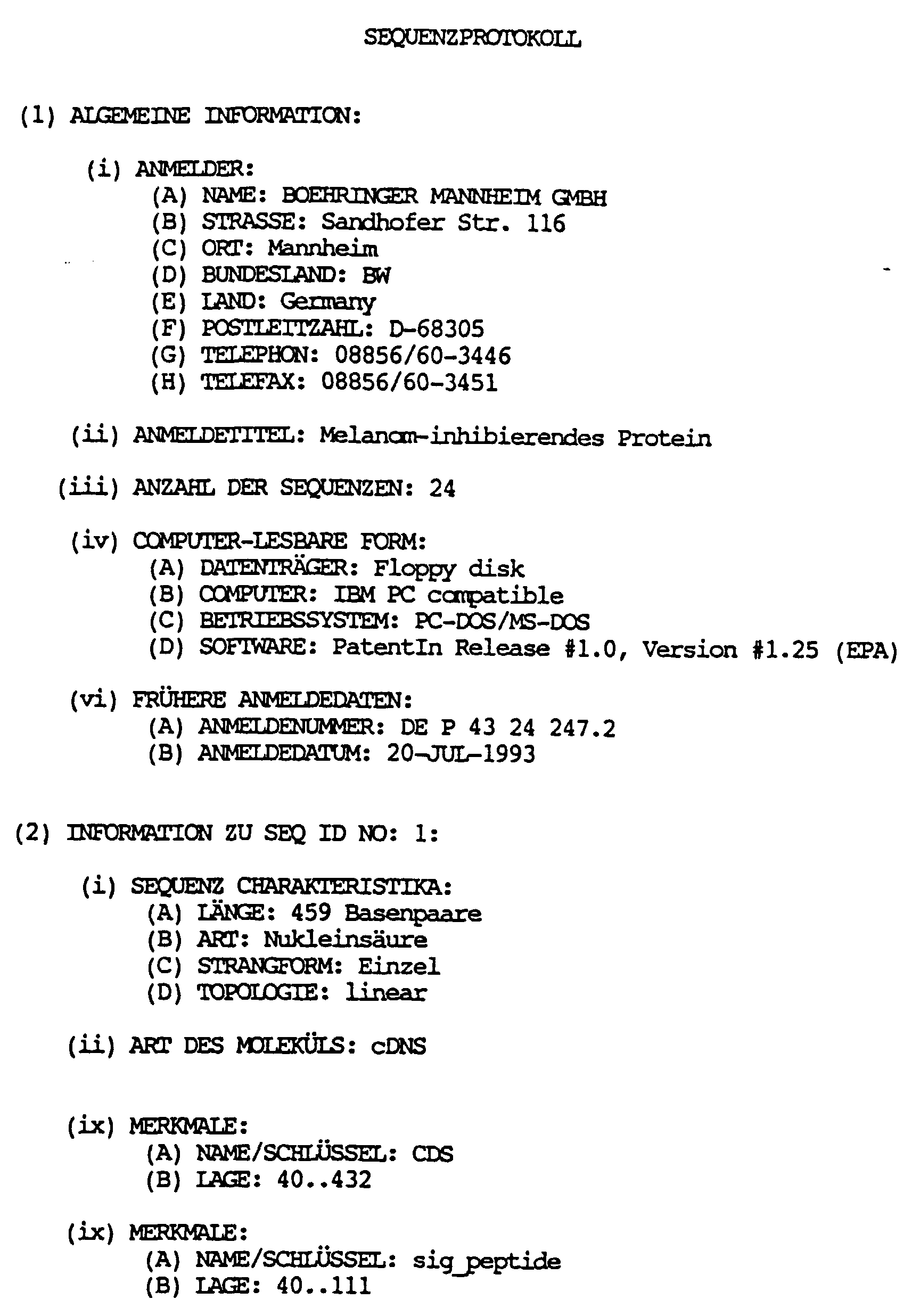
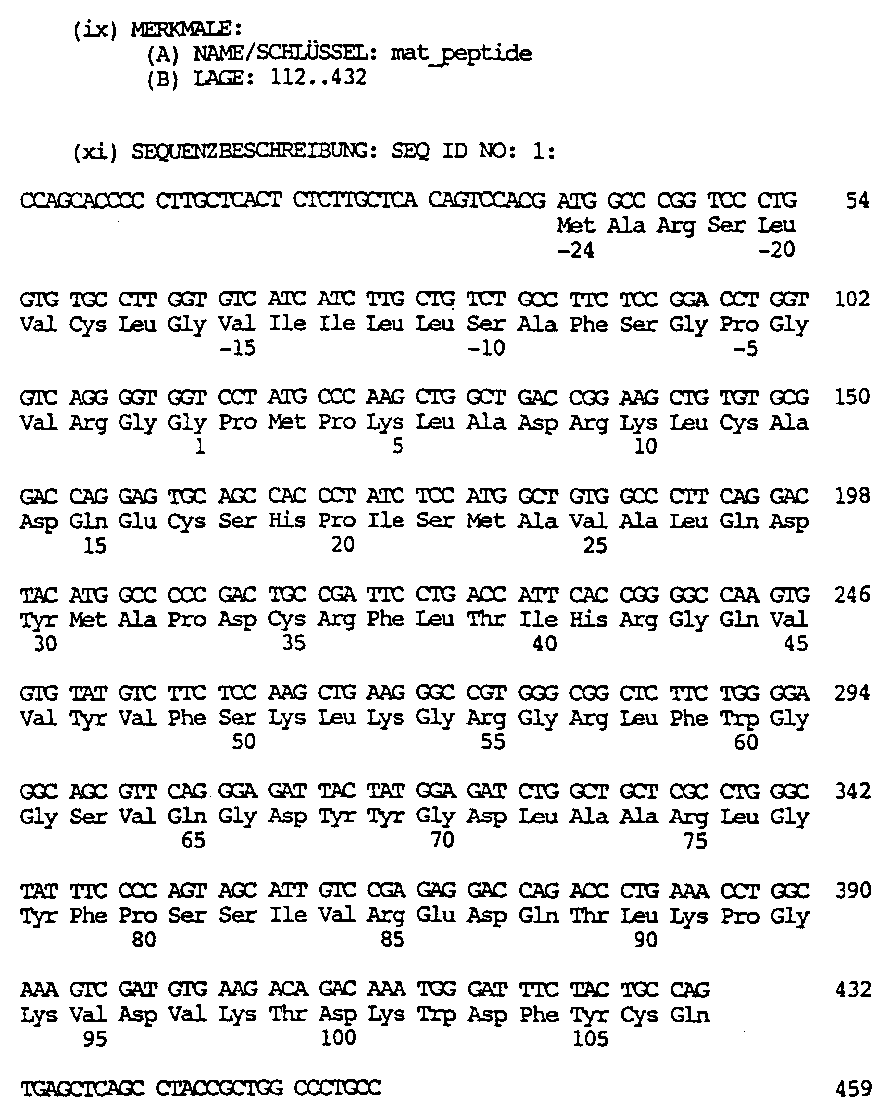
- a protein which is encoded by the Nucleotides 40 - 432 or 112 to 432 from SEQ ID NO 1 or of DNA sequences which are due to the degeneration of the genetic codes for a polypeptide with the same amino acid sequence would encode.
- the MIA protein from HTZ 19-dM has a molecular weight from approx. 11 kD, is stable against heat (3 minutes at 100 ° C) and sensitive to proteases, such as. B. Trypsin.
- Another object of the invention are melanoma inhibiting Proteins from mammalian cells, e.g. Mouse, Rat, beef, sheep, which are essentially analogous Way the growth of cell lines HTZ19-dM and ATCC Inhibit CRL 1424, like the human MIA protein.
- These proteins which are analogous to the human MIA protein, can be obtained by using a hybridization sample, which contains sequences coding for human MIA, a cDNA library of the corresponding mammal is screened according to methods familiar to the person skilled in the art and about the sequence comparison with the DNA and protein sequence the corresponding for human and murine MIA (SEQ ID NO: 1-5) coding segment is identified.
- a preferred subject of the invention is murine MIA protein and the nucleic acid sequence coding therefor (SEQ ID NO: 4).
- the murine protein is encoded from nucleotides 110-499 or 179-499 of SEQ ID NO: 4.
- the invention Protein obtained in large quantities in a reproducible manner become.
- suitable expression vectors integrated.
- such an expression vector contains a regulatable / inducible one Promoter.
- suitable host cells such as. B. E. coli, as a prokaryotic Host cells or Saccharomyces cerevisiae, Teratocarcinoma cell line PA-1 sc 9117 (Büttner et al., Mol. Cell.
- Another object of the invention is a method to obtain a MIA protein by isolation from the Culture supernatant of the melanoma cell HTZ 19-dM over a gel chromatographic separation and purification of a Fraction having a molecular weight of approx. 11 kD (SDS-PAGE, not reduced), via reverse phase HPLC. In this way, about 0.2 ⁇ g / l culture supernatant can be preserved.
- the natural MIA protein in the isolation / purification of an acid treatment subject is provided.
- a pH value of around 2 is advantageous.
- Acetic acid for example, is suitable as the acid.
- the detection of transformed or transduced Host cells that produce the MIA protein recombinantly, and the protein are preferably purified about antibodies that bind to this protein.
- Such antibodies are with the help of the invention Protein as an antigen or immunogen in a simple way available by known methods.
- Another object of the invention is therefore Use of the protein according to the invention with melanoma inhibitors Antibody production activity, that bind to this protein.
- Another object of the invention is therefore antibodies against the MIR protein, which are obtainable by Immunization of an animal with a MIR protein and Obtaining the antibodies from the serum or spleen cells of the immunized animals.
- the MIA protein not only on melanoma cells but also to a lesser extent on other tumor cells, such as.
- This protein works in very low concentrations (nanogram range).
- This protein is therefore suitable for the manufacture of a therapeutic agent for tumor therapy.
- Such a therapeutic agent is particularly suitable for the therapy of malignant melanomas, malignant gliomas, bronchial carcinomas (in particular small cell bronchial carcinoma, SCLC) and neuroblastomas.
- the melanoma-inhibiting Protein that induced interleukin 2-dependent or phytohemagglutinin Proliferation peripheral Blood lymphocytes suppressed. Furthermore, the cytotoxicity reduced by T lymphocytes.
- the melanoma-inhibiting Protein is therefore also suitable for production a therapeutic agent that is used as an immunosuppressant finds.
- Another object of the invention is therefore Use of a protein according to the invention for the production a therapeutic agent that is used in tumor therapy or as an immunosuppressant.
- this is protein according to the invention optionally with the usual auxiliaries, fillers and / or additives used together processed in a pharmaceutical formulation.
- Another object of the invention is therefore a therapeutic composition containing an inventive Melanoma-inhibiting protein, if necessary together with the commonly used auxiliary, filling and / or Additives.
- Another object of the invention is the use of sequences of the MIA gene, preferably of for one Protein coding sequences or MIA activity activating sequences from the 5 'untranslated Field in gene therapy and especially for manufacturing of drugs for gene therapy.
- Retroviruses (Mulligan, R.C. (1991) in Nobel Symposium 8: Ethiology of human disease at the DNA level (Lindsten, J. and Pattersun Editors), pages 143 - 189, Raven Press), Adeno Associated Virus (McLughlin, J.-Virol. 62 (1988), 1963), Vaccinia Virus (Moss et al., Ann. Rev. Immunol. 5 (1987) 305) Bovine Papilloma Virus (Rasmussen et al., Methods Enzymol. 139 (1987) 642) or Viruses from the group of herpes viruses, such as Epstein Barr Virus (Margolskee et al., Mol. Cell. Biol. 8 (1988) 2937) or herpes simplex virus.
- Epstein Barr Virus Margolskee et al., Mol. Cell. Biol. 8 (1988) 2937
- herpes simplex virus such as Epstein Barr Virus (
- Non-viral delivery systems are also known.
- DNA for this "naked" nucleic acid is usually preferred DNA, used, or nucleic acid along with one Auxiliary, such as with transfer reagents (liposomes, Dendromers, polylysine-transferrin conjugates (Wagner, 1990; Felgner et al., Proc. Natl. Acad. Sci. USA 84 (1987) 7413)).
- Another preferred gene therapy method is based on homologous recombination. It can either the gene coding for the MIA protein in one or multiple copies in the genome of somatic cells be introduced and / or endogenously in the cells existing MIA gene modulated, preferably activated become.
- the targeting DNA is a DNA that is complementary (homologous) to a region of genomic DNA (preferably in or near the MIA gene). If two homologous pieces of single-stranded DNA (e.g. the Targeting DNA and the genomic DNA) in immediate Come close to each other, they hybridize and form one Double strand helix. Through a recombination event then the MIA gene fragment and the targeting DNA into the genome to get integrated. This homologous recombination can performed both in vitro and in vivo (on the patient) become.
- a DNA is used which is for a protein encoded with MIA activity, a fragment that expresses MIA inhibited (knock-out sequence), or a fragment, which is able, after integration into the genome a cell the expression of a protein with MIA activity to activate in this cell.
- a fragment can be, for example, a promoter and / or enhancer region be heterologous to the corresponding MIA region or after integration into the MIA gene, that in itself silent or low-expressed MIA gene transcriptionally and / or activated in translation.
- one or more MIA genes are in the target cell (target cell) newly introduced or that in essential transcriptionally silent gene in the genome of a Mammalian cell activated so that the mammalian cell is capable will produce endogenous MIA protein.
- a DNA construct in the genome by homologous recombination inserted this DNA construct comprising: a DNA regulatory element, which is capable of expression modulate, preferably stimulate this gene, if it is operationally linked to it; and a or several DNA target segments which are homologous to one Region in this genome that are within or in the Is close to this gene.
- This construct is in one Inserted into the genome of the mammalian cell that the regulatory segment is operatively linked to the gene which codes for the protein with MIA activity.
- the construct still includes amplifying Sequences, especially if genes are responsible for a protein encode with MIA activity, introduced into the cell become.
- the construct is a regulatory element, one or more MIA genes and one or more target segments.
- the target segments are selected so that they can be used with a suitable Hybridize region of the genome, with the inserted exogenous MIA genes expressed after homologous recombination become.
- homologous recombination can be initiated.
- the homologous recombination takes place during DNA replication or mitosis of the cells.
- Such a DNA can be used to manufacture a tumor therapeutic or Production of a homologous or heterologous MIA protein be used in a host organism.
- Nucleic acid sequences of the MIA protein it is possible to provide a test with the nucleic acids which code for MIA proteins, can be detected. Such detection can, for example, in cells or cell lysates. Such evidence can by means of nucleic acid diagnostics.
- the sample to be examined is included brought into contact with a probe which Hybridize MIA protein coding nucleic acid sequence would. Hybridization between the probe and Nucleic acids from the sample show the presence of expressed MIA proteins.
- Such procedures are Known in the art and for example in WO 89/06698, EP-A 0 200 362, USP 2915082, EP-A 0 063 879, EP-A 0 173 251, EP-A 0 128 018.
- the nucleic acid of the sample coding for an MIA protein amplified before the test for example using the known PCR technique.
- a derivatized (marked) Nucleic acid probe used. This probe comes with a denatured DNA or RNA bound to a support brought into contact with the sample and the temperature, Ionic strength, pH value and other buffer conditions - depending on the length of the nucleic acid sample and the resulting melting temperature of the expected Hybrids - chosen so that the labeled DNA or RNA can bind to homologous DNA or RNA (hybridization, see also J. Mol. Biol. 98 (1975), 503; Proc. Natl. Acad. Sci.
- Membranes or carrier materials based on nitrocellulose e.g. Schleicher and Schüll, BA 85, Amersham Hybond, C.
- nitrocellulose e.g. Schleicher and Schüll, BA 85, Amersham Hybond, C.
- reinforced or bound powdered nitrocellulose or with different functional groups e.g. Nitro group
- derivatized nylon membranes e.g. Schleicher and Schull, Nytran; NEN, Gene Screen; Amersham Hybond M .; Pall Biodyne.
- Hybridizing DNA or RNA is detected then in that the carrier after thorough washing and Saturate to prevent nonspecific binding is incubated with an antibody or antibody fragment.
- the antibody or antibody fragment is against that incorporated in the nucleic acid probe during derivatization Substance directed.
- the antibody in turn is marked. However, it can also be a directly labeled DNA be used. After incubation with the antibodies is washed again to remove only specifically bound Detect antibody conjugates. The determination is made then over the label of the antibody or antibody fragment according to known methods.
- MIA is therefore a valuable prognostic marker in the Tumor diagnosis (metastases, course).
- HTZ 19-dM cells are defined as monolayers serum-free tissue culture medium (50% Dulbecco's minimal essential medium, 50% F-12, Boehringer Mannheim GmbH) with 0.8 mmol / l L-glutamine (Gibco, U.K.), non-essential Amino acids (Gibco, U.K.), 10 ⁇ g / ml transferrin (Boehringer Mannheim GmbH, catalog no.1073974), 30 nmol / l sodium selenite (Sigma) and 4 ⁇ g / ml gentamycin (Merck) cultivated. The cell culture supernatant of this culture is taken every 3 - 4 days and stored at -70 ° C until purification.
- the cell culture supernatants are cleaned for purification 0.45 ⁇ m filter (Becton Dickinson, Heidelberg) filtered and by membrane ultrafiltration with Amicon YM 2 membranes (Exclusion limit 2000 D, Amico Danvers Massachusetts, USA) to a final volume of 1% of the original volume concentrated.
- the material obtained is used for 30 Dialysed for hours against 0.1 mol / l acetic acid (dialysis membrane with exclusion limit of 1000 D, Reichelt, Heidelberg) and then at 100,000 g for one hour 4 ° C ultracentrifuged. The pellet is discarded and the Supernatant lyophilized for further processing.
- the eluate is collected in 1.5 ml fractions. Aliquots are lyophilized and, as described in Example 5, examined for anti-tumor effects.
- the MIA amino acid sequences in the sequencer certainly.
- the C-terminal and one near the N-terminus located peptide sequence were used as the basis for synthesis two primers selected.
- the primers are to degenerate oligonucleotides with restriction enzyme sites attached.
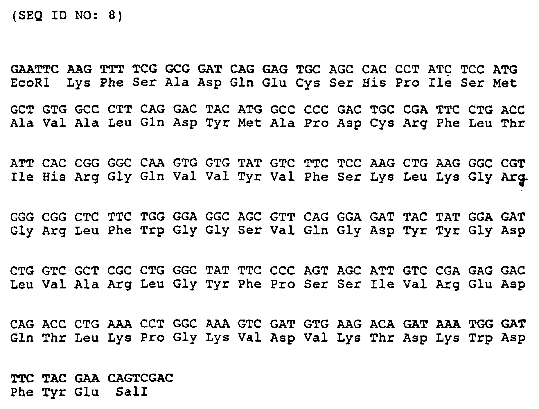
- Upstream Primer 1 (sense) (UP 1) (SEQ ID NO: 6)
- the dash (/) means that either the base is in front or after the dash at this point.
- This Oligonucleotide is a mixture of 32 different ones Molecules, covering almost all possible codons are. Only at positions 12 and 13 are G-T Mismatches with the target sequence are possible, resulting in stability the hybrid is not increased, but also not decreased.
- An EcoRI linker also hangs at the 5 'end to be able to easily clone any product after PCR. In front of it there are 3 non-specific bases at the 5 'end, so the restriction interface is not on the edge because restriction enzymes cut less well there.
- DP 1 corresponds to the 8 C-terminal amino acids, is 8 times degenerates and contains a Sal I interface.
- the cloned insert was sequenced using the T-7 Deaza Sequencing Kit from Pharmacia.
- the T-3 and T-7 primers (Stratagene) were available as primers.
- the following picture resulted from the overlap of the read sequences (primer printed in bold):
- a plasmid obtained in this way is pbs L7MIA, which in the German collection of microorganisms and Zellkulturen GmbH (DSM) in Braunschweig, Germany on July 14, 1993 (DSM 8420).
- Pre-hybridization (2 hours) and hybridization (16 hours) were carried out at 60 ° C in 6 x SSC, 5 x Denhardt's solution, 100 ⁇ g ml salmon sperm DNA and 0.1% SDS.
- the 32 P-dCTP-labeled sample was added to the hybridization mixture in a concentration of 1 ⁇ 10 6 cpm / ml.
- the filters were then washed twice at 60 ° C. for 20 minutes in 2 ⁇ SSC, 0.1% SDS, then twice for 20 minutes in 1 ⁇ SSC, 0.1% SDS and finally twice for 20 minutes in 0.25 ⁇ SSC, 0.1% SDS.
- the filters were then dried and exposed to X-ray film for 24 to 48 hours.
- the plaques providing a positive hybridization signal in this method were isolated and confirmed by re-hybridization.
- the human genomic DNA contained as an insert in these phages was characterized by Southern hybridization using MIA cDNA samples.
- the phage DNA was cleaved with the restriction endonuclease XbaI, separated on a 0.8% agarose gel and then transferred to nitrocellulose according to Southern (J. Mol.Biol. 98 (1975), 503).
- the filters thus obtained were hybridized with the complete MIA cDNA as a sample under the conditions described above, two XbaI fragments of approximately 1.4 kb and approximately 2.2 kb providing positive signals.
- Example 2a A commercially available (Novagene, New York) cDNA library from a 13.5 day old mouse embryo in the Vector Lambda EXlox (Palazzolo et al. (1990), Gene 88, 25 - 36) was plated as described in Example 2a.
- the hybridization sample was the one coding for human MIA cDNA from Example 2a used in radioactively labeled form.
- the hybridization conditions were identical to those described in Example 2b, with the exception of the temperatures with hybridization and washing, which here is 55 ° C cheat. Those obtained in the way and by re-hybridization confirmed plaques of existing cDNA inserts were sequenced.
- the sequence of the insert with the complete coding DNA of murine MIA is in SEQ ID NO 4 shown.
- a murine genomic DNA library was created (from the liver of an adult BALB / C mouse in the vector EMBL3 (Frischholz et al. (1983), J. Mol. Biol. 170, 827), commercially available from Clontech, Palo Alto CA) in analog Using as described in Example 2b the murine MIA cDNA from Example 2c as a sample. The conditions were identical to those in Example 2b described. The further procedure also took place in analogous way.
- a concentration can be determined at which this 3 H thymidine incorporation is inhibited by 50% compared to the untreated control (IC 50 value in Table 1 below).
- IC 50 value in Table 1 below.
- Antiproliferative effect of the melanoma-inhibiting protein on various tumor cells Tumor cell line IC 50 ( ⁇ g / ml) a) Melanoma cell lines HTZ 19-dM 1.2 ATCC HTB 69 3.7 HTZ 320 1.35 HTZ 318 3.5 ATCC CRL 1424 2.1 b) Neuroblastoma lines Kelly 80 c) glioblastoma ATCC HTB17 10th d) Astrocytoma HTZ 243 5 HTZ 209 5
- a modified Boyden chamber system (Albini et al. (1987), Cancer Res. 47, 3239-3245) is used to determine the invasive activity of MIA.
- the chambers were purchased from Costar (Blind Well Chamber No. 441200).
- 52 ⁇ l of Matrigel (Becton Dickinson Cat. No. 40234) are applied to the polycarbonate filter (pore size 8 ⁇ m, Costar No. 150446).
- the lower chamber is filled with 210 ⁇ l fibroblast-conditioned medium.
- Fibroblasts from normal human skin are kept between the 10th and 20th passage for 24 hours in DMEM medium (Gibco) without the addition of fetal calf serum.
- the medium conditioned in this way is used undiluted as a chemoattractant.
- 2 ⁇ 10 5 of the tumor cells to be examined are placed in 800 ⁇ l DMEM (Gibco, without fetal calf serum) with or without MIA active ingredient in the upper chamber of the Boyden apparatus.
- Human tumor cells see example 3a or example 8
- animal tumor cells such as B16 (ATCC CRL 6322) can be tested for the inhibition of their migration behavior by MIA using the described method.
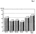
- melanoma-inhibiting protein Without the addition of the melanoma-inhibiting protein, about 10% of tumor cells migrate from the upper chamber to the lower chamber within 4 hours, where they remain attached to the underside of the Matrigel membrane. They are fixed there, colored and then counted. If the human or murine melanoma inhibiting protein MIA is added to the upper chamber, the migration of the cells is strongly inhibited. 1 shows the inhibition values obtained ( [Number of cells in the lower chamber, experiment with MIA] / [Number of cells in the lower chamber, experiment without MIA] x 100% ).
- MIA inhibits T cell mediated cytotoxic activity
- the CD4 + T cell line D7.1 which is specific against a peptide from myelin basic protein (MBP, amino acid position 87-106), is able to present both MBP and peptide 87-106 in a standard 51 Cr release assay lysing (targets: Daudi cells, R. Martin, U. Utz, JE Coligan, JR Richert, M. Flerlage, E. Robinson, R. Stone, WE Biddison, DE McFarlin, HF MacFarland, Diversity in fine specificity and T cell receptor usage of the human CD 4 + cytotoxic T-cell response specific for the immunodominant myelin basic protein peptide 87-106, J. Immunol.
- MIA inhibits the cytotoxic activity of lymphokine-activated peripheral blood lymphocytes (LAK cells)
- LAK cells are not clonally expanded, lymphokine-activated peripheral blood lymphocytes, predominantly T lymphocytes (A.A. Rayner, E.A. Grimm, M.T. Lotze, E.W. Chu, S.A. Rosenberg, Lymphokine activated killer (LAK) cells: Analysis of factors relevant for immunotherapy of human cancer, Cancer 55 (1985), 1327-1333).
- LAK cells are experimented on their cytotoxicity versus HTZ-19 melanoma cells as targets in one Microcytotoxicity assay tested.
- effector-target ratios of 1: 1, 5: 1 and 10: 1 there is a maximum cytotoxicity (CTX) of just under 40%. This will after adding MIA (concentration as in Example 4 a) strongly inhibited, by a maximum of 80% with a low effector-target ratio, as it is most likely local - on the tumor itself - is to be expected (Fig. 3).
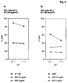
- MIA inhibits the IL-2 dependent or phytohemagglutinin dependent Lymphocyte proliferation
- PBMC Peripheral blood lymphocytes
- PHA phythaemagglutinin
- IL-2 interleukin-2
- MIA as a fusion protein with an as "Carrier” in E.coli
- suitable protein can for example the commercially available vector pQE40 (Cat. No. 33403, DIAGEN GmbH, Düsseldorf) can be used.
- pQE40 Cat. No. 33403, DIAGEN GmbH, Düsseldorf
- This vector is between the existing ones Restriction interfaces SphI and HindIII one for the mature form of MIA coding cDNA fragment inserted.
- Such a fragment is easiest by PCR amplification manufactured under known techniques under Using the cloned MIA cDNA as a template and two suitable primer (5'-GATGCATGCGGTCCTATGCCCAAGCTG-3 '(SEQ ID NO: 9) and 5'-GATAAGCTTTCACTGGCAGTAGAAATC-3 '(SEQ ID NO: 10)).
- the PCR fragment obtained is treated with SphI and HindIII cut and in the same treated vector pQE40 ligated.
- the resulting plasmid expresses a Fusion protein from DHFR (dihydrofolate reductase as "Carrier") and MIA.
- DHFR dihydrofolate reductase as "Carrier”
- MIA Magnetic Ink Characteristics
- the fusion protein carries at the N-terminus 6 histidines that follow the manufacturer's instructions according to help purify the fusion protein of Ni chelate gel materials can be used.
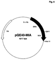
- Such processes are described in EP-A 0 282 042 and EP-A 0 253 303, the content of which is the subject of this disclosure Registration is described. 6 shows the expression plasmid pQE40-MIA.
- the content of MIA can be optimized by the Carrier protein is eliminated by DHFR and if possible by a small peptide is replaced, which has the same functions Fulfills.
- suitable peptides are, for example MetArgGlySerHisHisHisHisHisHisGlySerSerArgProPro (SEQ ID NO: 13) (this peptide can be derived from the IgA protease immediately following amino acid sequence of the mature MIA to be split off; are such procedures described in WO 91/11520, the content of which is the subject the disclosure of this application) or MetArgGlySerHisHisHisHisHisHisHisHisGlySerValAspAspAspAspLys- (SEQ ID NO: 14) (this peptide can be derived from the enterokinase immediately following amino acid sequence of the mature MIA can be split off).
- Expression plasmids the code for such MIA-peptide fusions, such as can be prepared as follows: A PCR amplification with MIA cDNA (Seq.ID No 1) as template and the primers 5'-AAAAAGGATCCAGCCGGCCGCCCGGTCCTATGCCCAAGCTGGC-3 ' (SEQ ID NO: 15) and 5'-GGCGAGCAGCCAGATCTCCATAG-3 '(SEQ ID NO: 16) a fragment which was cut with BamHI and BglII becomes.
- the expression vector pQE40-MIA is also included BamHI and BglII restricted, the smaller of the resulting Fragments are discarded and described by the PCR fragment replaced.
- the MetArgGlySerHisHisHisHisHisHisHisGlySerValAspAspAspAspLys-MIA fusion protein (SEQ ID NO: 14) is used in a completely analogous manner Primer 5'-AAAAAAGGATCCGTTGATGATGACGATAAAGGTCCTATGCCCAAGCTGGC-3 ' (SEQ ID NO: 17) and 5'-GGCGAGCAGCCAGATCTCCATAG-3 ' (SEQ ID NO: 16) received.
- fusion proteins from peptides and MIA can be made in an analogous manner and under control more suitable (preferably stronger and more inducible) Promoters in one of the numerous for E.coli described plasmids cloned and for expression to be brought.
- Another alternative is the Fusion of MIA with a peptide which is found in E. coli Secretion of the fusion protein into the periplasm with subsequent Cleavage and release of MIA leads. This method is described in WO 88/09373, the The subject matter of the disclosure of this application is.
- the expression plasmid pQE40-MIA (or a similar Expression plasmid, which for example by use of another basic vector or another "Carrier" proteins or peptides can be obtained; Such alternatives are also in Example 5a described in Methods of Enzymology 185 (Gene Expression Technology), ed. David V. Goeddel, Academic Press 1991) is transfected into a suitable E. coli strain, which sufficient expression of the lac repressor has so that an inducible expression of the MIA fusion protein can be achieved. Suitable for this is for example the strain E.coli M15 [pREP4], which together is commercially available with pQE40 (Diagen GmbH, Düsseldorf), or other E. coli strains such as E.
- E.coli UT5600 (Earhart et al. (1979), FEMS Microbiology Letters 6, 277-280) or E.coli BL21 (Grodberg and Dunn (1988), J.Bacteriol. 170, 1245-1253), previously with a lac repressor expressing helper plasmid such as pUBS520 (Brinckmann et al. (1989) Gene 85, 109-114) or pUBS500 (EP-B 0 373 365) were transfected.
- helper plasmid such as pUBS520 (Brinckmann et al. (1989) Gene 85, 109-114) or pUBS500 (EP-B 0 373 365) were transfected.
- MIA can then like can be obtained as follows: E.coli M15 [pREP4 / pQE40-MIA] on LB medium up to an optical density of 0.6 (measured at 550 nm), then IPTG (isopropyl- ⁇ -D-thiogalactopyranoside, Boehringer Mannheim GmbH) in added to a final concentration of 1 mM and then Cultivated for 4 hours. The cells will centrifuged, in 100 mM sodium phosphate buffer pH 7.5 taken up with 300 mM NaCl and by freezing three times and thawing with subsequent ultrasound treatment lysed.
- Lysate is considered Ni-NTA agarose (Diagen GmbH) the maximum binding capacity specified by the manufacturer added and overnight at room temperature incubated with mixing.
- the one so loaded with fusion protein Gel material is centrifuged at low speed, twice with 100 mM sodium phosphate buffer pH 7.5 and washed twice with sodium phosphate buffer pH 6.1. MIA is then cleaved from the fusion protein by incubating the gel material with IgA protease (Boehringer Mannheim GmbH) in 100 mM sodium phosphate buffer pH 7.5 overnight at 37 ° C. The gel material will separated by centrifugation and the MIA-containing Supernatant after sterile filtration in the in the examples 3 and 4 described activity tests used.
- the DNA sequence coding for MIA is in a way modified that an efficient expression in E.coli enables.
- a PCR amplification with human MIA cDNA (Seq. ID No 1) as template and the Primers 1 (5'-AAAAACATATGGGACCAATGCCAAAATTAGCAGATCGTAAATTATGTGCAGATCAGGAG-3 ' (SEQ ID NO: 19) and 2 (5'-AAAAAAAGCTTTCACTGGCAGTAGAAATC-3 ' (SEQ ID NO: 20)).
- Primers 1 5'-AAAAAAAGCTTTCACTGGCAGTAGAAATC-3 ' (SEQ ID NO: 20)
- Primer 1 changes the MIA coding sequence in the N-terminal area so that with unchanged MIA amino acid sequence the DNA and thus the mRNA sequence is optimized for E.coli.
- the start codon Met is added and at this point a recognition sequence the restriction endonuclease NdeI introduced, the one subsequent cloning of the MIA coding modified in this way Fragment into a vector that allows one (preferably strong and inducible) promoters and a translation initiation sequence ("Shine-Dalgarno sequence") with the NdeI interface placed below contains.
- Primer 2 serves as a 3 'counter primer and contains a HindIII interface so that the insertion of the modified MIA coding fragment in the vector can be done as a NdeI HindIII fragment.
- the so modified MIA coding sequence is in SEQ ID NO: 18 shown.
- An expression plasmid produced in this way is p11379, which is filed under the accession number DSM 9267 June 29, 1994 at the German Collection of Microorganisms and Zellkulturen GmbH, D-38124 Braunschweig has been.
- An expression plasmid for fusion-free MIA is used Expression transfected into a suitable E. coli strain.
- Expression plasmids such as p11379 are strains which have a sufficiently high intracellular concentration at the lac repressor. Such tribes can by transfecting a second plasmid such as pREP4 (Diagen GmbH), pUBS500 or pUBS520 (Brinckmann et al. (1989), Gene 85, 109-114).
- the used E. coli strains should preferably be low have cell-specific protease activity, such as this E.coli UT5600 (Earhart et al.
- MIA-containing protein aggregates from E. coli fermentations are in 6 M Guanidinium hydrochloride, 100mM TrisHCl pH 8, 1mM EDTA solubilized, then adjusted to pH 3-4 and dialyzed against 4 M guanidinium hydrochloride pH 3.5.
- the solubilized protein is then renatured in 1 M arginine pH 8, 1 mM EDTA, 5 mM GSH (glutathione reduced) and 0.5 mM GSSG (glutathione oxidized).
- MIA can, for example, add a renaturation approach of 1.4 M ammonium sulfate by adsorption on hydrophobic Gel matrices such as Fractogel TSK Butyl (E. Merck, Darmstadt) followed by elution in 20 mM TrisHCl pH 7 become.
- hydrophobic Gel matrices such as Fractogel TSK Butyl (E. Merck, Darmstadt) followed by elution in 20 mM TrisHCl pH 7 become.
- expression can also be as described in Example 9 can be achieved by homologous recombination in vitro.
- Such promoters and enhancers mostly come from Viruses such as SV40, hCMV, Polyoma or retroviruses.
- promoter-enhancer systems can also be used, those specific to a particular cell or tissue type such as WAP, MMTV or immunoglobulin promoters, or systems that are inducible, such as Metallothionein promoters.
- a vector complements the MIA cDNA (if one is used) with donor and Acceptor signals for RNA processing as well a signal for poly A addition.
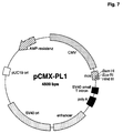
- a suitable one Vector is, for example, pCMX-pL1 (Umesono et al. (1991), Cell 65, 1255-1266) shown in Fig. 7 is.
- the expression plasmid pCMX-pL1-MIA is incorporated into the human teratocarcinoma line PA-1sc9177 (Büttner et al. (1991), Mol. Cell. Biol. 11, 3573-3583) according to the methods described (Büttner et al. (1993) Mol. Cell. Biol. 13, 4174-4185) transfected, whereby 200,000 cells per 100 mm culture dish with 5 ⁇ g DNA be transfected.
- the cells are made after transfection in MEM (Gibco) without the addition of fetal calf serum cultured, after 48 hours in the cell culture supernatant MIA is detectable.
- a coding for MIA is used DNA segment, preferably the human MIA cDNA (SEQ ID NO 1), introduced in vectors that are different from AcMNPV (Autographa californica nuclear polyhedrosis virus) or Derive BmNPV (Bombyx mori nuclear polyhedrosis virus).
- the MIA cDNA is initially under control a strong promoter suitable for insect cells brought (O'Reilly DR, Miller LK and Luckow VA 1992; Baculovirus Expression Vectors - A Laboratory Manual; W.H. Freeman & Co, New York), such as the polH promoter or of the p10 promoter.
- the polH promoter works as follows:
- a DNA fragment coding for MIA with interfaces for the restriction endonucleases EcoRI (adjacent to the 5 'end of the later MIA transcript) and PstI (adjacent to the 3' end of the later MIA transcript) is obtained by PCR amplification according to known techniques Use of the MIA cDNA as template and the primer 5'-CGTGAATTCAACATGGCCCGGTCCCTGGTGTGC-3 '(SEQ ID NO: 21) and 5'-TATCTGCAGTCACTGGCAGTAGAAATCCCA-3' (SEQ ID NO: 22).
- This fragment is cut with EcoRI and PstI (to produce the corresponding cohesive ends) and in the transfer vector pVL1393 restricted with the same endonucleases (O'Reilly DR, Miller LK and Luckow VA 1992; Baculovirus Expression Vectors - A Laboratory Manual; WHFreeman & Co, New York), which is commercially available (PharMingen, San Diego, CA or Invitrogen Corporation, San Diego, CA).
- the resulting transfer expression plasmid pVL1393-MIA is transfected for growth in E. coli K12 and plasmid DNA is prepared according to established methods.
- the transfer of the MIA DNA under control of the polH promoter from the transfer plasmid into the baculovirus vector is carried out by homologous recombination according to established methods (O'Reilly et al. 1992, see above).
- BaculoGold DNA linearized AcNPV virus DNA with lethal deletion and lacZ expression controlled by the polH promoter, commercially available from PharMingen, order number 21100D
- 2 ⁇ g pVL1393-MIA are mixed, incubated for 5 minutes at room temperature and then with 1 ml 125 mM Hepes pH 7.1, 125 mM CaCl2, 140 mM NaCl added.
- This mixture is added to 2 ⁇ 10 6 SF9 insect cells (Invitrogen, order number B825-01) in a culture dish with a diameter of 60 mm, which had previously been overlaid with 1 ml Grace's medium with 10% fetal calf serum. After 4 hours of incubation at 4 ° C, the DNA-containing medium is removed and the cells are incubated in fresh medium for 4 days at 27 ° C. The recombinant baculoviruses obtained in this way are then purified twice via plaque formation (O'Reilly et al.
- viruses which have inserted MIA by homologous recombination being distinguished by the lack of ⁇ -galactosidase activity ( optically recognizable by the absence of a blue color in the presence of 5-bromo-4-chloro-3-indolyl- ⁇ -D-galactopyranoside) from the wild-type viruses used (AcNPV with lethal deletion and lacZ expression controlled by the polH promoter, commercially available from PharMingen, order number 21100D).
- SF9 cells are infected according to established methods (O'Reilly et al.
- MIA mRNA is present in a particular cell and, accordingly, MIA mRNA is present
- nucleic acid hybridization such as Northern hybridization, in-situ hybridization, dot or slot hybridization, and the like derived diagnostic techniques (Sambrook et al. 1989, Molecular Cloning - A Laboratory Manual; Cold Spring Harbor Laboratory Press; Nucleic Acid Hybridization - A Practical Approach, ed. BD Hames and SJ Higgins (1985), IRL Press; WO 89/06698, EP-A 0 200 362, USP 2915082, EP-A 0 063 879, EP-A 0 173 251, EP-A 0 128 018).
- MIA-specific primers PCR Protocols - A Guide to Methods and Applications; ed. MA Innis, DH Gelfand, JJ Sninsky, TJ White (1990), Academic Press Inc; PCR - A Practical Approach; Ed. MJ McPherson, P Quirke, GR Taylor (1991), IRL Press).
- Tables 2A and 2B show the MIA expression in various human tumors, tumor lines and normal cells, which in this case was determined by Northern hybridization using the radiolabelled human MIA cDNA (Seq. ID No. 1). For this, the RNA was isolated from the cells listed according to the method of Chomczynski and Sacchi (Anal. Biochem.
- the hybridization was carried out at 68 ° C in 5 x SSC, 5 x Denhardt, 0.5% SDS, 10% dextran sulfate and 100 ⁇ g / ml salmon sperm DNA.
- the membranes were then washed twice in 1 ⁇ SSC at 68 ° C. for one hour each and then exposed to X-ray film.
- MIA coding nucleic acids therapeutic purposes
- MIA inhibits the proliferation and metastasis of Tumor cells.
- This effect can be in an animal model or to a patient not only through the exogenous supply of MIA protein are caused, but also by that Introduction of a DNA segment that is either for MIA encoded under a suitable promoter or one contains suitable promoter by homologous recombination before integrating the cell's own MIA gene into the genome can.
- this promoter has to Sequence sections flanked by the sequences of the human (or animal in the case of an animal model) MIA Gens in the 5'-untranslated region as far as possible are homologous or preferably identical (WO 91/09955).
- Example is the therapeutic effect of an MIA coding DNA segment shown in an animal model.
- the injection of murine B16 melanoma cells (ATCC CRL 6322) into the tail vein of C57BL mice with subsequent quantification of lung metastases represents an established in vivo metastasis model. 100,000 cells of the B16 melanoma line were retrobulbar injected into C57BL mice (Day 1, 16 animals) After 48 hours, 100 ⁇ g of the MIA expression plasmid pCMX-PL1-MIA (Example 7a) in TE (10 mM TrisCl pH 8.0, 1 mM EDTA) were mixed with DOTAP transfection reagent (Leventis and Silvius (1990) in 8 animals.
Abstract
Description
- in situ-Hybridisierung mit fixierten ganzen Zellen mit fixierten Gewebesabstrichen und isolierten Metaphasen-Chromosomen,
- colony-Hybridisierung (Zellen) und plaque-Hybridisierung (Phagen und Viren),
- Northern-Hybridisierung (RNA-Nachweis),
- Serum-Analytik (z. B. Zelltypanalyse von Zellen in Serum, durch slot-blot-Analyse),
- nach Amplifikation (z.B. PCR-Technik).
- Fig. 1
- zeigt die invasionshemmende Wirkung von humanem MIA (Hemmung der Zellwanderung mit und ohne MIA in %). B16 + mMIA: Test mit murinem MIA.
- Fig. 2
- Inhibition von T-Cell-vermittelter cytotoxischer Aktivität durch MIA als %Lyse von CD4+ T-Zellen.
- Fig. 3
- Inhibition der cytotoxischen Aktivität von LAK-Zellen durch MIA.
- Fig. 4
- Inhibition der Phytohämagglutinin-abhängigen Lymphozytenproliferation durch MIA (MIA-Konzentration in ng/ml).
- Fig. 5
- Inhibition der IL-2-stimulierten PBMC Proliferation durch MIA (MIA-Konzentration in ng/ml).
- Fig. 6
- Plasmidkarte des Expressionsplasmids pQE40-MIA (Beispiel 5a).
- Fig. 7
- Plasmidkarte des Vektors pCMX-PL1 (Beispiel 7a).
- Gradientenprogramm:
- Lösung A: 0,06 % TFA in Wasser
Lösung B: 0,056 % TFA, 80 % Acetonitril
- 2 - 25 % Lösung B in 5 min
- 25 - 50 % Lösung B in 120 min
- 50 - 100 % Lösung B in 5 min
- zurück auf 2 % in 5 min
- Säule: minoRPC (Pharmacia)
- HPLC-Gradientenmischer, Pumpe und Detektor: Pharmacia
EcoRI site
SalI site
- 5 µl
- 10 x PCR Puffer
- 3 µl
- HTZ-19 Gesamt-RNA
- 1 µl
- 100 mM MgCl2
- 10 µl
- 2,5 mM dNTP
- 0,5 µl
- Placenta RNase Inhibitor
- 2 µl
- DP 1 (1 µg)
- 1 µl
- Reverse Transkriptase
- 1 µl
- UP 1 (1 µg)
- 5 µl
- 10 x PCR Puffer
- 70,5 µl
- H2O
- 1 µl
- Taq-Polymerase
| Antiproliferative Wirkung des Melanom-inhibierenden Proteins auf verschiedene Tumorzellen | |
| Tumorzellinie | IC 50 (µg/ml) |
| a) Melanomzellinien | |
| HTZ 19-dM | 1,2 |
| ATCC HTB 69 | 3,7 |
| HTZ 320 | 1,35 |
| HTZ 318 | 3,5 |
| ATCC CRL 1424 | 2,1 |
| b) Neuroblastomlinien | |
| Kelly | 80 |
| c) Glioblastom | |
| ATCC HTB17 | 10 |
| d) Astrocytom | |
| HTZ 243 | 5 |
| HTZ 209 | 5 |
| Tumor | Anzahl getestet | positiv im Northern Blot |
| Astrocytom | 10 | 3 |
| Oligodendrogliom | 4 | 0 |
| Ependymom | 3 | 0 |
| Neuroblastom | 4 | 0 |
| Glioblastom | 13 | 0 |
| Colon-CA | 2 | 1 |
| malignes Melanom ZNS-Metastase | 6 | 6 |
| Medulloblastom | 2 | 0 |
| Mamma-CA | 1 | 0 |
| Bronchial-CA ZNS-Metastase | 2 | 0 |
| Normalzellen | Anzahl getestet | positiv im Northern Blot |
| embryonale Fibroblasten | 2 | 0 |
| mononukleäre Blutzellen (3 Spender) | 3 | 0 |


Claims (3)
- Verwendung von expressionsaktivierenden Nukleinsäurefragmenten aus dem 5'-untranslatierten Bereich von SEQ ID NO:3 zur Herstellung von viralen oder nicht-viralen Vektoren für die Gentherapie.
- Verwendung nach Anspruch 1, dadurch gekennzeichnet, daß das Nukleinsäurefragment eine Promotor- und/oder Enhancer-Region ist.
- Verwendung nach Anspruch 1, dadurch gekennzeichnet, daß das Fragment nach Integration ins Genom einer eukaryontischen Zelle die Expression eines Proteins mit MIA-Aktivität aktivieren kann.
Applications Claiming Priority (3)
| Application Number | Priority Date | Filing Date | Title |
|---|---|---|---|
| DE4324247 | 1993-07-20 | ||
| DE4324247 | 1993-07-20 | ||
| EP94925364A EP0710248B1 (de) | 1993-07-20 | 1994-07-19 | Melanom-inhibierendes protein |
Related Parent Applications (1)
| Application Number | Title | Priority Date | Filing Date |
|---|---|---|---|
| EP94925364.5 Division | 1995-03-10 |
Publications (2)
| Publication Number | Publication Date |
|---|---|
| EP0947583A2 true EP0947583A2 (de) | 1999-10-06 |
| EP0947583A3 EP0947583A3 (de) | 2000-05-03 |
Family
ID=6493222
Family Applications (2)
| Application Number | Title | Priority Date | Filing Date |
|---|---|---|---|
| EP99101554A Withdrawn EP0947583A3 (de) | 1993-07-20 | 1994-07-19 | Expressionsaktivierende Nukleinsäurefragmente des Melanom-inhibierenden Proteins (MIA) |
| EP94925364A Expired - Lifetime EP0710248B1 (de) | 1993-07-20 | 1994-07-19 | Melanom-inhibierendes protein |
Family Applications After (1)
| Application Number | Title | Priority Date | Filing Date |
|---|---|---|---|
| EP94925364A Expired - Lifetime EP0710248B1 (de) | 1993-07-20 | 1994-07-19 | Melanom-inhibierendes protein |
Country Status (14)
| Country | Link |
|---|---|
| US (1) | US5770366A (de) |
| EP (2) | EP0947583A3 (de) |
| JP (1) | JP3435163B2 (de) |
| CN (1) | CN1056617C (de) |
| AT (1) | ATE185152T1 (de) |
| AU (1) | AU7531294A (de) |
| CA (1) | CA2167693C (de) |
| DE (2) | DE4425481A1 (de) |
| DK (1) | DK0710248T3 (de) |
| ES (1) | ES2139751T3 (de) |
| GR (1) | GR3032212T3 (de) |
| IL (2) | IL110385A (de) |
| WO (1) | WO1995003328A2 (de) |
| ZA (1) | ZA945278B (de) |
Families Citing this family (20)
| Publication number | Priority date | Publication date | Assignee | Title |
|---|---|---|---|---|
| DE19653358A1 (de) * | 1996-12-20 | 1998-06-25 | Boehringer Mannheim Gmbh | Immunoassay zum Nachweis von MIA |
| EP0972022A2 (de) | 1997-01-21 | 2000-01-19 | Human Genome Sciences, Inc. | Für rezeptoren kodierende polynukleotide und polypeptide |
| US6492144B1 (en) * | 1997-05-30 | 2002-12-10 | Diagen Corporation | Methods for detection of nucleic acid sequences in urine |
| USRE39920E1 (en) | 1997-05-30 | 2007-11-13 | Xenomics, Inc. | Methods for detection of nucleic acid sequences in urine |
| EP0945507A1 (de) * | 1998-03-27 | 1999-09-29 | Boehringer Mannheim Gmbh | Tumorspezifische Expressionskontrollregion und deren Verwendung |
| US20020103360A1 (en) * | 1998-09-01 | 2002-08-01 | Yang Pan | Novel protein related to melanoma-inhibiting protein and uses thereof |
| US20030072767A1 (en) * | 1998-09-30 | 2003-04-17 | Alexander Gaiger | Compositions and methods for WT1 specific immunotherapy |
| EP1025871A1 (de) * | 1999-01-28 | 2000-08-09 | F. Hoffmann-La Roche Ag | Verwendung eines melanonhemmenden Faktors zur Reperatur von Knorpelgewebe und Knochen |
| US6713293B1 (en) * | 1999-02-08 | 2004-03-30 | Friedrich Grummt | Encapsulated cells containing an amplified expression vector as a drug delivery device |
| AU5705600A (en) | 1999-06-30 | 2001-01-22 | Takeda Chemical Industries Ltd. | Novel polypeptide and dna thereof |
| AU2001232967A1 (en) * | 2000-01-25 | 2001-08-07 | Hyseq, Inc. | Methods and materials relating to novel growth regulatory-like polypeptides and polynucleotides |
| EP1133994A1 (de) * | 2000-03-11 | 2001-09-19 | Biognostik Gesellschaft für biomolekulare Diagnostik mbH | Verfahren zür Umwandlung des immunosuppresive effekts von MIA (Melanoma Inhibitory Activity) |
| IL150679A0 (en) * | 2000-03-23 | 2003-02-12 | Akzo Nobel Nv | Use of mia in immunotherapy |
| US6613534B2 (en) | 2001-03-20 | 2003-09-02 | Wake Forest University Health Sciences | MAP-2 as a determinant of metastatic potential |
| US20100212037A1 (en) * | 2002-10-16 | 2010-08-19 | Anja Katrin Bosserhoff | Mia-2 protein |
| RU2480769C2 (ru) * | 2006-07-28 | 2013-04-27 | Новартис Аг | Применение белка с меланома-ингибирующей активностью (миа) в качестве раннего индикатора терапевтического ответа при меланоме |
| ES2415245T5 (es) * | 2007-08-22 | 2016-12-23 | Trovagene, Inc. | Métodos de uso de miARN para la detección de muerte celular in vivo |
| RU2588658C2 (ru) | 2010-04-27 | 2016-07-10 | ЭсСиАйЭл Текнолоджи ГмбХ | Стабильные водные композиции белка mia/cd-rap |
| CA2807431A1 (en) * | 2010-08-30 | 2012-03-08 | F. Hoffmann-La Roche Ag | Prokaryotic expression construct |
| ITRM20110134A1 (it) * | 2011-03-22 | 2012-09-23 | Matteo Bordignon | Inibitori di mia (attività inibitoria melanoma) per identificare, prevenire e curare la vitiligine |
-
1994
- 1994-07-19 CN CN94192822A patent/CN1056617C/zh not_active Expired - Lifetime
- 1994-07-19 CA CA002167693A patent/CA2167693C/en not_active Expired - Lifetime
- 1994-07-19 ZA ZA945278A patent/ZA945278B/xx unknown
- 1994-07-19 DE DE4425481A patent/DE4425481A1/de not_active Withdrawn
- 1994-07-19 AT AT94925364T patent/ATE185152T1/de active
- 1994-07-19 DK DK94925364T patent/DK0710248T3/da active
- 1994-07-19 WO PCT/EP1994/002369 patent/WO1995003328A2/de active IP Right Grant
- 1994-07-19 EP EP99101554A patent/EP0947583A3/de not_active Withdrawn
- 1994-07-19 EP EP94925364A patent/EP0710248B1/de not_active Expired - Lifetime
- 1994-07-19 AU AU75312/94A patent/AU7531294A/en not_active Abandoned
- 1994-07-19 JP JP50493195A patent/JP3435163B2/ja not_active Expired - Lifetime
- 1994-07-19 US US08/578,649 patent/US5770366A/en not_active Expired - Lifetime
- 1994-07-19 DE DE59408791T patent/DE59408791D1/de not_active Expired - Lifetime
- 1994-07-19 ES ES94925364T patent/ES2139751T3/es not_active Expired - Lifetime
- 1994-07-20 IL IL11038594A patent/IL110385A/xx not_active IP Right Cessation
- 1994-07-20 IL IL154248A patent/IL154248A/en not_active IP Right Cessation
-
1999
- 1999-12-22 GR GR990403299T patent/GR3032212T3/el unknown
Non-Patent Citations (2)
| Title |
|---|
| R.APFEL ET AL.: "Purification and analysis of growth regulating proteins secreted by a human melanoma cell line" MELANOMA RES., Bd. 2, Nr. 5-6, 1992, Seiten 327-36, XP002131794 * |
| U.BOGDAHN ET AL.: "Autocrine Tumor Cell Growth-inhibiting Activities from Human Malignant Melanoma" CANCER RESEARCH, Bd. 49, 1989, Seiten 5358-5363, XP002131793 * |
Also Published As
| Publication number | Publication date |
|---|---|
| DE4425481A1 (de) | 1995-03-02 |
| JPH09500531A (ja) | 1997-01-21 |
| IL110385A (en) | 2003-12-10 |
| DE59408791D1 (de) | 1999-11-04 |
| CA2167693C (en) | 2007-01-30 |
| CA2167693A1 (en) | 1995-02-02 |
| EP0710248A1 (de) | 1996-05-08 |
| US5770366A (en) | 1998-06-23 |
| CN1056617C (zh) | 2000-09-20 |
| WO1995003328A3 (de) | 1995-03-16 |
| AU7531294A (en) | 1995-02-20 |
| EP0710248B1 (de) | 1999-09-29 |
| ES2139751T3 (es) | 2000-02-16 |
| JP3435163B2 (ja) | 2003-08-11 |
| DK0710248T3 (da) | 2000-04-10 |
| CN1133049A (zh) | 1996-10-09 |
| IL154248A0 (en) | 2003-09-17 |
| ATE185152T1 (de) | 1999-10-15 |
| GR3032212T3 (en) | 2000-04-27 |
| WO1995003328A2 (de) | 1995-02-02 |
| EP0947583A3 (de) | 2000-05-03 |
| ZA945278B (en) | 1996-01-19 |
| IL110385A0 (en) | 1994-10-21 |
| IL154248A (en) | 2008-08-07 |
Similar Documents
| Publication | Publication Date | Title |
|---|---|---|
| EP0710248B1 (de) | Melanom-inhibierendes protein | |
| DE60121808T2 (de) | Verwendung eines polypeptids, welches die extrazelluläre domäne von il-20ra und il-20rb enthält, zur behandlung von entzündungen | |
| DE69834852T2 (de) | NL3 TIE Rezeptortyrosinekinase Ligandhomologe | |
| DE3852255T3 (de) | Tumor-Nekrose-Faktor-inhibierendes Protein und dessen Reinigung | |
| DE69534733T2 (de) | NEUARTIGE AUF p53 ANSPRECHENDE GENE | |
| EP0417563B1 (de) | TNF-bindende Proteine | |
| DE69824755T2 (de) | Cytokinähnliches polypeptid-10 aus säugetieren | |
| DE69734397T2 (de) | Tumor-nekrose-faktor ähnliches ligand | |
| DE69535063T2 (de) | Denaturierter low-density lipoprotein (ldl) rezeptor | |
| EP0892047A2 (de) | Humanes und murines Semaphorin L | |
| DE69434997T2 (de) | Menschliches Chondromodulin-I-Protein | |
| US5888764A (en) | Human fas gene promoter region | |
| WO1996022370A9 (en) | Human fas gene promoter region | |
| CA2251262C (en) | Nucleic acid encoding a nervous tissue sodium channel | |
| DE69836278T2 (de) | Allele formen von menschlichem stat3 | |
| EP1237910B1 (de) | Mit trp-proteinen verwandtes protein mtr1 und dieses codierende dna-sequenz | |
| WO1998045428A1 (de) | Fanconi-gen ii | |
| CA2323506C (en) | Mammalian blood loss-induced gene, kd312 | |
| DE60217081T2 (de) | Gen beteiligt an v(d)j rekombination und/oder dna reparatur | |
| DE69636737T2 (de) | Neues immunoregulatorisches protein lst-1 | |
| EP0613498A1 (de) | Rezeptorderivate mit bindungsstellen für humane rhinoviren | |
| WO2002057433A9 (de) | Hipk-kinasen zur verwendung in der beeinflussung von zellteilung und zellproliferation | |
| JP2000514654A (ja) | 下垂体分化因子およびその使用方法 | |
| WO2000046244A1 (de) | cDNA-SEQUENZ EINES INTERAKTORS FANCIP1 DES FANCONI-ANÄMIE-PROTEINS DER KOMPLEMENTATIONSGRUPPE A | |
| EP0939806A1 (de) | Fanconi-gen i |
Legal Events
| Date | Code | Title | Description |
|---|---|---|---|
| PUAI | Public reference made under article 153(3) epc to a published international application that has entered the european phase |
Free format text: ORIGINAL CODE: 0009012 |
|
| AC | Divisional application: reference to earlier application |
Ref document number: 710248 Country of ref document: EP |
|
| AK | Designated contracting states |
Kind code of ref document: A2 Designated state(s): AT BE CH DE DK ES FR GB GR IE IT LI LU NL PT SE |
|
| RIN1 | Information on inventor provided before grant (corrected) |
Inventor name: KALUZA, BRIGITTE, DR. Inventor name: BUETTNER, REINHARD, DR. Inventor name: BOGDAHN, ULRICH, PROF.DR. |
|
| PUAL | Search report despatched |
Free format text: ORIGINAL CODE: 0009013 |
|
| RIN1 | Information on inventor provided before grant (corrected) |
Inventor name: KALUZA, BRIGITTE, DR. Inventor name: BUETTNER, REINHARD, DR. Inventor name: BOGDAHN, ULRICH, PROF.DR. |
|
| RIC1 | Information provided on ipc code assigned before grant |
Free format text: 7C 07K 14/52 A, 7C 12N 15/07 B, 7C 12N 15/19 B, 7A 61K 38/22 B, 7C 07K 14/47 B |
|
| AK | Designated contracting states |
Kind code of ref document: A3 Designated state(s): AT BE CH DE DK ES FR GB GR IE IT LI LU NL PT SE |
|
| RIN1 | Information on inventor provided before grant (corrected) |
Inventor name: KALUZA, BRIGITTE, DR. Inventor name: BUETTNER, REINHARD, DR. Inventor name: BOGDAHN, ULRICH, PROF.DR. |
|
| 17P | Request for examination filed |
Effective date: 20001103 |
|
| 17Q | First examination report despatched |
Effective date: 20010607 |
|
| STAA | Information on the status of an ep patent application or granted ep patent |
Free format text: STATUS: THE APPLICATION IS DEEMED TO BE WITHDRAWN |
|
| 18D | Application deemed to be withdrawn |
Effective date: 20011018 |