EP0911059B1 - Ablation catheter with sensing electrodes - Google Patents
Ablation catheter with sensing electrodes Download PDFInfo
- Publication number
- EP0911059B1 EP0911059B1 EP98308643A EP98308643A EP0911059B1 EP 0911059 B1 EP0911059 B1 EP 0911059B1 EP 98308643 A EP98308643 A EP 98308643A EP 98308643 A EP98308643 A EP 98308643A EP 0911059 B1 EP0911059 B1 EP 0911059B1
- Authority
- EP
- European Patent Office
- Prior art keywords
- ablation
- heart
- probe
- signals
- electrode
- Prior art date
- Legal status (The legal status is an assumption and is not a legal conclusion. Google has not performed a legal analysis and makes no representation as to the accuracy of the status listed.)
- Expired - Lifetime
Links
Images

Classifications
-
- A—HUMAN NECESSITIES
- A61—MEDICAL OR VETERINARY SCIENCE; HYGIENE
- A61B—DIAGNOSIS; SURGERY; IDENTIFICATION
- A61B5/00—Measuring for diagnostic purposes; Identification of persons
- A61B5/06—Devices, other than using radiation, for detecting or locating foreign bodies ; determining position of probes within or on the body of the patient
-
- A—HUMAN NECESSITIES
- A61—MEDICAL OR VETERINARY SCIENCE; HYGIENE
- A61B—DIAGNOSIS; SURGERY; IDENTIFICATION
- A61B18/00—Surgical instruments, devices or methods for transferring non-mechanical forms of energy to or from the body
- A61B18/04—Surgical instruments, devices or methods for transferring non-mechanical forms of energy to or from the body by heating
-
- A—HUMAN NECESSITIES
- A61—MEDICAL OR VETERINARY SCIENCE; HYGIENE
- A61N—ELECTROTHERAPY; MAGNETOTHERAPY; RADIATION THERAPY; ULTRASOUND THERAPY
- A61N1/00—Electrotherapy; Circuits therefor
- A61N1/02—Details
- A61N1/04—Electrodes
- A61N1/05—Electrodes for implantation or insertion into the body, e.g. heart electrode
- A61N1/056—Transvascular endocardial electrode systems
-
- A—HUMAN NECESSITIES
- A61—MEDICAL OR VETERINARY SCIENCE; HYGIENE
- A61N—ELECTROTHERAPY; MAGNETOTHERAPY; RADIATION THERAPY; ULTRASOUND THERAPY
- A61N1/00—Electrotherapy; Circuits therefor
- A61N1/02—Details
- A61N1/04—Electrodes
- A61N1/06—Electrodes for high-frequency therapy
-
- A—HUMAN NECESSITIES
- A61—MEDICAL OR VETERINARY SCIENCE; HYGIENE
- A61N—ELECTROTHERAPY; MAGNETOTHERAPY; RADIATION THERAPY; ULTRASOUND THERAPY
- A61N1/00—Electrotherapy; Circuits therefor
- A61N1/40—Applying electric fields by inductive or capacitive coupling ; Applying radio-frequency signals
- A61N1/403—Applying electric fields by inductive or capacitive coupling ; Applying radio-frequency signals for thermotherapy, e.g. hyperthermia
-
- A—HUMAN NECESSITIES
- A61—MEDICAL OR VETERINARY SCIENCE; HYGIENE
- A61B—DIAGNOSIS; SURGERY; IDENTIFICATION
- A61B18/00—Surgical instruments, devices or methods for transferring non-mechanical forms of energy to or from the body
- A61B18/04—Surgical instruments, devices or methods for transferring non-mechanical forms of energy to or from the body by heating
- A61B18/12—Surgical instruments, devices or methods for transferring non-mechanical forms of energy to or from the body by heating by passing a current through the tissue to be heated, e.g. high-frequency current
- A61B18/14—Probes or electrodes therefor
- A61B18/1492—Probes or electrodes therefor having a flexible, catheter-like structure, e.g. for heart ablation
-
- A—HUMAN NECESSITIES
- A61—MEDICAL OR VETERINARY SCIENCE; HYGIENE
- A61B—DIAGNOSIS; SURGERY; IDENTIFICATION
- A61B18/00—Surgical instruments, devices or methods for transferring non-mechanical forms of energy to or from the body
- A61B2018/00005—Cooling or heating of the probe or tissue immediately surrounding the probe
- A61B2018/00041—Heating, e.g. defrosting
-
- A—HUMAN NECESSITIES
- A61—MEDICAL OR VETERINARY SCIENCE; HYGIENE
- A61B—DIAGNOSIS; SURGERY; IDENTIFICATION
- A61B34/00—Computer-aided surgery; Manipulators or robots specially adapted for use in surgery
- A61B34/20—Surgical navigation systems; Devices for tracking or guiding surgical instruments, e.g. for frameless stereotaxis
- A61B2034/2046—Tracking techniques
- A61B2034/2051—Electromagnetic tracking systems
-
- A—HUMAN NECESSITIES
- A61—MEDICAL OR VETERINARY SCIENCE; HYGIENE
- A61B—DIAGNOSIS; SURGERY; IDENTIFICATION
- A61B5/00—Measuring for diagnostic purposes; Identification of persons
- A61B5/06—Devices, other than using radiation, for detecting or locating foreign bodies ; determining position of probes within or on the body of the patient
- A61B5/061—Determining position of a probe within the body employing means separate from the probe, e.g. sensing internal probe position employing impedance electrodes on the surface of the body
- A61B5/062—Determining position of a probe within the body employing means separate from the probe, e.g. sensing internal probe position employing impedance electrodes on the surface of the body using magnetic field
Definitions
- the present invention relates generally to cardiac therapeutic devices, and specifically to catheter devices for treating electrical disorders of the heart by endocardial ablation.
- Atrial fibrillation is a well-known disorder of the heart, which causes hemodynamic efficiency to be reduced and, in serious cases, can lead to cardiac embolization, stroke, ventricular arrhythmias and other potentially fatal complications.
- AF is frequently engendered by abnormal electrical conduction paths within the heart muscle.
- electrical activation signals are conducted in an orderly way through the atrium and into the ventricle, passing each point in the heart only once in each heart cycle.
- Electrical activation signals at different locations in the heart are well correlated, taking into account normal propagation delays from one region of the heart to another.
- the atrial muscle fibers contract in proper synchrony, to pump blood through the atrium.
- this orderly contraction is lost, it is believed, as multiple, changing, spatially disorganized activation wavelets sweep across the surface of the atria, resulting in irregular patterns of electrical activation.
- a given atrial muscle fiber is activated to contract multiple times in each heart cycle, and fibrillation takes the place of normal contraction.
- Fig. 1 schematically illustrates abnormal activation paths as encountered in atrial tissue 10 of a heart undergoing AF, following the description of Botteron and Smith.
- Multiple wavelets 12 are generated by reentrant activation signals, following circuitous paths in tissue 10.
- Each wavelet dominates a corresponding one of a plurality of spatial domains 14, so that within a given domain, the activation signals will generally be highly mutually correlated, while there will be little or no correlation between signals in different domains.
- the boundaries between domains 14, shown as dashed lines in the figure are generally not fixed. Rather, the paths of wavelets 12 and domains 14 typically vary over time.
- the minimum size of any one of the domains 14 is generally controlled by the minimum circumference of the circle described by the corresponding wavelet 12, which is roughly equal to a tissue dimension value D (referred to by Botteron and Smith as the tissue wavelength), given by the product of the tissue's conduction velocity and its refractory period. Typically D is on the order of 18 mm.
- a catheter having an RF ablation electrode is passed percutaneously through a blood vessel into the atrium of the heart.
- the electrode at the catheter tip is brought into contact with one or more sites in the endocardium where an abnormal conduction path is believed to pass, and the electrode is activated to ablate the site(s) and, it is hoped, break the abnormal path(s).
- U.S. patent 5,450,846 whose disclosure describes a catheter, which may be repeatedly repositioned inside the heart, comprising an ablator at its distal tip and pairs of noncontacting sensing electrodes arrayed around the outside of the catheter near the distal end.
- Each electrode pair senses local electrogram signals generated in the endocardium in a small area near the side of the catheter that it faces. Differences in the activation times in the signals sensed by the pairs of electrodes are used to estimate the direction of the activation vector in the vicinity of the catheter, so as to guide the operator in positioning the ablator.
- This catheter is useful, however, only when electrical activation paths within the heart are relatively orderly, and not in the chaotic jumble of activation paths that generally characterizes AF.
- the present invention provides an intracardiac apparatus as defined in claim 1.
- the effectiveness of the ablation in interrupting the abnormal path can be determined by observing and comparing a correlation between the electrical signals at two points on either side of the site, before the ablation and again after the ablation. If the correlation is substantially changed, then the ablation is judged to have been effective.
- a cardiac catheter has a distal end that can be inserted into a chamber of the heart of a subject.
- the catheter includes an ablation device, preferably an RF ablation electrode, near its distal end, and first and second electrodes, preferably bipolar electrodes, for sensing electrical signals in the heart tissue.
- first and second electrodes preferably bipolar electrodes, for sensing electrical signals in the heart tissue.
- one electrode is positioned on either side of the ablation device, spaced by a suitable distance.
- the proximal end of the catheter outside the subject's body, is connected to a control console, which receives signals from the electrodes and includes signal processing circuitry for analyzing the signals, and which also supplies energy to the ablation device upon command of a user.
- the first and second electrodes are spaced far enough apart on the catheter, and the distal end of the catheter is suitably positioned in contact with the endocardium, so that there will be a measurable propagation delay between the activation signals at the locations of the first and second electrodes under conditions of normal conduction.
- This normal propagation delay is different from the delay encountered in the presence of AF.
- the correlation of the signals generally decreases exponentially with the distance between the electrodes, the electrodes should be close enough together to sense this correlation.
- the distance between the first and second electrodes is preferably larger than the width of lesions to be created in the heart tissue by the ablation device, but smaller than the tissue dimension value D, as described in the Background of the Invention.
- the catheter further includes a position sensor, which is used to determine the position of the distal end of the catheter and/or the ablation device relative to the heart or to another external frame of reference.
- the position sensor comprises one or more coils, which generate electrical signals in response to an externally-applied magnetic field, for example, as described in PCT patent publication number WO96/05768 and in U.S. patent 5,391,199.
- other types of position sensors known in the art may be used.
- the catheter includes a pre-ablation testing device, adjacent to the ablation device, near its distal end, as described, for example, in U.S. patent 5,281,213.
- the testing device comprises a miniature cooler, for example a thermoelectric cooler, which is operated from the control console to cool the suspected site of the abnormal conduction path.
- cooling the heart tissue to a temperature of approximately 5°C reversibly interrupts the conduction of activation signals therethrough.
- Correlation coefficients before and after cooling, or other reversible testing are calculated and used as described above, to assess the potential effectiveness of ablating the site, before the ablation device is activated. In this way damage to the heart tissue may be limited to that which is actually needed and effective to interrupt abnormal conduction paths.
- the preferred embodiments described herein provide for both diagnosis and ablation of abnormal conduction paths, it will be appreciated that the present invention may be used to perform diagnosis and/or mapping of electrical conduction in the heart, without necessarily treating the abnormal conduction paths.
- apparatus for endocardiac therapy including:
- the probe includes a position sensor adjacent to the ablation device, which generates signals responsive to the position of the probe with respect to an external frame of reference.
- the ablation device includes an ablation electrode, and one of the at least two sensing electrodes includes the ablation electrode.
- the ablation device is positioned over the outer surface of the probe, substantially covering the distal end thereof.
- the ablation device and electrodes are positioned along an outer, radial surface of the probe, wherein one electrode is axially proximal to the ablation device, and the other electrode is axially distal to the ablation device.
- the system includes a cooler, for cooling the heart tissue, adjacent to the ablation device.
- the cooler is thermally coupled to the ablation device, which cools the heart tissue.
- apparatus for determining the effectiveness of treatment of abnormal conduction in the heart of a subject including:
- the probe includes a position sensor near the distal end thereof, which generates signals responsive to the position of the probe with respect to an external frame of reference.
- the probe is positioned at a plurality of locations in the heart, and the signal analyzer circuitry produces a map of the correlation measures determined at the plurality of locations.
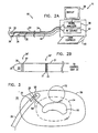
- Fig. 2A schematically illustrates a catheter system 18, including a catheter 20, for insertion into the heart of a subject, and an accompanying control unit 22 coupled to the proximal end of the catheter, in accordance with a preferred embodiment of the present invention.
- catheter 20 Near its distal end 24, catheter 20 includes an ablation device 26, preferably an RF ablation electrode, and two electrodes 28 and 30, preferably bipolar electrodes, for sensing electrical activation signals in the endocardium.
- one electrode 28 is distal to ablation device 26, and the other electrode 30 is proximal to it.
- Catheter 20 preferably also includes a position sensor 32, similarly adjacent to its distal end 24.
- Sensor 32 preferably comprises a plurality of coils, which generate signals responsive to externally-applied magnetic fields, as described in the above-mentioned W096/05768 PCT patent publication.
- the magnetic fields are preferably generated by field generators outside the body of the subject (not shown in the figures).
- the signals generated by the coils are used to continuously determine six-dimensional position and orientation information regarding distal end 24 of catheter 20.
- Other types of position sensors known in the art which are capable of determining three-dimensional position coordinates and one or two angular orientation coordinates, can also be used in the practice of the invention.
- Circuitry 36 processes the signals from electrodes 28 and 30 and calculates correlation coefficients, as will be described below. Circuitry 36 also determines the position of distal end 24 of catheter 20.
- Console 22 preferably also includes a display 38, on which the activation signal and position information is presented to a user, and user controls 40.
- the user controls are used to activate and de-activate an RF source 42 as desired, which source provides RF power to ablation electrode 26 over power wires 44.
- catheter 20 and console 22 also include means, known in the art, for steering distal end 24 of catheter 20, preferably controlled via the console.
- Fig. 2B schematically illustrates the distal portion of a catheter 21, which may be used in system 18 in place of catheter 20, in accordance with an alternative preferred embodiment of the present invention.
- catheter 21 includes wires 34 and 44 and, preferably, contains a position sensor, such as sensor 32, and a steering device, substantially as described above.
- catheter 21 is preferably 1-3 mm in diameter.
- Ablation electrode 26 comprises a conductive external layer 23 on catheter 21, extending proximally along the catheter from distal end 24 for a length of 2-8 mm.
- a bipolar electrode 28' comprises ablation electrode 26 and ring electrode 27, mutually coupled in bipolar fashion, as is known in the art.
- a second bipolar electrode 30' similarly comprises ring electrodes 31 and 33.
- device 26 is coupled via signal wires 34 to the circuitry, and thus functions as a sensing electrode.
- device 26 is coupled to RF source 42 and, preferably, disconnected from circuitry 36, so that the device functions as an ablation electrode.
- references to electrodes 28 and 30 will be understood to apply as well, wherever appropriate, to electrodes 28' and 30', mutatis mutandis.
- the electrical activation signals measured at two locations in the endocardium, for example using electrodes 28 and 30, will generally exhibit a high degree of correlation. This correlation is reflected in the two signals having similar frequency spectra and in the normalized correlation coefficient C of the two signals having a value close to one, where in which E 1 (t) and E 2 (t) are the respective activation signals sensed by electrodes 28 and 30; T is an integration time preferably corresponding to a number of cardiac cycles; ⁇ is a delay much smaller than T, corresponding generally to the difference in the arrival time of the activation signal at the position of electrode 28 from that at electrode 30; and
- the delay ⁇ will be approximately in the range 3-240 msec, given by the quotient of the distance separating electrodes 28 and 30, preferably about 12 mm, divided by the local conduction velocity in the heart, typically 0.05 to 4 m/sec.
- circuitry 36 determines and resolves values of ⁇ down to a resolution limit less than or equal to 2 msec.
- the circuitry preferably includes a band-pass filter, which removes low-frequency background and high-frequency transients in the electrogram signals received by the electrodes, prior to performing correlation calculations, as well as other pre-correlation signal conditioning circuitry.
- the electrogram signals may be passed through a 40-250 Hz bandpass filter, followed by an absolute value operation and a 20 Hz low-pass filter, as described in the above-mentioned articles by Botteron and Smith.
- the high correlation coefficient of the electrical activation signals is characteristic of the normal, cooperative contraction of the heart muscle fibers.
- this cooperative contraction is reduced or lost entirely within at least a portion of the heart muscle.
- a high correlation coefficient between the activation signals sensed at two points in the endocardium may be indicative of the presence of an abnormal conduction path.
- catheter 20 or 21 is, preferably, first used to create a correlation map of at least a portion of the atrium in which ablation is to take place.
- circuitry 36 receives signals from electrodes 28 and 30 and determines the correlation between the signals.
- the correlation mapping is performed in conjunction with measurements of the tissue conduction velocity and refractory period, so as to determine and map local values of the tissue dimension value, D, as described in the parent application to this one, U.S. Patent Application no. 08/476,200, (Patent no. US-A-5, 718, 241, International Patent Application WO 96/39929).
- areas of normal conduction will generally be characterized by consistent, high correlation coefficients at an appropriate delay ⁇ .
- the correlation coefficients will typically vary chaotically, and/or the delay ⁇ that gives a high correlation between the signals will differ from that expected for normal conduction.
- a consistently low correlation coefficient at a site may be indicative of a conduction block in the area of the site.
- Fig. 3 schematically illustrates possible reentrant conduction paths 52 and 54 in the vicinity of a conduction block 56 in heart tissue 10, for example within the atrium of the heart as illustrated in Fig. 1, and the operation of catheter 20 in ablating portions of the heart tissue in relation to these paths.
- paths 52 and 54 represent only two out of many possible paths for wavelets 12 (as shown in Fig. 1). Such paths generally need only to have a length greater than the local dimension value D and a curvature no less than a minimum radius a, as described in the parent application.
- Paths 52 and 54 are generally not fixed conduction paths, but rather transient paths, along which activation signals may be conducted, either around block 56 (path 54) or adjacent to it (path 52). As described above, activation signals traveling along path 52 or 54 will cause muscle fibers in the path to be activated at inappropriate times, frequently with multiple activations during the period of a single heart beat, leading to fibrillation.
- catheter 20 is shown to have already ablated a circumferential ablation line 58 (referred to as a ⁇ -type ablation line in the parent application), which interrupts path 52 and other, similar paths adjacent to block 56.
- Line 58 does not interrupt paths around block 56, such as path 54, however. Therefore, in Fig. 3, catheter 20 is shown in the course of ablating a radial ( ⁇ -type) ablation line 59.
- the catheter is positioned so that ablation electrode 26 can ablate a series of points in succession along line 59, until all paths such as path 54 have been interrupted.
- position sensor 32 shown in Fig. 2A, but omitted in Fig. 3 for simplicity
- the correlation coefficient C between the signals sensed by electrodes 28 and 30 along path 54 generally has a relatively high value. As described above, this value will commonly fluctuate, due to variations in the reentrant conduction currents during AF.
- the value of ⁇ in equation (1) is adjusted so as to maximize the value of C .
- ablation electrode 26 When the RF source is activated, ablation electrode 26 selectively ablates the heart tissue adjacent to the electrode, thereby gradually extending line 59. After the ablation, signals are again sensed by electrodes 28 and 30, and the correlation coefficient C is calculated, preferably while maintaining ⁇ at its pre-ablation value, and compared to the pre-ablation coefficient. If the coefficient has not changed substantially, catheter 20 is repositioned, and line 59 is extended further. Once line 59 has been extended sufficiently, the correlation coefficient between the signals from electrodes 28 and 30 is generally substantially reduced. The reduction in the correlation coefficient indicates that path 54 has been successfully interrupted.
- the correlation coefficient will increase to a value close to 1 for an appropriate choice of ⁇ .
- catheter 20 will preferably be repositioned to form an additional radial line, on the opposite side of block 56, for example, and the signal correlation measurements and ablation will be repeated.
- catheter system 18 allows the progress of the ablation procedure to be tracked, assessed and adjusted in real time, so as to provide optimal treatment for AF while minimizing unnecessary damage to the heart tissue.
- catheter 20 may be used to ablate a group of sites defining a complex shape, in order to completely cut an abnormal path.
- Ablation device 26 may comprise a standard ablation electrode, which generally produces ablation lesions at least 1 cm wide. In this case, electrodes 28 and 30 are placed about 2 cm apart.
- the average correlation coefficient between the signals sensed by these electrodes in the presence of AF, before ablation, will typically be no more than 0.3, since the correlation drops exponentially with distance, as described above (whereas in a normally-conducting heart, high correlation coefficients, approaching 1, are obtained even for mutually distant points, by appropriate choice of ⁇ ). Consequently, it may be difficult to observe the change in correlation that occurs after the ablation.
- ablation device 26 is preferably configured to ablate thin lesions, preferably no more than 3 to 5 mm wide, such as those shown in Fig. 3.
- device 26 may comprise, for example, a thin ablation electrode or, alternatively, an optical device for applying laser irradiation to heart tissue 50, as is known in the art. Electrodes 28 and 30 are then preferably placed no more than about 1 cm apart. The average correlation coefficient between the signals sensed by the electrodes at this distance, under conditions of AF, will typically be about 0.8. Thus, changes in the correlation may be more easily and accurately observed.
- Fig. 3 is drawn and described with reference to catheter 20, it will be understood that catheter 21, as shown in Fig. 2B, may be used in a substantially similar fashion.
- catheter 21 should be advanced distally so that electrodes 28' and 30' are on opposite sides of the lesion that has been ablated.
- the catheter may be drawn back in a proximal direction, for example, to determine whether the ablation has enhanced conduction along a normal path on the proximal side of the lesion.
- the magnitude of the correlation coefficient C is used as the indicator of changes in the correlation between signals E 1 and E 2
- other indicators may be used. For example, a change in the relative phases of E 1 and E 2 may be determined, by finding a first delay value ⁇ b , which gives the highest value of C before ablation, and then finding a second delay value ⁇ a , which gives the highest value of C after ablation, and comparing the two delay values.
- frequency spectra of E 1 and E 2 , ⁇ 1 ( ⁇ ) and ⁇ 2 ( ⁇ ) respectively may be calculated before and after ablation, for example, by means of a Fast Fourier Transform (FFT).
- FFT Fast Fourier Transform
- the correlation coefficient of ⁇ 1 and ⁇ 2 is then determined using a formula similar to equation (1), but with integration over ⁇ , rather than t, before and after ablation. Changes in the correlation coefficient are noted and used as described above.
- Other statistical properties of the spectra may similarly be analyzed and compared, before and after ablation, in order to assess the effectiveness of the ablation.
- signals E 1 and E 2 are simultaneously acquired by circuitry 36 from bipolar electrodes 28 and 30.
- the two signals may be acquired in sequence, preferably in sequential heart cycles, both before and after ablation. This technique may afford greater ease in signal acquisition and reduction of the electrical component count.
- acquisition of the signals is preferably synchronized with the heart beat, for example using the QRS complex of the heart's ECG as a trigger pulse.
- This synchronization is generally necessary if the correlation of E 1 and E 2 is to be analyzed in the time domain, as described above in reference to equation (1). Synchronization may usually be dispensed with, however, if the correlation or other statistical analysis is performed in the frequency domain, as long as the FFT or other transform is taken over a period that is much longer than a single heart beat.
- the present invention typically allows a physician to interrupt abnormal conduction paths more rapidly and with less unnecessary injury to the heart tissue than invasive methods known in the art, because it provides direct feedback as to the effectiveness of the operation at each site in the endocardium chosen for ablation. In the preferred embodiments of the present invention described above, however, there may still be sites in the heart tissue that are unnecessarily ablated.
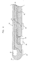
- Fig. 4 therefore, schematically illustrates, in sectional view, another preferred embodiment of the present invention in which the potential effectiveness of ablating a suspected site of an abnormal conduction path in the heart tissue is reversibly evaluated before ablation.
- catheter 20 is substantially similar to that shown in Fig. 1 and described with reference thereto, but with the addition of a thermoelectric cooler 60, which is thermally coupled to ablation electrode 26. Cooler 60 receives electrical power via wires 62 from a suitable power supply (not shown in the figures), as is known in the art, in console 22.
- catheter 20 as shown in Fig. 4 is positioned so that ablation electrode 26 is in contact with the endocardium at the site of a suspected abnormal conduction path.
- the correlation of activation signals sensed by bipolar electrodes 28 and 30 is measured by signal analyzer circuitry 36.
- Cooler 60 is then activated, by providing electrical current through wires 62, so as to cool ablation electrode 26, preferably to about -10°C.
- the cooled electrode chills the heart tissue adjacent thereto to a suitable temperature, preferably around 5°C.
- thermoelectric cooler 60 located generally in contact with ablation electrode 26 and cooling the heart tissue through the electrode
- the cooler may be placed in any convenient position alongside electrode 26, as long as it can adequately cool the tissue adjacent to the electrode.
- Other methods of reversibly interrupting the electrical conduction of the tissue may similarly be used, for example, by locally injecting a conduction-inhibiting drug into the endocardium.
- ablation electrode 26 is disposed axially along a radial outer surface of catheter 20, with bipolar electrodes 28 and 30 similarly disposed, one distal and one proximal. In other preferred embodiments of the present invention, however, electrodes 26, 28 and 30 may be placed in any convenient configuration.
- electrodes 26, 28 and 30 may be suitably mounted on a substantially rigid structure at the distal end of catheter 20, as described, for example, in PCT patent application no. PCT/IL97/00009, Publication no. WO 97/24983, filed January 8, 1997, which is assigned to the assignee of the present patent application and whose disclosure is incorporated herein by reference.
- three or more bipolar sensing electrodes may be mounted on the structure, and the mutual correlations of the activation signals that they receive may be compared in order to more precisely position the ablation electrode.
- the correlation information may be combined with conduction velocity information, determined as described in the '009 PCT application.
- the combined information is preferably used for mapping electrical activity in the heart before ablation treatment and, additionally or alternatively, for identifying suitable sites for ablation and determining the efficacy of ablation treatment carried out at the sites, as described herein.
Description
- The present invention relates generally to cardiac therapeutic devices, and specifically to catheter devices for treating electrical disorders of the heart by endocardial ablation.
- Atrial fibrillation (AF) is a well-known disorder of the heart, which causes hemodynamic efficiency to be reduced and, in serious cases, can lead to cardiac embolization, stroke, ventricular arrhythmias and other potentially fatal complications.
- AF is frequently engendered by abnormal electrical conduction paths within the heart muscle. Normally, electrical activation signals are conducted in an orderly way through the atrium and into the ventricle, passing each point in the heart only once in each heart cycle. Electrical activation signals at different locations in the heart are well correlated, taking into account normal propagation delays from one region of the heart to another. In response to local activation signals, the atrial muscle fibers contract in proper synchrony, to pump blood through the atrium. In AF, however, this orderly contraction is lost, it is believed, as multiple, changing, spatially disorganized activation wavelets sweep across the surface of the atria, resulting in irregular patterns of electrical activation. A given atrial muscle fiber is activated to contract multiple times in each heart cycle, and fibrillation takes the place of normal contraction.
- These phenomena are described in detail by Gregory W. Botteron and Joseph M. Smith in an article entitled, "A Technique for Measurement of the Extent of Spatial Organization of Atrial Activation During Atrial Fibrillation in the Intact Human Heart," in IEEE Transactions on Biomedical Engineering 12 (June 1995), pages 579-586, and in a second article entitled, "Quantitative Assessment of the Spatial Organization of Atrial Fibrillation in the Intact Human Heart," in Circulation 93 (February 1, 1996), pages 513-518.
- Fig. 1 schematically illustrates abnormal activation paths as encountered in
atrial tissue 10 of a heart undergoing AF, following the description of Botteron and Smith.Multiple wavelets 12 are generated by reentrant activation signals, following circuitous paths intissue 10. Each wavelet dominates a corresponding one of a plurality ofspatial domains 14, so that within a given domain, the activation signals will generally be highly mutually correlated, while there will be little or no correlation between signals in different domains. It will be understood that the boundaries betweendomains 14, shown as dashed lines in the figure, are generally not fixed. Rather, the paths ofwavelets 12 anddomains 14 typically vary over time. - The minimum size of any one of the
domains 14 is generally controlled by the minimum circumference of the circle described by thecorresponding wavelet 12, which is roughly equal to a tissue dimension value D (referred to by Botteron and Smith as the tissue wavelength), given by the product of the tissue's conduction velocity and its refractory period. Typically D is on the order of 18 mm. - It will be appreciated that electrical activation signals at two locations in mutual proximity, within the
same domain 14, will generally be well correlated. This correlation has been found to drop off inversely, generally exponentially, as a function of distance, so that in conditions of AF, signals at more distant locations, in different domains, are poorly correlated. By comparison, under conditions of orderly conduction within the heart tissue, the electrical activation signals will be well-correlated over substantially the entire heart, taking into account normal conduction delays between one location in the heart and another. - Although drug therapy or implantation of a pacemaker is frequently useful in controlling AF, when these methods are unsuccessful, the preferred method of treatment of the condition is to invasively interrupt the abnormal conduction paths in the heart. Preferably, a catheter having an RF ablation electrode is passed percutaneously through a blood vessel into the atrium of the heart. The electrode at the catheter tip is brought into contact with one or more sites in the endocardium where an abnormal conduction path is believed to pass, and the electrode is activated to ablate the site(s) and, it is hoped, break the abnormal path(s).
- Generally, however, it is difficult or impossible to know the precise abnormal conduction path. Furthermore, even if an abnormal path is broken at one site, other abnormal paths may exist or new paths may arise at other sites, which paths will cause the AF to continue even after the one or more sites in the endocardium are ablated. In response to this difficulty, some cardiologists and cardiac surgeons have used a "maze procedure," as described, for example, by T. Bruce Ferguson, Jr., and James L. Cox in an article entitled, "Surgery for Atrial Fibrillation," in Cardiac Electrophysiology: From Cell to Bedside, Second Edition, Douglas P. Zipes and Jose Jalife, eds., (W.B. Saunders Company, 1995), pages 1563-1576. In this procedure, multiple elongated strips in the endocardium are surgically cut or ablated in a direction generally parallel to the desired, normal, direction of conduction in the atrium. This procedure is time-consuming and causes far more damage to the endocardium than would be necessary if the abnormal paths could be selectively ablated.
- U.S. patent 5,450,846, whose disclosure describes a catheter, which may be repeatedly repositioned inside the heart, comprising an ablator at its distal tip and pairs of noncontacting sensing electrodes arrayed around the outside of the catheter near the distal end. Each electrode pair senses local electrogram signals generated in the endocardium in a small area near the side of the catheter that it faces. Differences in the activation times in the signals sensed by the pairs of electrodes are used to estimate the direction of the activation vector in the vicinity of the catheter, so as to guide the operator in positioning the ablator. This catheter is useful, however, only when electrical activation paths within the heart are relatively orderly, and not in the chaotic jumble of activation paths that generally characterizes AF.
- Systems for determining electrical activity in the heart are also disclosed in U.S. patent 5, 605, 159, U.S. patent 5, 487, 391 and European patent application EP 0, 499, 491.
- The disclosures of U.S. patent 5, 605, 159 form the bans for the preamble of
claim 1 appended hereto. - It is an object of some aspects of the present invention to provide devices for diagnosis and mapping of abnormal conduction paths in the heart of a subject.
- It is another object of some aspects of the present invention to provide devices for selective ablation of sites in the heart tissue of a subject so as to interrupt such abnormal conduction paths.
- It is a further object of some aspects of the present invention to provide devices for ablating a site in the heart tissue in the vicinity of an abnormal conduction path and measuring electrical signals in the heart adjacent to the site so as to determine whether the path has been interrupted.
- The present invention provides an intracardiac apparatus as defined in
claim 1. The effectiveness of the ablation in interrupting the abnormal path can be determined by observing and comparing a correlation between the electrical signals at two points on either side of the site, before the ablation and again after the ablation. If the correlation is substantially changed, then the ablation is judged to have been effective. - In preferred embodiments of the present invention, a cardiac catheter has a distal end that can be inserted into a chamber of the heart of a subject. The catheter includes an ablation device, preferably an RF ablation electrode, near its distal end, and first and second electrodes, preferably bipolar electrodes, for sensing electrical signals in the heart tissue. Preferably, one electrode is positioned on either side of the ablation device, spaced by a suitable distance. The proximal end of the catheter, outside the subject's body, is connected to a control console, which receives signals from the electrodes and includes signal processing circuitry for analyzing the signals, and which also supplies energy to the ablation device upon command of a user.
- Preferably, the first and second electrodes are spaced far enough apart on the catheter, and the distal end of the catheter is suitably positioned in contact with the endocardium, so that there will be a measurable propagation delay between the activation signals at the locations of the first and second electrodes under conditions of normal conduction. This normal propagation delay is different from the delay encountered in the presence of AF. On the other hand, since under conditions of AF, the correlation of the signals generally decreases exponentially with the distance between the electrodes, the electrodes should be close enough together to sense this correlation. Thus, the distance between the first and second electrodes is preferably larger than the width of lesions to be created in the heart tissue by the ablation device, but smaller than the tissue dimension value D, as described in the Background of the Invention.
- In some preferred embodiments of the present invention, the catheter further includes a position sensor, which is used to determine the position of the distal end of the catheter and/or the ablation device relative to the heart or to another external frame of reference. Preferably, the position sensor comprises one or more coils, which generate electrical signals in response to an externally-applied magnetic field, for example, as described in PCT patent publication number WO96/05768 and in U.S. patent 5,391,199. Alternatively, other types of position sensors known in the art may be used.
- In some preferred embodiments of the present invention, the catheter includes a pre-ablation testing device, adjacent to the ablation device, near its distal end, as described, for example, in U.S. patent 5,281,213. Preferably, the testing device comprises a miniature cooler, for example a thermoelectric cooler, which is operated from the control console to cool the suspected site of the abnormal conduction path. As described in the '213 patent, cooling the heart tissue to a temperature of approximately 5°C reversibly interrupts the conduction of activation signals therethrough. Correlation coefficients before and after cooling, or other reversible testing, are calculated and used as described above, to assess the potential effectiveness of ablating the site, before the ablation device is activated. In this way damage to the heart tissue may be limited to that which is actually needed and effective to interrupt abnormal conduction paths.
- Although preferred embodiments are described herein with reference to treatment of AF, the principles and methods of the present invention may also be applied to treat other heart disorders, for example, atrial flutter. Those skilled in the art will also appreciate that other methods of treating abnormal heart tissue may also be used, particularly other methods of tissue ablation, in place of RF ablation as described herein.
- Furthermore, although the preferred embodiments described herein provide for both diagnosis and ablation of abnormal conduction paths, it will be appreciated that the present invention may be used to perform diagnosis and/or mapping of electrical conduction in the heart, without necessarily treating the abnormal conduction paths.
- There is also provided, in accordance with a preferred embodiment of the present invention, apparatus for endocardiac therapy, including:
- an elongate probe for insertion into the heart of a subject, the probe having distal and proximal ends and including an ablation device for ablating heart tissue and at least two sensing electrodes, preferably bipolar electrodes, for sensing activation signals in the heart tissue adjacent to the ablation device; and
- signal analyzer circuitry, which receives the activation signals from the probe and calculates a correlation measure thereof.
-
- Preferably, the probe includes a position sensor adjacent to the ablation device, which generates signals responsive to the position of the probe with respect to an external frame of reference.
- Further preferably, the ablation device includes an ablation electrode, and one of the at least two sensing electrodes includes the ablation electrode.
- Preferably, the ablation device is positioned over the outer surface of the probe, substantially covering the distal end thereof.
- Additionally or alternatively, the ablation device and electrodes are positioned along an outer, radial surface of the probe, wherein one electrode is axially proximal to the ablation device, and the other electrode is axially distal to the ablation device.
- Preferably, the system includes a cooler, for cooling the heart tissue, adjacent to the ablation device.
- Alternatively, the cooler is thermally coupled to the ablation device, which cools the heart tissue.
- There is further provided, in accordance with a preferred embodiment of the present invention, apparatus for determining the effectiveness of treatment of abnormal conduction in the heart of a subject, including:
- an elongate probe for insertion into the heart of the heart, the probe having distal and proximal ends and including at least two sensing electrodes, preferably bipolar electrodes, near the distal end of the probe, for sensing activation signals in the heart tissue; and
- signal analyzer circuitry, which receives and determines a correlation measure of the activation signals and compares a first correlation measure determined before the treatment with a second correlation measure determined after the treatment.
-
- Preferably, the probe includes a position sensor near the distal end thereof, which generates signals responsive to the position of the probe with respect to an external frame of reference.
- Preferably, the probe is positioned at a plurality of locations in the heart, and the signal analyzer circuitry produces a map of the correlation measures determined at the plurality of locations.
- The present invention will be more fully understood from the following detailed description of the preferred embodiments thereof, taken together with the drawings in which:
-
- Fig. 1 is a schematic illustration of atrial tissue undergoing fibrillation, useful in understanding the principles of the present invention;
- Fig. 2A is a schematic illustration of a catheter system, in accordance with a preferred embodiment of the present invention;
- Fig. 2B is a schematic illustration of the distal portion of a catheter, in accordance with an alternative preferred embodiment of the present invention;
- Fig. 3 is a schematic illustration showing the distal end of the catheter of Fig. 2A in contact with areas of abnormal electrical conduction in the heart tissue; and
- Fig. 4 is a schematic, sectional illustration of the distal end of a catheter, in accordance with another preferred embodiment of the present invention.
-
- Reference is now made to Fig. 2A, which schematically illustrates a
catheter system 18, including acatheter 20, for insertion into the heart of a subject, and an accompanyingcontrol unit 22 coupled to the proximal end of the catheter, in accordance with a preferred embodiment of the present invention. Near itsdistal end 24,catheter 20 includes anablation device 26, preferably an RF ablation electrode, and twoelectrodes electrode 28 is distal toablation device 26, and theother electrode 30 is proximal to it. -
Catheter 20 preferably also includes aposition sensor 32, similarly adjacent to itsdistal end 24. -
Sensor 32 preferably comprises a plurality of coils, which generate signals responsive to externally-applied magnetic fields, as described in the above-mentioned W096/05768 PCT patent publication. The magnetic fields are preferably generated by field generators outside the body of the subject (not shown in the figures). The signals generated by the coils are used to continuously determine six-dimensional position and orientation information regardingdistal end 24 ofcatheter 20. Other types of position sensors known in the art, which are capable of determining three-dimensional position coordinates and one or two angular orientation coordinates, can also be used in the practice of the invention. - Activation signals sensed by
electrodes position sensor 32 are conveyed viasignal wires 34 to signalanalyzer circuitry 36 inconsole 22.Circuitry 36 processes the signals fromelectrodes Circuitry 36 also determines the position ofdistal end 24 ofcatheter 20. -
Console 22 preferably also includes a display 38, on which the activation signal and position information is presented to a user, and user controls 40. The user controls are used to activate and de-activate anRF source 42 as desired, which source provides RF power toablation electrode 26 overpower wires 44. Preferably,catheter 20 andconsole 22 also include means, known in the art, for steeringdistal end 24 ofcatheter 20, preferably controlled via the console. - Fig. 2B schematically illustrates the distal portion of a
catheter 21, which may be used insystem 18 in place ofcatheter 20, in accordance with an alternative preferred embodiment of the present invention. For clarity of illustration, only external elements ofcatheter 21 are shown in Fig. 2B. It will be understood, however, thatcatheter 21 includeswires sensor 32, and a steering device, substantially as described above.Catheter 21 is preferably 1-3 mm in diameter.Ablation electrode 26 comprises a conductiveexternal layer 23 oncatheter 21, extending proximally along the catheter fromdistal end 24 for a length of 2-8 mm. Aring electrode 27, having a width of 1-2 mm, surroundscatheter 21 proximal todevice 26, with a non-conducting gap of preferably about 1-2 mm betweendevice 26 andelectrode 27. Twomore ring electrodes electrode 27,surround catheter 21, preferably at a distance of about 8-18 mm, most preferably about 12 mm, fromelectrode 27 and with a gap of about 1-2 mm betweenelectrodes - In the preferred embodiment shown in Fig. 2B, a bipolar electrode 28' comprises
ablation electrode 26 andring electrode 27, mutually coupled in bipolar fashion, as is known in the art. A second bipolar electrode 30' similarly comprisesring electrodes system 18 in whichcircuitry 36 receives and processes activation signals from electrodes 28' and 30',device 26 is coupled viasignal wires 34 to the circuitry, and thus functions as a sensing electrode. During ablation,device 26 is coupled toRF source 42 and, preferably, disconnected fromcircuitry 36, so that the device functions as an ablation electrode. In the description of the present invention that follows, references toelectrodes - Under normal conditions of sinus rhythm, the electrical activation signals measured at two locations in the endocardium, for
example using electrodes C of the two signals having a value close to one, wherein which E1(t) and E2(t) are the respective activation signals sensed by
electrodes electrode 28 from that atelectrode 30; and
- In the preferred embodiments of the present invention described above with reference to Figs. 2A and 2B, under typical conditions, in the absence of fibrillation, the delay Δ will be approximately in the range 3-240 msec, given by the quotient of the
distance separating electrodes circuitry 36 determines and resolves values of Δ down to a resolution limit less than or equal to 2 msec. For this purpose, the circuitry preferably includes a band-pass filter, which removes low-frequency background and high-frequency transients in the electrogram signals received by the electrodes, prior to performing correlation calculations, as well as other pre-correlation signal conditioning circuitry. For example, prior to the correlation calculation, the electrogram signals may be passed through a 40-250 Hz bandpass filter, followed by an absolute value operation and a 20 Hz low-pass filter, as described in the above-mentioned articles by Botteron and Smith. - As described above, the high correlation coefficient of the electrical activation signals is characteristic of the normal, cooperative contraction of the heart muscle fibers. In the presence of abnormal, parasitic conduction paths in the heart, however, as is the case in atrial fibrillation, this cooperative contraction is reduced or lost entirely within at least a portion of the heart muscle. Under these conditions, a high correlation coefficient between the activation signals sensed at two points in the endocardium may be indicative of the presence of an abnormal conduction path.
- Thus, when AF is encountered or suspected,
catheter circuitry 36 receives signals fromelectrodes - In such a map, areas of normal conduction will generally be characterized by consistent, high correlation coefficients at an appropriate delay Δ. At sites of parasitic conduction, however, the correlation coefficients will typically vary chaotically, and/or the delay Δ that gives a high correlation between the signals will differ from that expected for normal conduction. On the other hand, a consistently low correlation coefficient at a site may be indicative of a conduction block in the area of the site. After making the map, the sites of parasitic conduction are preferentially ablated, while areas of normal conduction or of conduction block are generally not ablated.
- Fig. 3 schematically illustrates possible
reentrant conduction paths conduction block 56 inheart tissue 10, for example within the atrium of the heart as illustrated in Fig. 1, and the operation ofcatheter 20 in ablating portions of the heart tissue in relation to these paths. It will be understood thatpaths Paths path - At the point in time illustrated in Fig. 3,
catheter 20 is shown to have already ablated a circumferential ablation line 58 (referred to as a Ψ-type ablation line in the parent application), which interruptspath 52 and other, similar paths adjacent to block 56.Line 58 does not interrupt paths aroundblock 56, such aspath 54, however. Therefore, in Fig. 3,catheter 20 is shown in the course of ablating a radial (λ-type)ablation line 59. The catheter is positioned so thatablation electrode 26 can ablate a series of points in succession alongline 59, until all paths such aspath 54 have been interrupted. Preferably, position sensor 32 (shown in Fig. 2A, but omitted in Fig. 3 for simplicity) is used to position the catheter in the desired locations and to track the catheter's position as it is steered to various locations in the heart. - Before
RF source 42 is activated, the correlation coefficientC between the signals sensed byelectrodes path 54 generally has a relatively high value. As described above, this value will commonly fluctuate, due to variations in the reentrant conduction currents during AF. Preferably, the value of Δ in equation (1) is adjusted so as to maximize the value ofC . - When the RF source is activated,
ablation electrode 26 selectively ablates the heart tissue adjacent to the electrode, thereby gradually extendingline 59. After the ablation, signals are again sensed byelectrodes C is calculated, preferably while maintaining Δ at its pre-ablation value, and compared to the pre-ablation coefficient. If the coefficient has not changed substantially,catheter 20 is repositioned, andline 59 is extended further. Onceline 59 has been extended sufficiently, the correlation coefficient between the signals fromelectrodes path 54 has been successfully interrupted. On the other hand, if as a result of the ablation, normal conduction has come to prevail in lace of AF in heart tissue 10 (at least in the portion of the tissue against whichcatheter 20 is positioned), the correlation coefficient will increase to a value close to 1 for an appropriate choice of Δ. - Alternatively, it may occur that even after
line 59 has been completely ablated, fromblock 56 toline 58, there is still a substantial correlation, indicative of reentrant paths, between the signals atelectrodes catheter 20 will preferably be repositioned to form an additional radial line, on the opposite side ofblock 56, for example, and the signal correlation measurements and ablation will be repeated. In either case, the use ofcatheter system 18 allows the progress of the ablation procedure to be tracked, assessed and adjusted in real time, so as to provide optimal treatment for AF while minimizing unnecessary damage to the heart tissue. - It will be understood that the types of conduction abnormalities and the geometries of
ablation lines catheter 20 may be used to ablate a group of sites defining a complex shape, in order to completely cut an abnormal path. -
Ablation device 26 may comprise a standard ablation electrode, which generally produces ablation lesions at least 1 cm wide. In this case,electrodes - To overcome this difficulty,
ablation device 26 is preferably configured to ablate thin lesions, preferably no more than 3 to 5 mm wide, such as those shown in Fig. 3. For this purpose,device 26 may comprise, for example, a thin ablation electrode or, alternatively, an optical device for applying laser irradiation to heart tissue 50, as is known in the art.Electrodes - Although Fig. 3 is drawn and described with reference to
catheter 20, it will be understood thatcatheter 21, as shown in Fig. 2B, may be used in a substantially similar fashion. Whencatheter 21 is used, however, the area adjacent to bipolar electrode 28' is ablated byablation device 26. Therefore, to measure the signal correlation post-ablation,catheter 21 should be advanced distally so that electrodes 28' and 30' are on opposite sides of the lesion that has been ablated. Alternatively, the catheter may be drawn back in a proximal direction, for example, to determine whether the ablation has enhanced conduction along a normal path on the proximal side of the lesion. In other cases, it may be sufficient to measure the signal correlation pre-ablation, to ascertain that abnormal conduction exists at the site to be ablated, and to forego the post-ablation measurement. - While in the preferred embodiment described above, the magnitude of the correlation coefficient
C , at a fixed value of Δ, is used as the indicator of changes in the correlation between signals E1 and E2, in other preferred embodiments of the present invention, other indicators may be used. For example, a change in the relative phases of E1 and E2 may be determined, by finding a first delay value Δb, which gives the highest value ofC before ablation, and then finding a second delay value Δa, which gives the highest value ofC after ablation, and comparing the two delay values. - Alternatively, frequency spectra of E1 and E2, ε1(ω) and ε2(ω) respectively, may be calculated before and after ablation, for example, by means of a Fast Fourier Transform (FFT). The correlation coefficient of ε1 and ε2 is then determined using a formula similar to equation (1), but with integration over ω, rather than t, before and after ablation. Changes in the correlation coefficient are noted and used as described above. Other statistical properties of the spectra, as are known in the art, may similarly be analyzed and compared, before and after ablation, in order to assess the effectiveness of the ablation.
- Preferably, signals E1 and E2 are simultaneously acquired by
circuitry 36 frombipolar electrodes - The present invention typically allows a physician to interrupt abnormal conduction paths more rapidly and with less unnecessary injury to the heart tissue than invasive methods known in the art, because it provides direct feedback as to the effectiveness of the operation at each site in the endocardium chosen for ablation. In the preferred embodiments of the present invention described above, however, there may still be sites in the heart tissue that are unnecessarily ablated.
- Fig. 4, therefore, schematically illustrates, in sectional view, another preferred embodiment of the present invention in which the potential effectiveness of ablating a suspected site of an abnormal conduction path in the heart tissue is reversibly evaluated before ablation. As shown in Fig. 4,
catheter 20 is substantially similar to that shown in Fig. 1 and described with reference thereto, but with the addition of athermoelectric cooler 60, which is thermally coupled toablation electrode 26.Cooler 60 receives electrical power viawires 62 from a suitable power supply (not shown in the figures), as is known in the art, inconsole 22. - As in the preferred embodiments described earlier,
catheter 20 as shown in Fig. 4 is positioned so thatablation electrode 26 is in contact with the endocardium at the site of a suspected abnormal conduction path. The correlation of activation signals sensed bybipolar electrodes signal analyzer circuitry 36.Cooler 60 is then activated, by providing electrical current throughwires 62, so as to coolablation electrode 26, preferably to about -10°C. The cooled electrode chills the heart tissue adjacent thereto to a suitable temperature, preferably around 5°C. As described, for example, in the above-mentioned U.S. patent no. 5,281,213, cooling the heart tissue to a temperature in this range prevents the tissue from responding to or conducting activation signals, as though the tissue had been ablated. Unlike ablation, however, when cooler 60 is de-activated orablation electrode 26 is removed from contact with the tissue, and the tissue returns to a normal temperature of approximately 37°C, normal function and conduction by the tissue are restored. - Thus, when cooler 60 has been activated, and the tissue at the site of the suspected abnormality, adjacent to
electrode 26, has been cooled to the desired temperature, the correlation of the activation signals sensed bybipolar electrodes RF source 42 is then activated to permanently ablate the site and interrupt the abnormal path. On the other hand, if there is no substantial change in the correlation, the site is allowed to re-warm and return to normal function with no unnecessary injury to the tissue. - Although Fig. 4 shows thermoelectric cooler 60 located generally in contact with
ablation electrode 26 and cooling the heart tissue through the electrode, it will be appreciated that other types of coolers, known in the art, may also be used. Moreover, the cooler may be placed in any convenient position alongsideelectrode 26, as long as it can adequately cool the tissue adjacent to the electrode. Other methods of reversibly interrupting the electrical conduction of the tissue may similarly be used, for example, by locally injecting a conduction-inhibiting drug into the endocardium. - It will also be understood that although the above preferred embodiments include an RF electrode for ablation, other ablation devices and methods known in the art, for example, microwave ablation or alcohol injection, may equally be used.
- In the preferred embodiments of the present invention shown in the figures and described above,
ablation electrode 26 is disposed axially along a radial outer surface ofcatheter 20, withbipolar electrodes electrodes - For example,
electrodes catheter 20, as described, for example, in PCT patent application no. PCT/IL97/00009, Publication no. WO 97/24983, filed January 8, 1997, which is assigned to the assignee of the present patent application and whose disclosure is incorporated herein by reference. Furthermore, three or more bipolar sensing electrodes may be mounted on the structure, and the mutual correlations of the activation signals that they receive may be compared in order to more precisely position the ablation electrode. The correlation information may be combined with conduction velocity information, determined as described in the '009 PCT application. The combined information is preferably used for mapping electrical activity in the heart before ablation treatment and, additionally or alternatively, for identifying suitable sites for ablation and determining the efficacy of ablation treatment carried out at the sites, as described herein. - It will be further appreciated that the preferred embodiments described above are cited by way of example, and the full scope of the invention is limited only by the claims.
Claims (11)
- Intracardiac apparatus, comprising:an elongate probe (20;21) for insertion into the heart of a subject, said probe (20;21) having distal and proximal ends and comprising at least two sensing electrodes (28,30) near the distal end of the probe (20), for sensing signals in the heart tissue, andsignal analyzer circuitry (22) for receiving the signals from the probe (20;21) and calculating a correlation measure of the signals; characterised in that the apparatus further comprises a tissue ablation device (26) adapted to selectively ablate the heart tissue if said correlation measure is indicative of an electrical disorder.
- Apparatus of claim 1, wherein the sensing electrodes (20;21) comprise bipolar electrodes.
- Apparatus of claim 1 or claim 2, wherein the probe (20;21) comprises a position sensor (32), for generating signals responsive to the position of the probe (20;21) with respect to an external frame of reference.
- Apparatus of any one of claims 1 to 3, wherein the probe (20;21) is positionable at a plurality of locations in the heart, and the signal analyzer circuitry (22) is adapted to produce a map of the correlation measures determined at the plurality of locations.
- Apparatus of any one of claims 1 to 4, wherein the probe (20;21) comprises the tissue ablation device adjacent the distal end of the probe (20;21).
- Apparatus of claim 5, wherein the ablation device (26) comprises an ablation electrode, and wherein one of the at least two sensing electrodes (28,30) is comprised in the ablation electrode.
- Apparatus of claim 5 or claim 6, wherein the ablation device (26) is disposed over the outer surface of the probe (21), substantially covering the distal end thereof.
- Apparatus of any one of claims 5 to 7, wherein the ablation device (26) and electrodes (28;30) are positioned along an outer, radial surface of the probe (20), wherein one electrode (30) is axially proximal to the ablation device (26), and the other electrode (28) is axially distal to the ablation device (26).
- Apparatus of any one of the preceding claims comprising a cooler, for cooling the heart tissue, adjacent to the ablation device (26).
- Apparatus of any one of the preceding claims, wherein the ablation device (26) is configured to ablate lesions no more than 5mm wide.
- Apparatus of any one of the preceding claims, wherein the at least two sensing electrodes (28,30) are adapted to sense signals in sequential heart cycles both before and after ablation of the heart tissue.
Applications Claiming Priority (2)
| Application Number | Priority Date | Filing Date | Title |
|---|---|---|---|
| US08/956,687 US5954665A (en) | 1995-06-07 | 1997-10-23 | Cardiac ablation catheter using correlation measure |
| US956687 | 1997-10-23 |
Publications (3)
| Publication Number | Publication Date |
|---|---|
| EP0911059A2 EP0911059A2 (en) | 1999-04-28 |
| EP0911059A3 EP0911059A3 (en) | 2000-04-12 |
| EP0911059B1 true EP0911059B1 (en) | 2003-09-03 |
Family
ID=25498553
Family Applications (1)
| Application Number | Title | Priority Date | Filing Date |
|---|---|---|---|
| EP98308643A Expired - Lifetime EP0911059B1 (en) | 1997-10-23 | 1998-10-22 | Ablation catheter with sensing electrodes |
Country Status (8)
| Country | Link |
|---|---|
| US (1) | US5954665A (en) |
| EP (1) | EP0911059B1 (en) |
| JP (1) | JP4245707B2 (en) |
| AU (1) | AU735274B2 (en) |
| CA (1) | CA2250805C (en) |
| DE (1) | DE69817723T2 (en) |
| ES (1) | ES2206851T3 (en) |
| IL (1) | IL126611A (en) |
Cited By (1)
| Publication number | Priority date | Publication date | Assignee | Title |
|---|---|---|---|---|
| WO2014012133A1 (en) * | 2012-07-16 | 2014-01-23 | Lions Eye Institute Limited | Irradiation method and apparatus |
Families Citing this family (132)
| Publication number | Priority date | Publication date | Assignee | Title |
|---|---|---|---|---|
| US6096037A (en) | 1997-07-29 | 2000-08-01 | Medtronic, Inc. | Tissue sealing electrosurgery device and methods of sealing tissue |
| US6176857B1 (en) * | 1997-10-22 | 2001-01-23 | Oratec Interventions, Inc. | Method and apparatus for applying thermal energy to tissue asymmetrically |
| US6161047A (en) * | 1998-04-30 | 2000-12-12 | Medtronic Inc. | Apparatus and method for expanding a stimulation lead body in situ |
| US6319241B1 (en) * | 1998-04-30 | 2001-11-20 | Medtronic, Inc. | Techniques for positioning therapy delivery elements within a spinal cord or a brain |
| US6546271B1 (en) | 1999-10-01 | 2003-04-08 | Bioscience, Inc. | Vascular reconstruction |
| US6892091B1 (en) * | 2000-02-18 | 2005-05-10 | Biosense, Inc. | Catheter, method and apparatus for generating an electrical map of a chamber of the heart |
| US6458123B1 (en) * | 2000-04-27 | 2002-10-01 | Biosense Webster, Inc. | Ablation catheter with positional sensor |
| US6546935B2 (en) | 2000-04-27 | 2003-04-15 | Atricure, Inc. | Method for transmural ablation |
| US6511500B1 (en) | 2000-06-06 | 2003-01-28 | Marc Mounir Rahme | Use of autonomic nervous system neurotransmitters inhibition and atrial parasympathetic fibers ablation for the treatment of atrial arrhythmias and to preserve drug effects |
| US6905492B2 (en) * | 2000-07-31 | 2005-06-14 | Galil Medical Ltd. | Planning and facilitation systems and methods for cryosurgery |
| US20040087877A1 (en) | 2000-08-23 | 2004-05-06 | Besz William John | Catheter locator apparatus and method of use |
| US20040138621A1 (en) | 2003-01-14 | 2004-07-15 | Jahns Scott E. | Devices and methods for interstitial injection of biologic agents into tissue |
| US7740623B2 (en) | 2001-01-13 | 2010-06-22 | Medtronic, Inc. | Devices and methods for interstitial injection of biologic agents into tissue |
| US6743225B2 (en) | 2001-03-27 | 2004-06-01 | Uab Research Foundation | Electrophysiologic measure of endpoints for ablation lesions created in fibrillating substrates |
| US6740083B2 (en) * | 2001-07-09 | 2004-05-25 | Scimed Life Systems, Inc. | Distal catheter assembly with proximal mounting member |
| US6623514B1 (en) * | 2001-08-01 | 2003-09-23 | Origin Medsystems, Inc. | Method of cooling an organ |
| US6671533B2 (en) * | 2001-10-11 | 2003-12-30 | Irvine Biomedical Inc. | System and method for mapping and ablating body tissue of the interior region of the heart |
| US20070038056A1 (en) | 2001-10-11 | 2007-02-15 | Carlo Pappone | System and methods for locating and ablating arrhythomogenic tissues |
| US8974446B2 (en) | 2001-10-11 | 2015-03-10 | St. Jude Medical, Inc. | Ultrasound ablation apparatus with discrete staggered ablation zones |
| US7967816B2 (en) | 2002-01-25 | 2011-06-28 | Medtronic, Inc. | Fluid-assisted electrosurgical instrument with shapeable electrode |
| DE60312821T2 (en) * | 2002-02-12 | 2007-12-13 | Oratec Interventions, Inc., Memphis | RADIO FREQUENCY ABLATION DEVICE FOR ARTHROSCOPY |
| GB2395620B (en) * | 2002-11-19 | 2004-09-29 | Nec Technologies | Cellular network acquisition method and apparatus |
| US7367970B2 (en) * | 2003-11-11 | 2008-05-06 | Biosense Webster Inc. | Externally applied RF for pulmonary vein isolation |
| DE10355275B4 (en) * | 2003-11-26 | 2009-03-05 | Siemens Ag | catheter device |
| US10258285B2 (en) | 2004-05-28 | 2019-04-16 | St. Jude Medical, Atrial Fibrillation Division, Inc. | Robotic surgical system and method for automated creation of ablation lesions |
| US9782130B2 (en) | 2004-05-28 | 2017-10-10 | St. Jude Medical, Atrial Fibrillation Division, Inc. | Robotic surgical system |
| US8755864B2 (en) | 2004-05-28 | 2014-06-17 | St. Jude Medical, Atrial Fibrillation Division, Inc. | Robotic surgical system and method for diagnostic data mapping |
| US8528565B2 (en) | 2004-05-28 | 2013-09-10 | St. Jude Medical, Atrial Fibrillation Division, Inc. | Robotic surgical system and method for automated therapy delivery |
| US10863945B2 (en) | 2004-05-28 | 2020-12-15 | St. Jude Medical, Atrial Fibrillation Division, Inc. | Robotic surgical system with contact sensing feature |
| WO2005120376A2 (en) | 2004-06-02 | 2005-12-22 | Medtronic, Inc. | Ablation device with jaws |
| US10413188B2 (en) * | 2004-11-17 | 2019-09-17 | Lawrence Livermore National Security, Llc | Assessment of tissue or lesion depth using temporally resolved light scattering spectroscopy |
| EP1658818A1 (en) | 2004-11-23 | 2006-05-24 | Biosense Webster, Inc. | Externally applied rf for pulmonary vein isolation |
| US7976518B2 (en) | 2005-01-13 | 2011-07-12 | Corpak Medsystems, Inc. | Tubing assembly and signal generator placement control device and method for use with catheter guidance systems |
| US8155910B2 (en) | 2005-05-27 | 2012-04-10 | St. Jude Medical, Atrial Fibrillation Divison, Inc. | Robotically controlled catheter and method of its calibration |
| US8583220B2 (en) * | 2005-08-02 | 2013-11-12 | Biosense Webster, Inc. | Standardization of catheter-based treatment for atrial fibrillation |
| US7877128B2 (en) * | 2005-08-02 | 2011-01-25 | Biosense Webster, Inc. | Simulation of invasive procedures |
| US7681579B2 (en) | 2005-08-02 | 2010-03-23 | Biosense Webster, Inc. | Guided procedures for treating atrial fibrillation |
| US7918847B2 (en) * | 2005-08-29 | 2011-04-05 | Washington University | Method and associated system for the interventional treatment of atrial fibrillation |
| US8038625B2 (en) * | 2005-09-15 | 2011-10-18 | St. Jude Medical, Atrial Fibrillation Division, Inc. | System and method for three-dimensional mapping of electrophysiology information |
| US8229545B2 (en) * | 2005-09-15 | 2012-07-24 | St. Jude Medical, Atrial Fibrillation Division, Inc. | System and method for mapping complex fractionated electrogram information |
| WO2007038861A1 (en) * | 2005-10-04 | 2007-04-12 | Hopital Du Sacre-Coeur De Montreal | Method and apparatus for predicting potential occurrence of atrial fibrillation events |
| US10362959B2 (en) * | 2005-12-06 | 2019-07-30 | St. Jude Medical, Atrial Fibrillation Division, Inc. | System and method for assessing the proximity of an electrode to tissue in a body |
| US20090177111A1 (en) * | 2006-12-06 | 2009-07-09 | Miller Stephan P | System and method for displaying contact between a catheter and tissue |
| US8449535B2 (en) * | 2005-12-06 | 2013-05-28 | St. Jude Medical, Atrial Fibrillation Division, Inc. | System and method for assessing coupling between an electrode and tissue |
| US8406866B2 (en) | 2005-12-06 | 2013-03-26 | St. Jude Medical, Atrial Fibrillation Division, Inc. | System and method for assessing coupling between an electrode and tissue |
| CA2631940C (en) * | 2005-12-06 | 2016-06-21 | St. Jude Medical, Atrial Fibrillation Division, Inc. | Assessment of electrode coupling for tissue ablation |
| US8403925B2 (en) | 2006-12-06 | 2013-03-26 | St. Jude Medical, Atrial Fibrillation Division, Inc. | System and method for assessing lesions in tissue |
| US8998890B2 (en) | 2005-12-06 | 2015-04-07 | St. Jude Medical, Atrial Fibrillation Division, Inc. | Assessment of electrode coupling for tissue ablation |
| US9254163B2 (en) | 2005-12-06 | 2016-02-09 | St. Jude Medical, Atrial Fibrillation Division, Inc. | Assessment of electrode coupling for tissue ablation |
| US9492226B2 (en) | 2005-12-06 | 2016-11-15 | St. Jude Medical, Atrial Fibrillation Division, Inc. | Graphical user interface for real-time RF lesion depth display |
| US8603084B2 (en) | 2005-12-06 | 2013-12-10 | St. Jude Medical, Atrial Fibrillation Division, Inc. | System and method for assessing the formation of a lesion in tissue |
| US7918850B2 (en) * | 2006-02-17 | 2011-04-05 | Biosense Wabster, Inc. | Lesion assessment by pacing |
| CA2642568C (en) * | 2006-02-22 | 2015-11-24 | Custom Medical Applications, Inc. | Ablation instruments and related methods |
| US20070232949A1 (en) * | 2006-03-31 | 2007-10-04 | Ep Medsystems, Inc. | Method For Simultaneous Bi-Atrial Mapping Of Atrial Fibrillation |
| US20090171203A1 (en) * | 2006-05-02 | 2009-07-02 | Ofer Avital | Cryotherapy Insertion System and Method |
| WO2007129310A2 (en) | 2006-05-02 | 2007-11-15 | Galil Medical Ltd. | Cryotherapy insertion system and method |
| US7774051B2 (en) * | 2006-05-17 | 2010-08-10 | St. Jude Medical, Atrial Fibrillation Division, Inc. | System and method for mapping electrophysiology information onto complex geometry |
| US7988639B2 (en) * | 2006-05-17 | 2011-08-02 | St. Jude Medical, Atrial Fibrillation Division, Inc. | System and method for complex geometry modeling of anatomy using multiple surface models |
| US7515954B2 (en) | 2006-06-13 | 2009-04-07 | Rhythmia Medical, Inc. | Non-contact cardiac mapping, including moving catheter and multi-beat integration |
| US7729752B2 (en) * | 2006-06-13 | 2010-06-01 | Rhythmia Medical, Inc. | Non-contact cardiac mapping, including resolution map |
| US8010186B1 (en) * | 2006-07-19 | 2011-08-30 | Pacesetter, Inc. | System and related methods for identifying a fibrillation driver |
| US8197494B2 (en) | 2006-09-08 | 2012-06-12 | Corpak Medsystems, Inc. | Medical device position guidance system with wireless connectivity between a noninvasive device and an invasive device |
| WO2008045877A2 (en) | 2006-10-10 | 2008-04-17 | St. Jude Medical, Atrial Fibrillation Division, Inc. | Electrode tip and ablation system |
| US9585586B2 (en) | 2006-12-29 | 2017-03-07 | St. Jude Medical, Atrial Fibrillation Division, Inc. | Navigational reference dislodgement detection method and system |
| US9220439B2 (en) * | 2006-12-29 | 2015-12-29 | St. Jude Medical, Atrial Fibrillation Division, Inc. | Navigational reference dislodgement detection method and system |
| US20080190438A1 (en) * | 2007-02-08 | 2008-08-14 | Doron Harlev | Impedance registration and catheter tracking |
| US9549689B2 (en) * | 2007-03-09 | 2017-01-24 | St. Jude Medical, Atrial Fibrillation Division, Inc. | System and method for correction of inhomogeneous fields |
| US7825925B2 (en) | 2007-03-09 | 2010-11-02 | St. Jude Medical, Atrial Fibrillation Division, Inc. | Method and system for repairing triangulated surface meshes |
| US10433929B2 (en) | 2007-03-09 | 2019-10-08 | St. Jude Medical, Atrial Fibrillation Division, Inc. | System and method for local deformable registration of a catheter navigation system to image data or a model |
| KR101000320B1 (en) | 2008-04-15 | 2010-12-13 | (주) 태웅메디칼 | Bipolar electrode type guide wire and Catheter system using the same |
| US8386014B2 (en) * | 2007-06-21 | 2013-02-26 | The Trustees Of Columbia University In The City Of New York | Systems and methods for implementing heart geometrical measurements |
| US8175679B2 (en) * | 2007-12-26 | 2012-05-08 | St. Jude Medical, Atrial Fibrillation Division, Inc. | Catheter electrode that can simultaneously emit electrical energy and facilitate visualization by magnetic resonance imaging |
| US8103327B2 (en) | 2007-12-28 | 2012-01-24 | Rhythmia Medical, Inc. | Cardiac mapping catheter |
| US8290578B2 (en) * | 2007-12-28 | 2012-10-16 | St. Jude Medical, Atrial Fibrillation Division, Inc. | Method and apparatus for complex impedance compensation |
| US9675410B2 (en) * | 2007-12-28 | 2017-06-13 | St. Jude Medical, Atrial Fibrillation Division, Inc. | Flexible polymer electrode for MRI-guided positioning and radio frequency ablation |
| US10660690B2 (en) * | 2007-12-28 | 2020-05-26 | St. Jude Medical, Atrial Fibrillation Division, Inc. | System and method for measurement of an impedance using a catheter such as an ablation catheter |
| US9204927B2 (en) | 2009-05-13 | 2015-12-08 | St. Jude Medical, Atrial Fibrillation Division, Inc. | System and method for presenting information representative of lesion formation in tissue during an ablation procedure |
| US8538509B2 (en) | 2008-04-02 | 2013-09-17 | Rhythmia Medical, Inc. | Intracardiac tracking system |
| US8676303B2 (en) | 2008-05-13 | 2014-03-18 | The Regents Of The University Of California | Methods and systems for treating heart instability |
| US8357149B2 (en) * | 2008-06-05 | 2013-01-22 | Biosense Webster, Inc. | Filter for simultaneous pacing and ablation |
| US8206385B2 (en) * | 2008-06-09 | 2012-06-26 | St. Jude Medical, Atrial Fibrillation Division, Inc. | Catheter assembly with front-loaded tip and multi-contact connector |
| CN104545894A (en) | 2008-10-09 | 2015-04-29 | 加利福尼亚大学董事会 | Device and process for automatically positioning biological rhythm disorder source |
| US8137343B2 (en) | 2008-10-27 | 2012-03-20 | Rhythmia Medical, Inc. | Tracking system using field mapping |
| US9320565B2 (en) * | 2008-12-31 | 2016-04-26 | St. Jude Medical, Atrial Fibrillation Division, Inc. | Ablation devices, systems and method for measuring cooling effect of fluid flow |
| KR101164887B1 (en) * | 2009-03-19 | 2012-07-19 | 서울대학교병원 | Bipolar Radiofrequency Ablation Catheter |
| US9398862B2 (en) | 2009-04-23 | 2016-07-26 | Rhythmia Medical, Inc. | Multi-electrode mapping system |
| US8905968B2 (en) | 2009-04-29 | 2014-12-09 | Encephalon Technologies, Llc | System for cooling and pressurizing fluid |
| US8571647B2 (en) * | 2009-05-08 | 2013-10-29 | Rhythmia Medical, Inc. | Impedance based anatomy generation |
| US8103338B2 (en) | 2009-05-08 | 2012-01-24 | Rhythmia Medical, Inc. | Impedance based anatomy generation |
| US10398326B2 (en) | 2013-03-15 | 2019-09-03 | The Regents Of The University Of California | System and method of identifying sources associated with biological rhythm disorders |
| US10434319B2 (en) | 2009-10-09 | 2019-10-08 | The Regents Of The University Of California | System and method of identifying sources associated with biological rhythm disorders |
| US9392948B2 (en) | 2011-12-09 | 2016-07-19 | The Regents Of The University Of California | System and method of identifying sources for biological rhythms |
| US9332915B2 (en) | 2013-03-15 | 2016-05-10 | The Regents Of The University Of California | System and method to identify sources associated with biological rhythm disorders |
| AU2011237661B2 (en) | 2010-04-08 | 2015-05-21 | The Regents Of The University Of California | Methods, system and apparatus for the detection, diagnosis and treatment of biological rhythm disorders |
| US9002442B2 (en) | 2011-01-13 | 2015-04-07 | Rhythmia Medical, Inc. | Beat alignment and selection for cardiac mapping |
| US8428700B2 (en) | 2011-01-13 | 2013-04-23 | Rhythmia Medical, Inc. | Electroanatomical mapping |
| US9901303B2 (en) | 2011-04-14 | 2018-02-27 | St. Jude Medical, Atrial Fibrillation Division, Inc. | System and method for registration of multiple navigation systems to a common coordinate frame |
| US10918307B2 (en) | 2011-09-13 | 2021-02-16 | St. Jude Medical, Atrial Fibrillation Division, Inc. | Catheter navigation using impedance and magnetic field measurements |
| US10362963B2 (en) | 2011-04-14 | 2019-07-30 | St. Jude Medical, Atrial Fibrillation Division, Inc. | Correction of shift and drift in impedance-based medical device navigation using magnetic field information |
| US8165666B1 (en) | 2011-05-02 | 2012-04-24 | Topera, Inc. | System and method for reconstructing cardiac activation information |
| US9107600B2 (en) | 2011-05-02 | 2015-08-18 | The Regents Of The University Of California | System and method for reconstructing cardiac activation information |
| US9050006B2 (en) | 2011-05-02 | 2015-06-09 | The Regents Of The University Of California | System and method for reconstructing cardiac activation information |
| KR20140070502A (en) | 2011-05-02 | 2014-06-10 | 더 리젠츠 오브 더 유니버시티 오브 캘리포니아 | System and method for targeting heart rhythm disorders using shaped ablation |
| WO2013036772A1 (en) | 2011-09-08 | 2013-03-14 | Corpak Medsystems, Inc. | Apparatus and method used with guidance system for feeding and suctioning |
| WO2013044182A1 (en) | 2011-09-22 | 2013-03-28 | The George Washington University | Systems and methods for visualizing ablated tissue |
| EP2757933B1 (en) | 2011-09-22 | 2019-02-06 | The George Washington University | Systems for visualizing ablated tissue |
| US10456196B2 (en) | 2011-12-15 | 2019-10-29 | Biosense Webster (Israel) Ltd. | Monitoring and tracking bipolar ablation |
| US9402556B2 (en) | 2012-06-11 | 2016-08-02 | Biosense Webster (Israel) Ltd. | Compensation for heart movement in a body coordinate system |
| US9031642B2 (en) * | 2013-02-21 | 2015-05-12 | Medtronic, Inc. | Methods for simultaneous cardiac substrate mapping using spatial correlation maps between neighboring unipolar electrograms |
| US8715199B1 (en) | 2013-03-15 | 2014-05-06 | Topera, Inc. | System and method to define a rotational source associated with a biological rhythm disorder |
| WO2014182680A1 (en) | 2013-05-06 | 2014-11-13 | Boston Scientific Scimed Inc. | Persistent display of nearest beat characteristics during real-time or play-back electrophysiology data visualization |
| CN105228510B (en) | 2013-05-14 | 2018-12-14 | 波士顿科学医学有限公司 | The expression and identification of the activity pattern of vector field are used during electrophysiology mapping |
| US10368764B2 (en) * | 2013-09-12 | 2019-08-06 | Topera, Inc. | System and method to select signal segments for analysis of a biological rhythm disorder |
| US11246654B2 (en) * | 2013-10-14 | 2022-02-15 | Boston Scientific Scimed, Inc. | Flexible renal nerve ablation devices and related methods of use and manufacture |
| US9687166B2 (en) | 2013-10-14 | 2017-06-27 | Boston Scientific Scimed, Inc. | High resolution cardiac mapping electrode array catheter |
| JP2017500550A (en) | 2013-11-20 | 2017-01-05 | ザ・ジョージ・ワシントン・ユニバーシティThe George Washingtonuniversity | System and method for hyperspectral analysis of cardiac tissue |
| JP2017522923A (en) | 2014-06-03 | 2017-08-17 | ボストン サイエンティフィック サイムド,インコーポレイテッドBoston Scientific Scimed,Inc. | Electrode assembly with atraumatic distal tip |
| WO2015187430A2 (en) | 2014-06-04 | 2015-12-10 | Boston Scientific Scimed, Inc. | Electrode assembly |
| US10925511B2 (en) | 2014-07-24 | 2021-02-23 | Cardiosolv Ablation Technologies, Inc. | System and method for cardiac ablation |
| KR102499045B1 (en) | 2014-11-03 | 2023-02-10 | 더 조지 워싱턴 유니버시티 | Systems and methods for lesion assessment |
| JP6771731B2 (en) | 2014-11-03 | 2020-10-21 | 460メディカル・インコーポレイテッド460Medical, Inc. | Contact evaluation system and method |
| US10213245B2 (en) | 2015-03-10 | 2019-02-26 | PAVmed Inc. | Continuous flow balloon catheter systems and methods of use |
| US10779904B2 (en) | 2015-07-19 | 2020-09-22 | 460Medical, Inc. | Systems and methods for lesion formation and assessment |
| WO2017031197A1 (en) | 2015-08-20 | 2017-02-23 | Boston Scientific Scimed Inc. | Flexible electrode for cardiac sensing and method for making |
| US10143374B2 (en) | 2015-09-07 | 2018-12-04 | Ablacon Inc. | Systems, devices, components and methods for detecting the locations of sources of cardiac rhythm disorders in a patient's heart |
| EP3352648B1 (en) | 2015-09-26 | 2022-10-26 | Boston Scientific Scimed Inc. | Multiple rhythm template monitoring |
| WO2017053927A1 (en) | 2015-09-26 | 2017-03-30 | Boston Scientific Scimed Inc. | Systems and methods for anatomical shell editing |
| US10405766B2 (en) | 2015-09-26 | 2019-09-10 | Boston Scientific Scimed, Inc. | Method of exploring or mapping internal cardiac structures |
| WO2017053921A1 (en) | 2015-09-26 | 2017-03-30 | Boston Scientific Scimed Inc. | Intracardiac egm signals for beat matching and acceptance |
| US10576263B2 (en) * | 2017-04-03 | 2020-03-03 | Biosense Webster (Israel) Ltd. | Tissue conduction velocity |
| US10635577B2 (en) | 2017-04-07 | 2020-04-28 | International Business Machines Corporation | Integration times in a continuous integration environment based on statistical modeling |
| CN107260297A (en) * | 2017-06-30 | 2017-10-20 | 上海微创电生理医疗科技股份有限公司 | A kind of radio frequency ablation catheter and radio frequency ablation system |
Citations (1)
| Publication number | Priority date | Publication date | Assignee | Title |
|---|---|---|---|---|
| EP0499491A2 (en) * | 1991-02-15 | 1992-08-19 | Cardiac Pathways Corporation | Endocardial mapping and ablation system and catheter probe and method |
Family Cites Families (58)
| Publication number | Priority date | Publication date | Assignee | Title |
|---|---|---|---|---|
| US4173228A (en) * | 1977-05-16 | 1979-11-06 | Applied Medical Devices | Catheter locating device |
| US4304239A (en) * | 1980-03-07 | 1981-12-08 | The Kendall Company | Esophageal probe with balloon electrode |
| US4431005A (en) * | 1981-05-07 | 1984-02-14 | Mccormick Laboratories, Inc. | Method of and apparatus for determining very accurately the position of a device inside biological tissue |
| US4444195A (en) * | 1981-11-02 | 1984-04-24 | Cordis Corporation | Cardiac lead having multiple ring electrodes |
| US4499493A (en) * | 1983-02-22 | 1985-02-12 | The Board Of Trustees Of The Leland Stanford Junior University | Multiple measurement noise reducing system using artifact edge identification and selective signal processing |
| US4613866A (en) * | 1983-05-13 | 1986-09-23 | Mcdonnell Douglas Corporation | Three dimensional digitizer with electromagnetic coupling |
| US4812976A (en) * | 1983-07-22 | 1989-03-14 | Lundy Research Laboratories, Inc. | Method and apparatus for characterizing the unknown state of a physical system |
| US4522212A (en) * | 1983-11-14 | 1985-06-11 | Mansfield Scientific, Inc. | Endocardial electrode |
| US4573473A (en) * | 1984-04-13 | 1986-03-04 | Cordis Corporation | Cardiac mapping probe |
| US4697595A (en) * | 1984-07-24 | 1987-10-06 | Telectronics N.V. | Ultrasonically marked cardiac catheters |
| US4628937A (en) * | 1984-08-02 | 1986-12-16 | Cordis Corporation | Mapping electrode assembly |
| JPS6162444A (en) * | 1984-08-14 | 1986-03-31 | コンシ−リオ・ナツイオナ−レ・デツレ・リチエルケ | Method and apparatus for detecting frequent pulse generatingposition |
| US4699147A (en) * | 1985-09-25 | 1987-10-13 | Cordis Corporation | Intraventricular multielectrode cardial mapping probe and method for using same |
| US4821731A (en) * | 1986-04-25 | 1989-04-18 | Intra-Sonix, Inc. | Acoustic image system and method |
| US4945305A (en) * | 1986-10-09 | 1990-07-31 | Ascension Technology Corporation | Device for quantitatively measuring the relative position and orientation of two bodies in the presence of metals utilizing direct current magnetic fields |
| US4940064A (en) * | 1986-11-14 | 1990-07-10 | Desai Jawahar M | Catheter for mapping and ablation and method therefor |
| US4922912A (en) * | 1987-10-21 | 1990-05-08 | Hideto Watanabe | MAP catheter |
| FR2622098B1 (en) * | 1987-10-27 | 1990-03-16 | Glace Christian | METHOD AND AZIMUTAL PROBE FOR LOCATING THE EMERGENCY POINT OF VENTRICULAR TACHYCARDIES |
| US4777955A (en) * | 1987-11-02 | 1988-10-18 | Cordis Corporation | Left ventricle mapping probe |
| GB2212267B (en) * | 1987-11-11 | 1992-07-29 | Circulation Res Ltd | Methods and apparatus for the examination and treatment of internal organs |
| US4899750A (en) * | 1988-04-19 | 1990-02-13 | Siemens-Pacesetter, Inc. | Lead impedance scanning system for pacemakers |
| US5000190A (en) * | 1988-06-22 | 1991-03-19 | The Cleveland Clinic Foundation | Continuous cardiac output by impedance measurements in the heart |
| US5054496A (en) * | 1988-07-15 | 1991-10-08 | China-Japan Friendship Hospital | Method and apparatus for recording and analyzing body surface electrocardiographic peak maps |
| US5025786A (en) * | 1988-07-21 | 1991-06-25 | Siegel Sharon B | Intracardiac catheter and method for detecting and diagnosing myocardial ischemia |
| CA1292572C (en) * | 1988-10-25 | 1991-11-26 | Fernando C. Lebron | Cardiac mapping system simulator |
| US4945912A (en) * | 1988-11-25 | 1990-08-07 | Sensor Electronics, Inc. | Catheter with radiofrequency heating applicator |
| US5056517A (en) * | 1989-07-24 | 1991-10-15 | Consiglio Nazionale Delle Ricerche | Biomagnetically localizable multipurpose catheter and method for magnetocardiographic guided intracardiac mapping, biopsy and ablation of cardiac arrhythmias |
| US5104393A (en) * | 1989-08-30 | 1992-04-14 | Angelase, Inc. | Catheter |
| US5220924A (en) * | 1989-09-28 | 1993-06-22 | Frazin Leon J | Doppler-guided retrograde catheterization using transducer equipped guide wire |
| EP0419729A1 (en) * | 1989-09-29 | 1991-04-03 | Siemens Aktiengesellschaft | Position finding of a catheter by means of non-ionising fields |
| US5012814A (en) * | 1989-11-09 | 1991-05-07 | Instromedix, Inc. | Implantable-defibrillator pulse detection-triggered ECG monitoring method and apparatus |
| US5154501A (en) * | 1990-10-19 | 1992-10-13 | Angelase, Inc. | Process for identification of an active site of ventricular tachycardia and for electrode attachment of an endocardial defibrilator |
| US5172699A (en) * | 1990-10-19 | 1992-12-22 | Angelase, Inc. | Process of identification of a ventricular tachycardia (VT) active site and an ablation catheter system |
| US5054492A (en) * | 1990-12-17 | 1991-10-08 | Cardiovascular Imaging Systems, Inc. | Ultrasonic imaging catheter having rotational image correlation |
| US5161536A (en) * | 1991-03-22 | 1992-11-10 | Catheter Technology | Ultrasonic position indicating apparatus and methods |
| US5246016A (en) * | 1991-11-08 | 1993-09-21 | Baxter International Inc. | Transport catheter and multiple probe analysis method |
| US5222501A (en) * | 1992-01-31 | 1993-06-29 | Duke University | Methods for the diagnosis and ablation treatment of ventricular tachycardia |
| US5281213A (en) * | 1992-04-16 | 1994-01-25 | Implemed, Inc. | Catheter for ice mapping and ablation |
| US5295484A (en) * | 1992-05-19 | 1994-03-22 | Arizona Board Of Regents For And On Behalf Of The University Of Arizona | Apparatus and method for intra-cardiac ablation of arrhythmias |
| US5341807A (en) * | 1992-06-30 | 1994-08-30 | American Cardiac Ablation Co., Inc. | Ablation catheter positioning system |
| US5311873A (en) * | 1992-08-28 | 1994-05-17 | Ecole Polytechnique | Comparative analysis of body surface potential distribution during cardiac pacing |
| ATE160273T1 (en) * | 1992-09-23 | 1997-12-15 | Endocardial Solutions Inc | ENDOCARDIAL MAPPING SYSTEM |
| US5297549A (en) * | 1992-09-23 | 1994-03-29 | Endocardial Therapeutics, Inc. | Endocardial mapping system |
| US5335663A (en) * | 1992-12-11 | 1994-08-09 | Tetrad Corporation | Laparoscopic probes and probe sheaths useful in ultrasonic imaging applications |
| US5385146A (en) * | 1993-01-08 | 1995-01-31 | Goldreyer; Bruce N. | Orthogonal sensing for use in clinical electrophysiology |
| US5433198A (en) * | 1993-03-11 | 1995-07-18 | Desai; Jawahar M. | Apparatus and method for cardiac ablation |
| WO1997024981A2 (en) * | 1996-01-08 | 1997-07-17 | Biosense Inc. | Cardiac electro-mechanics |
| US5391199A (en) * | 1993-07-20 | 1995-02-21 | Biosense, Inc. | Apparatus and method for treating cardiac arrhythmias |
| US5782824A (en) * | 1993-09-20 | 1998-07-21 | Abela Laser Systems, Inc. | Cardiac catheter anchoring |
| US5582609A (en) * | 1993-10-14 | 1996-12-10 | Ep Technologies, Inc. | Systems and methods for forming large lesions in body tissue using curvilinear electrode elements |
| US5487385A (en) * | 1993-12-03 | 1996-01-30 | Avitall; Boaz | Atrial mapping and ablation catheter system |
| US5487391A (en) * | 1994-01-28 | 1996-01-30 | Ep Technologies, Inc. | Systems and methods for deriving and displaying the propagation velocities of electrical events in the heart |
| US5529067A (en) * | 1994-08-19 | 1996-06-25 | Novoste Corporation | Methods for procedures related to the electrophysiology of the heart |
| ES2210662T3 (en) * | 1994-08-19 | 2004-07-01 | Biosense, Inc. | MEDICAL SYSTEMS OF DIAGNOSIS, TREATMENT AND IMAGE. |
| WO1996025094A1 (en) * | 1995-02-17 | 1996-08-22 | Ep Technologies, Inc. | Systems and methods for analyzing biopotential morphologies in body tissue |
| US5605159A (en) * | 1996-02-16 | 1997-02-25 | Smith; Joseph M. | System and method for determining spatial organization of atrial activation |
| US5741249A (en) * | 1996-10-16 | 1998-04-21 | Fidus Medical Technology Corporation | Anchoring tip assembly for microwave ablation catheter |
| US5792140A (en) * | 1997-05-15 | 1998-08-11 | Irvine Biomedical, Inc. | Catheter having cooled multiple-needle electrode |
-
1997
- 1997-10-23 US US08/956,687 patent/US5954665A/en not_active Expired - Lifetime
-
1998
- 1998-10-15 IL IL12661198A patent/IL126611A/en not_active IP Right Cessation
- 1998-10-21 AU AU89447/98A patent/AU735274B2/en not_active Expired
- 1998-10-21 CA CA002250805A patent/CA2250805C/en not_active Expired - Lifetime
- 1998-10-22 EP EP98308643A patent/EP0911059B1/en not_active Expired - Lifetime
- 1998-10-22 JP JP31983798A patent/JP4245707B2/en not_active Expired - Lifetime
- 1998-10-22 DE DE69817723T patent/DE69817723T2/en not_active Expired - Lifetime
- 1998-10-22 ES ES98308643T patent/ES2206851T3/en not_active Expired - Lifetime
Patent Citations (1)
| Publication number | Priority date | Publication date | Assignee | Title |
|---|---|---|---|---|
| EP0499491A2 (en) * | 1991-02-15 | 1992-08-19 | Cardiac Pathways Corporation | Endocardial mapping and ablation system and catheter probe and method |
Cited By (1)
| Publication number | Priority date | Publication date | Assignee | Title |
|---|---|---|---|---|
| WO2014012133A1 (en) * | 2012-07-16 | 2014-01-23 | Lions Eye Institute Limited | Irradiation method and apparatus |
Also Published As
| Publication number | Publication date |
|---|---|
| IL126611A0 (en) | 1999-08-17 |
| JPH11239581A (en) | 1999-09-07 |
| JP4245707B2 (en) | 2009-04-02 |
| ES2206851T3 (en) | 2004-05-16 |
| US5954665A (en) | 1999-09-21 |
| AU8944798A (en) | 1999-05-13 |
| DE69817723T2 (en) | 2004-07-01 |
| EP0911059A2 (en) | 1999-04-28 |
| CA2250805A1 (en) | 1999-04-23 |
| IL126611A (en) | 2003-03-12 |
| EP0911059A3 (en) | 2000-04-12 |
| CA2250805C (en) | 2009-09-01 |
| AU735274B2 (en) | 2001-07-05 |
| DE69817723D1 (en) | 2003-10-09 |
Similar Documents
| Publication | Publication Date | Title |
|---|---|---|
| EP0911059B1 (en) | Ablation catheter with sensing electrodes | |
| US11213347B2 (en) | Monitoring and tracking bipolar ablation | |
| CN107205772B (en) | Timed energy delivery | |
| CA2725670C (en) | Sensing contact of ablation catheter using differential temperature measurements | |
| EP1502555B1 (en) | Apparatus for pulmonary vein mapping and ablation | |
| EP1343427B1 (en) | Apparatus for mapping | |
| US7047068B2 (en) | Microelectrode catheter for mapping and ablation | |
| RU2526964C2 (en) | Dual-purpose lasso catheter with irrigation | |
| US20040092806A1 (en) | Microelectrode catheter for mapping and ablation | |
| CN110573100B (en) | Therapeutic device with multifunctional sensing element and method of use | |
| WO2004073535A2 (en) | Apparatus and method for assessing tissue ablation transmurality | |
| EP2097030A2 (en) | Devices and methods for ablating near av groove |
Legal Events
| Date | Code | Title | Description |
|---|---|---|---|
| PUAI | Public reference made under article 153(3) epc to a published international application that has entered the european phase |
Free format text: ORIGINAL CODE: 0009012 |
|
| AK | Designated contracting states |
Kind code of ref document: A2 Designated state(s): DE ES FR GB IT NL |
|
| AX | Request for extension of the european patent |
Free format text: AL;LT;LV;MK;RO;SI |
|
| PUAL | Search report despatched |
Free format text: ORIGINAL CODE: 0009013 |
|
| AK | Designated contracting states |
Kind code of ref document: A3 Designated state(s): AT BE CH CY DE DK ES FI FR GB GR IE IT LI LU MC NL PT SE |
|
| AX | Request for extension of the european patent |
Free format text: AL;LT;LV;MK;RO;SI |
|
| 17P | Request for examination filed |
Effective date: 20000918 |
|
| AKX | Designation fees paid |
Free format text: DE ES FR GB IT NL |
|
| 17Q | First examination report despatched |
Effective date: 20020826 |
|
| GRAH | Despatch of communication of intention to grant a patent |
Free format text: ORIGINAL CODE: EPIDOS IGRA |
|
| GRAS | Grant fee paid |
Free format text: ORIGINAL CODE: EPIDOSNIGR3 |
|
| GRAA | (expected) grant |
Free format text: ORIGINAL CODE: 0009210 |
|
| AK | Designated contracting states |
Kind code of ref document: B1 Designated state(s): DE ES FR GB IT NL |
|
| REG | Reference to a national code |
Ref country code: GB Ref legal event code: FG4D |
|
| REF | Corresponds to: |
Ref document number: 69817723 Country of ref document: DE Date of ref document: 20031009 Kind code of ref document: P |
|
| REG | Reference to a national code |
Ref country code: ES Ref legal event code: FG2A Ref document number: 2206851 Country of ref document: ES Kind code of ref document: T3 |
|
| ET | Fr: translation filed | ||
| PLBE | No opposition filed within time limit |
Free format text: ORIGINAL CODE: 0009261 |
|
| STAA | Information on the status of an ep patent application or granted ep patent |
Free format text: STATUS: NO OPPOSITION FILED WITHIN TIME LIMIT |
|
| 26N | No opposition filed |
Effective date: 20040604 |
|
| REG | Reference to a national code |
Ref country code: GB Ref legal event code: 732E |
|
| NLS | Nl: assignments of ep-patents |
Owner name: BIOSENSE WEBSTER, INC. Effective date: 20050603 |
|
| REG | Reference to a national code |
Ref country code: FR Ref legal event code: TP |
|
| REG | Reference to a national code |
Ref country code: FR Ref legal event code: PLFP Year of fee payment: 19 |
|
| REG | Reference to a national code |
Ref country code: FR Ref legal event code: PLFP Year of fee payment: 20 |
|
| PGFP | Annual fee paid to national office [announced via postgrant information from national office to epo] |
Ref country code: FR Payment date: 20170918 Year of fee payment: 20 |
|
| PGFP | Annual fee paid to national office [announced via postgrant information from national office to epo] |
Ref country code: DE Payment date: 20171018 Year of fee payment: 20 |
|
| PGFP | Annual fee paid to national office [announced via postgrant information from national office to epo] |
Ref country code: GB Payment date: 20171018 Year of fee payment: 20 Ref country code: NL Payment date: 20171016 Year of fee payment: 20 Ref country code: ES Payment date: 20171102 Year of fee payment: 20 Ref country code: IT Payment date: 20171024 Year of fee payment: 20 |
|
| REG | Reference to a national code |
Ref country code: DE Ref legal event code: R071 Ref document number: 69817723 Country of ref document: DE |
|
| REG | Reference to a national code |
Ref country code: NL Ref legal event code: MK Effective date: 20181021 |
|
| REG | Reference to a national code |
Ref country code: GB Ref legal event code: PE20 Expiry date: 20181021 |
|
| PG25 | Lapsed in a contracting state [announced via postgrant information from national office to epo] |
Ref country code: GB Free format text: LAPSE BECAUSE OF EXPIRATION OF PROTECTION Effective date: 20181021 |
|
| REG | Reference to a national code |
Ref country code: ES Ref legal event code: FD2A Effective date: 20200724 |
|
| PG25 | Lapsed in a contracting state [announced via postgrant information from national office to epo] |
Ref country code: ES Free format text: LAPSE BECAUSE OF EXPIRATION OF PROTECTION Effective date: 20181023 |