EP0875584B1 - Nucleic acid assays - Google Patents
Nucleic acid assays Download PDFInfo
- Publication number
- EP0875584B1 EP0875584B1 EP98303458A EP98303458A EP0875584B1 EP 0875584 B1 EP0875584 B1 EP 0875584B1 EP 98303458 A EP98303458 A EP 98303458A EP 98303458 A EP98303458 A EP 98303458A EP 0875584 B1 EP0875584 B1 EP 0875584B1
- Authority
- EP
- European Patent Office
- Prior art keywords
- nucleic acid
- amplification
- reaction
- probe
- detection
- Prior art date
- Legal status (The legal status is an assumption and is not a legal conclusion. Google has not performed a legal analysis and makes no representation as to the accuracy of the status listed.)
- Expired - Lifetime
Links
Images


Classifications
-
- G—PHYSICS
- G01—MEASURING; TESTING
- G01N—INVESTIGATING OR ANALYSING MATERIALS BY DETERMINING THEIR CHEMICAL OR PHYSICAL PROPERTIES
- G01N35/00—Automatic analysis not limited to methods or materials provided for in any single one of groups G01N1/00 - G01N33/00; Handling materials therefor
- G01N35/02—Automatic analysis not limited to methods or materials provided for in any single one of groups G01N1/00 - G01N33/00; Handling materials therefor using a plurality of sample containers moved by a conveyor system past one or more treatment or analysis stations
- G01N35/026—Automatic analysis not limited to methods or materials provided for in any single one of groups G01N1/00 - G01N33/00; Handling materials therefor using a plurality of sample containers moved by a conveyor system past one or more treatment or analysis stations having blocks or racks of reaction cells or cuvettes
-
- C—CHEMISTRY; METALLURGY
- C12—BIOCHEMISTRY; BEER; SPIRITS; WINE; VINEGAR; MICROBIOLOGY; ENZYMOLOGY; MUTATION OR GENETIC ENGINEERING
- C12Q—MEASURING OR TESTING PROCESSES INVOLVING ENZYMES, NUCLEIC ACIDS OR MICROORGANISMS; COMPOSITIONS OR TEST PAPERS THEREFOR; PROCESSES OF PREPARING SUCH COMPOSITIONS; CONDITION-RESPONSIVE CONTROL IN MICROBIOLOGICAL OR ENZYMOLOGICAL PROCESSES
- C12Q1/00—Measuring or testing processes involving enzymes, nucleic acids or microorganisms; Compositions therefor; Processes of preparing such compositions
- C12Q1/68—Measuring or testing processes involving enzymes, nucleic acids or microorganisms; Compositions therefor; Processes of preparing such compositions involving nucleic acids
- C12Q1/6813—Hybridisation assays
- C12Q1/6834—Enzymatic or biochemical coupling of nucleic acids to a solid phase
-
- C—CHEMISTRY; METALLURGY
- C12—BIOCHEMISTRY; BEER; SPIRITS; WINE; VINEGAR; MICROBIOLOGY; ENZYMOLOGY; MUTATION OR GENETIC ENGINEERING
- C12Q—MEASURING OR TESTING PROCESSES INVOLVING ENZYMES, NUCLEIC ACIDS OR MICROORGANISMS; COMPOSITIONS OR TEST PAPERS THEREFOR; PROCESSES OF PREPARING SUCH COMPOSITIONS; CONDITION-RESPONSIVE CONTROL IN MICROBIOLOGICAL OR ENZYMOLOGICAL PROCESSES
- C12Q1/00—Measuring or testing processes involving enzymes, nucleic acids or microorganisms; Compositions therefor; Processes of preparing such compositions
- C12Q1/68—Measuring or testing processes involving enzymes, nucleic acids or microorganisms; Compositions therefor; Processes of preparing such compositions involving nucleic acids
- C12Q1/6844—Nucleic acid amplification reactions
- C12Q1/6846—Common amplification features
-
- C—CHEMISTRY; METALLURGY
- C12—BIOCHEMISTRY; BEER; SPIRITS; WINE; VINEGAR; MICROBIOLOGY; ENZYMOLOGY; MUTATION OR GENETIC ENGINEERING
- C12Q—MEASURING OR TESTING PROCESSES INVOLVING ENZYMES, NUCLEIC ACIDS OR MICROORGANISMS; COMPOSITIONS OR TEST PAPERS THEREFOR; PROCESSES OF PREPARING SUCH COMPOSITIONS; CONDITION-RESPONSIVE CONTROL IN MICROBIOLOGICAL OR ENZYMOLOGICAL PROCESSES
- C12Q2600/00—Oligonucleotides characterized by their use
- C12Q2600/166—Oligonucleotides used as internal standards, controls or normalisation probes
-
- Y—GENERAL TAGGING OF NEW TECHNOLOGICAL DEVELOPMENTS; GENERAL TAGGING OF CROSS-SECTIONAL TECHNOLOGIES SPANNING OVER SEVERAL SECTIONS OF THE IPC; TECHNICAL SUBJECTS COVERED BY FORMER USPC CROSS-REFERENCE ART COLLECTIONS [XRACs] AND DIGESTS
- Y10—TECHNICAL SUBJECTS COVERED BY FORMER USPC
- Y10T—TECHNICAL SUBJECTS COVERED BY FORMER US CLASSIFICATION
- Y10T436/00—Chemistry: analytical and immunological testing
- Y10T436/14—Heterocyclic carbon compound [i.e., O, S, N, Se, Te, as only ring hetero atom]
- Y10T436/142222—Hetero-O [e.g., ascorbic acid, etc.]
- Y10T436/143333—Saccharide [e.g., DNA, etc.]
Definitions
- the present invention relates to the detection of specific nucleic acid sequences in a. target test sample.
- the present invention relates to the automated detection of specific nucleic acid sequences which are either unamplified or amplified nucleic acid sequences (amplicons).
- the present invention relates to the use of automated amplification, methods and compositions for monitoring successful amplification, improved methods for reducing the chance for contamination, and the use of unified reaction buffers and unit dose aliquots of reaction components for amplification.
- enzymatic amplification of nucleic acid sequences will enhance the ability to detect such nucleic acid sequences.
- the currently known amplification schemes can be broadly grouped into two classes based on whether, the enzymatic amplification reactions are driven by continuous cycling of the temperature between the denaturation temperature, the primer annealing temperature, and the amplicon (product of enzymatic amplification of nucleic acid) synthesis temperature, or whether the temperature is kept constant throughout the enzymatic amplification process (isothermal amplification).
- Typical cycling nucleic acid amplification technologies thermocycling
- PCR polymerase chain reaction
- LCR ligase chain reaction
- reactions which are isothermal include: transcription-mediated amplification (TMA), nucleic acid sequence-based amplification (NASBA), and strand displacement amplification (SDA).
- TMA transcription-mediated amplification
- NASBA nucleic acid sequence-based amplification
- SDA strand displacement amplification
- U.S. Patent documents which discuss nucleic acid amplification include 4,683,195 ; 4,683,202 ; 5,130,238 ; 4,876,187 ; 5,030,557 ; 5,399,491 ; 5,409,818 ; 5,485,184 ; 5,409,818 ; 5,554,517 ; 5,437,990 and 5,554,516 . It is well known that methods such as those described in these patents permit the amplification and detection of nucleic acids without requiring cloning, and are responsible for the most sensitive assays for nucleic acid sequences.
- thermostable enzymes Prior to the discovery of thermostable enzymes, methods that used thermocycling were made extremely difficult by the requirement for the addition of fresh enzyme after each denaturation step, since initially the elevated temperatures required for denaturation also inactivated the polymerases. Once thermostable enzymes were discovered, cycling nucleic acid amplification became a far more simplified procedure where the addition of enzyme was only needed at the beginning of the reaction. Thus reaction tubes did not need to be opened and new enzyme did not need to be added during the reaction, allowed for an improvement in efficiency and accuracy as the risk of contamination was reduced, and the cost of enzymes was also reduced.
- An example of a thermostable enzyme is the polymerase isolated from the organism Thermophilus aquaticus.
- isothermal amplification can require the combined activity of multiple enzyme activities for which no optimal thermostable variants have been described.
- the initial step of an amplification reaction will usually require denaturation of the nucleic acid target, for example in the TMA reaction, the initial denaturation step is usually ⁇ 65°C, but can be typically ⁇ 95°C, and is used when required to remove the secondary structure of the target nucleic acid.
- the reaction mixture is then cooled to a lower temperature which allows for primer annealing, and is the optimal reaction temperature for the combined activities of the amplification enzymes.
- the enzymes are generally a T7 RNA polymerase and a reverse transcriptase (which includes endogenous RNase H activity).
- the temperature of the reaction is kept constant through out the subsequent isothermal amplification cycle.
- thermostable enzymes Because of the lack of suitable thermostable enzymes, some isothermal amplifications will generally require the addition of enzymes to the reaction mixture after denaturation at high temperature, and cool-down to a lower temperature. This requirement is inconvenient, and requires the opening of the amplification reaction tube, which introduces a major opportunity for contamination.
- Typical reaction protocols require the use of several different buffers, tailored to optimize the activity of the particular enzyme being used at certain steps in the reaction, or for optimal resuspension of reaction components.
- a typical PCR 10x amplification buffer will contain 500mM KCl and 100mM Tris HCl, pH 8.4, the concentration of MgCl 2 will depend upon the nucleic acid target sequence and primer set of interest.
- Reverse transcription buffer (5x) typically contains 400mM Tris-Cl, pH 8.2; 400mM KCl and 300 mM MgCl 2
- Murine Maloney Leukemia Virus reverse transcriptase buffer (5x) typically contains 250mM Tris-Cl, pH 8.3; 375mM KCl; 50mM DTT (Dithiothreitol) and 15mM MgCl 2 .
- reaction buffers can be prepared in bulk from stock chemicals
- most commercially available amplification products provide bulk packaged reagents and specific buffers for use with the amplification protocol.
- commercially available manual amplification assays for detection of clinically significant pathogens for example Gen-Probe Inc.
- Chlamydia, and Mycobacterium tuberculosis detection assays requires several manual manipulations to perform the assay, including dilution of the test sample in a sample dilution buffer (SDB), combination of the diluted sample with amplification reaction reagents such as oligonucleotides and specific oligonucleotide promoter/primers which have been reconstituted in an amplification reconstitution buffer (ARB), and finally, the addition to this reaction mixture of enzymes reconstituted in an enzyme dilution buffer (EDB).
- SDB sample dilution buffer
- ARB amplification reconstitution buffer
- EDB enzyme dilution buffer
- reaction buffer mixture and a unified combination buffer will both simplify automation of the process and reduce the chance of contamination.
- Nucleic acid probe assays, and combination amplification/probe assays can be rapid, sensitive, highly specific, and usually require precise handling in order to minimize contamination with non-specific nucleic acids, and are thus prime candidates for automation. As with conventional nucleic acid detection protocols, it is generally required to utilize a detection probe oligonucleotide sequence which is linked by some means to a detectable signal generating component.
- a detection probe oligonucleotide sequence which is linked by some means to a detectable signal generating component.
- One possible probe detection system is described in U.S. Patent 4,581,333 .
- nucleic acid detection system targeting unamplified or amplified nucleic acid
- a combined automated amplification/detection system will generally be adaptable to the use of nucleic acid capture oligonucleotides that are attached to some form of solid support system. Examples of such attachment and methods for attachment of nucleic acid to solid support are found in U.S. Patent 5,489,653 and 5,510,084 .
- Automation of amplification, detection, and a combination of amplification and detection is desirable to reduce the requirement of multiple user interactions with the assay.
- Apparatus and methods for optically analyzing test materials are described for example in U.S. Patent 5,122,284 .
- Automation is generally believed to be more economical, efficient, reproducible and accurate for the processing of clinical assays.
- the use of amplification of nucleic acid sequences, and automation at one or more phases of a assay protocol can enhance the utility of the assay protocol and its utility in a clinical setting.
- Nucleic acid amplification is highly sensitive to reaction conditions, and the failure to amplify and/or detect any specific nucleic acid sequences in a sample may be due to error in the amplification process as much as being due to absence of desired target sequence.
- Amplification reactions are notoriously sensitive to reaction conditions and have generally required including control reactions with known nucleic acid target and primers in separate reaction vessels treated at the same time.
- internal control sequences added into the test reaction mixture would truly control for the success of the amplification process in the subject test reaction mixture and would be most useful.
- U.S. Patent 5,457,027 teaches certain internal control sequences which are useful as an internal oligonucleotide standard in isothermal amplification reactions for Mycobacterium tuberculosis.
- a single assay reaction for the detection of nucleic acid sequences is limited to the detection of a single target nucleic acid sequence.
- This single target limitation increases costs and time required to perform clinical diagnostic assays and verification control reactions.
- the detection of more than one nucleic acid sequence in a sample using a single assay would greatly enhance the efficiency of sample analysis and would be of a great economic benefit by reducing costs, for example helping to reduce the need for multiple clinical assays.
- the detection of more than one nucleic acid target sequence in a single assay would reduce the chance of erroneous results.
- multiple detection would greatly enhance the utility and benefit using internal control sequences and allow for the rapid validation of negative results.
- EP-A-0 622 464 which relates to a nucleic acid assay procedure
- US-A-5,229,297 which relates to a containment cuvette for PCR and a method of use.
- the present invention comprises methods for the automated isothermal amplification and detection of a specific nucleic acid in a test sample to be tested comprising:
- test sample includes samples taken from living patients, from non-living patients, from surfaces, gas, vacuum or liquids, from tissues, bodily fluids, swabs from body surfaces or cavities, and any similar source.
- buffer as used here encompasses suitable formulations of buffer which can support the effective activity of a label, for example an enzyme placed into such buffer when treated at the appropriate temperature for activity and given the proper enzymatic substrate and templates as needed.
- specific oligonucleotide nucleic acid primers means an oligonucleotide having a nucleic acid sequence which is substantially complementary to and will specifically hybridize/anneal to a target nucleic acid of interest and may optionally contain a promoter sequence recognized by RNA polymerase.
- reaction vessel means a container in which a chemical reaction can be performed and preferably capable of withstanding temperatures of anywhere from about -80°C to 100°C.
- the instant invention further provides for the method described above, wherein the reaction buffer is a unified buffer and as such is suitable for denaturation nucleic acids and annealing of nucleic acids, and is further capable of sustaining the enzymatic activity of nucleic acid polymerization and amplification enzyme. Further encompassed by the invention is the method wherein the nucleic acid amplification enzyme is administered in the second reaction chamber as a single assay dose amount in a lyophilized pellet, and the reaction chamber is sealed prior to the amplification step.
- An apparatus for the automated detection of more than one nucleic acid target sequences or amplicons comprising a solid phase receptacle (SPR ® pipet-like devise) coated with at least two distinct zones of a capture nucleic acid oligonucleotide is described.
- SPR ® pipet-like devise solid phase receptacle coated with at least two distinct zones of a capture nucleic acid oligonucleotide
- the application teaches a method for the automated detection of more than one nucleic acid target sequence comprising contacting a solid phase receptacle (SPR ® pipet-like devise) coated with at least two distinct capture nucleic acid oligonucleotides in a single or multiple zones to a sample to be tested and detecting a signal(s) from specifically bound probe.
- the SPR is coated with two distinct zones of capture nucleic acid oligonucleotides which are specific for different nucleic acid sequence targets.
- the SPR is coated with at least one capture probe for a target nucleic acid sequence, and one capture probe for an amplification control nucleic acid sequence which when detected confirms that amplification did take place.
- the present invention also comprises an internal amplification randomly generated positive control nucleic acid including the nucleic acid sequence of RIC1 and a second internal amplification positive control nucleic acid having the nucleic acid sequence of RIC2.
- the application also comprises internal amplification positive control nucleic acids having the nucleic acid sequence of CRIC-2, GRIC, MRIC and HRIC, and further comprises a method for generating an internal amplification positive control nucleic acid consisting of:
- one embodiment of the present invention provides a method for the detection of the presence or absence of a single-stranded or double-stranded first nucleic acid in a sample by automated isothermal amplification of the said first nucleic acid in a dual chamber reaction vessel, the said dual chamber reaction vessel comprising two reaction chambers, a first and a second, which may be placed in fluid communication with each other, such that the said fluid communication may be controllably interrupted, characterised in that it comprises:
- Another such embodiment provides a device for the automated detection of at least two target nucleic acid sequences, characterised in that it comprises a pipette-like device having an internal and an external surface and an internal cavity defined by the said internal surface, the said internal surface being coated with at least two capture nucleic acid sequences which bind to the said target nucleic acid sequences to form specific hybridization complexes and the said target nucleic acids being discretely detected in the said device.
- a further such embodiment provides a method for the automated detection of the presence or absence of a first target nucleic acid and a second target nucleic acid in a sample, characterised in that it comprises:
- TMA reaction on-line in a VIDAS or off-line in a separate instrument (with detection occurring on a VIDAS instrument) requires modification of the chemistry used to perform the reaction manually. First, bulk packaged reagents have been modified into single aliquot doses, and second, the buffer components of the reaction have been altered to form a single comprehensive multifunctional unified buffer solution.
- the reagents are prepared as lyophilized "cakes" of multiple-assay quantities.
- the amplification and enzyme reagents thus must be reconstituted in bulk and aliquoted for individual assays.
- TMA on the VIDAS system improves on the above manual method by utilizing single dose pellets of lyophilized reaction components that can be resuspended in a single unified buffer which will support sample dilution, denaturation of nucleic acids, annealing of nucleic acids, and desired enzymatic activity.
- the combined Unified Buffer used in this example consists of a combination of standard commercially available Gen-Probe Inc. Sample Dilution Buffer (SDB), Amplification Reconstitution Buffer (ARB), and Enzyme Dilution Buffer (EDB) in a 2:1:1 ratio.
- SDB Sample Dilution Buffer
- ARB Amplification Reconstitution Buffer
- EDB Enzyme Dilution Buffer
- Control reactions were prepared using Gen-Probe Control reagents which were reconstituted in the normal 1.5ml of ARB or EDB according to instructions provided in the Gen-Probe kit.
- 25 ⁇ l of the reconstituted amplification reagent was combined with 50 ⁇ l of the SDB with the positive control nucleic acid (+).
- the mixture was also heated to 95°C for 10 minutes and then cooled to 42°C for 5 minutes.
- Table 1 demonstrates that comparable results are obtained when using the single dose aliquots of dried amplification and enzyme reagent.
- the data shows that the results were comparable using three separate buffers (ARB, EDB and SDB) and one unified combined buffer (SDB, ARB and EDB combined at a ratio of 2:1:1) to resuspend the reagents and run the reactions.
- reagent pellets can be made by aliquoting an aqueous solution of the reagent of choice (that has been combined with an appropriate excipient, such as D(+) Trehalose ( ⁇ -D-Glucopyranosyl- ⁇ -D-glucopyranoside, purchased from Pfanstiehl Laboratories, Inc., Waukegan, IL) into a cryogenic fluid, and then using sublimation to remove the water from the pellet.
- an appropriate excipient such as D(+) Trehalose ( ⁇ -D-Glucopyranosyl- ⁇ -D-glucopyranoside, purchased from Pfanstiehl Laboratories, Inc., Waukegan, IL) into a cryogenic fluid, and then using sublimation to remove the water from the pellet.
- the reagent/trehalose mixture is aliquoted (drops) into the cryogenic fluid, it forms a spherical frozen pellet. These pellets are then placed in a lyophilizer where the frozen water molecules sublimate during the vacuum cycle. The result of this procedure is small, stable, non-flaking reagent pellets which can be dispensed into the appropriate packaging.
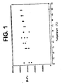
- Single dose aliquot pellets of reagents which contained RT, T7 and sugar were subjected to a wide range of temperatures to examine pellet stability. After being subject to a test temperature for 10 minutes, the pellets were then used for CT amplification. The results are graphed in Figure 1 . The results show that the single dose reagent pellet remains stable even after to exposure to high temperatures for 10 minutes.
- the prepared amplification pellets were placed in a tube to which was added 75 ⁇ l of a mixture of ARB and SDB (mixed in a 1:2 ratio) with positive control nucleic acid. This sample was then heated to 95°C for 10 minutes and then cooled to 42°C for 5 minutes. To this was added 25 ⁇ l of enzyme reagent, which had been reconstituted using standard Gen-Probe Inc. procedure. This mixture was allowed to incubate for one hour at 42°C. The reactions were then analyzed by the HPA procedure, as described above. The results of this test are reported as RLU in Table 2, and labeled AMP Pellets(+). As above, negative control reactions were run without nucleic acid (-).
- the prepared enzyme pellets were tested by heating 100 ⁇ l of a combination of SDB with positive control nucleic acid, EDB, and the standard reconstituted amplification reagent (in a 2:1:1 ratio) at 95°C for 10 minutes and then cooled to 42°C for 5 minutes. The total volume of the reaction mix was added to the prepared enzyme pellet. After the pellet was dissolved, the reaction was heated to 42°C for one hour and then subjected to HPA analysis as above. The results of this test are reported as RLU in Table 2 below, labeled Enzyme Pellet (+). Control reactions were prepared using standard Gen-Probe Inc. reagents following standard procedure. Data reported as RLU, standard C. Trachomatis TMA/HPA reaction.
- RNA reverse transcriptase RNA polymerase RNase H
- FIG. 3A is a schematic representation of a disposable dual chamber reaction vessel 10 and the heating steps associated therewith to perform a TMA reaction in accordance with one possible embodiment of the invention.
- Chamber A contains the amplification mix, namely nucleotides, primers, MgCl 2 and other salts and buffer components.
- Chamber B contains the amplification enzyme that catalyzes the amplification reaction, e.g., T7 and/or RT.
- T7 and/or RT amplification enzyme that catalyzes the amplification reaction
- T7 and/or RT amplification enzyme that catalyzes the amplification reaction
- the temperature of chamber A is then cooled down to allow primer annealing. Subsequently, the solution of chamber A is brought into contact with chamber B.
- Chambers A and B now in fluid communication with each other, are then maintained at the optimum temperature for the amplification reaction, e.g., 42 degrees C.
- the optimum temperature for the amplification reaction e.g. 42 degrees C.
- Figure 3B is a schematic representation of an alternative form of the invention in which two separate reaction chambers 12 and 14 are combined to form a dual chamber reaction vessel 10.
- Chamber A is pre-loaded during a manufacturing step with an amplification mix, namely nucleotides, primers, MgCl 2 and other salts and buffer components.
- Chamber B is pre-loaded during manufacturing with the amplification enzyme that catalyzes the amplification reaction, e.g., T7 and/or RT.
- Fluid sample is then introduced into chamber A.
- the targets are heated for denaturation to 95°C in chamber A.
- the solution in chamber A is brought into contact with the enzymes in chamber B to trigger the isothermal amplification reaction.
- reaction vessel is designed such that, after having brought the contents of chambers A and B into contact, the amplification chamber does not allow any exchange of materials with the environment, a closed system amplification is realized that minimizes the risk of contaminating the amplification reaction with heterologous targets or amplification products from previous reactions.
- FIG. 3C is a schematic representation of two alternative dual chamber reaction vessels 10 and 10' that are snapped into place in a test strip 19 for processing with a solid phase receptacle and optical equipment in accordance with a preferred embodiment of the invention.
- a unidirectional flow system is provided.
- the sample is first introduced into chamber A for heating to the denaturation temperature.
- Chamber A contains the dried amplification reagent mix 16.
- the fluid is transferred to chamber B containing the dried enzyme 18 in the form of a pellet.
- Chamber B is maintained at 42°C after the fluid sample is introduced into Chamber B.
- the amplification reaction takes place in Chamber B at the optimum reaction temperature (e.g., 42°C).
- the test strip 19 is then processed in a machine such as the VIDAS instrument available from bioMérieux Vitek, Inc., the assignee of the present invention. Persons of skill in the art are familiar with the VIDAS instrument.
- the steps of heating and cooling of chamber A could be performed prior to the insertion of the dual chamber disposable reaction vessel 10 or 10' into the test strip 19 , or, alternatively, suitable heating elements could be placed adjacent to the left hand end 24 of the test strip 19 in order to provide the proper temperature control of the reaction chamber A.
- the stand alone amplification processing station of Figures 4-14 described below, incorporates suitable heating elements and control systems to provide the proper temperature control for the reaction vessel 10.
- FIG 4 is a schematic representation of an alternative embodiment of a dual chamber reaction vessel 10 " formed from two separate interlocking vessels 10A and 10B that are combined in a manner to permit a fluid sample in one chamber to flow to the other, with the combined dual chamber vessel 10 " placed into a test strip 19 such as described above in Figure 3C .
- the fluid sample is introduced into chamber A, which contains the dried amplification reagent mix 16.
- Vessel A is then heated off-line to 95 degrees C, then cooled to 42 degrees C.
- the two vessels A and B are brought together by means of a conventional snap fit between complementary locking surfaces on the tube projection 26 on chamber B and the recessed conduit 28 on chamber A.
- the mixing of the sample solution from chamber A with the enzyme from chamber B occurs since the two chambers are in fluid communication with each other, as indicated by the arrow 30.
- the sample can then be amplified in the combined dual chamber disposable reaction vessel 10 " off-line, or on-line by snapping the combined disposable vessel 10 " into a modified VIDAS strip.
- the VIDAS instrument could perform the detection of the amplification reaction in known fashion.
- FIG. 5 is a perspective view of a stand-alone amplification processing system 200 for the test strips 19 having the dual chamber reaction vessels in accordance with a presently preferred form of the invention.
- the system 200 consists of two identical amplification stations 202 and 204, a power supply module 206, a control circuitry module 208, a vacuum tank 210 and connectors 212 for the power supply module 206.
- the tank 210 has hoses 320 and 324 for providing vacuum to amplification stations 202 and 204 and ultimately to a plurality of vacuum probes (one per strip) in the manner described above for facilitating transfer of fluid from the first chamber to the second chamber.
- the vacuum subsystem is described below in conjunction with Figure 14 .
- the amplification stations 202 and 204 each have a tray for receiving at least one of the strips and associated temperature control, vacuum and valve activation subsystems for heating the reaction wells of the strip to the proper temperatures, transferring fluid from the first chamber in the dual chamber reaction wells to the second chamber, and activating a valve such as a thimble valve or preferably a ball valve to open the fluid channel to allow the fluid to flow between the two chambers.
- a valve such as a thimble valve or preferably a ball valve to open the fluid channel to allow the fluid to flow between the two chambers.
- the stations 202 and 204 are designed as stand alone amplification stations for performing the amplification reaction in an automated manner after the patient or clinical sample has been added to the first chamber of the dual chamber reaction vessel described above.
- the processing of the strips after the reaction is completed with an SPR takes place in a separate machine, such as the VIDAS instrument. Specifically, after the strips have been placed in the stations 202 and 204 and the reaction run in the stations, the strips are removed from the stations 202 and 204 and placed into a VIDAS instrument for subsequent processing and analysis in known fashion.
- the entire system 200 is under microprocessor control by an amplification system interface board (not shown in Figure 5 ).
- the control system is shown in block diagram form in Figure 12 and will be described later.
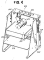
- Figure 6 one of the amplification stations 202 is shown in a perspective view.
- the other amplification station is of identical design and construction.
- Figure 7 is a perspective view of the front of the module of Figure 6 .
- the station includes a vacuum probe slide motor 222 and vacuum probes slide cam wheel 246 that operate to slide a set of vacuum probes 244 (shown in Figure 7 ) for the thimble valves up and down relative to a vacuum probes slide 246 to open the thimble valves and apply vacuum so as to draw the fluid from the first chamber of the reaction vessel 10 to the second chamber.
- the vacuum probes 244 reciprocate within annular recesses provided in the vacuum probes slide 246. Obviously, proper registry of the pin structure and vacuum probe 244 with corresponding structure in the test strip as installed on the tray needs to be observed.
- the station includes side walls 228 and 230 that provide a frame for the station 202.
- Tray controller board 229 is mounted between the side walls 228 and 230.
- the electronics module for the station 202 is installed on the tray controller board 229.
- a set of tray thermal insulation covers 220 are part of a thermal subsystem and are provided to envelop a tray 240 ( Figure 7 ) that receives one or more of the test strips.
- the insulation covers 220 help maintain the temperature of the tray 240 at the proper temperatures.
- the thermal subsystem also includes a 42°C Peltier heat sink 242, a portion of which is positioned adjacent to the second chamber in the dual chamber reaction vessel in the test strip to maintain that chamber at the proper temperature for the enzymatic amplification reaction.
- a 95°C heat sink 250 is provided for the front of the tray 240 for maintaining the first chamber of the reaction well in the test strip at the denaturation temperature.
- Figure 8 is another perspective view of the module of Figure 7 , showing the 95°C heat sink 250 and a set of fins 252. Note that the 95°C heat sink 250 is positioned to the front of and slightly below the tray 240.
- the 42°C heat sink 242 is positioned behind the heat sink 250.
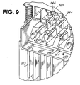
- FIG 9 is a detailed perspective view of a portion of the tray 240 that holds the test strips (not shown) as seen from above.
- the tray 240 includes a front portion having a base 254, a plurality of discontinuous raised parallel ridge structures 256 with recessed slots 258 for receiving the test strips.
- the base of the front 254 of the tray 240 is in contact with the 95°C heat sink 250.
- the side walls of the parallel raised ridges 256 at positions 256A and 256B are placed as close as possible to the first and second chambers of the reaction vessel 10 of Figure 3A so as to reduce thermal resistance.
- the base of the rear of the tray 240 is in contact with a 42°C Peltier heat sink, as best seen in Figure 8 .
- portion 256B of the raised ridge for the rear of the tray is physically isolated from portion 256A for the front of the tray, and portion 256B is in contact with the 42°C heat sink so as to keep the second chamber of the reaction vessel in the test strip at the proper temperature.
- the vacuum probes 244 include a rubber gasket 260.
- the gaskets 260 are positioned on the upper surface of the test strip surrounding the vacuum port in the dual chamber reaction vessel so as to make a tight seal and permit vacuum to be drawn on the second chamber.
- Figure 10 is an isolated perspective view of the test strip holder or tray 240 of Figure 9 , showing two test strips installed in the tray 240.
- the tray 240 has a plurality of lanes or slots 241 receiving up to six test strips 19 for simultaneous processing.
- Figure 10 shows the heat sinks 242 and 250 for maintaining the respective portions of the tray 240 and ridges 256 at the proper temperature.
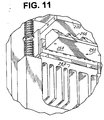
- Figure 11 is a detailed perspective view of the test strip holder or tray 240 as seen from below.
- the 95°C Peltier heat sink which would be below front portion 254 has been removed in order to better illustrate the rear heat sink 242 beneath the rear portion of the tray 240.
- FIG 12 is a block diagram of the electronics and control system of the amplification processing system of Figure 5 .
- the control system is divided into two boards 310 and 311, section A 310 at the top of the diagram devoted to amplification module or station 202 and the other board 311 (section B) devoted to the other module 204.
- the two boards 310 and 311 are identical and only the top section 310 will be discussed.
- the two boards 310 and 311 are connected to an amplification station interface board 300.
- the interface board 300 communicates with a stand alone personal computer 304 via a high speed data bus 302.
- the personal computer 304 is a conventional IBM compatible computer with hard disk drive, video monitor, etc.
- the stations 202 and 204 are under control by the interface board 300.
- the board 310 for station 202 controls the front tray 240 which is maintained at a temperature of 95°C by two Peltier heat sink modules, a pair of fans and a temperature sensor incorporated into the front portion 254 of the tray 240.
- the back of the tray is maintained at a temperature of 42°C by two Peltier modules and a temperature sensor.
- the movement of the vacuum probes 244 is controlled by the probes motor 222.
- Position sensors are provided to provide input signals to the tray controller board as to the position of the vacuum probes 244.
- the tray controller board 310 includes a set of drivers 312 for the active and passive components of the system which receive data from the temperature and position sensors and issue commands to the active components, i.e., motors, fans, Peltier modules, etc.
- the drivers are responsive to commands from the amplification interface board 300.
- the interface board also issues commands to the vacuum pump for the vacuum subsystem, as shown.
- FIG 13 is a diagram of the vacuum subsystem 320 for the amplification processing stations 202 and 204 of Figure 5 .
- the subsystem includes a 1 liter plastic vacuum tank 210 which is connected via an inlet line 322 to a vacuum pump 323 for generating a vacuum in the tank 210.
- a vacuum supply line 324 is provided for providing vacuum to a pair of pinch solenoid valves 224 (see Figure 6 ) via supply lines 324A and 324B.
- These vacuum supply lines 324A and 324B supply vacuum to a manifold 226 distributing the vacuum to the vacuum probes 244.
- the vacuum system 320 also includes a differential pressure transducer 321 for monitoring the presence of vacuum in the tank 210. The transducer 321 supplies pressure signals to the interface board 300 of Figure 12 .
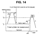
- FIG 14 is a representative graph of the thermal cycle profile of the station of Figure 5 .
- a first temperature T1 e.g., a denaturation temperature
- a ramp down of temperature as indicated at 404 occurs and the temperature of the reaction solution in the first chamber of the reaction vessel 10 cools to temperature T2.
- a fluid transfer occurs in which the solution in the first chamber is conveyed to the second chamber.
- Temperature T2 is maintained for an appropriate amount of time for the reaction of interest, such as one hour.
- the temperature is raised rapidly to a temperature T3 of 65°C to stop the amplification reaction.
- TMA temperature
- reaction vessels and amplification station components are also envisioned, and such alternative embodiments are encompassed in the present disclosure.
- VIDAS in-line simple rapid nucleic acid amplification and detection assay for the clinical laboratory for the detection of Mtb in test samples which can be completed in a short time.
- the entire assay is designed to take place on a single test strip, minimizing the potential for target or amplicon contamination.
- the amplification based assay is capable of detection of Mtb where the sample contains only 5 cells similar to the sensitivity achieved by the Gen-Probe commercial kit.
- the amplification based assay utilizes isothermal transcription-mediated amplification (TMA) targeting unique sequences of rRNA, followed by hybridization and enzyme-linked fluorescent detection of nucleic acid probe (amplicon) in the VIDAS instrument.
- TMA isothermal transcription-mediated amplification
- the amplification/detection assay can detect approximately 1 fg of Mtb rRNA, or less than one Mtb organism per test, and is specific for all members of the Mtb complex. Specific probes for the detection of Mtb can be found in C. Mlessnesst, 1994, J. Clin. Microbiol. 32, 2707 .
- Standard smears for acid-fast bacilli are not always reliable as a diagnostic tool, and even when positive may be a mycobateria other than Mtb.
- standard methods for diagnosis of tuberculosis requires culturing the slow-growing bacteria, and may take up to 6 weeks or longer. During this time, the patient is usually isolated. Initial results are that this automated test matches or exceeds the clinical sensitivity of the culture method, and offers a highly sensitive method to rapidly (in less than three hours) detect Mtb in infected samples, thereby aiding rapid diagnosis, isolation and treatment.
- a 450 ⁇ l volume of specimen is added to 50 ⁇ l of specimen dilution buffer in a lysing tube containing glass beads, sonicated for 15 minutes at room temperature to lyse organisms, heat inactivated for 15 minutes at 95°C.
- isothermal amplification was conducted as per a commercially available manual assay kit (Gen-Probe Inc.) following the kit instructions using standard kit reagents. However, similar assays can be conducted using the modified components as described in the Examples above.
- the detection system requires hybridization of the target nucleic acid or amplicon to a specific capture nucleic acid bound to a solid support, (in the VIDAS system called a "solid phase receptacle” SPR pipet-like device), and to a labeled detection probe nucleic acid (for example where the label can be alkaline phosphatase, a chemiluminescent signal compound, or other reagent that will allow for specific detection of bound probe).
- a solid phase receptacle for example where the label can be alkaline phosphatase, a chemiluminescent signal compound, or other reagent that will allow for specific detection of bound probe.
- the SPR transfers the probe-target hybrid to an enzyme substrate, whereby the detectable signal is triggered from the bound probe and detected by the assay instrument.
- the detection probe is conjugated to alkaline phosphatase, and once placed in contact with substrate of methyl umbelliferyl phosphate (MUMP), the substrate is converted into 4-methyl umbelliferone (4-MU) by the alkaline phosphatase.
- MUMP methyl umbelliferyl phosphate
- the 4-MU produces fluorescence which is measured and recorded by the standard VIDAS instrument as relative fluorescence units (RFU).
- REU relative fluorescence units
- Generally controls are prepared in a matrix of specimen dilution buffer with positive controls containing 5fg of Mtb rRNA, or the equivalent rRNA of approximately 1 M. tb cell.
- Sensitivity of the automated probe assay can be determined by testing dilutions of lysed M. tb cells.
- the cell lysates can generally be prepared with a 1 ⁇ l loop of cells (the assumption being that there are approximately 1x10 9 colony forming units (CFU) per 1 ⁇ l loop-full, based upon previous titration and CFU experiments). Dilutions of the Mtb lysates can then be tested with the automated probe assay.
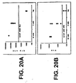
- Figure 20A is a graph showing detection of Mtb amplicons according to the Gen-Probe kit.
- Figure 20B is a graph showing detection of Mtb amplicons from the same reactions as in Figure 20A by the VIDAS instrument.
- Figure 21 is a graph showing amplification and detection of Mtb nucleic acids on the modified VIDAS apparatus. Enzyme was used in liquid form and amplification was performed in-line with VIDAS assay instrument.
- Figure 22 is a graph showing amplification and detection of Mtb nucleic acids on the modified VIDAS apparatus using the binary/dual chamber disposable reaction vessel.
- the denaturation step was performed off-line of the VIDAS instrument, amplification and amplicon detection was performed in-line with VIDAS instrument.
- CT infection Chlamydia trachomatis
- CT Chlamydia trachomatis
- the cervical samples were collected with a Gen-Probe sample collection kit containing Gen-Probe transport medium; the urine samples were collected into standard urine collection devices. All samples were stored at 4°C.
- Cervical swabs were centrifuged at 425xg for 5 minutes to bring all liquid to the bottom of the tube.
- the swabs were then treated with 40 ⁇ l Gen-Probe Specimen Preparation Reagent and incubated at 60°C for 10 minutes. 20 ⁇ l of the treated sample was then pipetted into 400 ⁇ l of sample dilution buffer (SDB).
- SDB sample dilution buffer
- Samples were amplified using the TMA protocol, and rRNA targets were hybridized to oligomer conjugated to AMVE copolymer and an oligomer conjugated to alkaline phosphatase. See for example U.S. Patent 5,489,653 and 5,510,084 .
- the solid phase receptacle (SPR pipet-like device) carries the bound hybrids through successive wash steps and finally into the substrate 4-MUP.
- the alkaline phosphatase converts the substrate to fluoresence 4-MU, which is detected by the VIDAS assay machine and recorded as relative fluorescence units.
- Table 5 illustrates the results of clinical cervical swab specimen testing for CT comparing results from the Gen-Probe manual AMP-CT assay and the VIDAS automated probe assay.
- Table 6 illustrates the results of clinical urine specimen testing comparing the results of manual AMP-CT assay and the VIDAS automated probe assay.
- nucleic acid probes can be substantially increased through the detection of multiple different nucleic acid molecules, and the use of internal positive controls.
- An automated method has been devised for use with the VIDAS instrument (bioMérieux Vitek, Inc.) which can discretely detect at least two different nucleic acid sequences in one assay reaction, and is termed the Multiplex protocol.
- VIDAS instrument bioMérieux Vitek, Inc.
- a nucleic acid amplification procedure, or a processed test sample may be screened for more than one amplified nucleic acid molecule in the same assay.
- This method relies on the spatial separation of discrete nucleic acid probes which can specifically capture different target nucleic acid sequences (amplicons), on the SPR pipet-like device of the VIDAS instrument.
- the SPR is a disposable pipet-like tip which enables fluid movements as well as acting as the solid support for affinity capture. Multiplex capture by SPR is demonstrated using capture probes specific for Chlamydia trachomatis ( ⁇ T) and Neisseria gonorrhoeae (NG).
- ⁇ T Chlamydia trachomatis
- NG Neisseria gonorrhoeae
- FIG. 15 illustrates a schematic of the operation of the multiplex VIDAS detection.
- the SPR tips are coated in two distinct zones with oligonucleotide nucleic acid sequences which are used to specifically capture complementary nucleic acid sequences (amplicons) with their corresponding specific reporter or detector probe nucleic acids labeled with alkaline phosphatase (AKP). Following washes to remove unbound reporter probes, AKP localized to the SPR bottom is detected with the fluorescent substrate 4-MUP. The AKP is stripped from the bottom of the SPR with NaOH or other reagents which promote denaturation of nucleic acid hybrids or inactivate alkaline phosphatase activity.
- amplicons complementary nucleic acid sequences
- AKP alkaline phosphatase
- the enzyme reaction well is emptied, washed, and re-filled with fresh 4-MUP.
- the new substrate is exposed to the bottom of the SPR and any residual fluorescence is measured.
- AKP-reporter probe bound to the top of the SPR is detected by immersing the SPR in the 4-MUP and representing the presence of the second amplicons.
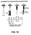
- Figure 16 illustrates the production of SPR with two distinct capture zones.
- the SPR is inserted tip-first into a silicon plug, which are held in a rack.
- Differential pressure is used to uniformly draw a solution of a specific capture probe at about 1 ⁇ g/ml, conjugated to AMVE copolymer, into all SPRs at one time.
- the amount of fluid drawn into each SPR, and thus the size of the zone, is controlled by regulating the amount of pressure in the system.
- Attachment of the conjugate to the SPR surface is achieved by passive adsorption for several hours at room temperature. After washing, and drying, the SPRs are capped with a small adhesive disc and inserted into new racks in a tip-down orientation. The lower portion of the SPR is then similarly coated with a second capture probe conjugate.
- SPRs are stable when stored dry at 4°C.
- Figure 17 illustrates a preferred embodiment of the VIDAS apparatus strip configuration for multiplex detection.
- the strip can be pre-filled with 200 ⁇ l of AKP-probe mix (about 1x10 12 molecules of each probe) in hybridization buffer in well X1, 600 ⁇ l of wash buffer in wells X3, X4, X5, 600 ⁇ l of stripping reagent in wells X6 and X7, and 400 ⁇ l of AKP substrate in X8 and sealed with foil.
- a foil-sealed optical cuvette (XA) containing 300 ⁇ l of 4-MUP is snapped into the strip, and the strips are inserted into the VIDAS instrument at 37°C.
- the multiplex VIDAS protocol is then executed using SPRs coated with two capture probes in distinct zones.
- the VIDAS multiplex protocol can involve many steps.
- the validation test protocol contained thirteen (13) basic steps as follows:
- Hybridization, substrate, wash and stripping steps involve multiple cycles of pipeting the respective solution into the SPR, holding the solution for a defined period of time, and pipeting the solution out of the SPR. Hold times for hybridization, substrate and washing or stripping are 3.0, 0.5 and 0.17 minutes respectively. The fluorescence signal is detected by the apparatus. Total assay time for the research protocol was about 1.75 hours but can be reduced to about 75 minutes.
- Figure 18 illustrates and graphs the results of verification of the VIDAS multiplex protocol executed as described above, except the SPR was homogeneously coated with only a single capture probe for Neisseria gonorrhoeae (NG).
- the number of NG oligonucleotide targets in the test sample was varied from 0, 1x10 10 , or 1x10 11 molecules in the test sample.
- the data shown are averages of replicate samples.
- the graph as illustrated is divided into two parts; the left and right halves show the results of two fluorescent measurements from the lower and the upper zones of the SPR, respectively.
- the measurements taken from the bottom zone after stripping the lower area of bound nucleic acid, and exposure for about 11 minutes in fresh 4-MUP substrate was approximately 46 RFU for all samples tested, and was equivalent to background fluorescence measured. This measurement is shown by the 0 time point in the center of the graph.
- the graph illustrates two sequential sets of measurements of fluorescence from a single SPR, the first set of measurements being taken from the bottom half of the SPR (left half of the graph), and a second set of measurements taken from the top of the SPR (the right of the graph). This experiment validates that the Multiplex protocol and zone coated SPR procedure yield essentially identical results, as indicated by the fluorescence intensities in the left- and right-hand parts of the graph, from the upper and lower portions of the SPR.
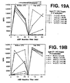
- Figure 19 illustrates Multiplex detection of CT and NG oligonucleotide targets at different input amounts.
- Figure 19A is a graph showing the results when 1x10 12 CT targets were mixed with 0, 1x10 9 , 1x10 10 , 1x10 11 , or 1x10 12 , NG targets, and detected with the VIDAS instrument using the multiplex protocol and SPRs coated with CT capture probes on the bottom zone of the SPR, and NG capture probes on the top zone of the SPR.
- Figure 19B illustrates the results when 1x10 12 NG targets was mixed with 0, 1x10 9 , 1x10 10 , 1x10 11 , or 1x10 12 , CT targets, and detected with the VIDAS instrument using the multiplex protocol and SPRs coated with CT capture probes on the bottom zone of the SPR, and NG capture probes on the top zone of the SPR.
- the data is graphed as above where the graph illustrates two sequential sets of measurements of fluorescence from a single SPR, the first set of measurements being taken from the bottom half of the SPR (left half of the graph), and a second set of measurements taken from the top of the SPR (the right of the graph) with verification of stripping of the SPR in the center of the graph.
- this experiment shows that the two zones of the SPR act independently in the Multiplex protocol, since high fluorescence signals from one zone do not interfere with signals produced from the second zone, regardless of whether these latter signals are high (1x10 12 ), or low (1x10 9 , just detectable), or negative.
- Table 7 summarizes the data obtained by Multiplex VIDAS detection of CT and NG in a sample at various target levels, reported in RFUs. TABLE 7 Detection of CT and NG targets in sample RFUs A none B - 1x10 9 1x10 10 1x10 11 1x10 12 1x10 13 none C 43 D /40 B 43/116 46/693 62/7116 174/11817 273/12136 1x10 9 189/41 246/118 169/773 220/5750 422/12522 399/11401 1x10 10 1736/41 2258/125 1937/734 1931/6639 2128/12390 2371/11180 1x10 11 10339/48 9815/145 9858/760 9369/4571 9784/11825 10252/10312 1x10 12 12149/49 13520/148 12940/796 13593/4397 11239/11786 10158/9900 1x10 13 11545/57 11713/121 10804/815 12805/5404
- the multiplex VIDAS protocol is clearly operative and enables the rapid and discrete detection of more than one different nucleic acid in a sample.
- This protocol, and the SPR coating can be manipulated in many formats to present coating zones of different surface area with different sized gaps between two or more detection zones.
- the SPR can be coated with nucleic acids which are designed to capture different regions of the same nucleic acid sequence to detect, for example, truncated gene expression, different alleles or alternatively spliced genes.
- the SPR can be coated to capture amplicons from internal control nucleic acid molecules which can be used to detect and confirm successful nucleic acid amplification reactions.
- the VIDAS Multiplex protocol is a flexible method for detection of more than one nucleic acid sequence in the same sample, in a single assay.
- control sequences composed of functional building blocks of sequences chosen by random generation of nucleic acid sequences for use as amplification reaction internal positive controls ideally requires that the control sequences be specifically designed to be used for the various nucleic acid amplification protocols including but not limited to PCR, LCR, TMA, NASBA, and SDA.
- the internal control nucleic acid sequence, in combination with the appropriate sequence specific oligonucleotide primers or promoter-primers will generate a positive amplification signal if the amplification reaction was successfully completed.
- the internal control nucleic acid is useful regardless of the nucleic acid sequences present in the target organism, the host organism, or nucleic acids present in the normal flora or in the environment.
- the internal control sequences should not be substantially similar to any nucleic acid sequences present in a clinical setting, including human, pathogenic organisms. normal flora organisms, or environmental organisms which could interfere with the amplification and detection of the internal control sequences.
- the internal control sequences of the instant invention are comprised of functional blocks of sequences chosen from a list of randomly generated nucleic acid sequences.
- the functional blocks are segments which provide for a special property needed to allow for amplification, capture, and detection of the amplification product.
- the internal control sequences are most useful when the functional blocks meet certain functional requirements of the amplification protocol, such as: a) a primer binding site on the anti-sense strand; b) a capture site; c) a detector probe binding site; d) a T7-promoter containing primer binding site on the sense strand.
- Each of these functional elements has its own particular constraints, such as length, %G-C content, Tm, lack of homology to known sequences, and absence of secondary structural features (i.e. free from dimer formation or hairpin structures) which can be used to select the appropriate sequence.
- randomly generated functional blocks of sequences can be screened for the desired functional properties before use in constructing internal control sequences.
- a random sequence generator was used to generate strings of numbers; each number being limited to the range from 0.000 to 4.000.
- the length of the strings is flexible and chosen based upon the desired lengths of the functional blocks.
- Each number in the string (i.e. n1, n2, n3, n4 ... nx where x is the length of the string) was then assigned a corresponding nucleotide as follows: guanosine (G) if 0 ⁇ n ⁇ 1; adenosine (A) if 1 ⁇ n ⁇ 2; thymidine (T) if 2 ⁇ n ⁇ 3; and cytosine (C) if 3 ⁇ n ⁇ 4.
- G guanosine
- A adenosine
- T thymidine
- C cytosine
- Figure 23 illustrates the results generated by the method described showing a collection of strings of nucleic acid sequences and screening for specific functional parameters.
- the internal control sequence can include DNA, RNA, modified oligonucleotides, or any combination of nucleic acids, such that the illustrated sequences using DNA nomenclature can be readily adapted as desired to the appropriate nucleic acid.
- IC sequences were then constructed by assembling the functional blocks (selected at random) in the proper order. Finally, the assembled internal control sequence were then examined to insure that overall sequence and structural constraints were maintained. For example, in a TMA reaction, the internal control sequence should not have significant base-pairing potential between the two primer binding sites or from stable 3' dimer structures. Those internal control sequences which pass through these layers of screening were then physically produced using overlapping oligonucleotides and tested for performance in actual amplification/detection assays.
- any one functional block may have some homology to sequences present in a clinical setting (a perfect match of a 21 nucleotide block is expected at a random frequency of 1 in 4e12 sequences or about 4x10 12 ; generated sequences were screened against the GenBank data base) it is highly unlikely that all functional blocks will be found to have substantial homology. Since the internal control nucleic acid sequences are constructed of a group of functional blocks placed in tandem, the chance possibility that a natural nucleic acid sequence will have an identical string of nucleic acid sequence blocks in the same tandem organization is remote.
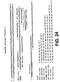
- Random Internal Control 1 (RIC1) is shown in Figure 24 with the possible oligonucleotide primers/probes for amplification and detection of the control sequence.
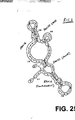
- Figure 25 shows an analysis of the possible secondary structure of the RIC1 molecule.
- RIC1 was constructed using randomly generated strings ran16, ran19, ran21 and ran33. The functional blocks requiring primer binding were met by ran16 and ran19, while the capture site was satisfied by ran21 and the detector probe binding site was met by ran33.
- a capture probe or detection probe sequence designation can be interchanged, as along as the appropriate linker molecule is attached to the appropriate probe, wherein a reporter probe oligonucleotide is linked to a means for generating a detectable signal, and the capture probe oligonucleotide is linked to a means for adhering the capture probe to an appropriate support.
- the probes and oligos are described with the understanding that in the case of double stranded DNA, the complementary strand can be the target or as appropriate can be converted for use as the strand for detection.
- one or ordinary skill in the art will be able to modify the sequences as disclosed to generate alternative probes and primers which are suitable for use in an equivalent fashion as described herein.
- Random Internal Control 2 (RIC2) is shown in Figure 26 with the possible oligonucleotide primers/probes for amplification and detection of the control sequence.
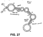
- Figure 27 shows an analysis of the possible secondary structure of the RIC2 sequence. Similarly to RIC1, RIC2 was constructed using randomly generated strings ran27, ran32, ran39 and ran51. Thus, illustrating that it is also possible that the functional blocks requiring primer binding, capture probe binding, detector probe binding can be met by alternative random sequences generated by the method described above.
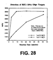
- Figure 28 illustrates results from detection of RIC1 DNA, where the ran21 was the capture probe and ran33 was an enzyme-linked detector-probe, and shows that detection occurs under standard assay conditions with expected fluorescence intensities.
- Figure 29 shows that RIC1 RNA, amplified by TMA and detected on a VIDAS instrument (bioMérieux Vitek, Inc.) using the enzyme-linked detection system, has a limit of sensitivity of about 1000 molecules of RIC1 RNA (without optimization of conditions). Similar analysis of RIC2 sequences was performed and found to be similar to RIC1. It is significant that the amplification and detection system of the internal control functioned effectively under the conditions optimized for the selected target.
- SPRs can be homogeneously coated with a mixture of different capture nucleic acid sequences in a single, whole-SPR zone.
- two capture nucleic acid sequences can be combined in one zone, one specific for a target test sequence, and one specific for a target test sequence, and one specific for an internal control sequence.
- Target amplicons, if present, and internal control amplicons are simultaneously hybridized to the SPR in the presence of labeled probe sequences specific for the target amplicons.
- a first signal read is done to so that the presence or absence of label on the SPR is determined to ascertain the presence or absence of the test target.
- a second hybridization is then done (sequential hybridization) to the SPR using a detection label probe specific for the internal control.
- the SPR is washed to remove excess unbound detection probe, and the second label is measured to indicate the presence or absence of the internal control.
- a positive signal from the IC second read confirms the functionality of the ainplification/detection system. If the first and second label are the same, an additive signal will result from the positive first read and the positive second IC read. If both the first signal is negative and the second IC signal is also negative, then the amplitication/detection functionality failed, which could be due to for example, sample interference or mechanical failure. In this case the test result is reported invalid (false negative) and re-testing is recommended. If the labels are different then neither sequential hybridizations nor sequential detection steps would be necessary.
- NCCLS Document MM3-A Molecular Diagnostic Methods for Infectious Diseases; Approved Guideline, p. 55, March 1995.
- This type of protocol can also be used for a combined GC/CT/internal control assay, if a screening approach is allowed (no discrimination between GC and/or CT positives during the first read).
- GC and CT specific signals have to be resolved by running the CT and GC specific assays on screen positive samples (5-10% of cases, depending on prevalence) SPRs would be coated homogeneously with 3 capture probes (CT/GC/internal control).
- the IC could share a capture probe with either CT or GC.
- internal control nucleic acid sequences have been designed and validated for use in various amplification and detection systems including an internal control for Chlamydia trachomatis (CT) assay identified as CRIC-2; for a Neisseria gonorrhoeae (NG) assay identified as GRIC; and for Mycobacterium tuberculosis (MT) identified as MRIC.
- CT Chlamydia trachomatis
- NG Neisseria gonorrhoeae
- MRIC Mycobacterium tuberculosis
- An internal control was generated for HIV assays identified as HRIC, wherein both the capture probe sequence and reporter probe sequence were derived from random sequence.
- the sequence of the internal control, and the corresponding target sequence are shown in Figure 30 .
- the Random Sequence Probe #1082 can be used as the reporter probe, when suitably conjugated to a reporter molecule as described previously.
- a capture oligonucleotide Random Sequence Probe #1081 has been designed for use in the capture of the control sequence, for improved quantitation by elimination of competition between the target amplicons and IC amplicons for a common capture probe.
Description
- The present invention relates to the detection of specific nucleic acid sequences in a. target test sample.
- In particular, the present invention relates to the automated detection of specific nucleic acid sequences which are either unamplified or amplified nucleic acid sequences (amplicons).
- In addition, the present invention relates to the use of automated amplification, methods and compositions for monitoring successful amplification, improved methods for reducing the chance for contamination, and the use of unified reaction buffers and unit dose aliquots of reaction components for amplification.
- The development of techniques for the manipulation of nucleic acids, the amplification of such nucleic acids when necessary, and the subsequent detection of specific sequences of nucleic acids or amplicons has generated extremely sensitive and nucleic acid sequence specific assays for the diagnosis of disease and/or identification of pathogenic organisms in a test sample.
- When necessary, enzymatic amplification of nucleic acid sequences will enhance the ability to detect such nucleic acid sequences. Generally, the currently known amplification schemes can be broadly grouped into two classes based on whether, the enzymatic amplification reactions are driven by continuous cycling of the temperature between the denaturation temperature, the primer annealing temperature, and the amplicon (product of enzymatic amplification of nucleic acid) synthesis temperature, or whether the temperature is kept constant throughout the enzymatic amplification process (isothermal amplification). Typical cycling nucleic acid amplification technologies (thermocycling) are polymerase chain reaction (PCR), and ligase chain reaction (LCR). Specific protocols for such reactions are discussed in, for example, Short Protocols in Molecular Biology, 2rd Edition, A Compendium of Methods from Current Protocols in Molecular Biology, (Eds. Ausubel et al., John Wiley & Sons, New York, 1992) . Reactions which are isothermal include: transcription-mediated amplification (TMA), nucleic acid sequence-based amplification (NASBA), and strand displacement amplification (SDA).
- U.S. Patent documents which discuss nucleic acid amplification include
4,683,195 ;4,683,202 ;5,130,238 ;4,876,187 ;5,030,557 ;5,399,491 ;5,409,818 ;5,485,184 ;5,409,818 ;5,554,517 ;5,437,990 and5,554,516 . It is well known that methods such as those described in these patents permit the amplification and detection of nucleic acids without requiring cloning, and are responsible for the most sensitive assays for nucleic acid sequences. However, it is equally well recognized that along with the sensitivity of detection possible with nucleic acid amplification, the ease of contamination by minute amounts of unwanted exogenous nucleic acid sequences is extremely great. Contamination by unwanted exogenous DNA or RNA nucleic acids is equally likely. The utility of amplification reactions will be enhanced by methods to control the introduction of unwanted exogenous nucleic acids and other contaminants. - Prior to the discovery of thermostable enzymes, methods that used thermocycling were made extremely difficult by the requirement for the addition of fresh enzyme after each denaturation step, since initially the elevated temperatures required for denaturation also inactivated the polymerases. Once thermostable enzymes were discovered, cycling nucleic acid amplification became a far more simplified procedure where the addition of enzyme was only needed at the beginning of the reaction. Thus reaction tubes did not need to be opened and new enzyme did not need to be added during the reaction, allowed for an improvement in efficiency and accuracy as the risk of contamination was reduced, and the cost of enzymes was also reduced. An example of a thermostable enzyme is the polymerase isolated from the organism Thermophilus aquaticus.
- In general, isothermal amplification can require the combined activity of multiple enzyme activities for which no optimal thermostable variants have been described. The initial step of an amplification reaction will usually require denaturation of the nucleic acid target, for example in the TMA reaction, the initial denaturation step is usually ≥ 65°C, but can be typically ≥ 95°C, and is used when required to remove the secondary structure of the target nucleic acid.
- The reaction mixture is then cooled to a lower temperature which allows for primer annealing, and is the optimal reaction temperature for the combined activities of the amplification enzymes. For example, in TMA the enzymes are generally a T7 RNA polymerase and a reverse transcriptase (which includes endogenous RNase H activity). The temperature of the reaction is kept constant through out the subsequent isothermal amplification cycle.
- Because of the lack of suitable thermostable enzymes, some isothermal amplifications will generally require the addition of enzymes to the reaction mixture after denaturation at high temperature, and cool-down to a lower temperature. This requirement is inconvenient, and requires the opening of the amplification reaction tube, which introduces a major opportunity for contamination.
- Thus, it would be most useful if such reactions could be more easily performed with a reduced risk of contamination by methods which would allow for integrated denaturation and amplification without the need for manual enzyme transfer.
- Typical reaction protocols require the use of several different buffers, tailored to optimize the activity of the particular enzyme being used at certain steps in the reaction, or for optimal resuspension of reaction components. For example, while a typical PCR 10x amplification buffer will contain 500mM KCl and 100mM Tris HCl, pH 8.4, the concentration of MgCl2 will depend upon the nucleic acid target sequence and primer set of interest. Reverse transcription buffer (5x) typically contains 400mM Tris-Cl, pH 8.2; 400mM KCl and 300 mM MgCl2, whereas Murine Maloney Leukemia Virus reverse transcriptase buffer (5x) typically contains 250mM Tris-Cl, pH 8.3; 375mM KCl; 50mM DTT (Dithiothreitol) and 15mM MgCl2.
- While such reaction buffers can be prepared in bulk from stock chemicals, most commercially available amplification products provide bulk packaged reagents and specific buffers for use with the amplification protocol. For example, commercially available manual amplification assays for detection of clinically significant pathogens (for example Gen-Probe Inc. Chlamydia, and Mycobacterium tuberculosis detection assays) requires several manual manipulations to perform the assay, including dilution of the test sample in a sample dilution buffer (SDB), combination of the diluted sample with amplification reaction reagents such as oligonucleotides and specific oligonucleotide promoter/primers which have been reconstituted in an amplification reconstitution buffer (ARB), and finally, the addition to this reaction mixture of enzymes reconstituted in an enzyme dilution buffer (EDB).
- The preparation and use of multiple buffers which requires multiple manual additions to the reaction mixture introduces a greater chance for contamination. It would be most useful to have a single unified buffer which could be used in all phases of an amplification protocol. In particular, with the commercially available TMA assays described above, the requirement for three buffers greatly complicates automation of such a protocol.
- Bulk packaging of the enzyme or other reaction components by manufacturers, may require reconstitution of the components in large quantities, and the use of stock amounts of multiple reagents, can be wasteful when less than the maximal number of reactions are to be carried out, as some of these components may be stable for only a short time. This process of reconstitution also requires multiple manipulations by the user of the stock reagents, and aliquoting of individual reaction amounts of reagents from stocks which creates a major opportunity for contamination.
- Methods and compositions for the preparation of bulk quantities of preserved proteins are known, see for example,
U.S. Patent 5,098,893 ;4,762,857 ;4,457,916 ;4,891,319 ;5,026,566 and International Patent PublicationsWO 89/06542 WO 93/00806 WO 95/33488 WO 89/00012 - The requirement for many changes of buffer and the multiple addition of reagents complicates the automation of such reactions. A single dose unit of reaction buffer mixture, and a unified combination buffer will both simplify automation of the process and reduce the chance of contamination.
- Nucleic acid probe assays, and combination amplification/probe assays can be rapid, sensitive, highly specific, and usually require precise handling in order to minimize contamination with non-specific nucleic acids, and are thus prime candidates for automation. As with conventional nucleic acid detection protocols, it is generally required to utilize a detection probe oligonucleotide sequence which is linked by some means to a detectable signal generating component. One possible probe detection system is described in
U.S. Patent 4,581,333 . - In addition, automation of a nucleic acid detection system targeting unamplified or amplified nucleic acid, or a combined automated amplification/detection system will generally be adaptable to the use of nucleic acid capture oligonucleotides that are attached to some form of solid support system. Examples of such attachment and methods for attachment of nucleic acid to solid support are found in
U.S. Patent 5,489,653 and5,510,084 . - Automation of amplification, detection, and a combination of amplification and detection is desirable to reduce the requirement of multiple user interactions with the assay. Apparatus and methods for optically analyzing test materials are described for example in
U.S. Patent 5,122,284 . Automation is generally believed to be more economical, efficient, reproducible and accurate for the processing of clinical assays. Thus with the superior sensitivity and specificity of nucleic acid detection assays, the use of amplification of nucleic acid sequences, and automation at one or more phases of a assay protocol can enhance the utility of the assay protocol and its utility in a clinical setting. - Nucleic acid amplification is highly sensitive to reaction conditions, and the failure to amplify and/or detect any specific nucleic acid sequences in a sample may be due to error in the amplification process as much as being due to absence of desired target sequence. Amplification reactions are notoriously sensitive to reaction conditions and have generally required including control reactions with known nucleic acid target and primers in separate reaction vessels treated at the same time. However, internal control sequences added into the test reaction mixture would truly control for the success of the amplification process in the subject test reaction mixture and would be most useful.
U.S. Patent 5,457,027 teaches certain internal control sequences which are useful as an internal oligonucleotide standard in isothermal amplification reactions for Mycobacterium tuberculosis. - However it would be extremely useful to have a general method of generating internal control sequences, that would be useful as internal controls of the various amplification procedures, which are specifically tailored to be unaffected by the nucleic acid sequences present in the target organism, the host organism, or nucleic acids present in the normal flora or in the environment. Generally, such internal control sequences should not be substantially similar to any nucleic acid sequences present in a clinical setting, including human, pathogenic organism, normal flora organisms, or environmental organisms which could interfere with the amplification and detection of the internal control sequences.
- In general, a single assay reaction for the detection of nucleic acid sequences is limited to the detection of a single target nucleic acid sequence. This single target limitation increases costs and time required to perform clinical diagnostic assays and verification control reactions. The detection of more than one nucleic acid sequence in a sample using a single assay would greatly enhance the efficiency of sample analysis and would be of a great economic benefit by reducing costs, for example helping to reduce the need for multiple clinical assays.
- Multiple analyte detection in a single assay has been applied to antibody detection of analyte as in for example International Patent Publication number
WO 89/00290 WO 93/21346 - In addition to reducing cost, time required, the detection of more than one nucleic acid target sequence in a single assay would reduce the chance of erroneous results. In particular multiple detection would greatly enhance the utility and benefit using internal control sequences and allow for the rapid validation of negative results.
- Reference may also be made to
EP-A-0 622 464 , which relates to a nucleic acid assay procedure, and toUS-A-5,229,297 , which relates to a containment cuvette for PCR and a method of use. - The present invention comprises methods for the automated isothermal amplification and detection of a specific nucleic acid in a test sample to be tested comprising:
- a) combining a test sample to be tested with a buffer, a mixture of free nucleotides, specific oligonucleotide primers, and optionally thermostable nucleic acid polymerization enzyme, in a first reaction vessel and placing the reaction vessel in an automated apparatus such that;
- b) the automated apparatus heats the first reaction vessel to a temperature, and for a time sufficient to denature, if necessary, the nucleic acid in the sample to be tested;
- c) the automated apparatus cools the first reaction vessel to a temperature such that oligonucleotide primers can specifically anneal to the target nucleic acid;
- d) the automated apparatus transfers the reaction mixture from the first reaction vessel to a second reaction vessel, and brings the reaction mixture in contact with thermolabile nucleic acid amplification enzyme;
- e) the automated apparatus maintains the temperature of the second reaction vessel at a temperature which allows primer mediated amplification of the nucleic acid;
- f) the automated apparatus contacts the amplified nucleic acid in the second reaction vessel with a capture nucleic acid specific for the nucleic acid to be tested such that they form a specifically-bound nucleic acid-capture probe complex;
- g) the automated apparatus optionally washes the specifically captured amplified nucleic acid such that non-specifically bound nucleic acid is washed away from the specifically-bound nucleic acid-capture probe complex;
- h) the automated apparatus contacts the specifically-bound nucleic acid-capture probe complex with a labeled nucleic acid probe specific for the amplified nucleic acid such that a complex is formed between the specifically amplified nucleic acid and the labeled nucleic acid probe;
- i) the automated apparatus washes the specifically-bound nucleic acid-capture probe-labeled probe complex such that non-specifically bound labeled probe nucleic acid is washed away from the specifically bound complex;
- j) the automated apparatus contacts the specifically bound complex with a solution wherein an detection reaction between the labeled nucleic acid probe is effected between the solution and the label attached to the nucleic acid such that a detectable signal is generated from the sample in proportion the amount of specifically-bound amplified nucleic acid in the sample;
wherein the steps h, i, and j may occur sequentially or simultaneously; - k) the automated apparatus detects the signal and optionally displays a value for the signal, or optionally records a value for the signal.
- As used herein, the term test sample includes samples taken from living patients, from non-living patients, from surfaces, gas, vacuum or liquids, from tissues, bodily fluids, swabs from body surfaces or cavities, and any similar source. The term buffer as used here encompasses suitable formulations of buffer which can support the effective activity of a label, for example an enzyme placed into such buffer when treated at the appropriate temperature for activity and given the proper enzymatic substrate and templates as needed. The term specific oligonucleotide nucleic acid primers means an oligonucleotide having a nucleic acid sequence which is substantially complementary to and will specifically hybridize/anneal to a target nucleic acid of interest and may optionally contain a promoter sequence recognized by RNA polymerase. The term reaction vessel means a container in which a chemical reaction can be performed and preferably capable of withstanding temperatures of anywhere from about -80°C to 100°C.
- The instant invention further provides for the method described above, wherein the reaction buffer is a unified buffer and as such is suitable for denaturation nucleic acids and annealing of nucleic acids, and is further capable of sustaining the enzymatic activity of nucleic acid polymerization and amplification enzyme. Further encompassed by the invention is the method wherein the nucleic acid amplification enzyme is administered in the second reaction chamber as a single assay dose amount in a lyophilized pellet, and the reaction chamber is sealed prior to the amplification step.
- An apparatus for the automated detection of more than one nucleic acid target sequences or amplicons comprising a solid phase receptacle (SPR® pipet-like devise) coated with at least two distinct zones of a capture nucleic acid oligonucleotide is described.
- The application teaches a method for the automated detection of more than one nucleic acid target sequence comprising contacting a solid phase receptacle (SPR® pipet-like devise) coated with at least two distinct capture nucleic acid oligonucleotides in a single or multiple zones to a sample to be tested and detecting a signal(s) from specifically bound probe. In one embodiment of the invention, the SPR is coated with two distinct zones of capture nucleic acid oligonucleotides which are specific for different nucleic acid sequence targets. In another embodiment, the SPR is coated with at least one capture probe for a target nucleic acid sequence, and one capture probe for an amplification control nucleic acid sequence which when detected confirms that amplification did take place.
- The present invention also comprises an internal amplification randomly generated positive control nucleic acid including the nucleic acid sequence of RIC1 and a second internal amplification positive control nucleic acid having the nucleic acid sequence of RIC2.
- The application also comprises internal amplification positive control nucleic acids having the nucleic acid sequence of CRIC-2, GRIC, MRIC and HRIC, and further comprises a method for generating an internal amplification positive control nucleic acid consisting of:
- generating random nucleic acid sequences of at least 10 nucleotides in length, screening said random nucleic acid sequence and selecting for specific functionality, combining in tandem a number of such functionally selected nucleic acid sequences, and screening the combined nucleic acid sequence and optionally selecting against formation of intra-strand nucleic acid dimers, or the formation of hairpin structures.
- Presently preferred embodiments of the invention will be described in conjunction with the appended drawings, wherein like reference numerals refer to like elements in the various views, and in which:
-
Figure 1 is a graph illustrating single dose reagent pellet temperature stability; -
Figure 2 illustrates the general TMA protocol; -
Figure 3A is a schematic representation of a disposable dual chamber reaction vessel and the heating steps associated therewith to perform a TMA reaction in accordance with one possible embodiment of the invention; -
Figure 3B is a schematic representation of alternative form of the invention in which two separate reaction chambers are combined to form a dual chamber reaction vessel; -
Figure 3C is a schematic representation of two alternative embodiments of a dual chamber reaction vessel that are snapped into place in a test strip for processing with a solid phase receptacle and optical equipment in accordance with a preferred embodiment of the invention; -
Figure 4 is a schematic representation of an alternative embodiment of a dual chamber reaction vessel formed from two separate chambers that are combined in a manner to permit a fluid sample in one chamber to be transferred to the other chamber, with the combined dual chamber vessel placed into a test strip such as illustrated inFigure 3C ; -
Figure 5 is a perspective view of a stand-alone amplification processing station for the test strips having the dual chamber reaction vessels in accordance with a presently preferred form of the invention; -
Figure 6 is a perspective view of one of the amplification modules ofFigure 4 as seen from the rear of the module; -
Figure 7 is a perspective view of the front of the module ofFigure 5 : -
Figure 8 is another perspective view of the module ofFigure 7 ; -
Figure 9 is a detailed perspective view of a portion of the test strip holder and 95°C Peltier heating subsystems of the module ofFigures 6-8 ; -
Figure 10 is an isolated perspective view of the test strip holder ofFigure 9 , showing two test strips installed in the test strip holder; -
Figure 11 is a detailed perspective view of the test strip holder or tray ofFigure 7 ; -
Figure 12 is a block diagram of the electronics of the amplification processing station ofFigure 7 ; -
Figure 13 is a diagram of the vacuum subsystem for the amplification processing station ofFigure 6 ; and -
Figure 14 is a graph of the thermal cycle of the station ofFigure 6 . -
Figure 15 illustrates a schematic of the operation of the multiplex VIDAS detection. -
Figure 16 illustrates the production of SPR with two distinct capture zones; -
Figure 17 illustrates the VIDAS apparatus strip configuration for multiplex detection; -
Figure 18 illustrates and graphs the results of verification of the VIDAS multiplex protocol detecting only Neisseria gonorrhoeae (NG) target; -
Figure 19A is a graph showing the results when 1x10¹² Chlamydia trachomatis (CT) targets were mixed with 0, 1x10⁹, 1x10¹⁰, 1x10¹¹, or 1x10¹², NG targets, and detected with the VIDAS instrument using the multiplex protocol and SPRs coated with CT capture probes on the bottom zone of the SPR, and NG capture probes on the top zone of the SPR. -
Figure 19B illustrates the results when 1x10¹² NG targets was mixed with 0, 1x10⁹, 1x10¹⁰, 1x10¹¹, or 1x10¹², NG targets, and detected with the VIDAS instrument using the multiplex protocol and SPR coated with CT capture probes on the bottom zone of the SPR, and NG capture probes on the top zone of the SPR. -
Figure 20A is a diagram showing detection of Mtb nucleic acid by VIDAS apparatus after amplification. -
Figure 20B is a diagram showing detection of Mtb nucleic acid by VIDAS apparatus. -
Figure 21 is a diagram showing detection of Mtb nucleic acid by VIDAS apparatus after amplification. -
Figure 22 is a graph showing detection of Mtb nucleic acid by VIDAS apparatus after amplification using the binary/dual chamber protocol. -
Figure 23 illustrates the results generated by the method described showing a collection of strings of nucleic acid sequences and screening for specific functional parameters. -
Figure 24 shows the nucleic acid sequence of Random Internal Control 1 (RIC1) with the possible oligonucleotide primers/probes for amplification and detection of the control sequence. -
Figure 25 shows an analysis of the possible secondary structural components of the RIC1 sequence. -
Figure 26 shows the nucleic acid sequence of Random Internal Control 2 (RIC2) with the possible oligonucleotide primers/probes for amplification and detection of the control sequence. -
Figure 27 shows an analysis of the possible secondary structural components of the RIC2 sequence. -
Figure 28 illustrates results from detection of RIC1 DNA, where the ran21 was the capture probe and ran33 was an enzyme-linked detector-probe, and shows that amplification and detection occurs under standard assay conditions.
Figure 29 shows that RIC1 RNA, amplified by TMA and the chemically activated signal detected on a VIDAS instrument (BioMérieux Vitek, Inc.) using the enzyme-linked detection system, has a limit of sensitivity of about 1000 molecules of RIC1 RNA (without optimization of conditions).
Figure 30 shows the nucleic acid sequence for internal control oligonucleotides designed for assays for detecting: Chlamydia trachomatis (CT) identified as CRIC-2; for Neisseria gonorrhoeae (NG) identified as GRIC; for Mycobacterium tuberculosis (MT) identified as MRIC; and internal control for HIV identified as HRIC. - More particularly, one embodiment of the present invention provides a method for the detection of the presence or absence of a single-stranded or double-stranded first nucleic acid in a sample by automated isothermal amplification of the said first nucleic acid in a dual chamber reaction vessel, the said dual chamber reaction vessel comprising two reaction chambers, a first and a second, which may be placed in fluid communication with each other, such that the said fluid communication may be controllably interrupted, characterised in that it comprises:
- (a) combining in the said first reaction chamber: a sample, the said sample potentially containing the said first nucleic acid, reaction buffer, a mixture of free nucleotides, a first and second specific oligonucleotide primer, and placing the said reaction vessel in an automated apparatus such that;
- (b) the automated apparatus heats the first reaction chamber to a sufficient temperature and for a sufficient time to render any double-stranded first nucleic acid in the sample to be tested into sufficient single-stranded nucleic acid such that a hybridization product may form, the said hybridization product comprising the said first nucleic acid and at least one of the said first and second oligonucleotide primer;
- (c) the automated apparatus then cools the first reaction chamber to a sufficient temperature such that the said hybridization product forms, if the said first nucleic acid is present;
- (d) the automated apparatus then transfers the reaction mixture from the first reaction chamber to the said second reaction chamber via the said controllable fluid communication, such that the reaction mixture is brought into contact with a nucleic acid polymerisation enzyme;
- (e) the automated apparatus maintains the temperature of the second reaction chamber at a sufficient temperature which allows for the specific oligonucleotide primer mediated amplification of the said first nucleic acid, if present;
- (f) the automated apparatus then contacts any amplicon product from the said first nucleic acid in the second reaction chamber with a capture nucleic acid specific for the said amplicon from the said first nucleic acid such that a specifically-bound nucleic acid-capture probe hybridization complex may form;
- (g) the automated apparatus optionally washes the hybridization complex mixture such that non-specifically bound nucleic acid is washed away from the specifically-bound nucleic acid-capture probe complex;
- (h) the automated apparatus contacts the specifically-bound nucleic acid-capture probe complex with a labeled nucleic acid probe specific for the said amplicon produced from the said first nucleic acid such that a specifically-bound nucleic acid-capture probe-labeled probe complex may form;
- (i) the automated apparatus optionally washes the specifically-bound nucleic acid-capture probe-labeled probe complex such that non-specifically bound labeled probe nucleic acid is washed away from the specifically-bound nucleic acid-capture probe-labeled probe complex;
and - (j) the automated apparatus detects the presence or absence of the said generated signal and optionally displays a value for the signal, and optionally records a value for the signal, the automated apparatus contacting the specifically-bound nucleic acid-capture probe-labeled probe complex with a solution such that a detectable signal is generated if the said amplicon and first nucleic acid are present, the signal generated from the sample being proportional to the amount of the said first nucleic acid in the sample;
each of (h), (i) and (j) being performed sequentially or concurrently. - Another such embodiment provides a device for the automated detection of at least two target nucleic acid sequences, characterised in that it comprises a pipette-like device having an internal and an external surface and an internal cavity defined by the said internal surface, the said internal surface being coated with at least two capture nucleic acid sequences which bind to the said target nucleic acid sequences to form specific hybridization complexes and the said target nucleic acids being discretely detected in the said device.
- A further such embodiment provides a method for the automated detection of the presence or absence of a first target nucleic acid and a second target nucleic acid in a sample, characterised in that it comprises:
- (a) contacting the said sample with a such a device, which is coated with a first capture nucleic acid which may form a specific hybridization complex with the said first nucleic acid, and a second capture nucleic acid which may form a specific hybridization complex with the said second nucleic acid;
- (b) allowing specific hybridization complex to form if the said nucleic acid is present;
- (c) contacting the said solid phase receptacle hybridization complex with a first detection nucleic acid, which may form a specific hybridization detection complex with the said first nucleic acid, and is conjugated to a means for generating a detectable signal selected enzyme, chromophore, chemiluminescent compound, radioisotope and fluorophore;
- (d) allowing specific detection complex to form and then generating the said detectable signal;
- (e) detecting the said signal if the said first nucleic acid is in the said sample;
- (f) contacting the said solid phase receptacle hybridization complex with a second detection nucleic acid, which may form a specific hybridization detection complex with the said second nucleic acid, and is conjugated to a means for generating a detectable signal selected from enzyme, chromophore, chemiluminescent compound, radioisotope and fluorophore;
- (g) allowing specific detection complex to form and then generating the said detectable signal;
- (h) detecting the said signal if the said second nucleic acid is in the said sample;
- (i) optionally, between operations, the said hybridization complex being washed to remove excess non-specifically bound nucleic acid;
and - (j) the absence of a detectable signal correlating with the absence of the said nucleic acid in said sample.
- The following examples are provided to better illustrate certain embodiments of the present invention.
- The implementation of a TMA reaction (see
U.S. Patent 5,437,990 ) on-line in a VIDAS or off-line in a separate instrument (with detection occurring on a VIDAS instrument) requires modification of the chemistry used to perform the reaction manually. First, bulk packaged reagents have been modified into single aliquot doses, and second, the buffer components of the reaction have been altered to form a single comprehensive multifunctional unified buffer solution. - Under the current manual technology, the reagents are prepared as lyophilized "cakes" of multiple-assay quantities. The amplification and enzyme reagents thus must be reconstituted in bulk and aliquoted for individual assays.
- Thus the automated form of TMA on the VIDAS system improves on the above manual method by utilizing single dose pellets of lyophilized reaction components that can be resuspended in a single unified buffer which will support sample dilution, denaturation of nucleic acids, annealing of nucleic acids, and desired enzymatic activity.
- To test the feasibility of single dose amplification reagents, standard Chlamydia TMA Amplification and Enzyme reagents (Gen-Probe Inc.), the bulk reagents were reconstituted in 0.75 ml of water. 12.5µl of either the water reconstituted amplification or enzyme reagent (i.e. a single dose aliquot ) were aliquoted into microcentrifuge tubes. These tubes were placed in a vacuum centrifuge with low heat to remove water. The end result of this procedure was microcentrifuge tube containing a small, dry cake of either enzyme or amplification reagent at the bottom of the tube.
- The combined Unified Buffer used in this example, consists of a combination of standard commercially available Gen-Probe Inc. Sample Dilution Buffer (SDB), Amplification Reconstitution Buffer (ARB), and Enzyme Dilution Buffer (EDB) in a 2:1:1 ratio. To each dried amplification reagent microfuge tube was added 100µl of the combined Unified Buffer, and positive control nucleic acid (+), and overlaid with 100µl of silicone oil. The tube was then heated to 95°C for 10 minutes and then cooled to 42°C for 5 minutes. The 200µl total volume was then transferred to a tube containing the dried enzyme reagent. This was then gently mixed to resuspend the enzyme reagent, and the solution was heated for one hour at 42°C.
- Control reactions were prepared using Gen-Probe Control reagents which were reconstituted in the normal 1.5ml of ARB or EDB according to instructions provided in the Gen-Probe kit. In each control reaction 25µl of the reconstituted amplification reagent was combined with 50µl of the SDB with the positive control nucleic acid (+). The mixture was also heated to 95°C for 10 minutes and then cooled to 42°C for 5 minutes. To this was added 25µl of the reconstituted enzyme reagent and incubated at 42°C for one hour. Negative control had no nucleic acid.
- Both the test Unified Buffer (Unified) reactions and the standard Control (Control) reactions were then subjected to the Gen-Probe Inc. standard Hybridization Protection Assay (HPA) protocol. Briefly, 100µl of a Chlamydia trachomatis specific nucleic acid probe was added to each tube and allowed to hybridize for 15 minutes at 60°C. Then 300µl of Selection Reagent was added to each tube and the differential hydrolysis of hybridized and unhybridized probe was allowed to occur for 10 minutes. The tubes were then read in a Gen-Probe Inc.
Leader 50 luminometer and the resultant data recorded as Relative Light Units (RLU) detected from the label, as shown in Table 1 below. Data reported as RLU, standard C. Trachomatis TMA/HPA reaction.TABLE 1 Unified single dose aliquot of amplification and enzyme reagents Control (+) Unified (+) Control (-) Unified (-) 2,264,426 2,245,495 6,734 3,993 2,156,498 2,062,483 3,484 3,765 1,958,742 2,418,531 5,439 5,836 2,451,872 2,286,773 2,346,131 1,834,198 - The data in Table 1 demonstrates that comparable results are obtained when using the single dose aliquots of dried amplification and enzyme reagent. In addition, the data shows that the results were comparable using three separate buffers (ARB, EDB and SDB) and one unified combined buffer (SDB, ARB and EDB combined at a ratio of 2:1:1) to resuspend the reagents and run the reactions.
- In order to simplify the single dose aliquoting of reagents, methods which will allow for pelletization of these reagents in single does aliquots were used. Briefly, reagent pellets (or beads) can be made by aliquoting an aqueous solution of the reagent of choice (that has been combined with an appropriate excipient, such as D(+) Trehalose (α-D-Glucopyranosyl-α-D-glucopyranoside, purchased from Pfanstiehl Laboratories, Inc., Waukegan, IL) into a cryogenic fluid, and then using sublimation to remove the water from the pellet. Once the reagent/trehalose mixture is aliquoted (drops) into the cryogenic fluid, it forms a spherical frozen pellet. These pellets are then placed in a lyophilizer where the frozen water molecules sublimate during the vacuum cycle. The result of this procedure is small, stable, non-flaking reagent pellets which can be dispensed into the appropriate packaging. Single dose aliquot pellets of reagents which contained RT, T7 and sugar were subjected to a wide range of temperatures to examine pellet stability. After being subject to a test temperature for 10 minutes, the pellets were then used for CT amplification. The results are graphed in
Figure 1 . The results show that the single dose reagent pellet remains stable even after to exposure to high temperatures for 10 minutes. - The extraordinary stability of enzymes dried in trehalose has been previously reported (Colaco et al., 1992, Bio/Technology, 10, 1007) which has renewed interest in research on long-term stabilization of proteins has become a topic of interest (Franks, 1994, Bio/Technology, 12, 253). The resulting pellets of the amplification reagent and enzyme reagents were tested by use in C. Trachomatis TMA/HPA reactions.
- The prepared amplification pellets were placed in a tube to which was added 75µl of a mixture of ARB and SDB (mixed in a 1:2 ratio) with positive control nucleic acid. This sample was then heated to 95°C for 10 minutes and then cooled to 42°C for 5 minutes. To this was added 25µl of enzyme reagent, which had been reconstituted using standard Gen-Probe Inc. procedure. This mixture was allowed to incubate for one hour at 42°C. The reactions were then analyzed by the HPA procedure, as described above. The results of this test are reported as RLU in Table 2, and labeled AMP Pellets(+). As above, negative control reactions were run without nucleic acid (-).
- The prepared enzyme pellets were tested by heating 100µl of a combination of SDB with positive control nucleic acid, EDB, and the standard reconstituted amplification reagent (in a 2:1:1 ratio) at 95°C for 10 minutes and then cooled to 42°C for 5 minutes. The total volume of the reaction mix was added to the prepared enzyme pellet. After the pellet was dissolved, the reaction was heated to 42°C for one hour and then subjected to HPA analysis as above. The results of this test are reported as RLU in Table 2 below, labeled Enzyme Pellet (+). Control reactions were prepared using standard Gen-Probe Inc. reagents following standard procedure. Data reported as RLU, standard C. Trachomatis TMA/HPA reaction.
TABLE 2 Single dose aliquot of pelleted amplification and enzyme reagents Control (+) Amp Pellets (+) Amp Pellets (-) Enzyme Pellets (+) Enzyme Pellets (-) 2,363,342 2,451,387 2,619 2,240,989 3,418 2,350,028 2.215,235 2,358 3,383,195 1,865 2,168,393 2,136,645 3,421 2,596,041 2,649 2,412,876 2,375,541 2,247 2,342,288 1,653 - The data in Table 2 demonstrate that there was no significant difference when using the standard Gen-Probe Inc. reagents, or the dried, prepared, single dose amplification reagent pellet, or the enzyme reagent pellet. Thus the single dose aliquots of reagents are suitable for use with a single unified buffer for application to automation using a VIDAS system.
- In order to automate the isothermal amplification assay reaction for use with clinical assay apparatus, such as a VIDAS instrument (bioMérieux Vitek, Inc.), a novel dual-chamber reaction vessel has been designed to implement the use of the unified buffer and single reaction aliquot reagent pellets described above in isothermal amplification assay of test samples which can be further used in combination with a stand alone processing station.
- The use of two chambers will facilitate keeping separate the heat stable sample/amplification reagent (containing the specific primers and nucleotides) from the heat labile enzymatic components (i.e. RNA reverse transcriptase, RNA polymerase RNase H).
-
Figure 3A is a schematic representation of a disposable dualchamber reaction vessel 10 and the heating steps associated therewith to perform a TMA reaction in accordance with one possible embodiment of the invention. Chamber A contains the amplification mix, namely nucleotides, primers, MgCl2 and other salts and buffer components. Chamber B contains the amplification enzyme that catalyzes the amplification reaction, e.g., T7 and/or RT. After addition of the targets (or patient sample) into chamber A, heat is applied to chamber A to denature the DNA nucleic acid targets and/or remove RNA secondary structure. The temperature of chamber A is then cooled down to allow primer annealing. Subsequently, the solution of chamber A is brought into contact with chamber B. Chambers A and B, now in fluid communication with each other, are then maintained at the optimum temperature for the amplification reaction, e.g., 42 degrees C. By spatially separating chamber A from chamber B, and applying the heat for denaturation to chamber A only, the thermolabile enzymes in chamber B are protected from inactivation during the denaturation step. -
Figure 3B is a schematic representation of an alternative form of the invention in which twoseparate reaction chambers 12 and 14 are combined to form a dualchamber reaction vessel 10. Like the embodiment ofFigure 3A , Chamber A is pre-loaded during a manufacturing step with an amplification mix, namely nucleotides, primers, MgCl2 and other salts and buffer components. Chamber B is pre-loaded during manufacturing with the amplification enzyme that catalyzes the amplification reaction, e.g., T7 and/or RT. Fluid sample is then introduced into chamber A. The targets are heated for denaturation to 95°C in chamber A. After cooling chamber A to 42°C, the solution in chamber A is brought into contact with the enzymes in chamber B to trigger the isothermal amplification reaction. - If the reaction vessel is designed such that, after having brought the contents of chambers A and B into contact, the amplification chamber does not allow any exchange of materials with the environment, a closed system amplification is realized that minimizes the risk of contaminating the amplification reaction with heterologous targets or amplification products from previous reactions.
-
Figure 3C is a schematic representation of two alternative dualchamber reaction vessels 10 and 10' that are snapped into place in atest strip 19 for processing with a solid phase receptacle and optical equipment in accordance with a preferred embodiment of the invention. In the embodiments ofFigure 3 , a unidirectional flow system is provided. The sample is first introduced into chamber A for heating to the denaturation temperature. Chamber A contains the driedamplification reagent mix 16. After cooling, the fluid is transferred to chamber B containing the driedenzyme 18 in the form of a pellet. Chamber B is maintained at 42°C after the fluid sample is introduced into Chamber B. The amplification reaction takes place in Chamber B at the optimum reaction temperature (e.g., 42°C). After the reaction is completed, thetest strip 19 is then processed in a machine such as the VIDAS instrument available from bioMérieux Vitek, Inc., the assignee of the present invention. Persons of skill in the art are familiar with the VIDAS instrument. - The steps of heating and cooling of chamber A could be performed prior to the insertion of the dual chamber
disposable reaction vessel 10 or 10' into thetest strip 19 , or, alternatively, suitable heating elements could be placed adjacent to theleft hand end 24 of thetest strip 19 in order to provide the proper temperature control of the reaction chamber A. The stand alone amplification processing station ofFigures 4-14 , described below, incorporates suitable heating elements and control systems to provide the proper temperature control for thereaction vessel 10. -
Figure 4 is a schematic representation of an alternative embodiment of a dualchamber reaction vessel 10 " formed from twoseparate interlocking vessels 10A and 10B that are combined in a manner to permit a fluid sample in one chamber to flow to the other, with the combineddual chamber vessel 10 " placed into atest strip 19 such as described above inFigure 3C . The fluid sample is introduced into chamber A, which contains the driedamplification reagent mix 16. Vessel A is then heated off-line to 95 degrees C, then cooled to 42 degrees C. The two vessels A and B are brought together by means of a conventional snap fit between complementary locking surfaces on thetube projection 26 on chamber B and the recessedconduit 28 on chamber A. The mixing of the sample solution from chamber A with the enzyme from chamber B occurs since the two chambers are in fluid communication with each other, as indicated by thearrow 30. The sample can then be amplified in the combined dual chamberdisposable reaction vessel 10 " off-line, or on-line by snapping the combineddisposable vessel 10 " into a modified VIDAS strip. The VIDAS instrument could perform the detection of the amplification reaction in known fashion. -
Figure 5 is a perspective view of a stand-aloneamplification processing system 200 for thetest strips 19 having the dual chamber reaction vessels in accordance with a presently preferred form of the invention. Thesystem 200 consists of twoidentical amplification stations power supply module 206, acontrol circuitry module 208, avacuum tank 210 andconnectors 212 for thepower supply module 206. Thetank 210 hashoses amplification stations Figure 14 . - The
amplification stations - The
stations stations stations - The
entire system 200 is under microprocessor control by an amplification system interface board (not shown inFigure 5 ). The control system is shown in block diagram form inFigure 12 and will be described later. - Referring now to
Figure 6 , one of theamplification stations 202 is shown in a perspective view. The other amplification station is of identical design and construction.Figure 7 is a perspective view of the front of the module ofFigure 6 . - Referring to these figures, the station includes a vacuum
probe slide motor 222 and vacuum probes slidecam wheel 246 that operate to slide a set of vacuum probes 244 (shown inFigure 7 ) for the thimble valves up and down relative to a vacuum probes slide 246 to open the thimble valves and apply vacuum so as to draw the fluid from the first chamber of thereaction vessel 10 to the second chamber. The vacuum probes 244 reciprocate within annular recesses provided in the vacuum probes slide 246. Obviously, proper registry of the pin structure andvacuum probe 244 with corresponding structure in the test strip as installed on the tray needs to be observed. - The station includes
side walls station 202.Tray controller board 229 is mounted between theside walls station 202 is installed on thetray controller board 229. - A set of tray thermal insulation covers 220 are part of a thermal subsystem and are provided to envelop a tray 240 (
Figure 7 ) that receives one or more of the test strips. The insulation covers 220 help maintain the temperature of thetray 240 at the proper temperatures. The thermal subsystem also includes a 42°CPeltier heat sink 242, a portion of which is positioned adjacent to the second chamber in the dual chamber reaction vessel in the test strip to maintain that chamber at the proper temperature for the enzymatic amplification reaction. A 95°C heat sink 250 is provided for the front of thetray 240 for maintaining the first chamber of the reaction well in the test strip at the denaturation temperature.Figure 8 is another perspective view of the module ofFigure 7 , showing the 95°C heat sink 250 and a set offins 252. Note that the 95°C heat sink 250 is positioned to the front of and slightly below thetray 240. The 42°C heat sink 242 is positioned behind theheat sink 250. -
Figure 9 is a detailed perspective view of a portion of thetray 240 that holds the test strips (not shown) as seen from above. Thetray 240 includes a front portion having a base 254, a plurality of discontinuous raisedparallel ridge structures 256 with recessedslots 258 for receiving the test strips. The base of thefront 254 of thetray 240 is in contact with the 95°C heat sink 250. The side walls of the parallel raisedridges 256 atpositions 256A and 256B are placed as close as possible to the first and second chambers of thereaction vessel 10 ofFigure 3A so as to reduce thermal resistance. The base of the rear of thetray 240 is in contact with a 42°C Peltier heat sink, as best seen inFigure 8 . The portion 256B of the raised ridge for the rear of the tray is physically isolated fromportion 256A for the front of the tray, and portion 256B is in contact with the 42°C heat sink so as to keep the second chamber of the reaction vessel in the test strip at the proper temperature. - Still referring to
Figure 9 , the vacuum probes 244 include arubber gasket 260. When the vacuum probes 244 are lowered by the vacuum probe motor 222 (Figure 6 ) thegaskets 260 are positioned on the upper surface of the test strip surrounding the vacuum port in the dual chamber reaction vessel so as to make a tight seal and permit vacuum to be drawn on the second chamber. -
Figure 10 is an isolated perspective view of the test strip holder ortray 240 ofFigure 9 , showing two test strips installed in thetray 240. Thetray 240 has a plurality of lanes orslots 241 receiving up to sixtest strips 19 for simultaneous processing.Figure 10 shows theheat sinks tray 240 andridges 256 at the proper temperature. -
Figure 11 is a detailed perspective view of the test strip holder ortray 240 as seen from below. The 95°C Peltier heat sink which would be belowfront portion 254 has been removed in order to better illustrate therear heat sink 242 beneath the rear portion of thetray 240. -
Figure 12 is a block diagram of the electronics and control system of the amplification processing system ofFigure 5 . The control system is divided into twoboards section A 310 at the top of the diagram devoted to amplification module orstation 202 and the other board 311 (section B) devoted to theother module 204. The twoboards top section 310 will be discussed. The twoboards station interface board 300. - The
interface board 300 communicates with a stand alonepersonal computer 304 via a highspeed data bus 302. Thepersonal computer 304 is a conventional IBM compatible computer with hard disk drive, video monitor, etc. In a preferred embodiment, thestations interface board 300. - The
board 310 forstation 202 controls thefront tray 240 which is maintained at a temperature of 95°C by two Peltier heat sink modules, a pair of fans and a temperature sensor incorporated into thefront portion 254 of thetray 240. The back of the tray is maintained at a temperature of 42°C by two Peltier modules and a temperature sensor. The movement of the vacuum probes 244 is controlled by the probes motor 222. Position sensors are provided to provide input signals to the tray controller board as to the position of the vacuum probes 244. Thetray controller board 310 includes a set of drivers 312 for the active and passive components of the system which receive data from the temperature and position sensors and issue commands to the active components, i.e., motors, fans, Peltier modules, etc. The drivers are responsive to commands from theamplification interface board 300. The interface board also issues commands to the vacuum pump for the vacuum subsystem, as shown. -
Figure 13 is a diagram of thevacuum subsystem 320 for theamplification processing stations Figure 5 . The subsystem includes a 1 literplastic vacuum tank 210 which is connected via an inlet line 322 to avacuum pump 323 for generating a vacuum in thetank 210. Avacuum supply line 324 is provided for providing vacuum to a pair of pinch solenoid valves 224 (seeFigure 6 ) via supply lines 324A and 324B. These vacuum supply lines 324A and 324B supply vacuum to a manifold 226 distributing the vacuum to the vacuum probes 244. Note the pointedtips 245 of the vacuum probes 244 for piercing the film or membrane 64 covering thestrip 19. Thevacuum system 320 also includes adifferential pressure transducer 321 for monitoring the presence of vacuum in thetank 210. Thetransducer 321 supplies pressure signals to theinterface board 300 ofFigure 12 . -
Figure 14 is a representative graph of the thermal cycle profile of the station ofFigure 5 . As indicated inline 400, after an initial ramp up 402 in the temperature lasting less than a minute, a first temperature T1 is reached (e.g., a denaturation temperature) which is maintained for a predetermined time period, such as 5-10 minutes, at which time a reaction occurs in the first chamber of the reaction vessel. Thereafter, a ramp down of temperature as indicated at 404 occurs and the temperature of the reaction solution in the first chamber of thereaction vessel 10 cools to temperature T2. After a designated amount of time after cooling to temperature T2, a fluid transfer occurs in which the solution in the first chamber is conveyed to the second chamber. Temperature T2 is maintained for an appropriate amount of time for the reaction of interest, such as one hour. Attime 406, the temperature is raised rapidly to a temperature T3 of 65°C to stop the amplification reaction. For a TMA reaction, it is important that the ramp up time fromtime 406 totime 408 is brief, that is, less than 2 minutes and preferably less than one minute. Preferably, all the ramp up and ramp down of temperatures occur in less than a minute. - Other embodiments of reaction vessels and amplification station components are also envisioned, and such alternative embodiments are encompassed in the present disclosure.
- Using the VIDAS instrument (bioMérieux Vitek, Inc.), modified to 42°C, we have developed an in-line simple rapid nucleic acid amplification and detection assay for the clinical laboratory for the detection of Mtb in test samples which can be completed in a short time. The entire assay is designed to take place on a single test strip, minimizing the potential for target or amplicon contamination. The amplification based assay is capable of detection of Mtb where the sample contains only 5 cells similar to the sensitivity achieved by the Gen-Probe commercial kit.
- The amplification based assay utilizes isothermal transcription-mediated amplification (TMA) targeting unique sequences of rRNA, followed by hybridization and enzyme-linked fluorescent detection of nucleic acid probe (amplicon) in the VIDAS instrument.
- The amplification/detection assay can detect approximately 1 fg of Mtb rRNA, or less than one Mtb organism per test, and is specific for all members of the Mtb complex. Specific probes for the detection of Mtb can be found in C. Mabilat, 1994, J. Clin. Microbiol. 32, 2707.
- Standard smears for acid-fast bacilli are not always reliable as a diagnostic tool, and even when positive may be a mycobateria other than Mtb. Currently, standard methods for diagnosis of tuberculosis requires culturing the slow-growing bacteria, and may take up to 6 weeks or longer. During this time, the patient is usually isolated. Initial results are that this automated test matches or exceeds the clinical sensitivity of the culture method, and offers a highly sensitive method to rapidly (in less than three hours) detect Mtb in infected samples, thereby aiding rapid diagnosis, isolation and treatment.
- A 450µl volume of specimen is added to 50 µl of specimen dilution buffer in a lysing tube containing glass beads, sonicated for 15 minutes at room temperature to lyse organisms, heat inactivated for 15 minutes at 95°C. Where required, isothermal amplification was conducted as per a commercially available manual assay kit (Gen-Probe Inc.) following the kit instructions using standard kit reagents. However, similar assays can be conducted using the modified components as described in the Examples above.
- In order for the automated detection assay to operate, the detection system requires hybridization of the target nucleic acid or amplicon to a specific capture nucleic acid bound to a solid support, (in the VIDAS system called a "solid phase receptacle" SPR pipet-like device), and to a labeled detection probe nucleic acid (for example where the label can be alkaline phosphatase, a chemiluminescent signal compound, or other reagent that will allow for specific detection of bound probe).
- In an automated systems such as the VIDAS, after several wash steps to remove unbound probe, the SPR transfers the probe-target hybrid to an enzyme substrate, whereby the detectable signal is triggered from the bound probe and detected by the assay instrument. In one embodiment, the detection probe is conjugated to alkaline phosphatase, and once placed in contact with substrate of methyl umbelliferyl phosphate (MUMP), the substrate is converted into 4-methyl umbelliferone (4-MU) by the alkaline phosphatase. The 4-MU produces fluorescence which is measured and recorded by the standard VIDAS instrument as relative fluorescence units (RFU). When target nucleic acid is not present, no detection probe is bound, and no substrate is converted, thus no fluorescence is detected.
- Generally controls are prepared in a matrix of specimen dilution buffer with positive controls containing 5fg of Mtb rRNA, or the equivalent rRNA of approximately 1 M. tb cell. Sensitivity of the automated probe assay can be determined by testing dilutions of lysed M. tb cells. The cell lysates can generally be prepared with a 1 µl loop of cells (the assumption being that there are approximately 1x10⁹ colony forming units (CFU) per 1 µl loop-full, based upon previous titration and CFU experiments). Dilutions of the Mtb lysates can then be tested with the automated probe assay.
-
Figure 20A is a graph showing detection of Mtb amplicons according to the Gen-Probe kit.Figure 20B is a graph showing detection of Mtb amplicons from the same reactions as inFigure 20A by the VIDAS instrument. -
Figure 21 is a graph showing amplification and detection of Mtb nucleic acids on the modified VIDAS apparatus. Enzyme was used in liquid form and amplification was performed in-line with VIDAS assay instrument. -
Figure 22 is a graph showing amplification and detection of Mtb nucleic acids on the modified VIDAS apparatus using the binary/dual chamber disposable reaction vessel. The denaturation step was performed off-line of the VIDAS instrument, amplification and amplicon detection was performed in-line with VIDAS instrument. - Using the VIDAS instrument (bioMérieux Vitek, Inc.), we have developed a simple, fully automated, highly specific assay for the rapid detection of Chlamydia trachomatis (CT) from test samples. The test utilizes isothermal TMA targeting unique sequences of the rRNA followed by hybridization and enzyme-linked fluorescence detection. The automated test specifically detects all the clinically important serovars of Chlamydia trachomatis (CT) from urogenital specimens in less than two hours. We obtained an analytical sensitivity of 0.5fg of rRNA, or the equivalent of approximately 1/10th of an elementary body of Chlamydia trachomatis (CT). Agreement between the automated test and Gen-Probe's Amplified CT test for two hundred and seven (207) clinical endocervical swabs and urines showed complete agreement.
- Chlamydia trachomatis (CT) infection is the leading cause of sexually transmitted disease in the United States and Europe. It is currently estimated that about four million new CT infections occur each year in the United States.
- Chlamydia trachomatis (CT) is a small intracellular parasite that causes infections in both females and males, adults and newborns. The greatest challenge to the control of CT infection is that as many as 75 % of infected women and 50% of infected men are asymptomatic. This results in a large reservoir of unrecognized infected individuals who can transmit the CT infection. The rapid and simple detection of CT infection would greatly assist identification infected individuals.
- Coded samples (n= 207) were obtained from patients with symptoms consistent with CT infection. The cervical samples were collected with a Gen-Probe sample collection kit containing Gen-Probe transport medium; the urine samples were collected into standard urine collection devices. All samples were stored at 4°C.
- Cervical swabs were centrifuged at 425xg for 5 minutes to bring all liquid to the bottom of the tube. The swabs were then treated with 40 µl Gen-Probe Specimen Preparation Reagent and incubated at 60°C for 10 minutes. 20 µl of the treated sample was then pipetted into 400 µl of sample dilution buffer (SDB).
- Two ml of each urine sample was warmed to 37°C for 10 minutes and microfuged at 12,000xg for 5 minutes. The supernatant was discarded and 300µl of sample dilution buffer was added to each specimen. All 15 serovars of CT were used for inclusive samples, specimens were quantified and 20µl of specimens containing 4x10² IFU/ml (inclusion forming unit per ml) of each serovar was added to 400µl of SDB. A panel of exclusive urogenital microorganisms was obtained and quantified and 20µl of 2x10⁹/ml microorganisms were pipeted into 400µl of SDB. Positive control containing 0.5fg rRNA or the equivalent of 0.1 CT elementary body was diluted in SDB.
- Samples were amplified using the TMA protocol, and rRNA targets were hybridized to oligomer conjugated to AMVE copolymer and an oligomer conjugated to alkaline phosphatase. See for example
U.S. Patent 5,489,653 and5,510,084 . As described above, the solid phase receptacle (SPR pipet-like device) carries the bound hybrids through successive wash steps and finally into the substrate 4-MUP. The alkaline phosphatase converts the substrate to fluoresence 4-MU, which is detected by the VIDAS assay machine and recorded as relative fluorescence units. - Table 2B below illustrates detection of CT by VIDAS automated assay following amplification as RFV (RFV = RFU - Background RFU) against concentration of CT rRNA. Dilutions of C. trachomatis purified rRNA from 0 to 200 molecules were amplified (n=3) and detected in VIDAS automated probe assay. Detection limit is 20 molecules of purified rRNA.
TABLE 2B: CT Detection by VIDAS rRNA Input Molecules VIDAS RFV 0 1 2 121 20 3260 200 8487 - Amplifications and detection were carried out in the presence of each of the following ATCC organisms with detections reported as RFV in Table 3 below.
TABLE 3 Exclusivity panel for CT Bacillus subtilis 33 Branhamella catarrhalis 15Candida albicans 26Chlamydia pneumoniae 39Chlamydia psittaci 11 Escherichia coli 11 Klebsiella pneumoniae 13 Lactobacillus acidophilus 27 Neisseria elongata 44 Neisseria lactamica 18 Neisseria meningilidis- D 61Neisseria meningitidis-Y 52 Propionibacterium acnes 14 Pseudomonas aeruginosa 13 Staphylococcus aureus 13 Streptococcus agalactiae 16Streptococcus bovis 45Streptococcus pneumoniae 34 Yersinia enterolitica 11 Chlamydia trachomatis 10673 Negative Control 12 - Analytical specificity for Chlamydia serovars data reported as RFV is shown in Table 4 below.
TABLE 4 Inclusivity Panel for CT Serovar A 5421 Serovar B 7247 Serovar Ba 9626 Serovar C 8066 Serovar D 10849 Serovar E 4608 Serovar F 9916 Serovar G 10082 Serovar H 7769 Serovar I 9733 Serovar J 9209 Serovar K 2423 Serovar L1 10786 Serovar L2 1812 Serovar L3 5883 Positive Control 3775 Negative Control 9 - Table 5 below illustrates the results of clinical cervical swab specimen testing for CT comparing results from the Gen-Probe manual AMP-CT assay and the VIDAS automated probe assay.

- Table 6 below illustrates the results of clinical urine specimen testing comparing the results of manual AMP-CT assay and the VIDAS automated probe assay.

- Thus there was perfect agreement in assay results between the automated probe assay using the VIDAS instrument and the manual Gen-Probe AMP-CT assay.
- The value of diagnostic tests based on nucleic acid probes can be substantially increased through the detection of multiple different nucleic acid molecules, and the use of internal positive controls. An automated method has been devised for use with the VIDAS instrument (bioMérieux Vitek, Inc.) which can discretely detect at least two different nucleic acid sequences in one assay reaction, and is termed the Multiplex protocol. Thus a nucleic acid amplification procedure, or a processed test sample may be screened for more than one amplified nucleic acid molecule in the same assay. This method relies on the spatial separation of discrete nucleic acid probes which can specifically capture different target nucleic acid sequences (amplicons), on the SPR pipet-like device of the VIDAS instrument. The SPR is a disposable pipet-like tip which enables fluid movements as well as acting as the solid support for affinity capture. Multiplex capture by SPR is demonstrated using capture probes specific for Chlamydia trachomatis (∈T) and Neisseria gonorrhoeae (NG).
-
Figure 15 illustrates a schematic of the operation of the multiplex VIDAS detection. The SPR tips are coated in two distinct zones with oligonucleotide nucleic acid sequences which are used to specifically capture complementary nucleic acid sequences (amplicons) with their corresponding specific reporter or detector probe nucleic acids labeled with alkaline phosphatase (AKP). Following washes to remove unbound reporter probes, AKP localized to the SPR bottom is detected with the fluorescent substrate 4-MUP. The AKP is stripped from the bottom of the SPR with NaOH or other reagents which promote denaturation of nucleic acid hybrids or inactivate alkaline phosphatase activity. The enzyme reaction well is emptied, washed, and re-filled with fresh 4-MUP. To confirm removal of AKP from the bottom of the SPR, the new substrate is exposed to the bottom of the SPR and any residual fluorescence is measured. Finally, AKP-reporter probe bound to the top of the SPR is detected by immersing the SPR in the 4-MUP and representing the presence of the second amplicons. -
Figure 16 illustrates the production of SPR with two distinct capture zones. The SPR is inserted tip-first into a silicon plug, which are held in a rack. Differential pressure is used to uniformly draw a solution of a specific capture probe at about 1 µg/ml, conjugated to AMVE copolymer, into all SPRs at one time. The amount of fluid drawn into each SPR, and thus the size of the zone, is controlled by regulating the amount of pressure in the system. Attachment of the conjugate to the SPR surface is achieved by passive adsorption for several hours at room temperature. After washing, and drying, the SPRs are capped with a small adhesive disc and inserted into new racks in a tip-down orientation. The lower portion of the SPR is then similarly coated with a second capture probe conjugate. SPRs are stable when stored dry at 4°C. -
Figure 17 illustrates a preferred embodiment of the VIDAS apparatus strip configuration for multiplex detection. The strip can be pre-filled with 200 µl of AKP-probe mix (about 1x10¹² molecules of each probe) in hybridization buffer in well X1, 600µl of wash buffer in wells X3, X4, X5, 600µl of stripping reagent in wells X6 and X7, and 400µl of AKP substrate in X8 and sealed with foil. A foil-sealed optical cuvette (XA) containing 300µl of 4-MUP is snapped into the strip, and the strips are inserted into the VIDAS instrument at 37°C. The multiplex VIDAS protocol is then executed using SPRs coated with two capture probes in distinct zones. - The VIDAS multiplex protocol can involve many steps. For example the validation test protocol contained thirteen (13) basic steps as follows:
- 1. Transfer of 203µl target from X0 to AKP-probes in X1,
- 2. Hybridize and capture to the entire SPR,
- 3. Wash SPR (316µl) twice with PBS/Tween (X3, X4),
- 4. 4-MUP to SPR bottom (89.6µl) in XA for 5.3 minutes then read signal,
- 5. 4-MUP to SPR bottom (89.6µl) in XA for 14.8 minutes then read signal (optional),
- 6. Transfer used substrate from XA to X2 (5 x 67.1µl),
- 7. Strip AKP from SPR bottom (112.6µl) with NaOH (X7),
- 8. Wash XA with fresh NaOH (3 x 112.6µl; X6 to XA to X6),
- 9. Wash XA with PBS/Tween (3 x 112.6µl; X5 to XA to X5),
- 10. Transfer fresh 4-MUP from X8 to XA (6 x 48µl),
- 11. 4-MUP to SPR bottom (89.6µl) in XA for 10.7 minutes then read signal,
- 12. 4-MUP to SPR top (294µl) in XA for 5.5 minutes then read signal,
- 13. 4-MUP to SPR top (294µl) in XA for 15 minutes then read signal (optional).
- Hybridization, substrate, wash and stripping steps involve multiple cycles of pipeting the respective solution into the SPR, holding the solution for a defined period of time, and pipeting the solution out of the SPR. Hold times for hybridization, substrate and washing or stripping are 3.0, 0.5 and 0.17 minutes respectively. The fluorescence signal is detected by the apparatus. Total assay time for the research protocol was about 1.75 hours but can be reduced to about 75 minutes.
-
Figure 18 illustrates and graphs the results of verification of the VIDAS multiplex protocol executed as described above, except the SPR was homogeneously coated with only a single capture probe for Neisseria gonorrhoeae (NG). The number of NG oligonucleotide targets in the test sample was varied from 0, 1x10¹⁰, or 1x10¹¹ molecules in the test sample. The data shown are averages of replicate samples. The graph as illustrated is divided into two parts; the left and right halves show the results of two fluorescent measurements from the lower and the upper zones of the SPR, respectively. The measurements taken from the bottom zone after stripping the lower area of bound nucleic acid, and exposure for about 11 minutes in fresh 4-MUP substrate was approximately 46 RFU for all samples tested, and was equivalent to background fluorescence measured. This measurement is shown by the 0 time point in the center of the graph. Thus the graph illustrates two sequential sets of measurements of fluorescence from a single SPR, the first set of measurements being taken from the bottom half of the SPR (left half of the graph), and a second set of measurements taken from the top of the SPR (the right of the graph). This experiment validates that the Multiplex protocol and zone coated SPR procedure yield essentially identical results, as indicated by the fluorescence intensities in the left- and right-hand parts of the graph, from the upper and lower portions of the SPR. -
Figure 19 illustrates Multiplex detection of CT and NG oligonucleotide targets at different input amounts.Figure 19A is a graph showing the results when 1x10¹² CT targets were mixed with 0, 1x10⁹, 1x10¹⁰, 1x10¹¹, or 1x10¹², NG targets, and detected with the VIDAS instrument using the multiplex protocol and SPRs coated with CT capture probes on the bottom zone of the SPR, and NG capture probes on the top zone of the SPR.Figure 19B illustrates the results when 1x10¹² NG targets was mixed with 0, 1x10⁹, 1x10¹⁰, 1x10¹¹, or 1x10¹², CT targets, and detected with the VIDAS instrument using the multiplex protocol and SPRs coated with CT capture probes on the bottom zone of the SPR, and NG capture probes on the top zone of the SPR. The data is graphed as above where the graph illustrates two sequential sets of measurements of fluorescence from a single SPR, the first set of measurements being taken from the bottom half of the SPR (left half of the graph), and a second set of measurements taken from the top of the SPR (the right of the graph) with verification of stripping of the SPR in the center of the graph. Importantly, this experiment shows that the two zones of the SPR act independently in the Multiplex protocol, since high fluorescence signals from one zone do not interfere with signals produced from the second zone, regardless of whether these latter signals are high (1x10¹²), or low (1x10⁹, just detectable), or negative. - Table 7 below summarizes the data obtained by Multiplex VIDAS detection of CT and NG in a sample at various target levels, reported in RFUs.
TABLE 7 Detection of CT and NG targets in sample RFUsA noneB - 1x10⁹ 1x10¹⁰ 1x10¹¹ 1x10¹² 1x10¹³ noneC 43D/40B 43/116 46/693 62/7116 174/11817 273/12136 1x10⁹ 189/41 246/118 169/773 220/5750 422/12522 399/11401 1x10¹⁰ 1736/41 2258/125 1937/734 1931/6639 2128/12390 2371/11180 1x10¹¹ 10339/48 9815/145 9858/760 9369/4571 9784/11825 10252/10312 1x10¹² 12149/49 13520/148 12940/796 13593/4397 11239/11786 10158/9900 1x10¹³ 11545/57 11713/121 10804/815 12805/5404 12305/12326 11416/10490 A Data is reported in RFUs, after -5 minute exposure of 4-MUP to bound AKP-probe
B Columns are data for that number of NG targets in sample
C Rows are the data for that number of CT targets in sample
D The first value reported is RFU detected from the CT assay portion
E The second value reported is RFU detected from the NG assay portion - Thus the multiplex VIDAS protocol is clearly operative and enables the rapid and discrete detection of more than one different nucleic acid in a sample. This protocol, and the SPR coating can be manipulated in many formats to present coating zones of different surface area with different sized gaps between two or more detection zones. The SPR can be coated with nucleic acids which are designed to capture different regions of the same nucleic acid sequence to detect, for example, truncated gene expression, different alleles or alternatively spliced genes. The SPR can be coated to capture amplicons from internal control nucleic acid molecules which can be used to detect and confirm successful nucleic acid amplification reactions. Thus the VIDAS Multiplex protocol is a flexible method for detection of more than one nucleic acid sequence in the same sample, in a single assay.
- The construction of internal control sequences composed of functional building blocks of sequences chosen by random generation of nucleic acid sequences for use as amplification reaction internal positive controls ideally requires that the control sequences be specifically designed to be used for the various nucleic acid amplification protocols including but not limited to PCR, LCR, TMA, NASBA, and SDA. The internal control nucleic acid sequence, in combination with the appropriate sequence specific oligonucleotide primers or promoter-primers will generate a positive amplification signal if the amplification reaction was successfully completed.
- Ideally, the internal control nucleic acid is useful regardless of the nucleic acid sequences present in the target organism, the host organism, or nucleic acids present in the normal flora or in the environment. Generally, the internal control sequences should not be substantially similar to any nucleic acid sequences present in a clinical setting, including human, pathogenic organisms. normal flora organisms, or environmental organisms which could interfere with the amplification and detection of the internal control sequences.
- The internal control sequences of the instant invention are comprised of functional blocks of sequences chosen from a list of randomly generated nucleic acid sequences. The functional blocks are segments which provide for a special property needed to allow for amplification, capture, and detection of the amplification product. For example, in a TMA reaction, the internal control sequences are most useful when the functional blocks meet certain functional requirements of the amplification protocol, such as: a) a primer binding site on the anti-sense strand; b) a capture site; c) a detector probe binding site; d) a T7-promoter containing primer binding site on the sense strand. Each of these functional elements has its own particular constraints, such as length, %G-C content, Tm, lack of homology to known sequences, and absence of secondary structural features (i.e. free from dimer formation or hairpin structures) which can be used to select the appropriate sequence. Thus randomly generated functional blocks of sequences can be screened for the desired functional properties before use in constructing internal control sequences.
- In order to construct internal control sequences having the desired properties comprising a specified number of functional blocks and satisfying the desired constraints within each block, a random sequence generator was used to generate strings of numbers; each number being limited to the range from 0.000 to 4.000. The length of the strings is flexible and chosen based upon the desired lengths of the functional blocks.
- Each number in the string (i.e. n1, n2, n3, n4 ... nx where x is the length of the string) was then assigned a corresponding nucleotide as follows: guanosine (G) if 0 < n ≤ 1; adenosine (A) if 1 < n ≤ 2; thymidine (T) if 2 < n ≤ 3; and cytosine (C) if 3 < n ≤ 4. A large collection of such strings was produced and screened for those meeting the sequence and structural requirements of each functional block.
Figure 23 illustrates the results generated by the method described showing a collection of strings of nucleic acid sequences and screening for specific functional parameters. The internal control sequence can include DNA, RNA, modified oligonucleotides, or any combination of nucleic acids, such that the illustrated sequences using DNA nomenclature can be readily adapted as desired to the appropriate nucleic acid. - Potential internal control (IC) sequences were then constructed by assembling the functional blocks (selected at random) in the proper order. Finally, the assembled internal control sequence were then examined to insure that overall sequence and structural constraints were maintained. For example, in a TMA reaction, the internal control sequence should not have significant base-pairing potential between the two primer binding sites or from stable 3' dimer structures. Those internal control sequences which pass through these layers of screening were then physically produced using overlapping oligonucleotides and tested for performance in actual amplification/detection assays.
- Although any one functional block may have some homology to sequences present in a clinical setting (a perfect match of a 21 nucleotide block is expected at a random frequency of 1 in 4e12 sequences or about 4x10¹²; generated sequences were screened against the GenBank data base) it is highly unlikely that all functional blocks will be found to have substantial homology. Since the internal control nucleic acid sequences are constructed of a group of functional blocks placed in tandem, the chance possibility that a natural nucleic acid sequence will have an identical string of nucleic acid sequence blocks in the same tandem organization is remote.
- Two specific internal control sequences have been constructed using the method described above. Random Internal Control 1 (RIC1) is shown in
Figure 24 with the possible oligonucleotide primers/probes for amplification and detection of the control sequence.Figure 25 shows an analysis of the possible secondary structure of the RIC1 molecule. RIC1 was constructed using randomly generated strings ran16, ran19, ran21 and ran33. The functional blocks requiring primer binding were met by ran16 and ran19, while the capture site was satisfied by ran21 and the detector probe binding site was met by ran33. The choice of a capture probe or detection probe sequence designation can be interchanged, as along as the appropriate linker molecule is attached to the appropriate probe, wherein a reporter probe oligonucleotide is linked to a means for generating a detectable signal, and the capture probe oligonucleotide is linked to a means for adhering the capture probe to an appropriate support. The probes and oligos are described with the understanding that in the case of double stranded DNA, the complementary strand can be the target or as appropriate can be converted for use as the strand for detection. Thus in the appropriate circumstance, one or ordinary skill in the art will be able to modify the sequences as disclosed to generate alternative probes and primers which are suitable for use in an equivalent fashion as described herein. - Random Internal Control 2 (RIC2) is shown in
Figure 26 with the possible oligonucleotide primers/probes for amplification and detection of the control sequence.Figure 27 shows an analysis of the possible secondary structure of the RIC2 sequence. Similarly to RIC1, RIC2 was constructed using randomly generated strings ran27, ran32, ran39 and ran51. Thus, illustrating that it is also possible that the functional blocks requiring primer binding, capture probe binding, detector probe binding can be met by alternative random sequences generated by the method described above.
Figure 28 illustrates results from detection of RIC1 DNA, where the ran21 was the capture probe and ran33 was an enzyme-linked detector-probe, and shows that detection occurs under standard assay conditions with expected fluorescence intensities.
Figure 29 shows that RIC1 RNA, amplified by TMA and detected on a VIDAS instrument (bioMérieux Vitek, Inc.) using the enzyme-linked detection system, has a limit of sensitivity of about 1000 molecules of RIC1 RNA (without optimization of conditions). Similar analysis of RIC2 sequences was performed and found to be similar to RIC1. It is significant that the amplification and detection system of the internal control functioned effectively under the conditions optimized for the selected target. - As an alternative approach for Multiplex detection using internal controls (IC), SPRs can be homogeneously coated with a mixture of different capture nucleic acid sequences in a single, whole-SPR zone. For example, two capture nucleic acid sequences can be combined in one zone, one specific for a target test sequence, and one specific for a target test sequence, and one specific for an internal control sequence. Target amplicons, if present, and internal control amplicons are simultaneously hybridized to the SPR in the presence of labeled probe sequences specific for the target amplicons. Following washing, a first signal read is done to so that the presence or absence of label on the SPR is determined to ascertain the presence or absence of the test target. A second hybridization is then done (sequential hybridization) to the SPR using a detection label probe specific for the internal control. The SPR is washed to remove excess unbound detection probe, and the second label is measured to indicate the presence or absence of the internal control. If the first signal is negative, a positive signal from the IC second read confirms the functionality of the ainplification/detection system. If the first and second label are the same, an additive signal will result from the positive first read and the positive second IC read. If both the first signal is negative and the second IC signal is also negative, then the amplitication/detection functionality failed, which could be due to for example, sample interference or mechanical failure. In this case the test result is reported invalid (false negative) and re-testing is recommended. If the labels are different then neither sequential hybridizations nor sequential detection steps would be necessary.
- There is great interest in the use of internal controls, the underlying rational being that "... if the sample will not support the amplification of the internal control, it is unlikely to support the amplification of the target nucleic acid sequence." (NCCLS Document MM3-A, Molecular Diagnostic Methods for Infectious Diseases; Approved Guideline, p. 55, March 1995).
- Using a sequential hybridization approach with multiple detector probes, it has been possible to design protocols which allow for the discrete detection of first signal read (i.e. pure CT signal) and an additive "mixed" second signal read (i.e. additive CT and IC signals; see Table 7A below). This protocol will not need stripping. For example, Table 7A shows the results when different mixtures of CT and IC synthetic targets were first captured with homogeneously coated SPRs and first hybridized with the CT detector probe. After the first read, hybridization was performed with the IC detector probe, followed by a second read (same substrate).
- This type of protocol can also be used for a combined GC/CT/internal control assay, if a screening approach is allowed (no discrimination between GC and/or CT positives during the first read). GC and CT specific signals have to be resolved by running the CT and GC specific assays on screen positive samples (5-10% of cases, depending on prevalence) SPRs would be coated homogeneously with 3 capture probes (CT/GC/internal control). Alternatively, the IC could share a capture probe with either CT or GC.
TABLE 7A: Homogeneous Coated SPR Detection of multiple signals Target CT 1st Read IC 2nd Read Bkg. RFU 1010 CT 7077 8608 58 1010 IC 58 4110 56 1010 IC/CT 5594 8273 57 1010 IC/CT 5712 8317 57 no target 66 89 57 - Thus internal control sequences described above are useful for application with VIDAS apparatus with coated SPR and the use of the Multiplex system to provide for combined assay detection of a nucleic acid and monitoring control for successful reaction..
- Refinement of the randomly generated internal control sequences will allow for optimization of such internal control sequences for specific assay systems. Following the methods described above, internal control nucleic acid sequences have been designed and validated for use in various amplification and detection systems including an internal control for Chlamydia trachomatis (CT) assay identified as CRIC-2; for a Neisseria gonorrhoeae (NG) assay identified as GRIC; and for Mycobacterium tuberculosis (MT) identified as MRIC. An internal control was generated for HIV assays identified as HRIC, wherein both the capture probe sequence and reporter probe sequence were derived from random sequence. The sequence of the internal control, and the corresponding target sequence are shown in
Figure 30 . In each of these internal control sequences, the Random Sequence Probe #1082 can be used as the reporter probe, when suitably conjugated to a reporter molecule as described previously. In the HIV internal control, a capture oligonucleotide Random Sequence Probe #1081 has been designed for use in the capture of the control sequence, for improved quantitation by elimination of competition between the target amplicons and IC amplicons for a common capture probe. -
- (1) GENERAL INFORMATION:
- (i) APPLICANT: Catanzrariti, Luigi
Kluttz, Bryan W
Vera-Garcia, Marcela
Burg, J. L
Moe, James G.
McKinley, Geoff A. - (ii) TITLE OF INVENTION: Improved Nucleic Acid Assays
- (iii) NUMBER OF SEQUENCES: 18
- (iv) CORRESPONDENCE ADDRESS:
- (A) ADDRESSEE: McDonnell Boehnen Hulbert & Berghoff
- (B) STREET: 300 S. Wacker Dr., Suite 3200
- (C) CITY: Chicago
- (D) STATE: IL
- (E) COUNTRY: USA
- (F) ZIP: 60606
- (v) COMPUTER READABLE FORM:
- (A) MEDIUM TYPE: Floppy disk
- (B) COMPUTER: IBM PC compatible
- (C) OPERATING SYSTEM: PC-DOS/MS-DOS
- (D) SOFTWARE: PatentIn Release #1.0, Version #1.30
- (vi) CURRENT APPLICATION DATA:
- (A) APPLICATION NUMBER:
- (B) FILING DATE:
- (C) CLASSIFICATION:
- (vii) PRIOR APPLICATION DATA:
- (A) APPLICATION NUMBER:
US 08/850,171 (B) FILING DATE: 02-MAY-1997
- (A) APPLICATION NUMBER:
- (viii) ATTORNEY/AGENT INFORMATION:
- (A) NAME: Chao, Mark
- (B) REGISTRATION NUMBER: 37,293
- (C) REFERENCE/DOCKET NUMBER: 97,195
- (ix) TELECOMMUNICATION INFORMATION:
- (A) TELEPHONE: 312-913-0001
- (B) TELEFAX: 312-913-0002
- (i) APPLICANT: Catanzrariti, Luigi
- (2) INFORMATION FOR SEQ ID NO:1:
- (i) SEQUENCE CHARACTERISTICS:
- (A) LENGTH: 87 base pairs
- (B) TYPE: nucleic acid
- (C) STRANDEDNESS: both
- (D) TOPOLOGY: unknown
- (ii) MOLECULE TYPE: other nucleic acid
- (A) DESCRIPTION: /desc = "random
internal control 1" (ix) FEATURE: - (A) NAME/KEY: misc_feature
- (B) LOCATION: 4..24
- (D) OTHER INFORMATION: /note= "RAN16 TMA primer" (ix) FEATURE:
- (A) NAME/KEY: misc_feature
- (B) LOCATION: 46..66
- (D) OTHER INFORMATION: /note= "RAN21 AMVE-probe, amino link at 5' end"
- (A) DESCRIPTION: /desc = "random
- (ix) FEATURE:
- (A) NAME/KEY: misc_feature
- (B) LOCATION: 25..45
- (D) OTHER INFORMATION: /note= "RAN33 AKP-probe, amino link at the 5' end"
- (ix) FEATURE:
- (A) NAME/KEY: misc_feature
- (B) LOCATION: 1..87
- (D) OTHER INFORMATION: /note= "RICI target" (xi) SEQUENCE DESCRIPTION: SEQ ID NO:1:

- (i) SEQUENCE CHARACTERISTICS:
- (2) INFORMATION FOR SEQ ID NO:2:
- (i) SEQUENCE CHARACTERISTICS:
- (A) LENGTH: 48 base pairs
- (B) TYPE: nucleic acid
- (C) STRANDEDNESS: single
- (D) TOPOLOGY: linear
- (ii) MOLECULE TYPE: other nucleic acid
- (A) DESCRIPTION: /desc = "Random
internal control 1 detection oligo"
- (A) DESCRIPTION: /desc = "Random
- (xi) SEQUENCE DESCRIPTION: SEQ ID NO:2: CAATACGCCT GACATCAGCC CTTACAGAAA GACTTGCTCA CCCATGAG 48 (2) INFORMATION FOR SEQ ID NO:3:
- (i) SEQUENCE CHARACTERISTICS:
- (A) LENGTH: 56 base pairs
- (B) TYPE: nucleic acid
- (C) STRANDEDNESS: single
- (D) TOPOLOGY: linear (ii) MOLECULE TYPE: other nucleic acid
- (A) DESCRIPTION: /desc = "RIC1 top oligo" (ix) FEATURE:
- (A) NAME/KEY: misc_feature
- (B) LOCATION: 3..22
- (D) OTHER INFORMATION: /note= "T3 Promoter"
- (i) SEQUENCE CHARACTERISTICS:
- (xi) SEQUENCE DESCRIPTION: SEQ ID NO:3: GCAATTAACC CTCACTAAAG GGAGCGAATG TTAGGGCACA TCATGGGTGA GCAGTC 56 (2) INFORMATION FOR SEQ ID NO:4:
- (i) SEQUENCE CHARACTERISTICS:
- (A) LENGTH: 64 base pairs
- (B) TYPE: nucleic acid
- (C) STRANDEDNESS: single
- (D) TOPOLOGY: linear
- (ii) MOLECULE TYPE: other nucleic acid
(A) DESCRIPTION: /des = "RIC1 bottom oligo" - (xi) SEQUENCE DESCRIPTION: SEQ ID NO:4:

- (i) SEQUENCE CHARACTERISTICS:
- (2) INFORMATION FOR SEQ ID NO:5:
- (i) SEQUENCE CHARACTERISTICS:
- (A) LENGTH: 48 base pairs
- (B) TYPE: nucleic acid
- (C) STRANDEDNESS: single
- (D) TOPOLOGY: linear
- (ii) MOLECULE TYPE: other nucleic acid
- (A) DESCRIPTION: /desc = "T7 promoter/RAN19 primer" (ix) FEATURE:
- (A) NAME/KEY: misc_feature
- (B) LOCATION: 28..48
- (D) OTHER INFORMATION: /note= "RAN19 primer"
- (xi) SEQUENCE DESCRIPTION: SEQ ID NO:5:AATTTAATAC GACTCACTAT AGGGAGATCT GGTCGTCATG CTTGTCAA 48
- (i) SEQUENCE CHARACTERISTICS:
- (2) INFORMATION FOR SEQ ID NO:6:
- (i) SEQUENCE CHARACTERISTICS:
- (A) LENGTH: 96 base pairs
- (B) TYPE: nucleic acid
- (C) STRANDEDNESS: both
- (D) TOPOLOGY: unknown
- (ii) MOLECULE TYPE: other nucleic acid
- (A) DESCRIPTION: /desc = "
Random Internal Control 2" (ix) FEATURE: - (A) NAME/KEY: misc_feature
- (B) LOCATION: 1..96
- (D) OTHER INFORMATION: /note= "RIC2 target" (ix) FEATURE:
- (A) NAME/KEY: misc_feature
- (B) LOCATION: 1..26
- (D) OTHER INFORMATION: /note= "RAN51 TMA primer" (ix) FEATURE:
- (A) NAME/KEY: misc_feature
- (B) LOCATION: 28..48
- (D) OTHER INFORMATION: /note= "RAN27 AMVE-probe, amino link at 5' end"
(ix) FEATURE: - (A) NAME/KEY: misc_feature
- (B) LOCATION: 49..69
- (D) OTHER INFORMATION: /note= "RAN32 AKP-probe, amino link at 5' end"
- (A) DESCRIPTION: /desc = "
- (xi) SEQUENCE DESCRIPTION: SEQ ID NO:6:

- (i) SEQUENCE CHARACTERISTICS:
- (2) INFORMATION FOR SEQ ID NO:7:
- (i) SEQUENCE CHARACTERISTICS:
- (A) LENGTH: 47 base pairs
- (B) TYPE: nucleic acid
- (C) STRANDEDNESS: single
- (D) TOPOLOGY: linear
- (ii) MOLECULE TYPE: other nucleic acid
(A)- DESCRIPTION: /desc = "RIC2 detection oligo" - (xi) SEQUENCE DESCRIPTION: SEQ ID NO:7:
AAGTATTCGC CGTGCTGAGT CGTTCCGTAC ACTGGCTTCT GTTATAC 47 (2) INFORMATION FOR SEQ ID NO:8: - (i) SEQUENCE CHARACTERISTICS:
- (A) LENGTH: 67 base pairs
- (B) TYPE: nucleic acid
- (C) STRANDEDNESS: single
- (D) TOPOLOGY: linear (ii) MOLECULE TYPE: other nucleic acid
- (A) DESCRIPTION: /desc = "RIC2 Top oligo" (ix) FEATURE:
- (A) NAME/KEY: misc_feature
- (B) LOCATION: 3..22
- (D) OTHER INFORMATION: /note= "T3 promoter" (xi) SEQUENCE DESCRIPTION: SEQ ID NO:8:

- (i) SEQUENCE CHARACTERISTICS:
- (2) INFORMATION FOR SEQ ID NO:9:
- (i) SEQUENCE CHARACTERISTICS:
- (A) LENGTH: 66 base pairs
- (B) TYPE: nucleic acid
- (C) STRANDEDNESS: single
- (D) TOPOLOGY: linear (ii) MOLECULE TYPE: other nucleic acid
- (A) DESCRIPTION: /desc = "RIC2 bottom oligo" (xi) SEQUENCE DESCRIPTION: SEQ ID NO:9:

- (i) SEQUENCE CHARACTERISTICS:
- (2) INFORMATION FOR SEQ ID NO:10:
- (i) SEQUENCE CHARACTERISTICS:
- (A) LENGTH: 52 base pairs
- (B) TYPE: nucleic acid
- (C) STRANDEDNESS: single
- (D) TOPOLOGY: linear
- (ii) MOLECULE TYPE: other nucleic acid (A)_DESCRIPTION: /desc = "T7 promoter/RAN39 primer" (ix) FEATURE:
- (A) NAME/KEY: misc_feature
- (B) LOCATION: 28..52
- (D) OTHER INFORMATION: /note- "RAN39 primer"
- (xi) SEQUENCE DESCRIPTION: SEQ ID NO:10:
AATTTAATAC GACTCACTAT AGGGAGAACG CACTCCTCTA GGTCTGGTAG CA 52 (2) INFORMATION FOR SEQ ID NO:11: - (i) SEQUENCE CHARACTERISTICS:
- (A) LENGTH: 111 base pairs
- (B) TYPE: nucleic acid
- (C) STRANDEDNESS: both
- (D) TOPOLOGY: unknown
- (ii) MOLECULE TYPE: other nucleic acid (A) DESCRIPTION: /desc = "CT internal control target" (xi) SEQUENCE DESCRIPTION: SEQ ID NO:11:

- (i) SEQUENCE CHARACTERISTICS:
- (2) INFORMATION FOR SEQ ID NO:12:
- (i) SEQUENCE CHARACTERISTICS:
- (A) LENGTH: 111 base pairs
- (B) TYPE: nucleic acid
- (C) STRANDEDNESS: both
- (D) TOPOLOGY: unknown
- (ii) MOLECULE TYPE: other nucleic acid
- (A) DESCRIPTION: /desc = "CT internal control" (ix) FEATURE:
- (A) NAME/KEY: misc_feature
- (B) LOCATION: 34..54
- (D) OTHER INFORMATION: /note= "Random Seq Probe #1082 (reporter) "
- (xi) SEQUENCE DESCRIPTION: SEQ ID NO:12:

- (i) SEQUENCE CHARACTERISTICS:
- (2) INFORMATION FOR SEQ ID NO:13:
- (i) SEQUENCE CHARACTERISTICS:
- (A) LENGTH: 110 base pairs
- (B)_TYPE: nucleic acid
- (C)_ STRANDEDNESS: both
- (D) TOPOLOGY: unknown
- (ii) MOLECULE TYPE: other nucleic acid (A) DESCRIPTION: /desc = "NG internal control target" (xi) SEQUENCE DESCRIPTION: SEQ ID NO:13:

- (i) SEQUENCE CHARACTERISTICS:
- (2) INFORMATION FOR SEQ ID NO:14:
- (i) SEQUENCE CHARACTERISTICS:
- (A) LENGTH: 110 base pairs
- (B) TYPE: nucleic acid
- (C) STRANDEDNESS: both
- (D) TOPOLOGY: unknown
(ii) MOLECULE TYPE: other nucleic acid - (A) DESCRIPTION: /desc = "NG internal control" (ix) FEATURE:
- (A) NAME/KEY: misc_feature
- (B) LOCATION: 29..49
- (D) OTHER INFORMATION: /note= "Random Seq Probe #1082 (Reporter)"
- (xi) SEQUENCE DESCRIPTION: SEQ ID NO:14:

- (i) SEQUENCE CHARACTERISTICS:
- (2) INFORMATION FOR SEQ ID NO:15:
- (i) SEQUENCE CHARACTERISTICS:
- (A) LENGTH: 125 base pairs
- (B) TYPE: nucleic acid
- (C) STRANDEDNESS: double
- (D) TOPOLOGY: unknown
- (ii) MOLECULE TYPE: other nucleic acid (A) DESCRIPTION: /desc = "MT internal control target"
- (xi) SEQUENCE DESCRIPTION: SEQ ID NO:15:


- (i) SEQUENCE CHARACTERISTICS:
- (2) INFORMATION FOR SEQ ID NO:16:
- (i) SEQUENCE CHARACTERISTICS:
- (A)- LENGTH: 96 base pairs
- (B) TYPE: nucleic acid
- (C) STRANDEDNESS: double
- (D) TOPOLOGY: unknown
- (ii) MOLECULE TYPE: other nucleic acid
- (A) DESCRIPTION: /desc = "MT internal control" (ix) FEATURE:
- (A) NAME/KEY: misc_feature
- (B) VOCATION: 54..74
- (D) OTHER INFORMATION: /note= "Random Seq Probe #1082 (Reporter)"
- (xi) SEQUENCE DESCRIPTION: SEQ ID NO:16:

- (i) SEQUENCE CHARACTERISTICS:
- (2) INFORMATION FOR SEQ ID NO:17:
- (i) SEQUENCE CHARACTERISTICS:
- (A) LENGTH: 109 base pairs
- (B) TYPE: nucleic acid
- (C) STRANDEDNESS: double
- (D) TOPOLOGY: unknown (ii) MOLECULE TYPE: other nucleic acid
- (A) DESCRIPTION: /desc = "HIV internal control target"
- (xi) SEQUENCE DESCRIPTION: SEQ ID NO:17:

- (i) SEQUENCE CHARACTERISTICS:
- (2) INFORMATION FOR SEQ ID NO:18:
- (i) SEQUENCE CHARACTERISTICS:
- (A) LENGTH: 90 base pairs
- (B) TYPE: nucleic acid
- (C) STRANDEDNESS: double
- (D) TOPOLOGY: unknown
- (ii) MOLECULE TYPE: other nucleic acid
- (A) DESCRIPTION:/desc = "HIV internal control"
- (ix) FEATURE:
- (A) NAME/KEY: misc_feature
- (B)- LOCATION: 20..40
- (D) OTHER INFORMATION: /note= "Random Sequence Probe #1082 (Reporter)"
- (ix) FEATURE :
- (A) NAME/KEY: misc_feature
- (B) LOCATION: 41..61
- (D) OTHER INFORMATION: /note= "Random Sequence Probe #1081 (Capture)"
- (xi) SEQUENCE DESCRIPTION: SEQ ID N0:18:

- (i) SEQUENCE CHARACTERISTICS:
Claims (7)
- A method for the detection of the presence or absence of a single-stranded or double-stranded first nucleic acid in a sample by automated isothermal amplification of the said first nucleic acid in a dual chamber reaction vessel, the said dual chamber reaction vessel comprising two reaction chambers, a first and a second, which may be placed in fluid communication with each other, such that the said fluid communication may be controllably interrupted, characterised in that it comprises:(a) combining in the said first reaction chamber: a sample, the said sample potentially containing the said first nucleic acid, reaction buffer, a mixture of free nucleotides, a first and second specific oligonucleotide primer, and placing the said reaction vessel in an automated apparatus such that;(b) the first reaction chamber is automatically heated to a sufficient temperature and for a sufficient time to render any double-stranded first nucleic acid in the sample to be tested into sufficient single-stranded nucleic acid such that a hybridization product may form, the said hybridization product comprising the said first nucleic acid and at least one of the said first and second oligonucleotide primer;(c) the first reaction chamber is then automatically cooled to a sufficient temperature such that the said hybridization product forms, if the said first nucleic acid is present;(d) the reaction mixture is then automatically transferred from the first reaction chamber to the said second reaction chamber via the said controllable fluid communication, such that the reaction mixture is brought into contact with a nucleic acid polymerisation enzyme;(e) the temperature of the second reaction chamber being automatically maintained at a sufficient temperature which allows for the specific oligonucleotide primer mediated amplification of the said first nucleic acid, if present;(f) any amplicon product from the said first nucleic acid in the second reaction chamber is then automatically contacted with a capture nucleic acid specific for the said amplicon from the said first nucleic acid such that a specifically-bound nucleic acid-capture probe hybridization complex may form;(g) optionally, the hybridization complex mixture is automatically washed such that non-specifically bound nucleic acid is washed away from the specifically-bound nucleic acid-capture probe complex;(h) the specifically-bound nucleic acid-capture probe complex is automatically contacted with a labeled nucleic acid probe specific for the said amplicon produced from the said first nucleic acid such that a specifically-bound nucleic acid-capture probe-labeled probe complex may form;(i) optionally, the specifically-bound nucleic acid-capture probe-labeled probe complex is automatically washed such that non-specifically bound labeled probe nucleic acid is washed away from the specifically-bound nucleic acid-capture probe-labeled probe complex;
and(j) the presence or absence of the said generated signal is automatically detected and optionally displayed as a value for the signal, and optionally recorded as a value for the signal, the specifically-bound nucleic acid-capture probe-labeled probe complex is automatically contacted with a solution such that a detectable signal is generated if the said amplicon and first nucleic acid are present, the signal generated from the sample being proportional to the amount of the said first nucleic acid in the sample;
each of (h), (i) and (j) being performed sequentially or concurrently. - A method as claimed in claim 1 wherein the nucleic acid amplification enzyme is placed in the said second reaction chamber as a single assay dose amount in a lyophilized pellet and the said reaction chamber is sealed prior to use.
- A method as claimed in claim 1 wherein the nucleic acid amplification enzyme is a thermostable enzyme.
- A method as claimed in claim 3 wherein the said thermostable nucleic acid amplification enzyme is placed in the first reaction chamber.
- A method as claimed in claim 1 wherein it further incorporates internal control molecules.
- A method as claimed in claim 1 wherein it further includes amplification and detection of a second nucleic acid sequence.
- A method as claimed in claim 1 wherein it further includes detection of primer control sequences.
Applications Claiming Priority (2)
| Application Number | Priority Date | Filing Date | Title |
|---|---|---|---|
| US85017197A | 1997-05-02 | 1997-05-02 | |
| US850171 | 1997-05-02 |
Publications (3)
| Publication Number | Publication Date |
|---|---|
| EP0875584A2 EP0875584A2 (en) | 1998-11-04 |
| EP0875584A3 EP0875584A3 (en) | 2001-12-05 |
| EP0875584B1 true EP0875584B1 (en) | 2010-09-15 |
Family
ID=25307435
Family Applications (1)
| Application Number | Title | Priority Date | Filing Date |
|---|---|---|---|
| EP98303458A Expired - Lifetime EP0875584B1 (en) | 1997-05-02 | 1998-05-01 | Nucleic acid assays |
Country Status (8)
| Country | Link |
|---|---|
| US (6) | US6558901B1 (en) |
| EP (1) | EP0875584B1 (en) |
| JP (1) | JP4526609B2 (en) |
| KR (1) | KR19980086701A (en) |
| BR (1) | BR9801572A (en) |
| CA (1) | CA2230967C (en) |
| DE (1) | DE69841895D1 (en) |
| ES (1) | ES2350038T3 (en) |
Cited By (4)
| Publication number | Priority date | Publication date | Assignee | Title |
|---|---|---|---|---|
| US9415392B2 (en) | 2009-03-24 | 2016-08-16 | The University Of Chicago | Slip chip device and methods |
| US9447461B2 (en) | 2009-03-24 | 2016-09-20 | California Institute Of Technology | Analysis devices, kits, and related methods for digital quantification of nucleic acids and other analytes |
| US9464319B2 (en) | 2009-03-24 | 2016-10-11 | California Institute Of Technology | Multivolume devices, kits and related methods for quantification of nucleic acids and other analytes |
| US10196700B2 (en) | 2009-03-24 | 2019-02-05 | University Of Chicago | Multivolume devices, kits and related methods for quantification and detection of nucleic acids and other analytes |
Families Citing this family (70)
| Publication number | Priority date | Publication date | Assignee | Title |
|---|---|---|---|---|
| US6444444B1 (en) * | 1996-07-10 | 2002-09-03 | Aventis Pasteur Limited | Genes encoding mycobacterial proteins associated with cell binding and cell entry and uses thereof |
| IL124275A (en) * | 1997-05-02 | 2002-03-10 | Bio Merieux Vitek Inc | Method for generating nucleic acid sequences |
| US6558901B1 (en) * | 1997-05-02 | 2003-05-06 | Biomerieux Vitek | Nucleic acid assays |
| US6849400B1 (en) * | 1997-07-23 | 2005-02-01 | Gen-Probe Incorporated | Methods for detecting and measuring spliced nucleic acids |
| US6582962B1 (en) * | 1998-02-27 | 2003-06-24 | Ventana Medical Systems, Inc. | Automated molecular pathology apparatus having independent slide heaters |
| US8337753B2 (en) | 1998-05-01 | 2012-12-25 | Gen-Probe Incorporated | Temperature-controlled incubator having a receptacle mixing mechanism |
| EP1614475B1 (en) | 1998-05-01 | 2007-05-30 | Gen-Probe Incorporated | Device for agitating the fluid contents of a container |
| US6235479B1 (en) * | 1999-04-13 | 2001-05-22 | Bio Merieux, Inc. | Methods and devices for performing analysis of a nucleic acid sample |
| DE19952723C2 (en) * | 1999-10-26 | 2002-10-31 | Epigenomics Ag | Device and method for hybridizing double-stranded DNA samples on oligomer arrays |
| GB2368809B (en) * | 2000-09-15 | 2004-09-29 | Norchip As | Microfabricated reaction chamber system |
| WO2004083806A2 (en) | 2003-01-22 | 2004-09-30 | University Of South Florida | Autonomous genosensor apparatus and methods for use |
| JP4782668B2 (en) | 2003-03-31 | 2011-09-28 | エフ.ホフマン−ラ ロシュ アーゲー | Compositions and methods for detecting certain flaviviruses, including members of the Japanese encephalitis virus serogroup |
| FR2854695B1 (en) * | 2003-05-07 | 2006-01-21 | Biomerieux Sa | REACTION MODULE FOR BIOLOGICAL ANALYSIS |
| US20070172886A1 (en) * | 2003-05-07 | 2007-07-26 | Bruno Colin | Reaction module for biological analysis |
| WO2004104229A2 (en) * | 2003-05-16 | 2004-12-02 | Vickery Michael C L | Internal control nucleic acid molecule for nucleic acid amplification systems |
| GB0317335D0 (en) * | 2003-07-24 | 2003-08-27 | Sec Dep For The Home Departmen | Improvements in and relating to interpretation |
| US7090803B1 (en) * | 2003-10-28 | 2006-08-15 | American Bio Medica Corporation | Lateral flow immunoassay device |
| US20050164204A1 (en) * | 2004-01-27 | 2005-07-28 | Reed Thomas D. | Single use lyophilized rnase reagents, and kits and methods for using same |
| WO2005072456A2 (en) * | 2004-01-28 | 2005-08-11 | Gerard Biotech, Llc | Single use lyophilized enzymatic reagents, and kits and methods for using same |
| US7939251B2 (en) | 2004-05-06 | 2011-05-10 | Roche Molecular Systems, Inc. | SENP1 as a marker for cancer |
| FI20040768A0 (en) * | 2004-06-04 | 2004-06-04 | Teemu Korpimaeki | Method for stabilizing assay reagents, reagent tank containing stabilized assay reagents and its use |
| WO2006000647A1 (en) * | 2004-06-29 | 2006-01-05 | Wallac Oy | Integrated non-homogeneous nucleic acid amplification and detection |
| US20060068398A1 (en) * | 2004-09-24 | 2006-03-30 | Cepheid | Universal and target specific reagent beads for nucleic acid amplification |
| JP2008000002A (en) * | 2004-09-30 | 2008-01-10 | Sysmex Corp | NUCLEIC ACID AMPLIFICATION PRIMER FOR DETECTING RIBULOSE BISPHOSPHATE CARBOXYLASE SMALL CHAIN 1A (RBCS-1A) GENE AND/OR mRNA OF THE GENE AND INSPECTION METHOD USING THE GENE AND/OR mRNA OF THE GENE AS INTERNAL STANDARD |
| US20060223080A1 (en) * | 2004-11-09 | 2006-10-05 | Gen-Probe Incorporated | Compositions and methods for detecting group a streptococci |
| CA2599576C (en) | 2005-03-10 | 2015-09-15 | Gen-Probe Incorporated | Systems and methods to perform assays for detecting or quantifying analytes within samples |
| CN101365781A (en) * | 2005-11-21 | 2009-02-11 | 阿普尔拉公司 | Portable preparation, analysis, and detection apparatus for nucleic acid processing |
| EP3450019A1 (en) | 2006-07-28 | 2019-03-06 | Diagnostics for the Real World, Ltd | Device and system for processing a sample |
| CN1888902B (en) * | 2006-08-11 | 2011-05-18 | 杭州优思达生物技术有限公司 | Full closed target nucleic amplifier fast testing device |
| US20080261216A1 (en) * | 2006-09-08 | 2008-10-23 | The Regents Of The University Of Michigan | HERV Group II Viruses In Lymphoma And Cancer |
| GB0701253D0 (en) | 2007-01-23 | 2007-02-28 | Diagnostics For The Real World | Nucleic acid amplification and testing |
| CA2683990A1 (en) * | 2007-04-25 | 2008-11-06 | 3M Innovative Properties Company | Chemical component and processing device assembly |
| US8835157B2 (en) * | 2007-04-25 | 2014-09-16 | 3M Innovative Properties Company | Supported reagents, methods, and devices |
| EP2191897B1 (en) | 2007-06-21 | 2014-02-26 | Gen-Probe Incorporated | Instrument and receptacles for performing processes |
| WO2009006446A2 (en) * | 2007-06-28 | 2009-01-08 | Integrated Dna Technologies, Inc. | Methods for cloning small rna species |
| CN103902809B (en) | 2007-07-23 | 2017-11-28 | 香港中文大学 | Determine that nucleotide sequence is unbalance using multiple labels |
| WO2009024773A1 (en) | 2007-08-17 | 2009-02-26 | Diagnostics For The Real World, Ltd | Device, system and method for processing a sample |
| US7644610B2 (en) * | 2007-08-24 | 2010-01-12 | Baker Hughes Incorporated | Automated formation fluid clean-up to sampling switchover |
| WO2009061864A1 (en) * | 2007-11-06 | 2009-05-14 | 3M Innovative Properties Company | Processing device tablet |
| JP5616786B2 (en) * | 2008-05-12 | 2014-10-29 | オリンパス株式会社 | Fecal treatment container |
| WO2011099855A1 (en) * | 2010-02-12 | 2011-08-18 | Vereniging Voor Christelijk Hoger Onderwijs, | Method for combined monitoring of detection of at least two molecular targets and to a kit therefore |
| EP2588629B1 (en) * | 2010-06-30 | 2017-05-17 | Gen-Probe Incorporated | Method and apparatus for identifying analyte-containing samples using single-read determination of analyte and process control signals |
| EP2749887A3 (en) | 2010-07-23 | 2014-10-01 | Beckman Coulter, Inc. | System Or Method Of Including Analytical Units |
| US9046507B2 (en) | 2010-07-29 | 2015-06-02 | Gen-Probe Incorporated | Method, system and apparatus for incorporating capacitive proximity sensing in an automated fluid transfer procedure |
| AU2012222178B2 (en) | 2011-02-24 | 2014-12-18 | Gen-Probe Incorporated | Systems and methods for distinguishing optical signals of different modulation frequencies in an optical signal detector |
| JP2014532881A (en) | 2011-11-07 | 2014-12-08 | ベックマン コールター, インコーポレイテッド | Magnetic braking for specimen transport systems |
| BR112014011043A2 (en) | 2011-11-07 | 2017-06-13 | Beckman Coulter Inc | specimen container detection |
| BR112014010955A2 (en) | 2011-11-07 | 2017-06-06 | Beckman Coulter Inc | system and method for processing samples |
| US9482684B2 (en) | 2011-11-07 | 2016-11-01 | Beckman Coulter, Inc. | Centrifuge system and workflow |
| EP3373015A1 (en) | 2011-11-07 | 2018-09-12 | Beckman Coulter Inc. | Aliquotter system and workflow |
| US9446418B2 (en) | 2011-11-07 | 2016-09-20 | Beckman Coulter, Inc. | Robotic arm |
| GB201122121D0 (en) * | 2011-12-22 | 2012-02-01 | Animal Health Trust | Diagnostic test for bacterial pathogens |
| US20130280696A1 (en) | 2012-04-23 | 2013-10-24 | Elliott Millenson | Devices and methods for detecting analyte in bodily fluid |
| AU2012380717B2 (en) | 2012-05-24 | 2018-08-16 | Fundacio Institucio Catalana De Recerca I Estudis Avancats | Method for the identification of the origin of a cancer of unknown primary origin by methylation analysis |
| EP2972404B1 (en) | 2013-03-15 | 2021-11-24 | Abbott Laboratories | Automated diagnostic analyzers having rear accessible track systems and related methods |
| JP6165961B2 (en) | 2013-03-15 | 2017-07-19 | アボット・ラボラトリーズAbbott Laboratories | Diagnostic analyzer with pre-process carousel and associated method |
| JP6351703B2 (en) | 2013-03-15 | 2018-07-04 | アボット・ラボラトリーズAbbott Laboratories | Automatic diagnostic analyzer with vertically arranged carousel and related methods |
| US9984201B2 (en) | 2015-01-18 | 2018-05-29 | Youhealth Biotech, Limited | Method and system for determining cancer status |
| GB201510723D0 (en) * | 2015-06-18 | 2015-08-05 | Alere Switzerland Gmbh | High throughput isothermal nucleic acid amplification |
| EP3464642A4 (en) | 2016-05-31 | 2020-02-19 | The Regents of the University of California | Methods for evaluating, monitoring, and modulating aging process |
| CN107847515B (en) | 2016-07-06 | 2021-01-29 | 优美佳生物技术有限公司 | Solid tumor methylation marker and application thereof |
| WO2018009703A1 (en) | 2016-07-06 | 2018-01-11 | Youhealth Biotech, Limited | Breast and ovarian cancer methylation markers and uses thereof |
| US10093986B2 (en) | 2016-07-06 | 2018-10-09 | Youhealth Biotech, Limited | Leukemia methylation markers and uses thereof |
| US10427162B2 (en) | 2016-12-21 | 2019-10-01 | Quandx Inc. | Systems and methods for molecular diagnostics |
| WO2018161031A1 (en) | 2017-03-02 | 2018-09-07 | Youhealth Biotech, Limited | Methylation markers for diagnosing hepatocellular carcinoma and lung cancer |
| CN110494754B (en) | 2017-03-29 | 2024-01-26 | 环球生物研究株式会社 | System for processing and measuring object to be detected |
| CN107460108A (en) * | 2017-07-23 | 2017-12-12 | 新疆昆泰锐生物技术有限公司 | A kind of PCR reaction systems of continuous sample introduction are prepared and sampling device and PCR instrument |
| EP4175755A1 (en) | 2020-07-02 | 2023-05-10 | Ultima Genomics, Inc. | Methods and systems for nucleic acid analysis |
| CN114480098B (en) * | 2022-02-15 | 2023-10-03 | 吉林正业生物制品股份有限公司 | Portable chicken bursa mycoplasma detection device and method thereof |
| WO2024069939A1 (en) * | 2022-09-30 | 2024-04-04 | 株式会社Mirai Genomics | Nucleic acid amplification cartridge and nucleic acid amplification method |
Family Cites Families (65)
| Publication number | Priority date | Publication date | Assignee | Title |
|---|---|---|---|---|
| US4087248A (en) | 1976-07-26 | 1978-05-02 | Miles Laughton E | Multiple assay machine and method |
| FR2422956A1 (en) | 1978-04-13 | 1979-11-09 | Pasteur Institut | METHOD OF DETECTION AND CHARACTERIZATION OF A NUCLEIC ACID OR OF A SEQUENCE OF THE SAME, AND ENZYMATIC REAGENT FOR THE IMPLEMENTATION OF THIS PROCESS |
| US4457916A (en) | 1982-08-31 | 1984-07-03 | Asahi Kasei Kogyo Kabushiki Kaisha | Method for stabilizing Tumor Necrosis Factor and a stable aqueous solution or powder containing the same |
| DE3682047D1 (en) | 1985-07-09 | 1991-11-21 | Quadrant Bioresources Ltd | PROTECTION OF PROTEINS AND SIMILAR. |
| US4762857A (en) | 1986-05-30 | 1988-08-09 | E. I. Du Pont De Nemours And Company | Trehalose as stabilizer and tableting excipient |
| US5541308A (en) | 1986-11-24 | 1996-07-30 | Gen-Probe Incorporated | Nucleic acid probes for detection and/or quantitation of non-viral organisms |
| US5202231A (en) * | 1987-04-01 | 1993-04-13 | Drmanac Radoje T | Method of sequencing of genomes by hybridization of oligonucleotide probes |
| GB8715238D0 (en) | 1987-06-29 | 1987-08-05 | Quadrant Bioresources Ltd | Food process |
| AU2087388A (en) | 1987-07-02 | 1989-01-30 | In Vitro Technologies, Inc. | Capillary device for immunoassay of multiple analytes |
| CA1340843C (en) | 1987-07-31 | 1999-12-07 | J. Lawrence Burg | Selective amplification of target polynucleotide sequences |
| US5124246A (en) * | 1987-10-15 | 1992-06-23 | Chiron Corporation | Nucleic acid multimers and amplified nucleic acid hybridization assays using same |
| GB8801338D0 (en) | 1988-01-21 | 1988-02-17 | Quadrant Bioresources Ltd | Preservation of viruses |
| US4983523A (en) * | 1988-04-08 | 1991-01-08 | Gene-Trak Systems | Methods for preparing sample nucleic acids for hybridization |
| US5229297A (en) | 1989-02-03 | 1993-07-20 | Eastman Kodak Company | Containment cuvette for PCR and method of use |
| GB8903593D0 (en) | 1989-02-16 | 1989-04-05 | Pafra Ltd | Storage of materials |
| CA2049043A1 (en) * | 1989-03-10 | 1990-09-11 | Mark L. Collins | Immobilized oligo-nucleotide probes and uses therefor |
| US5217862A (en) | 1989-05-24 | 1993-06-08 | Amoco Corporation | Method for the detection of Neisseria gonorrhoeae |
| JP3170714B2 (en) | 1989-05-31 | 2001-05-28 | ジーン―トラック・システムス | Nucleic acid probe for detecting Chlamydia trachomatis |
| US5143854A (en) * | 1989-06-07 | 1992-09-01 | Affymax Technologies N.V. | Large scale photolithographic solid phase synthesis of polypeptides and receptor binding screening thereof |
| US5766849A (en) | 1989-07-11 | 1998-06-16 | Gen-Probe Incorporated | Methods of amplifying nucleic acids using promoter-containing primer sequence |
| US5856088A (en) | 1989-07-11 | 1999-01-05 | Gen-Probe Incorporated | Detection of human immunodeficiency virus type 1 |
| US5219727A (en) * | 1989-08-21 | 1993-06-15 | Hoffmann-Laroche Inc. | Quantitation of nucleic acids using the polymerase chain reaction |
| US5122284A (en) | 1990-06-04 | 1992-06-16 | Abaxis, Inc. | Apparatus and method for optically analyzing biological fluids |
| US5395521A (en) * | 1991-05-31 | 1995-03-07 | Board Of Regents, The University Of Texas System | Automated column equilibration, column loading, column washing and column elution |
| GB9114202D0 (en) | 1991-07-01 | 1991-08-21 | Quadrant Holdings Cambridge | Blood products |
| FR2679255B1 (en) | 1991-07-17 | 1993-10-22 | Bio Merieux | METHOD OF IMMOBILIZING A NUCLEIC FRAGMENT BY PASSIVE FIXING ON A SOLID SUPPORT, SOLID SUPPORT THUS OBTAINED AND ITS USE. |
| US5521300A (en) | 1991-08-13 | 1996-05-28 | Norval B. Galloway | Oligonucleotides complementary to mycobacterial nucleic acids |
| FR2688788B1 (en) | 1992-03-17 | 1994-05-13 | Bio Merieux | WATER-SOLUBLE COMPOUNDS DERIVED FROM A HOMOPOLYMER OR COPOLYMER OF MALEIC ANHYDRIDE, AND APPLICATIONS OF SAID COMPOUNDS TO THE SUPPORT OF BIOLOGICAL MOLECULES |
| AU3945893A (en) | 1992-04-09 | 1993-11-18 | Abbott Laboratories | Assay for detection of HIV antigen and HIV antibody |
| US5498392A (en) | 1992-05-01 | 1996-03-12 | Trustees Of The University Of Pennsylvania | Mesoscale polynucleotide amplification device and method |
| US5587128A (en) | 1992-05-01 | 1996-12-24 | The Trustees Of The University Of Pennsylvania | Mesoscale polynucleotide amplification devices |
| AU681082B2 (en) | 1992-05-06 | 1997-08-21 | Gen-Probe Incorporated | Nucleic acid sequence amplification method, composition and kit |
| US5715385A (en) * | 1992-07-10 | 1998-02-03 | Lsi Logic Corporation | Apparatus for 2-D affine transformation of images |
| WO1994002634A1 (en) * | 1992-07-24 | 1994-02-03 | University Of South Australia | Amplification and detection process |
| FR2694754B1 (en) | 1992-08-12 | 1994-09-16 | Bio Merieux | Mycobacteria DNA fragments, amplification primers, hybridization probes, reagents and method for detecting detection of mycobacteria. |
| AU4843893A (en) * | 1992-09-02 | 1994-03-29 | Scripps Research Institute, The | Coupled isothermal polynucleotide amplification and translation system |
| US5780273A (en) | 1993-04-09 | 1998-07-14 | Amoco Corporation | Insertion elements and amplifiable nucleic acids |
| CA2119701A1 (en) | 1993-04-16 | 1994-10-17 | Colleen M. Nycz | Nucleic acid assay procedure |
| US5457027A (en) | 1993-05-05 | 1995-10-10 | Becton, Dickinson And Company | Internal controls for isothermal nucleic acid amplification reactions |
| US6709813B1 (en) * | 1993-05-14 | 2004-03-23 | Ortho-Clinical Diagnostics, Inc. | Diagnostic compositions, elements, methods and test kits for amplification and detection of human CMV DNA using primers having matched melting temperatures |
| US5837832A (en) * | 1993-06-25 | 1998-11-17 | Affymetrix, Inc. | Arrays of nucleic acid probes on biological chips |
| FR2709310B1 (en) | 1993-07-23 | 1995-09-29 | Bio Merieux | Nucleotide fragment of 23S ribosomal RNA from mycobacteria, probes and primers, reagent and method of detection. |
| US6136529A (en) | 1993-09-03 | 2000-10-24 | Gen-Probe Incorporated | Nucleic acid probes to Mycobacterium avium complex |
| FR2710075B1 (en) * | 1993-09-15 | 1995-10-27 | Bio Merieux | Reagent and method for the detection of a nucleotide sequence with signal amplification. |
| US5645801A (en) | 1993-10-21 | 1997-07-08 | Abbott Laboratories | Device and method for amplifying and detecting target nucleic acids |
| US5605793A (en) * | 1994-02-17 | 1997-02-25 | Affymax Technologies N.V. | Methods for in vitro recombination |
| KR100238833B1 (en) | 1994-03-10 | 2000-03-02 | 다니엘 엘. 캐시앙 | Method for suppressing inhibition of enzyme-mediated reactions by ionic detergents |
| JP4142095B2 (en) | 1994-06-02 | 2008-08-27 | クアドラント・ドラツグ・デリバリー・リミテツド | Method for preventing aggregation of various substances during rehydration or melting, and composition obtained by the method |
| US6060288A (en) * | 1994-08-03 | 2000-05-09 | Mosaic Technologies | Method for performing amplification of nucleic acid on supports |
| US5646801A (en) * | 1994-08-30 | 1997-07-08 | International Business Machines Corporation | Direct access storage device with improved reliability flex cable mounting |
| US5589336A (en) * | 1994-11-10 | 1996-12-31 | New York Blood Center | Diagnostic method and kit for determining Kell blood group genotype |
| DE19503685C2 (en) | 1995-01-30 | 2000-05-31 | Invitek Gmbh | Process for the preparation of complex multi-enzymatic storage-stable reaction mixtures and their use |
| US5556771A (en) | 1995-02-10 | 1996-09-17 | Gen-Probe Incorporated | Stabilized compositions of reverse transcriptase and RNA polymerase for nucleic acid amplification |
| FR2730545B1 (en) * | 1995-02-13 | 1997-03-28 | Bio Merieux | FROZEN STATIC VALVE AND PROCESSING ENCLOSURE CONTROLLED BY AT LEAST ONE VALVE |
| US5747252A (en) | 1995-06-07 | 1998-05-05 | Gen-Probe Incorporated | Nucleic acid probes and amplification oligonucleotides for Neisseria species |
| US5710029A (en) | 1995-06-07 | 1998-01-20 | Gen-Probe Incorporated | Methods for determining pre-amplification levels of a nucleic acid target sequence from post-amplification levels of product |
| US5856174A (en) * | 1995-06-29 | 1999-01-05 | Affymetrix, Inc. | Integrated nucleic acid diagnostic device |
| US5994066A (en) * | 1995-09-11 | 1999-11-30 | Infectio Diagnostic, Inc. | Species-specific and universal DNA probes and amplification primers to rapidly detect and identify common bacterial pathogens and associated antibiotic resistance genes from clinical specimens for routine diagnosis in microbiology laboratories |
| US5697409A (en) * | 1996-02-21 | 1997-12-16 | Biomerieux Vitek, Inc. | Diluting and pipetting stations for sample testing machine |
| US5762873A (en) * | 1996-02-21 | 1998-06-09 | Biomerieux Vitek, Inc. | Automatic sample testing machine |
| CA2199213C (en) * | 1996-03-11 | 2007-04-17 | John Wesley Backus | Amplifying and detecting target nucleic acids using a post amplification incubation step |
| US5804384A (en) * | 1996-12-06 | 1998-09-08 | Vysis, Inc. | Devices and methods for detecting multiple analytes in samples |
| US5786182A (en) * | 1997-05-02 | 1998-07-28 | Biomerieux Vitek, Inc. | Dual chamber disposable reaction vessel for amplification reactions, reaction processing station therefor, and methods of use |
| IL124275A (en) * | 1997-05-02 | 2002-03-10 | Bio Merieux Vitek Inc | Method for generating nucleic acid sequences |
| US6558901B1 (en) * | 1997-05-02 | 2003-05-06 | Biomerieux Vitek | Nucleic acid assays |
-
1998
- 1998-04-24 US US09/065,914 patent/US6558901B1/en not_active Expired - Lifetime
- 1998-05-01 DE DE69841895T patent/DE69841895D1/en not_active Expired - Lifetime
- 1998-05-01 CA CA002230967A patent/CA2230967C/en not_active Expired - Lifetime
- 1998-05-01 ES ES98303458T patent/ES2350038T3/en not_active Expired - Lifetime
- 1998-05-01 KR KR1019980015754A patent/KR19980086701A/en not_active Application Discontinuation
- 1998-05-01 EP EP98303458A patent/EP0875584B1/en not_active Expired - Lifetime
- 1998-05-04 BR BR9801572A patent/BR9801572A/en not_active IP Right Cessation
- 1998-05-06 JP JP12356498A patent/JP4526609B2/en not_active Expired - Lifetime
-
1999
- 1999-02-05 US US09/245,940 patent/US6586234B1/en not_active Expired - Lifetime
- 1999-02-05 US US09/245,939 patent/US6300068B1/en not_active Expired - Lifetime
-
2000
- 2000-05-31 US US09/586,546 patent/US6528632B1/en not_active Expired - Fee Related
-
2001
- 2001-07-09 US US09/901,181 patent/US7309588B2/en not_active Expired - Lifetime
-
2007
- 2007-07-19 US US11/780,164 patent/US20080240985A1/en not_active Abandoned
Cited By (5)
| Publication number | Priority date | Publication date | Assignee | Title |
|---|---|---|---|---|
| US9415392B2 (en) | 2009-03-24 | 2016-08-16 | The University Of Chicago | Slip chip device and methods |
| US9447461B2 (en) | 2009-03-24 | 2016-09-20 | California Institute Of Technology | Analysis devices, kits, and related methods for digital quantification of nucleic acids and other analytes |
| US9464319B2 (en) | 2009-03-24 | 2016-10-11 | California Institute Of Technology | Multivolume devices, kits and related methods for quantification of nucleic acids and other analytes |
| US9493826B2 (en) | 2009-03-24 | 2016-11-15 | California Institute Of Technology | Multivolume devices, kits and related methods for quantification and detection of nucleic acids and other analytes |
| US10196700B2 (en) | 2009-03-24 | 2019-02-05 | University Of Chicago | Multivolume devices, kits and related methods for quantification and detection of nucleic acids and other analytes |
Also Published As
| Publication number | Publication date |
|---|---|
| CA2230967A1 (en) | 1998-11-02 |
| EP0875584A3 (en) | 2001-12-05 |
| DE69841895D1 (en) | 2010-10-28 |
| CA2230967C (en) | 2009-10-27 |
| ES2350038T3 (en) | 2011-01-17 |
| JPH10304890A (en) | 1998-11-17 |
| US6586234B1 (en) | 2003-07-01 |
| US20080240985A1 (en) | 2008-10-02 |
| BR9801572A (en) | 1999-06-29 |
| US7309588B2 (en) | 2007-12-18 |
| US6558901B1 (en) | 2003-05-06 |
| US20040248087A1 (en) | 2004-12-09 |
| KR19980086701A (en) | 1998-12-05 |
| US6300068B1 (en) | 2001-10-09 |
| JP4526609B2 (en) | 2010-08-18 |
| US6528632B1 (en) | 2003-03-04 |
| EP0875584A2 (en) | 1998-11-04 |
Similar Documents
| Publication | Publication Date | Title |
|---|---|---|
| EP0875584B1 (en) | Nucleic acid assays | |
| US7329493B2 (en) | One-tube nested PCR for detecting Mycobacterium tuberculosis | |
| US8288524B2 (en) | Molecular diagnosis of bacteremia | |
| EP1921159A1 (en) | Probe, probe set, probe-immobilized carrier, and genetic testing method | |
| EP1921164A1 (en) | Probe, probe set, probe-immobilized carrier, and genetic testing method | |
| CN102666872A (en) | Detection of nucleic acids in crude matrices | |
| WO2007114518A1 (en) | Probe, probe set, probe-immobilized carrier, and genetic testing method | |
| WO2007114507A1 (en) | Probe, probe set, probe-immobilized carrier, and genetic testing method | |
| EP0687737B1 (en) | Detection of Treponema pallidum and Haemophilus ducreyi | |
| WO2007114510A1 (en) | Probe, probe set, probe-immobilized carrier, and genetic testing method | |
| JP2006525809A5 (en) | ||
| US7262008B2 (en) | Nucleic acid assays | |
| EP3578668B1 (en) | Methods for the detection and analysis of mycobacterium tuberculosis | |
| CA2302882A1 (en) | Amplification and detection of campylobacter jejuni and campylobacter coli | |
| JP2787017B2 (en) | Amplification and detection of mycobacterial nucleic acids | |
| AU769852B2 (en) | Improved nucleic acid assays | |
| JPH10323189A (en) | Oligonucleotide for detection or strain identification of acid-fast bacterium and its application | |
| US20050058985A1 (en) | Method and kit for identifying vancomycin-resistant enterococcus | |
| MXPA98003500A (en) | Best nucleic acid experiments | |
| EP3066210B1 (en) | Detection of streptococcus pneumoniae | |
| Crawford | Molecular approaches to the detection of mycobacteria | |
| Wilson | Detection of Mycobacterium tuberculosis by a Calorimetric Polymerase Chain Reaction |
Legal Events
| Date | Code | Title | Description |
|---|---|---|---|
| PUAI | Public reference made under article 153(3) epc to a published international application that has entered the european phase |
Free format text: ORIGINAL CODE: 0009012 |
|
| 17P | Request for examination filed |
Effective date: 19980518 |
|
| AK | Designated contracting states |
Kind code of ref document: A2 Designated state(s): AT BE CH CY DE DK ES FI FR GB GR IE IT LI LU MC NL PT SE Kind code of ref document: A2 Designated state(s): BE CH DE ES FR GB IT LI |
|
| AX | Request for extension of the european patent |
Free format text: AL;LT;LV;MK;RO;SI |
|
| PUAL | Search report despatched |
Free format text: ORIGINAL CODE: 0009013 |
|
| AK | Designated contracting states |
Kind code of ref document: A3 Designated state(s): AT BE CH CY DE DK ES FI FR GB GR IE IT LI LU MC NL PT SE |
|
| AX | Request for extension of the european patent |
Free format text: AL;LT;LV;MK;RO;SI |
|
| RIC1 | Information provided on ipc code assigned before grant |
Free format text: 7C 12Q 1/68 A |
|
| RIN1 | Information on inventor provided before grant (corrected) |
Inventor name: MCKINLEY, GEOFF A. Inventor name: MOE, JAMES G. Inventor name: BURG, J. LAWRENCE Inventor name: VERA-GARCIA, MARCELA Inventor name: KLUTTZ, BRYAN W. Inventor name: CATANZARIT, LUIGI |
|
| AKX | Designation fees paid |
Free format text: BE CH DE ES FR GB IT LI |
|
| 17Q | First examination report despatched |
Effective date: 20031001 |
|
| GRAJ | Information related to disapproval of communication of intention to grant by the applicant or resumption of examination proceedings by the epo deleted |
Free format text: ORIGINAL CODE: EPIDOSDIGR1 |
|
| GRAP | Despatch of communication of intention to grant a patent |
Free format text: ORIGINAL CODE: EPIDOSNIGR1 |
|
| GRAP | Despatch of communication of intention to grant a patent |
Free format text: ORIGINAL CODE: EPIDOSNIGR1 |
|
| GRAS | Grant fee paid |
Free format text: ORIGINAL CODE: EPIDOSNIGR3 |
|
| GRAA | (expected) grant |
Free format text: ORIGINAL CODE: 0009210 |
|
| AK | Designated contracting states |
Kind code of ref document: B1 Designated state(s): BE CH DE ES FR GB IT LI |
|
| REG | Reference to a national code |
Ref country code: GB Ref legal event code: FG4D Ref country code: CH Ref legal event code: EP |
|
| REG | Reference to a national code |
Ref country code: CH Ref legal event code: PFA Owner name: BIOMERIEUX, INC. Free format text: BIOMERIEUX VITEK, INC.#595 ANGLUM DRIVE#HAZELWOOD, MISSOURI 63042-2905 (US) -TRANSFER TO- BIOMERIEUX, INC.#100 RODOLPHE STREET#DURHAM, NC 27712 (US) Ref country code: CH Ref legal event code: NV Representative=s name: KIRKER & CIE S.A. |
|
| REF | Corresponds to: |
Ref document number: 69841895 Country of ref document: DE Date of ref document: 20101028 Kind code of ref document: P |
|
| REG | Reference to a national code |
Ref country code: ES Ref legal event code: FG2A Effective date: 20110104 |
|
| PG25 | Lapsed in a contracting state [announced via postgrant information from national office to epo] |
Ref country code: BE Free format text: LAPSE BECAUSE OF FAILURE TO SUBMIT A TRANSLATION OF THE DESCRIPTION OR TO PAY THE FEE WITHIN THE PRESCRIBED TIME-LIMIT Effective date: 20100915 |
|
| PLBE | No opposition filed within time limit |
Free format text: ORIGINAL CODE: 0009261 |
|
| STAA | Information on the status of an ep patent application or granted ep patent |
Free format text: STATUS: NO OPPOSITION FILED WITHIN TIME LIMIT |
|
| 26N | No opposition filed |
Effective date: 20110616 |
|
| REG | Reference to a national code |
Ref country code: DE Ref legal event code: R097 Ref document number: 69841895 Country of ref document: DE Effective date: 20110616 |
|
| REG | Reference to a national code |
Ref country code: FR Ref legal event code: PLFP Year of fee payment: 19 |
|
| REG | Reference to a national code |
Ref country code: FR Ref legal event code: PLFP Year of fee payment: 20 |
|
| PGFP | Annual fee paid to national office [announced via postgrant information from national office to epo] |
Ref country code: CH Payment date: 20170527 Year of fee payment: 20 Ref country code: FR Payment date: 20170525 Year of fee payment: 20 Ref country code: DE Payment date: 20170530 Year of fee payment: 20 Ref country code: GB Payment date: 20170530 Year of fee payment: 20 |
|
| PGFP | Annual fee paid to national office [announced via postgrant information from national office to epo] |
Ref country code: IT Payment date: 20170524 Year of fee payment: 20 Ref country code: ES Payment date: 20170601 Year of fee payment: 20 |
|
| REG | Reference to a national code |
Ref country code: DE Ref legal event code: R071 Ref document number: 69841895 Country of ref document: DE |
|
| REG | Reference to a national code |
Ref country code: CH Ref legal event code: PL |
|
| REG | Reference to a national code |
Ref country code: GB Ref legal event code: PE20 Expiry date: 20180430 |
|
| PG25 | Lapsed in a contracting state [announced via postgrant information from national office to epo] |
Ref country code: GB Free format text: LAPSE BECAUSE OF EXPIRATION OF PROTECTION Effective date: 20180430 |
|
| REG | Reference to a national code |
Ref country code: ES Ref legal event code: FD2A Effective date: 20200903 |
|
| PG25 | Lapsed in a contracting state [announced via postgrant information from national office to epo] |
Ref country code: ES Free format text: LAPSE BECAUSE OF EXPIRATION OF PROTECTION Effective date: 20180502 |