EP0809422A1 - Method and system for detecting and correcting erroneous exposures generated during x-ray imaging - Google Patents
Method and system for detecting and correcting erroneous exposures generated during x-ray imaging Download PDFInfo
- Publication number
- EP0809422A1 EP0809422A1 EP97303383A EP97303383A EP0809422A1 EP 0809422 A1 EP0809422 A1 EP 0809422A1 EP 97303383 A EP97303383 A EP 97303383A EP 97303383 A EP97303383 A EP 97303383A EP 0809422 A1 EP0809422 A1 EP 0809422A1
- Authority
- EP
- European Patent Office
- Prior art keywords
- ray
- exposure
- patient
- erroneous
- beam quality
- Prior art date
- Legal status (The legal status is an assumption and is not a legal conclusion. Google has not performed a legal analysis and makes no representation as to the accuracy of the status listed.)
- Granted
Links
Images


Classifications
-
- H—ELECTRICITY
- H05—ELECTRIC TECHNIQUES NOT OTHERWISE PROVIDED FOR
- H05G—X-RAY TECHNIQUE
- H05G1/00—X-ray apparatus involving X-ray tubes; Circuits therefor
- H05G1/08—Electrical details
- H05G1/60—Circuit arrangements for obtaining a series of X-ray photographs or for X-ray cinematography
-
- H—ELECTRICITY
- H05—ELECTRIC TECHNIQUES NOT OTHERWISE PROVIDED FOR
- H05G—X-RAY TECHNIQUE
- H05G1/00—X-ray apparatus involving X-ray tubes; Circuits therefor
- H05G1/08—Electrical details
- H05G1/26—Measuring, controlling or protecting
- H05G1/30—Controlling
- H05G1/38—Exposure time
- H05G1/42—Exposure time using arrangements for switching when a predetermined dose of radiation has been applied, e.g. in which the switching instant is determined by measuring the electrical energy supplied to the tube
- H05G1/44—Exposure time using arrangements for switching when a predetermined dose of radiation has been applied, e.g. in which the switching instant is determined by measuring the electrical energy supplied to the tube in which the switching instant is determined by measuring the amount of radiation directly
-
- A—HUMAN NECESSITIES
- A61—MEDICAL OR VETERINARY SCIENCE; HYGIENE
- A61B—DIAGNOSIS; SURGERY; IDENTIFICATION
- A61B6/00—Apparatus for radiation diagnosis, e.g. combined with radiation therapy equipment
- A61B6/50—Clinical applications
- A61B6/508—Clinical applications for non-human patients
Definitions
- the present invention relates generally to radiography, and more particularly to improving image quality by detecting and correcting erroneous exposures generated during x-ray imaging.
- an x-ray tube is used to irradiate a patient with a beam of x-rays.
- the x-rays pass through a patient, exposing a photographic film stored in a cassette.
- the photographic film is generally comprised of a sheet of translucent supporting material coated on one or both sides with a photosensitized emulsion.
- the photosensitized emulsion is activated by exposure to photons of different wavelengths within the electromagnetic spectrum, including the visible light band and the x-ray band. Activation of the photosensitized emulsion creates a latent image on the emulsion.
- the latent image appears on the film as the relative darkening of the emulsion proportional to the amount of exposure.
- a part of the body interposed between the beam of x-rays and the film absorbs the x-rays in variable degrees depending on the internal composition of the part being x-rayed. More specifically, x-ray transmission through the part is affected by its thickness and material composition, as well as the quality of the x-ray beam striking the object. High energy x-rays penetrate further through the part, while low energy x-rays are easily absorbed.
- the latent image After the latent image has been created, it is then developed by bringing a developer material in contact with the image. Developing the latent image makes it visible and allows a radiologist to make a diagnosis based on the image.
- the radiologist or x-ray technician selects both the quality of the x-ray beam and the amount of x-rays to be generated.
- the quality of the x-ray beam is selected by varying voltage and filtration, while the amount of x-rays generated is selected by varying current and duration of the exposure.
- Both the quality of the x-ray beam and the amount of x-rays generated has a direct influence on the quality of the x-ray image, which in turn effects the accuracy of the diagnosis made by the radiologist.
- the first procedure is an automated method of selecting x-ray settings based on exposure guide tables.
- the operator chooses the anatomic view of how a patient's body part is to be imaged and the estimated size of the patient.
- the exposure guide tables are then used to provide a value for the beam quality and the amount of x-rays for the desired anatomical view and approximate patient size.
- the second procedure known as automatic exposure control, utilizes a sensor such as an ion chamber placed behind the image plane to monitor the amount of x-rays passing through the film. When a sufficient amount of x-rays have passed through the sensor to achieve an acceptable film density, the exposure is terminated. While the automatic exposure control procedure does control the film density over the sensor quite well, it does not change the quality of the x-rays being absorbed by the patient. Another problem is that this procedure does not compensate for grossly incorrect setting errors. For example, it is fairly common to misplace the sensor underneath the spinal column or outside the rib cage on obese patients during a lung exposure. Both locations will have vastly different x-ray absorption characteristics than if placed under the lung.
- both of the above exposure selection procedures are incapable of detecting incorrect images until after the exposure is taken and the film is developed, exposures may have to be repeated several times.
- both exposure selection procedures are incapable of selecting beam quality based on measurements from the actual patient and anatomy being imaged, which may necessitate additional exposures. More exposures results in decreased productivity, more costs, and more dosage to the patient. Therefore, there is a need for detecting and correcting incorrect exposures in order to improve image quality. By improving image quality on the initial exposure, the need for retakes will decrease, which will increase productivity, patient care and decrease cost and the amount of x-ray dosage to the patient.
- the present invention seeks to primarily to provide a method and system for detecting and correcting erroneous exposures during medical x-ray imaging.
- the present invention also seeks to improve image quality generated during medical x-ray imaging.
- a method for detecting and/or correcting an erroneous exposure generated during x-ray imaging of a patient comprising the steps of: electing x-ray imaging settings for imaging the patient; generating x-ray beam quality and x-ray quantity values from the selected x-ray imaging settings; predicting an exposure rate from the x-ray beam quality and x-ray quantity values; exposing the patient to an x-ray beam having the generated x-ray beam quality and x-ray quantity values; determining a total exposure to the patient; comparing the total exposure to the predicted exposure rate, resulting in a compared value; and using the compared value to determine if the patient is being exposed at an erroneous level.
- a system for detecting and/or correcting an erroneous exposure generated during x-ray imaging of a patient comprising: means for selecting x-ray imaging settings for imaging the patient; means for generating x-ray beam quality and x-ray quantity values from the selected x-ray imaging settings; means for predicting an exposure rate from the x-ray beam quality and x-ray quantity values; an x-ray tube for exposing the patient to an x-ray beam having the generated x-ray beam quality and x-ray quantity values; a sensor for measuring an actual exposure rate of the patient; means for determining total exposure from the actual exposure rate; means for comparing the total exposure to the predicted exposure rate, resulting in a compared value; and means for using the compared value to determine if the patient is being exposed at an erroneous level.
- x-ray imaging settings for imaging the patient are selected.
- X-ray beam quality and x-ray quantity values are generated from the selected x-ray imaging settings.
- An exposure rate is predicted from the x-ray beam quality and x-ray quantity values.
- the patient is then exposed to an x-ray beam having the generated x-ray beam quality and x-ray quantity values.
- the total exposure rate is determined and then compared to the predicted exposure rate. The comparison is then used to determine if the patient is being exposed at an erroneous level.
- the total exposure may be compared with a predetermined threshold and the exposure may be continued until the total exposure is less than the predetermined threshold level.
- a method and system for correcting an erroneous exposure generated during x-ray imaging of a patient In the present invention, x-ray imaging settings for imaging the patient are selected. X-ray beam quality and x-ray quantity values are generated from the selected x-ray imaging settings. An exposure rate is predicted from the x-ray beam quality and x-ray quantity values. The patient is then exposed to an x-ray beam having the generated x-ray beam quality and x-ray quantity values. The total exposure rate is determined and then compared to the predicted exposure rate. The comparison is then used to determine if the patient is being exposed at a correct level. If the exposure is' at a correct level, then the exposure is completed. However, if the exposure is not at a correct level, then the exposure is adjusted to a new exposure level.
- the exposure of the patient may be adjusted to a new x-ray quality value.
- the new -ray quality may use a table having new x-ray quality values corresponding to the predicted and total exposure rates.
- the total exposure may be used to determine the actual size of the patient and the new x-ray value may be determined from the actual size of the patient.
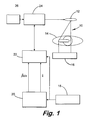
- Fig. 1 shows a schematic diagram of a medical radiological x-ray system 10 according to the present invention.
- the present invention is described with reference to a medical radiological system, it can be used in other applications that use x-ray imaging systems such as nondestructive testing and veterinary radiological x-ray systems.
- an x-ray tube 12 irradiates a particular part of a patient 14 with a beam of x-rays.
- the x-rays pass through the patient, exposing a photographic film stored in a cassette 16.
- the photographic film is generally comprised of a sheet of translucent supporting material coated on one or both sides with a photosensitized emulsion.
- a sensor 18 such as an ion chamber is placed behind the cassette 16 to monitor the amount of x-rays passing through the film.
- the sensor 18 outputs the amount of photons passing through the film to a signal processing unit 20 and an exposure control unit 22.
- the signal processing unit 20 amplifies and integrates the amount of photons to produce a summation value represented by ⁇ dx and ⁇ .
- the exposure control unit 22, such as a processor, receives the summation value from the signal processing unit 20 and the output from the sensor 18 and uses the techniques described below in further detail to detect and correct erroneous exposures.
- the exposure control unit 22 then instructs an x-ray generator controller 24 powered by a power source 26 to either change the amount of voltage and current being provided to the x-ray tube 12 or when to stop providing the present amount of voltage and current being sent to the x-ray tube.
- the amount of x-rays sent from the x-ray tube 12 is controlled by the x-ray generator controller 24.
- X-ray imaging settings are selected by a radiologist or a x-ray technician and are based on exposure guide tables. Prior to imaging a patient, the radiologist or x-ray technician chooses the anatomic view to be imaged and estimates the size of the patient. The exposure guide tables are then examined and used to provide a value for the beam quality, kVp, and the amount of x-rays, mAs, for the desired anatomical view and approximate patient size.
- An example of an exposure guide table is shown in Fig. 2.
- the exposure guide table suggests that 75 kVp and 3 mAs be used as values for the beam quality and the amount of x-rays, respectively.
- Fig. 3a is graph showing the relationship between total exposure and time with the suggested kV p and mAs values used as cutoffs.
- the graph shows how film exposure increases linearly during an ideal x-ray procedure as shown by an optimum trajectory line 28. As the time increases so does the exposure of x-rays. Eventually, the x-ray tube 12 is shut off when the exposure reaches the suggested kVp value as shown by line 30.
- the graph shows a mAs value suggested from an exposure guide table which is used for exposure duration as shown by line 32. In the ideal case, all three of these lines intersect at a single point 34, indicating that the exposure is on track.
- Fig. 3b is a graph showing the relationship between exposure rate, which is the derivative of total exposure and time. If there is an error in selecting the anatomic view or estimating the patient size or a misalignment of the sensor, then these lines will no longer intersect at a single point. Any one of these errors will cause the film to be either overexposed or underexposed and may cause overexposure to the patient. In order to account for these errors, the erroneous exposure needs to be detected and corrected to provide the proper amount of tube voltage.
- the present invention is able to detect either an overexposed or an underexposed condition by tracking the total exposure rate of the patient and comparing it to a predicted exposure rate.
- Fig. 4 describes the series of steps performed by the exposure control unit 22 for detecting erroneous exposures.
- the operation is initiated at 40 where the radiologist or x-ray technician selects the anatomic view to be imaged and estimates the size of the patient.
- An illustrative list of possible anatomic views for different parts of the body is shown in Fig. 2.
- an exposure guide table such as the one in Fig. 2 is used at 42 to suggest a value for the beam quality, kV, and the amount of x-rays, mAs.
- the exposure guide table would suggest that 55 kVp and 2.5 mAs be used as values for the beam quality and the amount of x-rays, respectively.
- the film threshold i.e., brightness
- the exposure time is determined from the suggested mAs value and the current being provided by the x-ray generator controller 24.
- the predicted exposure rate is determined at 46 by dividing the brightness by the exposure time.
- the patient is then exposed to an x-ray beam at 48 having the suggested x-ray beam quality (kV) and x-ray quantity (mAs) values.
- the actual exposure rate to the patient is then measured at 50.
- the actual exposure rate is then integrated at 52 to determine the total exposure, which is compared to the predicted exposure rate at 54.
- the compared value is then used to determine if the exposure of the patient is within a predetermined tolerance. If the compared value is not within the predetermined tolerance at 56, then the exposure is stopped at 58 and the operator is signaled to recheck the x-ray settings. However, if the compared value is within the predetermined tolerance at 56, then the exposure is continued at 60 and the total exposure is compared to a predetermined threshold value at 62. If the total exposure is less than the predetermined threshold, then the exposure continues until the total exposure is equal to the predetermined threshold. Once the total exposure is equal to the predetermined threshold then the exposure is stopped at 58 and an image on the film is recorded.
- the present invention is able to correct these exposures by quickly suggesting a new voltage value (kV) and adjusting the x-ray generator 24 by the suggested amount.
- Fig. 5 sets forth the operation performed by the exposure control unit 22 to correct erroneous exposures.
- the radiologist or x-ray technician selects the anatomic view to be imaged and estimates the size of the patient at 64. After the anatomic view and patient size has been selected an exposure guide table such as the one in Fig. 2 is used at 66 to suggest a value for the beam quality (kV) and the amount of x-rays (mAs).
- the film threshold i.e., brightness
- the predicted exposure rate is determined at 70 by dividing the brightness by the exposure time.
- the patient is then exposed to an x-ray beam at 72 having the suggested x-ray beam quality (kV) and x-ray quantity (mAs) values.
- the actual exposure rate to the patient is then measured at 74.
- the actual exposure rate is then integrated at 76 to determine the total exposure which is compared to the predicted exposure rate at 78.
- the compared value is then used to determine if the exposure of the patient is within a predetermined tolerance. If the compared value is not within the predetermined tolerance at 80, then the exposure is continued at 82. However, if the compared value is within the predetermined tolerance at 80, then the actual patient size is computed at 84.
- the patient size is computed by using the brightness function for the imaging system which is defined as the function of kV, mAs, and the patient size. Thus, if the brightness, kV, and mAs values are known, then the patient size can be computed. Since the brightness rate (i.e., exposure rate), kV, and mAs values are known, then the patient size can be computed Then the computed actual patient-size is used to determine a new x-ray beam quality value (kV) at 86. The new x-ray beam quality value (kV) is determined by using a look-up table containing suggested values for adjusting kV given the initial kV, mAs, and patient size values.
- the brightness function for the imaging system which is defined as the function of kV, mAs, and the patient size.
- the look-up table will generate a suggested new kV value.
- the exposure of the patient is adjusted according to the new x-ray beam quality value (kV) and the exposure is continued at 88.
- the total exposure to the patient is then determined at 90.
- the total exposure is then compared to a predetermined threshold value at 92. If the total exposure is less than the predetermined threshold, then the exposure is continued at 88. Steps 88-92 continue until the total exposure equals the predetermined threshold. Once the total exposure equals the predetermined threshold then the exposure is continued at 82 and the image on the film is later recorded.
- a second embodiment of correcting erroneous exposures is set forth in the flow chart of Fig. 6.
- the operation is initiated at 94 where the radiologist or x-ray technician selects the anatomic view to be imaged and estimates the size of the patient.
- An illustrative list of possible anatomic views for different parts of the body is shown in Fig. 2.
- an exposure guide table such as the one in Fig. 2 is used at 96 to suggest a value for the beam quality (kV) and the amount of x-rays (mAs).
- the film threshold i.e., brightness
- exposure time is determined.
- the predicted exposure rate is determined at 100 by dividing the brightness by the exposure time.
- the patient is then exposed at 102 to an x-ray beam having the suggested x-ray beam quality (kV) and x-ray quantity (mAs) values.
- the actual exposure rate exposed to the patient is then measured at 104.
- the actual exposure rate is then integrated at 106 to determine total exposure which is compared to the predicted exposure rate at 108.
- the compared value is then used to determine if the patient is being exposed at a correct level. If the compared value is satisfactory at 110, then the exposure is completed at 112. However, if the compared value is not satisfactory at 110, then the exposure is adjusted at 114 and a new x-ray beam quality value (kV) is suggested at 116.
- the new x-ray beam quality value (kV) is suggested by using a look-up table as previously described to suggest a new x-ray quality value from the anatomical view and the predicted and actual exposure rates. More specifically, the selected anatomical view is used to index a row of the table. Each row represents a pairwise association from exposure rate to x-ray quality (kV). The actual exposure rate is located in the table, giving a suggested x-ray quality value, then interpolation may be used if a more precise solution is needed. In addition to using a look-up table it is within the scope of the present invention to use other mechanisms such as fuzzy logic and empirical curve fitting. After locating a new x-ray quality value, the exposure is continued at 118.
- the total exposure to the patient is then determined at 120.
- the total exposure is then compared to a predetermined threshold value at 122. If the total exposure is less than the predetermined threshold, then the exposure is continued at 118. Steps 118-122 continue until the total exposure equals the predetermined threshold. Once the total exposure equals the predetermined threshold then the exposure is completed at 112 and the image on the film is later recorded.
- the present invention has disclosed a procedure for detecting erroneous exposures that arise because of operator errors such as misalignment or setting errors. By detecting some of the incorrect exposures before completion, and aborting them before completed, patients will receive fewer x-rays, operating costs will decrease, while patient care will improve due to faster diagnosis, less repositioning, and increased availability.
- the present invention has disclosed a mechanism for improving image quality by correcting erroneous exposures. In particular, the present invention improves image quality by controlling the exposure rate as well as the total exposure through modifications to the x-ray beam quality and x-ray beam quantity values.
Abstract
Description
- The present invention relates generally to radiography, and more particularly to improving image quality by detecting and correcting erroneous exposures generated during x-ray imaging.
- In x-ray imaging systems such as a medical system, an x-ray tube is used to irradiate a patient with a beam of x-rays. The x-rays pass through a patient, exposing a photographic film stored in a cassette. The photographic film is generally comprised of a sheet of translucent supporting material coated on one or both sides with a photosensitized emulsion. The photosensitized emulsion is activated by exposure to photons of different wavelengths within the electromagnetic spectrum, including the visible light band and the x-ray band. Activation of the photosensitized emulsion creates a latent image on the emulsion. The latent image appears on the film as the relative darkening of the emulsion proportional to the amount of exposure. A part of the body interposed between the beam of x-rays and the film absorbs the x-rays in variable degrees depending on the internal composition of the part being x-rayed. More specifically, x-ray transmission through the part is affected by its thickness and material composition, as well as the quality of the x-ray beam striking the object. High energy x-rays penetrate further through the part, while low energy x-rays are easily absorbed. After the latent image has been created, it is then developed by bringing a developer material in contact with the image. Developing the latent image makes it visible and allows a radiologist to make a diagnosis based on the image.
- For each exposure that is taken, the radiologist or x-ray technician selects both the quality of the x-ray beam and the amount of x-rays to be generated. In particular, the quality of the x-ray beam is selected by varying voltage and filtration, while the amount of x-rays generated is selected by varying current and duration of the exposure. Both the quality of the x-ray beam and the amount of x-rays generated has a direct influence on the quality of the x-ray image, which in turn effects the accuracy of the diagnosis made by the radiologist.
- Currently, there are two procedures that assure exposure during medical x-ray imaging. The first procedure is an automated method of selecting x-ray settings based on exposure guide tables. In this procedure, the operator chooses the anatomic view of how a patient's body part is to be imaged and the estimated size of the patient. The exposure guide tables are then used to provide a value for the beam quality and the amount of x-rays for the desired anatomical view and approximate patient size. Once the settings are made, no further changes are made and the image is taken at these settings. However, if there was an error in the settings, placement, etc., then there is no way to detect it until the film has been developed. Another problem with the exposure guide tables is that these tables give suggested settings only for patients of approximately average composition. Excessively thin, muscular, and obese patients fall outside the range of these tables. The second procedure, known as automatic exposure control, utilizes a sensor such as an ion chamber placed behind the image plane to monitor the amount of x-rays passing through the film. When a sufficient amount of x-rays have passed through the sensor to achieve an acceptable film density, the exposure is terminated. While the automatic exposure control procedure does control the film density over the sensor quite well, it does not change the quality of the x-rays being absorbed by the patient. Another problem is that this procedure does not compensate for grossly incorrect setting errors. For example, it is fairly common to misplace the sensor underneath the spinal column or outside the rib cage on obese patients during a lung exposure. Both locations will have vastly different x-ray absorption characteristics than if placed under the lung.
- Since both of the above exposure selection procedures are incapable of detecting incorrect images until after the exposure is taken and the film is developed, exposures may have to be repeated several times. In addition, both exposure selection procedures are incapable of selecting beam quality based on measurements from the actual patient and anatomy being imaged, which may necessitate additional exposures. More exposures results in decreased productivity, more costs, and more dosage to the patient. Therefore, there is a need for detecting and correcting incorrect exposures in order to improve image quality. By improving image quality on the initial exposure, the need for retakes will decrease, which will increase productivity, patient care and decrease cost and the amount of x-ray dosage to the patient.
- Therefore, the present invention seeks to primarily to provide a method and system for detecting and correcting erroneous exposures during medical x-ray imaging.
- The present invention also seeks to improve image quality generated during medical x-ray imaging.
- According to a first aspect of the invention, there is provided a method for detecting and/or correcting an erroneous exposure generated during x-ray imaging of a patient, comprising the steps of: electing x-ray imaging settings for imaging the patient; generating x-ray beam quality and x-ray quantity values from the selected x-ray imaging settings; predicting an exposure rate from the x-ray beam quality and x-ray quantity values; exposing the patient to an x-ray beam having the generated x-ray beam quality and x-ray quantity values; determining a total exposure to the patient; comparing the total exposure to the predicted exposure rate, resulting in a compared value; and using the compared value to determine if the patient is being exposed at an erroneous level.
- According to a second aspect of the invention, there is provided a system for detecting and/or correcting an erroneous exposure generated during x-ray imaging of a patient the system comprising: means for selecting x-ray imaging settings for imaging the patient; means for generating x-ray beam quality and x-ray quantity values from the selected x-ray imaging settings; means for predicting an exposure rate from the x-ray beam quality and x-ray quantity values; an x-ray tube for exposing the patient to an x-ray beam having the generated x-ray beam quality and x-ray quantity values; a sensor for measuring an actual exposure rate of the patient; means for determining total exposure from the actual exposure rate; means for comparing the total exposure to the predicted exposure rate, resulting in a compared value; and means for using the compared value to determine if the patient is being exposed at an erroneous level.
- Thus, generally there is provided a method and a system for detecting an erroneous exposure generated during x-ray imaging of a patient. In the present invention, x-ray imaging settings for imaging the patient are selected. X-ray beam quality and x-ray quantity values are generated from the selected x-ray imaging settings. An exposure rate is predicted from the x-ray beam quality and x-ray quantity values. The patient is then exposed to an x-ray beam having the generated x-ray beam quality and x-ray quantity values. The total exposure rate is determined and then compared to the predicted exposure rate. The comparison is then used to determine if the patient is being exposed at an erroneous level.
- The total exposure may be compared with a predetermined threshold and the exposure may be continued until the total exposure is less than the predetermined threshold level.
- In accordance with another embodiment of the present invention, there is provided a method and system for correcting an erroneous exposure generated during x-ray imaging of a patient. In the present invention, x-ray imaging settings for imaging the patient are selected. X-ray beam quality and x-ray quantity values are generated from the selected x-ray imaging settings. An exposure rate is predicted from the x-ray beam quality and x-ray quantity values. The patient is then exposed to an x-ray beam having the generated x-ray beam quality and x-ray quantity values. The total exposure rate is determined and then compared to the predicted exposure rate. The comparison is then used to determine if the patient is being exposed at a correct level. If the exposure is' at a correct level, then the exposure is completed. However, if the exposure is not at a correct level, then the exposure is adjusted to a new exposure level.
- Thus the exposure of the patient may be adjusted to a new x-ray quality value. The new -ray quality may use a table having new x-ray quality values corresponding to the predicted and total exposure rates. The total exposure may be used to determine the actual size of the patient and the new x-ray value may be determined from the actual size of the patient.
- The invention will now be described in greater detail, by way of example, with reference to the drawings in which:
- Fig. 1 shows a schematic diagram of a medical radiological x-ray system according to the present invention;
- Fig. 2 is an example of an exposure guide table according to the present invention;
- Figs. 3a-3b are graphs showing the relationship between total exposure and time and exposure rate and time, respectively;
- Fig. 4 is a flow chart describing the operation of detecting erroneous exposures according to the present invention;
- Fig. 5 is a flow chart describing the operation of correcting erroneous exposures according to the present invention; and
- Fig. 6 is a flow chart describing the operation of correcting erroneous exposures according to another embodiment.
- Fig. 1 shows a schematic diagram of a medical
radiological x-ray system 10 according to the present invention. Although the present invention is described with reference to a medical radiological system, it can be used in other applications that use x-ray imaging systems such as nondestructive testing and veterinary radiological x-ray systems. In the medicalradiological x-ray system 10, anx-ray tube 12 irradiates a particular part of a patient 14 with a beam of x-rays. The x-rays pass through the patient, exposing a photographic film stored in acassette 16. The photographic film is generally comprised of a sheet of translucent supporting material coated on one or both sides with a photosensitized emulsion. Asensor 18 such as an ion chamber is placed behind thecassette 16 to monitor the amount of x-rays passing through the film. Thesensor 18 outputs the amount of photons passing through the film to asignal processing unit 20 and anexposure control unit 22. Thesignal processing unit 20 amplifies and integrates the amount of photons to produce a summation value represented by ∫ẋdx and ẋ. Theexposure control unit 22, such as a processor, receives the summation value from thesignal processing unit 20 and the output from thesensor 18 and uses the techniques described below in further detail to detect and correct erroneous exposures. Theexposure control unit 22 then instructs anx-ray generator controller 24 powered by apower source 26 to either change the amount of voltage and current being provided to thex-ray tube 12 or when to stop providing the present amount of voltage and current being sent to the x-ray tube. - In the present invention, the amount of x-rays sent from the
x-ray tube 12 is controlled by thex-ray generator controller 24. X-ray imaging settings are selected by a radiologist or a x-ray technician and are based on exposure guide tables. Prior to imaging a patient, the radiologist or x-ray technician chooses the anatomic view to be imaged and estimates the size of the patient. The exposure guide tables are then examined and used to provide a value for the beam quality, kVp, and the amount of x-rays, mAs, for the desired anatomical view and approximate patient size. An example of an exposure guide table is shown in Fig. 2. For example, if a chest is to be x-rayed, and the radiologist or x-ray technician chooses the upright anterior/posterior (AP) anatomic view with a screen and determines that the patient is of average size, then the exposure guide table suggests that 75 kVp and 3 mAs be used as values for the beam quality and the amount of x-rays, respectively. - Fig. 3a is graph showing the relationship between total exposure and time with the suggested kVp and mAs values used as cutoffs. The graph shows how film exposure increases linearly during an ideal x-ray procedure as shown by an
optimum trajectory line 28. As the time increases so does the exposure of x-rays. Eventually, thex-ray tube 12 is shut off when the exposure reaches the suggested kVp value as shown byline 30. In addition, the graph shows a mAs value suggested from an exposure guide table which is used for exposure duration as shown byline 32. In the ideal case, all three of these lines intersect at asingle point 34, indicating that the exposure is on track. Encompassing thepoint 34 aretolerance lines - Typically, most of the erroneous exposures are due to operator error. For example, the radiologist or the x-ray technician may misread the exposure guide tables or may incorrectly estimate the patient size. Another common source of error is that there may be a misalignment between the x-ray source, the patient, the cassette, and the sensor. As mentioned above, these errors will lead to either the underexposure or the overexposure of the x-ray film, necessitating that another exposure be made at a later date. These erroneous exposure not only decrease productivity and increase operating costs, but also hinders patient care by increasing diagnosis time, increases the total x-ray dose to the patient, and increases patient handling.
- The present invention is able to detect either an overexposed or an underexposed condition by tracking the total exposure rate of the patient and comparing it to a predicted exposure rate. Fig. 4 describes the series of steps performed by the
exposure control unit 22 for detecting erroneous exposures. In this embodiment, the operation is initiated at 40 where the radiologist or x-ray technician selects the anatomic view to be imaged and estimates the size of the patient. An illustrative list of possible anatomic views for different parts of the body is shown in Fig. 2. After the anatomic view and patient size has been selected an exposure guide table such as the one in Fig. 2 is used at 42 to suggest a value for the beam quality, kV, and the amount of x-rays, mAs. For example, if an ankle is to be x-rayed, and the radiologist or x-ray technician chooses the anterior/posterior (AP) anatomic view with a screen and determines that the patient is of average size, then the exposure guide table would suggest that 55 kVp and 2.5 mAs be used as values for the beam quality and the amount of x-rays, respectively. At 44, the film threshold (i.e., brightness) and exposure time is determined. In particular, the brightness value is determined from the suggested kVp value and the film type, while the exposure time is determined from the suggested mAs value and the current being provided by thex-ray generator controller 24. Next, the predicted exposure rate is determined at 46 by dividing the brightness by the exposure time. The patient is then exposed to an x-ray beam at 48 having the suggested x-ray beam quality (kV) and x-ray quantity (mAs) values. The actual exposure rate to the patient is then measured at 50. The actual exposure rate is then integrated at 52 to determine the total exposure, which is compared to the predicted exposure rate at 54. The compared value is then used to determine if the exposure of the patient is within a predetermined tolerance. If the compared value is not within the predetermined tolerance at 56, then the exposure is stopped at 58 and the operator is signaled to recheck the x-ray settings. However, if the compared value is within the predetermined tolerance at 56, then the exposure is continued at 60 and the total exposure is compared to a predetermined threshold value at 62. If the total exposure is less than the predetermined threshold, then the exposure continues until the total exposure is equal to the predetermined threshold. Once the total exposure is equal to the predetermined threshold then the exposure is stopped at 58 and an image on the film is recorded. - In addition to detecting erroneous exposures, the present invention is able to correct these exposures by quickly suggesting a new voltage value (kV) and adjusting the
x-ray generator 24 by the suggested amount. Fig. 5 sets forth the operation performed by theexposure control unit 22 to correct erroneous exposures. In this embodiment, the radiologist or x-ray technician selects the anatomic view to be imaged and estimates the size of the patient at 64. After the anatomic view and patient size has been selected an exposure guide table such as the one in Fig. 2 is used at 66 to suggest a value for the beam quality (kV) and the amount of x-rays (mAs). At 68, the film threshold (i.e., brightness) and exposure time is determined. Next, the predicted exposure rate is determined at 70 by dividing the brightness by the exposure time. The patient is then exposed to an x-ray beam at 72 having the suggested x-ray beam quality (kV) and x-ray quantity (mAs) values. The actual exposure rate to the patient is then measured at 74. The actual exposure rate is then integrated at 76 to determine the total exposure which is compared to the predicted exposure rate at 78. The compared value is then used to determine if the exposure of the patient is within a predetermined tolerance. If the compared value is not within the predetermined tolerance at 80, then the exposure is continued at 82. However, if the compared value is within the predetermined tolerance at 80, then the actual patient size is computed at 84. The patient size is computed by using the brightness function for the imaging system which is defined as the function of kV, mAs, and the patient size. Thus, if the brightness, kV, and mAs values are known, then the patient size can be computed. Since the brightness rate (i.e., exposure rate), kV, and mAs values are known, then the patient size can be computed Then the computed actual patient-size is used to determine a new x-ray beam quality value (kV) at 86. The new x-ray beam quality value (kV) is determined by using a look-up table containing suggested values for adjusting kV given the initial kV, mAs, and patient size values. Thus, if the initial kV, mAs, and actual patient size are known, then the look-up table will generate a suggested new kV value. The exposure of the patient is adjusted according to the new x-ray beam quality value (kV) and the exposure is continued at 88. The total exposure to the patient is then determined at 90. The total exposure is then compared to a predetermined threshold value at 92. If the total exposure is less than the predetermined threshold, then the exposure is continued at 88. Steps 88-92 continue until the total exposure equals the predetermined threshold. Once the total exposure equals the predetermined threshold then the exposure is continued at 82 and the image on the film is later recorded. In an alternative embodiment, instead of continuing exposure at 88 after the decision at 92, it is possible to go to either the decision at 80 or to compute the patient size at 84. - A second embodiment of correcting erroneous exposures is set forth in the flow chart of Fig. 6. In the second embodiment, the operation is initiated at 94 where the radiologist or x-ray technician selects the anatomic view to be imaged and estimates the size of the patient. An illustrative list of possible anatomic views for different parts of the body is shown in Fig. 2. After the anatomic view and patient size has been selected an exposure guide table such as the one in Fig. 2 is used at 96 to suggest a value for the beam quality (kV) and the amount of x-rays (mAs). At 98, the film threshold (i.e., brightness) and exposure time is determined. Next, the predicted exposure rate is determined at 100 by dividing the brightness by the exposure time. The patient is then exposed at 102 to an x-ray beam having the suggested x-ray beam quality (kV) and x-ray quantity (mAs) values. The actual exposure rate exposed to the patient is then measured at 104. The actual exposure rate is then integrated at 106 to determine total exposure which is compared to the predicted exposure rate at 108. The compared value is then used to determine if the patient is being exposed at a correct level. If the compared value is satisfactory at 110, then the exposure is completed at 112. However, if the compared value is not satisfactory at 110, then the exposure is adjusted at 114 and a new x-ray beam quality value (kV) is suggested at 116. The new x-ray beam quality value (kV) is suggested by using a look-up table as previously described to suggest a new x-ray quality value from the anatomical view and the predicted and actual exposure rates. More specifically, the selected anatomical view is used to index a row of the table. Each row represents a pairwise association from exposure rate to x-ray quality (kV). The actual exposure rate is located in the table, giving a suggested x-ray quality value, then interpolation may be used if a more precise solution is needed. In addition to using a look-up table it is within the scope of the present invention to use other mechanisms such as fuzzy logic and empirical curve fitting. After locating a new x-ray quality value, the exposure is continued at 118. The total exposure to the patient is then determined at 120. The total exposure is then compared to a predetermined threshold value at 122. If the total exposure is less than the predetermined threshold, then the exposure is continued at 118. Steps 118-122 continue until the total exposure equals the predetermined threshold. Once the total exposure equals the predetermined threshold then the exposure is completed at 112 and the image on the film is later recorded.
- The present invention has disclosed a procedure for detecting erroneous exposures that arise because of operator errors such as misalignment or setting errors. By detecting some of the incorrect exposures before completion, and aborting them before completed, patients will receive fewer x-rays, operating costs will decrease, while patient care will improve due to faster diagnosis, less repositioning, and increased availability. In addition to detecting erroneous exposures, the present invention has disclosed a mechanism for improving image quality by correcting erroneous exposures. In particular, the present invention improves image quality by controlling the exposure rate as well as the total exposure through modifications to the x-ray beam quality and x-ray beam quantity values.
- It is therefore apparent that there has been provided in accordance with the present invention, a method and system for detecting and correcting an erroneous exposure generated during x-ray imaging that fully satisfy the aims and advantages and objectives hereinbefore set forth. The present invention has been described with reference to several embodiments, however, it will be appreciated that variations and modifications can be effected by a person of ordinary skill in the art without departing from the scope of the invention. For example, it is within the scope of the present invention to use the invention to improve exposures in digital x-ray systems.
Claims (10)
- A method for detecting and/or correcting an erroneous exposure generated during x-ray imaging of a patient, comprising the steps of:selecting x-ray imaging settings for imaging the patient;generating x-ray beam quality and x-ray quantity values from the selected x-ray imaging settings;predicting an exposure rate from the x-ray beam quality and x-ray quantity values;exposing the patient to an x-ray beam having the generated x-ray beam quality and x-ray quantity values;determining a total exposure to the patient;comparing the total exposure to the predicted exposure rate, resulting in a compared value; andusing the compared value to determine if the patient is being exposed at an erroneous level.
- A method according to claim 1, wherein the selected x-ray imaging settings comprise an anatomic view and an approximation of size of the patient.
- A method according to claim 1 or 2, further comprising the step of stopping the exposure if the patient is being exposed at an erroneous level.
- A method according to claim 1, 2 or 3, wherein the step of using the compared value to determine if the patient is being exposed at an erroneous level comprises comparing the compared value to a predetermined tolerance.
- A method according to any one of claims 1 to 4, further comprising the step of suggesting a new x-ray quality value if the patient is being exposed at an incorrect level.
- A system for detecting and/or correcting an erroneous exposure generated during x-ray imaging of a patient the system comprising:means for selecting x-ray imaging settings for imaging the patient;means for generating x-ray beam quality and x-ray quantity values from the selected x-ray imaging settings;means for predicting an exposure rate from the x-ray beam quality and x-ray quantity values;an x-ray tube for exposing the patient to an x-ray beam having the generated x-ray beam quality and x-ray quantity values;a sensor for measuring an actual exposure rate of the patient;means for determining total exposure from the actual exposure rate;means for comparing the total exposure to the predicted exposure rate, resulting in a compared value; andmeans for using the compared value to determine if the patient is being exposed at an erroneous level.
- A system according to claim 6, wherein the selected x-ray imaging settings comprise an anatomic view and an approximation of size of the patient.
- A system according to claim 6 to 7, further comprising means for stopping the exposure if the patient is being exposed at an erroneous level.
- A system according to claim 6, 7 or 8 wherein the using means comprises means for comparing the compared value to a predetermined tolerance.
- A system according to any one of claims 6 to 10, further comprising means for suggesting a new x-ray quality value if the patient is being exposed at an incorrect level.
Applications Claiming Priority (2)
| Application Number | Priority Date | Filing Date | Title |
|---|---|---|---|
| US08/650,677 US5694449A (en) | 1996-05-20 | 1996-05-20 | Method and system for detecting and correcting erroneous exposures generated during x-ray imaging |
| US650677 | 1996-05-20 |
Publications (2)
| Publication Number | Publication Date |
|---|---|
| EP0809422A1 true EP0809422A1 (en) | 1997-11-26 |
| EP0809422B1 EP0809422B1 (en) | 2004-01-02 |
Family
ID=24609844
Family Applications (1)
| Application Number | Title | Priority Date | Filing Date |
|---|---|---|---|
| EP97303383A Expired - Lifetime EP0809422B1 (en) | 1996-05-20 | 1997-05-19 | Method and system for detecting and correcting erroneous exposures generated during x-ray imaging |
Country Status (4)
| Country | Link |
|---|---|
| US (1) | US5694449A (en) |
| EP (1) | EP0809422B1 (en) |
| JP (1) | JPH1043170A (en) |
| DE (1) | DE69727007T2 (en) |
Cited By (4)
| Publication number | Priority date | Publication date | Assignee | Title |
|---|---|---|---|---|
| WO2000067006A2 (en) * | 1999-04-28 | 2000-11-09 | Siemens Aktiengesellschaft | Computer tomography |
| FR2796516A1 (en) * | 1999-07-12 | 2001-01-19 | Gen Electric | Optimization of image quality in an X-ray system using model-based optimization for clinical applications |
| EP1069807A3 (en) * | 1999-07-12 | 2004-01-14 | General Electric Company | Exposure management and control system and method |
| EP1549115A3 (en) * | 2003-12-26 | 2007-11-07 | GE Medical Systems Global Technology Company LLC | Exposure calculation method and radiography system |
Families Citing this family (16)
| Publication number | Priority date | Publication date | Assignee | Title |
|---|---|---|---|---|
| JP4424758B2 (en) * | 1997-04-24 | 2010-03-03 | コーニンクレッカ フィリップス エレクトロニクス エヌ ヴィ | X-ray inspection apparatus including an exposure control system |
| US6422751B1 (en) * | 1998-08-07 | 2002-07-23 | General Electric Company | Method and system for prediction of exposure and dose area product for radiographic x-ray imaging |
| US6330299B1 (en) | 2000-06-10 | 2001-12-11 | Ge Medical Systems Global Technology Company, Llc | System and method for determining dose area product in an X-ray imaging system |
| US20050031082A1 (en) * | 2001-07-24 | 2005-02-10 | Haaga John R. | X-ray dose control based on patient size |
| US6687327B2 (en) * | 2001-11-15 | 2004-02-03 | Ge Medical Systems Global Technology Co., Llc | System and method of medical imaging having task and/or patient size dependent processing |
| US20040179651A1 (en) * | 2003-03-12 | 2004-09-16 | Canon Kabushiki Kaisha | Automated quality control for digital radiography |
| JP2006529052A (en) * | 2003-05-16 | 2006-12-28 | コーニンクレッカ フィリップス エレクトロニクス エヌ ヴィ | Method and apparatus for exposing an X-ray image |
| US20050053199A1 (en) * | 2003-09-04 | 2005-03-10 | Miles Dale A. | Portable x-ray device and method |
| US7006600B1 (en) * | 2004-01-15 | 2006-02-28 | Progeny, Inc. | Integrated digital dental x-ray system |
| WO2010122917A1 (en) * | 2009-04-24 | 2010-10-28 | 株式会社 日立メディコ | X-ray diagnostic device and method for controlling x-ray diaphragm |
| CN101926650B (en) * | 2009-06-26 | 2014-04-30 | Ge医疗系统环球技术有限公司 | Device and method for calculating actual skin entrance dose rate and X-ray machine |
| WO2011024608A1 (en) * | 2009-08-28 | 2011-03-03 | 株式会社 日立メディコ | Mobile x-ray device, control method for x-ray irradiation, and control program for mobile x-ray device |
| JP5689734B2 (en) * | 2011-04-20 | 2015-03-25 | 株式会社東芝 | X-ray CT system |
| JP6309250B2 (en) | 2012-11-14 | 2018-04-11 | キヤノンメディカルシステムズ株式会社 | X-ray CT apparatus, control program for X-ray CT apparatus |
| DE102016123846A1 (en) * | 2016-12-08 | 2018-06-14 | Visus Health It Gmbh | Detector tape for X-ray film |
| EP3420909A1 (en) * | 2017-06-27 | 2019-01-02 | Koninklijke Philips N.V. | X-ray misuse protection |
Citations (5)
| Publication number | Priority date | Publication date | Assignee | Title |
|---|---|---|---|---|
| US4845771A (en) * | 1987-06-29 | 1989-07-04 | Picker International, Inc. | Exposure monitoring in radiation imaging |
| US5301220A (en) * | 1992-09-03 | 1994-04-05 | Picker International, Inc. | Multi-mode acquisition x-ray imaging method and apparatus |
| WO1995027922A1 (en) * | 1994-04-07 | 1995-10-19 | Stig Svensson | Apparatus for making x-ray images |
| DE19525376A1 (en) * | 1994-07-18 | 1996-01-25 | Instrumentarium Corp | X ray imaging apparatus illumination value measurement |
| WO1996020579A1 (en) * | 1994-12-23 | 1996-07-04 | Philips Electronics N.V. | X-ray examination apparatus comprising an exposure control circuit |
Family Cites Families (3)
| Publication number | Priority date | Publication date | Assignee | Title |
|---|---|---|---|---|
| US4119856A (en) * | 1973-09-07 | 1978-10-10 | Siemens Aktiengesellschaft | X-ray diagnostic apparatus for producing series exposures |
| DE2546948C3 (en) * | 1975-10-20 | 1980-05-29 | Siemens Ag, 1000 Berlin Und 8000 Muenchen | X-ray diagnostic system for X-ray recordings with means for organ-programmed setting of the recording values and with an automatic X-ray exposure device |
| US5319696A (en) * | 1992-10-05 | 1994-06-07 | General Electric Company | X-ray dose reduction in pulsed systems by adaptive X-ray pulse adjustment |
-
1996
- 1996-05-20 US US08/650,677 patent/US5694449A/en not_active Expired - Fee Related
-
1997
- 1997-05-16 JP JP9126900A patent/JPH1043170A/en not_active Withdrawn
- 1997-05-19 EP EP97303383A patent/EP0809422B1/en not_active Expired - Lifetime
- 1997-05-19 DE DE69727007T patent/DE69727007T2/en not_active Expired - Fee Related
Patent Citations (5)
| Publication number | Priority date | Publication date | Assignee | Title |
|---|---|---|---|---|
| US4845771A (en) * | 1987-06-29 | 1989-07-04 | Picker International, Inc. | Exposure monitoring in radiation imaging |
| US5301220A (en) * | 1992-09-03 | 1994-04-05 | Picker International, Inc. | Multi-mode acquisition x-ray imaging method and apparatus |
| WO1995027922A1 (en) * | 1994-04-07 | 1995-10-19 | Stig Svensson | Apparatus for making x-ray images |
| DE19525376A1 (en) * | 1994-07-18 | 1996-01-25 | Instrumentarium Corp | X ray imaging apparatus illumination value measurement |
| WO1996020579A1 (en) * | 1994-12-23 | 1996-07-04 | Philips Electronics N.V. | X-ray examination apparatus comprising an exposure control circuit |
Cited By (6)
| Publication number | Priority date | Publication date | Assignee | Title |
|---|---|---|---|---|
| WO2000067006A2 (en) * | 1999-04-28 | 2000-11-09 | Siemens Aktiengesellschaft | Computer tomography |
| WO2000067006A3 (en) * | 1999-04-28 | 2001-04-19 | Siemens Ag | Computer tomography |
| US6870898B1 (en) | 1999-04-28 | 2005-03-22 | Siemens Aktiengesellschaft | Computed tomography apparatus with automatic parameter modification to prevent impermissible operating states |
| FR2796516A1 (en) * | 1999-07-12 | 2001-01-19 | Gen Electric | Optimization of image quality in an X-ray system using model-based optimization for clinical applications |
| EP1069807A3 (en) * | 1999-07-12 | 2004-01-14 | General Electric Company | Exposure management and control system and method |
| EP1549115A3 (en) * | 2003-12-26 | 2007-11-07 | GE Medical Systems Global Technology Company LLC | Exposure calculation method and radiography system |
Also Published As
| Publication number | Publication date |
|---|---|
| US5694449A (en) | 1997-12-02 |
| DE69727007T2 (en) | 2004-10-28 |
| JPH1043170A (en) | 1998-02-17 |
| DE69727007D1 (en) | 2004-02-05 |
| EP0809422B1 (en) | 2004-01-02 |
Similar Documents
| Publication | Publication Date | Title |
|---|---|---|
| EP0809422B1 (en) | Method and system for detecting and correcting erroneous exposures generated during x-ray imaging | |
| US7545915B2 (en) | Dose rate control in an X-ray system | |
| EP0942682B1 (en) | Adjustable computer tomography device | |
| RU2507619C2 (en) | Portable x-ray detector with grid sensing unit and x-ray imaging system for automatic exposure setting for portable x-ray detector | |
| US7116756B2 (en) | X-ray diagnostic apparatus with a body mass index calculator for controlling x-ray emissions | |
| USRE33634E (en) | Method and structure for optimizing radiographic quality by controlling X-ray tube voltage, current focal spot size and exposure time | |
| US4763343A (en) | Method and structure for optimizing radiographic quality by controlling X-ray tube voltage, current, focal spot size and exposure time | |
| EP1420618B1 (en) | X-Ray imaging apparatus | |
| US6459765B1 (en) | Automatic exposure control and optimization in digital x-ray radiography | |
| Gingold et al. | Contrast and dose with Mo-Mo, Mo-Rh, and Rh-Rh target-filter combinations in mammography. | |
| TW201817286A (en) | Automatic exposure control system for a digital X-ray imaging device and method thereof | |
| EP0291502B1 (en) | A method and device for controlling the x-radiation of an x-ray apparatus, in particular that of a mammographic apparatus | |
| EP1219147B1 (en) | Automatic exposure control for dental panoramic and cephalographic x-ray equipment | |
| GB2105032A (en) | X-ray generator incorporating automatic correction of a dose-determining exposure parameter | |
| EP0346530A1 (en) | Method and structure for optimizing radiographic quality by controlling X-ray tube voltage, current, focal spot size and exposure time | |
| US7110494B2 (en) | X-ray diagnostic apparatus with a patient weighting device associated with the patient positioning table | |
| Young et al. | Dose and image quality in mammography with an automatic beam quality system | |
| US4035649A (en) | X-ray generator for a tomography apparatus | |
| EP0777406A1 (en) | Automatic exposure metering system for radiology equipment used in mammography | |
| CN110269635B (en) | Method and device for calculating automatic exposure parameters of X-ray imaging equipment | |
| JP2001176692A (en) | X-ray photography device | |
| Eraso et al. | Clinical and in vitro film quality comparison of manual and automatic exposure control in panoramic radiography | |
| Busch et al. | Adaption of the Quality Criteria Concept to Digital Radiology | |
| Geise et al. | Routine quality control tests for film–screen mammographic systems with automatic exposure control | |
| Jenkins et al. | Automatic Exposure Control |
Legal Events
| Date | Code | Title | Description |
|---|---|---|---|
| PUAI | Public reference made under article 153(3) epc to a published international application that has entered the european phase |
Free format text: ORIGINAL CODE: 0009012 |
|
| AK | Designated contracting states |
Kind code of ref document: A1 Designated state(s): DE NL |
|
| 17P | Request for examination filed |
Effective date: 19980526 |
|
| 17Q | First examination report despatched |
Effective date: 20020125 |
|
| GRAH | Despatch of communication of intention to grant a patent |
Free format text: ORIGINAL CODE: EPIDOS IGRA |
|
| GRAS | Grant fee paid |
Free format text: ORIGINAL CODE: EPIDOSNIGR3 |
|
| GRAA | (expected) grant |
Free format text: ORIGINAL CODE: 0009210 |
|
| AK | Designated contracting states |
Kind code of ref document: B1 Designated state(s): DE NL |
|
| REF | Corresponds to: |
Ref document number: 69727007 Country of ref document: DE Date of ref document: 20040205 Kind code of ref document: P |
|
| PGFP | Annual fee paid to national office [announced via postgrant information from national office to epo] |
Ref country code: NL Payment date: 20040429 Year of fee payment: 8 |
|
| PGFP | Annual fee paid to national office [announced via postgrant information from national office to epo] |
Ref country code: DE Payment date: 20040630 Year of fee payment: 8 |
|
| PLBE | No opposition filed within time limit |
Free format text: ORIGINAL CODE: 0009261 |
|
| STAA | Information on the status of an ep patent application or granted ep patent |
Free format text: STATUS: NO OPPOSITION FILED WITHIN TIME LIMIT |
|
| 26N | No opposition filed |
Effective date: 20041005 |
|
| PG25 | Lapsed in a contracting state [announced via postgrant information from national office to epo] |
Ref country code: NL Free format text: LAPSE BECAUSE OF NON-PAYMENT OF DUE FEES Effective date: 20051201 Ref country code: DE Free format text: LAPSE BECAUSE OF NON-PAYMENT OF DUE FEES Effective date: 20051201 |
|
| NLV4 | Nl: lapsed or anulled due to non-payment of the annual fee |
Effective date: 20051201 |