EP0422762A2 - Method for assessing the sensitivity of HIV-1 to zidovudine and oligonucleotides therefore - Google Patents
Method for assessing the sensitivity of HIV-1 to zidovudine and oligonucleotides therefore Download PDFInfo
- Publication number
- EP0422762A2 EP0422762A2 EP90308721A EP90308721A EP0422762A2 EP 0422762 A2 EP0422762 A2 EP 0422762A2 EP 90308721 A EP90308721 A EP 90308721A EP 90308721 A EP90308721 A EP 90308721A EP 0422762 A2 EP0422762 A2 EP 0422762A2
- Authority
- EP
- European Patent Office
- Prior art keywords
- oligonucleotide
- hiv
- dna
- nucleic acid
- region
- Prior art date
- Legal status (The legal status is an assumption and is not a legal conclusion. Google has not performed a legal analysis and makes no representation as to the accuracy of the status listed.)
- Granted
Links
Images


Classifications
-
- C—CHEMISTRY; METALLURGY
- C12—BIOCHEMISTRY; BEER; SPIRITS; WINE; VINEGAR; MICROBIOLOGY; ENZYMOLOGY; MUTATION OR GENETIC ENGINEERING
- C12Q—MEASURING OR TESTING PROCESSES INVOLVING ENZYMES, NUCLEIC ACIDS OR MICROORGANISMS; COMPOSITIONS OR TEST PAPERS THEREFOR; PROCESSES OF PREPARING SUCH COMPOSITIONS; CONDITION-RESPONSIVE CONTROL IN MICROBIOLOGICAL OR ENZYMOLOGICAL PROCESSES
- C12Q1/00—Measuring or testing processes involving enzymes, nucleic acids or microorganisms; Compositions therefor; Processes of preparing such compositions
- C12Q1/70—Measuring or testing processes involving enzymes, nucleic acids or microorganisms; Compositions therefor; Processes of preparing such compositions involving virus or bacteriophage
- C12Q1/701—Specific hybridization probes
- C12Q1/702—Specific hybridization probes for retroviruses
- C12Q1/703—Viruses associated with AIDS
-
- C—CHEMISTRY; METALLURGY
- C07—ORGANIC CHEMISTRY
- C07K—PEPTIDES
- C07K14/00—Peptides having more than 20 amino acids; Gastrins; Somatostatins; Melanotropins; Derivatives thereof
- C07K14/005—Peptides having more than 20 amino acids; Gastrins; Somatostatins; Melanotropins; Derivatives thereof from viruses
-
- C—CHEMISTRY; METALLURGY
- C12—BIOCHEMISTRY; BEER; SPIRITS; WINE; VINEGAR; MICROBIOLOGY; ENZYMOLOGY; MUTATION OR GENETIC ENGINEERING
- C12Q—MEASURING OR TESTING PROCESSES INVOLVING ENZYMES, NUCLEIC ACIDS OR MICROORGANISMS; COMPOSITIONS OR TEST PAPERS THEREFOR; PROCESSES OF PREPARING SUCH COMPOSITIONS; CONDITION-RESPONSIVE CONTROL IN MICROBIOLOGICAL OR ENZYMOLOGICAL PROCESSES
- C12Q1/00—Measuring or testing processes involving enzymes, nucleic acids or microorganisms; Compositions therefor; Processes of preparing such compositions
- C12Q1/68—Measuring or testing processes involving enzymes, nucleic acids or microorganisms; Compositions therefor; Processes of preparing such compositions involving nucleic acids
- C12Q1/6813—Hybridisation assays
- C12Q1/6816—Hybridisation assays characterised by the detection means
-
- C—CHEMISTRY; METALLURGY
- C12—BIOCHEMISTRY; BEER; SPIRITS; WINE; VINEGAR; MICROBIOLOGY; ENZYMOLOGY; MUTATION OR GENETIC ENGINEERING
- C12N—MICROORGANISMS OR ENZYMES; COMPOSITIONS THEREOF; PROPAGATING, PRESERVING, OR MAINTAINING MICROORGANISMS; MUTATION OR GENETIC ENGINEERING; CULTURE MEDIA
- C12N2740/00—Reverse transcribing RNA viruses
- C12N2740/00011—Details
- C12N2740/10011—Retroviridae
- C12N2740/16011—Human Immunodeficiency Virus, HIV
- C12N2740/16211—Human Immunodeficiency Virus, HIV concerning HIV gagpol
- C12N2740/16222—New viral proteins or individual genes, new structural or functional aspects of known viral proteins or genes
-
- C—CHEMISTRY; METALLURGY
- C12—BIOCHEMISTRY; BEER; SPIRITS; WINE; VINEGAR; MICROBIOLOGY; ENZYMOLOGY; MUTATION OR GENETIC ENGINEERING
- C12Q—MEASURING OR TESTING PROCESSES INVOLVING ENZYMES, NUCLEIC ACIDS OR MICROORGANISMS; COMPOSITIONS OR TEST PAPERS THEREFOR; PROCESSES OF PREPARING SUCH COMPOSITIONS; CONDITION-RESPONSIVE CONTROL IN MICROBIOLOGICAL OR ENZYMOLOGICAL PROCESSES
- C12Q2600/00—Oligonucleotides characterized by their use
- C12Q2600/106—Pharmacogenomics, i.e. genetic variability in individual responses to drugs and drug metabolism
-
- Y—GENERAL TAGGING OF NEW TECHNOLOGICAL DEVELOPMENTS; GENERAL TAGGING OF CROSS-SECTIONAL TECHNOLOGIES SPANNING OVER SEVERAL SECTIONS OF THE IPC; TECHNICAL SUBJECTS COVERED BY FORMER USPC CROSS-REFERENCE ART COLLECTIONS [XRACs] AND DIGESTS
- Y10—TECHNICAL SUBJECTS COVERED BY FORMER USPC
- Y10S—TECHNICAL SUBJECTS COVERED BY FORMER USPC CROSS-REFERENCE ART COLLECTIONS [XRACs] AND DIGESTS
- Y10S435/00—Chemistry: molecular biology and microbiology
- Y10S435/974—Aids related test
Definitions
- the present invention relates to a method for assessing the sensitivity of an HIV-1 sample to zidovudine, and to diagnostic assays for use in such assessment.
- Retroviruses form a sub-group of viruses which, in order to replicate, must first 'reverse transcribe' the RNA of their genome into DNA ('transcription' conventionally describes the synthesis of from DNA). Once in the form of DNA, the viral genome is incorporated into the host cell genome, allowing it to take full advantage of the host cell's transcription/translation machinery for the purposes of replication. Once incorporated, the viral DNA is virtually indistinguishable from the host's DNA and, in this state, the virus may persist for as long as the cell lives. As it is believed to be invulnerable to attack in this form, any treatment must be directed at another stage of the virus life cycle and would, have to be continued until all virus-infected cells have died.
- HIV-1 human immunodeficiency virus
- HTLV III human T-cell lymphotropic virus III
- ARV AIDS associated retrovirus
- LAV lymphadenopathy associated virus
- This virus has been shown preferentially to infect and destroy T-cells bearing the OKT4 surface marker and is now generally accepted as the aetiologic agent of AIDS.
- the patient progressively loses his set of T-cells, upsetting the overall balance of the immune system, reducing his ability to combat other infections and predisposing him to opportunistic infections which frequently prove fatal.
- the usual cause of death in AIDS victims is by opportunistic infection, such as pneumonia or virally induced cancers, and not as a direct result of HIV infection.
- HIV-1 has also been recovered from other tissue types, including B-cells expressing the T4 marker, macrophages and non-blood associated tissue in the central nervous system.
- This infection of the central nervous system has been discovered in patients expressing classical AIDS symptoms and is associated with progressive demyelination, leading to wasting and such symptoms as encephalopathy, progressive dysarthria, ataxia and disorientation.
- Further conditions associated with HIV infection are the asymptomatic carrier state, progressive generalised lymphadenopathy (PGL) and AIDS-related complex (ARC). HIV-1 can also be present in other tissues or physiological fluids such as urine, plasma, blood, serum, semen, tears, saliva or cerebrospinal fluid.
- zidovudine 3′-Azido-3′-deoxythymidine
- Zidovudine 3′-Azido-3′-deoxythymidine
- HIV human immunodeficiency virus
- RT viral reverse transcriptase
- HIV-1 isolates can be made by co-cultivation of peripheral blood lymphocytes (PBL's) with cells of the continuous line MT-2.
- PBL's peripheral blood lymphocytes
- MT-2 continuous line
- This procedure can take anything from between four and fourteen days before HIV-1 can be isolated.
- the diagnosis of resistant strains of HIV-1 relies firstly on the isolation of virus and then on sensitivity testing by a tissue culture method and so is consequently extremely slow.
- the present inventors have discovered the basis for resistance of HIV-1 to zidovudine at the nucleic acid level. Five nucleotide substitutions have been identified in the HIV-1 genome which result in a change of four amino acids in the RT protein. This discovery has important implications for the detection of resistant HIV isolates because of the highly conserved nature of these nucleotide mutations in RT conferring resistance to zidovudine, and opens the way to the routine detection of such resistant isolates.
- a method for assessing the sensitivity of an HIV-1 sample to zidovudine which comprises:
- RNA isolated from HIV-1 samples in this methodology.
- Suitable cells for supporting the growth of HIV-1 such as MT-2 cells, are firstly infected with an HIV-1 isolate and incubated for a period of time. The cells are recovered by centrifugation. DNA can then be isolated by digestion of the cells with proteinase K in the presence of EDTA and a detergent such as SDS, followed by extraction with phenol (see Example 1 for the methodology used by the inventors for the isolation of HIV-1 DNA).
- RNA can be isolated using the following methodology. Suitable cells are again infected and incubated for a period of time. The cells are recovered by centrifugation. The cells are resuspended in an RNA extraction buffer followed by digestion using a proteinase digestion buffer and digestion with proteinase K. Proteins are removed in the presence of a phenol/chloroform mixture. RNA can then be recovered following further centriguation steps. (Maniatis, T., et al , Molecular Coning, A Laboratory Manual, 2nd Edition, Cold Spring Harbor Laboratory Press, (1989)).
- Nucleic acid may be selectively amplified using the technique of polymerase chain reaction (PCR), which is an in vitro method for producing large amounts of a specific nucleic acid fragment of defined length and sequence from small amounts of a template.
- PCR polymerase chain reaction
- the PCR is comprised of standard reactants using Mg2+ concentration, oligonucleotide primers and temperature cycling conditions for amplification of the RT gene using the primers.
- the primers are chosen such that they will amplify the entire RT gene or a selected sequence which incorporates nucleotides corresponding to a region of the wild-type DNA sequence of HIV-1 between the nucleotides at the 2328- and 2784- positions set forth in Figure 1.
- Example 2 provides a description of PCR used to amplify target nucleic acid.
- RNA cannot be amplified directly by PCR. Its corresponding cDNA must be first of all synthesised. Synthesis of cDNA is normally carried out by primed reverse transcription using oligo-dT primers.
- primers are chosen such that they will amplify the nucleic acid sequence for RT or a selected sequence which incorporates nucleotides corresponding to the region of RNA corresponding to the wild-type DNA sequence or to the region of the mutant DNA sequence set forth in Figure 1 between the nucleotides 2328- and 2784-.
- oligonucleotide primer which is complementary to a region of the RNA strand which is up-stream of the corresponding sequence of the wild-type DNA sequence between nucleotides 2328- and 2784-.
- cDNA prepared by this methodology can then be used in the same way as for the DNA already discussed.
- the next stage of the methodology is to hybridise to the nucleic acid an oligonucleotlde which is complementary to a region of the wild-type DNA sequence (or its corresponding RNA) or to a region of the mutant DNA sequence (or its corresponding RNA) set forth in Figure 1 and terminating at the 3′- end with the nucleotide at the 2328-, 2338-, 2772-, 2773- or 2784- position.
- the oligonucleotide primer has at its 3′-end a nucleotide which is complementary to a mutant genotype, that is a genotype which has a nucleotide change at the 2328-, 2338-, 2772-, 2773- or 2784- position as set forth in Figure 1 then polymerisation of the nucleic acid sequence will only occur if the nucleic acid of the sample is the same as the mutant genotype. Polymerisation of a wild type nucleic acid sequence will not occur or at least not to a significant extent because of a mis-match of nucleotides at the 3′- end of the oligonucleotide primer and the nucleic acid sequence of the sample.
- the oligonucleotide primer has at its 3′-end a nucleotide which is complementary to the wild-type genotype, that is a genotype which has the wild type nucleotide at the corresponding 2328-, 2338-, 2772-, 2773- or 2784- position as set forth in Figure 1, then there will be polymerisation of a nucleic acid sequence which is wild-type at that position. There will be no polymerisation of a nucleic acid which has a mutant nucleotide at the 3′-position.
- each oligonucleotide is 15-20 nucleotides.
- the oligonucleotide can be prepared according to methodology well known to the man skilled in the art (Koster, H., Drug Research, 30 p548 (1980); Koster, H., Tetrahedron Letters pl527 (1972); Caruthers, Tetrahedron Letters, p7l9, (1980); Tetrahedron Letters, pl859, (1981); Tetrahedron Letters 24, p.245, (1983); Gate. M., Nucleic Acid Research 8 , p1081, (1980)) and is generally prepared using an automated DNA synthesiser such as an Applied Biosystems 381A synthesiser.
- oligonucleotide primer extended product It is convenient to determine the presence of an oligonucleotide primer extended product.
- the means for carrying out the detection is by using an appropriate label.
- the label may be conveniently attached to the oligonucleotide primer or to some other molecule which will bind the primer extended polymerisation product.
- the label may be for example an enzyme, radioisotope or fluorochrome.
- a preferred label may be biotin which could be subsequently detected using streptavidin conjugated to an enzyme such as peroxidase or alkaline phosphatase.
- the presence of an oligonucleotide primer extended polymerisation product can be detected for example by running the polymerisation reaction on an agarose gel and observing a specific DNA fragment labelled with ethidium bromide, or Southern blotted and autoradiographed to detect the presence or absence of bands corresponding to polymerised product. If a predominant band is present which corresponds only to the labelled oligonucleotide then this indicates that polymerisation has not occurred. If bands are present of the correct predicted size, this would indicate that polymerisation has occurred.
- DNA isolated from patients' lymphocytes as described herein is used as a template for PCR amplification using synthetic oligonucleotide primers which either match or mis-match with the amplified sequences.
- PCR using the Amplification Refractory Mutation system ("ARMS") (Newton, C.R., et al . Nucleic Acids Research, 17 , p.2503, (1989))
- ARMS Amplification Refractory Mutation system
- Synthetic oligonucleotides are produced that anneal to the regions adjacent to and including the specific mutations such that the 3′- end of the oligonucleotide either matches or mismatches with a mutant or wild-type sequence.
- PCR is carried out which results in the identification of a DNA fragment (using gel electrophoresis) where a match has occurred or no fragment where a mismatch occurred.
- DNA is extracted from HIV-1 infected T-cells as described herein and subjected to "ARMS" PCR analysis using these primers. If the virus is sensitive a 210bp fragment is generated with oligonucleotide B + (1) but not with B + (2). By contrast, if the virus is zidovudine resistant a 210bp fragment is generated with B + (2) but not with B + (1).
- the presence of a fragment is identified by using an oligonucleotide primer as described above, i.e. by attempting polymerisation using an oligonucleotide primer which may be labelled for the amplified DNA fragment under stringent conditions which only allow hybridisation of complementary DNA (the only difference is that differential hybridisation does not have to be performed as fragments of DNA amplified by the "ARMS" method will be the same whether derived from mutant or wild-type DNA, so a common oligonucleotide can be used to detect the presence of these fragments.
- the sequence of such an oligonucleotide is derived from a DNA sequence within the DNA fragment that is conserved amongst HIV-1 strains).
- the above PCR assay may be adapted to enable direct detection of mutations associated with zidovudine resistance in DNA from PBL samples from infected individuals that have not been cultured to obtain virus.
- this material generally contains considerably less HIV-1 DNA than that in infected lymphoid cultures a "double PCR" (or nested set) protocol can be used (Simmonds, P., Balfe, P, Peutherer, J.F., Ludlam, C.A., Bishop, J.O. and Leigh Brown, A.J., J.Virol ., 64 , 864-872, (1990)) to boost the amount of target HIV-1 RT DNA signal in the samples.
- the double PCR overcomes the problem of limited amplification of a rare template sequence. Initially a fragment may be amplified from within the RT region which encompasses all the commonly observed mutations. A small amount of the pre-amplified material may be used in the second PCR with primer pairs designed to allow discrimination of wild type and mutant residue
- a suitable test kit for use in an assay to determine the resistance status of an HIV-1 sample to zidovudine which makes use of a methodology according to the first aspect of the invention, comprises an oligonucleotide being complementary to a region of the wild-type DNA sequence (or its corresponding RNA) or to a region of the mutant DNA sequence set forth in Figure 1 (or its corresponding RNA) and terminating at the 3′- end with the nucleotide in the 2328-, 2338-, 2772-, 2773- or 2784- position, other materials required for polymerisation of the nucleic acid from the 3′- end of the oligonucleotide and means for determining the presence of an oligonucleotide primer extended product.
- Such other materials include appropriate enzymes, buffer and washing solutions, and a label and a substrate for the label if necessary. If PCR is used to amplify nucleic acid then additional materials such as appropriate oligonucleotide primers which will amplify a region of the wild-type DNA sequence (or its corresponding RNA) or a region of the mutant DNA sequence set forth in Figure 1 (or its corresponding RNA) containing one or more of the nucleotides at the 2328-, 2338-, 2772-, 2773- or 2784- position, appropriate enzymes and dNTP's should be included.
- a method for determining the sensitivity of an HIV-1 sample to zidovudine which comprises:
- the oligonucleotide is so designed to form a perfectly matched hybrid with its complement.
- Nucleic acid (DNA or RNA) is isolated from a sample by the aforementioned methods as described for the first aspect of the invention.
- PCR may be used to amplify the RT DNA (or its corresponding RNA) or preferably to amplify a region of the RT DNA (or its corresponding RNA) which incorporates DNA (or its corresponding RNA) containing one or more of the nucleotides at the 2328-, 2338-, 2772-, 2775- and/or 2784- position (see Example 2).
- nucleic acid is then used to hybridise to oligonucleotides complementary to a region of the wild-type DNA sequence (or its corresponding RNA) or to a region of the mutant DNA sequence set forth in Figure 1 (or its corresponding RNA) containing one or more of the nucleotides at the aforementioned positions.
- the oligonucleotide may be of any length depending on the number of nucleotide positions of interest which are being examined. If the oligonucleotide is designed to include a nucleotide at only one position of interest then this nucleotide is preferably at or close to the centre position of the oligonucleotide.
- one oligonucleotide probe for detection of the mutation at nucleotide 2328- would be complementary to the mutated RT gene sequence and would include at its nucleotide corresponding to nucleotide 2328- the nucleotide complementary to the mutated 2328- nucleotide.
- a second oligonucleotide probe for the wild type HIV-1 RT would include at its nucleotide corresponding to nucleotide 2328- the nucleotide complementary to the wild-type 2328- nucleotide.
- Oligonucleotide probes designed to detect one or more of the mutations referred to in Figure 1 are used to detect a specific mutation or that there has been a mutation.
- specific hybridisation conditions are set so that a hybrid is only formed when the nucleotide or nucleotides at one or more of the 2328-, 2338-, 2772-, 2773- and 2784- position is or are complementary to the corresponding nucleotide or nucleotides of the oligonucleotide which either permits hybridisation or no hybridisation.
- the oligonucleotide probe has a DNA sequence which is complementary to a wild-type nucleic acid sequence at one or more of its nucleotides corresponding to the 2328-, 2338-, 2772-, 2773- or 2784- position of the DNA sequence of Figure 1 then this oligonucleotide will hybridise perfectly to wild-type nucleic acid. If there is no hybridisation then this would suggest that the nucleic acid isolated from the sample contains one or more mutations.
- the oligonucleotide probe has a DNA sequence which is complementary to a mutant nucleic acid sequence at one or more of its nucleotides corresponding to the 2328-, 2338-, 2772-, 2773- or 2784- positions of the DNA sequence of Figure 1 then this oligonucleotide will hybridise perfectly to mutant nucleic acid. If there is no hybridisation this would suggest that the nucleic acid isolated from the sample contains no such mutation or mutations.
- the oligonucleotide probes may be labelled as a means of detection as for the first aspect of the invention.
- hybridisation and subsequent removal of non-hybridised nucleic acids are performed under stringent conditions which only allow hybridisation of the complementary DNA and not the oligonucleotide containing a mismatch (i.e. oligonucleotide specific hybridisation as described for the detection of sickle cell mutation using the ⁇ -globin or HLA-DQ ⁇ gene (Saiki, R.K., et al ., Nature, 324 , p163, (1986)), the activated Ras gene (Ver Laan-de, Vries, M., et al. , Gene 50 , 313, (1986)) and ⁇ -thalassaemia Wong, C., et al ., Nature, 330 , p384, (1987)).
- the hybridisation may be carried out by immobilisation of the RT nucleic acid sequence onto nitrocellulose, nylon or other solid matrix (eg. dot-blot). It is convenient to determine the presence of an hybrid by using the means of a label.
- the chemically synthesised oligonucleotide probes can be suitably labelled using enzyme, radiosotope or fluorochrome.
- a preferred label may be biotin which could be subsequently detected using streptavidin conjugated to an enzyme such as peroxidase or alkaline phosphatase.
- hybridisation may be carried out by immobilisation of the chemically synthesised oligonucleotides referred to above, which are unlabelled, onto a solid support referred to above and subsequent hybridisation by a labelled RT nucleic acid sequence as described previously.
- Results would be readily interpreted as the isolated nucleic acid would hybridise to either the wild type oligonucleotide or the mutant oligonucleotide.
- a suitable test kit for use in an assay to determine the sensitivity of an HIV-1 sample to zidovudine which makes use of a methodology according to the second aspect of the invention comprises an oligonucleotide being complementary to a region of the wild-type DNA sequence (or its corresponding RNA) or to a region of the mutant DNA sequence set forth in Figure 1 (or its corresponding RNA) containing one or more of the nucleotides at the corresponding 2328-, 2338-, 2772-, 2773- and/or 2784- position, other materials required to permit hybridisation. Such materials include appropriate buffers and washing solutions and a label and a substrate for the label if necessary. Normally the oligonucleotide would be labelled.
- PCR is used to amplify nucleic acid prior to hybridisation then additional materials such as appropriate oligonucleotide primers which will amplify a region of the wild-type DNA sequence (or its corresponding RNA) or a region of the mutant DNA sequence set forth in Figure 1 (or its corresponding RNA) containing one or more of the nucleotides at the 2328-, 2338-, 2772-, 2773- or 2784- position, appropriate enzymes and dNTP's (deoxy nucleotide triphosphates) should be included.
- oligonucleotide primers which will amplify a region of the wild-type DNA sequence (or its corresponding RNA) or a region of the mutant DNA sequence set forth in Figure 1 (or its corresponding RNA) containing one or more of the nucleotides at the 2328-, 2338-, 2772-, 2773- or 2784- position
- appropriate enzymes and dNTP's deoxy nucleotide triphosphat
- the dNTP's in the amplification may or may not be coupled to a detector molecule such as a radiosotope, biotin, fluorochrome or enzyme.
- RNA amplification system It is also possible to detect zidovudine resistant mutations in the HIV-1 RT RNA isolated from clinical samples using an RNA amplification system.
- RNA amplification system Using the methodology described by Guatelli et al . (Proc.Natl.Acad.Sci, (USA), 87 , 1874-1878, (March 1990)) a target nucleic acid sequence can be replicated (amplified) exponentially in vitro under isothermal conditions by using three enzymatic activities essential to retroviral replication: reverse transcriptase, RNase H and a DNA-dependant RNA polymerase. Such a methodology may be employed followed by an hybridisation step to distinguish mutant from wild-type nucleotides as discussed previously.
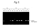
- Figure 5 shows a direct analysis of RT codon 215 in DNA from non-cultured PBL samples.
- Early and late PBL samples obtained from three pateients with AIDS (ptA, ptB and ptL) receiving zidovudine therapy were subjected to "double" PCR to discriminate between wild type and mutant residues.
- Oligonucleotide primers A and NE1 were used in the initial PCR, producing a 768 bp fragment (see figure 4) and material from this amplification was used in the second PCR where primer B was paired separately with primer 3W (WT) or 3M (M) to discriminate between wild type and mutant nucleotides at codon 215.
- Products (15 ⁇ l) from the second PCR were separated on a 1.5% agarose gel and stained with ethidium bromide.
- the numbers above pairs of lanes on the gel refer to the treatment time which each sample was obtained (in months). All patients received 1200 mg of zidovudine per day throughout the treatment period.
- DNA was extracted from MT-2 cells infected with HIV isolates as follows: 2x10⁶ MT-2 cells were infected with HIV-1 at a multiplicity of approx. 0.1 TCID50/cell and incubated at 37 o C for 3-4 days in RPM1 - 1640 medium supplemented with 10% foetal calf serum, polybrene (2 ⁇ g/ml) and antibiotics ( RPMI/10) (Larder, B.A., Kemp, S.D., Science, 246 , 1155-1158, (1989)).
- DNA was extracted from the cell pellet by lysis in 0.4% SDS, 25 mM Tris-HCl, pH8, 25mM. EDTA, and 150 mM NaCl and followed by Proteinase K (1mg/ml) digestion for 1 hr at 50 o C. Nucleic acid was recovered by phenol/chloroform extraction and ethanol precipitation, was treated with RNase A (10 ⁇ g/ml) and DNA was subjected to further phenol/chloroform extraction before ethanol precipitation.
- the complete RT coding region (1.7kb) was obtained for each HIV-1 isolate using polymerase chain reaction (PCR) amplification of infected cell DNA.
- PCR is a powerful technique for selectively amplifying DNA or RNA sequences. It is an in vitro method for producing large amounts of a specific DNA fragment of defined length and sequence from small amounts of a template.
- PCR is based on the enzymatic amplification of a DNA fragment that is flanked by two oligonucleotide primers that hybridise to opposite strands of the target sequence. The primers are oriented with their 3′ ends pointing towards each other. Repeated cycles of heat denaturation of the template, annealing of the primers to their complementary sequences and extension of the annealed primers with a DNA polymerase result in the amplification of the segment defined by the 5′ ends of the PCR primers.
- each cycle essentially doubles the amount of the DNA fragment produced in the previous cycle. This results in the exponential accumulation of the specific target fragment, up to several millionfold in a few hours.
- the method can be used with a complex template such as genomic DNA and can amplify a single-copy gene contained therein. It is also capable of amplifying a single molecule of target in a complex mixture of RNAs or DNAs and can, under some conditions, produce fragments up to ten kilobase pairs long. (White, T.J., Arnheim, N., and E. Hich, A., Technical Focus Vol. 5, No. 6, p185 June 1989).
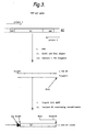
- FIG. 3 A diagramatic representation of the scheme used to amplify and clone HIV RT is shown in Figure 3.
- Total DNA was extracted from cells infected with HIV isolates and the entire 1.7kb fragment of the RT coding region was obtained by PCR (Saiki, R.K., et al . Science 239 , 487 (1988)) HIV-1 DNA was obtained as in Example 1. Approximately 1 ⁇ g of this DNA was used per PCR reaction to amplify the complete RT coding region.
- Each reaction mix contained 25mM KC1, 1.5mM MgCl2, 50mM tris-HCl, pH8.3, 0.1mg/ml bovine serum albumin, 0.2mM each of dATP, dGTP, dCTP, TTP, 0.25 ⁇ g each primer and 2.5 units of Taq DNA Polymerase (Perkin-Elmer Cetus).
- oligonucleotide primers made using an Applied Biosystems 381A synthesiser, were as follows: at the 5′ end of RT, 5′ - TTGCACTTTGAATTCTCCCATTAG - 3′ and 5′ - TGTACTTTGAATTCCCCCATTAG - 3′ (two primers were used to accommodate sequence variation seen in this region) and at the 3′ end, 5′ -CTTATCTATTCCATCTAGAAATAGT-3′ 3′.
- HIV RT obtained by PCR amplification was digested with EcoRI and XbaI , whose recognition sites were built into the 5′ and 3′ ends by the PCR primers, fragments were purified from agarose gels and ligated with the replicative form (RF) of M13mp18, previously digested with EcoRI and XbaI .
- HIV RT (1.7kb) obtained by PCR amplification was digested with EcoRI and XbaI (BRL), purified from agarose gels and ligated with the Ml3 vector mplB pre-digested with EcoRI and XbaI (see figure 3).
- the ligation mixture was used to transform E.coli (strain TG-1) made competent as described (D.Hanahan, J.Mol.Biol. 166 , 557 (1983)). Single-stranded DNA was prepared from these constructs for nucleotide sequencing. Clones able to express functional RT enzyme were sequenced using the dideoxynucleotide chain-termination method (Sanger, F., Nicklen, S., Coulson, A.R., Proc. Natl. Acad. Sci., USA, 74 , 5463, (1977)). Table 1 shows details of the RT clones obtained by this procedure and lists the HIV isolates from which they were derived. The complete sequence of 6 RT clones was obtained (sensitive and resistant isolate pairs the three individuals) and partial sequence data were obtained from additional isolates.
- PCR primers can be designed to incorporate a restriction enzyme recognition site such as EcoR1 at the 5′ end of the HIV-1 RT gene and a restriction enzyme such as Xba1 at the 3′ end. Digestion of each fragment with restriction enzymes allows subsequent cloning into a suitable vector such as an M13 mp18 based vector.
- HIV isolates derived from untreated and treated individuals are shown in Table 1 with duration of therapy at the time they were obtained and the zidovudine sensitivity. Where multiple isolates were obtained from the same individual, isolates are lettered in temporal order. Fifty per cent inhibitory dose (ID50) values were obtained by plaque-reduction assay with HT4-6C cell monolayers (Larder, B.A. Darby. G., Richman, D.D., Science, 243 , 1731, (1989); Chesebro, B.D., Wehrly, K., J.Virol., 62 , 3779 (1988).).
- the amino acid residue at four positions in HIV-1 RT important for zidovudine resistance are illustrated for HIV isolates. Wild-type residues at the positions of interest are as follows: Asp 67, Lys 70, Thr 215 and Lys 219.
- an infectious molecular clone of HIV containing only the four mutations described above was constructed (the isolate A0l2D) to assess the sensitivity of virus produced from this clone by transfection of T-cells.
- a 2.55kb fragment of the HIV Pol gene from infectious clone HXB2-D (Fisher, A.G., et al ., Nature 316 , 262, 1985) inserted into the M13 vector mp19, was used as a target for site-directed mutagenesis (Larder, B.A., Kemp, S.D., Purifoy, D.J.M., Proc. Natl. Acad. Sci., USA, vol 86 ; p.4803 (1989)
- DNA prepared from wild-type and mutant infectious clones was then used to transfect the T-cell lymphoblastoid line, MT-4 (Harada, S., Koyanagi, U., Yamamoto, N., Science, 229 , 563, (1985)). by electroporation. Virus-induced CPE was observed in each culture at similar times (after 2-4 days) and virus stocks were prepared 6-7 days post transfection.
- Wild type and mutant HIV isolates were titrated by plaque assay in the HeLa-CD4+ cell line, HT4-6C, and then tested for sensitivity to zidovrndine by plaque-reduction assay in HT4-6C cells (Larder, B.A., Darby, G., Richman, D.D., Science, 243 , 1731, (1989); Chesebro. B.D., Wehrly, K., J.Virol., 62 3779, (1988)).
- the results of these experiments as shown in Table 2 clearly demonstrate that the mutant virus constructed by site-directed mutagenesis was highly resistant to zidovudine.
- the ID50 value for HIVRTMC increased about 100-fold compared to wild-type virus and the magnitude of resistance was similar to that of naturally occurring HIV isolates containing similar mutations in the RT gene (Table 2).
- DNA for PCR was obtained from MT-2 cells (2x10⁶) infected with HIV isolates at a multiplicity of 0.1 TCID50/ml (fifty per cent tissue culture infectious dose per ml, as determined by terminal dilution of virus stocks in MT-2 cells) and incubated at 37 o C for 3-4 days in RPMI 1640 medium supplemented with 10% fetal bovine serum, polybrene (2ug/ml) and antibiotics. DNA was extracted from cell pellets by lysis in 0.4% SDS, 25mM Tris HCl, PHB, 25mM EDTA and 150mN NaCl.
- DNA was recovered by phenol extraction and ethanol precipitation Approximately 1 ⁇ g of this material was used per PCR reaction (100 ⁇ l) which contained: 25mM KCl, 50mN Tris HCl, PHB.3, 0.lmg/ml bovine serum albumin (BSA), 0.2mM each of dATP, dGTP, dCTP and dTTP, 0.25 ⁇ g of each oligonucleotide primer with the following concentrations of MgCl2 for specific combinations of primer pairs: Primer A with 1W and 1M (to analyse codon 67): 2mM MgCl2 Primer A with 2W and 2M (to analyse codon 70): 5mN MgCl2 Primer B with 3W and 3M (to analyse codon 215): 1.5mM MgCl2 Primer B with 4W and 4M (to analyse codon 219): 1.5mN MgCl2 Reaction mixture
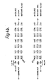
- oligonucleotide primers (Fig.4) were synthesised using an Applied Biosystems 381A machine. 10 ⁇ l of each reaction mixture was run on 1.5% TBE agarose gels and DNA was visualized by ethidium bromide staining. The sequence of PCR primers 2W, 2M, 3W and 3M are shown in Fig. 4b and c.
- primer A 5′-TTCCCATTAGTCCTATT-3′
- primer B 5′-GGATGGAAAGGATCACC-3′
- primer 1W 5′-TTTTCTCCATTTAGTACTGAC-3′
- primer 1M 5′-TTTTCTCCATCTAGTACTGAT-3′
- primer 4W 5′-AGGTTCTTTCTGATGTTTTAT-3′
- primer 4M 5′-AGGTTCTTTCTGATGTTTTAG-3′.
- Variants were constructed to mimic mutants identified by DNA sequence analysis of clinical isolates and in addition, a number of mutants were made containing combinations of changes in RT not yet seen.
- the nucleotide sequence of RT derived from the proviral clone HKB2-D (Fisher, A.G., Collatti, E., Ratner, L., Gallo, R.C. and Wong Staal, F., Nature (London), 316 , 262-265, (1985)) was initially altered by site directed mutagenesis.
- a gene DNA fragment from M13 clones containing altered RT was mixed with HXB2-D that had a BalI restriction enzyme fragment encompassing the RT region removed.
- Mutations in RT were created by site-directed mutagenesis of the previously described M13 clone mpRT1/H, which contains a 2.55kb Bgl II to EcoRI fragment of the wild type HIV pol gene (Larder, B.A., Kemp, S.D., and Purifoy, D.J.M., Proc.Natl.Acad.Sci., (USA), 86 , 4803-4807 (1989)). All mutants were verified by nucleotide sequence analysis (Sanger, F., Nicklen, S, and Coulson, A.R.
- Wild type virus HXB2 and mutants were derived by homologous recombination in MT-2 cells (Clavel, F., Hogan, M.D., Willey, R.L., Strebel, K., Martin, M.A.
- Wild type infectious clone HXB2-D (Fisher, A.G., Collati, E., Ratner, L., Gallo, R.C., and Wong-Staal, F., Nature (London), 316 , 262-265, (1985)) was digested with Msc I (Bal I) , releasing a 1.9kb fragment comprising the majority of RT (Ratner, L. et al ., Nature (London) 313, 277-284, (1985)) and the large fragment was purified from agarose gels.
- the replicative form (double stranded DNA) of mpRT1/H or mutated clones were digested with EcoRI and Hind III to linearize the pol gene fragment and 5 ⁇ g of each was mixed with 5 ⁇ g of gel purified HXB2-D DNA (with the Msc I fragment removed). Each mixture was used to transfect MT-2 cells by electroporation as described (Larder, B.A., Kemp, S.D. supra .) and infectious virus (recovered in culture supernatants at around 14 days post transfection) was stored in aliquots at -70 o C. The genotype of mutant viruses were verified by DNA sequence analysis of RT cloned from these isolates by PCR.
- Virus isolates recovered from these cultures were tested for sensitivity to zidovudine by plaque reduction assay in HT4-6C cells.
- Virus variants were generally more resistant with increasing numbers of mutations (table 4), which was in broad agreement with DNA sequence data obtained from clinical isolates (table 3).
- recombinants RTMF (Thr215 ->) Tyr) and RTMC/WT ->Asp67 ->Asn, Lys70 ->Arg) were less resistant to zidovudine than variant RTMC/F (Asp67 ->Asn, Lys70 ->Arg, Thr215 ->Tyr) and the degree of resistance of RTMC/F was about equal to the sum of ID50 values seen with RTMF and RTMC/WT.
- PCR assay (Example 5) was adapted to enable direct detection of mutations associated with zidovudine resistance in DNA from PBL samples from infected individuals that had not been cultured to obtain virus.
- this material generally contains considerably less HIV DNA than that in infected lymphoid cell cultures, we used a "double PCR" (or nested set) protocol (Simmonds, P., et al ., Supra .) to boost the amount of target HIV RT DNA signal in the samples. Therefore, we initially amplified a 768b fragment from within the RT region which encompassed all the commonly observed mutations (PCR primers A and NE1 were used in this amplification, see Figure 4a).
- DNA was extracted from about 2x10⁶ PBLs as described above for MT-2 cell cultures. 0.5- ⁇ g of this material was used in the initial PCR (10 ⁇ l) which contained 25mM KCl, 1.5mM MgCl2, 50mM Tris HCl, pH 8.3, 0.1 mg/ml BSA, 0.2mM each of dATP, dGTP, dCTP and dTTP plus 0.25 ⁇ g of each primer (primer A and primer NE1). Reaction mixtures were processed as described in Example 5, and subjected to 30 cycles of 1 min 94 o C, 1 min 45 o C and 2 min 72 o C.
- samples were extracted by the addition of 100 ⁇ l chloroform and 0.5 ⁇ l of each was added directly to reaction tubes containing all constituents of the relevant PCR mixture.
- Samples were overlaid with 100 ⁇ l light mineral oil and heated to 94 o C for 5 mins prior to thermal cycling.
- Reaction mixtures and thermal cycle times for this second round of PCR were exactly as described above for selective amplification (Example 5) to detect specific mutations (i.e. primer A was paired with 2W or 2M, or primer B was paired with 3W or 3M).
- Inclusion of 1 ⁇ g herring sperm DNA in each reaction was found to enhance the selectivity of the second PCR.
- PCR primer NE1 was as follows: 5′-TCATTGACAGTCCAGCT- 3′. This primer anneals downstream of primers 3W and 3M as shown in Fig. 4a.
Abstract
Description
- The present invention relates to a method for assessing the sensitivity of an HIV-1 sample to zidovudine, and to diagnostic assays for use in such assessment.
- One group of viruses which has recently assumed a particular importance are the retroviruses. Retroviruses form a sub-group of viruses which, in order to replicate, must first 'reverse transcribe' the RNA of their genome into DNA ('transcription' conventionally describes the synthesis of from DNA). Once in the form of DNA, the viral genome is incorporated into the host cell genome, allowing it to take full advantage of the host cell's transcription/translation machinery for the purposes of replication. Once incorporated, the viral DNA is virtually indistinguishable from the host's DNA and, in this state, the virus may persist for as long as the cell lives. As it is believed to be invulnerable to attack in this form, any treatment must be directed at another stage of the virus life cycle and would, have to be continued until all virus-infected cells have died.
- A species of retrovirus has also been reproducibly isolated from patients with AIDS and is now named as human immunodeficiency virus (HIV-1) and is also known as human T-cell lymphotropic virus III (HTLV III), AIDS associated retrovirus (ARV), or lymphadenopathy associated virus (LAV).
- This virus has been shown preferentially to infect and destroy T-cells bearing the OKT⁴ surface marker and is now generally accepted as the aetiologic agent of AIDS. The patient progressively loses his set of T-cells, upsetting the overall balance of the immune system, reducing his ability to combat other infections and predisposing him to opportunistic infections which frequently prove fatal. Thus, the usual cause of death in AIDS victims is by opportunistic infection, such as pneumonia or virally induced cancers, and not as a direct result of HIV infection.
- The complete nucleotide sequence of the AIDS virus HIV-1 or as it was previously known HTLV-III has been elucidated (Ratner, L., et al. Nature, Vol.313, p277, 24 January 1985).
- Recently, HIV-1 has also been recovered from other tissue types, including B-cells expressing the T⁴ marker, macrophages and non-blood associated tissue in the central nervous system. This infection of the central nervous system has been discovered in patients expressing classical AIDS symptoms and is associated with progressive demyelination, leading to wasting and such symptoms as encephalopathy, progressive dysarthria, ataxia and disorientation. Further conditions associated with HIV infection are the asymptomatic carrier state, progressive generalised lymphadenopathy (PGL) and AIDS-related complex (ARC). HIV-1 can also be present in other tissues or physiological fluids such as urine, plasma, blood, serum, semen, tears, saliva or cerebrospinal fluid.
- 3′-Azido-3′-deoxythymidine (hereafter called "zidovudine") is used in the control of HIV infections including AIDS and ARC. Zidovudine is a thymidine analogue whose triphosphate form inhibits the replication of the human immunodeficiency virus (HIV) by competitive binding to viral reverse transcriptase (RT) and DNA chain termination after phosphorylation by cellular enzymes (Furman, P.A et al., Proc. Natl. Acad. Sci.USA, 1986 83: 8333). It is an effective antiviral agent both in vitro and in vivo against a variety of retroviruses (Mitsuya H et al., Cancer Res., 1987, 47: 3190; Ruprecht R.M et al., Nature, 1986, 323: 476.) and has been demonstrated to improve the quality and length of life of patients with AIDS and advanced ARC (Fischl M.A et al., N.Engl.J.Med., 1987, 317: 185; Schmitt F.A et al, N.Engl.J.Med 1988, 319: 1573; Creagh-Kirk T et al, J. Am.Med.Assoc., 1988 260: 3009) and also in asymptomatics with low CD₄⁺ cell levels.
- As with any anti-infective agent, concern about the potential development of resistance has engendered extensive investigations evaluating factors which might potentially alter the sensitivity of retroviruses to zidovudine.
- A study carried out to measure zidovudine sensitivity of HIV isolates from patients with AIDS or ARC after zidovudine treatment has in fact revealed that a number of isolates from patients treated for six months or more showed reduced sensitivity to zidovudine whereas isolates from untreated individuals and those treated for less than six months showed uniform sensitivity to the drug (Larder, B.A., Darby, G, and Richman, D.D., Science, Vol. 243, 1731, 31st March 1989).
- At present the way to determine the sensitivity of HIV-1 strains to zidovudine is to isolate HIV-1 from a patient's peripheral blood lymphocytes. HIV-1 isolates can be made by co-cultivation of peripheral blood lymphocytes (PBL's) with cells of the continuous line MT-2. (Harada, S.,Koyanagi,Y., Yamamoto, N., Science 229, 563 (1985). This procedure can take anything from between four and fourteen days before HIV-1 can be isolated. The diagnosis of resistant strains of HIV-1 relies firstly on the isolation of virus and then on sensitivity testing by a tissue culture method and so is consequently extremely slow.
- The present inventors have discovered the basis for resistance of HIV-1 to zidovudine at the nucleic acid level. Five nucleotide substitutions have been identified in the HIV-1 genome which result in a change of four amino acids in the RT protein. This discovery has important implications for the detection of resistant HIV isolates because of the highly conserved nature of these nucleotide mutations in RT conferring resistance to zidovudine, and opens the way to the routine detection of such resistant isolates.
- It is possible to carry out a diagnostic assay for the screening of bodily samples from patients for an assessment of the sensitivity of HIV-1 to zidovudine. Using the knowledge of the mutations identified as important in the development of highly resistant strains of HIV-1 such an assay can be developed.
- An analysis of a group of resistant mutants was carried out by nucleotide sequencing which allowed the identification of mutations in the HIV RT gene that confer resistance to zidovudine. The complete RT coding region (1.7kb) was obtained for each isolate using polymerase chain reaction (PCR) amplification of infected cell DNA. The nucleotide changes at the five positions in HIV-1 RT that confer resistance to zidovudine are illustrated in Figure 1. Numbering of the nucleotides of the HIV-1 RT gene is as reported by Ratner et al (Ratneret al., Nature, 313, 277, (1985)).
- It is demonstrated that these specific mutations confer zidovudine-resistance, as an infectious molecular HIV clone containing only these nucleotide changes is resistant to zidovudine. (See Example 4).
- From analysis of clinical samples it appears that the sensitivity of HIV-1 to zidovudine changes over a period of time. It appears that mutations may occur at any of one or more of the five identified sites as the time from the onset of treatment with zidovudine advances and it is clear than an HIV-1 sample which carries all five mutations is highly resistant to zidovudine.
- At this time it is not possible to predict any order of occurence of these mutations, although particular attention is being focussed on the two nucleotides of the wild-type DNA sequence (or its corresponding RNA) or to the two nucleotides of the mutant DNA sequence (or its corresponding RNA) set forth in figure 1 at the 2772- and 2773- positions.
- Accordingly in a first aspect of the invention there is provided a method for assessing the sensitivity of an HIV-1 sample to zidovudine, which comprises:
- (i) isolating nucleic acid from the sample,
- (ii) hybridising an oligonucleotide to the nucleic acid, the oligonucleotide being complementary to a region of the wild-type DNA sequence (or its corresponding RNA) or to a region of the mutant DNA sequence set forth in Figure 1 (or its corresponding RNA) and terminating at the 3′-end with the nucleotide in the 2328-, 2338-, 2772-, 2773- or 2784- position,
- (iii) attempting polymerisation of the nucleic acid from the 3′end of the oligonucleotide,
- (iv) ascertaining whether or not an oligonucleotide primer extended product is present.
- It is possible to use genomic DNA or RNA isolated from HIV-1 samples in this methodology. Suitable cells for supporting the growth of HIV-1, such as MT-2 cells, are firstly infected with an HIV-1 isolate and incubated for a period of time. The cells are recovered by centrifugation. DNA can then be isolated by digestion of the cells with proteinase K in the presence of EDTA and a detergent such as SDS, followed by extraction with phenol (see Example 1 for the methodology used by the inventors for the isolation of HIV-1 DNA).
- Well-known extraction and purification procedures are available for the isolation of RNA from a sample. RNA can be isolated using the following methodology. Suitable cells are again infected and incubated for a period of time. The cells are recovered by centrifugation. The cells are resuspended in an RNA extraction buffer followed by digestion using a proteinase digestion buffer and digestion with proteinase K. Proteins are removed in the presence of a phenol/chloroform mixture. RNA can then be recovered following further centriguation steps. (Maniatis, T., et al, Molecular Coning, A Laboratory Manual, 2nd Edition, Cold Spring Harbor Laboratory Press, (1989)).
- Although it is possible to use unamplified nucleic acid, due to the relative scarcity of nucleic acid in an HIV-1 sample it is preferable to amplify it. Nucleic acid may be selectively amplified using the technique of polymerase chain reaction (PCR), which is an in vitro method for producing large amounts of a specific nucleic acid fragment of defined length and sequence from small amounts of a template.
- The PCR is comprised of standard reactants using Mg²⁺ concentration, oligonucleotide primers and temperature cycling conditions for amplification of the RT gene using the primers. The primers are chosen such that they will amplify the entire RT gene or a selected sequence which incorporates nucleotides corresponding to a region of the wild-type DNA sequence of HIV-1 between the nucleotides at the 2328- and 2784- positions set forth in Figure 1. Example 2 provides a description of PCR used to amplify target nucleic acid.
- RNA cannot be amplified directly by PCR. Its corresponding cDNA must be first of all synthesised. Synthesis of cDNA is normally carried out by primed reverse transcription using oligo-dT primers. Advantageously, primers are chosen such that they will amplify the nucleic acid sequence for RT or a selected sequence which incorporates nucleotides corresponding to the region of RNA corresponding to the wild-type DNA sequence or to the region of the mutant DNA sequence set forth in Figure 1 between the nucleotides 2328- and 2784-. This could be achieved by preparing an oligonucleotide primer which is complementary to a region of the RNA strand which is up-stream of the corresponding sequence of the wild-type DNA sequence between nucleotides 2328- and 2784-. cDNA prepared by this methodology (see Maniatis, T., et al., supra.) can then be used in the same way as for the DNA already discussed.
- The next stage of the methodology is to hybridise to the nucleic acid an oligonucleotlde which is complementary to a region of the wild-type DNA sequence (or its corresponding RNA) or to a region of the mutant DNA sequence (or its corresponding RNA) set forth in Figure 1 and terminating at the 3′- end with the nucleotide at the 2328-, 2338-, 2772-, 2773- or 2784- position.
- Conditions and reagents are then provided to permit polymerisation of the nucleic acid from the 3′-end of the oligonucleotide primer. Such polymerisation reactions are well-known in the art.
- If the oligonucleotide primer has at its 3′-end a nucleotide which is complementary to a mutant genotype, that is a genotype which has a nucleotide change at the 2328-, 2338-, 2772-, 2773- or 2784- position as set forth in Figure 1 then polymerisation of the nucleic acid sequence will only occur if the nucleic acid of the sample is the same as the mutant genotype. Polymerisation of a wild type nucleic acid sequence will not occur or at least not to a significant extent because of a mis-match of nucleotides at the 3′- end of the oligonucleotide primer and the nucleic acid sequence of the sample.
- If the oligonucleotide primer has at its 3′-end a nucleotide which is complementary to the wild-type genotype, that is a genotype which has the wild type nucleotide at the corresponding 2328-, 2338-, 2772-, 2773- or 2784- position as set forth in Figure 1, then there will be polymerisation of a nucleic acid sequence which is wild-type at that position. There will be no polymerisation of a nucleic acid which has a mutant nucleotide at the 3′-position.
- The preferred length of each oligonucleotide is 15-20 nucleotides.
- The oligonucleotide can be prepared according to methodology well known to the man skilled in the art (Koster, H., Drug Research, 30 p548 (1980); Koster, H., Tetrahedron Letters pl527 (1972); Caruthers, Tetrahedron Letters, p7l9, (1980); Tetrahedron Letters, pl859, (1981); Tetrahedron Letters 24, p.245, (1983); Gate. M., Nucleic Acid Research 8, p1081, (1980)) and is generally prepared using an automated DNA synthesiser such as an Applied Biosystems 381A synthesiser.
- It is convenient to determine the presence of an oligonucleotide primer extended product. The means for carrying out the detection is by using an appropriate label.
- The label may be conveniently attached to the oligonucleotide primer or to some other molecule which will bind the primer extended polymerisation product.
- The label may be for example an enzyme, radioisotope or fluorochrome. A preferred label may be biotin which could be subsequently detected using streptavidin conjugated to an enzyme such as peroxidase or alkaline phosphatase. The presence of an oligonucleotide primer extended polymerisation product can be detected for example by running the polymerisation reaction on an agarose gel and observing a specific DNA fragment labelled with ethidium bromide, or Southern blotted and autoradiographed to detect the presence or absence of bands corresponding to polymerised product. If a predominant band is present which corresponds only to the labelled oligonucleotide then this indicates that polymerisation has not occurred. If bands are present of the correct predicted size, this would indicate that polymerisation has occurred.
- For example, DNA isolated from patients' lymphocytes as described herein is used as a template for PCR amplification using synthetic oligonucleotide primers which either match or mis-match with the amplified sequences.
- The feasibility of PCR in detecting such mutations has already been demonstrated. PCR using the Amplification Refractory Mutation system ("ARMS") (Newton, C.R., et al. Nucleic Acids Research, 17, p.2503, (1989)) Synthetic oligonucleotides are produced that anneal to the regions adjacent to and including the specific mutations such that the 3′- end of the oligonucleotide either matches or mismatches with a mutant or wild-type sequence. PCR is carried out which results in the identification of a DNA fragment (using gel electrophoresis) where a match has occurred or no fragment where a mismatch occurred.
- For example, using the 2 oligonucleotides below as PCR primers:
5′-ATG TTT TTT GTC TGG TGT GGT-3′ = (1)
OR
5′-ATG TTT TTT GTC TGG TGT GAA-3′ = (2)
plus the common oligonucleotide primer:
"B" = 5′- GGA TGG AAA GGA TCA CC-3′
it is possible to distinguish between sensitive and resistant virus. DNA is extracted from HIV-1 infected T-cells as described herein and subjected to "ARMS" PCR analysis using these primers. If the virus is sensitive a 210bp fragment is generated with oligonucleotide B + (1) but not with B + (2). By contrast, if the virus is zidovudine resistant a 210bp fragment is generated with B + (2) but not with B + (1). - The presence of a fragment is identified by using an oligonucleotide primer as described above, i.e. by attempting polymerisation using an oligonucleotide primer which may be labelled for the amplified DNA fragment under stringent conditions which only allow hybridisation of complementary DNA (the only difference is that differential hybridisation does not have to be performed as fragments of DNA amplified by the "ARMS" method will be the same whether derived from mutant or wild-type DNA, so a common oligonucleotide can be used to detect the presence of these fragments. The sequence of such an oligonucleotide is derived from a DNA sequence within the DNA fragment that is conserved amongst HIV-1 strains).
- The above PCR assay may be adapted to enable direct detection of mutations associated with zidovudine resistance in DNA from PBL samples from infected individuals that have not been cultured to obtain virus. As this material generally contains considerably less HIV-1 DNA than that in infected lymphoid cultures a "double PCR" (or nested set) protocol can be used (Simmonds, P., Balfe, P, Peutherer, J.F., Ludlam, C.A., Bishop, J.O. and Leigh Brown, A.J., J.Virol., 64, 864-872, (1990)) to boost the amount of target HIV-1 RT DNA signal in the samples. The double PCR overcomes the problem of limited amplification of a rare template sequence. Initially a fragment may be amplified from within the RT region which encompasses all the commonly observed mutations. A small amount of the pre-amplified material may be used in the second PCR with primer pairs designed to allow discrimination of wild type and mutant residues.
- A suitable test kit for use in an assay to determine the resistance status of an HIV-1 sample to zidovudine which makes use of a methodology according to the first aspect of the invention, comprises an oligonucleotide being complementary to a region of the wild-type DNA sequence (or its corresponding RNA) or to a region of the mutant DNA sequence set forth in Figure 1 (or its corresponding RNA) and terminating at the 3′- end with the nucleotide in the 2328-, 2338-, 2772-, 2773- or 2784- position, other materials required for polymerisation of the nucleic acid from the 3′- end of the oligonucleotide and means for determining the presence of an oligonucleotide primer extended product. Such other materials include appropriate enzymes, buffer and washing solutions, and a label and a substrate for the label if necessary. If PCR is used to amplify nucleic acid then additional materials such as appropriate oligonucleotide primers which will amplify a region of the wild-type DNA sequence (or its corresponding RNA) or a region of the mutant DNA sequence set forth in Figure 1 (or its corresponding RNA) containing one or more of the nucleotides at the 2328-, 2338-, 2772-, 2773- or 2784- position, appropriate enzymes and dNTP's should be included.
- In a second aspect of the invention there is provided a method for determining the sensitivity of an HIV-1 sample to zidovudine which comprises:
- (i) isolating the nucleic acid from the sample,
- (ii) hybridising the nucleic acid with an oligonucleotide being complementary to a region of the wild-type DNA sequence (or its corresponding RNA) or to a region of the mutant DNA sequence set forth in Figure 1 (or its corresponding RNA) containing one or more of the nucleotides at the corresponding 2328-, 2338-, 2772-, 2773- and/or 2784- position,
- (iii) ascertaining whether or not any of the resulting hybrids of the oligonucleotide and nucleic acid have complementary nucleotides at one of these positions.
- Preferably the oligonucleotide is so designed to form a perfectly matched hybrid with its complement.
- Nucleic acid (DNA or RNA) is isolated from a sample by the aforementioned methods as described for the first aspect of the invention.
- Similarly, PCR may be used to amplify the RT DNA (or its corresponding RNA) or preferably to amplify a region of the RT DNA (or its corresponding RNA) which incorporates DNA (or its corresponding RNA) containing one or more of the nucleotides at the 2328-, 2338-, 2772-, 2775- and/or 2784- position (see Example 2).
- In the second stage of this methodology the nucleic acid is then used to hybridise to oligonucleotides complementary to a region of the wild-type DNA sequence (or its corresponding RNA) or to a region of the mutant DNA sequence set forth in Figure 1 (or its corresponding RNA) containing one or more of the nucleotides at the aforementioned positions.
- The oligonucleotide may be of any length depending on the number of nucleotide positions of interest which are being examined. If the oligonucleotide is designed to include a nucleotide at only one position of interest then this nucleotide is preferably at or close to the centre position of the oligonucleotide.
- For example, referring to Figure 1, one oligonucleotide probe for detection of the mutation at nucleotide 2328- would be complementary to the mutated RT gene sequence and would include at its nucleotide corresponding to nucleotide 2328- the nucleotide complementary to the mutated 2328- nucleotide. A second oligonucleotide probe for the wild type HIV-1 RT would include at its nucleotide corresponding to nucleotide 2328- the nucleotide complementary to the wild-type 2328- nucleotide. Oligonucleotide probes designed to detect one or more of the mutations referred to in Figure 1 are used to detect a specific mutation or that there has been a mutation.
- In order to ascertain whether or not the oligonucleotide and nucleic acid sequence have formed a matched hybrid, specific hybridisation conditions are set so that a hybrid is only formed when the nucleotide or nucleotides at one or more of the 2328-, 2338-, 2772-, 2773- and 2784- position is or are complementary to the corresponding nucleotide or nucleotides of the oligonucleotide which either permits hybridisation or no hybridisation. It is important to establish for example the temperature of the reaction and the concentration of salt solution before carrying out the hybridisation step to find conditions that are stringent enough to guarantee specificity (Maniatis, T., et al., Molecular Cloning, A Laboratory Manual, 2nd edition, Cold Spring Harbour Laboratory Press, (1989)). If the oligonucleotide probe has a DNA sequence which is complementary to a wild-type nucleic acid sequence at one or more of its nucleotides corresponding to the 2328-, 2338-, 2772-, 2773- or 2784- position of the DNA sequence of Figure 1 then this oligonucleotide will hybridise perfectly to wild-type nucleic acid. If there is no hybridisation then this would suggest that the nucleic acid isolated from the sample contains one or more mutations.
- If the oligonucleotide probe has a DNA sequence which is complementary to a mutant nucleic acid sequence at one or more of its nucleotides corresponding to the 2328-, 2338-, 2772-, 2773- or 2784- positions of the DNA sequence of Figure 1 then this oligonucleotide will hybridise perfectly to mutant nucleic acid. If there is no hybridisation this would suggest that the nucleic acid isolated from the sample contains no such mutation or mutations. The oligonucleotide probes may be labelled as a means of detection as for the first aspect of the invention.
- The hybridisation and subsequent removal of non-hybridised nucleic acids are performed under stringent conditions which only allow hybridisation of the complementary DNA and not the oligonucleotide containing a mismatch (i.e. oligonucleotide specific hybridisation as described for the detection of sickle cell mutation using the β-globin or HLA-DQα gene (Saiki, R.K., et al., Nature, 324, p163, (1986)), the activated Ras gene (Ver Laan-de, Vries, M., et al., Gene 50, 313, (1986)) and β-thalassaemia Wong, C., et al., Nature, 330, p384, (1987)).
- The hybridisation may be carried out by immobilisation of the RT nucleic acid sequence onto nitrocellulose, nylon or other solid matrix (eg. dot-blot). It is convenient to determine the presence of an hybrid by using the means of a label. For example, the chemically synthesised oligonucleotide probes can be suitably labelled using enzyme, radiosotope or fluorochrome. A preferred label may be biotin which could be subsequently detected using streptavidin conjugated to an enzyme such as peroxidase or alkaline phosphatase.
- Alternatively the hybridisation may be carried out by immobilisation of the chemically synthesised oligonucleotides referred to above, which are unlabelled, onto a solid support referred to above and subsequent hybridisation by a labelled RT nucleic acid sequence as described previously.
- In both situations described above for hybridisation suitable control reactions will be incorporated to determine that efficient hybridisation has occurred. (eg the hybridisation of oligonucleotides to a complementary oligonucleotide).
- Results would be readily interpreted as the isolated nucleic acid would hybridise to either the wild type oligonucleotide or the mutant oligonucleotide.
- A suitable test kit for use in an assay to determine the sensitivity of an HIV-1 sample to zidovudine which makes use of a methodology according to the second aspect of the invention comprises an oligonucleotide being complementary to a region of the wild-type DNA sequence (or its corresponding RNA) or to a region of the mutant DNA sequence set forth in Figure 1 (or its corresponding RNA) containing one or more of the nucleotides at the corresponding 2328-, 2338-, 2772-, 2773- and/or 2784- position, other materials required to permit hybridisation. Such materials include appropriate buffers and washing solutions and a label and a substrate for the label if necessary. Normally the oligonucleotide would be labelled. If PCR is used to amplify nucleic acid prior to hybridisation then additional materials such as appropriate oligonucleotide primers which will amplify a region of the wild-type DNA sequence (or its corresponding RNA) or a region of the mutant DNA sequence set forth in Figure 1 (or its corresponding RNA) containing one or more of the nucleotides at the 2328-, 2338-, 2772-, 2773- or 2784- position, appropriate enzymes and dNTP's (deoxy nucleotide triphosphates) should be included.
- In one alternate format of the assay, the dNTP's in the amplification may or may not be coupled to a detector molecule such as a radiosotope, biotin, fluorochrome or enzyme.
- It is also possible to detect zidovudine resistant mutations in the HIV-1 RT RNA isolated from clinical samples using an RNA amplification system. Using the methodology described by Guatelli et al. (Proc.Natl.Acad.Sci, (USA), 87, 1874-1878, (March 1990)) a target nucleic acid sequence can be replicated (amplified) exponentially in vitro under isothermal conditions by using three enzymatic activities essential to retroviral replication: reverse transcriptase, RNase H and a DNA-dependant RNA polymerase. Such a methodology may be employed followed by an hybridisation step to distinguish mutant from wild-type nucleotides as discussed previously.
- The following examples are for illustration only and are not intended to limit the invention in any way. In the accompanying drawings:
- Figure 1 shows the nucleotide changes at the five positions in HIV-1 RT that confer resistance to zidovudine. Numbering of the nucleotides is as reported by Ratner et al., Nature, 313, 277, (1985).
- Figure 2 shows the amino acid changes at the four positions in HIV-1 RT that confer resistance to zidovudine.
- Figure 3 shows the scheme used to amplify and clone HIV RT.
- Figure 4 shows mutational analysis of HIV RT using the polymerase reaction in which
- a. shows a schematic representation of the HIV-1 genome showing the location and orientation of oligonucleotide primers in the RT coding region used in PCR reactions.
Primers ) and are used separately with primer A to generate a DNA fragment of 227bp.
Primers ) and are used separately with primer B to generate a 210bp fragment);
- b. shows details of
PCR primers codon 70. The sequence of each primer is shown with variation at the 3′ end highlighted in bold letters where a correct match forms with target HIV DNA. In this situation Taq DNA polymerase can initiate DNA synthesis from the 3′ end of the primer (<-----). Nucleotide sequence numbers according to Ratner et al., supra are shown in parenthesis. - c. shows
PCR primers codon 215. Sequence variation at the 3′ end is highlighted in bold letters where a correct match is formed with target HIV DNA.
- a. shows a schematic representation of the HIV-1 genome showing the location and orientation of oligonucleotide primers in the RT coding region used in PCR reactions.
- Figure 5 shows a direct analysis of
RT codon 215 in DNA from non-cultured PBL samples. Early and late PBL samples obtained from three pateients with AIDS (ptA, ptB and ptL) receiving zidovudine therapy were subjected to "double" PCR to discriminate between wild type and mutant residues. Oligonucleotide primers A and NE1 were used in the initial PCR, producing a 768 bp fragment (see figure 4) and material from this amplification was used in the second PCR where primer B was paired separately withprimer 3W (WT) or 3M (M) to discriminate between wild type and mutant nucleotides atcodon 215. Products (15µl) from the second PCR were separated on a 1.5% agarose gel and stained with ethidium bromide. The numbers above pairs of lanes on the gel refer to the treatment time which each sample was obtained (in months). All patients received 1200 mg of zidovudine per day throughout the treatment period. - The isolation of HIV-1 was achieved according to methodology reported by Larder et al. (Larder, B.A., Darby, G., Richman, D., Science, 243, 1731, (1989))
- DNA was extracted from MT-2 cells infected with HIV isolates as follows:
2x10⁶ MT-2 cells were infected with HIV-1 at a multiplicity of approx. 0.1 TCID₅₀/cell and incubated at 37oC for 3-4 days in RPM1 - 1640 medium supplemented with 10% foetal calf serum, polybrene (2µg/ml) and antibiotics (=RPMI/10) (Larder, B.A., Kemp, S.D., Science, 246, 1155-1158, (1989)). - DNA was extracted from the cell pellet by lysis in 0.4% SDS, 25 mM Tris-HCl, pH8, 25mM. EDTA, and 150 mM NaCl and followed by Proteinase K (1mg/ml) digestion for 1 hr at 50oC. Nucleic acid was recovered by phenol/chloroform extraction and ethanol precipitation, was treated with RNase A (10µg/ml) and DNA was subjected to further phenol/chloroform extraction before ethanol precipitation.
- The complete RT coding region (1.7kb) was obtained for each HIV-1 isolate using polymerase chain reaction (PCR) amplification of infected cell DNA.
- PCR is a powerful technique for selectively amplifying DNA or RNA sequences. It is an in vitro method for producing large amounts of a specific DNA fragment of defined length and sequence from small amounts of a template. PCR is based on the enzymatic amplification of a DNA fragment that is flanked by two oligonucleotide primers that hybridise to opposite strands of the target sequence. The primers are oriented with their 3′ ends pointing towards each other. Repeated cycles of heat denaturation of the template, annealing of the primers to their complementary sequences and extension of the annealed primers with a DNA polymerase result in the amplification of the segment defined by the 5′ ends of the PCR primers. Since the extension product of each primer can serve as a template for the other primer, each cycle essentially doubles the amount of the DNA fragment produced in the previous cycle. This results in the exponential accumulation of the specific target fragment, up to several millionfold in a few hours. The method can be used with a complex template such as genomic DNA and can amplify a single-copy gene contained therein. It is also capable of amplifying a single molecule of target in a complex mixture of RNAs or DNAs and can, under some conditions, produce fragments up to ten kilobase pairs long. (White, T.J., Arnheim, N., and E. Hich, A., Technical Focus Vol. 5, No. 6, p185 June 1989).
- A diagramatic representation of the scheme used to amplify and clone HIV RT is shown in Figure 3. Total DNA was extracted from cells infected with HIV isolates and the entire 1.7kb fragment of the RT coding region was obtained by PCR (Saiki, R.K., et al. Science 239, 487 (1988)) HIV-1 DNA was obtained as in Example 1. Approximately 1µg of this DNA was used per PCR reaction to amplify the complete RT coding region. Each reaction mix (100ul) contained 25mM KC1, 1.5mM MgCl₂, 50mM tris-HCl, pH8.3, 0.1mg/ml bovine serum albumin, 0.2mM each of dATP, dGTP, dCTP, TTP, 0.25µg each primer and 2.5 units of Taq DNA Polymerase (Perkin-Elmer Cetus). These mixtures were heated at 100oC for 2mins prior to addition of Taq DNA polymerase, overlaid with 100µl light mineral oil and subjected to 30 cycles consisting of a denaturation step (1min 30secs, 94oC) primer annealing (2min, 37oC) and DNA synthesis (10min, 72oC) using a Perkin-Elmer Cetus DNA thermal cycler. The oligonucleotide primers, made using an Applied Biosystems 381A synthesiser, were as follows: at the 5′ end of RT, 5′ - TTGCACTTTGAATTCTCCCATTAG - 3′ and 5′ - TGTACTTTGAATTCCCCCATTAG - 3′ (two primers were used to accommodate sequence variation seen in this region) and at the 3′ end, 5′ -CTTATCTATTCCATCTAGAAATAGT-3′ 3′.
- HIV RT obtained by PCR amplification was digested with EcoRI and XbaI, whose recognition sites were built into the 5′ and 3′ ends by the PCR primers, fragments were purified from agarose gels and ligated with the replicative form (RF) of M13mp18, previously digested with EcoRI and XbaI. HIV RT (1.7kb) obtained by PCR amplification (Saiki, R.K., et al. Science 239, 487 (1988)) was digested with EcoRI and XbaI (BRL), purified from agarose gels and ligated with the Ml3 vector mplB pre-digested with EcoRI and XbaI (see figure 3). The ligation mixture was used to transform E.coli (strain TG-1) made competent as described (D.Hanahan, J.Mol.Biol. 166, 557 (1983)). Single-stranded DNA was prepared from these constructs for nucleotide sequencing. Clones able to express functional RT enzyme were sequenced using the dideoxynucleotide chain-termination method (Sanger, F., Nicklen, S., Coulson, A.R., Proc. Natl. Acad. Sci., USA, 74, 5463, (1977)). Table 1 shows details of the RT clones obtained by this procedure and lists the HIV isolates from which they were derived. The complete sequence of 6 RT clones was obtained (sensitive and resistant isolate pairs the three individuals) and partial sequence data were obtained from additional isolates.
- PCR primers can be designed to incorporate a restriction enzyme recognition site such as EcoR1 at the 5′ end of the HIV-1 RT gene and a restriction enzyme such as Xba1 at the 3′ end. Digestion of each fragment with restriction enzymes allows subsequent cloning into a suitable vector such as an M13 mp18 based vector.
- HIV isolates derived from untreated and treated individuals are shown in Table 1 with duration of therapy at the time they were obtained and the zidovudine sensitivity. Where multiple isolates were obtained from the same individual, isolates are lettered in temporal order. Fifty per cent inhibitory dose (ID₅₀) values were obtained by plaque-reduction assay with HT4-6C cell monolayers (Larder, B.A. Darby. G., Richman, D.D., Science, 243, 1731, (1989); Chesebro, B.D., Wehrly, K., J.Virol., 62, 3779 (1988).).
- The amino acid residue at four positions in HIV-1 RT important for zidovudine resistance are illustrated for HIV isolates. Wild-type residues at the positions of interest are as follows:
Asp 67,Lys 70,Thr 215 andLys 219. - Comparison of predicted complete amino acid RT sequences from sensitive and highly-resistant isolate pairs obtained from the same individuals (patients A012, A0l8 and PO22) revealed substantial differences of between 14-17 residues. In each case however, we identified amino acid changes at four residues (Asp 67 -> Asn, Lys 70 -> Arg, Thr 215 -> Phe or Tyr, and Lys 210 -> Gln) in the highly resistant isolates not seen in the sensitive counterparts (Fig 2). Analysis of sensitive and highly-resistant isolate pairs from a further two individuals (patients A036 and PO26) revealed the same mutations at
positions Table 1 HIV ISOLATE DURATION OF THERAPY (Months) ZIDOVUDINE SENSITIVITY (ID₅₀µM) RT ACTIVITY M13 CLONE (% Control) AMINO ACID RESIDUES IN RT 67 70 215 219 A012B 2 0.01 20 Asp Lys Thr Lys A012D 26 2 85 Asn Arg Phe Gln A018A 0 0.01 62 Asp Lys Thr Lys A018C 14 2.3 79 Asn Arg Tyr Gln A036B 2 0.01 47 Asp Lys Thr Lys A036C 11 0.6 52 Asp Lys Tyr Lys A036D 20 5.6 46 Asn Arg Tyr Lys P022A 1 0.01 20 Asp Lys Thr Lys P022C 16 1.4 67 Asn Arg Phe Gln P026A 0 0.01 76 Asp Lys Thr Lys P026B 11 2.8 78 Asn Arg Tyr Lys P035A 6 0.56 55 Asp Lys Tyr Lys - To test whether multiple mutations in the RT gene could account for high level resistance, an infectious molecular clone of HIV containing only the four mutations described above was constructed (the isolate A0l2D) to assess the sensitivity of virus produced from this clone by transfection of T-cells. A 2.55kb fragment of the HIV Pol gene from infectious clone HXB2-D (Fisher, A.G., et al., Nature 316, 262, 1985) inserted into the M13 vector mp19, was used as a target for site-directed mutagenesis (Larder, B.A., Kemp, S.D., Purifoy, D.J.M., Proc. Natl. Acad. Sci., USA, vol 86; p.4803 (1989)
- Specific nucleotide changes were simultaneously introduced into the RT gene using two synthetic oligonucleotides and mutations were confirmed by nucleotide sequencing (Sanger, F., Nicklen, S.A., Coulson, A.R., Proc. Natl. Acad. Sci., USA. 74, 5463, (1977)). To reconstruct the full-length clone, a 1.9 kb BalI restriction fragment containing the mutations in RT was removed from the pol gene M13 clone and transferred into HXB2-D (Larder, B.A., Kemp, S.D., Purifoy, D.J.M., Proc. Natl. Acad. Sci., USA, Vol. 86, p4803, (1989)). DNA prepared from wild-type and mutant infectious clones was then used to transfect the T-cell lymphoblastoid line, MT-4 (Harada, S., Koyanagi, U., Yamamoto, N., Science, 229, 563, (1985)). by electroporation. Virus-induced CPE was observed in each culture at similar times (after 2-4 days) and virus stocks were prepared 6-7 days post transfection. Wild type and mutant HIV isolates (HXB2-D and HIVRTMC respectively) were titrated by plaque assay in the HeLa-CD4⁺ cell line, HT4-6C, and then tested for sensitivity to zidovrndine by plaque-reduction assay in HT4-6C cells (Larder, B.A., Darby, G., Richman, D.D., Science, 243, 1731, (1989); Chesebro. B.D., Wehrly, K., J.Virol., 62 3779, (1988)). The results of these experiments as shown in Table 2 clearly demonstrate that the mutant virus constructed by site-directed mutagenesis was highly resistant to zidovudine. The ID₅₀ value for HIVRTMC increased about 100-fold compared to wild-type virus and the magnitude of resistance was similar to that of naturally occurring HIV isolates containing similar mutations in the RT gene (Table 2).
Table 2 HIV ISOLATE ZIDOVUDINE SENSITIVITY (Mean ID₅₀,µM) FOLD INCREASE HXB2-D 0.013 (0.005) 1 HIVRTMC 1.28 (0.24) 98 A012B 0.013 (0.005) 1 A012D 2.56 (1.03) 197 - DNA for PCR was obtained from MT-2 cells (2x10⁶) infected with HIV isolates at a multiplicity of 0.1 TCID₅₀/ml (fifty per cent tissue culture infectious dose per ml, as determined by terminal dilution of virus stocks in MT-2 cells) and incubated at 37oC for 3-4 days in RPMI 1640 medium supplemented with 10% fetal bovine serum, polybrene (2ug/ml) and antibiotics. DNA was extracted from cell pellets by lysis in 0.4% SDS, 25mM Tris HCl, PHB, 25mM EDTA and 150mN NaCl. After digestion with proteinase K (1mg/ml, 1h at 50oC) DNA was recovered by phenol extraction and ethanol precipitation Approximately 1µg of this material was used per PCR reaction (100µl) which contained: 25mM KCl, 50mN Tris HCl, PHB.3, 0.lmg/ml bovine serum albumin (BSA), 0.2mM each of dATP, dGTP, dCTP and dTTP, 0.25µg of each oligonucleotide primer with the following concentrations of MgCl₂ for specific combinations of primer pairs: Primer A with 1W and 1M (to analyse codon 67): 2mM MgCl₂
Primer A with 2W and 2M (to analyse codon 70): 5mN MgCl₂
Primer B with 3W and 3M (to analyse codon 215): 1.5mM MgCl₂
Primer B with 4W and 4M (to analyse codon 219): 1.5mN MgCl₂
Reaction mixtures were heated at 100oC for 2 mins prior to addition of Taq DNA polymerase (2.5 units, Perkin-Elmer Cetus), overlaid with 100µl light mineral oil and subjected to 30 cycles consisting of a denaturation step (1min, 94oC), primer annealing (30 sec, 40oC for reactions to analyse codon 70 (primer A with 2W and 2M) and codon 219 (primer B with 4W and 4M) or 45oC for reactions to analyse codon 67 (primer A with 1W and 1M) and codon 215 (primer B with 3W and 3M) and DNA synthesis (30 sec, 72oC) using a Perkin-Elmer Cetus DNA thermal cycler. The oligonucleotide primers (Fig.4) were synthesised using an Applied Biosystems 381A machine. 10µl of each reaction mixture was run on 1.5% TBE agarose gels and DNA was visualized by ethidium bromide staining. The sequence ofPCR primers
primer A, 5′-TTCCCATTAGTCCTATT-3′;
primer B, 5′-GGATGGAAAGGATCACC-3′;
primer 1W, 5′-TTTTCTCCATTTAGTACTGAC-3′;
primer 1M, 5′-TTTTCTCCATCTAGTACTGAT-3′;
primer 4W, 5′-AGGTTCTTTCTGATGTTTTAT-3′;
primer 4M, 5′-AGGTTCTTTCTGATGTTTTAG-3′. - Variants were constructed to mimic mutants identified by DNA sequence analysis of clinical isolates and in addition, a number of mutants were made containing combinations of changes in RT not yet seen. The nucleotide sequence of RT derived from the proviral clone HKB₂-D (Fisher, A.G., Collatti, E., Ratner, L., Gallo, R.C. and Wong Staal, F., Nature (London), 316, 262-265, (1985)) was initially altered by site directed mutagenesis. A gene DNA fragment from M13 clones containing altered RT was mixed with HXB2-D that had a BalI restriction enzyme fragment encompassing the RT region removed. These mixtures were used to co-transfect MT-2 cells by electroporation in order to allow formation of infectious virus variants through homologous recombination. Mutations in RT were created by site-directed mutagenesis of the previously described M13 clone mpRT1/H, which contains a 2.55kb Bgl II to EcoRI fragment of the wild type HIV pol gene (Larder, B.A., Kemp, S.D., and Purifoy, D.J.M., Proc.Natl.Acad.Sci., (USA), 86, 4803-4807 (1989)). All mutants were verified by nucleotide sequence analysis (Sanger, F., Nicklen, S, and Coulson, A.R. Proc.Natl.Acad.Sci., USA, 74, 5463-5467 (1977)). Wild type virus HXB2 and mutants were derived by homologous recombination in MT-2 cells (Clavel, F., Hogan, M.D., Willey, R.L., Strebel, K., Martin, M.A. and Repaske, R., J.viroi., 63, 1455-1459 (1989); Srinivasan, A., et al., Proc.Natl.Acad.Sci., (USA) 86, 6388-6392 (1989)) by co-transfecting a wild type full length HIV clone missing most of RT and the gene fragment from mpRT1/H M13 clones. Wild type infectious clone HXB2-D (Fisher, A.G., Collati, E., Ratner, L., Gallo, R.C., and Wong-Staal, F., Nature (London), 316, 262-265, (1985)) was digested with Msc I (Bal I), releasing a 1.9kb fragment comprising the majority of RT (Ratner, L. et al., Nature (London) 313, 277-284, (1985)) and the large fragment was purified from agarose gels. The replicative form (double stranded DNA) of mpRT1/H or mutated clones were digested with EcoRI and Hind III to linearize the pol gene fragment and 5µg of each was mixed with 5µg of gel purified HXB2-D DNA (with the Msc I fragment removed). Each mixture was used to transfect MT-2 cells by electroporation as described (Larder, B.A., Kemp, S.D. supra.) and infectious virus (recovered in culture supernatants at around 14 days post transfection) was stored in aliquots at -70oC. The genotype of mutant viruses were verified by DNA sequence analysis of RT cloned from these isolates by PCR. Virus isolates recovered from these cultures were tested for sensitivity to zidovudine by plaque reduction assay in HT4-6C cells. Virus variants were generally more resistant with increasing numbers of mutations (table 4), which was in broad agreement with DNA sequence data obtained from clinical isolates (table 3). For example, recombinants RTMF (Thr215 ->) Tyr) and RTMC/WT ->Asp67 ->Asn, Lys70 ->Arg) were less resistant to zidovudine than variant RTMC/F (Asp67 ->Asn, Lys70 ->Arg, Thr215 ->Tyr) and the degree of resistance of RTMC/F was about equal to the sum of ID₅₀ values seen with RTMF and RTMC/WT.


- The above PCR assay (Example 5) was adapted to enable direct detection of mutations associated with zidovudine resistance in DNA from PBL samples from infected individuals that had not been cultured to obtain virus. As this material generally contains considerably less HIV DNA than that in infected lymphoid cell cultures, we used a "double PCR" (or nested set) protocol (Simmonds, P., et al., Supra.) to boost the amount of target HIV RT DNA signal in the samples. Therefore, we initially amplified a 768b fragment from within the RT region which encompassed all the commonly observed mutations (PCR primers A and NE1 were used in this amplification, see Figure 4a). A small amount of this pre-amplified material was then used in the second PCR with primer pairs designed to allow discrimination of wild type and mutant residues. Results of a typical analysis of
amino acid residue 215 are shown in Figure 5. PBL samples were obtained from three individuals with AIDS prior to initiation of zidovudine therapy and after 12-17 months of therapy. The pre-treatment samples all appeared to be wild type, whilst the post-treatment initiation samples were either mutant or appeared a mixture of both (Figure 5). - DNA was extracted from about 2x10⁶ PBLs as described above for MT-2 cell cultures. 0.5-µg of this material was used in the initial PCR (10µl) which contained 25mM KCl, 1.5mM MgCl₂, 50mM Tris HCl, pH 8.3, 0.1 mg/ml BSA, 0.2mM each of dATP, dGTP, dCTP and dTTP plus 0.25µg of each primer (primer A and primer NE1). Reaction mixtures were processed as described in Example 5, and subjected to 30 cycles of 1
min 94oC, 1min 45oC and 2 min 72oC. Following amplification, samples were extracted by the addition of 100µl chloroform and 0.5µl of each was added directly to reaction tubes containing all constituents of the relevant PCR mixture. Samples were overlaid with 100µl light mineral oil and heated to 94oC for 5 mins prior to thermal cycling. Reaction mixtures and thermal cycle times for this second round of PCR were exactly as described above for selective amplification (Example 5) to detect specific mutations (i.e. primer A was paired with 2W or 2M, or primer B was paired with 3W or 3M). Inclusion of 1µg herring sperm DNA in each reaction was found to enhance the selectivity of the second PCR. In addition, due to the exteme sensitivity of "double" PCR, stringent precautions were taken to avoid cross-contamination of samples and PCR reagents. Control reactions were always performed with no added DNA to ensure no spurious amplification had occurred. The sequence of PCR primer NE1 was as follows: 5′-TCATTGACAGTCCAGCT- 3′. This primer anneals downstream ofprimers
Claims (8)
Applications Claiming Priority (2)
| Application Number | Priority Date | Filing Date | Title |
|---|---|---|---|
| GB8918226 | 1989-08-09 | ||
| GB898918226A GB8918226D0 (en) | 1989-08-09 | 1989-08-09 | Dna assays |
Publications (3)
| Publication Number | Publication Date |
|---|---|
| EP0422762A2 true EP0422762A2 (en) | 1991-04-17 |
| EP0422762A3 EP0422762A3 (en) | 1992-08-05 |
| EP0422762B1 EP0422762B1 (en) | 1995-05-31 |
Family
ID=10661415
Family Applications (1)
| Application Number | Title | Priority Date | Filing Date |
|---|---|---|---|
| EP90308721A Expired - Lifetime EP0422762B1 (en) | 1989-08-09 | 1990-08-08 | Method for assessing the sensitivity of HIV-1 to zidovudine and oligonucleotides therefore |
Country Status (12)
| Country | Link |
|---|---|
| US (2) | US5409810A (en) |
| EP (1) | EP0422762B1 (en) |
| JP (1) | JP3105907B2 (en) |
| AU (1) | AU638475B2 (en) |
| CA (1) | CA2022942C (en) |
| DE (1) | DE69019795T2 (en) |
| DK (1) | DK0422762T3 (en) |
| ES (1) | ES2072983T3 (en) |
| GB (1) | GB8918226D0 (en) |
| IE (1) | IE69846B1 (en) |
| NZ (1) | NZ234812A (en) |
| ZA (1) | ZA906263B (en) |
Cited By (11)
| Publication number | Priority date | Publication date | Assignee | Title |
|---|---|---|---|---|
| WO1992016180A2 (en) * | 1991-03-13 | 1992-10-01 | Siska Diagnostics, Inc. | Detection of 3'-azido-3'-deoxythymidine resistance |
| EP0530794A1 (en) * | 1991-09-06 | 1993-03-10 | Roche Diagnostics GmbH | Method for detecting nucleic acids which are similar to one another |
| WO1995028500A1 (en) * | 1994-04-19 | 1995-10-26 | Stichting Centraal Laboratorium Van De Bloedtransfusiedienst Van Het Nederlandse Rode Kruis | Nucleic acids and methods for the discrimination between syncytium inducing and non syncytium inducing variants of the human immunodeficiency virus |
| US5631128A (en) * | 1992-05-14 | 1997-05-20 | Leland Stanford Junior University | Polymerase chain reaction assays for monitoring antiviral therapy and making therapeutic decisions in the treatment of acquired immunodeficiency syndrome |
| WO1997027332A1 (en) * | 1996-01-26 | 1997-07-31 | Innogenetics N.V. | Method for detection of drug-induced mutations in the reverse transcriptase gene |
| EP1026261A2 (en) * | 1999-02-03 | 2000-08-09 | Ortho-Clinical Diagnostics, Inc. | Enhancement of the specificity of nucleic acid amplification by carrier nucleic acid |
| EP2020450A1 (en) * | 2000-09-01 | 2009-02-04 | Gen-Probe Incorporated | Amplification of HIV-1 sequences for detection of sequences associated with drug-resistance mutations |
| US8076062B2 (en) | 2002-07-01 | 2011-12-13 | Tibotec Pharmaceuticals Ltd. | Mutational profiles in HIV-1 protease correlated with phenotypic drug resistance |
| US8563236B2 (en) | 1999-05-28 | 2013-10-22 | Virco, N.V. | Mutational profiles in HIV-1 reverse transcriptase correlated with phenotypic drug resistance |
| US8574831B2 (en) | 2000-10-20 | 2013-11-05 | Virco N.V. | Mutational profiles in HIV-1 reverse transcriptase correlated with phenotypic drug resistance |
| US8592161B2 (en) | 2002-07-01 | 2013-11-26 | Janssen R&D Ireland | Mutational profiles in HIV-1 protease correlated with phenotypic drug resistance |
Families Citing this family (18)
| Publication number | Priority date | Publication date | Assignee | Title |
|---|---|---|---|---|
| USRE39282E1 (en) * | 1989-08-09 | 2006-09-12 | Smithkline Beecham Corporation | Nucleic acid derivatives |
| CA2042093C (en) * | 1990-05-09 | 2002-12-24 | Gyula Hadlaczky | Cell line carrying an excess of mammalian centromeres |
| US5827648A (en) * | 1995-12-13 | 1998-10-27 | Chiron Corporation | Differential hybridization for relative quantification of variant populations |
| US6265152B1 (en) | 1995-12-22 | 2001-07-24 | Visible Genetics Inc. | Method and kit for evaluation of HIV mutations |
| US6007983A (en) * | 1995-12-22 | 1999-12-28 | Visible Genetics Inc. | Method and kit for evaluation of HIV mutations |
| US6077697A (en) * | 1996-04-10 | 2000-06-20 | Chromos Molecular Systems, Inc. | Artificial chromosomes, uses thereof and methods for preparing artificial chromosomes |
| US20030033617A1 (en) * | 1996-04-10 | 2003-02-13 | Gyula Hadlaczky | Artificial chromosomes, uses thereof and methods for preparing artificial chromosomes |
| US6025155A (en) * | 1996-04-10 | 2000-02-15 | Chromos Molecular Systems, Inc. | Artificial chromosomes, uses thereof and methods for preparing artificial chromosomes |
| US20020160970A1 (en) * | 1996-04-10 | 2002-10-31 | Gyula Hadlaczky | Artificial chromosomes, uses thereof and methods for preparing artificial chromosomes |
| US6830887B2 (en) | 1997-03-18 | 2004-12-14 | Bayer Healthcare Llc | Method and kit for quantitation and nucleic acid sequencing of nucleic acid analytes in a sample |
| MXPA00012842A (en) | 1998-06-24 | 2004-06-22 | Univ Emory | Use of 3'-azido-2',3'-dideoxyuridine in combination with further anti-hiv drugs for the manufacture of a medicament for the treatment of hiv. |
| US7635690B2 (en) | 1999-01-22 | 2009-12-22 | Emory University | HIV-1 mutations selected for by β-2′,3′-didehydro-2′,3′-dideoxy-5-fluorocytidine |
| US7115584B2 (en) * | 1999-01-22 | 2006-10-03 | Emory University | HIV-1 mutations selected for by β-2′,3′-didehydro-2′,3′-dideoxy-5-fluorocytidine |
| US7074556B2 (en) * | 1999-03-02 | 2006-07-11 | Invitrogen Corporation | cDNA synthesis improvements |
| CN105483142A (en) * | 2000-09-28 | 2016-04-13 | 生物源食物及科学公司 | FAD4, FAD5, FAD5-2, and FAD6, novel fatty acid desaturase family members and uses thereof |
| US6834479B2 (en) * | 2002-10-03 | 2004-12-28 | Src Innovations, Llc | Bagging machine having a collapsible tunnel |
| KR101591986B1 (en) * | 2014-02-26 | 2016-02-05 | 연세대학교 산학협력단 | Excavating bit with expandable wings |
| KR101796939B1 (en) * | 2016-02-19 | 2017-11-13 | 박민서 | Air pressure excavator and excavating work using this same |
Citations (1)
| Publication number | Priority date | Publication date | Assignee | Title |
|---|---|---|---|---|
| EP0229701A2 (en) * | 1986-01-10 | 1987-07-22 | F. Hoffmann-La Roche Ag | Detection of viruses by amplification and hybridization |
-
1989
- 1989-08-09 GB GB898918226A patent/GB8918226D0/en active Pending
-
1990
- 1990-08-08 DK DK90308721.1T patent/DK0422762T3/en active
- 1990-08-08 DE DE69019795T patent/DE69019795T2/en not_active Expired - Lifetime
- 1990-08-08 ES ES90308721T patent/ES2072983T3/en not_active Expired - Lifetime
- 1990-08-08 AU AU60828/90A patent/AU638475B2/en not_active Expired
- 1990-08-08 EP EP90308721A patent/EP0422762B1/en not_active Expired - Lifetime
- 1990-08-08 NZ NZ234812A patent/NZ234812A/en unknown
- 1990-08-08 IE IE286090A patent/IE69846B1/en not_active IP Right Cessation
- 1990-08-08 ZA ZA906263A patent/ZA906263B/en unknown
- 1990-08-08 CA CA002022942A patent/CA2022942C/en not_active Expired - Lifetime
- 1990-08-09 JP JP02211465A patent/JP3105907B2/en not_active Expired - Lifetime
-
1992
- 1992-12-01 US US07/984,255 patent/US5409810A/en not_active Ceased
-
1997
- 1997-04-23 US US08/839,010 patent/USRE37918E1/en not_active Expired - Lifetime
Patent Citations (1)
| Publication number | Priority date | Publication date | Assignee | Title |
|---|---|---|---|---|
| EP0229701A2 (en) * | 1986-01-10 | 1987-07-22 | F. Hoffmann-La Roche Ag | Detection of viruses by amplification and hybridization |
Non-Patent Citations (9)
| Title |
|---|
| AIDS vol. 5, no. 2, February 1991, LONDON GB pages 137 - 144; B.A.LARDER ET AL.: 'Zidovudine resistance predicted by direct detection of mutations in DNA from HIV-infected lymphocytes' * |
| NATURE. vol. 313, 24 January 1985, LONDON GB pages 277 - 284; L.RATNER ET AL.: 'Complete nucleotide sequence of the AIDS virus, HTLV-III' * |
| NATURE. vol. 324, 13 November 1986, LONDON GB pages 163 - 166; R.K.SAIKI ET ALL: 'Analysis of enzymatically amplified beta-globin and HLA-DQalpha DNA with allele-specific oligonucleotide probes' * |
| NUCLEIC ACIDS RESEARCH. vol. 17, no. 7, 11 April 1989, ARLINGTON, VIRGINIA US pages 2503 - 2516; C.R.NEWTON ET AL.: 'Analysis of any point mutation in DNA. The amplification refractory mutation system (ARMS)' * |
| PROCEEDINGS OF THE NATIONAL ACADEMY OF SCIENCES OF USA. vol. 88, April 1991, WASHINGTON US pages 3092 - 3096; A.J.JAPOUR ET AL.: 'Detection of human immunodeficiency virus type 1 clinical isolates with reduced sensitivity to zidovudine and dideoxyinosine by RNA.RNA hybridization' * |
| SCIENCE, vol. 243, no. 4899, 31 March 1989, Lancaster, PA (US); B.A. LARDER et al., pp. 1731-1743/ * |
| SCIENCE, vol. 246, no. 4934, 01 December 1989, Lancaster, PA (US); B.A. LARDER et al., pp. 1155-1158/ * |
| SCIENCE. vol. 243, no. 4899, 31 March 1989, LANCASTER, PA US pages 1731 - 1734; B.A.LARDER ET AL.: 'HIV with reduced sensitivity to zidovudine (AZT) isolated during prolonged therapy' * |
| SCIENCE. vol. 246, no. 4934, 1 December 1989, LANCASTER, PA US pages 1155 - 1158; B.A.LARDER ET AL.: 'Multiple mutations in HIV-1 reverse transcriptase confer high-level resistance to zidovudine (AZT)' * |
Cited By (23)
| Publication number | Priority date | Publication date | Assignee | Title |
|---|---|---|---|---|
| WO1992016180A3 (en) * | 1991-03-13 | 1992-10-29 | Siska Diagnostics Inc | Detection of 3'-azido-3'-deoxythymidine resistance |
| WO1992016180A2 (en) * | 1991-03-13 | 1992-10-01 | Siska Diagnostics, Inc. | Detection of 3'-azido-3'-deoxythymidine resistance |
| EP0530794A1 (en) * | 1991-09-06 | 1993-03-10 | Roche Diagnostics GmbH | Method for detecting nucleic acids which are similar to one another |
| US5605794A (en) * | 1991-09-06 | 1997-02-25 | Boehringer Mannheim Gmbh | Method of detecting variant nucleic acids |
| US5728530A (en) * | 1991-09-06 | 1998-03-17 | Boehringer Mannheim Gmbh | Method of detecting variant nucleic acids using extension oligonucleotides which differ from each other at both a position corresponding to the variant nucleic acids and one or more additional positions |
| US6503705B2 (en) | 1992-05-14 | 2003-01-07 | Leland Stanford Junior University | Polymerase chain reaction assays for monitoring antiviral therapy and making therapeutic decisions in the treatment of acquired immunodeficiency syndrome |
| US5631128A (en) * | 1992-05-14 | 1997-05-20 | Leland Stanford Junior University | Polymerase chain reaction assays for monitoring antiviral therapy and making therapeutic decisions in the treatment of acquired immunodeficiency syndrome |
| US5650268A (en) * | 1992-05-14 | 1997-07-22 | Leland Stanford Junior University | Polymerase chain reaction assays for monitoring antiviral therapy and making therapeutic decisions in the treatment of acquired immunodeficiency syndrome |
| USRE38352E1 (en) | 1992-05-14 | 2003-12-16 | Leland Stanford Junior University | Polymerase chain reaction assays for monitoring antiviral therapy and making therapeutic decisions in the treatment of acquired immunodeficiency syndrome |
| US5856086A (en) * | 1992-05-14 | 1999-01-05 | Leland Stanford Junior University | Polymerase chain reaction assays for monitoring antiviral therapy and making therapeutic decisions in the treatment of acquired immunodeficiency syndrome |
| WO1995028500A1 (en) * | 1994-04-19 | 1995-10-26 | Stichting Centraal Laboratorium Van De Bloedtransfusiedienst Van Het Nederlandse Rode Kruis | Nucleic acids and methods for the discrimination between syncytium inducing and non syncytium inducing variants of the human immunodeficiency virus |
| AU719691B2 (en) * | 1996-01-26 | 2000-05-18 | Innogenetics N.V. | Method for detection of drug-induced mutations in the reverse transcriptase gene |
| US6331389B1 (en) | 1996-01-26 | 2001-12-18 | Innogenetics N.V. | Method for detection of drug-induced mutations in the reverse transcriptase gene |
| US6087093A (en) * | 1996-01-26 | 2000-07-11 | Innogenetics N.V. | Method for detection of drug-induced mutations in the reverse transcriptase gene |
| WO1997027332A1 (en) * | 1996-01-26 | 1997-07-31 | Innogenetics N.V. | Method for detection of drug-induced mutations in the reverse transcriptase gene |
| US6713251B2 (en) | 1996-01-26 | 2004-03-30 | Innogenetics N.V. | Method for detection of drug-induced mutations in the reverse transcriptase gene |
| EP1026261A2 (en) * | 1999-02-03 | 2000-08-09 | Ortho-Clinical Diagnostics, Inc. | Enhancement of the specificity of nucleic acid amplification by carrier nucleic acid |
| EP1026261A3 (en) * | 1999-02-03 | 2003-07-30 | Ortho-Clinical Diagnostics, Inc. | Enhancement of the specificity of nucleic acid amplification by carrier nucleic acid |
| US8563236B2 (en) | 1999-05-28 | 2013-10-22 | Virco, N.V. | Mutational profiles in HIV-1 reverse transcriptase correlated with phenotypic drug resistance |
| EP2020450A1 (en) * | 2000-09-01 | 2009-02-04 | Gen-Probe Incorporated | Amplification of HIV-1 sequences for detection of sequences associated with drug-resistance mutations |
| US8574831B2 (en) | 2000-10-20 | 2013-11-05 | Virco N.V. | Mutational profiles in HIV-1 reverse transcriptase correlated with phenotypic drug resistance |
| US8076062B2 (en) | 2002-07-01 | 2011-12-13 | Tibotec Pharmaceuticals Ltd. | Mutational profiles in HIV-1 protease correlated with phenotypic drug resistance |
| US8592161B2 (en) | 2002-07-01 | 2013-11-26 | Janssen R&D Ireland | Mutational profiles in HIV-1 protease correlated with phenotypic drug resistance |
Also Published As
| Publication number | Publication date |
|---|---|
| EP0422762B1 (en) | 1995-05-31 |
| GB8918226D0 (en) | 1989-09-20 |
| JPH03133400A (en) | 1991-06-06 |
| USRE37918E1 (en) | 2002-12-03 |
| AU6082890A (en) | 1991-02-14 |
| CA2022942C (en) | 1999-12-21 |
| DE69019795D1 (en) | 1995-07-06 |
| AU638475B2 (en) | 1993-07-01 |
| JP3105907B2 (en) | 2000-11-06 |
| CA2022942A1 (en) | 1991-02-10 |
| EP0422762A3 (en) | 1992-08-05 |
| DE69019795T2 (en) | 1995-11-02 |
| NZ234812A (en) | 1991-09-25 |
| IE902860A1 (en) | 1991-02-27 |
| DK0422762T3 (en) | 1995-07-24 |
| ES2072983T3 (en) | 1995-08-01 |
| IE69846B1 (en) | 1996-10-02 |
| ZA906263B (en) | 1992-04-29 |
| US5409810A (en) | 1995-04-25 |
Similar Documents
| Publication | Publication Date | Title |
|---|---|---|
| EP0422762B1 (en) | Method for assessing the sensitivity of HIV-1 to zidovudine and oligonucleotides therefore | |
| Moutouh et al. | Recombination leads to the rapid emergence of HIV-1 dually resistant mutants under selective drug pressure. | |
| US7777020B2 (en) | Nucleotide sequences derived from the env gene of HIV-1 | |
| St. Clair et al. | Resistance to ddI and sensitivity to AZT induced by a mutation in HIV-1 reverse transcriptase | |
| JP3152605B2 (en) | Cloned DNA comprising a DNA insert fragment capable of hybridizing with genomic RNA of lymph node damage associated virus (LAV) | |
| US6713251B2 (en) | Method for detection of drug-induced mutations in the reverse transcriptase gene | |
| US8575324B2 (en) | Methods and reagents for molecular detection of HIV-1 groups M, N and O | |
| Lopez-Galindez et al. | Characterization of genetic variation and 3'-azido-3'-deoxythymidine-resistance mutations of human immunodeficiency virus by the RNase A mismatch cleavage method. | |
| US6020123A (en) | Oligonucleotide sequences for the amplification of the genome of the retroviruses of the HIV-2 and SIV type, and their uses for in vitro diagnosis of the infections due to these viruses | |
| Campbell et al. | Extensive envelope heterogeneity of simian immunodeficiency virus in tissues from infected macaques | |
| US7022814B1 (en) | Nucleotide sequences derived from the genome of retroviruses of the HIV-1, HIV-2 and SIV type, and their uses in particular for the amplification of the genomes of these retroviruses and for the in vitro diagnosis of the diseases due to these viruses | |
| EP0481215A1 (en) | Method for the detection of an HIV-1 genome by a two-step PCR method and oligonucleotide used in the same | |
| USRE39282E1 (en) | Nucleic acid derivatives | |
| Meas et al. | Phylogenetic relationships of bovine immunodeficiency virus in cattle and buffaloes based on surface envelope gene sequences | |
| Hamakado et al. | Development of a supersensitive polymerase chain reaction method for human T lymphotropic virus type II (HTLV-II) and detection of HTLV-II proviral DNA from blood donors in Japan | |
| EP1390380B1 (en) | Methods and primers for evaluating hiv-1 mutations | |
| JPH09511905A (en) | Nucleic acids and methods for discrimination between syncytium-inducible and non-syncytium-inducible mutants of human immunodeficiency virus | |
| Tanuri et al. | Sequence Diversity of the Reverse |
Legal Events
| Date | Code | Title | Description |
|---|---|---|---|
| PUAI | Public reference made under article 153(3) epc to a published international application that has entered the european phase |
Free format text: ORIGINAL CODE: 0009012 |
|
| 17P | Request for examination filed |
Effective date: 19900818 |
|
| AK | Designated contracting states |
Kind code of ref document: A2 Designated state(s): BE CH DE DK ES FR GB IT LI NL SE |
|
| PUAL | Search report despatched |
Free format text: ORIGINAL CODE: 0009013 |
|
| AK | Designated contracting states |
Kind code of ref document: A3 Designated state(s): BE CH DE DK ES FR GB IT LI NL SE |
|
| 17Q | First examination report despatched |
Effective date: 19940728 |
|
| GRAA | (expected) grant |
Free format text: ORIGINAL CODE: 0009210 |
|
| ITF | It: translation for a ep patent filed |
Owner name: BARZANO' E ZANARDO ROMA S.P.A. |
|
| AK | Designated contracting states |
Kind code of ref document: B1 Designated state(s): BE CH DE DK ES FR GB IT LI NL SE |
|
| REF | Corresponds to: |
Ref document number: 69019795 Country of ref document: DE Date of ref document: 19950706 |
|
| REG | Reference to a national code |
Ref country code: DK Ref legal event code: T3 |
|
| REG | Reference to a national code |
Ref country code: ES Ref legal event code: FG2A Ref document number: 2072983 Country of ref document: ES Kind code of ref document: T3 |
|
| ET | Fr: translation filed | ||
| PLBQ | Unpublished change to opponent data |
Free format text: ORIGINAL CODE: EPIDOS OPPO |
|
| PLBI | Opposition filed |
Free format text: ORIGINAL CODE: 0009260 |
|
| PLBF | Reply of patent proprietor to notice(s) of opposition |
Free format text: ORIGINAL CODE: EPIDOS OBSO |
|
| 26 | Opposition filed |
Opponent name: AFFYMETRIX, INC. Effective date: 19960229 |
|
| NLR1 | Nl: opposition has been filed with the epo |
Opponent name: AFFYMETRIX, INC. |
|
| PLBF | Reply of patent proprietor to notice(s) of opposition |
Free format text: ORIGINAL CODE: EPIDOS OBSO |
|
| PLBF | Reply of patent proprietor to notice(s) of opposition |
Free format text: ORIGINAL CODE: EPIDOS OBSO |
|
| PLBF | Reply of patent proprietor to notice(s) of opposition |
Free format text: ORIGINAL CODE: EPIDOS OBSO |
|
| PLBL | Opposition procedure terminated |
Free format text: ORIGINAL CODE: EPIDOS OPPC |
|
| PLBM | Termination of opposition procedure: date of legal effect published |
Free format text: ORIGINAL CODE: 0009276 |
|
| STAA | Information on the status of an ep patent application or granted ep patent |
Free format text: STATUS: OPPOSITION PROCEDURE CLOSED |
|
| 27C | Opposition proceedings terminated |
Effective date: 19970419 |
|
| NLR2 | Nl: decision of opposition | ||
| REG | Reference to a national code |
Ref country code: GB Ref legal event code: IF02 |
|
| REG | Reference to a national code |
Ref country code: CH Ref legal event code: PFA Owner name: THE WELLCOME FOUNDATION LIMITED Free format text: THE WELLCOME FOUNDATION LIMITED#EUSTON ROAD 183-193 P.O. BOX 129#LONDON NW1 2BP (GB) -TRANSFER TO- THE WELLCOME FOUNDATION LIMITED#EUSTON ROAD 183-193 P.O. BOX 129#LONDON NW1 2BP (GB) |
|
| PGFP | Annual fee paid to national office [announced via postgrant information from national office to epo] |
Ref country code: DK Payment date: 20090714 Year of fee payment: 20 Ref country code: ES Payment date: 20090803 Year of fee payment: 20 Ref country code: FR Payment date: 20090806 Year of fee payment: 20 |
|
| PGFP | Annual fee paid to national office [announced via postgrant information from national office to epo] |
Ref country code: CH Payment date: 20090811 Year of fee payment: 20 Ref country code: SE Payment date: 20090807 Year of fee payment: 20 Ref country code: NL Payment date: 20090814 Year of fee payment: 20 Ref country code: GB Payment date: 20090708 Year of fee payment: 20 Ref country code: DE Payment date: 20090831 Year of fee payment: 20 |
|
| PGFP | Annual fee paid to national office [announced via postgrant information from national office to epo] |
Ref country code: BE Payment date: 20090910 Year of fee payment: 20 |
|
| PGFP | Annual fee paid to national office [announced via postgrant information from national office to epo] |
Ref country code: IT Payment date: 20090812 Year of fee payment: 20 |
|
| REG | Reference to a national code |
Ref country code: CH Ref legal event code: PL |
|
| REG | Reference to a national code |
Ref country code: NL Ref legal event code: V4 Effective date: 20100808 |
|
| REG | Reference to a national code |
Ref country code: DK Ref legal event code: EUP |
|
| BE20 | Be: patent expired |
Owner name: THE *WELLCOME FOUNDATION LTD Effective date: 20100808 |
|
| REG | Reference to a national code |
Ref country code: GB Ref legal event code: PE20 Expiry date: 20100807 |
|
| EUG | Se: european patent has lapsed | ||
| REG | Reference to a national code |
Ref country code: ES Ref legal event code: FD2A Effective date: 20100809 |
|
| PG25 | Lapsed in a contracting state [announced via postgrant information from national office to epo] |
Ref country code: NL Free format text: LAPSE BECAUSE OF EXPIRATION OF PROTECTION Effective date: 20100808 |
|
| PG25 | Lapsed in a contracting state [announced via postgrant information from national office to epo] |
Ref country code: GB Free format text: LAPSE BECAUSE OF EXPIRATION OF PROTECTION Effective date: 20100807 |
|
| PG25 | Lapsed in a contracting state [announced via postgrant information from national office to epo] |
Ref country code: DE Free format text: LAPSE BECAUSE OF EXPIRATION OF PROTECTION Effective date: 20100808 |