EP0330120A2 - Fluorescence detection type electrophoresis apparatus - Google Patents
Fluorescence detection type electrophoresis apparatus Download PDFInfo
- Publication number
- EP0330120A2 EP0330120A2 EP89102908A EP89102908A EP0330120A2 EP 0330120 A2 EP0330120 A2 EP 0330120A2 EP 89102908 A EP89102908 A EP 89102908A EP 89102908 A EP89102908 A EP 89102908A EP 0330120 A2 EP0330120 A2 EP 0330120A2
- Authority
- EP
- European Patent Office
- Prior art keywords
- gel
- concentration
- migration
- fluorescence
- polyacrylamide
- Prior art date
- Legal status (The legal status is an assumption and is not a legal conclusion. Google has not performed a legal analysis and makes no representation as to the accuracy of the status listed.)
- Granted
Links
Images


Classifications
-
- G—PHYSICS
- G01—MEASURING; TESTING
- G01N—INVESTIGATING OR ANALYSING MATERIALS BY DETERMINING THEIR CHEMICAL OR PHYSICAL PROPERTIES
- G01N27/00—Investigating or analysing materials by the use of electric, electrochemical, or magnetic means
- G01N27/26—Investigating or analysing materials by the use of electric, electrochemical, or magnetic means by investigating electrochemical variables; by using electrolysis or electrophoresis
- G01N27/416—Systems
- G01N27/447—Systems using electrophoresis
- G01N27/44704—Details; Accessories
- G01N27/44717—Arrangements for investigating the separated zones, e.g. localising zones
- G01N27/44721—Arrangements for investigating the separated zones, e.g. localising zones by optical means
Definitions
- the present invention relates to an apparatus for determining base sequences of DNA or RNA. More specifically, the invention relates to a fluorescence detection type electrophoresis apparatus adapted to shortening the measuring time.
- base sequences of DNA'S have been determined by labelling a DNA fragment with a radioactive element, transferring a pattern onto a photograph through autoradiography, the pattern being subjected to the electrophoresis gel separation depending upon the length thereof, and reading the DNA band pattern involving, however, laborious work and time.
- a method therefore, has been developed for determining the base sequence by separating and detecting DNA fragments in real time by using fluorescence label instead of using radioactive label.
- the real time detection method using fluorescence label and the fluorescence detection type electrophoresis apparatus used for this method have been disclosed, for example, in Journal of Biochemical and Biophysical Methods, 13, 1986, pp. 315-323 , Nature, Vol. 321, 1986, pp. 674-679 , and Science, Vol. 238, 1987, pp. 336-341 .
- the object of the present invention is to provide a fluorescence detection type electrophoresis apparatus which is capable of carrying out the measurement within short periods of time overcoming difficulties involved in the above-mentioned prior art in determining the base sequences of DNA or RNA.
- the above object of the present invention is achieved by optimizing various conditions in the electrophoresis gel migration.
- the object of the invention is achieved by selecting the polyacrylamide concentration of gel used for the electrophoresis separation device to be 2 to 6% (hereinafter, the polyacrylamide concentration is represented by the percentage of weight/volume (g/ml) of the total monomer concentration).
- the fluorescence detection type electrophoresis apparatus of the present invention comprises an electrophoresis separation device for electrophoresis- separating a sample labelled with fluorescence, an excitation light source for exciting the sample, and detection means for detecting the fluorescence emitted by the sample that is excited, in order to determine the base sequences of the sample, wherein use is made of a gel plate having 2 to 6% of a polyacrylamide concentration as the electrophoresis separation device.
- the electrophoresis separation device is the one in which DNA or RNA fragments are allowed to undergo electrophoresis to effect the electrophoresis separation depending upon the length of fragments, and includes at least, for example, an electrophoresis gel sandwitched between two pieces of transparent plates (quartz plate, etc.) and means for applying an electric field in the direction of migration.
- the reason why the conventional fluorescence detection type electrophoresis apparatus requires a migration time of as long as 5 to 10 hours is attributed to that details of the electrophoresis gel migration phonomenon have not yet been clarified, that the real time detection method requires a long migration lane since the position resolution of fluorescence detection is poorer than the position resolution at the time of reading the band by visually observing the autoradiogram, and that in spite of this fact the measurement is taken using the electrophoresis separation device having an acrylamide concentration which is usually as high as about 8% just like in the autoradiography.
- the present invention was accomplished based upon the above-mentioned discovery by the present inventors.
- the time t shortens with the increase in the intensity of electric field resulting, however, in the generation of Joule's heat. Therefore, the temperature of the gel plate increases to make it difficult to separate the DNA band. Measurement can be taken within short periods of time if the DNA band is discriminated under the conditions of an electric field intensity and a temperature that do not hinder the measurement and if the gel concentration and the length l of migration lane are so selected that the time t is minimized.
- the present invention employs the art of the conventional fluorescence detection type electrophoresis apparatus with the exception that the gel plate in the electrophoresis separation device has a polyacrylamide concentration of 2 to 6%.
- Fig. 1 illustrates a fluorescence detection type electrophoresis apparatus according to the present invention.
- a laser beam 1 for excitation emitted from a laser source 31 passes through a lens 2 and enters into an electrophoresis gel 4 from the side surface thereof.
- the electrophoresis gel 4 is held by quartz plates 3.
- a sample 32 such as DNA labelled with fluorescence starts migration from the upper end of the gel 4 and proceeds toward the lower end of the gel 4 while undergoing electrophoresis separation.
- the laser beam 1 falls on a place separated by a predetermined distance away from a point where migration started, and the fluorescence from the fluorescence-labelled DNA that passes therethrough is collected by a lens 6 equipped with a filter 5, amplified through an image amplifier 7, permitted to pass through a relay lens 8, and is detected by a Vidicon camera 9 (trade name of RCA).
- the signals that are obtained are processed by a computer 10 and are outputted
- the laser beam may be incident on the front surface while scanning over a predetermined lane instead of falling on the side surface.
- the migration time varies depending upon the distance l from the point where migration started to a portion irradiated with the laser beam, the concentration C of polyacrylamide, and the voltage V applied across the upper end and the lower end of the gel plate (or electric field intensity E in the gel).
- the concentration of polyacrylamide of the gel plate can be easily learned from the amount of background fluorescence when irradiated with the laser beam or from the migration time of the DNA fragment.
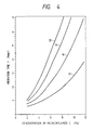
- Fig. 4 shows relationships between the concentration of polyacrylamide in the gel and the migration time (details of Fig. 4 will be described later). The concentration of polyacrylamide of the gel can be learned from the migration time.
- the gel having a polyacrylamide concentration of, for example, 6% is prepared to measure the amount of background fluorescence and, then, a newly prepared gel is measured for its amount of background fluorescence and is compared with the case of 6%, so that the polyacrylamide concentration of the gel can be learned.
- reference numeral 30 denotes an electrophoresis separation device (here, however, means for applying an electric field is a widely known one and is not diagrammed), and 33 denotes means for detecting fluorescence.
- Fig. 2 shows relationships between the base length (represented by the number of N of bases) of DNA fragments in the gels having various polyacrylamide concentrations and the migration time t.
- the curves 11, 12, 13, 14, 15 and 16 represent the cases where the gels have polyacrylamide concentrations of 2, 3, 4, 5, 6 and 8%.
- the migration time t0 becomes equal to l ⁇ g(T)/E0 in the equation (3) irrespective of the concentration C.
- the migration speed v(N) of the DNA fragment can be expressed as,
- Fig. 3 shows practically measured dependency of the band interval upon the concentration for various base lengths.
- the migration distance is 22 cm.
- the curves 17, 18, 19 and 20 represent the cases where the DNA fragments have base lengths (represented by the number N of bases) of 100, 200, 300 and 400.
- the base length is as short as about 100
- the band internval d increases with the increase in the gel concentration C.
- the base length is 300 or 400
- the band interval becomes nearly constant provided the gel concentration C is greater than 4%.
- the DNA band width ⁇ varies nearly in proportion to ⁇ l without almost depending upon the gel concentration.
- Fig. 4 is a diagram showing the dependency of migration time of various base lengths upon the concentration of polyacrylamide in the gel, and wherein curves 17, 18, 19 and 20 represent the cases where the DNA fragments have base lengths (represented by the number N of bases) of 100, 200, 300 and 400. It will be recognized from Fig. 4 that the migration time increases nearly in proportion to the square power of the concentration C.
- the migration time t is a function of the length l of migration lane and the gel concentration C.
- ⁇ 0(C, T) can be found using ⁇ 0bs / ⁇ l from the measured band interval ⁇ 0bs when the length of migration lane is l0.
- Table 1 shows polyacrylamide concentrations C that minimize the migration time required for discriminating the neighboring bands, the lengths l of migration lane and the migration times t when the gel plate has a thickness of 0.3 mm and the electric field intensity is 50 V/cm for various base lengths.
- Table 1 Base length N Concentration C of polyacrylamide Length l of migration lane Migration time t 100 6.2% 6 cm 22 min. 200 4.3% 15 cm 56 min. 300 3.2% 28 cm 106 min. 400 2.6% 41 cm 156 min.
- the gel concentrations should preferably be 4.3 to 6.2, 3.2 to 4.3, and 2.6 to 3.2%.
- up to 300 bases can be measured in about 1.5 hours.
- Fig. 5 shows the results of measurement using the gel having a polyacrylamide concentration of 3% and a migration lane of a length of 22 cm. The separation is not sufficient since the length of migration lane is shorter than 28 cm. It will, however, be comprehended that up to about 300 bases can be identified. The migration time of DNA fragment of a base length of 300 was 77 minutes.
- the spectrum 56 is the one obtained from a DNA fragment group in which a base of nucleic acid at the terminal is guanine
- the spectrum 57 is the one obtained from a DNA group in which a base of nucleic acid at the terminal is thymine.
- the gels having various polyacrylamide concentrations were prepared in the same manner as in the prior art. Described below is a procedure for preparing a gel having a polyacrylamide concentration of 6%. Water is added to a mixture consisting of 1.14 g of an acrylamide monomer, 0.06 g of an N,N′-methylenebisacrylamide (the total amount with the acrylamide monomer is 1.2 g), 0.16 g of trishydroxyaminomethane, 0.08 g of boric acid, 0.014 g of EDTA ⁇ 2Na and 6.3 g of urea, such that the total amount is 20 ml (i.e., the total amount of acrylamide monomer and N,N′-methylenebisacrylamide is 0.06 g in 1 ml).
- the DNA fragments that had hitherto been measured requiring a time of 5 to 10 hours can now be measured requiring a time of a little more than one hour.
- the measuring time required by the fluorescence detection type electrophoresis apparatus can be strikingly shortened.
- the light scattered by the gel and background fluorescence from impureties and/or gel imposes lower limit for the detection.
- the gel having a low concentration however, the background fluorescence decreases, too, and the detection can be realized maintaining high sensitivity.
Landscapes
- Health & Medical Sciences (AREA)
- Life Sciences & Earth Sciences (AREA)
- Molecular Biology (AREA)
- Chemical & Material Sciences (AREA)
- Biochemistry (AREA)
- Electrochemistry (AREA)
- Physics & Mathematics (AREA)
- Analytical Chemistry (AREA)
- Chemical Kinetics & Catalysis (AREA)
- General Health & Medical Sciences (AREA)
- General Physics & Mathematics (AREA)
- Immunology (AREA)
- Pathology (AREA)
- Investigating, Analyzing Materials By Fluorescence Or Luminescence (AREA)
- Measuring Or Testing Involving Enzymes Or Micro-Organisms (AREA)
- Investigating Or Analysing Biological Materials (AREA)
Abstract
Description
- The present invention relates to an apparatus for determining base sequences of DNA or RNA. More specifically, the invention relates to a fluorescence detection type electrophoresis apparatus adapted to shortening the measuring time.
- So far, base sequences of DNA'S have been determined by labelling a DNA fragment with a radioactive element, transferring a pattern onto a photograph through autoradiography, the pattern being subjected to the electrophoresis gel separation depending upon the length thereof, and reading the DNA band pattern involving, however, laborious work and time. A method, therefore, has been developed for determining the base sequence by separating and detecting DNA fragments in real time by using fluorescence label instead of using radioactive label. The real time detection method using fluorescence label and the fluorescence detection type electrophoresis apparatus used for this method have been disclosed, for example, in Journal of Biochemical and Biophysical Methods, 13, 1986, pp. 315-323 , Nature, Vol. 321, 1986, pp. 674-679 , and Science, Vol. 238, 1987, pp. 336-341 .
- According to the above prior art, however, a time which is as long as 5 to 10 hours is required from the start of migration of DNA fragments to the completion of measurement.
- The object of the present invention is to provide a fluorescence detection type electrophoresis apparatus which is capable of carrying out the measurement within short periods of time overcoming difficulties involved in the above-mentioned prior art in determining the base sequences of DNA or RNA.
- The above object of the present invention is achieved by optimizing various conditions in the electrophoresis gel migration. Concretely speaking, the object of the invention is achieved by selecting the polyacrylamide concentration of gel used for the electrophoresis separation device to be 2 to 6% (hereinafter, the polyacrylamide concentration is represented by the percentage of weight/volume (g/ml) of the total monomer concentration).
- That is, the fluorescence detection type electrophoresis apparatus of the present invention comprises an electrophoresis separation device for electrophoresis- separating a sample labelled with fluorescence, an excitation light source for exciting the sample, and detection means for detecting the fluorescence emitted by the sample that is excited, in order to determine the base sequences of the sample, wherein use is made of a gel plate having 2 to 6% of a polyacrylamide concentration as the electrophoresis separation device.
- Here, the electrophoresis separation device is the one in which DNA or RNA fragments are allowed to undergo electrophoresis to effect the electrophoresis separation depending upon the length of fragments, and includes at least, for example, an electrophoresis gel sandwitched between two pieces of transparent plates (quartz plate, etc.) and means for applying an electric field in the direction of migration.
- According to the study by the present inventors, the reason why the conventional fluorescence detection type electrophoresis apparatus requires a migration time of as long as 5 to 10 hours is attributed to that details of the electrophoresis gel migration phonomenon have not yet been clarified, that the real time detection method requires a long migration lane since the position resolution of fluorescence detection is poorer than the position resolution at the time of reading the band by visually observing the autoradiogram, and that in spite of this fact the measurement is taken using the electrophoresis separation device having an acrylamide concentration which is usually as high as about 8% just like in the autoradiography. The present invention was accomplished based upon the above-mentioned discovery by the present inventors.
- Here, if a DNA fragment having a base length N migrates at a speed v(N), the time t required for migrating the migration lane of a length ℓ is given by,
t=ℓ/v(N) (1) - The migration speed v(N) varies in proportion to the intensity of electric field, and the proportional coefficient v₀(N, C, T) is a function of the number of bases in the DNA fragment or the base length N, the polyacrylamide concentration C and the temperature T. Therefore, the equation (1) can be rewritten as,
t=ℓ/E·v₀(N,C,T) (2) - The time t shortens with the increase in the intensity of electric field resulting, however, in the generation of Joule's heat. Therefore, the temperature of the gel plate increases to make it difficult to separate the DNA band. Measurement can be taken within short periods of time if the DNA band is discriminated under the conditions of an electric field intensity and a temperature that do not hinder the measurement and if the gel concentration and the length ℓ of migration lane are so selected that the time t is minimized.
- The present invention employs the art of the conventional fluorescence detection type electrophoresis apparatus with the exception that the gel plate in the electrophoresis separation device has a polyacrylamide concentration of 2 to 6%.
-
- Fig. 1 is a schematic perspective view illustrating a fluorescence detection type electrophoresis apparatus according to an embodiment of the present invention;
- Fig. 2 is a graph illustrating relationships between the base length of a DNA fragment and the migration time in the gel plates having various polyacrylamide concentrations;
- Fig. 3 is a graph showing relationships between the polyacrylamide concentration and the band interval of DNA fragments of various base lengths in the gel plates;
- Fig. 4 is a graph showing relationships between the polyacrylamide concentration and the migration time of DNA fragments of various base lengths in the gel plates; and
- Fig. 5 is a graph showing spectra of DNA fragments when use is made of a gel plate having a polyacrylamide concentration of 3% and a length of migration lane of 22 cm.
- An embodiment of the present invention will now be described in conjunction with Figs. 1 to 5.
- Fig. 1 illustrates a fluorescence detection type electrophoresis apparatus according to the present invention. A
laser beam 1 for excitation emitted from alaser source 31 passes through alens 2 and enters into anelectrophoresis gel 4 from the side surface thereof. Theelectrophoresis gel 4 is held byquartz plates 3. Asample 32 such as DNA labelled with fluorescence starts migration from the upper end of thegel 4 and proceeds toward the lower end of thegel 4 while undergoing electrophoresis separation. Thelaser beam 1 falls on a place separated by a predetermined distance away from a point where migration started, and the fluorescence from the fluorescence-labelled DNA that passes therethrough is collected by alens 6 equipped with afilter 5, amplified through animage amplifier 7, permitted to pass through arelay lens 8, and is detected by a Vidicon camera 9 (trade name of RCA). The signals that are obtained are processed by acomputer 10 and are outputted The laser beam may be incident on the front surface while scanning over a predetermined lane instead of falling on the side surface. As will be understood from the equation (2), the migration time varies depending upon the distance ℓ from the point where migration started to a portion irradiated with the laser beam, the concentration C of polyacrylamide, and the voltage V applied across the upper end and the lower end of the gel plate (or electric field intensity E in the gel). - The concentration of polyacrylamide of the gel plate can be easily learned from the amount of background fluorescence when irradiated with the laser beam or from the migration time of the DNA fragment.
- When the gel plate is maintained at a temperature of 48°C and the electric field intensity is maintained at 50 V/cm, a time of about 85 minutes is required to migrate a DNA fragment of a 100 base length by 22 cm when the gel has a plyacrylamide concentration of 6%, and a longer period of time is required when the gel has a higher concentration. Fig. 4 shows relationships between the concentration of polyacrylamide in the gel and the migration time (details of Fig. 4 will be described later). The concentration of polyacrylamide of the gel can be learned from the migration time.
- Background fluorescence when the gel plate is irradiated with the laser beam increases in proportion to the concentration of polyacrylamide. Therefore, the gel having a polyacrylamide concentration of, for example, 6% is prepared to measure the amount of background fluorescence and, then, a newly prepared gel is measured for its amount of background fluorescence and is compared with the case of 6%, so that the polyacrylamide concentration of the gel can be learned.
- In Fig. 1,
reference numeral 30 denotes an electrophoresis separation device (here, however, means for applying an electric field is a widely known one and is not diagrammed), and 33 denotes means for detecting fluorescence. - Fig. 2 shows relationships between the base length (represented by the number of N of bases) of DNA fragments in the gels having various polyacrylamide concentrations and the migration time t. In Fig. 2, the
curves
t =(f(C,T)N+g(T)) (3)
where f(C, T) and g(T) are a function of the polyacrylamide concentration C and a function of the temperature T, and ℓ denotes the length of the migration lane. - When the base length is approximated to zero, the migration time t₀ becomes equal to ℓ·g(T)/E₀ in the equation (3) irrespective of the concentration C.
- From the equations (1) and (3), the migration speed v(N) of the DNA fragment can be expressed as,

- The interval d between the neighboring bands after the migration for a time t is given by,
d=(v(N)-v(N+1))t = ℓ/(N+g(T)/f(C,T)) (5) - It will be understood from this equation that when the base length N is great, the band interval d approaches ℓ/N without much depending upon the concentration C of polyacrylamide in the gel.
- Fig. 3 shows practically measured dependency of the band interval upon the concentration for various base lengths. The migration distance is 22 cm. In Fig. 3, the
curves
ω=ω₀(N,T)√ℓ (6)
where the proportional constant ω₀(N, T) is a function of the number N of bases and the temperature T. - Fig. 4 is a diagram showing the dependency of migration time of various base lengths upon the concentration of polyacrylamide in the gel, and wherein
curves - From d = ω and the equation (6), the length ω, i.e., ℓ=ω₀²(N,T)(N+g)(T)/f(C,T))² (7)
is found and is substituted for the equation (3) to obtain the following equation,
- From the equation (8), the value of C obtained as a solution of dt/dC = O stands for a gel concentration, i.e., the value C that minimizes the migration time t stands for a gel concentration that is to be found.
- From the following equation,

- That is, the gel concentrations C with which (t-t₀)=f(C,T)N=
 ·2g(T)=2t₀
holds true on Fig. 4 make the migration time t minimum, the gel concentrations being 6.2%, 4.3%, 3.2% and 2.6% when the base lengths are 100, 200, 300 and 400. In the practical analysis, the base lengths in many cases lie from 200 to 300. In this case, the gel concentration C should be from 3.2 to 4.3%. In the case of Fig. 4, the migration time t₀ was 28 minutes. Therefore, the time t with which t - t₀ = 2t₀ holds is one hour and 24 minutes. - By substituting f(C, T) = 2g(T) for the equation (7), the length ℓ of gel migration necessary for the separation is found to be,
ℓ=N²ω₀²(C,T) (10)
- From the equation (6), ω₀(C, T) can be found using ω0bs/√ℓ from the measured band interval ω0bs when the length of migration lane is ℓ₀.
- Table 1 shows polyacrylamide concentrations C that minimize the migration time required for discriminating the neighboring bands, the lengths ℓ of migration lane and the migration times t when the gel plate has a thickness of 0.3 mm and the electric field intensity is 50 V/cm for various base lengths.
Table 1 Base length N Concentration C of polyacrylamide Length ℓ of migration lane Migration time t 100 6.2% 6 cm 22 min. 200 4.3% 15 cm 56 min. 300 3.2% 28 cm 106 min. 400 2.6% 41 cm 156 min. - The values tabulated above are only rough indications that may undergo the change to some extent depending upon the migration voltage, the temperature of the migration gel and the thickness of the gel. To realize the high-speed migration, however, it will be recognized that the concentration of polyacrylamide be 2 to 6%, which is much lower than that of the conventional art.
- It will further be recognized that when the base lengths are 100 to 200, 200 to 300, and 300 to 400, the gel concentrations should preferably be 4.3 to 6.2, 3.2 to 4.3, and 2.6 to 3.2%.
- Under the above-mentioned conditions, up to 300 bases can be measured in about 1.5 hours.
- According to migration conditions corresponding to the base lengths shown in Table 1, it needs not be pointed out that DNA fragments having shorter base lengths can also be measured.
- Fig. 5 shows the results of measurement using the gel having a polyacrylamide concentration of 3% and a migration lane of a length of 22 cm. The separation is not sufficient since the length of migration lane is shorter than 28 cm. It will, however, be comprehended that up to about 300 bases can be identified. The migration time of DNA fragment of a base length of 300 was 77 minutes.
- In Fig. 5, the
spectrum 56 is the one obtained from a DNA fragment group in which a base of nucleic acid at the terminal is guanine, and thespectrum 57 is the one obtained from a DNA group in which a base of nucleic acid at the terminal is thymine. - In this embodiment, the gels having various polyacrylamide concentrations were prepared in the same manner as in the prior art. Described below is a procedure for preparing a gel having a polyacrylamide concentration of 6%. Water is added to a mixture consisting of 1.14 g of an acrylamide monomer, 0.06 g of an N,N′-methylenebisacrylamide (the total amount with the acrylamide monomer is 1.2 g), 0.16 g of trishydroxyaminomethane, 0.08 g of boric acid, 0.014 g of EDTA·2Na and 6.3 g of urea, such that the total amount is 20 ml (i.e., the total amount of acrylamide monomer and N,N′-methylenebisacrylamide is 0.06 g in 1 ml). This is a polyacrylamide concentration of 0.06 g/ml which is expressed as 6%. Then, there are added 64 µℓ of ammonium persulfate of a concentration of 15% and 10.7 µℓ of an N,N,N′,N-tetramethylethylene diamine, and the mixture is left to stand at room temperature, whereby polymerization takes place to form a gel having a concentration of 6%.
- According to the present invention as is obvious from the aforementioned embodiment, the DNA fragments that had hitherto been measured requiring a time of 5 to 10 hours can now be measured requiring a time of a little more than one hour. Thus, the measuring time required by the fluorescence detection type electrophoresis apparatus can be strikingly shortened.
- In the fluorescence measuring system, furthermore, the light scattered by the gel and background fluorescence from impureties and/or gel imposes lower limit for the detection. With the gel having a low concentration, however, the background fluorescence decreases, too, and the detection can be realized maintaining high sensitivity.
- Though the aforementioned embodiment has dealt with the case of DNA, substantially the same results are obtained even in the case of RNA.
Claims (5)
Applications Claiming Priority (2)
| Application Number | Priority Date | Filing Date | Title |
|---|---|---|---|
| JP63039385A JP2804038B2 (en) | 1988-02-24 | 1988-02-24 | Base sequence determination method |
| JP39385/88 | 1988-02-24 |
Publications (3)
| Publication Number | Publication Date |
|---|---|
| EP0330120A2 true EP0330120A2 (en) | 1989-08-30 |
| EP0330120A3 EP0330120A3 (en) | 1990-09-12 |
| EP0330120B1 EP0330120B1 (en) | 1995-06-28 |
Family
ID=12551541
Family Applications (1)
| Application Number | Title | Priority Date | Filing Date |
|---|---|---|---|
| EP89102908A Expired - Lifetime EP0330120B1 (en) | 1988-02-24 | 1989-02-20 | Fluorescence detection type electrophoresis apparatus |
Country Status (5)
| Country | Link |
|---|---|
| US (1) | US4971677A (en) |
| EP (1) | EP0330120B1 (en) |
| JP (1) | JP2804038B2 (en) |
| CN (1) | CN1019860B (en) |
| DE (1) | DE68923193T2 (en) |
Cited By (4)
| Publication number | Priority date | Publication date | Assignee | Title |
|---|---|---|---|---|
| EP0401821A1 (en) * | 1989-06-07 | 1990-12-12 | Hitachi Software Engineering Co., Ltd. | Electrophoresis pattern reading system of fluorescence type |
| EP0497468A2 (en) * | 1991-02-01 | 1992-08-05 | Beckman Instruments, Inc. | Method of improving signal-to-noise in electropherogram |
| US5242567A (en) * | 1990-05-22 | 1993-09-07 | Hitachi Software Engineering Co., Ltd. | Fluorescent pattern reading apparatus |
| EP1109014A2 (en) * | 1995-05-19 | 2001-06-20 | Iowa State University Research Foundation, Inc. | Multiplexed capillary electrophoresis system |
Families Citing this family (16)
| Publication number | Priority date | Publication date | Assignee | Title |
|---|---|---|---|---|
| US5230781A (en) * | 1984-03-29 | 1993-07-27 | Li-Cor, Inc. | Sequencing near infrared and infrared fluorescence labeled DNA for detecting using laser diodes |
| US5360523A (en) * | 1984-03-29 | 1994-11-01 | Li-Cor, Inc. | DNA sequencing |
| US5246866A (en) * | 1987-12-23 | 1993-09-21 | Hitachi Software Engineering Co., Ltd. | Method for transcription of a DNA sequence |
| JPH0743353B2 (en) * | 1990-05-31 | 1995-05-15 | 株式会社島津製作所 | Fluorescence detection type gel electrophoresis device |
| JP2785530B2 (en) * | 1991-09-13 | 1998-08-13 | 株式会社日立製作所 | Electrophoresis device |
| US5137609A (en) * | 1992-01-31 | 1992-08-11 | Biometric Imaging Inc. | Differential separation assay |
| US5424841A (en) * | 1993-05-28 | 1995-06-13 | Molecular Dynamics | Apparatus for measuring spatial distribution of fluorescence on a substrate |
| JP3340544B2 (en) * | 1993-12-24 | 2002-11-05 | 株式会社日立製作所 | Separation sampling device and method |
| US5627022A (en) * | 1994-11-01 | 1997-05-06 | Visible Genetics Inc. | Microgels for use in medical diagnosis and holders useful in fabricating same |
| US5507934A (en) * | 1994-11-01 | 1996-04-16 | Visible Genetics Inc. | Apparatus for preparing gels for use in electrophoretic separations and similar applications |
| JPH10513553A (en) * | 1995-01-23 | 1998-12-22 | マーレイ,アンソニー・ジェイ | Analysis of biological molecules |
| US5717602A (en) * | 1996-02-05 | 1998-02-10 | Kenning; Gregory G. | Automated electrophoresis and analysis system |
| US6436641B1 (en) * | 2000-04-17 | 2002-08-20 | Visible Genetics Inc. | Method and apparatus for DNA sequencing |
| KR20030031306A (en) * | 2001-10-13 | 2003-04-21 | 주식회사 커벡스 | High-compression method of gel image for eletrophoresis |
| CN1854716B (en) * | 2005-04-18 | 2012-03-21 | 微奥基因科技常州有限公司 | Integrated and light-source variable electrophoretic separating analyzer and its usage |
| CN101916029B (en) * | 2010-07-26 | 2011-12-28 | 亚亚科技股份有限公司 | High-performance illuminator for fluorescence photography |
Citations (3)
| Publication number | Priority date | Publication date | Assignee | Title |
|---|---|---|---|---|
| WO1982003128A1 (en) * | 1981-03-04 | 1982-09-16 | Commerce Us | Silver stains for protein in gels |
| EP0165013A2 (en) * | 1984-06-08 | 1985-12-18 | THE UNITED STATES OF AMERICA as represented by the Secretary United States Department of Commerce | Rapid visualization system for gel electrophoresis |
| EP0294524A1 (en) * | 1987-06-09 | 1988-12-14 | The Perkin-Elmer Corporation | Real time scanning electrophoresis apparatus for DNA sequencing |
Family Cites Families (6)
| Publication number | Priority date | Publication date | Assignee | Title |
|---|---|---|---|---|
| US4123343A (en) * | 1977-06-14 | 1978-10-31 | American Home Products Corporation | Purification of glycoproteins and immunization therewith |
| JPS6162843A (en) * | 1984-08-13 | 1986-03-31 | Hitachi Ltd | Fluorescence detection type electrophoretic apparatus |
| JPS61173158A (en) * | 1985-01-02 | 1986-08-04 | カリフオルニア・インステイテユ−ト・オブ・テクノロジ− | Method of determining arrangement of deoxyribonucleic acid |
| US4668667A (en) * | 1985-08-01 | 1987-05-26 | Norwich Eaton Pharmaceuticals, Inc. | Acylphosphorotriamides useful as lipid-altering agents |
| US4855225A (en) * | 1986-02-07 | 1989-08-08 | Applied Biosystems, Inc. | Method of detecting electrophoretically separated oligonucleotides |
| JP2550106B2 (en) * | 1987-10-30 | 1996-11-06 | 株式会社日立製作所 | Optical dispersion detection type electrophoretic device |
-
1988
- 1988-02-24 JP JP63039385A patent/JP2804038B2/en not_active Expired - Fee Related
-
1989
- 1989-02-15 US US07/310,645 patent/US4971677A/en not_active Expired - Lifetime
- 1989-02-20 DE DE68923193T patent/DE68923193T2/en not_active Expired - Lifetime
- 1989-02-20 EP EP89102908A patent/EP0330120B1/en not_active Expired - Lifetime
- 1989-02-23 CN CN89101129A patent/CN1019860B/en not_active Expired
Patent Citations (3)
| Publication number | Priority date | Publication date | Assignee | Title |
|---|---|---|---|---|
| WO1982003128A1 (en) * | 1981-03-04 | 1982-09-16 | Commerce Us | Silver stains for protein in gels |
| EP0165013A2 (en) * | 1984-06-08 | 1985-12-18 | THE UNITED STATES OF AMERICA as represented by the Secretary United States Department of Commerce | Rapid visualization system for gel electrophoresis |
| EP0294524A1 (en) * | 1987-06-09 | 1988-12-14 | The Perkin-Elmer Corporation | Real time scanning electrophoresis apparatus for DNA sequencing |
Non-Patent Citations (4)
| Title |
|---|
| ANALYTICAL BIOCHEMISTRY, vol. 54, no. 1, July 1973, pages 102-114, Academic Press, Inc.; E. WEIDEKAMM et al.: "A new sensitive, rapid fluoroscence technique for the determination of proteins in gel electrophoresis and in solution" * |
| JOURNAL OF BIOCHEMICAL AND BIOPHYSICAL METHODS, vol. 13, 1986, pages 315-323, Elsevier Science Publishers B.V. (Biomedical Division); W. ANSORGE et al.: "A non-radioactive automated method for DNA sequence determination" * |
| JOURNAL OF BIOCHEMICAL AND BIOPHYSICAL METHODS, vol. 9, 1984, pages 33-47, Elsevier Science Publishers B.V.; W. ANSORGE et al.: "System for DNA sequencing with resolution of up to 600 base pairs" * |
| NUCLEIC ACIDS RESEARCH, vol. 15, no. 11, 1987, pages 5693-4602, IRL Press Ltd, Oxford, GB; W. ANSORGE et al.: "Automated DNA sequencing: ultrasensitive detection of fluorescent bands during electrophoresis" * |
Cited By (8)
| Publication number | Priority date | Publication date | Assignee | Title |
|---|---|---|---|---|
| EP0401821A1 (en) * | 1989-06-07 | 1990-12-12 | Hitachi Software Engineering Co., Ltd. | Electrophoresis pattern reading system of fluorescence type |
| US5069769A (en) * | 1989-06-07 | 1991-12-03 | Hitachi Software Engineering Co., Ltd. | Electrophoresis pattern reading system of fluorescence type |
| USRE36826E (en) * | 1989-06-07 | 2000-08-22 | Hitachi Software Engineering Co., Ltd. | Electrophoresis pattern reading system of fluorescence type |
| US5242567A (en) * | 1990-05-22 | 1993-09-07 | Hitachi Software Engineering Co., Ltd. | Fluorescent pattern reading apparatus |
| EP0497468A2 (en) * | 1991-02-01 | 1992-08-05 | Beckman Instruments, Inc. | Method of improving signal-to-noise in electropherogram |
| EP0497468A3 (en) * | 1991-02-01 | 1993-08-11 | Beckman Instruments, Inc. | Method of improving signal-to-noise in electropherogram |
| EP1109014A2 (en) * | 1995-05-19 | 2001-06-20 | Iowa State University Research Foundation, Inc. | Multiplexed capillary electrophoresis system |
| EP1109014A3 (en) * | 1995-05-19 | 2002-08-28 | Iowa State University Research Foundation, Inc. | Multiplexed capillary electrophoresis system |
Also Published As
| Publication number | Publication date |
|---|---|
| CN1036639A (en) | 1989-10-25 |
| JPH01214752A (en) | 1989-08-29 |
| EP0330120B1 (en) | 1995-06-28 |
| EP0330120A3 (en) | 1990-09-12 |
| DE68923193T2 (en) | 1996-02-08 |
| US4971677A (en) | 1990-11-20 |
| CN1019860B (en) | 1992-12-30 |
| DE68923193D1 (en) | 1995-08-03 |
| JP2804038B2 (en) | 1998-09-24 |
Similar Documents
| Publication | Publication Date | Title |
|---|---|---|
| EP0330120A2 (en) | Fluorescence detection type electrophoresis apparatus | |
| Karger et al. | Multiwavelength fluorescence detection for DNA sequencing using capillary electrophoresis | |
| US6488832B2 (en) | Array based electrophoretic system for the analysis of multiple biological samples | |
| Chen et al. | Low-cost, high-sensitivity laser-induced fluorescence detection for DNA sequencing by capillary gel electrophoresis | |
| EP0314045B1 (en) | Wavelength dispersion electrophoresis apparatus | |
| Kostichka et al. | High speed automated DNA sequencing in ultrathin slab gels | |
| EP0626578B1 (en) | Apparatus for gel electrophoresis | |
| GB2314622A (en) | Electrophoresis separation and detection | |
| US6290831B1 (en) | Electrophoretic system for real time detection of multiple electrophoresed biopolymers | |
| US20050259256A1 (en) | Device and method for measurement | |
| JPH01112147A (en) | Method for determining base sequence of nucleic acid | |
| JPS6321556A (en) | Dna base sequence determining apparatus | |
| JP2610870B2 (en) | Base sequencer | |
| JPS613043A (en) | Apparatus for determining base sequence of nucleic acid | |
| JPH05296978A (en) | Electrophoretic device | |
| JP2914333B2 (en) | Base sequence determination method | |
| EP0275440A2 (en) | Photo detection system for DNA base analysis | |
| JPH01209351A (en) | Base sequence determining apparatus | |
| JP2853706B2 (en) | Base sequence determination method | |
| JP2840586B2 (en) | Light detection electrophoresis device | |
| US6793790B1 (en) | Sample collection system for gel electrophoresis | |
| Djouadi et al. | Dynamics of single‐stranded DNA migration in denaturing polyacrylamide slab‐gel electrophoresis | |
| JPH10176992A (en) | Dna-base-array decision apparatus | |
| JPH04244954A (en) | Apparatus for determining base sequence | |
| Lassiter | Analysis of near-infrared dye-labeled Sanger sequencing fragments with gel electrophoresis using the time-resolved flourescence lifetime indentification methods |
Legal Events
| Date | Code | Title | Description |
|---|---|---|---|
| PUAI | Public reference made under article 153(3) epc to a published international application that has entered the european phase |
Free format text: ORIGINAL CODE: 0009012 |
|
| AK | Designated contracting states |
Kind code of ref document: A2 Designated state(s): DE GB |
|
| PUAL | Search report despatched |
Free format text: ORIGINAL CODE: 0009013 |
|
| AK | Designated contracting states |
Kind code of ref document: A3 Designated state(s): DE GB |
|
| 17P | Request for examination filed |
Effective date: 19901212 |
|
| 17Q | First examination report despatched |
Effective date: 19920805 |
|
| GRAA | (expected) grant |
Free format text: ORIGINAL CODE: 0009210 |
|
| AK | Designated contracting states |
Kind code of ref document: B1 Designated state(s): DE GB |
|
| REF | Corresponds to: |
Ref document number: 68923193 Country of ref document: DE Date of ref document: 19950803 |
|
| PLBE | No opposition filed within time limit |
Free format text: ORIGINAL CODE: 0009261 |
|
| STAA | Information on the status of an ep patent application or granted ep patent |
Free format text: STATUS: NO OPPOSITION FILED WITHIN TIME LIMIT |
|
| 26N | No opposition filed | ||
| REG | Reference to a national code |
Ref country code: GB Ref legal event code: IF02 |
|
| PGFP | Annual fee paid to national office [announced via postgrant information from national office to epo] |
Ref country code: GB Payment date: 20080114 Year of fee payment: 20 |
|
| PGFP | Annual fee paid to national office [announced via postgrant information from national office to epo] |
Ref country code: DE Payment date: 20080307 Year of fee payment: 20 |
|
| REG | Reference to a national code |
Ref country code: GB Ref legal event code: PE20 Expiry date: 20090219 |
|
| PG25 | Lapsed in a contracting state [announced via postgrant information from national office to epo] |
Ref country code: GB Free format text: LAPSE BECAUSE OF EXPIRATION OF PROTECTION Effective date: 20090219 |