EP0181635A2 - Probes and methods useful for detecting chromosomal translocations - Google Patents
Probes and methods useful for detecting chromosomal translocations Download PDFInfo
- Publication number
- EP0181635A2 EP0181635A2 EP85114436A EP85114436A EP0181635A2 EP 0181635 A2 EP0181635 A2 EP 0181635A2 EP 85114436 A EP85114436 A EP 85114436A EP 85114436 A EP85114436 A EP 85114436A EP 0181635 A2 EP0181635 A2 EP 0181635A2
- Authority
- EP
- European Patent Office
- Prior art keywords
- antibody
- detecting
- labelled
- molecules
- polypeptide
- Prior art date
- Legal status (The legal status is an assumption and is not a legal conclusion. Google has not performed a legal analysis and makes no representation as to the accuracy of the status listed.)
- Ceased
Links
Images


Classifications
-
- C—CHEMISTRY; METALLURGY
- C12—BIOCHEMISTRY; BEER; SPIRITS; WINE; VINEGAR; MICROBIOLOGY; ENZYMOLOGY; MUTATION OR GENETIC ENGINEERING
- C12Q—MEASURING OR TESTING PROCESSES INVOLVING ENZYMES, NUCLEIC ACIDS OR MICROORGANISMS; COMPOSITIONS OR TEST PAPERS THEREFOR; PROCESSES OF PREPARING SUCH COMPOSITIONS; CONDITION-RESPONSIVE CONTROL IN MICROBIOLOGICAL OR ENZYMOLOGICAL PROCESSES
- C12Q1/00—Measuring or testing processes involving enzymes, nucleic acids or microorganisms; Compositions therefor; Processes of preparing such compositions
- C12Q1/68—Measuring or testing processes involving enzymes, nucleic acids or microorganisms; Compositions therefor; Processes of preparing such compositions involving nucleic acids
- C12Q1/6813—Hybridisation assays
- C12Q1/6827—Hybridisation assays for detection of mutation or polymorphism
-
- C—CHEMISTRY; METALLURGY
- C07—ORGANIC CHEMISTRY
- C07K—PEPTIDES
- C07K14/00—Peptides having more than 20 amino acids; Gastrins; Somatostatins; Melanotropins; Derivatives thereof
- C07K14/82—Translation products from oncogenes
-
- C—CHEMISTRY; METALLURGY
- C07—ORGANIC CHEMISTRY
- C07K—PEPTIDES
- C07K16/00—Immunoglobulins [IGs], e.g. monoclonal or polyclonal antibodies
- C07K16/18—Immunoglobulins [IGs], e.g. monoclonal or polyclonal antibodies against material from animals or humans
- C07K16/32—Immunoglobulins [IGs], e.g. monoclonal or polyclonal antibodies against material from animals or humans against translation products of oncogenes
-
- C—CHEMISTRY; METALLURGY
- C12—BIOCHEMISTRY; BEER; SPIRITS; WINE; VINEGAR; MICROBIOLOGY; ENZYMOLOGY; MUTATION OR GENETIC ENGINEERING
- C12Q—MEASURING OR TESTING PROCESSES INVOLVING ENZYMES, NUCLEIC ACIDS OR MICROORGANISMS; COMPOSITIONS OR TEST PAPERS THEREFOR; PROCESSES OF PREPARING SUCH COMPOSITIONS; CONDITION-RESPONSIVE CONTROL IN MICROBIOLOGICAL OR ENZYMOLOGICAL PROCESSES
- C12Q1/00—Measuring or testing processes involving enzymes, nucleic acids or microorganisms; Compositions therefor; Processes of preparing such compositions
- C12Q1/68—Measuring or testing processes involving enzymes, nucleic acids or microorganisms; Compositions therefor; Processes of preparing such compositions involving nucleic acids
- C12Q1/6876—Nucleic acid products used in the analysis of nucleic acids, e.g. primers or probes
- C12Q1/6883—Nucleic acid products used in the analysis of nucleic acids, e.g. primers or probes for diseases caused by alterations of genetic material
- C12Q1/6886—Nucleic acid products used in the analysis of nucleic acids, e.g. primers or probes for diseases caused by alterations of genetic material for cancer
-
- C—CHEMISTRY; METALLURGY
- C12—BIOCHEMISTRY; BEER; SPIRITS; WINE; VINEGAR; MICROBIOLOGY; ENZYMOLOGY; MUTATION OR GENETIC ENGINEERING
- C12Q—MEASURING OR TESTING PROCESSES INVOLVING ENZYMES, NUCLEIC ACIDS OR MICROORGANISMS; COMPOSITIONS OR TEST PAPERS THEREFOR; PROCESSES OF PREPARING SUCH COMPOSITIONS; CONDITION-RESPONSIVE CONTROL IN MICROBIOLOGICAL OR ENZYMOLOGICAL PROCESSES
- C12Q2600/00—Oligonucleotides characterized by their use
- C12Q2600/156—Polymorphic or mutational markers
-
- C—CHEMISTRY; METALLURGY
- C12—BIOCHEMISTRY; BEER; SPIRITS; WINE; VINEGAR; MICROBIOLOGY; ENZYMOLOGY; MUTATION OR GENETIC ENGINEERING
- C12Q—MEASURING OR TESTING PROCESSES INVOLVING ENZYMES, NUCLEIC ACIDS OR MICROORGANISMS; COMPOSITIONS OR TEST PAPERS THEREFOR; PROCESSES OF PREPARING SUCH COMPOSITIONS; CONDITION-RESPONSIVE CONTROL IN MICROBIOLOGICAL OR ENZYMOLOGICAL PROCESSES
- C12Q2600/00—Oligonucleotides characterized by their use
- C12Q2600/158—Expression markers
Definitions
- chromosomal translocations are known to be associated with human cancer.
- the chromosomal rearrangements that occur as a result of these translocations may activate human cellular oncogenes that have been rearranged.
- Chronic myelocytic leukemia is a pluripotent stem cell disease characterized by the presence of the Philadelphia (Ph') chromosome in the leukemic cells of 96% of all CML patients.
- the Ph' chromosome is the result of a translocation between chromosomes 22 and 9 (1).
- the human c-abl oncogene has been mapped to the long (q) arm of chromosome 9 (2). By analysis of somatic cell hybrids, we have shown that this oncogene is located on the Ph' (22q-) chromosome in Ph' positive CML demonstrating that c-abl is involved in the 9; 22 translocation (3).
- CM L The location of the c-abl oncogene adjacent to the translocation breakpoint in CM L was shown by the isolation of a DNA fragment from the 9q+ chromosome of a CML patient: this fragment contained both sequences of chromosome 9 and 22.
- the breakpoint had occurred 14 kb immediately 5' of the v-ab1 homologous sequences and resulted in a 9q + chromosome in which the tip of chromosome 9 including the v-abl homologous sequences was replaced by a portion of chromosome 22 (4).
- the isolated chromosome 22 sequences of this hybrid DNA fragment enabled us to study their role in the Ph' translocation in greater detail.
- a breakpoint cluster region (bcr) was identified on chromosome 22; the DNAs of all (over 30) Ph' positive CML patients examined to date have breakpoints in this region of up to 5.8 kb, strongly suggesting a role for bcr in chronic myelocytic leukemia (5).
- patients with the Ph' translocation could be identified using bcr specific genomic probes, see our pending patent application U.S. Serial No. 571,911 filed January 18, 1984, the contents of which are hereby incorporated by reference,this was experimentally difficult and in many cases it involved the use of several distinct probes.
- Lozzio and Lozzio (6) reported the isolation of a cell line K-562, from the pleural fluid of an adult patient with CML.
- This cell line expresses phenotypic markers of erythroid lineage and displays induced and spontaneous globin synthesis (7).
- We and others (4, 8, 9) have shown that the c-abl oncogene and the immunoglobulin light chain constant region (C) are amplified at least fourfold in this cell line.
- another human oncogene, c-sis normally located on chromosome 22 but transposed to chromosome 9 in the Ph' translocation (10), is non-amplified (4).
- K-562 contains a Ph' chromosome which is at least fourfold amplified; cytogenetic data are, however, not confirmative, as we cannot detect a Ph' chromosome in different pedigrees of this cell line and others (6) have suggested the presence of a single Ph' chromosome.
- Such findings leave the question as to whether amplification of the c-abl oncogene in K-562 is correlated with the presence of an amplified Ph' translocation, unresolved. If such a correlation exists, it would unambiguously demonstrate that K-562 is derived from Ph' positive leukemic cells of a CM L patient. We demonstrate the presence of a Ph' chromosomal breakpoint in the DNA of K-562.
- K-562 contains a breakpoint on chromosome 22 within the breakpoint cluster region on chromosome 22.
- K-562 DNA contains in contrast to DNAs isolated from patient material, amplified remnants of the Ph'-chromosome, including C, a part of bcr and c-abl. These amplified regions do not represent multiple copies of intact Ph'-chromosomes, but rather are present on one acrocentric marker chromosome (9). Most probably, the regions originate from a multiplication of a large region of DNA from the original Ph' chromosome. Restriction enzyme analysis with various restriction enzymes shows that all copies of the bcr breakpoint region contain identically sized breakpoint fragments.
- K-562 cells Although no difference in the degree of c-abi amplification can be observed in different passages of K-562, there is no evidence available that the original leukemic cells of the patient from which K-562 was established contained more than one Ph'-chromosome. More likely, amplification occurred after establishment of the cell line, leading to selection of cells with a growth advantage. K-562 cells also differ from other CM L cells in that the 9q+ chromosome cannot be detected by Southern blot analysis. This in concordance with results of cytogenetical analysis in which the 9q + chromosome was found to be absent from K-562 (9).
- This invention concerns construction and use of DNA probes to detect aberrant chromosomes resulting from translocations such as the Philadelphia translocation.
- the invention also concerns methods of detecting these same translocations by assaying for mRNA and protein products derived from these aberrant chromosomes.
- a single-stranded nucleic acid molecule is provided that is useful as a probe for detecting chromosomal translocations and has a nucleotide sequence which comprises at least one sequence present within an exon of a eucaryotic chromosomal gene and complementary to the mRNA encoded by the exon.
- the eucaryotic chromosomal gene is characterized by the presence within it of at least one chromosomal breakpoint site into which another nucleotide sequence may be translocated.
- the sequences are present within exons located 3' of chromosomal breakpoint sites into which an oncogene may be translocated.
- the translocation is the Philadelphia translocation
- the eucaryotic chromosomal gene is on human chromosome 22 and is characterized by the presence within it of a breakpoint cluster region and the translocated sequence is at least part of the c-abl oncogene.
- the single-stranded nucleic acid molecules may be DNA or RNA, preferably from about 20 to about 8,500 base pairs in length and may be labelled with detectable moieties, e.g. radioactive isotopes. These labelled probe molecules may used to detect chromosomal translocations, e.g. the Philadelphia translocation.
- the invention also provides a purified, mRNA molecule useful as an indicator of a chromosomal translocation.
- the mRNA is a transcript of a DNA sequence which comprises at least a portion of an exon of a eucaryotic chromosomal gene and at least a portion of another nucleotide sequence which has translocated into the gene.
- the nucleotide sequences are present within exons located 5' of the translocated sequence.
- the nucleotide sequences are located on the breakpoint cluster region of human chromosome 22 (bcr) and the translocated nucleotide sequence is at least part of the c-abl oncogene.
- the invention further provides polypeptides encoded by the purified, mRNA molecules, e.g. a hcrlc-abl polypeptide encoded by the bcr/c-abl mRNA molecule.
- Antibodies e.g. monoclonal antibodies, may be produced which are directed to antigenic determinants on this polypeptide. These antibodies may be used to detect a polypeptide, e.g. a bcr/c-abl polypeptide, present as a result of a chromosomal translocation, e.g. the Philadelphia translocation, and to diagnose an associated disease, e.g. chronic myelocytic leukemia, acute lymphocytic leukemia or acute myelocytic leukemia.
- an associated disease e.g. chronic myelocytic leukemia, acute lymphocytic leukemia or acute myelocytic leukemia.
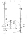
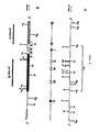
- Fig. 1 Restriction enzyme map of the K-562 breakpoint region on chromosome 22.
- A is the restriction enzyme map of human chromosome 22 sequences: the 5.8 kb Bg1 II - Bam HI (bcr) region encompasses the 0.6 HB and 1.2 HBg probes.
- D is the restriction enzyme map of the Ph' chromosome in K-562: the Ph' chromosomal breakpoint is indicated with an arrow.
- B and C represent more detailed restriction enzyme maps of the indicated regions of A and D.
- the solid bars represent chromosome 22 sequences whereas the open bars indicate sequences originating from chromosome 9. Probes used in the study are shown above A and below D.
- Fig. 2 Restriction enzyme map of the plasmid VI-3 CDNA insert.
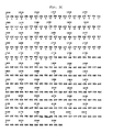
- Fig. 3 The DNA sequence and translation of VI-3 sequence.
- a detailed restriction enzyme map of VI-3 is shown.
- the black bar indicates the open reading frame sequences, the thin line non-translated sequences.
- the 3' end of the cDNA containing the poly A tail is depicted as An.
- A Ava I
- Ap Apa I
- B BstE II
- H Hind III
- Hf Hinf I
- P Pst I
- PII Pvu II
- S Sau 3A
- B g Bgl II
- Bs Bst E II.
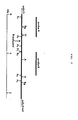
- Fig. 6 Schematic representation of the molecular consequence of the Ph translocation.
- a break occurs within an intron of the bcr gene on chromosome 22 and within or 5' to the c-abl oncogene on chromosome 9; these DNA sequences are physically joined on the Ph chromosome in a head to tail fashion; with bcr centromeric and c-abl telomeric.
- This configuration allows for the transcription of a hybrid mRNA, consisting of 5' bcr exons fused to c-abl coding sequences. This mRNA is translated into a hybrid polypeptide with a bcr amino-terminus and c-abl carboxy-terminus.
- Fig. 7 Genomic organization of the bcr gene.
- restriction enzyme map of chromosome 22 sequences encompassing bcr is shown in A. Exons are indicated by black boxes below the restriction enzyme map. The position of the numbered exons were located by hybridization to the VI-3 cDNA. The asterisk indicates a polymorphic Bgl II restriction enzyme site.
- a restriction enzyme map of the breakpoint cluster region is shown in B, with the exons as indicated in A. Below the map, the approximate positions of the breakpoints in different CML DNAs are indicated by horizontal or vertical arrows.
- the breakpoint of the 22q- chromosome from DNA 0319129 was cloned as a 9.5 kb Bgl II fragment in Charon 30 according to previously described methods (37). 9q+ fragments were isolated by cloning a 7.2 kb Bam HI fragment and a 7.7 kb EcoRI fragment into charon 30 and lambda gt wes respectively.
- F ig. 8 Breakpoint sequences of the DNAs of CML patients.
- the sequence of 0319129 DNA is shown in A; the sequences are in a 5' - 3' orientation.
- Normal chromosome 9 sequences (first line) are from non-CML DNA; the 9q+ and 22q- sequences (second and third lines) from DNA of the patients 0319129.
- Normal chromosome 22 sequences (fourth line) are from non-CML DNA.
- An arrow points to the breakpoint on chromosomes 9 and 22 1 the nucleotide C found both in the 9q + and 22q- sequences at the breakpoint is boxed. Limited regions of homology between the normal chromosome 9 and 22 sequences are underlined.
- the present invention concerns a single-stranded nucleic acid molecule useful as a probe for detecting a chromosomal translocation.
- the molecule has a sequence which comprises at least one sequence present within an exon of a eucaryotic chromosomal gene and complementary to the mRNA encoded by the exon.
- the eucaryotic chromosomal gene is characterized by at least one chromosomal breakpoint site into which another nucleotide sequence may be translocated.
- the invention also concerns nucleic acid molecules which are substantially complementary to these single-stranded nucleic acid molecules. These complementary nucleic acid molecules may also be used as probes for nucleic acid hybridization analysis of chromosomal breakpoints.
- the single-stranded nucleic acid molecule may be a DNA molecule or an RNA molecule and may be comprised exclusively or essentially of sequences present within exons or alternatively may be comprised of sequences present within both exons and introns. Desirably, at least one sequence present within an exon is located 3' of the chromosomal breakpoint site or sites. Typically, a plurality of sequences present within a plurality of exons are employed and are located 3' of the chromosomal breakpoint sites.
- This single-stranded nucleic acid molecule is of a length that permits the use of the molecule as a probe in nucleic acid hybridization experiments, preferably from about 20 base pairs to about 8,500 base pairs.
- DNA molecules of different sizes may be prepared by methods known to those of ordinary skill in the art, e.g. restriction enzyme cleavage or oligonucleotide synthesis.
- the eucaryotic chromosomal gene in which the exon nucleotide sequences are located is a human chromosomal and is characterized by the presence within it of a breakpoint cluster revision (bcr).
- the breakpoint site may be located within an intron or an exon.
- the gene is located on human chromosome 22 and the exon nucleotide sequences correspond to one or more of the bcr family of nucleotide sequences.
- the gene is located in the bcr which is specifically involved in the Philadelphia translocation in patients with chronic myelocytic leukemia (CML).
- CML chronic myelocytic leukemia
- the single-stranded DNA molecules of this invention may be included in a double-stranded D N A molecule, thus vector molecules may be constructed which comprise the DNA molecules of this invention, i.e. the DNA molecule may be incorporated as an insert in a vector such as a plasmid, e.g. the plasmid VI-3, a cosmid or a bacteriophage.
- a vector such as a plasmid, e.g. the plasmid VI-3, a cosmid or a bacteriophage.
- These vector molecules may be prepared by standard recombinant DNA techniques known to those of ordinary skill in the art.
- the molecules may be subcloned into plasmid vectors, such as the SP6 vector series, (26, 27) for the generation of complementary RNA molecules suitable for use as probes in hybridization procedures.
- the single-stranded DNA molecules of this invention may be labelled with detectable moieties. These labelled molecules may then be used in various nucleic acid hybridization methods known to those of ordinary skill in the art.
- the molecules may be labelled with any type of a detectable moiety such as a fluorescent, enzymatic or radioactive marker, e.g. by nick translation with 32p .
- These labelled molecules may be used to detect chromosomal translocations on a given eucaryotic chromosome.
- One such method comprises cleaving the chromosomal DNA from the chromosome with an appropriate restriction enzyme or enzymes under suitable conditions so as to produce DNA fragments. The resulting fragments are treated to obtain single-stranded DNA molecules.
- These single-stranded molecules are contacted with labelled single-stranded nucleic acid probe molecules of this invention under suitable conditions permitting hybridization of complementary molecules. The presence of hybridized molecules is detected and thereby the presence of a chromosomal translocation is detected by analyzing the results of the hybridization.
- the invention further provides a method of detecting a chromosomal translocation which comprises cleaving the chromosomal DNA with an appropriate restriction enzyme or enzymes and treating the resulting fragments by gel electrophoresis and denaturation to obtain single-stranded DNA molecules.
- the single-stranded DNA molecules are recovered from the gel and immobilized on a suitable solid support, e.g. a nitrocellulose filter.
- the immobilized single-stranded DNA fragments are then contacted with the labelled single-stranded nucleic acid probe molecules of this invention under suitable conditions permitting hybridization of complementary single-stranded molecules.
- the hybridized molecules so formed are identified, e.g. by autoradiography, and abnormalities in the restriction patterns of the chromosomal DNA caused by the chromosomal translocation are identified.
- a specific embodiment of the invention concerns a method of detecting the Philadelphia translocation in a human subject which comprises cleaving the subject's total chromosomal DNA with an appropriate restriction enzyme or enzymes under suitable conditions so as to produce DNA fragments. The resulting fragments are then treated to obtain single-stranded DNA molecules. The single-stranded DNA molecules so obtained are contacted with single-stranded nucleic acid molecules complementary to exon nucleotides of the bcr region of chromosome 22 under suitable conditions permitting hybridization of complementary molecules. The presence of the hybridized molecules is detected and thereby the chromosomal translocation is detected.
- One such method concerns detecting the Philadelphia translocation in a human subject by cleaving the subject's total chromosomal DNA with an appropriate restriction enzyme or enzymes under suitable conditions so as to produce DNA fragments.
- the resulting fragments so obtained are treated by gel electrophoresis and denaturation to obtain single-stranded DNA molecules.
- the single-stranded DNA molecules so obtained are recovered from the gel and immobilized on a suitable solid support, e.g. a nitrocellulose filter.
- the immobilized, single-stranded DNA molecules are then contacted with the labelled, single-stranded nucleic acid molecules complementary to the exons of the bcr region of chromosome 22, e.g.
- hybridized molecules so formed are identified e.g. by autoradiography, thereby detecting abnormalities in the restriction patterns of the chromosomal DNA caused by the Philadelphia translocation.
- probe molecules of this invention are not limited to those methods discussed herein.
- the probes of this invention may also be used in other nucleic acid hybridization methods known to those of ordinary skill in the art, e.g. in zitp- hybridization.
- the present invention also concerns a purified, hybrid mRNA molecule useful as an indicator of a chromosomal translocation.
- the mRNA molecule is a transcript of a DNA sequence which comprises at least a portion of an exon nucleotide sequence of a eucaryotic chromosomal gene and at least a portion of another nucleotide sequence which has translocated into the gene.
- the exon nucleotide sequences of the gene are located 5' of the translocated sequence.
- the translocated nucleotide sequence is at least part of an oncogene.
- the exon nucleotide sequences 5' of the translocated sequence are derived from either the bcr region of chromosome 22 or from one or more of the three bcr related sequences on chromosome 22.
- the purified mRNA molecule is a bcr/c-abl mRNA molecule from about 6.0 to about 9.0 kb in length. This molecule is a result of the Philadelphia translocation in which the c-abl oncogene from chromosome 9 is translocated to chromosome 22. The identification and isolation of this molecule is described in the experimental section of the specification.
- the mRNA molecules of this invention may be purified by any means known to those of ordinary skill in the art, e.g. purified by gel electrophoresis, column chromatography, density gradient centrifugation or nucleic acid hybridization methods. In certain embodiments they may be isolated and purified by hybridizing them to single-stranded DNA molecules which are substantially complementary to the mRNA molecule that is to be purified. Such single-stranded c-DNA molecules may be bound to a solid support, e.g. a c-DNA molecule complementary to the bcr/c-abl mRNA bound to cellulose beads or a nitrocellulose filter. After hybridization of complimentary strands unbound nucleic molecules may be washed off. The nucleic acid duplexes may then be denatured and the mRNA molecules may be obtained in a purified state.
- a solid support e.g. a c-DNA molecule complementary to the bcr/c-abl mRNA bound to cellulose beads or a
- the purified, hybrid mRNA molecules of this invention may also be used to prepare DNA or cRNA probes.
- cDNA or cRNA can be produced enzymatically, e.g. cDNA produced with reverse transcriptase.
- These cDNA molecules may then be cloned in a suitable vector system by standard recombinant DNA methods known to those of ordinary skill in the art.
- Various probe molecules may then be constructed from the cloned cDNA.
- the purified mRNA or cRNA may be labelled with a detectable moiety and used as a probe, e.g. enzymatically labelling the RNA with 32P by polynucleotide kinase. This mRNA probe may then be used to identify and isolate the translocated genes present on the aberrant chromosome by using standard molecular biology techniques known to those of ordinary skill in the art.
- the current invention also concerns polypeptides encoded by the purified, hybrid mRNA molecules. These polypeptides contain polypeptide sequences which are encoded for originally by two different genes. One such polypeptide is the bcr/c-abl protein encoded for by the bcr/c-abl mRNA. This polypeptide may contain a portion encoded for by the exon nucleotide sequences of the bcr region of chromosome 22 and a portion encoded by the oncogene c-abl.
- Antibodies to these polypeptides may also be prepared which may be directed against antigenic determinants on the polypeptide, e.g. determinants specifically created by the fusion of the two genes, e.g. determinants present in neither of the proteins of the fused genes separately, but present in the hybrid protein. Such antibodies may be used in various diagnostic or therapeutic methods. In other embodiments of the invention more than one antibody to the polypeptide may be prepared, each antibody directed to an antigenic determinant present on a different portion of the polypeptide. One such antibody may be directed to an antigenic determinant present on the portion of the polypeptide encoded for by the gene located on the chromosome into which another sequence has translocated, e.g.
- the other antibody may be directed to an antigenic determinant located on the portion of the polypeptide encoded for by the nucleotide sequence that has been translocated into the chromosome, e.g. a portion encoded for by an oncogene such as the oncogene c-abl.
- These antibodies may be used to diagnose a disease related to a chromosomal translocation which produces a specific protein product, e.g. CML evidenced by the Philadelphia translocation and the presence of the bcr/c-abl polypeptide.
- two different antibodies directed to two different sites on the polypeptide may be used in immunometric or sandwich type assays.
- either or both of the antibodies are monoclonal antibodies.
- the antibodies of this invention may also be labelled with detectable moieties, such as radioactive, e.g. 1 25I, fluorescent, e.g. fluorescein, or enzymatic, e.g. peroxidase, moieties.
- Suitable conditions for formation of an antibody-antigen complex comprise solution phase and emulsion reaction systems, preferably aqueous, maintained at a pH value from about 6.0 to about 8.0 and at a temperature from about 40C to about 450C.
- the polyclonal antibodies and monoclonal antibodies of this invention may be prepared by methods known to those of ordinary skill in the art.
- the monoclonal antibodies may be prepared by techniques as disclosed by Kohler, et al., Nature Vol. 256: 995-497 (1975). The process involves injecting a mouse or other animal with an immunogen. The mouse is later sacrificed and cells taken from its spleen are fused with myeloma cells, producing hybridomas. The population of hybridomas produced in vitro is screened to isolate individual clones each of which secretes a single antibody species to the antigen. The antibodies produced by the hybridomas are then screened to identify those having the highest affinity for the immunogenic substance of interest.
- Monoclonal antibodies directed to different portions of a hybrid polypeptide may be produced by using the entire polypeptide itself as an immunogen and then screening the hybridomas produced for antibodies directed to antigenic determinants on different portions of the polypeptide.
- fragments of the various portions of the hybrid polypeptides e.g. a portion of the bcr/c-abl polypeptide which is encoded by the exon nucleotide sequences present on the bcr region of chromosome 22, may be used as immunogens.
- large amounts of the polypeptide could be produced with knowledge of the restriction data disclosed herein and use of standard prokaryotic and eucaryotic expression systems known to those of ordinary skill in the art.
- Another approach is to use synthetic peptides analogous to portions of the hybrid polypeptide as an immunogen, e.g. a synthetic polypeptide analogous to an antigenic determinant present on the bcr portion of the bcr/c-abl polypeptide.
- One embodiment of the invention concerns a method of detecting a chromosomal translocation, e.g. the P hila- delphia translocation by determining the presence of a polypeptide encoded by a mRNA resulting from the translocation, e.g. the bcr/c-abl polypeptide.
- the method comprises contacting a sample containing the polypeptide with an antibody directed to an antigenic site on the polypeptide, the antibody being labelled with a detectable moiety, under suitable conditions permitting the formation of an antibody-antigen complex.
- the antibody may be a polyclonal or monoclonal antibody.
- the unbound, labelled antibody is then separated from the labelled antibody-antigen complex, and the presence of the labelled antibody-antigen complex is then detected by suitable means, e.g. autoradiography or fluorescent detection.
- the antibodies of this invention may also be used in a method of detecting chromosomal translocations in which two different antibodies directed against antigenic determinants on different fractions of the polypeptide are used.
- a sample containing the polypeptide is contacted with a measured amount of a first antibody directed to an antigenic site on the portion of the polypeptide encoded for by the exon nucleotide sequences of the gene into which another nucleotide sequence has been translocated under suitable conditions so that an antibody-antigen complex is formed.
- the first antibody is labelled with a detectable moiety.
- the labelled antibody-antigen complex is then contacted with a second antibody directed to an antigenic site on the portion of the polypeptide encoded for by the nucleotide sequence which has translocated into chromosome, e.g. an oncogene, under suitable conditions permitting the formation of a labelled antibody-antigen-antibody complex.
- the second antibody is bound to a suitable solid support, e.g. a microtiter well, a cellulose bead, a nitrocellulose filter.
- the unbound labelled antibody is then separated from the labelled antibody-antigen-antibody complex bound to the solid support and then either the amount of bound labelled antibody-antigen-antibody complex or the amount of unreacted labelled antibody is measured in order to determine the presence of the polypeptide.
- this procedure may be conducted by using a method wherein the first antibody is not labelled but is attached to the solid support and the second antibody is labelled with the detectable moiety.
- a specific embodiment of the invention concerns a method of detecting the Philadelphia translocation by detecting the presence of the bcr/c-abl polypeptide.
- the method comprises contacting a sample obtained from the subject with a measured amount of first antibody to an antigenic site on the portion of the polypeptide encoded for by the c-abl oncogene, under suitable conditions permitting the formation of an antibody-antigen complex.
- the first antibody is labelled with a detectable moiety.
- the complex so formed is contacted with a second antibody to an antigenic site on the portion of the polypeptide encoded for by exon nucleotide sequences of the gene on chromosome 22, under suitable conditions permitting the formation of a labelled antibody-antigen-antibody complex.
- the second antibody is bound to a suitable solid support.
- the unbound labelled antibody is separated from the labelled antibody-antigen-antibody complex bound to the solid support. Either the amount of labelled antibody-antigen-antibody complex bound to the solid support or the unbound labelled antibody is measured in order to detect the presence of the polypeptide. Alternatively, this procedure may be conducted by using a method wherein the first antibody is not labelled but is attached to the solid support and the second antibody is labelled with .a detectable moiety.
- Another method of detecting a chromosomal translocation on a given eucaryotic chromosome such as the Philadelphia translocation comprises isolating poly A mRNA ' molecules encoded for by genes on the chromosome, e.g. mRNA from a human subject which contains mRNA encoded by genes on the Ph chromosome, and separating the poly A mRNAs, e.g. by gel electrophoresis.
- the separated RNA molecules are immobilized on a suitable solid support, e.g. immobilized in the dried gel or on a nitrocellulose filter, and contacted with labeled probe RNA molecules of this invention under suitable conditions permitting hybridization of complementary molecules.
- the presence of the hybridized molecules is detected and thereby the presence of the chromosomal translocation is detected.
- Another embodiment of the invention concerns methods of diagnosing CM L , AML or ALL in a human subject by detecting the presence of the Philadelphia translocation.
- Ph' positive leukemias including K-562
- genomic DNA probes such as 1.2 HBg is inadequate for their detection.
- a cosmid library was constructed from K-562 DNA partially digested with Mbo I, according to published procedures (11). Numerous colonies of the approximately 100,000 recombinants hybridized with 0.6 HB probe. Three such positives containing overlapping portions of the same region were selected for further restriction enzyme analysis (Fig. lD). It is evident from a comparison of the detailed restriction enzyme maps of normal chromosome 22 sequences (Fig. 1B) and K-562 DNA (Fig. lC) that the homology between the two terminates 3'to the most 5' Ava I site.
- a 1.OE probe prepared from K-562 DNA immediately 3' to the breakpoint hybridizes to DNA isolated from somatic cell hybrids containing human chromosome 9 in the absence of human chromosome 22, but not from hybrids containing chromosome 22. This indicates that the sequences isolated from K-562 DNA come from different chromosomes and contain the breakpoint of the 22q-chromosome. The entire region is amplified at least fourfold. The chromosome 9 specific sequences are also amplified, as demonstrated by the strong hybridization of the 1.OE probe to K-562 DNA in comparison with control DN A .
- the amplification of chromosome 9 sequences begins at the point where the breakpoint has occurred on chromosome 9 in the Ph' translocation and extends in the direction of the telomere of the chromosome, including the c-abl oncogene.
- the amplified region may be relatively large, as the distance between the breakpoint on chromosome 9 and the most 5' v-abl homologous exon is, at minimum, 64 kb.
- GM 637 cDNA containing plasmid were obtained from Hiroto Okayama.
- This library is a cDNA library of the SV-40 transformed human fibroblast cell line GM 637. The construction of the library is described in Okayama and Berg (21). It would be clear to one skilled in the art that any representative human cDNA library may be used. It is also obvious that one skilled in the art could prepare a comparable cDNA library from the human GM 637 cell line, which is available from the NIGMS Human Genetic Mutants Cell Repository, Camden, New Jersey.
- E. coli DH1 was transformed with the 10 micrograms of GM 637 cDNA containing plasmids by standard recombinant DNA procedures known to those of ordinary skill in the art.
- the transformed E. coli cultures were plated out on ten large plates. Replicas of these plates were made and the bacteria were scraped off for culturing.
- An SV-cDNA library was constructed by preparing ten large scale plasmid preparations from these recombinant bacteria. The preparations were designated SV-1 thru SV-10.
- each plasmid preparation was digested in 100 microliters of Sail restriction enzyme buffer with 42 units of the restriction enzyme Sail. The digestion was for 4 hours under suitable buffer and temperature conditions known to those of ordinary skill in the art. After digestion each of the DNA samples was extracted with phenol and ethanol precipitated. The samples were then electrophoresed on a 0.6% agarose gel for 4 days at 5-18 volts. The DNA in the gel was transferred to a nitrocellulose filter (Schleicher & Schuell) according to the procedure of Southern. (15).
- the 0.6 HB probe was nick translated with 32 p and was hybridized to the cDNA samples immobilized on the nitrocellulose filters according to procedures known to those of ordinary skill in the art. (17). After hybridization was completed the filters were washed under the high stringency conditions of 0.3 x SSC at 650C. Autoradiography of the washed filters showed hybridization to DNA bands of about 5 kb in all of the samples except the sample of preparation SV-6.
- the entire SV-cDNA library was then replated onto large nitrocellulose filters (Millipore HATF). Replicas of these filters were made and hybridized to the 0.6 HB probe. Positive colonies were replated onto small nitrocellulose filters at a density suitable for the isolation of single colonies. These filters were replicated and hybridized to the 0.6 HB probe. Colonies that hybridized to the probe were picked and grown up on a scale suitable for large scale DNA isolation.
- plasmid VI-3 contained the largest insert, about 2.2 kb, and was chosen for still further analysis.
- a detailed restriction enzyme map of this insert was constructed (Fig. 2) and the 5'-3' orientation of the cDNA was determined.
- the cDNA insert of plasmid VI-3 was then used as a probe in hybridization experiments with Southern blots of genomic DNA of human chromosome 22 including the bcr region. This region of chromosome 22 is contained in the cosmid clones Ca 22-1 and Ca 22-2.
- the cosmid clone preparation, restriction digest, electrophoresis, blotting and hybridization procedures were performed according to standard methods known to those of ordinary skill in the art. Numerous restriction enzyme fragments which are not contiguous in the genomic DNA hybridized to the VI-3 probe indicating that this cDNA is indeed a reverse transcribed copy of part of an mRNA encoded for by a gene that encompasses within its coding region the bcr.
- the VI-3 cDNA insert has been sequenced (Fig. 3). The sequencing was performed according to the dideoxy method (22).
- VI-3 cDNA encompasses, at a minimum, 13 exons dispersed over a region of approximately 45 kb in the genomic DNA. Of these 13 exons, four are within the bcr region; one encompasses nucleotides 481-836 in the cDNA. The remaining exons are located 5' and 3' to the bcr region.
- VI-3 cDNA insert e.g. 500 base pairs
- a number of such probes can be constructed from this cDNA insert.
- Such probes will preferably be derived from the region of the VI-3 cDNA insert within the most 5' Bgl II restriction site and the most 5' Ava I restriction site (Fig. 2) and will detect all chromosomal abnormalities in the bcr region, including the breakpoint fragment of the K-562 cell line.
- the VI-3 cDNA sequence is useful as a probe for detecting the Philadelphia translocation.
- This probe may be prepared as described in this specification or alternatively it or a substantially similar polynucleotide molecule may be prepared by any method of polynucleotide synthesis known to the art, e.g. enzymatic or chemical polynucleotide synthesis. Probes synthesized by this method can be used to probe cDNA libraries or genomic DNA for chromosome 22 DNA sequences. By using such probes, one of ordinary skill in the art can isolate chromosome 22 bcr sequences from any representative human cDNA library or from genomic DNA.
- the DNA sequence of the breakpoint region of K-562 was compared with the normal genomic chromosome 22 DNA sequences.
- the two sequences are identical up to a point approximately in the center of the region shown in Fig. 5. 3' of this point, chromosome 9 sequences are joined to those of chromosome 22. No sequence homology is apparent between the chromosome 9 and 22 DNA sequences. Lack of homology between sequences involved in the 9;22 recombination event in CML is in concordance with the results obtained from sequence analysis of the 8;14 translocation in Burkitt lymphoma, in which no obvious homology was detected between the recombined sequence (13).
- the Philadelphia translocation therefore causes a break within a gene, of which the bcr is a part.
- chromosome 9 sequences including the c-abl oncogene are joined to the gene containing the bcr.
- polyA RNA was isolated from K-562 and control HELA cells.
- a c-abl specific probe detects a 6.0 and a 7.0 kb mRNA in both HELA cells and in K-562.
- a novel c-abl homologous mRNA of 8.5 kb is detected in K-562.
- Novel c-abl homologous mRNAs have also been detected in CM L patient material (19, 20).
- This 8.5 kb mRNA appears analogous to the 8.5 kb mRNA in K-562 in that it hybridizes to probe A but not to probe B. Therefore, this mRNA is most probably characteristic of CML patients exhibiting the Philadelphia translocation. It follows that a bcr/c-abl protein product of this mRNA is also present and that it is highly probable that this protein is implicated in the causation and or progression of CML.
- Probe A and B have also been used in Southern blot hybridization analysis of DNA derived from the K-562 and GM 637 cell lines. The probes hybridized to the expected restriction fragments. These results confirm that probe A picks up the abnormal restriction pattern associated with the Philadelphia translocation and is thus a good hybridization probe and useful in the diagnosis of CML.
- VI-3 cDNA is represented in a gene family.
- This gene family contains, at minimum, 4 members, as a single exon containing probe isolated from the 3' region of Ca 22-2 hybridizes to at least 4 discrete fragments in total human DNA cut with different restriction enzymes. Two members of this family have been molecularly cloned from the K-562 cosmid library described in this specification. Overlapping cosmids, spanning a region of 80 kb have been isolated, which contain a region of extremely close homology to the VI-3 3' sequences.
- cosmids of which the cosmid VKII-1 is the prototype, contain sequences also located on chromosome 22, but 5' (more toward the centromere) of the bcr region.
- the second member has been isolated in one cosmid spanning a region of 40 kb.
- This cosmid, VKII-3 also contains sequences hybridizing to the 0.46 kb Ps + fragment mentioned above.
- PG Me25-Nu contains human chromosome 22 as its only human chromosome complement.
- the three members of this gene family on chromosome 22 are: a) the sequences represented by cosmid VKII-1 b) the sequences present in the bcr region and c) sequences 3' (telomeric) of the bcr region represented by the cosmid VKII-3, which are not amplified in K-562 and which are presumably translocated from chromosome 22 in the Ph'- translocation.
- chromosome 22 Since the bcr region on chromosome 22 is specifically and consistently involved in the translocation resulting in the formation of the Ph'- chromosome, it is reasonable to assume that other members of its family may also be involved in specific translocations known to occur between chromosome 22 and other chromosomes, be it in neoplasia, such as in Ewing Sarcoma, or in hereditary disorders, such as the di-Georgio syndrome. The methods and probes of this invention, therefore may be useful in diagnosing other diseases associated with specific chromosomal translocations.
- the cDNA contains one long open reading frame starting at the polyG tail at the 5' end and continuing to nucleotide 1770 where a stopcodon is encountered. All other reading frames have numerous stopcodons within the entire region.
- the long open reading frame has the coding capacity for 589 amino acid residues corresponding to a protein of approximately 65,000 Mw.
- a polyadenylation signal is found at nucleotide 2182 followed by a polyA tail beginning at base 2208, indicating that the cDNA contains the complete 3' end of the gene.
- the DNA sequence of the junction point of the translocation might provide additional information, concerning the mechanism of chromosomal translocation.
- Figure 8A the sequence of the region containing the crossover point for translocation in the DNA of patient 0319129 is shown and compared with normal chromosome 22 and 9 DNA sequences.
- the chromosomal break has occurred in a rather “precise” manner, leading to the generation of a 22q- and 9q + sequence exactly reflecting the sequence of the normal chromosome 22 and 9 DNA sequences.
- a C nucleotide is found both in the 9q+ and 22q- sequence, whereas the chromosome 22 sequence contains a G at that position.
- both chromosomes 9 and 22 contain limited stretches of homology near the break (Fig. 8A underlined); a DNA search revealed homology of this region to human Alu-repetitive sequences.
- the chromosome 9 sequences of 9q+ fragment contain 13 nucleotide changes within a 81 bp stretch as compared to the normal chromosome 9 sequences. These changes may reflect differences between individuals (the control chromosome 9 sequences were isolated from a human lung carcinoma cosmid library); however, the number of nucleotide substitutions would be very high in such event.
- the chromosome 22 part of the 9q + fragment contains no nucleotide changes within either intron or exon sequences, favoring an explanation of inefficient DNA repair upon deletion of chromosome 9 sequences.
- bcr is part of a gene oriented with its 5' end toward the centromere of chromosome 22.
- Philadelphia translocation a break occurs within the b U , and sequences from chromosome 9 containing the human c-abl oncogene in all Ph'-positive cases examined (5, 33), are translocated to the 3' of the truncated bcr gene.
- the joining of bcr and c-abl sequences is highly specific for CML as this configuration has been found in complex translocations (33) and even in the leukemic cells of one CML patient cytogenetically lacking the Ph' chromosome (34).
- c-abl Since the orientation of c-abl on chromosome 9 is also centromere 5' - 3' - telomere, bcr and c-abl are joined in a head- to-tail fashion on the Ph' chromosome. Although the distance from the most v-abl homologous exon to the physical breakpoint may vary from 14 kb (in the case of patient 0319129) to over 100 kb (in K562), the effect of the translocation on the expression of the bcr gene and c-abl seems to be very similar in different patients: in K562 and in Ph'-positive CML patients abnormal RNA transcripts of around 8.5 kb are detected, which hybridize both to c-abl and 5' bcr, exon probes (28, 30).
- hybrid mRNAs must be the result of transcription initiating at the promoter of the bcr gene. Depending on the exact location of the breakpoint, the transcript will include all 5' exons in addition to either exons 1 and 2 or exons indicated 1, 2 and 3 of the bcr. Transcription continues into the c-abl oncogene, including, at minimum, the most 5' v-abl homologous exon and all exons 3' of it including the phosphotyrosine acceptor site (35). If the inclusion of exon 3 in the hybrid mRNA has any effect on the progression of the disease is as of yet unknown.
- Chromosomal aberrations may be generated by specific events involving recombination - prone DNA sequences: alternatively, such recombination events could occur almost at random. In either case, a very limited number of translocations will result in gene alterations leading to the disruption of normal growth and differentiation.
- Ph' translocation we have found that b reakpoints on chromosome 9 are spread over a region of up to 100 kb. The breakpoints on chromosome 22 occur within a smaller region of around 5.0 kb. Nonetheless, no sequence homology can be found comparing breakpoint regions of different CML patients or coding regions of c-ab1 and bcr genes.
- Restriction enzymes were purchased from either New England Biolabs or Bethesda Research Laboratories (BRL) and were used according to the suppliers' specifications. DNAs were digested with restriction enzymes, subjected to electrophoresis through 0.75% agarose gels, and transferred to nitrocellulose (Schleicher and Schuell, pH 7.9) essentially as described by Southern (25). Nick translation of probes and filter hybridization were as described (23, 24). Specific activity of the probes was 2-5 x 10⁸ cpm/ microgram. After hybridization, filters were exposed to XAR-2 film (Kodak) for up to 5 days at -700C with Dupont Lightning Plus intensifying screens.
- DNA probes were prepared by digestion of DNA with appropriate restriction enzymes, followed by electrophoresis through low-melting-point agarose (BRL). Desired bands were excised from gels and brought into solution by heating at 65 0 C for 30 min. Agarose was removed by two extractions with phenol equilibrated with 0.3 M NaOAc, pH 5.0, and one extraction with phe- nol/chloroform/isoamyl alcohol (25:24:1). DNA was precipitated with ethanol and 0.2 M NaOAc, pH 4.8, in the presence of 20 micrograms/ml Dextran T-500 (Pharmacia) as carrier.
- BBL low-melting-point agarose
- the cell lines K-562 and GM 637 were obtained from the NIGMS, Human Genetic Mutants Cell Repository, Institute for Medical Research, Copewood and Davis Streets, Camden, New Jersey, 08103.
- Patient 2252 is a 45 year old white male. He presented with CML on 1/26/83 and had the standard t(9;22) translocation. His karyotype (46, XY, Ph') has remained unchanged. His peripheral blood is normal.
- Patient 2397 is a 43 year old female with a 46, XX, t(9;22), Ph' karyotype.
- the first CML diagnosis was on 10/17/83. She has been evaluated regularly; her karyotype remains unchanged. Her peripheral blood is normal.
- Patient 1708 is a white female presented with CML on 7/24/80. All 40 cells examined had the 46, XX, t(9;22), Ph' karyotype. The latest bone marrow aspirate was done on 7/9/84. In the 40 cells examined similar cytogenetic findings were found. She is in the chronic phase. For the last six years cytogenetic evaluation has been done serially. Peripheral blood cells stimulated with P HA are normal.
- Patient 2172 is a 52 year old white male. He was originally diagnosed as having CML on 9/22/82. He has been followed since then for every three months. His karyotype remains 46, XY, t(9 1 22), Ph'. The PHA stimulated peripheral blood is normal.
- Total RN A was isolated according to the LiCl/urea method (18). Fresh bone marrow samples (1-2.5 ml) were homogenized in 25 ml 3M LiCl, 6 M urea on ice for 2 x 30 seconds in a polytron homogenizer. The homogenate was kept 3 hours on ice. The precipitate was centrifuged at 10,000 g for 1 hour and dissolved in 3.2 ml of 1:1 mixture of buffer (10 mM Tris-HCl, pH 7.5, 5 mM EDTA, 0.2% SDS) and phenol: chloroform: isoamylalcohol (25:24:1).
- the 32 p labelled RNA probes are generated by transcription labeling according to the pSP riboprobe method (Promega Biotech). 1-5 x 10⁸ cpm 32P labelled RNA was used per hybridization, corresponding to 0.5-2.5 x 10⁸ cpm/microgram input DN A . Filters were exposed to XAR-2 film (Kodak) at -70 o C with Dupont Lighting plus intensifying screens.
- Blots were first prehybridized in 100 ml 50% formamide, 50 mM sodium phosphate buffer pH 6.5, 5 x 5 SSC, 1 mM EDTA, 0.1% SDS, 2.5 x Denhardts, 250 micrograms/ml salmon sperm DNA, 250 micrograms/ml calfs liver RNA, 10 micrograms/ml poly A for four hours at 550C.
- Hybridization was in 35 ml hybridization mix (4 parts pre- hybridization mix and 1 part 50% dextran sulphate) overnight at 550C.
- the filters were washed 2 x 20 minutes in 20 mM sodium phosphate buffer, pH 6.5 1 mM EDTA, 0.1% SDS, 2.5 x SSC, at 550C. Following this the filters were washed 2 x 20 minutes in 20 mM sodium phosphate buffer, pH 6.5, 1 mM EDTA, 0.1% SDS, 0.3 x SSC at 650C, and similarly, in the same solution but with 0.1 and 0.03 x SSC consecutively, at 65 0 C.
- a bcr/abl mRNA exists in the leukemic cells of CML patients we would expect to detect an abnormal sized abl transcript; the same abnormal sized mRNA should hybridize with the 5' bcr probe (probe A) but not with probe B, the 3' bcr probe as the genomic sequences corresponding to this probe are translocated to chromosome 9 in the t(9;22).
- All patients have an 8.5 kb transcript that hybridizes both with the c-ab1 and the 5' specific bcr probe.
- the 3' specific bcr probe does not hybridize with this transcript, which excludes that it is a normal bcr messenger.
- the 8.5 kb messenger seems to be characteristic of Ph' positive cells, as it is not found in either normal control cells or cells of other types of leukemia (19,29).
- Three bands around 7.5, 7.0 and 4.5 kb are detected with both the 5' and the 3' bcr probe, indicating that these may represent the normal bcr mRNAs.
- the 7.5, 7.0 and 4.5 kb transcripts are also present in other human cell lines and tissues, including several other myeloid and lymphoid cell lines, erythroid precursor cells, skin, gut, kidney and spleen. None of the samples tested contained the 8.5 kb bcr related RNA.
- the bone marrow cells from all the patients contain a 6.0 and 7.0 kb c-ab1 transcript. These transcripts are also present in normal cells (29) and represent the normal c-abl mRNAs. Presumably both are initiated at the same promoter but terminate differently at the 3' end of the abl oncogene. Although all five patients have both the 8.5 kb hybrid and the normal abl transcripts, the hybrid transcript is expressed at higher level.
- v-abl probes an abnormally sized c-abl RNA was detected in the human ALL cell line SMS-SB (41). Translation of a hybrid mRNA, as found in the five CML patients, would result in a bcr/abl fusion protein. Evidence for the existence of such protein has been found in the CMLO cell line K562, in which a similar bcr-c-abl transcript is found, an abnormally sized c-abl protein of 210 K molecular weight (P210) (14) is detected, suggesting that this protein is the translational product of the bcr/abl mRNA.
- P210 K molecular weight
- mice Although the tumorigenic potential of this protein is unknown, it cannot be coincidence that it involved the c-ab1 oncogene since v-ab1, the result of a recombination of murine leukemia virus and the mouse c-abl oncogene (4) induces a rapid B cell lymphoma in mice (43, 44).
Abstract
Description
- Throughout this application various publications are referenced by numbers within parentheses. Full citations for these publications may be found at the end of the specification immediately preceding the claims. The disclosures of these publications in their entireties are hereby incorporated by reference into this application in order to more fully describe the state of the art to which this invention pertains.
- Certain specific chromosomal translocations are known to be associated with human cancer. The chromosomal rearrangements that occur as a result of these translocations may activate human cellular oncogenes that have been rearranged.
- Chronic myelocytic leukemia (CML) is a pluripotent stem cell disease characterized by the presence of the Philadelphia (Ph') chromosome in the leukemic cells of 96% of all CML patients. The Ph' chromosome is the result of a translocation between
chromosomes 22 and 9 (1). The human c-abl oncogene has been mapped to the long (q) arm of chromosome 9 (2). By analysis of somatic cell hybrids, we have shown that this oncogene is located on the Ph' (22q-) chromosome in Ph' positive CML demonstrating that c-abl is involved in the 9; 22 translocation (3). The location of the c-abl oncogene adjacent to the translocation breakpoint in CML was shown by the isolation of a DNA fragment from the 9q+ chromosome of a CML patient: this fragment contained both sequences ofchromosome chromosome 9 including the v-abl homologous sequences was replaced by a portion of chromosome 22 (4). The isolatedchromosome 22 sequences of this hybrid DNA fragment enabled us to study their role in the Ph' translocation in greater detail. A breakpoint cluster region (bcr) was identified onchromosome 22; the DNAs of all (over 30) Ph' positive CML patients examined to date have breakpoints in this region of up to 5.8 kb, strongly suggesting a role for bcr in chronic myelocytic leukemia (5). Although patients with the Ph' translocation could be identified using bcr specific genomic probes, see our pending patent application U.S. Serial No. 571,911 filed January 18, 1984, the contents of which are hereby incorporated by reference,this was experimentally difficult and in many cases it involved the use of several distinct probes. - In 1975, Lozzio and Lozzio (6) reported the isolation of a cell line K-562, from the pleural fluid of an adult patient with CML. This cell line expresses phenotypic markers of erythroid lineage and displays induced and spontaneous globin synthesis (7). We and others (4, 8, 9) have shown that the c-abl oncogene and the immunoglobulin light chain constant region (C) are amplified at least fourfold in this cell line. In contrast, another human oncogene, c-sis, normally located on
chromosome 22 but transposed tochromosome 9 in the Ph' translocation (10), is non-amplified (4). These data suggest that K-562 contains a Ph' chromosome which is at least fourfold amplified; cytogenetic data are, however, not confirmative, as we cannot detect a Ph' chromosome in different pedigrees of this cell line and others (6) have suggested the presence of a single Ph' chromosome. Such findings leave the question as to whether amplification of the c-abl oncogene in K-562 is correlated with the presence of an amplified Ph' translocation, unresolved. If such a correlation exists, it would unambiguously demonstrate that K-562 is derived from Ph' positive leukemic cells of a CML patient. We demonstrate the presence of a Ph' chromosomal breakpoint in the DNA of K-562. The breakpoint has occurred in bcr confirming our previous results that a breakpoint in bcr is highly specific for CML. These findings establish that the progenitor cell of K-562 contains the Ph' translocation and that C and c-abl are located on the same amplification unit. - The present findings demonstrate that the CML cell line K-562, as all Ph' positive CML patient material examined to date, contains a breakpoint on
chromosome 22 within the breakpoint cluster region onchromosome 22. However, K-562 DNA contains in contrast to DNAs isolated from patient material, amplified remnants of the Ph'-chromosome, including C, a part of bcr and c-abl. These amplified regions do not represent multiple copies of intact Ph'-chromosomes, but rather are present on one acrocentric marker chromosome (9). Most probably, the regions originate from a multiplication of a large region of DNA from the original Ph' chromosome. Restriction enzyme analysis with various restriction enzymes shows that all copies of the bcr breakpoint region contain identically sized breakpoint fragments. - Although no difference in the degree of c-abi amplification can be observed in different passages of K-562, there is no evidence available that the original leukemic cells of the patient from which K-562 was established contained more than one Ph'-chromosome. More likely, amplification occurred after establishment of the cell line, leading to selection of cells with a growth advantage. K-562 cells also differ from other CML cells in that the 9q+ chromosome cannot be detected by Southern blot analysis. This in concordance with results of cytogenetical analysis in which the 9q+ chromosome was found to be absent from K-562 (9).
- The absence of the 9q+ chromosome strengthens the hypothesis that the 22q- chromosome is critical to the malignant proliferation of these leukemic cells. In K-562, the amplification of human c-abl DNA sequences is coupled to an increased level of expression of an abnormally sized 8.5 kb c-abl mRNA, (12, 19) indicating active transcription from the translocated and amplified c-abl oncogene. Recently, an altered human c-abl protein was demonstrated in K-562, differing both in size and in the presence of associated protein kinase activity from the normal human protein (21). However, these previous studies of altered c-abl protein and mRNA did not determine that these molecules were hybrid molecules. We demonstrate that in fact a hybrid mRNA and protein product exist as a result of the Philadelphia translocation. We have invented novel diagnostic tests for CML and other diseases associated with similar translocations based on the hybrid nature of these aberrant gene products.
- This invention concerns construction and use of DNA probes to detect aberrant chromosomes resulting from translocations such as the Philadelphia translocation. The invention also concerns methods of detecting these same translocations by assaying for mRNA and protein products derived from these aberrant chromosomes.
- A single-stranded nucleic acid molecule is provided that is useful as a probe for detecting chromosomal translocations and has a nucleotide sequence which comprises at least one sequence present within an exon of a eucaryotic chromosomal gene and complementary to the mRNA encoded by the exon. The eucaryotic chromosomal gene is characterized by the presence within it of at least one chromosomal breakpoint site into which another nucleotide sequence may be translocated.
- In one embodiment of the invention the sequences are present within exons located 3' of chromosomal breakpoint sites into which an oncogene may be translocated. In a presently preferred embodiment of the invention, the translocation is the Philadelphia translocation, the eucaryotic chromosomal gene is on
human chromosome 22 and is characterized by the presence within it of a breakpoint cluster region and the translocated sequence is at least part of the c-abl oncogene. - The single-stranded nucleic acid molecules may be DNA or RNA, preferably from about 20 to about 8,500 base pairs in length and may be labelled with detectable moieties, e.g. radioactive isotopes. These labelled probe molecules may used to detect chromosomal translocations, e.g. the Philadelphia translocation.
- The invention also provides a purified, mRNA molecule useful as an indicator of a chromosomal translocation. The mRNA is a transcript of a DNA sequence which comprises at least a portion of an exon of a eucaryotic chromosomal gene and at least a portion of another nucleotide sequence which has translocated into the gene. In one embodiment of the invention the nucleotide sequences are present within exons located 5' of the translocated sequence. In a presently preferred embodiment invention the nucleotide sequences are located on the breakpoint cluster region of human chromosome 22 (bcr) and the translocated nucleotide sequence is at least part of the c-abl oncogene.
- The invention further provides polypeptides encoded by the purified, mRNA molecules, e.g. a hcrlc-abl polypeptide encoded by the bcr/c-abl mRNA molecule. Antibodies, e.g. monoclonal antibodies, may be produced which are directed to antigenic determinants on this polypeptide. These antibodies may be used to detect a polypeptide, e.g. a bcr/c-abl polypeptide, present as a result of a chromosomal translocation, e.g. the Philadelphia translocation, and to diagnose an associated disease, e.g. chronic myelocytic leukemia, acute lymphocytic leukemia or acute myelocytic leukemia.
- A is the restriction enzyme map of
human chromosome 22 sequences: the 5.8 kb Bg1 II - Bam HI (bcr) region encompasses the 0.6 HB and 1.2 HBg probes. D is the restriction enzyme map of the Ph' chromosome in K-562: the Ph' chromosomal breakpoint is indicated with an arrow. B and C represent more detailed restriction enzyme maps of the indicated regions of A and D. The solid bars representchromosome 22 sequences whereas the open bars indicate sequences originating fromchromosome 9. Probes used in the study are shown above A and below D. (A= Ava I; B= Bam HI; Bg= Bg1 II; E= Eco RI; H= Hind III; K= Kpn I; P= Pst I; S= Sst I; Xh= Xho I.) - The isolation of the VI-3 cDNA plasmid is described in detail in the specification. Poly G represents the 5' end of the cDNA; the poly A tail of the 3' is also indicated. The breakpoint in K562 is in an intron; exons 5' of the arrow will remain on
chromosome 22 and constitute part of the hybrid mRNA. Probes A and B are 5' and 3' fragments of the cDNA subcloned into a pSP vector. (Poly A tail= 3'; Pv= Pvu II; Bg= Bgl II; H= Hind III; Ps= Pst I; A= Ava I.) - Amino acid residues are indicated with one letter symbol below the sequence. The exact position of exons designated 1-6 within VI-3 bcr cDNA is shown by the numbers above the sequence.. DNA sequencing was performed using the dideoxy-chain termination method on restriction enzyme fragments of the cDNA subcloned into M13 phage. All regions were sequenced on both strands.
- A detailed restriction enzyme map of VI-3 is shown. The black bar indicates the open reading frame sequences, the thin line non-translated sequences. The 3' end of the cDNA containing the poly A tail is depicted as An. (A = Ava I, Ap = Apa I, B = BstE II, H = Hind III, Hf = Hinf I, P = Pst I, PII = Pvu II, S = Sau 3A, Bg = Bgl II, Bs = Bst E II.)
- Top:
normal chromosome 22 DNA sequences. Bottom: the corresponding DNA sequences of the breakpoint region in K-562. An 0.37 kb Ava I/Eco RI fragment of K-562 (Fig. 1C) and an 0.37 kb Ava I/Pst 1 (Fig. 1B) fragment ofnormal chromosome 22 were sequenced according to the dideoxy method essentially as described in 22. The region shown is located 241 bp 3' to the Ava I site. - In the Ph -translocation, a break occurs within an intron of the bcr gene on
chromosome 22 and within or 5' to the c-abl oncogene onchromosome 9; these DNA sequences are physically joined on the Ph chromosome in a head to tail fashion; with bcr centromeric and c-abl telomeric. This configuration allows for the transcription of a hybrid mRNA, consisting of 5' bcr exons fused to c-abl coding sequences. This mRNA is translated into a hybrid polypeptide with a bcr amino-terminus and c-abl carboxy-terminus. - The restriction enzyme map of
chromosome 22 sequences encompassing bcr is shown in A. Exons are indicated by black boxes below the restriction enzyme map. The position of the numbered exons were located by hybridization to the VI-3 cDNA. The asterisk indicates a polymorphic Bgl II restriction enzyme site. A restriction enzyme map of the breakpoint cluster region is shown in B, with the exons as indicated in A. Below the map, the approximate positions of the breakpoints in different CML DNAs are indicated by horizontal or vertical arrows. (A = Ava I, B = BamHI, Bg = Bgl II, E = EcoRI, H = Hind III, P = Pst I, S = Sma I, Ss = Sst I, X = Xho I.) - The breakpoint of the 22q- chromosome from
DNA 0319129 was cloned as a 9.5 kb Bgl II fragment inCharon 30 according to previously described methods (37). 9q+ fragments were isolated by cloning a 7.2 kb Bam HI fragment and a 7.7 kb EcoRI fragment intocharon 30 and lambda gt wes respectively. - The sequence of 0319129 DNA is shown in A; the sequences are in a 5' - 3' orientation.
Normal chromosome 9 sequences (first line) are from non-CML DNA; the 9q+ and 22q- sequences (second and third lines) from DNA of thepatients 0319129.Normal chromosome 22 sequences (fourth line) are from non-CML DNA. An arrow points to the breakpoint onchromosomes normal chromosome normal chromosome normal chromosome chromosome 9 sequences indicate nucleotide differences at those positions with the 9q+ chromosome. The beginning of exon 3 (see Fig. 6B) in the 9q+ and 22 sequence is indicated in the figure. Small restriction enzyme fragments containing the breakpoints were chosen for sequence analysis, based on restriction enzyme mapping data and comparison withnormal chromosome - A. Restriction enzyme map of bcr; the 5.8 kb breakpoint cluster region, in which all breakpoints occur on
chromosome 22 in the Ph'-translocation, is indicated above the map. The location of exons immediately 5', 3' and within bcr are schematically shown (not drawn to scale) beneath the map. Number exons are referred to in text. - B. Restriction enzyme map of the bcr cDNA; numbers and shading of regions in the cDNA correspond to the exons similarly marked in A. The bcr cDNA probes used in this study are shown beneath the map. (A = Ava I, B = BamH I, Bg = Bg1 II, Bs = BstE II, E = EcoRI, H = Hind III, P = Pst I, Pv = Pvu II. The AAAN at the 3' end of the cDNA indicates the poly A tail.)
- The present invention concerns a single-stranded nucleic acid molecule useful as a probe for detecting a chromosomal translocation. The molecule has a sequence which comprises at least one sequence present within an exon of a eucaryotic chromosomal gene and complementary to the mRNA encoded by the exon. The eucaryotic chromosomal gene is characterized by at least one chromosomal breakpoint site into which another nucleotide sequence may be translocated.
- The invention also concerns nucleic acid molecules which are substantially complementary to these single-stranded nucleic acid molecules. These complementary nucleic acid molecules may also be used as probes for nucleic acid hybridization analysis of chromosomal breakpoints.
- The single-stranded nucleic acid molecule may be a DNA molecule or an RNA molecule and may be comprised exclusively or essentially of sequences present within exons or alternatively may be comprised of sequences present within both exons and introns. Desirably, at least one sequence present within an exon is located 3' of the chromosomal breakpoint site or sites. Typically, a plurality of sequences present within a plurality of exons are employed and are located 3' of the chromosomal breakpoint sites.
- This single-stranded nucleic acid molecule is of a length that permits the use of the molecule as a probe in nucleic acid hybridization experiments, preferably from about 20 base pairs to about 8,500 base pairs. DNA molecules of different sizes may be prepared by methods known to those of ordinary skill in the art, e.g. restriction enzyme cleavage or oligonucleotide synthesis.
- Preferably, the eucaryotic chromosomal gene in which the exon nucleotide sequences are located is a human chromosomal and is characterized by the presence within it of a breakpoint cluster revision (bcr). The breakpoint site may be located within an intron or an exon. In one embodiment of this invention, the gene is located on
human chromosome 22 and the exon nucleotide sequences correspond to one or more of the bcr family of nucleotide sequences. In a specific embodiment of the invention, the gene is located in the bcr which is specifically involved in the Philadelphia translocation in patients with chronic myelocytic leukemia (CML). In over 90% of patients with CML and a lower, but significant, fraction of patients with acute myelocytic leukemia (AML) and acute lymphocytic leukemia (ALL), at least part of the nucleotide sequences of the c-abl oncogene which is normally located onchromosome 9 are translocated tochromosome 22 in the Philadelphia translocation. The construction of a single-stranded DNA molecule of this embodiment of the invention is described in the Experimental Section of the specification. Several different probe molecules may be constructed from this DNA molecule by methods such as restriction enzyme cleavage or oligonucleotide synthesis. For instance, the plasmid VI-3 may be used to construct several different probes as shown in Fig. 2. This molecule has been sequenced and this sequence appears in the Fig. 3. Other single-stranded DNA molecules useful as probes for other chromosomal translocations may be constructed using the methods of this invention as described in the Experimental Section. - The single-stranded DNA molecules of this invention may be included in a double-stranded DNA molecule, thus vector molecules may be constructed which comprise the DNA molecules of this invention, i.e. the DNA molecule may be incorporated as an insert in a vector such as a plasmid, e.g. the plasmid VI-3, a cosmid or a bacteriophage. These vector molecules may be prepared by standard recombinant DNA techniques known to those of ordinary skill in the art. Alternatively, the molecules may be subcloned into plasmid vectors, such as the SP6 vector series, (26, 27) for the generation of complementary RNA molecules suitable for use as probes in hybridization procedures.
- The single-stranded DNA molecules of this invention may be labelled with detectable moieties. These labelled molecules may then be used in various nucleic acid hybridization methods known to those of ordinary skill in the art. The molecules may be labelled with any type of a detectable moiety such as a fluorescent, enzymatic or radioactive marker, e.g. by nick translation with 32p.
- These labelled molecules may be used to detect chromosomal translocations on a given eucaryotic chromosome. One such method comprises cleaving the chromosomal DNA from the chromosome with an appropriate restriction enzyme or enzymes under suitable conditions so as to produce DNA fragments. The resulting fragments are treated to obtain single-stranded DNA molecules. These single-stranded molecules are contacted with labelled single-stranded nucleic acid probe molecules of this invention under suitable conditions permitting hybridization of complementary molecules. The presence of hybridized molecules is detected and thereby the presence of a chromosomal translocation is detected by analyzing the results of the hybridization.
- The invention further provides a method of detecting a chromosomal translocation which comprises cleaving the chromosomal DNA with an appropriate restriction enzyme or enzymes and treating the resulting fragments by gel electrophoresis and denaturation to obtain single-stranded DNA molecules. The single-stranded DNA molecules are recovered from the gel and immobilized on a suitable solid support, e.g. a nitrocellulose filter. The immobilized single-stranded DNA fragments are then contacted with the labelled single-stranded nucleic acid probe molecules of this invention under suitable conditions permitting hybridization of complementary single-stranded molecules. The hybridized molecules so formed are identified, e.g. by autoradiography, and abnormalities in the restriction patterns of the chromosomal DNA caused by the chromosomal translocation are identified.
- A specific embodiment of the invention concerns a method of detecting the Philadelphia translocation in a human subject which comprises cleaving the subject's total chromosomal DNA with an appropriate restriction enzyme or enzymes under suitable conditions so as to produce DNA fragments. The resulting fragments are then treated to obtain single-stranded DNA molecules. The single-stranded DNA molecules so obtained are contacted with single-stranded nucleic acid molecules complementary to exon nucleotides of the bcr region of
chromosome 22 under suitable conditions permitting hybridization of complementary molecules. The presence of the hybridized molecules is detected and thereby the chromosomal translocation is detected. - One such method concerns detecting the Philadelphia translocation in a human subject by cleaving the subject's total chromosomal DNA with an appropriate restriction enzyme or enzymes under suitable conditions so as to produce DNA fragments. The resulting fragments so obtained are treated by gel electrophoresis and denaturation to obtain single-stranded DNA molecules. The single-stranded DNA molecules so obtained are recovered from the gel and immobilized on a suitable solid support, e.g. a nitrocellulose filter. The immobilized, single-stranded DNA molecules are then contacted with the labelled, single-stranded nucleic acid molecules complementary to the exons of the bcr region of
chromosome 22, e.g. a fragment of an insert of the plasmid VI-3 such as probe A, Fig. 2, under suitable conditions permitting hybridization of complementary single-stranded molecules. The hybridized molecules so formed are identified e.g. by autoradiography, thereby detecting abnormalities in the restriction patterns of the chromosomal DNA caused by the Philadelphia translocation. - The methods of using the probe molecules of this invention are not limited to those methods discussed herein. The probes of this invention may also be used in other nucleic acid hybridization methods known to those of ordinary skill in the art, e.g. in zitp- hybridization.
- The present invention also concerns a purified, hybrid mRNA molecule useful as an indicator of a chromosomal translocation. The mRNA molecule is a transcript of a DNA sequence which comprises at least a portion of an exon nucleotide sequence of a eucaryotic chromosomal gene and at least a portion of another nucleotide sequence which has translocated into the gene.
- In certain embodiments of the invention the exon nucleotide sequences of the gene are located 5' of the translocated sequence. In other embodiments the translocated nucleotide sequence is at least part of an oncogene. In another embodiment of the invention the exon nucleotide sequences 5' of the translocated sequence are derived from either the bcr region of
chromosome 22 or from one or more of the three bcr related sequences onchromosome 22. In one specific embodiment the purified mRNA molecule is a bcr/c-abl mRNA molecule from about 6.0 to about 9.0 kb in length. This molecule is a result of the Philadelphia translocation in which the c-abl oncogene fromchromosome 9 is translocated tochromosome 22. The identification and isolation of this molecule is described in the experimental section of the specification. - The mRNA molecules of this invention may be purified by any means known to those of ordinary skill in the art, e.g. purified by gel electrophoresis, column chromatography, density gradient centrifugation or nucleic acid hybridization methods. In certain embodiments they may be isolated and purified by hybridizing them to single-stranded DNA molecules which are substantially complementary to the mRNA molecule that is to be purified. Such single-stranded c-DNA molecules may be bound to a solid support, e.g. a c-DNA molecule complementary to the bcr/c-abl mRNA bound to cellulose beads or a nitrocellulose filter. After hybridization of complimentary strands unbound nucleic molecules may be washed off. The nucleic acid duplexes may then be denatured and the mRNA molecules may be obtained in a purified state.
- The purified, hybrid mRNA molecules of this invention may also be used to prepare DNA or cRNA probes. Once the mRNA molecules have been purified, cDNA or cRNA can be produced enzymatically, e.g. cDNA produced with reverse transcriptase. These cDNA molecules may then be cloned in a suitable vector system by standard recombinant DNA methods known to those of ordinary skill in the art. Various probe molecules may then be constructed from the cloned cDNA. Alternatively the purified mRNA or cRNA may be labelled with a detectable moiety and used as a probe, e.g. enzymatically labelling the RNA with 32P by polynucleotide kinase. This mRNA probe may then be used to identify and isolate the translocated genes present on the aberrant chromosome by using standard molecular biology techniques known to those of ordinary skill in the art.
- The current invention also concerns polypeptides encoded by the purified, hybrid mRNA molecules. These polypeptides contain polypeptide sequences which are encoded for originally by two different genes. One such polypeptide is the bcr/c-abl protein encoded for by the bcr/c-abl mRNA. This polypeptide may contain a portion encoded for by the exon nucleotide sequences of the bcr region of
chromosome 22 and a portion encoded by the oncogene c-abl. - Antibodies to these polypeptides may also be prepared which may be directed against antigenic determinants on the polypeptide, e.g. determinants specifically created by the fusion of the two genes, e.g. determinants present in neither of the proteins of the fused genes separately, but present in the hybrid protein. Such antibodies may be used in various diagnostic or therapeutic methods. In other embodiments of the invention more than one antibody to the polypeptide may be prepared, each antibody directed to an antigenic determinant present on a different portion of the polypeptide. One such antibody may be directed to an antigenic determinant present on the portion of the polypeptide encoded for by the gene located on the chromosome into which another sequence has translocated, e.g. the exon nucleotide sequences on
chromosome 22 encoding the bcr region of the bcr/c-abl polypeptide. The other antibody may be directed to an antigenic determinant located on the portion of the polypeptide encoded for by the nucleotide sequence that has been translocated into the chromosome, e.g. a portion encoded for by an oncogene such as the oncogene c-abl. These antibodies may be used to diagnose a disease related to a chromosomal translocation which produces a specific protein product, e.g. CML evidenced by the Philadelphia translocation and the presence of the bcr/c-abl polypeptide. In certain embodiments of the invention two different antibodies directed to two different sites on the polypeptide may be used in immunometric or sandwich type assays. - In a preferred embodiment of the invention, either or both of the antibodies are monoclonal antibodies. The antibodies of this invention may also be labelled with detectable moieties, such as radioactive, e.g. 125I, fluorescent, e.g. fluorescein, or enzymatic, e.g. peroxidase, moieties.
- Suitable conditions for formation of an antibody-antigen complex comprise solution phase and emulsion reaction systems, preferably aqueous, maintained at a pH value from about 6.0 to about 8.0 and at a temperature from about 40C to about 450C.
- The polyclonal antibodies and monoclonal antibodies of this invention may be prepared by methods known to those of ordinary skill in the art. The monoclonal antibodies may be prepared by techniques as disclosed by Kohler, et al., Nature Vol. 256: 995-497 (1975). The process involves injecting a mouse or other animal with an immunogen. The mouse is later sacrificed and cells taken from its spleen are fused with myeloma cells, producing hybridomas. The population of hybridomas produced in vitro is screened to isolate individual clones each of which secretes a single antibody species to the antigen. The antibodies produced by the hybridomas are then screened to identify those having the highest affinity for the immunogenic substance of interest.
- Monoclonal antibodies directed to different portions of a hybrid polypeptide may be produced by using the entire polypeptide itself as an immunogen and then screening the hybridomas produced for antibodies directed to antigenic determinants on different portions of the polypeptide. Alternatively, fragments of the various portions of the hybrid polypeptides, e.g. a portion of the bcr/c-abl polypeptide which is encoded by the exon nucleotide sequences present on the bcr region of
chromosome 22, may be used as immunogens. For either purpose, large amounts of the polypeptide could be produced with knowledge of the restriction data disclosed herein and use of standard prokaryotic and eucaryotic expression systems known to those of ordinary skill in the art. Another approach is to use synthetic peptides analogous to portions of the hybrid polypeptide as an immunogen, e.g. a synthetic polypeptide analogous to an antigenic determinant present on the bcr portion of the bcr/c-abl polypeptide. - One embodiment of the invention concerns a method of detecting a chromosomal translocation, e.g. the Phila- delphia translocation by determining the presence of a polypeptide encoded by a mRNA resulting from the translocation, e.g. the bcr/c-abl polypeptide. The method comprises contacting a sample containing the polypeptide with an antibody directed to an antigenic site on the polypeptide, the antibody being labelled with a detectable moiety, under suitable conditions permitting the formation of an antibody-antigen complex. The antibody may be a polyclonal or monoclonal antibody. The unbound, labelled antibody is then separated from the labelled antibody-antigen complex, and the presence of the labelled antibody-antigen complex is then detected by suitable means, e.g. autoradiography or fluorescent detection.
- The antibodies of this invention, both polyclonal and monoclonal, may also be used in a method of detecting chromosomal translocations in which two different antibodies directed against antigenic determinants on different fractions of the polypeptide are used. In this embodiment, a sample containing the polypeptide is contacted with a measured amount of a first antibody directed to an antigenic site on the portion of the polypeptide encoded for by the exon nucleotide sequences of the gene into which another nucleotide sequence has been translocated under suitable conditions so that an antibody-antigen complex is formed. The first antibody is labelled with a detectable moiety. The labelled antibody-antigen complex is then contacted with a second antibody directed to an antigenic site on the portion of the polypeptide encoded for by the nucleotide sequence which has translocated into chromosome, e.g. an oncogene, under suitable conditions permitting the formation of a labelled antibody-antigen-antibody complex. The second antibody is bound to a suitable solid support, e.g. a microtiter well, a cellulose bead, a nitrocellulose filter. The unbound labelled antibody is then separated from the labelled antibody-antigen-antibody complex bound to the solid support and then either the amount of bound labelled antibody-antigen-antibody complex or the amount of unreacted labelled antibody is measured in order to determine the presence of the polypeptide.
- Alternatively this procedure may be conducted by using a method wherein the first antibody is not labelled but is attached to the solid support and the second antibody is labelled with the detectable moiety.
- A specific embodiment of the invention concerns a method of detecting the Philadelphia translocation by detecting the presence of the bcr/c-abl polypeptide. The method comprises contacting a sample obtained from the subject with a measured amount of first antibody to an antigenic site on the portion of the polypeptide encoded for by the c-abl oncogene, under suitable conditions permitting the formation of an antibody-antigen complex. The first antibody is labelled with a detectable moiety. The complex so formed is contacted with a second antibody to an antigenic site on the portion of the polypeptide encoded for by exon nucleotide sequences of the gene on
chromosome 22, under suitable conditions permitting the formation of a labelled antibody-antigen-antibody complex. The second antibody is bound to a suitable solid support. The unbound labelled antibody is separated from the labelled antibody-antigen-antibody complex bound to the solid support. Either the amount of labelled antibody-antigen-antibody complex bound to the solid support or the unbound labelled antibody is measured in order to detect the presence of the polypeptide. Alternatively, this procedure may be conducted by using a method wherein the first antibody is not labelled but is attached to the solid support and the second antibody is labelled with .a detectable moiety. - Another method of detecting a chromosomal translocation on a given eucaryotic chromosome such as the Philadelphia translocation comprises isolating poly A mRNA ' molecules encoded for by genes on the chromosome, e.g. mRNA from a human subject which contains mRNA encoded by genes on the Ph chromosome, and separating the poly A mRNAs, e.g. by gel electrophoresis. The separated RNA molecules are immobilized on a suitable solid support, e.g. immobilized in the dried gel or on a nitrocellulose filter, and contacted with labeled probe RNA molecules of this invention under suitable conditions permitting hybridization of complementary molecules. The presence of the hybridized molecules is detected and thereby the presence of the chromosomal translocation is detected.
- Another embodiment of the invention concerns methods of diagnosing CML, AML or ALL in a human subject by detecting the presence of the Philadelphia translocation.
- In DNAs of the majority of Ph' positive CML patients examined to date, the presence of a breakpoint on
chromosome 22 can be demonstrated using a 1.2 HBg probe (5 and Fig. lA). For example, abnormal EcoRI restriction enzyme fragments were clearly present in the DNAs of a representative group of CML patients designated 02120185, 0319129 and 0311068. Restriction enzyme fragments containing these breakpoints have been molecularly cloned (4, 5) and shown to represent 9q+ fragments. In K-562, a cell line isolated from the pleural fluid of an adult with CML, abnormal DNA fragments could not be detected either with EcoRI or with each of several other restriction enzymes tested after hybridization to the 1.2 HBg probe. Similarly, DNAs from several additional Ph' positive CML patients could not be shown as Ph' positive by use of this probe. This could indicate that certain Ph' positive leukemias, including K-562, do not contain breakpoints onchromosome 22 within bcr or alternatively, that use of genomic DNA probes, such as 1.2 HBg is inadequate for their detection. - To examine this more thoroughly, a probe more to the 5' (0.6 HB, see Fig. lA) within bcr was prepared, and hybridized to the DNA of K-562 digested with different enzymes. (5) This probe detects, in addition to the normal 5.0 kb Bg1 II fragment, abnormal Bg1 II fragments in K-562 DNA. Moreover, one of these fragments is amplified at least fourfold. Abnormal amplified restriction enzyme fragments in K-562 could also be detected using other enzymes. Since the 0.6 HB probe has detected 22q- fragments in the DNAs of a number of CML patients (5), it seemed likely that the abnormal amplified fragments in K-562 represent amplified sequences on the 22q- chromosome.
- To analyze this in more detail, a cosmid library was constructed from K-562 DNA partially digested with Mbo I, according to published procedures (11). Numerous colonies of the approximately 100,000 recombinants hybridized with 0.6 HB probe. Three such positives containing overlapping portions of the same region were selected for further restriction enzyme analysis (Fig. lD). It is evident from a comparison of the detailed restriction enzyme maps of
normal chromosome 22 sequences (Fig. 1B) and K-562 DNA (Fig. lC) that the homology between the two terminates 3'to the most 5' Ava I site. - A 1.OE probe prepared from K-562 DNA immediately 3' to the breakpoint (see Fig. 1D) hybridizes to DNA isolated from somatic cell hybrids containing
human chromosome 9 in the absence ofhuman chromosome 22, but not fromhybrids containing chromosome 22. This indicates that the sequences isolated from K-562 DNA come from different chromosomes and contain the breakpoint of the 22q-chromosome. The entire region is amplified at least fourfold. Thechromosome 9 specific sequences are also amplified, as demonstrated by the strong hybridization of the 1.OE probe to K-562 DNA in comparison with control DNA. Thus the amplification ofchromosome 9 sequences begins at the point where the breakpoint has occurred onchromosome 9 in the Ph' translocation and extends in the direction of the telomere of the chromosome, including the c-abl oncogene. The amplified region may be relatively large, as the distance between the breakpoint onchromosome 9 and the most 5' v-abl homologous exon is, at minimum, 64 kb. - These results indicate that the cell line K-562 does contain a breakpoint within bcr; however, this breakpoint could not be detected with a probe originating from the 3' region of the bcr. To obtain probes which can detect the Ph' breakpoint in all patient material and to examine if the bcr region is part of a gene, experiments were initiated to isolate a bcr-specific cDNA. Of several genomic DNA probes tested, the 0.6HB probe (Fig. 1) was found to hybridize to RNA on a Northern blot, indicating that it contains one or more exons. The 0.6 HB probe therefore, was used to screen a human cDNA library.
- Ten micrograms of GM 637 cDNA containing plasmid were obtained from Hiroto Okayama. This library is a cDNA library of the SV-40 transformed human fibroblast cell line GM 637. The construction of the library is described in Okayama and Berg (21). It would be clear to one skilled in the art that any representative human cDNA library may be used. It is also obvious that one skilled in the art could prepare a comparable cDNA library from the human GM 637 cell line, which is available from the NIGMS Human Genetic Mutants Cell Repository, Camden, New Jersey.
- E. coli DH1 was transformed with the 10 micrograms of GM 637 cDNA containing plasmids by standard recombinant DNA procedures known to those of ordinary skill in the art. The transformed E. coli cultures were plated out on ten large plates. Replicas of these plates were made and the bacteria were scraped off for culturing. An SV-cDNA library was constructed by preparing ten large scale plasmid preparations from these recombinant bacteria. The preparations were designated SV-1 thru SV-10.
- Ten micrograms of each plasmid preparation was digested in 100 microliters of Sail restriction enzyme buffer with 42 units of the restriction enzyme Sail. The digestion was for 4 hours under suitable buffer and temperature conditions known to those of ordinary skill in the art. After digestion each of the DNA samples was extracted with phenol and ethanol precipitated. The samples were then electrophoresed on a 0.6% agarose gel for 4 days at 5-18 volts. The DNA in the gel was transferred to a nitrocellulose filter (Schleicher & Schuell) according to the procedure of Southern. (15).
- The 0.6 HB probe was nick translated with 32p and was hybridized to the cDNA samples immobilized on the nitrocellulose filters according to procedures known to those of ordinary skill in the art. (17). After hybridization was completed the filters were washed under the high stringency conditions of 0.3 x SSC at 650C. Autoradiography of the washed filters showed hybridization to DNA bands of about 5 kb in all of the samples except the sample of preparation SV-6.
- The entire SV-cDNA library was then replated onto large nitrocellulose filters (Millipore HATF). Replicas of these filters were made and hybridized to the 0.6 HB probe. Positive colonies were replated onto small nitrocellulose filters at a density suitable for the isolation of single colonies. These filters were replicated and hybridized to the 0.6 HB probe. Colonies that hybridized to the probe were picked and grown up on a scale suitable for large scale DNA isolation.
- Two of the positive colonies consisting of the plasmids VI-3 and VII-1 were further analyzed. Of these, the plasmid VI-3 contained the largest insert, about 2.2 kb, and was chosen for still further analysis. A detailed restriction enzyme map of this insert was constructed (Fig. 2) and the 5'-3' orientation of the cDNA was determined.
- The cDNA insert of plasmid VI-3 was then used as a probe in hybridization experiments with Southern blots of genomic DNA of
human chromosome 22 including the bcr region. This region ofchromosome 22 is contained in the cosmid clones Ca 22-1 and Ca 22-2. The cosmid clone preparation, restriction digest, electrophoresis, blotting and hybridization procedures were performed according to standard methods known to those of ordinary skill in the art. Numerous restriction enzyme fragments which are not contiguous in the genomic DNA hybridized to the VI-3 probe indicating that this cDNA is indeed a reverse transcribed copy of part of an mRNA encoded for by a gene that encompasses within its coding region the bcr. - The VI-3 cDNA insert has been sequenced (Fig. 3). The sequencing was performed according to the dideoxy method (22).
- Together with the hybridization data of VI-3 with Ca 22-2 and Ca 22-1, it was possible to establish that the sequence of the VI-3 cDNA encompasses, at a minimum, 13 exons dispersed over a region of approximately 45 kb in the genomic DNA. Of these 13 exons, four are within the bcr region; one encompasses nucleotides 481-836 in the cDNA. The remaining exons are located 5' and 3' to the bcr region.
- It is therefore possible to isolate a relatively small region of the VI-3 cDNA insert, e.g. 500 base pairs, and use this as a probe to detect both the 9q+ and 22q-chromosomes in CML patients. A number of such probes can be constructed from this cDNA insert. Such probes will preferably be derived from the region of the VI-3 cDNA insert within the most 5' Bgl II restriction site and the most 5' Ava I restriction site (Fig. 2) and will detect all chromosomal abnormalities in the bcr region, including the breakpoint fragment of the K-562 cell line.
- The VI-3 cDNA sequence is useful as a probe for detecting the Philadelphia translocation. This probe may be prepared as described in this specification or alternatively it or a substantially similar polynucleotide molecule may be prepared by any method of polynucleotide synthesis known to the art, e.g. enzymatic or chemical polynucleotide synthesis. Probes synthesized by this method can be used to probe cDNA libraries or genomic DNA for
chromosome 22 DNA sequences. By using such probes, one of ordinary skill in the art can isolatechromosome 22 bcr sequences from any representative human cDNA library or from genomic DNA. - To exactly determine the location of the breakpoint in K-562, the DNA sequence of the breakpoint region of K-562 was compared with the normal
genomic chromosome 22 DNA sequences. The two sequences are identical up to a point approximately in the center of the region shown in Fig. 5. 3' of this point,chromosome 9 sequences are joined to those ofchromosome 22. No sequence homology is apparent between thechromosome - In addition, these sequence results demonstrate unambiguously that the breakpoint in K-562 has occurred within an intron of the bcr gene; DNA probes prepared from the VI-3 bcr CDNA were used in hybridization experiments to determine that the break is located in a region corresponding to the PstI/PvuII fragment in the VI-3 cDNA (see arrow Fig. 2); exons 5' of the breakpoint indicated in the figure remain on
chromosome 22 in K-562. As in K-562, the chromosomal breakpoints in four other DNAs isolated from specimens of different patients have been located within introns of the bcr region. - The Philadelphia translocation therefore causes a break within a gene, of which the bcr is a part. In this translocation,
chromosome 9 sequences including the c-abl oncogene are joined to the gene containing the bcr. - To examine the effect of the translocation of the human c-ab1 oncogene on its expression, polyA RNA was isolated from K-562 and control HELA cells. In concordance with results obtained by others (12), a c-abl specific probe detects a 6.0 and a 7.0 kb mRNA in both HELA cells and in K-562. In addition, a novel c-abl homologous mRNA of 8.5 kb is detected in K-562. Novel c-abl homologous mRNAs have also been detected in CML patient material (19, 20). Since the exact locations of the breakpoint in bcr is known for K-562, hybridization experiments were also performed in order to determine if the abnormal 8.5 kb mRNA found in CML cell line K562 was a bcr/c-abl mRNA as shown in Fig. 6. The experiments involved hybridization of the K-562 c-abl mRNA on blots as described. The hybridization analysis was performed using three different probes; a) a 0.6 kb EcoRI/BamHl fragment of genomic DNA from the 5' region of the c-abl gene containing an exon, b) a Pvu II/Pst I fragment of VI-3 cDNA 5' of the K-562 breakpoint (probe A, Fig. 2) and c) a Pst I/PvuII fragment of VI-3 cDNA, 3' of the K-562 breakpoint (probe B, Fig. 2). The c-abl probe detected the 8.5 kb amplified mRNA as in previous experiments. Probe A also detected the abnormal 8.5 kb mRNA, however probe B failed to detect this abnormal mRNA. These results conclusively show that in the Ph' positive cell line K-562 a bcr/c-ab1 mRNA is present. Moreover, we have detected an abnormally sized, 8.5 kb mRNA hybridizing to probe A in the RNAs isolated from bone marrow cells of 5 CML patients. This 8.5 kb mRNA appears analogous to the 8.5 kb mRNA in K-562 in that it hybridizes to probe A but not to probe B. Therefore, this mRNA is most probably characteristic of CML patients exhibiting the Philadelphia translocation. It follows that a bcr/c-abl protein product of this mRNA is also present and that it is highly probable that this protein is implicated in the causation and or progression of CML.
- Probe A and B have also been used in Southern blot hybridization analysis of DNA derived from the K-562 and GM 637 cell lines. The probes hybridized to the expected restriction fragments. These results confirm that probe A picks up the abnormal restriction pattern associated with the Philadelphia translocation and is thus a good hybridization probe and useful in the diagnosis of CML.
- Part of the VI-3 sequences described above does not seem to be present on a single-copy in the human genome. When a 5' probe is prepared from the VI-3 cDNA, encompassing all 5' sequences included in the 5' PvuII restriction enzyme fragment (see Fig. 2) and hybridized to total human DNA digested with different restriction enzymes, only fragments corresponding to the genomic DNA represented by the bcr region are seen under stringent washing conditions. Under less stringent washing conditions other sequences are also detected which hybridize to a 0.46 Ps+ fragment in the VI-3 cDNA. These sequences may also be considered members of the bcr gene family, albeit less conserved. However, all probes prepared from VI-3 3' of the PvuII site detect multiple hybridizing fragments even after high stringency washings in human DNA. This indicates that at least part of the VI-3 cDNA is represented in a gene family. This gene family contains, at minimum, 4 members, as a single exon containing probe isolated from the 3' region of Ca 22-2 hybridizes to at least 4 discrete fragments in total human DNA cut with different restriction enzymes. Two members of this family have been molecularly cloned from the K-562 cosmid library described in this specification. Overlapping cosmids, spanning a region of 80 kb have been isolated, which contain a region of extremely close homology to the VI-3 3' sequences. These cosmids, of which the cosmid VKII-1 is the prototype, contain sequences also located on
chromosome 22, but 5' (more toward the centromere) of the bcr region. The second member has been isolated in one cosmid spanning a region of 40 kb. This cosmid, VKII-3, also contains sequences hybridizing to the 0.46 kb Ps+ fragment mentioned above. Of this gene family, at least three members are located onchromosome 22; as demonstrated by their presence in the DNA of a somatic cell hybrid, PG Me25-Nu (3). PG Me 25-Nu containshuman chromosome 22 as its only human chromosome complement. The three members of this gene family onchromosome 22 are: a) the sequences represented by cosmid VKII-1 b) the sequences present in the bcr region and c) sequences 3' (telomeric) of the bcr region represented by the cosmid VKII-3, which are not amplified in K-562 and which are presumably translocated fromchromosome 22 in the Ph'- translocation. Since the bcr region onchromosome 22 is specifically and consistently involved in the translocation resulting in the formation of the Ph'- chromosome, it is reasonable to assume that other members of its family may also be involved in specific translocations known to occur betweenchromosome 22 and other chromosomes, be it in neoplasia, such as in Ewing Sarcoma, or in hereditary disorders, such as the di-Georgio syndrome. The methods and probes of this invention, therefore may be useful in diagnosing other diseases associated with specific chromosomal translocations. - The largest cDNA, VI-3, containing an insert of 2.2 kb, was characterized in detail by restriction enzyme mapping (Fig. 4) and sequence analysis. The DNA sequence and translation are shown below. Amino acid residues are indicated with a one letter symbol below the sequence. The exact position of exons designated 1-6 (see Fig. 7A) within VI-3 bcr cDNA is shown by the numbers above the sequence. DNA sequencing was performed using the dideoxy termination method (22) on restriction fragments of the cDNA subcloned in M13 phage (36). All regions were sequenced on both strands.
- The cDNA contains one long open reading frame starting at the polyG tail at the 5' end and continuing to nucleotide 1770 where a stopcodon is encountered. All other reading frames have numerous stopcodons within the entire region. The long open reading frame has the coding capacity for 589 amino acid residues corresponding to a protein of approximately 65,000 Mw. At the 3' end, a polyadenylation signal is found at nucleotide 2182 followed by a polyA tail beginning at base 2208, indicating that the cDNA contains the complete 3' end of the gene. Although translational start sequences are encountered at the 5' end, it is unlikely that this cDNA contains a complete copy of the mRNA as Northern blot hybridizations indicate the presence of bcr mRNAs with approximate sizes of 4.0 and 6.5 kb. Computer searches of newly isolated protein sequences derived directly from proteins or deduced from cDNA nucleotide sequences frequently result in the identification of proteins with partial homology. Such information is valuable, usually enabling the assignment of a preliminary function to an unknown protein. Therefore, the PIR FASTP program (32) was used to search for bcr homologous proteins: no proteins with significant homology were found, indicating that at present the bcr protein exhibits an unidentified cellular function
- To determine the orientation of the bcr gene on
chromosome 22, 5' and 3' probes were prepared from the VI-3 cDNA and hybridized to cosmids (5) containinghuman chromosome 22 sequences. This established that the 5' end of the bcr gene is toward the centromere ofchromosome 22 and is retained after the Ph' translocation; the 3' end of the bcr gene points in the direction of the telomere and is translocated tochromosome 9 in the t(9;22). The cDNA hybridizes to restriction enzyme fragments distributed over a region of up to 45 kb ofchromosome 22 DNA (Fig. 6A). Within this region, a minimum of 13 exons are present. To determine the exact position and number of exons within the breakpoint cluster region, all hybridizing regions in bcr were sequenced and compared with the VI-3 cDNA. As shown in Fig. 6B, four relatively small exons designated 1-4, are present within bcr and vary in size from 76-105 bp; in the cDNA, these exons correspond to nucleotides 483-836. Since bcr was defined as the area onchromosome 22 in which the Ph' breakpoints are found, it can be thus concluded that the breakpoints occur within a gene. - After determining the position of the exons within bcr, the question was addressed as to whether the breakpoints occur in exon or intron regions. For CML DNAs such as that of CML patient C481 this question is readily answered: Previously (5), we demonstrated a breakpoint within a 1.2 kb H/Bg bcr fragment in some of the CML DNAs such as that of patient C481. As no coding sequences are located within this region (see Fig. 6) patients such as C481 must have a chromosomal breakpoint in the intron between the exons designated 3 and 4.
- A less simple situation was encountered in the DNAs of patients 0311068 and 7701C. Nonetheless, cloning of 9q+ breakpoint fragments from these DNAs and restriction enzyme analysis followed by Southern hybridization enabled us to locate the breakpoints between
exons 2 and 3 (see Fig. 6). The breakpoints in the previously cloned (4,5) 9q+ breakpoint fragments ofpatients patient 0319129 and of the cell line K562 (28). DNA sequence analysis of the breakpoint regions of these fragments, in addition to that of the sequence of the correspondingchromosome 22 regions allowed us to define the point of translocation for these DNAs (see Fig. 6). None of the breakpoints analyzed here could be located within an exon, indicating that in the Ph' translocation breakpoints occur within intron regions of bcr. For 4 of the 6 DNAs analyzed, the translocation would result in the transcription of a mRNA that includes thebcr exons 1 and 2 (see Fig. 6), in two CML DNAs the third exon would be additionally incorporated in the bcr/c-abl mRNA. The third exon only encodes 25 amino acids, not substantially increasing the size of the polypeptide. - The DNA sequence of the junction point of the translocation might provide additional information, concerning the mechanism of chromosomal translocation. In Figure 8A, the sequence of the region containing the crossover point for translocation in the DNA of
patient 0319129 is shown and compared withnormal chromosome normal chromosome chromosome 22 sequence contains a G at that position. It is noteworthy, that bothchromosomes - Similar stretches of homologous sequences between
chromosome chromosome 9 and 22 (see Fig. 8B) which are not present in the region sequenced of thenormal chromosomes chromosome 22 sequences tochromosome 9, adeletion affecting chromosome 9 sequences occurred. The arrow indicating the breakpoint on chromosome 9 (see Fig. 8B) would then represent the point of deletion. In addition, thechromosome 9 sequences of 9q+ fragment contain 13 nucleotide changes within a 81 bp stretch as compared to thenormal chromosome 9 sequences. These changes may reflect differences between individuals (thecontrol chromosome 9 sequences were isolated from a human lung carcinoma cosmid library); however, the number of nucleotide substitutions would be very high in such event. Moreover, thechromosome 22 part of the 9q+ fragment contains no nucleotide changes within either intron or exon sequences, favoring an explanation of inefficient DNA repair upon deletion ofchromosome 9 sequences. - These results indicate, that the bcr is part of a gene oriented with its 5' end toward the centromere of
chromosome 22. In the Philadelphia translocation, a break occurs within the bU, and sequences fromchromosome 9 containing the human c-abl oncogene in all Ph'-positive cases examined (5, 33), are translocated to the 3' of the truncated bcr gene. The joining of bcr and c-abl sequences is highly specific for CML as this configuration has been found in complex translocations (33) and even in the leukemic cells of one CML patient cytogenetically lacking the Ph' chromosome (34). Since the orientation of c-abl onchromosome 9 is also centromere 5' - 3' - telomere, bcr and c-abl are joined in a head- to-tail fashion on the Ph' chromosome. Although the distance from the most v-abl homologous exon to the physical breakpoint may vary from 14 kb (in the case of patient 0319129) to over 100 kb (in K562), the effect of the translocation on the expression of the bcr gene and c-abl seems to be very similar in different patients: in K562 and in Ph'-positive CML patients abnormal RNA transcripts of around 8.5 kb are detected, which hybridize both to c-abl and 5' bcr, exon probes (28, 30). Definitive proof, that a hybrid mRNA exists, has been provided by the molecular cloning of a hybrid cDNA from K562 cells (31). The hybrid mRNAs must be the result of transcription initiating at the promoter of the bcr gene. Depending on the exact location of the breakpoint, the transcript will include all 5' exons in addition to eitherexons exon 3 in the hybrid mRNA has any effect on the progression of the disease is as of yet unknown. - Chromosomal aberrations may be generated by specific events involving recombination - prone DNA sequences: alternatively, such recombination events could occur almost at random. In either case, a very limited number of translocations will result in gene alterations leading to the disruption of normal growth and differentiation. In the Ph' translocation, we have found that breakpoints on
chromosome 9 are spread over a region of up to 100 kb. The breakpoints onchromosome 22 occur within a smaller region of around 5.0 kb. Nonetheless, no sequence homology can be found comparing breakpoint regions of different CML patients or coding regions of c-ab1 and bcr genes. Therefore the conclusion seems to be justified that the processes underlying the Ph' translocation are random recombination events. Once such recombinations result in a genomic configuration that enables the transcription of a hybrid bcr/abl, mRNA, it can result in malignant proliferation of specific cell types. That such a hybrid mRNA is translated into protein is highly probable, as an abnormally sized 210,000 MW c-abl protein was detected in K-562 cells. In contrast to the normal c-abl protein, the P210 has tyrosine kinase activity (19). - Restriction enzymes were purchased from either New England Biolabs or Bethesda Research Laboratories (BRL) and were used according to the suppliers' specifications. DNAs were digested with restriction enzymes, subjected to electrophoresis through 0.75% agarose gels, and transferred to nitrocellulose (Schleicher and Schuell, pH 7.9) essentially as described by Southern (25). Nick translation of probes and filter hybridization were as described (23, 24). Specific activity of the probes was 2-5 x 10⁸ cpm/ microgram. After hybridization, filters were exposed to XAR-2 film (Kodak) for up to 5 days at -700C with Dupont Lightning Plus intensifying screens.
- DNA probes were prepared by digestion of DNA with appropriate restriction enzymes, followed by electrophoresis through low-melting-point agarose (BRL). Desired bands were excised from gels and brought into solution by heating at 650C for 30 min. Agarose was removed by two extractions with phenol equilibrated with 0.3 M NaOAc, pH 5.0, and one extraction with phe- nol/chloroform/isoamyl alcohol (25:24:1). DNA was precipitated with ethanol and 0.2 M NaOAc, pH 4.8, in the presence of 20 micrograms/ml Dextran T-500 (Pharmacia) as carrier.
- Northern blot analysis of c-ab1 mRNA in K-562 was conducted as follows: 20 micrograms of polyA RNA of K-562 and HELA cells was electrophoresed on a 1.2% agarose gel in the presence of formaldehyde (13). After blotting, nitrocellulose filters were hybridized to either the previously described 0.6 kb Bam HI/EcoRI probe representing the most 5' v-abl homologous exon of human c-abl (14, 15), or to probe A or B (see text and Fig. 2). Total RNA was isolated according to the LiCl/urea method (16). PolyA RNA was obtained after one passage of the RNA over oligo dT cellulose. Denatured, kinased λ -Hind III was used as a molecular weight standard.
- Construction of the cosmid library, as well as procedures for the screening, isolation, and growth of recombinants, was as described (11, 25).
- The cell lines K-562 and GM 637 were obtained from the NIGMS, Human Genetic Mutants Cell Repository, Institute for Medical Research, Copewood and Davis Streets, Camden, New Jersey, 08103.
- Five patients with Ph' positive CML have been studied. Four were found to have a normal translocation, one had a complex translocation. Patient 2128 is a 37 year old east Indian male, who was diagnosed to have CML on 6/17/82. He had a complex t(9;12), t(12;22) translocation (38). Chromosome studies revealed a 46, XY, Ph' karyotype. He is still in the chronic phase. His peripheral blood is normal.
- Patient 2252 is a 45 year old white male. He presented with CML on 1/26/83 and had the standard t(9;22) translocation. His karyotype (46, XY, Ph') has remained unchanged. His peripheral blood is normal.
- Patient 2397 is a 43 year old female with a 46, XX, t(9;22), Ph' karyotype. The first CML diagnosis was on 10/17/83. She has been evaluated regularly; her karyotype remains unchanged. Her peripheral blood is normal.
- Patient 1708 is a white female presented with CML on 7/24/80. All 40 cells examined had the 46, XX, t(9;22), Ph' karyotype. The latest bone marrow aspirate was done on 7/9/84. In the 40 cells examined similar cytogenetic findings were found. She is in the chronic phase. For the last six years cytogenetic evaluation has been done serially. Peripheral blood cells stimulated with PHA are normal.
- Patient 2172 is a 52 year old white male. He was originally diagnosed as having CML on 9/22/82. He has been followed since then for every three months. His karyotype remains 46, XY, t(9122), Ph'. The PHA stimulated peripheral blood is normal.
- Initially, 20 metaphases were banded by G-banding each time a sample was drawn. Following this, 20 cells were banded by R-banding as described by Verma and Lubs (39). Metaphases were photographed in color, but printed in black and white. Cytogenetic evaluation was done by projecting the color slides.
- Total RNA was isolated according to the LiCl/urea method (18). Fresh bone marrow samples (1-2.5 ml) were homogenized in 25 ml 3M LiCl, 6 M urea on ice for 2 x 30 seconds in a polytron homogenizer. The homogenate was kept 3 hours on ice. The precipitate was centrifuged at 10,000 g for 1 hour and dissolved in 3.2 ml of 1:1 mixture of buffer (10 mM Tris-HCl, pH 7.5, 5 mM EDTA, 0.2% SDS) and phenol: chloroform: isoamylalcohol (25:24:1). The waterphase was extracted two times with phenol/chloroform/ isoamylalcohol (25:24:1) and the RNA precipitated overnight at -20oC with 2.5 vol absolute ethanol and 011 vol. 3M NaOAc (pH 5.2). The RNA was rinsed three times in 70% ethanol and stored at -70oC. Poly A RNA was obtained after one passage of the RNA over oligo dT cellulose. 10 micrograms of Poly A RNA was electrophoresed on a 1% agarose gel in the presence of formaldehyde (16). Blotting to nitrocellulose (Schleicher and Schuell, pH 79) was essentially as described (16).
- The 32p labelled RNA probes are generated by transcription labeling according to the pSP riboprobe method (Promega Biotech). 1-5 x 10⁸ cpm 32P labelled RNA was used per hybridization, corresponding to 0.5-2.5 x 10⁸ cpm/microgram input DNA. Filters were exposed to XAR-2 film (Kodak) at -70oC with Dupont Lighting plus intensifying screens.
- Blots were first prehybridized in 100
ml 50% formamide, 50 mM sodium phosphate buffer pH 6.5, 5 x 5 SSC, 1 mM EDTA, 0.1% SDS, 2.5 x Denhardts, 250 micrograms/ml salmon sperm DNA, 250 micrograms/ml calfs liver RNA, 10 micrograms/ml poly A for four hours at 550C. Hybridization was in 35 ml hybridization mix (4 parts pre- hybridization mix and 1part 50% dextran sulphate) overnight at 550C. The filters were washed 2 x 20 minutes in 20 mM sodium phosphate buffer, pH 6.5 1 mM EDTA, 0.1% SDS, 2.5 x SSC, at 550C. Following this the filters were washed 2 x 20 minutes in 20 mM sodium phosphate buffer, pH 6.5, 1 mM EDTA, 0.1% SDS, 0.3 x SSC at 650C, and similarly, in the same solution but with 0.1 and 0.03 x SSC consecutively, at 650C. - To demonstrate the presence of bcr/c-abl mRNAs in the bone marrow cells of CML patients a number of probes were required to perform the Northern blot analysis.
- The majority of all Ph' breakpoints have been shown to occur in the intron sequences between the exons indicated as II and III or in the intron between the exons III and IV (see Fig. 9A) (5). This implies that the part of the cDNA corresponding to probe A (see Figure 9B) remains on
chromosome 22 while the cDNA sequences corresponding to probe B are translocated tochromosome 9 in the Ph' translocation. Both probes were inserted in reverse orientation in pSP vectors enabling the isolation of 32p labelled RNA complementary (antisense) to the normal bcr mRNA. Labelled probes were generated using this vector construct because previous experiments demonstrated that such probes result in an enhanced signal of at least five fold in Northern hybridizations compared with conventional nick-translated DNA probes. Similarly, a human c-abl fragment encompassing the most 5' v-abl homologous exon (probe C) was inserted into a pSP vector (17). - If a bcr/abl mRNA exists in the leukemic cells of CML patients we would expect to detect an abnormal sized abl transcript; the same abnormal sized mRNA should hybridize with the 5' bcr probe (probe A) but not with probe B, the 3' bcr probe as the genomic sequences corresponding to this probe are translocated to
chromosome 9 in the t(9;22). - All patients have an 8.5 kb transcript that hybridizes both with the c-ab1 and the 5' specific bcr probe. The 3' specific bcr probe does not hybridize with this transcript, which excludes that it is a normal bcr messenger. The 8.5 kb messenger seems to be characteristic of Ph' positive cells, as it is not found in either normal control cells or cells of other types of leukemia (19,29). Three bands around 7.5, 7.0 and 4.5 kb are detected with both the 5' and the 3' bcr probe, indicating that these may represent the normal bcr mRNAs. The 7.5, 7.0 and 4.5 kb transcripts are also present in other human cell lines and tissues, including several other myeloid and lymphoid cell lines, erythroid precursor cells, skin, gut, kidney and spleen. None of the samples tested contained the 8.5 kb bcr related RNA.
- The bone marrow cells from all the patients contain a 6.0 and 7.0 kb c-ab1 transcript. These transcripts are also present in normal cells (29) and represent the normal c-abl mRNAs. Presumably both are initiated at the same promoter but terminate differently at the 3' end of the abl oncogene. Although all five patients have both the 8.5 kb hybrid and the normal abl transcripts, the hybrid transcript is expressed at higher level.
- Cells from patients with Ph' positive CML and different CML cell lines contain an abnormal c-abl related transcript of approximately 8.5 kb (19, 20, 29). We demonstrate that this abnormal mRNA is present in the RNAs of each five Ph' positive CML patients and that it represents a bcr/c-abl transcript.
- The specific presence of this novel transcript in leukemic cells of CML patients supports once more the view that both c-abl and bcr are involved in CML and that the fusion of bcr c-abl sequences is a crucial event in the disease. The recent demonstration (40) of a breakpoint within bcr and the location of genomic bcr and c-ab1 sequences on
chromosome 12 in a CML patient with a t(9;12) lacking a cytogenetically detectable Ph' chromosome provides further evidence. - In addition, this result emphasizes that cytogenetic detection of the Ph' chromosome has its limits as a prognostic and diagnostic marker. In approximately 15-25% of acute lymphoblastic leukemia (ALL) patients a Ph' chromosome is found in the leukemic cells. The inability to obtain high quality metaphase chromosome spreads from the leukemic cells of these patients has thus far hampered accurate diagnosis of this type of ALL. bcr DNA probes such as the ones used in this invention provide an alternative diagnostic tool for the detection of the Ph' translocation in Ph'- positive ALL and in some cytogenetically Ph' negative CML patients. Moreover, it is not unlikely, that in some ALL patients a hybrid transcript may also be found. Using v-abl probes, an abnormally sized c-abl RNA was detected in the human ALL cell line SMS-SB (41). Translation of a hybrid mRNA, as found in the five CML patients, would result in a bcr/abl fusion protein. Evidence for the existence of such protein has been found in the CMLO cell line K562, in which a similar bcr-c-abl transcript is found, an abnormally sized c-abl protein of 210 K molecular weight (P210) (14) is detected, suggesting that this protein is the translational product of the bcr/abl mRNA. Although the tumorigenic potential of this protein is unknown, it cannot be coincidence that it involved the c-ab1 oncogene since v-ab1, the result of a recombination of murine leukemia virus and the mouse c-abl oncogene (4) induces a rapid B cell lymphoma in mice (43, 44).
- How the bcr part of the abnormal bcr-c-abl hybrid protein could attribute to a possible tumorigenic activity is for the moment, unknown. However, studies with the CML cell line K562 may provide insight how the protein is altered: the abnormal P210 has tyrosine kinase activity in contrast to the normal P150 c-abl protein (14, 47). The bcr moiety of this molecule could therefore have altered the structure of the c-abl protein and so unmasked its associated tyrosine kinase activity.
-
- 1. J.D. Rowley, Nature 243, 290 (1973).
- 2. N. Heisterkamp et al., Nature 299, 747 (1982).
- 3. A. de Klein et al.,
Nature 300, 765 (1982). - 4. N. Heisterkamp et al., Nature 306, 239 (1983).
- 5. J. Groffen et al., Cell 36, 93 (1984).
- 6. C.B. Lozzio and B.B. Lozzio, Blood 45, 321 (1975).
- 7. H.P. Koeffler and D.W. Golde, Blood 56, 344 (1980).
- 8. S.J. Collins and M.T. Groundine, Proc. Natl. Acad. Sci. U.S.A. 80, 4813 (1983).
- 9. J.R. Selden et al., Proc. Natl. Acad. Sci. U.S.A. 80, 7289 (1983).
- 10. J. Groffen et al., J. Exp. Med. 158, 9 (1983).
- 11. J. Groffen et al., Science 216, 1136 (1982).
- 12. S.J. Collins, I. Kubonishi, I. Miyoshi, M.T. Groudine, Science 225, 74 (1984).
- 13. J. Battey, et al., Cell 34, 799 (1983).
- 14. J.B. Konopka, S.M. Watanabe, O.N. Witte, Cell 37, 1035 (1984).
- 15. E. Southern, J. Mol. Biol. 98, 503 (1975).
- 16. T. Maniatis, E.F. Fritsch, J. Sambrook, Molecular Cloning (Cold Spring Harbor Laboratory, Cold Spring Harbor, N.Y. 1982), p. 202
- 17. N. Heisterkamp, J. Groffen, J.R. Stephenson, J. Mol. App. Genet. 2, 57 (1983).
- 18. C. Auffray and F. Rougeon, Eur. J. Biochem 107, 303 (1980).
- 19. E. Canaani et al., The Lancet 1984-1, 593 (1984).
- 20. S.J. Collins et al., Science, 225, 72 (1984)
- 21. H. Okayama and P. Berg, Mol. Cell. Biol. 3, 280 (1983).
- 22. Sanger et al, Proc Natl. Acad Sci USA 74, 5463 (1977).
- 23. Bernards R. Flavell R.A., Nucleic Acids Res 8: 1521-34 (1980).
- 24. Flavell R.A., Cell 15: 25-41 (1978).
- 25. Grosveld F.G. et al, Gene. 13 227 (1981).
- 26. Green, M.R., Maniatis, T. and Melton, D.A. Cell 32 681-694 (1983).
- 27. Melton, D.A., Krieg P, Green M.R. et al., in press Nucleic Acids Res (1984).
- 28. Grosveld, G. et al., submitted for publication.
- 29. Gale, R.P. and Cananni, E. Proc. Natl. Acad. Sci. USA 81, 5648 (1984).
- 30. Stam, K. et al., manuscript in preparation.
- 31. Cananni, E. et al., submitted for publication.
- 32. Lipman, D.F. and Pearson, W.R., Science, in press.
- 33. Bartram, C.R. et al. Nature 306, 277-280 (1983).
- 34. Bartram, C.R. et al., EMBO J., in press.
- 35. Groffen, J., Heisterkamp, N., Reynolds, F. Jr. and Stephenson, J.R. Nature 304, 167-169 (1983).
- 36. Mesing, J. and Viera, J. Gene 19, 269-276 (1982).
- 37. Groffen, J., Heisterkamp, N., Blennerhassett, G. and Stephenson, J.R. Virology 126, 213-227 (1983).
- 38. Chemitiganti, S., Verma, R.S., Silver, R.T., Coleman, M., and Dosik, H. Canc. Gen. and Cyto- gen. (1985) ; 14:61-65.
- 39. Verma, R.S. and Lubs, H.A., Am. J. Hum. Genet. (1975); 27:110-117.
- 40. Bartram C.R., Kleihauer E, de Klein A, Grosveld G, Teyssier J.R., Heisterkamp N., and Groffen J. EMBO J, in press.
- 41. Ozzanne, B., Wheeler, T., Zack, J., Smith, G. and Dale, B. Nature (1982); 299:744-747.
- 42. Wang, J.Y.J., Ledley, F., Goff, S., Lee, R., Groner, Y, and Baltimore, D. Cell (1984); 36:349-356.
- 43. Rosenberg N., Baltimore, D. and Scher, C.D. Proc Natl Acad Sci USA (1975); 72:1932-1936.
- 44. Abelson, H.T. and Rabstein, L.S. Cancer Res (1970); 30:2213-2222.
- 45. Ulrich, A., Bell, J.R., Chen, E.Y., Herrera, R., Petrusselli, M., Dull, T.J., Gray, A., Coussens, L., Liao, Y.C., Tsubokawa, M., Mason, A., Seeburg, P.H., Grunfeld, C., Rosen, O.M. and Ramachandran, J. Nature (1985); 313756-761.
- 46. Downward, J., Yarden, Y., Mayes, E., Scrace, G., Totty, N., Stockwell, P., Ullrich, A., Schlessinger, J, and Waterfield, M.D. Nature (1984); 307:521-525.
- 47. Kloetzer, W., Kurzrock, R., Smith, L., Talpaz, M., Spiller, M., Gutterman, J. and Arlinghaus, R. Virology (1985); 140:230-238.
Claims (76)
Applications Claiming Priority (4)
| Application Number | Priority Date | Filing Date | Title |
|---|---|---|---|
| US67129684A | 1984-11-14 | 1984-11-14 | |
| US671296 | 1984-11-14 | ||
| US74917885A | 1985-06-26 | 1985-06-26 | |
| US749178 | 1985-06-26 |
Publications (2)
| Publication Number | Publication Date |
|---|---|
| EP0181635A2 true EP0181635A2 (en) | 1986-05-21 |
| EP0181635A3 EP0181635A3 (en) | 1988-06-15 |
Family
ID=27100516
Family Applications (1)
| Application Number | Title | Priority Date | Filing Date |
|---|---|---|---|
| EP85114436A Ceased EP0181635A3 (en) | 1984-11-14 | 1985-11-13 | Probes and methods useful for detecting chromosomal translocations |
Country Status (3)
| Country | Link |
|---|---|
| EP (1) | EP0181635A3 (en) |
| IL (1) | IL76997A0 (en) |
| NZ (1) | NZ214164A (en) |
Cited By (8)
| Publication number | Priority date | Publication date | Assignee | Title |
|---|---|---|---|---|
| US5057410A (en) * | 1988-08-05 | 1991-10-15 | Cetus Corporation | Chimeric messenger RNA detection methods |
| US5591587A (en) * | 1983-08-17 | 1997-01-07 | The Scripps Research Institute | Polypeptide-induced monoclonal receptors to protein ligands |
| WO1998024928A2 (en) * | 1996-12-06 | 1998-06-11 | Niels Pallisgaard | Detection of chromosomal abnormalities |
| US6025126A (en) * | 1991-10-28 | 2000-02-15 | Arch Development Corporation | Methods and compositions for the detection of chromosomal aberrations |
| WO2002044719A1 (en) * | 2000-11-29 | 2002-06-06 | Statens Serum Institut | A screening assay for detecting chromosome rearrangements or recombinations |
| US6551789B1 (en) | 1983-08-17 | 2003-04-22 | The Scripps Research Institute | Process for characterizing a biological sample using patterns of oncogene expression |
| WO2003060064A2 (en) * | 2001-07-12 | 2003-07-24 | Incyte Genomics, Inc. | Intracellular signaling molecules |
| US6849400B1 (en) | 1997-07-23 | 2005-02-01 | Gen-Probe Incorporated | Methods for detecting and measuring spliced nucleic acids |
Citations (6)
| Publication number | Priority date | Publication date | Assignee | Title |
|---|---|---|---|---|
| EP0108564A1 (en) * | 1982-11-04 | 1984-05-16 | The Regents Of The University Of California | Methods for oncogenic detection |
| WO1984002721A1 (en) * | 1983-01-10 | 1984-07-19 | Gen Probe Inc | Method for detecting, identifying, and quantitating organisms and viruses |
| EP0117727A2 (en) * | 1983-02-25 | 1984-09-05 | The President And Fellows Of Harvard College | Detection of rearranged DNA |
| WO1984004108A1 (en) * | 1983-04-09 | 1984-10-25 | Kyowa Hakko Kogyo Kk | Novel immunoglobulin-coding dna |
| US4483920A (en) * | 1982-05-17 | 1984-11-20 | Hahnemann University | Immobilization of message RNA directly from cells onto filter material |
| EP0152999A2 (en) * | 1984-01-18 | 1985-08-28 | THE UNITED STATES OF AMERICA as represented by the Secretary United States Department of Commerce | Deoxyribonucleic acid molecules useful as probes for detecting deleterious genes incorporated into chromosomal DNA |
-
1985
- 1985-11-11 IL IL76997A patent/IL76997A0/en not_active IP Right Cessation
- 1985-11-12 NZ NZ214164A patent/NZ214164A/en unknown
- 1985-11-13 EP EP85114436A patent/EP0181635A3/en not_active Ceased
Patent Citations (6)
| Publication number | Priority date | Publication date | Assignee | Title |
|---|---|---|---|---|
| US4483920A (en) * | 1982-05-17 | 1984-11-20 | Hahnemann University | Immobilization of message RNA directly from cells onto filter material |
| EP0108564A1 (en) * | 1982-11-04 | 1984-05-16 | The Regents Of The University Of California | Methods for oncogenic detection |
| WO1984002721A1 (en) * | 1983-01-10 | 1984-07-19 | Gen Probe Inc | Method for detecting, identifying, and quantitating organisms and viruses |
| EP0117727A2 (en) * | 1983-02-25 | 1984-09-05 | The President And Fellows Of Harvard College | Detection of rearranged DNA |
| WO1984004108A1 (en) * | 1983-04-09 | 1984-10-25 | Kyowa Hakko Kogyo Kk | Novel immunoglobulin-coding dna |
| EP0152999A2 (en) * | 1984-01-18 | 1985-08-28 | THE UNITED STATES OF AMERICA as represented by the Secretary United States Department of Commerce | Deoxyribonucleic acid molecules useful as probes for detecting deleterious genes incorporated into chromosomal DNA |
Non-Patent Citations (2)
| Title |
|---|
| NATURE, vol. 306, no. 5940, 17th November 1983, pages 239-42: H. HEISTERKAMP et al.: "Localization of the c-abl oncogene adjacent to a translocation break point in chronic myelocytic leukaemia" * |
| NATURE, vol. 306, no. 5940, 17th November 1983, pages 277-80: C.R. BARTRAM et al.: "Translocation of c-abl oncogene correlates with the presence of Philadelphia chromosome in chronic myelocytic leukaemia" * |
Cited By (11)
| Publication number | Priority date | Publication date | Assignee | Title |
|---|---|---|---|---|
| US5591587A (en) * | 1983-08-17 | 1997-01-07 | The Scripps Research Institute | Polypeptide-induced monoclonal receptors to protein ligands |
| US6551789B1 (en) | 1983-08-17 | 2003-04-22 | The Scripps Research Institute | Process for characterizing a biological sample using patterns of oncogene expression |
| US5057410A (en) * | 1988-08-05 | 1991-10-15 | Cetus Corporation | Chimeric messenger RNA detection methods |
| US6025126A (en) * | 1991-10-28 | 2000-02-15 | Arch Development Corporation | Methods and compositions for the detection of chromosomal aberrations |
| US6576421B1 (en) | 1991-10-28 | 2003-06-10 | Arch Development Corporation | Methods and compositions for the detection of chromosomal aberrations |
| WO1998024928A2 (en) * | 1996-12-06 | 1998-06-11 | Niels Pallisgaard | Detection of chromosomal abnormalities |
| WO1998024928A3 (en) * | 1996-12-06 | 1999-03-25 | Niels Pallisgaard | Detection of chromosomal abnormalities |
| US6849400B1 (en) | 1997-07-23 | 2005-02-01 | Gen-Probe Incorporated | Methods for detecting and measuring spliced nucleic acids |
| WO2002044719A1 (en) * | 2000-11-29 | 2002-06-06 | Statens Serum Institut | A screening assay for detecting chromosome rearrangements or recombinations |
| WO2003060064A2 (en) * | 2001-07-12 | 2003-07-24 | Incyte Genomics, Inc. | Intracellular signaling molecules |
| WO2003060064A3 (en) * | 2001-07-12 | 2003-12-11 | Incyte Genomics Inc | Intracellular signaling molecules |
Also Published As
| Publication number | Publication date |
|---|---|
| NZ214164A (en) | 1989-05-29 |
| IL76997A0 (en) | 1986-04-29 |
| AU4990985A (en) | 1986-05-29 |
| EP0181635A3 (en) | 1988-06-15 |
| AU594029B2 (en) | 1990-03-01 |
Similar Documents
| Publication | Publication Date | Title |
|---|---|---|
| EP0252685B1 (en) | Diagnostic methods for detecting lymphomas in humans | |
| US5202429A (en) | DNA molecules having human BCL-2 gene sequences | |
| US5350840A (en) | Localization and characterization of the Wilms' tumor gene | |
| US5783666A (en) | APC (adenomatous polyosis coli) protein | |
| CA2100472C (en) | Inherited and somatic mutations of apc gene in colerectal cancer of humans | |
| EP0398955B1 (en) | Diagnosis of retinoblastoma | |
| US6197500B1 (en) | DNA sequences related to fragile X syndrome | |
| WO2002090541A1 (en) | Determination of a genetic predisposition for behavioural disorders | |
| EP0181635A2 (en) | Probes and methods useful for detecting chromosomal translocations | |
| US5641672A (en) | Cloning and uses of the genetic locus bcl-6 | |
| US7005276B1 (en) | Nucleic acid molecules correlated with the Rhesus weak D phenotype | |
| CA2071105A1 (en) | Nucleotide sequences | |
| US5556786A (en) | Anhidrotic ectodermal dysplasia gene and method of detecting same | |
| US7252949B2 (en) | Nucleic acid molecules correlated with the rhesus weak D phenotype | |
| US7553629B2 (en) | Nucleic acid molecules correlated with the Rhesus weak D phenotype | |
| WO1991009140A1 (en) | Probe for detection of the fragile x syndrome | |
| WO1999003883A1 (en) | Compositions and methods based upon the tuberous sclerosis-1 (tsc1) gene and gene product | |
| US5853988A (en) | Diagnosis of retinoblastoma | |
| US7384735B1 (en) | Retinoblastoma nucleic acids | |
| US7223842B1 (en) | Detection of proteins whose absence is associated with a neoplasm | |
| WO1998006871A1 (en) | Materials and methods relating to the diagnosis and prophylactic and therapeutic treatment of papillary renal cell carcinoma | |
| JPS61227784A (en) | Probe useful for detection of chromosme translocation | |
| AU2002250731A1 (en) | Determination of a genetic predisposition for behavioural disorders |
Legal Events
| Date | Code | Title | Description |
|---|---|---|---|
| PUAI | Public reference made under article 153(3) epc to a published international application that has entered the european phase |
Free format text: ORIGINAL CODE: 0009012 |
|
| AK | Designated contracting states |
Kind code of ref document: A2 Designated state(s): AT BE CH DE FR GB IT LI LU NL SE |
|
| PUAL | Search report despatched |
Free format text: ORIGINAL CODE: 0009013 |
|
| AK | Designated contracting states |
Kind code of ref document: A3 Designated state(s): AT BE CH DE FR GB IT LI LU NL SE |
|
| 17P | Request for examination filed |
Effective date: 19881110 |
|
| 17Q | First examination report despatched |
Effective date: 19910502 |
|
| STAA | Information on the status of an ep patent application or granted ep patent |
Free format text: STATUS: THE APPLICATION HAS BEEN REFUSED |
|
| 18R | Application refused |
Effective date: 19930411 |
|
| RIN1 | Information on inventor provided before grant (corrected) |
Inventor name: STEPHENSON, JOHN R. Inventor name: HEISTERKAMP, NORA Inventor name: GROFFEN, JOHN |