EP0046004A1 - Concentrating zone method in heterogeneous immunoassays - Google Patents
Concentrating zone method in heterogeneous immunoassays Download PDFInfo
- Publication number
- EP0046004A1 EP0046004A1 EP81302826A EP81302826A EP0046004A1 EP 0046004 A1 EP0046004 A1 EP 0046004A1 EP 81302826 A EP81302826 A EP 81302826A EP 81302826 A EP81302826 A EP 81302826A EP 0046004 A1 EP0046004 A1 EP 0046004A1
- Authority
- EP
- European Patent Office
- Prior art keywords
- immunosorbing
- zone
- mip
- signal
- assay device
- Prior art date
- Legal status (The legal status is an assumption and is not a legal conclusion. Google has not performed a legal analysis and makes no representation as to the accuracy of the status listed.)
- Granted
Links
- 238000000034 method Methods 0.000 title claims abstract description 48
- 238000003018 immunoassay Methods 0.000 title claims abstract description 18
- 239000007788 liquid Substances 0.000 claims abstract description 106
- 239000012491 analyte Substances 0.000 claims abstract description 67
- 239000003446 ligand Substances 0.000 claims abstract description 47
- 238000004519 manufacturing process Methods 0.000 claims abstract description 18
- 230000001900 immune effect Effects 0.000 claims abstract description 15
- 238000003556 assay Methods 0.000 claims description 122
- 102000004190 Enzymes Human genes 0.000 claims description 66
- 108090000790 Enzymes Proteins 0.000 claims description 66
- 239000000523 sample Substances 0.000 claims description 61
- 239000000463 material Substances 0.000 claims description 31
- 239000000758 substrate Substances 0.000 claims description 27
- 239000012488 sample solution Substances 0.000 claims description 26
- 239000002243 precursor Substances 0.000 claims description 8
- 230000004888 barrier function Effects 0.000 claims description 7
- 238000010668 complexation reaction Methods 0.000 claims description 2
- 238000012360 testing method Methods 0.000 abstract description 30
- 239000012530 fluid Substances 0.000 abstract description 10
- 239000000243 solution Substances 0.000 description 116
- 239000010410 layer Substances 0.000 description 75
- 239000003153 chemical reaction reagent Substances 0.000 description 66
- 229940088598 enzyme Drugs 0.000 description 59
- 239000000975 dye Substances 0.000 description 37
- BQJCRHHNABKAKU-KBQPJGBKSA-N morphine Chemical class O([C@H]1[C@H](C=C[C@H]23)O)C4=C5[C@@]12CCN(C)[C@@H]3CC5=CC=C4O BQJCRHHNABKAKU-KBQPJGBKSA-N 0.000 description 35
- 150000001875 compounds Chemical class 0.000 description 30
- 239000002904 solvent Substances 0.000 description 24
- 229960005181 morphine Drugs 0.000 description 23
- 239000000427 antigen Substances 0.000 description 21
- 239000003054 catalyst Substances 0.000 description 20
- 239000003814 drug Substances 0.000 description 20
- 230000027455 binding Effects 0.000 description 19
- 229940079593 drug Drugs 0.000 description 19
- 108090000623 proteins and genes Proteins 0.000 description 18
- 239000000203 mixture Substances 0.000 description 17
- 239000000047 product Substances 0.000 description 17
- 102000004169 proteins and genes Human genes 0.000 description 17
- -1 antibodies Proteins 0.000 description 15
- 108091007433 antigens Proteins 0.000 description 15
- 102000036639 antigens Human genes 0.000 description 15
- 238000001514 detection method Methods 0.000 description 15
- 239000002207 metabolite Substances 0.000 description 15
- FAPWRFPIFSIZLT-UHFFFAOYSA-M Sodium chloride Chemical compound [Na+].[Cl-] FAPWRFPIFSIZLT-UHFFFAOYSA-M 0.000 description 14
- 125000005647 linker group Chemical group 0.000 description 14
- 238000006243 chemical reaction Methods 0.000 description 12
- 238000005259 measurement Methods 0.000 description 11
- 230000002285 radioactive effect Effects 0.000 description 11
- MHAJPDPJQMAIIY-UHFFFAOYSA-N Hydrogen peroxide Chemical compound OO MHAJPDPJQMAIIY-UHFFFAOYSA-N 0.000 description 10
- 238000005406 washing Methods 0.000 description 10
- 108010001336 Horseradish Peroxidase Proteins 0.000 description 9
- 230000006870 function Effects 0.000 description 9
- 230000035945 sensitivity Effects 0.000 description 9
- WQZGKKKJIJFFOK-GASJEMHNSA-N Glucose Natural products OC[C@H]1OC(O)[C@H](O)[C@@H](O)[C@@H]1O WQZGKKKJIJFFOK-GASJEMHNSA-N 0.000 description 8
- 241000700605 Viruses Species 0.000 description 8
- 239000008103 glucose Substances 0.000 description 8
- 229940088597 hormone Drugs 0.000 description 8
- 239000005556 hormone Substances 0.000 description 8
- 125000006850 spacer group Chemical group 0.000 description 8
- 108010015776 Glucose oxidase Proteins 0.000 description 7
- 239000004366 Glucose oxidase Substances 0.000 description 7
- 239000000872 buffer Substances 0.000 description 7
- 229940116332 glucose oxidase Drugs 0.000 description 7
- 235000019420 glucose oxidase Nutrition 0.000 description 7
- 244000005700 microbiome Species 0.000 description 7
- 239000011780 sodium chloride Substances 0.000 description 7
- IJGRMHOSHXDMSA-UHFFFAOYSA-N Atomic nitrogen Chemical compound N#N IJGRMHOSHXDMSA-UHFFFAOYSA-N 0.000 description 6
- 229910019142 PO4 Inorganic materials 0.000 description 6
- 238000010276 construction Methods 0.000 description 6
- 230000002255 enzymatic effect Effects 0.000 description 6
- 229910052739 hydrogen Inorganic materials 0.000 description 6
- 239000001257 hydrogen Substances 0.000 description 6
- NBIIXXVUZAFLBC-UHFFFAOYSA-K phosphate Chemical compound [O-]P([O-])([O-])=O NBIIXXVUZAFLBC-UHFFFAOYSA-K 0.000 description 6
- 239000010452 phosphate Substances 0.000 description 6
- 229920003023 plastic Polymers 0.000 description 6
- 239000004033 plastic Substances 0.000 description 6
- 239000007787 solid Substances 0.000 description 6
- 238000012289 standard assay Methods 0.000 description 6
- OKTJSMMVPCPJKN-UHFFFAOYSA-N Carbon Chemical compound [C] OKTJSMMVPCPJKN-UHFFFAOYSA-N 0.000 description 5
- 102000004157 Hydrolases Human genes 0.000 description 5
- 108090000604 Hydrolases Proteins 0.000 description 5
- 238000010521 absorption reaction Methods 0.000 description 5
- 229910052799 carbon Inorganic materials 0.000 description 5
- 239000003547 immunosorbent Substances 0.000 description 5
- UHOVQNZJYSORNB-UHFFFAOYSA-N monobenzene Natural products C1=CC=CC=C1 UHOVQNZJYSORNB-UHFFFAOYSA-N 0.000 description 5
- 229910052757 nitrogen Inorganic materials 0.000 description 5
- 238000003860 storage Methods 0.000 description 5
- 239000000126 substance Substances 0.000 description 5
- KWGRBVOPPLSCSI-WPRPVWTQSA-N (-)-ephedrine Chemical compound CN[C@@H](C)[C@H](O)C1=CC=CC=C1 KWGRBVOPPLSCSI-WPRPVWTQSA-N 0.000 description 4
- UFHFLCQGNIYNRP-UHFFFAOYSA-N Hydrogen Chemical compound [H][H] UFHFLCQGNIYNRP-UHFFFAOYSA-N 0.000 description 4
- 108060003951 Immunoglobulin Proteins 0.000 description 4
- ZMXDDKWLCZADIW-UHFFFAOYSA-N N,N-Dimethylformamide Chemical compound CN(C)C=O ZMXDDKWLCZADIW-UHFFFAOYSA-N 0.000 description 4
- DEXMFYZAHXMZNM-UHFFFAOYSA-N Narceine Chemical compound OC(=O)C1=C(OC)C(OC)=CC=C1C(=O)CC1=C(CCN(C)C)C=C(OCO2)C2=C1OC DEXMFYZAHXMZNM-UHFFFAOYSA-N 0.000 description 4
- 102000004316 Oxidoreductases Human genes 0.000 description 4
- 108090000854 Oxidoreductases Proteins 0.000 description 4
- VYPSYNLAJGMNEJ-UHFFFAOYSA-N Silicium dioxide Chemical compound O=[Si]=O VYPSYNLAJGMNEJ-UHFFFAOYSA-N 0.000 description 4
- FPIPGXGPPPQFEQ-OVSJKPMPSA-N all-trans-retinol Chemical compound OC\C=C(/C)\C=C\C=C(/C)\C=C\C1=C(C)CCCC1(C)C FPIPGXGPPPQFEQ-OVSJKPMPSA-N 0.000 description 4
- 125000003118 aryl group Chemical group 0.000 description 4
- QVGXLLKOCUKJST-UHFFFAOYSA-N atomic oxygen Chemical compound [O] QVGXLLKOCUKJST-UHFFFAOYSA-N 0.000 description 4
- 238000011161 development Methods 0.000 description 4
- 230000004069 differentiation Effects 0.000 description 4
- 230000000694 effects Effects 0.000 description 4
- 230000005670 electromagnetic radiation Effects 0.000 description 4
- 150000002148 esters Chemical class 0.000 description 4
- 150000004676 glycans Chemical class 0.000 description 4
- 102000018358 immunoglobulin Human genes 0.000 description 4
- 238000011534 incubation Methods 0.000 description 4
- NOESYZHRGYRDHS-UHFFFAOYSA-N insulin Chemical compound N1C(=O)C(NC(=O)C(CCC(N)=O)NC(=O)C(CCC(O)=O)NC(=O)C(C(C)C)NC(=O)C(NC(=O)CN)C(C)CC)CSSCC(C(NC(CO)C(=O)NC(CC(C)C)C(=O)NC(CC=2C=CC(O)=CC=2)C(=O)NC(CCC(N)=O)C(=O)NC(CC(C)C)C(=O)NC(CCC(O)=O)C(=O)NC(CC(N)=O)C(=O)NC(CC=2C=CC(O)=CC=2)C(=O)NC(CSSCC(NC(=O)C(C(C)C)NC(=O)C(CC(C)C)NC(=O)C(CC=2C=CC(O)=CC=2)NC(=O)C(CC(C)C)NC(=O)C(C)NC(=O)C(CCC(O)=O)NC(=O)C(C(C)C)NC(=O)C(CC(C)C)NC(=O)C(CC=2NC=NC=2)NC(=O)C(CO)NC(=O)CNC2=O)C(=O)NCC(=O)NC(CCC(O)=O)C(=O)NC(CCCNC(N)=N)C(=O)NCC(=O)NC(CC=3C=CC=CC=3)C(=O)NC(CC=3C=CC=CC=3)C(=O)NC(CC=3C=CC(O)=CC=3)C(=O)NC(C(C)O)C(=O)N3C(CCC3)C(=O)NC(CCCCN)C(=O)NC(C)C(O)=O)C(=O)NC(CC(N)=O)C(O)=O)=O)NC(=O)C(C(C)CC)NC(=O)C(CO)NC(=O)C(C(C)O)NC(=O)C1CSSCC2NC(=O)C(CC(C)C)NC(=O)C(NC(=O)C(CCC(N)=O)NC(=O)C(CC(N)=O)NC(=O)C(NC(=O)C(N)CC=1C=CC=CC=1)C(C)C)CC1=CN=CN1 NOESYZHRGYRDHS-UHFFFAOYSA-N 0.000 description 4
- 229910052760 oxygen Inorganic materials 0.000 description 4
- 239000001301 oxygen Substances 0.000 description 4
- XQYZDYMELSJDRZ-UHFFFAOYSA-N papaverine Chemical compound C1=C(OC)C(OC)=CC=C1CC1=NC=CC2=CC(OC)=C(OC)C=C12 XQYZDYMELSJDRZ-UHFFFAOYSA-N 0.000 description 4
- 239000008363 phosphate buffer Substances 0.000 description 4
- 229920001282 polysaccharide Polymers 0.000 description 4
- 239000005017 polysaccharide Substances 0.000 description 4
- 108090000765 processed proteins & peptides Proteins 0.000 description 4
- 239000000376 reactant Substances 0.000 description 4
- 108010088751 Albumins Proteins 0.000 description 3
- 102000009027 Albumins Human genes 0.000 description 3
- 241000304886 Bacilli Species 0.000 description 3
- BTBUEUYNUDRHOZ-UHFFFAOYSA-N Borate Chemical compound [O-]B([O-])[O-] BTBUEUYNUDRHOZ-UHFFFAOYSA-N 0.000 description 3
- 102100035882 Catalase Human genes 0.000 description 3
- 108010053835 Catalase Proteins 0.000 description 3
- 102400000739 Corticotropin Human genes 0.000 description 3
- 101800000414 Corticotropin Proteins 0.000 description 3
- RTZKZFJDLAIYFH-UHFFFAOYSA-N Diethyl ether Chemical compound CCOCC RTZKZFJDLAIYFH-UHFFFAOYSA-N 0.000 description 3
- VFRROHXSMXFLSN-UHFFFAOYSA-N Glc6P Natural products OP(=O)(O)OCC(O)C(O)C(O)C(O)C=O VFRROHXSMXFLSN-UHFFFAOYSA-N 0.000 description 3
- 108010051696 Growth Hormone Proteins 0.000 description 3
- BAWFJGJZGIEFAR-NNYOXOHSSA-N NAD zwitterion Chemical compound NC(=O)C1=CC=C[N+]([C@H]2[C@@H]([C@H](O)[C@@H](COP([O-])(=O)OP(O)(=O)OC[C@@H]3[C@H]([C@@H](O)[C@@H](O3)N3C4=NC=NC(N)=C4N=C3)O)O2)O)=C1 BAWFJGJZGIEFAR-NNYOXOHSSA-N 0.000 description 3
- 241000220317 Rosa Species 0.000 description 3
- 102100038803 Somatotropin Human genes 0.000 description 3
- 102100028709 Thyroxine-binding globulin Human genes 0.000 description 3
- XSQUKJJJFZCRTK-UHFFFAOYSA-N Urea Chemical compound NC(N)=O XSQUKJJJFZCRTK-UHFFFAOYSA-N 0.000 description 3
- 229930013930 alkaloid Natural products 0.000 description 3
- 239000008364 bulk solution Substances 0.000 description 3
- 238000006555 catalytic reaction Methods 0.000 description 3
- ZPUCINDJVBIVPJ-LJISPDSOSA-N cocaine Chemical class O([C@H]1C[C@@H]2CC[C@@H](N2C)[C@H]1C(=O)OC)C(=O)C1=CC=CC=C1 ZPUCINDJVBIVPJ-LJISPDSOSA-N 0.000 description 3
- 239000003086 colorant Substances 0.000 description 3
- IDLFZVILOHSSID-OVLDLUHVSA-N corticotropin Chemical compound C([C@@H](C(=O)N[C@@H](CO)C(=O)N[C@@H](CCSC)C(=O)N[C@@H](CCC(O)=O)C(=O)N[C@@H](CC=1NC=NC=1)C(=O)N[C@@H](CC=1C=CC=CC=1)C(=O)N[C@@H](CCCNC(N)=N)C(=O)N[C@@H](CC=1C2=CC=CC=C2NC=1)C(=O)NCC(=O)N[C@@H](CCCCN)C(=O)N1[C@@H](CCC1)C(=O)N[C@@H](C(C)C)C(=O)NCC(=O)N[C@@H](CCCCN)C(=O)N[C@@H](CCCCN)C(=O)N[C@@H](CCCNC(N)=N)C(=O)N[C@@H](CCCNC(N)=N)C(=O)N1[C@@H](CCC1)C(=O)N[C@@H](C(C)C)C(=O)N[C@@H](CCCCN)C(=O)N[C@@H](C(C)C)C(=O)N[C@@H](CC=1C=CC(O)=CC=1)C(=O)N1[C@@H](CCC1)C(=O)N[C@@H](CC(N)=O)C(=O)NCC(=O)N[C@@H](C)C(=O)N[C@@H](CCC(O)=O)C(=O)N[C@@H](CC(O)=O)C(=O)N[C@@H](CCC(O)=O)C(=O)N[C@@H](CO)C(=O)N[C@@H](C)C(=O)N[C@@H](CCC(O)=O)C(=O)N[C@@H](C)C(=O)N[C@@H](CC=1C=CC=CC=1)C(=O)N1[C@@H](CCC1)C(=O)N[C@@H](CC(C)C)C(=O)N[C@@H](CCC(O)=O)C(=O)N[C@@H](CC=1C=CC=CC=1)C(O)=O)NC(=O)[C@@H](N)CO)C1=CC=C(O)C=C1 IDLFZVILOHSSID-OVLDLUHVSA-N 0.000 description 3
- 229960000258 corticotropin Drugs 0.000 description 3
- 230000002401 inhibitory effect Effects 0.000 description 3
- 230000001788 irregular Effects 0.000 description 3
- 230000009871 nonspecific binding Effects 0.000 description 3
- 150000002978 peroxides Chemical class 0.000 description 3
- 229920001308 poly(aminoacid) Polymers 0.000 description 3
- 125000002924 primary amino group Chemical group [H]N([H])* 0.000 description 3
- 102000004196 processed proteins & peptides Human genes 0.000 description 3
- 230000009870 specific binding Effects 0.000 description 3
- XUIIKFGFIJCVMT-UHFFFAOYSA-N thyroxine-binding globulin Natural products IC1=CC(CC([NH3+])C([O-])=O)=CC(I)=C1OC1=CC(I)=C(O)C(I)=C1 XUIIKFGFIJCVMT-UHFFFAOYSA-N 0.000 description 3
- 238000012546 transfer Methods 0.000 description 3
- XLYOFNOQVPJJNP-UHFFFAOYSA-N water Substances O XLYOFNOQVPJJNP-UHFFFAOYSA-N 0.000 description 3
- TVKPTWJPKVSGJB-XHCIOXAKSA-N (3s,5s,8r,9s,10s,13r,14s,17r)-3,5,14-trihydroxy-13-methyl-17-(6-oxopyran-3-yl)-2,3,4,6,7,8,9,11,12,15,16,17-dodecahydro-1h-cyclopenta[a]phenanthrene-10-carbaldehyde Chemical compound C=1([C@H]2CC[C@]3(O)[C@H]4[C@@H]([C@]5(CC[C@H](O)C[C@@]5(O)CC4)C=O)CC[C@@]32C)C=CC(=O)OC=1 TVKPTWJPKVSGJB-XHCIOXAKSA-N 0.000 description 2
- UCTWMZQNUQWSLP-VIFPVBQESA-N (R)-adrenaline Chemical compound CNC[C@H](O)C1=CC=C(O)C(O)=C1 UCTWMZQNUQWSLP-VIFPVBQESA-N 0.000 description 2
- 229930182837 (R)-adrenaline Natural products 0.000 description 2
- FPIPGXGPPPQFEQ-UHFFFAOYSA-N 13-cis retinol Natural products OCC=C(C)C=CC=C(C)C=CC1=C(C)CCCC1(C)C FPIPGXGPPPQFEQ-UHFFFAOYSA-N 0.000 description 2
- RNIPJYFZGXJSDD-UHFFFAOYSA-N 2,4,5-triphenyl-1h-imidazole Chemical class C1=CC=CC=C1C1=NC(C=2C=CC=CC=2)=C(C=2C=CC=CC=2)N1 RNIPJYFZGXJSDD-UHFFFAOYSA-N 0.000 description 2
- GYMFBYTZOGMSQJ-UHFFFAOYSA-N 2-methylanthracene Chemical compound C1=CC=CC2=CC3=CC(C)=CC=C3C=C21 GYMFBYTZOGMSQJ-UHFFFAOYSA-N 0.000 description 2
- LVSPDZAGCBEQAV-UHFFFAOYSA-N 4-chloronaphthalen-1-ol Chemical compound C1=CC=C2C(O)=CC=C(Cl)C2=C1 LVSPDZAGCBEQAV-UHFFFAOYSA-N 0.000 description 2
- FTOAOBMCPZCFFF-UHFFFAOYSA-N 5,5-diethylbarbituric acid Chemical compound CCC1(CC)C(=O)NC(=O)NC1=O FTOAOBMCPZCFFF-UHFFFAOYSA-N 0.000 description 2
- CJIJXIFQYOPWTF-UHFFFAOYSA-N 7-hydroxycoumarin Natural products O1C(=O)C=CC2=CC(O)=CC=C21 CJIJXIFQYOPWTF-UHFFFAOYSA-N 0.000 description 2
- 229930008281 A03AD01 - Papaverine Natural products 0.000 description 2
- 102000002260 Alkaline Phosphatase Human genes 0.000 description 2
- 108020004774 Alkaline Phosphatase Proteins 0.000 description 2
- 241000894006 Bacteria Species 0.000 description 2
- BVKZGUZCCUSVTD-UHFFFAOYSA-L Carbonate Chemical compound [O-]C([O-])=O BVKZGUZCCUSVTD-UHFFFAOYSA-L 0.000 description 2
- 241000606161 Chlamydia Species 0.000 description 2
- 239000000055 Corticotropin-Releasing Hormone Substances 0.000 description 2
- 229920000742 Cotton Polymers 0.000 description 2
- NBSCHQHZLSJFNQ-GASJEMHNSA-N D-Glucose 6-phosphate Chemical compound OC1O[C@H](COP(O)(O)=O)[C@@H](O)[C@H](O)[C@H]1O NBSCHQHZLSJFNQ-GASJEMHNSA-N 0.000 description 2
- KCXVZYZYPLLWCC-UHFFFAOYSA-N EDTA Chemical compound OC(=O)CN(CC(O)=O)CCN(CC(O)=O)CC(O)=O KCXVZYZYPLLWCC-UHFFFAOYSA-N 0.000 description 2
- 241000283073 Equus caballus Species 0.000 description 2
- 102000003951 Erythropoietin Human genes 0.000 description 2
- 108090000394 Erythropoietin Proteins 0.000 description 2
- 102000003886 Glycoproteins Human genes 0.000 description 2
- 108090000288 Glycoproteins Proteins 0.000 description 2
- ZRALSGWEFCBTJO-UHFFFAOYSA-N Guanidine Chemical compound NC(N)=N ZRALSGWEFCBTJO-UHFFFAOYSA-N 0.000 description 2
- NYHBQMYGNKIUIF-UUOKFMHZSA-N Guanosine Chemical compound C1=NC=2C(=O)NC(N)=NC=2N1[C@@H]1O[C@H](CO)[C@@H](O)[C@H]1O NYHBQMYGNKIUIF-UUOKFMHZSA-N 0.000 description 2
- AXUYMUBJXHVZEL-UHFFFAOYSA-N Hellebrigenin Natural products C1=CC(=O)OC=C1C1CCC2(O)C1(C)CCC(C1(CC3)C=O)C2CCC1(O)CC3OC1OC(CO)C(O)C(O)C1O AXUYMUBJXHVZEL-UHFFFAOYSA-N 0.000 description 2
- 208000007514 Herpes zoster Diseases 0.000 description 2
- SIKJAQJRHWYJAI-UHFFFAOYSA-N Indole Chemical compound C1=CC=C2NC=CC2=C1 SIKJAQJRHWYJAI-UHFFFAOYSA-N 0.000 description 2
- 102000004877 Insulin Human genes 0.000 description 2
- 108090001061 Insulin Proteins 0.000 description 2
- WTDRDQBEARUVNC-LURJTMIESA-N L-DOPA Chemical compound OC(=O)[C@@H](N)CC1=CC=C(O)C(O)=C1 WTDRDQBEARUVNC-LURJTMIESA-N 0.000 description 2
- WTDRDQBEARUVNC-UHFFFAOYSA-N L-Dopa Natural products OC(=O)C(N)CC1=CC=C(O)C(O)=C1 WTDRDQBEARUVNC-UHFFFAOYSA-N 0.000 description 2
- NQTADLQHYWFPDB-UHFFFAOYSA-N N-Hydroxysuccinimide Chemical compound ON1C(=O)CCC1=O NQTADLQHYWFPDB-UHFFFAOYSA-N 0.000 description 2
- 239000004677 Nylon Substances 0.000 description 2
- 102400000050 Oxytocin Human genes 0.000 description 2
- 101800000989 Oxytocin Proteins 0.000 description 2
- XNOPRXBHLZRZKH-UHFFFAOYSA-N Oxytocin Natural products N1C(=O)C(N)CSSCC(C(=O)N2C(CCC2)C(=O)NC(CC(C)C)C(=O)NCC(N)=O)NC(=O)C(CC(N)=O)NC(=O)C(CCC(N)=O)NC(=O)C(C(C)CC)NC(=O)C1CC1=CC=C(O)C=C1 XNOPRXBHLZRZKH-UHFFFAOYSA-N 0.000 description 2
- 108010071690 Prealbumin Proteins 0.000 description 2
- 102000007584 Prealbumin Human genes 0.000 description 2
- 102000003946 Prolactin Human genes 0.000 description 2
- 108010057464 Prolactin Proteins 0.000 description 2
- LOUPRKONTZGTKE-WZBLMQSHSA-N Quinine Chemical compound C([C@H]([C@H](C1)C=C)C2)C[N@@]1[C@@H]2[C@H](O)C1=CC=NC2=CC=C(OC)C=C21 LOUPRKONTZGTKE-WZBLMQSHSA-N 0.000 description 2
- SMWDFEZZVXVKRB-UHFFFAOYSA-N Quinoline Chemical compound N1=CC=CC2=CC=CC=C21 SMWDFEZZVXVKRB-UHFFFAOYSA-N 0.000 description 2
- CDBYLPFSWZWCQE-UHFFFAOYSA-L Sodium Carbonate Chemical compound [Na+].[Na+].[O-]C([O-])=O CDBYLPFSWZWCQE-UHFFFAOYSA-L 0.000 description 2
- NINIDFKCEFEMDL-UHFFFAOYSA-N Sulfur Chemical compound [S] NINIDFKCEFEMDL-UHFFFAOYSA-N 0.000 description 2
- 239000004098 Tetracycline Substances 0.000 description 2
- IQFYYKKMVGJFEH-XLPZGREQSA-N Thymidine Chemical compound O=C1NC(=O)C(C)=CN1[C@@H]1O[C@H](CO)[C@@H](O)C1 IQFYYKKMVGJFEH-XLPZGREQSA-N 0.000 description 2
- 108010000259 Thyroxine-Binding Globulin Proteins 0.000 description 2
- 102000014034 Transcortin Human genes 0.000 description 2
- 108010011095 Transcortin Proteins 0.000 description 2
- 229920004891 Triton X-101 Polymers 0.000 description 2
- 239000002250 absorbent Substances 0.000 description 2
- 230000002745 absorbent Effects 0.000 description 2
- OIRDTQYFTABQOQ-KQYNXXCUSA-N adenosine Chemical compound C1=NC=2C(N)=NC=NC=2N1[C@@H]1O[C@H](CO)[C@@H](O)[C@H]1O OIRDTQYFTABQOQ-KQYNXXCUSA-N 0.000 description 2
- 150000003973 alkyl amines Chemical class 0.000 description 2
- 125000000217 alkyl group Chemical group 0.000 description 2
- 230000000890 antigenic effect Effects 0.000 description 2
- 239000012911 assay medium Substances 0.000 description 2
- 125000004429 atom Chemical group 0.000 description 2
- 239000007853 buffer solution Substances 0.000 description 2
- RYYVLZVUVIJVGH-UHFFFAOYSA-N caffeine Chemical compound CN1C(=O)N(C)C(=O)C2=C1N=CN2C RYYVLZVUVIJVGH-UHFFFAOYSA-N 0.000 description 2
- 125000004432 carbon atom Chemical group C* 0.000 description 2
- 125000002915 carbonyl group Chemical group [*:2]C([*:1])=O 0.000 description 2
- 229920002678 cellulose Polymers 0.000 description 2
- 239000001913 cellulose Substances 0.000 description 2
- LOUPRKONTZGTKE-UHFFFAOYSA-N cinchonine Natural products C1C(C(C2)C=C)CCN2C1C(O)C1=CC=NC2=CC=C(OC)C=C21 LOUPRKONTZGTKE-UHFFFAOYSA-N 0.000 description 2
- OROGSEYTTFOCAN-DNJOTXNNSA-N codeine Chemical compound C([C@H]1[C@H](N(CC[C@@]112)C)C3)=C[C@H](O)[C@@H]1OC1=C2C3=CC=C1OC OROGSEYTTFOCAN-DNJOTXNNSA-N 0.000 description 2
- 230000009137 competitive binding Effects 0.000 description 2
- 239000002131 composite material Substances 0.000 description 2
- 230000021615 conjugation Effects 0.000 description 2
- 230000008878 coupling Effects 0.000 description 2
- 238000010168 coupling process Methods 0.000 description 2
- 238000005859 coupling reaction Methods 0.000 description 2
- KWGRBVOPPLSCSI-UHFFFAOYSA-N d-ephedrine Natural products CNC(C)C(O)C1=CC=CC=C1 KWGRBVOPPLSCSI-UHFFFAOYSA-N 0.000 description 2
- 125000000664 diazo group Chemical group [N-]=[N+]=[*] 0.000 description 2
- 238000009792 diffusion process Methods 0.000 description 2
- 238000010790 dilution Methods 0.000 description 2
- 239000012895 dilution Substances 0.000 description 2
- 239000003344 environmental pollutant Substances 0.000 description 2
- 229960002179 ephedrine Drugs 0.000 description 2
- 229960005139 epinephrine Drugs 0.000 description 2
- 229940105423 erythropoietin Drugs 0.000 description 2
- 239000004744 fabric Substances 0.000 description 2
- 239000007850 fluorescent dye Substances 0.000 description 2
- OVBPIULPVIDEAO-LBPRGKRZSA-N folic acid Chemical compound C=1N=C2NC(N)=NC(=O)C2=NC=1CNC1=CC=C(C(=O)N[C@@H](CCC(O)=O)C(O)=O)C=C1 OVBPIULPVIDEAO-LBPRGKRZSA-N 0.000 description 2
- 229930182470 glycoside Natural products 0.000 description 2
- 150000002338 glycosides Chemical class 0.000 description 2
- 229910052736 halogen Inorganic materials 0.000 description 2
- 150000002367 halogens Chemical class 0.000 description 2
- QRMZSPFSDQBLIX-UHFFFAOYSA-N homovanillic acid Chemical compound COC1=CC(CC(O)=O)=CC=C1O QRMZSPFSDQBLIX-UHFFFAOYSA-N 0.000 description 2
- 150000002431 hydrogen Chemical class 0.000 description 2
- 230000002209 hydrophobic effect Effects 0.000 description 2
- 125000002887 hydroxy group Chemical group [H]O* 0.000 description 2
- 229940125396 insulin Drugs 0.000 description 2
- 230000001678 irradiating effect Effects 0.000 description 2
- BTNMPGBKDVTSJY-UHFFFAOYSA-N keto-phenylpyruvic acid Chemical compound OC(=O)C(=O)CC1=CC=CC=C1 BTNMPGBKDVTSJY-UHFFFAOYSA-N 0.000 description 2
- 208000032839 leukemia Diseases 0.000 description 2
- 230000031700 light absorption Effects 0.000 description 2
- 229920002521 macromolecule Polymers 0.000 description 2
- 239000002609 medium Substances 0.000 description 2
- DZVCFNFOPIZQKX-LTHRDKTGSA-M merocyanine Chemical compound [Na+].O=C1N(CCCC)C(=O)N(CCCC)C(=O)C1=C\C=C\C=C/1N(CCCS([O-])(=O)=O)C2=CC=CC=C2O\1 DZVCFNFOPIZQKX-LTHRDKTGSA-M 0.000 description 2
- 230000004048 modification Effects 0.000 description 2
- 238000012986 modification Methods 0.000 description 2
- 210000004940 nucleus Anatomy 0.000 description 2
- 229920001778 nylon Polymers 0.000 description 2
- 230000008520 organization Effects 0.000 description 2
- 229960001723 oxytocin Drugs 0.000 description 2
- XNOPRXBHLZRZKH-DSZYJQQASA-N oxytocin Chemical compound C([C@H]1C(=O)N[C@H](C(N[C@@H](CCC(N)=O)C(=O)N[C@@H](CC(N)=O)C(=O)N[C@@H](CSSC[C@H](N)C(=O)N1)C(=O)N1[C@@H](CCC1)C(=O)N[C@@H](CC(C)C)C(=O)NCC(N)=O)=O)[C@@H](C)CC)C1=CC=C(O)C=C1 XNOPRXBHLZRZKH-DSZYJQQASA-N 0.000 description 2
- 125000000636 p-nitrophenyl group Chemical group [H]C1=C([H])C(=C([H])C([H])=C1*)[N+]([O-])=O 0.000 description 2
- 229960001789 papaverine Drugs 0.000 description 2
- 244000045947 parasite Species 0.000 description 2
- 239000000575 pesticide Substances 0.000 description 2
- 150000003014 phosphoric acid esters Chemical class 0.000 description 2
- 231100000719 pollutant Toxicity 0.000 description 2
- 229920000642 polymer Polymers 0.000 description 2
- 229920001184 polypeptide Polymers 0.000 description 2
- 150000004032 porphyrins Chemical class 0.000 description 2
- OXCMYAYHXIHQOA-UHFFFAOYSA-N potassium;[2-butyl-5-chloro-3-[[4-[2-(1,2,4-triaza-3-azanidacyclopenta-1,4-dien-5-yl)phenyl]phenyl]methyl]imidazol-4-yl]methanol Chemical compound [K+].CCCCC1=NC(Cl)=C(CO)N1CC1=CC=C(C=2C(=CC=CC=2)C2=N[N-]N=N2)C=C1 OXCMYAYHXIHQOA-UHFFFAOYSA-N 0.000 description 2
- 239000002244 precipitate Substances 0.000 description 2
- 229940097325 prolactin Drugs 0.000 description 2
- AQHHHDLHHXJYJD-UHFFFAOYSA-N propranolol Chemical compound C1=CC=C2C(OCC(O)CNC(C)C)=CC=CC2=C1 AQHHHDLHHXJYJD-UHFFFAOYSA-N 0.000 description 2
- 230000001681 protective effect Effects 0.000 description 2
- LOUPRKONTZGTKE-LHHVKLHASA-N quinidine Chemical compound C([C@H]([C@H](C1)C=C)C2)C[N@@]1[C@H]2[C@@H](O)C1=CC=NC2=CC=C(OC)C=C21 LOUPRKONTZGTKE-LHHVKLHASA-N 0.000 description 2
- 239000000700 radioactive tracer Substances 0.000 description 2
- 230000004044 response Effects 0.000 description 2
- 229960003471 retinol Drugs 0.000 description 2
- 235000020944 retinol Nutrition 0.000 description 2
- 239000011607 retinol Substances 0.000 description 2
- QZAYGJVTTNCVMB-UHFFFAOYSA-N serotonin Chemical compound C1=C(O)C=C2C(CCN)=CNC2=C1 QZAYGJVTTNCVMB-UHFFFAOYSA-N 0.000 description 2
- 239000002356 single layer Substances 0.000 description 2
- JQWHASGSAFIOCM-UHFFFAOYSA-M sodium periodate Chemical compound [Na+].[O-]I(=O)(=O)=O JQWHASGSAFIOCM-UHFFFAOYSA-M 0.000 description 2
- 238000001179 sorption measurement Methods 0.000 description 2
- PFNFFQXMRSDOHW-UHFFFAOYSA-N spermine Chemical compound NCCCNCCCCNCCCN PFNFFQXMRSDOHW-UHFFFAOYSA-N 0.000 description 2
- 150000003431 steroids Chemical class 0.000 description 2
- 229910052717 sulfur Inorganic materials 0.000 description 2
- 239000011593 sulfur Substances 0.000 description 2
- 229960002180 tetracycline Drugs 0.000 description 2
- 229930101283 tetracycline Natural products 0.000 description 2
- 235000019364 tetracycline Nutrition 0.000 description 2
- 150000003522 tetracyclines Chemical class 0.000 description 2
- ZFXYFBGIUFBOJW-UHFFFAOYSA-N theophylline Chemical compound O=C1N(C)C(=O)N(C)C2=C1NC=N2 ZFXYFBGIUFBOJW-UHFFFAOYSA-N 0.000 description 2
- UMGDCJDMYOKAJW-UHFFFAOYSA-N thiourea Chemical compound NC(N)=S UMGDCJDMYOKAJW-UHFFFAOYSA-N 0.000 description 2
- 239000002753 trypsin inhibitor Substances 0.000 description 2
- ORHBXUUXSCNDEV-UHFFFAOYSA-N umbelliferone Chemical compound C1=CC(=O)OC2=CC(O)=CC=C21 ORHBXUUXSCNDEV-UHFFFAOYSA-N 0.000 description 2
- SFVVQRJOGUKCEG-OPQSFPLASA-N β-MSH Chemical compound C1C[C@@H](O)[C@H]2C(COC(=O)[C@@](O)([C@@H](C)O)C(C)C)=CCN21 SFVVQRJOGUKCEG-OPQSFPLASA-N 0.000 description 2
- INLFWQCRAJUDCR-IQVMEADQSA-N (1R,2S,4S,5'S,6R,7S,8R,9S,12S,13S)-5',7,9,13-tetramethylspiro[5-oxapentacyclo[10.8.0.02,9.04,8.013,18]icosane-6,2'-oxane] Chemical compound O([C@@H]1[C@@H]([C@]2(CC[C@@H]3[C@@]4(C)CCCCC4CC[C@H]3[C@@H]2C1)C)[C@@H]1C)[C@]11CC[C@H](C)CO1 INLFWQCRAJUDCR-IQVMEADQSA-N 0.000 description 1
- YFGBQHOOROIVKG-BHDDXSALSA-N (2R)-2-[[(2R)-2-[[2-[[2-[[(2S)-2-amino-3-(4-hydroxyphenyl)propanoyl]amino]acetyl]amino]acetyl]amino]-3-phenylpropanoyl]amino]-4-methylsulfanylbutanoic acid Chemical compound C([C@H](C(=O)N[C@H](CCSC)C(O)=O)NC(=O)CNC(=O)CNC(=O)[C@@H](N)CC=1C=CC(O)=CC=1)C1=CC=CC=C1 YFGBQHOOROIVKG-BHDDXSALSA-N 0.000 description 1
- CUKWUWBLQQDQAC-VEQWQPCFSA-N (3s)-3-amino-4-[[(2s)-1-[[(2s)-1-[[(2s)-1-[[(2s,3s)-1-[[(2s)-1-[(2s)-2-[[(1s)-1-carboxyethyl]carbamoyl]pyrrolidin-1-yl]-3-(1h-imidazol-5-yl)-1-oxopropan-2-yl]amino]-3-methyl-1-oxopentan-2-yl]amino]-3-(4-hydroxyphenyl)-1-oxopropan-2-yl]amino]-3-methyl-1-ox Chemical compound C([C@@H](C(=O)N[C@@H]([C@@H](C)CC)C(=O)N[C@@H](CC=1NC=NC=1)C(=O)N1[C@@H](CCC1)C(=O)N[C@@H](C)C(O)=O)NC(=O)[C@@H](NC(=O)[C@H](CCCN=C(N)N)NC(=O)[C@@H](N)CC(O)=O)C(C)C)C1=CC=C(O)C=C1 CUKWUWBLQQDQAC-VEQWQPCFSA-N 0.000 description 1
- QGKMIGUHVLGJBR-UHFFFAOYSA-M (4z)-1-(3-methylbutyl)-4-[[1-(3-methylbutyl)quinolin-1-ium-4-yl]methylidene]quinoline;iodide Chemical compound [I-].C12=CC=CC=C2N(CCC(C)C)C=CC1=CC1=CC=[N+](CCC(C)C)C2=CC=CC=C12 QGKMIGUHVLGJBR-UHFFFAOYSA-M 0.000 description 1
- LMDZBCPBFSXMTL-UHFFFAOYSA-N 1-Ethyl-3-(3-dimethylaminopropyl)carbodiimide Substances CCN=C=NCCCN(C)C LMDZBCPBFSXMTL-UHFFFAOYSA-N 0.000 description 1
- XKMZQPCNKRGCBF-UHFFFAOYSA-N 1-[4-(1h-benzimidazole-2-carbonyl)phenyl]pyrrole-2,5-dione Chemical compound N=1C2=CC=CC=C2NC=1C(=O)C(C=C1)=CC=C1N1C(=O)C=CC1=O XKMZQPCNKRGCBF-UHFFFAOYSA-N 0.000 description 1
- ADEORFBTPGKHRP-UHFFFAOYSA-N 1-[7-(dimethylamino)-4-methyl-2-oxochromen-3-yl]pyrrole-2,5-dione Chemical compound O=C1OC2=CC(N(C)C)=CC=C2C(C)=C1N1C(=O)C=CC1=O ADEORFBTPGKHRP-UHFFFAOYSA-N 0.000 description 1
- UHDGCWIWMRVCDJ-UHFFFAOYSA-N 1-beta-D-Xylofuranosyl-NH-Cytosine Natural products O=C1N=C(N)C=CN1C1C(O)C(O)C(CO)O1 UHDGCWIWMRVCDJ-UHFFFAOYSA-N 0.000 description 1
- TUISHUGHCOJZCP-UHFFFAOYSA-N 1-fluoranthen-3-ylpyrrole-2,5-dione Chemical compound O=C1C=CC(=O)N1C1=CC=C2C3=C1C=CC=C3C1=CC=CC=C12 TUISHUGHCOJZCP-UHFFFAOYSA-N 0.000 description 1
- TZMSYXZUNZXBOL-UHFFFAOYSA-N 10H-phenoxazine Chemical compound C1=CC=C2NC3=CC=CC=C3OC2=C1 TZMSYXZUNZXBOL-UHFFFAOYSA-N 0.000 description 1
- SCSMTGPIQWEYHG-UHFFFAOYSA-N 2,4-diphenylfuran-3-one Chemical compound O=C1C(C=2C=CC=CC=2)OC=C1C1=CC=CC=C1 SCSMTGPIQWEYHG-UHFFFAOYSA-N 0.000 description 1
- BWZQQWZDHCULEX-LBWFRXLOSA-N 2-[(4r,4ar,7s,7ar,12bs)-7,9-dihydroxy-3-methyl-1,2,4,4a,7,7a,10,13-octahydro-4,12-methanobenzofuro[3,2-e]isoquinoline-9-yl]acetic acid Chemical compound O[C@H]([C@@H]1O2)C=C[C@H]3[C@]4([H])N(C)CC[C@@]31C1=C2C(O)(CC(O)=O)CC=C1C4 BWZQQWZDHCULEX-LBWFRXLOSA-N 0.000 description 1
- KCYXYAIVKQSDFP-WMRVMTBBSA-N 2-[[(4r,4ar,7s,7ar,12bs)-7-hydroxy-3-methyl-2,4,4a,7,7a,13-hexahydro-1h-4,12-methanobenzofuro[3,2-e]isoquinoline-9-yl]oxy]acetic acid Chemical compound O[C@H]([C@@H]1O2)C=C[C@H]3[C@]4([H])N(C)CC[C@]13C1=C2C(OCC(O)=O)=CC=C1C4 KCYXYAIVKQSDFP-WMRVMTBBSA-N 0.000 description 1
- IVLXQGJVBGMLRR-UHFFFAOYSA-N 2-aminoacetic acid;hydron;chloride Chemical compound Cl.NCC(O)=O IVLXQGJVBGMLRR-UHFFFAOYSA-N 0.000 description 1
- FXOFHRIXQOZXNZ-UHFFFAOYSA-N 2-aminoethyl 2,3-dihydroxypropyl hydrogen phosphate;5-(dimethylamino)naphthalene-1-sulfonic acid Chemical compound NCCOP(O)(=O)OCC(O)CO.C1=CC=C2C(N(C)C)=CC=CC2=C1S(O)(=O)=O FXOFHRIXQOZXNZ-UHFFFAOYSA-N 0.000 description 1
- 239000001903 2-oxo-3-phenylpropanoic acid Substances 0.000 description 1
- OALHHIHQOFIMEF-UHFFFAOYSA-N 3',6'-dihydroxy-2',4',5',7'-tetraiodo-3h-spiro[2-benzofuran-1,9'-xanthene]-3-one Chemical compound O1C(=O)C2=CC=CC=C2C21C1=CC(I)=C(O)C(I)=C1OC1=C(I)C(O)=C(I)C=C21 OALHHIHQOFIMEF-UHFFFAOYSA-N 0.000 description 1
- FPQQSJJWHUJYPU-UHFFFAOYSA-N 3-(dimethylamino)propyliminomethylidene-ethylazanium;chloride Chemical compound Cl.CCN=C=NCCCN(C)C FPQQSJJWHUJYPU-UHFFFAOYSA-N 0.000 description 1
- IICCLYANAQEHCI-UHFFFAOYSA-N 4,5,6,7-tetrachloro-3',6'-dihydroxy-2',4',5',7'-tetraiodospiro[2-benzofuran-3,9'-xanthene]-1-one Chemical compound O1C(=O)C(C(=C(Cl)C(Cl)=C2Cl)Cl)=C2C21C1=CC(I)=C(O)C(I)=C1OC1=C(I)C(O)=C(I)C=C21 IICCLYANAQEHCI-UHFFFAOYSA-N 0.000 description 1
- IGHBXJSNZCFXNK-UHFFFAOYSA-N 4-chloro-7-nitrobenzofurazan Chemical compound [O-][N+](=O)C1=CC=C(Cl)C2=NON=C12 IGHBXJSNZCFXNK-UHFFFAOYSA-N 0.000 description 1
- MMODPNWQZVBPDI-UHFFFAOYSA-N 5-acetamido-5-isothiocyanato-2-[2-(2-sulfophenyl)ethenyl]cyclohexa-1,3-diene-1-sulfonic acid Chemical compound C1=CC(NC(=O)C)(N=C=S)CC(S(O)(=O)=O)=C1C=CC1=CC=CC=C1S(O)(=O)=O MMODPNWQZVBPDI-UHFFFAOYSA-N 0.000 description 1
- USSIQXCVUWKGNF-UHFFFAOYSA-N 6-(dimethylamino)-4,4-diphenylheptan-3-one Chemical compound C=1C=CC=CC=1C(CC(C)N(C)C)(C(=O)CC)C1=CC=CC=C1 USSIQXCVUWKGNF-UHFFFAOYSA-N 0.000 description 1
- DTFZXNJFEIZTJR-UHFFFAOYSA-N 6-anilinonaphthalene-2-sulfonic acid Chemical compound C1=CC2=CC(S(=O)(=O)O)=CC=C2C=C1NC1=CC=CC=C1 DTFZXNJFEIZTJR-UHFFFAOYSA-N 0.000 description 1
- FWEOQOXTVHGIFQ-UHFFFAOYSA-N 8-anilinonaphthalene-1-sulfonic acid Chemical compound C=12C(S(=O)(=O)O)=CC=CC2=CC=CC=1NC1=CC=CC=C1 FWEOQOXTVHGIFQ-UHFFFAOYSA-N 0.000 description 1
- 150000005027 9-aminoacridines Chemical group 0.000 description 1
- OGOYZCQQQFAGRI-UHFFFAOYSA-N 9-ethenylanthracene Chemical compound C1=CC=C2C(C=C)=C(C=CC=C3)C3=CC2=C1 OGOYZCQQQFAGRI-UHFFFAOYSA-N 0.000 description 1
- GJCOSYZMQJWQCA-UHFFFAOYSA-N 9H-xanthene Chemical compound C1=CC=C2CC3=CC=CC=C3OC2=C1 GJCOSYZMQJWQCA-UHFFFAOYSA-N 0.000 description 1
- 241000186361 Actinobacteria <class> Species 0.000 description 1
- 241000186042 Actinomyces bovis Species 0.000 description 1
- 241000186041 Actinomyces israelii Species 0.000 description 1
- 241000186045 Actinomyces naeslundii Species 0.000 description 1
- 241000251468 Actinopterygii Species 0.000 description 1
- ZKHQWZAMYRWXGA-KQYNXXCUSA-N Adenosine triphosphate Chemical compound C1=NC=2C(N)=NC=NC=2N1[C@@H]1O[C@H](COP(O)(=O)OP(O)(=O)OP(O)(O)=O)[C@@H](O)[C@H]1O ZKHQWZAMYRWXGA-KQYNXXCUSA-N 0.000 description 1
- 239000000275 Adrenocorticotropic Hormone Substances 0.000 description 1
- 229920000936 Agarose Polymers 0.000 description 1
- 241000588813 Alcaligenes faecalis Species 0.000 description 1
- RGCKGOZRHPZPFP-UHFFFAOYSA-N Alizarin Natural products C1=CC=C2C(=O)C3=C(O)C(O)=CC=C3C(=O)C2=C1 RGCKGOZRHPZPFP-UHFFFAOYSA-N 0.000 description 1
- 102400000344 Angiotensin-1 Human genes 0.000 description 1
- 101800000734 Angiotensin-1 Proteins 0.000 description 1
- 102400000345 Angiotensin-2 Human genes 0.000 description 1
- 101800000733 Angiotensin-2 Proteins 0.000 description 1
- 108010064733 Angiotensins Proteins 0.000 description 1
- 102000015427 Angiotensins Human genes 0.000 description 1
- 241001225321 Aspergillus fumigatus Species 0.000 description 1
- 241000351920 Aspergillus nidulans Species 0.000 description 1
- 229930003347 Atropine Natural products 0.000 description 1
- 241000711404 Avian avulavirus 1 Species 0.000 description 1
- 108090001008 Avidin Proteins 0.000 description 1
- 241000193738 Bacillus anthracis Species 0.000 description 1
- 241000193755 Bacillus cereus Species 0.000 description 1
- 241000194107 Bacillus megaterium Species 0.000 description 1
- 244000063299 Bacillus subtilis Species 0.000 description 1
- 235000014469 Bacillus subtilis Nutrition 0.000 description 1
- 241000606685 Bartonella bacilliformis Species 0.000 description 1
- 241000606108 Bartonella quintana Species 0.000 description 1
- KZFBHCCLJSAHBQ-UHFFFAOYSA-N Benzoylecgonine Natural products CN1C2CCC1C(C(C2)OC(=C)c3ccccc3)C(=O)O KZFBHCCLJSAHBQ-UHFFFAOYSA-N 0.000 description 1
- DWRXFEITVBNRMK-UHFFFAOYSA-N Beta-D-1-Arabinofuranosylthymine Natural products O=C1NC(=O)C(C)=CN1C1C(O)C(O)C(CO)O1 DWRXFEITVBNRMK-UHFFFAOYSA-N 0.000 description 1
- 241000335423 Blastomyces Species 0.000 description 1
- 241000588832 Bordetella pertussis Species 0.000 description 1
- 241000180135 Borrelia recurrentis Species 0.000 description 1
- 108091003079 Bovine Serum Albumin Proteins 0.000 description 1
- 102400000967 Bradykinin Human genes 0.000 description 1
- 101800004538 Bradykinin Proteins 0.000 description 1
- 241000589567 Brucella abortus Species 0.000 description 1
- 241001148106 Brucella melitensis Species 0.000 description 1
- 241001148111 Brucella suis Species 0.000 description 1
- FERIUCNNQQJTOY-UHFFFAOYSA-M Butyrate Chemical compound CCCC([O-])=O FERIUCNNQQJTOY-UHFFFAOYSA-M 0.000 description 1
- FERIUCNNQQJTOY-UHFFFAOYSA-N Butyric acid Natural products CCCC(O)=O FERIUCNNQQJTOY-UHFFFAOYSA-N 0.000 description 1
- 108010074051 C-Reactive Protein Proteins 0.000 description 1
- 102100032752 C-reactive protein Human genes 0.000 description 1
- 239000002126 C01EB10 - Adenosine Substances 0.000 description 1
- 108060001064 Calcitonin Proteins 0.000 description 1
- 102000055006 Calcitonin Human genes 0.000 description 1
- ZKQDCIXGCQPQNV-UHFFFAOYSA-N Calcium hypochlorite Chemical compound [Ca+2].Cl[O-].Cl[O-] ZKQDCIXGCQPQNV-UHFFFAOYSA-N 0.000 description 1
- 241001493160 California encephalitis virus Species 0.000 description 1
- 241000222122 Candida albicans Species 0.000 description 1
- 241000712083 Canine morbillivirus Species 0.000 description 1
- VBGLYOIFKLUMQG-UHFFFAOYSA-N Cannabinol Chemical compound C1=C(C)C=C2C3=C(O)C=C(CCCCC)C=C3OC(C)(C)C2=C1 VBGLYOIFKLUMQG-UHFFFAOYSA-N 0.000 description 1
- 244000025254 Cannabis sativa Species 0.000 description 1
- 235000012766 Cannabis sativa ssp. sativa var. sativa Nutrition 0.000 description 1
- 235000012765 Cannabis sativa ssp. sativa var. spontanea Nutrition 0.000 description 1
- 108010075016 Ceruloplasmin Proteins 0.000 description 1
- 102100023321 Ceruloplasmin Human genes 0.000 description 1
- 201000006082 Chickenpox Diseases 0.000 description 1
- 239000004099 Chlortetracycline Substances 0.000 description 1
- 102000003914 Cholinesterases Human genes 0.000 description 1
- 108090000322 Cholinesterases Proteins 0.000 description 1
- 102000011022 Chorionic Gonadotropin Human genes 0.000 description 1
- 108010062540 Chorionic Gonadotropin Proteins 0.000 description 1
- 235000001258 Cinchona calisaya Nutrition 0.000 description 1
- 241001668502 Cladophialophora carrionii Species 0.000 description 1
- 241000193155 Clostridium botulinum Species 0.000 description 1
- 241000186581 Clostridium novyi Species 0.000 description 1
- 241000193468 Clostridium perfringens Species 0.000 description 1
- 241000193466 Clostridium septicum Species 0.000 description 1
- 241000193470 Clostridium sporogenes Species 0.000 description 1
- 241000186528 Clostridium tertium Species 0.000 description 1
- 241000193449 Clostridium tetani Species 0.000 description 1
- 241000223205 Coccidioides immitis Species 0.000 description 1
- 241000204955 Colorado tick fever virus Species 0.000 description 1
- 241000186216 Corynebacterium Species 0.000 description 1
- 241000709687 Coxsackievirus Species 0.000 description 1
- MIKUYHXYGGJMLM-GIMIYPNGSA-N Crotonoside Natural products C1=NC2=C(N)NC(=O)N=C2N1[C@H]1O[C@@H](CO)[C@H](O)[C@@H]1O MIKUYHXYGGJMLM-GIMIYPNGSA-N 0.000 description 1
- 201000007336 Cryptococcosis Diseases 0.000 description 1
- 241000221204 Cryptococcus neoformans Species 0.000 description 1
- UHDGCWIWMRVCDJ-PSQAKQOGSA-N Cytidine Natural products O=C1N=C(N)C=CN1[C@@H]1[C@@H](O)[C@@H](O)[C@H](CO)O1 UHDGCWIWMRVCDJ-PSQAKQOGSA-N 0.000 description 1
- 241000701022 Cytomegalovirus Species 0.000 description 1
- NYHBQMYGNKIUIF-UHFFFAOYSA-N D-guanosine Natural products C1=2NC(N)=NC(=O)C=2N=CN1C1OC(CO)C(O)C1O NYHBQMYGNKIUIF-UHFFFAOYSA-N 0.000 description 1
- XUIIKFGFIJCVMT-GFCCVEGCSA-N D-thyroxine Chemical compound IC1=CC(C[C@@H](N)C(O)=O)=CC(I)=C1OC1=CC(I)=C(O)C(I)=C1 XUIIKFGFIJCVMT-GFCCVEGCSA-N 0.000 description 1
- XPDXVDYUQZHFPV-UHFFFAOYSA-N Dansyl Chloride Chemical compound C1=CC=C2C(N(C)C)=CC=CC2=C1S(Cl)(=O)=O XPDXVDYUQZHFPV-UHFFFAOYSA-N 0.000 description 1
- 241000725619 Dengue virus Species 0.000 description 1
- 229920002307 Dextran Polymers 0.000 description 1
- SHIBSTMRCDJXLN-UHFFFAOYSA-N Digoxigenin Natural products C1CC(C2C(C3(C)CCC(O)CC3CC2)CC2O)(O)C2(C)C1C1=CC(=O)OC1 SHIBSTMRCDJXLN-UHFFFAOYSA-N 0.000 description 1
- LTMHDMANZUZIPE-AMTYYWEZSA-N Digoxin Natural products O([C@H]1[C@H](C)O[C@H](O[C@@H]2C[C@@H]3[C@@](C)([C@@H]4[C@H]([C@]5(O)[C@](C)([C@H](O)C4)[C@H](C4=CC(=O)OC4)CC5)CC3)CC2)C[C@@H]1O)[C@H]1O[C@H](C)[C@@H](O[C@H]2O[C@@H](C)[C@H](O)[C@@H](O)C2)[C@@H](O)C1 LTMHDMANZUZIPE-AMTYYWEZSA-N 0.000 description 1
- IIUZTXTZRGLYTI-UHFFFAOYSA-N Dihydrogriseofulvin Natural products COC1CC(=O)CC(C)C11C(=O)C(C(OC)=CC(OC)=C2Cl)=C2O1 IIUZTXTZRGLYTI-UHFFFAOYSA-N 0.000 description 1
- 241001466953 Echovirus Species 0.000 description 1
- 241000991587 Enterovirus C Species 0.000 description 1
- 241000186810 Erysipelothrix rhusiopathiae Species 0.000 description 1
- 108010049003 Fibrinogen Proteins 0.000 description 1
- 102000008946 Fibrinogen Human genes 0.000 description 1
- 102000012673 Follicle Stimulating Hormone Human genes 0.000 description 1
- 108010079345 Follicle Stimulating Hormone Proteins 0.000 description 1
- 241000122864 Fonsecaea pedrosoi Species 0.000 description 1
- 241000233866 Fungi Species 0.000 description 1
- ZNDMLUUNNNHNKC-UHFFFAOYSA-N G-strophanthidin Natural products CC12CCC(C3(CCC(O)CC3(O)CC3)CO)C3C1(O)CCC2C1=CC(=O)OC1 ZNDMLUUNNNHNKC-UHFFFAOYSA-N 0.000 description 1
- 102400000921 Gastrin Human genes 0.000 description 1
- 108010052343 Gastrins Proteins 0.000 description 1
- 108010010803 Gelatin Proteins 0.000 description 1
- CEAZRRDELHUEMR-URQXQFDESA-N Gentamicin Chemical compound O1[C@H](C(C)NC)CC[C@@H](N)[C@H]1O[C@H]1[C@H](O)[C@@H](O[C@@H]2[C@@H]([C@@H](NC)[C@@](C)(O)CO2)O)[C@H](N)C[C@@H]1N CEAZRRDELHUEMR-URQXQFDESA-N 0.000 description 1
- 229930182566 Gentamicin Natural products 0.000 description 1
- 102000006395 Globulins Human genes 0.000 description 1
- 108010044091 Globulins Proteins 0.000 description 1
- 102400000321 Glucagon Human genes 0.000 description 1
- 108060003199 Glucagon Proteins 0.000 description 1
- 229920002683 Glycosaminoglycan Polymers 0.000 description 1
- 102000005744 Glycoside Hydrolases Human genes 0.000 description 1
- 108010031186 Glycoside Hydrolases Proteins 0.000 description 1
- 108010086677 Gonadotropins Proteins 0.000 description 1
- 102000006771 Gonadotropins Human genes 0.000 description 1
- UXWOXTQWVMFRSE-UHFFFAOYSA-N Griseoviridin Natural products O=C1OC(C)CC=C(C(NCC=CC=CC(O)CC(O)C2)=O)SCC1NC(=O)C1=COC2=N1 UXWOXTQWVMFRSE-UHFFFAOYSA-N 0.000 description 1
- QXZGBUJJYSLZLT-UHFFFAOYSA-N H-Arg-Pro-Pro-Gly-Phe-Ser-Pro-Phe-Arg-OH Natural products NC(N)=NCCCC(N)C(=O)N1CCCC1C(=O)N1C(C(=O)NCC(=O)NC(CC=2C=CC=CC=2)C(=O)NC(CO)C(=O)N2C(CCC2)C(=O)NC(CC=2C=CC=CC=2)C(=O)NC(CCCN=C(N)N)C(O)=O)CCC1 QXZGBUJJYSLZLT-UHFFFAOYSA-N 0.000 description 1
- 241001501603 Haemophilus aegyptius Species 0.000 description 1
- 241000606768 Haemophilus influenzae Species 0.000 description 1
- 241000606766 Haemophilus parainfluenzae Species 0.000 description 1
- 108050005077 Haptoglobin Proteins 0.000 description 1
- 102000014702 Haptoglobin Human genes 0.000 description 1
- 241000193159 Hathewaya histolytica Species 0.000 description 1
- 108010026027 Hemopexin Proteins 0.000 description 1
- 102000013271 Hemopexin Human genes 0.000 description 1
- 241000700721 Hepatitis B virus Species 0.000 description 1
- 206010019799 Hepatitis viral Diseases 0.000 description 1
- 241000709721 Hepatovirus A Species 0.000 description 1
- GVGLGOZIDCSQPN-PVHGPHFFSA-N Heroin Chemical compound O([C@H]1[C@H](C=C[C@H]23)OC(C)=O)C4=C5[C@@]12CCN(C)[C@@H]3CC5=CC=C4OC(C)=O GVGLGOZIDCSQPN-PVHGPHFFSA-N 0.000 description 1
- 208000009889 Herpes Simplex Diseases 0.000 description 1
- 241000238631 Hexapoda Species 0.000 description 1
- 102000006947 Histones Human genes 0.000 description 1
- 108010033040 Histones Proteins 0.000 description 1
- 241000228404 Histoplasma capsulatum Species 0.000 description 1
- 101000837639 Homo sapiens Thyroxine-binding globulin Proteins 0.000 description 1
- RKUNBYITZUJHSG-UHFFFAOYSA-N Hyosciamin-hydrochlorid Natural products CN1C(C2)CCC1CC2OC(=O)C(CO)C1=CC=CC=C1 RKUNBYITZUJHSG-UHFFFAOYSA-N 0.000 description 1
- 102000001706 Immunoglobulin Fab Fragments Human genes 0.000 description 1
- 108010054477 Immunoglobulin Fab Fragments Proteins 0.000 description 1
- LPHGQDQBBGAPDZ-UHFFFAOYSA-N Isocaffeine Natural products CN1C(=O)N(C)C(=O)C2=C1N(C)C=N2 LPHGQDQBBGAPDZ-UHFFFAOYSA-N 0.000 description 1
- ZAGRKAFMISFKIO-UHFFFAOYSA-N Isolysergic acid Natural products C1=CC(C2=CC(CN(C2C2)C)C(O)=O)=C3C2=CNC3=C1 ZAGRKAFMISFKIO-UHFFFAOYSA-N 0.000 description 1
- 102000004195 Isomerases Human genes 0.000 description 1
- 108090000769 Isomerases Proteins 0.000 description 1
- 241001221585 Keratinomyces Species 0.000 description 1
- 239000005909 Kieselgur Substances 0.000 description 1
- 102000007330 LDL Lipoproteins Human genes 0.000 description 1
- 108010007622 LDL Lipoproteins Proteins 0.000 description 1
- MKXZASYAUGDDCJ-SZMVWBNQSA-N LSM-2525 Chemical compound C1CCC[C@H]2[C@@]3([H])N(C)CC[C@]21C1=CC(OC)=CC=C1C3 MKXZASYAUGDDCJ-SZMVWBNQSA-N 0.000 description 1
- 108090001090 Lectins Proteins 0.000 description 1
- 102000004856 Lectins Human genes 0.000 description 1
- 241000589902 Leptospira Species 0.000 description 1
- 241001550390 Leptospira interrogans serovar Canicola Species 0.000 description 1
- 102400000243 Leu-enkephalin Human genes 0.000 description 1
- 108010022337 Leucine Enkephalin Proteins 0.000 description 1
- NNJVILVZKWQKPM-UHFFFAOYSA-N Lidocaine Chemical compound CCN(CC)CC(=O)NC1=C(C)C=CC=C1C NNJVILVZKWQKPM-UHFFFAOYSA-N 0.000 description 1
- 108090001030 Lipoproteins Proteins 0.000 description 1
- 102000004895 Lipoproteins Human genes 0.000 description 1
- 241000186779 Listeria monocytogenes Species 0.000 description 1
- 108060001084 Luciferase Proteins 0.000 description 1
- 239000005089 Luciferase Substances 0.000 description 1
- 102000009151 Luteinizing Hormone Human genes 0.000 description 1
- 108010073521 Luteinizing Hormone Proteins 0.000 description 1
- 102000004317 Lyases Human genes 0.000 description 1
- 108090000856 Lyases Proteins 0.000 description 1
- 241001444203 Madurella mycetomatis Species 0.000 description 1
- 241000712079 Measles morbillivirus Species 0.000 description 1
- 108010007013 Melanocyte-Stimulating Hormones Proteins 0.000 description 1
- XADCESSVHJOZHK-UHFFFAOYSA-N Meperidine Chemical compound C=1C=CC=CC=1C1(C(=O)OCC)CCN(C)CC1 XADCESSVHJOZHK-UHFFFAOYSA-N 0.000 description 1
- NPPQSCRMBWNHMW-UHFFFAOYSA-N Meprobamate Chemical compound NC(=O)OCC(C)(CCC)COC(N)=O NPPQSCRMBWNHMW-UHFFFAOYSA-N 0.000 description 1
- 102400000988 Met-enkephalin Human genes 0.000 description 1
- 108010042237 Methionine Enkephalin Proteins 0.000 description 1
- 241000893980 Microsporum canis Species 0.000 description 1
- 241001430197 Mollicutes Species 0.000 description 1
- 102000001621 Mucoproteins Human genes 0.000 description 1
- 108010093825 Mucoproteins Proteins 0.000 description 1
- 241000711386 Mumps virus Species 0.000 description 1
- 241000186367 Mycobacterium avium Species 0.000 description 1
- 241000187482 Mycobacterium avium subsp. paratuberculosis Species 0.000 description 1
- 241000186366 Mycobacterium bovis Species 0.000 description 1
- 241000186362 Mycobacterium leprae Species 0.000 description 1
- 241000187479 Mycobacterium tuberculosis Species 0.000 description 1
- 241000202934 Mycoplasma pneumoniae Species 0.000 description 1
- 102100030856 Myoglobin Human genes 0.000 description 1
- 108010062374 Myoglobin Proteins 0.000 description 1
- OVBPIULPVIDEAO-UHFFFAOYSA-N N-Pteroyl-L-glutaminsaeure Natural products C=1N=C2NC(N)=NC(=O)C2=NC=1CNC1=CC=C(C(=O)NC(CCC(O)=O)C(O)=O)C=C1 OVBPIULPVIDEAO-UHFFFAOYSA-N 0.000 description 1
- CHJJGSNFBQVOTG-UHFFFAOYSA-N N-methyl-guanidine Natural products CNC(N)=N CHJJGSNFBQVOTG-UHFFFAOYSA-N 0.000 description 1
- UCIRZXAGRBPGJM-UHFFFAOYSA-N NC1=CC=CN=C1.NC1=CC=CN=C1.ICCCCCCCCCCI Chemical compound NC1=CC=CN=C1.NC1=CC=CN=C1.ICCCCCCCCCCI UCIRZXAGRBPGJM-UHFFFAOYSA-N 0.000 description 1
- 241000893976 Nannizzia gypsea Species 0.000 description 1
- DDUHZTYCFQRHIY-UHFFFAOYSA-N Negwer: 6874 Natural products COC1=CC(=O)CC(C)C11C(=O)C(C(OC)=CC(OC)=C2Cl)=C2O1 DDUHZTYCFQRHIY-UHFFFAOYSA-N 0.000 description 1
- 241000588652 Neisseria gonorrhoeae Species 0.000 description 1
- 241000588650 Neisseria meningitidis Species 0.000 description 1
- 206010028980 Neoplasm Diseases 0.000 description 1
- 241000187678 Nocardia asteroides Species 0.000 description 1
- 241001503696 Nocardia brasiliensis Species 0.000 description 1
- PHVGLTMQBUFIQQ-UHFFFAOYSA-N Nortryptiline Chemical compound C1CC2=CC=CC=C2C(=CCCNC)C2=CC=CC=C21 PHVGLTMQBUFIQQ-UHFFFAOYSA-N 0.000 description 1
- 102000011931 Nucleoproteins Human genes 0.000 description 1
- 108010061100 Nucleoproteins Proteins 0.000 description 1
- 241000702244 Orthoreovirus Species 0.000 description 1
- 241000193157 Paraclostridium bifermentans Species 0.000 description 1
- 241000526686 Paracoccidioides brasiliensis Species 0.000 description 1
- 208000002606 Paramyxoviridae Infections Diseases 0.000 description 1
- 102000003982 Parathyroid hormone Human genes 0.000 description 1
- 108090000445 Parathyroid hormone Proteins 0.000 description 1
- 206010033976 Paravaccinia Diseases 0.000 description 1
- 241000606860 Pasteurella Species 0.000 description 1
- 229930182555 Penicillin Natural products 0.000 description 1
- JGSARLDLIJGVTE-MBNYWOFBSA-N Penicillin G Chemical compound N([C@H]1[C@H]2SC([C@@H](N2C1=O)C(O)=O)(C)C)C(=O)CC1=CC=CC=C1 JGSARLDLIJGVTE-MBNYWOFBSA-N 0.000 description 1
- 102000057297 Pepsin A Human genes 0.000 description 1
- 108090000284 Pepsin A Proteins 0.000 description 1
- 102000003992 Peroxidases Human genes 0.000 description 1
- 241001531356 Phialophora verrucosa Species 0.000 description 1
- 108010089430 Phosphoproteins Proteins 0.000 description 1
- 102000007982 Phosphoproteins Human genes 0.000 description 1
- OAICVXFJPJFONN-UHFFFAOYSA-N Phosphorus Chemical compound [P] OAICVXFJPJFONN-UHFFFAOYSA-N 0.000 description 1
- 241000709664 Picornaviridae Species 0.000 description 1
- 102000004576 Placental Lactogen Human genes 0.000 description 1
- 108010003044 Placental Lactogen Proteins 0.000 description 1
- 239000000381 Placental Lactogen Substances 0.000 description 1
- 102000013566 Plasminogen Human genes 0.000 description 1
- 108010051456 Plasminogen Proteins 0.000 description 1
- KNAHARQHSZJURB-UHFFFAOYSA-N Propylthiouracile Chemical compound CCCC1=CC(=O)NC(=S)N1 KNAHARQHSZJURB-UHFFFAOYSA-N 0.000 description 1
- 102000007327 Protamines Human genes 0.000 description 1
- 108010007568 Protamines Proteins 0.000 description 1
- 108010067787 Proteoglycans Proteins 0.000 description 1
- 102000016611 Proteoglycans Human genes 0.000 description 1
- 241000589517 Pseudomonas aeruginosa Species 0.000 description 1
- 102000003743 Relaxin Human genes 0.000 description 1
- 108090000103 Relaxin Proteins 0.000 description 1
- 241000702263 Reovirus sp. Species 0.000 description 1
- 241000725643 Respiratory syncytial virus Species 0.000 description 1
- AUNGANRZJHBGPY-SCRDCRAPSA-N Riboflavin Chemical compound OC[C@@H](O)[C@@H](O)[C@@H](O)CN1C=2C=C(C)C(C)=CC=2N=C2C1=NC(=O)NC2=O AUNGANRZJHBGPY-SCRDCRAPSA-N 0.000 description 1
- 241000606723 Rickettsia akari Species 0.000 description 1
- 241000606720 Rickettsia australis Species 0.000 description 1
- 241000606697 Rickettsia prowazekii Species 0.000 description 1
- 241000711897 Rinderpest morbillivirus Species 0.000 description 1
- 241000710799 Rubella virus Species 0.000 description 1
- XPLMVSBXKPYLDD-UHFFFAOYSA-N SC=C=[N+]=[N-] Chemical group SC=C=[N+]=[N-] XPLMVSBXKPYLDD-UHFFFAOYSA-N 0.000 description 1
- 241000223598 Scedosporium boydii Species 0.000 description 1
- 108010086019 Secretin Proteins 0.000 description 1
- 102100037505 Secretin Human genes 0.000 description 1
- 241000710961 Semliki Forest virus Species 0.000 description 1
- 229920005654 Sephadex Polymers 0.000 description 1
- 239000012507 Sephadex™ Substances 0.000 description 1
- 241000710960 Sindbis virus Species 0.000 description 1
- 208000001203 Smallpox Diseases 0.000 description 1
- 241000589970 Spirochaetales Species 0.000 description 1
- 241001085826 Sporotrichum Species 0.000 description 1
- 241000191963 Staphylococcus epidermidis Species 0.000 description 1
- 241001478880 Streptobacillus moniliformis Species 0.000 description 1
- 241000193998 Streptococcus pneumoniae Species 0.000 description 1
- 241000193996 Streptococcus pyogenes Species 0.000 description 1
- 241000194024 Streptococcus salivarius Species 0.000 description 1
- ODJLBQGVINUMMR-UHFFFAOYSA-N Strophanthidin Natural products CC12CCC(C3(CCC(O)CC3(O)CC3)C=O)C3C1(O)CCC2C1=CC(=O)OC1 ODJLBQGVINUMMR-UHFFFAOYSA-N 0.000 description 1
- CYQFCXCEBYINGO-UHFFFAOYSA-N THC Natural products C1=C(C)CCC2C(C)(C)OC3=CC(CCCCC)=CC(O)=C3C21 CYQFCXCEBYINGO-UHFFFAOYSA-N 0.000 description 1
- JZRWCGZRTZMZEH-UHFFFAOYSA-N Thiamine Natural products CC1=C(CCO)SC=[N+]1CC1=CN=C(C)N=C1N JZRWCGZRTZMZEH-UHFFFAOYSA-N 0.000 description 1
- RYYWUUFWQRZTIU-UHFFFAOYSA-N Thiophosphoric acid Chemical class OP(O)(S)=O RYYWUUFWQRZTIU-UHFFFAOYSA-N 0.000 description 1
- AUYYCJSJGJYCDS-LBPRGKRZSA-N Thyrolar Chemical compound IC1=CC(C[C@H](N)C(O)=O)=CC(I)=C1OC1=CC=C(O)C(I)=C1 AUYYCJSJGJYCDS-LBPRGKRZSA-N 0.000 description 1
- 102000011923 Thyrotropin Human genes 0.000 description 1
- 108010061174 Thyrotropin Proteins 0.000 description 1
- 108010048889 Thyroxine-Binding Proteins Proteins 0.000 description 1
- 102000009488 Thyroxine-Binding Proteins Human genes 0.000 description 1
- 102000004357 Transferases Human genes 0.000 description 1
- 108090000992 Transferases Proteins 0.000 description 1
- 102000004338 Transferrin Human genes 0.000 description 1
- 108090000901 Transferrin Proteins 0.000 description 1
- 241000045663 Trematosphaeria grisea Species 0.000 description 1
- 241001045770 Trichophyton mentagrophytes Species 0.000 description 1
- 239000007983 Tris buffer Substances 0.000 description 1
- YZCKVEUIGOORGS-NJFSPNSNSA-N Tritium Chemical compound [3H] YZCKVEUIGOORGS-NJFSPNSNSA-N 0.000 description 1
- 229940122618 Trypsin inhibitor Drugs 0.000 description 1
- 206010046865 Vaccinia virus infection Diseases 0.000 description 1
- 206010046980 Varicella Diseases 0.000 description 1
- 241000870995 Variola Species 0.000 description 1
- 241000700647 Variola virus Species 0.000 description 1
- GXBMIBRIOWHPDT-UHFFFAOYSA-N Vasopressin Natural products N1C(=O)C(CC=2C=C(O)C=CC=2)NC(=O)C(N)CSSCC(C(=O)N2C(CCC2)C(=O)NC(CCCN=C(N)N)C(=O)NCC(N)=O)NC(=O)C(CC(N)=O)NC(=O)C(CCC(N)=O)NC(=O)C1CC1=CC=CC=C1 GXBMIBRIOWHPDT-UHFFFAOYSA-N 0.000 description 1
- 108010004977 Vasopressins Proteins 0.000 description 1
- 102000002852 Vasopressins Human genes 0.000 description 1
- 241000607626 Vibrio cholerae Species 0.000 description 1
- BZHJMEDXRYGGRV-UHFFFAOYSA-N Vinyl chloride Chemical compound ClC=C BZHJMEDXRYGGRV-UHFFFAOYSA-N 0.000 description 1
- 229920002433 Vinyl chloride-vinyl acetate copolymer Polymers 0.000 description 1
- 102000050760 Vitamin D-binding protein Human genes 0.000 description 1
- 101710179590 Vitamin D-binding protein Proteins 0.000 description 1
- 241000710772 Yellow fever virus Species 0.000 description 1
- 241000607479 Yersinia pestis Species 0.000 description 1
- PNDPGZBMCMUPRI-XXSWNUTMSA-N [125I][125I] Chemical compound [125I][125I] PNDPGZBMCMUPRI-XXSWNUTMSA-N 0.000 description 1
- KIPLYOUQVMMOHB-MXWBXKMOSA-L [Ca++].CN(C)[C@H]1[C@@H]2[C@@H](O)[C@H]3C(=C([O-])[C@]2(O)C(=O)C(C(N)=O)=C1O)C(=O)c1c(O)cccc1[C@@]3(C)O.CN(C)[C@H]1[C@@H]2[C@@H](O)[C@H]3C(=C([O-])[C@]2(O)C(=O)C(C(N)=O)=C1O)C(=O)c1c(O)cccc1[C@@]3(C)O Chemical compound [Ca++].CN(C)[C@H]1[C@@H]2[C@@H](O)[C@H]3C(=C([O-])[C@]2(O)C(=O)C(C(N)=O)=C1O)C(=O)c1c(O)cccc1[C@@]3(C)O.CN(C)[C@H]1[C@@H]2[C@@H](O)[C@H]3C(=C([O-])[C@]2(O)C(=O)C(C(N)=O)=C1O)C(=O)c1c(O)cccc1[C@@]3(C)O KIPLYOUQVMMOHB-MXWBXKMOSA-L 0.000 description 1
- 241000606834 [Haemophilus] ducreyi Species 0.000 description 1
- XJLXINKUBYWONI-DQQFMEOOSA-N [[(2r,3r,4r,5r)-5-(6-aminopurin-9-yl)-3-hydroxy-4-phosphonooxyoxolan-2-yl]methoxy-hydroxyphosphoryl] [(2s,3r,4s,5s)-5-(3-carbamoylpyridin-1-ium-1-yl)-3,4-dihydroxyoxolan-2-yl]methyl phosphate Chemical compound NC(=O)C1=CC=C[N+]([C@@H]2[C@H]([C@@H](O)[C@H](COP([O-])(=O)OP(O)(=O)OC[C@@H]3[C@H]([C@@H](OP(O)(O)=O)[C@@H](O3)N3C4=NC=NC(N)=C4N=C3)O)O2)O)=C1 XJLXINKUBYWONI-DQQFMEOOSA-N 0.000 description 1
- 239000002253 acid Substances 0.000 description 1
- 239000000999 acridine dye Substances 0.000 description 1
- 239000012190 activator Substances 0.000 description 1
- 239000000654 additive Substances 0.000 description 1
- 229960005305 adenosine Drugs 0.000 description 1
- 230000002776 aggregation Effects 0.000 description 1
- 238000004220 aggregation Methods 0.000 description 1
- 229940005347 alcaligenes faecalis Drugs 0.000 description 1
- 150000001299 aldehydes Chemical class 0.000 description 1
- 125000001931 aliphatic group Chemical group 0.000 description 1
- HFVAFDPGUJEFBQ-UHFFFAOYSA-M alizarin red S Chemical compound [Na+].O=C1C2=CC=CC=C2C(=O)C2=C1C=C(S([O-])(=O)=O)C(O)=C2O HFVAFDPGUJEFBQ-UHFFFAOYSA-M 0.000 description 1
- 150000001336 alkenes Chemical class 0.000 description 1
- WQZGKKKJIJFFOK-PHYPRBDBSA-N alpha-D-galactose Chemical compound OC[C@H]1O[C@H](O)[C@H](O)[C@@H](O)[C@H]1O WQZGKKKJIJFFOK-PHYPRBDBSA-N 0.000 description 1
- DEDGUGJNLNLJSR-UHFFFAOYSA-N alpha-hydroxycinnamic acid Natural products OC(=O)C(O)=CC1=CC=CC=C1 DEDGUGJNLNLJSR-UHFFFAOYSA-N 0.000 description 1
- PNEYBMLMFCGWSK-UHFFFAOYSA-N aluminium oxide Inorganic materials [O-2].[O-2].[O-2].[Al+3].[Al+3] PNEYBMLMFCGWSK-UHFFFAOYSA-N 0.000 description 1
- 125000003368 amide group Chemical group 0.000 description 1
- 150000001408 amides Chemical class 0.000 description 1
- 150000001409 amidines Chemical class 0.000 description 1
- 229960004821 amikacin Drugs 0.000 description 1
- LKCWBDHBTVXHDL-RMDFUYIESA-N amikacin Chemical compound O([C@@H]1[C@@H](N)C[C@H]([C@@H]([C@H]1O)O[C@@H]1[C@@H]([C@@H](N)[C@H](O)[C@@H](CO)O1)O)NC(=O)[C@@H](O)CCN)[C@H]1O[C@H](CN)[C@@H](O)[C@H](O)[C@H]1O LKCWBDHBTVXHDL-RMDFUYIESA-N 0.000 description 1
- 150000001413 amino acids Chemical class 0.000 description 1
- 229940126575 aminoglycoside Drugs 0.000 description 1
- 229960000836 amitriptyline Drugs 0.000 description 1
- KRMDCWKBEZIMAB-UHFFFAOYSA-N amitriptyline Chemical compound C1CC2=CC=CC=C2C(=CCCN(C)C)C2=CC=CC=C21 KRMDCWKBEZIMAB-UHFFFAOYSA-N 0.000 description 1
- 230000003321 amplification Effects 0.000 description 1
- 239000003098 androgen Substances 0.000 description 1
- 229940030486 androgens Drugs 0.000 description 1
- ORWYRWWVDCYOMK-HBZPZAIKSA-N angiotensin I Chemical compound C([C@@H](C(=O)N[C@@H]([C@@H](C)CC)C(=O)N[C@@H](CC=1NC=NC=1)C(=O)N1[C@@H](CCC1)C(=O)N[C@@H](CC=1C=CC=CC=1)C(=O)N[C@@H](CC=1NC=NC=1)C(=O)N[C@@H](CC(C)C)C(O)=O)NC(=O)[C@@H](NC(=O)[C@H](CCCN=C(N)N)NC(=O)[C@@H](N)CC(O)=O)C(C)C)C1=CC=C(O)C=C1 ORWYRWWVDCYOMK-HBZPZAIKSA-N 0.000 description 1
- 150000008064 anhydrides Chemical class 0.000 description 1
- 125000000129 anionic group Chemical group 0.000 description 1
- 150000001454 anthracenes Chemical class 0.000 description 1
- 239000003242 anti bacterial agent Substances 0.000 description 1
- 230000001078 anti-cholinergic effect Effects 0.000 description 1
- 229940088710 antibiotic agent Drugs 0.000 description 1
- 229940125715 antihistaminic agent Drugs 0.000 description 1
- 239000000739 antihistaminic agent Substances 0.000 description 1
- 239000007864 aqueous solution Substances 0.000 description 1
- KBZOIRJILGZLEJ-LGYYRGKSSA-N argipressin Chemical compound C([C@H]1C(=O)N[C@@H](CCC(N)=O)C(=O)N[C@@H](CC(N)=O)C(=O)N[C@@H](CSSC[C@@H](C(N[C@@H](CC=2C=CC(O)=CC=2)C(=O)N1)=O)N)C(=O)N1[C@@H](CCC1)C(=O)N[C@@H](CCCN=C(N)N)C(=O)NCC(N)=O)C1=CC=CC=C1 KBZOIRJILGZLEJ-LGYYRGKSSA-N 0.000 description 1
- 229940091771 aspergillus fumigatus Drugs 0.000 description 1
- RKUNBYITZUJHSG-SPUOUPEWSA-N atropine Chemical compound O([C@H]1C[C@H]2CC[C@@H](C1)N2C)C(=O)C(CO)C1=CC=CC=C1 RKUNBYITZUJHSG-SPUOUPEWSA-N 0.000 description 1
- 229960000396 atropine Drugs 0.000 description 1
- 150000001538 azepines Chemical class 0.000 description 1
- 239000000987 azo dye Substances 0.000 description 1
- 229940065181 bacillus anthracis Drugs 0.000 description 1
- 230000001580 bacterial effect Effects 0.000 description 1
- 229960002319 barbital Drugs 0.000 description 1
- 229940125717 barbiturate Drugs 0.000 description 1
- 229940092528 bartonella bacilliformis Drugs 0.000 description 1
- 230000008901 benefit Effects 0.000 description 1
- IQFYYKKMVGJFEH-UHFFFAOYSA-N beta-L-thymidine Natural products O=C1NC(=O)C(C)=CN1C1OC(CO)C(O)C1 IQFYYKKMVGJFEH-UHFFFAOYSA-N 0.000 description 1
- 239000003613 bile acid Substances 0.000 description 1
- 230000008827 biological function Effects 0.000 description 1
- 235000010290 biphenyl Nutrition 0.000 description 1
- 150000004074 biphenyls Chemical class 0.000 description 1
- 239000003114 blood coagulation factor Substances 0.000 description 1
- 229940098773 bovine serum albumin Drugs 0.000 description 1
- QXZGBUJJYSLZLT-FDISYFBBSA-N bradykinin Chemical compound NC(=N)NCCC[C@H](N)C(=O)N1CCC[C@H]1C(=O)N1[C@H](C(=O)NCC(=O)N[C@@H](CC=2C=CC=CC=2)C(=O)N[C@@H](CO)C(=O)N2[C@@H](CCC2)C(=O)N[C@@H](CC=2C=CC=CC=2)C(=O)N[C@@H](CCCNC(N)=N)C(O)=O)CCC1 QXZGBUJJYSLZLT-FDISYFBBSA-N 0.000 description 1
- 229940056450 brucella abortus Drugs 0.000 description 1
- 229940038698 brucella melitensis Drugs 0.000 description 1
- 239000008366 buffered solution Substances 0.000 description 1
- FFSAXUULYPJSKH-UHFFFAOYSA-N butyrophenone Chemical class CCCC(=O)C1=CC=CC=C1 FFSAXUULYPJSKH-UHFFFAOYSA-N 0.000 description 1
- 229960001948 caffeine Drugs 0.000 description 1
- VJEONQKOZGKCAK-UHFFFAOYSA-N caffeine Natural products CN1C(=O)N(C)C(=O)C2=C1C=CN2C VJEONQKOZGKCAK-UHFFFAOYSA-N 0.000 description 1
- 229940095731 candida albicans Drugs 0.000 description 1
- ZTGXAWYVTLUPDT-UHFFFAOYSA-N cannabidiol Natural products OC1=CC(CCCCC)=CC(O)=C1C1C(C(C)=C)CC=C(C)C1 ZTGXAWYVTLUPDT-UHFFFAOYSA-N 0.000 description 1
- 229960003453 cannabinol Drugs 0.000 description 1
- 229940054025 carbamate anxiolytics Drugs 0.000 description 1
- 150000004657 carbamic acid derivatives Chemical class 0.000 description 1
- 239000004202 carbamide Substances 0.000 description 1
- 150000001733 carboxylic acid esters Chemical class 0.000 description 1
- 150000001735 carboxylic acids Chemical class 0.000 description 1
- 125000002057 carboxymethyl group Chemical group [H]OC(=O)C([H])([H])[*] 0.000 description 1
- 230000003177 cardiotonic effect Effects 0.000 description 1
- 150000003943 catecholamines Chemical class 0.000 description 1
- 210000003855 cell nucleus Anatomy 0.000 description 1
- 229920002301 cellulose acetate Polymers 0.000 description 1
- 210000001175 cerebrospinal fluid Anatomy 0.000 description 1
- 230000008859 change Effects 0.000 description 1
- AOXOCDRNSPFDPE-UKEONUMOSA-N chembl413654 Chemical compound C([C@H](C(=O)NCC(=O)N[C@H](CC=1C2=CC=CC=C2NC=1)C(=O)N[C@H](CCSC)C(=O)N[C@H](CC(O)=O)C(=O)N[C@H](CC=1C=CC=CC=1)C(N)=O)NC(=O)[C@@H](C)NC(=O)[C@@H](CCC(O)=O)NC(=O)[C@@H](CCC(O)=O)NC(=O)[C@@H](CCC(O)=O)NC(=O)[C@H](CCC(O)=O)NC(=O)[C@H](CCC(O)=O)NC(=O)[C@H](CC(C)C)NC(=O)[C@H](CC=1C2=CC=CC=C2NC=1)NC(=O)[C@H]1N(CCC1)C(=O)CNC(=O)[C@@H](N)CCC(O)=O)C1=CC=C(O)C=C1 AOXOCDRNSPFDPE-UKEONUMOSA-N 0.000 description 1
- 239000003795 chemical substances by application Substances 0.000 description 1
- 229940097572 chloromycetin Drugs 0.000 description 1
- CYDMQBQPVICBEU-UHFFFAOYSA-N chlorotetracycline Natural products C1=CC(Cl)=C2C(O)(C)C3CC4C(N(C)C)C(O)=C(C(N)=O)C(=O)C4(O)C(O)=C3C(=O)C2=C1O CYDMQBQPVICBEU-UHFFFAOYSA-N 0.000 description 1
- ZPEIMTDSQAKGNT-UHFFFAOYSA-N chlorpromazine Chemical compound C1=C(Cl)C=C2N(CCCN(C)C)C3=CC=CC=C3SC2=C1 ZPEIMTDSQAKGNT-UHFFFAOYSA-N 0.000 description 1
- 229960001076 chlorpromazine Drugs 0.000 description 1
- 229960004475 chlortetracycline Drugs 0.000 description 1
- CYDMQBQPVICBEU-XRNKAMNCSA-N chlortetracycline Chemical compound C1=CC(Cl)=C2[C@](O)(C)[C@H]3C[C@H]4[C@H](N(C)C)C(O)=C(C(N)=O)C(=O)[C@@]4(O)C(O)=C3C(=O)C2=C1O CYDMQBQPVICBEU-XRNKAMNCSA-N 0.000 description 1
- 235000019365 chlortetracycline Nutrition 0.000 description 1
- 229940048961 cholinesterase Drugs 0.000 description 1
- 229940015047 chorionic gonadotropin Drugs 0.000 description 1
- 238000007813 chromatographic assay Methods 0.000 description 1
- 239000003593 chromogenic compound Substances 0.000 description 1
- 210000000349 chromosome Anatomy 0.000 description 1
- 238000003776 cleavage reaction Methods 0.000 description 1
- 229960003920 cocaine Drugs 0.000 description 1
- 229960004126 codeine Drugs 0.000 description 1
- 239000005515 coenzyme Substances 0.000 description 1
- 230000000295 complement effect Effects 0.000 description 1
- 239000012141 concentrate Substances 0.000 description 1
- 239000000356 contaminant Substances 0.000 description 1
- ZYGHJZDHTFUPRJ-UHFFFAOYSA-N coumarin Chemical compound C1=CC=C2OC(=O)C=CC2=C1 ZYGHJZDHTFUPRJ-UHFFFAOYSA-N 0.000 description 1
- UHDGCWIWMRVCDJ-ZAKLUEHWSA-N cytidine Chemical compound O=C1N=C(N)C=CN1[C@H]1[C@H](O)[C@@H](O)[C@H](CO)O1 UHDGCWIWMRVCDJ-ZAKLUEHWSA-N 0.000 description 1
- 230000003247 decreasing effect Effects 0.000 description 1
- CYQFCXCEBYINGO-IAGOWNOFSA-N delta1-THC Chemical compound C1=C(C)CC[C@H]2C(C)(C)OC3=CC(CCCCC)=CC(O)=C3[C@@H]21 CYQFCXCEBYINGO-IAGOWNOFSA-N 0.000 description 1
- 230000006866 deterioration Effects 0.000 description 1
- 229960001985 dextromethorphan Drugs 0.000 description 1
- NIJJYAXOARWZEE-UHFFFAOYSA-N di-n-propyl-acetic acid Natural products CCCC(C(O)=O)CCC NIJJYAXOARWZEE-UHFFFAOYSA-N 0.000 description 1
- 238000002405 diagnostic procedure Methods 0.000 description 1
- 229960002069 diamorphine Drugs 0.000 description 1
- 125000002576 diazepinyl group Chemical class N1N=C(C=CC=C1)* 0.000 description 1
- UZBQIPPOMKBLAS-UHFFFAOYSA-N diethylazanide Chemical compound CC[N-]CC UZBQIPPOMKBLAS-UHFFFAOYSA-N 0.000 description 1
- RGLYKWWBQGJZGM-ISLYRVAYSA-N diethylstilbestrol Chemical compound C=1C=C(O)C=CC=1C(/CC)=C(\CC)C1=CC=C(O)C=C1 RGLYKWWBQGJZGM-ISLYRVAYSA-N 0.000 description 1
- 229960000452 diethylstilbestrol Drugs 0.000 description 1
- QONQRTHLHBTMGP-UHFFFAOYSA-N digitoxigenin Natural products CC12CCC(C3(CCC(O)CC3CC3)C)C3C11OC1CC2C1=CC(=O)OC1 QONQRTHLHBTMGP-UHFFFAOYSA-N 0.000 description 1
- SHIBSTMRCDJXLN-KCZCNTNESA-N digoxigenin Chemical compound C1([C@@H]2[C@@]3([C@@](CC2)(O)[C@H]2[C@@H]([C@@]4(C)CC[C@H](O)C[C@H]4CC2)C[C@H]3O)C)=CC(=O)OC1 SHIBSTMRCDJXLN-KCZCNTNESA-N 0.000 description 1
- LTMHDMANZUZIPE-PUGKRICDSA-N digoxin Chemical compound C1[C@H](O)[C@H](O)[C@@H](C)O[C@H]1O[C@@H]1[C@@H](C)O[C@@H](O[C@@H]2[C@H](O[C@@H](O[C@@H]3C[C@@H]4[C@]([C@@H]5[C@H]([C@]6(CC[C@@H]([C@@]6(C)[C@H](O)C5)C=5COC(=O)C=5)O)CC4)(C)CC3)C[C@@H]2O)C)C[C@@H]1O LTMHDMANZUZIPE-PUGKRICDSA-N 0.000 description 1
- 229960005156 digoxin Drugs 0.000 description 1
- LTMHDMANZUZIPE-UHFFFAOYSA-N digoxine Natural products C1C(O)C(O)C(C)OC1OC1C(C)OC(OC2C(OC(OC3CC4C(C5C(C6(CCC(C6(C)C(O)C5)C=5COC(=O)C=5)O)CC4)(C)CC3)CC2O)C)CC1O LTMHDMANZUZIPE-UHFFFAOYSA-N 0.000 description 1
- SWSQBOPZIKWTGO-UHFFFAOYSA-N dimethylaminoamidine Natural products CN(C)C(N)=N SWSQBOPZIKWTGO-UHFFFAOYSA-N 0.000 description 1
- UXGNZZKBCMGWAZ-UHFFFAOYSA-N dimethylformamide dmf Chemical compound CN(C)C=O.CN(C)C=O UXGNZZKBCMGWAZ-UHFFFAOYSA-N 0.000 description 1
- 201000010099 disease Diseases 0.000 description 1
- 208000037265 diseases, disorders, signs and symptoms Diseases 0.000 description 1
- 238000004090 dissolution Methods 0.000 description 1
- 238000009826 distribution Methods 0.000 description 1
- 229930002995 diterpene alkaloid Natural products 0.000 description 1
- 229960004242 dronabinol Drugs 0.000 description 1
- 230000009977 dual effect Effects 0.000 description 1
- GVGYEFKIHJTNQZ-RFQIPJPRSA-N ecgonine benzoate Chemical compound O([C@@H]1[C@@H]([C@H]2CC[C@@H](C1)N2C)C(O)=O)C(=O)C1=CC=CC=C1 GVGYEFKIHJTNQZ-RFQIPJPRSA-N 0.000 description 1
- 238000006911 enzymatic reaction Methods 0.000 description 1
- 229960003133 ergot alkaloid Drugs 0.000 description 1
- 229940011871 estrogen Drugs 0.000 description 1
- 239000000262 estrogen Substances 0.000 description 1
- 150000002170 ethers Chemical class 0.000 description 1
- ZMMJGEGLRURXTF-UHFFFAOYSA-N ethidium bromide Chemical compound [Br-].C12=CC(N)=CC=C2C2=CC=C(N)C=C2[N+](CC)=C1C1=CC=CC=C1 ZMMJGEGLRURXTF-UHFFFAOYSA-N 0.000 description 1
- 229960005542 ethidium bromide Drugs 0.000 description 1
- AEOCXXJPGCBFJA-UHFFFAOYSA-N ethionamide Chemical compound CCC1=CC(C(N)=S)=CC=N1 AEOCXXJPGCBFJA-UHFFFAOYSA-N 0.000 description 1
- HAPOVYFOVVWLRS-UHFFFAOYSA-N ethosuximide Chemical compound CCC1(C)CC(=O)NC1=O HAPOVYFOVVWLRS-UHFFFAOYSA-N 0.000 description 1
- 229960002767 ethosuximide Drugs 0.000 description 1
- 125000000816 ethylene group Chemical group [H]C([H])([*:1])C([H])([H])[*:2] 0.000 description 1
- 238000000605 extraction Methods 0.000 description 1
- 229940012952 fibrinogen Drugs 0.000 description 1
- 108091005899 fibrous proteins Proteins 0.000 description 1
- 102000034240 fibrous proteins Human genes 0.000 description 1
- 235000019688 fish Nutrition 0.000 description 1
- FVTCRASFADXXNN-SCRDCRAPSA-N flavin mononucleotide Chemical compound OP(=O)(O)OC[C@@H](O)[C@@H](O)[C@@H](O)CN1C=2C=C(C)C(C)=CC=2N=C2C1=NC(=O)NC2=O FVTCRASFADXXNN-SCRDCRAPSA-N 0.000 description 1
- FVTCRASFADXXNN-UHFFFAOYSA-N flavin mononucleotide Natural products OP(=O)(O)OCC(O)C(O)C(O)CN1C=2C=C(C)C(C)=CC=2N=C2C1=NC(=O)NC2=O FVTCRASFADXXNN-UHFFFAOYSA-N 0.000 description 1
- GNBHRKFJIUUOQI-UHFFFAOYSA-N fluorescein Chemical compound O1C(=O)C2=CC=CC=C2C21C1=CC=C(O)C=C1OC1=CC(O)=CC=C21 GNBHRKFJIUUOQI-UHFFFAOYSA-N 0.000 description 1
- 229960000304 folic acid Drugs 0.000 description 1
- 235000019152 folic acid Nutrition 0.000 description 1
- 239000011724 folic acid Substances 0.000 description 1
- 229940028334 follicle stimulating hormone Drugs 0.000 description 1
- 235000013305 food Nutrition 0.000 description 1
- 238000009472 formulation Methods 0.000 description 1
- 125000000524 functional group Chemical group 0.000 description 1
- 238000007306 functionalization reaction Methods 0.000 description 1
- 229930182830 galactose Natural products 0.000 description 1
- 239000000499 gel Substances 0.000 description 1
- 239000008273 gelatin Substances 0.000 description 1
- 229920000159 gelatin Polymers 0.000 description 1
- 235000019322 gelatine Nutrition 0.000 description 1
- 235000011852 gelatine desserts Nutrition 0.000 description 1
- 229960002518 gentamicin Drugs 0.000 description 1
- 239000011521 glass Substances 0.000 description 1
- MASNOZXLGMXCHN-ZLPAWPGGSA-N glucagon Chemical compound C([C@@H](C(=O)N[C@H](C(=O)N[C@@H](CCC(N)=O)C(=O)N[C@@H](CC=1C2=CC=CC=C2NC=1)C(=O)N[C@@H](CC(C)C)C(=O)N[C@@H](CCSC)C(=O)N[C@@H](CC(N)=O)C(=O)N[C@@H]([C@@H](C)O)C(O)=O)C(C)C)NC(=O)[C@H](CC(O)=O)NC(=O)[C@H](CCC(N)=O)NC(=O)[C@H](C)NC(=O)[C@H](CCCNC(N)=N)NC(=O)[C@H](CCCNC(N)=N)NC(=O)[C@H](CO)NC(=O)[C@H](CC(O)=O)NC(=O)[C@H](CC(C)C)NC(=O)[C@H](CC=1C=CC(O)=CC=1)NC(=O)[C@H](CCCCN)NC(=O)[C@H](CO)NC(=O)[C@H](CC=1C=CC(O)=CC=1)NC(=O)[C@H](CC(O)=O)NC(=O)[C@H](CO)NC(=O)[C@@H](NC(=O)[C@H](CC=1C=CC=CC=1)NC(=O)[C@@H](NC(=O)CNC(=O)[C@H](CCC(N)=O)NC(=O)[C@H](CO)NC(=O)[C@@H](N)CC=1NC=NC=1)[C@@H](C)O)[C@@H](C)O)C1=CC=CC=C1 MASNOZXLGMXCHN-ZLPAWPGGSA-N 0.000 description 1
- 229960004666 glucagon Drugs 0.000 description 1
- 102000010705 glucose-6-phosphate dehydrogenase activity proteins Human genes 0.000 description 1
- 108040005050 glucose-6-phosphate dehydrogenase activity proteins Proteins 0.000 description 1
- 239000002622 gonadotropin Substances 0.000 description 1
- DDUHZTYCFQRHIY-RBHXEPJQSA-N griseofulvin Chemical compound COC1=CC(=O)C[C@@H](C)[C@@]11C(=O)C(C(OC)=CC(OC)=C2Cl)=C2O1 DDUHZTYCFQRHIY-RBHXEPJQSA-N 0.000 description 1
- 229960002867 griseofulvin Drugs 0.000 description 1
- 239000000122 growth hormone Substances 0.000 description 1
- 229940029575 guanosine Drugs 0.000 description 1
- 150000004820 halides Chemical class 0.000 description 1
- 208000006454 hepatitis Diseases 0.000 description 1
- 231100000283 hepatitis Toxicity 0.000 description 1
- 125000005842 heteroatom Chemical group 0.000 description 1
- 125000000623 heterocyclic group Chemical group 0.000 description 1
- IPCSVZSSVZVIGE-UHFFFAOYSA-M hexadecanoate Chemical compound CCCCCCCCCCCCCCCC([O-])=O IPCSVZSSVZVIGE-UHFFFAOYSA-M 0.000 description 1
- 229940098197 human immunoglobulin g Drugs 0.000 description 1
- OROGSEYTTFOCAN-UHFFFAOYSA-N hydrocodone Natural products C1C(N(CCC234)C)C2C=CC(O)C3OC2=C4C1=CC=C2OC OROGSEYTTFOCAN-UHFFFAOYSA-N 0.000 description 1
- 230000033444 hydroxylation Effects 0.000 description 1
- 238000005805 hydroxylation reaction Methods 0.000 description 1
- 150000002463 imidates Chemical class 0.000 description 1
- 150000002466 imines Chemical class 0.000 description 1
- 229960004801 imipramine Drugs 0.000 description 1
- BCGWQEUPMDMJNV-UHFFFAOYSA-N imipramine Chemical compound C1CC2=CC=CC=C2N(CCCN(C)C)C2=CC=CC=C21 BCGWQEUPMDMJNV-UHFFFAOYSA-N 0.000 description 1
- 238000007654 immersion Methods 0.000 description 1
- 229940127121 immunoconjugate Drugs 0.000 description 1
- 229940099472 immunoglobulin a Drugs 0.000 description 1
- 229940027941 immunoglobulin g Drugs 0.000 description 1
- 229940072221 immunoglobulins Drugs 0.000 description 1
- PZOUSPYUWWUPPK-UHFFFAOYSA-N indole Natural products CC1=CC=CC2=C1C=CN2 PZOUSPYUWWUPPK-UHFFFAOYSA-N 0.000 description 1
- RKJUIXBNRJVNHR-UHFFFAOYSA-N indolenine Natural products C1=CC=C2CC=NC2=C1 RKJUIXBNRJVNHR-UHFFFAOYSA-N 0.000 description 1
- 206010022000 influenza Diseases 0.000 description 1
- 229910010272 inorganic material Inorganic materials 0.000 description 1
- 239000011147 inorganic material Substances 0.000 description 1
- 230000002452 interceptive effect Effects 0.000 description 1
- 239000012948 isocyanate Substances 0.000 description 1
- 150000002513 isocyanates Chemical class 0.000 description 1
- SFVVQRJOGUKCEG-UHFFFAOYSA-N isoechinatine Natural products C1CC(O)C2C(COC(=O)C(O)(C(C)O)C(C)C)=CCN21 SFVVQRJOGUKCEG-UHFFFAOYSA-N 0.000 description 1
- 229930013397 isoquinoline alkaloid Natural products 0.000 description 1
- 150000002537 isoquinolines Chemical class 0.000 description 1
- 150000002540 isothiocyanates Chemical class 0.000 description 1
- 238000005304 joining Methods 0.000 description 1
- 229930182823 kanamycin A Natural products 0.000 description 1
- 150000003951 lactams Chemical class 0.000 description 1
- 239000002523 lectin Substances 0.000 description 1
- URLZCHNOLZSCCA-UHFFFAOYSA-N leu-enkephalin Chemical compound C=1C=C(O)C=CC=1CC(N)C(=O)NCC(=O)NCC(=O)NC(C(=O)NC(CC(C)C)C(O)=O)CC1=CC=CC=C1 URLZCHNOLZSCCA-UHFFFAOYSA-N 0.000 description 1
- QDLAGTHXVHQKRE-UHFFFAOYSA-N lichenxanthone Natural products COC1=CC(O)=C2C(=O)C3=C(C)C=C(OC)C=C3OC2=C1 QDLAGTHXVHQKRE-UHFFFAOYSA-N 0.000 description 1
- 229960004194 lidocaine Drugs 0.000 description 1
- 239000007791 liquid phase Substances 0.000 description 1
- 239000006193 liquid solution Substances 0.000 description 1
- 230000004807 localization Effects 0.000 description 1
- HWYHZTIRURJOHG-UHFFFAOYSA-N luminol Chemical compound O=C1NNC(=O)C2=C1C(N)=CC=C2 HWYHZTIRURJOHG-UHFFFAOYSA-N 0.000 description 1
- 229940040129 luteinizing hormone Drugs 0.000 description 1
- ZAGRKAFMISFKIO-QMTHXVAHSA-N lysergic acid Chemical compound C1=CC(C2=C[C@H](CN([C@@H]2C2)C)C(O)=O)=C3C2=CNC3=C1 ZAGRKAFMISFKIO-QMTHXVAHSA-N 0.000 description 1
- 229910052943 magnesium sulfate Inorganic materials 0.000 description 1
- 239000011159 matrix material Substances 0.000 description 1
- 235000013372 meat Nutrition 0.000 description 1
- 230000007246 mechanism Effects 0.000 description 1
- 239000012528 membrane Substances 0.000 description 1
- 229960004815 meprobamate Drugs 0.000 description 1
- 229960001797 methadone Drugs 0.000 description 1
- 230000005012 migration Effects 0.000 description 1
- 238000013508 migration Methods 0.000 description 1
- 235000013336 milk Nutrition 0.000 description 1
- 239000008267 milk Substances 0.000 description 1
- 210000004080 milk Anatomy 0.000 description 1
- 208000020298 milker nodule Diseases 0.000 description 1
- 210000003470 mitochondria Anatomy 0.000 description 1
- 208000008588 molluscum contagiosum Diseases 0.000 description 1
- 229930013053 morphinan alkaloid Natural products 0.000 description 1
- VRBDBUHDIOJNGW-UHFFFAOYSA-N n,n-dimethylnaphtho[2,3-h]cinnolin-6-amine Chemical compound C1=CC=C2C=C3C(N(C)C)=CC4=CC=NN=C4C3=CC2=C1 VRBDBUHDIOJNGW-UHFFFAOYSA-N 0.000 description 1
- OUAAURDVPDKVAK-UHFFFAOYSA-N n-phenyl-1h-benzimidazol-2-amine Chemical compound N=1C2=CC=CC=C2NC=1NC1=CC=CC=C1 OUAAURDVPDKVAK-UHFFFAOYSA-N 0.000 description 1
- IHRUNHAGYIHWNV-UHFFFAOYSA-N naphtho[2,3-h]cinnoline Chemical compound C1=NN=C2C3=CC4=CC=CC=C4C=C3C=CC2=C1 IHRUNHAGYIHWNV-UHFFFAOYSA-N 0.000 description 1
- BOPGDPNILDQYTO-NNYOXOHSSA-N nicotinamide-adenine dinucleotide Chemical compound C1=CCC(C(=O)N)=CN1[C@H]1[C@H](O)[C@H](O)[C@@H](COP(O)(=O)OP(O)(=O)OC[C@@H]2[C@H]([C@@H](O)[C@@H](O2)N2C3=NC=NC(N)=C3N=C2)O)O1 BOPGDPNILDQYTO-NNYOXOHSSA-N 0.000 description 1
- 229930027945 nicotinamide-adenine dinucleotide Natural products 0.000 description 1
- 150000002829 nitrogen Chemical class 0.000 description 1
- 239000002736 nonionic surfactant Substances 0.000 description 1
- 229960001158 nortriptyline Drugs 0.000 description 1
- 238000003199 nucleic acid amplification method Methods 0.000 description 1
- 150000007523 nucleic acids Chemical class 0.000 description 1
- 102000039446 nucleic acids Human genes 0.000 description 1
- 108020004707 nucleic acids Proteins 0.000 description 1
- 239000002777 nucleoside Substances 0.000 description 1
- 125000003835 nucleoside group Chemical group 0.000 description 1
- 239000002773 nucleotide Substances 0.000 description 1
- 125000003729 nucleotide group Chemical group 0.000 description 1
- QIQXTHQIDYTFRH-UHFFFAOYSA-N octadecanoic acid Chemical compound CCCCCCCCCCCCCCCCCC(O)=O QIQXTHQIDYTFRH-UHFFFAOYSA-N 0.000 description 1
- JRZJOMJEPLMPRA-UHFFFAOYSA-N olefin Natural products CCCCCCCC=C JRZJOMJEPLMPRA-UHFFFAOYSA-N 0.000 description 1
- 150000003901 oxalic acid esters Chemical class 0.000 description 1
- 125000003431 oxalo group Chemical group 0.000 description 1
- ADIMAYPTOBDMTL-UHFFFAOYSA-N oxazepam Chemical compound C12=CC(Cl)=CC=C2NC(=O)C(O)N=C1C1=CC=CC=C1 ADIMAYPTOBDMTL-UHFFFAOYSA-N 0.000 description 1
- 229960004535 oxazepam Drugs 0.000 description 1
- 150000002923 oximes Chemical class 0.000 description 1
- 229940094443 oxytocics prostaglandins Drugs 0.000 description 1
- 239000000199 parathyroid hormone Substances 0.000 description 1
- 229960001319 parathyroid hormone Drugs 0.000 description 1
- 230000036961 partial effect Effects 0.000 description 1
- 244000052769 pathogen Species 0.000 description 1
- 229940049954 penicillin Drugs 0.000 description 1
- 229940111202 pepsin Drugs 0.000 description 1
- 239000000813 peptide hormone Substances 0.000 description 1
- 108040007629 peroxidase activity proteins Proteins 0.000 description 1
- 125000002080 perylenyl group Chemical group C1(=CC=C2C=CC=C3C4=CC=CC5=CC=CC(C1=C23)=C45)* 0.000 description 1
- CSHWQDPOILHKBI-UHFFFAOYSA-N peryrene Natural products C1=CC(C2=CC=CC=3C2=C2C=CC=3)=C3C2=CC=CC3=C1 CSHWQDPOILHKBI-UHFFFAOYSA-N 0.000 description 1
- 229960000482 pethidine Drugs 0.000 description 1
- 239000007793 ph indicator Substances 0.000 description 1
- RDOWQLZANAYVLL-UHFFFAOYSA-N phenanthridine Chemical group C1=CC=C2C3=CC=CC=C3C=NC2=C1 RDOWQLZANAYVLL-UHFFFAOYSA-N 0.000 description 1
- DDBREPKUVSBGFI-UHFFFAOYSA-N phenobarbital Chemical compound C=1C=CC=CC=1C1(CC)C(=O)NC(=O)NC1=O DDBREPKUVSBGFI-UHFFFAOYSA-N 0.000 description 1
- 229960002695 phenobarbital Drugs 0.000 description 1
- ISWSIDIOOBJBQZ-UHFFFAOYSA-N phenol group Chemical group C1(=CC=CC=C1)O ISWSIDIOOBJBQZ-UHFFFAOYSA-N 0.000 description 1
- 150000002990 phenothiazines Chemical class 0.000 description 1
- 125000002467 phosphate group Chemical group [H]OP(=O)(O[H])O[*] 0.000 description 1
- 229910052698 phosphorus Inorganic materials 0.000 description 1
- 239000011574 phosphorus Substances 0.000 description 1
- 229920005671 poly(vinyl chloride-propylene) Polymers 0.000 description 1
- 229920002401 polyacrylamide Polymers 0.000 description 1
- 229920001515 polyalkylene glycol Polymers 0.000 description 1
- 229920000098 polyolefin Polymers 0.000 description 1
- 244000144977 poultry Species 0.000 description 1
- 235000013594 poultry meat Nutrition 0.000 description 1
- 238000002360 preparation method Methods 0.000 description 1
- DQMZLTXERSFNPB-UHFFFAOYSA-N primidone Chemical compound C=1C=CC=CC=1C1(CC)C(=O)NCNC1=O DQMZLTXERSFNPB-UHFFFAOYSA-N 0.000 description 1
- 229960002393 primidone Drugs 0.000 description 1
- 229960000244 procainamide Drugs 0.000 description 1
- REQCZEXYDRLIBE-UHFFFAOYSA-N procainamide Chemical compound CCN(CC)CCNC(=O)C1=CC=C(N)C=C1 REQCZEXYDRLIBE-UHFFFAOYSA-N 0.000 description 1
- 239000000583 progesterone congener Substances 0.000 description 1
- 229960003712 propranolol Drugs 0.000 description 1
- 150000003180 prostaglandins Chemical class 0.000 description 1
- 229940070353 protamines Drugs 0.000 description 1
- 239000012460 protein solution Substances 0.000 description 1
- 150000003212 purines Chemical class 0.000 description 1
- DLOBKMWCBFOUHP-UHFFFAOYSA-N pyrene-1-sulfonic acid Chemical compound C1=C2C(S(=O)(=O)O)=CC=C(C=C3)C2=C2C3=CC=CC2=C1 DLOBKMWCBFOUHP-UHFFFAOYSA-N 0.000 description 1
- 150000003220 pyrenes Chemical class 0.000 description 1
- 229930002339 quinazoline alkaloid Natural products 0.000 description 1
- 125000002294 quinazolinyl group Chemical class N1=C(N=CC2=CC=CC=C12)* 0.000 description 1
- 229960001404 quinidine Drugs 0.000 description 1
- 229960000948 quinine Drugs 0.000 description 1
- 229930002341 quinoline alkaloid Natural products 0.000 description 1
- 150000003248 quinolines Chemical class 0.000 description 1
- 230000005855 radiation Effects 0.000 description 1
- 239000000941 radioactive substance Substances 0.000 description 1
- 239000011541 reaction mixture Substances 0.000 description 1
- 238000006268 reductive amination reaction Methods 0.000 description 1
- 239000003488 releasing hormone Substances 0.000 description 1
- 230000003252 repetitive effect Effects 0.000 description 1
- HSSLDCABUXLXKM-UHFFFAOYSA-N resorufin Chemical compound C1=CC(=O)C=C2OC3=CC(O)=CC=C3N=C21 HSSLDCABUXLXKM-UHFFFAOYSA-N 0.000 description 1
- 230000002441 reversible effect Effects 0.000 description 1
- 229930187593 rose bengal Natural products 0.000 description 1
- 229940081623 rose bengal Drugs 0.000 description 1
- STRXNPAVPKGJQR-UHFFFAOYSA-N rose bengal A Natural products O1C(=O)C(C(=CC=C2Cl)Cl)=C2C21C1=CC(I)=C(O)C(I)=C1OC1=C(I)C(O)=C(I)C=C21 STRXNPAVPKGJQR-UHFFFAOYSA-N 0.000 description 1
- YGSDEFSMJLZEOE-UHFFFAOYSA-M salicylate Chemical compound OC1=CC=CC=C1C([O-])=O YGSDEFSMJLZEOE-UHFFFAOYSA-M 0.000 description 1
- 229960001860 salicylate Drugs 0.000 description 1
- 210000003296 saliva Anatomy 0.000 description 1
- 238000005070 sampling Methods 0.000 description 1
- 229930182490 saponin Natural products 0.000 description 1
- 150000007949 saponins Chemical class 0.000 description 1
- 235000017709 saponins Nutrition 0.000 description 1
- 230000007017 scission Effects 0.000 description 1
- 238000012216 screening Methods 0.000 description 1
- KQPKPCNLIDLUMF-UHFFFAOYSA-N secobarbital Chemical compound CCCC(C)C1(CC=C)C(=O)NC(=O)NC1=O KQPKPCNLIDLUMF-UHFFFAOYSA-N 0.000 description 1
- 229960002060 secobarbital Drugs 0.000 description 1
- 229960002101 secretin Drugs 0.000 description 1
- OWMZNFCDEHGFEP-NFBCVYDUSA-N secretin human Chemical compound C([C@@H](C(=O)N[C@H](C(=O)N[C@@H](CO)C(=O)N[C@@H](CCC(O)=O)C(=O)N[C@@H](CC(C)C)C(=O)N[C@@H](CO)C(=O)N[C@@H](CCCNC(N)=N)C(=O)N[C@@H](CC(C)C)C(=O)N[C@@H](CCCNC(N)=N)C(=O)N[C@@H](CCC(O)=O)C(=O)NCC(=O)N[C@@H](C)C(=O)N[C@@H](CCCNC(N)=N)C(=O)N[C@@H](CC(C)C)C(=O)N[C@@H](CCC(N)=O)C(=O)N[C@@H](CCCNC(N)=N)C(=O)N[C@@H](CC(C)C)C(=O)N[C@@H](CC(C)C)C(=O)N[C@@H](CCC(N)=O)C(=O)NCC(=O)N[C@@H](CC(C)C)C(=O)N[C@@H](C(C)C)C(N)=O)[C@@H](C)O)NC(=O)[C@@H](NC(=O)CNC(=O)[C@H](CC(O)=O)NC(=O)[C@H](CO)NC(=O)[C@@H](N)CC=1NC=NC=1)[C@@H](C)O)C1=CC=CC=C1 OWMZNFCDEHGFEP-NFBCVYDUSA-N 0.000 description 1
- 238000000926 separation method Methods 0.000 description 1
- 229940076279 serotonin Drugs 0.000 description 1
- 210000002966 serum Anatomy 0.000 description 1
- 239000000741 silica gel Substances 0.000 description 1
- 229910002027 silica gel Inorganic materials 0.000 description 1
- 239000000377 silicon dioxide Substances 0.000 description 1
- 239000012279 sodium borohydride Substances 0.000 description 1
- 229910000033 sodium borohydride Inorganic materials 0.000 description 1
- 229910000029 sodium carbonate Inorganic materials 0.000 description 1
- OSQUFVVXNRMSHL-LTHRDKTGSA-M sodium;3-[(2z)-2-[(e)-4-(1,3-dibutyl-4,6-dioxo-2-sulfanylidene-1,3-diazinan-5-ylidene)but-2-enylidene]-1,3-benzoxazol-3-yl]propane-1-sulfonate Chemical compound [Na+].O=C1N(CCCC)C(=S)N(CCCC)C(=O)C1=C\C=C\C=C/1N(CCCS([O-])(=O)=O)C2=CC=CC=C2O\1 OSQUFVVXNRMSHL-LTHRDKTGSA-M 0.000 description 1
- RPLOPBHEZLFENN-HTMVYDOJSA-M sodium;4-[(2r,3r)-2-[(2,2-dichloroacetyl)amino]-3-hydroxy-3-(4-nitrophenyl)propoxy]-4-oxobutanoate Chemical compound [Na+].[O-]C(=O)CCC(=O)OC[C@@H](NC(=O)C(Cl)Cl)[C@H](O)C1=CC=C([N+]([O-])=O)C=C1 RPLOPBHEZLFENN-HTMVYDOJSA-M 0.000 description 1
- 239000011343 solid material Substances 0.000 description 1
- 229940063675 spermine Drugs 0.000 description 1
- 239000003381 stabilizer Substances 0.000 description 1
- 229930003352 steroid alkaloid Natural products 0.000 description 1
- ODJLBQGVINUMMR-HZXDTFASSA-N strophanthidin Chemical compound C1([C@H]2CC[C@]3(O)[C@H]4[C@@H]([C@]5(CC[C@H](O)C[C@@]5(O)CC4)C=O)CC[C@@]32C)=CC(=O)OC1 ODJLBQGVINUMMR-HZXDTFASSA-N 0.000 description 1
- 238000006467 substitution reaction Methods 0.000 description 1
- 125000000446 sulfanediyl group Chemical group *S* 0.000 description 1
- 239000004094 surface-active agent Substances 0.000 description 1
- 229920002994 synthetic fiber Polymers 0.000 description 1
- 229940090016 tegretol Drugs 0.000 description 1
- 229940063650 terramycin Drugs 0.000 description 1
- WHRNULOCNSKMGB-UHFFFAOYSA-N tetrahydrofuran thf Chemical compound C1CCOC1.C1CCOC1 WHRNULOCNSKMGB-UHFFFAOYSA-N 0.000 description 1
- 125000003831 tetrazolyl group Chemical group 0.000 description 1
- 229960000278 theophylline Drugs 0.000 description 1
- KYMBYSLLVAOCFI-UHFFFAOYSA-N thiamine Chemical compound CC1=C(CCO)SCN1CC1=CN=C(C)N=C1N KYMBYSLLVAOCFI-UHFFFAOYSA-N 0.000 description 1
- 229960003495 thiamine Drugs 0.000 description 1
- 235000019157 thiamine Nutrition 0.000 description 1
- 239000011721 thiamine Substances 0.000 description 1
- 229940104230 thymidine Drugs 0.000 description 1
- 229960000874 thyrotropin Drugs 0.000 description 1
- 230000001748 thyrotropin Effects 0.000 description 1
- 229940034208 thyroxine Drugs 0.000 description 1
- 210000001519 tissue Anatomy 0.000 description 1
- 229960000707 tobramycin Drugs 0.000 description 1
- NLVFBUXFDBBNBW-PBSUHMDJSA-N tobramycin Chemical compound N[C@@H]1C[C@H](O)[C@@H](CN)O[C@@H]1O[C@H]1[C@H](O)[C@@H](O[C@@H]2[C@@H]([C@@H](N)[C@H](O)[C@@H](CO)O2)O)[C@H](N)C[C@@H]1N NLVFBUXFDBBNBW-PBSUHMDJSA-N 0.000 description 1
- 239000003053 toxin Substances 0.000 description 1
- 231100000765 toxin Toxicity 0.000 description 1
- 108700012359 toxins Proteins 0.000 description 1
- 239000012581 transferrin Substances 0.000 description 1
- 230000009466 transformation Effects 0.000 description 1
- 239000001003 triarylmethane dye Substances 0.000 description 1
- 229940035722 triiodothyronine Drugs 0.000 description 1
- 229910052722 tritium Inorganic materials 0.000 description 1
- GPRLSGONYQIRFK-MNYXATJNSA-N triton Chemical compound [3H+] GPRLSGONYQIRFK-MNYXATJNSA-N 0.000 description 1
- 230000007306 turnover Effects 0.000 description 1
- HFTAFOQKODTIJY-UHFFFAOYSA-N umbelliferone Natural products Cc1cc2C=CC(=O)Oc2cc1OCC=CC(C)(C)O HFTAFOQKODTIJY-UHFFFAOYSA-N 0.000 description 1
- 108020004732 unclassified proteins Proteins 0.000 description 1
- 102000006670 unclassified proteins Human genes 0.000 description 1
- 241001529453 unidentified herpesvirus Species 0.000 description 1
- 238000011144 upstream manufacturing Methods 0.000 description 1
- 210000002700 urine Anatomy 0.000 description 1
- 208000007089 vaccinia Diseases 0.000 description 1
- MSRILKIQRXUYCT-UHFFFAOYSA-M valproate semisodium Chemical compound [Na+].CCCC(C(O)=O)CCC.CCCC(C([O-])=O)CCC MSRILKIQRXUYCT-UHFFFAOYSA-M 0.000 description 1
- 229960000604 valproic acid Drugs 0.000 description 1
- 229960003726 vasopressin Drugs 0.000 description 1
- 229940118696 vibrio cholerae Drugs 0.000 description 1
- 230000003612 virological effect Effects 0.000 description 1
- 230000000007 visual effect Effects 0.000 description 1
- 239000011782 vitamin Substances 0.000 description 1
- 229940088594 vitamin Drugs 0.000 description 1
- 229930003231 vitamin Natural products 0.000 description 1
- 235000013343 vitamin Nutrition 0.000 description 1
- 239000011800 void material Substances 0.000 description 1
- 229940051021 yellow-fever virus Drugs 0.000 description 1
Images
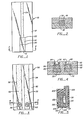
Classifications
-
- G—PHYSICS
- G01—MEASURING; TESTING
- G01N—INVESTIGATING OR ANALYSING MATERIALS BY DETERMINING THEIR CHEMICAL OR PHYSICAL PROPERTIES
- G01N33/00—Investigating or analysing materials by specific methods not covered by groups G01N1/00 - G01N31/00
- G01N33/48—Biological material, e.g. blood, urine; Haemocytometers
- G01N33/50—Chemical analysis of biological material, e.g. blood, urine; Testing involving biospecific ligand binding methods; Immunological testing
- G01N33/53—Immunoassay; Biospecific binding assay; Materials therefor
- G01N33/558—Immunoassay; Biospecific binding assay; Materials therefor using diffusion or migration of antigen or antibody
-
- Y—GENERAL TAGGING OF NEW TECHNOLOGICAL DEVELOPMENTS; GENERAL TAGGING OF CROSS-SECTIONAL TECHNOLOGIES SPANNING OVER SEVERAL SECTIONS OF THE IPC; TECHNICAL SUBJECTS COVERED BY FORMER USPC CROSS-REFERENCE ART COLLECTIONS [XRACs] AND DIGESTS
- Y10—TECHNICAL SUBJECTS COVERED BY FORMER USPC
- Y10S—TECHNICAL SUBJECTS COVERED BY FORMER USPC CROSS-REFERENCE ART COLLECTIONS [XRACs] AND DIGESTS
- Y10S435/00—Chemistry: molecular biology and microbiology
- Y10S435/805—Test papers
-
- Y—GENERAL TAGGING OF NEW TECHNOLOGICAL DEVELOPMENTS; GENERAL TAGGING OF CROSS-SECTIONAL TECHNOLOGIES SPANNING OVER SEVERAL SECTIONS OF THE IPC; TECHNICAL SUBJECTS COVERED BY FORMER USPC CROSS-REFERENCE ART COLLECTIONS [XRACs] AND DIGESTS
- Y10—TECHNICAL SUBJECTS COVERED BY FORMER USPC
- Y10S—TECHNICAL SUBJECTS COVERED BY FORMER USPC CROSS-REFERENCE ART COLLECTIONS [XRACs] AND DIGESTS
- Y10S435/00—Chemistry: molecular biology and microbiology
- Y10S435/81—Packaged device or kit
-
- Y—GENERAL TAGGING OF NEW TECHNOLOGICAL DEVELOPMENTS; GENERAL TAGGING OF CROSS-SECTIONAL TECHNOLOGIES SPANNING OVER SEVERAL SECTIONS OF THE IPC; TECHNICAL SUBJECTS COVERED BY FORMER USPC CROSS-REFERENCE ART COLLECTIONS [XRACs] AND DIGESTS
- Y10—TECHNICAL SUBJECTS COVERED BY FORMER USPC
- Y10S—TECHNICAL SUBJECTS COVERED BY FORMER USPC CROSS-REFERENCE ART COLLECTIONS [XRACs] AND DIGESTS
- Y10S435/00—Chemistry: molecular biology and microbiology
- Y10S435/968—High energy substrates, e.g. fluorescent, chemiluminescent, radioactive
-
- Y—GENERAL TAGGING OF NEW TECHNOLOGICAL DEVELOPMENTS; GENERAL TAGGING OF CROSS-SECTIONAL TECHNOLOGIES SPANNING OVER SEVERAL SECTIONS OF THE IPC; TECHNICAL SUBJECTS COVERED BY FORMER USPC CROSS-REFERENCE ART COLLECTIONS [XRACs] AND DIGESTS
- Y10—TECHNICAL SUBJECTS COVERED BY FORMER USPC
- Y10S—TECHNICAL SUBJECTS COVERED BY FORMER USPC CROSS-REFERENCE ART COLLECTIONS [XRACs] AND DIGESTS
- Y10S435/00—Chemistry: molecular biology and microbiology
- Y10S435/975—Kit
-
- Y—GENERAL TAGGING OF NEW TECHNOLOGICAL DEVELOPMENTS; GENERAL TAGGING OF CROSS-SECTIONAL TECHNOLOGIES SPANNING OVER SEVERAL SECTIONS OF THE IPC; TECHNICAL SUBJECTS COVERED BY FORMER USPC CROSS-REFERENCE ART COLLECTIONS [XRACs] AND DIGESTS
- Y10—TECHNICAL SUBJECTS COVERED BY FORMER USPC
- Y10S—TECHNICAL SUBJECTS COVERED BY FORMER USPC CROSS-REFERENCE ART COLLECTIONS [XRACs] AND DIGESTS
- Y10S436/00—Chemistry: analytical and immunological testing
- Y10S436/80—Fluorescent dyes, e.g. rhodamine
-
- Y—GENERAL TAGGING OF NEW TECHNOLOGICAL DEVELOPMENTS; GENERAL TAGGING OF CROSS-SECTIONAL TECHNOLOGIES SPANNING OVER SEVERAL SECTIONS OF THE IPC; TECHNICAL SUBJECTS COVERED BY FORMER USPC CROSS-REFERENCE ART COLLECTIONS [XRACs] AND DIGESTS
- Y10—TECHNICAL SUBJECTS COVERED BY FORMER USPC
- Y10S—TECHNICAL SUBJECTS COVERED BY FORMER USPC CROSS-REFERENCE ART COLLECTIONS [XRACs] AND DIGESTS
- Y10S436/00—Chemistry: analytical and immunological testing
- Y10S436/807—Apparatus included in process claim, e.g. physical support structures
Definitions
- the ability to employ naturally occurring receptors or antibodies directed to specific compounds in assaying for the presence of a compound of interest has created a burgeoning immunoassay business.
- a homologous pair usually an immunological pair, involving a ligand and a receptor (antiligand) is involved, wherein one of the members of the immunological pair (mip) is labeled with a label which provides a detectible signal.
- the immunoassay methodology results in a distribution of the signal label between signal label bound in a complex of the mips and unbound signal label.
- the differentiation between bound and unbound signal label can be as a result of physical separation of bound from unbound signal label or modulation of the detectible signal between bound and unbound signal label.
- immunoassays have been directed to quantitative determination of a wide variety of compounds of interest, particularly drugs, in clinical laboratories requiring relatively sophisticated equipment and careful technique. Immunoassays have found less extensive commercial application where semi-quantitative or qualitative results would be acceptable and where the determination would involve non-laboratory personnel, such as in a home or a medical practitioner's office. Even in the clinical laboratory, simple and rapid screening tests employing inexperienced personnel could serve to provide substantial economies.
- One consideration is to provide substantial differentiation between the observed signal resulting from signal label when bound as compared to unbound. Another consideration is to minimize interference from endogenous materials in the sample suspected of containing the compound of interest. A further consideration is the ease with which the observed signal can be detected and serve to differentiate between concentrations in the concentration range of interest. Other factors include the ease of preparation of the reagents, the accuracy with which samples and reagent solutions must be prepared and measured, the storage stability of the reagents, the number of steps required in the protocol, and the proficiency and accuracy with which each of the steps must be performed.
- the observed result should be minimally affected by variations in the manner in which the protocol is carried out or provide for simple techniques for performing the various steps.
- U.S. Patent No. 4,168,146 describes an immunoassay test strip.
- U.S. Patents No. 3,990,850 and 4,055,394 describe diagnostic test cards.
- a wide variety of patents and patent applications provide an extensive literature of different techniques for producing detectible signals in immunoassays. The following list is merely illustrative of some of these techniques which can find application in this invention. The following is a list of United States patents and patent applications and a general statement 4 the type of label involved:
- Novel non-chromatographic assay devices and methods employing such devices are described for the determination of members of an immunological pair (mip).
- the device has an immunosorbing zone to which a mip is fixed against diffusive movement.
- the immunosorbing zone serves as the entry for the sample and reagent solutions.
- liquid-receiving zone In liquid-receiving relationship, either directly or indirectly with the immunosorbing zone and normally extending transversely therefrom is a liquid absorbing zone, which serves to draw liquid through the immunosorbing zone, store liquid and may serve to control the rate at which the liquid is drawn through the immunosorbing zone.
- the immunosorbing zone may include one or more members of the signal producing system which are bound to the zone in a manner to permit or inhibit diffusive movement of the signal producing system component.
- the amount of signal label bound in a detection zone in the immunosorbing zone is related to the amount of analyte in the sample.
- the method involves contacting the assay device with the liquid sample to which may have been added one or more components of the signal producing system; followed by contact with one or more successive solutions which contain any remaining components of the signal producing system and serve to wash the immunosorbing zone free of non-specifically bound signal label.
- the signal producing system provides for a detectible signal in the immunosorbing zone which can be . compared to a signal level based on a standard having a known amount of analyte.
- Method and apparatus are provided for performing immunoassays.
- the method and apparatus are devised so as to provide for a narrow zone through which relatively large amounts of fluid pass with relatively uniform flow.
- the zone serves to concentrate both a small amount of analyte in a sample, as well as reagents for producing a detectible signal,, while inhibiting the localization of non-specifically bound materials which would result in production of background signal unrelated to the amount of analyte in the medium.
- assay devices are provided which serve to receive a liquid sample and liquid solutions, transmit the solutions through an immunosorbing zone. to which a member of an immunological pair (mip) is bound and to draw the liquid and non-specifically bound materials into a storage zone or reservoir.
- the reagent solutions provide for production of a detectible signal in the immunosorbing zone whichmay be compared to a standard related to a specific amount of analyte in a sample.
- the immunoassay method is adaptable to a wide variety of reagent combinations which have previously been employed in other immunoassays, both homogeneous and heterogeneous.
- the conditions under which these other assays have been carried out will normally be applicable in the subject method.
- the devices of the subject invention allow for a simplicity of protocol as compared to prior art methods, while providing for qualitative or quantitative results analogous to prior art methods.
- the detectible signal may be observed visually or by means of various apparatuses, such as spectrophotometers, fluorometers, scintillation counters, etc.
- the analyte to be determined will be a mip.
- the specificity of the homologous mips provides a means for discriminating between the analyte of interest and other materials which may be in the sample.
- the mip in the immunosorbing zone one can provide for specific binding of a component to the immunosorbing zone which results in production of a detectible signal.
- the amount of such component in the immunosorbing zone can be related to the amount of analyte in the sample.
- reagents which will comprise the signal producing system.
- the key reagent in the signal producing system is the one which serves as a signal label and is conjugated to a mip.
- the choice of protocol will determine whether an increase or decrease in the amount of the conjugate of signal label and mip which is specifically bound in the immunosorbing zone determines the amount of analyte in the assay medium.
- Analyte - the compound or composition to be measured which is a mip and may be a ligand, which is mono- or polyepitopic, that is, having one or plurality of determinant sites, haptenic or antigenic, a single compound on plurality of compounds which share at least one common epitppic or determinant site; or a receptor.
- Mip - a member of an immunological pair, consisting of two different molecules, where one of the molecules has an area on the surface or in a cavity which specifically binds to a particular spatial and polar organization of the other molecule.
- the members of the immunological pair are referred to as ligand and receptor (antiligand) and members of a specific pair are referred to as homologous.
- Ligand analog - a modified ligand which can compete with the analogous ligand for a receptor, the modification of the ligand providing means to join the ligand analog to another molecule; or where the ligand has a functionality which is used for bonding directly to another molecule, the ligand portion of the conjugate will be referred to as ligand analog.
- the ligand analog will normally differ from the ligand by more than replacement of a hydrogen with a bond, which links the ligand analog to another molecule, e.g. label or hub.
- tp a poly(ligand-analog).
- the assay device has at least one bibulous layer, normally having two or more layers, which will be relatively free of non-specific adsorptivity for the materials of interest.
- the device has as one element a relatively small immunosorbing zone having a mip non-diffusively fixed in the zone. The mip remains substantially immobilized on a bibulous solid support during the course of the assay; and as a second element a reservoir zone, either directly or indirectly in liquid-receiving relationship with the immunosorbing zone.
- Immunosorbing zone - a bibulous solid film, layer or sheet to which a mip is non-diffusively bound; the immunosorbing zone has a relatively small fluid capacity as compared to the total assay device capacity.
- One or more members of the signal producing system may be bound directly or indirectly to the immunosorbing zone.
- the immunosorbing zone has specific binding capability for the homologous mip.
- Within the immunosorbing zone may be one or more zones in tandem or overlapping. Included within the immunosorbing zone will be a detection zone, which may be the same or different from the zone to which the mip is bound.
- Liquid absorbing zone - a bibulous solid material either directly or indirectly in liquid receiving relationship with the immunosorbing zone and acting as a reservoir or storage zone capable of storing a substantially greater liquid volume than the immunosorbing zone.
- the zone acts as a pump to pull liquid through and out of the immunosorbing zone.
- Signal producing system - the signal producing system may have one or more components, at least one component being conjugated to a mip.
- the signal producing system produces a measurable signal which is detectible by external means, usually the measurement of electromagnetic radiation, which signal will be produced in a detection zone in the immunosorbing zone.
- the signal producing system will involve radioactive substances, enzymes and chromogenic substrates, and chromophores, where chromophores include dyes which absorb light in the ultraviolet or visible region, phosphors, fluorescers, and chemiluminescers. While for the most part the signals can be absorption or emission of electromagnetic radiation, usually in the ultraviolet or visible range, other detectible signals will also find application in particular situations.
- Signal generator the compound which provides for production of the detectible signal.
- the signal generator may be produced during the assay or be present initially and produce the signal without undergoing any chemical change. Where a chemical reaction is involved, the compound undergoing the reaction to produce the signal is referred to as the signal generator precursor.
- Label - the label may be any molecule conjugated to another molecule or support and may be arbitrarily chosen.
- the labels will be the mip bound to the assay device as a support and members of the signal producing system whether bound to the mip or to the assay device as a support.
- Mip-support conjugate - in the immunosorbing zone the mip bound to the assay device support, which may be bound covalently or non-covalently, directly or indirectly.
- the mip is bound substantially permanently to the support, so as not to migrate during the traverse of the sample and reagent solutions through the immunosorbing zone.
- the mip serves to bind its homologous member to the immunosorbing zone.
- Signal label-mip conjugate - a member of the signal producing system, which is directly or indirectly bonded to a mip, which is or becomes bound to the immunosorbing zone to provide for production of a detectible signal in the detection zone.
- Signal label-support conjugate a member of the signal producing system bound, covalently or noncovalently, directly or indirectly, to the immunosorbing zone which acts in conjunction with the signal label-mip conjugate to produce a signal in the detection zone.
- the subject method employs an assay device in conjunction with a sample solution and usually onp or more reagent or wash solutions.
- the method involves contacting the immunosorbing zone with a sample solution, where the liquid holding capacity of the immunosorbing zone is substantially smaller than the volume of the sample, as well as the portion of the sample which traverses the immunosorbing zone.
- the immunosorbing zone has mip fixed to a solid support, where the mip is inhibited from diffusing from the zone.
- the analyte in the sample solution passing through the immunosorbing zone binds to the mip of the mip-support conjugate and is concentrated in the immunosorbing zone.
- the signal label-mip conjugate may be involved in a number of ways: (1) initially present in the immunosorbing zone non-covalently bound through immunological complexation to the mip-support conjugate and displaced by the analyte; (2) present with the sample and competes or acts in concert with the analyte to become bound to the mip-support conjugate; or (3) may be added subsequently.
- the immunosorbing zone After contacting the sample, the immunosorbing zone will be contacted with any remaining members of the signal producing system, and as appropriate, wash solutions, where a detectible signal will be developed at the detection zone in relation to the amount of analyte in the medium.
- wash solutions where a detectible signal will be developed at the detection zone in relation to the amount of analyte in the medium.
- the reagent solutions will also serve as wash solutions to minimize the number of steps in the protocol.
- the subject method is distinguished from prior art methods which involve diffusion of solutes diffusing to and away from a layer immersed in a liquid.
- the layer encounters a continuously changing solution composition as solute becomes bound to the layer or dissolves into the liquid.
- the mip containing layer in contact with the solution continuously contacts substantially the same solution composition as the solution diffuses through the layer.
- the concentrations of solutes in the solution in the mip containing layer remain relatively constant during contact of the immunosorbing zone with a solution.
- the structure of the assay device with the immunosorbing zone acting as the entry port can be designed in a variety of ways.
- the assay device except for the * immunosorbing zone must be isolated, normally by a non-permeable barrier, so that the solution is restricted to being absorbed through the immunosorbing zone prior to being absorbed by the liquid absorbing zone.
- the immunosorbing zone is exposed by an opening in the non-permeable barrier. With this construction, the assay device can be conveniently immersed in a solution.
- the solutions may be applied to the immunosorbing zone in a horizontal position, either dropwise, as a slowly flowing stream, or in a container surrounding the immunosorbing zone.
- the sample may or may not be subject to prior treatment, but usually will not be treated.
- buffer with or without one or more components of the signal producing system or other reagents may be included.
- the immunosorbing zone of the assay device is contacted with the sample and absorbs at least a portion of the sample, so that a substantial proportion of the sample solution enters through and traverses the immunosorbing zone and extends into the storage zone.
- the volumes of the solutions normally need not be premeasured.
- the amount of solution absorbed can be controlled by the distance the liquid traverses the liquid absorbing zone.
- Analytes of interest may be present in a wide variety of environments.
- Analytes of interest include drugs, hormones, macromolecules, and microorganisms, which may be found in physiological fluids, such as blood- whole serum or plasma-urine, cerebral spinal fluid, ocular lens-liquid, and saliva; synthetic chemicals; pollutants in water And air; trace compounds, toxins and microorganisms in food, e.g. milk, meat, poultry and fish; or the like.
- physiological fluids such as blood- whole serum or plasma-urine, cerebral spinal fluid, ocular lens-liquid, and saliva
- synthetic chemicals pollutants in water And air
- trace compounds, toxins and microorganisms in food e.g. milk, meat, poultry and fish
- any organic substance which can serve as a ligand or any receptor for which a ligand may be obtained can be determined, so long as such analyte can be introduced into a liquid system in a form in which it can bind to the homologous mip.
- one or mor! components of the signal producing system may be present with the analyte in the sample solution.
- a solution having the signal label-mip conjugate may be followed by at least one additional solution, to ensure the removal of non-specific binding of the signal label-mip conjugate in the immunosorbing zone and/or introduce remaining components of the signal producing system.
- the signal label-mip conjugate solution can be sorbed in the immunosorbing zone prior to the addition of the remaining members of the signal producing system in a subsequent reagent solution.
- the subsequent reagent solution then serves as a wash to remove non-specifically bound signal label-mip conjugate.
- nothing is added to the sample which is contacted first, and then followed by one or more additional solutions containing the signal label-mip conjugage and the other components of the signal producing system.
- protocols will be available, the particular configuration being subject to: the nature of the analyte, i.e., whether it is a mono- or polyepitopic ligand or a receptor; the nature of the signal label-mip conjugate; and the nature and number of components in the signal producing system. Therefore, a number of illustrative examples of protocols will be given, which are not exhaustive, but are rather embodiments of protocols which serve to indicate the numerous opportunities for variation.
- Table I indicates the various combinations of analytes and reagents which may be employed in the subject invention to provide a signal in the6mmunosorb- ing zone related to the amount of analyte.
- Protocol 2 involves combining the sample and mip components of the signal producing system and contacting the assay device with the resulting solution.
- the assay device strip would then be contacted with a wash solution that optionally would contain the other components of the signal producing system to ensure the complete removal of nonspecific binding mip.
- the assay device could be contacted with the sample, followed by contact with the mip component of the signal producing system, followed optionally by a wash solution which if employed would usually contain the other components of the signal producing system.
- the L-mip could be initially bound to the mip-support conjugate, so that upon immersion in the sample the L-mip would be displaced by the hapten analyte. No further steps would be necessary where the signal label is a radioactive label or fluorescer.
- the signal label and signal producing system Depending on the nature of the signal label and signal producing system, one would observe the signal, by irradiating with light for a fluorescer and observing the level of fluorescence; providing for a catalyst system to produce a dye, fluorescer, or chemiluminescence, where the dye could be observed visually or in a spectrophotometer and the fluorescer could be observed visually or in a fluorometer; or in the case of chemiluminescence or a radioactive label, by employing a radiation counter. Where the appropriate equipment is not available, it will normally be desirable to have a chromophore produced which results in a visible color. Where sophisticated equipment is involved, any of the techniques is applicable.
- an incubation step may be involved. Incubation steps involving combining solutions can vary from about 5sec to 16hrs or m9re, generally varying from about lmin to lhr, more usually from about 5min to 30min.
- the time during which the assay device is contacted with each solution will depend upon the volume of the solution to be sorbed and the rate at which it diffuses into the liquid absorbing zone. This time will be controlled in accordance with the concentration of the mip in the solution, the concentration of mip in the area of the immunosorbing zone, the binding constant of the homologous mips, the rate of production of signal, and the like.
- the manner in which the time for diffusion of the solutions through the immunosorbing zone may be controlled will involve the composition, construction, size and shape of the immunosorbing and liquid absorbing zones, the temperature, the solvent, and the like. In view of the wide variety of opportunities for use of the subject method and devices, no particular time range ascribed to the subject method would be meaningful.
- the various solution combinings, contacting of solutions with the assay device, and readings will be carried out at a temperature in the range of about 0 to 50°C, more usually in the range of about 15 to 40°C.
- temperatures will generally range from about 15 to 50°C, frequently 20 to 35°C, and conveniently ambient temperatures.
- the temperature may be controlled or uncontrolled, conveniently employing ambient temperatures. Frequently, when employing ambient temperatures, one or more control strips may be employed for comparison with the observed signal from the sample strip.
- the solutions which are employed will normally be buffered at a pH in the range of about 5 to 11, more usually 5 to 10, and preferably about 6 to 9.
- the pH is chosen so as to maintain a significant level of specific binding by the mips while optimizing signal producing efficiency.
- various pH's may be used with different solutions.
- Various buffers may be used to achieve the desired pH and maintain the desired pH during the determination.
- Illustrative buffers include borate, phosphate, carbonate, tris, barbital, and the like.
- the particular buffer employed is not critical to this invention, but in individual assays, one buffer may be preferred over another.
- the concentration of analyte which may be assayed will generally vary from about 10-4 to 10 -15 , more usually from about 10 -6 to 10 -13 M. Considerations such as whether the assay is qualitative, semi-quantitative, or quantitative, the particular detection technique and the concentration of the analyte of interest will normally determine the concentration of the other reagents.
- concentrations of various reagents will vary widely depending upon which protocols are employed, the nature of the analyte, the mip in the immunosorbing zone, the required sensitivity of the assay, and the like. In some instances, large excesses of one or the other of the mips may be employed, while in some protocols the sensitivity of the assay will be responsive to variations in the mip ratios.
- the analyte is a polyepitopic antigen
- the assay device is first contacted with the sample, followed by contact with the signal producing system solution, desirably followed by a wash solution.
- antiligand is the analyte-and
- the protocol involves the combination of the analyte and catalyst-antiligand conjugate, prior to contacting the antigen-support conjugate, the sensitivity of the assay will be related to the ratio of the analyte and catalyst-antiligand concentration.
- the concentration of the reagents will depend on the binding constant of the antiligand, or the binding constant profile where an antiserum is used, as well as the required sensitivity of the assay. Desirably, when feasible, the concentration of the signal label-mip will be.sufficiently low to minimize non-specific binding or occlusion within the immunosorbing zone, particularly the detection zone.
- the molar ratios can only be very broadly stated.
- the number of molecules of signal label-mip conjugate drawn into the immunosorbing zone will usually be not les:; than about 0.1 times the minimum number of moles of analyte based on binding sites and hot more than about 1,000 times the maximum moles of analyte based on binding sites drawn into the immunosorbing zone, more usually from about 0.1 to 100 times the moles of analyte based on binding sites.
- the mip of the mip-support.conjugate there will usually be not less than about 0.001 times the minimum number of moles of analyte based on binding sites drawn into theimmunosorbing zone. As already indicated, depending upon the protocols, there can be large excesses over stoichiometric.
- the amount of mip in the immunosorbing zone will be at least sufficient to provide a detectible signal when fully bound by signal label mip conjugate. (The above reference to sample or analyte intends the volume or number which traverses the immunosorbing zone, and not the total volume of the sample solution or total amount of analyte which contacts the assay device.)
- a ligand or receptor in the immunosorbing zone will depend upon a number of factors.
- catalyst-mip conjugate where the mip is antibody to the receptor enhances the assay response by permitting many catalysts to become bound to each molecule of the analyte in the immunosorbing zone.
- the assay device must then be contacted with at least three separate solutions in the order of sample, followed by antibody, followed by signal label-mip. The purity of the ligand or receptor will also be of significance.
- the subject invention provides for a mechanism for removing the signal label bound to other than a mip from the immunosorbing zone.
- the simplicity of the protocol it is desirable to minimize the number of measurements which could have a significant effect on the assay result.
- the assay device is first contacted with the sample alone it is possible to avoid any measurement.
- the sample is combined with other solutions, it is desirable to limit the required measurements to the size of the sample apd, as appropriate, its subsequent dilution to provide the assay sample.
- the addition of the remaining reagents should allow for substantial variation in volumes and concentrations without affecting the assay result.
- exemplary protocols will be described using mono-, polyepitopic or receptor analytes and as signal labels: a signal label not requiring the addition of other chemical reagents for production of a signal; a signal label requiring additional reagents for production of a signal; and a signal label which requires additional reagents for production of a signal and interacts with a component of the signal producing system initially present in the immunosorbing zone of the assay device. Also described will be a protocol requiring a complex immunosorbing zone, where the signal label-mip conjugate is initially present in the immunosorbing zone.
- the first exemplary protocol will employ a hapten (monoepitopic ligand) as the analyte and a fluorescer as the signal label, where the fluorescer is bound to a hapten analog.
- a hapten monoepitopic ligand
- fluorescer fluorescer
- the specimen suspected of containing the hapten is combined with a buffered solution of the hapten-fluorescer reagent to a predetermined volume.
- the assay device containing antihapten in the immunosprbing zone is immersed in the sample and the assay sample solution sorbed to a predetermined distance in the liquid absorbing zone.
- the assay device is then removed from the assay sample solution and introduced into a buffer solution, where the buffer solution courses through the immunosorbing zone to ensure the complete removal of any non-specifically bound hapten-fluorescer reagent in the immunosorbing zone. If the concentration of hapten-fluorescer in the reagent solution is sufficiently low compared to the amount of hapten-fluorescer bound for a positive result, then the washing step may be dispensed with. The assay device may then be introduced into a fluorometer and the fluorescence determined.
- a standard assay device could be employed which would be carried through the same steps as the assay device for the specimen.
- the standard assay device would have a predetermined amount of antibody in the immunosorbing zone and when introduced into the reagent solution in the absence of hapten analyte would absorb an amount of hapten-fluorescer reagent which would relate to a specific concentration of hapten.
- a standard assay device could be employed that could be contacted with the analyte but would give a standard response unrelated to the amount of analyte.
- the washings with the assay device of the subject invention differ from the washings employed in other assays employing dipsticks or test strips.
- the test strip is introduced into a solution which is free of any reagent and any non-specifically bound materials allowed to diffuse from the test strip into the solution. It must thereafter be removed from the wash solution.
- the solution passing through the immunosorbing zone carries non-specifically bound materials with it into the liquid absorbing zone, so that the immunosorbing zone is continuously contacted with fresh solution free of non-specifically binding signal label.
- an antigen is employed, where the antigen may act as a bridge between antiligand in the immunosorbing zone and signal label (in this case an enzyme) bound to antibody.
- the-assay device having antiligand is immersed in a specimen sample suspected of having ligand, usually undiluted. A predetermined amount of the assay sample solution diffuses through the immunosorbing zone into the liquid absorbing zone, where antigen in the assay sample solution becomes bound to the antiligand in the immunosorbing zone in proportion to the concentration of antigen in the solution.
- the assay device is removed and immersed in a buffered reagent solution, which can have a large excess of enzyme-antiligand as the reagent.
- the enzyme-antiligand will bind to any ligand bound in the immunosorbing zone.
- the volume of liquid sorbed may be determined by the distance the solvent front travels. Relatively short incubation times are necessary for the binding of the enzyme-antiligand reagent to the antigen in the immunosorbing zone, since the subject method provides for continuously replenishing the reagent to the immunosorbing zone at a relatively high reagent concentration.
- the assay device is removed and introduced into a substrate solution. Enzyme catalysis results in transformation of the substrate to a colored product, which may be deposited in the immunosorbing zone. Once again, one can control the volume of the substrate solution which traverses the immunosorbing zone by following the solvent front in the liquid absorbing zone.
- the standard assay device would be treated in the identical manner.
- antibody is the analyte and the signal producing system will involve two enzymes.
- the signal label-mip reagent will be an enzyme whose substrate is the product of an enzyme bound in the immunosorbing zone.
- the assay device will have antibody as the mip in the immunosorbing zone.
- the sample would be mixed with a predetermined volume of the enzyme-antigen reagent and incubated for sufficient time to ensure the substantially complete binding of any antibody in the sample with the enzyme-antigen reagent.
- the assay device would then be introduced into the assay sample solution and the solution sorbed through the immunosorbing zone, so that any remaining unbound enzyme-antigen would become bound in the immunosorbing zone.
- the assay device After a predetermined volume has moved through the immunosorbing zone as evidenced by movement of the solvent front in the liquid absorbing zone, the assay device would be removed and placed in a substrate solution.
- the substrate solution would have substrate for the enzyme that is bound to the assay device, as well as any additional reagents necessary for the two enzymes, except for the substrate of the enzyme-antigen reagent, which is the product of the enzyme bound to the device.
- a wash would normally not be necessary in this mode, since the substrate solution would wash the immunosorbing zone free of any of the enzyme-antigen:antibody complex.
- a predetermined volume of the substrate solution will move through the immunosorbing zone providing a colored product in the immunosorbing zone.
- the substrate solution could be combined with the sample and enzyme-antigen solution followed by immersing the assay device in the mixture.
- a complex immunosorbing zone having a plurality of overlapping or tandem layers.
- the analyte is a hapten
- the first layer which is initially contacted by the solutions would have antihapten complexed with an enzyme-hapten conjugate.
- An intermediate layer may be provided to inhibit transfer of comp p unds from the third to the first layer.
- the third layer wopld have a reagent to cause the signal producing substances to be formed on that layer as for example enzyme, anti(signal label), antienzyme, etc.
- a second enzyme which is bound to the third layer.
- the second enzyme is characterized by producing a product which is the substrate for the enzyme of the enzyme-hapten conjugate.
- the intermediate layer inhibits the second enzyme product from migrating to the first layer.
- the second enzyme will be illustrated by glucose oxidase and the enzyme of the enzyme-hapten conjugate by horse radish peroxidase (HRP).
- HRP-hapten is bound to antihapten in the first layer of the immunosorbing zone, while glucose oxidase, and anti-HRP, are covalently bonded to the third layer.
- Catalase is present in the intermediate layer or only two layers need be involved and catalase could be present in the first layer. The catalase serves to destroy any adventitious hydrogen peroxide which might migrate from the third layer to the first layer.
- the assay is performed by adding the sample solution to the first layer which overlays the other layers, so that the solution migrates downwardly. Hapten in the sample displaces HRP-hapten in the first layer, so that the HRP-hapten migrates downwardly to the third layer and is captured by anti-HRP.
- the sample solution is combined with or followed by a reagent solution containing glucose and a dye precursor.
- The.glucose and dye precursor will migrate to the third layer carrying any non-specifically bound HRP-hapten in the first two layers to the third layer.
- glucose will serve to form hydrogen peroxide, which in turn will react with the dye precursor in a reaction catalysed by HRP to produce a dye.
- the intensity of the color produced in the third layer (detection zone) will be related to the amount of hapten in the sample.
- the assay device would be viewed from the rear where the third layer would be observable.
- the standards employed may be widely varied with the assay devices of the subject invention. In some instances, it will be sufficient to provide-a series of graduated colors which can be compared with the color produced on the assay device. For a more quantitative result, it would be desirable to have one or more assay devices having a predetermined amount of a mip present which assay devices would then be treated in substantially the same manner as the assay device employed with the specimen. Where the mip on the assay device i's the same as the analyte, one could employ the assay device in the identical manner. Where the mip on the assay device is the homologous mip to the analyte, one would normally avoid combining the standard assay device with the specimen.
- the components employed in this subject assay will be the assay device, the analyte, the signal producing system, which includes the signal label-mip conjugate, and as appropriate one of the other mips.
- the assay device has two parts: an immunosorbing zone which uniquely serves as the inlet port for the solutions, and a liquid absorbing zone which serves to draw the solutions through the immunosorbing zone and store the solutions as appropriate.
- the assay device may have many configurations.
- One configuration is a test strip involving an extended bibulous strip as the liquid absorbing zone and a narrow region in the same plane or preferably as a contiguous layer which serves as the immunosorbing zone.
- the test strip is an elongated device, having two primary portions, a relatively small immunosorbing zone, and a relatively large liquid absorbing zone.
- the immunosorbing zone will normally be kept as small as possible commensurate with its function.
- the immunosorbing zone has as its primary distinction mip within its boundaries and optionally one or more members of the signal producing system.
- test strip While the dimensions of the test strip may vary widely, the length will normally be substantially greater than the width at least two times greater than the width, and may be ten times or more greater than the width, and the width and thickness may be regular or irregular.
- the strip may be a single material or a plurality of materials, so long as each of the zones is in liquid receiving relationship with the adjacent zone. The materials may draw liquid by absorption, normally without any substantial adsorption or chromatographic effect. That is, all of the materials, except for the mips involved, will flow relatively evenly and freely through the zones.
- Various natural or synthetic materials may be employed, which may be individual materials or combinations of materials, which may be organic, inorganic, or combinations thereof. Where more than one layer is involved different layers may have different properties for different functions. Immunosorbent layers need not have high capacity for retaining liquid, as contracted with liquid absorbents. For detection of electromagnetic radiation, the detection zone may be opaque, or transluscent or preferably transparent.
- polysaccharides e.g. cellulose materials,-such as paper and cellulose acetate; silica, inorganic materials, such as deactivated alumina, diatomaceous earth, MgS0 4 , or other inorganic finely divided material conveniently substantially uniformly dispersed in a porous polymer matrix, with polymers such as vinyl chloride, vinyl chloride-propylene copolymer, and vinyl chloride-vinyl acetate copolymer; cloth, both naturally occurring e.g. cotton and synthetic e.g. nylon cloth; porous gels, e.g. silica gel, agarose, dextran, and gelatin ; polymeric films, e.g.
- polysaccharides e.g. cellulose materials,-such as paper and cellulose acetate
- silica inorganic materials, such as deactivated alumina, diatomaceous earth, MgS0 4 , or other inorganic finely divided material conveniently substantially uniformly dispersed in a por
- the assay device material are able to absorb liquid, particularly aqueous solutions, without substantially impeding the movement of the solutes employed in the assay.
- the materials are bibulous; they are porous and allow the flow of solutions; the materials for the immunosorbent layers are preferably non-chromatographic; they have reasonable strength or strength can be imparted by means of a support, and they do not interfere with production of the signal by the signal generator.
- the liquid absorbing zone is protected from direct contact with the sample and reagent solutions. This will be conveniently achieved by having the immunosorbing zone as the only exposed portion of the test strip or having the area around the immunosorbing zone enclosed to prevent sample or other reagent solution contact with the liquid absorbing zone.
- the immunosorbing zone may be the edge of the test strip, or may be an exposed. zone on the face of the test strip, or may extend beyond, be aligned with or internal to an impermeable protective cover. A significant factor in the construction is that the flow of solution through the exposed surface is reasonably uniform.
- the exposed surface of the immunosorbing zone will generally be not less than at least about lmm in its smallest dimension and will usually be at least about 1.5, more usually at least about 2mm in its smallest dimension.
- the immunosorbing zone will usually be not greater than about 2cm in its largest dimension, generally being about .2 to 1cm in its largest dimension.
- the thickness of the immunosorbing zone will generally vary from about 10 ⁇ -5mm, most usually 50p to about 3mm, preferably about 0.2mm to lmm.
- the immunosorbing zone may be any configuration, e.g. circular, elliptical, square, rectangular, or irregular.
- the significant factor is that the surface of the detection zone can be scanned for signal.
- the surface will normally be planar or slightly curved, either concave or convex, with no more than about 60° of arc.
- the immunosorbing zone may be a single layer or a plurality of layers and may produce the signal in the upper or lower portion of the immunosorbing zone (upper and lower relate to upstream and downstream in relation to the flow of liquid).
- the immunosorbing zone includes as a single layer or the upper layer, mip bound to a bibulous support. In some protocols the signal label-mip conjugate may be complexed with the mip-support conjugate.
- a lower layer will have anti(signal label) or a member of the signal producing system non-diffusively bound to a bibulous support.
- the lower layer conjugate serves to capture the signal label-mip conjugate or products produced by the signal label-mip conjugate which migrate from the mip-support conjugate layer.
- Signal labels and anti(signal labels) are chosen so that the anti(signal label) will not significantly interfere with production of signal in the lower layer. Reactants are also chosen to generate a readily detectable signal.
- One or more additional layers may be involved, particularly between the mip-support conjugate layer and the lower layer. These layers may serve as barriers to inhibit migration of components of the signal producing system from the lower to the upper layer; as filters; for flow control; or the like.
- the flow through the immunosorbing zone should be reasonable uniform. Many bibulous materials will be nonuniform, so that flow through the mip containing entry zone may vary from region to region. To offset the inhomogeneity of layers in the immunosorbing zone, various composite structures may be employed.
- One structure employs a first bibulous layer to which mip is bound and which resists the through flow of liquid resulting in a substantial pressure differential across the layer. Behind the layer is a relatively open layer providing minimal resistance to flow. This second layer is intermediate the first layer and the li q ûid absorbing zone.
- Another construction is to have three layers, where the first mip containing layer has relatively low resistance to liquid flow, but is backed by a second layer in uniform contact with the first layer, which second layer is substantially more resistive to liquid flow. This results in impeding the rapid flow through one or more regions of the mip contining layer, resulting in a more uniform flow through the first layer.
- the second layer is followed by a porous layer which does not resist flow.
- Each of the layers will be uniformly pressed together to avoid the occurrence of flow paths.
- a wide variety of compositions of known flow characteristics can be used for constructing the composite structure.
- a spacer may be used which defines an area behind the immunosorbent layer that permits flow normal to and lateral to the immunosorbent layer surface.
- Materials which may be used for low flow resistance include paper; nylon, wire, glass or polyolefin mesh or mat; or cotton or the like.
- the shape should be varied to ensure good uniform contact with the bounding layers, being flat, concave or convex, where the two opposing surfaces may be the same or different.
- the upper and/or lower layer may serve as the detection zone for determining the detectable signal. If desired, detectable signal could be produced in both layers, so that two values associated with the amount of analyte could be measured to provide an internal check.
- the liquid absorbing zone is in liquid receiving contact with the immunosorbing zone.
- the liquid absorbing zone provides a plurality of functions.
- the first function is to serve as a receptacle or storage area for the fluid imbibed by the immunosorbing zone.
- a second function which can be served but need not, is to control the rate at which the fluid traverses the immunosorbing zone.
- the liquid absorbing zone has a small dimension or is of a different material from the immunosorbing zone, it can actio control the rate at which fluid passes through the immunosorbing zone. By having irregular dimensions, it can further act to provide for different rates depending upon whether the sample or reagent solutions are being imbibed.
- a further function for the liquid absorbing zone is to measure the amount of liquid which is imbibed.
- pH indicators could be used. Any technique which provides a clear indication of the presence of the solvent front can be employed.
- the liquid absorbing zone will generally have a length of at least about 1.5 usually 2cm and not more than about 30cm, usually not more than about 20cm.
- the width may vary from about 0.1mm to about 3cm. These dimensions are primarily for control of the rate and amount of solution imbibed and for convenience in handling and providing for ease of observation of the solvent front.
- the thickness will generally be about 0.1mm to 5mm, usually from about 0.5 to 3mm.
- the liquid absorbing zone may be partially or substantially completely enclosed in a protective casing, conveniently a clear or partially or in some situations completely opaque casing. While the entire liquid absorbing zone need not be encased, a sufficient portion of it should be covered to prevent the liquid absorbing zone from directly contacting the sample solution. In almost all cases it will be necessary that a uniform amount of the sample solution pass through the immunosorbing zone. This can be best achieved by avoiding contact of the liquid absorbing zone with the assay sample solution.
- the enclosure may be removable or irremovable, may provide for one or more windows and will normally be of a sturdy inert impermeable material which will provide mechanical protection for the bibulous material comprising the various zones and will not. interfere with the performance of the assay. Normally an air opening will be provided to prevent the entrapment of air within the enclosure.
- a test strip device 10 is shown with plastic enclosure 12, enclosing a bibulous shovel shaped strip 14.
- the butt end 16 of the strip is slightly internal of the plastic enclosure 12, so as to be protected from mechanical contact and deterioration.
- the butt end 16 serves as an immunosorbing zone, with mip bonded to the butt end 16 to a sufficient depth, so that the development of the signal by the signal label can be readily detected.
- the strip 14 is graduated at 20 and 22 to allow for accurate measurement of .the volume of the sample solution and reagent solutions which have traversed the immunosorbing zone 16.
- the strip 14 has a narrow extended strip 24 which controls the rate of flow of the last solution.
- the strip 24 extends to an opening 26 in the plastic enclosure 12 which allows for the escape of air as the solutions move up the strip 14.
- the enclosure 12 is comprised of a front sheet 30 and a back sheet 32, with a clear front sheet 30 to allow for observing the movement of the fluids up the bibulous strip 14.
- the strip need only be dipped into a sample solution and removed when the sample front has reached graduation 20.
- the test strip 10 is then removed from the sample solution and introduced into a reagent solution and maintained in the reagent solution, until the solvent front has reached graduation 22.
- the test strip is then removed from the reagent solution and introduced into a wash solution until a sufficient amount of the wash solution has passed through the immunosorbing zone as evidenced by the solvent front moving along the narrow extended strip 24.
- the immunosorbing zone butt end 16 may then be observed for production of a detectible signal ae-evidence of any analyte being present in the sample.
- a test strip device 50 is depicted having two strips, a specimen testing member 52 and a standard strip member 54. Ports 56 and 60 provide openings in the plastic enclosure 62. Immunoabsorbing disk 64 would have antibody bound to it (indicated by the letters Ab) and the immunosorbing disk 66 of the standard 54 would have antigen bound to it (indicated by the letters Ag) in a predetermined amount to provide for a signal level related to an antigen concentration.
- the bibulous strips 70 and 72 for example, cellulose or paper strips, extend from the immunosorbing zones 64 and 66, respectively, beyond the enclosure 62 to openings 74 and 76 in the plastic enclosure 62 which allow for escape of air from the enclosure. The bottom ends of 70 and 72 are protected from solution.
- the immunosorbing disks 64 and 66 are seated on porous spacers 80 and 82 respectively.
- the porous spacers are inert and substantially non-resistant to fluid flow. There is substantially uniform contact between the immunosorbent zone disks 64 and 66 and the surfaces 84 and 86 of the porous spacers 80 and 82. Thus, solutions exiting from the immunosorbing zone disks 64 and 66. encounter substantially no resistance to their flow.
- the spacers 80 and 82 are bounded downstream by the liquid absorbing strips 70 and 72 which absorb the liquid from the spacers 80 and 82 respectively, acting as pumps to withdraw the liquid as the spacers fill with liquid.
- the above constructions for the specimen and standard are encased in a plastic holder which can be made of one or two or more parts and assembled to hold the various parts of the test strip in alignment and liquid receiving relationship.
- Fig. 5 is depicted an alternative embodiment of the subject test strip device.
- the immunosorbing zone and a portion of the liquid absorbing zone are shown.
- An enclosure 100 is provided with window 102.
- An immunosorbing bibulous disk 104 having mip bound to it serves as the entry port for sample and solutions.
- the bibulous disk should be relatively uniform in its liquid absorbing properties, but relatively non-resistant to the flow of liquid through the disk.
- a bibulous liquid flow resistant disk 106 which has greater resistance to liquid flow than the immunosorbing disk 104.
- the two disks, 104 and 106 are uniformly pressed against each other at the interface 110.
- Spacer 112 Behind the flow resistant disk 106 is spacer 112, which is a ring to allow the free flow of liquid between the flow resistant disk 106 and the liquid absorbing strip 114.
- the immunosorbing disk 104 When the device is introduced into a sample or reagent solution, the immunosorbing disk 104 will rapidly absorb the liquid which will move through the disk and encounter the more resistant disk 106. The disk 106 will control the rate of flow. Thus, inhomogeneities present in the immunosorbing disk 104 will not result in rapid flow through one or more hot spots, inhibiting lack of uniformity of liquid flow through the immunosorbing disk 104. After the solution has passed through the flow resistant disk 106, it will continue through the open space 116 of ring 112 and be absorbed by the liquid absorbing strip 114.
- the ligand analytes of this invention are characterized by being monoepitopic or polyepitopic.
- the polyepitopic ligand analytes will normally be poly(amino acids) i.e. polypeptides and proteins, polysaccharides, nucleic acids, and combinations thereof.
- Such combinations of assemblages include bacteria, viruses, chromosomes, genes, mitochondria, nuclei, cell membranes, and the like.
- the polyepitopic ligand analytes employed in the subject invention will have a molecular weight of at least about 5,000, more usually at least about 10,000.
- the poly(amino acid) category the poly(amino acids) of interest will generally be from about 5,000 to 5,000,000 molecular weight, more usually from about 20,000 to 1,000,000 molecular weight; among the hormones of interest, the molecular weights will usually range from about 5,000 to 60,000 molecular weight.
- proteins may be considered as to the family of proteins having similar structural features, proteins having particular biological functions, proteins related to specific microorganisms, particularly disease causing microorganisms, etc.
- proteins found in the human plasma are important clinically and include:
- Important blood clotting factors include:
- Important protein hormones include:
- polymeric materials of interest are mucopolysaccharides and polysaccharides.
- Illustrative antigenic polysaccharides derived from microorganisms are as follows:
- microorganisms which are assayed may be intact, lysed, ground or otherwise fragmented, and the resulting composition or portion, e.g. by extraction, assayed.
- Microorganisms of interest include:
- Hemophilus-Bordetella group Hemophilus influenzae, H. ducreyi H. hemophilus H. aegypticus H. parainfluenzae Bordetella pertussis
- Pasteurella pestis Pasteurella tulareusis
- Chlamydia unclassifiable parasites bacterial/viral
- Chlamydia agents naming uncertain
- Herpes simplex Varicella Chocken pox
- Herpes Zoster Shingles
- Influenza (A, B, and C) Parainfluenza (1-4) Mumps Virus Newcastle Disease Virus Measles Virus Rinderpest Virus Canine Distemper Virus Respiratory Syncytial Virus Rubella Virus
- the monoepitopic ligand analytes will generally be from about 100 to 2,000 molecular weight, more usually 125 to 1,000 molecular weight.
- the analytes of interest include drugs, metabolites, pesticides, pollutants, and the like. Included among drugs of interest are the alkaloids.
- alkaloids include morphine alkaloids, which includes morphine, codeine,' heroin, dextromethorphan, their derivatives and metabolites; cocaine alkaloids, which includes cocaine and benzoyl ecgonine, their derivatives and metabolites; ergot alkaloids, which includes the diethylamide of lysergic acid; steroid alkaloids; iminazoyl alkaloids; quinazoline alkaloids, isoquinoline alkaloids; quinoline alkaloids; which includes quinine and quinidine; diterpene alkaloids; their derivatives and metabolites.
- the next group of drugs includes steroids, which includes the estrogens, gestogens, androgens, andrenocortical steroids, bile acids, cardiotonic glycosides and aglycones, which includes digoxin and digoxigenin, saponins and sapogenins, their derivatives and metabolites. Also included are the steriod mimetic substances, such as diethylstilbestrol.
- lactams having from 5 to 6 annular members, which include the barbiturates, e.g. phenobarbital and secobarbital, diphenylhydantonin, primidone, ethosuximide, and their metabolites.
- the next group of drugs is amindalkylbenzenes, with alkyl of from 2 to 3 carbon atoms, which includes the amphetamines, catecholamines, which includes ephedrine, L-dopa, epinephrine, narceine, papaverine, their metabolites.
- the next group of drugs is aminoalkylbenzenes, with alkyl of from 2 to 3 carbon atoms, which includes ephedrine, L-dopa, epinephrine, narceine, papaverine, their metabolites and derivatives.
- the next group of drugs is benzheterocyclics which include oxazepam, chlorpromazine, tegretol, imipramine, their derivatives and metabolites, the heterocyclic rings being azepines, diazepines and phenothiazines.
- the next group of drugs is purines, which includes theophylline, caffeine, their metabolites and derivatives.
- the next group of drugs includes those derived from marijuana, which includes cannabinol and tetrahydrocannabinol.
- the next group of drugs includes the vitamins such as A, B, e.g. B12; C, D, E and K, folic acid, thiamine.
- prostaglandins which differ by the degree and sites of hydroxylation and unsaturation.
- antibiotics which include penicillin, chloromycetin, actinomycetin, tetracycline, terramycin, their metabolites and derivatives.
- the next group of drugs is the nucleosides and nucleotides, which include ATP, NAD, FMN, adenosine, guanosine, thymidine, and cytidine with their appropriate sugar and phosphate substituents.
- the next group of drugs is miscellaneous individual drugs which include methadone, meprobamate, serotonin, meperidine, amitriptyline, nortriptyline, lidocaine, procaineamide, acetylprocaineamide, propranolol, griseofulvin, valproic acid, butyrophenones, antihistamines, anticholinergic drugs, such as atropine, their metabolites and derivatives.
- the next group of compounds is amino acids and small peptides which include polyiodothyronines e.g. thyroxine, and triiodothyronine, oxytocin, ACTH, angiotensin, met-and leu-enkephalin their metabolites and derivatives.
- polyiodothyronines e.g. thyroxine, and triiodothyronine
- oxytocin ACTH
- angiotensin met-and leu-enkephalin
- Metabolites related to diseased states include spermine, galactose, phenylpyruvic acid, and porphyrin Type 1.
- the next group of drugs is aminoglycosides, such as gentamicin, kanamicin, tobramycin, and amikacin.
- pesticides of interest are polyhalogenated biphenyls, phosphate esters, thiophosphates, carbamates, polyhalogenated sulfenamides, their metabolites and derivatives.
- the molecular weights will generally range from 10,000 to 2x10 6 , more usually from 10,000 to 10 6 .
- the molecular weights will generally vary from about 160,000 to about 10 6 .
- Enzymes will normally range from about 10,000 to 6000,000 in molecular weight.
- Natural receptors vary widely, generally being at least about 25,000 molecular weight and may be ' 10 6 or higher molecular weight, including such materials as avidin, thyroxine binding globulin, thyroxine binding prealbumin, transcortin, etc.
- the ligand analog will differ from the ligand either by replacement of a hydrogen or a functionality with a bond or a linking group which has a functionality for forming a covalent bond to another molecule having an active functionality, such as an hydroxyl, amino, aryl, thio, olefin, etc., where the resulting compound differs from the ligand by more than substitution of a hydrogen by the molecule to which it is conjugated.
- the linking group will normally have from 1-20 atoms other than hydrogen, which are carbon, oxygen, sulfur, nitrogen, and halogen of atomic number 17-35.
- the functionalities which are involved include carbonyl, both oxo and non-oxo, active halogen, diazo, mercapto, ethylene, particularly activated ethylene, amino, and the like.
- the number of heteroatoms will generally range from about 0-6, more usually from about 1-6, and preferably from about 1-4.
- a description of linking groups may be found in U.S. Patent No. 3,817,837, which disclosure is incorporated herein by reference.
- the linking groups will be aliphatic, although with diazo groups, aromatic groups are involved. Generally, the linking group is a divalent chain having about 1-10, more usually from about 1-6 atoms in the chain. Oxygen will normally be present as oxo or oxy, bonded to carbon and hydrogen, preferably bonded solely to carbon, while nitrogen will normally be present as amino, bonded solely to carbon, or amido, while sulfur would ba analogous to oxygen.
- Linking groups which find particular application with conjugation to polypeptides are those involving carboxylic acids which may be used in conjunction with diimides, or as mixed anhydrides with carbonate monoesters or as active carboxylic esters e.g. N-hydroxy succinimide or p-nitrophenyl. Nitrogen analogs may be employed as imidoesters.
- Aldehydes can be used to form imines under reductive amination conditions e.g. in the presence of borohydrides, to produce alkylamines.
- Other non-oxo carbonyl groups which may be employed include isocyanates and isothiocyanates.
- active halide may be employed, particularly bromoacetyl groups.
- the ligand will have one or more functional groups which may be employed as the.site for linking the linking group.
- functional groups which may be employed as the.site for linking the linking group.
- hydroxy, amino and aryl groups, particularly activated aryl groups find use.
- oximes may be prepared from oxo functionalities and the hydroxyl used as a site for joining to a linking group, such as carboxymethyl.
- linking group will vary widely, depending upon the functionalities which are present in the ligand, in the compound to which the ligand is to be conjugated, the nature and length of the linking group desired, and the like.
- the signal producing system will have at least one member and may have two or more members.
- the signal producing system provides for the production of a product which allows for a detectible signal in an amount or at a level related to the amount of analyte in a sample.
- the signal produced is normally on a solid support, so that the measurement will normally be from the solid support.
- the measurement will normally involve electromagnetic radiation absorption or emission.
- signal producing systems may be employed.
- various members of the signal producing system may be employed as the signal label-mip conjugate or as signal label-support conjugate.
- the signal label-mip conjugate may be provided in solution or complexed to the mip-support conjugate.
- the various conjugates may be joined covalently or non-covalently, directly or indirectly.
- the particular linking group will depend upon the nature of the two moieties to be bonded and their respective functions.
- a large number of linking groups and methods for linking are taught in the literature.
- a significant number of linking groups are set forth in the section on ligand analogs.
- Binding can also be achieved by the use of receptors.
- an antigen may be bound to a support through the intermediacy of a receptor e.g. antibody, for the antigen.
- the receptor in turn may be bound covalently or non-covalently e.g. adsorption to a support.
- the signal producing systems will be divided into the following categories: chromogens; catalyzed reactions; chemiluminesence; and radioactive labels.
- the chromogens will include compounds which absorb light in a distinctive range, so that a color may be observed, or emit light when irradiated with light of a particular wavelength or wavelength range e.g: fluorescers.
- the use of dyes as the signal label will not provide for the desired sensitivity. While one can provide for a signal label involving a plurality of light absorptive functionalities, generally the concentration will be insufficient to provide a sufficient signal. However, by providing for a reasonable depth of immunobinding of signal label as well as a sufficient number.of chromophoric functionalities bound to a mip, with a substantially transparent immunosorbing zone, it is feasible to employ dyes absorbing light in the visual region as the signal label.
- dye may be varied widely, being primarily chosen to provide an intense color with minimum absorption by the immunosorbing zone support.
- Illustrative dye types include quinoline dyes, triarylmethane dyes, acridine dyes, alizarin dyes, phthaleins, insect dyes, azo dyes, anthraquinoid dyes, cyanine dyes, phenazathionium dyes, and phenazaoxonium dyes.
- fluorescers may be employed either by themselves or in conjunction with quencher molecules.
- Fluorescers of interest fall into a variety of categories having certain primary functionalities. These primary functionalities include 1- and 2-aminoaphthalene, p,p'-diaminostilbenes, pyrenes, quaternary phenanthridine salts, 9-aminoacridines, p,p'-diaminobenzophenone imines, anthracenes, oxacarbocyanine, merocyanine, 3-aminoequilenin, perylene, bis-benzoxazole, bis-p-oxazolyl benzene, 1,2-benzophenazin, retinol, bis-3-aminopyridinium salts, hellebrigenin, tetracycline, sterophenol, benzimidazolylphenylamine, 2-oxo-3-chromen, indole, xanthene, 7-hydroxycoumarin, phenoxazine, salicylate, strophanthidin,
- Individual fluorescent compounds which have functionalities for linking or can be modified to incorporate such functionalities include dansyl chloride, fluoresceins such as 3,6-dihydroxy-9-phenylxanthhydrol, rhodamineisothiocyanate, N-phenyl 1-amino-8-sulfonatonaphthalene, N-phenyl 2-amino-6-sulfonatonaphthalene, 4-acetamido-4-isothiocyanatostilbene-2,2'-disulfonic acid, pyrene-3-sulfonic acid, 2- ⁇ toluidinonaphthalene-6-sulfonate, N-phenyl, N-methyl 2-amino- naphthalene-6-sulfonate, ethidium bromide, atebrihe, auromine-0, 2-(9'-anthroyl)palmitate, dansyl phosphatidylethanolamine, N , N '-dioctade
- fluorescers should absorb light above about 300nm, preferably above 350nm and more preferably above about 400nm, usually emitting at wavelengths greater than 10nm higher than the wavelength of the light absorbed.
- the primary concern is that proteins present in the immunosorbing zone do not interfere.
- the absorption and emission characteristics of the bound dye may differ from the unbound dye. Therefore, when referring to the various wavelength ranges and characteristics of the dyes, it is intended to indicate the dyes as employed and not the dye which is unconjugated and characterized in an arbitrary solvent.
- Fluorescers are preferred to absorptive dyes to the extent that a single fluorescer can provide for multiplication of a signal. By irradiating a fluorescer with light, one can obtain a plurality of emissions. Thus, a single label can provide for a plurality of measurable events.
- enzymatic and nonenzymatic catalysts may be employed.
- enzymatic catalysts will be employed, since they frequently-provide for more rapid reactions, a desirable versatility in the variety of reactions, and have well characterized properties.
- Oxidoreductases and Class 3 hydrolases although enzymes of Class 2, Transferases, Class 4 Lyases and Class 5, Isomerases, can also be of interest in particular situations.
- the following table has specific subclasses of enzymes and specific enzymes within the subclass which are of particular interest.
- oxidoreductases those involving NAD or NADP, oxygen or hydrogen peroxide are of particular interest.
- hydrolases those involving phosphate and glycosides are of particular interest.
- Coupled catalysts usually two or more enzymes, where the product of one enzyme serves as the substrate of the other enzyme.
- One or more enzymes are bound to the surface, while one enzyme is always bound to a mip.
- two enzymes can be bound to a mip and an additional enzyme may be bound to the surface.
- the solute will be the substrate of any one of the enzymes, but preferably of an enzyme bound to the surface.
- the enzymatic reaction may involve modifying the solute to a product which is the substrate of another enzyme or production of a compound which does not include a substantial portion of the solute, which serves as an enzyme substrate.
- the first situation may be illustrated by glucose-6-phosphate being catalytically hydrolyzed by alkaline phosphatase to glucose, wherein glucose is a substrate for glucose oxidase.
- the second situation may be illustrated by glucose formed from glucose-6-phosphate being oxidized by glucose oxidase to provide hydrogen peroxide which would enzymatically react with the signal generator precursor and a peroxide to produce the signal generator.
- Coupled catalysts can also involve an enzyme with a non-enzymatic catalyst.
- the enzyme can produce a reactant which undergoes a reaction catalyzed by the non-enzymatic catalyst or the non-enzymatic catalyst may produce a substrate (includes coenzymes) for the enzyme.
- G6PDH could catalyze the conversion of NAD and G6P to NADH which reacts with tetrazolium salts to produce an insoluble dye.
- nonenzymatic catalysts which may be employed in this invention are found in U.S. Patent No. 4,160,645, the appropriate portions of which are incorporated herein by reference.
- the nonenzymatic catalysts employ as reactants a first compound which reacts by a 1-electron transfer and a second compound which reacts by a 2-electron transfer, where the two reactants are capable of reacting with each other slowly, if at all, in the absence of the catalyst.
- combinations of enzymes may be employed to provide a signal generating compound at the surface.
- combinations of hydrolases may be employed to produce an insoluble signal generator.
- a single hydrolase may act in a substantially equivalent manner to an enzyme pair by employing the appropriate substrate.
- combinations of hydrolases and oxidoreductases can provide the signal generator.
- combinations of oxidoreductases may be used to produce an insoluble signal generator.
- the following table is illustrative of various combinations which may be employed to provide for preferential production of the signal generating compound at the surface.
- Usually there will be a preferred catalyst at the surface since as indicated previously, by appropriate choice of the catalyst at the surface, a greater number of reagents may be combined in a single formulation.
- the first enzyme is intended to be bound to the surface and the second enzyme to a mip, although in particular situations it may be desirable to reverse their positions.
- the dyes indicated above may be substituted with other dyes which have the proper solubility requirements or which can be modified to have the proper solubility requirements for the subject invention.
- the dye will have a tendency to bind to the surface.
- any incremental amount of dye which diffuses from the bulk solution to the surface will not significantly affect the amount of dye which precipitates on the surface.
- either light absorption by the dye or, if fluorescent, light emission may be measured.
- electroactive compounds may be produced and electrical properties at the surface measured.
- the environment of the signal generator can be selectively modified, upon binding to the surface, so as to provide a detectable signal.
- the local pH at the surface will be made substantially different from the bulk solution by having charged groups on the surface.
- Fluorescer-quencher pairs may also be employed where the solute produces an insoluble quencher molecule, which binds to the surface. Increasing amounts of the quencher molecule on the surface will result in substantially decreased fluorescence from the fluorescent molecules bonded to the surface.
- a substrate as a signal label.
- a substrate including co-factor
- various chromogens may be modified by an enzymatically labile functionality which results in a product which is non-fluorescent.
- phosphate esters or glycosidyl ethers of phenolic fluorogens e.g. umbelliferone or fluorescein may be prepared which are non-fluorogenic.
- the resulting product upon enzymatic cleavage of the ester or ether bond with, for example, alkaline phosphatase or glycosidase, the resulting product would be fluorescent.
- the enzyme would be a reagent provided in the reagent solution for developing the signal generator compound.
- the co-factor would be cycled producing a chromogenic product which would precipitate on the surface. This type of reaction has been described above.
- chemiluminescent source An alternative source of light as a detectible signal is a chemiluminescent source.
- the chemiluminescent source involves a compound which becomes electronically excited by a chemical reaction and may then emit light which serves as the detectible signal or donates energy to a fluorescent acceptor.
- a diverse number of families of compounds have been found to provide chemiluminescence under a variety of conditions.
- One family of compounds is 2,3-dihydro-l,-4-phthalazinedione.
- the most popular compound is luminol, which is the 5-amino compound.
- Other members of the family include the 5-amino-6,7,8-trimethoxy- and the dimethylamino-[ca]benz analog. These compounds can be made to luminesce with alkaline hydrogen peroxide or calcium hypochlorite and base.
- Another family of compounds is the 2,4,5-triphenyl- imidazoles, with lophine as the common name for the parent product.
- Chemiluminescent analogs include para-dimethylamino and -methoxy substituents.
- Chemiluminescence may also be obtained with oxalates, usually oxalyl active esters e.g. p-nitrophenyl and a peroxide e.g. hydrogen. peroxide, under basic conditions.
- oxalates usually oxalyl active esters e.g. p-nitrophenyl and a peroxide e.g. hydrogen. peroxide, under basic conditions.
- luciferins may be used in conjunction with luciferase or lucigenins.
- radioisotope find common use. These include tritium ( 3 H), radioactive iodine (125I), radioactive carbon ( 14 C ), radioactive phosphorus ( 32 P); or the like. Methods for labelihg of compounds with radioactive labels are well known in the art.
- enzyme substrates may be included in the assay medium.
- co-factors may be included in the assay medium.
- activators may be included in the assay medium.
- buffers will normally be present, as well as stabilizers.
- additional proteins may be included, such as albumins; or surfactants, particularly non-ionic surfactants e.g. polyalkylene glycols, or the like.
- assay devices and compositions For performing the subject assays, assay devices and compositions will be provided, which will be selected to provide for maximizing the sensitivity of the assay.
- the reagent solution will normally include the signal label-mip conjugate.
- the amount of reagents will be premeasured so that upon performing the assay, accurate measurements will not be required.
- reaction mixture was then applied to a 2x30cm column of G50 Sephadex and eluted with 0.1M phosphate, pH7.6, O.lM KC1 and the protein monitored.
- the fractions in the void volume were pooled to provide 5.0ml having a concentration of 0.2mg/ml.
- the morphine/HRP molar ratio was found to be 1.86 with a concentration of HRP of 200pg/ml.
- SS 589 black ribbon filter paper disks 0.193mm thick, 12.5cm dia. were activated in 0.1M sodium periodate for 5hrs. at room temperature.
- 2ml of the appropriate protein solution in 0.55M borate, pH8.5, 0.2M NaCl was added to the disks and the mixture allowed to stand for 2 hours at room temperature.
- 3.0ml of a lmg/ml NaBH 4 solution was added to the disks and the mixture allowed to stand for 3hrs. at room temperature, followed by termination by extensively washing the disks in 0.055M borate, pH8.5, with 0.2M NaCl.
- the amount of protein bound to the disk was determined by a radiolabeled tracer.
- the total protein coupled to the disk was 18.2 ⁇ g/cm 2 .
- An assay device was prepared employing a strip of SS410 paper, 0.895mm thick, wide by 3.5cm long and a 3/16" dia. disk, prepared as described previously, positioned at one end of the strip. The strip was then covered with Scotch tape to prevent any contact with solutions, except for an opening over the disk. Sample solutions were prepared having varying concentrations of morphine in synthetic urine (see infra, Table V). The device was dipped into the sample and remained there for 30secs., during which time the solvent front had risen 3cm.
- the device was then removed and dipped into a development solution containing 6 ⁇ l of a 1:100 diluted HRP-morphine conjugate, prepared as described above, per ml of a 0.05M glucose, 200pg/ml 4-Cl-l-naphthol solution, buffered to pH7.0 with 0.1M phosphate, EDTA, and NaCl.
- the device was kept in the development solution for 3mins., during which time the solvent front rose 0.5cm.
- the colors are determined by comparison with a color chart developed by repetitive dilution of the developed dye to provide a range of colors in distinguishable steps.
- Table IV indicates the morphine concentration and the color produced on the disk.
- the paper was produced employing a ratio of 3mg antimorphine to 5mg glucose oxidase. For comparison, a second solution was employed, where the concentration of the HRP-morphine conjugate was 25pl/ml.
- the assay device was a 3/16" circular disk to which antibody to morphine was bound as well as glucose oxidase in varying ratios placed on a highly absorbent 1/8" wide long strip and the assembly encased in a hydrophobic barrier with a 1/8" orofice over the disk.
- the amount of HRP-morphine conjugate was varied.
- the developer solution or signal producing system solution was 200pg/ml 4-Cl-1-naphthol, 0.05M glucose, pH7.0 phosphate buffer with EDTA and NaCl.
- the solvent front of the sample rose 3cm while the development solution solvent front rose an additional 0.5cm.
- a fluorescent label was employed.
- a test strip device was prepared using the papers described previously, which was 9cm long containing a liquid absorbing strip 3/16" wide. Adjacent one end was placed a 1 ⁇ 4" diameter disk bearing antimorphine. The disk was in uniform contact with the liquid absorbing strip. The entire assembly enclosed in a hydrophobic barrier, except for a 3/16" diameter opening above the antimorphine containing disk.
- the protocol employed is as follows.
- the device is immersed into a sample containing either no or 5pg/ml morphine and the solvent front allowed to rise 2cm.
- the device is then removed from the sample solution and immersed in a solution of approximately 25pg/ml of a fluorescein- morphine conjugate in 0.1M phosphate buffer, pH7.0.
- the device is maintained in the reagent solution until the solvent front has moved an additional 3cm.
- the device is then removed from the reagent solution and placed in a washing solution of O.lM phosphate, pH7.0 and maintained in the wash solution until the solvent front has moved an additional 4cm.
- the disks are then visualized under short wave ultraviolet light and a clear difference in fluorescence intensity was visually observed between samples having no morphine and samples having 5pg/ml morphine.
- the device is immersed into a 1ml sample containing 50ng of 3 H-morphine and varying concentrations of morphine: 0; 100ng; and 2,000ng.
- the solvent front has moved 3cm
- the device is removed and immersed into O.lM phosphate, pH7.0 wash solution with 0.2M NaCl and maintained until the solvent front has moved up 4cm.
- the disk is removed and placed in a tube containing 1ml of O.lM glycine-HCl, pH2.2. After shaking for 30min., a 500pl aliquot of the solution is dissolved into 10ml of scintillation cocktail and read for Imin.
- the results are in cpm, for 0 morphine 48861; for 100ng morphine 18441; and for 2,000ng morphine 1781.
- the device is immersed in a sample for a sufficient time for the solvent front to move up the strip 2cm.
- the device is then removed from the sample and immersed in a solution containing 2 0pl 125 I-HRP- morphine/ml in 0.1M phosphate buffer, pH7.0 with 0.1% Triton X-101 and 0.2M NaCl and maintained until the solvent front moves 3cm.
- the device is then removed and immersed into 0.1M phosphate buffer, pH7.0 with 0.1% Triton X-101 and 0.2M NaCl and maintained until the solvent front has moved 4cm.
- the portion of the device having the disk is cut-out and read on a gamma counter for lmin.
- the results for 0, 100 and 2,000ng/ml were 6240; 2717 and 2030 respectively.
- ligands and their receptors may be qualitatively or quantitatively determined by employing various techniques. Protocols having various numbers of steps, including as few as one can be employed for a determination of an analyte. Furthermore, the number of reagent measurements can be minimized and means for measuring provided which can be readily employed by lay personnel.
Abstract
Description
- The ability to employ naturally occurring receptors or antibodies directed to specific compounds in assaying for the presence of a compound of interest has created a burgeoning immunoassay business. In each of the assays, a homologous pair, usually an immunological pair, involving a ligand and a receptor (antiligand) is involved, wherein one of the members of the immunological pair (mip) is labeled with a label which provides a detectible signal. The immunoassay methodology results in a distribution of the signal label between signal label bound in a complex of the mips and unbound signal label. The differentiation between bound and unbound signal label can be as a result of physical separation of bound from unbound signal label or modulation of the detectible signal between bound and unbound signal label.
- For the most part, immunoassays have been directed to quantitative determination of a wide variety of compounds of interest, particularly drugs, in clinical laboratories requiring relatively sophisticated equipment and careful technique. Immunoassays have found less extensive commercial application where semi-quantitative or qualitative results would be acceptable and where the determination would involve non-laboratory personnel, such as in a home or a medical practitioner's office. Even in the clinical laboratory, simple and rapid screening tests employing inexperienced personnel could serve to provide substantial economies.
- In developing an immunoassay, there are many considerations. One consideration is to provide substantial differentiation between the observed signal resulting from signal label when bound as compared to unbound. Another consideration is to minimize interference from endogenous materials in the sample suspected of containing the compound of interest. A further consideration is the ease with which the observed signal can be detected and serve to differentiate between concentrations in the concentration range of interest. Other factors include the ease of preparation of the reagents, the accuracy with which samples and reagent solutions must be prepared and measured, the storage stability of the reagents, the number of steps required in the protocol, and the proficiency and accuracy with which each of the steps must be performed. Therefore, in developing an assay which can have application with untrained personnel, such as assays to be performed in the home, in forensic medicine, by medical practitioners, or the like, the observed result should be minimally affected by variations in the manner in which the protocol is carried out or provide for simple techniques for performing the various steps.
- U.S. Patent No. 4,168,146 describes an immunoassay test strip. U.S. Patents No. 3,990,850 and 4,055,394 describe diagnostic test cards. A wide variety of patents and patent applications provide an extensive literature of different techniques for producing detectible signals in immunoassays. The following list is merely illustrative of some of these techniques which can find application in this invention. The following is a list of United States patents and patent applications and a
general statement 4 the type of label involved: - U.S. Patent Nos 3,646,346, Radioactive Label; 3,654,090, 3,791,932 and 3,817,838, Enzyme Labels; 3,996,345, Fluorescer-Quencher Labels; 4,062,733, Radioactive Label; 4,067,959, Fluorescer or Enzyme Label; 4,104,029, Chemiluminescent Label; and 4,160,645, Non-Enzymatic Catalyst Label. See U.S. Patent Nos. 3,966,897 for an electrophoretic technique employing an antibody zon2 and 4,120,945 for an RIA where labeled analyte is initially bound to a solid support through antibody. U.S. Application Serial No. 893,650, filed April 5, 1978, employs enzyme pair labels; 893,910, filed April 5, 1978, chemically induced fluorescent label; and 61,099, filed August 26, 1979, enzyme anionic charge labels.
- Novel non-chromatographic assay devices and methods employing such devices are described for the determination of members of an immunological pair (mip). The device has an immunosorbing zone to which a mip is fixed against diffusive movement. The immunosorbing zone serves as the entry for the sample and reagent solutions.
- In liquid-receiving relationship, either directly or indirectly with the immunosorbing zone and normally extending transversely therefrom is a liquid absorbing zone, which serves to draw liquid through the immunosorbing zone, store liquid and may serve to control the rate at which the liquid is drawn through the immunosorbing zone.
- Employed in the method in conjunction with the device is a signal producing system which has a signal label member conjugated to a mip. The immunosorbing zone may include one or more members of the signal producing system which are bound to the zone in a manner to permit or inhibit diffusive movement of the signal producing system component. In accordance with the method protocol, the amount of signal label bound in a detection zone in the immunosorbing zone is related to the amount of analyte in the sample.
- The method involves contacting the assay device with the liquid sample to which may have been added one or more components of the signal producing system; followed by contact with one or more successive solutions which contain any remaining components of the signal producing system and serve to wash the immunosorbing zone free of non-specifically bound signal label. The signal producing system provides for a detectible signal in the immunosorbing zone which can be . compared to a signal level based on a standard having a known amount of analyte.
-
- FIG. 1 is a plan view of an assay device;
- FIG. 2 is an enlarged cross-sectional view of the assay device of FIG. 1, taken along line 2-2;
- FIG. 3 is a plan view of an assay device having a sample test strip and a standard test strip;
- FIG. 4 is a cross-sectional view of the assay device of FIG. 3 along lines 4-4.
- FIG. 5 is a partial cross-sectional elevation of an alternate embodiment of the subject invention.
- Method and apparatus are provided for performing immunoassays. The method and apparatus are devised so as to provide for a narrow zone through which relatively large amounts of fluid pass with relatively uniform flow. The zone serves to concentrate both a small amount of analyte in a sample, as well as reagents for producing a detectible signal,, while inhibiting the localization of non-specifically bound materials which would result in production of background signal unrelated to the amount of analyte in the medium. Specifically, assay devices are provided which serve to receive a liquid sample and liquid solutions, transmit the solutions through an immunosorbing zone. to which a member of an immunological pair (mip) is bound and to draw the liquid and non-specifically bound materials into a storage zone or reservoir. The reagent solutions provide for production of a detectible signal in the immunosorbing zone whichmay be compared to a standard related to a specific amount of analyte in a sample. <
- The immunoassay method is adaptable to a wide variety of reagent combinations which have previously been employed in other immunoassays, both homogeneous and heterogeneous. The conditions under which these other assays have been carried out will normally be applicable in the subject method. Thus, the devices of the subject invention allow for a simplicity of protocol as compared to prior art methods, while providing for qualitative or quantitative results analogous to prior art methods. By appropriate choice of the components for producing a detectible signal, the detectible signal may be observed visually or by means of various apparatuses, such as spectrophotometers, fluorometers, scintillation counters, etc.
- The analyte to be determined will be a mip. The specificity of the homologous mips provides a means for discriminating between the analyte of interest and other materials which may be in the sample. Thus, by appropriate choice of the mip in the immunosorbing zone, one can provide for specific binding of a component to the immunosorbing zone which results in production of a detectible signal. The amount of such component in the immunosorbing zone can be related to the amount of analyte in the sample.
- In addition to the assay device, there will be employed one or more reagents which will comprise the signal producing system. The key reagent in the signal producing system is the one which serves as a signal label and is conjugated to a mip. The choice of protocol will determine whether an increase or decrease in the amount of the conjugate of signal label and mip which is specifically bound in the immunosorbing zone determines the amount of analyte in the assay medium.
- Analyte - the compound or composition to be measured, which is a mip and may be a ligand, which is mono- or polyepitopic, that is, having one or plurality of determinant sites, haptenic or antigenic, a single compound on plurality of compounds which share at least one common epitppic or determinant site; or a receptor.
- Mip - a member of an immunological pair, consisting of two different molecules, where one of the molecules has an area on the surface or in a cavity which specifically binds to a particular spatial and polar organization of the other molecule. The members of the immunological pair are referred to as ligand and receptor (antiligand) and members of a specific pair are referred to as homologous.
- a) Ligand - any organic compoundifor which a receptor naturally exists or can be prepared;
- b) Receptor (antiligand) - any macromolecular compound or composition capable of recognizing (having an enhanced binding affinity to) a particular spatial and polar organization of a molecule, i.e. epitopic or determinant site. Illustrative receptors include naturally occurring receptors, e.g. thyroxine binding globulin, antibodies, enzymes, Fab fragments, lectins, and the like. The term antibody is employed in this case as illustrative of and to more generally denote receptor;
- c) Antireceptor - in some situations, a receptor can serve a dual function of binding to a ligand and serving as a ligand to a receptor (antireceptor), such that the ligand and the antireceptor, which cannot bind directly to each other, are joined by the receptor to provide for an immunological linkage. Antireceptors may frequently be antibodies, protein A, rheumatoid factor, Clq. or the like.
- Ligand analog - a modified ligand which can compete with the analogous ligand for a receptor, the modification of the ligand providing means to join the ligand analog to another molecule; or where the ligand has a functionality which is used for bonding directly to another molecule, the ligand portion of the conjugate will be referred to as ligand analog. The ligand analog will normally differ from the ligand by more than replacement of a hydrogen with a bond, which links the ligand analog to another molecule, e.g. label or hub. When a plurality of ligand analogs are joined together, particularly by bonding to a central nucleus, e.g. hub, the resulting aggregation will be referred tp as a poly(ligand-analog). 0
- Assay device - the assay device has at least one bibulous layer, normally having two or more layers, which will be relatively free of non-specific adsorptivity for the materials of interest. The device has as one element a relatively small immunosorbing zone having a mip non-diffusively fixed in the zone. The mip remains substantially immobilized on a bibulous solid support during the course of the assay; and as a second element a reservoir zone, either directly or indirectly in liquid-receiving relationship with the immunosorbing zone.
- a) Immunosorbing zone - a bibulous solid film, layer or sheet to which a mip is non-diffusively bound; the immunosorbing zone has a relatively small fluid capacity as compared to the total assay device capacity. One or more members of the signal producing system may be bound directly or indirectly to the immunosorbing zone. The immunosorbing zone has specific binding capability for the homologous mip.
- Within the immunosorbing zone may be one or more zones in tandem or overlapping. Included within the immunosorbing zone will be a detection zone, which may be the same or different from the zone to which the mip is bound.
- b) Liquid absorbing zone - a bibulous solid material either directly or indirectly in liquid receiving relationship with the immunosorbing zone and acting as a reservoir or storage zone capable of storing a substantially greater liquid volume than the immunosorbing zone. The zone acts as a pump to pull liquid through and out of the immunosorbing zone.
- Signal producing system - the signal producing system may have one or more components, at least one component being conjugated to a mip. The signal producing system produces a measurable signal which is detectible by external means, usually the measurement of electromagnetic radiation, which signal will be produced in a detection zone in the immunosorbing zone. For the most part, the signal producing system will involve radioactive substances, enzymes and chromogenic substrates, and chromophores, where chromophores include dyes which absorb light in the ultraviolet or visible region, phosphors, fluorescers, and chemiluminescers. While for the most part the signals can be absorption or emission of electromagnetic radiation, usually in the ultraviolet or visible range, other detectible signals will also find application in particular situations.
- Signal generator - the compound which provides for production of the detectible signal. The signal generator may be produced during the assay or be present initially and produce the signal without undergoing any chemical change. Where a chemical reaction is involved, the compound undergoing the reaction to produce the signal is referred to as the signal generator precursor.
- Label - the label may be any molecule conjugated to another molecule or support and may be arbitrarily chosen. In the subject invention, the labels will be the mip bound to the assay device as a support and members of the signal producing system whether bound to the mip or to the assay device as a support.
- Mip-support conjugate - in the immunosorbing zone, the mip bound to the assay device support, which may be bound covalently or non-covalently, directly or indirectly. The mip is bound substantially permanently to the support, so as not to migrate during the traverse of the sample and reagent solutions through the immunosorbing zone. The mip serves to bind its homologous member to the immunosorbing zone.
- Signal label-mip conjugate - a member of the signal producing system, which is directly or indirectly bonded to a mip, which is or becomes bound to the immunosorbing zone to provide for production of a detectible signal in the detection zone.
- Signal label-support conjugate - a member of the signal producing system bound, covalently or noncovalently, directly or indirectly, to the immunosorbing zone which acts in conjunction with the signal label-mip conjugate to produce a signal in the detection zone.
- The subject method employs an assay device in conjunction with a sample solution and usually onp or more reagent or wash solutions. The method involves contacting the immunosorbing zone with a sample solution, where the liquid holding capacity of the immunosorbing zone is substantially smaller than the volume of the sample, as well as the portion of the sample which traverses the immunosorbing zone.
- The immunosorbing zone has mip fixed to a solid support, where the mip is inhibited from diffusing from the zone. The analyte in the sample solution passing through the immunosorbing zone binds to the mip of the mip-support conjugate and is concentrated in the immunosorbing zone.
- The signal label-mip conjugate may be involved in a number of ways: (1) initially present in the immunosorbing zone non-covalently bound through immunological complexation to the mip-support conjugate and displaced by the analyte; (2) present with the sample and competes or acts in concert with the analyte to become bound to the mip-support conjugate; or (3) may be added subsequently.
- After contacting the sample, the immunosorbing zone will be contacted with any remaining members of the signal producing system, and as appropriate, wash solutions, where a detectible signal will be developed at the detection zone in relation to the amount of analyte in the medium. In most protocols, the reagent solutions will also serve as wash solutions to minimize the number of steps in the protocol.
- The subject method is distinguished from prior art methods which involve diffusion of solutes diffusing to and away from a layer immersed in a liquid. Thus the layer encounters a continuously changing solution composition as solute becomes bound to the layer or dissolves into the liquid. In the subject invention, the mip containing layer in contact with the solution continuously contacts substantially the same solution composition as the solution diffuses through the layer. Thus, the concentrations of solutes in the solution in the mip containing layer remain relatively constant during contact of the immunosorbing zone with a solution.
- The structure of the assay device with the immunosorbing zone acting as the entry port can be designed in a variety of ways. The assay device except for the*immunosorbing zone must be isolated, normally by a non-permeable barrier, so that the solution is restricted to being absorbed through the immunosorbing zone prior to being absorbed by the liquid absorbing zone. The immunosorbing zone is exposed by an opening in the non-permeable barrier. With this construction, the assay device can be conveniently immersed in a solution.
- Alternatively, the solutions may be applied to the immunosorbing zone in a horizontal position, either dropwise, as a slowly flowing stream, or in a container surrounding the immunosorbing zone.
- The sample may or may not be subject to prior treatment, but usually will not be treated. When prior treatment is employed, buffer with or without one or more components of the signal producing system or other reagents may be included. When the sample solution is ready for sampling, the immunosorbing zone of the assay device is contacted with the sample and absorbs at least a portion of the sample, so that a substantial proportion of the sample solution enters through and traverses the immunosorbing zone and extends into the storage zone. With the subject device, the volumes of the solutions normally need not be premeasured. The amount of solution absorbed can be controlled by the distance the liquid traverses the liquid absorbing zone.
- In this manner, a relatively large sample volume may pass through the immunosorbing zone, resulting in substantial concentration of the analyte in the limited volume of the immunosorbing zone. Limited only be the capacity of the liquid absorbing zone and the binding constants of the homologous mips, lower concentrations of analyte can be detected by permitting larger volumes of sample to be absorbed and transferred through the immunosorbing zone.
- The analyte of interest may be present in a wide variety of environments. Analytes of interest include drugs, hormones, macromolecules, and microorganisms, which may be found in physiological fluids, such as blood- whole serum or plasma-urine, cerebral spinal fluid, ocular lens-liquid, and saliva; synthetic chemicals; pollutants in water And air; trace compounds, toxins and microorganisms in food, e.g. milk, meat, poultry and fish; or the like. The fact is that any organic substance which can serve as a ligand or any receptor for which a ligand may be obtained can be determined, so long as such analyte can be introduced into a liquid system in a form in which it can bind to the homologous mip.
- Depending upon the protocol, one or mor! components of the signal producing system may be present with the analyte in the sample solution. A solution having the signal label-mip conjugate may be followed by at least one additional solution, to ensure the removal of non-specific binding of the signal label-mip conjugate in the immunosorbing zone and/or introduce remaining components of the signal producing system. Where more than one component is included in the signal producing system, conveniently the signal label-mip conjugate solution can be sorbed in the immunosorbing zone prior to the addition of the remaining members of the signal producing system in a subsequent reagent solution. The subsequent reagent solution then serves as a wash to remove non-specifically bound signal label-mip conjugate. Alternatively, nothing is added to the sample which is contacted first, and then followed by one or more additional solutions containing the signal label-mip conjugage and the other components of the signal producing system.
- A wide variety of protocols will be available, the particular configuration being subject to: the nature of the analyte, i.e., whether it is a mono- or polyepitopic ligand or a receptor; the nature of the signal label-mip conjugate; and the nature and number of components in the signal producing system. Therefore, a number of illustrative examples of protocols will be given, which are not exhaustive, but are rather embodiments of protocols which serve to indicate the numerous opportunities for variation.
- The following Table I indicates the various combinations of analytes and reagents which may be employed in the subject invention to provide a signal in the6mmunosorb- ing zone related to the amount of analyte.

- The various combinations can involve two basic protocols.
Protocol 2 involves combining the sample and mip components of the signal producing system and contacting the assay device with the resulting solution. For some applications, the assay device strip would then be contacted with a wash solution that optionally would contain the other components of the signal producing system to ensure the complete removal of nonspecific binding mip. Alternatively, in protocol 1 the assay device could be contacted with the sample, followed by contact with the mip component of the signal producing system, followed optionally by a wash solution which if employed would usually contain the other components of the signal producing system. Additionally, with 1 and 7 the L-mip could be initially bound to the mip-support conjugate, so that upon immersion in the sample the L-mip would be displaced by the hapten analyte. No further steps would be necessary where the signal label is a radioactive label or fluorescer. - For
combinations 2 and 8 one would combine the sample with the combined signal label-mip and other mip and contact the assay device with the resulting solution. - Depending on the nature of the signal label and signal producing system, one would observe the signal, by irradiating with light for a fluorescer and observing the level of fluorescence; providing for a catalyst system to produce a dye, fluorescer, or chemiluminescence, where the dye could be observed visually or in a spectrophotometer and the fluorescer could be observed visually or in a fluorometer; or in the case of chemiluminescence or a radioactive label, by employing a radiation counter. Where the appropriate equipment is not available, it will normally be desirable to have a chromophore produced which results in a visible color. Where sophisticated equipment is involved, any of the techniques is applicable.
- After each combination of reagents, an incubation step may be involved. Incubation steps involving combining solutions can vary from about 5sec to 16hrs or m9re, generally varying from about lmin to lhr, more usually from about 5min to 30min. By contrast, the time during which the assay device is contacted with each solution will depend upon the volume of the solution to be sorbed and the rate at which it diffuses into the liquid absorbing zone. This time will be controlled in accordance with the concentration of the mip in the solution, the concentration of mip in the area of the immunosorbing zone, the binding constant of the homologous mips, the rate of production of signal, and the like. The manner in which the time for diffusion of the solutions through the immunosorbing zone may be controlled will involve the composition, construction, size and shape of the immunosorbing and liquid absorbing zones, the temperature, the solvent, and the like. In view of the wide variety of opportunities for use of the subject method and devices, no particular time range ascribed to the subject method would be meaningful.
- For the most part, the various solution combinings, contacting of solutions with the assay device, and readings will be carried out at a temperature in the range of about 0 to 50°C, more usually in the range of about 15 to 40°C. For the combining of the various reagents, as well as the incubation steps, temperatures will generally range from about 15 to 50°C, frequently 20 to 35°C, and conveniently ambient temperatures. Depending upon whether the measurement is qualitative, semi-quantitative or quantitative, the temperature may be controlled or uncontrolled, conveniently employing ambient temperatures. Frequently, when employing ambient temperatures, one or more control strips may be employed for comparison with the observed signal from the sample strip.
- The solutions which are employed will normally be buffered at a pH in the range of about 5 to 11, more usually 5 to 10, and preferably about 6 to 9. The pH is chosen so as to maintain a significant level of specific binding by the mips while optimizing signal producing efficiency. Obviously, various pH's may be used with different solutions. Various buffers may be used to achieve the desired pH and maintain the desired pH during the determination.. Illustrative buffers include borate, phosphate, carbonate, tris, barbital, and the like. The particular buffer employed is not critical to this invention, but in individual assays, one buffer may be preferred over another.
- The concentration of analyte which may be assayed will generally vary from about 10-4 to 10-15 , more usually from about 10-6 to 10-13 M. Considerations such as whether the assay is qualitative, semi-quantitative, or quantitative, the particular detection technique and the concentration of the analyte of interest will normally determine the concentration of the other reagents.
- The concentrations of various reagents will vary widely depending upon which protocols are employed, the nature of the analyte, the mip in the immunosorbing zone, the required sensitivity of the assay, and the like. In some instances, large excesses of one or the other of the mips may be employed, while in some protocols the sensitivity of the assay will be responsive to variations in the mip ratios.
- By way of illustration, if the analyte is a polyepitopic antigen, one could have excesses of antiligand as antiligand-support conjugate and as signal label-antiligand conjugate, without seriously affecting the sensitivity of the assay, provided that the assay device is first contacted with the sample, followed by contact with the signal producing system solution, desirably followed by a wash solution. Where antiligand is the analyte-and the protocol involves the combination of the analyte and catalyst-antiligand conjugate, prior to contacting the antigen-support conjugate, the sensitivity of the assay will be related to the ratio of the analyte and catalyst-antiligand concentration.
- In addition to the considerations involving the protocol, the concentration of the reagents will depend on the binding constant of the antiligand, or the binding constant profile where an antiserum is used, as well as the required sensitivity of the assay. Desirably, when feasible, the concentration of the signal label-mip will be.sufficiently low to minimize non-specific binding or occlusion within the immunosorbing zone, particularly the detection zone.
- In view of the wide differences in ratios resulting from variations in the properties and nature of the various reagents, the molar ratios can only be very broadly stated. For example, based on the analyte range of interest, the number of molecules of signal label-mip conjugate drawn into the immunosorbing zone will usually be not les:; than about 0.1 times the minimum number of moles of analyte based on binding sites and hot more than about 1,000 times the maximum moles of analyte based on binding sites drawn into the immunosorbing zone, more usually from about 0.1 to 100 times the moles of analyte based on binding sites. So far as the mip of the mip-support.conjugate, there will usually be not less than about 0.001 times the minimum number of moles of analyte based on binding sites drawn into theimmunosorbing zone. As already indicated, depending upon the protocols, there can be large excesses over stoichiometric. The amount of mip in the immunosorbing zone will be at least sufficient to provide a detectible signal when fully bound by signal label mip conjugate. (The above reference to sample or analyte intends the volume or number which traverses the immunosorbing zone, and not the total volume of the sample solution or total amount of analyte which contacts the assay device.)
- In developing protocols for the method, certain basic considerations will govern the order of addition and the combinations of reagents. Where a large excess of signal label-mip conjugate is employed, one will-usually not combine the signal label-mip conjugate with the analyte prior to combining the analyte with the assay device. Where the signal label is a catalyst, one will normally not combine the signal label-mip with the signal label substrates under conditions in which a reaction will take place prior to the binding of the signal label-mip to the test device. Chemiluminescence involves similar considerations as those for catalyst labels.
- Various protocols will have different degrees of complexity. Considerations affecting complexity are volumetric measurements, number of steps, timing accuracy, and probability of background interference. Usually, premea- surement will be avoided and while the device may be contacted with only one solution, usually it will be contacted with at least two solutions. Normally, after contacting the device with the signal label-mip conjugate, a subsequent contact with a wash solution containing reagents, such as enzyme substrates will often be employed. In this way, one can ensure the substantial absence of nonspecific.binding and occlusion of the signal label-mip in the immunosorbing zone. The subject method therefore offers particular advantages, where a catalyst, particularly an enzyme, is involved. In prior methods, a separate wash step was required. In the subject invention, the addition of reagents serves as a wash.
- The choice in providing for a ligand or receptor in the immunosorbing zone will depend upon a number of factors. When a polyepitopic ligand is the analyte, the use of catalyst-mip conjugate where the mip is antibody to the receptor (
Protocols 16 and 17) enhances the assay response by permitting many catalysts to become bound to each molecule of the analyte in the immunosorbing zone. However, usually the assay device must then be contacted with at least three separate solutions in the order of sample, followed by antibody, followed by signal label-mip. The purity of the ligand or receptor will also be of significance. While contaminants in antigen or antibody used for preparing the mip-support conjugate will rarely affect the assay, functionalization of very impure antigens or antibodies with the signal label may be undesirable. However, the subject invention provides for a mechanism for removing the signal label bound to other than a mip from the immunosorbing zone. - As previously indicated, a further consideration is the simplicity of the protocol. In the subject invention, it is desirable to minimize the number of measurements which could have a significant effect on the assay result. Where the assay device is first contacted with the sample alone it is possible to avoid any measurement. Where the sample is combined with other solutions, it is desirable to limit the required measurements to the size of the sample apd, as appropriate, its subsequent dilution to provide the assay sample. Particularly, for semi-quantitative or qualitative results, the addition of the remaining reagents should allow for substantial variation in volumes and concentrations without affecting the assay result.
- .An illustration of the situation is the use of an assay device having antibody in the immunosorbing zone. The signal label-mip also employs antibody and the sample is an antigen. By employing two steps, first contacting the assay device with the sample, followed by contacting the assay device with the signal label-mip conjugate, the amount of signal label-mip conjugate greater than a predetermined minimum will not significantly affect the assay result, when a subsequent step is employed to reduce occluded and non-specifically bound signal label-mip. By contrast, if one had combined the sample and signal label-antibody conjugate initially, where there is a large excess of signal label-antibody, this would result in inhibiting the binding of antigen to the antibody-support conjugate in the immunosorbing zone.
- To further illustrate the subject invention, exemplary protocols will be described using mono-, polyepitopic or receptor analytes and as signal labels: a signal label not requiring the addition of other chemical reagents for production of a signal; a signal label requiring additional reagents for production of a signal; and a signal label which requires additional reagents for production of a signal and interacts with a component of the signal producing system initially present in the immunosorbing zone of the assay device. Also described will be a protocol requiring a complex immunosorbing zone, where the signal label-mip conjugate is initially present in the immunosorbing zone.
- The first exemplary protocol will employ a hapten (monoepitopic ligand) as the analyte and a fluorescer as the signal label, where the fluorescer is bound to a hapten analog. In this protocol, the specimen suspected of containing the hapten is combined with a buffered solution of the hapten-fluorescer reagent to a predetermined volume. The assay device containing antihapten in the immunosprbing zone is immersed in the sample and the assay sample solution sorbed to a predetermined distance in the liquid absorbing zone. The assay device is then removed from the assay sample solution and introduced into a buffer solution, where the buffer solution courses through the immunosorbing zone to ensure the complete removal of any non-specifically bound hapten-fluorescer reagent in the immunosorbing zone. If the concentration of hapten-fluorescer in the reagent solution is sufficiently low compared to the amount of hapten-fluorescer bound for a positive result, then the washing step may be dispensed with. The assay device may then be introduced into a fluorometer and the fluorescence determined.
- Desirably, a standard assay device could be employed which would be carried through the same steps as the assay device for the specimen. The standard assay device would have a predetermined amount of antibody in the immunosorbing zone and when introduced into the reagent solution in the absence of hapten analyte would absorb an amount of hapten-fluorescer reagent which would relate to a specific concentration of hapten. Thus, by comparing the standard with the specimen, one could determine whether the amount of hapten was above or below the predetermined level. Alternatively a standard assay device could be employed that could be contacted with the analyte but would give a standard response unrelated to the amount of analyte.
- It should be noted that the washings with the assay device of the subject invention differ from the washings employed in other assays employing dipsticks or test strips. In other washings, the test strip is introduced into a solution which is free of any reagent and any non-specifically bound materials allowed to diffuse from the test strip into the solution. It must thereafter be removed from the wash solution. By contrast, in the subject invention, the solution passing through the immunosorbing zone carries non-specifically bound materials with it into the liquid absorbing zone, so that the immunosorbing zone is continuously contacted with fresh solution free of non-specifically binding signal label. Thus, with small volumes, one can provide for efficient removal of non-specifically bound signal label from the immunosorbing zone without contami- natihg the solution.
- In the second protocol, an antigen is employed, where the antigen may act as a bridge between antiligand in the immunosorbing zone and signal label (in this case an enzyme) bound to antibody. In this protocol, the-assay device having antiligand is immersed in a specimen sample suspected of having ligand, usually undiluted. A predetermined amount of the assay sample solution diffuses through the immunosorbing zone into the liquid absorbing zone, where antigen in the assay sample solution becomes bound to the antiligand in the immunosorbing zone in proportion to the concentration of antigen in the solution. The assay device is removed and immersed in a buffered reagent solution, which can have a large excess of enzyme-antiligand as the reagent.
- As the reagent solution diffuses through the immunosorbing zone, the enzyme-antiligand will bind to any ligand bound in the immunosorbing zone. The volume of liquid sorbed may be determined by the distance the solvent front travels. Relatively short incubation times are necessary for the binding of the enzyme-antiligand reagent to the antigen in the immunosorbing zone, since the subject method provides for continuously replenishing the reagent to the immunosorbing zone at a relatively high reagent concentration. After the predetermined volume has been sorbed by the assay device, the assay device is removed and introduced into a substrate solution. Enzyme catalysis results in transformation of the substrate to a colored product, which may be deposited in the immunosorbing zone. Once again, one can control the volume of the substrate solution which traverses the immunosorbing zone by following the solvent front in the liquid absorbing zone.
- One could have a standard assay device by having an assay device having a predetermined amount of antigen in place of antibody present in the immunosorbing zone. The standard assay device would be treated in the identical manner.
- In the third protocol, antibody is the analyte and the signal producing system will involve two enzymes. The signal label-mip reagent will be an enzyme whose substrate is the product of an enzyme bound in the immunosorbing zone. The assay device will have antibody as the mip in the immunosorbing zone. The sample would be mixed with a predetermined volume of the enzyme-antigen reagent and incubated for sufficient time to ensure the substantially complete binding of any antibody in the sample with the enzyme-antigen reagent. The assay device would then be introduced into the assay sample solution and the solution sorbed through the immunosorbing zone, so that any remaining unbound enzyme-antigen would become bound in the immunosorbing zone.
- After a predetermined volume has moved through the immunosorbing zone as evidenced by movement of the solvent front in the liquid absorbing zone, the assay device would be removed and placed in a substrate solution. The substrate solution would have substrate for the enzyme that is bound to the assay device, as well as any additional reagents necessary for the two enzymes, except for the substrate of the enzyme-antigen reagent, which is the product of the enzyme bound to the device. A wash would normally not be necessary in this mode, since the substrate solution would wash the immunosorbing zone free of any of the enzyme-antigen:antibody complex. A predetermined volume of the substrate solution will move through the immunosorbing zone providing a colored product in the immunosorbing zone. Alternatively, where washing is not required, the substrate solution could be combined with the sample and enzyme-antigen solution followed by immersing the assay device in the mixture.
- Finally, one could employ a complex immunosorbing zone having a plurality of overlapping or tandem layers. Where the analyte is a hapten, the first layer which is initially contacted by the solutions would have antihapten complexed with an enzyme-hapten conjugate. An intermediate layer may be provided to inhibit transfer of comppunds from the third to the first layer. The third layer wopld have a reagent to cause the signal producing substances to be formed on that layer as for example enzyme, anti(signal label), antienzyme, etc.
- In this case, to prevent signal production in the first layer, a second enzyme is provided which is bound to the third layer. The second enzyme is characterized by producing a product which is the substrate for the enzyme of the enzyme-hapten conjugate. The intermediate layer inhibits the second enzyme product from migrating to the first layer.
- For illustrative purposes, the second enzyme will be illustrated by glucose oxidase and the enzyme of the enzyme-hapten conjugate by horse radish peroxidase (HRP). The HRP-hapten is bound to antihapten in the first layer of the immunosorbing zone, while glucose oxidase, and anti-HRP, are covalently bonded to the third layer. Catalase is present in the intermediate layer or only two layers need be involved and catalase could be present in the first layer. The catalase serves to destroy any adventitious hydrogen peroxide which might migrate from the third layer to the first layer.
- The assay is performed by adding the sample solution to the first layer which overlays the other layers, so that the solution migrates downwardly. Hapten in the sample displaces HRP-hapten in the first layer, so that the HRP-hapten migrates downwardly to the third layer and is captured by anti-HRP. The sample solution is combined with or followed by a reagent solution containing glucose and a dye precursor. The.glucose and dye precursor will migrate to the third layer carrying any non-specifically bound HRP-hapten in the first two layers to the third layer. In the third layer, glucose will serve to form hydrogen peroxide, which in turn will react with the dye precursor in a reaction catalysed by HRP to produce a dye. The intensity of the color produced in the third layer (detection zone) will be related to the amount of hapten in the sample. The assay device would be viewed from the rear where the third layer would be observable.
- The standards employed may be widely varied with the assay devices of the subject invention. In some instances, it will be sufficient to provide-a series of graduated colors which can be compared with the color produced on the assay device. For a more quantitative result, it would be desirable to have one or more assay devices having a predetermined amount of a mip present which assay devices would then be treated in substantially the same manner as the assay device employed with the specimen. Where the mip on the assay device i's the same as the analyte, one could employ the assay device in the identical manner. Where the mip on the assay device is the homologous mip to the analyte, one would normally avoid combining the standard assay device with the specimen.
- By having a plurality of lanes on the assay device with a plurality of mips from different immunological pairs, one could run a simultaneous test for a number of different analytes. Except that the immunosorbing zone would be divided into a plurality of areas containing different ligands or different antiligands, the method and reagents would be the same.
- The components employed in this subject assay will be the assay device, the analyte, the signal producing system, which includes the signal label-mip conjugate, and as appropriate one of the other mips.
- The assay device has two parts: an immunosorbing zone which uniquely serves as the inlet port for the solutions, and a liquid absorbing zone which serves to draw the solutions through the immunosorbing zone and store the solutions as appropriate. The assay device may have many configurations. One configuration is a test strip involving an extended bibulous strip as the liquid absorbing zone and a narrow region in the same plane or preferably as a contiguous layer which serves as the immunosorbing zone.
- The test strip is an elongated device, having two primary portions, a relatively small immunosorbing zone, and a relatively large liquid absorbing zone. The immunosorbing zone will normally be kept as small as possible commensurate with its function. The immunosorbing zone has as its primary distinction mip within its boundaries and optionally one or more members of the signal producing system.
- While the dimensions of the test strip may vary widely, the length will normally be substantially greater than the width at least two times greater than the width, and may be ten times or more greater than the width, and the width and thickness may be regular or irregular. The strip may be a single material or a plurality of materials, so long as each of the zones is in liquid receiving relationship with the adjacent zone. The materials may draw liquid by absorption, normally without any substantial adsorption or chromatographic effect. That is, all of the materials, except for the mips involved, will flow relatively evenly and freely through the zones.
- Various natural or synthetic materials may be employed, which may be individual materials or combinations of materials, which may be organic, inorganic, or combinations thereof. Where more than one layer is involved different layers may have different properties for different functions. Immunosorbent layers need not have high capacity for retaining liquid, as contracted with liquid absorbents. For detection of electromagnetic radiation, the detection zone may be opaque, or transluscent or preferably transparent.
- Included among materials which may find use are polysaccharides, e.g. cellulose materials,-such as paper and cellulose acetate; silica, inorganic materials, such as deactivated alumina, diatomaceous earth, MgS04, or other inorganic finely divided material conveniently substantially uniformly dispersed in a porous polymer matrix, with polymers such as vinyl chloride, vinyl chloride-propylene copolymer, and vinyl chloride-vinyl acetate copolymer; cloth, both naturally occurring e.g. cotton and synthetic e.g. nylon cloth; porous gels, e.g. silica gel, agarose, dextran, and gelatin ; polymeric films, e.g. polyacrylamide; or the like. The important features of the assay device material are that they are able to absorb liquid, particularly aqueous solutions, without substantially impeding the movement of the solutes employed in the assay. In effect, the materials are bibulous; they are porous and allow the flow of solutions; the materials for the immunosorbent layers are preferably non-chromatographic; they have reasonable strength or strength can be imparted by means of a support, and they do not interfere with production of the signal by the signal generator.
- The liquid absorbing zone is protected from direct contact with the sample and reagent solutions. This will be conveniently achieved by having the immunosorbing zone as the only exposed portion of the test strip or having the area around the immunosorbing zone enclosed to prevent sample or other reagent solution contact with the liquid absorbing zone.
- One function of the immunosorbing zone is to contact the various solutions and draw the solutions into the test strip. The immunosorbing zone may be the edge of the test strip, or may be an exposed. zone on the face of the test strip, or may extend beyond, be aligned with or internal to an impermeable protective cover. A significant factor in the construction is that the flow of solution through the exposed surface is reasonably uniform.
- ' The exposed surface of the immunosorbing zone will generally be not less than at least about lmm in its smallest dimension and will usually be at least about 1.5, more usually at least about 2mm in its smallest dimension. The immunosorbing zone will usually be not greater than about 2cm in its largest dimension, generally being about .2 to 1cm in its largest dimension. The thickness of the immunosorbing zone will generally vary from about 10µ-5mm, most usually 50p to about 3mm, preferably about 0.2mm to lmm.
- The immunosorbing zone may be any configuration, e.g. circular, elliptical, square, rectangular, or irregular. The significant factor is that the surface of the detection zone can be scanned for signal. The surface will normally be planar or slightly curved, either concave or convex, with no more than about 60° of arc.
- The immunosorbing zone may be a single layer or a plurality of layers and may produce the signal in the upper or lower portion of the immunosorbing zone (upper and lower relate to upstream and downstream in relation to the flow of liquid). The immunosorbing zone includes as a single layer or the upper layer, mip bound to a bibulous support. In some protocols the signal label-mip conjugate may be complexed with the mip-support conjugate.
- Where there are multiple layers, and the detection zone is a separate layer a lower layer will have anti(signal label) or a member of the signal producing system non-diffusively bound to a bibulous support. The lower layer conjugate serves to capture the signal label-mip conjugate or products produced by the signal label-mip conjugate which migrate from the mip-support conjugate layer. Signal labels and anti(signal labels) are chosen so that the anti(signal label) will not significantly interfere with production of signal in the lower layer. Reactants are also chosen to generate a readily detectable signal.
- One or more additional layers may be involved, particularly between the mip-support conjugate layer and the lower layer. These layers may serve as barriers to inhibit migration of components of the signal producing system from the lower to the upper layer; as filters; for flow control; or the like.
- The flow through the immunosorbing zone should be reasonable uniform. Many bibulous materials will be nonuniform, so that flow through the mip containing entry zone may vary from region to region. To offset the inhomogeneity of layers in the immunosorbing zone, various composite structures may be employed.
- One structure employs a first bibulous layer to which mip is bound and which resists the through flow of liquid resulting in a substantial pressure differential across the layer. Behind the layer is a relatively open layer providing minimal resistance to flow. This second layer is intermediate the first layer and the liqûid absorbing zone.
- Another construction is to have three layers, where the first mip containing layer has relatively low resistance to liquid flow, but is backed by a second layer in uniform contact with the first layer, which second layer is substantially more resistive to liquid flow. This results in impeding the rapid flow through one or more regions of the mip contining layer, resulting in a more uniform flow through the first layer. The second layer is followed by a porous layer which does not resist flow.
- Each of the layers will be uniformly pressed together to avoid the occurrence of flow paths. A wide variety of compositions of known flow characteristics can be used for constructing the composite structure.
- Alternatively for low resistance to flow a spacer may be used which defines an area behind the immunosorbent layer that permits flow normal to and lateral to the immunosorbent layer surface.
- Materials which may be used for low flow resistance include paper; nylon, wire, glass or polyolefin mesh or mat; or cotton or the like. The shape should be varied to ensure good uniform contact with the bounding layers, being flat, concave or convex, where the two opposing surfaces may be the same or different.
- The upper and/or lower layer may serve as the detection zone for determining the detectable signal. If desired, detectable signal could be produced in both layers, so that two values associated with the amount of analyte could be measured to provide an internal check.
- The liquid absorbing zone is in liquid receiving contact with the immunosorbing zone. The liquid absorbing zone provides a plurality of functions. The first function is to serve as a receptacle or storage area for the fluid imbibed by the immunosorbing zone. A second function which can be served but need not, is to control the rate at which the fluid traverses the immunosorbing zone. Where the liquid absorbing zone has a small dimension or is of a different material from the immunosorbing zone, it can actio control the rate at which fluid passes through the immunosorbing zone. By having irregular dimensions, it can further act to provide for different rates depending upon whether the sample or reagent solutions are being imbibed.
- A further function for the liquid absorbing zone is to measure the amount of liquid which is imbibed. By providing for graduations at sequential positions extending away from the immunosorbing zone and along the liquid absorbing zone, one can determine when the solvent front is at a certain position and remove the test strip from the solution. Alternatively, one can provide for dyes which will become colored upon dissolution in or contact with the solvent front to provide a clear signal that the test strip should be removed. For example, pH indicators could be used. Any technique which provides a clear indication of the presence of the solvent front can be employed.
- In a test strip, the liquid absorbing zone will generally have a length of at least about 1.5 usually 2cm and not more than about 30cm, usually not more than about 20cm. The width may vary from about 0.1mm to about 3cm. These dimensions are primarily for control of the rate and amount of solution imbibed and for convenience in handling and providing for ease of observation of the solvent front. The thickness will generally be about 0.1mm to 5mm, usually from about 0.5 to 3mm.
- The liquid absorbing zone may be partially or substantially completely enclosed in a protective casing, conveniently a clear or partially or in some situations completely opaque casing. While the entire liquid absorbing zone need not be encased, a sufficient portion of it should be covered to prevent the liquid absorbing zone from directly contacting the sample solution. In almost all cases it will be necessary that a uniform amount of the sample solution pass through the immunosorbing zone. This can be best achieved by avoiding contact of the liquid absorbing zone with the assay sample solution.
- Depending upon the particular protocols involved and the construction of the test strip, the enclosure may be removable or irremovable, may provide for one or more windows and will normally be of a sturdy inert impermeable material which will provide mechanical protection for the bibulous material comprising the various zones and will not. interfere with the performance of the assay. Normally an air opening will be provided to prevent the entrapment of air within the enclosure.
- In Figs. 1 and 2, a test strip device 10 is shown with
plastic enclosure 12, enclosing a bibulous shovel shapedstrip 14. Thebutt end 16 of the strip is slightly internal of theplastic enclosure 12, so as to be protected from mechanical contact and deterioration. Thebutt end 16 serves as an immunosorbing zone, with mip bonded to thebutt end 16 to a sufficient depth, so that the development of the signal by the signal label can be readily detected. Thestrip 14 is graduated at 20 and 22 to allow for accurate measurement of .the volume of the sample solution and reagent solutions which have traversed theimmunosorbing zone 16. Thestrip 14 has a narrowextended strip 24 which controls the rate of flow of the last solution. Thestrip 24 extends to an opening 26 in theplastic enclosure 12 which allows for the escape of air as the solutions move up thestrip 14. Theenclosure 12 is comprised of a front sheet 30 and aback sheet 32, with a clear front sheet 30 to allow for observing the movement of the fluids up thebibulous strip 14. - In performing the assay, the strip need only be dipped into a sample solution and removed when the sample front has reached graduation 20. The test strip 10 is then removed from the sample solution and introduced into a reagent solution and maintained in the reagent solution, until the solvent front has reached
graduation 22. The test strip is then removed from the reagent solution and introduced into a wash solution until a sufficient amount of the wash solution has passed through the immunosorbing zone as evidenced by the solvent front moving along the narrowextended strip 24. - The immunosorbing
zone butt end 16 may then be observed for production of a detectible signal ae-evidence of any analyte being present in the sample. - In Figs. 3 and 4, a
test strip device 50 is depicted having two strips, aspecimen testing member 52 and astandard strip member 54.Ports plastic enclosure 62.Immunoabsorbing disk 64 would have antibody bound to it (indicated by the letters Ab) and theimmunosorbing disk 66 of the standard 54 would have antigen bound to it (indicated by the letters Ag) in a predetermined amount to provide for a signal level related to an antigen concentration. The bibulous strips 70 and 72, for example, cellulose or paper strips, extend from theimmunosorbing zones enclosure 62 to openings 74 and 76 in theplastic enclosure 62 which allow for escape of air from the enclosure. The bottom ends of 70 and 72 are protected from solution. - The
immunosorbing disks porous spacers immunosorbent zone disks surfaces porous spacers immunosorbing zone disks - The
spacers liquid absorbing strips spacers - In Fig. 5 is depicted an alternative embodiment of the subject test strip device. In this embodiment, the immunosorbing zone and a portion of the liquid absorbing zone are shown. An
enclosure 100 is provided withwindow 102. An immunosorbingbibulous disk 104 having mip bound to it serves as the entry port for sample and solutions. The bibulous disk should be relatively uniform in its liquid absorbing properties, but relatively non-resistant to the flow of liquid through the disk. Immediately behind theimmunosorbing disk 104 is a bibulous liquid flowresistant disk 106, which has greater resistance to liquid flow than theimmunosorbing disk 104. The two disks, 104 and 106, are uniformly pressed against each other at theinterface 110. - Behind the flow
resistant disk 106 is spacer 112, which is a ring to allow the free flow of liquid between the flowresistant disk 106 and the liquid absorbing strip 114. - When the device is introduced into a sample or reagent solution, the
immunosorbing disk 104 will rapidly absorb the liquid which will move through the disk and encounter the moreresistant disk 106. Thedisk 106 will control the rate of flow. Thus, inhomogeneities present in theimmunosorbing disk 104 will not result in rapid flow through one or more hot spots, inhibiting lack of uniformity of liquid flow through theimmunosorbing disk 104. After the solution has passed through the flowresistant disk 106, it will continue through theopen space 116 ofring 112 and be absorbed by the liquid absorbing strip 114. - The ligand analytes of this invention are characterized by being monoepitopic or polyepitopic. The polyepitopic ligand analytes will normally be poly(amino acids) i.e. polypeptides and proteins, polysaccharides, nucleic acids, and combinations thereof. Such combinations of assemblages include bacteria, viruses, chromosomes, genes, mitochondria, nuclei, cell membranes, and the like.
- For the most part, the polyepitopic ligand analytes employed in the subject invention will have a molecular weight of at least about 5,000, more usually at least about 10,000. In the poly(amino acid) category, the poly(amino acids) of interest will generally be from about 5,000 to 5,000,000 molecular weight, more usually from about 20,000 to 1,000,000 molecular weight; among the hormones of interest, the molecular weights will usually range from about 5,000 to 60,000 molecular weight.
- The wide variety of proteins may be considered as to the family of proteins having similar structural features, proteins having particular biological functions, proteins related to specific microorganisms, particularly disease causing microorganisms, etc.
- The following are classes-of proteins related by structure:
- protamines
- histones
- albumins
- globulins
- scleroproteins
- phosphoproteins
- mucoproteins
- chromoproteins
- lipoproteins
- nucleoproteins
- glycoproteins
- proteoglycans
- unclassified proteins, e.g. somatotropin, prolactin, insulin, pepsin
- A number of proteins found in the human plasma are important clinically and include:
- Prealbumin
- Albumin
- al-Lipoprotein
- α1-Acid glycoprotein
- al-Antitrypsin
- al-Glycoprotein
- Transcortin
- 4.6S-Postalbumin
- Tryptophan-poor
- α1-glycoprotein
- α1X-Glycoprotein
- Thyroxin-binding globulin
- Inter-a-trypsin-inhibitor
- Gc-globulin
- (Gc 1-1)
- (Gc 2-1)
- (Gc 2-2)
-
- (Hp 1-1)
- (Hp 2-1)
- (Hp 2-2)
- Ceruloplasmin
- Cholinesterase
- α2-Lipoprotein(s)
- Myoglobin
- C-Reactive Protein
- α2-Macroglobulin
- α2 -HS-glycoprotein
- Zn-a2-glycoprotein
- α2-Neuramino-glycoprotein
- Erythropoietin
- β-lipoprotein
- Transferrin
- Hemopexin
- Fibrinogen
- Plasminogen
- β2-glycoprotein I
- β2-glycoprotein II
- Immunoglobulin G
- (IgG) or yG-globulin Mol. formula: .
- γ2k2 or γ2λ2 Immunoglobulin A (IgA)
- or yA-globulin
- Mol. formula:
- (α2κ2 )n or (α2λ2)n Immunoglobulin M
- (IgM) or yM-globulin Mol. formula:
- (µ2κ2)5 or (µ2λ2)5 Immunoglobulin D(IgD)
- or yD-Globulin (yD) Mol. formula:
- (62K2) or δ2λ2)
- Immunoglobulin E (IgE)
- or yE-Globulin (yE) Mol. formula:
- (ε2κ2) or (ε2λ2) Free'κ and λ light chains Complement factors:
- C'1
- C'lq
- C'lr
- C'ls
- C'2
- C'3
- β1A
- a 2 D
- C'4
- C'5
- C'6
- C'7
- C'8
- C'9
- Important blood clotting factors include:


- Important protein hormones include:
- Peptide and Protein Hormones
- Parathyroid hormone
- (parathromone)
- Thyrocalcitonin
- Insulin
- Glucagon
- Relaxin
- Erythropoietin
- Melanotropin
- (melanocyte-stimulating
- hormone; intermedin)
- Somatotropin
- (growth hormone)
- Corticotropin
- (adrenocorticotropic hormone)
- Thyrotropin
- Follicle-stimulating hormone
- Luteinizing hormone
- (interstitial cell-stimulating
- hormone)
- Luteomammotropic hormone
- (luteotropin, prolactin)
- Gonadotropin
- (chorionic gonadotropin)
-
- Secretin
- Gastrin
- Angiotensin I and II
- Bradykinin
- Human placental lactogen
-
- Oxytocin
- Vasopressin
- Releasing factors (RF)
- CRF, LRF, TRF, Somatotropin-RF,
- GRF, FSH-RF, PIF, MIF
- Other polymeric materials of interest are mucopolysaccharides and polysaccharides.
- Illustrative antigenic polysaccharides derived from microorganisms are as follows:


- The microorganisms which are assayed may be intact, lysed, ground or otherwise fragmented, and the resulting composition or portion, e.g. by extraction, assayed. Microorganisms of interest include:
- Corynebacterium diptheriae
- Diplococcus pneumoniae
- Streptococci Streptococcus pyogenes Streptococcus salivarus
- Staphylococci Staphylococcus aureus Staphylococcus albus
-


- Hemophilus-Bordetella group Hemophilus influenzae, H. ducreyi H. hemophilus H. aegypticus H. parainfluenzae Bordetella pertussis
- Pasteurella pestis Pasteurella tulareusis
- Brucella melitensis Brucella abortus Brucella suis
- Bacillus anthracis Bacillus subtilis Bacillus megaterium Bacillus cereus
- Clostridium botulinum Clostridium tetani Clostridium perfringens Clostridium novyi Clostridium septicum Clostridium histolyticum Clostridium tertium Clostridium bifermentans Clostridium sporogenes
- Mycobacterium tuberculosis hominis
- Mycobacterium bovis
- Mycobacterium avium
- Mycobacterium leprae
- Mycobacterium paratuberculosis
- Actinomyces israelii Actinomyces bovis Actinomyces naeslundii Nocardia asteroides Nocardia brasiliensis
-

- Mycoplasma pneumoniae
- Listeria monocytogenes Erysipelothrix rhusiopathiae Streptobacillus moniliformis Donvania granulomatis Bartonella bacilliformis
- Rickettsia prowazekii Rickettsia mooseri Rickettsia rickettsii Rickettsia conori Rickettsia australis Rickettsia sibiricus Rickettsia akari Rickettsia tsutsugamushi Rickettsia burnetii Rickettsia quintana
- Cryptococcus neoformans
- Blastomyces dermatidis
- Histoplasma capsulatum
- Coccidioides immitis Paracoccidioides brasiliensis
- Candida albicans
- Aspergillus fumigatus
- Mucor corymbifer (Absidia corymbifera)

- Sporotrichum schenkii Fonsecaea pedrosoi Fonsecaea compacta Fonsecaea dermatidis Cladosporium carrionii Phialophora verrucosa Aspergillus nidulans Madurella mycetomi Madurella grisea Allescheria boydii Phialosphora jeanselmei Microsporum gypseum Trichophyton mentagrophytes Keratinomyces ajelloi Microsporum canis Trichophyton rubrum Microsporum andouini
- Herpes simplex Varicella (Chicken pox) Herpes Zoster (Shingles) Virus B Cytomegalovirus
- Variola (smallpox) Vaccinia Poxvirus bovis Paravaccinia Molluscum contagiosum
- Poliovirus Coxsackievirus Echoviruses Rhindviruses
- Influenza (A, B, and C) Parainfluenza (1-4) Mumps Virus Newcastle Disease Virus Measles Virus Rinderpest Virus Canine Distemper Virus Respiratory Syncytial Virus Rubella Virus
- Eastern Equine Eucephalitis Virus Western Equine Eucephalitis Virus Sindbis Virus Chikugunya Virus Semliki Forest Virus Mayora Virus St. Louis Encephalitis Virus California Encephalitis Virus. Colorado Tick Fever Virus Yellow Fever Virus Dengue Virus
- Reovirus Types 1-3
- Hepatitis A Virus Hepatitis B Virus
- Rauscher Leukemia Virus Gross Virus Maloney Leukemia Virus
- The monoepitopic ligand analytes will generally be from about 100 to 2,000 molecular weight, more usually 125 to 1,000 molecular weight. The analytes of interest include drugs, metabolites, pesticides, pollutants, and the like. Included among drugs of interest are the alkaloids. Among the alkaloids are morphine alkaloids, which includes morphine, codeine,' heroin, dextromethorphan, their derivatives and metabolites; cocaine alkaloids, which includes cocaine and benzoyl ecgonine, their derivatives and metabolites; ergot alkaloids, which includes the diethylamide of lysergic acid; steroid alkaloids; iminazoyl alkaloids; quinazoline alkaloids, isoquinoline alkaloids; quinoline alkaloids; which includes quinine and quinidine; diterpene alkaloids; their derivatives and metabolites.
- The next group of drugs includes steroids, which includes the estrogens, gestogens, androgens, andrenocortical steroids, bile acids, cardiotonic glycosides and aglycones, which includes digoxin and digoxigenin, saponins and sapogenins, their derivatives and metabolites. Also included are the steriod mimetic substances, such as diethylstilbestrol.
- The next group of drugs is lactams having from 5 to 6 annular members, which include the barbiturates, e.g. phenobarbital and secobarbital, diphenylhydantonin, primidone, ethosuximide, and their metabolites.
- The next group of drugs is amindalkylbenzenes, with alkyl of from 2 to 3 carbon atoms, which includes the amphetamines, catecholamines, which includes ephedrine, L-dopa, epinephrine, narceine, papaverine, their metabolites.
- The next group of drugs is aminoalkylbenzenes, with alkyl of from 2 to 3 carbon atoms, which includes ephedrine, L-dopa, epinephrine, narceine, papaverine, their metabolites and derivatives.
- The next group of drugs is benzheterocyclics which include oxazepam, chlorpromazine, tegretol, imipramine, their derivatives and metabolites, the heterocyclic rings being azepines, diazepines and phenothiazines.
- The next group of drugs is purines, which includes theophylline, caffeine, their metabolites and derivatives.
- The next group of drugs includes those derived from marijuana, which includes cannabinol and tetrahydrocannabinol.
- The next group of drugs includes the vitamins such as A, B, e.g. B12; C, D, E and K, folic acid, thiamine.
- The next group of drugs is prostaglandins, which differ by the degree and sites of hydroxylation and unsaturation.
- The next group of drugs is antibiotics, which include penicillin, chloromycetin, actinomycetin, tetracycline, terramycin, their metabolites and derivatives.
- The next group of drugs is the nucleosides and nucleotides, which include ATP, NAD, FMN, adenosine, guanosine, thymidine, and cytidine with their appropriate sugar and phosphate substituents.
- The next group of drugs is miscellaneous individual drugs which include methadone, meprobamate, serotonin, meperidine, amitriptyline, nortriptyline, lidocaine, procaineamide, acetylprocaineamide, propranolol, griseofulvin, valproic acid, butyrophenones, antihistamines, anticholinergic drugs, such as atropine, their metabolites and derivatives.
- The next group of compounds is amino acids and small peptides which include polyiodothyronines e.g. thyroxine, and triiodothyronine, oxytocin, ACTH, angiotensin, met-and leu-enkephalin their metabolites and derivatives.
- Metabolites related to diseased states include spermine, galactose, phenylpyruvic acid, and porphyrin Type 1.
- The next group of drugs is aminoglycosides, such as gentamicin, kanamicin, tobramycin, and amikacin.
- Among pesticides of interest are polyhalogenated biphenyls, phosphate esters, thiophosphates, carbamates, polyhalogenated sulfenamides, their metabolites and derivatives.
- For receptor analytes, the molecular weights will generally range from 10,000 to 2x10⁶, more usually from 10,000 to 106. For immunoglobulins, IgA, IgG, IgE and IgM, the molecular weights will generally vary from about 160,000 to about 10 6. Enzymes will normally range from about 10,000 to 6000,000 in molecular weight. Natural receptors vary widely, generally being at least about 25,000 molecular weight and may be '106 or higher molecular weight, including such materials as avidin, thyroxine binding globulin, thyroxine binding prealbumin, transcortin, etc.
- The ligand analog will differ from the ligand either by replacement of a hydrogen or a functionality with a bond or a linking group which has a functionality for forming a covalent bond to another molecule having an active functionality, such as an hydroxyl, amino, aryl, thio, olefin, etc., where the resulting compound differs from the ligand by more than substitution of a hydrogen by the molecule to which it is conjugated. The linking group will normally have from 1-20 atoms other than hydrogen, which are carbon, oxygen, sulfur, nitrogen, and halogen of atomic number 17-35. The functionalities which are involved include carbonyl, both oxo and non-oxo, active halogen, diazo, mercapto, ethylene, particularly activated ethylene, amino, and the like. The number of heteroatoms will generally range from about 0-6, more usually from about 1-6, and preferably from about 1-4. A description of linking groups may be found in U.S. Patent No. 3,817,837, which disclosure is incorporated herein by reference.
- For the most part, the linking groups will be aliphatic, although with diazo groups, aromatic groups are involved. Generally, the linking group is a divalent chain having about 1-10, more usually from about 1-6 atoms in the chain. Oxygen will normally be present as oxo or oxy, bonded to carbon and hydrogen, preferably bonded solely to carbon, while nitrogen will normally be present as amino, bonded solely to carbon, or amido, while sulfur would ba analogous to oxygen.
- Common functionalities in forming the covalent bond between the linking group and the molecule to be conjugated are alkylamine, amide, amidine, thioamide, urea, thiourea, guanidine, and diazo.
- Linking groups which find particular application with conjugation to polypeptides are those involving carboxylic acids which may be used in conjunction with diimides, or as mixed anhydrides with carbonate monoesters or as active carboxylic esters e.g. N-hydroxy succinimide or p-nitrophenyl. Nitrogen analogs may be employed as imidoesters. Aldehydes can be used to form imines under reductive amination conditions e.g. in the presence of borohydrides, to produce alkylamines. Other non-oxo carbonyl groups which may be employed include isocyanates and isothiocyanates. In addition, active halide may be employed, particularly bromoacetyl groups.
- In most instances, the ligand will have one or more functional groups which may be employed as the.site for linking the linking group. Particularly, hydroxy, amino and aryl groups, particularly activated aryl groups find use.. Also, oximes may be prepared from oxo functionalities and the hydroxyl used as a site for joining to a linking group, such as carboxymethyl.
- The choice of linking group will vary widely, depending upon the functionalities which are present in the ligand, in the compound to which the ligand is to be conjugated, the nature and length of the linking group desired, and the like.
- The signal producing system will have at least one member and may have two or more members. The signal producing system provides for the production of a product which allows for a detectible signal in an amount or at a level related to the amount of analyte in a sample. The signal produced is normally on a solid support, so that the measurement will normally be from the solid support. The measurement will normally involve electromagnetic radiation absorption or emission.
- A wide variety of different signal producing systems may be employed. Furthermore, as to each signal producing system, various members of the signal producing system may be employed as the signal label-mip conjugate or as signal label-support conjugate. In addition, the signal label-mip conjugate may be provided in solution or complexed to the mip-support conjugate.
- The various conjugates may be joined covalently or non-covalently, directly or indirectly. When bonded covalently, the particular linking group will depend upon the nature of the two moieties to be bonded and their respective functions. A large number of linking groups and methods for linking are taught in the literature. A significant number of linking groups are set forth in the section on ligand analogs.
- Binding can also be achieved by the use of receptors. For instance, an antigen may be bound to a support through the intermediacy of a receptor e.g. antibody, for the antigen. The receptor in turn may be bound covalently or non-covalently e.g. adsorption to a support.
- Arbitrarily, the signal producing systems will be divided into the following categories: chromogens; catalyzed reactions; chemiluminesence; and radioactive labels.
- The chromogens will include compounds which absorb light in a distinctive range, so that a color may be observed, or emit light when irradiated with light of a particular wavelength or wavelength range e.g: fluorescers.
- For the most part, the use of dyes as the signal label, which are measured by light absorption, will not provide for the desired sensitivity. While one can provide for a signal label involving a plurality of light absorptive functionalities, generally the concentration will be insufficient to provide a sufficient signal. However, by providing for a reasonable depth of immunobinding of signal label as well as a sufficient number.of chromophoric functionalities bound to a mip, with a substantially transparent immunosorbing zone, it is feasible to employ dyes absorbing light in the visual region as the signal label.
- One could greatly enhance the sensitivity of the system by employing soluble hub nuclei to which a plurality of ligands and signal labels are bound. Since the subject system allows for an extremely thorough washing of the immunosorbing zone, background values can be minimized.
- The choice of dye may be varied widely, being primarily chosen to provide an intense color with minimum absorption by the immunosorbing zone support. Illustrative dye types include quinoline dyes, triarylmethane dyes, acridine dyes, alizarin dyes, phthaleins, insect dyes, azo dyes, anthraquinoid dyes, cyanine dyes, phenazathionium dyes, and phenazaoxonium dyes.
- A wide variety of fluorescers may be employed either by themselves or in conjunction with quencher molecules.
- Fluorescers of interest fall into a variety of categories having certain primary functionalities. These primary functionalities include 1- and 2-aminoaphthalene, p,p'-diaminostilbenes, pyrenes, quaternary phenanthridine salts, 9-aminoacridines, p,p'-diaminobenzophenone imines, anthracenes, oxacarbocyanine, merocyanine, 3-aminoequilenin, perylene, bis-benzoxazole, bis-p-oxazolyl benzene, 1,2-benzophenazin, retinol, bis-3-aminopyridinium salts, hellebrigenin, tetracycline, sterophenol, benzimidazolylphenylamine, 2-oxo-3-chromen, indole, xanthene, 7-hydroxycoumarin, phenoxazine, salicylate, strophanthidin, porphyrins, triarylmethanes and flavin.
- Individual fluorescent compounds which have functionalities for linking or can be modified to incorporate such functionalities include dansyl chloride, fluoresceins such as 3,6-dihydroxy-9-phenylxanthhydrol, rhodamineisothiocyanate, N-phenyl 1-amino-8-sulfonatonaphthalene, N-phenyl 2-amino-6-sulfonatonaphthalene, 4-acetamido-4-isothiocyanatostilbene-2,2'-disulfonic acid, pyrene-3-sulfonic acid, 2- ^ toluidinonaphthalene-6-sulfonate, N-phenyl, N-methyl 2-amino- naphthalene-6-sulfonate, ethidium bromide, atebrihe, auromine-0, 2-(9'-anthroyl)palmitate, dansyl phosphatidylethanolamine, N,N'-dioctadecyl oxacarbocyanine, N,N'-dihexyl oxacarbocyanine, merocyanine, 4-(3'- pyrenyl)butyrate, d-3-aminodesoxyeguilenin, 12-(9'- anthroyl)stearate, 2-methylanthracene, 9-vinylanthracene, 2,2'-(vinylene-p-phenylene)-bis-benzoxazole, p-bis[2-(4-methyl-5-phenyloxazolyl)]benzene, 6-dimethylamino-1,2-benzophenazin, retinol, bis(3'-aminopyridinium) 1,10- decandiyl diiodide, sulfonaphthylhydrazone of hellebrigenin, chlortetracycline, N-(7-dimethylamino-4-methyl-2-oxo-3- chromenyl) maleimide, N-[p-(2-benzimidazoyl)-phenyl] maleimide, N-(4-fluoranthyl) maleimide, bis(homovanillic acid), resazarin, 4-chloro-7-nitro-2.1.3-benzooxadiazole, merocyanine 540, resorufin, rose bengal, and 2,4-diphenyl-3(2H)-furanone.
- Desirably fluorescers should absorb light above about 300nm, preferably above 350nm and more preferably above about 400nm, usually emitting at wavelengths greater than 10nm higher than the wavelength of the light absorbed. The primary concern is that proteins present in the immunosorbing zone do not interfere.
- It should be noted that the absorption and emission characteristics of the bound dye may differ from the unbound dye. Therefore, when referring to the various wavelength ranges and characteristics of the dyes, it is intended to indicate the dyes as employed and not the dye which is unconjugated and characterized in an arbitrary solvent.
- - Fluorescers are preferred to absorptive dyes to the extent that a single fluorescer can provide for multiplication of a signal. By irradiating a fluorescer with light, one can obtain a plurality of emissions. Thus, a single label can provide for a plurality of measurable events.
- Various other combinations and protocols could be employed depending upon the nature of the analyte.
- As already indicated, both enzymatic and nonenzymatic catalysts may be employed. Preferably, enzymatic catalysts will be employed, since they frequently-provide for more rapid reactions, a desirable versatility in the variety of reactions, and have well characterized properties.
- In choosing an enzyme, there will be many considerations in addition to those involved with the reaction of interest. These considerations include the stability of the enzyme, the desirability of a high turnover rate, the sensitivity of the rate to variations in the physical environment, the nature of the substrate(s) and product(s), the availability of the enzyme, the effect of conjugation of the enzyme.on the enzyme's properties.
- The following are categories of enzymes as set forth in accordance with the classification of the International Union of Biochemistry.


- Of particular interest will be enzymes which are in Class 1. Oxidoreductases and Class 3 hydrolases, although enzymes of
Class 2, Transferases,Class 4 Lyases and Class 5, Isomerases, can also be of interest in particular situations. - The following table has specific subclasses of enzymes and specific enzymes within the subclass which are of particular interest. Among the oxidoreductases, those involving NAD or NADP, oxygen or hydrogen peroxide are of particular interest. Among the hydrolases, those involving phosphate and glycosides are of particular interest.


- Of particular interest in the subject invention is the use of coupled catalysts, usually two or more enzymes, where the product of one enzyme serves as the substrate of the other enzyme. One or more enzymes are bound to the surface, while one enzyme is always bound to a mip. Alternatively, two enzymes can be bound to a mip and an additional enzyme may be bound to the surface.
- The solute will be the substrate of any one of the enzymes, but preferably of an enzyme bound to the surface. The enzymatic reaction may involve modifying the solute to a product which is the substrate of another enzyme or production of a compound which does not include a substantial portion of the solute, which serves as an enzyme substrate. The first situation may be illustrated by glucose-6-phosphate being catalytically hydrolyzed by alkaline phosphatase to glucose, wherein glucose is a substrate for glucose oxidase. The second situation may be illustrated by glucose formed from glucose-6-phosphate being oxidized by glucose oxidase to provide hydrogen peroxide which would enzymatically react with the signal generator precursor and a peroxide to produce the signal generator.
- Coupled catalysts can also involve an enzyme with a non-enzymatic catalyst. The enzyme can produce a reactant which undergoes a reaction catalyzed by the non-enzymatic catalyst or the non-enzymatic catalyst may produce a substrate (includes coenzymes) for the enzyme. For example, G6PDH could catalyze the conversion of NAD and G6P to NADH which reacts with tetrazolium salts to produce an insoluble dye.
- A wide variety of nonenzymatic catalysts which may be employed in this invention are found in U.S. Patent No. 4,160,645, the appropriate portions of which are incorporated herein by reference. The nonenzymatic catalysts employ as reactants a first compound which reacts by a 1-electron transfer and a second compound which reacts by a 2-electron transfer, where the two reactants are capable of reacting with each other slowly, if at all, in the absence of the catalyst.
- Various combinations of enzymes may be employed to provide a signal generating compound at the surface. Particularly, combinations of hydrolases may be employed to produce an insoluble signal generator. A single hydrolase may act in a substantially equivalent manner to an enzyme pair by employing the appropriate substrate. Alternatively, combinations of hydrolases and oxidoreductases can provide the signal generator. Also, combinations of oxidoreductases may be used to produce an insoluble signal generator. The following table is illustrative of various combinations which may be employed to provide for preferential production of the signal generating compound at the surface. Usually there will be a preferred catalyst at the surface, since as indicated previously, by appropriate choice of the catalyst at the surface, a greater number of reagents may be combined in a single formulation.
- In the following table the first enzyme is intended to be bound to the surface and the second enzyme to a mip, although in particular situations it may be desirable to reverse their positions.
-

-

-

-

- Quite obviously, many of the dyes indicated above may be substituted with other dyes which have the proper solubility requirements or which can be modified to have the proper solubility requirements for the subject invention. In addition, it should be appreciated, that by having a high localized concentration of the dye, the dye will have a tendency to bind to the surface. In addition, any incremental amount of dye which diffuses from the bulk solution to the surface will not significantly affect the amount of dye which precipitates on the surface. Depending upon the nature of the dye, either light absorption by the dye or, if fluorescent, light emission may be measured. Instead of dyes, electroactive compounds may be produced and electrical properties at the surface measured.
- Various techniques can be employed to generate a signal in the detection zone. For example, the environment of the signal generator can be selectively modified, upon binding to the surface, so as to provide a detectable signal. For example, one could hydrolyze an ester or ether to produce an insoluble pH sensitive dye at the surface. The local pH at the surface will be made substantially different from the bulk solution by having charged groups on the surface. By employing a signal generating compound which is sensitive to proton concentration, the observed signal from the product bound to the surface would differ greatly from the product in the bulk solution or liquid phase. Fluorescer-quencher pairs may also be employed where the solute produces an insoluble quencher molecule, which binds to the surface. Increasing amounts of the quencher molecule on the surface will result in substantially decreased fluorescence from the fluorescent molecules bonded to the surface.
- As an alternative to employing an enzyme as a signal label, one may employ a substrate as a signal label. However, in most instances, where a substrate (including co-factor) is the signal label of the signal label-mip conjugate, there will not be a high degree of amplification. Nevertheless, in some instances, this may be satisfactory. For example, various chromogens, particularly fluorescers, may be modified by an enzymatically labile functionality which results in a product which is non-fluorescent. For example, phosphate esters or glycosidyl ethers of phenolic fluorogens e.g. umbelliferone or fluorescein, may be prepared which are non-fluorogenic. However, upon enzymatic cleavage of the ester or ether bond with, for example, alkaline phosphatase or glycosidase, the resulting product would be fluorescent. In this mode, the enzyme would be a reagent provided in the reagent solution for developing the signal generator compound.
- Where a co-factor is employed, desirably the co-factor would be cycled producing a chromogenic product which would precipitate on the surface. This type of reaction has been described above.
- An alternative source of light as a detectible signal is a chemiluminescent source. The chemiluminescent source involves a compound which becomes electronically excited by a chemical reaction and may then emit light which serves as the detectible signal or donates energy to a fluorescent acceptor.
- A diverse number of families of compounds have been found to provide chemiluminescence under a variety of conditions. One family of compounds is 2,3-dihydro-l,-4-phthalazinedione. The most popular compound is luminol, which is the 5-amino compound. Other members of the family include the 5-amino-6,7,8-trimethoxy- and the dimethylamino-[ca]benz analog. These compounds can be made to luminesce with alkaline hydrogen peroxide or calcium hypochlorite and base. Another family of compounds is the 2,4,5-triphenyl- imidazoles, with lophine as the common name for the parent product. Chemiluminescent analogs include para-dimethylamino and -methoxy substituents. Chemiluminescence may also be obtained with oxalates, usually oxalyl active esters e.g. p-nitrophenyl and a peroxide e.g. hydrogen. peroxide, under basic conditions. Alternatively, luciferins may be used in conjunction with luciferase or lucigenins.
- Various radioisotope find common use. These include tritium (3H), radioactive iodine (125I), radioactive carbon (14 C), radioactive phosphorus (32P); or the like. Methods for labelihg of compounds with radioactive labels are well known in the art.
- Various ancillary materials will frequently be employed in the subject assays. Particularly, enzyme substrates, co-factors, activators, unlabelled mips, or the like may be included in the assay medium.
- In addition, buffers will normally be present, as well as stabilizers. Frequently in addition to these additives, additional proteins may be included, such as albumins; or surfactants, particularly non-ionic surfactants e.g. polyalkylene glycols, or the like.
- For performing the subject assays, assay devices and compositions will be provided, which will be selected to provide for maximizing the sensitivity of the assay.
- Where the signal label-mip conjugate is not initially bound to the assay device, the reagent solution will normally include the signal label-mip conjugate. In addition to the aforementioned conjugate, other materials will be involved as appropriate. Desirably, the amount of reagents will be premeasured so that upon performing the assay, accurate measurements will not be required.
- The following examples are offered by way of illustration and not by way of limitation.
- All percents and parts not otherwise indicated are by weight, except for mixtures of liquid which are by volume. When a solvent is not indicated, water is intended. All temperatures not otherwise indicated are Celsius. The following abbreviations are employed:
- CMM - o3-carboxymethyl morphine; HRP - horseradish peroxidase; NHS - N-hydroxy succinimide; DMF - N,N-dimethylformamide; THF - tetrahydrofuran; BSA - bovine serum albumin; HIgG - human immunoglobulin G; RT - room temperature; GO - glucose oxidase; EDCI-N-ethyl N-3-dimethylaminopropyl carbodiimide; SS-Schlesicher and Schnell.
- Into a reaction flask was combined 10µmoles O3-carboxymethylmorphine, 11µmoles of N-hydroxy succinimide and 12pmoles of EDCI in a total volume of about 1.2ml in DMF. After combining the reagents, the mixture was flushed with nitrogen and stirred overnight in a cold room. To 0.5ml HRP (2mg) in 50mM aqueous sodium carbonate (pH9.5) was added 150ml DMF, followed by 300pl of the above ester solution and the mixture allowed to stand overnight at 4°. The reaction mixture was then applied to a 2x30cm column of G50 Sephadex and eluted with 0.1M phosphate, pH7.6, O.lM KC1 and the protein monitored. The fractions in the void volume were pooled to provide 5.0ml having a concentration of 0.2mg/ml. By employing a radioactive tracer, the morphine/HRP molar ratio was found to be 1.86 with a concentration of HRP of 200pg/ml.
- The following is the exemplary protocol employed for protein coupling to a paper support. SS 589 black ribbon filter paper disks, 0.193mm thick, 12.5cm dia. were activated in 0.1M sodium periodate for 5hrs. at room temperature. After washing with water extensively, 2ml of the appropriate protein solution in 0.55M borate, pH8.5, 0.2M NaCl, was added to the disks and the mixture allowed to stand for 2 hours at room temperature. To the mixture was then added 3.0ml of a lmg/ml NaBH4 solution and the mixture allowed to stand for 3hrs. at room temperature, followed by termination by extensively washing the disks in 0.055M borate, pH8.5, with 0.2M NaCl. The amount of protein bound to the disk was determined by a radiolabeled tracer.
- The total protein coupled to the disk was 18.2 µg/cm2.
- An assay device was prepared employing a strip of SS410 paper, 0.895mm thick, wide by 3.5cm long and a 3/16" dia. disk, prepared as described previously, positioned at one end of the strip. The strip was then covered with Scotch tape to prevent any contact with solutions, except for an opening over the disk. Sample solutions were prepared having varying concentrations of morphine in synthetic urine (see infra, Table V). The device was dipped into the sample and remained there for 30secs., during which time the solvent front had risen 3cm. The device was then removed and dipped into a development solution containing 6µl of a 1:100 diluted HRP-morphine conjugate, prepared as described above, per ml of a 0.05M glucose, 200pg/ml 4-Cl-l-naphthol solution, buffered to pH7.0 with 0.1M phosphate, EDTA, and NaCl. The device was kept in the development solution for 3mins., during which time the solvent front rose 0.5cm.
- The colors are determined by comparison with a color chart developed by repetitive dilution of the developed dye to provide a range of colors in distinguishable steps. The following, Table IV, indicates the morphine concentration and the color produced on the disk. The paper was produced employing a ratio of 3mg antimorphine to 5mg glucose oxidase. For comparison, a second solution was employed, where the concentration of the HRP-morphine conjugate was 25pl/ml.

- It is evident from the above results, that one can obtain a substantial differentiation in value over an extremely short time between Ong/ml and lng/ml. Furthermore, the dynamic range varies from about Ong/ml of morphine to about 300ng/ml of morphine.
- The next study involved comparing the results with varying immunosorbing zones and different matrices. In each case the assay device was a 3/16" circular disk to which antibody to morphine was bound as well as glucose oxidase in varying ratios placed on a highly absorbent 1/8" wide long strip and the assembly encased in a hydrophobic barrier with a 1/8" orofice over the disk. The amount of HRP-morphine conjugate was varied. The developer solution or signal producing system solution was 200pg/ml 4-Cl-1-naphthol, 0.05M glucose, pH7.0 phosphate buffer with EDTA and NaCl. The solvent front of the sample rose 3cm while the development solution solvent front rose an additional 0.5cm.
- The following table indicates the results.


- In the following study, a fluorescent label was employed. A test strip device was prepared using the papers described previously, which was 9cm long containing a liquid absorbing strip 3/16" wide. Adjacent one end was placed a ¼" diameter disk bearing antimorphine. The disk was in uniform contact with the liquid absorbing strip. The entire assembly enclosed in a hydrophobic barrier, except for a 3/16" diameter opening above the antimorphine containing disk.
- The protocol employed is as follows. The device is immersed into a sample containing either no or 5pg/ml morphine and the solvent front allowed to rise 2cm. The device is then removed from the sample solution and immersed in a solution of approximately 25pg/ml of a fluorescein- morphine conjugate in 0.1M phosphate buffer, pH7.0. The device is maintained in the reagent solution until the solvent front has moved an additional 3cm. The device is then removed from the reagent solution and placed in a washing solution of O.lM phosphate, pH7.0 and maintained in the wash solution until the solvent front has moved an additional 4cm. The disks are then visualized under short wave ultraviolet light and a clear difference in fluorescence intensity was visually observed between samples having no morphine and samples having 5pg/ml morphine.
- In the next study, a radioactive label was employed using a device essentially as described above, except that a 1/8" liquid absorbing strip was employed, as well as a 1/8" opening. Two different procedures were employed, one involving competitive binding and the other involving sequential binding.
- In the competitive binding technique, the device is immersed into a 1ml sample containing 50ng of 3H-morphine and varying concentrations of morphine: 0; 100ng; and 2,000ng. After the solvent front has moved 3cm, the device is removed and immersed into O.lM phosphate, pH7.0 wash solution with 0.2M NaCl and maintained until the solvent front has moved up 4cm. The disk is removed and placed in a tube containing 1ml of O.lM glycine-HCl, pH2.2. After shaking for 30min., a 500pl aliquot of the solution is dissolved into 10ml of scintillation cocktail and read for Imin. The results are in cpm, for 0 morphine 48861; for 100ng morphine 18441; and for 2,000ng morphine 1781.
- In the sequential-test, the device is immersed in a sample for a sufficient time for the solvent front to move up the strip 2cm. The device is then removed from the sample and immersed in a solution containing 20pl 125 I-HRP- morphine/ml in 0.1M phosphate buffer, pH7.0 with 0.1% Triton X-101 and 0.2M NaCl and maintained until the solvent front moves 3cm. The device is then removed and immersed into 0.1M phosphate buffer, pH7.0 with 0.1% Triton X-101 and 0.2M NaCl and maintained until the solvent front has moved 4cm. The portion of the device having the disk is cut-out and read on a gamma counter for lmin. The following are the results where the first number indicates the concentration of morphine in ng/ml and the second the cpm: 0, 7187; 100, 2690; 2,000, 1755. When the test was repeated, except that Triton was not included in the buffer, the results for 0, 100 and 2,000ng/ml were 6240; 2717 and 2030 respectively.
- It is evident from the above results that various matrices can be tested without interfering with the result. The result can be analyzed visually or by an apparatus such as a densitometer or scintillation counter. The results are rapidly obtained with sharp differentiation between 0 and as little as 100ng/ml of morphine or less.
- In accordance with the subject invention, a simple rapid technique is provided whereby ligands and their receptors may be qualitatively or quantitatively determined by employing various techniques. Protocols having various numbers of steps, including as few as one can be employed for a determination of an analyte. Furthermore, the number of reagent measurements can be minimized and means for measuring provided which can be readily employed by lay personnel.
- Although the foregoing invention has been described in some detail by way of illustration and example for purposes of clarity of understanding, it will be obvious that certain changes and modifications may be practiced within the scope of the appended claims.
Claims (11)
Priority Applications (1)
| Application Number | Priority Date | Filing Date | Title |
|---|---|---|---|
| AT81302826T ATE10548T1 (en) | 1980-08-07 | 1981-06-23 | PROCEDURE OF CONCENTRATED ZONE IN HETEROGENOUS IMMUNOASSAYS. |
Applications Claiming Priority (2)
| Application Number | Priority Date | Filing Date | Title |
|---|---|---|---|
| US06/176,177 US4366241A (en) | 1980-08-07 | 1980-08-07 | Concentrating zone method in heterogeneous immunoassays |
| US176177 | 1980-08-07 |
Publications (2)
| Publication Number | Publication Date |
|---|---|
| EP0046004A1 true EP0046004A1 (en) | 1982-02-17 |
| EP0046004B1 EP0046004B1 (en) | 1984-11-28 |
Family
ID=22643311
Family Applications (1)
| Application Number | Title | Priority Date | Filing Date |
|---|---|---|---|
| EP81302826A Expired EP0046004B1 (en) | 1980-08-07 | 1981-06-23 | Concentrating zone method in heterogeneous immunoassays |
Country Status (9)
| Country | Link |
|---|---|
| US (1) | US4366241A (en) |
| EP (1) | EP0046004B1 (en) |
| JP (3) | JPS5763454A (en) |
| AT (1) | ATE10548T1 (en) |
| CA (1) | CA1174595A (en) |
| DE (1) | DE3167436D1 (en) |
| HK (1) | HK15887A (en) |
| MY (1) | MY8700300A (en) |
| SG (1) | SG89186G (en) |
Cited By (23)
| Publication number | Priority date | Publication date | Assignee | Title |
|---|---|---|---|---|
| EP0075379A1 (en) * | 1981-09-18 | 1983-03-30 | Syva Company | Competitive protein binding assay on a surface |
| US4522786A (en) * | 1983-08-10 | 1985-06-11 | E. I. Du Pont De Nemours And Company | Multilayered test device for detecting analytes in liquid test samples |
| EP0149168A1 (en) * | 1983-12-19 | 1985-07-24 | Daiichi Pure Chemicals Co. Ltd. | Immunoassay |
| EP0186799A1 (en) * | 1984-12-15 | 1986-07-09 | BEHRINGWERKE Aktiengesellschaft | Diagnostic device in the form of a sheet |
| GB2172704A (en) * | 1985-03-18 | 1986-09-24 | Nat Res Dev | Antigenically active protein and its use in the diagnosis of gonorrhoea |
| EP0207152A1 (en) * | 1984-12-20 | 1987-01-07 | Erich H Cerny | Solid phase diffusion assay. |
| EP0212603A2 (en) * | 1985-08-28 | 1987-03-04 | Miles Inc. | Multizone analytical element having labeled reagent concentration zone |
| FR2587488A1 (en) * | 1985-09-13 | 1987-03-20 | Environmental Diagnostics Inc | EXAMINATION DEVICE HAVING AN EXAMINATION CARD FOR DETERMINING THE PRESENCE OF ORGANIC MATERIALS, AND A METHOD OF USE THEREOF |
| WO1988003650A1 (en) * | 1986-11-13 | 1988-05-19 | Gene Labs, Inc. | Blood affinity diagnostic method |
| EP0271204A2 (en) * | 1986-11-07 | 1988-06-15 | Syntex (U.S.A.) Inc. | Immunochromatographic method and device |
| EP0286371A2 (en) * | 1987-04-07 | 1988-10-12 | Syntex (U.S.A.) Inc. | Immunoassay device |
| EP0304000A1 (en) * | 1987-08-19 | 1989-02-22 | Flemming Gmbh | Device for the detection of antigens or antibodiesin a liquid sample |
| EP0306336A2 (en) * | 1987-09-04 | 1989-03-08 | Syntex (U.S.A.) Inc. | Multiple port assay device |
| EP0310862A2 (en) * | 1987-10-08 | 1989-04-12 | Becton, Dickinson and Company | Process, test device, and test kit for a rapid assay having a visible readout |
| US4822732A (en) * | 1984-01-24 | 1989-04-18 | Sandstroem Gunnar | Method of concentrating and detecting biomolecules and cells |
| EP0342913A2 (en) * | 1988-05-17 | 1989-11-23 | Syntex (U.S.A.) Inc. | Method for immunochromatographic analysis |
| EP0409176A2 (en) * | 1989-07-17 | 1991-01-23 | The Rockefeller University | Method and materials for detecting pathology from alterations in estrogen metabolism |
| EP0520202A2 (en) * | 1991-06-28 | 1992-12-30 | Drägerwerk Aktiengesellschaft | Analyzing device for the immunological and quantitative determination of harmful substances |
| WO1993021530A1 (en) * | 1992-04-10 | 1993-10-28 | Messerschmitt-Bölkow-Blohm Gmbh | PROCESS AND DEVICE FOR INCREASING THE SENSITIVITY AND SELECTIVITY OF IMMUNO-ASSAYS, MOLECULE-RECEPTOR, DNA-COMPLEMENTARY DNA and FOREIGN MOLECULE-HOST MOLECULE INTERACTION ASSAYS |
| WO1994024563A1 (en) * | 1993-04-13 | 1994-10-27 | Serex, Inc. | An integrated packaging-holder device for immunochromatographic assays in flow-through or dipstick formats |
| EP0709675A3 (en) * | 1994-10-29 | 1996-07-31 | Nippon Gene Kk | Chromatogenous testing system for urinalysis |
| US5618926A (en) * | 1989-03-10 | 1997-04-08 | Hoffmann-La Roche Inc. | Immunoassay reagents |
| US6534320B2 (en) | 1987-07-13 | 2003-03-18 | Abbott Laboratories | Process for immunochromatography with colloidal particles |
Families Citing this family (1169)
| Publication number | Priority date | Publication date | Assignee | Title |
|---|---|---|---|---|
| US5622871A (en) * | 1987-04-27 | 1997-04-22 | Unilever Patent Holdings B.V. | Capillary immunoassay and device therefor comprising mobilizable particulate labelled reagents |
| US6630328B2 (en) | 1909-07-15 | 2003-10-07 | Genencor International, Inc. | Increasing production of proteins in gram-positive microorganisms |
| US4843000A (en) * | 1979-12-26 | 1989-06-27 | Syntex (U.S.A.) Inc. | Simultaneous calibration heterogeneous immunoassay |
| US4533629A (en) * | 1981-04-17 | 1985-08-06 | Syva Company | Simultaneous calibration heterogeneous immunoassay |
| US4849338A (en) * | 1982-07-16 | 1989-07-18 | Syntex (U.S.A.) Inc. | Simultaneous calibration heterogeneous immunoassay |
| US5156953A (en) * | 1979-12-26 | 1992-10-20 | Syntex (U.S.A.) Inc. | Simultaneous calibration heterogeneous immunoassay |
| US5432057A (en) * | 1979-12-26 | 1995-07-11 | Syva Company | Simultaneous calibration heterogeneous immunoassay |
| US4299916A (en) * | 1979-12-26 | 1981-11-10 | Syva Company | Preferential signal production on a surface in immunoassays |
| US4540659A (en) * | 1981-04-17 | 1985-09-10 | Syva Company | Simultaneous calibration heterogeneous immunoassay |
| JPS5782766A (en) * | 1980-11-11 | 1982-05-24 | Konishiroku Photo Ind Co Ltd | Amynological measuring element |
| US4774174A (en) * | 1981-01-23 | 1988-09-27 | Baxter Travenol Laboratories, Inc. | Solid phase system for ligand assay |
| US4786606A (en) * | 1981-01-23 | 1988-11-22 | Baxter Travenol Laboratories, Inc. | Solid phase system for ligand assay |
| US4517288A (en) * | 1981-01-23 | 1985-05-14 | American Hospital Supply Corp. | Solid phase system for ligand assay |
| US4752562A (en) * | 1981-01-23 | 1988-06-21 | Baxter Travenol Laboratories, Inc. | Detection of serum antibody and surface antigen by radial partition immunoassay |
| US4442204A (en) * | 1981-04-10 | 1984-04-10 | Miles Laboratories, Inc. | Homogeneous specific binding assay device and preformed complex method |
| US4447526A (en) * | 1981-04-20 | 1984-05-08 | Miles Laboratories, Inc. | Homogeneous specific binding assay with carrier matrix incorporating specific binding partner |
| US5624814A (en) * | 1981-08-20 | 1997-04-29 | Becton Dickinson And Company | Culture medium and method for culturing body fluids containing antibiotics |
| US4463090A (en) * | 1981-09-30 | 1984-07-31 | Harris Curtis C | Cascade amplification enzyme immunoassay |
| US5073484A (en) * | 1982-03-09 | 1991-12-17 | Bio-Metric Systems, Inc. | Quantitative analysis apparatus and method |
| US4654300A (en) * | 1982-04-02 | 1987-03-31 | Syntex (U.S.A.) Inc. | Fluorescent microbead quenching assay |
| IL68506A (en) * | 1982-05-04 | 1988-06-30 | Syva Co | Simultaneous calibration heterogeneous immunoassay method and device and kit for use therein |
| CA1228811A (en) | 1983-05-05 | 1987-11-03 | Robert G. Pergolizzi | Assay method utilizing polynucleotide sequences |
| USRE34405E (en) * | 1983-08-01 | 1993-10-12 | Abbott Laboratories | Determination of analytes in particle-containing medium |
| US4552839A (en) * | 1983-08-01 | 1985-11-12 | Syntex (U.S.A.) Inc. | Determination of analytes in particle-containing medium |
| US4791056A (en) * | 1984-03-27 | 1988-12-13 | Syntex (U.S.A.) Inc. | Calibration device for heterogeneous immunoassay |
| US4632901A (en) * | 1984-05-11 | 1986-12-30 | Hybritech Incorporated | Method and apparatus for immunoassays |
| US4624929A (en) * | 1984-12-03 | 1986-11-25 | Syntex (U.S.A.) Inc. | Sample collector and assay device and method for its use |
| SE458643B (en) * | 1984-12-07 | 1989-04-17 | Pharmacia Ab | DEVICE WITH COLUMN ELEMENTS DIVIDED IN SAMPLE PARTY AND REFERENCE PATRIAR |
| US5958790A (en) * | 1984-12-20 | 1999-09-28 | Nycomed Imaging As | Solid phase transverse diffusion assay |
| US4740468A (en) * | 1985-02-14 | 1988-04-26 | Syntex (U.S.A.) Inc. | Concentrating immunochemical test device and method |
| US5879881A (en) * | 1985-04-04 | 1999-03-09 | Hybritech, Incorporated | Solid phase system for use in ligand-receptor assays |
| US4824784A (en) * | 1985-04-08 | 1989-04-25 | Hygeia Sciences, Incorporated | Chromogenic solution for immunoassay |
| US4623461A (en) * | 1985-05-31 | 1986-11-18 | Murex Corporation | Transverse flow diagnostic device |
| US4670381A (en) * | 1985-07-19 | 1987-06-02 | Eastman Kodak Company | Heterogeneous immunoassay utilizing horizontal separation in an analytical element |
| US5240844A (en) * | 1985-09-13 | 1993-08-31 | Wie Siong I | Test kit for determining the presence of organic materials and method of utilizing same |
| US4675300A (en) * | 1985-09-18 | 1987-06-23 | The Board Of Trustees Of The Leland Stanford Junior University | Laser-excitation fluorescence detection electrokinetic separation |
| TW203120B (en) * | 1985-10-04 | 1993-04-01 | Abbott Lab | |
| US5501949A (en) * | 1985-12-10 | 1996-03-26 | Murex Diagnostics Corporation | Particle bound binding component immunoassay |
| DE3543749A1 (en) * | 1985-12-11 | 1987-06-19 | Boehringer Mannheim Gmbh | METHOD FOR PRODUCING A REAGENT PAPER FOR IMMUNOLOGICAL ANALYSIS |
| US4868108A (en) * | 1985-12-12 | 1989-09-19 | Hygeia Sciences, Incorporated | Multiple-antibody detection of antigen |
| EP0250557A4 (en) * | 1985-12-12 | 1990-01-11 | Dennison Mfg Co | Determination of test substances. |
| US5273877A (en) * | 1985-12-24 | 1993-12-28 | Massachusetts Institute Of Technology | Non-histological cell type determination |
| US4916056A (en) * | 1986-02-18 | 1990-04-10 | Abbott Laboratories | Solid-phase analytical device and method for using same |
| US5160701A (en) * | 1986-02-18 | 1992-11-03 | Abbott Laboratories | Solid-phase analytical device and method for using same |
| JPS62231168A (en) * | 1986-03-21 | 1987-10-09 | ハイブリテツク・インコ−ポレイテツド | Improvement method for forming internal standard for analyzing analite-receptor |
| US5190864A (en) * | 1986-04-15 | 1993-03-02 | Northeastern University | Enzyme amplification by using free enzyme to release enzyme from an immobilized enzyme material |
| US4937188A (en) * | 1986-04-15 | 1990-06-26 | Northeastern University | Enzyme activity amplification method for increasing assay sensitivity |
| US4851210A (en) * | 1986-05-22 | 1989-07-25 | Genelabs Incorporated | Blood typing device |
| AU7033787A (en) * | 1986-05-30 | 1987-12-22 | Quidel | Enzyme immunoassay device |
| AU602694B2 (en) * | 1986-06-09 | 1990-10-25 | Ortho Diagnostic Systems Inc. | Improved colloidal gold membrane assay |
| US4915812A (en) * | 1986-06-20 | 1990-04-10 | Molecular Devices Corporation | Zero volume cell |
| AU598820B2 (en) * | 1986-06-20 | 1990-07-05 | Molecular Devices Corporation | Zero volume electrochemical cell |
| US5120504A (en) * | 1986-07-14 | 1992-06-09 | Hybritech Incorporated | Apparatus for immunoassays with vent chennels in the container side wall |
| US4935346A (en) | 1986-08-13 | 1990-06-19 | Lifescan, Inc. | Minimum procedure system for the determination of analytes |
| US4790979A (en) * | 1986-08-29 | 1988-12-13 | Technimed Corporation | Test strip and fixture |
| US5085988A (en) * | 1986-09-05 | 1992-02-04 | Syntex (U.S.A.) Inc. | Immunoseparating strip |
| US5260194A (en) * | 1986-09-05 | 1993-11-09 | Syntex (U.S.A.) Inc. | Immunoseparating strip |
| US4959307A (en) * | 1986-09-05 | 1990-09-25 | Syntex (U.S.A.) Inc. | Immunoseparating strip |
| US5260193A (en) * | 1986-09-05 | 1993-11-09 | Syntex (U.S.A.) Inc. | Immunoseparating strip |
| US4963468A (en) * | 1986-09-05 | 1990-10-16 | Syntex (U.S.A.) Inc. | Immunoseparating strip |
| US20010023075A1 (en) * | 1992-04-03 | 2001-09-20 | Siu-Yin Wong | A immunodiagnositc device having a dessicant incorporated therein |
| US5763262A (en) * | 1986-09-18 | 1998-06-09 | Quidel Corporation | Immunodiagnostic device |
| DE3636060C3 (en) * | 1986-10-23 | 1994-04-07 | Behringwerke Ag | Bioaffine porous solid phase, process for their preparation and their use |
| ES2059339T3 (en) * | 1986-10-23 | 1994-11-16 | Behringwerke Ag | POROUS SOLID PHASES BIOAFINES, A PROCEDURE FOR THE PREPARATION OF POROUS SOLID PHASES BIOAFINES AND THEIR USE. |
| US5030558A (en) * | 1986-11-07 | 1991-07-09 | Syntex (U.S.A.) Inc. | Qualitative immunochromatographic method and device |
| US4859603A (en) * | 1987-01-05 | 1989-08-22 | Dole Associates, Inc. | Personal diagnostic kit |
| US4978502A (en) * | 1987-01-05 | 1990-12-18 | Dole Associates, Inc. | Immunoassay or diagnostic device and method of manufacture |
| US4769333A (en) * | 1987-01-05 | 1988-09-06 | Dole Associates, Inc. | Personal diagnostic kit |
| US5079142A (en) * | 1987-01-23 | 1992-01-07 | Synbiotics Corporation | Orthogonal flow immunoassays and devices |
| US4833087A (en) * | 1987-02-27 | 1989-05-23 | Eastman Kodak Company | Disposable container configured to produce uniform signal |
| US5250412A (en) * | 1987-03-23 | 1993-10-05 | Diamedix Corporation | Swab device and method for collecting and analyzing a sample |
| US4849340A (en) * | 1987-04-03 | 1989-07-18 | Cardiovascular Diagnostics, Inc. | Reaction system element and method for performing prothrombin time assay |
| DE3887771C5 (en) * | 1987-04-27 | 2009-06-04 | Inverness Medical Switzerland Gmbh | Immunoassays and devices therefor. |
| US4973549A (en) * | 1987-06-22 | 1990-11-27 | Chemtrak Corporation | Quantitative diagnostic assay employing signal producing agent bound to support and measuring migration distance of detectable signal |
| DE3730059A1 (en) * | 1987-09-08 | 1989-03-30 | Behringwerke Ag | METHOD FOR DETERMINING "SOLUBLE" FIBRINE |
| US4956302A (en) * | 1987-09-11 | 1990-09-11 | Abbott Laboratories | Lateral flow chromatographic binding assay device |
| EP0306772B1 (en) * | 1987-09-11 | 1993-03-10 | Abbott Laboratories | Lateral flow chromatographic binding assay device |
| US5006464A (en) * | 1987-10-01 | 1991-04-09 | E-Y Laboratories, Inc. | Directed flow diagnostic device and method |
| FR2621393B1 (en) * | 1987-10-05 | 1991-12-13 | Toledano Jacques | DEVICE FOR IMMUNOENZYMATIC DETECTION OF SUBSTANCES FROM A DROP OF BLOOD OR LIQUID FROM ANY BIOLOGICAL MEDIUM |
| US4999163A (en) * | 1987-10-29 | 1991-03-12 | Hygeia Sciences, Inc. | Disposable, pre-packaged device for conducting immunoassay procedures |
| US4943525A (en) * | 1987-11-02 | 1990-07-24 | Bioventures, Inc. | Simultaneous immunoassay for the determination of antigens and antibodies |
| US5256372A (en) * | 1987-11-06 | 1993-10-26 | Idexx Corporation | Dipstick test device including a removable filter assembly |
| US4818677A (en) * | 1987-12-03 | 1989-04-04 | Monoclonal Antibodies, Inc. | Membrane assay using focused sample application |
| JPH0630628B2 (en) * | 1987-12-16 | 1994-04-27 | キング醸造株式会社 | Method for measuring the number of viable cells and vitality |
| US5206177A (en) * | 1988-01-21 | 1993-04-27 | Boehringer Mannheim Corporation | Apparatus for determining an analyte and method therefor |
| US4963325A (en) * | 1988-05-06 | 1990-10-16 | Hygeia Sciences, Inc. | Swab expressor immunoassay device |
| US5374524A (en) * | 1988-05-10 | 1994-12-20 | E. I. Du Pont De Nemours And Company | Solution sandwich hybridization, capture and detection of amplified nucleic acids |
| US5137804A (en) * | 1988-05-10 | 1992-08-11 | E. I. Du Pont De Nemours And Company | Assay device and immunoassay |
| US5334513A (en) * | 1988-05-17 | 1994-08-02 | Syntex (U.S.A.) Inc. | Method for immunochromatographic analysis |
| US5164294A (en) * | 1988-05-17 | 1992-11-17 | Syntex (U.S.A.) Inc. | Method for immunochromatographic analysis |
| US5573919A (en) * | 1988-06-02 | 1996-11-12 | Carter-Wallace | Assay using an absorbent material |
| US5620845A (en) * | 1988-06-06 | 1997-04-15 | Ampcor, Inc. | Immunoassay diagnostic kit |
| AU2684488A (en) * | 1988-06-27 | 1990-01-04 | Carter-Wallace, Inc. | Test device and method for colored particle immunoassay |
| US5059522A (en) * | 1988-08-18 | 1991-10-22 | Wayne Lawrence G | Intrinsic enzyme dot blot immunoassay or indentification of microorganisms |
| WO1990005906A1 (en) * | 1988-11-14 | 1990-05-31 | Idexx Corporation | Dual absorbent analyte detection |
| US6306594B1 (en) | 1988-11-14 | 2001-10-23 | I-Stat Corporation | Methods for microdispensing patterened layers |
| US5200051A (en) * | 1988-11-14 | 1993-04-06 | I-Stat Corporation | Wholly microfabricated biosensors and process for the manufacture and use thereof |
| US5028535A (en) * | 1989-01-10 | 1991-07-02 | Biosite Diagnostics, Inc. | Threshold ligand-receptor assay |
| US5939272A (en) * | 1989-01-10 | 1999-08-17 | Biosite Diagnostics Incorporated | Non-competitive threshold ligand-receptor assays |
| US5252293A (en) * | 1989-01-17 | 1993-10-12 | Vladimir Drbal | Analytical slide with porous filter membrane |
| US6352862B1 (en) | 1989-02-17 | 2002-03-05 | Unilever Patent Holdings B.V. | Analytical test device for imuno assays and methods of using same |
| US5106761A (en) * | 1989-03-13 | 1992-04-21 | International Canine Genetics, Inc. | Method for detecting molecules in a liquid medium |
| US5340539A (en) * | 1989-03-16 | 1994-08-23 | Chemtrak, Inc. | Non-instrumented cholesterol assay |
| US5155049A (en) * | 1989-08-22 | 1992-10-13 | Terrapin Technologies, Inc. | Blotting technique for membrane assay |
| US5185127A (en) * | 1989-09-21 | 1993-02-09 | Becton, Dickinson And Company | Test device including flow control means |
| US5075078A (en) * | 1989-10-05 | 1991-12-24 | Abbott Laboratories | Self-performing immunochromatographic device |
| US5376556A (en) * | 1989-10-27 | 1994-12-27 | Abbott Laboratories | Surface-enhanced Raman spectroscopy immunoassay |
| US5147780A (en) * | 1989-12-01 | 1992-09-15 | Sangstat Medical Corporation | Multiwell stat test |
| US5646046A (en) * | 1989-12-01 | 1997-07-08 | Akzo Nobel N.V. | Method and instrument for automatically performing analysis relating to thrombosis and hemostasis |
| US5252496A (en) | 1989-12-18 | 1993-10-12 | Princeton Biomeditech Corporation | Carbon black immunochemical label |
| US5141850A (en) * | 1990-02-07 | 1992-08-25 | Hygeia Sciences, Inc. | Porous strip form assay device method |
| US5922615A (en) * | 1990-03-12 | 1999-07-13 | Biosite Diagnostics Incorporated | Assay devices comprising a porous capture membrane in fluid-withdrawing contact with a nonabsorbent capillary network |
| AU637480B2 (en) * | 1990-03-12 | 1993-05-27 | Biosite Diagnostics Incorporated | Bioassay device with non-absorbent textured capillary surface |
| US5559032A (en) * | 1990-06-29 | 1996-09-24 | Pomeroy; Patrick C. | Method and apparatus for post-transfer assaying of material on solid support |
| US5158869A (en) * | 1990-07-06 | 1992-10-27 | Sangstat Medical Corporation | Analyte determination using comparison of juxtaposed competitive and non-competitive elisa results and device therefore |
| AU658374B2 (en) * | 1990-09-14 | 1995-04-13 | Biosite Diagnostics Incorporated | Antibodies to complexes of ligand receptors and ligands and their utility in ligand-receptor assays |
| US5143852A (en) * | 1990-09-14 | 1992-09-01 | Biosite Diagnostics, Inc. | Antibodies to ligand analogues and their utility in ligand-receptor assays |
| ATE153138T1 (en) * | 1990-09-26 | 1997-05-15 | Akers Lab Inc | IMPROVED DETERMINATION METHOD FOR LIGANDS |
| US5231035A (en) * | 1991-01-11 | 1993-07-27 | Akers Research Corporation | Latex agglutination assay |
| US5998220A (en) | 1991-05-29 | 1999-12-07 | Beckman Coulter, Inc. | Opposable-element assay devices, kits, and methods employing them |
| US5877028A (en) | 1991-05-29 | 1999-03-02 | Smithkline Diagnostics, Inc. | Immunochromatographic assay device |
| US5869345A (en) * | 1991-05-29 | 1999-02-09 | Smithkline Diagnostics, Inc. | Opposable-element assay device employing conductive barrier |
| US5607863A (en) * | 1991-05-29 | 1997-03-04 | Smithkline Diagnostics, Inc. | Barrier-controlled assay device |
| US6168956B1 (en) | 1991-05-29 | 2001-01-02 | Beckman Coulter, Inc. | Multiple component chromatographic assay device |
| US5468648A (en) * | 1991-05-29 | 1995-11-21 | Smithkline Diagnostics, Inc. | Interrupted-flow assay device |
| US5264589A (en) * | 1991-06-06 | 1993-11-23 | Miles Inc. | Merocyanine protein error indicators |
| US5451504A (en) * | 1991-07-29 | 1995-09-19 | Serex, Inc. | Method and device for detecting the presence of analyte in a sample |
| US6007999A (en) * | 1991-07-31 | 1999-12-28 | Idexx Laboratories, Inc. | Reversible flow chromatographic binding assay |
| US5726010A (en) * | 1991-07-31 | 1998-03-10 | Idexx Laboratories, Inc. | Reversible flow chromatographic binding assay |
| ATE198991T1 (en) * | 1991-10-18 | 2001-02-15 | Synsorb Biotech Inc | TREATMENT OF BACTERIAL DYSENTERY |
| AU3439693A (en) * | 1992-01-22 | 1993-09-01 | Abbott Laboratories | Calibration reagents for semi-quantitative binding assays and devices |
| ES2151902T3 (en) * | 1992-03-10 | 2001-01-16 | Quidel Corp | MEANS OF SEPARATION OF RED BALLOONS TO DOSAGE THEM BY SPECIFIC UNION. |
| US5395754A (en) * | 1992-07-31 | 1995-03-07 | Hybritech Incorporated | Membrane-based immunoassay method |
| US5447838A (en) * | 1992-08-05 | 1995-09-05 | Hybritech Incorporated | Protein-dye conjugate for confirmation of correct dilution of calibrators |
| EP0583980A1 (en) * | 1992-08-20 | 1994-02-23 | Eli Lilly And Company | Method for generating monoclonal antibodies from rabbits |
| US5354692A (en) * | 1992-09-08 | 1994-10-11 | Pacific Biotech, Inc. | Analyte detection device including a hydrophobic barrier for improved fluid flow |
| AU670108B2 (en) | 1992-09-11 | 1996-07-04 | Becton Dickinson & Company | Improved antibodies to plasmodium falciparum |
| FI92882C (en) * | 1992-12-29 | 1995-01-10 | Medix Biochemica Ab Oy | Disposable test strip and process for its manufacture |
| US5344754A (en) * | 1993-01-13 | 1994-09-06 | Avocet Medical, Inc. | Assay timed by electrical resistance change and test strip |
| US6562333B1 (en) | 1993-06-14 | 2003-05-13 | Schering Corporation | Purified mammalian CTLA-8 antigens and related reagents |
| US5684144A (en) * | 1993-07-28 | 1997-11-04 | University Of North Texas | Escherichia coli csrA gene, protein encoded thereby, and methods of use thereof |
| US5837546A (en) * | 1993-08-24 | 1998-11-17 | Metrika, Inc. | Electronic assay device and method |
| US5770389A (en) * | 1993-09-27 | 1998-06-23 | Abbott Laboratories | Apparatus and method for determining the quanity of an analyte in a biological sample by means of transmission photometry |
| US5695928A (en) * | 1993-12-10 | 1997-12-09 | Novartis Corporation | Rapid immunoassay for detection of antibodies or antigens incorporating simultaneous sample extraction and immunogenic reaction |
| US5561045A (en) * | 1994-01-04 | 1996-10-01 | Intracel Corporation | Detection reagent, article, and immunoassay method |
| US5547833A (en) * | 1994-01-04 | 1996-08-20 | Intracel Corporation | Radial flow assay, delivering member, test kit, and methods |
| US6335160B1 (en) | 1995-02-17 | 2002-01-01 | Maxygen, Inc. | Methods and compositions for polypeptide engineering |
| US6406855B1 (en) | 1994-02-17 | 2002-06-18 | Maxygen, Inc. | Methods and compositions for polypeptide engineering |
| US5550032A (en) * | 1994-05-27 | 1996-08-27 | George Mason University | Biological assay for microbial contamination |
| ATE198623T1 (en) | 1994-08-12 | 2001-01-15 | Myriad Genetics Inc | METHOD FOR DETECTING PREDISPOSITION OF OVARIAL AND BREAST CANCER |
| JP2002503943A (en) | 1994-08-12 | 2002-02-05 | ミリアド・ジェネティックス・インコーポレイテッド | In vivo mutations and polymorphisms in 17q-linked breast and ovarian cancer susceptibility genes |
| US6974666B1 (en) * | 1994-10-21 | 2005-12-13 | Appymetric, Inc. | Methods of enzymatic discrimination enhancement and surface-bound double-stranded DNA |
| US8236493B2 (en) | 1994-10-21 | 2012-08-07 | Affymetrix, Inc. | Methods of enzymatic discrimination enhancement and surface-bound double-stranded DNA |
| US5633149A (en) * | 1994-12-07 | 1997-05-27 | Incyte Pharmaceuticals, Inc. | Polynucleotide encoding novel chemokine expressed in inflamed adenoid |
| US7005509B1 (en) | 1995-02-17 | 2006-02-28 | Incyte Corporation | Chemokine PANEC-1 polynucleotides and compositions and methods related thereto |
| US5712172A (en) | 1995-05-18 | 1998-01-27 | Wyntek Diagnostics, Inc. | One step immunochromatographic device and method of use |
| US5739041A (en) * | 1995-05-02 | 1998-04-14 | Carter Wallace, Inc. | Diagnostic detection device |
| US20010051350A1 (en) * | 1995-05-02 | 2001-12-13 | Albert Nazareth | Diagnostic detection device and method |
| GB0112226D0 (en) * | 2001-05-18 | 2001-07-11 | Danisco | Method of improving dough and bread quality |
| DE69638269D1 (en) | 1995-06-14 | 2010-11-18 | Univ California | HIGH-AFFINE HUMAN ANTIBODY AGAINST TUMORANTIGENE |
| WO1997006439A1 (en) | 1995-08-09 | 1997-02-20 | Quidel Corporation | Test strip and method for one step lateral flow assay |
| US7635597B2 (en) | 1995-08-09 | 2009-12-22 | Bayer Healthcare Llc | Dry reagent particle assay and device having multiple test zones and method therefor |
| US7455964B1 (en) | 1996-07-15 | 2008-11-25 | The Hospital For Sick Children | Genes from the 20q13 amplicon and their uses |
| NZ326525A (en) | 1995-12-18 | 1999-11-29 | Endorecherche Inc | Chromosome 13-linked breast cancer susceptibility gene |
| US5837492A (en) | 1995-12-18 | 1998-11-17 | Myriad Genetics, Inc. | Chromosome 13-linked breast cancer susceptibility gene |
| EP0880598A4 (en) | 1996-01-23 | 2005-02-23 | Affymetrix Inc | Nucleic acid analysis techniques |
| US7423143B2 (en) * | 1996-01-23 | 2008-09-09 | Affymetrix. Inc. | Nucleic acid labeling compounds |
| US6965020B2 (en) | 1996-01-23 | 2005-11-15 | Affymetrix, Inc. | Nucleic acid labeling compounds |
| US7468243B2 (en) * | 2001-03-12 | 2008-12-23 | Affymetrix, Inc. | 2-aminopyrimidin-4-one nucleic acid labeling compounds |
| US20040210045A1 (en) * | 1996-01-23 | 2004-10-21 | Mcgall Glenn | Nucleic acid labeling compounds |
| US7282327B2 (en) * | 1996-01-23 | 2007-10-16 | Affymetrix, Inc. | Nucleic acid labeling compounds |
| US6864059B2 (en) * | 1996-01-23 | 2005-03-08 | Affymetrix, Inc. | Biotin containing C-glycoside nucleic acid labeling compounds |
| US7291463B2 (en) * | 1996-01-23 | 2007-11-06 | Affymetrix, Inc. | Nucleic acid labeling compounds |
| US20010018514A1 (en) * | 1998-07-31 | 2001-08-30 | Mcgall Glenn H. | Nucleic acid labeling compounds |
| US5851777A (en) * | 1996-02-05 | 1998-12-22 | Dade Behring Inc. | Homogeneous sol-sol assay |
| US6924094B1 (en) | 1996-02-08 | 2005-08-02 | Affymetrix, Inc. | Chip-based species identification and phenotypic characterization of microorganisms |
| EP1728875A3 (en) | 1996-02-08 | 2007-05-09 | Affymetrix, Inc. | Chip-based speciation and phenotypic characterization of microorganisms |
| US6228575B1 (en) | 1996-02-08 | 2001-05-08 | Affymetrix, Inc. | Chip-based species identification and phenotypic characterization of microorganisms |
| US5741647A (en) * | 1996-02-16 | 1998-04-21 | Tam; Joseph Wing On | Flow through nucleic acid hybridisation uses thereof and a device thereof |
| US5882644A (en) * | 1996-03-22 | 1999-03-16 | Protein Design Labs, Inc. | Monoclonal antibodies specific for the platelet derived growth factor β receptor and methods of use thereof |
| AU742668B2 (en) * | 1996-03-22 | 2002-01-10 | Morehouse School Of Medicine | Immunogenic compositions comprising porphyromonas gingivalis peptides and methods |
| US5968839A (en) * | 1996-05-13 | 1999-10-19 | Metrika, Inc. | Method and device producing a predetermined distribution of detectable change in assays |
| WO1997044461A2 (en) * | 1996-05-22 | 1997-11-27 | Novopharm Biotech, Inc. | Antigen binding fragments that specifically detect cancer cells, nucleotides encoding the fragments, and use thereof for the prophylaxis and detection of cancers |
| US20050214827A1 (en) * | 1996-07-08 | 2005-09-29 | Burstein Technologies, Inc. | Assay device and method |
| US6342349B1 (en) | 1996-07-08 | 2002-01-29 | Burstein Technologies, Inc. | Optical disk-based assay devices and methods |
| JP4440344B2 (en) | 1996-08-16 | 2010-03-24 | シェーリング コーポレイション | Mammalian cell surface antigens; related reagents |
| US5945345A (en) * | 1996-08-27 | 1999-08-31 | Metrika, Inc. | Device for preventing assay interference using silver or lead to remove the interferant |
| JP4299886B2 (en) | 1996-09-24 | 2009-07-22 | タノックス インコーポレイテッド | Gene family encoding apoptosis-related peptides, peptides encoded thereby, and methods of use thereof |
| US20070299005A1 (en) * | 1996-09-24 | 2007-12-27 | Tanox, Inc. | Family of genes encoding apoptosis-related peptides, peptides encoded thereby and methods of use thereof |
| US5998221A (en) * | 1996-09-25 | 1999-12-07 | Becton, Dickinson And Company | Non-instrumented assay with quantitative and qualitative results |
| US6093809A (en) * | 1996-10-01 | 2000-07-25 | University Technology Corporation | Telomerase |
| US7585622B1 (en) | 1996-10-01 | 2009-09-08 | Geron Corporation | Increasing the proliferative capacity of cells using telomerase reverse transcriptase |
| US6261836B1 (en) | 1996-10-01 | 2001-07-17 | Geron Corporation | Telomerase |
| US6475789B1 (en) * | 1996-10-01 | 2002-11-05 | University Technology Corporation | Human telomerase catalytic subunit: diagnostic and therapeutic methods |
| US6808880B2 (en) * | 1996-10-01 | 2004-10-26 | Geron Corporation | Method for detecting polynucleotides encoding telomerase |
| US6610839B1 (en) * | 1997-08-14 | 2003-08-26 | Geron Corporation | Promoter for telomerase reverse transcriptase |
| BR9711844A (en) | 1996-10-01 | 2000-01-18 | Geron Corp | Preparation of isolated protein, substantially pure or recombinant, isolated trt protein, substantially pure or recombinant isolated trt protein, substantially pure or recombinant, isolated trt protein or a variant thereof, or a fragment, polynucleotide, synthetic, substantially pure or recombinant, isolated nucleic acid, encoding a trt protein, expression vector, transfected cell, non-human animal, or its progeny, transgenic non-human animal, antibody or its binding fragment, antibody, specifically immunoreactive under immunologically reactive conditions, for a trt protein or its immunogenic fragment, processes of determining whether a compound or treatment is a modulator of a trt activity or expression, of determining whether a test compound is a modulator of a trt activity, of preparing recombinant telomerase, to detect a trt gene product in a sample, to detect the presence of d and at least one telomerase positive human cell in a biological sample comprising human cells, to diagnose a telomerase-related condition in a mammal, to increase the proliferative ability of a vertebrate cell in vitro, to detect the presence of a sequence of polynucleotide encoding at least a part of a trt in a biological sample, to produce a polynucleotide, to detect the expression or presence of a trt in a biological sample, uses of an ampolinucleotide, an agent that increases the expression or activity of a trt, an inhibitor of the expression or telomerase activity of a protein, variant or fragment, pharmaceutical composition, substantially purified polypeptide, isolated polynucleotide sequence, telomerase complex, composition, substantially pure telomerase, isolated trt polypeptide, isolated nucleic acid. |
| GB9907461D0 (en) | 1999-03-31 | 1999-05-26 | King S College London | Neurite regeneration |
| US5710005A (en) * | 1996-10-29 | 1998-01-20 | Biocode, Inc. | Analyte detection with a gradient lateral flow device |
| CN1245534A (en) | 1996-12-13 | 2000-02-23 | 先灵公司 | Mammalian cell surface antigens and relating reagents |
| US5879951A (en) * | 1997-01-29 | 1999-03-09 | Smithkline Diagnostics, Inc. | Opposable-element assay device employing unidirectional flow |
| JP4649602B2 (en) | 1997-02-07 | 2011-03-16 | アメリカ合衆国 | Activity-dependent neutrophil factor III (ADNF III) |
| WO1998038917A1 (en) | 1997-03-05 | 1998-09-11 | Point Of Care Technologies, Inc. | Fluid specimen collecting and testing apparatus |
| US7622549B2 (en) * | 1997-04-18 | 2009-11-24 | Geron Corporation | Human telomerase reverse transcriptase polypeptides |
| US7413864B2 (en) * | 1997-04-18 | 2008-08-19 | Geron Corporation | Treating cancer using a telomerase vaccine |
| US20050013825A1 (en) * | 1997-04-18 | 2005-01-20 | Geron Corporation | Vaccine containing the catalytic subunit of telomerase for treating cancer |
| US6103536A (en) * | 1997-05-02 | 2000-08-15 | Silver Lake Research Corporation | Internally referenced competitive assays |
| JP2002514083A (en) | 1997-05-07 | 2002-05-14 | シェーリング コーポレイション | Human Toll-like receptor proteins, related reagents and methods |
| US5939252A (en) * | 1997-05-09 | 1999-08-17 | Lennon; Donald J. | Detachable-element assay device |
| US7115722B1 (en) | 1997-05-22 | 2006-10-03 | Viventia Biotech, Inc. | Antigen binding fragments that specifically detect cancer cells, nucleotides encoding the fragments, and use thereof for the prophylaxis and detection of cancers |
| US6258548B1 (en) | 1997-06-05 | 2001-07-10 | A-Fem Medical Corporation | Single or multiple analyte semi-quantitative/quantitative rapid diagnostic lateral flow test system for large molecules |
| US20020122807A1 (en) * | 1998-07-07 | 2002-09-05 | Dan Michael D. | Antigen binding fragments, designated 4B5, that specifically detect cancer cells, nucleotides encoding the fragments, and use thereof for the prophylaxis and detection of cancers |
| BR9811675A (en) | 1997-07-09 | 2000-09-19 | Schering Corp | Isolated genes for dentitically cell membrane protein |
| EP0927266A1 (en) | 1997-07-11 | 1999-07-07 | The University Of North Texas Health Science Center At Fort Worth | THE $i(ESCHERICHIA COLI csrB) GENE, RNA ENCODED THEREBY, AND METHODS OF USE THEREOF |
| US6762039B2 (en) * | 1997-07-15 | 2004-07-13 | Genencor International, Inc. | Bacillus subtillis with an inactivated cysteine protease-1 |
| US6723550B1 (en) | 1997-07-15 | 2004-04-20 | Genencor International, Inc. | Proteases from gram-positive organisms |
| US6506579B1 (en) | 1997-07-15 | 2003-01-14 | Genencor International, Inc. | Increasing production of proteins in gram-positive microorganisms using SecG |
| US20030157642A1 (en) | 1997-07-15 | 2003-08-21 | Caldwell Robert M. | Increasing production of proteins in gram-positive microorganisms |
| US6521421B1 (en) | 1997-07-16 | 2003-02-18 | Genencor International, Inc. | Expression vectors encoding Bacillus subtilis disulfide bond isomerase and methods of secreting proteins in gram-positive microorganisms using the same |
| PE20000183A1 (en) | 1997-07-25 | 2000-03-11 | Schering Corp | MAMMAL CYTOKINES AND RELATED REAGENTS |
| US6180411B1 (en) | 1997-08-01 | 2001-01-30 | The Regents Of The University Of California | Light-triggered indicators that memorize analyte concentrations |
| EP1798286B1 (en) | 1997-08-01 | 2011-09-07 | Schering Corporation | Mammalian cell membrane proteins and related reagents |
| US6960457B1 (en) * | 1997-09-04 | 2005-11-01 | Stanford University | Reversible immobilization of arginine-tagged moieties on a silicate surface |
| US7252950B1 (en) | 1997-09-04 | 2007-08-07 | The Regents Of The University Of California | Assays for detecting modulators of cytoskeletal function |
| US6521440B1 (en) | 1997-09-15 | 2003-02-18 | Genencor International, Inc. | Proteases from gram-positive organisms |
| US6300117B1 (en) * | 1997-09-15 | 2001-10-09 | Genencor International, Inc. | Proteases from gram-positive organisms |
| US7378244B2 (en) * | 1997-10-01 | 2008-05-27 | Geron Corporation | Telomerase promoters sequences for screening telomerase modulators |
| GB9724627D0 (en) | 1997-11-20 | 1998-01-21 | Genencor Int Bv | Gram positive microorganism formate pathway |
| GB9724629D0 (en) * | 1997-11-20 | 1998-01-21 | Genencor Int Bv | Alpha/beta hydrolase-fold enzymes |
| US6316241B1 (en) * | 1997-11-20 | 2001-11-13 | Genencor International, Inc. | Alpha/beta hydrolase-fold enzymes |
| GB9726398D0 (en) | 1997-12-12 | 1998-02-11 | Isis Innovation | Polypeptide and coding sequences |
| US6599731B1 (en) | 1997-12-30 | 2003-07-29 | Genencor International, Inc. | Proteases from gram positive organisms |
| GB9727470D0 (en) * | 1997-12-30 | 1998-02-25 | Genencor Int Bv | Proteases from gram positive organisms |
| US6465186B1 (en) * | 1997-12-30 | 2002-10-15 | Genecor International, Inc. | Proteases from gram positive organisms |
| US6528255B1 (en) * | 1997-12-30 | 2003-03-04 | Genencor International, Inc. | Proteases from gram positive organisms |
| US6780609B1 (en) | 1998-10-23 | 2004-08-24 | Genome Therapeutics Corporation | High bone mass gene of 1.1q13.3 |
| US6759243B2 (en) | 1998-01-20 | 2004-07-06 | Board Of Trustees Of The University Of Illinois | High affinity TCR proteins and methods |
| US6673914B1 (en) | 1998-01-22 | 2004-01-06 | John Wayne Cancer Institute | Human tumor-associated gene |
| US6764830B1 (en) | 1998-01-23 | 2004-07-20 | The Regents Of The University Of California | Thermomyces lanuginosus kinesin motor protein and methods of screening for modulators of kinesin proteins |
| US6267722B1 (en) | 1998-02-03 | 2001-07-31 | Adeza Biomedical Corporation | Point of care diagnostic systems |
| US6394952B1 (en) | 1998-02-03 | 2002-05-28 | Adeza Biomedical Corporation | Point of care diagnostic systems |
| US6596501B2 (en) | 1998-02-23 | 2003-07-22 | Fred Hutchinson Cancer Research Center | Method of diagnosing autoimmune disease |
| US7449320B2 (en) | 1998-03-04 | 2008-11-11 | Genencor International, Inc. | Truncated forms of pullulanase |
| US6284194B1 (en) | 1998-03-11 | 2001-09-04 | Albert E. Chu | Analytical assay device and methods using surfactant treated membranes to increase assay sensitivity |
| AU768018C (en) | 1998-03-20 | 2004-09-30 | Curators Of The University Of Missouri, The | Early pregnancy diagnosis using PAGS (pregnancy- associated glycoproteins) |
| US8062908B2 (en) * | 1999-03-29 | 2011-11-22 | Orasure Technologies, Inc. | Device for collection and assay of oral fluids |
| EP1696236B1 (en) | 1998-03-30 | 2014-07-16 | OraSure Technologies, Inc. | Collection device for assay of oral fluids |
| US6303081B1 (en) * | 1998-03-30 | 2001-10-16 | Orasure Technologies, Inc. | Device for collection and assay of oral fluids |
| CA2735260A1 (en) * | 1998-03-31 | 1999-10-07 | Esoterix Genetic Laboratories, Llc | Methods for the diagnosis and treatment of lung cancer by detection of proto-oncogene overexpression |
| US6337200B1 (en) * | 1998-03-31 | 2002-01-08 | Geron Corporation | Human telomerase catalytic subunit variants |
| US6274314B1 (en) | 1998-04-02 | 2001-08-14 | Nyxis Neurotherapies, Inc. | Diagnostic assay for the modified nucleosides pseudouridine, 7-methyladenosine, or 1-methyladenosine |
| US7108968B2 (en) * | 1998-04-03 | 2006-09-19 | Affymetrix, Inc. | Mycobacterial rpoB sequences |
| US6699969B1 (en) | 1998-04-14 | 2004-03-02 | The Regents Of The University Of California | Assays for the detection of microtubule depolymerization inhibitors |
| EP1071943A4 (en) | 1998-04-14 | 2005-08-10 | Univ California | Assays for the detection of microtubule depolymerization inhibitors |
| US6872537B1 (en) | 1998-04-14 | 2005-03-29 | Regents Of The University Of California | Assays for the detection of microtubule depolymerization inhibitors |
| US6348573B1 (en) | 1998-04-27 | 2002-02-19 | The Regents Of The University Of Michigan | Compositions and methods for identifying apoptosis signaling pathway inhibitors and activators |
| US6271044B1 (en) | 1998-05-06 | 2001-08-07 | University Of Pittsburgh Of The Commonwealth System Of Higher Education | Method and kit for detecting an analyte |
| AU5080699A (en) * | 1998-07-21 | 2000-02-14 | Burstein Laboratories, Inc. | Optical disc-based assay devices and methods |
| WO2000005396A1 (en) | 1998-07-21 | 2000-02-03 | Danisco A/S | Foodstuff |
| EP1806362B1 (en) | 1998-07-28 | 2010-07-21 | The Regents of the University of California | Nucleic acids encoding a G-protein coupled receptor involved in sensory transduction |
| US7402400B2 (en) | 2001-07-03 | 2008-07-22 | Regents Of The University Of California | Mammalian sweet taste receptors |
| SE9803099D0 (en) | 1998-09-13 | 1998-09-13 | Karolinska Innovations Ab | Nucleic acid transfer |
| EP2314696A1 (en) | 1998-09-21 | 2011-04-27 | Schering Corporation | Therapeutic uses of human Interleukin-B50 antagonist. |
| EP0997476A3 (en) | 1998-09-25 | 2000-07-19 | Schering-Plough | Antibodies to a mammalian Langerhans cell antigen and their uses |
| US7071324B2 (en) * | 1998-10-13 | 2006-07-04 | Brown University Research Foundation | Systems and methods for sequencing by hybridization |
| JP3622532B2 (en) * | 1998-10-13 | 2005-02-23 | 松下電器産業株式会社 | Reagent for enzyme immunoassay and analysis method using the same |
| JP4486256B2 (en) | 1998-10-22 | 2010-06-23 | ザ・ジェネラル・ホスピタル・コーポレイション | Biologically active peptides and peptide derivatives of parathyroid hormone (PTH) and parathyroid hormone related peptide (PTHrP) |
| US7297554B2 (en) * | 1998-11-18 | 2007-11-20 | Microdiagnostics, Inc. | Immunoassay system |
| US6638760B1 (en) | 1998-11-25 | 2003-10-28 | Pe Corporation (Ny) | Method and apparatus for flow-through hybridization |
| AR022236A1 (en) | 1999-01-11 | 2002-09-04 | Schering Corp | PURIFIED MAMMARY CYTOKINS; REAGENTS AND RELATED METHODS |
| WO2000042430A1 (en) * | 1999-01-15 | 2000-07-20 | Medtox Scientific, Inc. | Lateral flow test strip |
| US6156515A (en) * | 1999-02-09 | 2000-12-05 | Urocor, Inc. | Prostate-specific gene for diagnosis, prognosis and management of prostate cancer |
| CA2366706A1 (en) | 1999-03-11 | 2000-09-14 | Schering Corporation | Mammalian cytokines; related reagents and methods |
| AU4057300A (en) * | 1999-03-30 | 2000-10-16 | Board Of Regents, The University Of Texas System | Compositions and methods for modifying toxic effects of proteinacious compounds |
| AU5007200A (en) | 1999-05-13 | 2000-12-05 | Medical Research Council | Ox2 receptor homologs |
| AU5011400A (en) | 1999-05-14 | 2000-12-05 | Arbor Vita Corporation | Molecular interactions in t cells |
| PT1181366E (en) | 1999-06-01 | 2009-05-25 | Schering Corp | Mammalian receptor proteins; related reagents and methods |
| GB9914210D0 (en) | 1999-06-17 | 1999-08-18 | Danisco | Promoter |
| AU6383700A (en) | 1999-07-30 | 2001-02-19 | Schering Corporation | Mammalian cytokines; related reagents |
| EP3184637A1 (en) | 1999-09-09 | 2017-06-28 | Merck Sharp & Dohme Corp. | Mammalian interleukin-12 p40 and interleukin b30 complexes, antibodies, uses in pharmaceutical compositions |
| AU7734800A (en) * | 1999-09-29 | 2001-04-30 | General Hospital Corporation, The | Polypeptide derivatives of parathyroid hormone (pth) |
| US7332275B2 (en) | 1999-10-13 | 2008-02-19 | Sequenom, Inc. | Methods for detecting methylated nucleotides |
| US6306665B1 (en) | 1999-10-13 | 2001-10-23 | A-Fem Medical Corporation | Covalent bonding of molecules to an activated solid phase material |
| DK1226166T3 (en) | 1999-10-18 | 2010-05-25 | Prince Henrys Inst Med Res | Immune-interacting fragments of the alpha C subunit of inhibin |
| US7030228B1 (en) | 1999-11-15 | 2006-04-18 | Miltenyi Biotec Gmbh | Antigen-binding fragments specific for dendritic cells, compositions and methods of use thereof antigens recognized thereby and cells obtained thereby |
| EP2261256A3 (en) | 1999-11-24 | 2011-03-02 | Schering Corporation | Methods of inhibiting metastasis |
| US6458326B1 (en) | 1999-11-24 | 2002-10-01 | Home Diagnostics, Inc. | Protective test strip platform |
| JP4972264B2 (en) | 1999-12-06 | 2012-07-11 | ボード オブ トラスティーズ オブ ザ ユニバーシティ オブ イリノイ | High affinity TCR proteins and methods |
| FR2802214B1 (en) * | 1999-12-09 | 2005-07-22 | Biomerieux Sa | NOVEL ALIZARIN CHROMOGENIC SUBSTRATES, USES THEREOF AND COMPOSITIONS CONTAINING SUCH SUBSTRATES |
| US6544792B1 (en) | 1999-12-21 | 2003-04-08 | Genencor International, Inc. | Production of secreted polypeptides |
| ES2389719T3 (en) | 2000-01-25 | 2012-10-30 | The University Of Queensland | Proteins comprising conserved regions of the Neisseria meningitidis NhhA surface antigen |
| US20010051348A1 (en) * | 2000-01-28 | 2001-12-13 | Lee Chee Wee | Novel ligands and methods for preparing same |
| DE10003734A1 (en) * | 2000-01-28 | 2001-08-02 | Bosch Gmbh Robert | Detection method and device |
| KR20020086508A (en) | 2000-02-08 | 2002-11-18 | 상가모 바이오사이언스 인코포레이티드 | Cells for drug discovery |
| WO2001061342A1 (en) * | 2000-02-18 | 2001-08-23 | Matsushita Electric Industrial Co., Ltd. | Inspection chip for sensor measuring instrument |
| TW201006846A (en) | 2000-03-07 | 2010-02-16 | Senomyx Inc | T1R taste receptor and genes encidung same |
| CA2399202A1 (en) * | 2000-03-09 | 2001-09-13 | Heska Corporation | Use of recombinant antigens to determine the immune status of an animal |
| US7829313B2 (en) * | 2000-03-24 | 2010-11-09 | Eppendorf Array Technologies | Identification and quantification of a plurality of biological (micro)organisms or their components |
| US7875442B2 (en) * | 2000-03-24 | 2011-01-25 | Eppendorf Array Technologies | Identification and quantification of a plurality of biological (micro)organisms or their components |
| US8288128B2 (en) * | 2004-11-18 | 2012-10-16 | Eppendorf Array Technologies S.A. | Real-time quantification of multiple targets on a micro-array |
| US20030108927A1 (en) * | 2000-04-03 | 2003-06-12 | Kathryn Leishman | Compositions and methods for the prevention, treatment and detection of tuberculosis and other diseases |
| US6699722B2 (en) | 2000-04-14 | 2004-03-02 | A-Fem Medical Corporation | Positive detection lateral-flow apparatus and method for small and large analytes |
| SI1274450T1 (en) | 2000-04-18 | 2009-04-30 | Schering Corp | Medical uses of agonsist and antagonists of IL-174 |
| WO2001084149A2 (en) | 2000-04-29 | 2001-11-08 | University Of Iowa Research Foundation | Diagnostics and therapeutics for macular degeneration-related disorders |
| US7422743B2 (en) | 2000-05-10 | 2008-09-09 | Schering Corporation | Mammalian receptor protein DCRS5;methods of treatment |
| MXPA02011081A (en) | 2000-05-10 | 2003-03-10 | Schering Corp | Mammalian receptor proteins; related reagents and methods. |
| AUPQ755300A0 (en) | 2000-05-17 | 2000-06-08 | Monash University | Immune potentiating compositions |
| EP1283849A2 (en) | 2000-05-25 | 2003-02-19 | Schering Corporation | Human receptor proteins; related reagents and methods |
| WO2005000087A2 (en) * | 2003-06-03 | 2005-01-06 | Chiron Corporation | Gene products differentially expressed in cancerous colon cells and their methods of use ii |
| US6528321B1 (en) | 2000-06-26 | 2003-03-04 | Beckman Coulter, Inc. | Opposable-element chromatographic assay device for detection of analytes in whole blood samples |
| US6673614B2 (en) | 2000-06-27 | 2004-01-06 | Cell Genesys, Inc. | Rapid methods and kits for detection and semi-quantitation of anti-adenovirus antibody |
| US6958214B2 (en) * | 2000-07-10 | 2005-10-25 | Sequenom, Inc. | Polymorphic kinase anchor proteins and nucleic acids encoding the same |
| GB0020498D0 (en) | 2000-08-18 | 2000-10-11 | Sterix Ltd | Compound |
| US6372516B1 (en) | 2000-09-07 | 2002-04-16 | Sun Biomedical Laboratories, Inc. | Lateral flow test device |
| WO2002020569A2 (en) | 2000-09-08 | 2002-03-14 | Schering Corporation | Mammalian genes; related reagents and methods |
| WO2002020822A2 (en) | 2000-09-08 | 2002-03-14 | Board Of Regents, The University Of Texas System | Biopanning and rapid analysis of selective interactive ligands (brasil) |
| US6673623B1 (en) | 2000-09-12 | 2004-01-06 | Novocure, Inc. | Methods and compositions that control lipid production |
| ATE312932T1 (en) * | 2000-09-18 | 2005-12-15 | Genencor Int | TWIN ARGININE TRANSLOCATION IN BACILLUS |
| AU2001294654A1 (en) * | 2000-09-25 | 2002-04-08 | The Regents Of The University Of California | Model for neurodegenerative diseases involving amyloid accumulation |
| US7052831B2 (en) * | 2000-09-29 | 2006-05-30 | Becton Dickinson And Company | Detection of multiple analytes from a single sample using a multi-well, multi-analyte flow-through diagnostic test device |
| US7045283B2 (en) | 2000-10-18 | 2006-05-16 | The Regents Of The University Of California | Methods of high-throughput screening for internalizing antibodies |
| US6890734B2 (en) | 2000-11-10 | 2005-05-10 | Schering Corporation | Nucleic acids encoding a cytokine receptor complex |
| US20030054446A1 (en) * | 2000-11-29 | 2003-03-20 | Weber Bernard H. F. | Novel retina-specific human proteins C7orf9, C12orf7, MPP4 and F379 |
| AU2002220275A1 (en) * | 2000-12-08 | 2002-06-18 | The Board Of Trustees Of The University Of Illinois | Mutated class ii major histocompatibility proteins |
| TW201022287A (en) | 2001-01-03 | 2010-06-16 | Senomyx Inc | T1R taste receptors and genes encoding same |
| GB0101762D0 (en) * | 2001-01-23 | 2001-03-07 | Medical Res Council | Method |
| US7087389B2 (en) * | 2001-02-09 | 2006-08-08 | Council Of Scientific & Industrial Research | Highly cost-effective analytical device for performing immunoassays with ultra high sensitivity |
| US20020168663A1 (en) * | 2001-02-27 | 2002-11-14 | Phan Brigitte Chau | Methods for DNA conjugation onto solid phase including related optical biodiscs and disc drive systems |
| US6525330B2 (en) | 2001-02-28 | 2003-02-25 | Home Diagnostics, Inc. | Method of strip insertion detection |
| US6541266B2 (en) | 2001-02-28 | 2003-04-01 | Home Diagnostics, Inc. | Method for determining concentration of an analyte in a test strip |
| US6562625B2 (en) | 2001-02-28 | 2003-05-13 | Home Diagnostics, Inc. | Distinguishing test types through spectral analysis |
| US20030104410A1 (en) * | 2001-03-16 | 2003-06-05 | Affymetrix, Inc. | Human microarray |
| US20030228639A1 (en) * | 2001-03-19 | 2003-12-11 | Wright George L | Prostate cancer markers |
| US20030220475A1 (en) * | 2001-04-03 | 2003-11-27 | Fields Howard A. | Neutralizing immunogenic hev polypepetides |
| US7618789B2 (en) * | 2001-04-06 | 2009-11-17 | The United States Of America As Represented By The Secretary Of Department Of Health And Human Services | Use of semenogelin in the diagnosis, prognosis and treatment of cancer |
| WO2002084291A1 (en) * | 2001-04-12 | 2002-10-24 | Arkray, Inc. | Specimen analyzing implement |
| US6916626B1 (en) * | 2001-04-25 | 2005-07-12 | Rockeby Biomed Ltd. | Detection of Candida |
| ATE362315T1 (en) | 2001-05-18 | 2007-06-15 | Danisco | METHOD FOR PRODUCING DOUGH WITH AN ENZYME |
| DE60229846D1 (en) | 2001-05-22 | 2008-12-24 | Merck & Co Inc | BETA SECRETASE SUBSTRATE AND ITS USE |
| JP2005501011A (en) * | 2001-06-08 | 2005-01-13 | ユタ ベンチャー ザ セカンド リミテッド パートナーシップ | Tissue-specific inner membrane protein |
| US20070190534A1 (en) * | 2001-06-11 | 2007-08-16 | Genesis Genomics Inc. | Mitochondrial sites and genes associated with prostate cancer |
| EP2327985B2 (en) | 2001-06-26 | 2019-08-21 | Senomyx, Inc. | T1R Hetero-oligomeric taste receptors and cell lines that express said receptors and use thereof for identification of taste compounds |
| US7141416B2 (en) * | 2001-07-12 | 2006-11-28 | Burstein Technologies, Inc. | Multi-purpose optical analysis optical bio-disc for conducting assays and various reporting agents for use therewith |
| US7572765B2 (en) * | 2001-07-23 | 2009-08-11 | The General Hospital Corporation | Conformationally constrained parathyroid hormone (PTH) analogs |
| US7270959B2 (en) | 2001-07-25 | 2007-09-18 | Oakville Hong Kong Company Limited | Specimen collection container |
| US7300633B2 (en) | 2001-07-25 | 2007-11-27 | Oakville Hong Kong Company Limited | Specimen collection container |
| JP4265149B2 (en) * | 2001-07-25 | 2009-05-20 | セイコーエプソン株式会社 | Electro-optical device and method for manufacturing electro-optical device |
| US20030158133A1 (en) * | 2001-08-01 | 2003-08-21 | Movsesian Matthew A. | Isoform-selective inhibitors and activators of PDE3 cyclic nucleotide phosphodiesterases |
| US6833441B2 (en) | 2001-08-01 | 2004-12-21 | Abmaxis, Inc. | Compositions and methods for generating chimeric heteromultimers |
| US20030082596A1 (en) * | 2001-08-08 | 2003-05-01 | Michael Mittmann | Methods of genetic analysis of probes: test3 |
| US6855561B2 (en) * | 2001-09-10 | 2005-02-15 | Quidel Corporation | Method for adding an apparent non-signal line to a lateral flow assay |
| DE60233744D1 (en) | 2001-09-20 | 2009-10-29 | Univ Texas | DETERMINATION OF CIRCULATING THERAPEUTIC ANTIBODIES, ANTIGENS AND ANTIGEN-ANTIBODY COMPLEXES WITH ELISA TESTS |
| US7582425B2 (en) * | 2001-09-21 | 2009-09-01 | The Regents Of The University Of Michigan | Atlastin |
| US7108975B2 (en) * | 2001-09-21 | 2006-09-19 | Regents Of The University Of Michigan | Atlastin |
| MXPA04002817A (en) | 2001-09-27 | 2004-07-05 | Pioneer Hi Bred Int | Phytate polynucleotides and methods of use. |
| WO2003028582A2 (en) * | 2001-09-28 | 2003-04-10 | Aspenbio, Inc. | Bovine pregnancy test |
| CA2462883A1 (en) | 2001-10-12 | 2003-04-17 | Schering Corporation | Use of bispecific antibodies to regulate immune responses |
| US7537731B2 (en) * | 2001-10-19 | 2009-05-26 | Panasonic Corporation | Specific binding analysis device |
| US7175983B2 (en) * | 2001-11-02 | 2007-02-13 | Abmaxis, Inc. | Adapter-directed display systems |
| CA2467904C (en) | 2001-11-20 | 2009-04-07 | The Curators Of The University Of Missouri | Compositions and methods for accurate early pregnancy diagnosis |
| US6952704B2 (en) * | 2001-11-26 | 2005-10-04 | Microsoft Corporation | Extending a directory schema independent of schema modification |
| US20030099621A1 (en) | 2001-11-29 | 2003-05-29 | Robert Chow | Stem cell screening and transplantation therapy for HIV infection |
| EP1450859B1 (en) * | 2001-12-07 | 2012-04-25 | University of Wyoming | Methods and compositions for the diagnosis of asthma |
| JP4551660B2 (en) * | 2001-12-12 | 2010-09-29 | プロテオム システムズ リミテッド | Diagnostic inspection method |
| US7244393B2 (en) | 2001-12-21 | 2007-07-17 | Kimberly-Clark Worldwide, Inc. | Diagnostic device and system |
| US6837171B1 (en) | 2002-04-29 | 2005-01-04 | Palmer/Snyder Furniture Company | Lightweight table with unitized table top |
| US20030119203A1 (en) | 2001-12-24 | 2003-06-26 | Kimberly-Clark Worldwide, Inc. | Lateral flow assay devices and methods for conducting assays |
| US8367013B2 (en) * | 2001-12-24 | 2013-02-05 | Kimberly-Clark Worldwide, Inc. | Reading device, method, and system for conducting lateral flow assays |
| US20050003459A1 (en) * | 2002-01-30 | 2005-01-06 | Krutzik Siegfried Richard | Multi-purpose optical analysis disc for conducting assays and related methods for attaching capture agents |
| US7255986B2 (en) * | 2002-01-31 | 2007-08-14 | The Board Of Trustees Operating Michigan State University | Compositions for the diagnosis and treatment of epizootic catarrhal enteritis in ferrets |
| WO2003065985A2 (en) | 2002-02-01 | 2003-08-14 | Schering Corporation | Uses of mammalian cytokine; related reagents |
| US7094579B2 (en) | 2002-02-13 | 2006-08-22 | Xoma Technology Ltd. | Eukaryotic signal sequences for prokaryotic expression |
| EP1485461A4 (en) * | 2002-02-21 | 2005-06-22 | Eastern Virginia Med School | Protein biomarkers that distinguish prostate cancer from non-malignant cells |
| US7053188B2 (en) | 2002-02-22 | 2006-05-30 | Purdue Research Foundation | Monoclonal antibodies specific for neoplasia-specific NADH:disulfide reductase |
| WO2003072799A2 (en) | 2002-02-22 | 2003-09-04 | The Government Of The United States Of America, As Represented By The Secretary, Dept. Of Health And Human Services | Brother of the regulator of imprinted sites (boris) |
| US6703216B2 (en) | 2002-03-14 | 2004-03-09 | The Regents Of The University Of California | Methods, compositions and apparatuses for detection of gamma-hydroxybutyric acid (GHB) |
| US7332585B2 (en) | 2002-04-05 | 2008-02-19 | The Regents Of The California University | Bispecific single chain Fv antibody molecules and methods of use thereof |
| JP2004003976A (en) * | 2002-04-05 | 2004-01-08 | Matsushita Electric Ind Co Ltd | Method for analyzing specific bond, and device used for the same |
| CA2391438A1 (en) * | 2002-05-01 | 2003-11-01 | Procyon Biopharma Inc. | Psp94 diagnostic reagents and assays |
| US7534580B2 (en) * | 2002-05-01 | 2009-05-19 | Ambrilia Biopharma Inc. | PSP94 diagnostic reagents and assays |
| WO2003093493A2 (en) * | 2002-05-02 | 2003-11-13 | Aspenbio, Inc. | Pregnancy detection |
| EP1501863A4 (en) * | 2002-05-03 | 2007-01-24 | Sequenom Inc | Kinase anchor protein muteins, peptides thereof, and related methods |
| WO2003102595A1 (en) * | 2002-05-31 | 2003-12-11 | Genetype Pty Ltd | Maternal antibodies as fetal cell markers to identify and enrich fetal cells from maternal blood |
| NZ537208A (en) | 2002-06-27 | 2009-02-28 | Adipogen Pharmaceuticals Pty Ltd | Differentiation modulating agents and uses therefor |
| US7504215B2 (en) | 2002-07-12 | 2009-03-17 | Affymetrix, Inc. | Nucleic acid labeling methods |
| AU2003247869C1 (en) | 2002-07-15 | 2009-12-24 | Board Of Regents, The University Of Texas System | Selected antibodies binding to anionic phospholipids and aminophospholipids and their use in treating viral infections and cancer |
| US6916474B2 (en) | 2002-07-15 | 2005-07-12 | Board Of Regents, The University Of Texas System | Antibodies with increased affinities for anthrax antigens |
| US7794716B2 (en) | 2002-07-25 | 2010-09-14 | Glenveigh Pharmaceuticals, Llc | Antibody composition and passive immunization against pregnancy-induced hypertension |
| HUE025624T2 (en) | 2002-07-26 | 2016-04-28 | The Walter And Eliza Hall Inst Of Medical Res | Immunogenic compositions and diagnostic and therapeutic uses thereof |
| AU2003304411A1 (en) | 2002-08-01 | 2005-03-07 | The Regents Of The University Of California | Therapeutic monoclonal antibodies that neutralize botulinum neurotoxins |
| PT1534736E (en) | 2002-08-10 | 2010-09-07 | Univ Yale | Nogo receptor antagonists |
| DE60332248D1 (en) | 2002-08-13 | 2010-06-02 | N Dia Inc | DEVICES AND METHOD FOR DETECTING FRUIT WATER IN VAGINAL SECRETS |
| PT1535076E (en) | 2002-08-23 | 2008-11-03 | Bayer Healthcare Ag | Polypeptide biomarkers for diagnosing alzheimer`s disease |
| US7285424B2 (en) | 2002-08-27 | 2007-10-23 | Kimberly-Clark Worldwide, Inc. | Membrane-based assay devices |
| US7314763B2 (en) | 2002-08-27 | 2008-01-01 | Kimberly-Clark Worldwide, Inc. | Fluidics-based assay devices |
| US7432105B2 (en) | 2002-08-27 | 2008-10-07 | Kimberly-Clark Worldwide, Inc. | Self-calibration system for a magnetic binding assay |
| CN1695058B (en) | 2002-09-09 | 2011-04-13 | 生命树股份有限公司 | Methods of diagnosing cervical cancer |
| US6774758B2 (en) * | 2002-09-11 | 2004-08-10 | Kalyan P. Gokhale | Low harmonic rectifier circuit |
| US20050042621A1 (en) * | 2002-09-27 | 2005-02-24 | Affymetrix, Inc. | Method for preparing a nucleic acid sample for hybridization to an array |
| AU2003274629B2 (en) * | 2002-10-11 | 2009-03-12 | Zbx Corporation | Diagnostic devices |
| JP2006514614A (en) | 2002-10-24 | 2006-05-11 | ステリックス リミテッド | 11β-hydroxysteroid dehydrogenase type 1 and type 2 inhibitors |
| US7241626B2 (en) * | 2002-11-12 | 2007-07-10 | Strategic Diagnostics Inc. | Isolation and confirmation of analytes from test devices |
| US7250289B2 (en) * | 2002-11-20 | 2007-07-31 | Affymetrix, Inc. | Methods of genetic analysis of mouse |
| US7314750B2 (en) * | 2002-11-20 | 2008-01-01 | Affymetrix, Inc. | Addressable oligonucleotide array of the rat genome |
| US7781172B2 (en) | 2003-11-21 | 2010-08-24 | Kimberly-Clark Worldwide, Inc. | Method for extending the dynamic detection range of assay devices |
| WO2004051218A2 (en) | 2002-12-04 | 2004-06-17 | Applera Corporation | Multiplex amplification of polynucleotides |
| US7247500B2 (en) | 2002-12-19 | 2007-07-24 | Kimberly-Clark Worldwide, Inc. | Reduction of the hook effect in membrane-based assay devices |
| US20040121334A1 (en) | 2002-12-19 | 2004-06-24 | Kimberly-Clark Worldwide, Inc. | Self-calibrated flow-through assay devices |
| AU2003293562A1 (en) * | 2002-12-26 | 2004-07-22 | Meso Scale Technologies, Llc | Methods, compositions and kits for biomarker extraction |
| JP2006512075A (en) * | 2002-12-31 | 2006-04-13 | ファルマシア・アンド・アップジョン・カンパニー・エルエルシー | Canine CYP1A2 sequence |
| WO2004058938A2 (en) * | 2002-12-31 | 2004-07-15 | Pharmacia & Upjohn Company Llc | Canine l-pbe sequences |
| WO2004057940A2 (en) * | 2002-12-31 | 2004-07-15 | Pharmacia & Upjohn Company Llc | Canine dioxin/aryl hydrocarbon receptor sequences |
| US7560272B2 (en) | 2003-01-04 | 2009-07-14 | Inverness Medical Switzerland Gmbh | Specimen collection and assay container |
| MXPA05007653A (en) * | 2003-01-17 | 2005-09-30 | Danisco | Method. |
| EP3323887A3 (en) | 2003-01-17 | 2018-09-12 | DuPont Nutrition Biosciences ApS | Method for the in situ production of an emulsifier in a foodstuff |
| US20050196766A1 (en) | 2003-12-24 | 2005-09-08 | Soe Jorn B. | Proteins |
| US7955814B2 (en) | 2003-01-17 | 2011-06-07 | Danisco A/S | Method |
| WO2011063477A1 (en) | 2009-11-30 | 2011-06-03 | Queensland University Of Technology | Fibronectin: growth factor chimeras |
| US20100143442A1 (en) | 2003-02-05 | 2010-06-10 | Queensland University Of Technology | Growth factor complexes and modulation of cell migration and growth |
| AU2003900481A0 (en) | 2003-02-05 | 2003-02-20 | Queensland University Of Technology | Synthetic modulators of cell migration and growth |
| WO2004073492A2 (en) * | 2003-02-14 | 2004-09-02 | Massachusetts Eye And Ear Infirmary | Chlamydia pneumoniae associated chronic intraocular disorders and treatment thereof |
| EP1594900A2 (en) * | 2003-02-14 | 2005-11-16 | Salmedix, Inc. | Compositions and methods for the detection and treatment of methylthioadenosine phosphorylase deficient cancers |
| WO2004078999A1 (en) * | 2003-03-05 | 2004-09-16 | Genetic Technologies Limited | Identification of fetal dna and fetal cell markers in maternal plasma or serum |
| JP5438886B2 (en) | 2003-03-07 | 2014-03-12 | タフツ メディカル センター インコーポレイテッド | Treatment of IgA1 deposition disease |
| US7795220B2 (en) * | 2003-03-19 | 2010-09-14 | The General Hospital Corporation | Conformationally constrained parathyroid hormones with alpha-helix stabilizers |
| AU2003901325A0 (en) | 2003-03-21 | 2003-04-03 | Stephen Locarnini | Therapeutic, prophylactic and diagnostic agents |
| US20070043512A1 (en) * | 2003-03-26 | 2007-02-22 | Michael Rolph | Therapeutic and prophylactic compositions and uses therefor |
| US7851209B2 (en) | 2003-04-03 | 2010-12-14 | Kimberly-Clark Worldwide, Inc. | Reduction of the hook effect in assay devices |
| US20040197819A1 (en) | 2003-04-03 | 2004-10-07 | Kimberly-Clark Worldwide, Inc. | Assay devices that utilize hollow particles |
| DK2330132T3 (en) | 2003-04-04 | 2013-11-04 | Yeda Res & Dev | Antibodies to MMP2 or MMP9 and pharmaceutical compositions thereof to inhibit their metalloprotein activity |
| CN1832752A (en) * | 2003-04-16 | 2006-09-13 | 耶鲁大学 | Nogo-receptor antagonists for the treatment of conditions involving amyloid plaques |
| US7321065B2 (en) * | 2003-04-18 | 2008-01-22 | The Regents Of The University Of California | Thyronamine derivatives and analogs and methods of use thereof |
| EP1637883B1 (en) * | 2003-05-27 | 2009-11-25 | Mitsubishi Kagaku Iatron, Inc. | Immunochromatographic method |
| US20060246526A1 (en) * | 2003-06-02 | 2006-11-02 | Gyros Patent Ab | Microfluidic affinity assays with improved performance |
| AU2004247146A1 (en) * | 2003-06-12 | 2004-12-23 | Vaxgen, Inc. | HIV-1 envelope glycoproteins having unusual disulfide structure |
| JP4871128B2 (en) | 2003-07-17 | 2012-02-08 | ザ ジェネラル ホスピタル コーポレイション | A conformationally constrained parathyroid hormone (PTH) analog |
| US20050014734A1 (en) * | 2003-07-18 | 2005-01-20 | Genovate Biotechnology Co., Ltd. | Modulation of interleukin-10 by DHEA |
| NZ597500A (en) | 2003-08-06 | 2014-08-29 | Senomyx Inc | Novel flavors, flavor modifiers, tastants, taste enhancers, umami or sweet tastants, and/or enhancers and use thereof |
| JP2007511738A (en) | 2003-08-08 | 2007-05-10 | ジーンニュース インコーポレーテッド | Biomarkers for osteoarthritis and uses thereof |
| US20050064479A1 (en) * | 2003-08-12 | 2005-03-24 | Affymetrix, Inc. | Compounds and methods for post incorporation labeling of nucleic acids |
| CA2535901A1 (en) | 2003-08-14 | 2005-02-24 | Exelixis, Inc. | Ups as modifiers of the beta catenin pathway and methods of use |
| US7517495B2 (en) | 2003-08-25 | 2009-04-14 | Inverness Medical Switzerland Gmbh | Biological specimen collection and analysis system |
| EP2280285A1 (en) | 2003-09-15 | 2011-02-02 | Oklahoma Medical Research Foundation | Method of using cytokine assays to diagnose, treat, and evaluate inflammatory and autoimmune diseases |
| WO2005026735A2 (en) | 2003-09-18 | 2005-03-24 | Genmab A/S | Differentially expressed tumour-specific polypeptides for use in the diagnosis and treatment of cancer |
| US7378285B2 (en) * | 2003-09-22 | 2008-05-27 | Paul Lambotte | Devices for the detection of multiple analytes in a sample |
| US20050064435A1 (en) * | 2003-09-24 | 2005-03-24 | Xing Su | Programmable molecular barcodes |
| US20050079484A1 (en) * | 2003-10-10 | 2005-04-14 | Heineman William Richard | Method of detecting biological materials in liquid |
| ES2382780T3 (en) | 2003-10-21 | 2012-06-13 | Orion Genomics, Llc | Procedures for quantitative determination of methylation density in a DNA locus |
| US20050227251A1 (en) | 2003-10-23 | 2005-10-13 | Robert Darnell | Method of purifying RNA binding protein-RNA complexes |
| WO2005047324A2 (en) | 2003-11-10 | 2005-05-26 | Schering Corp | Interleukin-10 antibodies |
| US7943395B2 (en) | 2003-11-21 | 2011-05-17 | Kimberly-Clark Worldwide, Inc. | Extension of the dynamic detection range of assay devices |
| US7713748B2 (en) | 2003-11-21 | 2010-05-11 | Kimberly-Clark Worldwide, Inc. | Method of reducing the sensitivity of assay devices |
| US20050112703A1 (en) | 2003-11-21 | 2005-05-26 | Kimberly-Clark Worldwide, Inc. | Membrane-based lateral flow assay devices that utilize phosphorescent detection |
| EP2204385A1 (en) | 2003-11-25 | 2010-07-07 | The Government Of U.S.A, As Represented By Secretary, Department of Health and Human Sevices | Pseudomonas exotoxin A mutants and uses thereof |
| US7943089B2 (en) | 2003-12-19 | 2011-05-17 | Kimberly-Clark Worldwide, Inc. | Laminated assay devices |
| US7098040B2 (en) * | 2003-12-23 | 2006-08-29 | Kimberly-Clark Worldwide, Inc. | Self-contained swab-based diagnostic systems |
| ATE554106T1 (en) | 2003-12-23 | 2012-05-15 | Arbor Vita Corp | ANTIBODIES FOR ONCOGENIC STRAINS OF HPV AND METHOD OF USE THEREOF |
| US7863053B2 (en) * | 2003-12-23 | 2011-01-04 | Kimberly-Clark Worldwide, Inc. | Swab-based diagnostic systems |
| US7718408B2 (en) * | 2003-12-24 | 2010-05-18 | Danisco A/S | Method |
| GB0716126D0 (en) * | 2007-08-17 | 2007-09-26 | Danisco | Process |
| JP5697294B2 (en) | 2003-12-24 | 2015-04-08 | デュポン ニュートリション バイオサイエンシーズ エーピーエス | Glycolipid acyltransferase mutant and method for producing the same |
| US7150995B2 (en) | 2004-01-16 | 2006-12-19 | Metrika, Inc. | Methods and systems for point of care bodily fluid analysis |
| US20050186646A1 (en) * | 2004-01-26 | 2005-08-25 | Cruz Miguel A. | Rapid assay to detect ADAMTS-13 activity |
| AU2005206388A1 (en) * | 2004-01-27 | 2005-08-04 | Compugen Ltd. | Methods and systems for annotating biomolecular sequences |
| US7638093B2 (en) * | 2004-01-28 | 2009-12-29 | Dnt Scientific Research, Llc | Interrupted flow rapid confirmatory immunological testing device and method |
| US20080227220A1 (en) * | 2004-01-29 | 2008-09-18 | Maartje-Maria Franse | Lateral Flow Binding Assay |
| US20070178462A1 (en) * | 2004-02-18 | 2007-08-02 | Uebele Victor N | Nucleic acid molecules encoding novel murine low-voltage activated calcium channel proteins, designated-alpha1h, encoded proteins and methods of use thereof |
| GB0403864D0 (en) | 2004-02-20 | 2004-03-24 | Ucl Ventures | Modulator |
| WO2005085475A1 (en) | 2004-03-01 | 2005-09-15 | Applera Corporation | Methods, compositions and kits for use in polynucleotide amplification |
| GB0405637D0 (en) | 2004-03-12 | 2004-04-21 | Danisco | Protein |
| PL2712617T3 (en) | 2004-03-12 | 2017-06-30 | Intercept Pharmaceuticals, Inc. | Treatment of fibrosis using Fxr ligands |
| JP2007530946A (en) * | 2004-03-23 | 2007-11-01 | クイデル コーポレイション | Hybrid phase lateral flow assay |
| EP1745149A4 (en) | 2004-04-15 | 2008-08-06 | Univ Florida | Neural proteins as biomarkers for nervous system injury and other neural disorders |
| EP2520310B1 (en) | 2004-04-23 | 2016-03-30 | Zoetis Services LLC | Cellular permissivity factor for viruses, and uses thereof |
| US7796266B2 (en) | 2004-04-30 | 2010-09-14 | Kimberly-Clark Worldwide, Inc. | Optical detection system using electromagnetic radiation to detect presence or quantity of analyte |
| US7815854B2 (en) | 2004-04-30 | 2010-10-19 | Kimberly-Clark Worldwide, Inc. | Electroluminescent illumination source for optical detection systems |
| US7939251B2 (en) | 2004-05-06 | 2011-05-10 | Roche Molecular Systems, Inc. | SENP1 as a marker for cancer |
| US8586294B2 (en) | 2004-05-18 | 2013-11-19 | The Board Of Trustees Of The Leland Stanford Junior University | Detection of protein translocation by beta-galactosidase reporter fragment complementation |
| EP2583981A3 (en) | 2004-05-28 | 2013-07-31 | Agensys, Inc. | Antibodies and related molecules that bind to PSCA proteins |
| DK1766400T3 (en) | 2004-06-03 | 2009-07-20 | Meso Scale Technologies Llc | Procedure for performing whole blood assays |
| NZ551782A (en) | 2004-06-03 | 2010-03-26 | Athlomics Pty Ltd | Agents and methods for diagnosing stress |
| ATE551432T1 (en) | 2004-06-21 | 2012-04-15 | Univ Leland Stanford Junior | GENES AND PATHWAYS EXPRESSED DIFFERENTLY IN MANIC-DEPRESSIVE DISEASE AND/OR SEVERE DEPRESSIVE DISEASE |
| US8034553B2 (en) | 2004-06-24 | 2011-10-11 | Kimberly-Clark Worldwide, Inc. | Biomarkers for wound healing |
| US7521226B2 (en) | 2004-06-30 | 2009-04-21 | Kimberly-Clark Worldwide, Inc. | One-step enzymatic and amine detection technique |
| US7763454B2 (en) | 2004-07-09 | 2010-07-27 | Church & Dwight Co., Inc. | Electronic analyte assaying device |
| BRPI0513438A2 (en) | 2004-07-16 | 2011-01-04 | Danisco | enzymatic method for oil degumming |
| WO2006007664A1 (en) * | 2004-07-22 | 2006-01-26 | Genomics Research Partners Pty Ltd | Agents and methods for diagnosing osteoarthritis |
| WO2006015365A1 (en) * | 2004-07-30 | 2006-02-09 | Mount Sinai School Of Medicine Of New York University | Npc1l1 and npc1l1 inhibitors and methods of use thereof |
| WO2006017621A2 (en) * | 2004-08-02 | 2006-02-16 | Janssen Pharmaceutica N.V. | IRAK1c SPLICE VARIANT AND ITS USE |
| US20060073168A1 (en) * | 2004-08-11 | 2006-04-06 | Stephens David S | Neisseria meningitidis serogroup a capsular polysaccharide acetyltransferase, methods and compositions |
| WO2006023312A2 (en) | 2004-08-20 | 2006-03-02 | Carl Deirmengian | Diagnostic assay for source of inflammation |
| US7893197B2 (en) * | 2004-08-25 | 2011-02-22 | Janssen Pharmaceutica N.V. | Relaxin-3 chimeric polypeptides and their preparation and use |
| MX2007002294A (en) * | 2004-08-26 | 2007-10-19 | Engeneic Molecular Delivery Pty Ltd | Delivering functional nucleic acids to mammalian cells via bacterially derived, intact minicells. |
| ATE469245T1 (en) | 2004-09-14 | 2010-06-15 | Univ Colorado | GENETIC TARGETING-BASED METHOD FOR TREATMENT WITH BUCINDOLOL |
| US10583094B2 (en) | 2004-09-18 | 2020-03-10 | University Of Maryland | Therapeutic methods that target the NCCA-ATP channel |
| JP5085326B2 (en) | 2004-09-18 | 2012-11-28 | ユニバーシティ オブ メリーランド,ボルチモア | Treatment agent targeting NCCa-ATP channel and method of use thereof |
| US8685711B2 (en) | 2004-09-28 | 2014-04-01 | Singulex, Inc. | Methods and compositions for highly sensitive detection of molecules |
| US9040305B2 (en) | 2004-09-28 | 2015-05-26 | Singulex, Inc. | Method of analysis for determining a specific protein in blood samples using fluorescence spectrometry |
| GB0422052D0 (en) | 2004-10-04 | 2004-11-03 | Dansico As | Enzymes |
| US7390489B2 (en) | 2004-10-08 | 2008-06-24 | Komed Co., Ltd. | Monoclonal antibody selectively recognizing listeria monocytogenes, hybridoma producing the antibody, test kit comprising the antibody and detection method of listeria monocytogenes using the antibody |
| GB0423139D0 (en) | 2004-10-18 | 2004-11-17 | Danisco | Enzymes |
| WO2006047417A2 (en) | 2004-10-21 | 2006-05-04 | University Of Florida Research Foundation, Inc. | Detection of cannabinoid receptor biomarkers and uses thereof |
| ES2566670T3 (en) | 2004-10-29 | 2016-04-14 | Ratiopharm Gmbh | Remodeling and glucopegilation of fibroblast growth factor (FGF) |
| JP5525689B2 (en) * | 2004-11-02 | 2014-06-18 | ザ ガバメント オブ ザ ユナイテッド ステイツ オブ アメリカ アズ リプレゼンティッド バイ ザ セクレタリー デパートメント オブ ヘルス アンド ヒューマン サービシーズ | Compositions and methods for treating hyperproliferative disorders |
| ATE520029T1 (en) * | 2004-11-05 | 2011-08-15 | Hoffmann La Roche | BIOCHEMICAL DEVICE AND DETECTION METHOD |
| GB0424940D0 (en) | 2004-11-11 | 2004-12-15 | Danisco | Transcription factors |
| WO2006055635A2 (en) * | 2004-11-15 | 2006-05-26 | Mount Sinai School Of Medicine Of New York University | Compositions and methods for altering wnt autocrine signaling |
| EP1704258B1 (en) | 2004-11-18 | 2010-09-22 | Eppendorf Array Technologies | Real-time pcr of targets on a micro-array |
| DE602005025685D1 (en) | 2004-12-03 | 2011-02-10 | Schering Corp | BIOLOGICAL MARKERS FOR THE PRESELECTION OF PATIENTS FOR ANTI-IGF1R THERAPY |
| US7465587B2 (en) * | 2004-12-03 | 2008-12-16 | Genzyme Corporation | Diagnostic assay device |
| US20060205012A1 (en) * | 2004-12-09 | 2006-09-14 | Meso Scale Technologies, Llc | Diagnostic test |
| US7387890B2 (en) * | 2004-12-16 | 2008-06-17 | Chembio Diagnostic Systems, Inc. | Immunoassay devices and use thereof |
| US7504769B2 (en) * | 2004-12-16 | 2009-03-17 | E. I. Du Pont De Nemours + Company | Aromatic chalcogen compounds and their use |
| US7960513B2 (en) | 2004-12-17 | 2011-06-14 | Monash University | Antibody against a human ADAM protease |
| US7682817B2 (en) * | 2004-12-23 | 2010-03-23 | Kimberly-Clark Worldwide, Inc. | Microfluidic assay devices |
| US8697674B2 (en) | 2004-12-29 | 2014-04-15 | Naturon, Inc. | Xanthurenic acid derivative pharmaceutical compositions and methods related thereto |
| JP4004505B2 (en) * | 2005-01-11 | 2007-11-07 | 富士フイルム株式会社 | Channel member and sensor unit |
| US9568471B2 (en) | 2005-01-25 | 2017-02-14 | The Johns Hopkins University | Plasmodium diagnostic assay device |
| CN101146554B (en) | 2005-01-27 | 2012-10-10 | 加州大学评议会 | Therapeutic monoclonal antibodies that neutralize botulinum neurotoxins |
| US20060292700A1 (en) * | 2005-06-22 | 2006-12-28 | Naishu Wang | Diffused interrupted lateral flow immunoassay device and method |
| EP2520669A3 (en) | 2005-02-07 | 2013-02-27 | GeneNews Inc. | Mild osteoathritis biomarkers and uses thereof |
| GB0503546D0 (en) * | 2005-02-21 | 2005-03-30 | Hellenic Pasteur Inst | Antibody |
| WO2006098804A2 (en) | 2005-03-11 | 2006-09-21 | Chembio Diagnostic Systems, Inc. | Dual path immunoassay device |
| US7189522B2 (en) | 2005-03-11 | 2007-03-13 | Chembio Diagnostic Systems, Inc. | Dual path immunoassay device |
| US7939342B2 (en) * | 2005-03-30 | 2011-05-10 | Kimberly-Clark Worldwide, Inc. | Diagnostic test kits employing an internal calibration system |
| EP1863848A4 (en) | 2005-03-31 | 2009-09-23 | Agensys Inc | Antibodies and related molecules that bind to 161p2f10b proteins |
| US8048638B2 (en) * | 2005-04-01 | 2011-11-01 | University Of Florida Research Foundation, Inc. | Biomarkers of liver injury |
| AU2006232370B2 (en) * | 2005-04-01 | 2011-10-06 | Banyan Biomarkers | Biomakers of liver injury |
| EP1872136B9 (en) | 2005-04-04 | 2023-02-08 | Biogen MA Inc. | Methods and products for evaluating an immune response to a therapeutic protein |
| US20070154992A1 (en) | 2005-04-08 | 2007-07-05 | Neose Technologies, Inc. | Compositions and methods for the preparation of protease resistant human growth hormone glycosylation mutants |
| US20130022979A1 (en) | 2005-04-18 | 2013-01-24 | Genesis Genomics Inc. | 3.4kb MITOCHONDRIAL DNA DELETION FOR USE IN THE DETECTION OF CANCER |
| EP1877559B1 (en) | 2005-04-18 | 2010-10-13 | Mitomics Inc. | Mitochondrial mutations and rearrangements as a diagnostic tool for the detection of sun exposure, prostate cancer and other cancers |
| US7858384B2 (en) | 2005-04-29 | 2010-12-28 | Kimberly-Clark Worldwide, Inc. | Flow control technique for assay devices |
| US7803319B2 (en) | 2005-04-29 | 2010-09-28 | Kimberly-Clark Worldwide, Inc. | Metering technique for lateral flow assay devices |
| US7439079B2 (en) * | 2005-04-29 | 2008-10-21 | Kimberly-Clark Worldwide, Inc. | Assay devices having detection capabilities within the hook effect region |
| US20070021433A1 (en) | 2005-06-03 | 2007-01-25 | Jian-Qiang Fan | Pharmacological chaperones for treating obesity |
| CN104673905B (en) | 2005-06-20 | 2016-09-07 | 领先细胞医疗诊断有限公司 | Nucleic acid in detection individual cells and the method for rare cells in the heterogeneous maxicell group of qualification |
| US20090264305A1 (en) | 2005-07-07 | 2009-10-22 | Athlomics Pty Ltd. | Polynucleotide Marker Genes and their Expression, for Diagnosis of Endotoxemia |
| US20070015166A1 (en) * | 2005-07-14 | 2007-01-18 | Nilsen Thor W | Lateral flow methods and devices for detection of nucleic acid binding proteins |
| WO2007019541A2 (en) * | 2005-08-08 | 2007-02-15 | Onconon, Llc. | Antibody compositions, methods for treating neoplastic disease and methods for regulating fertility |
| US20070042504A1 (en) * | 2005-08-22 | 2007-02-22 | Yimin Yu | Method for determining substance or substances in liquid sample |
| US7829347B2 (en) * | 2005-08-31 | 2010-11-09 | Kimberly-Clark Worldwide, Inc. | Diagnostic test kits with improved detection accuracy |
| RS55788B1 (en) | 2005-08-31 | 2017-08-31 | Merck Sharp & Dohme | Engineered anti-il-23 antibodies |
| WO2007030560A2 (en) * | 2005-09-08 | 2007-03-15 | Philadelphia Health & Education Corporation, D/B/A Drexel University College Of Medicine | Identification of modulators of serine protease inhibitor kazal and their use as anti-cancer and anti-viral agents |
| US7608395B2 (en) * | 2005-09-15 | 2009-10-27 | Baylor Research Institute | Systemic lupus erythematosus diagnostic assay |
| WO2007041340A2 (en) * | 2005-09-30 | 2007-04-12 | The Trustees Of Columbia University In The City Of New York | Microfluidic cells with parallel arrays of individual dna molecules |
| IL172297A (en) | 2005-10-03 | 2016-03-31 | Compugen Ltd | Soluble vegfr-1 variants for diagnosis of preeclamsia |
| US20070202515A1 (en) * | 2005-10-12 | 2007-08-30 | Pathologica, Llc. | Promac signature application |
| US7910381B2 (en) * | 2005-10-13 | 2011-03-22 | BioAssay Works | Immuno gold lateral flow assay |
| US7344893B2 (en) * | 2005-10-13 | 2008-03-18 | Auric Enterprises, Llc | Immuno-gold lateral flow assay |
| US8153444B2 (en) * | 2005-10-13 | 2012-04-10 | Auric Enterprises, Llc | Immuno gold lateral flow assay |
| WO2007048092A2 (en) * | 2005-10-17 | 2007-04-26 | Spidertech, A Division Of Stoecker & Associates, A Subsidiary Of The Dermatology Center, Llc | Immunoassay for venom detection including noninvasive sample collection |
| CN102260742A (en) | 2005-10-21 | 2011-11-30 | 基因信息股份有限公司 | Method and apparatus for correlating levels of biomarker products with disease |
| MX2008005405A (en) * | 2005-10-28 | 2008-09-11 | Florida Internat University Bo | Horse: human chimeric antibodies. |
| AU2006311661B2 (en) | 2005-11-07 | 2011-05-26 | The Scripps Research Institute | Compositions and methods for controlling tissue factor signaling specificity |
| CA2629299C (en) | 2005-11-12 | 2017-08-22 | The Board Of Trustees Of The Leland Stanford Junior University | Fgf2-related methods for diagnosing and treating depression |
| US8124351B2 (en) | 2005-11-18 | 2012-02-28 | Board Of Regents, The University Of Texas System | Quantification of fusion proteins and their activity from chromosomal translocation |
| US7763453B2 (en) | 2005-11-30 | 2010-07-27 | Micronics, Inc. | Microfluidic mixing and analytic apparatus |
| WO2008002462A2 (en) * | 2006-06-23 | 2008-01-03 | Micronics, Inc. | Methods and devices for microfluidic point-of-care immunoassays |
| US9056291B2 (en) | 2005-11-30 | 2015-06-16 | Micronics, Inc. | Microfluidic reactor system |
| CA2630597A1 (en) | 2005-12-08 | 2007-06-14 | Novartis Ag | Effects of inhibitors of fgfr3 on gene transcription |
| US20070134814A1 (en) * | 2005-12-09 | 2007-06-14 | Kajander E O | Methods and compositions for the detection of calcifying nano-particles, identification and quantification of associated proteins thereon, and correlation to disease |
| US7279136B2 (en) * | 2005-12-13 | 2007-10-09 | Takeuchi James M | Metering technique for lateral flow assay devices |
| US7618810B2 (en) | 2005-12-14 | 2009-11-17 | Kimberly-Clark Worldwide, Inc. | Metering strip and method for lateral flow assay devices |
| US7745158B2 (en) * | 2005-12-14 | 2010-06-29 | Kimberly-Clark Worldwide, Inc. | Detection of secreted aspartyl proteases from Candida species |
| EP2674440B1 (en) | 2005-12-16 | 2019-07-03 | IBC Pharmaceuticals, Inc. | Multivalent immunoglobulin-based bioactive assemblies |
| CN108931509B (en) | 2005-12-21 | 2022-04-15 | 梅索斯卡莱科技公司 | Analytical device, method and reagent |
| US9347945B2 (en) | 2005-12-22 | 2016-05-24 | Abbott Molecular Inc. | Methods and marker combinations for screening for predisposition to lung cancer |
| CA2634797A1 (en) | 2005-12-22 | 2007-07-05 | Abbott Laboratories | Methods and marker combinations for screening for predisposition to lung cancer |
| US20090053694A1 (en) * | 2005-12-26 | 2009-02-26 | Kriksunov Leo B | Photochemically Amplified Bioassay |
| US7465544B2 (en) * | 2006-01-11 | 2008-12-16 | Wisconsin Alumni Research Foundation | Synthetic cofactor analogs of S-adenosylmethionine as ligatable probes of biological methylation and methods for their use |
| WO2007080597A2 (en) | 2006-01-16 | 2007-07-19 | Compugen Ltd. | Polynucleotide and polypeptide sequences and methods for diagnosis |
| US7871568B2 (en) | 2006-01-23 | 2011-01-18 | Quidel Corporation | Rapid test apparatus |
| US7794656B2 (en) | 2006-01-23 | 2010-09-14 | Quidel Corporation | Device for handling and analysis of a biological sample |
| ES2550099T3 (en) | 2006-01-27 | 2015-11-04 | Biogen Ma Inc. | Nogo receptor antagonists |
| AR059193A1 (en) | 2006-01-31 | 2008-03-12 | Bayer Schering Pharma Ag | MODULATION OF THE ACTIVITY OF MDL-1 FOR THE TREATMENT OF INFLAMMATORY DISEASES |
| EP2468294A1 (en) | 2006-02-03 | 2012-06-27 | Crc For Asthma And Airways Ltd | A method of modulating cellular activity and agents for use therein |
| LT1986661T (en) | 2006-02-08 | 2018-12-10 | Genzyme Corporation | Gene therapy for niemann-pick disease type a |
| US20070224701A1 (en) * | 2006-02-16 | 2007-09-27 | Becton, Dickinson And Company | Combination vertical and lateral flow immunoassay device |
| PL2676967T3 (en) | 2006-02-28 | 2019-12-31 | Biogen Ma Inc. | Methods of treating inflammatory and autoimmune diseases with natalizumab |
| KR20080104343A (en) | 2006-03-03 | 2008-12-02 | 엘란 파마슈티칼스, 인크. | Methods of treating inflammatory and autoimmune diseases with natalizumab |
| WO2007109733A2 (en) | 2006-03-21 | 2007-09-27 | The Johns Hopkins University | Diagnostic and prognostic markers and treatment strategies for multiple sclerosis |
| US20070232556A1 (en) * | 2006-03-31 | 2007-10-04 | Montine Thomas J | Methods and compositions for the treatment of neurological diseases and disorders |
| AU2007233320B2 (en) | 2006-04-04 | 2013-12-12 | Singulex, Inc. | Highly sensitive system and methods for analysis of troponin |
| EP3156799B1 (en) | 2006-04-04 | 2024-01-24 | Novilux, LLC | Analyzer and method for highly sensitive detection of analytes |
| WO2007118214A2 (en) * | 2006-04-07 | 2007-10-18 | The Government Of The United States Of America As Represented By The Secretary, Department Of Health And Human Services | Antibody compositions and methods for treatment of neoplastic disease |
| KR101387563B1 (en) | 2006-04-21 | 2014-04-25 | 세노믹스, 인코포레이티드 | Comestible compositions comprising high potency savory flavorants, and processes for producing them |
| FR2900341B1 (en) * | 2006-04-27 | 2012-09-14 | Centre Nat Rech Scient | USE OF MULTIVALENT SYNTHETIC LIGANDS OF SURFACE NUCLEOLIN FOR THE TREATMENT OF CANCER |
| KR20090034811A (en) * | 2006-05-09 | 2009-04-08 | 무스크 파운데이션 포 리서치 디벨롭먼트 | Detecting diastolic heart failure by protease and protease inhibitor plasma profiling |
| AU2007269714A1 (en) | 2006-07-03 | 2008-01-10 | Charles David Adair | Composition for modulating the expression of cell adhesion molecules |
| BRPI0714126A2 (en) * | 2006-07-11 | 2012-12-25 | Musc Found For Res Dev | prognosis of myocardial infarction followed by heart failure through protease profiling and protease inhibitor |
| US20080026394A1 (en) * | 2006-07-11 | 2008-01-31 | Antara Biosciences Inc. | Methods of detecting one or more cancer markers |
| JP5266219B2 (en) | 2006-07-14 | 2013-08-21 | ケモセントリックス, インコーポレイテッド | Triazolylphenylbenzenesulfonamides |
| JP2009545320A (en) * | 2006-08-04 | 2009-12-24 | ザ ジェネラル ホスピタル コーポレイション | Polypeptide derivative of parathyroid hormone (PTH) |
| WO2008021954A2 (en) * | 2006-08-09 | 2008-02-21 | Biogen Idec Ma Inc. | Method for distribution of a drug |
| JP2008043332A (en) | 2006-08-17 | 2008-02-28 | Panomics Inc | Quantitative determination of nucleic acid from tissue slide |
| US20100292090A1 (en) | 2006-08-25 | 2010-11-18 | Oncotherapy Science, Inc. | Prognostic markers and therapeutic targets for lung cancer |
| US7951776B2 (en) | 2006-09-01 | 2011-05-31 | American Type Culture Collection | Methods for treatment of type 1 diabetes |
| JP2008111824A (en) * | 2006-09-01 | 2008-05-15 | Nox Technologies Inc | NEOPLASM SPECIFIC tNOX ISOFORM AND METHOD THEREFOR |
| US7985542B2 (en) * | 2006-09-07 | 2011-07-26 | Institut Pasteur | Genomic morse code |
| US7569396B1 (en) | 2006-09-08 | 2009-08-04 | Purplecow Llc | Caffeine detection using internally referenced competitive assays |
| US9012162B2 (en) * | 2006-09-08 | 2015-04-21 | The Chancellor, Masters And Scholars Of The University Of Oxford | Clinical diagnosis of hepatic fibrosis using a novel panel of human serum protein biomarkers |
| US8050516B2 (en) * | 2006-09-13 | 2011-11-01 | Fluidigm Corporation | Methods and systems for determining a baseline during image processing |
| US8055034B2 (en) | 2006-09-13 | 2011-11-08 | Fluidigm Corporation | Methods and systems for image processing of microfluidic devices |
| EP2074230B1 (en) | 2006-10-11 | 2012-11-28 | Janssen Pharmaceutica NV | Compositions and methods for treating and diagnosing irritable bowel syndrome |
| EP1912067A1 (en) * | 2006-10-12 | 2008-04-16 | Eppendorf Array Technologies S.A. | Method for quantification of a target compound obtained from a biological sample upon chips |
| EP2426199A3 (en) | 2006-10-20 | 2012-08-22 | Danisco US Inc. | Polyol oxidases |
| EP2097750A2 (en) * | 2006-10-26 | 2009-09-09 | Abbott Laboratories | Immunoassay of analytes in samples containing endogenous anti-analyte antibodies |
| US20080305512A1 (en) * | 2006-10-26 | 2008-12-11 | Mattingly Phillip G | Assay for cardiac troponin autoantibodies |
| US7776605B2 (en) | 2006-10-26 | 2010-08-17 | Abbott Laboratories | Assay for cardiac troponin autoantibodies |
| CA2664114A1 (en) | 2006-10-27 | 2008-05-02 | Janssen Pharmaceutica Nv | A method for profiling kinase inhibitors |
| WO2008066655A2 (en) | 2006-11-02 | 2008-06-05 | Yale University | Assessment of oocyte competence |
| US8293684B2 (en) * | 2006-11-29 | 2012-10-23 | Exiqon | Locked nucleic acid reagents for labelling nucleic acids |
| WO2008067552A2 (en) * | 2006-11-30 | 2008-06-05 | Fluidigm Corporation | Method and apparatus for biological sample analysis |
| US8067179B2 (en) | 2006-11-30 | 2011-11-29 | Research Development Foundation | Immunoglobulin libraries |
| US20080138842A1 (en) * | 2006-12-11 | 2008-06-12 | Hans Boehringer | Indirect lateral flow sandwich assay |
| KR20090088950A (en) | 2006-12-14 | 2009-08-20 | 쉐링 코포레이션 | Engineered anti-tslp antibody |
| US7935538B2 (en) | 2006-12-15 | 2011-05-03 | Kimberly-Clark Worldwide, Inc. | Indicator immobilization on assay devices |
| US8377379B2 (en) | 2006-12-15 | 2013-02-19 | Kimberly-Clark Worldwide, Inc. | Lateral flow assay device |
| WO2008085895A2 (en) * | 2007-01-04 | 2008-07-17 | Musc Foundation For Research Development | Predicting atrial fibrillation recurrence by protease and protease inhibitor profiling |
| JP5690068B2 (en) * | 2007-01-11 | 2015-03-25 | エラスムス ユニバーシティ メディカル センター | Circular chromosome conformation capture (4C) |
| EP2111224B1 (en) | 2007-01-12 | 2016-07-13 | University of Maryland, Baltimore | Targeting ncca-atp channel for organ protection following ischemic episode |
| MX2009008021A (en) | 2007-01-25 | 2009-08-07 | Danisco | Production of a lipid acyltransferase from transformed bacillus licheniformis cells. |
| WO2008092164A2 (en) | 2007-01-26 | 2008-07-31 | University Of Louisville Research Foundation, Inc. | Methods of detecting autoantibodies for diagnosing and characterizing disorders |
| US8143046B2 (en) | 2007-02-07 | 2012-03-27 | Danisco Us Inc., Genencor Division | Variant Buttiauxella sp. phytases having altered properties |
| KR100855299B1 (en) * | 2007-02-16 | 2008-08-29 | 건국대학교 산학협력단 | Monoclonal antibodies specific against il-32 antigens, hybridoma producing the monoclonal antibodies and diagnostic systems using the monoclonal antibodies |
| RS52345B (en) | 2007-02-23 | 2012-12-31 | Schering Corporation | Engineered anti-il-23p19 antibodies |
| WO2008103473A1 (en) | 2007-02-23 | 2008-08-28 | Schering Corporation | Engineered anti-il-23p19 antibodies |
| CN101675076B (en) | 2007-02-28 | 2013-09-18 | 默沙东公司 | Engineered anti-il-23r antibodies |
| US7776618B2 (en) * | 2007-03-01 | 2010-08-17 | Church & Dwight Co., Inc. | Diagnostic detection device |
| CA2680556A1 (en) | 2007-03-12 | 2008-09-18 | Miraculins Inc. | Biomarkers of prostate cancer and uses thereof |
| WO2008118324A2 (en) | 2007-03-26 | 2008-10-02 | Macrogenics, Inc. | Composition and method of treating cancer with an anti-uroplakin ib antibody |
| WO2009005552A2 (en) * | 2007-03-29 | 2009-01-08 | Epitype Corporation | Methods and compositions for multivalent binding and methods for manufacture of rapid diagnostic tests |
| DK2865755T3 (en) | 2007-03-30 | 2017-05-15 | Engeneic Molecular Delivery Pty Ltd | BACTERIAL DERIVATIVE INTACT MINICELLS WITH REGULATORY RNA |
| CA2682299C (en) | 2007-04-06 | 2015-12-22 | Becton, Dickinson And Company | Compositions and methods for the identification of a carbapenemase gene |
| ES2529790T3 (en) | 2007-04-13 | 2015-02-25 | Dana-Farber Cancer Institute, Inc. | Methods of treating cancer resistant to therapeutic agents of ERBB |
| US8003314B2 (en) | 2007-04-16 | 2011-08-23 | Diagnostic Hybrids, Inc. | Methods for direct fluorescent antibody virus detection in liquids |
| JP5214723B2 (en) | 2007-04-30 | 2013-06-19 | ナノゲン・インコーポレイテッド | Multi-analyte assay |
| WO2008137475A2 (en) | 2007-05-01 | 2008-11-13 | Research Development Foundation | Immunoglobulin fc libraries |
| US8795682B2 (en) * | 2007-05-02 | 2014-08-05 | Emory University | Virus-like particles comprising chimeric human immunodeficiency virus (HIV)/mouse mammary tumor virus (MMTV) envelopes |
| WO2008153926A2 (en) | 2007-06-05 | 2008-12-18 | Yale University | Inhibitors of receptor tyrosine kinases and methods of use thereof |
| ES2609818T3 (en) | 2007-06-15 | 2017-04-24 | Xiamen University | Monoclonal antibodies that bind to H5 subtype avian influenza virus hemagglutinin and uses thereof |
| US7776877B2 (en) * | 2007-06-22 | 2010-08-17 | Chemocentryx, Inc. | N-(2-(hetaryl)aryl) arylsulfonamides and N-(2-(hetaryl) hetaryl arylsulfonamides |
| WO2009000089A1 (en) * | 2007-06-28 | 2008-12-31 | University Of Saskatchewan | Immunomodulatory compositions and methods for treating disease with modified host defense peptides |
| KR101790908B1 (en) | 2007-07-12 | 2017-10-26 | 케모센트릭스, 인크. | Fused heteroaryl pyridyl and phenyl benzenesuflonamides as ccr2 modulators for the treatment of inflammation |
| US8293685B2 (en) | 2007-07-26 | 2012-10-23 | The Regents Of The University Of California | Methods for enhancing bacterial cell display of proteins and peptides |
| EP2650009B8 (en) | 2007-08-01 | 2018-07-18 | The General Hospital Corporation | Screening methods using G-protein coupled receptors and related compositions |
| WO2009020923A1 (en) | 2007-08-03 | 2009-02-12 | Musc Foundation For Research Development | Human monoclonal antibodies and methods for producing the same |
| EP2952588A1 (en) | 2007-08-06 | 2015-12-09 | Orion Genomics, LLC | Single nucleotide polymorphism in igf2 for determining the allele-specific expression of the igf2 gene |
| EP2283028A2 (en) | 2007-08-08 | 2011-02-16 | The Board of Regents of The University of Texas System | Vegfr-1/nrp-1 targeting peptides |
| US8067164B2 (en) * | 2007-08-12 | 2011-11-29 | Integrated Dna Technologies, Inc. | Microarray system with improved sequence specificity |
| US20090136916A1 (en) * | 2007-08-13 | 2009-05-28 | Trustees Of Tufts College | Methods and microarrays for detecting enteric viruses |
| WO2009029550A2 (en) * | 2007-08-24 | 2009-03-05 | Singulex, Inc. | Highly sensitive system and methods for analysis of prostate specific antigen (psa) |
| EP2190469B1 (en) | 2007-09-04 | 2015-02-25 | Compugen Ltd. | Polypeptides and polynucleotides, and uses thereof as a drug target for producing drugs and biologics |
| US8053198B2 (en) * | 2007-09-14 | 2011-11-08 | Meso Scale Technologies, Llc | Diagnostic methods |
| EP3136103B1 (en) | 2007-10-05 | 2018-08-29 | Affymetrix, Inc. | Highly multiplexed assays |
| US8535617B2 (en) * | 2007-11-30 | 2013-09-17 | Kimberly-Clark Worldwide, Inc. | Blood cell barrier for a lateral flow device |
| US9103796B2 (en) | 2007-12-14 | 2015-08-11 | Kimberly-Clark Worldwide, Inc. | Multi-layered devices for analyte detection |
| US20090155753A1 (en) * | 2007-12-14 | 2009-06-18 | Kimberly-Clark Worldwide, Inc. | Behavior Tracking with Tracking Pods |
| CA2709217C (en) | 2007-12-19 | 2021-01-05 | Singulex, Inc. | Scanning analyzer for single molecule detection and methods of use |
| EP2245049B1 (en) | 2008-01-10 | 2015-05-20 | Research Development Foundation | Vaccines and diagnostics for the ehrlichioses |
| US20110052501A1 (en) * | 2008-01-31 | 2011-03-03 | Liat Dassa | Polypeptides and polynucleotides, and uses thereof as a drug target for producing drugs and biologics |
| US20090196852A1 (en) * | 2008-02-04 | 2009-08-06 | Watkinson D Tobin | Compositions and methods for diagnosing and treating immune disorders |
| EP2252312B1 (en) | 2008-02-07 | 2014-04-09 | Cornell University | Methods for preventing or treating insulin resistance |
| US20090311695A1 (en) * | 2008-03-12 | 2009-12-17 | Talat Nasim | Method |
| AU2009258038A1 (en) | 2008-03-14 | 2009-12-17 | Eastern Virginia Medical School | Imaging mass spectrometry for improved prostate cancer diagnostics |
| US8993231B2 (en) * | 2008-03-18 | 2015-03-31 | Marshall University Research Corporation | Methods for stem cell production and therapy |
| SG192453A1 (en) | 2008-03-28 | 2013-08-30 | Mitomics Inc | Aberrant mitochondrial dna, associated fusion transcripts and hybridization probes therefor |
| EP2107125A1 (en) | 2008-03-31 | 2009-10-07 | Eppendorf Array Technologies SA (EAT) | Real-time PCR of targets on a micro-array |
| DK2247304T3 (en) | 2008-04-02 | 2016-09-26 | Macrogenics Inc | Her2 / neu-specific antibodies and methods of use thereof |
| EP3045475B1 (en) | 2008-04-02 | 2017-10-04 | MacroGenics, Inc. | Bcr-complex-specific antibodies and methods of using same |
| AU2009234386B2 (en) | 2008-04-11 | 2014-09-04 | Meso Scale Technologies, Llc | Assay apparatuses, methods and reagents |
| GB0807161D0 (en) * | 2008-04-18 | 2008-05-21 | Danisco | Process |
| EP2274592A4 (en) * | 2008-04-23 | 2011-09-07 | Luminex Corp | Method for creating a standard for multiple analytes found in a starting material of biological origin |
| WO2009132124A2 (en) | 2008-04-24 | 2009-10-29 | The Trustees Of Columbia University In The City Of New York | Geometric patterns and lipid bilayers for dna molecule organization and uses thereof |
| CN102083996A (en) | 2008-04-30 | 2011-06-01 | 丹尼斯科公司 | Oxidation process |
| US9243030B2 (en) * | 2008-05-22 | 2016-01-26 | Centre National De La Recherche Scientifique (Cnrs) | Optically pure compounds for improved therapeutic efficiency |
| ES2675730T3 (en) | 2008-06-04 | 2018-07-12 | Macrogenics, Inc. | Antibodies with altered FcRn binding and methods of use thereof |
| WO2010011860A1 (en) * | 2008-07-23 | 2010-01-28 | Diabetomics, Llc | Methods for detecting pre-diabetes and diabetes |
| US20100021472A1 (en) * | 2008-07-25 | 2010-01-28 | Geetha Srikrishna | Methods for diagnosing and treating cancer |
| JP5628807B2 (en) | 2008-08-18 | 2014-11-19 | ソウル ナショナル ユニバーシティー インダストリー ファウンデーション | Method for regulating cancer metastasis or cancer cell migration by regulating intracellular levels of lysyl tRNA synthetase |
| GB2464183A (en) * | 2008-09-19 | 2010-04-14 | Singulex Inc | Sandwich assay |
| WO2010036323A1 (en) | 2008-09-25 | 2010-04-01 | University Of Massachusetts Medical School | Method of identifing interactions between genomic loci |
| US8101813B2 (en) * | 2008-10-30 | 2012-01-24 | Kimberly-Clark Worldwide, Inc. | Training progress indicator |
| AU2009313255B2 (en) | 2008-11-07 | 2014-08-07 | Research Development Foundation | Compositions and methods for the inhibition of Cripto/GRP78 complex formation and signaling |
| WO2010061393A1 (en) | 2008-11-30 | 2010-06-03 | Compugen Ltd. | He4 variant nucleotide and amino acid sequences, and methods of use thereof |
| EP2194381B1 (en) | 2008-12-03 | 2015-12-02 | Roche Diagnostics GmbH | Testing element with combined control and calibration zone |
| EP2370092A1 (en) | 2008-12-03 | 2011-10-05 | Research Development Foundation | Modulation of olfml-3 mediated angiogenesis |
| WO2010065867A1 (en) | 2008-12-04 | 2010-06-10 | Pioneer Hi-Bred International, Inc. | Methods and compositions for enhanced yield by targeted expression of knotted1 |
| CA2896413A1 (en) | 2008-12-05 | 2010-06-10 | Abraxis Bioscience Llc | Sparc binding peptides and uses thereof |
| CN102300874B (en) | 2008-12-08 | 2015-08-05 | 卡姆普根有限公司 | TMEM154 polypeptide and polynucleotide and as the purposes of drug targets producing medicine and biological products |
| WO2010066252A1 (en) | 2008-12-09 | 2010-06-17 | Dako Denmark A/S | Method for evaluating pre-treatment |
| WO2010068757A1 (en) | 2008-12-12 | 2010-06-17 | The United States Of America, As Represented By The Secretary, Department Of Health And Human Services | Compositions and methods useful for detecting and treating diseases associated with 16p chromosomal disruptions |
| CA2747244A1 (en) | 2008-12-12 | 2010-06-17 | University Of Florida Research Foundation, Inc. | Cell-based detection of apf through its interaction with ckap4 for diagnosis of interstitial cystitis |
| US8422740B2 (en) | 2009-01-15 | 2013-04-16 | Scott Dylewski | Methods for determining a liquid front position on a test strip |
| US8692873B2 (en) | 2009-01-15 | 2014-04-08 | Alverix, Inc. | Video-frame data receiver with low frame capture rate |
| US20100286926A1 (en) | 2009-02-11 | 2010-11-11 | Orion Genomics Llc | Combinations of polymorphisms for determining allele-specific expression of igf2 |
| JP2012517238A (en) | 2009-02-11 | 2012-08-02 | カリス エムピーアイ インコーポレイテッド | Molecular profiling of tumors |
| DE102009010563A1 (en) | 2009-02-16 | 2010-08-26 | Matthias W. Engel | Device for the detection of analytes in body fluids |
| US20110306069A1 (en) | 2009-02-19 | 2011-12-15 | Qingyu Wu | Corin As A Marker For Heart Failure |
| US20100240065A1 (en) * | 2009-03-18 | 2010-09-23 | Boehringer Ingelheim International Gmbh | Prolyl Hydroxylase Compositions and Methods of Use Thereof |
| US8481698B2 (en) * | 2009-03-19 | 2013-07-09 | The President And Fellows Of Harvard College | Parallel proximity ligation event analysis |
| JP5734947B2 (en) | 2009-03-27 | 2015-06-17 | ミトミクス インコーポレイテッドGenesis Genomics Inc. | Abnormal mitochondrial DNA, related fusion transcripts and translation products, and hybridization probes thereof |
| AU2010241947B2 (en) * | 2009-03-27 | 2014-01-16 | Gojo Industries, Inc. | Compositions and methods for screening and using compounds antagonizing spore-surface interactions |
| US20100261292A1 (en) | 2009-04-10 | 2010-10-14 | Meso Scale Technologies, Llc | Methods for Conducting Assays |
| US9447467B2 (en) | 2009-04-21 | 2016-09-20 | Genetic Technologies Limited | Methods for obtaining fetal genetic material |
| GB0908770D0 (en) | 2009-04-24 | 2009-07-01 | Danisco | Method |
| US20120046197A1 (en) | 2009-05-01 | 2012-02-23 | Meso Scale Technologies, Llc | Biomarkers of therapeutic responsiveness |
| EP2425253B1 (en) | 2009-05-01 | 2017-03-22 | Dako Denmark A/S | New antibody cocktail |
| US8168396B2 (en) | 2009-05-11 | 2012-05-01 | Diabetomics, Llc | Methods for detecting pre-diabetes and diabetes using differential protein glycosylation |
| US20120058916A1 (en) | 2009-05-13 | 2012-03-08 | Meso Scale Technologies, Llc. | Diagnostic methods for liver disorders |
| ES2897692T3 (en) | 2009-05-14 | 2022-03-02 | Shox Science Ltd | Clinical diagnosis of liver fibrosis using a new set of low-abundant human plasma protein biomarkers |
| WO2010134035A1 (en) | 2009-05-19 | 2010-11-25 | Danisco A/S | Use |
| US20120128673A1 (en) | 2009-05-20 | 2012-05-24 | Schering Corporation | Modulation of pilr receptors to treat microbial infections |
| KR20120122869A (en) | 2009-06-02 | 2012-11-07 | 보드 오브 리전츠, 더 유니버시티 오브 텍사스 시스템 | Identification of small molecules recognized by antibodies in subjects with neurodegenerative diseases |
| US8802427B2 (en) * | 2009-06-09 | 2014-08-12 | Church & Dwight Co., Inc. | Female fertility test |
| JP2012531197A (en) | 2009-06-25 | 2012-12-10 | デュポン ニュートリション バイオサイエンシーズ エーピーエス | protein |
| MY160472A (en) | 2009-07-17 | 2017-03-15 | Bioatla Llc | Simultaneous, integrated selection and evolution of antibody/protein performance and expression in production hosts |
| CA3203856A1 (en) | 2009-07-27 | 2011-02-10 | Meso Scale Technologies, Llc | Assay apparatuses, consumables and methods |
| WO2011012672A1 (en) | 2009-07-29 | 2011-02-03 | Pharnext | New diagnostic tools for alzheimer disease |
| AU2010289677B2 (en) | 2009-09-03 | 2014-07-31 | Merck Sharp & Dohme Llc | Anti-GITR antibodies |
| US8900850B2 (en) * | 2009-09-17 | 2014-12-02 | Michael J. Lane | Lateral flow based methods and assays for rapid and inexpensive diagnostic tests |
| WO2011038403A1 (en) | 2009-09-28 | 2011-03-31 | Yuling Luo | Methods of detecting nucleic acid sequences with high specificity |
| WO2011044186A1 (en) | 2009-10-06 | 2011-04-14 | The Board Of Trustees Of The University Of Illinois | Human single-chain t cell receptors |
| EP2485762B1 (en) | 2009-10-11 | 2017-12-13 | Biogen MA Inc. | Anti-vla-4 related assays |
| WO2011056688A2 (en) | 2009-10-27 | 2011-05-12 | Caris Life Sciences, Inc. | Molecular profiling for personalized medicine |
| US8592151B2 (en) * | 2009-11-17 | 2013-11-26 | Musc Foundation For Research Development | Assessing left ventricular remodeling via temporal detection and measurement of microRNA in body fluids |
| JP6174320B2 (en) | 2009-11-17 | 2017-08-02 | エムユーエスシー ファウンデーション フォー リサーチ ディベロップメント | Human monoclonal antibody against human nucleolin |
| GB0920089D0 (en) | 2009-11-17 | 2009-12-30 | Danisco | Method |
| US9733242B2 (en) | 2012-10-07 | 2017-08-15 | Sevident, Inc. | Devices for capturing analyte |
| US9910040B2 (en) | 2012-07-09 | 2018-03-06 | Sevident, Inc. | Molecular nets comprising capture agents and linking agents |
| WO2011067711A2 (en) | 2009-12-01 | 2011-06-09 | Compugen Ltd | Novel heparanase splice variant |
| US8652788B2 (en) | 2009-12-02 | 2014-02-18 | Abbott Laboratories | Assay for diagnosis of cardiac myocyte damage |
| US8835120B2 (en) * | 2009-12-02 | 2014-09-16 | Abbott Laboratories | Assay for cardiac troponin-T (cTnT) |
| US8183002B2 (en) | 2009-12-03 | 2012-05-22 | Abbott Laboratories | Autoantibody enhanced immunoassays and kits |
| US20110136141A1 (en) | 2009-12-03 | 2011-06-09 | Abbott Laboratories | Peptide reagents and method for inhibiting autoantibody antigen binding |
| WO2011084357A1 (en) | 2009-12-17 | 2011-07-14 | Schering Corporation | Modulation of pilr to treat immune disorders |
| GB0922377D0 (en) | 2009-12-22 | 2010-02-03 | Arab Gulf University The | Mutant LDL receptor |
| US11287423B2 (en) | 2010-01-11 | 2022-03-29 | Biogen Ma Inc. | Assay for JC virus antibodies |
| ES2930469T3 (en) | 2010-01-11 | 2022-12-14 | Biogen Ma Inc | Assay for jc virus antibodies |
| US9682132B2 (en) | 2010-01-26 | 2017-06-20 | Banyan Biomarkers, Inc | Compositions and methods relating to argininosucccinate synthetase |
| US8697078B2 (en) | 2010-01-27 | 2014-04-15 | Yeda Research And Development Co. Ltd. | Antibodies that inhibit metalloproteins |
| CA2786569C (en) | 2010-01-29 | 2019-04-09 | Micronics, Inc. | Sample-to-answer microfluidic cartridge |
| ES2719502T3 (en) | 2010-01-29 | 2019-07-10 | Advanced Cell Diagnostics Inc | Nucleic acid in situ detection methods |
| US8574832B2 (en) | 2010-02-03 | 2013-11-05 | Massachusetts Institute Of Technology | Methods for preparing sequencing libraries |
| TWI518325B (en) | 2010-02-04 | 2016-01-21 | 自治醫科大學 | Identification, assessment, and therapy of cancers with innate or acquired resistance to alk inhibitors |
| US8609092B2 (en) | 2010-02-08 | 2013-12-17 | Agensys, Inc. | Antibody drug conjugates (ADC) that bind to 161P2F10B proteins |
| WO2011106738A2 (en) | 2010-02-25 | 2011-09-01 | Fred Hutchinson Cancer Research Center | Use of tcr clonotypes as biomarkers for disease |
| EP2544688B1 (en) | 2010-03-02 | 2016-09-07 | President and Fellows of Harvard College | Methods and compositions for treatment of angelman syndrome |
| WO2011114251A1 (en) | 2010-03-18 | 2011-09-22 | Danisco A/S | Foodstuff |
| US10706955B2 (en) | 2010-03-23 | 2020-07-07 | Iogenetics, Llc | Bioinformatic processes for determination of peptide binding |
| US10745467B2 (en) | 2010-03-26 | 2020-08-18 | The Trustees Of Dartmouth College | VISTA-Ig for treatment of autoimmune, allergic and inflammatory disorders |
| US20150231215A1 (en) | 2012-06-22 | 2015-08-20 | Randolph J. Noelle | VISTA Antagonist and Methods of Use |
| CA2794483C (en) | 2010-03-26 | 2020-07-07 | Trustees Of Dartmouth College | Vista regulatory t cell mediator protein, vista binding agents and use thereof |
| GB2491795A (en) | 2010-04-06 | 2012-12-12 | Massachusetts Inst Technology | Gene-expression profiling with reduced numbers of transcript measurements |
| US9125931B2 (en) | 2010-04-06 | 2015-09-08 | Massachusetts Institute Of Technology | Post-transcriptional regulation of RNA-related processes using encoded protein-binding RNA aptamers |
| MX2012011667A (en) | 2010-04-09 | 2013-03-05 | Critical Care Diagnostics Inc | Soluble human st-2 antibodies and assays. |
| WO2011129382A1 (en) | 2010-04-16 | 2011-10-20 | Abbott Japan Co. Ltd. | Methods and reagents for diagnosing rheumatoid arthritis |
| WO2011133450A2 (en) | 2010-04-19 | 2011-10-27 | Meso Scale Technologies, Llc. | Serology assays |
| US10295472B2 (en) | 2010-05-05 | 2019-05-21 | Alverix, Inc. | Assay reader operable to scan a test strip |
| US20120190563A1 (en) | 2010-05-07 | 2012-07-26 | Abbott Laboratories | Methods for predicting sensitivity to treatment with a targeted tyrosine kinase inhibitor |
| JP2013525814A (en) | 2010-05-07 | 2013-06-20 | アッヴィ・インコーポレイテッド | Methods for predicting susceptibility to treatment with targeted tyrosine kinase inhibitors |
| BR112012028949B1 (en) | 2010-05-13 | 2020-11-17 | Chugai Seiyaku Kabushiki Kaisha | parathyroid hormone analog polypeptide, its use and production method, as well as nucleic acid, expression vector, cell and pharmaceutical composition |
| US20110287458A1 (en) | 2010-05-18 | 2011-11-24 | The Texas A&M University System | Method and composition for the diagnosis and monitoring of inflammatory diseases |
| CA2800257C (en) | 2010-05-26 | 2019-03-05 | The Board Of Trustees Of The University Of Illinois | Personal glucose meters for detection and quantification of a broad range of analytes |
| WO2011156715A2 (en) | 2010-06-10 | 2011-12-15 | The Regents Of The University Of Michigan | Methods of treating metabolic disorders and cardiovascular diseases |
| EP3327035A1 (en) | 2010-06-22 | 2018-05-30 | Precision Biologics Inc. | Colon and pancreas cancer specific antigens and antibodies |
| WO2012006056A2 (en) | 2010-06-29 | 2012-01-12 | Oregon Health & Science University | Ccr6 as a biomarker of alzheimer's disease |
| GB201011513D0 (en) | 2010-07-08 | 2010-08-25 | Danisco | Method |
| EP2407242A1 (en) | 2010-07-13 | 2012-01-18 | Dublin City University | Direct clone analysis and selection technology |
| SG10201902596TA (en) | 2010-07-16 | 2019-04-29 | Bioatla Llc | Novel methods of protein evolution |
| EP2593230A2 (en) | 2010-07-16 | 2013-05-22 | Vanderbilt University | Low resource processor using surface tension valves for extracting, concentrating and detecting molecular species |
| AU2011280967B2 (en) | 2010-07-23 | 2014-09-11 | Luminex Corporation | Co-coupling to control reactivity of reagents in immunoassays |
| US9120862B2 (en) | 2010-07-26 | 2015-09-01 | Abbott Laboratories | Antibodies relating to PIVKA-II and uses thereof |
| USD656852S1 (en) | 2010-08-06 | 2012-04-03 | Kimberly-Clark Worldwide, Inc. | Wetness indicator |
| US9018434B2 (en) | 2010-08-06 | 2015-04-28 | Kimberly-Clark Worldwide, Inc. | Absorbent articles with intricate graphics |
| US8956859B1 (en) | 2010-08-13 | 2015-02-17 | Aviex Technologies Llc | Compositions and methods for determining successful immunization by one or more vaccines |
| EP2616100B1 (en) | 2010-09-17 | 2016-08-31 | Compugen Ltd. | Compositions and methods for treatment of drug resistant multiple myeloma |
| AU2011305696A1 (en) | 2010-09-20 | 2013-05-02 | Advanced Cell Diagnostics, Inc. | Biomarkers for differentiating melanoma from benign nevus in the skin |
| EP2622091B1 (en) | 2010-09-23 | 2019-03-13 | Precision Biologics, Inc. | Colon and pancreas cancer peptidomimetics |
| WO2012040619A2 (en) | 2010-09-24 | 2012-03-29 | Massachusetts Eye And Ear Infirmary | Methods and compositions for prognosing and/or detecting age-related macular degeneration |
| KR102595129B1 (en) | 2010-09-29 | 2023-10-31 | 어젠시스 인코포레이티드 | Antibody drug conjugates (adc) that bind to 191p4d12 proteins |
| JP2013540127A (en) | 2010-10-04 | 2013-10-31 | サントル ナショナル ドゥ ラ ルシェルシュ シアンティフィク | Composition comprising a polyvalent artificial ligand of surface nucleolin and a glycosaminoglycan |
| EP2441520A1 (en) | 2010-10-12 | 2012-04-18 | Eppendorf AG | Real-time amplification and micro-array based detection of nucleic acid targets in a flow chip assay |
| EP4300100A2 (en) | 2010-10-14 | 2024-01-03 | Meso Scale Technologies, LLC | Reagent storage in an assay device |
| EP3034625B1 (en) | 2010-10-21 | 2017-10-04 | Advanced Cell Diagnostics, Inc. | An ultra sensitive method for in situ detection of nucleic acids |
| AR084358A1 (en) | 2010-12-27 | 2013-05-08 | Lilly Co Eli | COMPOSITIONS AND METHODS FOR IDENTIFYING AND DIFFERING VIRAL COMPONENTS OF MULTIVALENT VACCINES OF THE “BOILING FEVER” (BOVINE RESPIRATORY DISEASE COMPLEX (BRDC)) |
| US9220640B2 (en) | 2010-12-30 | 2015-12-29 | Kimberly-Clark Worldwide, Inc. | Absorbent article including two dimensional code made from an active graphic |
| US10670608B2 (en) | 2010-12-31 | 2020-06-02 | Bioatla, Llc | Comprehensive monoclonal antibody generation |
| DK2665833T3 (en) | 2011-01-17 | 2017-07-24 | Life Technologies Corp | WORKING PROCEDURE FOR DETECTING LIGANDS USING NUCLEIC ACIDS |
| US8486717B2 (en) | 2011-01-18 | 2013-07-16 | Symbolics, Llc | Lateral flow assays using two dimensional features |
| EP2477032A1 (en) | 2011-01-18 | 2012-07-18 | Baxter International Inc | Measurement of anti-amyloid antibodies in human blood |
| KR20130139153A (en) | 2011-01-18 | 2013-12-20 | 백스터 인터내셔널 인코포레이티드 | Measurement of anti-beta amyloid antibodies in human blood |
| DK2668290T3 (en) | 2011-01-28 | 2017-09-18 | Advanced Cell Diagnostics Inc | RNASCOPE® HPV ASSAY FOR DETERMINING HPV STATUS OF CANCER AND HEALTH CANCER AND LESSIONS |
| US8952132B2 (en) | 2011-02-07 | 2015-02-10 | Research Development Foundation | Engineered immunoglobulin FC polypeptides |
| US8603835B2 (en) | 2011-02-10 | 2013-12-10 | Chembio Diagnostic Systems, Inc. | Reduced step dual path immunoassay device and method |
| EP2678042B1 (en) | 2011-02-23 | 2018-05-09 | The Board of Trustees of the University of Illionis | Amphiphilic dendron-coils, micelles thereof and uses |
| EP2683835B1 (en) | 2011-03-08 | 2017-01-11 | King Abdullah University Of Science And Technology | Molecular biomarker set for early detection of ovarian cancer |
| CN103518135B (en) | 2011-03-11 | 2016-08-17 | 内布拉斯加大学董事委员会 | The biomarker of coronary artery disease |
| JP2014510533A (en) | 2011-03-28 | 2014-05-01 | ノバルティス アーゲー | Markers associated with cyclin-dependent kinase inhibitors |
| KR102489185B1 (en) | 2011-04-01 | 2023-01-17 | 이뮤노젠 아이엔씨 | Methods for increasing efficacy of folr1 cancer therapy |
| US9790262B2 (en) | 2011-04-05 | 2017-10-17 | Longevity Biotech, Inc. | Compositions comprising glucagon analogs and methods of making and using the same |
| MX2013011617A (en) | 2011-04-08 | 2013-11-21 | Danisco Us Inc | Compositions. |
| US20120328567A1 (en) | 2011-04-08 | 2012-12-27 | Steven Bushnell | Biomarkers predictive of therapeutic responsiveness to ifnb and uses thereof |
| WO2012142164A1 (en) | 2011-04-12 | 2012-10-18 | The United States Of America, As Represented By The Secretary, Department Of Health & Human Services | Human monoclonal antibodies that bind insulin-like growth factor (igf) i and ii |
| AU2012241373B2 (en) | 2011-04-15 | 2016-06-09 | Compugen Ltd. | Polypeptides and polynucleotides, and uses thereof for treatment of immune related disorders and cancer |
| EP2709728B1 (en) | 2011-05-20 | 2019-01-23 | Oligomerix, Inc. | Tau protease methods of use |
| WO2012161288A1 (en) | 2011-05-20 | 2012-11-29 | Abbott Japan Co. Ltd. | Immunoassay methods and reagents for decreasing nonspecific binding |
| JP6342325B2 (en) | 2011-05-25 | 2018-06-13 | イナート・ファルマ・ソシエテ・アノニムInnate Pharma Pharma S.A. | Anti-KIR antibodies for the treatment of inflammatory disorders |
| KR102039751B1 (en) | 2011-05-31 | 2019-11-01 | 바이오젠 엠에이 인코포레이티드 | Method of assessing risk of pml |
| AU2012275841A1 (en) | 2011-06-27 | 2014-01-16 | The Jackson Laboratory | Methods and compositions for treatment of cancer and autoimmune disease |
| CA2836855C (en) | 2011-06-30 | 2020-07-14 | Compugen Ltd. | Polypeptides and uses thereof for treatment of autoimmune disorders and infection |
| GB201112091D0 (en) | 2011-07-14 | 2011-08-31 | Gt Biolog Ltd | Bacterial strains isolated from pigs |
| WO2013010170A1 (en) | 2011-07-14 | 2013-01-17 | Lovell Mark A | Process for detection of alzheimer's disease from a serum sample |
| SG10201608671SA (en) | 2011-07-18 | 2016-12-29 | Harvard College | Engineered Microbe-Targeting Molecules and Uses Thereof |
| US8663637B2 (en) | 2011-08-05 | 2014-03-04 | Research Development Foundation | Methods and compositions for modulation of Olfml3 mediated angiogenesis |
| CA2845259A1 (en) | 2011-08-15 | 2013-02-21 | The University Of Chicago | Compositions and methods related to antibodies to staphylococcal protein a |
| BR112014004213A2 (en) | 2011-08-23 | 2017-06-20 | Found Medicine Inc | new kif5b-ret fusion molecules and their uses |
| EP2771349B1 (en) | 2011-09-16 | 2020-02-26 | Iogenetics, LLC. | Bioinformatic processes for determination of peptide binding |
| KR101425032B1 (en) | 2011-09-22 | 2014-07-31 | 재단법인 의약바이오컨버젼스연구단 | Novel use of leucyl tRNA synthetase |
| WO2013049455A1 (en) | 2011-09-29 | 2013-04-04 | William Blakely | Biodosimetry panels and methods |
| GB201117313D0 (en) | 2011-10-07 | 2011-11-16 | Gt Biolog Ltd | Bacterium for use in medicine |
| US20130164217A1 (en) | 2011-12-21 | 2013-06-27 | Meso Scale Technologies, Llc | Method of diagnosing, preventing and/or treating dementia & related disorders |
| CA2862270A1 (en) | 2011-12-29 | 2013-07-04 | Baylor Research Institute | Biomarkers of kawasaki disease |
| CN104136123B (en) | 2012-01-09 | 2017-03-01 | 精密公司 | Microfluidic reactor system |
| CA2862499C (en) | 2012-01-24 | 2020-10-27 | Cd Diagnostics, Inc. | System for detecting infection in synovial fluid |
| GB201201314D0 (en) | 2012-01-26 | 2012-03-07 | Isis Innovation | Composition |
| MX2014009289A (en) | 2012-02-01 | 2015-09-08 | Compugen Ltd | C10rf32 antibodies, and uses thereof for treatment of cancer. |
| US9567642B2 (en) | 2012-02-02 | 2017-02-14 | Massachusetts Institute Of Technology | Methods and products related to targeted cancer therapy |
| EP2812704A4 (en) | 2012-02-07 | 2016-03-23 | Intuitive Biosciences Inc | Mycobacterium tuberculosis specific peptides for detection of infection or immunization in non-human primates |
| KR102083693B1 (en) | 2012-02-29 | 2020-03-02 | 케모센트릭스, 인크. | Pyrazol-1-YL Benzene Sulfonamideas CCR9 Antagonists |
| US9567392B2 (en) | 2012-03-20 | 2017-02-14 | Biogen Ma Inc. | JCV neutralizing antibodies |
| US10024860B2 (en) | 2012-03-28 | 2018-07-17 | Massachusetts Institute Of Technology | Cancer-related extracellular matrix signatures and related methods and products |
| US9526800B2 (en) | 2012-03-28 | 2016-12-27 | Massachusetts Institute Of Technology | Cancer-related extracellular matrix signatures and related methods and products |
| US20140178869A1 (en) | 2012-04-05 | 2014-06-26 | Advanced Cell Diagnostics, Inc. | Detection of immunoglobulin light chain restriction by rna in situ hybridization |
| US20130280696A1 (en) | 2012-04-23 | 2013-10-24 | Elliott Millenson | Devices and methods for detecting analyte in bodily fluid |
| EP2841101B1 (en) | 2012-04-26 | 2019-08-07 | University Of Chicago | Compositions and methods related to antibodies that neutralize coagulase activity during staphylococcus aureus disease |
| KR102089526B1 (en) | 2012-05-10 | 2020-03-17 | 바이오아트라, 엘엘씨 | Multi-specific monoclonal antibodies |
| EP2855521A4 (en) | 2012-05-24 | 2016-03-02 | Mountgate Group Ltd | Compositions and methods related to prevention and treatment of rabies infection |
| US9492514B2 (en) | 2012-06-01 | 2016-11-15 | Icahn School Of Medicine At Mount Sinai | Ceramide levels in the treatment and prevention of infections |
| US9890215B2 (en) | 2012-06-22 | 2018-02-13 | King's College London | Vista modulators for diagnosis and treatment of cancer |
| WO2013192616A1 (en) | 2012-06-22 | 2013-12-27 | Htg Molecular Diagnostics, Inc. | Molecular malignancy in melanocytic lesions |
| AU2013277051B2 (en) | 2012-06-22 | 2018-06-07 | King's College London | Novel VISTA-Ig constructs and the use of VISTA-Ig for treatment of autoimmune, allergic and inflammatory disorders |
| CN104487594A (en) | 2012-07-09 | 2015-04-01 | 诺华股份有限公司 | Biomarkers associated with CDK inhibitors |
| US9651549B2 (en) | 2012-07-13 | 2017-05-16 | Genisphere, Llc | Lateral flow assays using DNA dendrimers |
| WO2014014788A2 (en) | 2012-07-18 | 2014-01-23 | President And Fellows Of Harvard College | Modification of surfaces for simulataneous repellency and targeted binding of desired moieties |
| US9874556B2 (en) | 2012-07-18 | 2018-01-23 | Symbolics, Llc | Lateral flow assays using two dimensional features |
| US20150191524A1 (en) | 2012-07-27 | 2015-07-09 | The Board Of Trustees Of The University Of Illinoi | Engineering t cell receptors |
| ES2742285T3 (en) | 2012-07-31 | 2020-02-13 | Novartis Ag | Markers associated with sensitivity to human double minute inhibitors 2 (MDM2) |
| EP2880448B1 (en) | 2012-08-03 | 2022-06-29 | Foundation Medicine, Inc. | Human papilloma virus as predictor of cancer prognosis |
| EA201990839A1 (en) | 2012-08-23 | 2019-08-30 | Эдженсис, Инк. | ANTIBODY MEDICINE (ADC) CONJUGATES THAT CONTACT PROTEINS 158P1D7 |
| AU2013312211B2 (en) | 2012-09-07 | 2018-03-29 | King's College London | VISTA modulators for diagnosis and treatment of cancer |
| WO2014043633A1 (en) | 2012-09-17 | 2014-03-20 | Agios Pharmaceuticals, Inc. | Use of e-cadherin and vimentin for selection of treatment responsive patients |
| EP2906596B1 (en) | 2012-10-12 | 2023-12-27 | Arizona Board of Regents on behalf of Arizona State University | Antibody based reagents that specifically recognize toxic oligomeric forms of tau |
| WO2014062635A1 (en) | 2012-10-19 | 2014-04-24 | Novartis Ag | Markers for acute lymphoblastic leukemia |
| US9395369B2 (en) | 2012-10-19 | 2016-07-19 | Regents Of The University Of Minnesota | Guinea pig cytomegalovirus (CIDMTR strain) |
| WO2014063097A1 (en) | 2012-10-19 | 2014-04-24 | Danisco Us Inc. | Stabilization of biomimetic membranes |
| EP2912064B1 (en) | 2012-10-24 | 2019-04-24 | Research Development Foundation | Jam-c antibodies and methods for treatment of cancer |
| EP2911692B1 (en) | 2012-10-26 | 2019-08-21 | The University of Queensland | Use of endocytosis inhibitors and antibodies for cancer therapy |
| US20150299803A1 (en) | 2012-11-05 | 2015-10-22 | Pronai Therapeutics, Inc. | Methods of Using Biomarkers for the Treatment of Cancer by Modulation of BCL2 Expression |
| EP2914621B1 (en) | 2012-11-05 | 2023-06-07 | Foundation Medicine, Inc. | Novel ntrk1 fusion molecules and uses thereof |
| BR112015011756A2 (en) | 2012-11-21 | 2017-07-11 | Agios Pharmaceuticals Inc | glutamase inhibitors and methods of use |
| CA2891972A1 (en) | 2012-11-21 | 2014-06-12 | Oslo Universitetssykehus Hf | Systems and methods for monitoring biological fluids |
| WO2014079011A1 (en) | 2012-11-22 | 2014-05-30 | Agios Pharmaceuticals, Inc. | Heterocyclic compounds for inhibiting glutaminase and their methods of use |
| KR102102123B1 (en) | 2012-12-21 | 2020-04-20 | 퍼킨엘머 헬스 사이언시즈, 아이엔씨. | Fluidic circuits and related manufacturing methods |
| CN107824233B (en) | 2012-12-21 | 2020-06-30 | 珀金埃尔默健康科学有限公司 | Low elasticity membranes for microfluidic applications |
| WO2014100725A1 (en) | 2012-12-21 | 2014-06-26 | Micronics, Inc. | Portable fluorescence detection system and microassay cartridge |
| WO2014106176A1 (en) | 2012-12-28 | 2014-07-03 | Precision Biologics, Inc. | Humanized monoclonal antibodies and methods of use for the diagnosis and treatment of colon and pancreas cancer |
| EP3637105A3 (en) | 2013-01-02 | 2020-07-29 | Qiagen Sciences, LLC | Methods for predicting time-to-delivery in pregnant women |
| JP6416785B2 (en) | 2013-01-04 | 2018-10-31 | メソ スケール テクノロジーズ エルエルシー | Instrument for electrochemiluminescence analysis using multiwell plate and contact platform |
| EP3939614A1 (en) | 2013-01-18 | 2022-01-19 | Foundation Medicine, Inc. | Methods of treating cholangiocarcinoma |
| WO2014116721A1 (en) | 2013-01-22 | 2014-07-31 | The Arizona Board Of Regents For And On Behalf Of Arizona State University | Geminiviral vector for expression of rituximab |
| US8968677B2 (en) | 2013-01-22 | 2015-03-03 | Quantum Design International, Inc. | Frazil ice conjugate assay device and method |
| GB2547875B (en) | 2013-02-01 | 2017-12-13 | Univ California | Methods for meta-genomics analysis of microbes |
| US9411930B2 (en) | 2013-02-01 | 2016-08-09 | The Regents Of The University Of California | Methods for genome assembly and haplotype phasing |
| EP2951290B1 (en) | 2013-02-01 | 2017-11-29 | The United States of America, as represented by The Secretary, Department of Health and Human Services | Method for generating retinal pigment epithelium (rpe) cells from induced pluripotent stem cells (ipscs) |
| US20140309263A1 (en) | 2013-02-01 | 2014-10-16 | Meso Scale Technologies, Llc | Biomarkers of therapeutic responsiveness |
| US20140221368A1 (en) | 2013-02-01 | 2014-08-07 | Meso Scale Technologies, Llc | Biomarkers of therapeutic responsiveness |
| US20160008282A1 (en) | 2013-02-22 | 2016-01-14 | The Board Of Trustees Of The Universityof Illinois | Transdermal Drug Delivery Using Amphiphilic Dendron-Coil Micelles |
| TWI582239B (en) | 2013-03-11 | 2017-05-11 | 諾華公司 | Markers associated with wnt inhibitors |
| WO2014159242A1 (en) | 2013-03-14 | 2014-10-02 | Novartis Ag | Notch 3 mutants and uses thereof |
| US9273349B2 (en) | 2013-03-14 | 2016-03-01 | Affymetrix, Inc. | Detection of nucleic acids |
| WO2014159239A2 (en) | 2013-03-14 | 2014-10-02 | Novartis Ag | Antibodies against notch 3 |
| EP2970909A4 (en) | 2013-03-15 | 2017-02-15 | The University of Chicago | Methods and compositions related to t-cell activity |
| WO2014145718A2 (en) | 2013-03-15 | 2014-09-18 | Longevity Biotech, Inc. | Peptides comprising non-natural amino acids and methods of making and using the same |
| JP2016515534A (en) | 2013-03-15 | 2016-05-30 | ヴェロ バイオ,エルエルシー | Methods for treating eclampsia and pre-eclampsia |
| EP3524618B1 (en) | 2013-03-15 | 2023-03-08 | GLAdiator Biosciences, Inc. | Gla domains as targeting agents |
| EP2976094B1 (en) | 2013-03-21 | 2019-08-28 | The Regents of The University of Michigan | Methods of treating metabolic disorders |
| GB201306536D0 (en) | 2013-04-10 | 2013-05-22 | Gt Biolog Ltd | Polypeptide and immune modulation |
| WO2014172390A2 (en) | 2013-04-15 | 2014-10-23 | Cedars-Sinai Medical Center | Methods for detecting cancer metastasis |
| WO2014176306A1 (en) | 2013-04-23 | 2014-10-30 | Montecito Bio Sciences Ltd | Flow through testing system with pressure indicator |
| US20160091485A1 (en) | 2013-04-25 | 2016-03-31 | The Broad Institute, Inc. | Markers for ezh2 inhibitors |
| WO2014182661A2 (en) | 2013-05-06 | 2014-11-13 | Alnylam Pharmaceuticals, Inc | Dosages and methods for delivering lipid formulated nucleic acid molecules |
| WO2014182844A1 (en) | 2013-05-07 | 2014-11-13 | Micronics, Inc. | Microfluidic devices and methods for performing serum separation and blood cross-matching |
| CA2911303C (en) | 2013-05-07 | 2021-02-16 | Micronics, Inc. | Methods for preparation of nucleic acid-containing samples using clay minerals and alkaline solutions |
| CN105189784B (en) | 2013-05-07 | 2020-06-30 | 珀金埃尔默健康科学有限公司 | Device for preparing and analyzing nucleic acids |
| GB201308658D0 (en) | 2013-05-14 | 2013-06-26 | Isis Innovation | Antibodies |
| JP6649250B2 (en) | 2013-05-21 | 2020-02-19 | プレジデント・アンド・フェロウズ・オブ・ハーバード・カレッジ | Engineered heme binding constructs and uses thereof |
| EP3004334A4 (en) | 2013-05-28 | 2016-12-21 | Biogen Ma Inc | Method of assessing risk of pml |
| US9442051B2 (en) * | 2013-06-10 | 2016-09-13 | Yingjian Wang | Methods to accelerate the binding of biological molecules |
| AU2014278537B2 (en) | 2013-06-12 | 2018-04-19 | The General Hospital Corporation | Methods, kits, and systems for multiplexed detection of target molecules and uses thereof |
| EP3010519B1 (en) | 2013-06-18 | 2021-04-07 | Dnatrix Inc. | Oncolytic adenovirus for use in a treatment of brain cancer |
| US20160296609A1 (en) | 2013-06-28 | 2016-10-13 | Baylor Research Institute | Dendritic cell asgpr targeting immunotherapeutics for multiple sclerosis |
| US9861633B2 (en) | 2013-07-17 | 2018-01-09 | Foundation Medicine, Inc. | Methods of treating urothelial carcinoma |
| US20150038365A1 (en) | 2013-08-01 | 2015-02-05 | Meso Scale Technologies, Llc | Lung cancer biomarkers |
| MX2016001233A (en) | 2013-08-01 | 2016-10-28 | Agensys Inc | Antibody drug conjugates (adc) that bind to cd37 proteins. |
| WO2015027120A1 (en) | 2013-08-21 | 2015-02-26 | Jiang Jean X | Compositions and methods for targeting connexin hemichannels |
| IL293871A (en) | 2013-08-30 | 2022-08-01 | Immunogen Inc | Antibodies and assays for detection of folate receptor 1 |
| CN108051590B (en) | 2013-09-13 | 2020-12-11 | Symbolics有限责任公司 | Lateral flow assay using two-dimensional test and control signal readout modes |
| WO2015050959A1 (en) | 2013-10-01 | 2015-04-09 | Yale University | Anti-kit antibodies and methods of use thereof |
| US20150098940A1 (en) | 2013-10-03 | 2015-04-09 | Oklahoma Medical Research Foundation | Biomarkers for Systemic Lupus Erythematosus Disease Activity, and Intensity and Flare |
| US9476884B2 (en) | 2013-10-04 | 2016-10-25 | University Of Massachusetts | Hybridization- independent labeling of repetitive DNA sequence in human chromosomes |
| WO2015063604A2 (en) | 2013-11-01 | 2015-05-07 | Imberti Luisa | BIOMARKERS PREDICTIVE OF THERAPEUTIC RESPONSIVENESS TO IFNβ AND USES THEREOF |
| EP3065775B1 (en) | 2013-11-08 | 2020-09-30 | The Board of Regents of the University of Texas System | Vh4 antibodies against gray matter neuron and astrocyte |
| EP3068783B1 (en) | 2013-11-15 | 2020-09-23 | The Board of Trustees of the Leland Stanford Junior University | Agonists of hypocretin receptor 2 for use for treating heart failure |
| IL245703B (en) | 2013-11-20 | 2022-07-01 | Univ Iowa Res Found | Methods and compositions for treating amyloid deposits |
| EP3071593B1 (en) | 2013-11-22 | 2019-03-13 | The Board of Trustees of the University of Illionis | Engineered high-affinity human t cell receptors |
| JP6620094B2 (en) | 2013-11-26 | 2019-12-11 | ザ ブリガム アンド ウィメンズ ホスピタル インコーポレイテッドThe Brigham and Women’s Hospital, Inc. | Compositions and methods for modulating immune responses |
| US10513546B2 (en) | 2013-12-18 | 2019-12-24 | President And Fellows Of Harvard College | CRP capture/detection of gram positive bacteria |
| DK3083936T3 (en) | 2013-12-19 | 2018-10-22 | Danisco Us Inc | USE OF HYDROPHOBINES TO INCREASE GAS TRANSFER IN AEROBE FERMENTATION PROCESSES |
| CA2935378C (en) | 2013-12-24 | 2023-04-18 | Janssen Pharmaceutica Nv | Anti-vista antibodies and fragments |
| US11014987B2 (en) | 2013-12-24 | 2021-05-25 | Janssen Pharmaceutics Nv | Anti-vista antibodies and fragments, uses thereof, and methods of identifying same |
| WO2015105888A1 (en) | 2014-01-07 | 2015-07-16 | Bioatla, Llc | Proteins targeting orthologs |
| CN106103483A (en) | 2014-01-13 | 2016-11-09 | 贝勒研究院 | The novel vaccine of the related disease of anti-HPV with HPV |
| JP6908381B2 (en) | 2014-01-29 | 2021-07-28 | デイナ ファーバー キャンサー インスティチュート,インコーポレイテッド | Antibodies to MUC1-C / extracellular domain (MUC1-C / ECD) |
| WO2015120273A1 (en) | 2014-02-07 | 2015-08-13 | The General Hospital Corporation | Differential diagnosis of hepatic neoplasms |
| US20150232935A1 (en) | 2014-02-14 | 2015-08-20 | The General Hospital Corporation | Methods for diagnosing igg4-related disease |
| EP3757224A3 (en) | 2014-02-27 | 2021-03-17 | Biogen MA Inc. | Method of assessing risk of pml |
| US20150247205A1 (en) | 2014-02-28 | 2015-09-03 | The General Hospital Corporation | Diagnosis of multiple myeloma and lymphoma |
| CN106231900B (en) | 2014-03-21 | 2019-05-28 | 阿吉奥斯制药公司 | Compound and its application method |
| CA2944488C (en) | 2014-04-02 | 2023-03-21 | Chembio Diagnostic Systems, Inc. | Immunoassay utilizing trapping conjugate |
| AU2015249666A1 (en) | 2014-04-25 | 2016-11-17 | The Brigham And Women's Hospital, Inc. | Compositions and methods for treating subjects with immune-mediated diseases |
| US10247729B2 (en) | 2014-05-05 | 2019-04-02 | Microbplex, Inc. | Media elaborated with newly synthesized antibodies (MENSA) and uses thereof |
| CN106415269B (en) | 2014-05-08 | 2020-11-27 | 贵州美鑫达医疗科技有限公司 | Direct immunohistochemical assay |
| WO2015172083A1 (en) | 2014-05-08 | 2015-11-12 | Biogen Ma Inc. | Dimethylfumarate and prodrugs for treatment of multiple sclerosis |
| WO2015175424A1 (en) | 2014-05-12 | 2015-11-19 | Biogen Ma Inc. | Biomarkers predictive of lupus progression and uses thereof |
| US9568404B2 (en) | 2014-05-16 | 2017-02-14 | Junyu Mai | Method and apparatus for biomolecule analysis |
| WO2015179435A1 (en) | 2014-05-19 | 2015-11-26 | Bayer Healthcare Llc | Optimized humanized monoclonal antibodies against activated protein c and uses thereof |
| JP6997619B2 (en) | 2014-06-11 | 2022-01-17 | キャシー・エイ・グリーン | Use of VISTA agonists and VISTA antagonists for suppression or enhancement of humoral immunity |
| GB201413240D0 (en) | 2014-07-25 | 2014-09-10 | Hansa Medical Ab | Method |
| EP3174980A4 (en) | 2014-08-01 | 2018-01-17 | Dovetail Genomics, LLC | Tagging nucleic acids for sequence assembly |
| WO2016026978A1 (en) | 2014-08-22 | 2016-02-25 | Universite Nice Sophia Antipolis | Methods and pharmaceutical compositions for treating drug addiction |
| EP3191592A1 (en) | 2014-09-11 | 2017-07-19 | Novartis AG | Inhibition of prmt5 to treat mtap-deficiency-related diseases |
| ES2804826T3 (en) | 2014-09-16 | 2021-02-09 | Synermore Biologics Co Ltd | Anti-EGFR Antibody and Uses of It |
| SG11201702793XA (en) | 2014-10-06 | 2017-05-30 | Chemocentryx Inc | Combination therapy of inhibitors of c-c chemokine receptor type 9 (ccr9) and anti-alha4beta7 integrin blocking antibodies |
| CN106973568B (en) | 2014-10-08 | 2021-07-23 | 诺华股份有限公司 | Biomarkers predictive of therapeutic responsiveness to chimeric antigen receptor therapy and uses thereof |
| WO2016061111A1 (en) | 2014-10-13 | 2016-04-21 | Life Technologies Corporation | Methods, kits & compositions for determining gene copy numbers |
| MX2017005268A (en) | 2014-10-24 | 2017-07-26 | Dupont Nutrition Biosci Aps | Proline tolerant tripeptidyl peptidases and uses thereof. |
| BR112017007949A2 (en) | 2014-10-24 | 2018-01-23 | Danisco Us Inc | method for alcohol production by the use of a tripeptidyl peptidase |
| US20160116466A1 (en) | 2014-10-27 | 2016-04-28 | Chembio Diagnostic Systems, Inc. | Rapid Screening Assay for Qualitative Detection of Multiple Febrile Illnesses |
| EP3227462B1 (en) | 2014-12-01 | 2020-04-22 | The Broad Institute, Inc. | Method for in situ determination of nucleic acid proximity |
| US10479997B2 (en) | 2014-12-01 | 2019-11-19 | Novartis Ag | Compositions and methods for diagnosis and treatment of prostate cancer |
| AU2015357463B2 (en) | 2014-12-05 | 2021-10-07 | Immunext, Inc. | Identification of VSIG8 as the putative vista receptor and its use thereof to produce vista/VSIG8 modulators |
| SG11201704660YA (en) | 2014-12-08 | 2017-07-28 | Berg Llc | Use of markers including filamin a in the diagnosis and treatment of prostate cancer |
| EP3193901B1 (en) | 2014-12-23 | 2018-04-04 | 4D Pharma Research Limited | Pirin polypeptide and immune modulation |
| EP3065748B1 (en) | 2014-12-23 | 2017-11-22 | 4D Pharma Research Limited | A bacteroides thetaiotaomicron strain and its use in reducing inflammation |
| AU2016219511B2 (en) | 2015-02-09 | 2020-11-12 | Research Development Foundation | Engineered immunoglobulin Fc polypeptides displaying improved complement activation |
| GB201502305D0 (en) | 2015-02-12 | 2015-04-01 | Hansa Medical Ab | Protein |
| GB201502306D0 (en) | 2015-02-12 | 2015-04-01 | Hansa Medical Ab | Protein |
| US9715573B2 (en) | 2015-02-17 | 2017-07-25 | Dovetail Genomics, Llc | Nucleic acid sequence assembly |
| EP3262411B1 (en) | 2015-02-25 | 2022-04-06 | Vanderbilt University | Antibody-mediated neutralization of marburg virus |
| UA122331C2 (en) | 2015-03-09 | 2020-10-26 | Едженсіс, Інк. | Antibody drug conjugates (adc) that bind to flt3 proteins |
| WO2016145409A1 (en) | 2015-03-11 | 2016-09-15 | The Broad Institute, Inc. | Genotype and phenotype coupling |
| US11807896B2 (en) | 2015-03-26 | 2023-11-07 | Dovetail Genomics, Llc | Physical linkage preservation in DNA storage |
| US9612534B2 (en) | 2015-03-31 | 2017-04-04 | Tokyo Electron Limited | Exposure dose homogenization through rotation, translation, and variable processing conditions |
| US11279768B1 (en) | 2015-04-03 | 2022-03-22 | Precision Biologics, Inc. | Anti-cancer antibodies, combination therapies, and uses thereof |
| US9927443B2 (en) | 2015-04-10 | 2018-03-27 | Conquerab Inc. | Risk assessment for therapeutic drugs |
| US10584162B2 (en) | 2015-04-23 | 2020-03-10 | Board Of Regents Of The Nevada System Of Higher Education, On Behalf Of The University Of Nevada, Reno | Fungal detection using mannan epitope |
| CN106117288B (en) | 2015-05-08 | 2019-10-15 | 生捷科技控股公司 | The reversible terminator of disulfide bond connection |
| EA038405B1 (en) | 2015-06-15 | 2021-08-24 | 4Д Фарма Рисёрч Лимитед | PHARMACEUTICAL COMPOSITIONS COMPRISING BIFIDOBACTERIUM BREVE BACTERIAL STRAINS FOR TREATING OR PREVENTING A DISEASE OR PATHOLOGICAL CONDITION MEDIATED BY IL-17 OR Th17 |
| MA41010B1 (en) | 2015-06-15 | 2020-01-31 | 4D Pharma Res Ltd | Compositions comprising bacterial strains |
| MA41060B1 (en) | 2015-06-15 | 2019-11-29 | 4D Pharma Res Ltd | Compositions comprising bacterial strains |
| SI3307288T1 (en) | 2015-06-15 | 2019-11-29 | 4D Pharma Res Ltd | Compositions comprising bacterial strains |
| AU2016278067B2 (en) | 2015-06-15 | 2022-09-22 | Cj Bioscience, Inc. | Compositions comprising bacterial strains |
| US11009509B2 (en) | 2015-06-24 | 2021-05-18 | Janssen Pharmaceutica Nv | Anti-VISTA antibodies and fragments |
| CA2990822A1 (en) | 2015-06-26 | 2016-12-29 | Dupont Nutrition Biosciences Aps | Aminopeptidases for protein hydrlyzates |
| CA2992139A1 (en) | 2015-07-10 | 2017-01-19 | West Virginia University | Markers of stroke and stroke severity |
| US10877045B2 (en) | 2015-07-21 | 2020-12-29 | Saint Louis University | Compositions and methods for diagnosing and treating endometriosis-related infertility |
| EP3331572A4 (en) | 2015-08-04 | 2019-05-01 | CD Diagnostics, Inc. | Methods for detecting adverse local tissue reaction (altr) necrosis |
| DK3785799T3 (en) | 2015-08-06 | 2023-03-06 | Lia Diagnostics Inc | METHODS OF MANUFACTURE FOR WATER DISPERSIBLE ASSAYS |
| WO2017024114A1 (en) | 2015-08-06 | 2017-02-09 | President And Fellows Of Harvard College | Improved microbe-binding molecules and uses thereof |
| US11214800B2 (en) | 2015-08-18 | 2022-01-04 | The Broad Institute, Inc. | Methods and compositions for altering function and structure of chromatin loops and/or domains |
| DK3341725T3 (en) | 2015-08-25 | 2021-09-06 | Prothena Biosciences Ltd | METHODS FOR DETECTING PHOSPHORYLATED ALPHA SYNUCLEIN |
| US10526408B2 (en) | 2015-08-28 | 2020-01-07 | Research Development Foundation | Engineered antibody FC variants |
| US11390908B2 (en) | 2015-09-02 | 2022-07-19 | University Of Massachusetts | Detection of gene loci with CRISPR arrayed repeats and/or polychromatic single guide ribonucleic acids |
| WO2017040930A2 (en) | 2015-09-03 | 2017-03-09 | The Trustees Of The University Of Pennsylvania | Biomarkers predictive of cytokine release syndrome |
| CA3036409C (en) | 2015-09-08 | 2023-07-11 | Erik J. Sontheimer | Dnase h activity of neisseria meningitidis cas9 |
| WO2017053567A1 (en) | 2015-09-22 | 2017-03-30 | Delta Tm Technologies | Designing customized protein-specific buffer systems |
| CN108368554B (en) | 2015-09-29 | 2022-09-09 | Htg分子诊断有限公司 | Method for subtype typing diffuse large B-cell lymphoma (DLBCL) |
| CA3001590A1 (en) | 2015-10-10 | 2017-04-13 | Intrexon Corporation | Improved therapeutic control of proteolytically sensitive, destabilized forms of interleukin-12 |
| ES2893100T3 (en) | 2015-10-12 | 2022-02-08 | Advanced Cell Diagnostics Inc | In situ detection of nucleotide variants in samples with a high level of noise, and related compositions and methods |
| SG11201803289VA (en) | 2015-10-19 | 2018-05-30 | Dovetail Genomics Llc | Methods for genome assembly, haplotype phasing, and target independent nucleic acid detection |
| US9377457B1 (en) | 2015-10-19 | 2016-06-28 | Naishu Wang | Progressive compression driven flow cartridge for analyte detecting strip and method |
| WO2017077392A1 (en) | 2015-11-06 | 2017-05-11 | Oslo Universitetssykehus Hf | Methods and devices for detecting methanol poisoning using formate oxidase |
| AU2016354440B2 (en) | 2015-11-09 | 2023-09-28 | The Children's Hospital Of Philadelphia | Glypican 2 as a cancer marker and therapeutic target |
| MD3209310T2 (en) | 2015-11-20 | 2018-06-30 | 4D Pharma Res Ltd | Compositions comprising bacterial strains |
| GB201520497D0 (en) | 2015-11-20 | 2016-01-06 | 4D Pharma Res Ltd | Compositions comprising bacterial strains |
| GB201520638D0 (en) | 2015-11-23 | 2016-01-06 | 4D Pharma Res Ltd | Compositions comprising bacterial strains |
| GB201520631D0 (en) | 2015-11-23 | 2016-01-06 | 4D Pharma Res Ltd | Compositions comprising bacterial strains |
| CN108925136B (en) | 2015-12-02 | 2022-02-01 | 斯特赛恩斯公司 | Antibodies specific for glycosylated BTLA (B and T lymphocyte attenuating factor) |
| WO2017096051A1 (en) | 2015-12-02 | 2017-06-08 | Stcube & Co., Inc. | Antibodies and molecules that immunospecifically bind to btn1a1 and the therapeutic uses thereof |
| JP2019507582A (en) | 2015-12-04 | 2019-03-22 | ボード・オブ・リージエンツ,ザ・ユニバーシテイ・オブ・テキサス・システム | SLC45A2 peptide for immunotherapy |
| EP3387154A4 (en) | 2015-12-09 | 2019-05-15 | Intuitive Biosciences, Inc. | Automated silver enhancement system |
| JOP20200228A1 (en) | 2015-12-21 | 2017-06-16 | Novartis Ag | Compositions and methods for decreasing tau expression |
| WO2017112492A2 (en) | 2015-12-22 | 2017-06-29 | The Regents Of The University Of Colorado, A Body Corporate | Protecting rnas from degradation using engineered viral rnas |
| WO2017139415A2 (en) | 2016-02-11 | 2017-08-17 | Qiagen Waltham, Inc. | Scavenger compounds for improved sequencing-by-synthesis |
| CN109196113B (en) | 2016-02-11 | 2022-10-04 | 奇根科学有限责任公司 | Polyphenol additives in sequencing by synthesis |
| CN116920085A (en) | 2016-02-12 | 2023-10-24 | 詹森药业有限公司 | anti-VISTA (B7H 5) antibodies |
| WO2017147186A1 (en) | 2016-02-22 | 2017-08-31 | Ursure, Inc. | System and method for detecting therapeutic agents to monitor adherence to a treatment regimen |
| CA3014911A1 (en) | 2016-02-23 | 2017-08-31 | Dovetail Genomics, Llc | Generation of phased read-sets for genome assembly and haplotype phasing |
| CA3013383A1 (en) | 2016-02-25 | 2017-08-31 | Dupont Nutrition Biosciences Aps | Method for producing a protein hydrolysate employing an aspergillus fumigatus tripeptidyl peptidase |
| CN116672445A (en) | 2016-02-26 | 2023-09-01 | 德克萨斯大学体系董事会 | Connexin (Cx) 43 hemichannel binding antibodies and uses thereof |
| EP3423488A4 (en) | 2016-02-29 | 2019-11-06 | Foundation Medicine, Inc. | Methods of treating cancer |
| GB201612191D0 (en) | 2016-07-13 | 2016-08-24 | 4D Pharma Plc | Compositions comprising bacterial strains |
| SI3313423T1 (en) | 2016-03-04 | 2019-07-31 | 4D Pharma Plc | Compositions comprising bacterial blautia strains for treating visceral hypersensitivity |
| US10458995B2 (en) | 2016-03-25 | 2019-10-29 | Muralidhar Reddy Moola | Combinatorial synthesis and biomarker development |
| MX2018011939A (en) | 2016-03-29 | 2019-05-20 | Stcube Inc | Dual function antibodies specific to glycosylated pd-l1 and methods of use thereof. |
| US11046782B2 (en) | 2016-03-30 | 2021-06-29 | Musc Foundation For Research Development | Methods for treatment and diagnosis of cancer by targeting glycoprotein A repetitions predominant (GARP) and for providing effective immunotherapy alone or in combination |
| US9903866B2 (en) | 2016-04-05 | 2018-02-27 | Conquerab Inc. | Portable devices for detection of antibodies against therapeutic drugs |
| EP3442993B1 (en) | 2016-04-13 | 2021-01-20 | INSERM (Institut National de la Santé et de la Recherche Médicale) | Methods and kits for the rapid detection of the escherichia coli o25b-st131 clone |
| US9702872B1 (en) | 2016-04-15 | 2017-07-11 | Dnt Scientific Research, Llc | Rapid diagnostic test device by driven flow technology |
| JP7184751B2 (en) | 2016-04-15 | 2022-12-06 | イミュネクスト インコーポレイテッド | ANTI-HUMAN VISTA ANTIBODY AND USES THEREOF |
| US9632083B1 (en) | 2016-04-15 | 2017-04-25 | Dnt Scientific Research, Llc | Rapid diagnostic test device by driven flow technology |
| WO2017194589A1 (en) | 2016-05-10 | 2017-11-16 | Vib Vzw | Inhibition of tau-mediated early synaptic dysfunction |
| IL262946B2 (en) | 2016-05-13 | 2023-03-01 | Dovetail Genomics Llc | Recovering long-range linkage information from preserved samples |
| CA3024490A1 (en) | 2016-05-16 | 2017-11-23 | President And Fellows Of Harvard College | Aqueous biomolecule coupling on co2-plasma-activated surfaces |
| WO2017214068A1 (en) | 2016-06-05 | 2017-12-14 | Berg Llc | Systems and methods for patient stratification and identification of potential biomarkers |
| AU2017277065A1 (en) | 2016-06-06 | 2019-01-17 | Medizinische Universität Wien | Method for the detection of an IgM antibody specific for a flavivirus in a sample |
| US9535061B1 (en) | 2016-06-29 | 2017-01-03 | Dnt Scientific Research, Llc | Multi-functional rapid diagnostic test device |
| WO2018013579A1 (en) | 2016-07-11 | 2018-01-18 | cARIZONA BOARD OF REGENTS ON BEHALF OF ARIZONA STATE UNIVERSITY | Sweat as a biofluid for analysis and disease identification |
| TWI802545B (en) | 2016-07-13 | 2023-05-21 | 英商4D製藥有限公司 | Compositions comprising bacterial strains |
| CA3031401A1 (en) | 2016-07-20 | 2018-01-25 | Vib Vzw | Therapeutic agents for neurological and psychiatric disorders |
| WO2018017673A1 (en) | 2016-07-20 | 2018-01-25 | Stcube, Inc. | Methods of cancer treatment and therapy using a combination of antibodies that bind glycosylated pd-l1 |
| WO2018045162A1 (en) | 2016-09-01 | 2018-03-08 | Biogen Ma Inc. | Biomarkers predictive of primary progressive multiple sclerosis and uses thereof |
| WO2018085679A1 (en) | 2016-11-04 | 2018-05-11 | Stave James W | Direct detection of microorganisms in patient samples by immunoassay |
| CN110121563A (en) | 2016-11-09 | 2019-08-13 | 奇根科学有限责任公司 | Light in being sequenced in synthesis as imaging agents protects mixture |
| KR20190088513A (en) | 2016-11-23 | 2019-07-26 | 케모센트릭스, 인크. | Treatment of local segmental glomerulosclerosis |
| WO2018100431A1 (en) | 2016-11-29 | 2018-06-07 | Genomic Vision | Method for designing a set of polynucleotide sequences for analysis of specific events in a genetic region of interest |
| GB201621123D0 (en) | 2016-12-12 | 2017-01-25 | 4D Pharma Plc | Compositions comprising bacterial strains |
| MX2019006866A (en) | 2016-12-16 | 2019-08-22 | Merck Patent Gmbh | Methods for the use of galectin 3 binding protein detected in the urine for monitoring the severity and progression of lupus nephritis. |
| US10935555B2 (en) | 2016-12-22 | 2021-03-02 | Qiagen Sciences, Llc | Determining candidate for induction of labor |
| US10656164B2 (en) | 2016-12-22 | 2020-05-19 | Qiagen Sciences, Llc | Screening asymptomatic pregnant woman for preterm birth |
| CA3051512A1 (en) | 2017-01-26 | 2018-08-02 | Zlip Holding Limited | Cd47 antigen binding unit and uses thereof |
| EP3574114B1 (en) | 2017-01-26 | 2022-06-29 | Oklahoma Medical Research Foundation | Biomarkers for systemic lupus erythematosus disease activity, and intensity and flare |
| WO2018163051A1 (en) | 2017-03-06 | 2018-09-13 | Novartis Ag | Methods of treatment of cancer with reduced ubb expression |
| WO2018203145A1 (en) | 2017-05-03 | 2018-11-08 | Oslo Universitetssykehus Hf | Systems and methods for monitoring biological fluids |
| EP3630136B1 (en) | 2017-05-22 | 2021-04-21 | 4D Pharma Research Limited | Compositions comprising bacterial strains |
| MA41708A (en) | 2017-05-24 | 2020-04-08 | 4D Pharma Res Ltd | COMPOSITIONS CONTAINING BACTERIAL STRAINS |
| CA3065301A1 (en) | 2017-05-31 | 2018-12-06 | Stcube & Co., Inc. | Antibodies and molecules that immunospecifically bind to btn1a1 and the therapeutic uses thereof |
| US20200131266A1 (en) | 2017-05-31 | 2020-04-30 | Stcube & Co., Inc. | Methods of treating cancer using antibodies and molecules that immunospecifically bind to btn1a1 |
| WO2018226671A1 (en) | 2017-06-06 | 2018-12-13 | Stcube & Co., Inc. | Methods of treating cancer using antibodies and molecules that bind to btn1a1 or btn1a1-ligands |
| IL292373A (en) | 2017-06-14 | 2022-06-01 | 4D Pharma Res Ltd | Compositions comprising a bacterial strain of the genus megasphera and uses thereof |
| WO2018229188A1 (en) | 2017-06-14 | 2018-12-20 | 4D Pharma Research Limited | Compositions comprising bacterial strains |
| US9784733B1 (en) | 2017-06-22 | 2017-10-10 | Dnt Scientific Research, Llc | Rapid diagnostic test device by driven flow technology |
| JP7421218B2 (en) | 2017-07-21 | 2024-01-24 | フンダシオ オスピタル ウニベルシタリ バル デブロン-インスティテュート デ レセルカ | AGRIN as a marker for endometrial cancer |
| WO2019016772A2 (en) | 2017-07-21 | 2019-01-24 | Novartis Ag | Compositions and methods to treat cancer |
| US9823244B1 (en) | 2017-07-31 | 2017-11-21 | Dnt Scientific Research, Llc | Digital progressive compression driven flow cartridge for analyte detecting device and method |
| SG11202000387YA (en) | 2017-08-25 | 2020-03-30 | Five Prime Therapeutics Inc | B7-h4 antibodies and methods of use thereof |
| KR20200041835A (en) | 2017-09-01 | 2020-04-22 | 쓰촨 케룬-바이오테크 바이오파마수티컬 컴퍼니 리미티드 | Recombinant bispecific antibodies |
| KR20200050975A (en) | 2017-09-05 | 2020-05-12 | 글래디에이터 바이오사이언시즈 인코포레이티드 | Method for targeting exosomes |
| WO2019055825A1 (en) | 2017-09-15 | 2019-03-21 | The Regents Of The University Of California | Inhibition of aminoacylase 3 (aa3) in the treatment of cancer |
| US20200255828A1 (en) | 2017-10-04 | 2020-08-13 | The Broad Institute, Inc. | Methods and compositions for altering function and structure of chromatin loops and/or domains |
| JP2020536919A (en) | 2017-10-11 | 2020-12-17 | ケモセントリックス, インコーポレイテッド | Treatment of focal segmental glomerulosclerosis with CCR2 antagonists |
| EP4116327A1 (en) | 2017-10-11 | 2023-01-11 | Board Of Regents, The University Of Texas System | Human pd-l1 antibodies and methods of use therefor |
| WO2019094595A2 (en) | 2017-11-09 | 2019-05-16 | Pinteon Therapeutics Inc. | Methods and compositions for the generation and use of humanized conformation-specific phosphorylated tau antibodies |
| CN109870581B (en) | 2017-12-04 | 2021-05-04 | 厦门万泰凯瑞生物技术有限公司 | Kit and method for quantitatively detecting HBsAg |
| CA3085324A1 (en) | 2017-12-15 | 2019-06-20 | Syngenta Participations Ag | Non-antibody ligands for detecting target proteins |
| EP3735589A2 (en) | 2018-01-05 | 2020-11-11 | Vanderbilt University | Antibody-mediated neutralization of chikungunya virus |
| WO2019155041A1 (en) | 2018-02-12 | 2019-08-15 | Vib Vzw | Gβγ COMPLEX ANTIBODIES AND USES THEREOF |
| US20200408771A1 (en) | 2018-02-22 | 2020-12-31 | The United States Of America,As Represented By The Secretary,Department Of Health And Human Services | Cd153 and/or cd30 in infection |
| CN111971308A (en) | 2018-03-02 | 2020-11-20 | 戊瑞治疗有限公司 | B7-H4 antibodies and methods of use thereof |
| WO2019173703A1 (en) | 2018-03-08 | 2019-09-12 | Meso Scale Technologies, Llc. | Genetic marker and/or biomarkers for traumatic brain injury, and ultrasensitive assays for biomarkers of traumatic brain injury |
| EP3768726A4 (en) | 2018-03-23 | 2021-12-22 | Board of Regents, The University of Texas System | Dual specificity antibodies to pd-l1 and pd-l2 and methods of use therefor |
| CN112292457A (en) | 2018-04-09 | 2021-01-29 | 领先细胞医疗诊断有限公司 | Method for further enhancing signal amplification for in situ detection of nucleic acids |
| JP7328324B2 (en) | 2018-04-24 | 2023-08-16 | アンプソース・バイオファーマ・シャンハイ・インコーポレイテッド | Antibodies against TIM-3 and uses thereof |
| WO2019213276A1 (en) | 2018-05-02 | 2019-11-07 | Novartis Ag | Regulators of human pluripotent stem cells and uses thereof |
| WO2019237119A1 (en) | 2018-06-08 | 2019-12-12 | The United States Of America As Represented By The Secretary Of The Department Of Health And Human Services | New compositions and detection methods for onchocerca volvulus infection |
| US20210277118A1 (en) | 2018-06-21 | 2021-09-09 | Daiichi Sankyo Company, Limited | Compositions including cd3 antigen binding fragments and uses thereof |
| WO2020014353A1 (en) | 2018-07-11 | 2020-01-16 | The United States Of America, As Represented By The Secretary, Department Of Health And Human Services | Monoclonal antibody for the detection of the antiretroviral drug emtricitabine (ftc, 2',3'-dideoxy-5-fluoro-3'-thiacytidine) |
| CN113164557A (en) | 2018-07-23 | 2021-07-23 | 因柯利尔疗法公司 | Methods of treating neurological disorders |
| WO2020023417A1 (en) | 2018-07-23 | 2020-01-30 | Enclear Therapies, Inc. | Methods of treating neurological disorders |
| US20210371518A1 (en) | 2018-09-13 | 2021-12-02 | The Board Of Regents Of The University Of Texas System | Novel lilrb4 antibodies and uses thereof |
| AU2019342197A1 (en) | 2018-09-21 | 2021-04-15 | President And Fellows Of Harvard College | Methods and compositions for treating diabetes, and methods for enriching mRNA coding for secreted proteins |
| EP3856753A4 (en) | 2018-09-28 | 2022-11-16 | Centrillion Technology Holdings Corporation | Disulfide-linked reversible terminators |
| KR20210077732A (en) | 2018-10-15 | 2021-06-25 | 유니버시티 오브 매사추세츠 | Programmable DNA base editing by NME2CAS9-deaminase fusion protein |
| US20210349088A1 (en) | 2018-10-18 | 2021-11-11 | Oklahoma Medical Research Foundation | Biomarkers For A Systemic Lupus Erythematosus (SLE) Disease Activity Immune Index That Characterizes Disease Activity |
| EP3872093A4 (en) | 2018-10-22 | 2022-10-19 | Shanghai Genbase Biotechnology Co., Ltd. | Anti-cldn18.2 antibody and uses thereof |
| WO2020083979A1 (en) | 2018-10-23 | 2020-04-30 | Glycardial Diagnostics, S.L. | Antibodies specific for glycosylated apoj and uses thereof |
| US10451613B1 (en) | 2018-11-09 | 2019-10-22 | Dnt Scientific Research, Llc | Rapid diagnostic test device and sampling method using driven flow technology |
| CN111196851B (en) | 2018-11-20 | 2021-11-16 | 厦门万泰凯瑞生物技术有限公司 | Specific antibody aiming at human anti-mullerian hormone and application thereof |
| JP2022513437A (en) | 2018-11-28 | 2022-02-08 | オレゴン ヘルス アンド サイエンス ユニバーシティー | Therapeutic factor XII antibody |
| CA3121170A1 (en) | 2018-11-30 | 2020-06-04 | Caris Mpi, Inc. | Next-generation molecular profiling |
| WO2020135201A1 (en) | 2018-12-28 | 2020-07-02 | 四川科伦博泰生物医药股份有限公司 | Antibody and use thereof |
| CN113454238A (en) | 2019-01-25 | 2021-09-28 | 哈佛大学校长及研究员协会 | Compositions and methods for synthesizing nucleic acids |
| US11300576B2 (en) | 2019-01-29 | 2022-04-12 | Arizona Board Of Regents On Behalf Of Arizona State University | DARPin reagents that distinguish Alzheimer's disease and Parkinson's disease samples |
| CN114555825A (en) | 2019-02-15 | 2022-05-27 | 领先细胞医疗诊断有限公司 | Method for multiplexed detection of nucleic acids by in situ hybridization |
| KR20220011123A (en) | 2019-04-11 | 2022-01-27 | 엔클리어 테라피스, 인크. | Cerebrospinal fluid improvement method and device and system therefor |
| US20220162338A1 (en) | 2019-04-16 | 2022-05-26 | Sichuan Kelun-Biotech Biopharmaceutical Co., Ltd. | Anti-fxi/fxia antibody and use thereof |
| SG11202111824UA (en) | 2019-04-30 | 2021-11-29 | Larimar Therapeutics Inc | Frataxin-sensitive markers for determining effectiveness of frataxin replacement therapy |
| US11549943B2 (en) | 2019-05-31 | 2023-01-10 | International Business Machines Corporation | Multiplexed lateral flow assay device |
| US11906515B2 (en) * | 2019-05-31 | 2024-02-20 | International Business Machines Corporation | Multiplexed lateral flow assay device |
| US11878297B2 (en) | 2019-06-04 | 2024-01-23 | Abbott Toxicology Limited | Fluid specimen testing |
| DE102019117289A1 (en) | 2019-06-27 | 2020-12-31 | Katja Werner | Carrier material, kit system and method for biological assays |
| DE102019117413A1 (en) * | 2019-06-27 | 2020-12-31 | Testo SE & Co. KGaA | Flow test unit, set and use of a flow test unit to carry out a detection reaction |
| JP2022541538A (en) | 2019-07-19 | 2022-09-26 | ザ・チルドレンズ・ホスピタル・オブ・フィラデルフィア | Chimeric antigen receptor containing glypican 2 binding domain |
| CA3145278A1 (en) | 2019-07-26 | 2021-02-04 | Vanderbilt University | Human monoclonal antibodies to enterovirus d68 |
| EP4032907A4 (en) | 2019-09-20 | 2023-07-05 | Shanghai Genbase Biotechnology Co., Ltd. | Bcma-targeted antibody and chimeric antigen receptor |
| JP2022549504A (en) | 2019-09-26 | 2022-11-25 | エスティーキューブ アンド カンパニー | Antibodies specific for glycosylated CTLA-4 and methods of use thereof |
| EP4039704A4 (en) | 2019-09-30 | 2023-09-20 | Sichuan Kelun-Biotech Biopharmaceutical Co., Ltd. | Anti-pd-1 antibody and use thereof |
| WO2021072277A1 (en) | 2019-10-09 | 2021-04-15 | Stcube & Co. | Antibodies specific to glycosylated lag3 and methods of use thereof |
| US20220397579A1 (en) | 2019-11-05 | 2022-12-15 | Meso Scale Technologies, Llc. | Methods and kits for quantitating radiation exposure |
| US10792360B1 (en) | 2019-11-21 | 2020-10-06 | Chemocentryx, Inc. | Compositions and methods for treating inflammatory bowel disease using CCR9 inhibitor and anti-TNF-alpha blocking antibodies |
| WO2021108666A2 (en) | 2019-11-26 | 2021-06-03 | Meso Scale Technologies, Llc. | Methods and kits for detecting autoimmune diseases |
| AU2020397802A1 (en) | 2019-12-02 | 2022-06-16 | Caris Mpi, Inc. | Pan-cancer platinum response predictor |
| WO2021119402A1 (en) | 2019-12-12 | 2021-06-17 | President And Fellows Of Harvard College | Compositions and methods for light-directed biomolecular barcoding |
| WO2021115240A1 (en) | 2019-12-13 | 2021-06-17 | 四川科伦博泰生物医药股份有限公司 | Anti-tslp antibody and uses thereof |
| CA3164996A1 (en) | 2019-12-20 | 2021-06-14 | Novarock Biotherapeutics, Ltd. | Anti-interleukin-23 p19 antibodies and methods of use thereof |
| EP3842452A1 (en) | 2019-12-26 | 2021-06-30 | Universitat Autònoma de Barcelona | Scaffold proteins and therapeutic nanoconjugates based on nidogen |
| CN113061165A (en) | 2020-01-02 | 2021-07-02 | 厦门大学 | EB virus BNLF2b gene encoding polypeptide and detection use thereof |
| WO2021156490A2 (en) | 2020-02-06 | 2021-08-12 | Vib Vzw | Corona virus binders |
| AU2021220847A1 (en) | 2020-02-11 | 2022-09-01 | Vanderbilt University | Human monoclonal antibodies to severe acute respiratory syndrome coronavirus 2 (SARS-CoV- 2) |
| CA3168337A1 (en) | 2020-02-17 | 2021-08-26 | Marie-Andree Forget | Methods for expansion of tumor infiltrating lymphocytes and use thereof |
| JP2023514654A (en) | 2020-02-25 | 2023-04-06 | ブイアイビー ブイゼットダブリュ | Allosteric regulator of leucine-rich repeat kinase 2 |
| WO2021195385A1 (en) | 2020-03-26 | 2021-09-30 | Vanderbilt University | HUMAN MONOCLONAL ANTIBODIES TO SEVERE ACUTE RESPIRATORY SYNDROME CORONAVIRUS 2 (SARS-GoV-2) |
| UY39135A (en) | 2020-03-26 | 2021-10-29 | Univ Vanderbilt | HUMAN MONOCLONAL ANTIBODIES TARGETED AGAINST SEVERE ACUTE RESPIRATORY SYNDROME CORONAVIRUS 2 (SARS-CoV-2) |
| TW202203916A (en) | 2020-03-31 | 2022-02-01 | 美商卡默森屈有限公司 | Compositions and methods for treating inflammatory bowel disease using ccr9 inhibitor and anti-il-23 blocking antibodies |
| AU2021261343A1 (en) | 2020-04-22 | 2022-11-03 | President And Fellows Of Harvard College | Isothermal methods, compositions, kits, and systems for detecting nucleic acids |
| WO2021222830A1 (en) | 2020-05-01 | 2021-11-04 | Meso Scale Technologies, Llc. | Viral serology assays |
| IL298184A (en) | 2020-05-15 | 2023-01-01 | Sichuan Kelun Biotech Biopharmaceutical Co Ltd | Antibody drug conjugate, preparation method therefor and use thereof |
| EP3939999A1 (en) | 2020-07-14 | 2022-01-19 | Fundación del Sector Público Estatal Centro Nacional de Investigaciones Oncológicas Carlos III (F.S.P. CNIO) | Interleukin 11 receptor alpha subunit (il11ra) neutralizing antibodies and uses thereof |
| WO2022047359A1 (en) | 2020-08-31 | 2022-03-03 | Berg Llc | Protein biomarkers for pancreatic cancer |
| US20230331867A1 (en) | 2020-09-04 | 2023-10-19 | Novarock Biotherapeutics, Ltd. | Nectin-4 antibodies and uses thereof |
| GB202105804D0 (en) | 2020-11-20 | 2021-06-09 | Univ Cape Town | Use of microvirin in the identification of mycobacterium tuberculosis mannose-capped lipoarabinomannan |
| MX2023006568A (en) | 2020-12-04 | 2023-06-16 | Univ Of Tennessee Research Foundation | Method for diagnosing amyloid diseases. |
| AU2021414160A1 (en) | 2020-12-31 | 2023-07-20 | Nona Biosciences (Suzhou) Co., Ltd. | Human lifr antigen binding protein, preparation method therefor, and application thereof |
| GB2603166A (en) | 2021-01-29 | 2022-08-03 | Thelper As | Therapeutic and Diagnostic Agents and Uses Thereof |
| EP4257154A1 (en) | 2021-02-09 | 2023-10-11 | Medilink Therapeutics (Suzhou) Co., Ltd. | Bioactive substance conjugate, preparation method therefor and use thereof |
| IL305318A (en) | 2021-02-19 | 2023-10-01 | Vib Vzw | Cation-independent mannose-6-phosphate receptor binders |
| WO2022183502A1 (en) | 2021-03-05 | 2022-09-09 | 上海吉倍生物技术有限公司 | Anti-cldn6 antibody and use thereof |
| EP4304660A1 (en) | 2021-03-08 | 2024-01-17 | ImmunoGen, Inc. | Methods for increasing efficacy of immunoconjugates targeting adam9 for the treatment of cancer |
| CN116829599A (en) | 2021-03-09 | 2023-09-29 | 四川科伦博泰生物医药股份有限公司 | ROR1 binding proteins and uses thereof |
| IL307530A (en) | 2021-04-06 | 2023-12-01 | Bpgbio Inc | Protein markers for estrogen receptor (er)-positive luminal a(la)-like and luminal b1 (lb1)-like breast cancer |
| IL307528A (en) | 2021-04-06 | 2023-12-01 | Bpgbio Inc | Protein markers for the prognosis of breast cancer progression |
| WO2022216846A1 (en) | 2021-04-06 | 2022-10-13 | Berg Llc | Protein markers for estrogen receptor (er)-positive-like and estrogen receptor (er)-negative-like breast cancer |
| AU2022260794A1 (en) | 2021-04-19 | 2023-11-23 | Temple Therapeutics BV | Methods for detecting or treating endometrial and ovarian hyperproliferative disorders |
| WO2022246213A1 (en) | 2021-05-21 | 2022-11-24 | Meso Scale Techologies, Llc. | Assays for viral strain determination |
| WO2023288046A1 (en) | 2021-07-15 | 2023-01-19 | President And Fellows Of Harvard College | Compositions and methods relating to cells with adhered particles |
| WO2023002039A1 (en) | 2021-07-23 | 2023-01-26 | Fundació Hospital Universitari Vall D'hebron - Institut De Recerca | Biomarkers for endometrial cancer |
| WO2023016828A2 (en) | 2021-07-30 | 2023-02-16 | Vib Vzw | Cation-independent mannose-6-phosphate receptor binders for targeted protein degradation |
| WO2023081070A1 (en) | 2021-11-02 | 2023-05-11 | University Of Massachusetts | Nme2Cas9 INLAID DOMAIN FUSION PROTEINS |
| US20230312743A1 (en) | 2021-11-19 | 2023-10-05 | Mirobio Limited | Engineered pd-1 antibodies and uses thereof |
| GB202201137D0 (en) | 2022-01-28 | 2022-03-16 | Thelper As | Therapeutic and diagnostic agents and uses thereof |
| EP4238988A1 (en) | 2022-03-01 | 2023-09-06 | Consejo Superior De Investigaciones Científicas | Antibodies against sars-cov-2 and uses thereof |
| WO2023192976A1 (en) | 2022-04-01 | 2023-10-05 | Board Of Regents, The University Of Texas System | Dual specificity antibodies to human pd-l1 and pd-l2 and methods of use therefor |
| GB202204813D0 (en) | 2022-04-01 | 2022-05-18 | Bradcode Ltd | Human monoclonal antibodies and methods of use thereof |
| WO2023196937A1 (en) | 2022-04-06 | 2023-10-12 | Larimar Therapeutics, Inc. | Frataxin-sensitive markers for monitoring frataxin replacement therapy |
| US20230416361A1 (en) | 2022-04-06 | 2023-12-28 | Mirobio Limited | Engineered cd200r antibodies and uses thereof |
| WO2023239710A1 (en) | 2022-06-08 | 2023-12-14 | Onkure, Inc. | Benzopyrimidin-4(3h)-ones as pi3k inhibitors |
| WO2023240201A1 (en) | 2022-06-08 | 2023-12-14 | Larimar Therapeutics, Inc. | Frataxin-sensitive markers for monitoring progression and treatment of leigh syndrome |
| WO2023239940A1 (en) | 2022-06-10 | 2023-12-14 | Research Development Foundation | Engineered fcriib selective igg1 fc variants and uses thereof |
| WO2023245184A1 (en) | 2022-06-17 | 2023-12-21 | Meso Scale Technologies, Llc. | Viral strain serology assays |
| WO2024028794A1 (en) | 2022-08-02 | 2024-02-08 | Temple Therapeutics BV | Methods for treating endometrial and ovarian hyperproliferative disorders |
| WO2024040020A1 (en) | 2022-08-15 | 2024-02-22 | Absci Corporation | Quantitative affinity activity specific cell enrichment |
| WO2024038160A1 (en) | 2022-08-18 | 2024-02-22 | Vib Vzw | Means and methods to modulate neuron excitability |
| WO2024054469A1 (en) | 2022-09-08 | 2024-03-14 | Onkure, Inc. | Isoquinolones as pi3k inhibitors |
| WO2024064024A1 (en) | 2022-09-19 | 2024-03-28 | Onkure, Inc. | ((4-oxo-3,4-dihydroquinazolin-8-yl)methyl)amine derivatives as p13k inhibitors for the treatment of cancer |
| WO2024064002A1 (en) | 2022-09-20 | 2024-03-28 | Onkure, Inc. | Hdac inhibitor oki-179 for the treatment of cancers resulting from a mapk pathway mutation |
| WO2024064904A1 (en) | 2022-09-23 | 2024-03-28 | Meso Scale Technologies, Llc. | Orthopoxvirus serology assays |
| WO2024068744A1 (en) | 2022-09-27 | 2024-04-04 | Vib Vzw | Antivirals against human parainfluenza virus |
Citations (7)
| Publication number | Priority date | Publication date | Assignee | Title |
|---|---|---|---|---|
| US3888629A (en) * | 1971-09-08 | 1975-06-10 | Kenneth Dawson Bagshawe | Performance of chemical or biological reactions within an absorbent matrix pad |
| FR2298798A1 (en) * | 1975-01-27 | 1976-08-20 | Kabk Ab | DEVICE AND METHOD FOR QUANTITATIVE IMMUNO-CHEMICAL DOSAGE |
| FR2356944A1 (en) * | 1976-07-02 | 1978-01-27 | Thyroid Diagnostics Inc | METHOD AND DEVICE FOR DETERMINING A CHARACTERISTIC OF A SAMPLE |
| US4160008A (en) * | 1978-01-26 | 1979-07-03 | Miles Laboratories, Inc. | Multilayered test device for determining the presence of a liquid sample component, and method of use |
| GB2019562A (en) * | 1978-04-05 | 1979-10-31 | Syva Co | Ligand-receptor assay method |
| EP0021214A1 (en) * | 1979-06-21 | 1981-01-07 | Ames-Yissum Limited | Multilayer column chromatography specific binding assay method, test device and test kit |
| EP0032286A2 (en) * | 1979-12-26 | 1981-07-22 | Syva Company | Method for analysis for a member of an immunological pair using a test surface; kit and test surface material therefor |
Family Cites Families (9)
| Publication number | Priority date | Publication date | Assignee | Title |
|---|---|---|---|---|
| US3811840A (en) * | 1969-04-01 | 1974-05-21 | Miles Lab | Test device for detecting low concentrations of substances in fluids |
| US4190496A (en) * | 1971-05-14 | 1980-02-26 | Syva Company | Homogeneous enzyme assay for antibodies |
| JPS4939720A (en) * | 1972-08-26 | 1974-04-13 | ||
| DE2436598C2 (en) * | 1974-07-30 | 1983-04-07 | Boehringer Mannheim Gmbh, 6800 Mannheim | Stable test strip for the detection of ingredients in liquids |
| CA1095819A (en) * | 1977-01-14 | 1981-02-17 | Eastman Kodak Company | Element for analysis of liquids |
| IL51354A (en) * | 1977-01-28 | 1979-07-25 | Ames Yissum Ltd | Assay method for the quantitative determination of a hapten in aqueous solution |
| US4226978A (en) * | 1978-03-13 | 1980-10-07 | Miles Laboratories, Inc. | β-Galactosyl-umbelliferone-labeled aminoglycoside antibiotics and intermediates in their preparation |
| US4233402A (en) * | 1978-04-05 | 1980-11-11 | Syva Company | Reagents and method employing channeling |
| US4258001A (en) * | 1978-12-27 | 1981-03-24 | Eastman Kodak Company | Element, structure and method for the analysis or transport of liquids |
-
1980
- 1980-08-07 US US06/176,177 patent/US4366241A/en not_active Expired - Lifetime
-
1981
- 1981-06-23 EP EP81302826A patent/EP0046004B1/en not_active Expired
- 1981-06-23 AT AT81302826T patent/ATE10548T1/en not_active IP Right Cessation
- 1981-06-23 DE DE8181302826T patent/DE3167436D1/en not_active Expired
- 1981-08-05 CA CA000383264A patent/CA1174595A/en not_active Expired
- 1981-08-07 JP JP56123946A patent/JPS5763454A/en active Granted
-
1986
- 1986-11-07 SG SG891/86A patent/SG89186G/en unknown
-
1987
- 1987-02-19 HK HK158/87A patent/HK15887A/en not_active IP Right Cessation
- 1987-12-30 MY MY300/87A patent/MY8700300A/en unknown
-
1990
- 1990-05-25 JP JP2134266A patent/JPH0654317B2/en not_active Expired - Lifetime
-
1994
- 1994-02-14 JP JP6017069A patent/JP2505979B2/en not_active Expired - Lifetime
Patent Citations (9)
| Publication number | Priority date | Publication date | Assignee | Title |
|---|---|---|---|---|
| US3888629A (en) * | 1971-09-08 | 1975-06-10 | Kenneth Dawson Bagshawe | Performance of chemical or biological reactions within an absorbent matrix pad |
| FR2298798A1 (en) * | 1975-01-27 | 1976-08-20 | Kabk Ab | DEVICE AND METHOD FOR QUANTITATIVE IMMUNO-CHEMICAL DOSAGE |
| GB1502563A (en) * | 1975-01-27 | 1978-03-01 | Kabi Ab | Diagnostic devices for immuno-chemical quantification |
| FR2356944A1 (en) * | 1976-07-02 | 1978-01-27 | Thyroid Diagnostics Inc | METHOD AND DEVICE FOR DETERMINING A CHARACTERISTIC OF A SAMPLE |
| GB1589234A (en) * | 1976-07-02 | 1981-05-07 | Thyroid Diagnostics Inc | Test strip and method for its use |
| US4160008A (en) * | 1978-01-26 | 1979-07-03 | Miles Laboratories, Inc. | Multilayered test device for determining the presence of a liquid sample component, and method of use |
| GB2019562A (en) * | 1978-04-05 | 1979-10-31 | Syva Co | Ligand-receptor assay method |
| EP0021214A1 (en) * | 1979-06-21 | 1981-01-07 | Ames-Yissum Limited | Multilayer column chromatography specific binding assay method, test device and test kit |
| EP0032286A2 (en) * | 1979-12-26 | 1981-07-22 | Syva Company | Method for analysis for a member of an immunological pair using a test surface; kit and test surface material therefor |
Non-Patent Citations (1)
| Title |
|---|
| CHEMICAL ABSTRACTS, Vol. 87, No. 25, 19 Decembre 1977 Ref.: 198906n, page 517 Columbus, Ohio (US) & Int. Arch. Allergy Appl. Immunol. 1977, 55(1-6), 82-5 H. ELWING et al.: "A Precipitate Adsorption on Surface Technique: a Combination of Immunodiffusion and Thin-layer Immunoassay" *the entire Abstract* * |
Cited By (40)
| Publication number | Priority date | Publication date | Assignee | Title |
|---|---|---|---|---|
| EP0075379A1 (en) * | 1981-09-18 | 1983-03-30 | Syva Company | Competitive protein binding assay on a surface |
| US4522786A (en) * | 1983-08-10 | 1985-06-11 | E. I. Du Pont De Nemours And Company | Multilayered test device for detecting analytes in liquid test samples |
| EP0149168A1 (en) * | 1983-12-19 | 1985-07-24 | Daiichi Pure Chemicals Co. Ltd. | Immunoassay |
| US4822732A (en) * | 1984-01-24 | 1989-04-18 | Sandstroem Gunnar | Method of concentrating and detecting biomolecules and cells |
| USRE38688E1 (en) * | 1984-12-15 | 2005-01-18 | Dade Behring Marburg Gmbh | Sheet-like diagnostic device |
| EP0186799A1 (en) * | 1984-12-15 | 1986-07-09 | BEHRINGWERKE Aktiengesellschaft | Diagnostic device in the form of a sheet |
| US4861711A (en) * | 1984-12-15 | 1989-08-29 | Behringwerke Aktiengesellschaft | Sheet-like diagnostic device |
| AU600657B2 (en) * | 1984-12-15 | 1990-08-23 | Dade Behring Marburg Gmbh | Sheet-like diagnostic device |
| USRE37437E1 (en) | 1984-12-15 | 2001-11-06 | Dade Behring Marburg, Gmbh | Sheet-like diagnostic device |
| EP0207152A1 (en) * | 1984-12-20 | 1987-01-07 | Erich H Cerny | Solid phase diffusion assay. |
| EP0207152A4 (en) * | 1984-12-20 | 1987-08-24 | Erich H Cerny | Solid phase diffusion assay. |
| GB2172704A (en) * | 1985-03-18 | 1986-09-24 | Nat Res Dev | Antigenically active protein and its use in the diagnosis of gonorrhoea |
| EP0212603A3 (en) * | 1985-08-28 | 1988-11-30 | Miles Inc. | Multizone analytical element having labeled reagent concentration zone |
| EP0212603A2 (en) * | 1985-08-28 | 1987-03-04 | Miles Inc. | Multizone analytical element having labeled reagent concentration zone |
| GB2180645A (en) * | 1985-09-13 | 1987-04-01 | Environmental Diagnostics Inc | A colorimetric testing device and method |
| FR2587488A1 (en) * | 1985-09-13 | 1987-03-20 | Environmental Diagnostics Inc | EXAMINATION DEVICE HAVING AN EXAMINATION CARD FOR DETERMINING THE PRESENCE OF ORGANIC MATERIALS, AND A METHOD OF USE THEREOF |
| GB2180645B (en) * | 1985-09-13 | 1989-12-28 | Environmental Diagnostics Inc | |
| EP0271204A2 (en) * | 1986-11-07 | 1988-06-15 | Syntex (U.S.A.) Inc. | Immunochromatographic method and device |
| EP0271204A3 (en) * | 1986-11-07 | 1989-10-25 | Syntex (U.S.A.) Inc. | Immunochromatographic method and device |
| WO1988003650A1 (en) * | 1986-11-13 | 1988-05-19 | Gene Labs, Inc. | Blood affinity diagnostic method |
| EP0286371A2 (en) * | 1987-04-07 | 1988-10-12 | Syntex (U.S.A.) Inc. | Immunoassay device |
| EP0286371A3 (en) * | 1987-04-07 | 1990-03-07 | Syntex (U.S.A.) Inc. | Immunoassay device |
| US6534320B2 (en) | 1987-07-13 | 2003-03-18 | Abbott Laboratories | Process for immunochromatography with colloidal particles |
| WO1989001626A1 (en) * | 1987-08-19 | 1989-02-23 | Flemming Gmbh | Process and device for detecting an antigen or antibody in a liquid test specimen |
| EP0304000A1 (en) * | 1987-08-19 | 1989-02-22 | Flemming Gmbh | Device for the detection of antigens or antibodiesin a liquid sample |
| EP0306336A3 (en) * | 1987-09-04 | 1990-11-07 | Syntex (U.S.A.) Inc. | Multiple port assay device |
| EP0306336A2 (en) * | 1987-09-04 | 1989-03-08 | Syntex (U.S.A.) Inc. | Multiple port assay device |
| EP0310862A3 (en) * | 1987-10-08 | 1989-07-26 | Becton Dickinson And Company | Process, test device, and test kit for a rapid assay having a visible readout |
| EP0310862A2 (en) * | 1987-10-08 | 1989-04-12 | Becton, Dickinson and Company | Process, test device, and test kit for a rapid assay having a visible readout |
| EP0342913A3 (en) * | 1988-05-17 | 1991-01-16 | Syntex (U.S.A.) Inc. | Method for immunochromatographic analysis |
| EP0342913A2 (en) * | 1988-05-17 | 1989-11-23 | Syntex (U.S.A.) Inc. | Method for immunochromatographic analysis |
| US5618926A (en) * | 1989-03-10 | 1997-04-08 | Hoffmann-La Roche Inc. | Immunoassay reagents |
| EP0409176A2 (en) * | 1989-07-17 | 1991-01-23 | The Rockefeller University | Method and materials for detecting pathology from alterations in estrogen metabolism |
| EP0409176A3 (en) * | 1989-07-17 | 1991-10-30 | The Rockefeller University | Method and materials for detecting pathology from alterations in estrogen metabolism |
| EP0520202A2 (en) * | 1991-06-28 | 1992-12-30 | Drägerwerk Aktiengesellschaft | Analyzing device for the immunological and quantitative determination of harmful substances |
| EP0520202A3 (en) * | 1991-06-28 | 1994-06-22 | Draegerwerk Ag | Analyzing device for the immunological and quantitative determination of harmful substances |
| WO1993021530A1 (en) * | 1992-04-10 | 1993-10-28 | Messerschmitt-Bölkow-Blohm Gmbh | PROCESS AND DEVICE FOR INCREASING THE SENSITIVITY AND SELECTIVITY OF IMMUNO-ASSAYS, MOLECULE-RECEPTOR, DNA-COMPLEMENTARY DNA and FOREIGN MOLECULE-HOST MOLECULE INTERACTION ASSAYS |
| US5500375A (en) * | 1993-04-13 | 1996-03-19 | Serex, Inc. | Integrated packaging-holder device for immunochromatographic assays in flow-through or dipstick formats |
| WO1994024563A1 (en) * | 1993-04-13 | 1994-10-27 | Serex, Inc. | An integrated packaging-holder device for immunochromatographic assays in flow-through or dipstick formats |
| EP0709675A3 (en) * | 1994-10-29 | 1996-07-31 | Nippon Gene Kk | Chromatogenous testing system for urinalysis |
Also Published As
| Publication number | Publication date |
|---|---|
| JPS5763454A (en) | 1982-04-16 |
| DE3167436D1 (en) | 1985-01-10 |
| JPH06317596A (en) | 1994-11-15 |
| US4366241B1 (en) | 1988-10-18 |
| US4366241A (en) | 1982-12-28 |
| MY8700300A (en) | 1987-12-31 |
| JP2505979B2 (en) | 1996-06-12 |
| JPH0654317B2 (en) | 1994-07-20 |
| SG89186G (en) | 1987-03-27 |
| JPH03142361A (en) | 1991-06-18 |
| EP0046004B1 (en) | 1984-11-28 |
| JPH0322589B2 (en) | 1991-03-27 |
| ATE10548T1 (en) | 1984-12-15 |
| CA1174595A (en) | 1984-09-18 |
| HK15887A (en) | 1987-02-27 |
Similar Documents
| Publication | Publication Date | Title |
|---|---|---|
| EP0046004B1 (en) | Concentrating zone method in heterogeneous immunoassays | |
| EP0032286B1 (en) | Method for analysis for a member of an immunological pair using a test surface; kit and test surface material therefor | |
| US4256834A (en) | Fluorescent scavenger particle immunoassay | |
| EP0022768B1 (en) | Macromolecular environment control in specific receptor assays | |
| US4650770A (en) | Energy absorbing particle quenching in light emitting competitive protein binding assays | |
| US4272506A (en) | Purification of reagents by disulfide immobilization | |
| US4220450A (en) | Chemically induced fluorescence immunoassay | |
| US4391904A (en) | Test strip kits in immunoassays and compositions therein | |
| US4277437A (en) | Kit for carrying out chemically induced fluorescence immunoassay | |
| US5296347A (en) | Bridge immunoassay | |
| CA2141451C (en) | Photoactivatable chemiluminescent matrices | |
| EP0267006B1 (en) | Qualitative immunochromatographic method and device | |
| EP0937257B1 (en) | Immunoassay device employing housing which can be separated in two parts | |
| US4517288A (en) | Solid phase system for ligand assay | |
| CA1333046C (en) | Multiple port assay device | |
| US5073484A (en) | Quantitative analysis apparatus and method | |
| US4857453A (en) | Immunoassay device | |
| US5468647A (en) | Method for immunochromatographic analysis | |
| EP0535133A1 (en) | Analytical test devices for competition assay for drugs of non-protein antigens using immunochromatographic techniques | |
| EP0063852B1 (en) | Energy absorbing particle quenching in light emitting competitive protein binding assays | |
| JP3493544B2 (en) | Antibody assay device | |
| WO1993017335A1 (en) | Bridge immunoassay | |
| WO2001044499A1 (en) | Modified labeled complement components for immunoassays | |
| Evans | The development of a fully automated immunoassay for phenytoin utilising fluorescence detection |
Legal Events
| Date | Code | Title | Description |
|---|---|---|---|
| PUAI | Public reference made under article 153(3) epc to a published international application that has entered the european phase |
Free format text: ORIGINAL CODE: 0009012 |
|
| AK | Designated contracting states |
Designated state(s): AT BE CH DE FR GB IT LU NL SE |
|
| 17P | Request for examination filed |
Effective date: 19820329 |
|
| ITF | It: translation for a ep patent filed |
Owner name: JACOBACCI & PERANI S.P.A. |
|
| GRAA | (expected) grant |
Free format text: ORIGINAL CODE: 0009210 |
|
| AK | Designated contracting states |
Designated state(s): AT BE CH DE FR GB IT LI LU NL SE |
|
| REF | Corresponds to: |
Ref document number: 10548 Country of ref document: AT Date of ref document: 19841215 Kind code of ref document: T |
|
| REF | Corresponds to: |
Ref document number: 3167436 Country of ref document: DE Date of ref document: 19850110 |
|
| ET | Fr: translation filed | ||
| PG25 | Lapsed in a contracting state [announced via postgrant information from national office to epo] |
Ref country code: LU Free format text: LAPSE BECAUSE OF NON-PAYMENT OF DUE FEES Effective date: 19850630 |
|
| PLBE | No opposition filed within time limit |
Free format text: ORIGINAL CODE: 0009261 |
|
| STAA | Information on the status of an ep patent application or granted ep patent |
Free format text: STATUS: NO OPPOSITION FILED WITHIN TIME LIMIT |
|
| 26N | No opposition filed | ||
| ITTA | It: last paid annual fee | ||
| EAL | Se: european patent in force in sweden |
Ref document number: 81302826.3 |
|
| REG | Reference to a national code |
Ref country code: CH Ref legal event code: PUE Owner name: SYVA COMPANY TRANSFER- BEHRINGWERKE AKTIENGESELLSC Ref country code: CH Ref legal event code: PFA Free format text: BEHRING DIAGNOSTICS GMBH TRANSFER- DADE BEHRING MARBURG GMBH |
|
| PGFP | Annual fee paid to national office [announced via postgrant information from national office to epo] |
Ref country code: FR Payment date: 20000601 Year of fee payment: 20 |
|
| PGFP | Annual fee paid to national office [announced via postgrant information from national office to epo] |
Ref country code: GB Payment date: 20000602 Year of fee payment: 20 Ref country code: DE Payment date: 20000602 Year of fee payment: 20 |
|
| PGFP | Annual fee paid to national office [announced via postgrant information from national office to epo] |
Ref country code: SE Payment date: 20000605 Year of fee payment: 20 Ref country code: CH Payment date: 20000605 Year of fee payment: 20 Ref country code: AT Payment date: 20000605 Year of fee payment: 20 |
|
| PGFP | Annual fee paid to national office [announced via postgrant information from national office to epo] |
Ref country code: NL Payment date: 20000615 Year of fee payment: 20 |
|
| NLS | Nl: assignments of ep-patents |
Owner name: BEHRING DIAGNOSTICS GMBH;BEHRINGWERKE AKTIENGESELL |
|
| NLT1 | Nl: modifications of names registered in virtue of documents presented to the patent office pursuant to art. 16 a, paragraph 1 |
Owner name: DADE BEHRING MARBURG GMBH |
|
| PGFP | Annual fee paid to national office [announced via postgrant information from national office to epo] |
Ref country code: BE Payment date: 20000704 Year of fee payment: 20 |
|
| REG | Reference to a national code |
Ref country code: GB Ref legal event code: 732E |
|
| BECA | Be: change of holder's address |
Free format text: 20000412 *DADE BEHRING MARBURG G.M.B.H.:EMIL-VON-BEHRING-STRASSE 76, 35041 MARBURG |
|
| BECH | Be: change of holder |
Free format text: 20000412 *DADE BEHRING MARBURG G.M.B.H.:EMIL-VON-BEHRING-STRASSE 76, 35041 MARBURG |
|
| BECN | Be: change of holder's name |
Effective date: 20000412 |
|
| BE20 | Be: patent expired |
Free format text: 20010623 *DADE BEHRING MARBURG G.M.B.H. |
|
| REG | Reference to a national code |
Ref country code: FR Ref legal event code: TP |
|
| PG25 | Lapsed in a contracting state [announced via postgrant information from national office to epo] |
Ref country code: LI Free format text: LAPSE BECAUSE OF EXPIRATION OF PROTECTION Effective date: 20010622 Ref country code: GB Free format text: LAPSE BECAUSE OF EXPIRATION OF PROTECTION Effective date: 20010622 Ref country code: CH Free format text: LAPSE BECAUSE OF EXPIRATION OF PROTECTION Effective date: 20010622 |
|
| PG25 | Lapsed in a contracting state [announced via postgrant information from national office to epo] |
Ref country code: NL Free format text: LAPSE BECAUSE OF EXPIRATION OF PROTECTION Effective date: 20010623 Ref country code: AT Free format text: LAPSE BECAUSE OF EXPIRATION OF PROTECTION Effective date: 20010623 |
|
| PG25 | Lapsed in a contracting state [announced via postgrant information from national office to epo] |
Ref country code: SE Free format text: THE PATENT HAS BEEN ANNULLED BY A DECISION OF A NATIONAL AUTHORITY Effective date: 20010629 |
|
| REG | Reference to a national code |
Ref country code: CH Ref legal event code: PL |
|
| REG | Reference to a national code |
Ref country code: GB Ref legal event code: PE20 Effective date: 20010622 |
|
| REG | Reference to a national code |
Ref country code: FR Ref legal event code: TP Ref country code: FR Ref legal event code: CD |
|
| EUG | Se: european patent has lapsed |
Ref document number: 81302826.3 |
|
| NLV7 | Nl: ceased due to reaching the maximum lifetime of a patent |
Effective date: 20010623 |