DE10217543B4 - Method for the spectrometric determination of the oxygen saturation of blood in the presence of optical disturbances - Google Patents
Method for the spectrometric determination of the oxygen saturation of blood in the presence of optical disturbances Download PDFInfo
- Publication number
- DE10217543B4 DE10217543B4 DE10217543A DE10217543A DE10217543B4 DE 10217543 B4 DE10217543 B4 DE 10217543B4 DE 10217543 A DE10217543 A DE 10217543A DE 10217543 A DE10217543 A DE 10217543A DE 10217543 B4 DE10217543 B4 DE 10217543B4
- Authority
- DE
- Germany
- Prior art keywords
- hemoglobin
- oxyhemoglobin
- values
- corrected
- blood
- Prior art date
- Legal status (The legal status is an assumption and is not a legal conclusion. Google has not performed a legal analysis and makes no representation as to the accuracy of the status listed.)
- Expired - Fee Related
Links
Images


Classifications
-
- A—HUMAN NECESSITIES
- A61—MEDICAL OR VETERINARY SCIENCE; HYGIENE
- A61B—DIAGNOSIS; SURGERY; IDENTIFICATION
- A61B5/00—Measuring for diagnostic purposes; Identification of persons
- A61B5/145—Measuring characteristics of blood in vivo, e.g. gas concentration, pH value; Measuring characteristics of body fluids or tissues, e.g. interstitial fluid, cerebral tissue
- A61B5/1455—Measuring characteristics of blood in vivo, e.g. gas concentration, pH value; Measuring characteristics of body fluids or tissues, e.g. interstitial fluid, cerebral tissue using optical sensors, e.g. spectral photometrical oximeters
- A61B5/14551—Measuring characteristics of blood in vivo, e.g. gas concentration, pH value; Measuring characteristics of body fluids or tissues, e.g. interstitial fluid, cerebral tissue using optical sensors, e.g. spectral photometrical oximeters for measuring blood gases
- A61B5/14555—Measuring characteristics of blood in vivo, e.g. gas concentration, pH value; Measuring characteristics of body fluids or tissues, e.g. interstitial fluid, cerebral tissue using optical sensors, e.g. spectral photometrical oximeters for measuring blood gases specially adapted for the eye fundus
Abstract
Verfahren zur Bestimmung der Sauerstoffsättigung von Blut in Gegenwart optischer Störgrößen, insbesondere durch ein das Blutgefäß umgebendes biologisches Gewebe und/oder des Blutes bzw. des Blutgefäßes selbst, bei dem durch Transmissions- bzw. Reflexionsmessung in einem Mess-Spektrum spektrale Messwerte (Mi) bei für Hämoglobin (Hb) und Oxihämoglobin (HbO2) isosbestischen Wellenlängen und mindestens ein anderer Messwert (Ma) bei einer Wellenlänge, bei welcher sich die Referenzwerte von Hämoglobin und Oxihämoglobin unterscheiden, erzeugt sowie mit bekannten Referenzwerten der Referenz-Spektren von Hämoglobin und Oxihämoglobin verglichen werden, dadurch gekennzeichnet, a) dass in dem Mess-Spektrum zumindest zwei besagte spektrale Messwerte (Mi1, Mi2), bei für Hämoglobin und Oxihämoglobin isosbestischen Wellenlängen (λi1, λi2) und zumindest der andere Messwert (Ma) bei einer Wellenlänge (λa), bei welcher sich die Referenzwerte von Hämoglobin und Oxihämoglobin in den Referenz-Spektren möglichst weitgehend unterscheiden, logarithmisch erfasst werden, wobei zumindest aus zwei der Messwerte (Mi1, Mi2) für isosbestische Wellenlängen (λi1, λi2) eine lineare Hilfsfunktion (FH) generiert wird,...Method for determining the oxygen saturation of blood in the presence of optical disturbances, in particular by means of a biological tissue surrounding the blood vessel and / or the blood or the blood vessel itself, in which spectral measurement values (Mi) contribute to transmission measurement or reflection measurement in a measurement spectrum for hemoglobin (Hb) and oxyhemoglobin (HbO2) isosbestic wavelengths and at least one other measurement value (Ma) at a wavelength at which the reference values of hemoglobin and oxyhemoglobin differ, are generated and compared with known reference values of the reference spectra of hemoglobin and oxyhemoglobin , characterized in that a) that in the measurement spectrum at least two said spectral measurement values (Mi1, Mi2), at isosbestic wavelengths for hemoglobin and oxyhemoglobin (λi1, λi2) and at least the other measurement value (Ma) at one wavelength (λa), at which the reference values of hemoglobin and oxyhemoglobin in distinguish the reference spectra as much as possible, logarithmically, whereby a linear auxiliary function (FH) is generated from at least two of the measured values (Mi1, Mi2) for isosbestic wavelengths (λi1, λi2), ...
Description
Die Erfindung betrifft ein Verfahren zur spektrometrischen Bestimmung der Sauerstoffsättigung von Blut in Gegenwart optischer Störgrößen, wie sie beispielsweise ebenfalls pigmentiertes sowie Licht streuendes Umgebungsgewebe und/oder auch die Blutgefäßwandung selbst bilden. Dieses Problem, Sauerstoffsättigung von Blut möglichst ohne den Einfluss dieser die Messgenauigkeit beeinflussenden Faktoren zu bestimmen tritt insbesondere bei nichtinvasiven in vivo- oder in vitro-Untersuchungen von Blutgefäßen auf, die vor, hinter bzw. in dem besagten pigmentierten und streuenden Gewebe liegen, beispielsweise bei der Untersuchung von Blutgefäßen des Augenhintergrundes oder anderer Gewebebereiche des Körpers, wie der Haut und endoskopisch zugänglicher Organe.The invention relates to a method for the spectrometric determination of the oxygen saturation of blood in the presence of optical disturbances, such as, for example, likewise pigmented and light-scattering surrounding tissue and / or the blood vessel wall itself. This problem of determining oxygen saturation of blood as far as possible without the influence of these factors influencing the measurement accuracy occurs in particular in noninvasive in vivo or in vitro examinations of blood vessels which lie before, behind or in said pigmented and scattering tissue, for example the examination of blood vessels of the fundus or other tissue areas of the body, such as the skin and endoscopically accessible organs.
Es ist allgemein bekannt, dass sich das Absorptionsspektrum des roten Blutfarbstoffes Hämoglobin mit der Sauerstoffsättigung ändert (beispielsweise van Assendelft O. W., Spectrophotometry of heamoglobin derivatives, Assen: Royal Vangorcum, 1970). Damit ist die Bestimmung der Sauerstoffsättigung einer Hämoglobinprobe durch den Vergleich des Spektrums der Probe mit den Spektren von vollständig oxygeniertem und vollständig reduziertem Hämoglobin möglich.It is well known that the absorption spectrum of the red blood pigment hemoglobin changes with oxygen saturation (for example, van Assendelft O.W., Spectrophotometry of heamoglobin derivatives, Assen: Royal Vangorcum, 1970). Thus, determining the oxygen saturation of a hemoglobin sample by comparing the spectrum of the sample with the spectra of fully oxygenated and fully reduced hemoglobin is possible.
Neuere Arbeiten zur Oximetrie am Augenhintergrund nach dem Lambert – Beerschen Gesetz, d. h. unter Berücksichtigung lediglich der Absorption, sind u. a. von Smith et al (Smith M. H., Denninghoff K. R., Lompado A., Hillman L. W., Effect of multiple light paths in retinal vessel oximetry, Appl. Opt. 39, 2000, 1183–1193) veröffentlicht worden. Zahlreiche patentrechtlich geschützte Anordnungen und Verfahren beruhen auf diesem Prinzip (beispielsweise
Untersuchungen von Hammer et al (Hammer M., Leistritz S., Leistritz L., Schweitzer D., Light paths in retinal vessel oxymetry, IEEE Trans Biomed Eng 48 (5), 2001, 592–8) zeigen jedoch, dass die auf retinalen Gefäßen gemessenen Reflexionsspektren nicht nur von der Absorption des Hämoglobins und der Streuung im Blut und dem die Gefäße umgebenden Gewebe beeinflusst werden, sondern auch von dem im retinalen Pigmentepithel und in der Aderhaut lokalisierten Melanin. Gleiches gilt auch für Gefäße in der Haut oder anderen pigmentierten Organen.However, studies by Hammer et al (Hammer M., Leistritz S., Leistritz L., Schweitzer D., Light paths in retinal vessel oxymetry, IEEE Trans Biomed Eng 48 (5), 2001, 592-8) show that the Retinal vessels measured reflectance spectra are influenced not only by the absorption of hemoglobin and the scattering in the blood and the tissue surrounding the vessels, but also by the located in the retinal pigment epithelium and in the choroidal melanin. The same applies to vessels in the skin or other pigmented organs.
Die Korrektur der Verfälschung der Hämoglobinspektren durch andere Chromophore und deren Korrektur für die spektroskopische Oximetrie ist in der bisherigen Literatur durch Normierung der auf einem Gefäß gemessenen Spektren auf Messungen neben dem Gefäß versucht worden (z. B.
Die exakte Lichtausbreitung im biologischen Gewebe lässt sich noch immer nicht vollständig physikalisch beschreiben. Selbst Anstrengungen, diese Vorgänge zur Störgrößeneliminierung möglichst umfassend und realitätsnah nachzuempfinden (
Der Erfindung liegt deshalb die Aufgabe zu Grunde, die Sauerstoffsättigung in einem möglichst einfachen und schnellen Verfahren dennoch mit hoher Genauigkeit zu bestimmen.The invention is therefore based on the object to determine the oxygen saturation in a simple and fast method yet with high accuracy.
Gelöst wird diese Aufgabe durch ein Verfahren zur Bestimmung der Sauerstoffsättigung von Blut mit den im Patentanspruch 1 angegebenen Merkmalen. This object is achieved by a method for determining the oxygen saturation of blood with the features specified in
Vorteilhaft ist es, wenn die spektralen Messwerte und Referenzdaten logarithmisch erfasst werden und die Hilfs- sowie Referenzfunktion jeweils durch eine Gerade gebildet werden, auf welcher zwei der Mess- bzw. Referenzwerte für isosbestische Wellenlängen liegen. Die aus linearer Hilfs- sowie Referenzfunktion gebildete Korrekturfunktion lässt eine ebenfalls lineare korrigierte Hilfsfunktion des korrigierten Mess-Spektrums entstehen. Die übrigen spektralen Messwerte, d. h. der spektrale Messwert für die dritte isosbestische Wellenlänge und der andere Messwert, bei einer Wellenlänge, bei welcher sich die Referenzwerte von Hämoglobin und Oxihämoglobin in den Referenz-Spektren möglichst weitgehend unterscheiden, werden mit einem konstanten Multiplikator beaufschlagt, welcher so bestimmt wird, dass der durch diese Streckung korrigierte dritte isosbestische Messwert des korrigierten Mess-Spektrums mit dem korrespondierenden Referenzwert der Referenz-Spektren übereinstimmt. In diesem speziellen Fall kann die Differenz der Referenzwerte für Hämoglobin und Oxihämoglobin linear zwischen 0 und 1 skaliert werden. Die Sauerstoffsättigung des Blutes wird aus dem auf die korrigierte Hilfsfunktion des korrigierten Mess-Spektrums umgerechneten anderen Messwert in Relation zu dieser Skala ermittelt.It is advantageous if the spectral measured values and reference data are recorded logarithmically and the auxiliary and reference functions are each formed by a straight line on which two of the measured or reference values for isosbestic wavelengths are located. The correction function formed from linear auxiliary and reference function can be a likewise linear corrected auxiliary function of the corrected measurement spectrum arise. The remaining spectral measurements, d. H. the spectral measured value for the third isosbestic wavelength and the other measured value, at a wavelength at which the reference values of hemoglobin and oxyhemoglobin differ as widely as possible in the reference spectra, are subjected to a constant multiplier, which is determined by this dilation corrected third isosbestic reading of the corrected measurement spectrum coincides with the corresponding reference value of the reference spectra. In this particular case, the difference in the reference values for hemoglobin and oxyhemoglobin can be linearly scaled between 0 and 1. The oxygen saturation of the blood is determined from the other measured value converted to the corrected auxiliary function of the corrected measuring spectrum in relation to this scale.
Für eine übersichtliche zweidimensionale Darstellung der Sauerstoffsättigung des Blutes werden vier monochromatische Einzelbilder bei den besagten Wellenlängen erzeugt und die Sauerstoffsättigung für jeden Bildpunkt ermittelt.For a clear two-dimensional representation of the oxygen saturation of the blood, four monochromatic individual images are generated at the said wavelengths and the oxygen saturation is determined for each pixel.
Überraschend hat sich gezeigt, dass sich mit dem vorgeschlagenen Verfahren im Vergleich zu den eingangs genannten Untersuchungsmethoden die Sauerstoffsättigung des Blutes mit gleicher Genauigkeit, jedoch mit wesentlich geringerem Aufwand (minimal vier Messwerte erforderlich) für Messung, Berechnung und Auswertung, bestimmen lässt. Das Verfahren ermöglicht eine zweidimensionale ortsabhängige Darstellung der Messergebnisse zur übersichtlichen und schnellen Auswertbarkeit. Es benötigt nur wenige Messwerte und verwendet lediglich lineare Transformationen. Mit diesen Vorzügen des geringen Aufwandes und der zeiteffektiven Messauswertung ist die vorgeschlagene Methode auch zum Screening sowie für Routine- bzw. Vorsorgeuntersuchungen geeignet.Surprisingly, it has been shown that the oxygen saturation of the blood can be determined with the same accuracy, but with much less effort (minimum four measured values required) for the measurement, calculation and evaluation with the proposed method compared to the examination methods mentioned above. The method enables a two-dimensional location-dependent representation of the measurement results for a clear and fast readability. It only needs a few measured values and only uses linear transformations. With these advantages of low effort and time-effective measurement evaluation, the proposed method is also suitable for screening as well as for routine or preventive examinations.
Die Erfindung soll nachstehend anhand eines in der Zeichnung dargestellten Ausführungsbeispiels näher erläutert werden.The invention will be explained in more detail below with reference to an embodiment shown in the drawing.
Es zeigen:Show it:
Die Reflexion oder die Transmission von Gewebe an einem Ort oder mit örtlicher Auflösung in einem Bild wird bei drei isosbestischen Wellenlängen λi1, λi2 und λi3 (λi1 = 522 nm, λi2 = 586 nm, λi3 = 569 nm) als Messdaten Mi1, Mi2 und Mi3 sowie bei einer anderen Wellenlänge λa (555 nm), bei der sich die Absorptionskoeffizienten von oxigeniertem und reduziertem Hämoglobin unterscheiden, als Messwert Ma gemessen und mit der Reflexion oder Transmission von Hämoglobin oder Vollblut mit Sauerstoffsättigungen von jeweils 0% und 100% bei diesen Wellenlängen (Referenzdaten R1, R2, R3,
- 1. Alle Meß- und Referenzdaten werden logarithmiert.
1 zeigt die Mess- und die Referenzdaten in logarithmischer Darstellung, in diesem Beispiel die Reflexion einer retinalen Vene (Messdaten) sowie die Transmission einer 0,1 mm dicken Schicht Vollblut (Referenzdaten). Der Anschaulichkeit halber sind die vollständigen Spektren zwischen 400 nm und 700 nm dargestellt. Die in diesem Beispiel verwendeten Wellenlängen λi1 = 522 nm, λi2 = 586 nm, λi3 = 569 nm und λa = 555 nm sind eingezeichnet. - 2. Eine lineare Hilfsfunktion FH der Wellenlänge wird im Mess-Spektrum so berechnet, dass ihre Werte bei den isosbestischen Wellenlängen λi1 und λi1 mit den Messdaten Mi1 und Mi2 bei diesen Wellenlängen übereinstimmen.
- 3. Eine lineare Referenzfunktion FR der Wellenlänge wird in den Referenzspektren so berechnet, dass ihre Werte bei den isosbestischen Wellenlängen λi1 und λi2 mit den Referenzdaten Ri1 und Ri2 bei diesen Wellenlängen übereinstimmen.
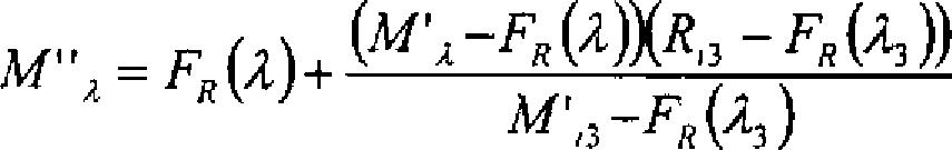
- 4. Die Messdaten werden bei jeder Wellenlänge um die Differenz der linearen Funktionen FH und FR additiv so korrigiert, dass sie bei den isosbestischen Wellenlängen λi1 und λi2 mit den Referenzdaten übereinstimmen: M'λ = Mλ + FR – FH.
2 zeigt wiederum die Referenzdaten, die lineare Referenzfunktion FR der Wellenlänge sowie die korrigierten Messdaten M'. Diese Korrektur kompensiert zusätzlich zur Absorption des Hämoglobins bestehende Extinktionen, deren Spektren imWellenlängenbereich 522 nm bis 586 nm als linear im logarithmischen Maßstab angenommen bzw. approximiert werden können. Im hier betrachteten Ausführungsbeispiel sind dies die Absorptionen des Melanin und der vorderen Augenmedien sowie die Streuung im Blut und im umgebenden Gewebe. - 5. Die korrigierten Messdaten M' werden so um die lineare Referenzfunktion FR gestreckt oder gestaucht, dass sie bei der isosbestischen Wellenlänge λi3 mit dem Referenzwert Ri3 übereinstimmen:
3 zeigt die in M'' resultierende Spreizung (ggf. auch Stauchung) der korrigierten Messdaten M' um die lineare Referenzfunktion FR, die so vorgenommen wird, dass korrigierte Messdaten und Referenzdaten bei der isosbestischen Wellenlänge λi3 (569 nm) übereinstimmen. Diese Korrektur kompensiert unterschiedliche Absolutwerte der Mess- und Referenzdaten, die durch unterschiedliche Beleuchtungs- und Meßbedingungen entstehen. - 6. Die Lage von M''a auf einer linear zwischen
R 0% / α und R 100% / α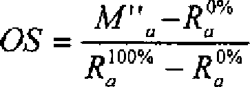
- 1. All measurement and reference data are logarithmized.
1 shows the measurement and reference data in logarithmic representation, in this example the reflection of a retinal vein (measurement data) and the transmission of a 0.1 mm thick layer of whole blood (reference data). For the sake of clarity, the complete spectra are shown between 400 nm and 700 nm. The wavelengths λ i1 = 522 nm, λ i2 = 586 nm, λ i3 = 569 nm and λ a = 555 nm used in this example are plotted. - 2. A linear auxiliary function F H of the wavelength is calculated in the measuring spectrum so that its values at the isosbestic wavelengths λ i1 and λ i1 coincide with the measured data M i1 and M i2 at these wavelengths.
- 3. A linear reference function F R of the wavelength is calculated in the reference spectra such that their values at the isosbestic wavelengths λ i1 and λ i2 coincide with the reference data R i1 and R i2 at these wavelengths.
- 4. The measured data are additive corrected at each wavelength by the difference of the linear functions F H and F R such that they match the reference data at the isosbestic wavelengths λ i1 and λ i2 : M ' λ = M λ + F R - F H.
2 again shows the reference data, the linear reference function F R of the wavelength and the corrected measurement data M '. This correction compensates in addition to the absorption of hemoglobin existing extinctions whose spectra in thewavelength range 522 nm to 586 nm can be assumed or approximated as linear on a logarithmic scale. In the exemplary embodiment considered here, these are the absorptions of the melanin and the anterior ocular media as well as the scattering in the blood and in the surrounding tissue. - 5. The corrected measured data M 'are stretched or compressed by the linear reference function F R such that they match the reference value R i3 at the isosbestic wavelength λ i3 :
3 shows the resulting in M '' spread (possibly also compression) of the corrected measurement data M 'to the linear reference function F R , which is made so that corrected measurement data and reference data at the isosbestischen wavelength λ i3 (569 nm) match. This correction compensates for different absolute values of the measurement and reference data resulting from different lighting and measuring conditions. - 6. The location of M '' a on a linear between
R 0% / α andR 100% / α
Die skalierte Ablesung der Sauerstoffsättigung (OS) zwischen den Werten 0 und 1 ist ebenfalls in
Aufstellung der verwendeten BezugszeichenList of used reference numbers
-
- MM
- – Messdaten- Measurement data
- M'M '
- – durch Addition der korrigierten Hilfsfunktion korrigierte Messdaten- corrected by adding the corrected auxiliary function measured data
- M''M ''
- – durch Beaufschlagung mit einem Faktor korrigierte Messdaten M'- measurement data M 'corrected by applying a factor
- FR F R
- – Referenzfunktion- Reference function
- OSOS
- – Sauerstoffsättigung- oxygen saturation
- λλ
- – Wellenlänge- Wavelength
Claims (3)
Priority Applications (4)
| Application Number | Priority Date | Filing Date | Title |
|---|---|---|---|
| DE10217543A DE10217543B4 (en) | 2002-04-17 | 2002-04-17 | Method for the spectrometric determination of the oxygen saturation of blood in the presence of optical disturbances |
| PCT/EP2003/004024 WO2003086193A1 (en) | 2002-04-17 | 2003-04-17 | Method for the spectroscopic determination of the oxygen saturation of blood in the presence of optical disturbance variables |
| US10/511,483 US7333842B2 (en) | 2002-04-17 | 2003-04-17 | Method for the spectroscopic determination of the oxygen saturation of blood in the presence of optical disturbance variables |
| US12/002,678 US20080194931A1 (en) | 2002-04-17 | 2007-12-18 | Method for the spectroscopic determination of the oxygen saturation of blood in the presence of optical disturbance varibles |
Applications Claiming Priority (1)
| Application Number | Priority Date | Filing Date | Title |
|---|---|---|---|
| DE10217543A DE10217543B4 (en) | 2002-04-17 | 2002-04-17 | Method for the spectrometric determination of the oxygen saturation of blood in the presence of optical disturbances |
Publications (2)
| Publication Number | Publication Date |
|---|---|
| DE10217543A1 DE10217543A1 (en) | 2003-11-06 |
| DE10217543B4 true DE10217543B4 (en) | 2012-12-06 |
Family
ID=28798595
Family Applications (1)
| Application Number | Title | Priority Date | Filing Date |
|---|---|---|---|
| DE10217543A Expired - Fee Related DE10217543B4 (en) | 2002-04-17 | 2002-04-17 | Method for the spectrometric determination of the oxygen saturation of blood in the presence of optical disturbances |
Country Status (3)
| Country | Link |
|---|---|
| US (2) | US7333842B2 (en) |
| DE (1) | DE10217543B4 (en) |
| WO (1) | WO2003086193A1 (en) |
Families Citing this family (10)
| Publication number | Priority date | Publication date | Assignee | Title |
|---|---|---|---|---|
| US7449339B2 (en) * | 1999-11-23 | 2008-11-11 | Nir Diagnostics Inc. | Spectroscopic method and apparatus for total hemoglobin measurement |
| EP1729644A2 (en) * | 2004-03-19 | 2006-12-13 | Board Of Supervisors Of Louisiana State University And Agricultural And Mechanical College | A method for evaluating relative oxygen saturation in body tissues |
| US20080221416A1 (en) * | 2007-03-09 | 2008-09-11 | Nellcor Puritan Bennett Llc | System and method for detection of macular degeneration using spectrophotometry |
| DE102007053074A1 (en) * | 2007-08-03 | 2009-05-14 | Carl Zeiss Meditec Ag | Method and measuring device for fluorescence measurement on the eye |
| US8437822B2 (en) | 2008-03-28 | 2013-05-07 | Covidien Lp | System and method for estimating blood analyte concentration |
| WO2017100685A1 (en) | 2015-12-10 | 2017-06-15 | Bioxytech Retina, Inc. | Methods and apparatus for measuring blood oxygenation of tissue |
| WO2017117668A1 (en) * | 2016-01-05 | 2017-07-13 | Vasile Diaconu | On line and real time optic nerve blood oxygenation mapping |
| JP6755831B2 (en) * | 2016-08-09 | 2020-09-16 | 花王株式会社 | How to evaluate skin condition |
| DE102017210548A1 (en) * | 2017-06-22 | 2018-12-27 | Zf Friedrichshafen Ag | Thickness-independent spectroscopic analysis of consumables |
| IL295373A (en) * | 2022-08-04 | 2024-03-01 | Spring Vision Ltd | Optical system and method for monitoring biological tissue condition |
Citations (9)
| Publication number | Priority date | Publication date | Assignee | Title |
|---|---|---|---|---|
| US4253744A (en) * | 1978-05-12 | 1981-03-03 | Minolta Camera Kabushiki Kaisha | Optical system for light measurement of an eye fund |
| US4305398A (en) * | 1977-12-30 | 1981-12-15 | Minolta Camera Kabushiki Kaisha | Eye fundus oximeter |
| US4485820A (en) * | 1982-05-10 | 1984-12-04 | The Johns Hopkins University | Method and apparatus for the continuous monitoring of hemoglobin saturation in the blood of premature infants |
| US5119814A (en) * | 1990-07-25 | 1992-06-09 | Minnich Thomas E | Method and apparatus for monitoring blood loss via retinal venous oxygen saturation |
| US5308919A (en) * | 1992-04-27 | 1994-05-03 | Minnich Thomas E | Method and apparatus for monitoring the arteriovenous oxygen difference from the ocular fundus |
| US5318022A (en) * | 1991-03-01 | 1994-06-07 | John Taboada | Method and apparatus for determining hemoglobin oxygenation such as in ocular and other vascular beds |
| DE4433827A1 (en) * | 1994-09-22 | 1996-03-28 | Univ Schiller Jena | Measuring substance parameters in material layer, esp. in vivo oxygen saturation in optically accessible blood containing structure |
| US5776060A (en) * | 1997-02-20 | 1998-07-07 | University Of Alabama In Huntsville | Method and apparatus for measuring blood oxygen saturation within a retinal vessel with light having several selected wavelengths |
| DE19920157A1 (en) * | 1999-04-29 | 2000-11-02 | Univ Schiller Jena | Determination of parameters of substances in media, especially oxygen saturation in living tissues, comprises using model function to take account of both back scattered and reflected light |
Family Cites Families (5)
| Publication number | Priority date | Publication date | Assignee | Title |
|---|---|---|---|---|
| DE3245939C2 (en) * | 1982-12-11 | 1985-12-19 | Fa. Carl Zeiss, 7920 Heidenheim | Device for generating an image of the fundus |
| US5935076A (en) | 1997-02-10 | 1999-08-10 | University Of Alabama In Huntsville | Method and apparatus for accurately measuring the transmittance of blood within a retinal vessel |
| EP1104254A2 (en) * | 1998-08-13 | 2001-06-06 | Whitland Research Limited | Optical device |
| US6501974B2 (en) * | 2001-01-22 | 2002-12-31 | Datex-Ohmeda, Inc. | Compensation of human variability in pulse oximetry |
| US6711425B1 (en) * | 2002-05-28 | 2004-03-23 | Ob Scientific, Inc. | Pulse oximeter with calibration stabilization |
-
2002
- 2002-04-17 DE DE10217543A patent/DE10217543B4/en not_active Expired - Fee Related
-
2003
- 2003-04-17 WO PCT/EP2003/004024 patent/WO2003086193A1/en not_active Application Discontinuation
- 2003-04-17 US US10/511,483 patent/US7333842B2/en not_active Expired - Fee Related
-
2007
- 2007-12-18 US US12/002,678 patent/US20080194931A1/en not_active Abandoned
Patent Citations (9)
| Publication number | Priority date | Publication date | Assignee | Title |
|---|---|---|---|---|
| US4305398A (en) * | 1977-12-30 | 1981-12-15 | Minolta Camera Kabushiki Kaisha | Eye fundus oximeter |
| US4253744A (en) * | 1978-05-12 | 1981-03-03 | Minolta Camera Kabushiki Kaisha | Optical system for light measurement of an eye fund |
| US4485820A (en) * | 1982-05-10 | 1984-12-04 | The Johns Hopkins University | Method and apparatus for the continuous monitoring of hemoglobin saturation in the blood of premature infants |
| US5119814A (en) * | 1990-07-25 | 1992-06-09 | Minnich Thomas E | Method and apparatus for monitoring blood loss via retinal venous oxygen saturation |
| US5318022A (en) * | 1991-03-01 | 1994-06-07 | John Taboada | Method and apparatus for determining hemoglobin oxygenation such as in ocular and other vascular beds |
| US5308919A (en) * | 1992-04-27 | 1994-05-03 | Minnich Thomas E | Method and apparatus for monitoring the arteriovenous oxygen difference from the ocular fundus |
| DE4433827A1 (en) * | 1994-09-22 | 1996-03-28 | Univ Schiller Jena | Measuring substance parameters in material layer, esp. in vivo oxygen saturation in optically accessible blood containing structure |
| US5776060A (en) * | 1997-02-20 | 1998-07-07 | University Of Alabama In Huntsville | Method and apparatus for measuring blood oxygen saturation within a retinal vessel with light having several selected wavelengths |
| DE19920157A1 (en) * | 1999-04-29 | 2000-11-02 | Univ Schiller Jena | Determination of parameters of substances in media, especially oxygen saturation in living tissues, comprises using model function to take account of both back scattered and reflected light |
Non-Patent Citations (6)
| Title |
|---|
| DELORI, F.C.: Noninvasive technique for oximetry of blood in retinal vessels, In: Appl. Opt. 27, 1988, S. 113-1125 * |
| HAMMER, M. et al.: Light paths in retinal vessel oximetry, In: IEEE Trans. Biomed. Eng. 48(5), 2001, 592-598 * |
| PITTMANN, R.N., DULING, B.R.: A new method for the measurement of percent oxyhemoglobin, J. Appl. Physiol. 38, 1975, S. 315-320 * |
| SCHWEITZER, D. et al.: In vivo measurement of the oxygen Saturation at the normal eye, In: Appl. Opt. 27, 1988, S. 113-1125 * |
| SMITH at al.: Effect of multiple light paths in retinal vessel oximetry, In: Appl. Opt. 39, 2000, S. 1183-1193 * |
| TWERSKY, V.: Absorption and multiple scattering by biological suspensions, In: J. Opt. Society of America 60, 1970, S. 1084-1093 * |
Also Published As
| Publication number | Publication date |
|---|---|
| WO2003086193A1 (en) | 2003-10-23 |
| DE10217543A1 (en) | 2003-11-06 |
| US20060063994A1 (en) | 2006-03-23 |
| US7333842B2 (en) | 2008-02-19 |
| US20080194931A1 (en) | 2008-08-14 |
Similar Documents
| Publication | Publication Date | Title |
|---|---|---|
| DE69727776T2 (en) | METHOD FOR DETERMINING THE FRACTIONAL OXYGEN SATURATION | |
| DE69333456T2 (en) | SYSTEM METHOD FOR NON-INVASIVE MONITORING OF HEMATOCRIT VALUE | |
| DE102004016435B4 (en) | Method for the spectrophotometric determination of the oxygen saturation of the blood in optically accessible blood vessels | |
| DE3008651C2 (en) | Device for measuring the pneusis function | |
| DE69724351T2 (en) | System for measuring tissue chromophores | |
| DE112012004064B4 (en) | diagnostic system | |
| DE112010004184B4 (en) | Auxiliary diagnostic device and auxiliary diagnostic method | |
| DE10217543B4 (en) | Method for the spectrometric determination of the oxygen saturation of blood in the presence of optical disturbances | |
| DE102007022666A1 (en) | An optical illumination system having a solid state lighting element that produces white light, and optical device equipped therewith | |
| DE602004001794T2 (en) | Method and device for in vitro or in vivo measurement of the concentration of a substance | |
| DE102006021769A1 (en) | Optical coherence tomograph | |
| WO2005094668A1 (en) | Method for measuring the vessel diameter of optically accessible blood vessels | |
| EP0505918B1 (en) | Apparatus and method for determining heart minute volumes | |
| DE3542167A1 (en) | METHOD FOR MEASURING THE EYE LENS TURBIDITY AND ARRANGEMENT FOR IMPLEMENTING THE METHOD | |
| DE112018000393T5 (en) | Endoscope system and image display device | |
| DE4242083C2 (en) | Sensor device for reproducible, non-invasive measurement of blood glucose | |
| DE602004006396T2 (en) | ANALYSIS OF A COMPOSITION WITH OBSERVATION | |
| DE10129652B4 (en) | Arrangement and method for determining the two-dimensional distribution of fundus pigments, in particular the macular pigment xanthophyll | |
| DE102019113283B4 (en) | Device for imaging skin lesions | |
| DE10321338A1 (en) | Method and device for determining blood components using the method of ratiometric absolute pulse spectroscopy | |
| DE102017215158B4 (en) | Detection system | |
| DE112012004879B4 (en) | Method and device for measuring hemoglobin | |
| DE102017109856A1 (en) | Method for determining the age of the lens | |
| DE690493C (en) | Colorimeter | |
| EP2382916A1 (en) | Method and device for determining the fat content of the human body |
Legal Events
| Date | Code | Title | Description |
|---|---|---|---|
| 8127 | New person/name/address of the applicant |
Owner name: CARL ZEISS MEDITEC AG, 07745 JENA, DE |
|
| 8110 | Request for examination paragraph 44 | ||
| R018 | Grant decision by examination section/examining division | ||
| R020 | Patent grant now final |
Effective date: 20130307 |
|
| R119 | Application deemed withdrawn, or ip right lapsed, due to non-payment of renewal fee |